JP2008538280A - Method for producing lipid bilayer membrane - Google Patents
Method for producing lipid bilayer membrane Download PDFInfo
- Publication number
- JP2008538280A JP2008538280A JP2008504448A JP2008504448A JP2008538280A JP 2008538280 A JP2008538280 A JP 2008538280A JP 2008504448 A JP2008504448 A JP 2008504448A JP 2008504448 A JP2008504448 A JP 2008504448A JP 2008538280 A JP2008538280 A JP 2008538280A
- Authority
- JP
- Japan
- Prior art keywords
- lipid
- planar
- lipid bilayer
- vesicles
- peptide
- Prior art date
- Legal status (The legal status is an assumption and is not a legal conclusion. Google has not performed a legal analysis and makes no representation as to the accuracy of the status listed.)
- Pending
Links
Images


Classifications
-
- G—PHYSICS
- G01—MEASURING; TESTING
- G01N—INVESTIGATING OR ANALYSING MATERIALS BY DETERMINING THEIR CHEMICAL OR PHYSICAL PROPERTIES
- G01N33/00—Investigating or analysing materials by specific methods not covered by groups G01N1/00 - G01N31/00
- G01N33/48—Biological material, e.g. blood, urine; Haemocytometers
- G01N33/50—Chemical analysis of biological material, e.g. blood, urine; Testing involving biospecific ligand binding methods; Immunological testing
- G01N33/53—Immunoassay; Biospecific binding assay; Materials therefor
- G01N33/531—Production of immunochemical test materials
- G01N33/532—Production of labelled immunochemicals
- G01N33/533—Production of labelled immunochemicals with fluorescent label
-
- B—PERFORMING OPERATIONS; TRANSPORTING
- B82—NANOTECHNOLOGY
- B82Y—SPECIFIC USES OR APPLICATIONS OF NANOSTRUCTURES; MEASUREMENT OR ANALYSIS OF NANOSTRUCTURES; MANUFACTURE OR TREATMENT OF NANOSTRUCTURES
- B82Y5/00—Nanobiotechnology or nanomedicine, e.g. protein engineering or drug delivery
-
- G—PHYSICS
- G01—MEASURING; TESTING
- G01N—INVESTIGATING OR ANALYSING MATERIALS BY DETERMINING THEIR CHEMICAL OR PHYSICAL PROPERTIES
- G01N33/00—Investigating or analysing materials by specific methods not covered by groups G01N1/00 - G01N31/00
- G01N33/48—Biological material, e.g. blood, urine; Haemocytometers
- G01N33/50—Chemical analysis of biological material, e.g. blood, urine; Testing involving biospecific ligand binding methods; Immunological testing
- G01N33/53—Immunoassay; Biospecific binding assay; Materials therefor
- G01N33/543—Immunoassay; Biospecific binding assay; Materials therefor with an insoluble carrier for immobilising immunochemicals
- G01N33/554—Immunoassay; Biospecific binding assay; Materials therefor with an insoluble carrier for immobilising immunochemicals the carrier being a biological cell or cell fragment, e.g. bacteria, yeast cells
-
- Y—GENERAL TAGGING OF NEW TECHNOLOGICAL DEVELOPMENTS; GENERAL TAGGING OF CROSS-SECTIONAL TECHNOLOGIES SPANNING OVER SEVERAL SECTIONS OF THE IPC; TECHNICAL SUBJECTS COVERED BY FORMER USPC CROSS-REFERENCE ART COLLECTIONS [XRACs] AND DIGESTS
- Y10—TECHNICAL SUBJECTS COVERED BY FORMER USPC
- Y10T—TECHNICAL SUBJECTS COVERED BY FORMER US CLASSIFICATION
- Y10T428/00—Stock material or miscellaneous articles
- Y10T428/31504—Composite [nonstructural laminate]
- Y10T428/31678—Of metal
Abstract
【課題】 基板上に支持ニ分子膜を形成するための方法を提供する。
【解決手段】 本発明は、固体担体上に平面状脂質ニ分子膜を製造する方法である。最初に脂質小胞溶液が固体担体上に堆積させ、次に両親媒性ペプチド溶液を脂質小胞溶液に加えることによって、脂質小胞を不安定化させ、固体担体上に平面状脂質ニ分子膜を製造する。また、本発明は、平面状脂質ニ分子膜が天然脂質により形成され、固体担体が未修飾金または酸化チタンにより作成された平面状支持脂質二分子膜を提供する。好ましくは、平面状支持二分子膜は連続している。平面状脂質二分子膜は、天然脂質、または限定するわけではないが、ホスファチジルコリン、ホスファチジルエタノールアミン、ホスファチジルセリン、ホスファチジルイノシトール、カルジオリピン、コレステロール、およびスフィンゴミエリンのような脂質の混合物であってもよい。
【選択図】図1PROBLEM TO BE SOLVED: To provide a method for forming a supporting bimolecular film on a substrate.
The present invention is a method for producing a planar lipid bilayer membrane on a solid support. The lipid vesicle solution is first deposited on the solid support, then the amphipathic peptide solution is added to the lipid vesicle solution to destabilize the lipid vesicles, and the planar lipid bilayer membrane on the solid support Manufacturing. The present invention also provides a planar supported lipid bilayer membrane in which a planar lipid bilayer membrane is formed of natural lipids and a solid carrier is made of unmodified gold or titanium oxide. Preferably, the planar support bilayer is continuous. The planar lipid bilayer may be a natural lipid or a mixture of lipids such as but not limited to phosphatidylcholine, phosphatidylethanolamine, phosphatidylserine, phosphatidylinositol, cardiolipin, cholesterol, and sphingomyelin.
[Selection] Figure 1
Description
本発明は、脂質膜に関し、より詳細には、固体担体上に脂質ニ分子膜を製造する方法に関する。 The present invention relates to lipid membranes, and more particularly to a method for producing a lipid bilayer membrane on a solid support.
シリコン酸化物または有機物層が修飾された表面上で、微小な単一層の小胞を融合させることによって形成された支持脂質二分子膜は、半導体、金被膜表面、光電子装置およびラボオンチップ(lab-on-a-chip)装置のような無機固体を生体機能化することができる。これらの脂質二分子膜は、タンパク質が結合された膜や、細胞過程、タンパク質−脂質相互作用、および生体情報の伝達を媒介する膜の特性や挙動に対する研究から、高い価値を有することが証明されている。生体膜は複雑であるため、研究対象となる1または数個の膜要素を分離したモデル膜システムを作り上げることが必要である。また、固体表面上にモデル膜を支持することによって、自然な状態の生体システムを効果的に研究するために、様々な表面分析技術を使用することができる。固体表面上に支持された膜は、生体センサ、プログラムドラッグデリバリ(programmed drug delivery)、医療用インプラントの適合性の促進や改善、および界面触媒の製造を含む応用を潜在的に有している。 A supported lipid bilayer formed by fusing a small monolayer of vesicles on a surface modified with a silicon oxide or organic layer is a semiconductor, gold-coated surface, optoelectronic device and lab-on-chip (lab -on-a-chip) Inorganic solids such as devices can be biofunctionalized. These lipid bilayers have proven to be of great value from studies on the properties and behavior of protein-bound membranes, cellular processes, protein-lipid interactions, and membranes that mediate the transmission of biological information. ing. Since biological membranes are complex, it is necessary to create a model membrane system in which one or several membrane elements to be studied are separated. Also, by supporting the model membrane on the solid surface, various surface analysis techniques can be used to effectively study the natural biological system. Membranes supported on solid surfaces have potential applications including biosensors, programmed drug delivery, promoting and improving the compatibility of medical implants, and the production of interfacial catalysts.
天然の生体システムを擬似するために、研究者は、ガラス、マイカ、自己集積化単分子膜、および石英のような基板に支持ニ分子膜を形成する小胞融合法を用いてきた。しかし、この方法は、金やTiO2のような好ましい固体に平面状の脂質二分子膜を形成する場合に、問題があることがわかっている。例えば、科学者は、特殊な合成を必要とする自己組織化単文子膜(SAMs)を使用して金表面を修飾しようとしてきたが、形成されたSAMsの構造は明確でないかもしれない。従って、好ましい基板上に支持ニ分子膜を形成するための新しい方法を作り出すことが必要とされている。 To simulate natural biological systems, researchers have used vesicle fusion methods that form a supported bilayer on a substrate such as glass, mica, self-assembled monolayer, and quartz. However, this method has been found to be problematic when forming a planar lipid bilayer on a preferred solid such as gold or TiO 2 . For example, scientists have attempted to modify gold surfaces using self-assembled monofilament membranes (SAMs) that require special synthesis, but the structure of the formed SAMs may not be clear. Accordingly, there is a need to create a new method for forming a supported bilayer on a preferred substrate.
本発明は、固体担体上に平面状脂質ニ分子膜を製造する方法である。この製造方法では、最初に、脂質小胞溶液が固体担体上に堆積させられる。次に、両親媒性ペプチド溶液を脂質小胞溶液に加えることによって、脂質小胞が不安定化される。この不安定化により、固体担体上に平面状の脂質ニ分子膜が製造される。好ましくは、両親媒性ペプチドは、α−ヘリックスペプチドである。より好ましくは、α−ヘリックスペプチドは、配列SEQ ID NO: 1の一部または全てを有するポリペプチドである。 The present invention is a method for producing a planar lipid bilayer on a solid support. In this production method, a lipid vesicle solution is first deposited on a solid support. The amphiphilic peptide solution is then destabilized by adding the amphiphilic peptide solution to the lipid vesicle solution. This destabilization produces a planar lipid bilayer on the solid support. Preferably, the amphiphilic peptide is an α-helix peptide. More preferably, the α-helix peptide is a polypeptide having part or all of the sequence SEQ ID NO: 1.
また、本発明は、平面状脂質ニ分子膜が天然脂質により作られ、固体担体が未修飾金または酸化チタンにより作成された平面状支持脂質二分子膜を提供する。好ましくは、平面状支持二分子膜は連続している。平面状脂質二分子膜は、天然脂質、または限定するわけではないが、ホスファチジルコリン、ホスファチジルエタノールアミン、ホスファチジルセリン、ホスファチジルイノシトール、カルジオリピン、コレステロール、およびスフィンゴミエリンのような脂質の混合物であってもよい。 The present invention also provides a planar supported lipid bilayer membrane in which a planar lipid bilayer membrane is made of natural lipid and a solid carrier is made of unmodified gold or titanium oxide. Preferably, the planar support bilayer is continuous. The planar lipid bilayer may be a natural lipid or a mixture of lipids such as but not limited to phosphatidylcholine, phosphatidylethanolamine, phosphatidylserine, phosphatidylinositol, cardiolipin, cholesterol, and sphingomyelin.
図1は、本発明に係る方法の概略図を示している。図1Aは固体基板120の上に堆積させられた脂質小胞110を示している。小胞は、固体基板120に吸着される。円130で示される大量の水が、固体基板120の表面に吸着されている小胞間と同様に、原型(intact)の小胞の内部に取り込まれている。両親媒性ペプチド(AP)溶液の付加の後に(図1B参照)、小胞は不安的化されて破壊され、破壊された小胞は溶解し、平面状の二分子膜を形成する。平面状二分子膜112が形成されるとき、小胞110内に取り込まれていた水は分散し、水層132を形成する。
FIG. 1 shows a schematic diagram of the method according to the invention. FIG. 1A shows a
両新媒性ペプチドは、好ましくはα−ヘリックスペプチドである。より好ましくは、両親媒性ペプチドは、HCV非構造性タンパク質NS5AのAHペプチドである。このタンパク質は、HCV分離株に保持されており、配列SEQ ID NO:1を有する。ペプチドの全体、ペプチド(AH_S1)の1〜16残基のアミノ酸、またはペプチド(AH_S2)の17−31残基のアミノ酸のいずれかが、脂質小胞を不安定化するために使用されてもよい。或いは、両親媒性α−ヘリックス構造が保たれている限り、ペプチドは完全な配列の配列SEQ ID NO: 1を有していなくてもよい。例えば、円偏光二色性によって示したとき、ペプチドはα−ヘリックス構造を維持しつつ、配列SEQ ID NO: 1と少なくとも約80%以上一致する配列を有しているかもしれない。ペプチド溶液の両親媒性ペプチドの濃度は、0.05μg/ml〜0.5μg/mlであることが好ましい。両親媒性ペプチドは、生理的緩衝液(Tris緩衝液、PBS緩衝液、およびHEPES緩衝液)やジメチルスルホキシド(DMSO)を含む様々な溶媒に含むことができる。 Both amphipathic peptides are preferably α-helix peptides. More preferably, the amphipathic peptide is the AH peptide of HCV nonstructural protein NS5A. This protein is retained in the HCV isolate and has the sequence SEQ ID NO: 1. Either the whole peptide, amino acids 1-16 of peptide (AH_S1), or amino acids 17-31 of peptide (AH_S2) may be used to destabilize lipid vesicles. . Alternatively, the peptide may not have the complete sequence SEQ ID NO: 1 as long as the amphiphilic α-helix structure is maintained. For example, as indicated by circular dichroism, the peptide may have a sequence that matches at least about 80% or more with the sequence SEQ ID NO: 1, while maintaining an α-helix structure. The concentration of the amphiphilic peptide in the peptide solution is preferably 0.05 μg / ml to 0.5 μg / ml. Amphiphilic peptides can be included in a variety of solvents including physiological buffers (Tris buffer, PBS buffer, and HEPES buffer) and dimethyl sulfoxide (DMSO).
本発明に適した脂質小胞は、好ましくは、直径が約25nm〜約80nmである。小胞は、限定するわけではないが、抽出法を含む公知技術を用いて調整することができる。小胞は、約100mM〜約250mMのNaCl濃度を有するTris、PBS、およびHEPES緩衝液といった生理的緩衝液中で、0.05mg/ml〜5mg/mlの濃度であることが好ましい。脂質または脂質の混合物のが、限定する趣旨ではなく、リン脂質を含む脂質小胞を形成するために使用され得る。脂質は、ホスファチジルコリン、ホスファチジルエタノールアミン、ホスファチジルセリン、ホスファチジルイノシトール、カルジオリピン、コレステロール、およびスフィンゴミエリンであることが好ましい。 Lipid vesicles suitable for the present invention preferably have a diameter of about 25 nm to about 80 nm. Vesicles can be prepared using known techniques including, but not limited to, extraction methods. Vesicles are preferably at a concentration of 0.05 mg / ml to 5 mg / ml in physiological buffers such as Tris, PBS, and HEPES buffer having a NaCl concentration of about 100 mM to about 250 mM. A lipid or mixture of lipids can be used to form lipid vesicles containing phospholipids, without limitation. The lipid is preferably phosphatidylcholine, phosphatidylethanolamine, phosphatidylserine, phosphatidylinositol, cardiolipin, cholesterol, and sphingomyelin.
多くの固体担体が、本発明に使用され得る。例えば、限定する趣旨ではなく、材料にはシリコンを含む材料、金、白金、および酸化チタンが含まれる。 Many solid carriers can be used in the present invention. For example, without limitation, materials include silicon-containing materials, gold, platinum, and titanium oxide.
また、本発明は、本発明の方法を用いて製造された平面状支持脂質二分子膜を提供する。好ましくは、脂質二分子膜は天然脂質から形成され、固体担体は未修飾金または酸化チタンから作成されている。リン脂質のような、いずれの天然脂質も二分子膜に用いることができる。好ましい脂質は、ホスファチジルコリン、ホスファチジルエタノールアミン、ホスファチジルセリン、ホスファチジルイノシトール、カルジオリピン、コレステロール、およびスフィンゴミエリンである。好ましくは、本発明に係る平面状脂質二分子膜は、連続的であり、例えば層内に隙間はない。 The present invention also provides a planar supported lipid bilayer membrane produced using the method of the present invention. Preferably, the lipid bilayer is formed from natural lipids and the solid support is made from unmodified gold or titanium oxide. Any natural lipid, such as phospholipids, can be used in the bilayer membrane. Preferred lipids are phosphatidylcholine, phosphatidylethanolamine, phosphatidylserine, phosphatidylinositol, cardiolipin, cholesterol, and sphingomyelin. Preferably, the planar lipid bilayer membrane according to the present invention is continuous, for example, there is no gap in the layer.
<実施例>
<金基板上での平面状脂質二分子膜の形成>
原型の小胞からの二分子膜の形成は、水晶振動子マイクロバランス装置(QCM−D)によって明らかにすることができる。QCM−Dの結果を解釈するために、ソルベリー(Sauerbrey)の式から算出されるΔfと吸着質量(Δm)との間の直線関係が用いられる。
<Example>
<Formation of planar lipid bilayer on gold substrate>
Formation of the bilayer membrane from the original vesicle can be revealed by a quartz crystal microbalance apparatus (QCM-D). In order to interpret the QCM-D results, a linear relationship between Δf and adsorption mass (Δm) calculated from the Sauerbrey equation is used.

ここで、Cは質量感度定数であり、5MHzのQCM−Dでは17.7ngcm−2Hz−1である。nは、倍音数である(基本的にはn=1であり、倍音では3,5,7である)。QCM−Dは、小胞融合過程の様々な研究に用いられており、固い脂質二分子膜および単分子膜と、柔軟で変形可能な小胞とを識別するために減衰(dissipation)が用いられている(例えば、非特許文献1参照)。
AHペプチドの小胞破壊能力を測定するために、30nmのポリカーボネート照射エッチング膜(PEC)を通過させた1−パルミトイル−2−オレオイル−sn−グリセロ−3−ホスホコリン(POPC)の単層小胞を金表面上でAHペプチドが存在しない状態で試験し、その後ペプチドを加えて二分子膜を形成させた。小胞が吸着しているとき、表面上に吸着した小胞間と同様に、原型の小胞内に取り込まれた大量の水が存在している。この取り込まれた水は、二分子膜の上面に存在する水と異なり、エネルギーを大きく減衰させる。このエネルギーの減衰における変化により、原型の小胞から二分子膜への変化を追跡することができる。 In order to determine the ability of AH peptides to destroy vesicles, unilamellar vesicles of 1-palmitoyl-2-oleoyl-sn-glycero-3-phosphocholine (POPC) passed through a 30 nm polycarbonate irradiated etching membrane (PEC) Were tested on the gold surface in the absence of AH peptide, and then the peptide was added to form a bilayer. When vesicles are adsorbed, there is a large amount of water taken up into the original vesicles, as well as between the vesicles adsorbed on the surface. Unlike the water existing on the upper surface of the bilayer membrane, the taken-in water greatly attenuates energy. This change in energy decay allows the change from the original vesicle to a bilayer to be tracked.
図2では、Δf(t)(図中の三角形)とΔD(t)(図中の円形)とが、周波数変化と減衰(dissipation)変化を表している。図2Aは、酸化金上への小胞の吸着を示している。周波数信号が安定した10分後(矢印1)に、POPC小胞溶液(0.1mg/ml、φ30nm PEC=59nm±0.2nm)が溶液セルに注入されている。50および55分後(矢印2および3)に、小胞を希釈するために使用したものと同じ緩衝液(10mM Tris(pH7.5)、150mM NaCl、1mM EDTAを含む18.2MΩ−cmミリQ水(MilliQ)(MilliPore、オレゴン州、アメリカ合衆国)の溶液)で基板を2度洗浄し、これにより金表面上に存在する原型の小胞の安定性が確認された。図2Bに示すように、60分後(矢印4)には、両親媒性のヘリックスペプチド(AHペプチド)溶液(0.05μg/ml)が金表面上の原型の小胞(φ30nm PEC=59nm±0.2nm)に加えられている。ペプチドは、小胞を不安定化し、破壊して完全な二分子膜を形成する。これは、25.5Hz±0.5の周波数の減少として現れ、最大で約0.08×10⁻⁶の減衰の減少が確認された。120および140分後(矢印5および6)には、小胞緩衝液で基板を2度洗浄し、これにより金表面上の二分子膜の安定性が確認された。二分子膜の膜厚を算出することができるソルベリーの式によれば、QCM−Dのパラメータは、小胞から薄く固い二分子膜への変化を示している。
In FIG. 2, Δf (t) (triangle in the figure) and ΔD (t) (circular in the figure) represent a change in frequency and a change in dissipation. FIG. 2A shows vesicle adsorption on gold oxide. Ten minutes after the frequency signal is stabilized (arrow 1), a POPC vesicle solution (0.1 mg / ml, φ30 nm PEC = 59 nm ± 0.2 nm) is injected into the solution cell. After 50 and 55 minutes (
図2Cでは、非両親媒性の非ヘリックスペプチド(NHペプチド)の効果を試験している。NHペプチドは、AHペプチドとは異なり、予測されるN末端ヘリックスに沿って、間隔をおいて配置されている3つの荷電アミノ酸を有しており、連続した疎水性パッチは残されていない。NHペプチドは、配列SEQ ID NO: 1の残基8にバリン(Val)の代わりにアスパラギン酸(Asp)、残基12にイソロイシン(Ile)の代わりにグルタミン酸(Glu)、残基19にフェニルアラニン(Phe)の代わりにアスパラギン酸(Asp)を有している。図2Cでは、周波数信号が安定した10分後(矢印1)に、POPC小胞溶液(0.1mg/ml、φ30nm PEC=59nm±0.2nm)が溶液セルに加えられている。60および70分後(矢印2および3)に、小胞緩衝液により基板を2度洗浄し、これにより金表面上に存在する原型の小胞の安定性が確認された。85分後(矢印4)には、NHペプチド溶液(0.05μg/ml)が金表面上の原型の小胞に加えられている。NHペプチドは、小胞を不安定化および破壊した証拠を示さなかった。160分後(矢印5)には、小胞緩衝液により基板を2度洗浄し、これにより金表面上に存在する原型の小胞の安定性が確認された。
In FIG. 2C, the effect of a non-amphiphilic non-helix peptide (NH peptide) is tested. The NH peptide, unlike the AH peptide, has three charged amino acids spaced along the predicted N-terminal helix, leaving no contiguous hydrophobic patches. The NH peptide comprises aspartic acid (Asp) instead of valine (Val) at
<TiO2基板上での平面状脂質二分子膜の形成>
AHペプチドのTiO2表面上の小胞破壊能力を測定するために、30nmのPEC膜を通過させたPOPCの単層小胞を金表面上でAHペプチドが存在しない状態で試験し、その後ペプチドを加えて二分子膜を形成させた(図3)。図3では、Δf(t)(図中の三角形)とΔD(t)(図中の円形)とが、周波数変化と減衰変化を表している。図3Aは、TiO2上への小胞の吸着を示している。周波数信号が安定した10分後(矢印1)に、POPC小胞溶液(0.1mg/ml、φ30nm PEC=59nm±0.2nm)が溶液セルに注入されている。60および65分後(矢印2および3)に、小胞を希釈するために使用したものと同じ緩衝液(10mM Tris(pH7.5)、150mM NaCl、1mM EDTAを含む18.2MΩ−cmミリQ水(MiliQ)(MilliPore、オレゴン州、アメリカ合衆国)の溶液)で基板を2度洗浄し、これによりTiO2表面上に存在する原型の小胞の安定性が確認された。図3Bに示すように、70分後(矢印4)には、AHペプチド溶液(0.05μg/ml)がTiO2表面上の原型の小胞(φ30nm PEC=59nm±0.2nm)に加えられている(図3B)。ペプチドは、小胞を不安定化し、破壊して完全な二分子膜を形成する。270分後(矢印5)には、小胞緩衝液で基板を2度洗浄し、これにより金表面上の二分子膜の安定性が確認された。
<Formation of planar lipid bilayer on TiO 2 substrate>
To measure the vesicle breakdown capability on TiO 2 surface of the AH peptide, a 30nm unilamellar vesicles POPC the PEC film was passed through the tested in the absence of AH peptides on the gold surface, followed peptide In addition, a bimolecular film was formed (FIG. 3). In FIG. 3, Δf (t) (triangle in the figure) and ΔD (t) (circular in the figure) represent frequency change and attenuation change. FIG. 3A shows the adsorption of vesicles on TiO 2 . Ten minutes after the frequency signal is stabilized (arrow 1), a POPC vesicle solution (0.1 mg / ml, φ30 nm PEC = 59 nm ± 0.2 nm) is injected into the solution cell. After 60 and 65 minutes (
図3Cでは、NHペプチドの効果を試験している。図3Cでは、周波数信号が安定した10分後(矢印1)に、POPC小胞溶液(0.1mg/ml、φ30nm PEC=59nm±0.2nm)が溶液セルに加えられている。40および50分後(矢印2および3)に、小胞緩衝液により基板を2度洗浄し、これによりTiO2表面上に存在する原型の小胞の安定性が確認された。60分後(矢印4)には、NHペプチド溶液(0.05μg/ml)がTiO2表面上の原型の小胞に加えられている。NHペプチドは、小胞を不安定化し、破壊した証拠を示さなかった。270分後(矢印5)には、小胞緩衝液により基板を2度洗浄し、これによりTiO2表面上に存在する原型の小胞の安定性が確認された。
In FIG. 3C, the effect of the NH peptide is tested. In FIG. 3C, a POPC vesicle solution (0.1 mg / ml, φ30 nm PEC = 59 nm ± 0.2 nm) is added to the
<本発明に係る脂質二分子膜の形成のAFM分析>
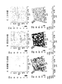
不安定化剤であるAHペプチドによる小胞の破壊と二分子膜の形成とを、直接的に表示し、確認するために、AFMを利用した。図4に分析の結果を示す。それぞれの列で、上段の像は二次元の平面図であり、中段の像は三次元の上方視点の図であり、グラフは上段の像に示された黒線に沿って行われた測定結果を示している。像は、高さモードにより与えられている。走査は図4A(上段図)の白色矢印によって示される方向に行われた。
<AFM analysis of formation of lipid bilayer according to the present invention>
AFM was used to directly display and confirm the destruction of vesicles and the formation of bilayers by the destabilizing agent AH peptide. FIG. 4 shows the result of the analysis. In each row, the upper image is a two-dimensional plan view, the middle image is a three-dimensional top view, and the graph is a measurement result taken along the black line shown in the upper image. Is shown. The image is given by the height mode. The scan was performed in the direction indicated by the white arrow in FIG. 4A (upper diagram).
図4Aでは、AFM測定は、Tris緩衝液(150mM NaCl、10mM Tris(pH7.5)、1mM EDTA)中の未修飾(裸)のTiO2基板表面に対して行われた。未修飾のTiO2表面の二乗平均平方根粗さ(Rq)の平均は、1.63±0.12nm(±S.E.、n=15)である。直径が59nm±0.2nmであるPOPC小胞(0.1mg/ml)は、注入システムを通して注意深く加えられ、30分間インキュベートされた後に、Tris緩衝液で3回、完全に洗い流された。図4Bに示すように、小胞410のような原型の小胞は、AFMで明瞭に確認され、平均Rqは2.70±0.15nm(±S.E.、n=15)に増加した。小胞を識別し、計数するために、粒度分析を行った。小胞以外の粒子の影響を最小化するため、直径50〜100nmの双曲線形状の物体のみ計数した。図4Bに示すAFMの図と、粒度分析により、13個の小胞が識別された。小胞の平均直径は74.57±4.07nm(±S.E.、n=13)であり、平均体積は3.32×10−5μm3±6.16×10−6μm3(±S.E.、n=13)であった。断面分析は、小胞の高さが約15nmであることを表している。
In FIG. 4A, AFM measurements were performed on the unmodified (naked) TiO 2 substrate surface in Tris buffer (150 mM NaCl, 10 mM Tris (pH 7.5), 1 mM EDTA). The average of the root mean square roughness (Rq) of the unmodified TiO 2 surface is 1.63 ± 0.12 nm (± SE, n = 15). POPC vesicles (0.1 mg / ml) with a diameter of 59 nm ± 0.2 nm were carefully added through the injection system and incubated for 30 minutes before being thoroughly washed out with Tris buffer three times. As shown in FIG. 4B, prototypic vesicles such as
図4Cに示すAFMの像は、ペプチド(0.05μg/ml)を注入し、像を走査する前に溶液を2時間インキュベートとした後の像であって、不安定化剤としてのAHペプチドの小胞に対する効果を示している。濃度0.05μg/mlのAHペプチドで120分間処理することによって小胞が破壊されたことを示すQCM−D測定の結果を、これらの像は明確に裏付けている。平均Rqが1.67±0.12nm(±S.E.、n=15)であることは、粗さが未修飾TiO2表面と同等になったことを示している。粒度分析は、小胞状の構造を識別することができず、AHペプチドが小胞を破壊し二分子膜を形成したことを示している(P≦0.001)。この結果は、QCM−Dの動力学データと一致する。 The image of AFM shown in FIG. 4C is the image after injecting peptide (0.05 μg / ml) and incubating the solution for 2 hours before scanning the image, with the AH peptide as a destabilizing agent. It shows the effect on vesicles. These images clearly support the results of QCM-D measurements that show that vesicles were destroyed by treatment with AH peptide at a concentration of 0.05 μg / ml for 120 minutes. An average Rq of 1.67 ± 0.12 nm (± SE, n = 15) indicates that the roughness is equivalent to the unmodified TiO 2 surface. Particle size analysis failed to discriminate vesicular structures, indicating that the AH peptide destroyed vesicles and formed a bilayer (P ≦ 0.001). This result is consistent with the QCM-D kinetic data.
当業者であれば理解できるように、様々な修正、置換、および変更を、本発明の趣旨から逸脱することなく実施することができる。本発明の範囲は、特許請求の範囲およびその均等の範囲によって決定されるべきである。 As will be appreciated by those skilled in the art, various modifications, substitutions and changes may be made without departing from the spirit of the invention. The scope of the present invention should be determined by the appended claims and their equivalents.
Claims (15)
a)前記固体担体上に脂質小胞溶液を堆積させ、
b)両親媒性ペプチド溶液を前記脂質小胞溶液に加えることによって、前記脂質小胞を不安定化し、
前記不安定化によって、前記固体担体上に前記平面状脂質二分子膜が製造されることを特徴とする平面状脂質二分子膜を製造する方法。 A method for producing a planar lipid bilayer on a solid support, comprising:
a) depositing a lipid vesicle solution on the solid support;
b) destabilizing the lipid vesicles by adding an amphiphilic peptide solution to the lipid vesicle solution;
A method for producing a planar lipid bilayer, wherein the planar lipid bilayer is produced on the solid support by the destabilization.
a)天然脂質を含む平面状脂質二分子膜と、
b)未修飾金または酸化チタンを含む固体担体と
を有し、
前記脂質二分子膜は、前記固体担体に支持されていることを特徴とする平面状支持脂質二分子膜。 A planar supported lipid bilayer,
a) a planar lipid bilayer containing a natural lipid;
b) a solid support comprising unmodified gold or titanium oxide;
A planar supported lipid bilayer membrane, wherein the lipid bilayer membrane is supported by the solid carrier.
Applications Claiming Priority (2)
| Application Number | Priority Date | Filing Date | Title |
|---|---|---|---|
| US66664705P | 2005-03-29 | 2005-03-29 | |
| PCT/US2006/012085 WO2006110350A2 (en) | 2005-03-29 | 2006-03-29 | Method of fabricating lipid bilayer membranes on solid supports |
Publications (2)
| Publication Number | Publication Date |
|---|---|
| JP2008538280A true JP2008538280A (en) | 2008-10-23 |
| JP2008538280A5 JP2008538280A5 (en) | 2009-04-23 |
Family
ID=37087499
Family Applications (1)
| Application Number | Title | Priority Date | Filing Date |
|---|---|---|---|
| JP2008504448A Pending JP2008538280A (en) | 2005-03-29 | 2006-03-29 | Method for producing lipid bilayer membrane |
Country Status (3)
| Country | Link |
|---|---|
| US (1) | US8211712B2 (en) |
| JP (1) | JP2008538280A (en) |
| WO (1) | WO2006110350A2 (en) |
Families Citing this family (16)
| Publication number | Priority date | Publication date | Assignee | Title |
|---|---|---|---|---|
| WO2009014615A2 (en) * | 2007-07-19 | 2009-01-29 | The Board Of Trustees Of The Leland Stanford Junior University | Amphipathic alpha-helical peptide compositions as antiviral agents |
| GB0716264D0 (en) | 2007-08-21 | 2007-09-26 | Isis Innovation | Bilayers |
| MX2010011680A (en) | 2008-04-25 | 2011-05-03 | Univ Northwestern | Nanostructures suitable for sequestering cholesterol. |
| CA2787156C (en) | 2010-01-19 | 2020-12-29 | Northwestern University | Synthetic nanostructures for delivery of oligonucleotides |
| EP2444808A1 (en) | 2010-10-21 | 2012-04-25 | Fredrik Höök | Method for fusion of lipid bilayers |
| US10894963B2 (en) | 2013-07-25 | 2021-01-19 | Exicure, Inc. | Spherical nucleic acid-based constructs as immunostimulatory agents for prophylactic and therapeutic use |
| US10568898B2 (en) | 2013-08-13 | 2020-02-25 | Northwestern University | Lipophilic nanoparticles for drug delivery |
| US10427124B2 (en) * | 2013-09-19 | 2019-10-01 | Nanyang Technological University | Methods for controlling assembly of lipids on a solid support |
| EP2886663A1 (en) | 2013-12-19 | 2015-06-24 | Centre National de la Recherche Scientifique (CNRS) | Nanopore sequencing using replicative polymerases and helicases |
| JP6741673B2 (en) | 2014-10-06 | 2020-08-19 | イグジキュア, インコーポレーテッドExicure, Inc. | Anti-TNF compound |
| FR3027679B1 (en) | 2014-10-28 | 2016-12-09 | Univ Claude Bernard Lyon | SUPPORT / PEPTIDE / LIPID BINOUCHE ASSEMBLY, METHODS OF PREPARATION AND DETECTION METHODS THEREOF |
| US10078092B2 (en) | 2015-03-18 | 2018-09-18 | Northwestern University | Assays for measuring binding kinetics and binding capacity of acceptors for lipophilic or amphiphilic molecules |
| EP3452598A4 (en) | 2016-05-06 | 2020-04-29 | Exicure, Inc. | Liposomal spherical nucleic acid (sna) constructs presenting antisense oligonucleotides (aso) for specific knockdown of interleukin 17 receptor mrna |
| CN110573517B (en) * | 2017-02-15 | 2024-03-29 | 南洋理工大学 | Method for preparing solid supported phospholipid bilayer |
| US11696954B2 (en) | 2017-04-28 | 2023-07-11 | Exicure Operating Company | Synthesis of spherical nucleic acids using lipophilic moieties |
| CN113599861B (en) * | 2021-08-13 | 2023-06-27 | 广东石油化工学院 | Method for separating cholesterol in phospholipid membrane by using modified substrate |
Citations (1)
| Publication number | Priority date | Publication date | Assignee | Title |
|---|---|---|---|---|
| JPH09236571A (en) * | 1996-02-27 | 1997-09-09 | Bayer Ag | Solid supporting film bio-sensor |
Family Cites Families (17)
| Publication number | Priority date | Publication date | Assignee | Title |
|---|---|---|---|---|
| US5364851A (en) | 1991-06-14 | 1994-11-15 | International Synthecon, Llc | Conformationally restricted biologically active peptides, methods for their production and uses thereof |
| US6306598B1 (en) | 1992-11-13 | 2001-10-23 | Regents Of The University Of California | Nucleic acid-coupled colorimetric analyte detectors |
| US5502022A (en) * | 1994-05-16 | 1996-03-26 | Biosepra, Inc. | Chromatography adsorbents utilizing mercapto heterocyclic ligands |
| US5521702A (en) * | 1994-06-14 | 1996-05-28 | Salamon; Zdzislaw | Reusable biocompatible interface for immobilization of materials on a solid support |
| US5997861A (en) * | 1994-10-31 | 1999-12-07 | Burstein Laboratories, Inc. | Antiviral supramolecules containing target-binding molecules and therapeutic molecules bound to spectrin |
| US6344436B1 (en) * | 1996-01-08 | 2002-02-05 | Baylor College Of Medicine | Lipophilic peptides for macromolecule delivery |
| US5864814A (en) * | 1996-12-04 | 1999-01-26 | Justsystem Corp. | Voice-generating method and apparatus using discrete voice data for velocity and/or pitch |
| ATE293135T1 (en) | 1998-12-22 | 2005-04-15 | Ciba Sc Pfersee Gmbh | AQUEOUS DISPERSIONS FOR TEXTILE FINISHING |
| US6541071B1 (en) * | 2000-03-23 | 2003-04-01 | Corning Incorporated | Method for fabricating supported bilayer-lipid membranes |
| WO2002089731A2 (en) | 2001-05-03 | 2002-11-14 | Stanford University | Agents for treatment of hcv and methods of use |
| WO2005084369A2 (en) * | 2004-03-03 | 2005-09-15 | The Regents Of The University Of California | Arrays of colloidal crystals |
| US20060068503A1 (en) * | 2004-09-10 | 2006-03-30 | John Cuppoletti | Solid support membranes for ion channel arrays and sensors: application ro rapid screening of pharmacological compounds |
| JP4263154B2 (en) * | 2004-09-30 | 2009-05-13 | 日立ソフトウエアエンジニアリング株式会社 | Functional fine particle array and method of using the same |
| SE0403139D0 (en) * | 2004-12-23 | 2004-12-23 | Nanoxis Ab | Device and use thereof |
| JP2007040974A (en) * | 2005-06-30 | 2007-02-15 | Omron Corp | Biomolecule immobilized substrate, biochip, and biosensor |
| US20070224637A1 (en) * | 2006-03-24 | 2007-09-27 | Mcauliffe Joseph C | Oxidative protection of lipid layer biosensors |
| KR20080012605A (en) * | 2006-08-04 | 2008-02-12 | 삼성에스디아이 주식회사 | Biomembrane devices with elastic energy barriers |
-
2006
- 2006-03-29 JP JP2008504448A patent/JP2008538280A/en active Pending
- 2006-03-29 WO PCT/US2006/012085 patent/WO2006110350A2/en active Application Filing
- 2006-03-29 US US11/887,669 patent/US8211712B2/en not_active Expired - Fee Related
Patent Citations (1)
| Publication number | Priority date | Publication date | Assignee | Title |
|---|---|---|---|---|
| JPH09236571A (en) * | 1996-02-27 | 1997-09-09 | Bayer Ag | Solid supporting film bio-sensor |
Also Published As
| Publication number | Publication date |
|---|---|
| US20090263670A1 (en) | 2009-10-22 |
| WO2006110350A3 (en) | 2007-03-15 |
| WO2006110350A2 (en) | 2006-10-19 |
| US8211712B2 (en) | 2012-07-03 |
Similar Documents
| Publication | Publication Date | Title |
|---|---|---|
| JP2008538280A (en) | Method for producing lipid bilayer membrane | |
| Dao et al. | Mixing block copolymers with phospholipids at the nanoscale: from hybrid polymer/lipid wormlike micelles to vesicles presenting lipid nanodomains | |
| Schuster et al. | Biomimetic interfaces based on S-layer proteins, lipid membranes and functional biomolecules | |
| US20070087328A1 (en) | Method of Coating Lipid Membranes | |
| Cooper et al. | A vesicle capture sensor chip for kinetic analysis of interactions with membrane-bound receptors | |
| Lundbæk et al. | Lipid bilayer electrostatic energy, curvature stress, and assembly of gramicidin channels | |
| Rossetti et al. | Asymmetric distribution of phosphatidyl serine in supported phospholipid bilayers on titanium dioxide | |
| Morandat et al. | Atomic force microscopy of model lipid membranes | |
| Sullan et al. | Direct correlation of structures and nanomechanical properties of multicomponent lipid bilayers | |
| Schuster et al. | S-layer-supported lipid membranes | |
| Liu et al. | Supported planar mammalian membranes as models of in vivo cell surface architectures | |
| EP1548444A1 (en) | An assay chip, and uses of said assay chip to determine molecular structures and functions | |
| Siegel et al. | Native ligands change integrin sequestering but not oligomerization in raft-mimicking lipid mixtures | |
| Saavedra V et al. | Compression, rupture, and puncture of model membranes at the molecular scale | |
| Kakimoto et al. | Morphology and physical properties of hydrophilic-polymer-modified lipids in supported lipid bilayers | |
| Dorvel et al. | Formation of tethered bilayer lipid membranes on gold surfaces: QCM-Z and AFM study | |
| Schuster et al. | S-layer stabilized lipid membranes | |
| Minic et al. | Immobilization of native membrane-bound rhodopsin on biosensor surfaces | |
| Milhiet et al. | AFM imaging of lipid domains in model membranes | |
| Nussio et al. | Nanomechanical characterization of phospholipid bilayer islands on flat and porous substrates: a force spectroscopy study | |
| Fonseka et al. | Nanomechanical properties of artificial lipid bilayers composed of fluid and polymerizable lipids | |
| Melikishvili et al. | The effect of polyethylene glycol-modified lipids on the interaction of HIV-1 derived peptide–dendrimer complexes with lipid membranes | |
| Debabov | Bacterial and archaeal S-layers as a subject of nanobiotechnology | |
| Damiati | Can we rebuild the Cell Membrane? | |
| Bobeth et al. | Formation of tubes during self-assembly of bacterial surface layers |
Legal Events
| Date | Code | Title | Description |
|---|---|---|---|
| A521 | Written amendment |
Free format text: JAPANESE INTERMEDIATE CODE: A523 Effective date: 20090305 |
|
| A621 | Written request for application examination |
Free format text: JAPANESE INTERMEDIATE CODE: A621 Effective date: 20090305 |
|
| A521 | Written amendment |
Free format text: JAPANESE INTERMEDIATE CODE: A523 Effective date: 20090316 |
|
| A521 | Written amendment |
Free format text: JAPANESE INTERMEDIATE CODE: A821 Effective date: 20090316 |
|
| A977 | Report on retrieval |
Free format text: JAPANESE INTERMEDIATE CODE: A971007 Effective date: 20110413 |
|
| A131 | Notification of reasons for refusal |
Free format text: JAPANESE INTERMEDIATE CODE: A131 Effective date: 20110419 |
|
| A02 | Decision of refusal |
Free format text: JAPANESE INTERMEDIATE CODE: A02 Effective date: 20111025 |