JP2008522666A - MRI-guided photodynamic therapy for cancer - Google Patents
MRI-guided photodynamic therapy for cancer Download PDFInfo
- Publication number
- JP2008522666A JP2008522666A JP2007544609A JP2007544609A JP2008522666A JP 2008522666 A JP2008522666 A JP 2008522666A JP 2007544609 A JP2007544609 A JP 2007544609A JP 2007544609 A JP2007544609 A JP 2007544609A JP 2008522666 A JP2008522666 A JP 2008522666A
- Authority
- JP
- Japan
- Prior art keywords
- contrast agent
- mri
- target tissue
- delivery system
- polymer
- Prior art date
- Legal status (The legal status is an assumption and is not a legal conclusion. Google has not performed a legal analysis and makes no representation as to the accuracy of the status listed.)
- Withdrawn
Links
Images


Classifications
-
- A—HUMAN NECESSITIES
- A61—MEDICAL OR VETERINARY SCIENCE; HYGIENE
- A61K—PREPARATIONS FOR MEDICAL, DENTAL OR TOILETRY PURPOSES
- A61K49/00—Preparations for testing in vivo
- A61K49/06—Nuclear magnetic resonance [NMR] contrast preparations; Magnetic resonance imaging [MRI] contrast preparations
-
- A—HUMAN NECESSITIES
- A61—MEDICAL OR VETERINARY SCIENCE; HYGIENE
- A61K—PREPARATIONS FOR MEDICAL, DENTAL OR TOILETRY PURPOSES
- A61K49/00—Preparations for testing in vivo
- A61K49/06—Nuclear magnetic resonance [NMR] contrast preparations; Magnetic resonance imaging [MRI] contrast preparations
- A61K49/08—Nuclear magnetic resonance [NMR] contrast preparations; Magnetic resonance imaging [MRI] contrast preparations characterised by the carrier
- A61K49/085—Nuclear magnetic resonance [NMR] contrast preparations; Magnetic resonance imaging [MRI] contrast preparations characterised by the carrier conjugated systems
-
- A—HUMAN NECESSITIES
- A61—MEDICAL OR VETERINARY SCIENCE; HYGIENE
- A61K—PREPARATIONS FOR MEDICAL, DENTAL OR TOILETRY PURPOSES
- A61K39/00—Medicinal preparations containing antigens or antibodies
- A61K39/385—Haptens or antigens, bound to carriers
-
- A—HUMAN NECESSITIES
- A61—MEDICAL OR VETERINARY SCIENCE; HYGIENE
- A61K—PREPARATIONS FOR MEDICAL, DENTAL OR TOILETRY PURPOSES
- A61K41/00—Medicinal preparations obtained by treating materials with wave energy or particle radiation ; Therapies using these preparations
- A61K41/0057—Photodynamic therapy with a photosensitizer, i.e. agent able to produce reactive oxygen species upon exposure to light or radiation, e.g. UV or visible light; photocleavage of nucleic acids with an agent
- A61K41/0071—PDT with porphyrins having exactly 20 ring atoms, i.e. based on the non-expanded tetrapyrrolic ring system, e.g. bacteriochlorin, chlorin-e6, or phthalocyanines
-
- A—HUMAN NECESSITIES
- A61—MEDICAL OR VETERINARY SCIENCE; HYGIENE
- A61K—PREPARATIONS FOR MEDICAL, DENTAL OR TOILETRY PURPOSES
- A61K47/00—Medicinal preparations characterised by the non-active ingredients used, e.g. carriers or inert additives; Targeting or modifying agents chemically bound to the active ingredient
- A61K47/50—Medicinal preparations characterised by the non-active ingredients used, e.g. carriers or inert additives; Targeting or modifying agents chemically bound to the active ingredient the non-active ingredient being chemically bound to the active ingredient, e.g. polymer-drug conjugates
- A61K47/51—Medicinal preparations characterised by the non-active ingredients used, e.g. carriers or inert additives; Targeting or modifying agents chemically bound to the active ingredient the non-active ingredient being chemically bound to the active ingredient, e.g. polymer-drug conjugates the non-active ingredient being a modifying agent
- A61K47/62—Medicinal preparations characterised by the non-active ingredients used, e.g. carriers or inert additives; Targeting or modifying agents chemically bound to the active ingredient the non-active ingredient being chemically bound to the active ingredient, e.g. polymer-drug conjugates the non-active ingredient being a modifying agent the modifying agent being a protein, peptide or polyamino acid
- A61K47/64—Drug-peptide, drug-protein or drug-polyamino acid conjugates, i.e. the modifying agent being a peptide, protein or polyamino acid which is covalently bonded or complexed to a therapeutically active agent
- A61K47/645—Polycationic or polyanionic oligopeptides, polypeptides or polyamino acids, e.g. polylysine, polyarginine, polyglutamic acid or peptide TAT
-
- A—HUMAN NECESSITIES
- A61—MEDICAL OR VETERINARY SCIENCE; HYGIENE
- A61K—PREPARATIONS FOR MEDICAL, DENTAL OR TOILETRY PURPOSES
- A61K49/00—Preparations for testing in vivo
- A61K49/06—Nuclear magnetic resonance [NMR] contrast preparations; Magnetic resonance imaging [MRI] contrast preparations
- A61K49/08—Nuclear magnetic resonance [NMR] contrast preparations; Magnetic resonance imaging [MRI] contrast preparations characterised by the carrier
- A61K49/10—Organic compounds
- A61K49/14—Peptides, e.g. proteins
- A61K49/146—Peptides, e.g. proteins the peptide being a polyamino acid, e.g. poly-lysine
-
- A—HUMAN NECESSITIES
- A61—MEDICAL OR VETERINARY SCIENCE; HYGIENE
- A61P—SPECIFIC THERAPEUTIC ACTIVITY OF CHEMICAL COMPOUNDS OR MEDICINAL PREPARATIONS
- A61P35/00—Antineoplastic agents
Abstract
Description
技術分野
本発明は、バイオテクノロジーおよび癌治療の領域に関し、より具体的には、腫瘍およびその他の標的組織および/または部位の磁気共鳴画像法ガイド下光線力学的治療の使用に関する。
TECHNICAL FIELD The present invention relates to the areas of biotechnology and cancer therapy, and more specifically to the use of magnetic resonance imaging-guided photodynamic therapy of tumors and other target tissues and / or sites.
関連出願の相互参照
本願は、2004年12月3日出願の米国仮出願番号60/633,255(これの完全な内容が参照により組み入れられる)に基づく優先権を主張する。
CROSS REFERENCE TO RELATED APPLICATIONS This application claims priority based on US Provisional Application No. 60 / 633,255, filed December 3, 2004, the entire contents of which are incorporated by reference.
連邦政府によって支援された研究または開発に関する声明
本明細書に記載された研究は、国立衛生研究所(National Institutes of Health)からのグラント、グラント番号CA097465によって一部分支援された。米国政府は、本発明における一定の権利を有し得る。
Statement on Research or Development Assisted by the Federal Government The work described herein was supported in part by grant number CA097465, a grant from the National Institutes of Health. The US government may have certain rights in the invention.
発明の背景
米国特許出願第20020095197号(特許文献1)(これの内容は参照により本明細書に組み入れられる)に開示されるように、PDT(すなわち、光化学療法(photochemotherapy))とは、可視光と、特定の波長の光への曝露によって活性化される光増感剤との組み合わせられた効果に基づく新興の癌治療である。光化学療法は周知であり(Hsi R.A.,Rosenthal D.I.,Glatstein E.,"Photodynamic therapy in the treatment of cancer:current state of the art,"Drugs 1999,57(5):725-734(非特許文献1);Moore J.V.,West C.M.L.,Whitehurst C.,"The biology of photodynamic therapy,"Phys.Med.Biol.1997;42:913-935(非特許文献2))、癌療法のために現在実施されているように、光増感性薬剤が対象に全身的に注射され、それが、腫瘍細胞における光増感性薬剤の優先的な取り込みをもたらす。次いで、その光増感性薬剤により吸収される特定のエネルギーおよび波長の可視光が、腫瘍部位に照明される。この照明は、光増感性薬剤を活性化し、例えば、その薬剤が局在化した細胞において細胞障害性の励起状態酸素分子の発生をもたらす。これらの分子は、細胞成分と高度に反応性であり、腫瘍細胞の治療(サイズ、数、または質量の減少)を提供する。ポルフィリン化合物が腫瘍に優先的に集積し、光増感をもたらし、これらの化合物の蛍光のため腫瘍検出を補助することが証明された20世紀半ばに、最初に、光線力学的療法は臨床的な関心を集めた。Doughertyは、腫瘍治療のための光線力学的療法の可能性を認識し、1970年代に皮膚の転移性腫瘍の治療におけるその使用を証明したことから、現代の光線力学的療法の作出の功績を認められている(Oleinick N.L.,Evans H.H.,"The photobiology of photodynamic therapy:cellular targets and mechanisms,"Radiat Res.1998;150:S146-56(非特許文献3))。
BACKGROUND OF THE INVENTION As disclosed in US Patent Application No. 20020095197 (the contents of which are incorporated herein by reference), PDT (ie, photochemotherapy) is visible light. And an emerging cancer treatment based on the combined effect of photosensitizers activated by exposure to light of specific wavelengths. Photochemotherapy is well known (Hsi RA, Rosenthal DI, Glatstein E., “Photodynamic therapy in the treatment of cancer: current state of the art,” Drugs 1999, 57 (5): 725-734 (Non-patent Document 1) ; Moore JV, West CML, Whitehurst C., "The biology of photodynamic therapy," Phys. Med. Biol. 1997; 42: 913-935 (Non-Patent Document 2)), currently being implemented for cancer therapy As such, the photosensitizing agent is injected systemically into the subject, which results in preferential uptake of the photosensitizing agent in the tumor cells. The tumor site is then illuminated with visible light of a specific energy and wavelength that is absorbed by the photosensitizing agent. This illumination activates the photosensitizing agent, for example, leading to the generation of cytotoxic excited state oxygen molecules in the cell where the agent is localized. These molecules are highly reactive with cellular components and provide treatment (reduction in size, number, or mass) of tumor cells. In the middle of the 20th century when porphyrin compounds proved to preferentially accumulate in tumors, provide photosensitization, and aid in tumor detection due to the fluorescence of these compounds, photodynamic therapy was first clinically Attracted interest. Dougherty recognized the potential of photodynamic therapy for tumor treatment and proved its use in the treatment of metastatic tumors of the skin in the 1970s, admitting the achievement of modern photodynamic therapy (Oleinick NL, Evans HH, “The photobiology of photodynamic therapy: cellular targets and mechanisms,” Radiat Res. 1998; 150: S146-56 (Non-patent Document 3)).
参照により本明細書に組み入れられる米国特許第6,825,343号(特許文献2)に開示されるように、光線力学的療法(「PDT」)は、一般に、典型的には可視域(近紫外であってもよい)の光を吸収し得る化合物の投与、それに続く、修飾または阻害効果が望まれる対象の位置の照射を含んでいる。PDTは、最初、腫瘍を治療するためにヘマトポルフィリンおよび関連化合物を使用して開発されたが、それは、これらの化合物が、(腫瘍のような)迅速に分裂する細胞に局在化するようであったためである。次いで、腫瘍に光が照射され得る。光はヘマトポルフィリンより吸収され、腫瘍が破壊される。それ以来、PDTは、動脈硬化巣、再狭窄、血流中の感染、慢性関節リウマチ、乾癬の治療のため、そして必ずしも腫瘍に限定されない眼状態の治療において有用であることが示されている。 As disclosed in US Pat. No. 6,825,343, which is incorporated herein by reference, photodynamic therapy (“PDT”) is typically typically in the visible range (in the near-UV) Administration of a compound capable of absorbing light) followed by irradiation of the location of interest where a modification or inhibition effect is desired. PDT was first developed using hematoporphyrin and related compounds to treat tumors, but it appears that these compounds localize to rapidly dividing cells (such as tumors). Because there was. The tumor can then be irradiated with light. Light is absorbed from hematoporphyrin and the tumor is destroyed. Since then, PDT has been shown to be useful for the treatment of arteriosclerotic lesions, restenosis, infections in the bloodstream, rheumatoid arthritis, psoriasis and in the treatment of ocular conditions that are not necessarily limited to tumors.
磁気共鳴画像法(「MRI」)における常磁性金属イオン、ランタノイド、ガドリニウムの使用は、よく立証されている。例えば、参照により本明細書に組み入れられる米国特許第20040204344号(特許文献3)およびその中の参照を参照のこと。常磁性金属キレートGd(III)-DTPA、Gd(III)-DOTA、およびそれらの誘導体は、周囲の水プロトンの緩和速度を増加させ、MRI用の造影剤として使用されている(1)。 The use of paramagnetic metal ions, lanthanoids, and gadolinium in magnetic resonance imaging (“MRI”) is well documented. See, for example, US Patent No. 20040204344 and references therein, which are incorporated herein by reference. Paramagnetic metal chelates Gd (III) -DTPA, Gd (III) -DOTA, and their derivatives increase the relaxation rate of surrounding water protons and are used as contrast agents for MRI (1) .
しかしながら、低分子量造影剤は、疾患組織を正常組織から効果的に識別することができない。画像コントラスト増強を改良するために、ポリ(アミノ酸)(2、3)、多糖(4、5)、デンドリマー(6〜8)、およびタンパク質(9、10)を含む医用ポリマーとこれらのGd(III)キレートを接合することにより、高分子Gd(III)錯体が開発されている。これらの高分子薬剤は、動物モデルにおいて、血液プール画像化および癌画像化のための優れたコントラスト増強を示した。残念ながら、高分子薬剤の臨床的な適用は、MRI検査後の遅い排泄(11、12)および薬剤の代謝によって放出されるGd(III)イオンの可能性のある不都合な副作用(13〜15)によって制限されている。 However, low molecular weight contrast agents cannot effectively distinguish diseased tissue from normal tissue. To improve image contrast enhancement, medical polymers including poly (amino acids) (2,3) , polysaccharides (4,5) , dendrimers (6-8) , and proteins (9,10) and their Gd (III ) Polymeric Gd (III) complexes have been developed by conjugating chelates. These macromolecular agents showed excellent contrast enhancement for blood pool imaging and cancer imaging in animal models. Unfortunately, the clinical application of macromolecular drugs is slow elimination after MRI examination (11, 12) and possible adverse side effects of Gd (III) ions released by drug metabolism (13-15) Limited by.
最近、高分子Gd(III)錯体のクリアランスを容易にするためのいくつかの努力がなされた。例えば、腎尿細管再吸収の阻止により腎クリアランスを容易にするために、リジンが、デンドリマーに基づく高分子薬剤と同時注射された(16)。Zheng-Rong Luは、分解性スペーサーを含むそのようなポリ(L-グルタミン酸)Gd(III)-DOTAコンジュゲートの一例を報告しており、その論文は参照により本明細書に組み入れられる(17)。しかしながら、このアプローチは、比較的高い分子量の高分子のクリアランスは容易にすることができない。MRI検査後のGd(III)錯体のクリアランスを加速し、かつ高分子造影剤の可能性のある副作用(side affects)を減少させるためには、高分子Gd(III)錯体の革新的な設計が必要とされる。 Recently, several efforts have been made to facilitate clearance of polymeric Gd (III) complexes. For example, lysine was co-injected with dendrimer-based polymeric drugs to facilitate renal clearance by preventing renal tubular reabsorption (16) . Zheng-Rong Lu reports an example of such a poly (L-glutamic acid) Gd (III) -DOTA conjugate containing a degradable spacer, the article of which is incorporated herein by reference (17) . However, this approach cannot facilitate clearance of relatively high molecular weight macromolecules. In order to accelerate the clearance of Gd (III) complexes after MRI and reduce the possible side effects of polymeric contrast agents, innovative design of polymeric Gd (III) complexes Needed.
LITT(レーザー誘導間質温熱療法(Laser-Induced Interstitial Thermotherapy))またはRFA(高周波アブレーション(Radio Frequency Ablation))のような癌のための現在利用可能なMRIガイド下療法は、侵襲性であり、不均一な病変を治療することが困難であること、および皮膚熱傷による安全性の懸念のような、問題のある短所を有する。 Currently available MRI-guided therapies for cancer such as LITT (Laser-Induced Interstitial Thermotherapy) or RFA (Radio Frequency Ablation) are invasive and non- It has problematic shortcomings, such as difficulty in treating uniform lesions and safety concerns due to skin burns.
また、現在使用されている低分子量造影剤は、特に腫瘍においては、速いクリアランス、低い緩和度、およびより低いコントラスト増強を有する。従って、現在、高分子量造影剤が、(EPR効果のため)受動的に腫瘍を標的とするためにより一般的に使用されている。これらのより高い分子量の造影剤は、腫瘍において、より高い緩和度を示し、従って、より良好なコントラスト増強を示す。 Also, currently used low molecular weight contrast agents have fast clearance, low relaxation, and lower contrast enhancement, especially in tumors. Thus, high molecular weight contrast agents are now more commonly used to passively target tumors (due to EPR effects). These higher molecular weight contrast agents show a higher degree of relaxation in the tumor and thus a better contrast enhancement.
発明の概要
本発明は、MRIガイド下光線力学的癌療法による癌治療のための新技術を含む。一つの態様において、この技術は、MRI造影剤により標識されたポリマー-光増感剤コンジュゲートの投与、コントラスト増強MRIによる腫瘍または癌組織の検出および位置の特定、ならびに腫瘍または癌組織(これらに制限はされない)のような標的組織へのレーザーの照射を含む。送達されたレーザー・エネルギーは、標的組織に集積した光増感剤を活性化し、標的細胞死および治療をもたらすであろう。本明細書に開示されたようなこの方法は、画像ガイド下アブレーションを含む他の画像ガイド下療法と比較して、より非侵襲性である。本発明は、MRI造影剤により標識されたポリマー-光増感剤コンジュゲートの使用、およびコントラスト増強MRIと光線力学的療法との組み合わせを含んでいる。
SUMMARY OF THE INVENTION The present invention includes a new technique for cancer treatment with MRI-guided photodynamic cancer therapy. In one embodiment, the technique includes administration of a polymer-photosensitizer conjugate labeled with an MRI contrast agent, detection and localization of tumor or cancer tissue by contrast-enhanced MRI, and tumor or cancer tissue Laser irradiation of the target tissue such as (but not limited to). The delivered laser energy will activate the photosensitizer accumulated in the target tissue, resulting in target cell death and therapy. This method, as disclosed herein, is more non-invasive compared to other image guided therapies including image guided ablation. The present invention includes the use of a polymer-photosensitizer conjugate labeled with an MRI contrast agent, and a combination of contrast-enhanced MRI and photodynamic therapy.
ポリマー骨格に共有結合的に付着した光増感薬(Mesochlorin e6)および造影剤(DOTA-Gd)を含有している高分子量ポリ-L-グルタミン酸(PLGA)-ガドリニウム(「Gd」)錯体の合成が、本明細書に開示される。これらの錯体は、当領域において現在使用されている他の錯体と比較して遅延の後に腫瘍における有意なコントラスト促進を示し、MRIガイド下PDTのために使用され得る。 Of high molecular weight poly-L-glutamic acid (PLGA) -gadolinium (“Gd”) complex containing a photosensitizer (Mesochlorin e 6 ) and a contrast agent (DOTA-Gd) covalently attached to the polymer backbone The synthesis is disclosed herein. These complexes show significant contrast enhancement in tumors after delay compared to other complexes currently used in the area and can be used for MRI-guided PDT.
コントラスト増強MRIは、非侵襲性の腫瘍検出のための有効なアプローチである。光線力学的療法(PDT)は、癌のような疾患の治療において臨床的に使用されている療法である。本発明の態様は、MRIガイド下光線力学的療法を提供するために、コントラスト増強MRIとPDTとを組み合わせる。 Contrast-enhanced MRI is an effective approach for noninvasive tumor detection. Photodynamic therapy (PDT) is a therapy that is used clinically in the treatment of diseases such as cancer. Embodiments of the present invention combine contrast enhanced MRI and PDT to provide MRI-guided photodynamic therapy.
本発明の一つの局面において、薬物送達系は、ポリマー、タンパク質、リポソーム、ナノ粒子を含む薬物担体、Gd、Fe、Mn錯体、または酸化鉄粒子を含むMRI造影剤;光増感剤または腫瘍ターゲティング剤のような組織ターゲティング剤を含有している。送達系は、標的組織へ造影剤および光増感剤を輸送し、次いで、その組織がコントラスト増強MRIにより対象において位置が特定される。レーザー・ビームが標的組織部位に向けられる。レーザー・エネルギーは光増感剤を活性化し、それが、一つの態様において、標的の細胞および組織を死滅させるかまたは破壊する高度に反応性の種を発生させる。 In one aspect of the invention, the drug delivery system comprises a polymer, protein, liposome, drug carrier comprising nanoparticles, MRI contrast agent comprising Gd, Fe, Mn complex, or iron oxide particles; photosensitizer or tumor targeting Contains a tissue targeting agent such as an agent. The delivery system transports the contrast agent and photosensitizer to the target tissue, which is then localized in the subject by contrast-enhanced MRI. A laser beam is directed at the target tissue site. Laser energy activates the photosensitizer, which in one embodiment generates a highly reactive species that kills or destroys target cells and tissues.
一つの例示的な態様において、方法は、癌または腫瘍細胞を治療するために使用される。しかしながら、本発明の方法は、MRIによりターゲティングされた標的組織の光線力学的癌治療に制限されない。それは、陽電子放射型断層撮影(PET)および単光子放射型コンピューター断層撮影(SPECT)によりターゲティングされた光線力学的治療のためにも使用され得る。PETおよびSPECTガイド下療法において、当業者は、送達系において(MRI造影剤を)PETおよびSPECTのプローブに置換することができる。 In one exemplary embodiment, the method is used to treat cancer or tumor cells. However, the methods of the present invention are not limited to photodynamic cancer treatment of target tissues targeted by MRI. It can also be used for photodynamic therapy targeted by positron emission tomography (PET) and single photon emission computed tomography (SPECT). In PET and SPECT-guided therapy, one of skill in the art can substitute PET and SPECT probes (MRI contrast agents) in the delivery system.
例示的な態様において、光増感剤コンジュゲートは、乳癌細胞を検出し治療するために使用される。その他の例示的な態様において、本発明の方法は、乳癌細胞、膵臓癌細胞、前立腺癌細胞、皮膚癌細胞、および本発明の方法を使用して治療され得るその他の悪性病変を検出し治療するために使用される。 In an exemplary embodiment, the photosensitizer conjugate is used to detect and treat breast cancer cells. In other exemplary embodiments, the methods of the invention detect and treat breast cancer cells, pancreatic cancer cells, prostate cancer cells, skin cancer cells, and other malignant lesions that can be treated using the methods of the invention. Used for.
もう一つの態様において、対象における標的組織の治療において有用な、新規の、光増感剤と接合したMRI造影剤の合成の方法が提供される。 In another embodiment, a novel method of synthesizing a MRI contrast agent conjugated with a photosensitizer useful in the treatment of a target tissue in a subject is provided.
発明の詳細な説明
「治療」とは、本明細書において使用されるように、標的組織内の細胞の数の減少、または標的組織のサイズ、形、もしくは質量の減少を与える、エネルギー源を使用した照射と併せた、ある用量のポリマー-光増感剤コンジュゲートの提供(provision)を含むものと定義される。腫瘍または癌細胞の治療とは、本明細書において使用されるように、生存可能な癌または腫瘍細胞のサイズ、形、質量、生存率、または数の減少を可能にする、レーザー・エネルギーを使用した照射と併せた、ある用量のポリマー-光増感剤コンジュゲートの提供(provision)を含むものと定義される。
Detailed Description of the Invention "Treatment", as used herein, uses an energy source that provides a reduction in the number of cells in a target tissue, or a reduction in the size, shape, or mass of a target tissue. Defined as including the provision of a dose of a polymer-photosensitizer conjugate in combination with irradiated. Treatment of a tumor or cancer cell uses laser energy, as used herein, that allows a reduction in the size, shape, mass, viability, or number of viable cancer or tumor cells Defined as including the provision of a dose of a polymer-photosensitizer conjugate in combination with irradiated.
本明細書において使用されるように、「有効量」とは、対象における疾患状態を改善、防止、または治療するのに有効な、対象に投与されるポリマー-光増感剤コンジュゲートおよびレーザー・エネルギーの量を意味する。 As used herein, an “effective amount” refers to a polymer-photosensitizer conjugate and laser dose administered to a subject that is effective to ameliorate, prevent, or treat a disease state in the subject. It means the amount of energy.
本発明の目的のために許容される薬学的担体には、薬物、宿主、または薬物送達装置を含む材料に悪影響を及ぼさない担体が含まれる。適当な薬学的担体には、滅菌水、生理食塩水、デキストロース、デキストロースを含む水または生理食塩水、(ヒマシ油1モル当たり約30〜35モルのエチレンオキシドを組み合わせた)ヒマシ油およびエチレンオキシドの縮合生成物、液体酸、低級アルカノール、脂肪酸のモノまたはジグリセリドのような乳化剤を含む、トウモロコシ油、落花生油、ゴマ油等のような油;またはホスファチド、例えば、レシチン等;単独の、またはレシチン、ポリオキシエチレン・ステアレートのような適当な分散剤を含む、懸濁化剤、例えば、カルボキシメチルセルロース・ナトリウム、アルギン酸ナトリウム、ポリ(ビニルピロリドン)の存在下でのグリコール、ポリアルキレン・グリコール、水性媒体等が含まれる。担体は、浸透増強剤およびポリマー-光増感剤コンジュゲートと共に、保存剤、安定剤、湿潤剤、乳化剤等のような佐剤を含有していてもよい。 Pharmaceutical carriers acceptable for the purposes of the present invention include carriers that do not adversely affect the drug, host, or material comprising the drug delivery device. Suitable pharmaceutical carriers include sterile water, saline, dextrose, water containing dextrose or saline, condensation product of castor oil and ethylene oxide (combined with about 30-35 moles of ethylene oxide per mole of castor oil) Products, liquid acids, lower alkanols, oils such as corn oil, peanut oil, sesame oil, etc., including emulsifiers such as mono- or diglycerides of fatty acids; or phosphatides such as lecithin; alone or lecithin, polyoxyethylene Contains suspending agents, including suitable dispersants such as stearate, eg sodium carboxymethylcellulose, sodium alginate, glycols in the presence of poly (vinyl pyrrolidone), polyalkylene glycols, aqueous media, etc. It is. The carrier may contain adjuvants such as preservatives, stabilizers, wetting agents, emulsifiers and the like, along with penetration enhancers and polymer-photosensitizer conjugates.
一般に、本発明の様々な態様は、光線力学的療法のための新規のポリマー-光増感薬コンジュゲートに関する。本発明のさらなる態様は、一般に、高分子量ポリマー-光増感薬コンジュゲートに関する。一態様において、高分子または高分子量ポリマーは、光線力学的療法のための常磁性標識されたポリマー・コンジュゲートである。一つの態様において、常磁性標識されたポリマー・コンジュゲートは、常磁性標識されたポリ-(L-グルタミン酸)-Mce6である。一態様において、接合されたポリマーは、PLGA-Mce6-[(Gd-DOTA))-ヘキサン・ジアミン]を含む。しかしながら、当業者は、本発明の様々な態様において機能するであろうその他のポリマー-光増感薬コンジュゲートを容易に同定すると思われる。 In general, various aspects of the invention relate to novel polymer-photosensitizer conjugates for photodynamic therapy. A further aspect of the invention relates generally to high molecular weight polymer-photosensitizer conjugates. In one embodiment, the high molecular weight or high molecular weight polymer is a paramagnetic labeled polymer conjugate for photodynamic therapy. In one embodiment, the paramagnetic labeled polymer conjugate is paramagnetic labeled poly- (L-glutamic acid) -Mce 6 . In one embodiment, the conjugated polymer comprises PLGA-Mce 6 -[(Gd-DOTA))-hexane diamine]. However, one of ordinary skill in the art would readily identify other polymer-photosensitizer conjugates that would function in various embodiments of the invention.
前述のように、光線力学的療法(PDT)は、当技術分野において一般的であり、一般に、米国特許第5,829,448号、米国特許第5,736,563号、米国特許第5,630,996号、米国特許第5,482,698号、および米国特許第6,889,723号(これらの全ての内容が、あたかも本明細書に完全に記述されているかのように、参照により本明細書に組み入れられる)に開示されたもののようなPDTの様々な方法が、本発明の態様と共に使用され得る。一態様において、本発明のPDTはMRIガイド下療法であり、ポリマー-光増感薬コンジュゲートの態様はコントラスト増強型である。一態様において、MRI造影剤により標識されたポリマー-光増感薬コンジュゲートである。さらなる態様は、陽電子放射型断層撮影(PET)および単光子放射型コンピューター断層撮影(SPECT)によりターゲティングされた光線力学的治療を含む。PETおよびSPECTガイド下療法において、当業者は、送達系においてMRI造影剤をPETおよびSPECTのプローブに置換することができる。 As noted above, photodynamic therapy (PDT) is common in the art and generally includes U.S. Patent No. 5,829,448, U.S. Patent No. 5,736,563, U.S. Patent No. 5,630,996, U.S. Patent No. 5,482,698, and Various methods of PDT, such as those disclosed in US Pat. No. 6,889,723, the entire contents of which are hereby incorporated by reference as if fully set forth herein. Can be used with embodiments of the present invention. In one embodiment, the PDT of the present invention is MRI-guided therapy and the polymer-photosensitizer conjugate embodiment is contrast-enhancing. In one embodiment, a polymer-photosensitizer conjugate labeled with an MRI contrast agent. Further embodiments include photodynamic therapy targeted by positron emission tomography (PET) and single photon emission computed tomography (SPECT). In PET and SPECT-guided therapy, one skilled in the art can replace the MRI contrast agent with PET and SPECT probes in the delivery system.
本発明は、(MRI造影剤のような)造影剤および光増感剤を含有している送達系;コントラスト増強MRIまたはその他の画像モダリティによる患者における標的組織の正確な位置付け;ならびに標的組織へのレーザー・エネルギーの画像ガイド下照射を含む。これは、対象における腫瘍組織の正確な位置付けおよびPDTによる標的組織の非侵襲性の治療を可能にする。 The present invention relates to a delivery system containing a contrast agent (such as an MRI contrast agent) and a photosensitizer; accurate positioning of the target tissue in a patient by contrast-enhanced MRI or other image modalities; Includes image guided irradiation of laser energy. This allows for precise positioning of the tumor tissue in the subject and non-invasive treatment of the target tissue with PDT.
従って、一つの態様において、本発明の薬物送達系は、ポリマー、タンパク質、リポソーム、ナノ粒子を含む薬物担体、Gd、Fe、Mn錯体、または酸化鉄粒子(これらに制限はされない)のようなMRI造影剤;光増感剤または腫瘍ターゲティング剤のようなその他の組織ターゲティング剤を含む。 Thus, in one embodiment, the drug delivery system of the present invention comprises an MRI such as, but not limited to, a polymer, protein, liposome, drug carrier including nanoparticles, Gd, Fe, Mn complex, or iron oxide particles. Contrast agents; include other tissue targeting agents such as photosensitizers or tumor targeting agents.
一態様において、送達系は、造影剤および光増感剤を、標的部位へと、または標的部位の近傍へと輸送または運搬する。一態様において、送達系は、標的組織および/または標的細胞である標的部位に運搬される。 In one aspect, the delivery system transports or carries the contrast agent and photosensitizer to or near the target site. In one embodiment, the delivery system is delivered to a target site that is a target tissue and / or target cell.
さらなる態様は、当技術分野において一般的な手段および/または方法によって標的組織の位置を特定する。一態様において、標的部位は、コントラスト増強MRIまたはその他の型のMRIにより位置が特定される。その他の態様は、標的部位に送達系を向ける。 Further embodiments identify the location of the target tissue by means and / or methods common in the art. In one embodiment, the target site is located by contrast enhanced MRI or other types of MRI. Other embodiments direct the delivery system to the target site.
次いで、レーザー、その他のエネルギー・パルス、ビーム、および/または光手段(photo means)のようなエネルギー源が、標的部位に向けられる/適用される/照明する。エネルギー・パルスは、光増感剤を活性化し、それにより、標的部位を死滅させ、かつ/または破壊する。一態様において、エネルギー・パルスは、光増感剤による、標的部位を死滅させ、かつ/または固定化する反応種の発生を開始させるかまたは引き起こす。 An energy source such as a laser, other energy pulse, beam, and / or photo means is then directed / applied / illuminated at the target site. The energy pulse activates the photosensitizer, thereby killing and / or destroying the target site. In one embodiment, the energy pulse initiates or causes the generation of reactive species that kill and / or immobilize the target site by the photosensitizer.
従って、本発明の方法の様々な態様は、一般に、対象において、またはインビトロで標的部位を死滅させるかまたは破壊する方法を含み、その方法は、造影剤および光増感剤を含む送達系を、薬学的に有効な量で、対象に、またはインビトロで投与する工程;送達系を標的組織に運搬する工程;画像モダリティにより標的組織の位置を特定する工程;ならびに標的組織を照明し、標的部位を死滅させるかまたは破壊する工程を含む。さらなる態様は、対象の標的部位を美容的に治療する方法を含み、その方法は、造影剤および光増感剤を含む送達系を、薬学的に有効な量、対象に投与する工程;送達系を標的組織に運搬する工程;ならびに標的組織を照明し、標的部位を治療する工程を含む。 Accordingly, various aspects of the methods of the invention generally include a method of killing or destroying a target site in a subject or in vitro, the method comprising a delivery system comprising a contrast agent and a photosensitizer. Administering to a subject or in vitro in a pharmaceutically effective amount; delivering a delivery system to the target tissue; locating the target tissue by image modality; and illuminating the target tissue to target the target site Including killing or destroying. A further aspect includes a method of cosmetically treating a target site in a subject, the method comprising administering to the subject a pharmaceutically effective amount of a delivery system comprising a contrast agent and a photosensitizer; Transporting the target tissue to the target tissue; and illuminating the target tissue to treat the target site.
本発明の送達系は、当技術分野において公知の任意の方法および/またはアプリケータによって投与され得る。一つの態様において、本発明の送達系は、注射により、例えば、注射器、注射針なしのインジェクター等により投与される。別の態様において、本発明の送達系は、注入(pouring)、塗布(wiping)、塗沫(smearing)等のような適用によって投与される。別の態様において、本発明の送達系は、経口投与される。 The delivery system of the present invention can be administered by any method and / or applicator known in the art. In one embodiment, the delivery system of the present invention is administered by injection, eg, by a syringe, an injector without a needle, or the like. In another embodiment, the delivery system of the invention is administered by an application such as pouring, wiping, smearing, and the like. In another embodiment, the delivery system of the present invention is administered orally.
一態様において、薬学的に有効な量の送達系を対象に投与する工程は、薬学的に有効な量の磁気共鳴画像法造影剤により標識されたポリマー-光増感コンジュゲートを投与することを含む。一態様において、薬学的に有効な量の磁気共鳴画像法造影剤により標識されたポリマー-光増感コンジュゲートは、ポリ-(L-グルタミン酸)-(2S,3S)-18-カルボキシ-20-(カルボキシメチル)-8,13-ジエチル-3,7,12,17-テトラメチル-クロリン-2-プロピオネート-[(Gd-1,4,7,10-テトラアザシクロドデカン-N,N',N'',N'''-テトラアセテート)-ヘキサンジアミン]である。 In one embodiment, the step of administering to the subject a pharmaceutically effective amount of the delivery system comprises administering a polymer-photosensitized conjugate labeled with a pharmaceutically effective amount of a magnetic resonance imaging contrast agent. Including. In one embodiment, the polymer-photosensitized conjugate labeled with a pharmaceutically effective amount of magnetic resonance imaging contrast agent is poly- (L-glutamic acid)-(2S, 3S) -18-carboxy-20- (Carboxymethyl) -8,13-diethyl-3,7,12,17-tetramethyl-chlorin-2-propionate-[(Gd-1,4,7,10-tetraazacyclododecane-N, N ', N ″, N ′ ″-tetraacetate) -hexanediamine].
様々な態様は、癌および/または腫瘍細胞のような悪性の細胞、奇胎、いぼ、増殖物(growths)等(これらに制限はされない)のような所望の組織または細胞を治療するかまたはそれらの治療を提供するために使用される方法および/または系を含む。 Various embodiments treat or treat malignant cells such as cancer and / or tumor cells, desired tissues or cells such as (but not limited to) moles, warts, growths, etc. Methods and / or systems used to provide treatment of
本発明のもう一つの態様において、対象における標的組織の治療において有用な、新規の、光増感剤に接合したMRI造影剤の合成の方法が、提供される。本発明の一つの局面において、本発明の錯体は、腫瘍細胞または癌細胞のような急速に分裂している細胞を標的とする。本発明のもう一つの局面において、本発明の錯体は、光増感剤に連結されたプローブの同一性に基づき、その他の組織特異的な細胞を標的とし得る。従って、本発明の態様は、腫瘍もしくは癌組織または癌細胞の治療のための薬学的に許容される組成物の使用を含む。 In another embodiment of the present invention, a novel method of synthesizing a MRI contrast agent conjugated to a photosensitizer useful in the treatment of a target tissue in a subject is provided. In one aspect of the invention, the complexes of the invention target rapidly dividing cells such as tumor cells or cancer cells. In another aspect of the invention, the complexes of the invention can target other tissue specific cells based on the identity of the probe linked to the photosensitizer. Accordingly, aspects of the present invention include the use of pharmaceutically acceptable compositions for the treatment of tumors or cancerous tissue or cancer cells.
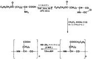
例示的なコンジュゲートの合成の方法は、以下の工程を含む:以下の工程を含む、ポリ-(L-グルタミン酸)-(2S,3S)-18-カルボキシ-20-(カルボキシメチル)-8,13-ジエチル-3,7,12,17-テトラメチル-クロリン-2-プロピオネート-[(Gd-1,4,7,10-テトラアザシクロドデカン-N,N',N'',N'''-テトラアセテート)-ヘキサンジアミン]を合成する方法:
γ-ベンジル・グルタメートをトリホスゲンと反応させ、γ-ベンジル・グルタメートのN-カルボキシ無水物を形成させる工程;
γ-ベンジル・グルタメートのN-カルボキシ無水物をトリブチルアミンと反応させ、ポリ-(ベンジル・グルタメート)を形成させる工程;
ポリ(ベンジル・グルタメート)を臭化水素とを反応させ、ポリ-(L-グルタメート)を形成させる工程;
ポリ-(L-グルタメート)をN-ヒドロキシスクシンイミドと反応させ、ポリ-(L-グルタミン酸)-N-ヒドロキシ・スクシンイミドを形成させる工程;
ポリ-(L-グルタミン酸)-N-ヒドロキシ・スクシンイミドを(2S,3S)-18-カルボキシ-20-(カルボキシメチル)-8,13-ジエチル-3,7,12,17-テトラメチル-クロリン-2-プロピオネートおよび1,4,7,10-テトラアザシクロドデカン-N,N',N'',N'''-テトラアセテート-ヘキサンジアミンと反応させる工程;ならびに
ポリ-(L-グルタミン酸)-(2S,3S)-18-カルボキシ-20-(カルボキシメチル)-8,13-ジエチル-3,7,12,17-テトラメチル-クロリン-2-プロピオネート-[(Gd-1,4,7,10-テトラアザシクロドデカン-N,N',N'',N'''-テトラアセテート)-ヘキサンジアミン]をガドリニウム・アセテートと反応させる工程。
An exemplary method of synthesizing a conjugate includes the following steps: poly- (L-glutamic acid)-(2S, 3S) -18-carboxy-20- (carboxymethyl) -8, which includes the following steps: 13-diethyl-3,7,12,17-tetramethyl-chlorin-2-propionate-[(Gd-1,4,7,10-tetraazacyclododecane-N, N ', N'',N'' Method for synthesizing '-tetraacetate) -hexanediamine]:
reacting γ-benzyl glutamate with triphosgene to form γ-benzyl glutamate N-carboxyanhydride;
reacting N-carboxyanhydride of γ-benzyl glutamate with tributylamine to form poly- (benzyl glutamate);
Reacting poly (benzyl glutamate) with hydrogen bromide to form poly- (L-glutamate);
Reacting poly- (L-glutamate) with N-hydroxysuccinimide to form poly- (L-glutamic acid) -N-hydroxysuccinimide;
Poly- (L-glutamic acid) -N-hydroxysuccinimide is converted to (2S, 3S) -18-carboxy-20- (carboxymethyl) -8,13-diethyl-3,7,12,17-tetramethyl-chlorine- Reacting with 2-propionate and 1,4,7,10-tetraazacyclododecane-N, N ′, N ″, N ′ ″-tetraacetate-hexanediamine; and poly- (L-glutamic acid)- (2S, 3S) -18-carboxy-20- (carboxymethyl) -8,13-diethyl-3,7,12,17-tetramethyl-chlorin-2-propionate-[(Gd-1,4,7, 10-tetraazacyclododecane-N, N ′, N ″, N ′ ″-tetraacetate) -hexanediamine] is reacted with gadolinium acetate.
当業者は、本発明の方法が癌細胞または腫瘍組織を標的とすることのみに制限されず、任意の悪性病変等も標的とすることを理解すると思われる。当技術分野には、対象におけるその他の組織を標的とするために有用な、本明細書に記載されたものに類似したその他の化学物質が存在する。同様に、当業者は、特定の化合物の合成の具体例の他に、本発明の様々な態様が、合成の方法および多数の利用可能な化合物を全て含むものであることが容易に明白であることを理解するであろう。 Those skilled in the art will appreciate that the methods of the present invention are not limited to targeting cancer cells or tumor tissue, but also target any malignant lesion or the like. There are other chemicals in the art that are similar to those described herein that are useful for targeting other tissues in a subject. Similarly, it will be readily apparent to those skilled in the art that, in addition to specific examples of the synthesis of specific compounds, the various aspects of the present invention include all methods of synthesis and a large number of available compounds. You will understand.
高分子量ポリ-グルタミン酸ポリマーは、コントラスト増強の増加によって示される腫瘍組織におけるより高い緩和度およびより高い集積を、2時間と比較して、24時間後に示す。腫瘍内または腫瘍周辺のポリマーの量は、画像モダリティ等の使用により照射されるべき正確な腫瘍体積の決定を補助し得る。従って、治療の時間は、悪性病変のサイズ、照明、エネルギー・ビーム、および/またはパルスの強度、組織における悪性病変の深さ、組織における悪性病変の組成等に依って変動し得る。一態様において、悪性病変は、約1〜約25分間、650nmのレーザーにより治療される。しかしながら、一般に、レーザーが、ビーム幅および/または時間(これらに制限はされない)のような正確なパラメーターの範囲内で使用された場合に周囲の組織に過度の傷害を引き起こさない限り、任意のサイズおよび/または強度のレーザーが、様々な態様により使用され得、かつ/または使用されるよう修飾され得る。 The high molecular weight poly-glutamic acid polymer shows higher relaxation and higher accumulation in tumor tissue as shown by increased contrast enhancement after 24 hours compared to 2 hours. The amount of polymer in or around the tumor can help determine the exact tumor volume to be irradiated, such as by using image modalities. Thus, the time of treatment may vary depending on the size of the malignancy, illumination, energy beam, and / or pulse intensity, the depth of the malignancy in the tissue, the composition of the malignancy in the tissue, and the like. In one embodiment, the malignant lesion is treated with a 650 nm laser for about 1 to about 25 minutes. However, in general, any size as long as the laser does not cause undue injury to the surrounding tissue when used within a range of precise parameters such as, but not limited to, beam width and / or time. And / or intensity lasers can be used and / or modified to be used in various ways.
様々な態様において、投与された送達系が標的部位の位置を特定するための時間が与えられるまで、標的部位の照明は遅延する。一態様において、0.1時間〜36時間の遅延が起こる。別の態様において、約1時間〜24時間の遅延が起こる。別の態様において、約18時間の遅延が起こる。 In various embodiments, the illumination of the target site is delayed until the administered delivery system is given time to locate the target site. In one embodiment, a delay of 0.1 hour to 36 hours occurs. In another embodiment, a delay of about 1 hour to 24 hours occurs. In another embodiment, a delay of about 18 hours occurs.
一態様において、本発明のポリマー-光増感剤コンジュゲートは、複数の用量で投与される。様々なその他の態様において、標的部位は、複数回、または複数の手順を通して照明され得る。 In one embodiment, the polymer-photosensitizer conjugates of the invention are administered in multiple doses. In various other embodiments, the target site can be illuminated multiple times or through multiple procedures.
様々な態様において、標的部位の画像が撮影される。一態様において、画像は送達媒体の投与前に撮影される。別の態様において、画像は送達媒体の投与時に撮影される。別の態様において、複数の画像が様々な時点で撮影される。様々な態様において、画像は、データを提供するため、方向を提供するため、進行をモニタリングするため等に使用される。 In various embodiments, an image of the target site is taken. In one aspect, the image is taken before administration of the delivery vehicle. In another embodiment, the image is taken upon administration of the delivery vehicle. In another aspect, multiple images are taken at various times. In various aspects, the images are used to provide data, provide directions, monitor progress, and the like.
本発明のもう一つの例示的な態様において、使用されるポリマーは、造影剤としてのDOTA-Gdおよび光増感薬としてのMse6を含むポリ-(L-グルタミン酸)である。 In another exemplary embodiment of the invention, the polymer used is poly- (L-glutamic acid) comprising DOTA-Gd as a contrast agent and Mse 6 as a photosensitizer.
本発明は、とりわけ、癌の治療において有用な薬学的組成物を調製する方法を開示する。一態様において、組成物はPLGA-Mce6-[(Gd-DOTA)]を含む。一態様において、組成物は化合物PLGA-Mce6-[-1,6-ヘキサンジアミン-(Gd-DOTA)]を含む。様々なPLGA-Mce6-(Gd-DOTA)コンジュゲートは、治療後の残存するGd錯体の対象からの迅速な排除を可能にするジスルフィド・リンカーを造影剤とポリマー担体との間に含有している。(コンジュゲートの生分解性特徴を提供するジスルフィド結合の形成に関しては、参照17、Lu,et al.を参照のこと。)これに類似したその他の生分解性リンカーは、当技術分野において公知であり、当業者は、コンジュゲートに生分解性特性を提供するために、その他のそのようなリンカーを代用し得ることが理解される。そのような生分解性ポリマー・コンジュゲートは、本発明に包含され、本発明のさらなる例示的な態様を構成する。 The present invention discloses, among other things, a method for preparing pharmaceutical compositions useful in the treatment of cancer. In one embodiment, the composition comprises PLGA-Mce 6 -[(Gd-DOTA)]. In one embodiment, the composition comprises the compound PLGA-Mce 6 -[-1,6-hexanediamine- (Gd-DOTA)]. Various PLGA-Mce 6- (Gd-DOTA) conjugates contain a disulfide linker between the contrast agent and the polymer carrier that allows rapid elimination of the remaining Gd complex from the subject after treatment. Yes. (See Reference 17, Lu, et al. For the formation of disulfide bonds that provide the biodegradable characteristics of the conjugate.) Other biodegradable linkers similar to this are known in the art. It will be appreciated that one of ordinary skill in the art can substitute other such linkers to provide biodegradable properties to the conjugate. Such biodegradable polymer conjugates are encompassed by the present invention and constitute a further exemplary aspect of the present invention.
従って、本発明の様々な態様は、薬学的に許容される量の化合物ポリ-(L-グルタミン酸)-(2S,3S)-18-カルボキシ-20-(カルボキシメチル)-8,13-ジエチル-3,7,12,17-テトラメチル-クロリン-2-プロピオネート-[(Gd-1,4,7,10-テトラアザシクロドデカン-N,N',N'',N'''-テトラアセテート)-ヘキサンジアミン]を含む、対象における標的組織の治療において有用な薬学的組成物を含む。 Thus, various embodiments of the present invention provide pharmaceutically acceptable amounts of the compound poly- (L-glutamic acid)-(2S, 3S) -18-carboxy-20- (carboxymethyl) -8,13-diethyl- 3,7,12,17-tetramethyl-chlorin-2-propionate-[(Gd-1,4,7,10-tetraazacyclododecane-N, N ', N' ', N' ''-tetraacetate A pharmaceutical composition useful in the treatment of a target tissue in a subject.
さらなる態様は、標的部位の治療のための薬品の製造における、ポリ-(L-グルタミン酸)-(2S,3S)-18-カルボキシ-20-(カルボキシメチル)-8,13-ジエチル-3,7,12,17-テトラメチル-クロリン-2-プロピオネート-[1,6-ヘキサンジアミン-(Gd-1,4,7,10-テトラアザシクロドデカン-N,N',N'',N'''-テトラアセテート)]を含む薬学的に許容される組成物の使用を含む。 A further embodiment is a poly- (L-glutamic acid)-(2S, 3S) -18-carboxy-20- (carboxymethyl) -8,13-diethyl-3,7 in the manufacture of a medicament for the treatment of a target site , 12,17-Tetramethyl-chlorin-2-propionate- [1,6-hexanediamine- (Gd-1,4,7,10-tetraazacyclododecane-N, N ', N' ', N' ' Use of a pharmaceutically acceptable composition comprising '-tetraacetate)].
さらなる態様は、標的部位の位置を特定または探索するための、薬学的に有効な量の磁気共鳴画像法造影剤により標識されたポリマー-光増感コンジュゲートの使用を含む。一つの態様において、薬剤は、ポリ-(L-グルタミン酸)-(2S,3S)-18-カルボキシ-20-(カルボキシメチル)-8,13-ジエチル-3,7,12,17-テトラメチル-クロリン-2-プロピオネート-[1,6-ヘキサンジアミン-(Gd-1,4,7,10-テトラアザシクロドデカン-N,N',N'',N'''-テトラアセテート)]である。 Further embodiments include the use of a polymer-photosensitized conjugate labeled with a pharmaceutically effective amount of a magnetic resonance imaging contrast agent to locate or locate a target site. In one embodiment, the agent is poly- (L-glutamic acid)-(2S, 3S) -18-carboxy-20- (carboxymethyl) -8,13-diethyl-3,7,12,17-tetramethyl- Chlorine-2-propionate- [1,6-hexanediamine- (Gd-1,4,7,10-tetraazacyclododecane-N, N ', N' ', N' ''-tetraacetate)] .
本発明のさらなる態様は、様々な系を含む。一つの態様において、磁気共鳴画像化造影剤、光増感剤、および画像モダリティを含む、標的組織の位置を特定するための系が開示される。別の態様において、磁気共鳴画像法造影剤;光増感剤、画像モダリティ;およびレーザーを含む、標的部位を治療するための系が開示される。本発明の系のさらなる態様は、送達媒体の適用のためのアプリケータを含み得る。 Further aspects of the invention include various systems. In one embodiment, a system for locating a target tissue is disclosed that includes a magnetic resonance imaging contrast agent, a photosensitizer, and an image modality. In another aspect, a system for treating a target site is disclosed that includes a magnetic resonance imaging contrast agent; a photosensitizer, an image modality; and a laser. A further aspect of the system of the present invention may include an applicator for application of the delivery vehicle.
本発明を制限することなく、本発明をさらに例示するため、以下の例示的な実施例が提供される。 The following illustrative examples are provided to further illustrate the present invention without limiting it.
実施例
材料および方法
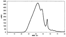
γ-ベンジル・グルタメートは、VWR(West Chester,PA)のため購入された。特別乾燥(Extra dry)溶媒;テトラヒドロフラン(THF)、酢酸エチル、および塩化メチレンは、Arcos Organics,NJから購入された。Meso chlorin e6およびCyclenは、Macrocyclic Incから購入された。Gd(OAc)3およびジ-tert-ブチルジカルボネート(t-boc)、tert-ブチルブロモアセテート、N-エチルジイソプロピルアミン、およびモノ-boc-1,6-ジアミノヘキサンは、Alfa Aesar(Ward Hill,MA)から購入された。[1-エチル-3-(3-ジメチルアミノプロピル)カルボジイミドHCl](EDC)は、TCI America(Portland,OR)から購入された。シリカゲル、メッシュ・サイズ230〜400は、Natland International Corp.,NCから購入された。全ての溶媒が、Fisher Scientificから購入され、特記しない限り、さらなる精製なしに使用された。Spectra/Por再生セルロース膜(MWCO=16〜18kDa)は、Spectrum Laboratories(Rancho Dominguez,CA)から購入された。PD-10脱塩カラムは、Amersham Bioscience(Uppsala,Sweden)から購入された。
Example Materials and Methods γ-Benzyl glutamate was purchased for VWR (West Chester, PA). Extra dry solvents; tetrahydrofuran (THF), ethyl acetate, and methylene chloride were purchased from Arcos Organics, NJ. Meso chlorin e 6 and Cyclen were purchased from Macrocyclic Inc. Gd (OAc) 3 and di-tert-butyl dicarbonate (t-boc), tert-butyl bromoacetate, N-ethyldiisopropylamine, and mono-boc-1,6-diaminohexane were obtained from Alfa Aesar (Ward Hill, MA). [1-Ethyl-3- (3-dimethylaminopropyl) carbodiimide HCl] (EDC) was purchased from TCI America (Portland, OR). Silica gel, mesh sizes 230-400 were purchased from Natland International Corp., NC. All solvents were purchased from Fisher Scientific and used without further purification unless otherwise noted. Spectra / Por regenerated cellulose membrane (MWCO = 16-18 kDa) was purchased from Spectrum Laboratories (Rancho Dominguez, Calif.). PD-10 desalting columns were purchased from Amersham Bioscience (Uppsala, Sweden).
細胞系(MDA-MB-231)、2μMグルタミンおよびトリプシンを含有しているLH-15培地は、American Type Culture Collection(ATCC,Manassas VA)から購入された。雌nu/nu胸腺欠損マウスは、NCI(Frederick,MD)から購入された。動物は、IACUC、University of Utah guidelinesに従って維持された。 Cell line (MDA-MB-231), LH-15 medium containing 2 μM glutamine and trypsin were purchased from the American Type Culture Collection (ATCC, Manassas VA). Female nu / nu athymic mice were purchased from NCI (Frederick, MD). Animals were maintained according to IACUC, University of Utah guidelines.
分子量決定は、UVおよび屈折率の検出器が装備されているSuperdex 200/Superose 6カラムを含むAKTA FPLC系(Amersham Biosciences Corp.,Piscataway,NJ)でのサイズ排除クロマトグラフィ(SEC)によって実施された。FPLCカラムは、ポリ[N-(2-ヒドロキシプロピル)メタクリルアミド]標準により較正された。1H NMRスペクトルは、25℃で、400MHzで、Varian INOVA 400で取得された。ESI-MSスペクトルは、University of Utah Mass Spectrometry and Proteonomic Core Facilityで、PE Sciex API III Mass Spectrometerで取得された。Meso chlorin含有量は、Cary Win UV-Vis分光計で決定された。Gd含有量は、誘導結合アルゴン・プラズマ発光分析法(Inductively Coupled argon Plasma-Optical Emission Spectrometer)(ICP-OES)(Perkin Elmer,Norwalk,CT,Optima 3100XL)を使用して決定された。最終的なコンジュゲートについてのT1緩和度測定は、標準的なインバージョン・リカバリー・シーケンス(inversion recovery sequence)を使用してSiemens Trio 3T MRIスキャナで取得された。光線力学的療法は、Diode Odysseyレーザー(650nm)(CAO Group,UT)を介して実施された。
Molecular weight determination was performed by size exclusion chromatography (SEC) on an AKTA FPLC system (Amersham Biosciences Corp., Piscataway, NJ) containing a
A. 常磁性標識された薬物-ポリマー錯体の合成
1. ポリ-(L-グルタミン酸)活性エステル(PLGA-OSu)の合成
a. ポリ-L-グルタミン酸(PLGA)の合成
高分子量ポリ-(L-グルタミン酸)は、以前に記載された方法によって合成された。簡単に説明すると、合成の第一工程は、THF(15ml)中でのγ-ベンジル・グルタメート(15g、0.06mol)のトリホスゲン(9.37g、0.03mol)との反応によるN-カルボキシ無水物の形成を含んでいた。反応をN2下で50℃で3時間実施し、NCAを沈殿させるためにn-ヘキサン(150ml)へ反応混合物を注入した。酢酸エチル(10ml)への溶解およびn-ヘキサン中での再沈殿により、生成物を再結晶化した。γ-ベンジル・グルタメートのNCAは、高い収率および純度で入手された。M.P=110℃
A. Synthesis of paramagnetically labeled drug-polymer complexes
1. Synthesis of poly- (L-glutamic acid) active ester (PLGA-OSu)
a. Synthesis of poly-L-glutamic acid (PLGA) High molecular weight poly- (L-glutamic acid) was synthesized by the method previously described. Briefly, the first step in the synthesis is the formation of N-carboxyanhydride by reaction of γ-benzyl glutamate (15 g, 0.06 mol) with triphosgene (9.37 g, 0.03 mol) in THF (15 ml). Was included. The reaction was carried out under N 2 at 50 ° C. for 3 hours and the reaction mixture was poured into n-hexane (150 ml) to precipitate NCA. The product was recrystallized by dissolution in ethyl acetate (10 ml) and reprecipitation in n-hexane. NCA of γ-benzyl glutamate was obtained in high yield and purity. MP = 110 ℃
高分子量ポリ-(ベンジル-γ-グルタメート)[PBLG]の合成のため、開始剤としてジクロロメタン中のトリブチルアミン10%溶液を使用した(M/I=100/1)、18mlの酢酸エチル:ジクロロメタン混合物(1:6v/v)中でのNCA(3G、11.4mol)の反応によって、開環重合を実施した。反応を、30Cで3時間およびR.Tで24時間、維持した。PBLGをメタノール:エーテル(2:1)混合物中で沈殿させ、ろ過し、エーテルで洗浄し、真空乾燥させた。 For the synthesis of high molecular weight poly- (benzyl-γ-glutamate) [PBLG], a 10% solution of tributylamine in dichloromethane was used as an initiator (M / I = 100/1), 18 ml of ethyl acetate: dichloromethane mixture Ring-opening polymerization was carried out by reaction of NCA (3G, 11.4 mol) in (1: 6 v / v). The reaction was maintained at 30 C for 3 hours and R.T for 24 hours. PBLG was precipitated in a methanol: ether (2: 1) mixture, filtered, washed with ether and dried in vacuo.
最終工程は、ポリ-L-グルタミン酸が析出するまで、溶液にHBrを通すことによる、ジクロロメタンに溶解したPBLG(300ml中1.0gm)上のベンジル基の除去を含んでいた。HBrガスは、テトラリンおよび臭素の反応により発生させた。PLGAを、アセトンを使用してさらに精製した。分子量決定は、UVおよび屈折率(RI)の検出器が装備されたSuperdex 200カラムでのSECを使用して実施された。RIピークに相当するUVピークの欠如により、PLGAのほぼ完全な脱保護が確認された。
The final step involved removal of the benzyl group on PBLG dissolved in dichloromethane (1.0 gm in 300 ml) by passing HBr through the solution until poly-L-glutamic acid precipitated. HBr gas was generated by the reaction of tetralin and bromine. PLGA was further purified using acetone. Molecular weight determination was performed using SEC on a
b. PLGA-OSuの合成
ポリ-(L-グルタミン酸)をN-ヒドロキシ・スクシンイミド(NHS)と反応させ、コハク酸エステル形成させた。簡単に説明すると、PLGAおよびNHSをDMFに溶解させ、反応を開始させるためにカップリング剤EDCを添加した。活性エステルをアセトン中で沈殿させ、真空下で乾燥させた。エステルの存在は、NMRによって確認された。
b. Synthesis of PLGA-OSu Poly- (L-glutamic acid) was reacted with N-hydroxysuccinimide (NHS) to form succinate. Briefly, PLGA and NHS were dissolved in DMF and coupling agent EDC was added to initiate the reaction. The active ester was precipitated in acetone and dried under vacuum. The presence of the ester was confirmed by NMR.
2. Gd-DOTA-1,6-ジアミノヘキサン(リンカー)の合成
a.DO3Aの合成
DO3Aは、高純度のDO3Aを入手するためわずかに変更された以前に記載された方法に従い、2工程で、cyclenを介して合成された。
2. Synthesis of Gd-DOTA-1,6-diaminohexane (linker)
a.Synthesis of DO 3 A
DO 3 A was synthesized via cyclen in two steps according to previously described methods that were slightly modified to obtain high purity DO 3 A.
第一工程において、cyclen(1.72gm、10mmol)およびDIPEA(10eq、100mmol、12.9gm)を、80mlのクロロホルムに溶解させた。完全に溶解した後、反応混合物を2工程でクロロホルム中でtert-ブチル・ブロモアセテートと反応させた。一回目の添加のため、40mlのクロロホルムに溶解したtert-ブチル・ブロモアセテート(2eq、20mmol、3.92gm)を、絶えず攪拌しながら溶液へ滴下にて添加することにより反応させた。完全な添加の後、反応物を12時間撹拌した。二回目の添加のため、20mlに溶解したtert-ブチル・ブロモアセテート(1eq、10mmol、1.96gm)を、反応混合物に滴下にて添加した。反応物をR.Tでさらに18時間撹拌した。溶液を真空で濃縮し、固体を最少量の塩化メチレンに溶解させ、さらなる精製のためシリカゲル・カラムに適用した。溶出剤系として酢酸エチル:メタノール(100%、50:1、20:1、および10:1)を使用した勾配溶出を実施した。TB-cyclen(tert-ブチル保護cyclen)は20:1画分に入手された。 In the first step, cyclen (1.72 gm, 10 mmol) and DIPEA (10 eq, 100 mmol, 12.9 gm) were dissolved in 80 ml of chloroform. After complete dissolution, the reaction mixture was reacted with tert-butyl bromoacetate in chloroform in two steps. For the first addition, tert-butyl bromoacetate (2 eq, 20 mmol, 3.92 gm) dissolved in 40 ml chloroform was reacted by dropwise addition to the solution with constant stirring. After complete addition, the reaction was stirred for 12 hours. For the second addition, tert-butyl bromoacetate (1 eq, 10 mmol, 1.96 gm) dissolved in 20 ml was added dropwise to the reaction mixture. The reaction was stirred at RT for an additional 18 hours. The solution was concentrated in vacuo and the solid was dissolved in a minimum amount of methylene chloride and applied to a silica gel column for further purification. Gradient elution was performed using ethyl acetate: methanol (100%, 50: 1, 20: 1, and 10: 1) as the eluent system. TB-cyclen (tert-butyl protected cyclen) was obtained in the 20: 1 fraction.
TB cyclenを最少量の冷トリフルオロ酢酸を使用して脱保護し(25℃、一夜)、DO3Aを形成させた。生成物をジエチルエーテルを使用して精製した。 TB cyclen was deprotected using a minimal amount of cold trifluoroacetic acid (25 ° C., overnight) to form DO 3 A. The product was purified using diethyl ether.
b. N-boc-1,6-ジアミノヘキサン・ブロモアセテート(中間体)の合成
1工程の過程において、N-boc-1,6-ジアミノヘキサン(1gm、4mmol)を、ジクロロメタン(30ml)中のブロモ酢酸のN-ヒドロキシ・スクシンイミド・エステル(1.32gm、5mmol)と0℃で反応させた。完全な添加の後、反応物を25Cで24時間撹拌した。NaOH(2×50ml)および水(2×50ml)での洗浄、有機溶媒での抽出により精製され、乾燥するまで濃縮されたブロモアセテート・エステル。
b. Synthesis of N-boc-1,6-diaminohexane bromoacetate (intermediate)
In the course of one step, N-boc-1,6-diaminohexane (1 gm, 4 mmol) was reacted with N-hydroxysuccinimide ester of bromoacetic acid (1.32 gm, 5 mmol) in dichloromethane (30 ml) at 0 ° C. I let you. After complete addition, the reaction was stirred at 25 C for 24 hours. Bromoacetate ester purified by washing with NaOH (2 × 50 ml) and water (2 × 50 ml), extraction with organic solvent and concentrated to dryness.
c. 合成DOTA-1,6-ジアミノヘキサン
20mlのメタノール:トリエチルアミン溶媒混合物(2:1)および過剰のK2CO3(2.76gm、20mmol)の中でのDO3A(0.7gm、2mmol)のN-boc-1,6-ジアミノヘキサン・ブロモアセテート(1.36gm、4mmol)との反応により、リンカーを合成した。反応を室温で24時間実施した。溶媒を蒸発させ、生成物を水に溶解させ、希HClで酸性化した。全ての酸を塩に変換し、KBrのような無機塩を溶解させるため、pHは、2〜3の間に調整した。水性溶液を真空で蒸発させ、リンカーをメタノール:トリエチルアミン(2:1)混合物から塩として抽出した。
c. Synthetic DOTA-1,6-diaminohexane
N-boc-1,6-diaminohexane · DO 3 A (0.7 gm, 2 mmol) in 20 ml methanol: triethylamine solvent mixture (2: 1) and excess K 2 CO 3 (2.76 gm, 20 mmol) The linker was synthesized by reaction with bromoacetate (1.36 gm, 4 mmol). The reaction was carried out at room temperature for 24 hours. The solvent was evaporated and the product was dissolved in water and acidified with dilute HCl. The pH was adjusted between 2-3 to convert all acids to salts and dissolve inorganic salts such as KBr. The aqueous solution was evaporated in vacuo and the linker was extracted as a salt from a methanol: triethylamine (2: 1) mixture.
3. Gd(III)-DOTA-リンカー-PLGAおよびGd-DOTA-リンカー-PLGA-Mce6錯体の合成
a. DOTA-リンカー-PLGAおよびDOTA-リンカー-PLGA-Mce6コンジュゲートの合成
PLGA活性エステル(PLGA-NHS、200mg、542mmol)を、meso chlorin e6(24mg、37mmol)と共にDMF(3ml)に溶解させ、30分間反応させた。次いで、DOTA-リンカー(300mg、600mmol)を溶液に添加した。反応を開始させるために各添加の後に過剰のDMAPを組み込んだ。反応を室温で24時間実施した。反応終了後、DMFを真空下で蒸発させ、残さを水に溶解させた。対照として使用するためのDOTA-リンカー-PLGAコンジュゲートを、Mce6を添加せずに、同様にして合成した。Gd(III)によるポリマー・コンジュゲートの錯体化を、さらなる精製なしに実施した。
3. Synthesis of Gd (III) -DOTA-linker-PLGA and Gd-DOTA-linker-PLGA-Mce 6 complexes
a. Synthesis of DOTA-linker-PLGA and DOTA-linker-PLGA-Mce 6 conjugates
PLGA active ester (PLGA-NHS, 200 mg, 542 mmol) was dissolved in DMF (3 ml) together with mesochlorine 6 (24 mg, 37 mmol) and allowed to react for 30 minutes. DOTA-linker (300 mg, 600 mmol) was then added to the solution. Excess DMAP was incorporated after each addition to initiate the reaction. The reaction was carried out at room temperature for 24 hours. After completion of the reaction, DMF was evaporated under vacuum and the residue was dissolved in water. A DOTA-linker-PLGA conjugate for use as a control was synthesized in the same manner without the addition of Mce 6 . Complexation of the polymer conjugate with Gd (III) was performed without further purification.
b. Gd(III)-DOTA-リンカー-PLGA-Mce6(Pol1)およびGd(III)-DOTA-リンカー-PLGA(Pol2)の合成
DI水中のポリマー・コンジュゲートの水性溶液を、pH8で、過剰のGd(III)アセテートと反応させた。一夜撹拌した後、希NaOHにより溶液のpHを11に増加させることにより、過剰のGdをGd2O3として析出させた。酸化物を析出させ、溶液のpHを5.5〜6の間に調整した。最終的な溶液を、真空下で濃縮し、カラムに適用した。溶出液を凍結乾燥した。
b. Synthesis of Gd (III) -DOTA-linker-PLGA-Mce 6 (Pol 1 ) and Gd (III) -DOTA-linker-PLGA (Pol 2 )
An aqueous solution of the polymer conjugate in DI water was reacted with excess Gd (III) acetate at pH8. After stirring overnight, excess Gd was precipitated as Gd 2 O 3 by increasing the pH of the solution to 11 with dilute NaOH. The oxide was precipitated and the pH of the solution was adjusted between 5.5-6. The final solution was concentrated under vacuum and applied to the column. The eluate was lyophilized.
B. 常磁性標識されたポリマー-光増感剤コンジュゲートの特徴決定(表1)
分子量の決定
ポリマー錯体の分子量は、AKTA FPLC系(Amersham Inc)でのサイズ排除クロマトグラフィを使用して決定された。錯体は、TRIS緩衝液pH7.4に溶解させられ、0.2μ膜でろ過された後、カラムに適用された。
B. Characterization of paramagnetic labeled polymer-photosensitizer conjugates (Table 1)
Determination of molecular weight The molecular weight of the polymer complex was determined using size exclusion chromatography on the AKTA FPLC system (Amersham Inc). The complex was dissolved in TRIS buffer pH 7.4, filtered through a 0.2 μ membrane and then applied to the column.
緩和度の決定
ポリマー錯体Pol1およびPol2のT1緩和度は、22〜1600msの変動するTIにより、標準的なインバージョン・リカバリー・シーケンスを使用して、Siemens Trio 3Tスキャナで決定された。
Determination of Relaxation The T1 relaxation of the polymer complexes Pol1 and Pol2 was determined with a Siemens Trio 3T scanner using a standard inversion recovery sequence with varying TI between 22 and 1600 ms.
Gd含有量の決定
Pol1およびPol2含有量のGd(III)は、標準曲線を構築するためにGd標準を使用して、誘導結合プラズマ発光分析法(ICP-OES)により決定された。
Determination of Gd content
Pol 1 and Pol 2 content Gd (III) was determined by inductively coupled plasma optical emission spectrometry (ICP-OES) using Gd standards to construct standard curves.
Mce6含有量の決定
Pol1についてのMeso chlorin e6含有量は、UV分光測光法により決定された。Mce6標準ポリマー・コンジュゲート溶液がメタノールで調製され、UV吸光度が650nmで測定された。
Determination of Mce 6 content
The Meso chlorin e 6 content for Pol 1 was determined by UV spectrophotometry. Mce 6 standard polymer conjugate solution was prepared in methanol and UV absorbance was measured at 650 nm.
C. 細胞培養
MDA-MB-231(ATCC)ヒト乳癌細胞系を、5%CO2で、2mMグルタミンおよび10%FBSを含むLH-15培養培地で培養した。細胞をコンフルエンスで採集し、トリパンブルーで染色した後に計数した。腫瘍モデルを作製するため、動物1匹当たり2×10⁶個の細胞を注射した。
C. Cell culture
The MDA-MB-231 (ATCC) human breast cancer cell line was cultured in LH-15 culture medium containing 2 mM glutamine and 10% FBS with 5% CO 2 . Cells were harvested at confluence and counted after staining with trypan blue. To create a tumor model, 2 × 10 6 cells were injected per animal.
D. 動物モデル
6週齢の雌胸腺欠損nu/nuマウスを、NCI(Frederick,MD)から購入した。腫瘍モデルを作製するため、各マウスの側腹部に、100μlのBD MATRIGEL、EHSマウス肉腫から抽出された可溶化(solubulized)された基底膜調製物と混合された100μlの培養培地の中の2×10⁶個のMB-231細胞を皮下注射した。磁気共鳴画像法および光線力学的療法の研究は、25〜40mm3の平均腫瘍サイズで実施された。
D. Animal model
Six week old female athymic nu / nu mice were purchased from NCI (Frederick, MD). To create a tumor model, each mouse flank contains 100 μl BD MATRIGEL, 2 × in 100 μl culture medium mixed with solubulized basement membrane preparation extracted from
(表1)Gd(ICP)およびMeso Chlorin(UV)の含有量の決定
(データは図6に例示された結果において使用されたマウスに関するものである)

実施例I
動物腫瘍モデル試験
異種移植片を作製するため、6匹の雌胸腺欠損nu/nuマウスに、MDA-MB231ヒト乳癌細胞(ATCC)を注射した。約1〜3×106個の細胞をMATRIGEL(BD Biosciences)と混合し、各マウスに注射した。マウスを腫瘍増殖に関して観察した。腫瘍体積をディジタル・ノギスを使用して測定した。腫瘍体積がおよそ20mm3になった時点で、マウスを画像法および療法のために選択した。
Example I
Animal tumor model test To produce xenografts, 6 female athymic nu / nu mice were injected with MDA-MB231 human breast cancer cells (ATCC). About 1-3 × 10 6 cells were mixed with MATRIGEL (BD Biosciences) and injected into each mouse. Mice were observed for tumor growth. Tumor volume was measured using digital calipers. When the tumor volume reached approximately 20 mm 3 , mice were selected for imaging and therapy.
実施例II
動物モデルの磁気共鳴画像法およびコンジュゲートの投与
MR画像法をSiemens Trio 3T MRIスキャナで実施した。麻酔薬(80mg/kg体重のケタミンおよび12mg/kg体重のキシラジンの混合物)の腹腔内または筋肉注射により、動物に麻酔をかけた。
Example II
Magnetic resonance imaging of animal models and administration of conjugates
MR imaging was performed with a Siemens Trio 3T MRI scanner. The animals were anesthetized by intraperitoneal or intramuscular injection of anesthetic (a mixture of 80 mg / kg body weight ketamine and 12 mg / kg body weight xylazine).
画像化の目的のため、動物をヒト用リスト・コイル(human wrist coil)に置き、コントラスト前画像(薬物および造影剤を含有しているポリマー・キレート系の注射前)を、3D FLASH(Fast Low Angle Shot)シーケンスを使用して入手した。 For imaging purposes, the animal is placed in a human wrist coil and a pre-contrast image (before injection of a polymer chelate containing drug and contrast agent) is obtained with 3D FLASH (Fast Low (Angle Shot) sequence.
コントラスト前画像を入手した後、高分子錯体を、0.09mmol Gd/kg体重および6mg/kgのMce6の用量で、尾静脈注射により注射した。画像取得は、注射後5、30、60、120分、および24時間目に設定された。動物画像法は多相T1加重シーケンスを使用して実施された。反復時間は、それぞれ、4〜8cm FIV(視野)と一致する約6ミリ秒〜2ミリ秒であり、各動物について組織薄片の厚さは0.5mmを選択した。画像化容量は、注射前に、そして注射後に迅速に連続して取得された。取得は、各々、約30秒を必要とした。 After obtaining pre-contrast images, the polymer complex was injected by tail vein injection at a dose of 0.09 mmol Gd / kg body weight and 6 mg / kg Mce 6 . Image acquisition was set at 5, 30, 60, 120 minutes, and 24 hours after injection. Animal imaging was performed using a polyphase T1 weighted sequence. The repetition time was approximately 6 to 2 milliseconds, consistent with 4-8 cm FIV (field of view), respectively, and a tissue slice thickness of 0.5 mm was chosen for each animal. Imaging volumes were acquired continuously before injection and rapidly after injection. Each acquisition required approximately 30 seconds.
類似した投薬量が、ヒトのような他の対象において有効であると予想される。ヒトにおいて、投薬量は、例えば、0.01〜0.03mmol Gd/kg体重を含み得る。同様に、例えば、1〜10mg/kg体重のMce6を含み得る投薬量が、ヒトのような他の対象において有効であると予想される。しかしながら、投薬量は、ルーチンの実験により決定されるように調整してもよい。 Similar dosages are expected to be effective in other subjects such as humans. In humans, dosages can include, for example, 0.01 to 0.03 mmol Gd / kg body weight. Similarly, dosages that can include, for example, 1-10 mg / kg body weight of Mce 6 are expected to be effective in other subjects, such as humans. However, the dosage may be adjusted as determined by routine experimentation.
実施例III
動物モデルにおける標的組織の特異的な照明
コントラスト増強が観察された後、Mce6の励起波長に相当する650nmの波長のレーザーを使用して腫瘍を照射した。
Example III
Specific illumination of the target tissue in the animal model After contrast enhancement was observed, the tumor was irradiated using a laser with a wavelength of 650 nm, which corresponds to the excitation wavelength of Mce 6 .
腫瘍内の高分子錯体の位置および集積を、MRIで観察した。次いで、注射後2時間目および18時間目に、220mW/cm2のエネルギーの入力および照射のため0.5mmのアパーチャーにより波長650nmの光(Denlaser)を使用して腫瘍を照射した。照射の時間は15分であった。次いで、無関係の照射を回避するため、マウスを直接光から隔離し、腫瘍体積の減少を測定した。 The position and accumulation of the polymer complex in the tumor were observed by MRI. The tumors were then irradiated using light of 650 nm wavelength (Denlaser) with a 0.5 mm aperture for energy input and irradiation of 220 mW / cm 2 at 2 and 18 hours after injection. The duration of irradiation was 15 minutes. The mice were then isolated directly from the light and the decrease in tumor volume was measured to avoid irrelevant irradiation.
結果は、注射前、注射後5、30、60、120分、および24時間目の胸腺欠損nu/nuマウスにおける腫瘍の画像を示す、図6の磁気共鳴画像に示される。組織薄片は、マウス1については心臓および腫瘍を別々に通っており、マウス2については心臓および腫瘍を同時に通っている。両方のマウスにおいて、24時間目の画像は、コントラスト前と比較して腫瘍を通るコントラスト増強を示しており、注射後5分目の心臓にもコントラスト増強が存在する。計6匹のマウスが上記の方法論を使用して調査され、全て、類似した結果を与えた。これらの結果は、コンジュゲートが24時間以内に標的組織に局在化し得ることを示している。
The results are shown in the magnetic resonance image of FIG. 6, showing images of tumors in athymic nu / nu mice before injection, 5, 30, 60, 120 minutes, and 24 hours after injection. Tissue slices pass separately through the heart and tumor for mouse 1 and simultaneously through the heart and tumor for
実施例IV
実施例VIIのためのポリマー・コンジュゲートおよび対照の合成および物理化学的特徴決定
Mce6およびGd(III)DOTA(Pol1およびPol3)またはGd(III)DOTAのみ(Pol2)を含有しているポリ-(-グルタミン酸)コンジュゲートを、同一分子量Mw=82kDaのPLGAから合成した。薬物および造影剤の異なる比率を、両方の活性モエティのための最適の含有量を有する最終的なコンジュゲートを作製するため、評価した(示されていない)。Meso chlorin e6は、その疎水性の性質、およびより良好な光の透過に至るより高い光の波長において励起される可能性のために使用された。臨床的に認可されている造影剤Gd-DOTAは、非環式金属キレートと比較してより高い安定性のために選ばれた。ポリマー・コンジュゲートおよび対照についての分子量、PDI、meso chlorin e6およびGdの含有量、ならびに緩和度を含む物理化学的特徴決定は、下記表2に与えられる。薬物を含むポリマー・コンジュゲートおよび薬物を含まないポリマー・コンジュゲートは、低分子量カウンターパートと比較して、より高い緩和度を示した。
Example IV
Synthesis and physicochemical characterization of polymer conjugates and controls for Example VII
Poly-(-glutamic acid) conjugates containing Mce 6 and Gd (III) DOTA (Pol 1 and Pol 3 ) or Gd (III) DOTA alone (Pol 2 ) were synthesized from PLGA with the same molecular weight Mw = 82 kDa did. Different ratios of drug and contrast agent were evaluated (not shown) to make the final conjugate with optimal content for both activity moieties. Meso chlorin e 6 was used because of its hydrophobic nature and its potential to be excited at higher light wavelengths leading to better light transmission. The clinically approved contrast agent Gd-DOTA was chosen for higher stability compared to acyclic metal chelates. The physicochemical characterization including molecular weight, PDI, mesochlorine 6 and Gd content, and relaxivity for polymer conjugates and controls is given in Table 2 below. The polymer conjugate with and without the drug showed a higher degree of relaxation compared to the low molecular weight counterpart.
(表2)(結果は図7〜10におけるマウスに関するものである)
MR画像法
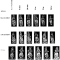
Pol1およびPol2により、マウス6匹の2つの群においてコントラスト増強が観察された。図7および図8は、それぞれ腫瘍および心臓を通る2D冠状MR画像を示す。図7の上パネルはPol1によるマウスを表し、下のパネルはPol2によるマウスを表す。図8の上パネルはPol1によるマウスを表し、下のパネルはPol2によるマウスを表す。Pol1およびPol2の両方について、注射後5分目に、腫瘍、心臓、および肝臓(示されないデータ)を含む全ての組織に、コントラスト前と比較して増加したコントラストが存在した。心臓においては、全ての時点で、対照に、より高いコントラストが存在し、腫瘍組織においては、全ての時点で、両方に、類似した増強が観察された。心臓におけるより高いシグナルは、対照ポリマーの高分子量画分によるものかもしれない。
MR imaging
With Pol 1 and Pol 2 , contrast enhancement was observed in two groups of 6 mice. Figures 7 and 8 show 2D coronal MR images through the tumor and heart, respectively. The upper panel of FIG. 7 represents mice with Pol 1 and the lower panel represents mice with Pol 2 . The upper panel of FIG. 8 represents mice with Pol 1 and the lower panel represents mice with Pol 2 . For both Pol 1 and Pol 2 , at 5 minutes post-injection, there was an increased contrast in all tissues including the tumor, heart, and liver (data not shown) compared to before contrast. In the heart, there was a higher contrast in the control at all time points, and in the tumor tissue, similar enhancements were observed at both time points at both time points. The higher signal in the heart may be due to the high molecular weight fraction of the control polymer.
両方のポリマーについての腫瘍組織におけるシグナル強度の半定量的評価は、注射後2時間目までに、増加したコントラストを示した。2時間目のコントラスト前と比較した両方の群についての腫瘍のシグナル強度の増加は100%である。注射後18時間目のシグナル強度は、Pol1およびPol2の両方についてコントラスト前より少なくとも30%高かった。IDLを使用したシグナル強度の定量的評価を、半定量分析から入手されたデータを確証するために実施した。 Semi-quantitative assessment of signal intensity in tumor tissue for both polymers showed increased contrast by 2 hours after injection. The increase in tumor signal intensity for both groups compared to pre-contrast at 2 hours is 100%. The signal intensity at 18 hours after injection was at least 30% higher for both Pol 1 and Pol 2 than before contrast. A quantitative assessment of signal intensity using IDL was performed to confirm the data obtained from the semi-quantitative analysis.
光線力学的療法の効力
第1の群に属するマウスについての腫瘍体積の減少、6匹のマウスがポリマー-薬物-CA(薬物)を受容し、6匹のマウスがポリマー-CAコンジュゲート(対照)を受容した。
Efficacy of photodynamic therapy Reduction of tumor volume for mice belonging to the first group, 6 mice receiving polymer-drug-CA (drug), 6 mice polymer-CA conjugate (control) Accepted.
図11は、Pol1を受容したマウスについて腫瘍体積の減少または腫瘍増殖の遅延が観察され、Pol2を注射されたマウス(対照群)が腫瘍の迅速な増加を示したことを例示している。薬物群のマウス6匹中4匹が、実験期間(90日)の後、生存していた。対照群のマウス6匹中5匹が、体重の10%より大きな腫瘍を有しており、屠殺された。 FIG. 11 illustrates that tumor volume reduction or tumor growth delay was observed for mice that received Pol 1 , and mice injected with Pol 2 (control group) showed a rapid increase in tumors . Four out of six mice in the drug group were alive after the experimental period (90 days). Five out of six mice in the control group had tumors larger than 10% of body weight and were sacrificed.
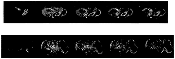
治療後のMR画像化
治療されたマウスおよび治療されていないマウスについての腫瘍取り込みの違いを決定するために、治療後のMR画像法のため、高分子量造影剤(Gd-DTPA)-シスチン・コポリマー(GDCP)を使用した。この薬剤は、類似した腫瘍モデルにおいて血管形成中の血管において比較的強いコントラスト増強を生じることが以前の研究において示されている。ポリマーの分子量および緩和度は、Pol1のものに類似しており、34kDaおよび5.33mM-1sec-1であった。
Post-treatment MR imaging High molecular weight contrast agent (Gd-DTPA) -cystine copolymer for post-treatment MR imaging to determine differences in tumor uptake between treated and untreated mice (GDCP) was used. Previous studies have shown that this drug produces a relatively strong contrast enhancement in angiogenic blood vessels in a similar tumor model. The molecular weight and relaxivity of the polymer was similar to that of Pol 1 and was 34 kDa and 5.33 mM −1 sec −1 .
図9は、注射後2、5、10、15、30分目の、GDCCを受容したマウスの3D MIPエコーMR画像を例示している。図10は、注射後2、5、10、15、30分目の、GDCCを受容したマウスの2DスピンエコーMR画像を例示している。両方の群のマウスについて、腫瘍は、周縁部増強を示した。しかしながら、対照群のマウスは、治療の欠如を示すより高いコントラスト増強を示した。 FIG. 9 illustrates 3D MIP echo MR images of mice receiving GDCC at 2, 5, 10, 15, 30 minutes after injection. FIG. 10 illustrates 2D spin echo MR images of mice receiving GDCC at 2, 5, 10, 15, 30 minutes after injection. For both groups of mice, the tumors showed marginal enhancement. However, the control group of mice showed higher contrast enhancement indicating lack of treatment.
結果
薬物を受容したマウスにおける腫瘍体積の減少は、対照と有意に異なっていた。さらに、上記の結果は、例えば、MRIから入手された動的なデータを使用して、照射の時間が決定され調整され得、増強された部位特異的光線力学的療法のための増加した効力に至ることを例示している。
Results The reduction in tumor volume in mice receiving the drug was significantly different from the control. In addition, the above results can be obtained, for example, using dynamic data obtained from MRI, and the time of irradiation can be determined and adjusted to increase efficacy for enhanced site-specific photodynamic therapy. This is an example.
本明細書に引用された公開、特許、および特許出願を含む全ての参照は、あたかも各参照が参照により組み入れられると個々に具体的に示され、完全に本明細書に記述されたかのような程度に、参照により本明細書に組み入れられる。 All references, including publications, patents, and patent applications cited in this specification are individually illustrated as if each reference was incorporated by reference, and to the extent that they were fully described herein. And incorporated herein by reference.
本発明はある種の態様において記載されているが、本発明は、さらに、この開示の本旨および範囲の中で修飾され得る。従って、本願は、その一般的な原理を使用した本発明の任意の変動、使用、または適応を包含するものとする。さらに、本願は、本発明が関係する技術分野における公知のまたは慣習的な実務の範囲内にあり、添付の特許請求の範囲の範囲内にあるような、本発明の開示からのそのような逸脱を包含するものとする。 While this invention has been described in certain embodiments, the present invention can be further modified within the spirit and scope of this disclosure. This application is therefore intended to cover any variations, uses, or adaptations of the invention using its general principles. Furthermore, this application is within the scope of known or customary practice in the technical field to which this invention pertains, and such departure from the disclosure of this invention as falling within the scope of the appended claims It shall be included.
参照
Claims (13)
造影剤および光増感剤を含む送達系を、薬学的に有効な量で、対象に、またはインビトロで投与する工程;
送達系を標的組織に運搬する工程;
画像モダリティにより標的組織の位置を特定する工程;ならびに
標的組織を照明し、標的部位を死滅させるかまたは破壊する工程。 A method of killing or destroying a target site in a subject or in vitro comprising the following steps:
Administering a delivery system comprising a contrast agent and a photosensitizer to a subject, or in vitro, in a pharmaceutically effective amount;
Transporting the delivery system to the target tissue;
Locating the target tissue by image modality; and illuminating the target tissue to kill or destroy the target site.
アプリケータ;および
レーザー
を含む、標的組織を治療するための系。 Magnetic resonance imaging photosensitized contrast agent;
A system for treating a target tissue, including an applicator; and a laser.
造影剤および光増感剤を含む送達系を、薬学的に有効な量で、対象に投与する工程;
送達系を標的組織に運搬する工程;ならびに
標的組織を照明し、標的部位を治療する工程。 A method of cosmetically treating a target site of interest comprising the following steps:
Administering to the subject a delivery system comprising a contrast agent and a photosensitizer in a pharmaceutically effective amount;
Delivering the delivery system to the target tissue; and illuminating the target tissue and treating the target site.
Applications Claiming Priority (2)
| Application Number | Priority Date | Filing Date | Title |
|---|---|---|---|
| US63325504P | 2004-12-03 | 2004-12-03 | |
| PCT/US2005/044012 WO2006060797A2 (en) | 2004-12-03 | 2005-12-02 | Mri guided photodynamic therapy for cancer |
Publications (2)
| Publication Number | Publication Date |
|---|---|
| JP2008522666A true JP2008522666A (en) | 2008-07-03 |
| JP2008522666A5 JP2008522666A5 (en) | 2009-01-29 |
Family
ID=36565853
Family Applications (1)
| Application Number | Title | Priority Date | Filing Date |
|---|---|---|---|
| JP2007544609A Withdrawn JP2008522666A (en) | 2004-12-03 | 2005-12-02 | MRI-guided photodynamic therapy for cancer |
Country Status (9)
| Country | Link |
|---|---|
| US (1) | US20090076571A1 (en) |
| EP (1) | EP1830879B1 (en) |
| JP (1) | JP2008522666A (en) |
| KR (1) | KR20070086803A (en) |
| AT (1) | ATE485836T1 (en) |
| AU (1) | AU2005311560A1 (en) |
| CA (1) | CA2589881A1 (en) |
| DE (1) | DE602005024437D1 (en) |
| WO (1) | WO2006060797A2 (en) |
Cited By (1)
| Publication number | Priority date | Publication date | Assignee | Title |
|---|---|---|---|---|
| JP2012529500A (en) * | 2009-06-12 | 2012-11-22 | エラスムス・ユニヴァーシティ・メディカル・センター・ロッテルダム | Targeted nanophotomedicine for photodynamic therapy of cancer |
Families Citing this family (8)
| Publication number | Priority date | Publication date | Assignee | Title |
|---|---|---|---|---|
| US20070128118A1 (en) | 2005-12-05 | 2007-06-07 | Nitto Denko Corporation | Polyglutamate-amino acid conjugates and methods |
| US20080181852A1 (en) * | 2007-01-29 | 2008-07-31 | Nitto Denko Corporation | Multi-functional Drug Carriers |
| WO2008124735A2 (en) * | 2007-04-10 | 2008-10-16 | Nitto Denko Corporation | Multi-functional polyglutamate drug carriers |
| WO2008141110A2 (en) * | 2007-05-09 | 2008-11-20 | Nitto Denko Corporation | Polyglutamate conjugates and polyglutamate-amino acid conjugates having a plurality of drugs |
| WO2009055542A1 (en) * | 2007-10-26 | 2009-04-30 | University Of Utah Research Foundation | Use of mri contrast agents for evaluating the treatment of tumors |
| WO2013119957A1 (en) * | 2012-02-10 | 2013-08-15 | Aidan Research And Consulting, Llc | Weight reduction through inactivation of gastric orexigenic mediator producing cells |
| TW201514188A (en) * | 2013-03-13 | 2015-04-16 | Lantheus Medical Imaging Inc | Process for manufacture of gadofosveset trisodium monohydrate |
| CN114870014B (en) * | 2022-05-18 | 2023-07-07 | 南京邮电大学 | Multifunctional anti-tumor polymer medicine and preparation method and application thereof |
Family Cites Families (5)
| Publication number | Priority date | Publication date | Assignee | Title |
|---|---|---|---|---|
| US5594136A (en) * | 1989-12-21 | 1997-01-14 | Pharmacyclics, Inc. | Texaphyrin solid supports and devices |
| US6340461B1 (en) * | 1996-12-17 | 2002-01-22 | David Stephen Terman | Superantigen based methods and compositions for treatment of diseases |
| DE19831217A1 (en) * | 1998-07-03 | 2000-01-05 | Schering Ag | New porphyrin derivatives, pharmaceutical compositions containing them and their use in photodynamic therapy and MRI diagnostics |
| US6534040B2 (en) * | 1999-12-23 | 2003-03-18 | Health Research, Inc. | Chlorin and bacteriochlorin-based aminophenyl DTPA and N2S2 conjugates for MR contrast media and radiopharmaceuticals |
| US20020197648A1 (en) * | 2001-05-02 | 2002-12-26 | Silva Robin M. | High throughput screening methods using magnetic resonance imaging agents |
-
2005
- 2005-12-02 JP JP2007544609A patent/JP2008522666A/en not_active Withdrawn
- 2005-12-02 DE DE602005024437T patent/DE602005024437D1/en active Active
- 2005-12-02 US US11/792,206 patent/US20090076571A1/en not_active Abandoned
- 2005-12-02 EP EP05853048A patent/EP1830879B1/en not_active Not-in-force
- 2005-12-02 AT AT05853048T patent/ATE485836T1/en not_active IP Right Cessation
- 2005-12-02 AU AU2005311560A patent/AU2005311560A1/en not_active Abandoned
- 2005-12-02 WO PCT/US2005/044012 patent/WO2006060797A2/en active Application Filing
- 2005-12-02 KR KR1020077014910A patent/KR20070086803A/en not_active Application Discontinuation
- 2005-12-02 CA CA002589881A patent/CA2589881A1/en not_active Abandoned
Cited By (1)
| Publication number | Priority date | Publication date | Assignee | Title |
|---|---|---|---|---|
| JP2012529500A (en) * | 2009-06-12 | 2012-11-22 | エラスムス・ユニヴァーシティ・メディカル・センター・ロッテルダム | Targeted nanophotomedicine for photodynamic therapy of cancer |
Also Published As
| Publication number | Publication date |
|---|---|
| US20090076571A1 (en) | 2009-03-19 |
| EP1830879A2 (en) | 2007-09-12 |
| WO2006060797A2 (en) | 2006-06-08 |
| ATE485836T1 (en) | 2010-11-15 |
| AU2005311560A1 (en) | 2006-06-08 |
| EP1830879A4 (en) | 2008-09-17 |
| CA2589881A1 (en) | 2006-06-08 |
| KR20070086803A (en) | 2007-08-27 |
| DE602005024437D1 (en) | 2010-12-09 |
| WO2006060797A3 (en) | 2006-08-24 |
| EP1830879B1 (en) | 2010-10-27 |
Similar Documents
| Publication | Publication Date | Title |
|---|---|---|
| Liang et al. | Theranostic porphyrin dyad nanoparticles for magnetic resonance imaging guided photodynamic therapy | |
| JP2008522666A (en) | MRI-guided photodynamic therapy for cancer | |
| US8323621B2 (en) | Multi-use multimodal imaging chelates | |
| Zhu et al. | Hyperbranched polymers for bioimaging | |
| Gao et al. | Theranostic nanodots with aggregation-induced emission characteristic for targeted and image-guided photodynamic therapy of hepatocellular carcinoma | |
| Feng et al. | Assembly of upconversion nanophotosensitizer in vivo to achieve scatheless real-time imaging and selective photodynamic therapy | |
| JP6585504B2 (en) | Porphyrin-modified telodendrimer | |
| JPH10500659A (en) | Texaphyrin metal complexes with improved functionality | |
| Yuzhakova et al. | In vivo multimodal tumor imaging and photodynamic therapy with novel theranostic agents based on the porphyrazine framework-chelated gadolinium (III) cation | |
| US20090311182A1 (en) | Macromolecular Delivery Systems for Non-Invasive Imaging, Evaluation and Treatment of Arthritis and Other Inflammatory Diseases | |
| Zhu et al. | Cascade-responsive nano-assembly for efficient photothermal-chemo synergistic inhibition of tumor metastasis by targeting cancer stem cells | |
| US20110110866A1 (en) | Elastin-like polypeptide and gadolinium conjugate for magnetic resonance imaging | |
| WO2009038776A1 (en) | Therapeutic nanoconjugates | |
| Wang et al. | Dual-mode imaging guided multifunctional theranosomes with mitochondria targeting for photothermally controlled and enhanced photodynamic therapy in vitro and in vivo | |
| Vaidya et al. | Contrast enhanced MRI‐guided photodynamic therapy for site‐specific cancer treatment | |
| Lee et al. | Supramolecular assembly based on host–guest interaction between beta-cyclodextrin and adamantane for specifically targeted cancer imaging | |
| Wang et al. | Folic Acid–Conjugated Pyropheophorbide a as the Photosensitizer Tested for In Vivo Targeted Photodynamic Therapy | |
| Jibin et al. | Nanohybrids of magnetically intercalated optical metamaterials for magnetic resonance/Raman imaging and in situ chemodynamic/photothermal therapy | |
| Sun et al. | Degradable FeCuS-lipid nanoparticles confer ultrasound-activated CO release and O2-independent radical production for synergistic therapy | |
| Zhang et al. | Versatile gadolinium (III)-phthalocyaninate photoagent for MR/PA imaging-guided parallel photocavitation and photodynamic oxidation at single-laser irradiation | |
| CN107337685B (en) | Folate-targeted Pyro photosensitive synthesis and application | |
| CN112546221A (en) | Tumor diagnosis and treatment medicine and preparation method and application thereof | |
| JP2022525791A (en) | Photoimmunotherapy and the drugs used for it | |
| Jiang et al. | Theranostic Nanoprobes with Aggregation‐Induced NIR‐II Emission: from Molecular Design to Biomedical Application | |
| Wang et al. | Methoxypolyethylene Glycol-Substituted Zinc Phthalocyanines for Multiple Tumor-Selective Fluorescence Imaging and Photodynamic Therapy |
Legal Events
| Date | Code | Title | Description |
|---|---|---|---|
| A521 | Request for written amendment filed |
Free format text: JAPANESE INTERMEDIATE CODE: A523 Effective date: 20081202 |
|
| A621 | Written request for application examination |
Free format text: JAPANESE INTERMEDIATE CODE: A621 Effective date: 20081202 |
|
| A761 | Written withdrawal of application |
Free format text: JAPANESE INTERMEDIATE CODE: A761 Effective date: 20100806 |
|
| A521 | Request for written amendment filed |
Free format text: JAPANESE INTERMEDIATE CODE: A821 Effective date: 20100806 |