JP2008502355A - Identification methods and compounds useful in the treatment of degenerative and inflammatory diseases - Google Patents
Identification methods and compounds useful in the treatment of degenerative and inflammatory diseases Download PDFInfo
- Publication number
- JP2008502355A JP2008502355A JP2007526437A JP2007526437A JP2008502355A JP 2008502355 A JP2008502355 A JP 2008502355A JP 2007526437 A JP2007526437 A JP 2007526437A JP 2007526437 A JP2007526437 A JP 2007526437A JP 2008502355 A JP2008502355 A JP 2008502355A
- Authority
- JP
- Japan
- Prior art keywords
- degradation
- mmp1
- polypeptide
- expression
- cells
- Prior art date
- Legal status (The legal status is an assumption and is not a legal conclusion. Google has not performed a legal analysis and makes no representation as to the accuracy of the status listed.)
- Pending
Links
Images


Classifications
-
- C—CHEMISTRY; METALLURGY
- C12—BIOCHEMISTRY; BEER; SPIRITS; WINE; VINEGAR; MICROBIOLOGY; ENZYMOLOGY; MUTATION OR GENETIC ENGINEERING
- C12Q—MEASURING OR TESTING PROCESSES INVOLVING ENZYMES, NUCLEIC ACIDS OR MICROORGANISMS; COMPOSITIONS OR TEST PAPERS THEREFOR; PROCESSES OF PREPARING SUCH COMPOSITIONS; CONDITION-RESPONSIVE CONTROL IN MICROBIOLOGICAL OR ENZYMOLOGICAL PROCESSES
- C12Q1/00—Measuring or testing processes involving enzymes, nucleic acids or microorganisms; Compositions therefor; Processes of preparing such compositions
- C12Q1/34—Measuring or testing processes involving enzymes, nucleic acids or microorganisms; Compositions therefor; Processes of preparing such compositions involving hydrolase
- C12Q1/37—Measuring or testing processes involving enzymes, nucleic acids or microorganisms; Compositions therefor; Processes of preparing such compositions involving hydrolase involving peptidase or proteinase
-
- A—HUMAN NECESSITIES
- A61—MEDICAL OR VETERINARY SCIENCE; HYGIENE
- A61P—SPECIFIC THERAPEUTIC ACTIVITY OF CHEMICAL COMPOUNDS OR MEDICINAL PREPARATIONS
- A61P19/00—Drugs for skeletal disorders
- A61P19/02—Drugs for skeletal disorders for joint disorders, e.g. arthritis, arthrosis
-
- A—HUMAN NECESSITIES
- A61—MEDICAL OR VETERINARY SCIENCE; HYGIENE
- A61P—SPECIFIC THERAPEUTIC ACTIVITY OF CHEMICAL COMPOUNDS OR MEDICINAL PREPARATIONS
- A61P29/00—Non-central analgesic, antipyretic or antiinflammatory agents, e.g. antirheumatic agents; Non-steroidal antiinflammatory drugs [NSAID]
-
- C—CHEMISTRY; METALLURGY
- C12—BIOCHEMISTRY; BEER; SPIRITS; WINE; VINEGAR; MICROBIOLOGY; ENZYMOLOGY; MUTATION OR GENETIC ENGINEERING
- C12N—MICROORGANISMS OR ENZYMES; COMPOSITIONS THEREOF; PROPAGATING, PRESERVING, OR MAINTAINING MICROORGANISMS; MUTATION OR GENETIC ENGINEERING; CULTURE MEDIA
- C12N15/00—Mutation or genetic engineering; DNA or RNA concerning genetic engineering, vectors, e.g. plasmids, or their isolation, preparation or purification; Use of hosts therefor
- C12N15/09—Recombinant DNA-technology
- C12N15/11—DNA or RNA fragments; Modified forms thereof; Non-coding nucleic acids having a biological activity
- C12N15/111—General methods applicable to biologically active non-coding nucleic acids
-
- C—CHEMISTRY; METALLURGY
- C12—BIOCHEMISTRY; BEER; SPIRITS; WINE; VINEGAR; MICROBIOLOGY; ENZYMOLOGY; MUTATION OR GENETIC ENGINEERING
- C12N—MICROORGANISMS OR ENZYMES; COMPOSITIONS THEREOF; PROPAGATING, PRESERVING, OR MAINTAINING MICROORGANISMS; MUTATION OR GENETIC ENGINEERING; CULTURE MEDIA
- C12N9/00—Enzymes; Proenzymes; Compositions thereof; Processes for preparing, activating, inhibiting, separating or purifying enzymes
- C12N9/14—Hydrolases (3)
- C12N9/48—Hydrolases (3) acting on peptide bonds (3.4)
- C12N9/50—Proteinases, e.g. Endopeptidases (3.4.21-3.4.25)
- C12N9/64—Proteinases, e.g. Endopeptidases (3.4.21-3.4.25) derived from animal tissue
- C12N9/6421—Proteinases, e.g. Endopeptidases (3.4.21-3.4.25) derived from animal tissue from mammals
- C12N9/6489—Metalloendopeptidases (3.4.24)
- C12N9/6491—Matrix metalloproteases [MMP's], e.g. interstitial collagenase (3.4.24.7); Stromelysins (3.4.24.17; 3.2.1.22); Matrilysin (3.4.24.23)
-
- G—PHYSICS
- G01—MEASURING; TESTING
- G01N—INVESTIGATING OR ANALYSING MATERIALS BY DETERMINING THEIR CHEMICAL OR PHYSICAL PROPERTIES
- G01N33/00—Investigating or analysing materials by specific methods not covered by groups G01N1/00 - G01N31/00
- G01N33/48—Biological material, e.g. blood, urine; Haemocytometers
- G01N33/50—Chemical analysis of biological material, e.g. blood, urine; Testing involving biospecific ligand binding methods; Immunological testing
- G01N33/68—Chemical analysis of biological material, e.g. blood, urine; Testing involving biospecific ligand binding methods; Immunological testing involving proteins, peptides or amino acids
- G01N33/6887—Chemical analysis of biological material, e.g. blood, urine; Testing involving biospecific ligand binding methods; Immunological testing involving proteins, peptides or amino acids from muscle, cartilage or connective tissue
-
- C—CHEMISTRY; METALLURGY
- C12—BIOCHEMISTRY; BEER; SPIRITS; WINE; VINEGAR; MICROBIOLOGY; ENZYMOLOGY; MUTATION OR GENETIC ENGINEERING
- C12N—MICROORGANISMS OR ENZYMES; COMPOSITIONS THEREOF; PROPAGATING, PRESERVING, OR MAINTAINING MICROORGANISMS; MUTATION OR GENETIC ENGINEERING; CULTURE MEDIA
- C12N2310/00—Structure or type of the nucleic acid
- C12N2310/10—Type of nucleic acid
- C12N2310/14—Type of nucleic acid interfering N.A.
-
- C—CHEMISTRY; METALLURGY
- C12—BIOCHEMISTRY; BEER; SPIRITS; WINE; VINEGAR; MICROBIOLOGY; ENZYMOLOGY; MUTATION OR GENETIC ENGINEERING
- C12N—MICROORGANISMS OR ENZYMES; COMPOSITIONS THEREOF; PROPAGATING, PRESERVING, OR MAINTAINING MICROORGANISMS; MUTATION OR GENETIC ENGINEERING; CULTURE MEDIA
- C12N2320/00—Applications; Uses
- C12N2320/10—Applications; Uses in screening processes
- C12N2320/12—Applications; Uses in screening processes in functional genomics, i.e. for the determination of gene function
-
- G—PHYSICS
- G01—MEASURING; TESTING
- G01N—INVESTIGATING OR ANALYSING MATERIALS BY DETERMINING THEIR CHEMICAL OR PHYSICAL PROPERTIES
- G01N2333/00—Assays involving biological materials from specific organisms or of a specific nature
- G01N2333/90—Enzymes; Proenzymes
- G01N2333/914—Hydrolases (3)
- G01N2333/948—Hydrolases (3) acting on peptide bonds (3.4)
- G01N2333/95—Proteinases, i.e. endopeptidases (3.4.21-3.4.99)
- G01N2333/964—Proteinases, i.e. endopeptidases (3.4.21-3.4.99) derived from animal tissue
- G01N2333/96425—Proteinases, i.e. endopeptidases (3.4.21-3.4.99) derived from animal tissue from mammals
- G01N2333/96427—Proteinases, i.e. endopeptidases (3.4.21-3.4.99) derived from animal tissue from mammals in general
- G01N2333/9643—Proteinases, i.e. endopeptidases (3.4.21-3.4.99) derived from animal tissue from mammals in general with EC number
- G01N2333/96486—Metalloendopeptidases (3.4.24)
-
- G—PHYSICS
- G01—MEASURING; TESTING
- G01N—INVESTIGATING OR ANALYSING MATERIALS BY DETERMINING THEIR CHEMICAL OR PHYSICAL PROPERTIES
- G01N2500/00—Screening for compounds of potential therapeutic value
- G01N2500/04—Screening involving studying the effect of compounds C directly on molecule A (e.g. C are potential ligands for a receptor A, or potential substrates for an enzyme A)
-
- G—PHYSICS
- G01—MEASURING; TESTING
- G01N—INVESTIGATING OR ANALYSING MATERIALS BY DETERMINING THEIR CHEMICAL OR PHYSICAL PROPERTIES
- G01N2800/00—Detection or diagnosis of diseases
- G01N2800/10—Musculoskeletal or connective tissue disorders
- G01N2800/101—Diffuse connective tissue disease, e.g. Sjögren, Wegener's granulomatosis
- G01N2800/102—Arthritis; Rheumatoid arthritis, i.e. inflammation of peripheral joints
Abstract
Description
(発明の分野)
本発明は、細胞外マトリックス(ECM)の分解をもたらす経路に関係するタンパク質の発現を抑制することができる化合物及び発現抑制剤の同定方法に関し、前記抑制は関節変性及びそのような分解及び/又は炎症が関与する疾患の予防及び治療に役立つ。
(Field of Invention)
The present invention relates to a method for identifying a compound and an expression inhibitor capable of suppressing the expression of a protein related to a pathway that leads to degradation of extracellular matrix (ECM), wherein the suppression includes joint degeneration and such degradation and / or Helps prevent and treat diseases involving inflammation.
細胞外マトリックスの分解が関与する疾患としては、それらには限定されないが、乾癬性関節炎、若年性関節炎、初期の関節炎、反応性関節炎、変形性関節炎、強直性脊椎炎、骨粗鬆症、腱炎及び歯周疾患のような骨格筋の疾患、癌転移、気道疾患(COPD、喘息)、腎臓及び肝臓の線維症、アテローム性動脈硬化症及び心不全のような心臓血管疾患並びに神経炎及び多発性硬化症のような神経系疾患がある。主に関節の変性が関与する疾患としては、それらには限定されないが、乾癬性関節炎、若年性関節炎、初期の関節炎、反応性関節炎、変形性関節炎、強直性脊椎炎がある。 Diseases involving extracellular matrix degradation include but are not limited to psoriatic arthritis, juvenile arthritis, early arthritis, reactive arthritis, osteoarthritis, ankylosing spondylitis, osteoporosis, tendonitis and teeth Skeletal muscle diseases such as perivascular disease, cancer metastasis, airway disease (COPD, asthma), kidney and liver fibrosis, cardiovascular diseases such as atherosclerosis and heart failure, and neuritis and multiple sclerosis There are such nervous system diseases. Diseases mainly involving joint degeneration include, but are not limited to, psoriatic arthritis, juvenile arthritis, early arthritis, reactive arthritis, osteoarthritis, ankylosing spondylitis.
関節リウマチ(RA)は慢性の関節変性疾患であり、関節構造の炎症及び破壊を特徴とする。阻止されないと疾患は関節機能の喪失による実質的な障害及び疼痛を、また早死にさえもたらす。RA療法の狙いは、したがって、関節の破壊を止めるために疾患を遅らせることではなく、寛解を達成することである。疾患予後の重大度の他に、RAの高い有病率(世界中で成人の約0.8%が影響を受ける)は、大きな社会経済的影響を意味する。(RAに関するレビューについては、Smolen及びSteiner (2003);Lee及びWeinblatt (2001);Choy及びPanayi (2001);O'Dell (2004)及びFirestein (2003)を参照する)。 Rheumatoid arthritis (RA) is a chronic joint degenerative disease characterized by inflammation and destruction of joint structures. If not prevented, the disease results in substantial disability and pain due to loss of joint function, and even premature death. The aim of RA therapy is therefore to achieve remission rather than delay the disease to stop joint destruction. In addition to the severity of disease prognosis, a high prevalence of RA (approximately 0.8% of adults are affected worldwide) represents a significant socio-economic impact. (For reviews on RA, see Smolen and Steiner (2003); Lee and Weinblatt (2001); Choy and Panayi (2001); O'Dell (2004) and Firestein (2003)).
RAが自己免疫疾患であることは広く受け入れられているが、疾患の「開始段階」を作動させる正確なメカニズムに関してコンセンサスはない。知られていることは、素因のある宿主で最初の引き金がカスケード事象を媒介し、それらが様々な細胞型(B細胞、T細胞、マクロファージ、線維芽細胞、内皮細胞、樹状細胞その他)の活性化を導くことである。同時に、様々なサイトカインの増産が関節及び関節周囲の組織で観察される(例えばTNF−α、IL−6、IL−1、IL−15、IL−18その他)。疾患が進行するとき、細胞活性化及びサイトカイン産生カスケードは無際限に継続するようになる。この初期段階で、関節構造の破壊はこのような早い段階ですでに非常に明白である。患者の30パーセントは診断時点で骨性浸食のエックス線撮影像を有し、この割合は2年後に60パーセントまで増加する。 While it is widely accepted that RA is an autoimmune disease, there is no consensus on the exact mechanism that triggers the “onset phase” of the disease. What is known is that the first trigger mediates a cascade event in a predisposed host, and they are of various cell types (B cells, T cells, macrophages, fibroblasts, endothelial cells, dendritic cells, etc.) Leading to activation. At the same time, increased production of various cytokines is observed in joints and periarticular tissues (eg TNF-α, IL-6, IL-1, IL-15, IL-18 etc.). As the disease progresses, cell activation and cytokine production cascades will continue indefinitely. At this early stage, the destruction of the joint structure is already very obvious at such an early stage. Thirty percent of patients have x-ray images of bone erosion at the time of diagnosis, which increases to 60 percent after two years.
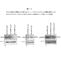
RA患者の関節の組織学的分析は、明らかにRA関連の分解過程に関係するメカニズムを明示する。滑膜は細胞層であり、下内張り、及び滑膜の空洞から関節嚢を分離する内層領域から構成される。炎症を起こした滑膜は、RAの病態生理学にとって重要である。正常患者及びRA患者の間の滑膜の組織学的差を、図1で示す。A.滑膜関節は、2つの隣接した骨末端から構成され、それぞれは軟骨層で覆われ、関節空間によって分離され、滑膜層及び関節嚢によって囲まれる。滑膜層は、滑膜細胞の薄い(1〜3細胞)層及び非常に血管の多い下内張り結合組織層からなる滑膜内層(軟骨及び骨に面する)で構成される。滑膜層は、軟骨を除いてほとんどすべての関節腔内の構造物を覆う。B.他の多くの形態の関節炎と同様に、関節リウマチ(RA)は、最初に、局所の又は浸潤した単核細胞の活性化と同様に各種の単核細胞の重要な流入を特徴とする滑膜層の炎症性反応(「滑膜炎」)を特徴とする。内膜層は過形成を生じ(>20細胞の厚さとなることもある)、滑膜層は拡大する。しかし、さらに、RAの特質は関節の破壊である。「浸食」とも呼ばれる軟骨分解及び隣接骨の破壊の徴候が起こるにしたがって、関節空間は狭くなるか消失する。滑膜層の破壊部分は、「パンヌス」と呼ばれる。滑膜細胞によって分泌される酵素は、軟骨分解を導く。 Histological analysis of the joints of RA patients clearly demonstrates the mechanisms involved in RA-related degradation processes. The synovium is a cell layer that is composed of an underlying lining and an inner layer region that separates the joint capsule from the synovial cavity. Inflamed synovium is important for the pathophysiology of RA. The synovial histological differences between normal and RA patients are shown in FIG. A. The synovial joint consists of two adjacent bone ends, each covered with a cartilage layer, separated by a joint space, and surrounded by a synovial layer and a joint capsule. The synovial layer is composed of a thin (1-3 cells) layer of synoviocytes and a synovial inner layer (facing cartilage and bone) consisting of a highly vascularized underlying lining connective tissue layer. The synovial layer covers almost all structures in the joint space except for cartilage. B. Like many other forms of arthritis, rheumatoid arthritis (RA) is initially characterized by a significant influx of various mononuclear cells as well as activation of local or infiltrating mononuclear cells. Characterized by an inflammatory reaction of the layer (“synovitis”). The intimal layer causes hyperplasia (may be> 20 cells thick) and the synovial layer expands. However, a further characteristic of RA is joint destruction. As the signs of cartilage degradation, also called “erosion,” and destruction of adjacent bone occur, the joint space narrows or disappears. The destroyed portion of the synovial layer is called “pannus”. Enzymes secreted by synovial cells lead to cartilage degradation.
この分析は、RA関連の関節分解を担う主なエフェクターはパンヌスであり、ここにおいて滑膜線維芽細胞は多様なタンパク質分解酵素を産生することから、軟骨及び骨の浸食の主要な作動因子であることを示す。進行したRA患者では、パンヌスは隣接した軟骨の分解を媒介して関節空間の狭小化を導き、また、隣接した骨及び軟骨に侵入する能力を有する。骨及び軟骨の組織はそれぞれ主にコラーゲンI又はIIで構成されるので、パンヌスの破壊的及び浸潤的な特性はコラーゲン溶解性プロテアーゼ、主にマトリックスメタロプロテイナーゼ(MMP)の分泌によって媒介される。軟骨の下の骨及び隣接した骨の浸食もRA過程の一部であり、主に骨及びパンヌスの境界面の破骨細胞の存在に起因する。破骨細胞は骨組織に付着して閉鎖コンパートメントを形成し、その中で破骨細胞は骨組織を分解するプロテアーゼ(カテプシンK、MMP9)を分泌する。関節内の破骨細胞集団は、活性化されたSF及びT細胞によるNFkBリガンド(RANKL)の受容体活性化剤の分泌によって誘導される前駆細胞からの骨芽細胞形成によって、異常に増加する。 This analysis shows that the main effector responsible for RA-related joint degradation is pannus, where synovial fibroblasts produce a variety of proteolytic enzymes and are therefore key agonists of cartilage and bone erosion It shows that. In advanced RA patients, pannus mediates the degradation of adjacent cartilage leading to joint space narrowing and has the ability to invade adjacent bone and cartilage. Since bone and cartilage tissues are mainly composed of collagen I or II, respectively, the destructive and invasive properties of pannus are mediated by the secretion of collagenolytic proteases, mainly matrix metalloproteinases (MMPs). Erosion of the bone beneath the cartilage and adjacent bone is also part of the RA process and is mainly due to the presence of osteoclasts at the bone and pannus interface. Osteoclasts attach to the bone tissue to form a closed compartment in which the osteoclast secretes a protease (cathepsin K, MMP9) that degrades the bone tissue. The osteoclast population within the joint is abnormally increased by osteoblast formation from progenitor cells induced by the activation of NFkB ligand (RANKL) receptor activators by activated SF and T cells.
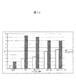
細胞外マトリックス(ECM)の安定性を規定する上で、各種のコラーゲンが重要な役割を果たす。コラーゲンI型及びコラーゲンII型は、例えば、それぞれ骨及び軟骨の主成分である。コラーゲンタンパク質は、一般的に、コラーゲン原繊維と称される多量体の構造物に組織化する。未変性のコラーゲン原繊維は、タンパク分解性の切断に非常に抵抗性である。僅かの種類のECM分解タンパク質が未変性のコラーゲンを分解する能力を有すると報告されており、それらはマトリックスメタロプロテアーゼ類(MMP)及びカテプシン類である。カテプシンの中では、主に破骨細胞で活性であるカテプシンKの特徴が最も解明されている。MMPの中では、MMP1、MMP2、MMP8、MMP13及びMMP14がコラーゲン溶解性特性を有することが知られている。滑膜線維芽細胞(SF)によるMMP1の発現増加及び関節炎疾患の進行間の相互関係は立証されていて、関節浸食過程の前兆となる(Cunnaneら、2001)。したがってRAとの関連で、MMP1は関連性の高いコラーゲン分解タンパク質を表す。インビトロでは、RA病理学と関連したサイトカイン(例えばTNF−α及びIL1β)による培養SFの処理は、これらの細胞によるMMP1の発現を増加させる(Andreakosら、2003)。したがって、SFによって発現されるMMP1のレベルを監視することはインビボで軟骨分解を担う浸食性表現型へのSFの活性化を示すものなので、RAの分野では重要な読み出し情報である。SFによるMMP1発現の抑制は、RAの治療のための価値ある治療手段となる。 Various collagens play an important role in defining the stability of the extracellular matrix (ECM). Collagen type I and collagen type II are, for example, main components of bone and cartilage, respectively. Collagen proteins are generally organized into multimeric structures called collagen fibrils. Native collagen fibrils are very resistant to proteolytic cleavage. A few types of ECM degrading proteins have been reported to have the ability to degrade native collagen, which are matrix metalloproteases (MMPs) and cathepsins. Among the cathepsins, the characteristics of cathepsin K, which is mainly active in osteoclasts, are most elucidated. Among MMPs, it is known that MMP1, MMP2, MMP8, MMP13 and MMP14 have collagen solubility properties. A correlation between increased expression of MMP1 by synovial fibroblasts (SF) and progression of arthritic disease has been established and is a precursor to the joint erosion process (Cunnane et al., 2001). Thus, in the context of RA, MMP1 represents a highly relevant collagenolytic protein. In vitro, treatment of cultured SF with cytokines associated with RA pathology (eg, TNF-α and IL1β) increases MMP1 expression by these cells (Andreakos et al., 2003). Thus, monitoring the level of MMP1 expressed by SF is an important readout in the field of RA because it indicates SF activation to the erosive phenotype responsible for cartilage degradation in vivo. Inhibition of MMP1 expression by SF provides a valuable therapeutic tool for the treatment of RA.
ECM分解タンパク質の活性は、RAと異なる様々な疾患、例えば関節の分解と関連する他の疾患の進行の原因となるか又は相関する可能性もある。これらの疾患としては、それらには限定されないが、乾癬性関節炎、若年性関節炎、初期の関節炎、反応性関節炎、変形性関節炎、及び強直性脊椎炎がある。本発明によって同定される化合物により、本明細書で記載されているMMPの発現に関係する標的を用いて治療することができる他の疾患は、骨粗鬆症、腱炎及び歯周疾患のような骨格筋の疾患(Gapskiら、2004)、癌転移(Coussensら、2002)、気道疾患(COPD、喘息)(Suzukiら、2004)、肺、腎臓線維症(Schanstraら、2002)、慢性C型肝炎と関連する肝臓線維症(Reiffら、2005)、アテローム性動脈硬化症及び心不全のような心臓血管疾患(Creemersら、2001)、並びに神経炎及び多発性硬化症のような神経系疾患(Rosenberg、2002)である。そのような疾患を患っている患者は、(ECMを分解から保護することによって)ECMを安定させることから利益を受けることができる。 The activity of ECM-degrading proteins may cause or correlate with the progression of various diseases different from RA, eg, other diseases associated with joint degradation. These diseases include, but are not limited to, psoriatic arthritis, juvenile arthritis, early arthritis, reactive arthritis, osteoarthritis, and ankylosing spondylitis. Other diseases that can be treated with the compounds identified by the present invention using targets related to the expression of MMPs described herein are skeletal muscles such as osteoporosis, tendinitis and periodontal disease Diseases (Gapski et al., 2004), cancer metastasis (Coussens et al., 2002), respiratory tract disease (COPD, asthma) (Suzuki et al., 2004), lung, kidney fibrosis (Schanstra et al., 2002), associated with chronic hepatitis C Liver fibrosis (Reiff et al., 2005), cardiovascular diseases such as atherosclerosis and heart failure (Creemers et al., 2001), and nervous system diseases such as neuritis and multiple sclerosis (Rosenberg, 2002) It is. Patients suffering from such diseases can benefit from stabilizing the ECM (by protecting the ECM from degradation).
(報告された開発)
NSAID(非ステロイド系抗炎症薬剤)は、RAと関連する疼痛を低減し、患者の生活の質を改善するために用いられる。しかし、これらの薬剤はRA関連の関節破壊を減速させない。
コルチコステロイドはX線撮影で検出されたようにRAの進行を低減することが発見されており、RA患者の一部(30〜60%)を治療するために低用量で用いられる。しかし、重大な副作用が長いコルチコステロイド使用に伴う(皮膚菲薄化、骨粗鬆症、白内障、高血圧、高脂血症)。
(Reported development)
NSAIDs (non-steroidal anti-inflammatory drugs) are used to reduce the pain associated with RA and improve the quality of life of patients. However, these drugs do not slow down RA-related joint destruction.
Corticosteroids have been found to reduce the progression of RA as detected by radiography and are used at low doses to treat some (30-60%) of RA patients. However, it has been associated with long-term use of corticosteroids (skin thinning, osteoporosis, cataracts, hypertension, hyperlipidemia).
合成DMARD(疾患修飾性抗リウマチ薬剤)(例えばメトトレキセート、レフルノミド(leflunomide)、スルファサラジン)は、主にRAの免疫炎症性成分に対処する。主な短所として、これらの薬剤は限定された効力を有するだけである(関節破壊はDMARDによって遅延するだけであり阻止されず、長期的には疾患が進行する)。効力の欠如は、メトトレキセートによる24ヵ月の治療の後、平均して患者の僅か30%がACR50スコアを達成するという事実が示す。このことは、米国リウマチ学会によると、患者の僅か30%が彼らの症状の50%の改善を成し遂げることを意味する(O'Dellら、1996)。さらに、DMARDの正確な作用機構はしばしば不明である。 Synthetic DMARDs (disease modifying anti-rheumatic drugs) (eg methotrexate, leflunomide, sulfasalazine) mainly address the immunoinflammatory components of RA. The main disadvantage is that these drugs only have limited efficacy (joint destruction is only delayed and not prevented by DMARD, and the disease progresses in the long term). The lack of efficacy is indicated by the fact that after 24 months of treatment with methotrexate, on average only 30% of patients achieve an ACR50 score. This means that according to the American College of Rheumatology, only 30% of patients achieve 50% improvement in their symptoms (O'Dell et al., 1996). Moreover, the exact mechanism of action of DMARD is often unclear.
生物学的DMARD(インフリキシマブ、エタナーセプト、アダリムマブ、リツキシマブ、CTLA4−Ig)は、RA病態生理学で重要な役割を演じるサイトカイン(例えばTNF−α)又は細胞(例えばT細胞若しくはB細胞)を不活性化する、タンパク質治療薬である(Kremerら、2003;Edwardsら、2004)。TNF−α−遮断薬(インフリキシマブ、エタナーセプト、アダリムマブ)及びメトトレキセートの併用療法は今日利用できる最も有効なRA治療法であるけれども、この療法でさえ12ヵ月の療法の後に患者の50〜60%で病徴の50%の改善(ACR50)を達成するだけであることは、特筆すべきである(St Clairら、2004)。抗TNF−α薬剤の有害事象の警告がいくつか存在し、この種の薬剤に関連する副作用が明らかにされている。感染症(結核)、血液学的事象及び脱髄性障害に対するリスクの増加が、TNF−α遮断薬に関して記載されている。(Gomez-Reinoら、2003も参照)。重大な副作用の他に、TNF−α遮断薬は生物製剤クラスの薬物治療法の一般的短所も共有するが、それらは不快な投与方法(注入部位反応を伴う頻繁な注射)及び高い生産コストである。後期開発段階のより新しい剤は、T細胞共刺激分子及びB細胞を標的とする。これらの薬剤の効力は、TNF−α遮断薬のそれと類似すると予想される。様々な標的療法は類似するが限定された効力を有するという事実は、RAについて複数の病原性因子があることを示唆する。このことも、RAに関連する病原性事象の理解が欠乏していることを示す。 Biological DMARDs (infliximab, etanercept, adalimumab, rituximab, CTLA4-Ig) inactivate cytokines (eg TNF-α) or cells (eg T cells or B cells) that play an important role in RA pathophysiology A protein therapeutic (Kremer et al., 2003; Edwards et al., 2004). Although TNF-α-blockers (infliximab, etanercept, adalimumab) and methotrexate combination therapy is the most effective RA treatment available today, even this therapy is ill in 50-60% of patients after 12 months of therapy. It should be noted that only a 50% improvement in symptoms (ACR 50) is achieved (St Clair et al., 2004). There are several adverse event warnings for anti-TNF-α drugs and the side effects associated with this type of drug have been clarified. Increased risk for infection (tuberculosis), hematological events and demyelinating disorders has been described for TNF-α blockers. (See also Gomez-Reino et al. 2003). In addition to serious side effects, TNF-α blockers also share the common disadvantages of biopharmaceutical class drug therapies, but they are uncomfortable to administer (frequent injections with injection site reactions) and high production costs. is there. Newer agents in late development stage target T cell costimulatory molecules and B cells. The potency of these drugs is expected to be similar to that of TNF-α blockers. The fact that the various targeted therapies are similar but have limited efficacy suggests that there are multiple virulence factors for RA. This also indicates a lack of understanding of pathogenic events associated with RA.
RAの現在の療法は、限定された効力(患者の30%で十分な療法が存在しない)のために十分でない。このことは、寛解を達成するための更なる手法を要求する。残りの疾患は進行性の関節損傷の、したがって進行性の障害のリスクを抱えているので、寛解が要求される。RA治療のために現在用いられる薬剤の主な標的である、RA疾患の免疫炎症性成分を抑制しても、この疾患の主要な特質である関節の悪化は阻止されない。 Current therapies for RA are not sufficient due to limited efficacy (30% of patients do not have sufficient therapy). This requires additional approaches to achieve remission. Remission is required because the rest of the disease is at risk of progressive joint damage and thus progressive disability. Suppressing the immunoinflammatory component of RA disease, the main target of drugs currently used for the treatment of RA, does not prevent the deterioration of joints, which is a major attribute of this disease.
RA患者の関節の組織学的分析は、パンヌスを関節分解の主な犯人となる攻撃的で浸潤性の組織としてはっきりと同定する。パンヌス内では、滑膜線維芽細胞は、RA発生病理の基礎である異常に引き起こされた免疫系の開始及び最終的な関節浸食の間の関係を象徴する。現在のRA療法は長期的にパンヌスの浸食活性を有効に消滅させるものではないので、パンヌスの生成及び/又は活性を抑制する新規薬剤及び/又は薬剤標的の発見は、新規RA治療法の開発のための重要な道標を意味する。 Histological analysis of the joints of RA patients clearly identifies pannus as an aggressive and invasive tissue that is the main offender of joint degradation. Within pannus, synovial fibroblasts symbolize the relationship between the abnormally initiated immune system initiation and ultimate joint erosion that is the basis of the pathogenesis of RA. Because current RA therapies do not effectively abolish pannus erosion activity in the long term, the discovery of new drugs and / or drug targets that inhibit pannus generation and / or activity is the development of new RA therapies. Meaning an important signpost for.
本発明は、MMP1などの細胞外マトリックス(ECM)分解プロテアーゼの発現をもたらす経路である種類のタンパク質が機能し、これらのタンパク質の活性の阻害剤はそのようなプロテアーゼの異常に高い発現が関与する疾患の治療に役立つという発見に基づく。 The present invention functions a class of proteins that are pathways leading to the expression of extracellular matrix (ECM) degrading proteases such as MMP1, and inhibitors of the activity of these proteins are involved in abnormally high expression of such proteases. Based on the discovery that it helps treat disease.
(発明の要約)
本発明は、細胞外マトリックス(ECM)の分解を抑制する化合物の同定方法に関するものであり、この方法は、化合物を、配列番号101〜125(以下、「ターゲット」)からなる群から選択されるアミノ酸配列を含むポリペプチド及びその断片と、前記ポリペプチドとその化合物との結合を可能にする条件下で接触させること、及び細胞外マトリックス(ECM)分解に関連した化合物−ポリペプチド特性を測定することを含む。
(Summary of the Invention)
The present invention relates to a method for identifying a compound that suppresses degradation of extracellular matrix (ECM), wherein the compound is selected from the group consisting of SEQ ID NOs: 101 to 125 (hereinafter “target”). Contacting a polypeptide comprising an amino acid sequence and fragments thereof under conditions that allow binding of the polypeptide to the compound, and measuring compound-polypeptide properties associated with extracellular matrix (ECM) degradation Including that.
本方法の態様としては、配列番号501〜564からなる群から選択されるものを含むターゲット及びその断片のポリペプチドを用いる化合物のインビトロアッセイ、並びに、ターゲット抑制に続いて効力の指標、例えば、ターゲット発現レベル及び/又はマトリックスメタロプロテイナーゼ1レベルを観察する細胞アッセイがある。
Embodiments of the method include in vitro assays of compounds using polypeptides comprising a target and fragments thereof, including those selected from the group consisting of SEQ ID NOs: 501-564, and indications of efficacy following target suppression, eg, targets There are cellular assays that observe expression levels and / or
本発明は、関節リウマチのような慢性関節変性疾患の治療又は予防に役立つ、アンチセンスポリヌクレオチド、リボザイム及び低分子干渉RNA(siRNA)の群から選択されるポリヌクレオチドを含む発現抑制剤、前記薬剤を含む医薬組成物にも関し、前記ポリヌクレオチドは配列番号101〜125及び501〜564からなる群から選択されるアミノ酸配列を含むポリペプチドをコードする天然ポリヌクレオチド配列と相補的であるか、又はそれから操作された核酸配列を含む。 The present invention relates to an expression inhibitor comprising a polynucleotide selected from the group of antisense polynucleotides, ribozymes and small interfering RNAs (siRNAs), which is useful for treating or preventing chronic joint degenerative diseases such as rheumatoid arthritis, Wherein the polynucleotide is complementary to a natural polynucleotide sequence encoding a polypeptide comprising an amino acid sequence selected from the group consisting of SEQ ID NOs: 101-125 and 501-564, or It contains the engineered nucleic acid sequence.
本発明の他の態様は、その疾患に患っているか感受性の対象において、発現抑制剤のターゲット発現を抑制するのに有効な量を含む医薬組成物を投与することによる、細胞外マトリックス(ECM)の分解が関与する病態の治療方法又は予防方法である。
本発明の他の態様は、細胞外マ トリックス(ECM)の分解を特徴とする病状に関する診断のための、対象におけるターゲット発現レベル指標の測定を含む方法である。
Another aspect of the present invention is an extracellular matrix (ECM) by administering to a subject suffering from or susceptible to the disease a pharmaceutical composition comprising an amount effective to inhibit target expression of the expression inhibitor. It is a method for treating or preventing a pathological condition involving the degradation of.
Another aspect of the invention is a method that includes measuring a target expression level indicator in a subject for diagnosis related to a disease state characterized by degradation of extracellular matrix (ECM).
本発明の他の態様は、炎症が関与する疾患、特に異常なマトリックスメタロプロテアーゼ活性を特徴とする疾患の治療に役立つ、治療法における本化合物の使用、医薬組成物及びそのような組成物の製造に関するものである。 Another aspect of the present invention is the use of the present compounds in therapeutic methods, pharmaceutical compositions and the preparation of such compositions useful for the treatment of diseases involving inflammation, in particular diseases characterized by abnormal matrix metalloprotease activity. It is about.
(詳細な説明)
以下の用語はそれとともに下で提示される意味を有するものとし、本発明の記載及び対象とする範囲を理解するために役立つ。
用語「薬剤」は、ポリペプチド、ポリヌクレオチド及び小分子を含む任意の分子を意味する。
(Detailed explanation)
The following terms shall have the meanings presented below with them and will be helpful in understanding the description and scope of the invention.
The term “agent” means any molecule, including polypeptides, polynucleotides and small molecules.
用語「アゴニスト」は、最も広い意味ではリガンドが結合する受容体を刺激するリガンドを指す。
用語「アッセイ」は、化合物の特定の特性を測定するために用いられる任意の方法を意味する。「スクリーニングアッセイ」は、化合物の集団からそれらの活性に基づいて化合物の特徴を明らかにするかそれらを選択するために用いられる方法を意味する。
用語「結合親和性」は、2つ以上の化合物が互いに非共有結合でどの程度強く結合するかを記述する特性である。結合親和性は、質的に(例えば「強い」、「弱い」、「高い」又は「低い」)、或いは量的に(例えば、KDの測定)特徴付けることができる。
The term “agonist” refers in the broadest sense to a ligand that stimulates the receptor to which it binds.
The term “assay” means any method used to measure a particular property of a compound. "Screening assay" means a method used to characterize or select compounds from a population of compounds based on their activity.
The term “binding affinity” is a property that describes how strongly two or more compounds bind to each other non-covalently. Binding affinity is qualitatively be characterized (e.g., "strong", "weak", "high" or "low"), or quantitatively (e.g., measurement of K D).
用語「担体」は、医薬組成物に媒体、嵩及び/又は使用可能な形態を提供するために医薬組成物の製剤で用いる無毒性材料を意味する。担体は賦形剤、安定剤又は水性のpH緩衝溶液などのそのような材料の1つ又は複数を含むことができる。生理的に許容できる担体の例としては:リン酸、クエン酸及び他の有機酸を含む水性又は固体の緩衝成分;アスコルビン酸を含む抗酸化剤;低分子(約10未満残基)ポリペプチド、血清アルブミン、ゼラチン又は免疫グロブリンなどのタンパク質;ポリビニルピロリドンなどの親水性ポリマー;グリシン、グルタミン、アスパラギン、アルギニン又はリジンなどのアミノ酸;グルコース、マンノース又はデキストリンを含む単糖、二糖及び他の炭水化物;EDTAなどのキレート剤;マンニトール又はソルビトールなどの糖アルコール;ナトリウムなどの塩形成対イオン;並びに/或いはTWEEN(商標)、ポリエチレングリコール(PEG)及びPLURONICS(商標)などの非イオン性界面活性剤である。 The term “carrier” refers to a non-toxic material used in the formulation of a pharmaceutical composition to provide the pharmaceutical composition with a vehicle, bulk and / or usable form. The carrier can include one or more of such materials, such as excipients, stabilizers or aqueous pH buffered solutions. Examples of physiologically acceptable carriers include: aqueous or solid buffer components including phosphoric acid, citric acid and other organic acids; antioxidants including ascorbic acid; small molecule (less than about 10 residues) polypeptides; Proteins such as serum albumin, gelatin or immunoglobulin; hydrophilic polymers such as polyvinylpyrrolidone; amino acids such as glycine, glutamine, asparagine, arginine or lysine; monosaccharides, disaccharides and other carbohydrates including glucose, mannose or dextrin; EDTA Chelating agents such as; sugar alcohols such as mannitol or sorbitol; salt-forming counterions such as sodium; and / or nonionic surfactants such as TWEEN ™, polyethylene glycol (PEG) and PLURONICS ™.
用語「複合体」は、2つ以上の化合物が互いと結合するときに形成される実体を意味する。
用語「化合物」は、本明細書では本発明のアッセイと関連して記載される「試験化合物」又は「薬剤候補化合物」との関連で用いられる。そうしたものとして、これらの化合物は合成又は天然供給源由来の有機又は無機の化合物を含む。化合物には、無機又は有機の化合物、例えば比較的低分子量を特徴とするポリヌクレオチド、脂質又はホルモン類似体が含まれる。他のバイオポリマー有機試験化合物としては、約2から約40のアミノ酸を含むペプチド及び約40から約500のアミノ酸を含むより大きなポリペプチド、例えば抗体若しくは抗体複合体がある。
The term “complex” means an entity formed when two or more compounds bind to each other.
The term “compound” is used herein in the context of a “test compound” or “drug candidate compound” described in connection with the assay of the present invention. As such, these compounds include organic or inorganic compounds derived from synthetic or natural sources. Compounds include inorganic or organic compounds, such as polynucleotides, lipids or hormone analogs characterized by a relatively low molecular weight. Other biopolymer organic test compounds include peptides containing from about 2 to about 40 amino acids and larger polypeptides containing from about 40 to about 500 amino acids, such as antibodies or antibody conjugates.
用語「病態」又は「疾患」は、症状(すなわち病気)の明白な提示又は異常な臨床指標(例えば生化学指標)の現れを意味する。或いは、用語「疾患」は、そのような症状又は異常な臨床指標を起こす遺伝的若しくは環境的リスク又はその傾向を指す。
用語「接触」又は「接触させる」は、インビトロ系であれインビボ系であれ、少なくとも2つの部分を一緒にすることを意味する。
The term “disease state” or “disease” means the overt presentation of symptoms (ie disease) or the appearance of abnormal clinical indicators (eg biochemical indicators). Alternatively, the term “disease” refers to a genetic or environmental risk or tendency to cause such symptoms or abnormal clinical indicators.
The term “contacting” or “contacting” means bringing at least two parts together, whether in vitro or in vivo.
用語「ポリペプチド誘導体」は、ポリペプチドの一続きの連続したアミノ酸残基を含み、そのタンパク質の生物的活性を保持するペプチド、オリゴペプチド、ポリペプチド、タンパク質及び酵素に関し、その例としては、そのポリペプチドの天然形態のアミノ酸配列と比較してアミノ酸変異を有するポリペプチドがある。誘導体は、ポリペプチドの天然形態のアミノ酸配列と比較して別の天然の、変化させられた、グリコシル化された、アシル化された、又は非天然のアミノ酸残基をさらに含むことができる。それは、ポリペプチドの天然形態のアミノ酸配列と比較して1つ又は複数の非アミノ酸置換基を含むこともでき、その例としてはそのアミノ酸配列と共有結合又は非共有結合で結合したリポーター分子又は他のリガンドがある。 The term “polypeptide derivative” refers to peptides, oligopeptides, polypeptides, proteins and enzymes that contain a series of contiguous amino acid residues of a polypeptide and retain the biological activity of the protein, examples of which include: There are polypeptides that have amino acid variations compared to the amino acid sequence of the native form of the polypeptide. Derivatives can further include other natural, altered, glycosylated, acylated, or non-natural amino acid residues as compared to the amino acid sequence of the native form of the polypeptide. It may also contain one or more non-amino acid substituents compared to the native amino acid sequence of the polypeptide, such as a reporter molecule or other covalently or non-covalently linked to the amino acid sequence. There are ligands.
用語「ポリヌクレオチドの誘導体」は、ポリヌクレオチドの一続き又は核酸残基を含むDNA分子、RNA分子及びオリゴヌクレオチドに関するものであり、その例として、ポリヌクレオチド天然形態の核酸配列と比較して核酸の変異を有することができるポリヌクレオチドがある。誘導体は、PNA、ポリシロキサン、及び2’−O−(2−メトキシ)エチルホスホロチオエートなどの修飾された骨格を有する核酸、非天然の核酸残基、又は1つ又は複数の核酸置換基、例えばメチル−、チオ−、サルフェート、ベンゾイル−、フェニル−、アミノ−、プロピル−、クロロ−及びメタノカルバヌクレオシド、或いはその検出を容易にするリポーター分子をさらに含むことができる。 The term “derivative of a polynucleotide” relates to DNA molecules, RNA molecules and oligonucleotides containing a stretch of polynucleotide or nucleic acid residues, for example, the nucleic acid sequence relative to the nucleic acid sequence of the polynucleotide in its natural form. There are polynucleotides that can have mutations. Derivatives include nucleic acids having modified backbones, such as PNA, polysiloxane, and 2'-O- (2-methoxy) ethyl phosphorothioate, non-natural nucleic acid residues, or one or more nucleic acid substituents such as methyl -, Thio-, sulfate, benzoyl-, phenyl-, amino-, propyl-, chloro- and methanocarb nucleosides, or reporter molecules that facilitate their detection may further be included.
用語「ECM分解性タンパク質」及び「ECM分解活性」は、骨及び軟骨で見られる細胞外マトリックスを分解することが可能なタンパク質及び活性をそれぞれ指す。
用語「有効量」又は「治療的有効量」は、医師又は他の臨床医によって探求されている対象の生物学的又は医学的な反応を導き出す化合物又は剤の量を意味する。
The terms “ECM degradable protein” and “ECM degrading activity” refer to proteins and activities, respectively, that are capable of degrading the extracellular matrix found in bone and cartilage.
The term “effective amount” or “therapeutically effective amount” means the amount of a compound or agent that elicits a biological or medical response in a subject that is being sought by a physician or other clinician.
用語「内因性」とは、哺乳類が自然に産生する物質を意味する。用語「プロテアーゼ」、「キナーゼ」又はGタンパク質結合受容体(「GPCR」)に関して内因性とは、それが哺乳類(例えばそれには限定されないがヒト)によって自然に産生されるものを意味する。対照的に、この関係における用語非内因性は、哺乳類(例えばそれには限定されないがヒト)によって自然に産生されないものを意味する。両用語は、「インビボ」及び「インビトロ」系の両方を記載するために用いることができる。例えば、それらには限定されないが、スクリーニング手法において、内因性又は非内因性のターゲットはインビトロスクリーニング系に関することでよい。それには限定されないが他の例として、哺乳類のゲノムが非内因性のターゲットを含むように操作された場合、インビボ系による候補化合物のスクリーニングが有効である。
用語「発現可能な核酸」は、タンパク性分子をコードする核酸、RNA分子又はDNA分子を意味する。
The term “endogenous” means a substance that a mammal naturally produces. Endogenous with respect to the term “protease”, “kinase” or G protein coupled receptor (“GPCR”) means that it is naturally produced by a mammal (eg, but not limited to a human). In contrast, the term non-endogenous in this context means one that is not naturally produced by a mammal (eg, but not limited to a human). Both terms can be used to describe both “in vivo” and “in vitro” systems. For example, but not limited to, in screening procedures, endogenous or non-endogenous targets may relate to in vitro screening systems. As another example, but not by way of limitation, screening of candidate compounds by an in vivo system is useful when the mammalian genome is engineered to contain non-endogenous targets.
The term “expressible nucleic acid” means a nucleic acid, RNA molecule or DNA molecule encoding a proteinaceous molecule.
用語「発現」は、トランスダクションによる内因性の発現と過剰発現をどちらも含む。
用語「発現抑制剤」は、正常に細胞内で発現する特定のポリペプチド又はタンパク質の転写、翻訳及び/又は発現に選択的に干渉するように設計されたポリヌクレオチドを意味する。より詳しくは、「発現抑制剤」は少なくとも、特定のポリペプチド又はタンパク質をコードするポリリボヌクレオチド配列中の少なくとも約17の連続したヌクレオチドと同一であるか相補的なヌクレオチド配列を含む、DNA又はRNA分子を含む。例示的な発現抑制分子としては、リボザイム、二本鎖siRNA分子、自己相補的一本鎖siRNA分子、遺伝子のアンチセンス構築物及び修飾された安定化骨格を有する合成RNAアンチセンス分子がある。
The term “expression” includes both endogenous expression and overexpression by transduction.
The term “expression inhibitor” refers to a polynucleotide designed to selectively interfere with the transcription, translation and / or expression of a specific polypeptide or protein normally expressed in a cell. More specifically, an “expression suppressor” is a DNA or RNA comprising at least a nucleotide sequence that is identical or complementary to at least about 17 contiguous nucleotides in a polyribonucleotide sequence encoding a particular polypeptide or protein Contains molecules. Exemplary expression-suppressing molecules include ribozymes, double-stranded siRNA molecules, self-complementary single-stranded siRNA molecules, antisense constructs of genes, and synthetic RNA antisense molecules having a modified stabilizing backbone.
用語「発現可能な核酸」は、タンパク性分子をコードする核酸、RNA分子又はDNA分子を意味する。
用語「ポリヌクレオチド断片」は、完全な配列と実質的に類似するが必ずしも同一でない活性を示す一続きの連続した核酸残基を含むオリゴヌクレオチドに関するものである。
用語「ポリペプチド断片」は、一続きの連続したアミノ酸残基を含み、完全な配列と実質的に類似するが必ずしも同一でない機能的活性を示すペプチド、オリゴペプチド、ポリペプチド、タンパク質及び酵素に関するものである。
The term “expressible nucleic acid” means a nucleic acid, RNA molecule or DNA molecule encoding a proteinaceous molecule.
The term “polynucleotide fragment” relates to an oligonucleotide comprising a stretch of contiguous nucleic acid residues that exhibit substantially similar but not necessarily identical activity to the complete sequence.
The term “polypeptide fragment” relates to peptides, oligopeptides, polypeptides, proteins and enzymes that comprise a series of contiguous amino acid residues and exhibit a functional activity that is substantially similar but not necessarily identical to the complete sequence. It is.
用語「ハイブリダイゼーション」は、塩基対形成を通して核酸鎖と相補鎖とを結合させる任意の方法を意味する。用語「ハイブリダイゼーション複合体」は、相補的塩基間での水素結合の形成によって、2つの核酸配列間で形成される複合体を指す。ハイブリダイゼーション複合体は溶液中で形成することができ(例えばC0t又はR0t分析)、又は溶液中に存在する1つの核酸配列と固体支持体(例えば紙、膜、フィルター、チップ、ピン又はガラススライド、又は細胞若しくはそれらの核酸が固定された他のいかなる適当な基質)上で固定された他の核酸配列との間で形成することができる。用語「ストリンジェントな条件」は、ポリヌクレオチドと請求されたポリヌクレオチドとの間でのハイブリダイゼーションを可能にする条件を指す。ストリンジェントな条件は塩濃度、有機溶剤、例えばホルムアミドの濃度、温度及び当技術分野で公知である他の条件によって規定することができる。詳細には、塩濃度を低くするか、ホルムアミドの濃度を高くするか、ハイブリダイゼーション温度を上昇させることにより、ストリンジェンシーを増大させることができる。 The term “hybridization” refers to any method of joining a nucleic acid strand and a complementary strand through base pairing. The term “hybridization complex” refers to a complex formed between two nucleic acid sequences by the formation of hydrogen bonds between complementary bases. Hybridization complexes can be formed in solution (eg C 0t or R 0t analysis) or a single nucleic acid sequence present in solution and a solid support (eg paper, membrane, filter, chip, pin or glass). Slides, or other nucleic acid sequences immobilized on cells or any other suitable substrate to which their nucleic acids are immobilized. The term “stringent conditions” refers to conditions that allow hybridization between a polynucleotide and the claimed polynucleotide. Stringent conditions can be defined by salt concentration, the concentration of organic solvents such as formamide, temperature, and other conditions known in the art. Specifically, stringency can be increased by lowering the salt concentration, increasing the formamide concentration, or raising the hybridization temperature.
用語「反応」との関係で用語「抑制する」又は「抑制性」は、化合物がない場合と対照的に化合物の存在下で反応が減少するか、阻止されることを意味する。
用語「抑制」は、過程の減少、ダウンレギュレーション、又はタンパク質若しくはポリペプチドの発現の消失又は最小化をもたらす、過程への刺激の除去を指す。
用語「誘導」は、タンパク質又はポリペプチドの発現をもたらす過程の誘導、アップレギュレーション又は刺激を指す。
用語「リガンド」は、内因性の天然の受容体に特異的な内因性の天然分子を意味する。
The term “inhibit” or “inhibitory” in the context of the term “reaction” means that the reaction is reduced or prevented in the presence of the compound as opposed to the absence of the compound.
The term “suppression” refers to the removal of a stimulus to a process that results in a decrease, down-regulation, or loss or minimization of protein or polypeptide expression.
The term “induction” refers to the induction, upregulation or stimulation of a process that results in the expression of a protein or polypeptide.
The term “ligand” means an endogenous natural molecule specific for an endogenous natural receptor.
用語「医薬として許容し得る塩類」は、本発明の化合物の、非毒性の無機及び有機の酸付加塩、並びに塩基付加塩を指す。これらの塩類は、本発明で役立つ化合物の最終的な単離及び精製の間に、in situで調製することができる。
用語「ポリペプチド」は、タンパク質、タンパク性分子、タンパク質の分画、ペプチド、オリゴペプチド、酵素(例えばキナーゼ、プロテアーゼ、GCPR、その他)に関するものである。
The term “pharmaceutically acceptable salts” refers to non-toxic, inorganic and organic acid addition salts and base addition salts of the compounds of the present invention. These salts can be prepared in situ during the final isolation and purification of the compounds useful in the present invention.
The term “polypeptide” relates to a protein, proteinaceous molecule, protein fraction, peptide, oligopeptide, enzyme (eg, kinase, protease, GCPR, etc.).
用語「ポリヌクレオチド」は、一本鎖又は二本鎖の、センス又はアンチセンスの配向性のポリ核酸、ストリンジェントな条件で特定のポリ核酸にハイブリダイズする相補的なポリ核酸、及びその塩基対の少なくとも約60パーセントで相同的なポリヌクレオチドを意味し、より好ましくはその塩基対の70パーセント、最も好ましくは90パーセント、特別な実施形態ではその塩基対の100パーセントが共通するポリヌクレオチドが好ましい。ポリヌクレオチドには、ポリリボ核酸、ポリデオキシリボ核酸及びそれらの合成類似体が含まれる。それには、修飾された骨格を有する核酸、例えばペプチド核酸(PNA)、ポリシロキサン及び2’−O−(2−メトキシ)エチルホスホロチオエートも含まれる。ポリヌクレオチドは、長さの範囲が約10から約5000の塩基、好ましくは約100から約4000の塩基、より好ましくは約250から約2500の塩基の配列によって記述される。ポリヌクレオチドの一実施形態は、長さが約10から約30の塩基を含む。ポリヌクレオチドの特別な実施形態は、約10から約22のヌクレオチドのポリリボヌクレオチド、より一般的には低分子干渉RNA(siRNA)と記載されるポリリボヌクレオチドである。他の特別な実施形態は、ペプチド核酸(PNA)、ポリシロキサン、及び2’−O−(2−メトキシ)エチルホスホロチオエートなどの修飾された骨格を有する核酸であり、又は非天然の核酸残基、又は1つ若しくは複数の核酸置換基、例えばメチル−、チオ−、サルフェート、ベンゾイル−、フェニル−、アミノ−、プロピル−、クロロ−及びメタノカルバヌクレオシド、又はその検出を容易にするリポーター分子を含む。 The term “polynucleotide” refers to single-stranded or double-stranded, sense or antisense oriented polynucleic acids, complementary polynucleic acids that hybridize to specific polynucleic acids under stringent conditions, and base pairs thereof. A polynucleotide that is homologous at least about 60 percent of the base pair, more preferably 70 percent of its base pairs, most preferably 90 percent, and in particular embodiments, polynucleotides that share 100 percent of the base pairs is preferred. Polynucleotides include polyribonucleic acids, polydeoxyribonucleic acids and synthetic analogs thereof. It also includes nucleic acids with modified backbones, such as peptide nucleic acids (PNA), polysiloxanes and 2'-O- (2-methoxy) ethyl phosphorothioate. A polynucleotide is described by a sequence ranging in length from about 10 to about 5000 bases, preferably from about 100 to about 4000 bases, more preferably from about 250 to about 2500 bases. One embodiment of the polynucleotide comprises about 10 to about 30 bases in length. A specific embodiment of a polynucleotide is a polyribonucleotide of about 10 to about 22 nucleotides, more commonly described as a small interfering RNA (siRNA). Other special embodiments are nucleic acids having modified backbones such as peptide nucleic acids (PNA), polysiloxanes, and 2′-O- (2-methoxy) ethyl phosphorothioate, or non-natural nucleic acid residues, Or one or more nucleic acid substituents such as methyl-, thio-, sulfate, benzoyl-, phenyl-, amino-, propyl-, chloro- and methanocarbanucleosides, or reporter molecules that facilitate their detection.
用語「ポリペプチド」は、タンパク質(例えばターゲット)、タンパク性分子、タンパク質分画、ペプチド及びオリゴペプチドに関するものである。
用語「溶媒和物」は、本発明に役立つ化合物と1つ以上の溶媒分子との物理的結合を意味する。この物理的結合には、水素結合が含まれる。ある例では、溶媒和物は、例えば1つ以上の溶媒分子が結晶質固体の結晶格子内に組み込まれた場合などに、単離が可能である。「溶媒和物」は、溶相及び単離可能な溶媒和物の両方を含む。代表的な溶媒和物としては、水和物、エタノレート及びメタノレートがある。
The term “polypeptide” relates to proteins (eg targets), proteinaceous molecules, protein fractions, peptides and oligopeptides.
The term “solvate” means a physical association of a compound useful in the present invention with one or more solvent molecules. This physical bond includes a hydrogen bond. In one example, a solvate can be isolated, for example, when one or more solvent molecules are incorporated into the crystalline lattice of a crystalline solid. “Solvate” encompasses both solution-phase and isolatable solvates. Representative solvates include hydrates, ethanolates and methanolates.
用語「対象」は、ヒト及び他の哺乳動物を含む。
用語「ターゲット」又は「ターゲット類」は、本アッセイに従ってMMP1レベルの誘導に関与していることが同定されたタンパク質を意味する。好ましいターゲットは、表1の配列番号101〜125と同定される。より好ましいターゲットは、表1で同定されるキナーゼ、プロテアーゼ及びGタンパク質結合型受容体(GPCR)である。
The term “subject” includes humans and other mammals.
The term “target” or “targets” refers to proteins that have been identified according to this assay as being involved in the induction of MMP1 levels. Preferred targets are identified as SEQ ID NOs: 101-125 in Table 1. More preferred targets are the kinases, proteases and G protein coupled receptors (GPCRs) identified in Table 1.
「治療的有効量」は、医師又は他の臨床医によって探求されている対象の生物学的又は医学的な反応を導き出す薬剤又は医薬の量を意味する。詳細には、細胞外マトリックスの分解を特徴とする病状を治療することに関して、用語「マトリックスメタロプロテアーゼ抑制有効量」は、対象の疾患患部組織でMMP1の産生の生物学的に意味がある減少をもたらし、細胞外マトリックス分解が有意に減少するような、本発明化合物の有効量を意味するものとする。マトリックスメタロプロテアーゼ抑制特性を有する化合物、又は「マトリックスメタロプロテアーゼ抑制化合物」は、細胞に有効量を投与するとそのような細胞内でMMP−Iの産生の生物学的に意味がある減少を引き起こすことができる化合物を意味する。 “Therapeutically effective amount” means the amount of a drug or pharmaceutical agent that elicits a biological or medical response in a subject that is being sought by a physician or other clinician. Specifically, with respect to treating a condition characterized by extracellular matrix degradation, the term “matrix metalloprotease inhibitory effective amount” refers to a biologically significant decrease in MMP1 production in a diseased tissue of interest. Resulting in an effective amount of a compound of the invention that results in a significant decrease in extracellular matrix degradation. Compounds having matrix metalloprotease inhibitory properties, or “matrix metalloprotease inhibitory compounds”, can cause a biologically significant reduction in the production of MMP-I in such cells when administered in an effective amount. A compound that can be produced.
用語「治療する」は、それらの障害若しくは病態の1つ又は複数の症状を含めて障害、疾患若しくは病態の発生を予防するかその病理を変更し、それによって軽減することを目的に実施する診療行為を意味する。したがって、「治療する」は治癒的な治療及び予防的若しくは防止的な処置の両方を指す。治療を要する者としては、すでにその障害を有する者、並びにその障害を予防すべき者が含まれる。用語「治療する」が上で定義されているように、本明細書で使用する関連語「治療」は、障害、症状、疾患又は病態を治療する行為を指す。 The term “treating” refers to practice performed for the purpose of preventing or altering the pathology of a disorder, disease or condition, including the symptoms of one or more of those disorders or conditions, or alleviating it. Means an act. Thus, “treat” refers to both curative treatment and prophylactic or preventative treatment. Those in need of treatment include those who already have the disorder as well as those who should prevent the disorder. As the term “treat” is defined above, the related term “treatment” as used herein refers to the act of treating a disorder, symptom, disease or condition.
(細胞外マトリックス分解とのターゲット関係に基づく出願人の発明)
上記したように、本発明はターゲットポリペプチドが細胞外マトリックス分解のアップレギュレーションおよび/又は誘導における因子であるという本発明者の発見に基づく。ECM分解タンパク質の活性は、関節の分解と関係する疾患を含め、細胞外マトリックスの分解の増加と関連する様々な疾患の原因となり、またその進行と相関すると考えられている。
(Applicant's invention based on target relationship with extracellular matrix degradation)
As noted above, the present invention is based on the inventors' discovery that the target polypeptide is a factor in up-regulation and / or induction of extracellular matrix degradation. The activity of ECM-degrading proteins is believed to cause and correlate with various diseases associated with increased extracellular matrix degradation, including those associated with joint degradation.
本発明は、細胞外マトリックスの分解を抑制する薬剤候補化合物のアッセイ方法に関するものであり、化合物を配列番号101〜125及び501〜564のアミノ酸配列を含むポリペプチドと、前記ポリペプチドとその化合物との結合を可能にする条件下で接触させること、及び前記ポリペプチドと前記化合物との間の複合体の形成を検出することを含む。複合体形成を測定する好ましい1手段は、前記ポリペプチドとの前記化合物の結合親和性を測定することである。 The present invention relates to a method for assaying a drug candidate compound that suppresses degradation of extracellular matrix. The compound comprises a polypeptide comprising the amino acid sequences of SEQ ID NOs: 101 to 125 and 501 to 564, the polypeptide and its compound, Contacting under conditions that allow binding of and detecting the formation of a complex between the polypeptide and the compound. One preferred means of measuring complex formation is to measure the binding affinity of the compound with the polypeptide.
より詳細には、本発明は細胞外マトリックス分解を抑制する薬剤の方法同定に関し、前記方法は、
(a)哺乳動物細胞の集団を、ターゲットポリペプチドとの結合親和性を示す1つ又は複数の化合物と接触させることと
(b)細胞外マトリックス分解に関係する化合物ポリペプチド特性を測定することと
をさらに含む。
More particularly, the present invention relates to method identification of agents that inhibit extracellular matrix degradation, said method comprising:
(A) contacting a population of mammalian cells with one or more compounds that exhibit binding affinity for the target polypeptide; (b) measuring compound polypeptide properties related to extracellular matrix degradation; Further included.
上で言及した化合物ポリペプチド特性は、ターゲットの発現に関連し、当業者によって選択される測定可能な現象である。測定可能な特性は、例えば、ポリペプチドターゲットのペプチドドメイン、例えば配列番号501〜564との結合親和性でよく、又は、細胞外マトリックス分解のいくつかの生化学的マーカーレベルのいずれか1つのレベルでよい。細胞外マトリックス分解は、例えばその過程で誘導される酵素のレベル、例えばMMP及び/又はカテプシンポリペプチドの発現を測定することによって測定することができる。 The compound polypeptide property referred to above is a measurable phenomenon that is associated with target expression and selected by those skilled in the art. The measurable property can be, for example, the binding affinity of the polypeptide target to the peptide domain, eg, SEQ ID NOs: 501-564, or any one of several biochemical marker levels of extracellular matrix degradation. It's okay. Extracellular matrix degradation can be measured, for example, by measuring the level of enzymes induced in the process, such as the expression of MMPs and / or cathepsin polypeptides.
本発明の好ましい一実施形態では、ターゲットポリペプチドは表1のリストに記載されている配列番号101〜125からなる群から選択されるアミノ酸配列を含む。

当業者の選択に従い、本アッセイ方法は一連の測定として機能するように設計することができ、そのそれぞれは薬剤候補化合物が実際にポリペプチドに作用して細胞外マトリックス分解を抑制するかどうかを決定するように設計される。例えば、化合物のポリペプチド又はその断片に対する結合親和性を測定するように設計されたアッセイは、試験化合物が対象に投与されたときに細胞外マトリックス分解の抑制に役立つかどうかを確認するために必要かもしれないが十分ではない。 According to the choice of those skilled in the art, the assay method can be designed to function as a series of measurements, each of which determines whether the drug candidate compound actually acts on the polypeptide to inhibit extracellular matrix degradation. Designed to do. For example, an assay designed to measure the binding affinity of a compound for a polypeptide or fragment thereof is necessary to determine if the test compound helps to inhibit extracellular matrix degradation when administered to a subject. Maybe but not enough.
そのような結合情報は、生化学経路のさらに下の異なる特性、例えばMMP−1発現を測定するアッセイで使用するための一組の試験化合物を同定する際に役立つはずである。そのような第2のアッセイは、ポリペプチドに結合親和性を有する試験化合物が実際に細胞外マトリックス分解を抑制することを確認するように設計することができる。偽陽性の読取値でないことを保証するために、適当な対照を常に設置しなければならない。 Such binding information should be useful in identifying a set of test compounds for use in assays that measure further properties below the biochemical pathway, such as MMP-1 expression. Such a second assay can be designed to confirm that a test compound having binding affinity for the polypeptide actually inhibits extracellular matrix degradation. Appropriate controls must always be in place to ensure that there are no false positive readings.
これらの測定の順序は本発明の実施に重要であるとは考えられておらず、任意の順序で実行することができる。例えば、情報が知られていない一組の化合物のスクリーニングアッセイを、ポリペプチドに対するそれらの化合物の結合親和性を考慮しながら最初に実施することができる。或いは、ポリペプチドドメインに対して結合親和性を有すると同定された一組の化合物、又はそのポリペプチドの阻害剤であると同定された化合物のクラスをスクリーニングすることができる。しかし、本アッセイが薬剤候補化合物の究極の用途に意味があるようにするために、細胞外マトリックス分解活性の測定が必要である。対照及び本発明のポリペプチドへの結合親和性の測定を含むバリデーション試験は、それにもかかわらず任意の治癒的又は診断的用途で有用な化合物を同定する際に役立つ。 The order of these measurements is not considered critical to the practice of the present invention and can be performed in any order. For example, a screening assay for a set of compounds for which no information is known can be initially performed while considering the binding affinity of those compounds for the polypeptide. Alternatively, a set of compounds identified as having binding affinity for a polypeptide domain or a class of compounds identified as being inhibitors of the polypeptide can be screened. However, in order for this assay to be meaningful for the ultimate use of drug candidate compounds, it is necessary to measure extracellular matrix degradation activity. Validation tests, including measurement of control and binding affinity for the polypeptides of the invention, are nonetheless useful in identifying compounds that are useful in any curative or diagnostic application.
本アッセイ方法は、1つ以上のターゲットタンパク質又はその断片を用いて、インビトロで実行することができる。選択されたターゲットの例示的なタンパク質ドメイン断片のアミノ酸配列は下記の表1Aのリストに記載された配列番号501〜564である。
ポリペプチドターゲットを有する化合物の結合親和性は、当技術分野で公知の方法、例えば表面プラズモン共鳴バイオセンサー(Biacore)、標識化合物による飽和結合分析(例えばScatchard及びLindmo分析)、示差UV分光光度計、蛍光偏光アッセイ、蛍光定量的イメージングプレートリーダー(FLIPR(商標))系、蛍光共鳴エネルギー転移及びバイオルミネセンス共鳴エネルギー転移によって測定することができる。化合物の結合親和性は、解離定数(Kd)、又はIC50若しくはEC50として表すこともできる。IC50は、他のリガンドのポリペプチドとの結合の50%抑制に必要な、化合物濃度を意味する。EC50は、ターゲット機能を測定する任意のアッセイで最大効果の50%を得るために必要な濃度を意味する。解離定数Kdは、リガンドがどの程度ポリペプチドに結合するかを見るための測定手段であり、ポリペプチド上の結合部位の正確に半分を飽和させるために必要なリガンド濃度と等価である。高親和性結合を有する化合物は、低いKd、IC50及びEC50値を有し、すなわち100nMから1pMの範囲である。中等度から低い親和性の結合は、高いKd、IC50及びEC50値と関連し、すなわちマイクロモル濃度の範囲である。 The binding affinity of a compound having a polypeptide target can be determined by methods known in the art, such as surface plasmon resonance biosensor (Biacore), saturation binding analysis with labeled compounds (eg Scatchard and Lindmo analysis), differential UV spectrophotometer, It can be measured by fluorescence polarization assay, fluorometric imaging plate reader (FLIPR ™) system, fluorescence resonance energy transfer and bioluminescence resonance energy transfer. The binding affinity of a compound can also be expressed as the dissociation constant (Kd), or IC50 or EC50. IC50 means the compound concentration required for 50% inhibition of binding of other ligands to the polypeptide. EC50 means the concentration required to obtain 50% of maximum effect in any assay that measures target function. The dissociation constant Kd is a measuring means to see how much the ligand binds to the polypeptide and is equivalent to the ligand concentration required to saturate exactly half of the binding sites on the polypeptide. Compounds with high affinity binding have low Kd, IC50 and EC50 values, i.e. in the range of 100 nM to 1 pM. Moderate to low affinity binding is associated with high Kd, IC50 and EC50 values, i.e. in the micromolar range.
本アッセイ方法は細胞アッセイで実行することもできる。ターゲットを発現する宿主細胞としては、内因性発現を有する細胞、又は例えばトランスダクションによってターゲットを過剰発現する細胞が可能である。ポリペプチドの内因性発現が、容易に測定することができるベースラインを測定するのに十分でないときは、ターゲットを過剰発現する宿主細胞を用いることができる。過剰発現は、ターゲット基質最終産物のレベルが内因性の発現による活性レベルより高い利点を有する。したがって、現在利用できる手法を用いてそのようなレベルを測定することは、より簡単である。 The assay method can also be performed in a cellular assay. Host cells that express the target can be cells that have endogenous expression or cells that overexpress the target, for example by transduction. When the endogenous expression of the polypeptide is not sufficient to determine a baseline that can be readily measured, host cells that overexpress the target can be used. Overexpression has the advantage that the level of target substrate end product is higher than the level of activity due to endogenous expression. It is therefore easier to measure such levels using currently available techniques.
細胞外マトリックス(ECM)分解を減少させる化合物を同定するための本方法の一実施形態は、ターゲットポリペプチド又はその機能的断片若しくは誘導体を発現する哺乳動物細胞の集団を培養すること、前記細胞集団でECM分解の第1のレベルを測定すること、前記細胞集団を化合物又は化合物の混合物へ露出させること、前記細胞集団の前記化合物又は前記化合物の混合物への露出の間又は後に前記細胞集団でECM分解の第2のレベルを測定すること、並びに、ECM分解を減少させる化合物を同定することを含む。上記したように、ECM分解はターゲットポリペプチド及び/又は既知のECM分解タンパク質の発現及び/又は活性を測定することによって測定することができる。好ましい実施形態では、前記ECM分解タンパク質はコラーゲンを分解することができ、より好ましくは、コラーゲンI型及び/又はコラーゲンII型を分解することができる。本発明の他の好ましい実施形態では、前記ECM分解タンパク質はマトリックスメタロプロテイナーゼ(MMP)であって、より好ましくはMMP1、MMP2、MMP3、MMP8、MMP9、MMP13及びMMP14からなる群から選択される。これとの関係で、最も好ましいECM分解タンパク質は、マトリックスメタロプロテアーゼ1(MMP1)である。さらに他の好ましい実施形態では、前記ECM分解タンパク質は、カテプシンKである。 One embodiment of the present method for identifying a compound that reduces extracellular matrix (ECM) degradation comprises culturing a population of mammalian cells expressing a target polypeptide or a functional fragment or derivative thereof, said cell population Measuring a first level of ECM degradation, exposing the cell population to a compound or mixture of compounds, ECM in the cell population during or after exposure of the cell population to the compound or mixture of compounds Measuring a second level of degradation, as well as identifying compounds that reduce ECM degradation. As described above, ECM degradation can be measured by measuring the expression and / or activity of the target polypeptide and / or known ECM degradation protein. In a preferred embodiment, the ECM-degrading protein is capable of degrading collagen, more preferably, collagen type I and / or collagen type II. In another preferred embodiment of the invention, the ECM degrading protein is a matrix metalloproteinase (MMP), more preferably selected from the group consisting of MMP1, MMP2, MMP3, MMP8, MMP9, MMP13 and MMP14. In this connection, the most preferred ECM degrading protein is matrix metalloprotease 1 (MMP1). In yet another preferred embodiment, the ECM degrading protein is cathepsin K.
ECM分解タンパク質の発現は、当技術分野で公知の方法、例えば特異抗体を用いるウェスタンブロット法又は特定のECM分解タンパク質を特異的に認識する抗体を用いるELISAで測定することができる。
ECM分解タンパク質の活性は、蛍光発生性の小ペプチド基質を用いて測定することができる。しかし、これらの基質の特異性はしばしば限定される。一般に、これらの基質の使用は、他のプロテアーゼの干渉を避けるために生化学アッセイでの精製プロテアーゼの試験に限定される。
本発明者らは、培養細胞の上清などの複合媒体中のコラーゲン分解酵素の活性のハイスループット検出を可能にするプロトコルを開発した。このプロトコルは、基質として蛍光標識された未変性のコラーゲンを利用する。
Expression of ECM-degraded protein can be measured by a method known in the art, for example, Western blot using a specific antibody or ELISA using an antibody that specifically recognizes a specific ECM-degraded protein.
The activity of ECM-degrading proteins can be measured using a fluorogenic small peptide substrate. However, the specificity of these substrates is often limited. In general, the use of these substrates is limited to testing purified proteases in biochemical assays to avoid interference with other proteases.
The inventors have developed a protocol that allows high-throughput detection of the activity of collagenolytic enzymes in complex media such as the supernatant of cultured cells. This protocol utilizes native collagen that is fluorescently labeled as a substrate.
本発明者らは、「ノックイン」ライブラリーを用いてECM分解に関係する標的遺伝子を同定した。この種のライブラリーは、細胞内の特定の遺伝子及び対応する遺伝子産物の発現及び活性を誘導する組換えアデノウイルスによってcDNA分子が細胞内に形質導入されるスクリーンである。ウイルスベクター内の各cDNAは、特定の天然遺伝子に対応する。ECM分解を刺激するcDNAを同定することによって、特定の遺伝子発現とECM分解との間の直接的な相互関係を導くことができる。ノックインライブラリーを用いて同定したターゲット遺伝子(そのタンパク質発現産物は本明細書では「ターゲット」ポリペプチドと称す)は、次に、ECM分解を阻止するために用いることができる化合物を同定するための本発明の方法で用いる。実際は、表3のリストに記載した配列(配列番号201〜324)を含むshRNA化合物及びそれに対応するアンチセンス配列は、これらのターゲット遺伝子の発現及び/又は活性を抑制し、細胞のECM分解活性を低減することから、ECM分解におけるこれらのターゲット遺伝子の役割が確認される。
表1で示すターゲット遺伝子は、異なる種類のポリペプチドをコードすることを理解されたい。例えば、本明細書で開示される(表1)配列番号114、117〜119で示すターゲットは、GPCRである。これらのGPCRのそれぞれはエフェクタータンパク質を活性化することができ、それにより細胞内の二次伝達物質レベルの変化が生じる。GPCRの活性は、そのような二次伝達物質の活性レベルを測定することによって測定することができる。細胞内の2つの重要で有用な二次伝達物質は、環状AMP(cAMP)及びCa2+である。活性レベルは当業者に公知の方法を用い、ELISA若しくは放射能技術により直接、又はCa2+と接触すると蛍光又はルミネッセンスシグナルを生成する基質を用いることにより、或いはリポーター遺伝子分析によって間接に測定することができる。 It should be understood that the target genes shown in Table 1 encode different types of polypeptides. For example, the target indicated by SEQ ID NO: 114, 117-119 disclosed in Table 1 (Table 1) is a GPCR. Each of these GPCRs can activate effector proteins, which results in changes in intracellular secondary transmitter levels. The activity of GPCRs can be measured by measuring the activity level of such secondary transmitters. Two important and useful secondary mediators in the cell are cyclic AMP (cAMP) and Ca 2+ . Activity levels can be measured using methods known to those skilled in the art, either directly by ELISA or radioactivity techniques, or by using a substrate that generates a fluorescent or luminescent signal when contacted with Ca 2+ , or indirectly by reporter gene analysis. it can.
1つ又は複数の二次伝達物質の活性レベルは、一般的にプロモーターによって調節されるリポーター遺伝子で測定することができ、このプロモーターはその二次伝達物質に反応性である。当技術分野で公知でありそのような目的のために用いられるプロモーターは、細胞内の環状AMPレベルに反応性である環状AMP反応性プロモーター、及び細胞内の細胞質Ca2+レベルに感受性のNF−AT反応性プロモーターである。リポーター遺伝子は、一般的に、容易に検出することができる遺伝子産物を有する。リポーター遺伝子は、宿主細胞内で安定して感染すること又は一過性にトランスフェクションすることができる。有用なリポーター遺伝子は、アルカリホスファターゼ、強調緑色蛍光タンパク質、不安定化緑色蛍光タンパク質、ルシフェラーゼ及びβ−ガラクトシダーゼである。 The level of activity of one or more secondary transmitters can be measured with a reporter gene that is generally regulated by a promoter, which is responsive to the secondary transmitter. Promoters known in the art and used for such purposes include cyclic AMP-responsive promoters that are responsive to intracellular cyclic AMP levels, and NF-AT that are sensitive to intracellular cytoplasmic Ca 2+ levels. It is a reactive promoter. Reporter genes generally have a gene product that can be easily detected. The reporter gene can be stably infected or transiently transfected in the host cell. Useful reporter genes are alkaline phosphatase, enhanced green fluorescent protein, destabilized green fluorescent protein, luciferase and β-galactosidase.
本明細書で開示されるターゲットの多くは、配列番号101〜108で示されるターゲットなどのキナーゼ及びホスファターゼである。化合物の存在下又は非存在下で実施される、基質のリン酸化をキナーゼ又はホスファターゼで測定することによるキナーゼ又はホスファターゼの活性を測定する特定の方法は、当技術分野で公知であり、一部は実施例で記載される。 Many of the targets disclosed herein are kinases and phosphatases such as the targets shown in SEQ ID NOs: 101-108. Certain methods of measuring kinase or phosphatase activity by measuring substrate phosphorylation with kinase or phosphatase, performed in the presence or absence of a compound, are known in the art and some are Described in the examples.
配列番号111、115、116、122、123及び125で表されるターゲットは、プロテアーゼ類である。プロテアーゼであるポリペプチドによる基質の切断を測定することによる化合物による抑制を測定する特定の方法は、当技術分野で公知である。
ポリペプチドを発現する細胞は自然にポリペプチドを発現する細胞でよく、又は先に述べたように、細胞はポリペプチドを発現するようにトランスフェクションすることができることを理解されたい。
The targets represented by SEQ ID NOs: 111, 115, 116, 122, 123 and 125 are proteases. Specific methods for measuring inhibition by a compound by measuring cleavage of the substrate by a polypeptide that is a protease are known in the art.
It should be understood that the cell that expresses the polypeptide may be a cell that naturally expresses the polypeptide, or, as mentioned above, the cell may be transfected to express the polypeptide.
一実施形態では、本発明の方法は細胞の集団をポリペプチドのアゴニストと接触させる段階をさらに含むことが好ましい。このことは、ある選択された細胞集団内でのポリペプチドの発現がその活性の適当な検出にとって低過ぎる場合の方法で役立つ。アゴニストを用いるとポリペプチドを誘導することができ、化合物がポリペプチドを抑制するならば適当な読み出しを可能にする。類似の考慮事項がECM分解の測定に適用される。好ましい一実施形態では、本方法で用いられる細胞は哺乳類の滑膜線維芽細胞であり、ECM分解活性を誘導するために用いることができる引き金は、関節炎の分野で関連するサイトカイン、例えばTNFα、IL1β、IL6、OSM、IL17及びMIF1αである。他の好ましい実施形態では、引き金は関節炎の分野で関連するサイトカイン産生細胞、例えば単球、マクロファージ、T細胞及びB細胞を接触させることによって生成する因子の混合物である。サイトカイン産生細胞は、因子の複合体及び偏らない混合物を産生することによって接触に応答する。用いられるサイトカイン産生細胞がパンヌスでも見られ、この引き金に適用されるサイトカインが関節リウマチ患者の滑液で見られるならば、最終的に産生される因子混合物は関節炎患者の関節に存在する因子の一部を含む。 In one embodiment, the method of the invention preferably further comprises contacting the population of cells with an agonist of the polypeptide. This is useful in methods where the expression of the polypeptide within a selected cell population is too low for proper detection of its activity. Agonists can be used to induce polypeptides and allow proper readout if the compound inhibits the polypeptide. Similar considerations apply to the measurement of ECM decomposition. In a preferred embodiment, the cells used in the method are mammalian synovial fibroblasts, and triggers that can be used to induce ECM degradation activity are cytokines associated with arthritis, such as TNFα, IL1β. , IL6, OSM, IL17 and MIF1α. In another preferred embodiment, the trigger is a mixture of factors produced by contacting cytokine producing cells relevant in the arthritis field, such as monocytes, macrophages, T cells and B cells. Cytokine producing cells respond to contact by producing a complex of factors and an unbiased mixture. If the cytokine-producing cells used are also found in pannus and the cytokine applied to this trigger is found in the rheumatoid arthritis patient's synovial fluid, the final mixture of factors produced is one of the factors present in the joints of arthritic patients. Part.
本発明は、さらに、細胞外マトリックス分解を抑制する化合物の方法同定に関し:
(a)化合物を、配列番号101〜125及び501〜564からなる群から選択されるアミノ酸配列を含むポリペプチドと接触させることと;
(b)ポリペプチドへの化合物の結合親和性を測定することと;
(c)前記ポリペプチドを発現する哺乳動物細胞の集団を少なくとも10マイクロモルの結合親和性を示す化合物と接触させることと;
(d)細胞外マトリックス分解を抑制する化合物を同定することを含む。
The invention further relates to method identification of compounds that inhibit extracellular matrix degradation:
(A) contacting the compound with a polypeptide comprising an amino acid sequence selected from the group consisting of SEQ ID NOs: 101-125 and 501-564;
(B) measuring the binding affinity of the compound to the polypeptide;
(C) contacting a population of mammalian cells expressing the polypeptide with a compound exhibiting a binding affinity of at least 10 micromolar;
(D) identifying a compound that inhibits extracellular matrix degradation.
細胞集団は、異なる手段を通して、例えば媒体内での直接インキュベーションによって又は細胞内への核酸移動によって、化合物又は化合物の混合物に露出させることができる。そのような移動は多種多様な手段により、例えば裸の単離されたDNA若しくはRNAの直接的トランスフェクションにより、又は組換えベクターなどの送達系によって達成することができる。リポソーム又は他の脂質ベースのベクターなどの他の送達手段を用いることもできる。好ましくは、核酸化合物は、組換えウイルスなどの(組換え)ベクターによって送達される。 The cell population can be exposed to the compound or mixture of compounds through different means, for example by direct incubation in the medium or by nucleic acid transfer into the cell. Such migration can be achieved by a variety of means, such as by direct transfection of naked isolated DNA or RNA, or by a delivery system such as a recombinant vector. Other delivery means such as liposomes or other lipid-based vectors can also be used. Preferably, the nucleic acid compound is delivered by a (recombinant) vector such as a recombinant virus.
ハイスループット目的のためには、抗体断片ライブラリー、ペプチドファージディスプレイライブラリー、ペプチドライブラリー(例えばLOPAP(商標)、Sigma Aldrich)、脂質ライブラリー(BioMol)、合成化合物ライブラリー(例えばLOPAC(商標)、Sigma Aldrich)、又は天然化合物ライブラリー(Specs、TimTec)などの化合物ライブラリーを用いることができる。 For high-throughput purposes, antibody fragment libraries, peptide phage display libraries, peptide libraries (eg LOPAP ™, Sigma Aldrich), lipid libraries (BioMol), synthetic compound libraries (eg LOPAC ™) Sigma Aldrich), or a compound library such as a natural compound library (Specs, TimTec).
好ましい薬剤候補化合物は、低分子量化合物である。低分子量化合物、すなわち500ダルトン以下の分子量のものは、生物系で優れた吸収性及び浸透性を有し、したがって500ダルトンを超える分子量を有する化合物よりも候補薬として成功する可能性が高い(Lipinskiら(1997))。ペプチドには、他の好ましいクラスの薬剤候補化合物が含まれる。多くのGPCRは、アゴニスト又はアンタゴニストとしてペプチドを有する。ペプチドは優れた候補薬であることができ、生殖ホルモン及び血小板凝集阻害薬など、商業的に価値あるペプチドの複数の例がある。天然化合物は、他の好ましいクラスの薬剤候補化合物である。そのような化合物は天然供給源で見られまたそれらから抽出され、それらはその後合成することができる。脂質は、他の好ましいクラスの薬剤候補化合物である。多くのGPCRは、リガンドとして脂質を有する。 Preferred drug candidate compounds are low molecular weight compounds. Low molecular weight compounds, i.e. those with molecular weights below 500 Dalton, have excellent absorbency and permeability in biological systems and are therefore more likely to be successful candidate drugs than compounds with molecular weights above 500 Dalton (Lipinski (1997)). Peptides include other preferred classes of drug candidate compounds. Many GPCRs have peptides as agonists or antagonists. Peptides can be excellent candidate drugs, and there are several examples of commercially valuable peptides such as reproductive hormones and platelet aggregation inhibitors. Natural compounds are another preferred class of drug candidate compounds. Such compounds are found in natural sources and extracted from them, which can then be synthesized. Lipids are another preferred class of drug candidate compounds. Many GPCRs have lipids as ligands.
他の好ましいクラスの薬剤候補化合物は抗体である。本発明は、ターゲットを対象とした抗体も提供する。これらの抗体は細胞中で内因的に産生されてターゲットと結合することができるか、組織に加えて細胞外に存在するターゲットポリペプチドと結合することができる。これらの抗体は、モノクローナル抗体又はポリクローナル抗体でよい。本発明は、キメラ、単鎖及びヒト化の抗体、さらにはまた、FAbフラグメント、FAb発現ライブラリーの生成物、並びにFvフラグメント及びFv発現ライブラリーの生成物を含む。他の実施形態では、化合物は天然の単一ドメイン抗体の最も小さな機能的断片であるナノ体でよい(Cortez-Retamozoら2004)。 Another preferred class of drug candidate compounds are antibodies. The present invention also provides antibodies directed to the target. These antibodies can be produced endogenously in the cell and bind to the target, or can bind to a target polypeptide present extracellularly in addition to the tissue. These antibodies may be monoclonal antibodies or polyclonal antibodies. The present invention includes chimeric, single chain and humanized antibodies, as well as FAb fragments, products of FAb expression libraries, and products of Fv fragments and Fv expression libraries. In other embodiments, the compound may be a nanobody that is the smallest functional fragment of a natural single domain antibody (Cortez-Retamozo et al 2004).
ある実施形態では、本発明の実施においてポリクローナル抗体を用いることができる。当業者は、ポリクローナル抗体を調製する方法を知っている。ポリクローナル抗体は哺乳動物で、例えば免疫化剤、及び所望によりアジュバントの1つ又は複数の注射によって作製することができる。一般的に、免疫化剤及び/又はアジュバントは、哺乳類では皮下又は腹腔内の複数の注射によって注入される。抗体は、無傷のターゲットタンパク質若しくはポリペプチドに対して、又はターゲットタンパク質若しくはポリペプチドの断片、コンジュゲート体を含む誘導体、若しくは他のエピトープに対して、例えば細胞膜中に包埋されたターゲット、又はファージディスプレイライブラリーなどの抗体可変部のライブラリーに対して作製することもできる。 In certain embodiments, polyclonal antibodies can be used in the practice of the invention. Those skilled in the art know how to prepare polyclonal antibodies. Polyclonal antibodies can be made in mammals, for example, by one or more injections of an immunizing agent, and optionally an adjuvant. Generally, immunizing agents and / or adjuvants are infused by multiple injections subcutaneously or intraperitoneally in mammals. Antibodies are directed against an intact target protein or polypeptide or against a target protein or polypeptide fragment, conjugate-containing derivative, or other epitope, for example, a target embedded in a cell membrane, or phage It can also be prepared for a library of antibody variable regions such as a display library.
免疫化する哺乳動物で免疫原性であることが知られているタンパク質に免疫化剤をコンジュゲートすることは、役立つ可能性がある。そのような免疫原性タンパク質の例としては、それらには限定されないが、キーホールリンペットヘモシアニン、血清アルブミン、ウシのサイログロブリン及び大豆トリプシンインヒビターがある。使用することができるアジュバントの例としては、フロインド完全アジュバント及びMPL−TDMアジュバント(一リン酸化リピドA、合成トレハロースジコリノミコレート)がある。当業者は、過度の実験をしなくても免疫化プロトコルを選択することができる。 Conjugating an immunizing agent to a protein known to be immunogenic in the mammal to be immunized may be helpful. Examples of such immunogenic proteins include, but are not limited to, keyhole limpet hemocyanin, serum albumin, bovine thyroglobulin, and soybean trypsin inhibitor. Examples of adjuvants that can be used include Freund's complete adjuvant and MPL-TDM adjuvant (monophosphorylated lipid A, synthetic trehalose dicorynomycolate). One skilled in the art can select an immunization protocol without undue experimentation.
いくつかの実施形態では、抗体はモノクローナル抗体でよい。モノクローナル抗体は、当業界周知の方法によって調製することができる。本発明のモノクローナル抗体は、宿主が抗体に対して免疫応答を起こすことを予防するために「ヒト化」されたものでよい。「ヒト化抗体」は、軽鎖及び/又は重鎖可変ドメインフレームワークの相補性決定部(CDR)及び/又は他の部分はヒト以外の免疫グロブリンに由来するが、分子の残りの部分は1つ又は複数のヒト免疫グロブリンに由来するものである。ヒト化抗体には、供与体又は受容体の非修飾軽鎖又はキメラ軽鎖と結合したヒト化重鎖、又はその逆を特徴とする抗体も含まれる。抗体のヒト化は、当技術分野で公知の方法によって達成することができる(例えば、Mark及びPadlan、(1994)「第4章。モノクローナル抗体のヒト化」、The Handbook of Experimental Pharmacology Vol. 113, Springer-Verlag, New York、を参照)。ヒト化抗体を発現するために、トランスジェニック動物を用いることができる。
In some embodiments, the antibody may be a monoclonal antibody. Monoclonal antibodies can be prepared by methods well known in the art. The monoclonal antibodies of the invention may be “humanized” to prevent the host from generating an immune response against the antibody. A “humanized antibody” is one in which the complementarity determining portion (CDR) and / or other portion of the light and / or heavy chain variable domain framework is derived from a non-human immunoglobulin while the rest of the molecule is 1 Derived from one or more human immunoglobulins. Humanized antibodies also include antibodies characterized by a humanized heavy chain associated with an unmodified or chimeric light chain of a donor or acceptor, or vice versa. Humanization of antibodies can be accomplished by methods known in the art (eg, Mark and Padlan, (1994) “
ヒト抗体は、ファージディスプレイライブラリーを含めて当技術分野で公知の様々な手法を用いて産生することもできる(Hoogenboom及びWinter、(1991) J. Mol. Biol. 227:381-8;Marksら、(1991) J. Mol. Biol. 222:581-97)。Coleら及びBoernerらの手法も、ヒトモノクローナル抗体の調製に利用できる(Coleら、(1985)「モノクローナル抗体及び癌療法」Alan R. Liss,p. 77;Boernerら、(1991) J. Immunol., 147(1): 86-95)。 Human antibodies can also be produced using various techniques known in the art, including phage display libraries (Hoogenboom and Winter, (1991) J. Mol. Biol. 227: 381-8; Marks et al. (1991) J. Mol. Biol. 222: 581-97). The techniques of Cole et al. And Boerner et al. Can also be used for the preparation of human monoclonal antibodies (Cole et al. (1985) “Monoclonal antibodies and cancer therapy” Alan R. Liss, p. 77; Boerner et al. (1991) J. Immunol. , 147 (1): 86-95).
単鎖抗体の産生のための当技術分野で公知の手法は、本発明のターゲットポリペプチド及びタンパク質に対する単鎖抗体を産生するために応用することができる。抗体は、一価抗体でよい。一価抗体の調製法は当技術分野で公知である。例えば、1つの方法は、免疫グロブリン軽鎖及び修飾された重鎖の組換え発現を含む。重鎖は、重鎖架橋性を阻止するために、一般にFc部の任意の点でトランケーションされる。或いは、架橋性を阻止するために関連するシステイン残基が他のアミノ酸残基で置換されるか削除される。 Techniques known in the art for the production of single chain antibodies can be applied to produce single chain antibodies against the target polypeptides and proteins of the invention. The antibody may be a monovalent antibody. Methods for preparing monovalent antibodies are known in the art. For example, one method involves recombinant expression of an immunoglobulin light chain and a modified heavy chain. The heavy chain is truncated generally at any point in the Fc portion to prevent heavy chain crosslinkability. Alternatively, the relevant cysteine residues are substituted or deleted with other amino acid residues to prevent crosslinking.
二重特異性抗体は少なくとも2つの異なる抗原に対して、好ましくは細胞表面タンパク質又は受容体又は受容体サブユニットに対して結合特異性を有する、モノクローナルの、好ましくはヒトの又はヒト化の抗体である。この場合は、結合特異性の1つはターゲットの1ドメインに対するものであり、他の1つは、同じか異なるターゲットの他のドメインに対するものである。 Bispecific antibodies are monoclonal, preferably human or humanized, antibodies that have binding specificities for at least two different antigens, preferably cell surface proteins or receptors or receptor subunits. is there. In this case, one of the binding specificities is for one domain of the target and the other is for another domain of the same or different target.
二重特異性抗体の作製法は当技術分野で公知である。従来法では、二重特異性抗体の組換え産生は2つの免疫グロブリン重鎖/軽鎖対の共発現に基づき、2つの重鎖は異なる特異性を有する(Milstein及びCuello、(1983) Nature 305:537-9)。免疫グロブリン重鎖及び軽鎖のランダムな組合せのために、これらのハイブリドーマ(クアドローマ)は10の異なる抗体分子の潜在的混合物を産生するが、僅か1つだけが正しい二重特異性構造を有する。アフィニティークロマトグラフィ段階は、通常、正しい分子の精製を達成する。類似した手法は、Trauneekerら、(1991) EMBO J. 10:3655-9、で開示される。 Methods for making bispecific antibodies are known in the art. In conventional methods, recombinant production of bispecific antibodies is based on the co-expression of two immunoglobulin heavy chain / light chain pairs, with the two heavy chains having different specificities (Milstein and Cuello, (1983) Nature 305). : 537-9). Because of the random combination of immunoglobulin heavy and light chains, these hybridomas (quadromas) produce a potential mixture of 10 different antibody molecules, but only one has the correct bispecific structure. The affinity chromatography step usually achieves correct molecular purification. A similar approach is disclosed in Trauneeker et al. (1991) EMBO J. 10: 3655-9.
他の好ましい実施形態によると、ターゲットへの結合親和性を有すると同定され、且つ/又は1つ又は複数のターゲットに対するアンタゴニスト活性などのダウンレギュレーション活性を有するとすでに同定された薬剤候補化合物を、アッセイ方法は用いる。 According to other preferred embodiments, drug candidate compounds that have been identified as having binding affinity for a target and / or having down-regulated activity, such as antagonist activity against one or more targets, are assayed. Use method.
さらに、本発明は細胞外マトリックス分解を抑制するための、哺乳動物細胞を、配列番号1〜25からなる群から選択されたヌクレオチド配列の少なくとも約17〜約30の連続したヌクレオチドと相補するポリリボヌクレオチド配列を含む発現抑制剤と接触させることを含む方法に関する。 Furthermore, the present invention relates to a polyribonucleic acid complementary to at least about 17 to about 30 contiguous nucleotides of a nucleotide sequence selected from the group consisting of SEQ ID NOs: 1-25 for inhibiting extracellular matrix degradation. It relates to a method comprising contacting with an expression inhibitor comprising a nucleotide sequence.
本発明の他の態様は、哺乳動物細胞を、ターゲットポリペプチドをコードするポリリボヌクレオチドの細胞内での翻訳を抑制する発現抑制剤と接触させることを含む、細胞外マトリックス分解の抑制方法に関するものである。特定の実施形態は、剤をターゲットmRNAと対合してそれによってターゲットポリペプチドの発現をダウンレギュレート又は阻止する働きをする少なくとも1つのアンチセンス鎖を含むポリヌクレオチドを含む組成物に関するものである。抑制剤は好ましくはアンチセンスポリヌクレオチド、リボザイム及び低分子干渉RNA(siRNA)を含み、前記薬剤は配列番号1〜25からなる群から選択される天然ポリヌクレオチド配列と相補的であるかそれから操作される核酸配列を含む。 Another aspect of the present invention relates to a method for inhibiting extracellular matrix degradation, which comprises contacting a mammalian cell with an expression inhibitor that inhibits translation of the polyribonucleotide encoding the target polypeptide in the cell. It is. Certain embodiments relate to compositions comprising a polynucleotide comprising at least one antisense strand that serves to pair an agent with a target mRNA, thereby down-regulating or preventing expression of the target polypeptide. . The inhibitor preferably comprises an antisense polynucleotide, a ribozyme and a small interfering RNA (siRNA), said agent being complementary to or engineered from a natural polynucleotide sequence selected from the group consisting of SEQ ID NOs: 1-25. Nucleic acid sequences.
本発明の特別な実施形態は、発現抑制剤がアンチセンスRNA、アンチセンスオリゴデオキシヌクレオチド(ODN)、配列番号1〜25をコードするポリリボヌクレオチドを切断するリボザイム、配列番号1〜25に対応するポリリボヌクレオチドの一部に十分に相同的であり、その結果そのsiRNA、好ましくはshRNAがターゲットポリペプチドへのターゲットポリリボヌクレオチドの翻訳に干渉する低分子干渉RNA(siRNA、好ましくはshRNA)からなる群から選択される方法に関するものである。 A special embodiment of the present invention corresponds to the expression inhibitor of antisense RNA, antisense oligodeoxynucleotide (ODN), a ribozyme that cleaves polyribonucleotides encoding SEQ ID NOs: 1-25, SEQ ID NOs: 1-25 Consists of small interfering RNA (siRNA, preferably shRNA) that is sufficiently homologous to a portion of the polyribonucleotide so that its siRNA, preferably shRNA, interferes with translation of the target polyribonucleotide into the target polypeptide It relates to a method selected from the group.
本発明の他の実施形態は、発現抑制剤がアンチセンスRNA、アンチセンスオリゴデオキシヌクレオチド(ODN)、配列番号1〜25によってコードされるポリリボヌクレオチドを切断するリボザイム、配列番号1〜25に対応するポリリボヌクレオチドの一部に十分に相同的であり、その結果そのsiRNA、好ましくはshRNAがターゲットポリペプチドへのターゲットポリリボヌクレオチドの翻訳に干渉する低分子干渉RNA(siRNA、好ましくはshRNA)を発現する核酸である方法に関するものである。好ましくは、発現抑制剤はアンチセンスRNA、リボザイム、アンチセンスオリゴデオキシヌクレオチド、又はsiRNA、好ましくはshRNAであり、配列番号1〜25からなる群から選択されたヌクレオチド配列の少なくとも約17から約30の連続したヌクレオチドと相補するポリリボヌクレオチド配列を含む。より好ましくは、発現抑制剤はアンチセンスRNA、リボザイム、アンチセンスオリゴデオキシヌクレオチド、又はsiRNA、好ましくはshRNAであり、配列番号1〜25からなる群から選択されたヌクレオチド配列の少なくとも約17から約25の連続したヌクレオチドと相補するポリリボヌクレオチド配列を含む。特別な実施形態は、配列番号201〜324からなる群から選択されるポリヌクレオチド配列と相補するポリリボヌクレオチド配列を含む。 In another embodiment of the present invention, the expression inhibitor corresponds to antisense RNA, antisense oligodeoxynucleotide (ODN), a ribozyme that cleaves polyribonucleotides encoded by SEQ ID NOs: 1-25, SEQ ID NOs: 1-25 A small interfering RNA (siRNA, preferably shRNA) that is sufficiently homologous to a portion of the polyribonucleotide so that the siRNA, preferably shRNA interferes with the translation of the target polyribonucleotide into the target polypeptide. It relates to a method which is a nucleic acid to be expressed. Preferably, the expression inhibitor is an antisense RNA, ribozyme, antisense oligodeoxynucleotide, or siRNA, preferably shRNA, of at least about 17 to about 30 nucleotide sequences selected from the group consisting of SEQ ID NOs: 1-25. Contains a polyribonucleotide sequence complementary to a contiguous nucleotide. More preferably, the expression inhibitor is an antisense RNA, ribozyme, antisense oligodeoxynucleotide, or siRNA, preferably shRNA, and at least about 17 to about 25 of the nucleotide sequence selected from the group consisting of SEQ ID NOs: 1-25. A polyribonucleotide sequence complementary to a contiguous nucleotide. A special embodiment comprises a polyribonucleotide sequence that is complementary to a polynucleotide sequence selected from the group consisting of SEQ ID NOs: 201-324.
アンチセンス核酸を用いる遺伝子発現のダウンレギュレーションは、翻訳又は転写のレベルで達成することができる。本発明のアンチセンス核酸は、好ましくはターゲットポリペプチド又は対応するメッセンジャーRNAをコードする核酸の全部又は一部と特異的にハイブリダイズすることができる核酸断片である。さらに、その転写一次産物のスプライシングを抑制することによってターゲットポリペプチドをコードすることができる核酸配列の発現を減少させる、アンチセンス核酸を設計することができる。それがターゲットをコードする核酸の発現をダウンレギュレートするか阻止することができる限り、任意の長さのアンチセンス配列が本発明の実施のために適当である。好ましくは、アンチセンス配列は長さが少なくとも約17ヌクレオチドである。アンチセンス核酸、アンチセンスRNAをコードするDNAの調製及び使用、並びにオリゴ及び遺伝子アンチセンスの使用は当技術分野で公知である。 Down-regulation of gene expression using antisense nucleic acids can be achieved at the level of translation or transcription. The antisense nucleic acid of the present invention is preferably a nucleic acid fragment capable of specifically hybridizing with all or part of the nucleic acid encoding the target polypeptide or the corresponding messenger RNA. Furthermore, antisense nucleic acids can be designed that reduce the expression of the nucleic acid sequence that can encode the target polypeptide by suppressing splicing of the primary transcription product. Any length of antisense sequence is suitable for the practice of the invention, so long as it can down-regulate or prevent expression of the nucleic acid encoding the target. Preferably, the antisense sequence is at least about 17 nucleotides in length. The preparation and use of antisense nucleic acids, DNA encoding antisense RNA, and the use of oligo and gene antisense are known in the art.
発現抑制剤の一実施形態は、配列番号1〜25を含む核酸にアンチセンスである核酸である。例えば、アンチセンス核酸(例えばDNA)は、配列番号1〜25を含む核酸の細胞内発現を抑制するための遺伝子治療として、インビトロで細胞に導入することができるか、又は対象にインビボ投与することができる。アンチセンスオリゴヌクレオチドは好ましくは、約17から約100のヌクレオチドを含む配列を含み、より好ましくは、アンチセンスオリゴヌクレオチドは約18から約30のヌクレオチドを含む。アンチセンス核酸は、逆向きで表される配列番号1〜25の配列から選択される約17から約30の連続したヌクレオチドから調製することができる。 One embodiment of an expression inhibitor is a nucleic acid that is antisense to a nucleic acid comprising SEQ ID NOs: 1-25. For example, an antisense nucleic acid (eg, DNA) can be introduced into a cell in vitro or administered in vivo to a subject as gene therapy to suppress intracellular expression of a nucleic acid comprising SEQ ID NOs: 1-25. Can do. The antisense oligonucleotide preferably comprises a sequence comprising from about 17 to about 100 nucleotides, more preferably the antisense oligonucleotide comprises from about 18 to about 30 nucleotides. Antisense nucleic acids can be prepared from about 17 to about 30 contiguous nucleotides selected from the sequences of SEQ ID NOs: 1-25 represented in the reverse orientation.
アンチセンス核酸は好ましくはオリゴヌクレオチドであり、デオキシリボヌクレオチドのみ、修飾されたデオキシリボヌクレオチドのみ又は両者の組合せからなり得る。アンチセンス核酸は、合成オリゴヌクレオチドでよい。安定性及び/又は選択性を改善するために、オリゴヌクレオチドは所望により化学的に修飾することができる。オリゴヌクレオチドは細胞内ヌクレアーゼによる分解に感受性であるので、修飾としては、例えば、ホスホジエステル結合の遊離酸素を置換する硫黄基の使用があり得る。この修飾は、ホスホロチオエート結合と呼ばれる。ホスホロチオエートアンチセンスオリゴヌクレオチドは、水溶性で、ポリ陰イオン性で、内因性のヌクレアーゼに抵抗性である。さらに、ホスホロチオエートアンチセンスオリゴヌクレオチドがそのターゲット部位にハイブリダイズするとき、RNA−DNA二重鎖は、ハイブリッド分子のmRNA成分を切断する内因性の酵素リボヌクレアーゼ(RNアーゼ)Hを活性化する。 Antisense nucleic acids are preferably oligonucleotides and can consist of deoxyribonucleotides only, modified deoxyribonucleotides only, or a combination of both. The antisense nucleic acid can be a synthetic oligonucleotide. Oligonucleotides can be optionally modified chemically to improve stability and / or selectivity. Since oligonucleotides are sensitive to degradation by intracellular nucleases, modifications can include, for example, the use of a sulfur group that displaces the free oxygen of the phosphodiester bond. This modification is called a phosphorothioate linkage. Phosphorothioate antisense oligonucleotides are water soluble, polyanionic, and resistant to endogenous nucleases. Furthermore, when the phosphorothioate antisense oligonucleotide hybridizes to its target site, the RNA-DNA duplex activates the endogenous enzyme ribonuclease (RNase) H that cleaves the mRNA component of the hybrid molecule.
さらに、ホスホラミダイト及びポリアミド(ペプチド)とのアンチセンスオリゴヌクレオチドの結合は、合成することができる。これらの分子は、ヌクレアーゼ分解に非常に抵抗性でなければならない。さらに、安定性を強化してターゲット部位へのアンチセンスオリゴヌクレオチドの結合を促進するために、化学基を糖部分の2’炭素及びピリミジンの5炭素(C−5)に加えることができる。修飾としては、2’−デオキシ、O−ペントキシ、O−プロポキシ、O−メトキシ、フルオロ、メトキシエトキシホスホロチオエート、修飾塩基、並びに当業者に公知の他の修飾があり得る。 Furthermore, the binding of antisense oligonucleotides to phosphoramidites and polyamides (peptides) can be synthesized. These molecules must be very resistant to nuclease degradation. In addition, chemical groups can be added to the 2 'carbon of the sugar moiety and the 5 carbon (C-5) of the pyrimidine to enhance stability and facilitate attachment of the antisense oligonucleotide to the target site. Modifications can include 2'-deoxy, O-pentoxy, O-propoxy, O-methoxy, fluoro, methoxyethoxy phosphorothioate, modified bases, and other modifications known to those skilled in the art.
ターゲットのレベルを低減する他の種類の発現抑制剤は、リボザイムである。リボザイムは、別々の触媒及び基質の結合ドメインを有する触媒性RNA分子(RNA酵素)である。基質結合配列はヌクレオチド相補性により、また恐らく非水素結合相互作用により、そのターゲット配列と結合する。触媒部分は、特定の部位でターゲットRNAを切断する。リボザイムの基質ドメインは、それを指定されたmRNA配列に向けるように工作することができる。リボザイムは、相補的な塩基対形成を通してターゲットmRNAを認識して、次に結合する。一旦それが正しいターゲット部位に結合するならば、リボザイムは酵素的にターゲットmRNAを切断する働きをする。リボザイムによるmRNAの切断は、対応するポリペプチドの合成を指示するその能力を無効にする。一旦リボザイムがそのターゲット配列を切断したならば、それは放出されて、他のmRNAで繰り返し結合及び切断することができる。 Another type of expression inhibitor that reduces the level of target is a ribozyme. Ribozymes are catalytic RNA molecules (RNA enzymes) that have separate catalytic and substrate binding domains. The substrate binding sequence binds to its target sequence by nucleotide complementarity and possibly by non-hydrogen bonding interactions. The catalytic moiety cleaves the target RNA at a specific site. The substrate domain of a ribozyme can be engineered to direct it to a specified mRNA sequence. Ribozymes recognize the target mRNA through complementary base pairing and then bind. Once it binds to the correct target site, the ribozyme serves to enzymatically cleave the target mRNA. Cleavage of mRNA by ribozymes negates its ability to direct the synthesis of the corresponding polypeptide. Once the ribozyme cleaves its target sequence, it is released and can repeatedly bind and cleave with other mRNAs.
リボザイム形態には、ハンマーヘッドモチーフ、ヘアピンモチーフ、デルタ型肝炎ウイルス、I群イントロン若しくはリボヌクレアーゼP RNA(RNAガイド配列と結合)モチーフ又はNeurospora VS RNAモチーフがある。これらの触媒RNA分子は細胞中で真核生物のプロモーターから発現させることができるので、ハンマーヘッド又はヘアピン構造を有するリボザイムは容易に調製される(Chenら、(1992) Nucleic Acids Res. 20:4581-9)。本発明のリボザイムは、真核細胞で適当なDNAベクターから発現させることができる。所望により、リボザイムの活性は、第2のリボザイムによる転写一次産物からのその放出によって高めることができる(Venturaら(1993) Nucleic Acids Res. 21: 3249-55)。 Ribozyme forms include hammerhead motifs, hairpin motifs, hepatitis delta virus, group I introns or ribonuclease P RNA (binding to RNA guide sequences) motifs or Neurospora VS RNA motifs. Since these catalytic RNA molecules can be expressed in cells from eukaryotic promoters, ribozymes with hammerhead or hairpin structures are readily prepared (Chen et al. (1992) Nucleic Acids Res. 20: 4581 -9). The ribozyme of the present invention can be expressed from a suitable DNA vector in eukaryotic cells. If desired, the activity of the ribozyme can be increased by its release from the primary transcription product by the second ribozyme (Ventura et al. (1993) Nucleic Acids Res. 21: 3249-55).
リボザイムは、オリゴデオキシリボ核酸を、転写後にターゲットmRNAにハイブリダイズする配列に連なるリボザイム触媒ドメイン(20ヌクレオチド)と結合することによって、化学的に合成することができる。オリゴデオキシリボ核酸は、基質結合配列をプライマーとして用いることにより増幅される。増幅生成物は、真核生物の発現ベクターにクローン化される。 Ribozymes can be chemically synthesized by binding oligodeoxyribonucleic acid to a ribozyme catalytic domain (20 nucleotides) linked to a sequence that hybridizes to the target mRNA after transcription. Oligodeoxyribonucleic acid is amplified by using a substrate binding sequence as a primer. The amplification product is cloned into a eukaryotic expression vector.
リボザイムは、DNA、RNA又はウイルスのベクターに挿入された転写単位から発現される。リボザイム配列の転写は、真核生物のRNAポリメラーゼI(ポルI)、RNAポリメラーゼII(ポルII)又はRNAポリメラーゼIII(ポルIII)のプロモーターから始動される。ポルII又はポルIIIプロモーターからの転写産物は、全ての細胞で高レベルで発現される。与えられた細胞型内の与えられたポルIIプロモーターのレベルは、近くの遺伝子調節配列によって決まる。原核生物のRNAポリメラーゼ酵素が適当な細胞で発現されるならば、原核生物のRNAポリメラーゼプロモーターも用いられる(Gao及びHuang、(1993) Nucleic Acids Res. 21: 2867-72)。これらのプロモーターから発現されるリボザイムは、哺乳動物細胞で機能することが証明された(Kashani-Sabetら(1992) Antisense Res. Dev. 2: 3-15)。 Ribozymes are expressed from transcription units inserted into DNA, RNA or viral vectors. Transcription of the ribozyme sequence is initiated from the promoter of a eukaryotic RNA polymerase I (Pol I), RNA polymerase II (Pol II) or RNA polymerase III (Pol III). Transcripts from the Pol II or Pol III promoter are expressed at high levels in all cells. The level of a given pol II promoter within a given cell type depends on nearby gene regulatory sequences. Prokaryotic RNA polymerase promoters are also used if prokaryotic RNA polymerase enzymes are expressed in appropriate cells (Gao and Huang, (1993) Nucleic Acids Res. 21: 2867-72). Ribozymes expressed from these promoters have been demonstrated to function in mammalian cells (Kashani-Sabet et al. (1992) Antisense Res. Dev. 2: 3-15).
特に好ましい抑制剤は、低分子干渉RNA(siRNA、好ましくは小さなヘアピンRNA、「shRNA」)である。siRNA、好ましくはshRNAは、抑制されたRNAに配列が相同的である二重鎖RNA(dsRNA)によって、遺伝子抑制の転写後過程を媒介する。本発明によるsiRNAは、配列番号1〜25で記載される配列の群、好ましくは配列番号201〜324で記載される配列の群から選択される連続した17〜25のヌクレオチド配列と相補的又は相同な17〜25のヌクレオチドのセンス鎖、並びにこのセンス鎖に相補的な17〜25のヌクレオチドのアンチセンス鎖を含む。最も好ましいsiRNAは、互いに100パーセント相補的なセンス鎖及びアンチセンス鎖、及びターゲットポリヌクレオチド配列を含む。好ましくは、siRNAはセンス鎖及びアンチセンス鎖を結合するループ領域をさらに含む。 Particularly preferred inhibitors are small interfering RNA (siRNA, preferably small hairpin RNA, “shRNA”). siRNA, preferably shRNA, mediates the post-transcriptional process of gene repression by double-stranded RNA (dsRNA) that is homologous in sequence to the repressed RNA. The siRNA according to the present invention is complementary or homologous to a group of sequences represented by SEQ ID NOs: 1 to 25, preferably 17 to 25 consecutive nucleotide sequences selected from the group of sequences represented by SEQ ID NOs: 201 to 324 A 17-25 nucleotide sense strand, as well as a 17-25 nucleotide antisense strand complementary to this sense strand. The most preferred siRNA comprises a sense and antisense strand that are 100 percent complementary to each other, and a target polynucleotide sequence. Preferably, the siRNA further comprises a loop region that binds the sense strand and the antisense strand.
本発明による自己相補一本鎖shRNA分子ポリヌクレオチドは、ループ領域リンカーによって接続されたセンス部分及びアンチセンス部分を含む。好ましくは、ループ領域配列は、長さが4〜30ヌクレオチド、より好ましくは長さが5〜15ヌクレオチド、最も好ましくは長さが8ヌクレオチドである。最も好ましい実施形態では、リンカー配列はUUGCUAUA(配列番号26、図16を参照)である。自己相補的一本鎖siRNAはヘアピンループを形成して、普通のdsRNAよりも安定である。さらに、それらはベクターからより容易に産生される A self-complementary single-stranded shRNA molecule polynucleotide according to the present invention comprises a sense portion and an antisense portion connected by a loop region linker. Preferably, the loop region sequence is 4-30 nucleotides in length, more preferably 5-15 nucleotides in length, and most preferably 8 nucleotides in length. In the most preferred embodiment, the linker sequence is UUGCUAUA (SEQ ID NO: 26, see FIG. 16). Self-complementary single-stranded siRNA forms a hairpin loop and is more stable than ordinary dsRNA. In addition, they are more easily produced from vectors
アンチセンスRNAに類似して、siRNAは核酸溶解性分解に対する抵抗性を保証するために、又は活性を強化するために、又は細胞分布を強化するために、又は細胞取り込みを強化するために修飾することができ、そのような修飾は修飾されたヌクレオシド間結合、修飾された核酸塩基、修飾された糖及び/又は1つ又は複数の部分若しくはコンジュゲート体へのsiRNA化学結合からなる。siRNA設計規則に適合しないヌクレオチド配列と比較してターゲット配列の減少が改善されたヌクレオチド配列が、これらのsiRNA設計規則に従って選択される(これらの規則及びsiRNA調製例の議論については、国際公開2004094636、2004年11月4日公表、及びUA20030198627が本明細書で参照により組み込まれている)。 Similar to antisense RNA, siRNA is modified to ensure resistance to nucleolytic degradation, or to enhance activity, or to enhance cell distribution, or to enhance cellular uptake. Such modifications can consist of modified internucleoside linkages, modified nucleobases, modified sugars and / or siRNA chemical linkages to one or more moieties or conjugates. Nucleotide sequences with improved reduction in target sequence compared to nucleotide sequences that do not conform to siRNA design rules are selected according to these siRNA design rules (for a discussion of these rules and siRNA preparation examples, see WO2004094636, Published November 4, 2004, and UA20030198627, incorporated herein by reference).
本発明は、細胞外マトリックス分解を抑制することができるポリヌクレオチドを発現することができるDNA発現ベクターを含み、本明細書で先に発現抑制剤として記載されている組成物及び前記組成物を用いる方法にも関する。 The present invention includes a DNA expression vector capable of expressing a polynucleotide capable of suppressing extracellular matrix degradation, and uses the composition described previously as an expression inhibitor herein and the composition Also related to the method.
これらの組成物及び方法の具体的な態様は、ターゲットポリペプチドと選択的に相互作用することができる細胞内結合タンパク質をコードするポリヌクレオチドの誘導された発現による、ターゲットポリペプチドの発現のダウンレギュレーション又は阻止に関する。細胞内結合タンパク質には、それが発現される細胞内のポリペプチドと選択的に相互作用又は結合して、ポリペプチドの機能を中和することができる任意のタンパク質が含まれる。好ましくは、細胞内結合タンパク質は、配列番号101〜125のターゲットポリペプチドのエピトープに、好ましくは配列番号501〜564のドメインに結合親和性を有する中和抗体又は中和抗体の断片である。より好ましくは、細胞内結合タンパク質は単鎖抗体である。 Specific aspects of these compositions and methods include down-regulation of target polypeptide expression by induced expression of a polynucleotide encoding an intracellular binding protein capable of selectively interacting with the target polypeptide. Or about prevention. Intracellular binding proteins include any protein that can selectively interact or bind to an intracellular polypeptide in which it is expressed to neutralize the function of the polypeptide. Preferably, the intracellular binding protein is a neutralizing antibody or a fragment of a neutralizing antibody that has binding affinity for the epitope of the target polypeptide of SEQ ID NO: 101-125, preferably the domain of SEQ ID NO: 501-564. More preferably, the intracellular binding protein is a single chain antibody.
この組成物の具体的な実施形態は、アンチセンスRNA、アンチセンスオリゴデオキシヌクレオチド(ODN)、配列番号101〜125をコードするポリリボヌクレオチドを切断するリボザイム、配列番号1〜25に対応するポリリボヌクレオチドの一部に十分に相同的であり、その結果そのsiRNAがターゲットポリペプチドへのターゲットポリリボヌクレオチドの翻訳に干渉する低分子干渉RNA(siRNA)からなる群から選択される発現抑制剤を含む。 Specific embodiments of this composition include antisense RNA, antisense oligodeoxynucleotide (ODN), a ribozyme that cleaves polyribonucleotides encoding SEQ ID NOs: 101-125, and a polyriboribonucleic acid corresponding to SEQ ID NOs: 1-25. An expression inhibitor selected from the group consisting of small interfering RNAs (siRNAs) that are sufficiently homologous to a portion of the nucleotides such that the siRNA interferes with translation of the target polyribonucleotide into the target polypeptide .
発現抑制剤を発現するポリヌクレオチドは、好ましくはベクター内に含まれる。ポリ核酸は核酸配列の発現を可能にするシグナルに作動可能的に結合されて、好ましくは組換えベクター構築物を利用して細胞内に導入され、この構築物は一旦ベクターが細胞に導入されるとアンチセンス核酸を発現する。様々なウイルスベースの系が利用でき、それらにはアデノウイルス、レトロウイルス、アデノ関連ウイルス、レンチウイルス、単純ヘルペスウイルス又はセンダイウイルスのベクター系が含まれ、全てはターゲット細胞内に発現抑制剤のポリヌクレオチド配列を導入して発現するために用いることができる。 The polynucleotide that expresses the expression inhibitor is preferably contained in a vector. The polynucleic acid is operably linked to a signal that allows expression of the nucleic acid sequence and is preferably introduced into the cell using a recombinant vector construct that is anti-antigen once the vector is introduced into the cell. Expresses sense nucleic acid. A variety of virus-based systems are available, including adenovirus, retrovirus, adeno-associated virus, lentivirus, herpes simplex virus, or Sendai virus vector systems, all of which contain polymorphic inhibitors of expression in the target cell. It can be used to introduce and express nucleotide sequences.
好ましくは、本発明の方法で用いられるウイルスベクターは、複製に欠陥がある。そのような複製不完全なベクターは、感染細胞内でのウイルスの複製のために必要な少なくとも1つの領域を通常含む。これらの領域は、当業者に公知の任意の手法によって除去する(全体若しくは一部)か、又は機能不全にすることができる。これらの手法には、全除去、置換、部分欠失又は必須の(複製のために)領域への1つ又は複数の塩基の付加が含まれる。そのような手法はインビトロ(単離されたDNA上で)又はin situで遺伝子操作手法を用いて、又は突然変異誘発物質での処理により実施することができる。好ましくは、複製欠陥ウイルスはそのゲノムのウイルス粒子の封入に必要な配列を保持する。 Preferably, the viral vector used in the method of the present invention is defective in replication. Such incompletely replicated vectors usually contain at least one region necessary for viral replication in infected cells. These regions can be removed (in whole or in part) or dysfunctional by any technique known to those skilled in the art. These techniques include total removal, substitution, partial deletion or addition of one or more bases to an essential (for replication) region. Such techniques can be performed using genetic engineering techniques in vitro (on isolated DNA) or in situ, or by treatment with mutagens. Preferably, the replication-defective virus retains the sequences necessary for encapsulation of viral particles of its genome.
好ましい一実施形態では、ウイルス要素はアデノウイルスに由来する。好ましくは、媒体はアデノウイルスカプシドに封入されたアデノウイルスベクター、その機能性部分、誘導体及び/又は類似体を含む。アデノウイルスの生物学的特徴は、分子レベルで比較的よく知られている。アデノウイルスベクターのための多くのツールがすでに開発され、これからも開発が継続されるので、アデノウイルスカプシドは本発明のライブラリーに組み込むための好ましい媒体となる。アデノウイルスは、多種多様な細胞に感染することができる。しかし、アデノウイルス血清型が異なると、細胞に対する好みも異なる。好ましい一実施形態で本発明のアデノウイルスカプシドが入ることができるターゲット細胞集団を組み合わせて拡張するために、媒体は少なくとも2つのアデノウイルスからのアデノウイルス繊維タンパク質を含む。好ましいアデノウイルス繊維タンパク質配列は、血清型17、45及び51である。これらのキメラベクターの手法又は構築及び発現は米国公表特許出願20030180258及び20040071660で開示され、これらは本明細書で参照によって組み込まれる。
In a preferred embodiment, the viral element is derived from an adenovirus. Preferably, the medium comprises an adenoviral vector encapsulated in an adenoviral capsid, functional parts, derivatives and / or analogues thereof. The biological characteristics of adenovirus are relatively well known at the molecular level. Since many tools for adenoviral vectors have been developed and will continue to be developed, adenoviral capsids are a preferred vehicle for incorporation into the libraries of the present invention. Adenovirus can infect a wide variety of cells. However, different adenovirus serotypes have different preferences for cells. In a preferred embodiment, the medium comprises adenoviral fiber proteins from at least two adenoviruses in order to combine and expand the target cell population into which the adenoviral capsids of the invention can enter. Preferred adenovirus fiber protein sequences are
好ましい一実施形態では、アデノウイルスに由来する核酸としては、アデノウイルス後期タンパク質、その機能部分、誘導体及び/又は類似体をコードする核酸がある。アデノウイルス後期タンパク質、例えばアデノウイルス繊維タンパク質は、好ましくはある細胞を媒体の標的にするために、又は媒体の細胞への送達増を誘導するために用いることができる。好ましくは、アデノウイルスに由来する核酸は基本的に全てのアデノウイルス後期タンパク質をコードし、アデノウイルスカプシド全体又はその機能部分、類似体及び/又は誘導体の形成を可能にする。好ましくは、アデノウイルスに由来する核酸としては、アデノウイルスE2A、又はその機能部分、誘導体及び/又は類似体をコードする核酸がある。好ましくは、アデノウイルスに由来する核酸としては、少なくとも1つのE4領域タンパク質又はその機能部分、誘導体及び/又は類似体をコードする核酸があり、それらは少なくとも部分的に、細胞内でのアデノウイルス由来核酸の複製を促進する。この用途の実施例で用いられるアデノウイルスベクターは、本発明の治療方法に役立つベクターの例である。 In a preferred embodiment, the nucleic acid derived from an adenovirus includes a nucleic acid encoding an adenovirus late protein, a functional portion, derivative and / or analog thereof. Adenovirus late proteins, such as adenovirus fiber proteins, can preferably be used to target certain cells to the medium or to induce increased delivery of the medium to the cells. Preferably, the nucleic acid derived from the adenovirus essentially encodes all adenovirus late proteins, allowing the formation of the entire adenovirus capsid or functional parts, analogs and / or derivatives thereof. Preferably, the nucleic acid derived from adenovirus includes a nucleic acid encoding adenovirus E2A, or a functional part, derivative and / or analogue thereof. Preferably, the adenovirus-derived nucleic acid includes a nucleic acid encoding at least one E4 region protein or a functional part, derivative and / or analogue thereof, which is at least partly derived from an adenovirus in a cell. Promotes nucleic acid replication. The adenoviral vectors used in the examples for this application are examples of vectors useful in the therapeutic methods of the present invention.
本発明のある実施形態では、レトロウイルス系を用いる。レトロウイルスは分裂細胞に感染する組み込み型ウイルスであり、それらの構築は当技術分野で公知である。レトロウイルスベクターは異なる種類のレトロウイルスから、例えばMoMuLV(「マウスモロニー白血病ウイルス」)、MSV(「マウスモロニー肉腫ウイルス」)、HaSV(「ハーベイ肉腫ウイルス」);SNV(「脾臓壊死ウイルス」);RSV(「ラウス肉腫ウイルス」)及びフレンドウイルスから構築することができる。レンチウイルス系も本発明の実施において用いることができる。レトロウイルス系及びヘルペスウイルス系は、神経細胞のトランスフェクションのための好ましい媒体である。 In one embodiment of the invention, a retroviral system is used. Retroviruses are integrative viruses that infect dividing cells and their construction is known in the art. Retroviral vectors are derived from different types of retroviruses, eg, MoMuLV (“Mouse Moloney Leukemia Virus”), MSV (“Mouse Moloney Sarcoma Virus”), HaSV (“Harvey Sarcoma Virus”); SNV (“Spleen Necrosis Virus”); It can be constructed from RSV (“Rous sarcoma virus”) and friend viruses. Lentiviral systems can also be used in the practice of the present invention. Retroviral and herpes viral systems are preferred vehicles for transfection of neuronal cells.
本発明の他の実施形態では、アデノ随伴ウイルス(「AAV」)が利用される。AAVウイルスは、安定して部位特異的に感染細胞のゲノムに組み込む、比較的小さな大きさのDNAウイルスである。それらは、細胞成長、形態及び分化に対するどんな影響をも誘導することなく広範囲の細胞に感染することができ、しかも、それらはヒトに病原性ではないようである。 In other embodiments of the invention, adeno-associated virus (“AAV”) is utilized. AAV viruses are relatively small DNA viruses that stably and site-specifically integrate into the genome of infected cells. They can infect a wide range of cells without inducing any effect on cell growth, morphology and differentiation, yet they do not appear to be pathogenic to humans.
ベクターの構築において、本発明のポリヌクレオチド剤は、1つ又は複数の調節領域に結合することができる。1つ又は複数の適当な調節領域の選択は、当業者の技術レベルの範囲で解決できる日常の問題である。調節領域はプロモーターを含み、エンハンサー、サプレッサー、その他を含むことができる。 In constructing vectors, the polynucleotide agents of the invention can be linked to one or more regulatory regions. The selection of one or more suitable regulatory regions is a routine problem that can be solved within the level of skill of those skilled in the art. Regulatory regions include promoters and can include enhancers, suppressors, etc.
本発明の発現ベクターで用いることができるプロモーターは、構成プロモーター及び調節された(誘導可能な)プロモーターの両方を含む。プロモーターは、宿主によっては原核生物性又は真核生物性でよい。本発明の実施に役立つ原核生物の(バクテリオファージを含む)プロモーターには、lac、lacZ、T3、T7、ラムダP.sub.r、P.sub.l及びtrpプロモーターがある。本発明の実施に役立つ真核生物(ウイルスのものを含む)プロモーターとしては以下のものがある。遍在性のプロモーター(例えば、HPRT、ビメンチン、アクチン、チューブリン)、中間フィラメントプロモーター(例えばデスミン、ニューロフィラメント、ケラチン、GFAP)、治療的遺伝子プロモーター(例えばMDR型、CFTR、第VIII因子)、組織特異プロモーター(例えば平滑筋細胞のアクチンプロモーター、又は内皮細胞で活性なFlt及びFlkプロモーター)、例えば動物転写調節領域であり、これらは組織特異性を示し、トランスジェニック動物で利用されている:膵臓腺房細胞で活性なエラスターゼI遺伝子調節領域(Swiftら(1984) Cell 38:639-46;Ornitzら(1986) Cold Spring Harbor Symp. Quant. Biol. 50:399-409;MacDonald(1987) Hepatology 7:425-515);膵臓β細胞で活性なインシュリン遺伝子調節領域(Hanahan(1985) Nature 315:115-22)、リンパ系細胞で活性な免疫グロブリン遺伝子調節領域(Grosschedlら(1984) Cell 38:647-58;Adamesら(1985) Nature 318:533-8;Alexanderら(1987) Mol. Cell. Biol. 7:1436-44)、精巣、乳腺、リンパ細胞及び肥満細胞で活性なマウス乳癌ウイルス調節領域(Lederら(1986) Cell 45:485-95)、肝臓で活性なアルブミン遺伝子調節領域(Pinkertら(1987) Genes and Devel. 1:268-76)、肝臓で活性なアルファフェトプロテイン遺伝子調節領域(Krumlaufら(1985) Mol. Cell. Biol., 5:1639-48;Hammerら(1987) Science 235:53-8)、肝臓で活性なα1抗トリプシン遺伝子調節領域(Kelseyら(1987) Genes and Devel., 1: 161-71)、骨髄性細胞で活性なβ−グロビン遺伝子調節領域(Mogramら(1985) Nature 315:338-40;Kolliasら(1986) Cell 46:89-94)、脳の乏突起神経膠細胞で活性なミエリン塩基性タンパク質遺伝子調節領域(Readheadら(1987) Cell 48:703-12)、骨格筋で活性なミオシン軽鎖2遺伝子調節領域(Sani (1985) Nature 314.283-6)、並びに視床下部で活性な性腺刺激ホルモン放出因子遺伝子調節領域(Masonら(1986) Science 234:1372-8)。
Promoters that can be used in the expression vectors of the present invention include both constitutive promoters and regulated (inducible) promoters. The promoter may be prokaryotic or eukaryotic depending on the host. Prokaryotic (including bacteriophage) promoters useful in the practice of the present invention include lac, lacZ, T3, T7, lambda P. sub. r, P.I. sub. There are l and trp promoters. Eukaryotic (including viral) promoters useful in the practice of the present invention include: Ubiquitous promoters (eg HPRT, vimentin, actin, tubulin), intermediate filament promoters (eg desmin, neurofilament, keratin, GFAP), therapeutic gene promoters (eg MDR type, CFTR, factor VIII), tissues Specific promoters (eg smooth muscle cell actin promoter, or Flt and Flk promoters active in endothelial cells), eg animal transcriptional regulatory regions, which exhibit tissue specificity and are utilized in transgenic animals: pancreatic glands Elastase I gene regulatory region active in tufted cells (Swift et al. (1984) Cell 38: 639-46; Ornitz et al. (1986) Cold Spring Harbor Symp. Quant. Biol. 50: 399-409; MacDonald (1987) Hepatology 7: 425-515); insulin gene regulatory region active in pancreatic β cells (Hanahan (1985) Nature 315: 115-22), phosphorus Immunoglobulin gene regulatory region active in cell lines (Grosschedl et al. (1984) Cell 38: 647-58; Adames et al. (1985) Nature 318: 533-8; Alexander et al. (1987) Mol. Cell. Biol. 7: 1436- 44), mouse mammary tumor virus regulatory region active in testis, mammary gland, lymphocytes and mast cells (Leder et al. (1986) Cell 45: 485-95), albumin gene regulatory region active in liver (Pinkert et al. (1987) Genes and Devel. 1: 268-76), the alpha-fetoprotein gene regulatory region active in the liver (Krumlauf et al. (1985) Mol. Cell. Biol., 5: 1639-48; Hammer et al. (1987) Science 235: 53-8), Α1-antitrypsin gene regulatory region active in the liver (Kelsey et al. (1987) Genes and Devel., 1: 161-71), β-globin gene regulatory region active in myeloid cells (Mogram et al. (1985) Nature 315: 338 -40; Kollias et al. (1986) Cell 46: 89-94), myelin basic protein gene regulatory region active in oligodendrocytes of the brain (Readhead et al. (1987) Cell 48: 703-12), skeleton The
本発明の実施において用いることができる他のプロモーターとしては、分裂細胞で優先して活性化されるプロモーター、刺激に応答するプロモーター(例えばステロイドホルモン受容体、レチン酸受容体)、テトラサイクリン調節転写モジュレータ、前初期のサイトメガロウイルス、レトロウイルスLTR、メタロチオネイン、SV40、Ela及びMLPプロモーターがある。 Other promoters that can be used in the practice of the invention include promoters that are preferentially activated in dividing cells, promoters that respond to stimuli (eg, steroid hormone receptors, retinoic acid receptors), tetracycline-regulated transcription modulators, There are early cytomegalovirus, retrovirus LTR, metallothionein, SV40, Ela and MLP promoters.
更なるベクター系としては、患者へのポリヌクレオチド剤の導入を促進する非ウイルス性の系がある。例えば、所望の配列をコードするDNAベクターは、インビボでリポフェクションによって導入することができる。リポソーム媒介トランスフェクションで遭遇する問題点を制限するように設計された合成陽イオン脂質は、マーカーをコードする遺伝子のインビボトランスフェクションのためのリポソームを調製するために用いることができる(Felgnerら(1987) Proc. Natl. Acad Sci. USA 84:7413-7);Mackeyら(1988) Proc. Natl. Acad. Sci. USA 85:8027-31;Ulmerら(1993) Science 259:1745-8を参照)。陽イオン脂質の使用は、負に荷電した核酸の被包性を促進し、そのうえ負に荷電した細胞膜との融合を促進することができる(Felgner及びRingold、(1989) Nature 337:387-8)。核酸移動に特に役立つ脂質化合物及び組成物は、国際特許公開95/18863、国際公開96/17823、及び米国特許第5459127号で記載されている。インビボで外来性の遺伝子を特定の臓器に導入するためのリポフェクションの使用は、一定の実用上の利点を有し、特定の細胞型にトランスフェクションを導入することは、細胞異質性の組織、例えば膵臓、肝臓、腎臓及び脳で特に有利である。脂質は、ターゲティング目的のために他の分子と化学的に結合することができる。ホルモン類若しくは神経伝達物質などの標的のペプチド、及び抗体などタンパク質、又は非ペプチド分子は、化学的にリポソームに結合することができた。他の分子も、インビボでの核酸のトランスフェクションを促進するために有用であり、例としては陽イオンオリゴペプチド(例えば国際特許公開95/21931)、DNA結合タンパク質に由来するペプチド(例えば国際特許公開96/25508)、又は陽イオンポリマー(例えば国際特許公開95/21931)がある。 Additional vector systems include non-viral systems that facilitate the introduction of polynucleotide agents into patients. For example, a DNA vector encoding the desired sequence can be introduced by lipofection in vivo. Synthetic cationic lipids designed to limit the problems encountered in liposome-mediated transfection can be used to prepare liposomes for in vivo transfection of genes encoding markers (Felgner et al. (1987). Proc. Natl. Acad Sci. USA 84: 7413-7); Mackey et al. (1988) Proc. Natl. Acad. Sci. USA 85: 8027-31; Ulmer et al. (1993) Science 259: 1745-8) . The use of cationic lipids can promote the encapsulation of negatively charged nucleic acids, as well as promote fusion with negatively charged cell membranes (Felgner and Ringold, (1989) Nature 337: 387-8). . Lipid compounds and compositions that are particularly useful for nucleic acid transfer are described in WO 95/18863, WO 96/17823, and US Pat. No. 5459127. The use of lipofection to introduce exogenous genes into specific organs in vivo has certain practical advantages, and introducing transfection into specific cell types can lead to cell heterogeneous tissues such as Particularly advantageous in the pancreas, liver, kidney and brain. Lipids can be chemically conjugated with other molecules for targeting purposes. Target peptides such as hormones or neurotransmitters, and proteins such as antibodies, or non-peptide molecules could be chemically bound to the liposomes. Other molecules are also useful for facilitating transfection of nucleic acids in vivo, such as cationic oligopeptides (eg International Patent Publication 95/21931), peptides derived from DNA binding proteins (eg International Patent Publications) 96/25508), or cationic polymers (eg, International Patent Publication No. 95/21931).
裸のDNAプラスミドとしてインビボでDNAベクターを導入することも可能である(米国特許第5693622号、第5589466号及び第5580859号を参照)。治療目的のための裸のDNAベクターは、当技術分野で公知の方法、例えばトランスフェクション、エレクトロポレーション、マイクロインジェクション、トランスダクション、細胞融合、DEAEデキストラン、リン酸カルシウム沈殿、遺伝子銃の使用、又はDNAベクタートランスポータの使用などで、所望の宿主細胞に導入することができる(例えば、Wilsonら(1992) J. Biol. Chem. 267:963-7;Wu及びWu、(1988) J. Biol. Chem. 263:14621-4;Hartmutら、1990年3月15日に出願のカナダ特許出願2012311;Williamsら(1991) Proc. Natl. Acad. Sci. USA 88:2726-30参照)。受容体媒介DNA送達手法を用いることができる(Curielら(1992) Hum. Gene Ther. 3:147-54;Wu及びWu、(1987) J. Biol. Chem. 262:4429-32)。 It is also possible to introduce a DNA vector in vivo as a naked DNA plasmid (see US Pat. Nos. 5,936,622, 5,589,466 and 5,808,859). Naked DNA vectors for therapeutic purposes are known in the art, such as transfection, electroporation, microinjection, transduction, cell fusion, DEAE dextran, calcium phosphate precipitation, use of a gene gun, or DNA vector It can be introduced into a desired host cell, such as by use of a transporter (eg, Wilson et al. (1992) J. Biol. Chem. 267: 963-7; Wu and Wu, (1988) J. Biol. Chem. 263: 14621-4; Canadian Patent Application 2012311 filed Mar. 15, 1990; Williams et al. (1991) Proc. Natl. Acad. Sci. USA 88: 2726-30). Receptor-mediated DNA delivery techniques can be used (Curiel et al. (1992) Hum. Gene Ther. 3: 147-54; Wu and Wu, (1987) J. Biol. Chem. 262: 4429-32).
本発明は、本明細書で先に記載したようにターゲット阻害剤及び/又は発現抑制剤として同定された1つ又は複数の化合物の有効量を含む、生体適合性の細胞外マトリックス分解抑制組成物も提供する。 The present invention relates to a biocompatible extracellular matrix degradation inhibitor composition comprising an effective amount of one or more compounds identified as target inhibitors and / or expression inhibitors as described herein above. Also provide.
生体適合性組成物は、本発明の化合物、ポリヌクレオチド、ベクター及び抗体が活性型で、例えば生物的活性を発揮することが可能な形態で維持されている、固体、液体、ゲル、又は他の形態であることができる組成物である。例えば、本発明の化合物はターゲットに対する逆作動薬、すなわちアンタゴニスト活性を有し、核酸は複製すること、メッセージを解釈すること、又はターゲットの相補的なmRNAにハイブリダイズすることができ、ベクターは、本明細書で先に記載されているように、ターゲット細胞をトランスフェクションすること、及びアンチセンス、抗体、リボザイム又はsiRNAを発現することができ、抗体は、ターゲットポリペプチドドメインと結合する。 Biocompatible compositions are solids, liquids, gels, or other in which the compounds, polynucleotides, vectors and antibodies of the invention are maintained in an active form, eg, in a form capable of exerting biological activity. A composition that can be in the form. For example, the compounds of the present invention have inverse agonist, i.e. antagonist activity, against the target, the nucleic acid can replicate, interpret the message, or hybridize to the complementary mRNA of the target, As described previously herein, the target cell can be transfected and an antisense, antibody, ribozyme or siRNA can be expressed, wherein the antibody binds to the target polypeptide domain.
好ましい生体適合性組成物は、例えば、トリス、リン酸又はHEPES緩衝液を用いて緩衝される、塩イオンを含む水溶液である。通常、塩イオンの濃度は生理的レベルと類似する。生体適合性溶液は、安定化剤及び保存料を含むことができる。より好ましい実施形態において、生体適合性組成物は医薬として許容し得る組成物である。そのような組成物は、局所、経口、腸管外、鼻腔内、皮下及び眼内の各経路による投与のために製剤化することができる。非経口的投与は、静脈内注射、筋肉内注射、動脈内注射又は注入手法を含むものとする。組成物は、所望により標準で公知の非毒性の生理的に許容される担体、アジュバント及び媒体を含む投薬単位製剤で、非経口的に投与することができる。 Preferred biocompatible compositions are aqueous solutions containing salt ions, buffered using, for example, Tris, phosphate or HEPES buffer. Usually, the concentration of salt ions is similar to the physiological level. The biocompatible solution can include stabilizers and preservatives. In a more preferred embodiment, the biocompatible composition is a pharmaceutically acceptable composition. Such compositions can be formulated for administration by topical, oral, parenteral, intranasal, subcutaneous and intraocular routes. Parenteral administration shall include intravenous injection, intramuscular injection, intraarterial injection or infusion techniques. The compositions can be administered parenterally in dosage unit formulations containing non-toxic, physiologically acceptable carriers, adjuvants and vehicles known in the art as desired.
本組成物発明の特に好ましい実施形態は、本明細書で先に記載されている発現抑制剤の治療的有効量を医薬として許容し得る担体と混合して含む、細胞外マトリックス分解抑制医薬組成物である。他の好ましい実施形態は、ECM分解に関係する病態又はその病態に対する感受性の治療又は予防のための医薬組成物であり、ターゲットアンタゴニスト又は逆作動薬、その医薬として許容し得る塩、水和物、溶媒和物又はそのプロドラッグの細胞外マトリックス分解抑制有効量を、医薬として許容し得る担体との混合物として含む。 A particularly preferred embodiment of the composition invention is a pharmaceutical composition that inhibits extracellular matrix degradation, comprising a therapeutically effective amount of an expression inhibitor previously described herein in admixture with a pharmaceutically acceptable carrier. It is. Another preferred embodiment is a pharmaceutical composition for the treatment or prevention of a condition associated with ECM degradation or susceptibility to the condition, a target antagonist or inverse agonist, a pharmaceutically acceptable salt, hydrate thereof, An effective amount of the solvate or prodrug thereof to inhibit extracellular matrix degradation is included as a mixture with a pharmaceutically acceptable carrier.
経口投与のための医薬組成物は、当技術分野で公知の医薬として許容し得る担体を経口投与に適当な投薬量で用いて製剤化することができる。そのような担体は、医薬組成物が患者による摂取のために錠剤、丸剤、糖衣丸、カプセル、液体、ゲル、シロップ剤、スラリー、懸濁液、などとして製剤化されるのを可能にする。経口用の医薬組成物は、活性化合物を固体賦形剤と組み合わせ、生じる混合物を任意選択に粉砕し、錠剤又は糖衣丸コアを得るために所望により適当な補助剤を加えた後に顆粒剤の混合物を加工することによって調製することができる。適当な賦形剤は、炭水化物又はタンパク質の充填剤、例えばラクトース、ショ糖、マンニトール又はソルビトールを含む糖類、トウモロコシ、小麦、米、ジャガイモ又は他の植物からの澱粉、メチルセルロース、ヒドロキシプロピルメチルセルロース又はナトリウムカルボキシメチルセルロースなどのセルロース、アラビアゴム及びトラガカンタゴムを含むゴム、及びゼラチン及びコラーゲンなどのタンパク質である。所望により、架橋ポリビニルピロリドン、寒天、アルギン酸又はその塩、例えばアルギン酸ナトリウムなどの崩壊剤又は可溶化剤を加えることができる。糖衣丸コアは適当なコーティングとともに用いることができ、例としてはアラビアゴム、タルク、ポリビニルピロリドン、カーボポルゲル、ポリエチレングリコール及び/又は二酸化チタンを含むこともできる濃縮糖液、ラッカー溶液並びに適当な有機溶媒又は溶媒混合物がある。色素又は顔料を製品識別のために、又は活性化合物の量、すなわち投薬量を明らかにするために、錠剤又は糖衣丸コーティングに加えることができる。 Pharmaceutical compositions for oral administration can be formulated using pharmaceutically acceptable carriers known in the art in dosages suitable for oral administration. Such carriers allow the pharmaceutical composition to be formulated as tablets, pills, dragees, capsules, liquids, gels, syrups, slurries, suspensions, etc. for ingestion by the patient. . Oral pharmaceutical compositions comprise a mixture of granules after combining the active compound with solid excipients, optionally crushing the resulting mixture and optionally adding appropriate adjuvants to obtain tablets or dragee cores Can be prepared by processing. Suitable excipients include carbohydrate or protein fillers such as lactose, sucrose, saccharides including mannitol or sorbitol, starch from corn, wheat, rice, potato or other plants, methylcellulose, hydroxypropylmethylcellulose or sodium carboxy Cellulose such as methylcellulose, gums including gum arabic and gum tragacanth, and proteins such as gelatin and collagen. If desired, disintegrating or solubilizing agents may be added, such as cross-linked polyvinyl pyrrolidone, agar, alginic acid or a salt thereof such as sodium alginate. Dragee cores can be used with suitable coatings, such as concentrated sugar solutions, lacquer solutions and suitable organic solvents, which can also contain gum arabic, talc, polyvinylpyrrolidone, carbopol gel, polyethylene glycol and / or titanium dioxide. There is a solvent mixture. Dyestuffs or pigments can be added to the tablets or dragee coatings for product identification or to characterize the quantity of active compound, ie dosage.
経口的に用いることができる医薬用製剤としては、ゼラチンで作られた押込嵌めカプセル、並びにゼラチン及び、グリセリン又はソルビトールなどのコーティングで作られた柔らかく、密封されたカプセルがある。押込嵌めカプセルは、ラクトース若しくは澱粉などの充填剤又は結合剤、タルク若しくはステアリン酸マグネシウムなどの滑剤、及び任意選択に安定剤と混合した有効成分を含むことができる。軟カプセルでは、活性化合物を、安定剤の有無にかかわらず、適当な液体、例えば脂肪油、液体、液体ポリエチレングリコールの中に溶かすか懸濁することができる。 Pharmaceutical preparations that can be used orally include push-fit capsules made of gelatin, as well as soft, sealed capsules made of gelatin and a coating such as glycerin or sorbitol. Indented capsules can contain active ingredients mixed with fillers or binders such as lactose or starch, lubricants such as talc or magnesium stearate, and optionally stabilizers. In soft capsules, the active compounds can be dissolved or suspended in suitable liquids, such as fatty oils, liquids, liquid polyethylene glycols, with or without stabilizers.
好ましい無菌の注射製剤は、無毒性の非経口的に許容できる、溶媒又は希釈液の溶液又は懸濁液でよい。医薬として許容し得る担体の例は、生理食塩水、緩衝生理食塩水、等張性生理食塩水(例えばリン酸一ナトリウム若しくは二ナトリウム、塩化ナトリウム、カリウム、カルシウム若しくはマグネシウム、又はそのような塩類の混合物)、リンガー液、ブドウ糖、水、滅菌水、グリセリン、エタノール、及びそれらの組合せである。1,3−ブタンジオール及び無菌の不揮発性油は、溶媒又は懸濁媒体として便利に使用される。いかなる低刺激性の不揮発性油も使用することができ、例としては合成のモノグリセリド又はジグリセリドがある。さらに、オレイン酸などの脂肪酸も、注射剤の調製で使用することができる。 A preferred sterile injectable preparation may be a non-toxic parenterally acceptable solvent or diluent solution or suspension. Examples of pharmaceutically acceptable carriers include saline, buffered saline, isotonic saline (eg, monosodium or disodium phosphate, sodium chloride, potassium, calcium or magnesium, or such salts). Mixture), Ringer's solution, dextrose, water, sterile water, glycerin, ethanol, and combinations thereof. 1,3-butanediol and sterile, fixed oils are conveniently used as a solvent or suspending medium. Any mild non-volatile oil can be used, examples being synthetic mono- or diglycerides. In addition, fatty acids such as oleic acid can be used in the preparation of injectables.
組成物媒体はヒドロゲルでもよく、それは任意の生体適合性又は非細胞傷害性のホモポリマー又はヘテロポリマー、例えば薬剤吸収スポンジの働きをする親水ポリアクリル酸ポリマーから調製される。それらのあるものは、例えば特にエチレン及び/又はプロピレンオキシドから得られるものは市販されている。ヒドロゲルは、例えば外科的インターベンションの間に、治療する組織の表面へ直接置くことができる。 The composition medium may be a hydrogel, which is prepared from any biocompatible or non-cytotoxic homopolymer or heteropolymer such as a hydrophilic polyacrylic acid polymer that acts as a drug-absorbing sponge. Some of them are commercially available, for example those obtained especially from ethylene and / or propylene oxide. The hydrogel can be placed directly on the surface of the tissue to be treated, for example, during surgical intervention.
本発明の医薬組成物の実施形態は、本発明のポリヌクレオチド抑制剤をコードする複製欠陥組換えウイルスベクター、及びポロキサマー(poloxamer)などのトランスフェクションエンハンサーを含む。ポロキサマーの例はPoloxamer407であり、それは市販(BASF、Parsippany、N.J.)されている無毒性、生体適合性のポリオールである。組換えウイルスを含浸させたポロキサマーは、例えば外科的診療行為の間に、治療する組織の表面へ直接置くことができる。ポロキサマーは基本的にヒドロゲルと同じ利点を有するが、粘度は低い。 Embodiments of the pharmaceutical composition of the invention include a replication defective recombinant viral vector encoding a polynucleotide inhibitor of the invention, and a transfection enhancer such as a poloxamer. An example of a poloxamer is Poloxamer 407, which is a non-toxic, biocompatible polyol that is commercially available (BASF, Parsippany, NJ). The poloxamer impregnated with the recombinant virus can be placed directly on the surface of the tissue to be treated, for example, during a surgical procedure. Poloxamers have essentially the same advantages as hydrogels, but have a lower viscosity.
活性発現抑制剤は、コロイド薬剤送達系(例えばリポソーム、アルブミンマイクロスフェア、マイクロエマルジョン、ナノ粒子及びナノカプセル)又はマクロエマルジョンにおいて、例えば界面重合により調製したマイクロカプセル、例えばそれぞれヒドロキシメチルセルロース又はゼラチンマイクロカプセル及びポリ−(メチルメタシレート)マイクロカプセルに封入することもできる。そのような手法は、Remington's Pharmaceutical Sciences (1980)、第16版、Osol, A.編、で開示されている。 Inhibitors of active expression can be prepared in colloid drug delivery systems (eg liposomes, albumin microspheres, microemulsions, nanoparticles and nanocapsules) or macroemulsions, eg microcapsules prepared by interfacial polymerization, eg hydroxymethylcellulose or gelatin microcapsules respectively. It can also be encapsulated in poly- (methyl metasylate) microcapsules. Such techniques are disclosed in Remington's Pharmaceutical Sciences (1980), 16th edition, Osol, A. Ed.
徐放性製剤を調製することができる。徐放性製剤の適当な例としては、抗体を含む固体疎水性ポリマーの半透過性マトリックスがあり、そのマトリックスは造形品、例えばフィルム又はマイクロカプセルの形状である。徐放性マトリックスの例には、ポリエステル類、ヒドロゲル(例えばポリ(2−ヒドロキシエチル−メタクリレート)、若しくはポリ(ビニルアルコール))、ポリ乳酸(米国特許第3773919号)、L−グルタミン酸及びγ−エチル−L−グルタミン酸のコポリマー、非分解性エチレン−酢酸ビニル、LUPRON DEPOT(商標)(乳酸グリコール酸コポリマー及び酢酸ロイプロリドからなる注射可能なマイクロスフェア)などの分解可能な乳酸グリコール酸コポリマー、並びにポリ−D−(−)−3−ヒドロキシ酪酸がある。エチレン−酢酸ビニル及び乳酸グリコール酸などのポリマーは100日以上の分子の放出を可能にするが、あるヒドロゲルはより短い期間、タンパク質を放出する。カプセル化抗体が長い間体内に残るときは、それらは37℃の湿気に曝露する結果変性又は凝集を起こし、結果として生物的活性の消失及び免疫原性の変化の可能性をもたらす。関係するメカニズムに応じて、安定化のために合理的な手法を考案することができる。例えば、凝集メカニズムがチオジスルフィド相互変換を通しての分子間S−S結合形成であることが判明した場合、スルフヒドリル残基を修飾し、酸性溶液から凍結乾燥し、適当な添加剤を用いて含水量を制御し、特定のポリマーマトリックス組成物を開発することにより安定化を達成することができる。 Sustained release formulations can be prepared. A suitable example of a sustained release formulation is a semi-permeable matrix of solid hydrophobic polymer containing antibodies, which matrix is in the form of a shaped article, such as a film or microcapsule. Examples of sustained release matrices include polyesters, hydrogels (eg, poly (2-hydroxyethyl-methacrylate) or poly (vinyl alcohol)), polylactic acid (US Pat. No. 3,773,919), L-glutamic acid and γ-ethyl. -Degradable lactic acid glycolic acid copolymers such as L-glutamic acid copolymer, non-degradable ethylene-vinyl acetate, LUPRON DEPOT ™ (injectable microspheres consisting of lactic glycolic acid copolymer and leuprolide acetate), and poly-D There is-(-)-3-hydroxybutyric acid. While polymers such as ethylene-vinyl acetate and lactate glycolic acid allow release of molecules for over 100 days, certain hydrogels release proteins for shorter periods of time. When encapsulated antibodies remain in the body for a long time, they undergo denaturation or aggregation as a result of exposure to moisture at 37 ° C, resulting in the loss of biological activity and the possibility of altered immunogenicity. Depending on the mechanism involved, rational approaches can be devised for stabilization. For example, if it turns out that the aggregation mechanism is intermolecular S—S bond formation through thiodisulfide interconversion, the sulfhydryl residue is modified, lyophilized from an acidic solution, and water content is reduced using appropriate additives. Stabilization can be achieved by controlling and developing specific polymer matrix compositions.
上で定義されたように、治療有効量はタンパク質、ポリヌクレオチド、ペプチド又はその抗体、アゴニスト若しくはアンタゴニストの、症状又は病態を改善する量を意味する。そのような化合物の治療効力及び毒性は、細胞培養又は実験動物で標準的な薬学手法、例えばED50(集団の50%で治療効果の見られる用量)及びLD50(集団の50%に致死的な用量)で測定することができる。毒性効果と治療効果との間の用量比を治療指数といい、LD50/ED50で表される。大きな治療指数を示す医薬組成物が好ましい。細胞培養アッセイ及び動物試験から得られたデータは、ヒトで使用される用量の範囲を処方するために使用される。そのような化合物の投薬量は、好ましくはほとんど毒性がなく、ED50を含む循環濃度範囲内にある。投薬量は、使用される剤形、患者の感受性及び投与経路に従ってこの範囲内で変動する。 As defined above, a therapeutically effective amount means an amount of a protein, polynucleotide, peptide or antibody, agonist or antagonist thereof that ameliorates a symptom or condition. The therapeutic efficacy and toxicity of such compounds is determined by standard pharmaceutical procedures in cell cultures or laboratory animals, such as ED50 (dose that is therapeutically effective in 50% of the population) and LD50 (dose that is lethal to 50% of the population). ) Can be measured. The dose ratio between toxic and therapeutic effects is called the therapeutic index and is expressed as LD50 / ED50. Pharmaceutical compositions that exhibit large therapeutic indices are preferred. Data obtained from cell culture assays and animal studies is used to formulate a range of doses for use in humans. The dosage of such compounds is preferably hardly toxic and is within a range of circulating concentrations that includes the ED50. The dosage will vary within this range according to the dosage form used, patient sensitivity and route of administration.
任意の化合物について、最初に、細胞培養アッセイで、又は動物モデルで、通常はマウス、ウサギ、イヌ若しくはブタで、治療有効量を推定することができる。動物モデルは、望ましい濃度域及び投与経路に到達するためにも用いられる。そのような情報は、次にヒトで役立つ用量及び投与経路を決定するために用いることができる。正確な投薬量は、治療される患者を考慮して個々の医師によって選択される。用量及び用法は、活性部分の十分なレベルを提供するために、又は所望の効果を維持するために調節される。考慮することができる更なる要素としては、患者の疾病状態の重症度、年齢、体重及び性別、食事、所望の治療期間、投与方法、投与の時間及び頻度、併用薬、反応感度並びに治療に対する寛容性/反応がある。長時間作用の医薬組成物は、特定の製剤の半減期及びクリアランス速度次第で3〜4日ごとに、毎週、又は2週間に1回投与してもよい。 For any compound, the therapeutically effective dose can be estimated initially either in cell culture assays or in animal models, usually mice, rabbits, dogs, or pigs. The animal model is also used to reach the desired concentration range and route of administration. Such information can then be used to determine useful doses and routes for administration in humans. The exact dosage will be chosen by the individual physician in view of the patient being treated. Dosage and administration are adjusted to provide sufficient levels of the active moiety or to maintain the desired effect. Additional factors that can be considered include: severity of the patient's disease state, age, weight and sex, diet, desired duration of treatment, method of administration, time and frequency of administration, concomitant medications, response sensitivity and tolerance to treatment There is sex / reaction. Long-acting pharmaceutical compositions may be administered every 3 to 4 days, every week, or once every two weeks, depending on the half-life and clearance rate of the particular formulation.
本発明による医薬組成物は、様々な方法によって対象に投与することができる。それらは標的組織に直接加えることができ、陽イオン脂質との複合体を形成することができ、リポソーム中に封入することができ、又は当技術分野で公知の他の方法によって標的細胞に送達することができる。所望の組織への局所投与は、直接注入、経皮吸収、カテーテル、注入ポンプ又はステントによって実施することができる。DNA、DNA/媒体複合体、又は組換えウイルス粒子は、治療部位に局所的に投与される。代替の送達経路としては、それらには限定されないが、静脈内注射、筋肉内注射、皮下注射、エアゾール吸入、経口用(錠剤若しくは丸剤)、局所、全身、目、腹腔内及び/又はクモ膜下の送達がある。リボザイム送達及び投与の例は、Sullivanら国際公開94/02595で提供されている。 The pharmaceutical composition according to the present invention can be administered to a subject by various methods. They can be added directly to the target tissue, can form complexes with cationic lipids, can be encapsulated in liposomes, or delivered to target cells by other methods known in the art. be able to. Topical administration to the desired tissue can be performed by direct injection, percutaneous absorption, catheter, infusion pump or stent. The DNA, DNA / vehicle complex, or recombinant viral particle is administered locally at the treatment site. Alternative delivery routes include, but are not limited to, intravenous injection, intramuscular injection, subcutaneous injection, aerosol inhalation, oral (tablet or pill), local, systemic, eye, intraperitoneal and / or arachnoid membrane There is lower delivery. An example of ribozyme delivery and administration is provided in Sullivan et al.
本発明による抗体は、ボーラスとしてだけ送達すること、経時的に注入すること、又はボーラスとしての投与及び経時的注入の両方が可能である。当業者は、タンパク質のものとは異なる製剤をポリヌクレオチドのために使用することができる。同様に、ポリヌクレオチド又はポリペプチドの送達は、特定の細胞、病態、部位、その他に特異的である。 The antibodies according to the invention can be delivered only as a bolus, infused over time, or both administered as a bolus and infused over time. One skilled in the art can use different formulations for polynucleotides than those of proteins. Similarly, delivery of polynucleotides or polypeptides is specific to particular cells, pathologies, sites, etc.
本明細書で先に議論されたように、本発明に役立つポリヌクレオチド剤をコードするDNAを導入するために、組換えウイルスを用いることができる。本発明による組換えウイルスは、通常、約104から約1014のpfuの用量の形で製剤化及び投与される。AAV及びアデノウイルスの場合、約106から約1011のpfuの用量を用いるのが好ましい。用語pfu(「プラーク形成単位」)は、ビリオン懸濁液の感染力に対応して、適当な細胞培養を感染させて形成されるプラーク数を測定することで決定される。ウイルス溶液のpfu力価を測定するための手法は、先行技術で十分記載されている。 As previously discussed herein, recombinant viruses can be used to introduce DNA encoding a polynucleotide agent useful in the present invention. The recombinant virus according to the invention is usually formulated and administered in a dosage of about 10 4 to about 10 14 pfu. In the case of AAV and adenovirus, it is preferred to use a dose of about 10 6 to about 10 11 pfu. The term pfu (“plaque-forming unit”) is determined by measuring the number of plaques formed by infecting an appropriate cell culture, corresponding to the infectivity of the virion suspension. Techniques for measuring the pfu titer of viral solutions are well described in the prior art.
本発明は、細胞外マトリックス分解を抑制するための、細胞外マトリックス分解に関係する病状を患っている対象に対して、本明細書で記載した細胞外マトリックス分解抑制医薬組成物を、好ましくは本発明の発現抑制剤の治療的有効量を投与することを含む方法も提供する。細胞外マトリックスの分解が関与する疾患としては、乾癬性関節炎、若年性関節炎、初期の関節炎、反応性関節炎、変形性関節炎、強直性脊椎炎、骨粗鬆症、腱炎及び歯周疾患のような骨格筋の疾患、癌転移、気道疾患(COPD、喘息)、腎臓及び肝臓の線維症、アテローム性動脈硬化症及び心不全のような心臓血管疾患並びに神経炎及び多発性硬化症のような神経系疾患がある。本発明に従う治療のためのより好ましい疾患は、乾癬性関節炎、若年性関節炎、初期の関節炎、反応性関節炎、変形性関節炎、強直性脊椎炎などの変形性関節疾患である。本方法に従う治療について最も好ましい変形性関節疾患は、関節リウマチである。 The present invention provides a pharmaceutical composition for inhibiting extracellular matrix degradation described herein, preferably for the subject suffering from a medical condition related to extracellular matrix degradation for inhibiting extracellular matrix degradation. Also provided is a method comprising administering a therapeutically effective amount of an expression inhibitor of the invention. Disorders involving extracellular matrix degradation include psoriatic arthritis, juvenile arthritis, early arthritis, reactive arthritis, osteoarthritis, ankylosing spondylitis, osteoporosis, tendonitis, and periodontal disease Diseases, cancer metastasis, airway disease (COPD, asthma), kidney and liver fibrosis, cardiovascular diseases such as atherosclerosis and heart failure, and nervous system diseases such as neuritis and multiple sclerosis . More preferred diseases for treatment according to the present invention are degenerative joint diseases such as psoriatic arthritis, juvenile arthritis, early arthritis, reactive arthritis, osteoarthritis, ankylosing spondylitis. The most preferred degenerative joint disease for treatment according to this method is rheumatoid arthritis.
本発明の発現抑制剤の対象患者への投与は、自己投与及び他の人による投与を含む。患者は既存の疾患又は医学的な状態のために治療が必要であることもあり、骨代謝の障害に影響される疾患及び医学的な状態のリスクを予防又は低減するために、予防的処置が必要かもしれない。本発明の発現抑制剤は、対象患者に対して経口、経皮、吸入、注射、経鼻、経直腸、又は徐放性製剤により送達することができる。 Administration of the expression inhibitor of the present invention to a subject patient includes self-administration and administration by another person. Patients may be in need of treatment for pre-existing diseases or medical conditions, and preventive measures are taken to prevent or reduce the risk of diseases and medical conditions affected by impaired bone metabolism. May be necessary. The expression-suppressing agent of the present invention can be delivered to a subject patient by oral, transdermal, inhalation, injection, nasal, rectal, or sustained release preparation.
本方法の好ましい療法は、炎症を特徴とする病状を患う対象に対して、その患者で異常なレベルの細胞外マトリックス分解を減少させ、好ましくは前記分解を担う無際限に継続する過程を終了させるのに十分な時間、本発明の発現抑制剤の有効抑制量を投与することを含む。本方法の特別な実施形態は、関節リウマチを患うかその発症に感受性の対象に対して、前記患者の関節でコラーゲン及び骨分解をそれぞれ低減又は予防し、好ましくは前記分解を担う自己永続的過程を終了させるのに十分な時間、本発明の発現抑制剤の有効なマトリックスメタロプロテアーゼ抑制量を投与することを含む。
本発明は、細胞外マトリックス分解が関与する疾患を治療又は予防するための医薬品の調製のための先に述べた剤の使用にも関する。
好ましくは、病的状態は関節炎である。より好ましくは、その病的状態は関節リウマチである。
Preferred therapies of this method reduce abnormal levels of extracellular matrix degradation in the patient, preferably terminate the process that continues indefinitely responsible for the degradation, for a subject suffering from a condition characterized by inflammation Administration of an effective inhibitory amount of the expression inhibitor of the present invention for a sufficient time. A special embodiment of the method is a self-perpetuating process that reduces or prevents collagen and bone degradation, respectively, preferably in the patient's joint, preferably for the subject suffering from or susceptible to the development of rheumatoid arthritis. Administration of an effective matrix metalloprotease inhibitory amount of the expression inhibitor of the present invention for a time sufficient to complete the treatment.
The invention also relates to the use of an agent as described above for the preparation of a medicament for treating or preventing a disease involving extracellular matrix degradation.
Preferably, the pathological condition is arthritis. More preferably, the pathological condition is rheumatoid arthritis.
本明細書で記載される本発明の実施で役立つポリペプチド及びポリヌクレオチドは、溶液中で遊離状態でも、固体支持体に固定されても、細胞表面で運ばれても、細胞内に置かれてもよい。本方法を実施するために、ターゲットポリペプチド又は化合物を固定してそのポリペプチドの非複合形態から複合体の分離を促進し、且つアッセイをオートメーション化することが可能である。化合物とのターゲットポリペプチドの相互作用(例えば、結合)は、反応体を収納するのに適当な任意の容器内で達成することができる。そのような容器の例としては、マイクロタイタープレート、試験管及び微量遠心管がある。一実施形態では、ポリペプチドのマトリックスへの結合を可能にするドメインを加える、融合タンパク質を提供することができる。例えば、ターゲットポリペプチドは「His」タグを付け、次にNi−NTAマイクロタイタープレート上へ吸着させることができ、又は、ターゲットポリペプチドとのProtA融合をIgGに吸着させ、それを次に細胞溶解物(例えば、(35)s標識)及び候補化合物と組み合わせ、混合物は複合体形成に好都合な条件下(例えば、生理的条件の塩及びpH)でインキュベートする。インキュベーションに続いて、プレートを洗浄して未結合の標識があれば除去し、マトリックスを固定化する。放射能の量は直接、又は複合体の解離後に上清で測定することができる。或いは、複合体をマトリックスから解離してSDS−PAGEで分離し、ターゲットタンパク質へのタンパク質結合のレベルを標準の電気泳動手法を用いてゲルから定量化することができる。 The polypeptides and polynucleotides useful in the practice of the invention described herein can be free in solution, immobilized on a solid support, transported on the cell surface, or placed within the cell. Also good. To perform this method, the target polypeptide or compound can be immobilized to facilitate separation of the complex from the uncomplexed form of the polypeptide, and the assay can be automated. Interaction (eg, binding) of the target polypeptide with the compound can be accomplished in any container suitable for housing the reactants. Examples of such containers are microtiter plates, test tubes and microcentrifuge tubes. In one embodiment, a fusion protein can be provided that adds a domain that allows binding of the polypeptide to a matrix. For example, the target polypeptide can be tagged with a “His” tag and then adsorbed onto a Ni-NTA microtiter plate, or the ProtA fusion with the target polypeptide can be adsorbed to IgG which is then cell lysed The product (eg (35) s- label) and the candidate compound are combined and the mixture is incubated under conditions favorable for complex formation (eg salt and pH at physiological conditions). Following incubation, the plate is washed to remove any unbound label and the matrix is immobilized. The amount of radioactivity can be measured in the supernatant directly or after dissociation of the complex. Alternatively, the complex can be dissociated from the matrix and separated by SDS-PAGE, and the level of protein binding to the target protein can be quantified from the gel using standard electrophoresis techniques.
マトリックス上のタンパク質の固定化のための他の手法も、化合物の同定方法で用いることができる。例えば、ターゲット又は化合物は、ビオチン及びストレプトアビジンのコンジュゲーションを利用して固定化することができる。ビオチン化ターゲットタンパク質分子は、当技術分野で公知の技術(例えばビオチン化キット、Pierce Chemicals、Rockford、Ill.)を用いてビオチン−NHS(N−ヒドロキシスクシンイミド)から調製して、ストレプトアビジンでコーティングされた96穴プレート(Pierce Chemical)のウェル内に固定化することができる。或いは、ターゲットと反応性であるがターゲットと化合物との結合を干渉しない抗体をプレートのウェルに誘導することができ、ターゲットは抗体コンジュゲーションによってウェルに捕捉することができる。先に述べたように、標識候補化合物の調製物はターゲットを提示するプレートのウェル内でインキュベートされ、ウェルに捕捉された複合体の量は定量することができる。 Other techniques for protein immobilization on the matrix can also be used in the compound identification method. For example, the target or compound can be immobilized utilizing conjugation of biotin and streptavidin. Biotinylated target protein molecules are prepared from biotin-NHS (N-hydroxysuccinimide) using techniques known in the art (eg, biotinylation kit, Pierce Chemicals, Rockford, Ill.) And coated with streptavidin. Can be immobilized in wells of a 96-well plate (Pierce Chemical). Alternatively, antibodies that are reactive with the target but do not interfere with the binding of the target to the compound can be directed to the well of the plate, and the target can be captured in the well by antibody conjugation. As noted above, the preparation of labeled candidate compounds is incubated in the well of the plate presenting the target and the amount of complex captured in the well can be quantified.
ターゲットポリペプチドをコードするポリヌクレオチドは、配列番号1〜25と同定される。出願人は、発現可能な形でのこれらのポリヌクレオチドによる哺乳動物細胞のトランスフェクションは細胞外マトリックス分解を促進する因子の放出を増加させることを明らかにした。
本発明は、ECM分解に関係する病的状態の診断のための、対象のゲノムDNA中の配列番号1〜25の遺伝子の少なくとも1つの核酸配列を決定すること、その配列をデータベース及び/又は健康者から得られる核酸配列と比較すること、及び病的状態の開始に関するいかなる差をも同定すること、とを含む方法にも関する。
Polynucleotides encoding the target polypeptide are identified as SEQ ID NOs: 1-25. Applicants have shown that transfection of mammalian cells with these polynucleotides in an expressible form increases the release of factors that promote extracellular matrix degradation.
The present invention relates to the determination of at least one nucleic acid sequence of the gene of SEQ ID NO: 1-25 in the genomic DNA of interest for diagnosis of pathological conditions related to ECM degradation, the sequence as a database and / or health It also relates to a method comprising comparing to a nucleic acid sequence obtained from a person and identifying any differences with respect to the onset of a pathological condition.
本発明の他の態様は、対象で細胞外マトリックス分解を伴う病的状態又はその病態への感受性を診断するための方法であって、生体試料中の配列番号101〜125からなる群から選択されるアミノ酸配列を含むポリペプチドの量を測定することと、その量を健康対象におけるそのポリペプチドの量と比較することとを含む方法に関し、このとき、健康対象と比較してのポリペプチド量の増加はその病的状態の存在の指標となる。
本発明は、以下の図及び実施例でさらに記述される。
Another aspect of the invention is a method for diagnosing a pathological condition involving extracellular matrix degradation or susceptibility to the pathological condition in a subject, selected from the group consisting of SEQ ID NOs: 101-125 in a biological sample. Measuring the amount of a polypeptide comprising an amino acid sequence and comparing the amount to the amount of the polypeptide in a healthy subject, wherein the amount of polypeptide compared to the healthy subject is Increase is an indication of the presence of the pathological condition.
The invention is further described in the following figures and examples.
整列させたアデノウイルスライブラリー(その作製及び使用は、国際公開99/64582に記載される)と組み合わせて用いると、以下のアッセイは軟骨の主成分であるコラーゲンを分解する滑膜の線維芽細胞(SF)の能力を調整する因子の発見に役立つ。候補因子は最初は一次アッセイで、続いて二次アッセイで篩にかけられる。実施例1は、マトリックスメタロプロテアーゼ1(MMP1)のタンパク質レベルの検出のためにELISAを用いるアデノウイルスcDNAライブラリーの一次アッセイスクリーニングの開発及び設定を記載し、本明細書で「MMP1アッセイ」と称する。実施例2は、該一次アッセイスクリーニング及びその結果を記載する。実施例3及び4は、より機能中心的で、SFの上清中でコラーゲン分解を検出し、本明細書では「コラーゲン分解アッセイ」と称する二次アッセイを記載する。実施例5は、SFにおける因子の内因性発現の試験を記載する。この方法は、様々なRAに由来するSF(RASF)のヒットの「発現プロファイリング」と称される。実施例6は、サイトカイン誘導性のSF MMP1発現に及ぼす様々な遺伝子活性の低下の影響を記載し、それによりRASFのコラーゲン溶解活性を決定する。 When used in combination with an aligned adenovirus library (its production and use is described in WO 99/64582), the following assay is a synovial fibroblast that degrades collagen, the main component of cartilage Useful for finding factors that adjust the ability of (SF). Candidate factors are first screened in the primary assay followed by the secondary assay. Example 1 describes the development and setup of a primary assay screen for an adenoviral cDNA library using ELISA for detection of protein levels of matrix metalloproteinase 1 (MMP1), referred to herein as the “MMP1 assay”. . Example 2 describes the primary assay screen and its results. Examples 3 and 4 describe a secondary assay that is more functionally centric and detects collagen degradation in the supernatant of SF and is referred to herein as a “collagen degradation assay”. Example 5 describes a test for endogenous expression of factors in SF. This method is referred to as “expression profiling” of hits of various RA-derived SFs (RASFs). Example 6 describes the effect of various gene activity reductions on cytokine-induced SF MMP1 expression, thereby determining the collagenolytic activity of RASF.
(用いた対照ウイルス)
これらの試験において用いた対照ウイルスを、下で列記する。国際公開99/64582で記載されているように、dE1/dE2Aアデノウイルスは、PER.E2Aパッケージング細胞内のヘルパープラスミドpWEAd5AflII−rITR.dE2Aの同時トランスフェクションによってこれらのアダプタープラスミドから生成される。
(Control virus used)
The control viruses used in these studies are listed below. As described in WO 99/64582, dE1 / dE2A adenovirus is PER. The helper plasmid pWEAd5AflII-rITR. Generated from these adapter plasmids by co-transfection of dE2A.
(A)陰性対照ウイルス:
Ad5−LacZ: 国際公開02/070744でpIPspAdApt6−lacZと記載される。
Ad5−ALPP: 1.9kbインサートは、pGT65−PLAP(Invitrogen)からNsiIによる消化によって単離され、平滑末端化され、EcoRIで消化され、EcoRI及びHpaIで消化されたpIPspAdApt6へクローン化される。
Ad5−eGFP: 国際公開02/070744でpIPspAdApt6−EGFPと記載される。
Ad5−eGFP_KD: 標的配列: GCTGACCCTGAAGTTCATC(配列番号401)。国際公開03/020931で記載されているようにSapI部位を用いてベクターへクローン化され、ウイルスが生成される。
Ad5−ルシフェラーゼ_KD_v13: 標的配列: GCTGACCCTGAAGTTCATC(配列番号402)。国際公開03/020931で記載されているようにSapI部位を用いてベクターへクローン化され、ウイルスが生成される。
Ad5−M6PR_KD_v1: 標的配列: GCTGACCCTGAAGTTCATC。国際公開03/020931で記載されているようにSapI部位を用いてベクターへクローン化され、ウイルスが生成される。
(A) Negative control virus:
Ad5-LacZ: described as pIPspAdApt6-lacZ in WO 02/070744.
Ad5-ALPP: The 1.9 kb insert is isolated from pGT65-PLAP (Invitrogen) by digestion with NsiI, blunted, digested with EcoRI, and cloned into EcoRI and HpaI digested pIPspAdApt6.
Ad5-eGFP: described as pIPspAdApt6-EGFP in WO 02/070744.
Ad5-eGFP_KD: Target sequence: GCTGACCCTGAAGTTCATC (SEQ ID NO: 401). It is cloned into a vector using the SapI site as described in WO 03/020931 and a virus is generated.
Ad5-luciferase_KD_v13: target sequence: GCTGACCCTGAAGTTCATC (SEQ ID NO: 402). It is cloned into a vector using the SapI site as described in WO 03/020931 and a virus is generated.
Ad5-M6PR_KD_v1: Target sequence: GCTGACCCTGAAGTTCATC. It is cloned into a vector using the SapI site as described in WO 03/020931 and a virus is generated.
(B)陽性対照ウイルス:
Ad5−RELA: RELAをコードするcDNAは、ヒト胎盤cDNAライブラリーでの以下のプライマーを用いたPCRで得られる:
上流側: GCGAAGCTTGCGGCATGGACGAACTGT(配列番号403)及び
下流側: GCGGGATCCCAGGCGTCACCCCCTTAG(配列番号404)。
1681bpのDNAインサートが生成され、その5’配列はNM_021975に対応する。プライマーは、PCR生成物がHindIII−BamHIクローニングによってpIPspAdapt6ベクターに挿入できるように設計される。
(B) Positive control virus:
Ad5-RELA: cDNA encoding RELA is obtained by PCR using the following primers in a human placenta cDNA library:
Upstream: GCGAAGCTTGGCGGCATGGACGAACTGT (SEQ ID NO: 403) and downstream: GCGGGATCCCAGGCGTCCACCCCCTTAG (SEQ ID NO: 404).
A 1681 bp DNA insert was generated whose 5 ′ sequence corresponds to NM — 021975. The primers are designed so that the PCR product can be inserted into the pIPspAdapt6 vector by HindIII-BamHI cloning.
Ad5−MMP1: pIPspAdapt6プラスミドにクローン化されたMMP1をコードするcDNAは、ヒト胎盤cDNAライブラリー(国際公開02/070744を参照)から、古典的なフィルターコロニーハイブリダイゼーション手法によって単離する。ヒト胎盤cDNAライブラリーは細菌に形質転換されて、寒天プレート上へプレーティングされる。何千もの個々のコロニーを選択して(Q-pix装置(Genetix)を用いる)、寒天プレート上で再整列させる。細菌を成長させた後、これらのプレートをハイブリダイゼーションフィルター上へオーバーレイする。これらのフィルターを対象に、MMP1特異的プローブによる古典的なハイブリダイゼーション法を実施する。このプローブは、以下のプライマーを用いて胎盤cDNAライブラリーのPCRによって得られる:
上流側: GTTCTGGGGTGTGGTGTCTCACAGC(配列番号405)、及び
下流側: CAAACTGAGCCACATCAGGCACTCC(配列番号406)。
ハイブリダイゼーション後のフィルター上の陽性シグナル点のそれに対応する位置の細菌コロニーを選択して、プラスミド調製のために用いる。5’配列の検証は、インサートの5’配列はNM_002421に対応することを確認する。
Ad5-MMP1: cDNA encoding MMP1 cloned into the pIPspAdapt6 plasmid is isolated from a human placenta cDNA library (see WO 02/070744) by the classical filter colony hybridization procedure. The human placenta cDNA library is transformed into bacteria and plated onto agar plates. Thousands of individual colonies are selected (using a Q-pix instrument (Genetix)) and realigned on an agar plate. After the bacteria have grown, these plates are overlaid on the hybridization filter. These filters are subjected to a classical hybridization method using an MMP1-specific probe. This probe is obtained by PCR of a placental cDNA library using the following primers:
Upstream: GTTCTGGGGTGGGTGTTCCACAGC (SEQ ID NO: 405), and downstream: CAAACTGAGCCACATCAGGCACTCC (SEQ ID NO: 406).
A bacterial colony at a position corresponding to that of the positive signal point on the filter after hybridization is selected and used for plasmid preparation. Verification of the 5 ′ sequence confirms that the 5 ′ sequence of the insert corresponds to NM_002421.
Ad5−TRAF6: TRAF6をコードするcDNAは、MMP1で記載したものと同じコロニーハイブリダイゼーション手法によって単離される。このTRAF6特異的プローブは、以下のプライマーを用いて胎盤cDNAライブラリーのPCRによって得られる:
上流側: CCAGTCTGAAAGTGACTGCTGTGTGG(配列番号407)、及び
下流側: CAACTGGACATTTGTGACCTGCATCC(配列番号408)。
ハイブリダイゼーション後のフィルター上の陽性シグナル点のそれに対応する位置の細菌コロニーを選択して、プラスミド調製のために用いる。5’配列の検証は、インサートの5’配列はNM_004620.2に対応することを確認する。
Ad5-TRAF6: cDNA encoding TRAF6 is isolated by the same colony hybridization procedure described for MMP1. This TRAF6-specific probe is obtained by PCR of a placental cDNA library using the following primers:
Upstream: CCAGTCTGAAAGTGACTGCTGTGTGGG (SEQ ID NO: 407), and downstream: CAACTGGACATTTGACCTGCATCC (SEQ ID NO: 408).
A bacterial colony at a position corresponding to that of the positive signal point on the filter after hybridization is selected and used for plasmid preparation. Verification of the 5 'sequence confirms that the 5' sequence of the insert corresponds to NM_004620.2.
Ad5−MMP13: MMP13のcDNAは、ヒト滑膜の線維芽細胞からのcDNA調製物から、PCRによって単離される。1498bpのPCR生成物を、HindIII/EcoRIクローン化手法を用いてpIPspAdapt6へクローン化する。配列検証は、インサートがNM_002427の塩基対18〜1497に対応することを確認する。 Ad5-MMP13: MMP13 cDNA is isolated by PCR from a cDNA preparation from human synovial fibroblasts. The 1498 bp PCR product is cloned into pIPspAdapt6 using the HindIII / EcoRI cloning procedure. Sequence verification confirms that the insert corresponds to base pairs 18-1497 of NM_002427.
Ad5−MYD88: このcDNAは、pIPspAdapt6に構築されたヒト胎盤cDNAライブラリーから単離される。MYD88の発現を媒介するウイルスは、Galapagos Genomicsで実施されたゲノムスクリーンの1つにおけるヒットとして同定される。インサートの配列検証は、インサートがNM_002468の塩基対40〜930に対応することを確認する。 Ad5-MYD88: This cDNA is isolated from a human placenta cDNA library constructed in pIPspAdapt6. Viruses that mediate the expression of MYD88 are identified as hits in one of the genomic screens performed at Galapagos Genomics. Sequence verification of the insert confirms that the insert corresponds to NM_002468 base pairs 40-930.
Ad5TNFRIA: このウイルスは、pIPspAdapt6に構築されたヒト胎盤cDNAライブラリーから単離される。MYD88の発現を媒介するウイルスは、Galapagos Genomicsで実施されたゲノムスクリーンの1つにおけるヒットとして同定される。1.4kbインサートの5’配列の検証は、インサートがNM_001065の塩基対958から始まることを示す。国際公開03/020931で記載されているようにウイルスが生成される。 Ad5TNFRIA: This virus is isolated from a human placenta cDNA library constructed in pIPspAdapt6. Viruses that mediate the expression of MYD88 are identified as hits in one of the genomic screens performed at Galapagos Genomics. Verification of the 5 'sequence of the 1.4 kb insert indicates that the insert starts at base pair 958 of NM_001065. Viruses are generated as described in WO 03/020931.
Ad5−MMP1_KD_v10: 標的配列: GCTGACCCTGAAGTTCATC(配列番号409)。国際公開03/020931で記載されているようにSapI部位を用いてベクターへクローン化され、ウイルスが生成される。 Ad5-MMP1_KD_v10: Target sequence: GCTGACCCTGAAGTTCATC (SEQ ID NO: 409). It is cloned into a vector using the SapI site as described in WO 03/020931 and a virus is generated.
(実施例1:MMPアッセイの開発)
マトリックスメタロプロテアーゼ(MMP)は様々な生理的役割を有し、例えば、それらは他のプロテアーゼ、成長因子の成熟、及び細胞外マトリックス成分の分解に関係する。MMP1はMMPファミリーのメンバーであって、骨及び軟骨の主成分である未変性のコラーゲンを分解することができる。滑膜線維芽細胞(SF)によるMMP1の発現増加は関節炎疾患の進行の診断指標であり、関節浸食過程の前兆となる(Cunnaneら、2001)。MMP1のSFによる発現は、サイトカインTNF−α及びIL1βなどの関節リウマチに関連した引き金によるSFの活性化によって増加し得る(Andreakosら、2003)。活性化されたSFによって生じるMMP1レベルの測定はRAとの関連で大いに意義があり、その理由はこの事象がパンヌスで見られるような浸食性表現型へ向けたSFの活性化レベルを反映するからである。活性化されたSFにおける候補標的タンパク質の発現の減少がこれらの細胞内でのMMP1発現の減少を導くならば、その標的はMMP1発現の調節に関与することが示され、したがってRAの治療のための治療的手法の開発に適切と考えられる。そのような標的タンパク質の同定は、さらに「Ad−cDNA」と称される、cDNAライブラリーの発現を媒介する組換えアデノウイルスの集団のスクリーニングを含む。本明細書で用いられる集団は、さらに「アデノウイルスcDNAライブラリー」又は「FlexSelect集団」(国際公開99/64582を参照)と称される。
(Example 1: Development of MMP assay)
Matrix metalloproteases (MMPs) have various physiological roles, for example, they are involved in other proteases, growth factor maturation, and degradation of extracellular matrix components. MMP1 is a member of the MMP family and can degrade native collagen, which is the main component of bone and cartilage. Increased expression of MMP1 by synovial fibroblasts (SF) is a diagnostic indicator of arthritic disease progression and a precursor to the joint erosion process (Cunnane et al., 2001). The expression of MMP1 by SF can be increased by activation of SF by triggers associated with rheumatoid arthritis such as the cytokines TNF-α and IL1β (Andreakos et al., 2003). The measurement of MMP1 levels produced by activated SF is highly relevant in the context of RA because this event reflects the activation level of SF towards the erosive phenotype as seen in pannus It is. If decreased expression of a candidate target protein in activated SF leads to decreased MMP1 expression in these cells, the target has been shown to be involved in the regulation of MMP1 expression and thus for the treatment of RA It is considered appropriate for the development of therapeutic methods. Identification of such target proteins further includes screening a population of recombinant adenoviruses that mediate expression of the cDNA library, referred to as “Ad-cDNA”. The populations used herein are further referred to as “adenovirus cDNA libraries” or “FlexSelect populations” (see WO99 / 64582).
MMP1アッセイは、最初に滑膜線維芽細胞(SF)のMMP1を産生する能力を試験することによって開発される。
A.MMP1を産生するSFの能力を評価するために、TRAF6及びMYD88の発現を媒介する一組のアデノウイルス、IL1β経路のアダプター分子、並びにMMP1発現を増加させると予想される免疫及び炎症性反応に関係する因子の発現を増加することが公知のNFκB転写因子のサブユニットであるp65/RelAが、SFを感染させるために用いられる(Vincenti及びBrinckerhoff、2002、を参照)。
The MMP1 assay is developed by first testing the ability of synovial fibroblasts (SF) to produce MMP1.
A. In order to assess the ability of SF to produce MMP1, a set of adenoviruses that mediate the expression of TRAF6 and MYD88, the adapter molecule of the IL1β pathway, and the immune and inflammatory responses expected to increase MMP1 expression P65 / RelA, a subunit of the NFκB transcription factor known to increase the expression of the factor to be used, is used to infect SF (see Vincenti and Brinckerhoff, 2002).
6穴プレートの1ウェルにつき40,000のSFをDMEM+10%FBSに接種して、細胞あたり7500のウイルス粒子(vp/細胞)の感染多重度(MOI)で感染させた。SFによるMMP1の発現を、先ずリアルタイムの定量PCRによってmRNAレベルで測定する。対照ウイルスに感染した細胞のRNAは、メーカーの説明書に従ってSV RNA単離キット(Promega)を用いて感染の48時間後に調製する。cDNAは、Multiscribe逆転写酵素(50U/μl、Applied Biosystems)及びランダム六量体を用いてこのRNAから調製する。cDNA合成を、1×TaqMan緩衝液A(PE Applied Biosystems)、5mM MgCl2、500mM総dNTP、2.5mMランダム六量体、0.4U/μl RNアーゼ阻害剤、及び1.25U/μl MultiScribe逆転写酵素から成る25μlの総容積で実施する。混合物を、25℃で10分、48℃で30分及び95℃で5分間、インキュベートする。特定のDNA生成物を、結果として生じるcDNAから、適したプライマー対を用いて40PCRサイクルの間、AmpliTaq Gold DNAポリメラーゼ(pplied Biosystems)で増幅する。特定のDNA生成物の増幅は、ABI PRISM(登録商標)7000配列検出システム上で監視する。以降のリアルタイムPCR反応では、1×SYBR Greenミックス(pplied Biosystems)、300nM順方向プライマー及び300nM逆方向プライマーからなる25μlの総容積中に、5μlのRT反応生成物が含まれた。各試料は、2反復で分析する。PCR反応を以下のプログラムを用いて実施する:95℃で10分間に続いて、95℃で15秒、60℃で1分のサイクルを40サイクル。各PCR反応の後、95℃で15秒間及び60℃で15秒間インキュベートし、続いて20分間95℃に温度を上昇させ、最後は95℃で15秒間にすることによって解離曲線を測定することによって生成物を分析する。MMP1、18S及びβ−アクチンの発現の検出のために用いられるプライマー対の配列は、表2で列記する。
40,000 SF per well of a 6-well plate was inoculated into DMEM + 10% FBS and infected at a multiplicity of infection (MOI) of 7500 virus particles (vp / cell) per cell. The expression of MMP1 by SF is first measured at the mRNA level by real-time quantitative PCR. RNA of cells infected with control virus is prepared 48 hours after infection using the SV RNA isolation kit (Promega) according to the manufacturer's instructions. cDNA is prepared from this RNA using Multiscribe reverse transcriptase (50 U / μl, Applied Biosystems) and random hexamers. cDNA synthesis was performed using 1 × TaqMan buffer A (PE Applied Biosystems), 5
MMP1はSYBR Green法を用いて検出されるが、PCR反応のための内部較正物質として使用される18S rRNAのレベルは、Taqmanプローブ(TaqMan(登録商標)リボソームRNA調節試薬、Applied Biosystems)を用いて測定される。増幅グラフ、及び生じる閾値Ct値は、試料中に存在する特定のmRNAの量の指標である。δ−δCt値が提示され、これはAd5−eGFPに感染した対照試料における発現レベルと比較して、陽性対照ウイルスに感染した試料におけるMMP1 mRNAの標準化された(18S較正物質と比較して)レベルを意味する。結果は、感染していないかAd5−eGFPに感染したSFと比較して、SFにおけるp65/RelA、TRAF6又はMYD88の発現によるMMP1 mRNAレベルの強いアップレギュレーションを示す。 MMP1 is detected using the SYBR Green method, but the level of 18S rRNA used as an internal calibrator for PCR reactions is determined using the Taqman probe (TaqMan® ribosomal RNA modulating reagent, Applied Biosystems). Measured. The amplification graph, and the resulting threshold Ct value, is an indication of the amount of specific mRNA present in the sample. A δ-δCt value is presented, which is a normalized (compared to the 18S calibrator) level of MMP1 mRNA in the sample infected with the positive control virus compared to the expression level in the control sample infected with Ad5-eGFP. Means. The results show a strong upregulation of MMP1 mRNA levels due to expression of p65 / RelA, TRAF6 or MYD88 in SF compared to SF not infected or infected with Ad5-eGFP.
SFによって発現されたMMP1のレベルは、ウェスタンブロット法によってタンパク質レベルでも測定される。感染の2日後に、リアルタイムPCR実験のために示した様々な組換えアデノウイルスに感染した細胞の上清を収集して、古典的なTCA沈殿によって15倍に濃縮する。上清の15μlは、10%ポリアクリルアミドゲルを用いてSDS−PAGEによって解析する。これらの実験のために用いる培地は、M199培地+1%FBSである。MMP1対照試料のために、Ad5−MMP1に感染した細胞の濃縮されない上清をゲルへ添加する。解析されたタンパク質を、ニトロセルロース膜上に移す。移動物の品質及び試料の均等の添加は、膜のPonceau-S染色で検査する。一次抗体としてヤギ抗MMP1ポリクローナル抗体(R&D Systems、1/500希釈溶液)及び二次抗体としてHRP結合ウサギ抗ヤギ抗体(DAKO、1/10000希釈溶液)、及びECLプラスHRP基質(Amersham Biosciences)を用いて、免疫検出を実施する。ウェスタンブロット法は、Ad5−eGFP感染細胞と比較して、p65/RelA、TRAF6又はMYD88の発現を媒介するアデノウイルスに感染したSFの上清中の、MMP1タンパク質レベルの大きな増加を示す。Ad5−MMP1感染細胞の上清では、非常に強いシグナルが検出される(図2、パネルB及びC)。 The level of MMP1 expressed by SF is also measured at the protein level by Western blot. Two days after infection, supernatants of cells infected with various recombinant adenoviruses shown for real-time PCR experiments are collected and concentrated 15-fold by classical TCA precipitation. 15 μl of the supernatant is analyzed by SDS-PAGE using a 10% polyacrylamide gel. The medium used for these experiments is M199 medium + 1% FBS. For the MMP1 control sample, the non-enriched supernatant of cells infected with Ad5-MMP1 is added to the gel. The analyzed protein is transferred onto a nitrocellulose membrane. The quality of the moving product and the equal addition of the sample are checked by Ponceau-S staining of the membrane. Using goat anti-MMP1 polyclonal antibody (R & D Systems, 1/500 dilution) as primary antibody, HRP-conjugated rabbit anti-goat antibody (DAKO, 1/10000 dilution) as secondary antibody, and ECL plus HRP substrate (Amersham Biosciences) To perform immunodetection. Western blot shows a large increase in MMP1 protein levels in the supernatant of SF infected with adenovirus mediating expression of p65 / RelA, TRAF6 or MYD88 compared to Ad5-eGFP infected cells. A very strong signal is detected in the supernatant of Ad5-MMP1-infected cells (FIG. 2, panels B and C).
Ad5−p65/RelA、Ad5−TRAF6又はAd5−MYD88感染SFの上清に存在する高レベルのMMP1タンパク質は、市販のMMP1活性ELISA(RPN2629、Amersham Biosciences)を用いて確認される。このELISAでは、MMP1はウェルに固定された抗体によって捕捉され、その量はその後MMP1基質の変換に基づいて定量化される。メーカーによる推奨通り、SFの非濃縮上清(ウェスタンブロット法実験について示したように調製する)の50μlをこのELISAで処理する。
これらの実験は、SF一般、及びスクリーニング及び検証実験で用いる細胞バッチの、炎症経路の誘起後にMMP1タンパク質を産生する能力を確認する。
High levels of MMP1 protein present in the supernatant of Ad5-p65 / RelA, Ad5-TRAF6 or Ad5-MYD88 infected SF are confirmed using a commercially available MMP1 activity ELISA (RPN2629, Amersham Biosciences). In this ELISA, MMP1 is captured by the antibody immobilized in the well and the amount is then quantified based on the conversion of the MMP1 substrate. As recommended by the manufacturer, 50 μl of non-concentrated supernatant of SF (prepared as shown for Western blot experiments) is treated with this ELISA.
These experiments confirm the ability of SF cells in general and the cell batches used in screening and validation experiments to produce MMP1 protein after induction of the inflammatory pathway.
MMP1の測定のための384穴フォーマットELISAが開発される。様々なELISAプロトコルと同様に様々な一次抗体が試験される。384穴プレートでSF上清中のMMP1レベルを測定するために、以下のプロトコルが開発、検証される:白色Lumitrac600 384穴プレート(Greiner)を2μg/mlの抗MMP1抗体MAB1346(Chemicon)でコーティングする。抗体は、緩衝液40(1LのmilliQ水中に1.21gのトリスベース(Sigma)、0.58gのNaCl(Calbiochem)及び5mlの10%NaN3(Sigma)、pH8.5に調節)で希釈する。4℃で一晩インキュベーションの後、プレートをPBS(10LのmilliQ中に80gのNaCl、2gのKCl(Sigma)、11.5gのNa2HPO4.7H2O及び2gのKH2PO4、pH7.4)で洗浄して、100μl/ウェルのカゼイン緩衝液(PBS中に2%カゼイン(VWR International))でブロックする。翌日、カゼイン緩衝液をELISAプレートから除去して、50μl/ウェルのEC緩衝液(1L milliQ中に4gのカゼイン、2.13gのNa2HPO4(Sigma)、2gのウシアルブミン(Sigma)、0.69gのNaH2PO4.H2O(Sigma)、0.5gのCHAPS(Roche)、23.3gのNaCl、4mlの0.5M EDTA pH8(Invitrogen)、5mlの10%NaN3、pH7.0に調節)によって置換する。0.25mMのDTT(Sigma)を、解凍した試料プレートに加える。EC緩衝液を除去した後、試料の20μlをELISAプレートへ移す。4℃で一晩のインキュベーションの後、プレートをPBSで2回、PBST(0.05%ツイーン20を含むPBS(Sigma))で1回洗浄して、35μl/ウェルのビオチン化抗MMP1抗体溶液(R&D)とインキュベートする。この二次抗体は、緩衝液C(2LのmilliQ中に0.82gのNaH2PO4.H2O、4.82gのNa2HPO4、46.6gのNaCl、20gのウシアルブミン及び4mlの0.5M EDTA pH8、pH7.0に調節)で、5μg/mlの濃度に希釈する。室温で2時間のインキュベーションの後、プレートは先に述べたように洗浄して、50μl/ウェルのストレプトアビジン−HRPコンジュゲート体(Biosource)とインキュベートする。ストレプトアビジン−HRPコンジュゲート体は、緩衝液Cで0.25μg/mlの濃度に希釈する。45分後に、プレートを前記の通り洗浄して、50μl/ウェルのBM Chem ELISA基質(Roche)と5分間インキュベートする。読み出しは、Luminoscan Ascent Liminometer(Labsystems)上で200ミリ秒の積分時間で、又はEnvisionリーダー(Perkin Elmer)で実施する。
A 384-well format ELISA for the measurement of MMP1 is developed. Various primary antibodies are tested as well as various ELISA protocols. To measure MMP1 levels in SF supernatant in 384 well plates, the following protocol is developed and validated:
開発したMMP1 ELISAで得られた典型的結果は、図3で示す。この実験のために、3000のSFを96穴プレートのDMEM+10%FBS内に接種する。24時間後に、SFはALPP、MYD88、MMP1の発現を媒介するアデノウイルスにより10000のMOIで感染させるか、未感染のままにする。感染の1日後、細胞の培地を、1%FBSを添加したM199培地(Invitrogen)で置換する。48時間のインキュベーションの後、上清を収集して384穴プレートへ移し、上述のMMP1 ELISA手法を実施する。頑健で、3.5倍を超えるアップレギュレーションのシグナルが観察される。この実験は、MMP1 ELISAの頑健性及び特異性を示した。 Typical results obtained with the developed MMP1 ELISA are shown in FIG. For this experiment, 3000 SFs are inoculated into DMEM + 10% FBS in a 96-well plate. After 24 hours, SF is infected at 10,000 MOI by adenovirus mediating the expression of ALPP, MYD88, MMP1, or left uninfected. One day after infection, the cell culture medium is replaced with M199 medium (Invitrogen) supplemented with 1% FBS. After 48 hours of incubation, the supernatant is collected and transferred to a 384 well plate and the MMP1 ELISA procedure described above is performed. Robust and up-regulated signals over 3.5 times are observed. This experiment showed the robustness and specificity of the MMP1 ELISA.
RAの分野に関連するサイトカイン(TNFα、IL1β及びOSM)又はその組合せによる処理後のSFによるMMP1発現の増加を監視する。結果を、図10の白色バーで示す。この実験のために、SFを96穴プレートに3000細胞/ウェルで接種する。24時間後に、培地は1%FBSを添加したM199培地に変更する。培地変更の1日後、サイトカイン又はその組合せを、各サイトカインが25ng/mlの最終濃度になるように培養物に加える。図3で記載されているように、サイトカイン添加の72時間後に上清を収集して、ELISAで処理する。図10の白色のバーで示すように、TNFαだけがMMP1発現のほぼ3倍の増加を誘導する。TNFα及びOSM及び/又はIL1βの組合せによるSFの誘起は、より高いMMP1発現レベルを導く。この実験は、RA発生病理に関係するサイトカインによって誘発されるSFによるMMP1発現の増加を測定するのに、開発したMMP1 ELISAの感度が十分であることを証明する。 The increase in MMP1 expression by SF after treatment with cytokines related to the field of RA (TNFα, IL1β and OSM) or combinations thereof is monitored. The results are shown as white bars in FIG. For this experiment, SF is seeded in a 96-well plate at 3000 cells / well. After 24 hours, the medium is changed to M199 medium supplemented with 1% FBS. One day after the medium change, cytokines or combinations thereof are added to the culture so that each cytokine is at a final concentration of 25 ng / ml. Supernatants are collected 72 hours after cytokine addition and treated with ELISA, as described in FIG. As indicated by the white bars in FIG. 10, only TNFα induces an almost 3-fold increase in MMP1 expression. Induction of SF by a combination of TNFα and OSM and / or IL1β leads to higher MMP1 expression levels. This experiment demonstrates that the sensitivity of the developed MMP1 ELISA is sufficient to measure the increase in MMP1 expression by SF induced by cytokines involved in RA developmental pathology.
(実施例2:MMP1アッセイにおける4224の組換えアデノウイルスのスクリーニング)
異なるスクリーニングにおけるアッセイの品質を評価するために、384穴対照プレートが作製される。このプレートの組成を図で4A示す。ウェルは、FlexSelectアデノウイルスcDNAライブラリーと同じ条件の下で産生された対照ウイルスで満たす。この対照プレートは、対角線に配列されて、3組の48陰性対照ウイルス(N1(Ad5−eGFP)、N2(Ad5−LacZ)、N3(Ad5−ALPP)、Bl:ブランコ、非感染)が介在する、3組の48陽性対照ウイルス(P1(Ad5−MMP1)、P2(Ad5−TRAF6)、P3(Ad5−MYD88))を含む。全てのウェルは、50μlのウイルス粗溶解液を含む。対照プレートに含まれるウイルスは、FlexSelect収集の構築のために適用したプロトコルに従って生成される。この対照プレートの複数の一定量を作製し、−80℃で保存する。
Example 2: Screening of 4224 recombinant adenoviruses in the MMP1 assay
To assess the quality of the assay in different screens, a 384 well control plate is made. The composition of this plate is shown 4A in the figure. Wells are filled with control virus produced under the same conditions as the FlexSelect adenovirus cDNA library. The control plate was diagonally arranged to produce 3 sets of 48 negative control viruses (N 1 (Ad5-eGFP), N 2 (Ad5-LacZ), N 3 (Ad5-ALPP), Bl: swing, uninfected) Contains three sets of 48 positive control viruses (P 1 (Ad5-MMP1), P 2 (Ad5-TRAF6), P 3 (Ad5-MYD88)). All wells contain 50 μl of crude virus lysate. Virus contained in the control plate is generated according to the protocol applied for the construction of the FlexSelect collection. Multiple aliquots of this control plate are made and stored at -80 ° C.
最適スクリーニングプロトコル:RASFを、10%ウシ胎仔血清(ICN)、100単位/mlのペニシリン(Invitrogen)及び100μg/mlストレプトマイシン(Invitrogen)を添加したDMEM培地(Invitrogen)で培養し、37℃及び10%CO2でインキュベートする。細胞は、週に一度、1/3分割によって継代培養する。スクリーニングで用いたRASFの最大継代数は、11である。スクリーニングのために、SFは0.1%ゼラチン(Merck)でコーティングされた透明な384穴プレート(Greiner)の25μlの滑膜細胞増殖培地(Cell Applications社)内に、1500細胞/ウェルの密度で接種する。一晩インキュベーションの後、細胞はGalapagos FlexSelectアデノウイルスcDNAライブラリーからの、3μlのAd−cDNAに感染させる。アデノウイルスライブラリーの平均力価が3×10⁹ウイルス粒子/mlであるので、これは6000のMOIを意味する。感染の24時間後に、培地を1%FCSを添加した50μlのM199培地に変更する。40μlの上清を72時間後に新しい透明な384穴プレート(Greiner)に収集して、さらにMMP1 ELISAで処理するまで−80℃で保存する。感染、培地変更及び培地収集段階は、TECAN Freedomピペッタで実施する。ELISA段階は実施例1で示すように実施する。 Optimal screening protocol: RASF was cultured in DMEM medium (Invitrogen) supplemented with 10% fetal calf serum (ICN), 100 units / ml penicillin (Invitrogen) and 100 μg / ml streptomycin (Invitrogen) at 37 ° C. and 10% Incubate with CO 2 . Cells are subcultured once a week by 1/3 split. The maximum passage number of RASF used in the screening is 11. For screening, SF was plated at a density of 1500 cells / well in 25 μl of synovial cell growth medium (Cell Applications) in a clear 384-well plate (Greiner) coated with 0.1% gelatin (Merck). Inoculate. After overnight incubation, the cells are infected with 3 μl of Ad-cDNA from the Galapagos FlexSelect adenovirus cDNA library. This means an MOI of 6000 since the average titer of the adenovirus library is 3 × 10 9 virus particles / ml. Twenty-four hours after infection, the medium is changed to 50 μl M199 medium supplemented with 1% FCS. 40 μl of the supernatant is collected after 72 hours in a new clear 384-well plate (Greiner) and stored at −80 ° C. until further treatment with MMP1 ELISA. Infection, media change and media collection steps are performed with a TECAN Freedom pipettor. The ELISA step is performed as shown in Example 1.
上述のプロトコルで試験された対照プレートの性能の代表例を、図4Bに示す。滑膜線維芽細胞は、TECAN384チャンネルピペッタを用いて、対照プレートに整然と含まれるウイルスの3μlで感染させる。培地は感染翌日に一新し、上清は72時間の産生時間後に収集して、前の実施例で記載した384穴フォーマットMMP1 ELISAを実施した。得られた生の発光シグナルを示す。 A representative example of the performance of a control plate tested with the protocol described above is shown in FIG. 4B. Synovial fibroblasts are infected with 3 μl of virus neatly contained in a control plate using a TECAN 384 channel pipettor. The medium was renewed the day after infection and the supernatant was collected after a production time of 72 hours and the 384 well format MMP1 ELISA described in the previous example was performed. The raw luminescence signal obtained is shown.
ストリンジェントなカットオフ、つまり全144の陰性対照ウイルスの平均プラスこれらの試料の標準偏差の4.5倍を適用した。予想通りに、Ad5−MMP1対照ウイルスは、アッセイで高スコアをマークし、48のAd5−MMP1ウイルスの全てはこのカットオフより上のヒットとして選択された。Ad5−MYD88対照ウイルスも頑健にスコアし、Ad5−MYD88対照ウイルスの84%は適用したカットオフより上で選択された。MMP1 mRNAレベルのより弱い増加をもたらしたより弱いAd5−TRAF6対照(実施例1を参照)の成績は良くなく、このカットオフが強いMMP1インデューサーを同定する可能性が高いことを示す。 A stringent cutoff was applied, ie the average of all 144 negative control viruses plus 4.5 times the standard deviation of these samples. As expected, the Ad5-MMP1 control virus marked a high score in the assay and all 48 Ad5-MMP1 viruses were selected as hits above this cutoff. Ad5-MYD88 control virus also scored robustly, 84% of Ad5-MYD88 control virus was selected above the applied cutoff. The weaker Ad5-TRAF6 control (see Example 1) that produced a weaker increase in MMP1 mRNA levels did not perform well, indicating that this cut-off is likely to identify a strong MMP1 inducer.
上述のRASFのMMP1アッセイは、Galapagos Genomicsで開発されたアデノウイルスcDNAライブラリー(FlexSelect(商標)収集)に対してスクリーニングされる。このアデノウイルス収集の主要部は、GPCR、キナーゼ、プロテアーゼ、ホスホジエステラーゼ及び核ホルモン受容体のような「医薬とし得る」クラスからの遺伝子のcDNAを含む。これらのcDNAの大部分は、下で簡単に説明したPCRに基づく手法によって得られる。RefSeqデータベースから得られる選択された遺伝子の配列に基づき、PCRプライマーはATG開始コドンから終止コドンまでの完全な読取り枠の増幅のために設計される。プライマーは、順方向プライマー及び逆方向プライマーが96穴プレート内でPCRレディ濃度で混合された整列したフォーマットで受け取られる。この時点から後は、整列したフォーマットは全ての取扱い(PCRからウイルス産生まで)を通して維持され、その結果整列したアデノウイルスcDNAライブラリーが得られる。PCR反応のテンプレートとして、胎盤、胎児肝臓、胎児脳及び脊髄のcDNAライブラリーが用いられる(Invitrogen又はEdge Biosystemsから)。単一のエクソンによってコードされる遺伝子については、ヒトゲノムDNAでPCR反応が実施される。増幅反応の後、PCR生成物の大きさが推定されて、配列情報に基づく予測された大きさと比較される。得られたPCR生成物は、96穴PCRクリーンアップ系(Wizard magnesil、Promega、Madison、WI、USA)で精製され、適当な制限酵素で消化され(AscI、NotI又はSalIの制限部位はプライマーに含まれる)、DNAライゲーションキットバージョン2(TaKaRa、Berkeley、CA、USA)を用いてアデノウイルスアダプタープラスミドpIspAdAdapt−10−Zeo(米国特許第6340595号で記載される)へ直接クローン化される。形質転換及び選択段階の後、国際公開99/64582で記載される手法に従い、1遺伝子につき、配列が確認されているクローンの複数がプラスミドDNAの調製及び以降のアデノウイルスの生成のために用いられる。 The RASF MMP1 assay described above is screened against an adenoviral cDNA library (FlexSelect ™ collection) developed at Galapagos Genomics. The main part of this adenovirus collection contains cDNAs for genes from “pharmaceutical” classes such as GPCRs, kinases, proteases, phosphodiesterases and nuclear hormone receptors. Most of these cDNAs are obtained by PCR-based techniques as briefly described below. Based on the sequence of selected genes obtained from the RefSeq database, PCR primers are designed for amplification of the complete reading frame from the ATG start codon to the stop codon. Primers are received in an aligned format in which forward and reverse primers are mixed at a PCR ready concentration in a 96-well plate. From this point onwards, the aligned format is maintained throughout all handling (from PCR to virus production), resulting in an aligned adenoviral cDNA library. Placental, fetal liver, fetal brain and spinal cord cDNA libraries are used as templates for PCR reactions (from Invitrogen or Edge Biosystems). For genes encoded by a single exon, a PCR reaction is performed on human genomic DNA. After the amplification reaction, the size of the PCR product is estimated and compared with the predicted size based on sequence information. The resulting PCR product was purified with a 96-well PCR cleanup system (Wizard magnesil, Promega, Madison, WI, USA) and digested with appropriate restriction enzymes (AscI, NotI or SalI restriction sites included in the primer). And directly cloned into the adenovirus adapter plasmid pIspAdAdapt-10-Zeo (described in US Pat. No. 6340595) using the DNA ligation kit version 2 (TaKaRa, Berkeley, Calif., USA). After the transformation and selection steps, according to the procedure described in WO 99/64582, several clones whose sequence has been confirmed per gene are used for the preparation of plasmid DNA and the subsequent generation of adenovirus. .
全FlexSelectアデノウイルスcDNAライブラリーは、スクリーニングされた時点で11×384穴プレートで構成された。4224試料は1705遺伝子を意味する。
MMP1アッセイは、上述の最適化されたプロトコルを用いてFlexSelectアデノウイルスcDNAライブラリーに対してスクリーニングされる。あらゆるcDNAライブラリープレートは、一次スクリーニング及び再スクリーニングで2反復でスクリーニングされる。このように、4つのデータポイントが各cDNAクローンについて得られる。スクリーニング結果及びヒットを同定するために実施された分析の代表例を、図5に示す。
The entire FlexSelect adenovirus cDNA library was composed of 11 × 384 well plates when screened. The 4224 sample means 1705 genes.
The MMP1 assay is screened against a FlexSelect adenoviral cDNA library using the optimized protocol described above. Every cDNA library plate is screened in duplicate with primary screening and rescreening. Thus, four data points are obtained for each cDNA clone. A representative example of the analysis performed to identify the screening results and hits is shown in FIG.
SFを384穴プレートに接種して、384穴プレート内に整列して含まれる(TECANピペッタを用いる)FlexSelect収集の384の異なる組換えアデノウイルスの3μlで感染させる。培地は、感染の翌日に一新する。上清を72産生時間後収集し、発光基質を用いてMMP1 ELISAを実施した。得られた生の発光シグナルを示す。あらゆる個々のウイルス、特にPRKCE、CASP10及びUSP21の発現を媒介するウイルスについて、一次スクリーニング(図5A)及び再スクリーニング(図5B)で得られた2つのデータポイント(図5A及びB)を示す。 SF is inoculated into a 384 well plate and infected with 3 μl of 384 different recombinant adenoviruses of FlexSelect collection (using a TECAN pipettor) contained in alignment within the 384 well plate. The medium is renewed the day after infection. Supernatants were collected after 72 production hours and an MMP1 ELISA was performed using a luminescent substrate. The raw luminescence signal obtained is shown. The two data points (FIGS. 5A and B) obtained from the primary screening (FIG. 5A) and rescreening (FIG. 5B) are shown for all individual viruses, particularly those that mediate expression of PRKCE, CASP10 and USP21.
ヒットコーリングのためのカットオフ値を決定するために、10%の最高値及び10%の最低値を除いた後にスクリーニングバッチ毎に得られた全てのデータポイントについて標準偏差の他に平均値も計算する。次にカットオフ値を、標準偏差の3倍プラス平均値と定義する。このカットオフは、図5のグラフで水平線として示される。スクリーニング及び再スクリーニング結果は、PRKCEをコードする4つのcDNA、USP21をコードする5つのcDNA及びCASP10をコードする4つのcDNAについて図6で示す。全4つのPRKCE cDNAクローンは一次スクリーニングと再スクリーニングの両方で2反復ともカットオフより上のスコアをマークし、5つのUSP21クローン中4つは一次スクリーニング及び再スクリーニングでカットオフより上のスコアをマークし、4つのCASP10cDNAクローン中3つは2反復とも一次スクリーニング及び再スクリーニングでスコアをマークした。これらのデータは、スクリーニング及びFlexSelect cDNA収集の品質を示す。 To determine the cut-off value for hit calling, calculate the mean as well as the standard deviation for all data points obtained per screening batch after excluding the 10% maximum and 10% minimum To do. Next, the cutoff value is defined as 3 times the standard deviation plus the average value. This cutoff is shown as a horizontal line in the graph of FIG. The screening and rescreening results are shown in FIG. 6 for 4 cDNAs encoding PRKCE, 5 cDNAs encoding USP21 and 4 cDNAs encoding CASP10. All four PRKCE cDNA clones scored above the cut-off in both primary screening and re-screening in 2 repeats, and 4 of the 5 USP21 clones scored above the cut-off in primary screening and re-screening And 3 out of 4 CASP10 cDNA clones were scored by primary screening and re-screening in both duplicates. These data indicate the quality of screening and FlexSelect cDNA collection.
指摘したように、あらゆるスクリーニングプレートは2反復でスクリーニング及び再スクリーニングされる。4データポイント中3つについてカットオフ値(平均値プラス標準偏差の3倍)より上のスコアをマークした試料だけを、ヒットとして選択する。さらに、複数のクローンがプラスのスコアの場合は、遺伝子につき最大で2つのクローンがコラーゲン分解アッセイによりさらに処理される。このように、229の遺伝子を表す253のヒットしたAd−cDNAを最終的に選択し、増殖させて、コラーゲン分解アッセイで試験する。 As indicated, every screening plate is screened and rescreened in duplicate. Only samples that scored above the cut-off value (mean value plus 3 standard deviations) for 3 out of 4 data points are selected as hits. In addition, if multiple clones have a positive score, up to two clones per gene are further processed by the collagen degradation assay. Thus, 253 hit Ad-cDNA representing 229 genes are finally selected, expanded and tested in the collagen degradation assay.
表1に列記される各種の標的遺伝子の発現を媒介する「ノックインウイルス」は、以下の通りに試験される。1日目に、3000細胞/ウェルの密度でゼラチンコーティングされた96穴プレートに、又は1500細胞/ウェルの密度で384穴プレート内の滑膜増殖培地にSFを接種する。接種の1日後に、細胞を図で示す量で又はMOIで感染させる。3日目に、培地は1%FBSを添加したM199培地に一新する。6日目に上清を収集して、上述のプロトコルに従ってそれに対してMMP1 ELISAを実施する。Ad5−ルシフェラーゼ、Ad5−eGFP又はAd5−空ウイルスは、陰性対照ウイルスとして用いる。SEPT1、TPST1、USP21、MKNK1、RIPK2(図13A)、PGPEP1、RIT1(図13B)、CAMK4、MST3、PRKCE(図13C)及びCD72、TM7SF1、GPR21(図13D)の発現を誘起する組換えアデノウイルスによるSFの感染は、その感染SFによるMMP1の発現増加を明らかに媒介した。図13で示す結果は、2反復のデータポイントの平均である。
“Knock-in viruses” that mediate the expression of various target genes listed in Table 1 are tested as follows. On
(実施例3:一次滑膜線維芽細胞(SF)のコラーゲン溶解活性の測定のためのスクリーニング法の開発:コラーゲン分解アッセイ)
MMP1アッセイは、SFにおけるMMP1発現の増加を媒介したヒットを選択するための第1のフィルターとして用いる。しかし、SFの上清に存在するMMP1の量は、未変性のコラーゲンの分解を媒介するのに十分でないかもしれない。さらに、MMP1の他に、単独で又はMMP1との相乗効果でコラーゲン分解を媒介する更なるプロテアーゼが、SFによって発現されるかもしれない。我々のヒットをSFのコラーゲン溶解活性を増加させるそれらの潜在能力の順に並べるために、本発明者らは、SF上清内で未変性コラーゲンの分解の程度を測定する機能性アッセイを開発した。以下に記すアッセイを実施するために用いた様々な試薬及び緩衝液は、特に指摘しない限りChondrex(Redmond、USA)からのものである。
(Example 3: Development of screening method for measurement of collagenolytic activity of primary synovial fibroblasts (SF): collagen degradation assay)
The MMP1 assay is used as a first filter to select hits that mediated an increase in MMP1 expression in SF. However, the amount of MMP1 present in the supernatant of SF may not be sufficient to mediate native collagen degradation. In addition to MMP1, additional proteases that mediate collagen degradation alone or in synergy with MMP1 may be expressed by SF. In order to order our hits in order of their potential to increase the collagenolytic activity of SF, we developed a functional assay that measures the extent of degradation of native collagen within the SF supernatant. The various reagents and buffers used to perform the assays described below are from Chondrex (Redmond, USA) unless otherwise indicated.
第1に、一次ヒト細胞のcDNAライブラリースクリーニングに適合するように、アッセイを開発する。第2の開発段階として、整列させた、中程度の処理能力アッセイに適合するように、そのアッセイを小型化する。小型化された構成での一次細胞で実施されたコラーゲンアッセイの感度は、非小型化された構成でのアッセイと比較して保存されることを実験は確認した。この知見を例示する典型的な実験の結果を、図6に示す。この実験のために、SF(3000細胞/ウェルの密度で96穴プレート内の1%FBS添加M199培地に接種)をAd5−ALPP、AD5−TRAF6又はAd5−MYD88に感染させる(MOI 10,000)。48時間(感染後)のインキュベーション時間の後、上清を収集して小型化及び非小型化のコラーゲン分解アッセイで試験する。コラーゲン分解レベルに比例する蛍光シグナルを示す。 First, an assay is developed to be compatible with cDNA library screening of primary human cells. As a second development step, the assay is miniaturized to fit an aligned, moderate throughput assay. Experiments have confirmed that the sensitivity of collagen assays performed on primary cells in a miniaturized configuration is preserved compared to assays in a non-miniaturized configuration. The results of a typical experiment illustrating this finding are shown in FIG. For this experiment, SF (inoculated in M199 medium supplemented with 1% FBS in a 96-well plate at a density of 3000 cells / well) is infected with Ad5-ALPP, AD5-TRAF6 or Ad5-MYD88 (MOI 10,000) . After an incubation time of 48 hours (post infection), supernatants are collected and tested in miniaturized and non-miniaturized collagen degradation assays. Fluorescence signal proportional to collagen degradation level.
「非小型化」コラーゲン分解アッセイプロトコル:100μlのSF上清又はrMMP1(R&D systems)若しくはキモトリプシン(Sigma)の指示量を添加したM199培地+1%FBSの100μlを、90μlの緩衝液Bと混合する。これらの混合物は、10μlのトリプシン活性化溶液又は10μlのAPMA(4−アミノフェニル酢酸水銀、最終2mM、Sigma)活性化溶液に加える。これらの活性化溶液は、これらのプロテアーゼを不活性態に保つMMPのプロドメインの除去を媒介する。トリプシン活性化の場合、混合物は35℃で60分間インキュベートし、続いて全ての非コラーゲン溶解性プロテアーゼを不活性化するためにSBTI(大豆トリプシンインヒビター)を添加するが、APMA活性化の場合は、混合物は35℃で10分間インキュベートする。100μlの緩衝液A及び100μlの未変性のFITC標識ウシコラーゲンI型(1mg/ml、0.01N酢酸溶液)を混合して活性化試料に加え、その後35℃で2時間のインキュベーション段階が続き、その間にコラゲナーゼはFITC標識コラーゲンを典型的な1/4及び3/4断片に切断する。反応は、停止液(1,10フェナントロリン、最終10mM、Sigma)を10μl添加して停止させる。大きなコラゲナーゼ断片は、10μlのエラスターゼ(「エンハンサー」溶液)の添加及び35℃で30分間のインキュベーションによってさらに消化する。試料を冷却した後に、未切断のコラーゲン断片を沈殿させるために400μlの抽出緩衝液を加える。これらの断片は、遠心工程(10,000 rpm、10分)によって消化されたコラーゲン断片から分離される。試料の200μlは、Fluostarリーダー(BMG)上で実施する蛍光測定(放射及び励起波長としてそれぞれ520nm、480nm)のために、黒い96穴プレートへ移す。 “Non-miniaturized” collagen degradation assay protocol: 100 μl of SF supernatant or 100 μl of M199 medium plus 1% FBS supplemented with the indicated amount of rMMP1 (R & D systems) or chymotrypsin (Sigma) is mixed with 90 μl of buffer B. These mixtures are added to 10 μl trypsin activation solution or 10 μl APMA (4-aminophenylmercuric acetate, final 2 mM, Sigma) activation solution. These activation solutions mediate the removal of the prodomain of MMP that keeps these proteases inactive. In the case of trypsin activation, the mixture is incubated at 35 ° C. for 60 minutes, followed by the addition of SBTI (soybean trypsin inhibitor) to inactivate all non-collagen soluble proteases, but in the case of APMA activation, The mixture is incubated at 35 ° C. for 10 minutes. 100 μl of Buffer A and 100 μl of native FITC-labeled bovine collagen type I (1 mg / ml, 0.01 N acetic acid solution) are mixed and added to the activated sample, followed by a 2 hour incubation step at 35 ° C., Meanwhile, collagenase cleaves FITC-labeled collagen into typical 1/4 and 3/4 fragments. The reaction is stopped by adding 10 μl of stop solution (1,10 phenanthroline, final 10 mM, Sigma). The large collagenase fragment is further digested by the addition of 10 μl elastase (“Enhancer” solution) and incubation at 35 ° C. for 30 minutes. After the sample has cooled, 400 μl of extraction buffer is added to precipitate uncut collagen fragments. These fragments are separated from the digested collagen fragments by a centrifugation step (10,000 rpm, 10 minutes). 200 μl of sample is transferred to a black 96-well plate for fluorescence measurements (520 nm and 480 nm as emission and excitation wavelengths, respectively) performed on a Fluostar reader (BMG).
「小型化されたコラーゲン分解アッセイ」プロトコル:96穴プレート(V底、Greiner)を、1ウェルにつき9μlの溶液B及び1μlのトリプシン溶液で満たす。10μlの試料を各ウェルに加え、34℃で15分間インキュベーションを行う。インキュベーションの後、1μlのSBTIを加える。20μlのFTTC−コラーゲン混合物(10μl FITC標識コラーゲンI型+10μl溶液A)を活性化された試料に加えて34℃で24時間の間インキュベーションを行う。1μlの1.10フェナントロリン(Sigma)を、反応混合物に加える。1μlのエンハンサー溶液(エラスターゼ)を加え、34℃で30分の間インキュベーションを行う。反応混合物が室温であるときは、40μlの抽出緩衝液を加え、プレートを密封して(Nuncシール)撹拌する。4000rpmで25分間の遠心(Beckman遠心機)の後、上清の50μlを黒いF底プレート(Greiner)に移し、Fluostarリーダー(BMG)を用いて480nmの励起波長、520nmの放射波長で蛍光を測定する。実験結果は図8に示すが、それらはAd5−TRAF6並びにAd5−MYD88感染細胞の上清におけるコラーゲンI型の分解の増加を示す。このように、SFの「MMP1アッセイ」で同定された2つの陽性対照も、SFのコラーゲン溶解能力の増加を媒介する。このことは、「MMP1アッセイ」におけるcDNAの能力は、SFの大局的コラーゲン溶解活性を増加させるその能力の予測を可能にするものであることを示唆する。小型化されたアッセイにおける蛍光シグナルのレベルは非小型化のアッセイと比較して低いけれども、Ad5−ALPP対照と比較しての陽性試料中の蛍光の相対的増加は維持される。このように、非小型化アッセイと同等の感度レベルを有する、SFの小型化コラーゲン分解アッセイが開発された。この結果は、上述のコラーゲン分解アッセイのために用いた方法が、一次細胞(この実施例ではヒトSF)のcDNAライブラリー(この実施例ではアデノウイルスフォーマット)のスクリーニングに適合することを立証する。様々な実験は、以下のプロトコルの態様が重要であることを証明した:
・細胞上清中の潜在性MMPの活性化のためのトリプシンの使用は、このアッセイを用いたコラゲナーゼ活性の検出に役立つ。
・未感染細胞の上清は、いかなる検出可能なバックグラウンドコラゲナーゼ活性も含まない。この低バックグラウンドシグナルを得るためには、血清含有量が低く(1%FBS)フェノールレッドを含まない培地(M199培地、フェノールレッドを含まない、Invitrogen)の使用が好ましいとされる。
・変性コラーゲン(ゼラチン)を分解する能力を有する酵素であるキモトリプシンによってコラーゲン分解は全く媒介されないので、このアッセイのために用いるコラーゲンはほとんど未変性の、3重ヘリックス構造である。用いるコラーゲンの未変性の特徴は、このアッセイにも好ましい。
“Miniaturized Collagen Degradation Assay” protocol: Fill a 96-well plate (V-bottom, Greiner) with 9 μl of solution B and 1 μl of trypsin solution per well. 10 μl of sample is added to each well and incubated at 34 ° C. for 15 minutes. After incubation, 1 μl SBTI is added. 20 μl of FTTC-collagen mixture (10 μl FITC labeled collagen type I + 10 μl solution A) is added to the activated sample and incubated at 34 ° C. for 24 hours. 1 μl of 1.10 phenanthroline (Sigma) is added to the reaction mixture. Add 1 μl of enhancer solution (elastase) and incubate at 34 ° C. for 30 minutes. When the reaction mixture is at room temperature, add 40 μl of extraction buffer, seal the plate (Nunc seal) and agitate. After centrifugation at 4000 rpm for 25 minutes (Beckman centrifuge), 50 μl of the supernatant is transferred to a black F-bottom plate (Greiner), and fluorescence is measured using a Fluostar reader (BMG) at an excitation wavelength of 480 nm and an emission wavelength of 520 nm. To do. The experimental results are shown in FIG. 8, which show increased degradation of collagen type I in the supernatant of Ad5-TRAF6 as well as Ad5-MYD88 infected cells. Thus, the two positive controls identified in the SF “MMP1 assay” also mediate an increase in the ability of SF to dissolve collagen. This suggests that the ability of cDNA in the “MMP1 assay” allows prediction of its ability to increase the global collagenolytic activity of SF. Although the level of fluorescent signal in the miniaturized assay is low compared to the non-miniaturized assay, the relative increase in fluorescence in the positive sample relative to the Ad5-ALPP control is maintained. Thus, a miniaturized collagen degradation assay for SF has been developed that has a level of sensitivity comparable to the non-miniaturized assay. This result demonstrates that the method used for the collagen degradation assay described above is compatible with screening of a primary cell (in this example, human SF) cDNA library (in this example, an adenovirus format). Various experiments have demonstrated that the following protocol aspects are important:
• The use of trypsin for activation of occult MMPs in cell supernatants helps to detect collagenase activity using this assay.
The supernatant of uninfected cells does not contain any detectable background collagenase activity. In order to obtain this low background signal, it is preferable to use a medium with low serum content (1% FBS) and no phenol red (M199 medium, no phenol red, Invitrogen).
Collagen degradation is not mediated at all by chymotrypsin, an enzyme that has the ability to degrade denatured collagen (gelatin), so the collagen used for this assay is a nearly native triple helix structure. The native characteristics of the collagen used are also preferred for this assay.
上記の小型化アッセイはコラーゲン分解について他の低処理能力検出法と比較され、そこでは以下の試料が試験される:Ad5−ALPP、Ad5−TRAF6、Ad5−PRKCD、Ad5−MMP13(MMP13は強力なコラゲナーゼである)又はAd5−TNFR1Aに、全て10,000のMOIで、非感染であるか感染したSF(96穴プレート内の1%FBS添加M199培地で培養)の上清。これらの試料で(前実施例で記載したプロトコルに従って)実行された小型化コラーゲン分解アッセイの結果を、図7に示す。示した組換えアデノウイルスによるSFの感染後、及び48時間の産生時間後に得られた上清について、小型化(蛍光ベースの)コラーゲン分解アッセイ及びコラーゲン分解の低処理能力視覚評価を実施する。後の試験のために、様々な上清を未変性コラーゲンとインキュベートする。反応混合物をポリアクリルアミドゲルで解析し、未変性コラーゲン(バンドA及びB)から3/4N末端TCA断片(バンドC及びD)へのヘテロトリマーコラーゲンI型原線維の分解を、クーマシー染色後に評価する。 The miniaturization assay described above is compared to other low throughput detection methods for collagen degradation, where the following samples are tested: Ad5-ALPP, Ad5-TRAF6, Ad5-PRKCD, Ad5-MMP13 (MMP13 is a powerful The supernatant of non-infected or infected SF (cultured in M199 medium supplemented with 1% FBS in a 96-well plate) at a MOI of 10,000, all of which is collagenase) or Ad5-TNFR1A. The results of a miniaturized collagen degradation assay performed on these samples (according to the protocol described in the previous example) are shown in FIG. Miniaturized (fluorescence-based) collagen degradation assays and low throughput visual assessment of collagen degradation are performed on the supernatants obtained after infection of SF with the indicated recombinant adenovirus and after a 48 hour production time. Various supernatants are incubated with native collagen for later testing. Evaluation The reaction mixture was analyzed by polyacrylamide gel, the degradation of heterotrimeric collagen I KataHara fibers to native collagen (Band A and B) from 3 / 4N terminal TC A fragment (band C and D), after Coomassie staining To do.
この実験のヒット対非ヒットのカットオフ値は、未感染対照試料のデータポイントの平均プラスこれらのデータポイントの標準偏差の3倍と定義し、図7の棒グラフ上の点線で示す。これらのデータは、Ad5−TRAF6及びAd5−MMP13陽性対照に加えて、PRKCD及びTNFR1Aの過剰発現後にSFのコラーゲン溶解能力が増加したことを示す。TNFalphaはRA発生病理に関係する公知の作動因子であるので、TNFa受容体であるTNFR1Aの過剰発現はコラーゲン分解の増加を導くことが予想される。この結果は、RA発生病理に関係する関連cDNAを同定するための我々の手法の正当性をさらに立証する。この実験では、PRKCDはSFによるコラーゲン分解の他の重要な媒介物質として同定される。 The cut-off value for hit vs. non-hit for this experiment is defined as the average of the data points of the uninfected control sample plus 3 times the standard deviation of these data points, and is indicated by the dotted line on the bar graph of FIG. These data indicate that, in addition to Ad5-TRAF6 and Ad5-MMP13 positive controls, the ability of SF to dissolve collagen after overexpression of PRKCD and TNFR1A is increased. Since TNFalpha is a known agonist involved in the pathogenesis of RA, overexpression of the TNFa receptor, TNFR1A, is expected to lead to increased collagen degradation. This result further validates our approach for identifying relevant cDNAs involved in RA developmental pathology. In this experiment, PRKCD is identified as another important mediator of collagen degradation by SF.
次に、同じ試料を以下の設定で試験する:試料の10μlを10μlのEDANS緩衝液(50mMトリス−HCl、pH7.5、150mM NaCl、10mM CaCl2、0.05%Brij−35、50μM ZnCl2)、10μlのコラーゲンI型溶液(IBFB、Germany、1mg/mlを0.01N酢酸に溶解する)と混合する。APMAを、2mMの最終濃度までこの反応混合液に加える。反応混合液を、35℃で48時間インキュベートする。25μlの反応混合液を次に沸騰させ、8%SDSポリアクリルアミドゲル(Novex)で解析し、次にクーマシーブルー染色を行う。未変性コラーゲンI型は、2つのα1及び1つのα2鎖から構成される3重へリックスである。これらの鎖は対照試料ではゲルの上で見ることができ、図7の下部の矢印A及びBで示す。陽性対照試料Ad5−MMP13、Ad5−TRAF6及びAd5−PRKCDでは、これらの2つのバンドは矢印C及びDで示す3/4N末端の「TCA」断片に切断される。この典型的制限パターンは、単一の位置でコラーゲン3重へリックスを切断して、それにより特徴的な1/4C末端「TCB」及び3/4N末端「TCA」断片を生成するMMP型コラゲナーゼの作用を示す。これらの結果は、コラーゲン分解アッセイで得られるシグナルと試験試料に存在するコラーゲン分解活性との間に存在する直接的関係を視覚的に確証する。これらのデータは、コラーゲン分解アッセイで得られるシグナルはMMP型コラゲナーゼの活性作用の結果であることも確証する。 The same sample is then tested with the following settings: 10 μl of the sample is loaded with 10 μl of EDANS buffer (50 mM Tris-HCl, pH 7.5, 150 mM NaCl, 10 mM CaCl 2 , 0.05% Brij-35, 50 μM ZnCl 2 ) Mix with 10 μl collagen type I solution (IBFB, Germany, 1 mg / ml dissolved in 0.01 N acetic acid). APMA is added to the reaction mixture to a final concentration of 2 mM. The reaction mixture is incubated at 35 ° C. for 48 hours. 25 μl of the reaction mixture is then boiled and analyzed on an 8% SDS polyacrylamide gel (Novex) followed by Coomassie blue staining. Native collagen type I is a triple helix composed of two α1 and one α2 chains. These strands are visible on the gel in the control sample and are indicated by arrows A and B at the bottom of FIG. In the positive control samples Ad5-MMP13, Ad5-TRAF6, and Ad5-PRKCD, these two bands are cut into 3/4 N-terminal “TC A ” fragments indicated by arrows C and D. This typical restriction pattern is an MMP type that cleaves the collagen triple helix at a single location, thereby producing the characteristic 1 / 4C-terminal “TC B ” and 3/4 N-terminal “TC A ” fragments. Shows the action of collagenase. These results visually confirm the direct relationship that exists between the signal obtained in the collagen degradation assay and the collagen degradation activity present in the test sample. These data also confirm that the signal obtained in the collagen degradation assay is the result of the active action of MMP-type collagenase.
軟骨の主成分はコラーゲンII型であるので、我々はFITC標識コラーゲンI型及びFITC標識コラーゲンII型で実施されたコラーゲン分解アッセイ読み出し情報を比較した。代表的な実験の結果は、図8に示す。この実験のために、Ad5−TRAF6、Ad5−ALPP又はAd5−MYD88を10,000のMOIで感染させたSF(96穴プレートで培養、M199+1%FBS中に3000細胞/ウェル)の上清を用いる。これらの細胞の上清を感染後48時間に収集し、図8で記載した非小型化コラーゲン分解アッセイ法を、FITC標識未変性コラーゲンI型又はFITC標識コラーゲンII型(FITC標識コラーゲンI型と同じ量)を用いて実施した。図8に示す結果は、コラーゲンII型の分解は低い蛍光シグナルを発生することを示し、コラーゲンI型と比較してコラーゲンII型のタンパク分解性に対する高い抵抗性を示唆する。コラーゲンII型を用いたときに得られる低いシグナルレベルにもかかわらず、本明細書でAd5−TRAF6によって例証されるように、コラーゲンII型分解の増加を媒介するcDNAが同定される。コラーゲン分解の誘導に向けたヒットの能力の順序は、コラーゲンI型で実行したアッセイと比較してコラーゲンII型で実行したコラーゲン分解アッセイで維持される。これらの結果は、このアッセイでコラーゲンI型の分解を誘導するヒットの能力は、コラーゲンII型の分解を誘導するその能力の予測を可能にするものであることを示す。 Since the major component of cartilage is collagen type II, we compared the collagen degradation assay readouts performed with FITC labeled collagen type I and FITC labeled collagen type II. The results of a representative experiment are shown in FIG. For this experiment, the supernatant of SF (cultured in 96-well plates, 3000 cells / well in M199 + 1% FBS) infected with Ad5-TRAF6, Ad5-ALPP or Ad5-MYD88 at a MOI of 10,000 is used. . The supernatants of these cells were collected 48 hours after infection, and the non-miniaturized collagen degradation assay described in FIG. 8 was performed using the same FITC-labeled native collagen type I or FITC-labeled collagen type II (same as FITC-labeled collagen type I). Amount). The results shown in FIG. 8 indicate that degradation of collagen type II generates a low fluorescence signal, suggesting a higher resistance to collagen type II proteolytic properties compared to collagen type I. Despite the low signal levels obtained when using collagen type II, cDNAs are identified that mediate increased collagen type II degradation, as exemplified herein by Ad5-TRAF6. The order of the ability of hits to induce collagen degradation is maintained in the collagen degradation assay performed on collagen type II compared to the assay performed on collagen type I. These results indicate that the ability of hits to induce collagen type I degradation in this assay allows for the prediction of its ability to induce collagen type II degradation.
(実施例4:コラーゲン分解アッセイにおける、「MMP1アッセイ」の253のヒットの試験及び1679の組換えアデノウイルスのスクリーニング)
一次滑膜線維芽細胞(SF)上のMMP1アッセイのヒットとして同定されたアデノウイルスをFlexSelectアデノウイルスcDNAライブラリーから選択し、PER.E2Aプロデューサー細胞に感染させて96穴プレートフォーマットで再増殖させる(国際公開99/64582を参照)。これらのプレートは、「MMP1ヒット増殖プレート」とさらに称される。これらのプレート上では、4つのAd5−ALPP及び4つのAd5−ルシフェラーゼ対照ウイルスも含まれる。これらのプレートの境界ウェルは、実験での偶発的な「周辺効果」を避けるために用いられない。MMP1ヒット増殖プレートは、50のヒットウイルス及び10の陰性対照ウイルスを含む。このウイルス材料は、次に3種類のMOIで、SFのコラーゲンI型分解アッセイにより2反復で以下の通りに試験される。SFをトリプシン処理して、96穴プレート(Nunc、透明プレート、組織培養処理)に接種する。トリプシン処理をしたSFを30,000細胞/mlの密度で滑膜細胞増殖培地(Cell Applications)の中に再懸濁し、この懸濁液の100μlをマルチドロップディスペンサー(Labsystems)により各ウェルに注入する。細胞を接種してから約24時間後に、Tecan Freedom200ピペッタ(Tecan)を用いて96穴MMP1ヒット増殖プレートに存在するウイルス材料の6、12又は18μlで細胞の2重感染を実施する。このように、MMP1ヒット増殖プレートの内容物を、接種されたSFを含む6つの96穴プレートへ移す。コラーゲン分解アッセイで各ヒットウイルスにつき6つのデータポイントを生成する。感染の約24時間後に、8チャンネルVacusafe装置(Integra)を用いてウイルス及び培地をセルから除去し、0.5%FBS添加M199培地の60μlを各ウェルに加える。
Example 4: Testing 253 hits of "MMP1 assay" and screening for 1679 recombinant adenoviruses in collagen degradation assay
Adenoviruses identified as hits in the MMP1 assay on primary synovial fibroblasts (SF) were selected from the FlexSelect adenovirus cDNA library and PER. Infect E2A producer cells and re-grow in 96-well plate format (see WO 99/64582). These plates are further referred to as “MMP1 hit growth plates”. On these plates, 4 Ad5-ALPP and 4 Ad5-luciferase control viruses are also included. The border wells of these plates are not used to avoid accidental “marginal effects” in the experiment. The MMP1 hit growth plate contains 50 hit viruses and 10 negative control viruses. This viral material is then tested in three replicates by SF collagen type I degradation assay at three MOIs as follows. SF is trypsinized and inoculated into a 96-well plate (Nunc, clear plate, tissue culture treatment). Trypsinized SF is resuspended in Synovial Cell Growth Medium (Cell Applications) at a density of 30,000 cells / ml and 100 μl of this suspension is injected into each well with a multidrop dispenser (Labsystems). . Approximately 24 hours after seeding the cells, double infection of the cells is performed with 6, 12 or 18 μl of viral material present in a 96-well MMP1 hit growth plate using a
培地更新の72時間後に、Tecan Freedom200ピペッタで上清を96穴プレート(V底、Greiner)へ移す。上清を使用まで−80℃で保存する。アッセイを実施するには、上清を解凍して、上の実施例3で記載した小型化コラーゲンI型分解アッセイのプロトコルに従ってアッセイを実施する。
72 hours after medium renewal, the supernatant is transferred to a 96-well plate (V bottom, Greiner) with a
ヒット選択は、以下の通りに実施される:各プレートについて、対照ウイルスを感染させた8つのウェルで得られた蛍光測定について、平均値及び標準偏差を計算する。ヒット対ノーヒットのカットオフは、これらの対照試料の平均プラス標準偏差の2倍と規定される。ウイルスが6データポイントの少なくとも3つについてカットオフ値より上のシグナルを誘導するならば、そのウイルスはヒットとみなされる。MMP1アッセイで同定された253のヒットは、この手法で再試験された。これらの中で、61のAd−cDNAがSFのコラーゲン溶解活性をかなり増加させ、冗長性を考慮したとき55の個々の遺伝子に相当した。これらの55のヒットの他に、スクリーニングで拾われた2つのAd−cDNAは、スクリーニングに対して原理の証明を与えた。これらのヒットの1つは、MMP1をコードした。他のヒットは、IKKβ(IKBKB)をコードする。このキナーゼはTNFαなどの炎症作動因子に対する細胞の反応で中心的な役割を担う。RAを治療指標とする小さな薬剤阻害剤は、現在IKKβを対象に設計されている(Andreakosら、2003)。RAの分野に関連するヒットが拾われるという事実は、我々のスクリーニング概念の品質及び用いた材料(アッセイ及びライブラリー)の品質を確証する。 Hit selection is performed as follows: For each plate, the mean and standard deviation are calculated for the fluorescence measurements obtained in 8 wells infected with control virus. The hit vs. no hit cutoff is defined as the average of these control samples plus twice the standard deviation. A virus is considered a hit if it induces a signal above the cutoff value for at least 3 of the 6 data points. 253 hits identified in the MMP1 assay were retested in this manner. Of these, 61 Ad-cDNAs significantly increased the collagen lytic activity of SF, corresponding to 55 individual genes when redundancy was considered. In addition to these 55 hits, the two Ad-cDNAs picked up in the screening gave proof of principle for the screening. One of these hits encoded MMP1. The other hit encodes IKKβ (IKBKB). This kinase plays a central role in the cellular response to inflammatory agonists such as TNFα. A small drug inhibitor with RA as a therapeutic index is currently designed for IKKβ (Andreakos et al., 2003). The fact that hits related to the field of RA are picked up confirms the quality of our screening concept and the quality of the materials (assays and libraries) used.
これらのヒットAd−cDNAに関する最終的な品質管理として、それらの同一性を配列分析によって確認される。配列分析のための手順は、以下の通りである。96穴プレートでPER.E2Aプロデューサー細胞を用いてヒットウイルスを増殖させる。PER.E2A細胞は、96穴プレート内の180μlの培地に、40,000細胞/ウェルの密度で接種する。細胞は、39℃で一晩、CO2濃度が10%の加湿インキュベータでインキュベートする。1日後、ヒットAd−cDNAを含むFlexSelect原液からの粗細胞溶解液の1μlで細胞を感染させる。細胞は、細胞変性効果(一般的に感染後7日に出現する細胞の膨張及び球状化)が現れるまで、さらに34℃、10%のCO2濃度でインキュベートする。上清を収集して、ウイルスの粗溶解物をプロテイナーゼKで処理する。12μlの粗溶解物を、無菌のPCR管内で4μlの緩衝液(1mg/mlのプロテイナーゼK(Roche Molecular Biochemicals、カタログ番号745723)及び0.45%ツイーン20(Roche Molecular Biochemicals、カタログ番号1335465)を添加した、MgCl2を含む1×Expand High Fidelity緩衝液(Roche Molecular Biochemicals、カタログ番号1332465)に加える。これらを55℃で2時間インキュベートした後、95℃で15分の不活化段階が続く。PCR反応については、1μlの溶解液を5μlのMgCl2を含む10× Expand High Fidelity緩衝液、0.5μlのdNTP混合液(各dNTPが10mM)、1μlの「順方向プライマー」(10mM原液、配列:5’GGT GGG AGG TCT ATA TAA GC;配列番号452)、1μlの「逆方向プライマー」(10mM原液、配列:5’GGA CAA ACC ACA ACT AGA ATG C;配列番号453)、0.2μlのExpand High Fidelity DNAポリメラーゼ(3.5U/μl、Roche Molecular Biochemicals)及び41.3μlのH2Oから構成されるPCRマスター混合液に加える。
As a final quality control for these hit Ad-cDNAs, their identity is confirmed by sequence analysis. The procedure for sequence analysis is as follows. A 96-well plate is used for PER. Hit viruses are propagated using E2A producer cells. PER. E2A cells are seeded at a density of 40,000 cells / well in 180 μl medium in a 96-well plate. The cells are incubated overnight at 39 ° C. in a humidified incubator with a CO 2 concentration of 10%. One day later, the cells are infected with 1 μl of crude cell lysate from FlexSelect stock solution containing hit Ad-cDNA. The cells are further incubated at 34 ° C., 10% CO 2 concentration, until a cytopathic effect (cell swelling and spheronization, which generally appears 7 days after infection) appears. The supernatant is collected and the crude virus lysate is treated with proteinase K. 12 μl of crude lysate is added 4 μl of buffer (1 mg / ml proteinase K (Roche Molecular Biochemicals, catalog number 745723) and 0.45% Tween 20 (Roche Molecular Biochemicals, catalog number 1335465) in a sterile PCR tube To the 1 × Expand High Fidelity buffer containing MgCl 2 (Roche Molecular Biochemicals, catalog number 1332465), which are incubated for 2 hours at 55 ° C., followed by an inactivation step of 15 minutes at 95 °
PCRは、PE Biosystems GeneAmp PCRシステム9700で、以下の通りに実施される:PCR混合物(合計50μl)を95℃で5分の間インキュベートする;各サイクルは95℃で15秒間、55℃で30秒間、68℃で4分間で実行し、35サイクルを繰り返す。68℃での最終的なインキュベーションは、7分間実施する。5μlのPCR混合物を2μlの6×ゲルローディング緩衝液と混合し、増幅生成物を解析するために0.5μg/μl臭化エチジウムを含む0.8%アガロースゲル上に加える。増幅した断片の大きさを、同じゲルに加えた標準のDNAラダーから推定する。配列決定分析については、標的アデノウイルスによって発現されたcDNAを、pIPspAdapt6プラスミドのSapI部位と連なるベクター配列に相補的なプライマーを用いて、PCRにより増幅する。PCR断片の配列を決定して、予想される配列と比較する。 PCR is performed on PE Biosystems GeneAmp PCR system 9700 as follows: PCR mix (total 50 μl) is incubated for 5 minutes at 95 ° C; each cycle is 95 ° C for 15 seconds, 55 ° C for 30 seconds Run at 68 ° C. for 4 minutes and repeat 35 cycles. The final incubation at 68 ° C is performed for 7 minutes. 5 μl of the PCR mixture is mixed with 2 μl of 6 × gel loading buffer and applied on a 0.8% agarose gel containing 0.5 μg / μl ethidium bromide to analyze the amplification products. The size of the amplified fragment is estimated from a standard DNA ladder added to the same gel. For sequencing analysis, cDNA expressed by the target adenovirus is amplified by PCR using primers complementary to the vector sequence linked to the SapI site of the pIPspAdapt6 plasmid. The sequence of the PCR fragment is determined and compared with the expected sequence.
(コラーゲン分解アッセイにおけるFlexSelect収集サブセットのスクリーニング)
ある因子が、MMP1以外のコラゲナーゼを通してSFのコラーゲン溶解活性の増加を媒介する可能性が存在する。そのような因子を同定するために、FlexSelect収集のサブセットを、SFのコラーゲン分解アッセイでスクリーニングする。キナーゼ及びGPCRの発現を媒介するAd−cDNAを主に含む、FlexSelect収集からの384穴プレートをスクリーニングする。以下のスクリーニングプロトコルを適用する。SFをトリプシン処理して、30,000細胞/mlの密度で滑膜細胞増殖培地(Cell Applications)の中に再懸濁する。この細胞懸濁液の100μlを、「マルチドロップ」ディスペンサー(Labsystems)により96穴プレート(Nunc、組織培養処理済)の各ウェルに注入する。細胞接種の約24時間後に、それらは以下の通りにライブラリーAd−cDNAに感染させる。処理するFlexSelectライブラリーアリコートプレート(384穴フォーマット、−80℃で保存)を、層流キャビネット内で室温で1時間解凍する。次に、更なる処理までプレートを4℃で保存する。
(Screening of FlexSelect collection subsets in collagen degradation assays)
There is a possibility that a factor mediates an increase in the collagenolytic activity of SF through collagenases other than MMP1. To identify such factors, a subset of the FlexSelect collection is screened in the SF collagen degradation assay. A 384-well plate from the FlexSelect collection is screened primarily containing Ad-cDNA that mediates the expression of kinases and GPCRs. The following screening protocol applies. SF is trypsinized and resuspended in synovial cell growth medium (Cell Applications) at a density of 30,000 cells / ml. 100 μl of this cell suspension is injected into each well of a 96-well plate (Nunc, tissue culture treated) with a “multidrop” dispenser (Labsystems). Approximately 24 hours after cell inoculation, they are infected with the library Ad-cDNA as follows. Thaw FlexSelect library aliquot plates (384-well format, stored at −80 ° C.) to be processed for 1 hour at room temperature in a laminar flow cabinet. The plate is then stored at 4 ° C. until further processing.
384穴アデノウイルスcDNAライブラリーアリコートプレートの4分の1の各ウェルのために、SFを含む96穴プレートのウェルへ10μlのウイルス粗溶解液を移す。この動作は、TECAN Freedom200ピペッタの96ニードルヘッドで実施する。各ウイルスは、2反復で分析する。このように、各384穴ウイルスライブラリーアリコートプレートのために、SFを含む8つの96穴プレートを感染させる。各ピペット操作段階の合間に、ピペッタの針を漂白洗浄場所で空にして、175μlの漂白剤(5%)で2回、200μlの水で2回、最後に200μlのエタノール(20%)で洗浄する。感染の約24時間後に、細胞の培地を更新する。ウイルス及び培地はVacusafe(Integra)デ除去し、0.5%FBSを添加した60μlの新しいM199培地を加える。培地の更新の72時間後に、TECAN Freedom200ピペッタを用いて細胞上清を感染SFを含む96穴プレートから96穴プレート(V底、Greiner)に移す。次いで、この試料について、小型化コラーゲンI型分解アッセイを実施する。このアッセイでは全体で1679の試料を2反復でスクリーニングし、これらは449の遺伝子に相当する。
For each one-quarter well of the 384-well adenovirus cDNA library aliquot plate, transfer 10 μl of crude virus lysate to the well of the 96-well plate containing SF. This operation is performed with a 96 needle head of a
ヒット選択のために以下の分析を実施する:各スクリーニングバッチにつき、最大値の10%及び最低値の10%を除去した後、平均及び標準偏差を全ての試料について計算する。上記のように、スクリーニングされた各Ad−cDNA試料について2つのデータポイントが得られる。両データポイントのスコアが平均プラス標準偏差の2倍よりも高い試料と同様に、2つのデータポイントの1つのスコアが平均プラス標準偏差の4倍よりも高いAd−cDNA試料をヒットとして選択する。2反復でスクリーニングした96のウイルス(1アッセイプレート)についてのスクリーニングで得られた結果の代表例を、図9に示す。各個々のウイルスについて、一次スクリーニングで得られた2つのデータポイント(A及びB)を示す。CASP10及びMMP3の発現を媒介するウイルスを示す。試料について得られたシグナルは、下記式を用いて標準偏差及び平均と比較して表す:[平均からの標準偏差の倍数=(試料値−平均値)/標準偏差]。ヒットコーリングのためのカットオフ(平均プラス標準偏差の2倍又は4倍)は、それぞれ実線又は点線で示す。スクリーニング結果を示した96のAd−cDNAの中で、4つ(その内3つは選択基準に従って評価された)はMMP3の発現を媒介し、4つ(その内3つは選択基準に従って評価された)はCASP10の発現を媒介した。冗長性を考慮すると79の遺伝子に相当する108のAd−cDNAは、この方法に従ってヒットとして選択される。
The following analysis is performed for hit selection: For each screening batch, after removing 10% of the maximum and 10% of the minimum, the mean and standard deviation are calculated for all samples. As described above, two data points are obtained for each Ad-cDNA sample screened. Similar to samples where the scores for both data points are higher than twice the average plus standard deviation, Ad-cDNA samples where one score for two data points is higher than four times the average plus standard deviation are selected as hits. A representative example of the results obtained by screening for 96 viruses screened in duplicate (1 assay plate) is shown in FIG. For each individual virus, two data points (A and B) obtained from the primary screen are shown. Figure 2 shows viruses that mediate the expression of CASP10 and MMP3. The signal obtained for the sample is expressed relative to the standard deviation and average using the following formula: [multiple of standard deviation from average = (sample value−average value) / standard deviation]. The cutoff for hit calling (
これらのヒットを再増殖させ、コラーゲン分解アッセイにおけるMMP1アッセイのヒットのスクリーニングのために記載した手法を用いて、再度スクリーニングする。108の一次ヒットの中で、20の個々の遺伝子に相当する31のヒットは、再スクリーニングでかなりのレベルのコラーゲンI型分解を媒介した。「MMP1アッセイ」を通してヒットとして同定され、コラーゲン分解アッセイで実証された55遺伝子の中の4つは、コラーゲン分解アッセイのFlexSelect収集のサブセットのスクリーニングでヒットとして同定された20の遺伝子にも存在するので、一次ヒトSFで発現又は活性化されるときコラーゲン溶解能力を増加させる合計71の遺伝子が同定された。このアッセイで同定された好ましいヒット遺伝子を、表1に列記する。コラーゲン分解アッセイにおけるこれらの性能を、表4に要約する。
(実施例5:RA患者の滑膜に由来するヒト一次滑膜線維芽細胞で同定されたターゲットの発現分析)
同定された全てのターゲットの発現量を、一次ヒト滑膜線維芽細胞の少なくとも3つの異なる分離株で測定する。
Example 5: Expression analysis of targets identified in human primary synovial fibroblasts derived from RA patient synovium
The expression level of all identified targets is measured in at least three different isolates of primary human synovial fibroblasts.
RASFの1分離株を、Cell Applications Inc.から低温保存された2継代目の細胞として得る(カタログ番号404−05)。これらの細胞を、10%(v/v)熱不活化FBS(ICN)及び1×Pen/Strep(Invitrogen)を添加したDMEM(Invitrogen)で培養して増殖させる。 One RASF isolate was purchased from Cell Applications Inc. Obtained as a second passage cell cryopreserved (catalog number 404-05). These cells are grown in culture in DMEM (Invitrogen) supplemented with 10% (v / v) heat-inactivated FBS (ICN) and 1 × Pen / Strep (Invitrogen).
RAと診断された患者の膝関節鏡検査法の間に得られた滑膜層生検材料を元に、他の2つの分離株を確立する。切除後、組織試料を15%(v/v)熱不活化FBS、1×ピルビン酸ナトリウム(Invitrogen)、1×抗生物質(Invitrogen)及び10%(v/v)DMSO(Sigma)を含むDMEM(Invitrogen)中で凍結させて、液体窒素中で保存する。細胞培養は、以下の通りにこれらの滑膜組織試料から開始する。2×抗生物質を添加したハンクス均衡塩類溶液(Invitrogen)で組織を完全に洗浄し、10%(v/v)熱不活化FBS、1×ピルビン酸ナトリウム、2×抗生物質を含むDMEM中で、37℃で一晩、0.2%(v/v)IV型コラゲナーゼ(Invitrogen)で消化する。細胞を収集して洗浄し、増殖培地(10%熱不活化FBS、1×ピルビン酸ナトリウム、1×抗生物質を加えたDMEM)中に再懸濁して、最後に6穴組織培養プレートの3つの異なるウェルにプレーティングする。非付着細胞は、3日後に増殖培地を変えることによって除去する。細胞が90〜95%の密集度に到達したとき、それらをトリプシン処理(0.25%トリプシン/1mM EDTA)によって収集し、25cm2の組織培養フラスコへ継代する。1/3分割によって更なる継代培養を行い、増殖培地は週に2回変更する。発現分析のためには、細胞は6〜10の継代培養で用いる。
Two other isolates are established based on synovial layer biopsy material obtained during knee arthroscopy of patients diagnosed with RA. After excision, the tissue samples were treated with DMEM (15% (v / v) heat inactivated FBS, 1 × sodium pyruvate (Invitrogen), 1 × antibiotic (Invitrogen) and 10% (v / v) DMSO (Sigma). Invitrogen) and stored in liquid nitrogen. Cell culture begins with these synovial tissue samples as follows. Wash tissue thoroughly with Hanks balanced salt solution (Invitrogen) supplemented with 2x antibiotics, 10% (v / v) heat inactivated FBS, 1x sodium pyruvate, 2x in DMEM containing antibiotics, Digest with 0.2% (v / v) type IV collagenase (Invitrogen) overnight at 37 ° C. Cells were harvested and washed and resuspended in growth medium (10% heat-inactivated FBS, 1 × sodium pyruvate, 1 × DMEM with antibiotics) and finally 3 of 6-well tissue culture plates. Plate in different wells. Non-adherent cells are removed after 3 days by changing the growth medium. When cells reach 90-95% confluency, they are harvested by trypsinization (0.25% trypsin / 1 mM EDTA) and passaged to 25
RNA調製のためには、一次ヒト滑膜線維芽細胞を10cmペトリ皿(500,000細胞/皿)に接種する。一晩インキュベーションの後、培地を1×Pen/Strepを含む1%(v/v)熱不活化FBSを加えた6mlのM199培地に更新する。24時間後に、「SV Total RNA Isolationキット」(Promega)を用いて全RNAを抽出する。収集する前に特定の試料を刺激する。この場合、収集する前の24時間、以下の培地を皿に加える:組換えヒトTNFα(25ng/ml)により、M199培地+新しいM199+1%FBS中で2倍希釈した1%FBSの液中で72時間誘起したTHP1細胞(ヒト単核球性細胞系)の上清。 For RNA preparation, primary human synovial fibroblasts are inoculated into 10 cm Petri dishes (500,000 cells / dish). After overnight incubation, the medium is renewed to 6 ml of M199 medium with 1% (v / v) heat inactivated FBS containing 1 × Pen / Strep. After 24 hours, total RNA is extracted using the “SV Total RNA Isolation Kit” (Promega). Stimulate specific samples before collection. In this case, the following medium is added to the dish for 24 hours prior to collection: 72% in a solution of 1% FBS diluted 2-fold in M199 medium + fresh M199 + 1% FBS with recombinant human TNFα (25 ng / ml). Time-induced supernatant of THP1 cells (human mononuclear cell line).
各試料中のRNA濃度は、「Ribogreen RNA定量キット」(Molecular Probes)を用いて蛍光定量的に定量化する。各調製物からのRNAの同程度の量を、Applied Biosystemsからの「Taqman逆転写キット」で第1の鎖のcDNAに逆転写する。つまり、40ngのRNAが、50pmolのランダム六量体、10Uのリボヌクレアーゼ阻害剤、25UのMultiscribe逆転写酵素、5mMのMgCl2及び0.5mMの各dNTPを含む反応混合物の各20μlに含まれる。反応混合物は25℃で10分間インキュベートし、続いて48℃で30分間のインキュベーション、及びサーモサイクラー(Dyad、MJ Research)内での逆転写酵素の熱失活(95℃で5分間)を実施する。反応液は、プログラムの終了直後に4℃に冷やす。得られたcDNAの複数回の凍結/解凍サイクルを避けるために、異なる試料を96穴プレートに貯留して等分し、−20℃で保存する。 The RNA concentration in each sample is quantified fluorometrically using a “Ribogreen RNA quantification kit” (Molecular Probes). A similar amount of RNA from each preparation is reverse transcribed into first strand cDNA with a “Taqman reverse transcription kit” from Applied Biosystems. That is, 40 ng of RNA is included in each 20 μl of a reaction mixture containing 50 pmol random hexamer, 10 U ribonuclease inhibitor, 25 U Multiscribe reverse transcriptase, 5 mM MgCl 2 and 0.5 mM each dNTP. The reaction mixture is incubated for 10 minutes at 25 ° C. followed by 30 minutes incubation at 48 ° C. and heat inactivation of reverse transcriptase (95 ° C. for 5 minutes) in a thermocycler (Dyad, MJ Research). . Cool the reaction to 4 ° C immediately after the end of the program. To avoid multiple freeze / thaw cycles of the resulting cDNA, different samples are stored in 96-well plates, aliquoted, and stored at -20 ° C.
リアルタイムPCR反応を実施して、「ABI PRISM7000配列検出システム機器」(Applied Biosystems)を用いて監視する。プライマーは「Primer Expressソフトウェアバージョン2.0」(Applied Biosystems)で設計され、Sigma-Genosysから購入した。プライマーの特異性は、BLASTN検索によって確認する。PCR混合物は、1×Sybr Green PCR Masterミックス(Applied Biosystems)、7.5 pmolの順方向プライマー及び逆方向プライマー、及び2μlの逆転写反応生成物からなり、総容積は25μlである。95℃で10分間の最初の変性段階の後、cDNA生成物を95℃で15秒、及び60℃で1分間からなるサイクルを40回繰り返して増幅し、その後60〜95℃のスローランプと規定される解離プロトコルを実施する。非特異的増幅を除外するために、解離プロトコルを用いて、各種遺伝子のPCR反応で単一のピークを確認する。異なる試料間でのcDNAの初期量の変動を標準化するために、ヒト18S rRNA及び「Taqman Universal PCR Mastermix no AmpErase UNG」のために自家製β−アクチンプライマー及びSYBR Green PCR Master Mix、又は「前開発したプライマー及びTaqmanプローブミックス」(全てApplied Biosystems)をメーカーの説明書に従って用いて、ハウスキーピング遺伝子β−アクチン/18S rRNAについて同じcDNAを用いた増幅反応を実施する。残留ゲノムDNAから生じるいかなる汚染を同定するために、各試料について、逆転写酵素を添加しなかった以外は同じ条件下で実施した対照(−RT)の逆転写反応からの生成物に対する、リアルタイムPCR反応が含まれる。閾値サイクル値(Ct)、すなわち、関心の増幅遺伝子の量が一定の閾値に到達したサイクル数を、各試料について測定する。各試料について、内因性対照(β−アクチン)のCt値を標的遺伝子で得たCt値から引き算することによって、ΔCt値を決定する。活性化されているか否かにかかわらず入手できる3つの滑膜分離株の少なくとも1つにおいて、このヒットで得たΔCt値が13.3のように低いならば、遺伝子は一次ヒトSFで発現されるとみなす。発現プロファイリング実験の結果を、表5に要約する。誘起されていないSF又は25%「複合サイトカイン混合物」で誘起されたSFにおける全ての標的遺伝子(表1で列記される)について得られたβ−アクチンと比較したDCt値は、この表5で示す。この研究で用いたプライマーを、表2に列記する。
(実施例6A:siRNA技術を用いて同定されたターゲットの試験)
SFにおけるアデノウイルスの発現又は因子の活性化がこれらの細胞のコラーゲン分解能力を増加させるときは、この因子の活性化はこれらの細胞によるコラーゲン分解を増加させるのに十分である。このことは、その因子がMMP1及び/又はコラーゲン分解に関係する他のプロテアーゼの調節にとって重要な情報伝達経路を制御するか、それらに働きかけをしていることを示す。しかし、因子がMMP1の発現又はコラーゲン分解に不可欠であることを確認するために、以下の「逆MMP1アッセイ」実験を実施する。これらの実験は、ターゲットタンパク質の抑制がサイトカイン誘導性のMMP1発現、コラーゲン分解を低減し、したがってECM分解が関与する疾患に治療効力を有するかどうかを判断する上で重要である。
Example 6A: Testing of targets identified using siRNA technology
When adenovirus expression or factor activation in SF increases the collagen degradation capacity of these cells, activation of this factor is sufficient to increase collagen degradation by these cells. This indicates that the factor controls or acts on signaling pathways important for the regulation of MMP1 and / or other proteases involved in collagen degradation. However, to confirm that the factor is essential for MMP1 expression or collagen degradation, the following “reverse MMP1 assay” experiment is performed. These experiments are important in determining whether suppression of the target protein reduces cytokine-induced MMP1 expression, collagen degradation, and thus has therapeutic efficacy in diseases involving ECM degradation.
このアッセイでは、SF内で過剰発現又は活性化されたときに、MMP1を発現するかコラーゲンを分解するこれらの細胞の効力を増加させるターゲット遺伝子に対応する複数の「ノックダウン」ウイルスを用いた。特定の「ノックダウン」ウイルスは、「MMPアッセイ」でFlexSelect収集のスクリーニングを通して同定されない他の3つの標的遺伝子(MAPKAPK5、FXYD5及びFZD4)に対しても設計される。「ノックダウン」ウイルスは、自己相補性一本鎖siRNA分子ポリヌクレオチドの発現を起動し、結果として標的ポリペプチドをコードする対応するmRNAレベルを減少させるアデノウイルスと定義される。siRNAポリヌクレオチドは、ターゲットポリペプチドをコードする遺伝子配列に基づいて設計され、siRNA設計規則に適合しないヌクレオチド配列と比較して標的配列の発現の減少が改善されたこれらのsiRNA設計規則に従って選択されるPCT/EP03/04362を参照)。必ずしも全てのsiRNAが与えられたターゲット遺伝子のmRNAレベルの低減に有効ではないので、複数のウイルスを生成して各ターゲット遺伝子について試験する。 This assay used multiple “knockdown” viruses that correspond to target genes that, when overexpressed or activated in SF, increase the potency of these cells to express MMP1 or degrade collagen. Specific “knockdown” viruses are also designed against the other three target genes (MAPKAPK5, FXYD5, and FZD4) that are not identified through the FlexSelect collection screen in the “MMP assay”. A “knock-down” virus is defined as an adenovirus that triggers the expression of a self-complementary single-stranded siRNA molecule polynucleotide, resulting in a decrease in the level of the corresponding mRNA encoding the target polypeptide. siRNA polynucleotides are designed based on the gene sequence encoding the target polypeptide and are selected according to these siRNA design rules that have improved reduction of target sequence expression compared to nucleotide sequences that do not conform to the siRNA design rules. See PCT / EP03 / 04362). Since not all siRNAs are effective in reducing the mRNA level of a given target gene, multiple viruses are generated and tested for each target gene.
SFを384穴又は96穴プレートに接種し、SF MMP1発現又はSFコラーゲン分解を調整するプレーヤーとして同定された標的に対して生成されたノックダウンウイルスを様々なMOIで感染させる。感染から5日後の、SF中の標的mRNAのレベルがノックダウンウイルスによって有効に低減された時点で、SFを関節炎の分野に関連した作動因子又は作動因子の混合物で「活性化」する。非感染SF、又は対照のノックダウンウイルスに感染したSFでは、この作動因子又は作動因子の混合物は、MMP1の発現及びコラーゲンを分解させる細胞の能力の増加を導く。 SF is inoculated into 384-well or 96-well plates and infected at various MOIs with knockdown viruses generated against targets identified as players that modulate SF MMP1 expression or SF collagen degradation. At 5 days after infection, when the level of target mRNA in SF is effectively reduced by the knockdown virus, SF is “activated” with an agonist or mixture of agonists associated with the arthritis field. In non-infected SF, or in SF infected with a control knockdown virus, this agonist or mixture of agonists leads to increased expression of MMP1 and the ability of the cell to degrade collagen.
作動因子の適用の2日後に、SFの上清中のMMP1のレベルをMMP1 ELISAで測定するか、又は、SFの上清によるコラーゲン分解をコラーゲン分解アッセイで測定する。ある標的遺伝子の発現量の減少が、適用したRA関連の作動因子に対する細胞反応の低下を導くならば、それはSFがこの作動因子に応答するためにこの標的遺伝子が不可欠なことを示す。したがって、この遺伝子のポリペプチド生成物の活性の抑制、又はこの遺伝子の発現の減少は、RAの治療のための適当な手法を表すかもしれない。 Two days after application of the agonist, the level of MMP1 in the SF supernatant is measured by MMP1 ELISA, or collagen degradation by the SF supernatant is measured by a collagen degradation assay. If a decrease in the expression level of a target gene leads to a decrease in cellular response to the applied RA-related agonist, it indicates that this target gene is essential for SF to respond to this agonist. Thus, suppression of the activity of the polypeptide product of this gene, or a decrease in expression of this gene may represent a suitable approach for the treatment of RA.
偏りのない研究のために、RAの分野に関連した因子の複合混合物を、以下の通りに生成する:代表的なヒト単球細胞系であるTHP−1細胞を、ヒト組換えTNFα(Sigma、25ng/ml)の存在下で48時間培養する。この細胞系の上清を次に収集して、更なる使用まで−80℃で保存する。単球はTNFアルファ作動因子に対して他の様々なサイトカイン及び因子を産生することによって応答するが、そのほとんどは炎症誘発性である。高レベルのTNFアルファに加えて単球(マクロファージ)もRA患者の罹患関節に存在するので、このように産生された作動因子混合物はRAの分野に関連し、RA患者の関節に存在する因子混合物を代表するものである。産生された混合物はまさに複合体であって、RAに関係することが未知の因子又は現在まで知られていない因子さえも含むかもしれないので、この方法の偏りがない特徴は重要な利点を意味する。 For unbiased research, a complex mixture of factors related to the field of RA is generated as follows: THP-1 cells, a representative human monocytic cell line, are transformed into human recombinant TNFα (Sigma, Culturing for 48 hours in the presence of 25 ng / ml). The cell line supernatant is then collected and stored at −80 ° C. until further use. Monocytes respond to TNF alpha agonists by producing a variety of other cytokines and factors, most of which are pro-inflammatory. Since monocytes (macrophages) are also present in affected joints of RA patients in addition to high levels of TNF alpha, the so-produced agonist mixture is relevant to the field of RA and the factor mixture present in RA patient joints. Is representative. The unbiased features of this method represent an important advantage because the produced mixture is just a complex and may contain factors that are unknown to RA or even factors that are not known to date To do.
図10の白色バーは、RAの分野に関連するサイトカイン(TNFα、IL1β及びOSM)又はそれらの組合せによる処理後のSF MMP1の発現の増加を示す。この実験のために、SFを96穴プレートに3000細胞/ウェルで接種する。24時間後、培地は1%FBSを添加したM199培地に交換する。培地交換の1日後、サイトカイン又はそれらの組合せを、各サイトカインが25ng/mlの最終濃度になるように培養物に加える。サイトカイン添加の72時間後に上清を収集して、MMP1 ELISAで処理する。この実験と平行して、未処理のままの、又は同じサイトカイン若しくはサイトカインの組合せによりM199培地+1%FBS中で48時間処理されたTHP1細胞の上清(M199+1%FBSで2倍に希釈)を用いて、同じプロトコルによりSFを誘起する。これらの試料のMMP1レベルは、図10に灰色バーで示す。TNFα処理THP1細胞の上清によるMMP1発現レベルの誘導は、組換えTNFα単独による誘導(3倍の誘導)と比較して強く(>4.5倍の誘導)、3つの精製サイトカイン(TNFアルファ、IL1β、OSM)の混合物によって得られる誘導のほぼ5倍に等しい。この結果は、TNFα誘導THP1細胞の上清が、MMP1産生の方へSFを誘導する更なる炎症誘発因子を含むことを示す。 The white bars in FIG. 10 show increased expression of SF MMP1 after treatment with cytokines related to the field of RA (TNFα, IL1β and OSM) or combinations thereof. For this experiment, SF is seeded in a 96-well plate at 3000 cells / well. After 24 hours, the medium is changed to M199 medium supplemented with 1% FBS. One day after the medium change, cytokines or combinations thereof are added to the culture so that each cytokine is at a final concentration of 25 ng / ml. Supernatants are collected 72 hours after cytokine addition and treated with MMP1 ELISA. In parallel with this experiment, supernatants of THP1 cells that were left untreated or treated in M199 medium + 1% FBS for 48 hours with the same cytokine or combination of cytokines (diluted 2-fold with M199 + 1% FBS) were used. Then, SF is induced by the same protocol. The MMP1 levels of these samples are shown as gray bars in FIG. The induction of MMP1 expression levels by the supernatant of TNFα-treated THP1 cells was stronger (> 4.5-fold induction) than that induced by recombinant TNFα alone (> 4.5-fold induction), and three purified cytokines (TNFalpha, Equal to approximately 5 times the induction obtained with a mixture of IL1β, OSM). This result indicates that the supernatant of TNFα-induced THP1 cells contains additional pro-inflammatory factors that induce SF towards MMP1 production.
他の実験では、TNFα誘起THP1細胞のSN(上清)に対するSFの反応の抑制を調査する。SFを384穴プレートに1500細胞/ウェルで接種し、感染させずにそのままにするか、対照のノックダウンウイルスAd5−eGFP_KD又は対照のノックインウイルスAd5−MMP1に感染させる。感染から1日後、古典的な消炎剤デキサメサゾン及びSB203580(p38アルファ及びp38ベータ(TNFα及び他のサイトカインに対する細胞の反応に関係するキナーゼ類)の阻害剤、Calbiochemで購入、100%DMSOに溶解)を、TNFα活性化THP1細胞のSNの2倍希釈液で細胞を誘起する1時間前に、それぞれ100nM及び5μMの最終濃度でSF培養物に加える。処理の72時間後にSNを収集して、MMP1 ELISAを実施する。結果を図11に示す:SFは感染させずにそのままにするか、対照のノックインウイルス(Ad5−MMP1_KI)又は対照のノックダウンウイルス(Ad5−eGFP_KD)に感染させる。MMP1レベルに比例する生の発光シグナルを示す。 In other experiments, suppression of SF response to SN (supernatant) of TNFα-induced THP1 cells is investigated. SF is seeded in 384-well plates at 1500 cells / well and left uninfected or infected with control knockdown virus Ad5-eGFP_KD or control knockin virus Ad5-MMP1. One day after infection, the classic anti-inflammatory agents dexamethasone and SB203580 (inhibitors of p38alpha and p38beta (kinases related to cellular responses to TNFα and other cytokines), purchased from Calbiochem, dissolved in 100% DMSO) Add to SF cultures at final concentrations of 100 nM and 5 μM, respectively, 1 hour prior to inducing cells with a 2-fold dilution of SN of TNFα-activated THP1 cells. SNs are collected 72 hours after treatment and an MMP1 ELISA is performed. The results are shown in FIG. 11: SF is left uninfected or infected with control knock-in virus (Ad5-MMP1_KI) or control knock-down virus (Ad5-eGFP_KD). The raw luminescence signal proportional to the MMP1 level is shown.
細胞の誘起は、MMP1発現の6倍の増加を導いた。さらに高いMMP1レベルがAd5−MMP1に感染させた試料で測定され、このことはTHP1 SN誘導のMMP1レベルはMMP1 ELISAを飽和させるものではないことを示す。デキサメサゾン及びSB203580で処理した試料で得られたMMP1レベルは、対照レベルよりもそれぞれ4倍及び3倍低く、このことは設定されたアッセイがSFの炎症性反応の阻害剤の同定に適当なことを示す。SFにおける遺伝子発現の有効な減少は、ノックダウンウイルスを用いるRNAi(RNA干渉)又はsiRNA二重鎖のトランスフェクションで得られる。 Cell induction led to a 6-fold increase in MMP1 expression. Higher MMP1 levels were measured in samples infected with Ad5-MMP1, indicating that THP1 SN-induced MMP1 levels do not saturate the MMP1 ELISA. MMP1 levels obtained in samples treated with dexamethasone and SB203580 are 4 and 3 times lower, respectively, than the control levels, indicating that the established assay is suitable for identifying inhibitors of SF inflammatory response Show. Effective reduction of gene expression in SF is obtained with RNAi (RNA interference) or siRNA duplex transfection using knockdown virus.
(実施例6B:Ad−siRNAによるターゲット遺伝子のmRNA発現の減少の分析)
一次ヒト滑膜線維芽細胞を、ゼラチンでコーティングした6穴プレート内の1×Pen/Strep(Invitrogen)を添加した2mlの滑膜増殖培地(Cell Applications Inc.)に接種する(75,000細胞/ウェル)。一晩インキュベーションの後、細胞を対象とする遺伝子を標的にするAd5−siRNAに3000のMOIで感染させる。陰性対照として、他のウェルを、蛍ルシフェラーゼ遺伝子を標的にするAd5−siRNAに同じMOIで感染させる。感染から5日後、培地を1%(v/v)熱不活化FBSを加えた2mlのM199培地に更新する。同時に、M199+1%FBSで2倍に希釈した「複合サイトカイン混合物」の溶液の2mlで培地を更新することによって、並行試料を刺激する。48時間後に、「SV Total RNA Isolationキット」(Promega)を用いて全RNAを抽出する。RNAを定量して、実施例5で記載されているようにcDNAを調製する。各試料について、ターゲット及び18S rRNA遺伝子についてリアルタイムPCR反応を実施して、ΔCt値を実施例5で前述の通り計算する。Ad5−siRNAによる感染の後に内因性のターゲットmRNAのノックダウン率を計算するために、方程式:相対発現=2ΔΔCtとΔΔCt=ΔCt(Ad5−ルシフェラーゼ−v13_KDに感染させた試料)−ΔCt(ターゲット特異的Ad5−siRNAに感染させた試料)、を用いて、先ずAd5−ルシフェラーゼ−v13_KDウイルスに感染させた対照試料と比較して値を表す。DCt値は、実施例5で示すようにβ−アクチンと比較しての発現を示す。表6は、大部分の選択されたAd5−siRNAによる感染後の、細胞が「複合サイトカイン混合物」で刺激されたかどうかに関係のない、ターゲットmRNAの60%を超える減少を示す。略記号「Rel Expr」は、相対発現を意味する。
Primary human synovial fibroblasts are inoculated (75,000 cells / cell) in 2 ml of synovial growth medium (Cell Applications Inc.) supplemented with 1 × Pen / Strep (Invitrogen) in a gelatin-coated 6-well plate. Well). After overnight incubation, cells are infected with Ad5-siRNA targeting the gene of interest at an MOI of 3000. As a negative control, other wells are infected with Ad5-siRNA targeting the firefly luciferase gene at the same MOI. Five days after infection, the medium is renewed to 2 ml of M199 medium supplemented with 1% (v / v) heat inactivated FBS. Simultaneously, the parallel samples are stimulated by renewing the medium with 2 ml of a solution of “complex cytokine mixture” diluted 2-fold with M199 + 1% FBS. After 48 hours, total RNA is extracted using the “SV Total RNA Isolation Kit” (Promega). RNA is quantified and cDNA is prepared as described in Example 5. For each sample, a real-time PCR reaction is performed on the target and 18S rRNA gene and the ΔCt value is calculated as described above in Example 5. To calculate the knockdown rate of endogenous target mRNA after infection with Ad5-siRNA, the equation: relative expression = 2 ΔΔCt and ΔΔCt = ΔCt (sample infected with Ad5-luciferase-v13_KD) -ΔCt (target specific Samples infected with specific Ad5-siRNA) are used to represent values relative to control samples first infected with Ad5- luciferase-v13_KD virus. DCt values show expression compared to β-actin as shown in Example 5. Table 6 shows more than 60% reduction in target mRNA after infection with most selected Ad5-siRNAs, regardless of whether the cells were stimulated with a “complex cytokine mixture”. The abbreviation “Rel Expr” means relative expression.
(実施例6C:Ad−siRNAウイルスは、MAPKAPK5、PRKCE及びCAMK4の発現をタンパク質レベルでノックダウンする働きをする。)
図14は、ヒト細胞でターゲット遺伝子(PRKCE、MAPKAPK5及びCAMK4)の発現をタンパク質レベルで減少させるAd−siRNAの機能を例示する。
MAPKAPK5、PRKCE及びCAMK4を標的にするsiRNAの発現を媒介する組換えアデノウイルスは、国際公開03/020931で記載されている手法によって生成する。それらに基づいてsiRNAが設計され組換えアデノウイルスを生成するために用いたこれらの遺伝子内の標的配列を、表3に列記する。
(Example 6C: Ad-siRNA virus serves to knock down the expression of MAPKAPK5, PRKCE and CAMK4 at the protein level.)
FIG. 14 illustrates the function of Ad-siRNA that reduces the expression of target genes (PRKCE, MAPKAPK5 and CAMK4) at the protein level in human cells.
Recombinant adenoviruses that mediate the expression of siRNA targeting MAPKAPK5, PRKCE and CAMK4 are generated by the procedure described in WO 03/020931. Table 3 lists the target sequences within these genes on which siRNAs were designed and used to generate recombinant adenoviruses.
MAPKAPK5を標的にするアデノウイルスの機能は、以下の通りに試験する。1日目に、500,000の一次ヒトSFを各ペトリ皿に接種する。1日後、4000のMOI(それらのウイルスについてQ−rt−PCRによって規定された力価(mlあたりのウイルス粒子数)に基づく)で、Ad5−MAPKAPK5−v2_KD、Ad5−MAPKAPK5−v8_KD又はAd5−eGFP−v5_KDに細胞を感染させる。7日目に、トリプシンEDTA溶液を用いる標準の手順により、細胞をペトリ皿から分離する。次に10%FBS添加DMEM増殖培地を加えて、トリプシンを中和する。次に、細胞を遠心工程(1000rpm、5分)により収集する。ペレットを、100μlの新しいRIPA緩衝液(50mMトリスpH7.5、150mM NaCl、1%デオキシコレート、1%トリトンX100、0.1%SDS)に溶解する。次に試料を10秒間超音波処理する。次に、業者によって説明されているようにBCAキット(Pierce、カタログ番号23227)を用い、BSAを標準として用いて試料のタンパク質濃度を測定する。RIPA緩衝液で19.5μlに希釈した30μgの細胞溶解物に対し、3.5μlの還元剤(NuPage還元剤No10、Invitrogen NP0004)及び7.5μlの試料緩衝液(NuPage LDS試料緩衝液、Invitrogen NP0007)を加える。次に30μlの試料を5分間沸騰して、10%ポリアクリルアミドゲル(Invitrogen NP0301)上に加える。次にゲルを、1×MOPS/SDS NuPage流動緩衝液(Invitrogen NP001)中で100Vで2時間流す。ゲル上のタンパク質の大きさを推定するために、10μlのSeablue Plus Prestained標準(Invitrogen LC5925)を用いる。次にゲル上のタンパク質を、100mlのNuPage Transfer緩衝液20*(NP0006−1)、400mlメタノール及び1500mlのMilli Q水を混ぜることによって調製したトランスファー緩衝液を用いてウェットブロッティング法により、PVDF膜(Invitrogen LC2002)の上に移す。移動の前に、膜はメタノール及びトランスファー緩衝液に先ず浸漬する。移動は、100Vで90分間実施する。次に膜を30分間ブロック緩衝液(PBST(0.1%ツイーン20添加PBS(Sigma、P1379))で調製した2%ブロックパウダー(Amersham、RPN2109))に浸漬してブロックする。ブロックした後に、ブロック緩衝液で250倍に希釈したMAPKAPK5に対するマウスモノクローナル抗体(BD Biosciences、カタログ番号612080)を用いて、免疫検出を実施する。この一次抗体との一晩のインキュベーションの後、膜を3回PBSTで洗浄して、ブロック緩衝液で50000倍に希釈した二次抗体((ポリクローナルヤギ抗マウスIg、HRP標識(DAKO P0447))と1時間インキュベートする。次にブロットを3回PBSTで洗浄し、メーカーの説明に従ってKodakimager上のECLアドバンス(RPN2109、Amersham)で検出を実施する。ウェスタンブロット法は、Ad5−eGFP−v5_KD陰性対照ウイルスに感染した細胞と比較して、Ad5−MAPKAPK5−v2_KD及びAd5−MAPKAPK5−v8_KD感染細胞内での低いMAPKAPK5発現量を明らかにした。30μg試料の等量の添加は、「ストリッピング手法」(PBST中で膜を5分間沸騰)によるMAPKAPK5抗体の除去後、β−アクチンの免疫検出によって実証する。β−アクチンの免疫検出はMAPKAPK5検出のために記載した方法で実施するが、一次抗体としてβ−アクチンに対するヤギポリクローナル抗体(Santa Cruz、SC-1615)を1000倍希釈で、及び二次抗体としてウサギ抗ヤギ抗体を50000倍希釈で用いる。この実験の結果を、図14Cに示す。
The function of adenovirus targeting MAPKAPK5 is tested as follows. On
PRKCEを標的にするアデノウイルス(Ad5−PRKCE−v11_KD)の機能はMAPKAPK5のために上述したものと同じプロトコルに従って試験するが、細胞の感染のために2000のMOIを用いることが異なる。ウェスタンブロット手法は、MAPKAPK5の検出のために記載したものと同じであるが、PRKCE特異抗体(BD Biosciences、カタログ番号610085)を250倍希釈で用いた点が異なる。MAPKAPK5の検出に関しては同じ二次抗体を用いる。結果を図14Bに示す。 The function of adenovirus targeting PRKCE (Ad5-PRKCE-v11_KD) is tested according to the same protocol as described above for MAPKAPK5, except that a 2000 MOI is used for infection of the cells. The Western blot procedure is the same as that described for detection of MAPKAPK5, except that a PRKCE specific antibody (BD Biosciences, catalog number 610085) was used at a 250-fold dilution. The same secondary antibody is used for detection of MAPKAPK5. The results are shown in FIG. 14B.
CAMK4を標的にするアデノウイルスの機能は、以下の通りに試験する:これらのアデノウイルスを用いて、以下の通りに6穴プレートで培養されたHek293T細胞を感染させる。1日目に、400000のHek293T細胞を各6穴プレートのDMEM+10%FBS内に接種する。1日後、500(それらのウイルスについてQ−rt−PCRによって定義された力価(mlあたりのウイルス粒子数)に基づく)のMOI(感染多重度)で、Ad5−CAMK4−v1_KD、CAMK4−CAMK4−v9_KD又はAd5−eGFP−v5_KDで細胞を感染させる。感染から1日後、培地を更新する。7日目に、トリプシンEDTA溶液を用いる標準の手順により、細胞をペトリ皿から分離する。細胞ペレット、ゲルの流動/ブロッティング及び免疫検出手法の取扱いはMAPKAPK5のために記載したものと同じであるが、40μgのタンパク質をゲル上に加え、CAMK4に対するマウスモノクローナル抗体(Santa Cruz、Sc−17762、ブロック緩衝液で100倍に希釈)を用いることが異なる。ウェスタンブロット法は、Ad5−eGFP−v5_KD陰性対照ウイルスに感染した細胞と比較して、Ad5−CAMK4−v1_KD及びAd5−CAMK4−v9_KD感染細胞内での低いCAMK4発現量を明らかにする。30μg試料の等量の添加を、「ストリッピング手法」によるCAMK4抗体の除去後、β−アクチンの免疫検出によって実証する。この実験の結果を、図14Aに示す。
これらの実験は、Ad−siRNAウイルスがヒト細胞で対応するMAPKAPK5、CAMK4及びPRKCEのポリペプチドの発現レベルを低減する働きをすることを証明する。
The function of adenoviruses targeting CAMK4 is tested as follows: These adenoviruses are used to infect Hek293T cells cultured in 6-well plates as follows. On
These experiments demonstrate that Ad-siRNA viruses serve to reduce the expression levels of the corresponding MAPKAPK5, CAMK4 and PRKCE polypeptides in human cells.
(実施例6D:一次SFにおけるAd−siRNAによる様々なターゲット遺伝子の発現の減少は、SF誘導性のMMP1発現を抑制する。)
図12は、ターゲット遺伝子の発現を低減するAd−siRNAによるサイトカイン誘導性のSF MMP1発現の減少を例示する。これらのAd−siRNAは、国際公開03/020931で記載されている手法によって生成する。それらに基づいてsiRNAが設計され組換えアデノウイルスを生成するために用いたこれらの遺伝子内の標的配列(KD SEQ)を、表3に列記する。
(Example 6D: Reduced expression of various target genes by Ad-siRNA in primary SF suppresses SF-induced MMP1 expression.)
FIG. 12 illustrates the cytokine-induced reduction of SF MMP1 expression by Ad-siRNA that reduces target gene expression. These Ad-siRNAs are generated by the technique described in WO 03/020931. The target sequences (KD SEQ) within these genes, on which siRNAs were designed and used to generate recombinant adenoviruses, are listed in Table 3.
「MMPアッセイ」におけるAd5−siRNAの効力は、以下の通りに試験する。1日目に、SF(継代培養9〜10)を3000細胞/ウェルの密度で96穴プレートの完全滑膜増殖培地(Cell Applications)に接種する。1日後、細胞を次第に多くなる量(図12Aで示す実験では3、7.5、12又は15μl、図12Bで示す実験では3、6、9、12及び、15μl、また、図12C及び12Dで示す実験では3、6、9及び12μl)のAd−siRNAで感染させる。以下のウイルスを陰性対照として用いる。Ad5−eGFP−v5_KD、Ad5−ルシフェラーゼ−v13_KD及びAd−M6PR−v1_KD。Ad5−MMP1−v10_KDを陽性対照ウイルスとして用いる。ウイルス負荷を中性ウイルスAd5−ルシフェラーゼ−v13_KDの添加によって矯正し、細胞上の最終ウイルス量を各ウェルで15μlにする。この矯正により、観察された影響が細胞に加えられたウイルス負荷の差から生じるものではないことが保証される。次に、活性化段階の前に細胞を5日間インキュベートする。この段階は、25μlの「複合作動因子」を添加した75μlのM199培地による、各ウェルの増殖培地の置換を含む。活性化段階から48時間後、上述のように上清を収集してそれに対してMMP1 ELISAを実施する。
The efficacy of Ad5-siRNA in the “MMP assay” is tested as follows. On
この実験の結果を、図12A、B、C及びDに示す。2反復のデータポイントの平均を、これらの図に示す。実験の品質は、MMP1自体を標的にするAd−siRNAウイルスの効力によって示される。この陽性対照ウイルスはSFによるMMP1発現を強く低減するが、ルシフェラーゼ、M6PR及びeGFPの発現を標的にするように設計された陰性対照ウイルスは、予想通りMMP1の発現レベルに影響しない。ターゲット遺伝子(GPR21、FZD4、TM7SF1、PGPEP1、SEPT1、CD72、FXYD5(図12A.);PRKCE、CAMK4、MAPKAPK5(図12B.);RIPK2、RIT1(図12C.)及びPPST1、USP21及びSTK24(図12D.))に対して設計されたAd−siRNAは、一次ヒトSFによる複合作動因子によって誘導されたMMP1発現の明らかな減少も導く。特定のターゲット遺伝子(例えばCAMK4、MAPKAPK5)については、2つの独立したAd−siRNAがSFによるサイトカイン誘導性のMMP1発現を減少させる効力を示した。図12A及びBではMMP1発現レベルを生データ(RLU)で示すが、図12C及び12Dでは、MMP1発現レベルを100%に設定したAd5−ルシフェラーゼ−v13_KDだけで感染させた試料と比較して表す。 The results of this experiment are shown in FIGS. 12A, B, C and D. The average of two replicate data points is shown in these figures. The quality of the experiment is indicated by the potency of the Ad-siRNA virus targeting MMP1 itself. While this positive control virus strongly reduces MMP1 expression by SF, negative control viruses designed to target the expression of luciferase, M6PR and eGFP do not affect the level of MMP1 expression as expected. Target genes (GPR21, FZD4, TM7SF1, PGPEP1, SEPT1, CD72, FXYD5 (FIG. 12A.); PRKCE, CAMK4, MAPKAPK5 (FIG. 12B.); RIPK2, RIT1 (FIG. 12C.) And PPST1, USP21 and STK24 (FIG. 12D) Ad-siRNA designed for.)) Also leads to a clear decrease in MMP1 expression induced by complex agonists by primary human SF. For certain target genes (eg, CAMK4, MAPKAPK5), two independent Ad-siRNAs showed efficacy in reducing cytokine-induced MMP1 expression by SF. 12A and B show the MMP1 expression level as raw data (RLU), while FIGS. 12C and 12D show the comparison with a sample infected with Ad5-luciferase-v13_KD alone with the MMP1 expression level set at 100%.
大部分のターゲット遺伝子については、各ターゲット遺伝子について設計された5つのAd−siRNAの少なくとも1つは、SFによるサイトカイン誘導性のMMP1発現の減少を媒介した。MKNK1及びCASP10の場合はそうではなかった。USP21及びMST3で観察された影響はより弱かった。
これらの遺伝子はSFでMMP1の発現を調整することが示される価値ある薬剤標的を代表することが、この実験から結論することができる。同様に、小分子化合物によるこれらの遺伝子のタンパク質生成物の活性の抑制は、「MMPアッセイ」において「複合サイトカイン」誘導性のMMP1発現を低減することが予想される。小分子化合物によるこれらの遺伝子のタンパク質生成物の活性の抑制は、RAと関連する関節の分解を低減することも予測される。
For most target genes, at least one of the five Ad-siRNAs designed for each target gene mediated the cytokine-induced decrease in MMP1 expression by SF. This was not the case with MKNK1 and CASP10. The effects observed with USP21 and MST3 were weaker.
It can be concluded from this experiment that these genes represent valuable drug targets shown to modulate the expression of MMP1 with SF. Similarly, inhibition of the protein product activity of these genes by small molecule compounds is expected to reduce “complex cytokine” -induced MMP1 expression in the “MMP assay”. Inhibition of the protein products activity of these genes by small molecule compounds is also expected to reduce joint degradation associated with RA.
(実施例6E:一次SFにおけるAd−siRNAによるMAPKAPK5及びCAMK4の発現の減少は、サイトカイン誘導性のコラーゲン分解を抑制する)
この実験では、未変性コラーゲンの分解はMMP1と異なるプロテアーゼの作用によるかもしれないので、MMP1 ELISAよりもさらにストリンジェントである、コラーゲンI型のサイトカイン誘導性の分解を低減するAd−siRNAの能力を測定する。この実験で用いるAd−siRNAを、国際公開03/020931に記載されている手法に従って生成する。この実験で用いる組換えAd−siRNAは、表3で列記した標的遺伝子の標的配列に基づいて生成した。
(Example 6E: Reduction of MAPKAPK5 and CAMK4 expression by Ad-siRNA in primary SF suppresses cytokine-induced collagen degradation)
In this experiment, degradation of native collagen may be due to the action of a protease different from MMP1, so that the ability of Ad-siRNA to reduce the cytokine-induced degradation of collagen type I, which is more stringent than MMP1 ELISA, was demonstrated. taking measurement. The Ad-siRNA used in this experiment is generated according to the procedure described in WO 03/020931. The recombinant Ad-siRNA used in this experiment was generated based on the target sequences of the target genes listed in Table 3.
上記「小型化未変性コラーゲンI型分解アッセイ」におけるAd5−siRNAの効力は、以下の通りに試験する:1日目に、SF(継代培養9〜10)を3000細胞/ウェルの密度で96穴プレートの完全滑膜増殖培地(Cell Applications)に接種する。1日後に、細胞を次第に増加する量(3、6、9、12及び15μl)の図で示したAd−siRNAで感染させる。以下のウイルスを陰性対照として用いる:Ad5−eGFP−v5_KD及びAd5−ルシフェラーゼ−v13_KD。ウイルス負荷を中性ウイルスAd5−ルシフェラーゼ−v13_KDの添加によって矯正し、各ウェルに加える最終ウイルス量を15μlにする。この矯正により、観察された影響が細胞に加えられたウイルス負荷の差から生じるものではないことが保証される。次に、活性化段階の前に細胞を5日間インキュベートする。この段階は、15μlの「複合作動因子」を添加した45μlのM199培地による、各ウェルの増殖培地の置換を含む。4日後に上清を収集して、前記プロトコルに従って小型化コラーゲンI型分解アッセイを実施する。この実験の結果を、図15に示す。
The efficacy of Ad5-siRNA in the above-mentioned “miniaturized native collagen type I degradation assay” is tested as follows: On
ルシフェラーゼ及びeGFPの発現を標的にするように設計された陰性対照ウイルスは、予想通りコラーゲン分解のレベルに影響しない。MAPKAPK5及びCAMK4を標的にするAd−siRNAは、一次ヒトSFによる複合作動因子誘導性のコラーゲン分解の明らかな減少を媒介する。これらの遺伝子は、SFによるコラーゲン分解を調整することが示される価値ある薬剤標的を代表することが、この実験から結論することができる。同様に、小分子化合物によるこれらの遺伝子のタンパク質生成物の活性の抑制は、SFによる「複合サイトカイン」誘導性のコラーゲン分解を低減することが予想される。小分子化合物によるこれらの遺伝子のタンパク質生成物の活性の抑制は、RAと関連する関節の分解を低減することも予測される。類似した実験では、Ad5−MMP1−v10_KDウイルスは、SFによるサイトカイン誘導性のコラーゲン分解を大きく低減することが示され、そのことは、MMP1自体がSFによるサイトカイン誘導性のコラーゲン分解を担う主なコラゲナーゼであるという事実を意味する。このように、このことは、RAと関連する軟骨の分解を低減するために、SFによるMMP1発現を調整することが十分であることを意味する。 Negative control viruses designed to target the expression of luciferase and eGFP do not affect the level of collagen degradation as expected. Ad-siRNA targeting MAPKAPK5 and CAMK4 mediates a clear reduction of complex agonist-induced collagen degradation by primary human SF. It can be concluded from this experiment that these genes represent valuable drug targets that have been shown to modulate collagen degradation by SF. Similarly, inhibition of the protein product activity of these genes by small molecule compounds is expected to reduce “complex cytokine” -induced collagen degradation by SF. Inhibition of the protein products activity of these genes by small molecule compounds is also expected to reduce joint degradation associated with RA. Similar experiments have shown that Ad5-MMP1-v10_KD virus greatly reduces cytokine-induced collagen degradation by SF, which is the main collagenase responsible for cytokine-induced collagen degradation by SF itself. Means the fact that Thus, this means that it is sufficient to regulate MMP1 expression by SF to reduce cartilage degradation associated with RA.
(参考文献)
Andreakos E, et al. (2003). Arthritis Rheum. 48: 1901-12.
Choy EH, Panayi GS. (2001). N Engl J Med. 344: 907-16.
Cortez-Retamozo V, et al. (2004). Cancer Res. 64(8): 2853-7
Coussens LM, et al. (2002). Science 295: 2387-92.
Creemers EE, et al. (2001). Circ Res. 2001 89:201-10
Cunnane G, et al. (2001). Arthritis Rheum 44: 2263-74.
Firestein GS. (2003). Nature. 423:356-61.
Gapski R, et al. (2004). J Periodontol. 75:441-52.
Gomez-Reino JJ, et al. (2003). Arthritis Rheum. 48: 2122-7.
Lee DM, Weinblatt ME (2001). Lancet. 358: 903-11.
Lipinski CA, et al. (2001). Advanced Drug Delivery Reviews. 46(1-3): 3-26.
Maini RN, et al. (2004). Arthritis Rheum. 50: 1051-65.
Rosenberg GA. (2002). Glia. 39:279-91.
Schanstra JP, et al. (2002). J Clin Invest. 110:371-9.
Smolen JS, Steiner G. (2003). Nat Rev Drug Discov. 2: 473-88.
Suzuki R, et al. (2004). Treat Respir Med. 3:17-27.
Vincenti MP, Brinckerhoff CE. (2002). Arthritis Res 4:157-64
(References)
Andreakos E, et al. (2003). Arthritis Rheum. 48: 1901-12.
Choy EH, Panayi GS. (2001). N Engl J Med. 344: 907-16.
Cortez-Retamozo V, et al. (2004). Cancer Res. 64 (8): 2853-7
Coussens LM, et al. (2002). Science 295: 2387-92.
Creemers EE, et al. (2001). Circ Res. 2001 89: 201-10
Cunnane G, et al. (2001). Arthritis Rheum 44: 2263-74.
Firestein GS. (2003). Nature. 423: 356-61.
Gapski R, et al. (2004). J Periodontol. 75: 441-52.
Gomez-Reino JJ, et al. (2003). Arthritis Rheum. 48: 2122-7.
Lee DM, Weinblatt ME (2001). Lancet. 358: 903-11.
Lipinski CA, et al. (2001). Advanced Drug Delivery Reviews. 46 (1-3): 3-26.
Maini RN, et al. (2004). Arthritis Rheum. 50: 1051-65.
Rosenberg GA. (2002). Glia. 39: 279-91.
Schanstra JP, et al. (2002). J Clin Invest. 110: 371-9.
Smolen JS, Steiner G. (2003). Nat Rev Drug Discov. 2: 473-88.
Suzuki R, et al. (2004). Treat Respir Med. 3: 17-27.
Vincenti MP, Brinckerhoff CE. (2002). Arthritis Res 4: 157-64
前述の記載は本来例示的及び説明的であり、本発明及びその好ましい実施形態を例示することが目的であることを、当業者は理解されよう。日常の実験を通して、本発明の精神から逸脱することなく加えることができる明らかな修正及び変更を、当業者は認めるであろう。このように、本発明は、上記の記載によってではなく、前記請求項及びそれらの同等物によって規定されるものとする。 Those skilled in the art will appreciate that the foregoing description is exemplary and explanatory in nature and is for the purpose of illustrating the invention and its preferred embodiments. Through routine experimentation, those skilled in the art will recognize obvious modifications and variations that may be made without departing from the spirit of the invention. Thus, the invention is intended to be defined not by the above description, but by the following claims and their equivalents.
Claims (28)
化合物を、配列番号101〜125及び501〜564からなる群から選択されるアミノ酸配列を含むポリペプチドと接触させること;及び
細胞外マトリックス(ECM)分解に関係する化合物−ポリペプチド特性を測定することを含む、前記方法。 A method of identifying a compound that inhibits extracellular matrix (ECM) degradation;
Contacting the compound with a polypeptide comprising an amino acid sequence selected from the group consisting of SEQ ID NOs: 101-125 and 501-564; and measuring a compound-polypeptide property related to extracellular matrix (ECM) degradation Said method.
対象において細胞外マトリックス(ECM)分解に関係する病的状態又はその病態に対する感受性を診断するための方法であって、前記対象から得られる生体試料中に存在する配列番号101〜125からなる群から選択されるアミノ酸配列を含むポリペプチドの第1の量を測定することと、前記第1の量を健康者集団で測定されるポリペプチド量の範囲と比較することとを含み、健康者で測定した量の範囲と比較した前記生体試料中のポリペプチドの量の増加は前記病的状態の存在を示すものである方法。 11. A method of treating a disease state selected from diseases involving abnormal cell expression of MMP1, comprising administering a therapeutically effective amount that inhibits matrix metalloproteinase of the drug according to claim 10.
A method for diagnosing a pathological condition related to extracellular matrix (ECM) degradation or susceptibility to the pathological condition in a subject, comprising a group consisting of SEQ ID NOs: 101 to 125 present in a biological sample obtained from the subject Measuring a first amount of a polypeptide comprising a selected amino acid sequence, and comparing the first amount to a range of polypeptide amounts measured in a healthy population, and measuring in a healthy subject A method wherein an increase in the amount of polypeptide in the biological sample relative to a range of amounts determined indicates the presence of the pathological condition.
Applications Claiming Priority (2)
| Application Number | Priority Date | Filing Date | Title |
|---|---|---|---|
| US57930704P | 2004-06-14 | 2004-06-14 | |
| PCT/EP2005/052754 WO2005121778A2 (en) | 2004-06-14 | 2005-06-14 | Methods for identification, and compounds useful for the treatment of degenerative & inflammatory diseases |
Publications (2)
| Publication Number | Publication Date |
|---|---|
| JP2008502355A true JP2008502355A (en) | 2008-01-31 |
| JP2008502355A5 JP2008502355A5 (en) | 2009-04-30 |
Family
ID=35503767
Family Applications (1)
| Application Number | Title | Priority Date | Filing Date |
|---|---|---|---|
| JP2007526437A Pending JP2008502355A (en) | 2004-06-14 | 2005-06-14 | Identification methods and compounds useful in the treatment of degenerative and inflammatory diseases |
Country Status (7)
| Country | Link |
|---|---|
| US (3) | US7306923B2 (en) |
| EP (4) | EP2256198A1 (en) |
| JP (1) | JP2008502355A (en) |
| AT (1) | ATE534736T1 (en) |
| CA (1) | CA2569511A1 (en) |
| MX (1) | MXPA06014577A (en) |
| WO (1) | WO2005121778A2 (en) |
Cited By (2)
| Publication number | Priority date | Publication date | Assignee | Title |
|---|---|---|---|---|
| JP2012513979A (en) * | 2008-12-29 | 2012-06-21 | テル ハショメール メディカル リサーチ,インフラストラクチャ アンド サービシーズ リミテッド | Peptides and compositions for the prevention of cell adhesion and methods for their use |
| JP2012522977A (en) * | 2009-04-01 | 2012-09-27 | ガラパゴス・ナムローゼ・フェンノートシャップ | Methods and means for the treatment of osteoarthritis |
Families Citing this family (13)
| Publication number | Priority date | Publication date | Assignee | Title |
|---|---|---|---|---|
| US7485468B2 (en) * | 2004-10-15 | 2009-02-03 | Galapagos Bv | Molecular targets and compounds, and methods to identify the same, useful in the treatment of joint degenerative and inflammatory diseases |
| AU2006329195B2 (en) * | 2005-12-22 | 2012-09-20 | John E. Davies | Viable cells from frozen umbilical cord tissue |
| MX2009013766A (en) | 2007-06-20 | 2010-02-01 | Galapagos Nv | Molecular targets and compounds, and methods to identify the same, useful in the treatment of bone and joint degenerative diseases. |
| TW200918099A (en) | 2007-09-14 | 2009-05-01 | Nitto Denko Corp | Drug carriers |
| US8450397B2 (en) * | 2007-09-25 | 2013-05-28 | Dunlop Sports Co. Ltd. | Golf ball |
| DE102007059839A1 (en) | 2007-12-11 | 2009-06-25 | Harald Dipl.-Ing. Feuerherm | Tubular preform and production of blow molded parts and method and apparatus for making the preform |
| CN102171239B (en) | 2008-09-24 | 2018-11-13 | 特尔哈肖梅尔医学研究基础设施和服务有限公司 | Peptide and composition for preventing cell adherence and its application method |
| US11174299B2 (en) | 2008-12-29 | 2021-11-16 | Dispersebio Ltd. | Peptides and compositions for prevention of cell adhesion and methods of using same |
| AU2015224504B2 (en) * | 2008-12-29 | 2018-02-22 | Tel Hashomer Medical Research, Infrastructure And Services Ltd | Peptides and compositions for prevention of cell adhesion and methods of using same |
| JP2015509487A (en) * | 2012-02-07 | 2015-03-30 | ラ ホーヤ インスティテュート フォー アレルギー アンド イムノロジーLa Jolla Institute For Allergy And Immunology | Methods and uses for epitopes from allergen proteins and immune response modulation |
| US20160138027A1 (en) * | 2013-03-14 | 2016-05-19 | The Board Of Trustees Of The Leland Stanford Junior University | Treatment of diseases and conditions associated with dysregulation of mammalian target of rapamycin complex 1 (mtorc1) |
| US20200182860A1 (en) * | 2017-09-13 | 2020-06-11 | Alexera Ab | Methods for identifying therapeutic agents which interact with stk24 |
| EP4090649A1 (en) * | 2020-01-14 | 2022-11-23 | The Trustees of Columbia University in the City of New York | Compositions and methods for targeted protein stabilization by redirecting endogenous deubiquitinases |
Family Cites Families (33)
| Publication number | Priority date | Publication date | Assignee | Title |
|---|---|---|---|---|
| US3773919A (en) | 1969-10-23 | 1973-11-20 | Du Pont | Polylactide-drug mixtures |
| US5354844A (en) | 1989-03-16 | 1994-10-11 | Boehringer Ingelheim International Gmbh | Protein-polycation conjugates |
| US5703055A (en) | 1989-03-21 | 1997-12-30 | Wisconsin Alumni Research Foundation | Generation of antibodies through lipid mediated DNA delivery |
| US5693622A (en) | 1989-03-21 | 1997-12-02 | Vical Incorporated | Expression of exogenous polynucleotide sequences cardiac muscle of a mammal |
| US5264618A (en) | 1990-04-19 | 1993-11-23 | Vical, Inc. | Cationic lipids for intracellular delivery of biologically active molecules |
| EP0786522A2 (en) | 1992-07-17 | 1997-07-30 | Ribozyme Pharmaceuticals, Inc. | Enzymatic RNA molecules for treatment of stenotic conditions |
| FR2714830B1 (en) | 1994-01-10 | 1996-03-22 | Rhone Poulenc Rorer Sa | Composition containing nucleic acids, preparation and uses. |
| FR2715847B1 (en) | 1994-02-08 | 1996-04-12 | Rhone Poulenc Rorer Sa | Composition containing nucleic acids, preparation and uses. |
| FR2727679B1 (en) | 1994-12-05 | 1997-01-03 | Rhone Poulenc Rorer Sa | NEW TRANSFECTION AGENTS AND THEIR PHARMACEUTICAL APPLICATIONS |
| FR2730637B1 (en) | 1995-02-17 | 1997-03-28 | Rhone Poulenc Rorer Sa | PHARMACEUTICAL COMPOSITION CONTAINING NUCLEIC ACIDS, AND USES THEREOF |
| AU2475899A (en) | 1998-01-29 | 1999-08-16 | Board Of Regents Of The University Of Oklahoma, The | Tyrosylprotein sulfotransferase polypeptides and polynucleotides |
| WO2000014250A1 (en) | 1998-09-09 | 2000-03-16 | The Board Of Regents Of The University Of Oklahoma | Tyrosylprotein sulfotransferase polypeptides and polynucleotides |
| WO1999041364A2 (en) * | 1998-02-13 | 1999-08-19 | The Wistar Institute | Compositions and methods for wound healing |
| US6413776B1 (en) | 1998-06-12 | 2002-07-02 | Galapagos Geonomics N.V. | High throughput screening of gene function using adenoviral libraries for functional genomics applications |
| US20020151681A1 (en) * | 1999-03-12 | 2002-10-17 | Rosen Craig A. | Nucleic acids, proteins and antibodies |
| US20050112118A1 (en) | 1999-12-02 | 2005-05-26 | Myriad Genetics, Incorporated | Compositions and methods for treating inflammatory disorders |
| US7332337B2 (en) | 2000-05-16 | 2008-02-19 | Galapagos Nv | Viral vectors having tissue tropism for T-lymphocytes, B- and mast cells |
| DE60138403D1 (en) | 2000-09-26 | 2009-05-28 | Crucell Holland Bv | ADENOVIRAL VECTORS FOR THE TRANSFER OF GENES IN CELLS OF THE SKELETAL MUSCULATORY OR MYOBLAST |
| US20020132823A1 (en) | 2001-01-17 | 2002-09-19 | Jiahuai Han | Assay method |
| WO2002070744A2 (en) | 2001-03-07 | 2002-09-12 | Galapagos Genomics B.V. | Adenoviral library assay for e2f regulatory genes and methods and compositions for screening compounds |
| US6537780B2 (en) | 2001-03-28 | 2003-03-25 | Applera Corporation | Isolated nucleic acid molecules encoding transferase enzymes |
| US6426221B1 (en) * | 2001-08-01 | 2002-07-30 | Isis Pharmaceuticals, Inc. | Antisense modulation of RIP2 expression |
| US20030198627A1 (en) | 2001-09-01 | 2003-10-23 | Gert-Jan Arts | siRNA knockout assay method and constructs |
| WO2003065006A2 (en) * | 2002-01-31 | 2003-08-07 | Millennium Pharmaceuticals, Inc. | Methods and compositions for treating cancer |
| US20070010434A1 (en) * | 2002-09-16 | 2007-01-11 | Genetech, Inc. | Novel compositions and methods for the treatment of immune related diseases |
| AU2003286711A1 (en) | 2002-10-25 | 2004-05-13 | Vertex Pharmaceuticals Incorporated | Indazolinone compositions useful as kinase inhibitors |
| JP2006515747A (en) * | 2002-11-01 | 2006-06-08 | ジェネンテック・インコーポレーテッド | Compositions and methods for the treatment of immune related diseases |
| WO2004065959A2 (en) * | 2003-01-23 | 2004-08-05 | Eirx Therapeutics Limited | Kinases and gpcrs involved in apoptosis |
| JP2006524497A (en) * | 2003-04-18 | 2006-11-02 | ノバルティス アクチエンゲゼルシャフト | High-throughput functional genomic screening method for osteoarthritis |
| AU2003224132A1 (en) | 2003-04-24 | 2004-11-19 | Galapagos Genomics N.V. | Effective sirna knock-down constructs |
| DE10328033A1 (en) | 2003-06-19 | 2005-01-05 | Bläß, Stefan, Dr. | Chip carrying DNA sequences associated with arthritis, useful e.g. for diagnosis, monitoring and drug development, also includes software for analysis and reference gene expression profiles |
| AU2004262369A1 (en) * | 2003-07-29 | 2005-02-10 | Bristol-Myers Squibb Company | Biomarkers of cyclin-dependent kinase modulation |
| US7485468B2 (en) * | 2004-10-15 | 2009-02-03 | Galapagos Bv | Molecular targets and compounds, and methods to identify the same, useful in the treatment of joint degenerative and inflammatory diseases |
-
2005
- 2005-06-14 AT AT05774134T patent/ATE534736T1/en active
- 2005-06-14 MX MXPA06014577A patent/MXPA06014577A/en active IP Right Grant
- 2005-06-14 JP JP2007526437A patent/JP2008502355A/en active Pending
- 2005-06-14 EP EP10174544A patent/EP2256198A1/en not_active Withdrawn
- 2005-06-14 EP EP10174539A patent/EP2256197A1/en not_active Withdrawn
- 2005-06-14 US US11/152,366 patent/US7306923B2/en not_active Expired - Fee Related
- 2005-06-14 CA CA002569511A patent/CA2569511A1/en not_active Abandoned
- 2005-06-14 WO PCT/EP2005/052754 patent/WO2005121778A2/en not_active Application Discontinuation
- 2005-06-14 EP EP10174541A patent/EP2270160A1/en not_active Withdrawn
- 2005-06-14 EP EP05774134A patent/EP1773997B1/en not_active Not-in-force
-
2007
- 2007-09-12 US US11/854,037 patent/US7919259B2/en not_active Expired - Fee Related
-
2011
- 2011-02-01 US US13/019,142 patent/US20110118140A1/en not_active Abandoned
Cited By (2)
| Publication number | Priority date | Publication date | Assignee | Title |
|---|---|---|---|---|
| JP2012513979A (en) * | 2008-12-29 | 2012-06-21 | テル ハショメール メディカル リサーチ,インフラストラクチャ アンド サービシーズ リミテッド | Peptides and compositions for the prevention of cell adhesion and methods for their use |
| JP2012522977A (en) * | 2009-04-01 | 2012-09-27 | ガラパゴス・ナムローゼ・フェンノートシャップ | Methods and means for the treatment of osteoarthritis |
Also Published As
| Publication number | Publication date |
|---|---|
| MXPA06014577A (en) | 2007-03-23 |
| EP2270160A1 (en) | 2011-01-05 |
| WO2005121778B1 (en) | 2007-02-01 |
| CA2569511A1 (en) | 2005-12-22 |
| US20110118140A1 (en) | 2011-05-19 |
| WO2005121778A3 (en) | 2006-11-23 |
| US7306923B2 (en) | 2007-12-11 |
| EP1773997B1 (en) | 2011-11-23 |
| ATE534736T1 (en) | 2011-12-15 |
| US7919259B2 (en) | 2011-04-05 |
| US20060014184A1 (en) | 2006-01-19 |
| EP1773997A2 (en) | 2007-04-18 |
| EP2256198A1 (en) | 2010-12-01 |
| WO2005121778A2 (en) | 2005-12-22 |
| US20090062137A1 (en) | 2009-03-05 |
| EP2256197A1 (en) | 2010-12-01 |
Similar Documents
| Publication | Publication Date | Title |
|---|---|---|
| JP4879906B2 (en) | Molecular targets and compounds useful for the treatment of joint degenerative and inflammatory diseases and methods for their identification | |
| JP2008502355A (en) | Identification methods and compounds useful in the treatment of degenerative and inflammatory diseases | |
| EP2565649B1 (en) | Molecular targets and compounds, and methods to identify the same, useful in the treatment of bone and joint degenerative diseases | |
| US10048250B2 (en) | Molecular targets and compounds, and methods to identify the same, useful in the treatment of fibrotic diseases | |
| JP2016522675A (en) | Molecular targets useful in the treatment of diseases associated with epithelial-mesenchymal transition and inhibitors of said targets | |
| JP6200929B2 (en) | Methods and means for the treatment of osteoarthritis | |
| US8673268B2 (en) | Molecular targets and compounds, and methods to identify the same, useful in the treatment of joint degenerative and inflammatory diseases |
Legal Events
| Date | Code | Title | Description |
|---|---|---|---|
| A521 | Request for written amendment filed |
Free format text: JAPANESE INTERMEDIATE CODE: A523 Effective date: 20080604 |
|
| A621 | Written request for application examination |
Free format text: JAPANESE INTERMEDIATE CODE: A621 Effective date: 20080604 |
|
| A521 | Request for written amendment filed |
Free format text: JAPANESE INTERMEDIATE CODE: A523 Effective date: 20090128 |
|
| A131 | Notification of reasons for refusal |
Free format text: JAPANESE INTERMEDIATE CODE: A131 Effective date: 20110419 |
|
| A601 | Written request for extension of time |
Free format text: JAPANESE INTERMEDIATE CODE: A601 Effective date: 20110712 |
|
| A602 | Written permission of extension of time |
Free format text: JAPANESE INTERMEDIATE CODE: A602 Effective date: 20110720 |
|
| A521 | Request for written amendment filed |
Free format text: JAPANESE INTERMEDIATE CODE: A523 Effective date: 20111014 |
|
| A131 | Notification of reasons for refusal |
Free format text: JAPANESE INTERMEDIATE CODE: A131 Effective date: 20120214 |
|
| A02 | Decision of refusal |
Free format text: JAPANESE INTERMEDIATE CODE: A02 Effective date: 20120731 |