JP2007536980A - Connector for use with multi-lumen piping - Google Patents
Connector for use with multi-lumen piping Download PDFInfo
- Publication number
- JP2007536980A JP2007536980A JP2007512715A JP2007512715A JP2007536980A JP 2007536980 A JP2007536980 A JP 2007536980A JP 2007512715 A JP2007512715 A JP 2007512715A JP 2007512715 A JP2007512715 A JP 2007512715A JP 2007536980 A JP2007536980 A JP 2007536980A
- Authority
- JP
- Japan
- Prior art keywords
- lumen
- connector
- bore
- passage
- port
- Prior art date
- Legal status (The legal status is an assumption and is not a legal conclusion. Google has not performed a legal analysis and makes no representation as to the accuracy of the status listed.)
- Pending
Links
Images


Classifications
-
- A—HUMAN NECESSITIES
- A61—MEDICAL OR VETERINARY SCIENCE; HYGIENE
- A61M—DEVICES FOR INTRODUCING MEDIA INTO, OR ONTO, THE BODY; DEVICES FOR TRANSDUCING BODY MEDIA OR FOR TAKING MEDIA FROM THE BODY; DEVICES FOR PRODUCING OR ENDING SLEEP OR STUPOR
- A61M39/00—Tubes, tube connectors, tube couplings, valves, access sites or the like, specially adapted for medical use
- A61M39/10—Tube connectors; Tube couplings
-
- A—HUMAN NECESSITIES
- A61—MEDICAL OR VETERINARY SCIENCE; HYGIENE
- A61B—DIAGNOSIS; SURGERY; IDENTIFICATION
- A61B1/00—Instruments for performing medical examinations of the interior of cavities or tubes of the body by visual or photographical inspection, e.g. endoscopes; Illuminating arrangements therefor
- A61B1/00112—Connection or coupling means
- A61B1/00121—Connectors, fasteners and adapters, e.g. on the endoscope handle
- A61B1/00128—Connectors, fasteners and adapters, e.g. on the endoscope handle mechanical, e.g. for tubes or pipes
-
- A—HUMAN NECESSITIES
- A61—MEDICAL OR VETERINARY SCIENCE; HYGIENE
- A61B—DIAGNOSIS; SURGERY; IDENTIFICATION
- A61B1/00—Instruments for performing medical examinations of the interior of cavities or tubes of the body by visual or photographical inspection, e.g. endoscopes; Illuminating arrangements therefor
- A61B1/012—Instruments for performing medical examinations of the interior of cavities or tubes of the body by visual or photographical inspection, e.g. endoscopes; Illuminating arrangements therefor characterised by internal passages or accessories therefor
- A61B1/018—Instruments for performing medical examinations of the interior of cavities or tubes of the body by visual or photographical inspection, e.g. endoscopes; Illuminating arrangements therefor characterised by internal passages or accessories therefor for receiving instruments
-
- A—HUMAN NECESSITIES
- A61—MEDICAL OR VETERINARY SCIENCE; HYGIENE
- A61M—DEVICES FOR INTRODUCING MEDIA INTO, OR ONTO, THE BODY; DEVICES FOR TRANSDUCING BODY MEDIA OR FOR TAKING MEDIA FROM THE BODY; DEVICES FOR PRODUCING OR ENDING SLEEP OR STUPOR
- A61M25/00—Catheters; Hollow probes
- A61M25/0021—Catheters; Hollow probes characterised by the form of the tubing
- A61M25/0023—Catheters; Hollow probes characterised by the form of the tubing by the form of the lumen, e.g. cross-section, variable diameter
- A61M25/0026—Multi-lumen catheters with stationary elements
- A61M2025/004—Multi-lumen catheters with stationary elements characterized by lumina being arranged circumferentially
Landscapes
- Health & Medical Sciences (AREA)
- Life Sciences & Earth Sciences (AREA)
- Surgery (AREA)
- Engineering & Computer Science (AREA)
- Heart & Thoracic Surgery (AREA)
- Animal Behavior & Ethology (AREA)
- Biomedical Technology (AREA)
- Veterinary Medicine (AREA)
- Public Health (AREA)
- General Health & Medical Sciences (AREA)
- Biophysics (AREA)
- Nuclear Medicine, Radiotherapy & Molecular Imaging (AREA)
- Physics & Mathematics (AREA)
- Medical Informatics (AREA)
- Molecular Biology (AREA)
- Radiology & Medical Imaging (AREA)
- Pathology (AREA)
- Optics & Photonics (AREA)
- Mechanical Engineering (AREA)
- Pulmonology (AREA)
- Anesthesiology (AREA)
- Hematology (AREA)
- Endoscopes (AREA)
Abstract
【課題】
【解決手段】内視鏡装置(10)で使用されるマルチルーメン型配管の通路と、マルチルーメン型配管に流体媒体を供給する管との間で流体連通を確立するのに適したコネクター(30)に関する。コネクター(30)は、該コネクターを通して外科手術器具を挿入、引き抜くように構成される。コネクター(30)は、長さ方向に延在する貫通ボアが形成された主要ボディ部(46)を備える。主要ボディ部(46)は、貫通ボアと流体連通する複数の横方向ポート(66、68、70)を備える。ボアは、マルチルーメン型配管の基端部(32)をボアに挿入しボアから引き抜くように構成される。横方向ポートは流体媒体を供給するための前記管を該ポート内に受け入れるよう構成される。
【選択図】図3【Task】
A connector (30) suitable for establishing fluid communication between a passage of a multi-lumen type pipe used in an endoscope apparatus (10) and a pipe for supplying a fluid medium to the multi-lumen type pipe. ) The connector (30) is configured to insert and withdraw a surgical instrument through the connector. The connector (30) includes a main body portion (46) formed with a through bore extending in the length direction. The main body portion (46) includes a plurality of lateral ports (66, 68, 70) in fluid communication with the through bore. The bore is configured to insert the base end (32) of the multi-lumen pipe into the bore and withdraw from the bore. A transverse port is configured to receive the tube for supplying a fluid medium into the port.
[Selection] Figure 3
Description
本発明は、概して、内視鏡の分野に係り、特に、結腸内部の異常を検査するため、可撓性管が直腸及び結腸内に挿入される間に結腸内視鏡処置を実行するために使用される内視鏡装置に関する。より詳しくは、本発明は、使い捨て可能なマルチルーメン管のためのコネクターに係り、該コネクターは、詳しくは、管を覆う使い捨てスリーブを有し、かつ、膨張時に直腸内に直腸内視鏡を推進させることを可能にする内視鏡で使用されるが、この内視鏡以外を除外するものではない。 The present invention relates generally to the field of endoscopy, and more particularly to performing colonoscopic procedures while a flexible tube is inserted into the rectum and colon to examine abnormalities within the colon. The present invention relates to an endoscope apparatus used. More particularly, the present invention relates to a connector for a disposable multi-lumen tube, which in particular has a disposable sleeve covering the tube and propels the rectal endoscope into the rectum when inflated. It is used in an endoscope that makes it possible to do this, but it does not exclude other than this endoscope.
結腸内に内視鏡を推進させるために膨張可能な可撓性スリーブを用いる内視鏡が知られている。 Endoscopes that use an inflatable flexible sleeve to propel the endoscope into the colon are known.
ヴォロシン(米国特許番号6,485,409)は、内視鏡プローブと、結腸内にプローブを差し向けるための曲げ区分(操舵ユニット)と、挿入管と、プローブに基端側で連結された、可撓性覆いスリーブ即ちシースと、を備える、内視鏡を開示する。内視鏡の曲げ区分は、プローブの後方に配置されている。スリーブは、その折り畳み区分が、挿入管とプローブヘッドとの間に配置されている、キャップ及び内部スピンドルの間に保持される態様で内視鏡に取り付けられる。膨張時には、折り畳み区分は、内部スピンドルのフランジを覆って開き、スリーブの内側部分は、末端方向に引っ張られる。 Volosin (US Pat. No. 6,485,409) is connected proximally to an endoscopic probe, a bending section (steering unit) for directing the probe into the colon, an insertion tube, and a probe. An endoscope comprising a flexible covering sleeve or sheath is disclosed. The bending section of the endoscope is arranged behind the probe. The sleeve is attached to the endoscope in such a way that its folded section is held between a cap and an internal spindle, which are arranged between the insertion tube and the probe head. Upon expansion, the folding section opens over the flange of the inner spindle and the inner portion of the sleeve is pulled distally.
PCT/IL03/00661号では、膨張前にディスペンサー内に保持される可撓性膨張可能スリーブを用いる内視鏡が記載されている。この内視鏡で用いられるディスペンサーは、輸送通路を形成する入口ポート及び出口ポートを有し、該輸送通路を通って、内視鏡が通過することができる。ディスペンサーは、内視鏡が基端方向に輸送通路を通って引き抜かれるとき、スリーブを捕捉するように構成されている。別の実施例では、ディスペンサーは該ディスペンサーに固定された外側スリーブを備え、この外側スリーブは、内視鏡が、外側スリーブが可撓性スリーブを覆うように引っ込められたときディスペンサーから延びるように構成されている。この構成のおかげで、可撓性スリーブ上の汚染物質が外側スリーブ内に残ったままとなり、内視鏡又は患者の身体外部の他の任意の物体若しくは領域と接触しない。内視鏡が可撓性スリーブから完全に取り外された後、ディスペンサーは、外側スリーブと共に廃棄される。 PCT / IL03 / 00661 describes an endoscope that uses a flexible inflatable sleeve that is held in a dispenser prior to inflation. The dispenser used in this endoscope has an entrance port and an exit port that form a transport passage, and the endoscope can pass through the transport passage. The dispenser is configured to capture the sleeve when the endoscope is withdrawn through the transport passage in the proximal direction. In another embodiment, the dispenser comprises an outer sleeve secured to the dispenser, the outer sleeve configured to extend from the dispenser when the endoscope is retracted so that the outer sleeve covers the flexible sleeve. Has been. Thanks to this configuration, contaminants on the flexible sleeve remain in the outer sleeve and do not contact the endoscope or any other object or region outside the patient's body. After the endoscope is completely removed from the flexible sleeve, the dispenser is discarded with the outer sleeve.
マルチルーメン型配管としても知られている内部スリーブが内視鏡に備え付けられていることが上記文献で言及されている。該配管は、通常、洗浄、吸引のため及び内視鏡器具を通過させるため要求されるような適切な通路若しくはルーメンにはめ込まれるからである。内視鏡を機能させるため、マルチルーメン型配管の基端部は、制御ユニットから水、空気及び真空を供給する各々の管又はホースに専用のコネクターを通して脱着可能に接続されている。更には、当該コネクターは、マルチルーメン型配管の通路の一つに沿って延在する、手術器具の挿入及び取り出し用の専用のポートも持っている。 It is mentioned in the above document that the endoscope is equipped with an internal sleeve, also known as a multi-lumen pipe. This is because the tubing is usually fitted into a suitable passage or lumen as required for cleaning, aspiration and passing the endoscopic instrument. In order for the endoscope to function, the base end of the multi-lumen pipe is detachably connected to each pipe or hose that supplies water, air, and vacuum from the control unit through a dedicated connector. In addition, the connector also has a dedicated port for insertion and removal of surgical instruments extending along one of the passages of the multi-lumen piping.
残念ながら、当該技術分野で知られているコネクターは、比較的複雑な設計であり、それらのコネクターは、幾つかの構成部品を備え、水、空気又は真空を供給するための設備品のポートを通して入っていくホースを密封するための専用の密封手段を必要としている。更には、マルチルーメン型の配管空気気密態様で設備品に接続し、これと同時に、水、空気又は真空がマルチルーメン型配管の各々のチャンネル内に入ることを確実にするためには、マルチルーメン型配管を、その通路が対応するホースと整列するようにさせる態様で配置するべきである。これは、内視鏡装置を内視鏡処置のため準備することを複雑にしかつ不便にする。 Unfortunately, the connectors known in the art are of relatively complex design, and they comprise several components, through the equipment port for supplying water, air or vacuum. A special sealing means is required to seal the incoming hose. Furthermore, in order to ensure that water, air, or vacuum enters each channel of the multi-lumen piping at the same time that it is connected to the equipment in a multi-lumen piping air-tight manner. The mold piping should be arranged in such a way that its passage is aligned with the corresponding hose. This complicates and inconveniences the preparation of the endoscopic device for endoscopic procedures.
本発明の目的は、内視鏡装置のマルチルーメン型配管で使用するための新しい改善された使い捨て型コネクターを提供することである。 It is an object of the present invention to provide a new and improved disposable connector for use in multi-lumen piping of an endoscopic device.
本発明の更なる目的は、便利に使用でき、制御ユニットから出た配管をマルチルーメン型配管の通路と整列させることを要しない、非常に簡単な構成を有する、新しい改善された使い捨て型コネクターを提供することである。 It is a further object of the present invention to provide a new and improved disposable connector having a very simple construction that can be conveniently used and does not require the piping from the control unit to be aligned with the passage of the multi-lumen piping. Is to provide.
本発明並びにその利点及び効果のより良好な理解のため、添付図面と組み合わせた本発明の実施例についての次の説明を参照する。 For a better understanding of the present invention and its advantages and advantages, reference is now made to the following description of embodiments of the invention in combination with the accompanying drawings.
図1を参照すると、本発明の内視鏡装置が、好ましくは次で説明する主要な構成要素を備えた結腸装置10として示されている。本装置は、その基端区分が作動ハンドル14に接続され、その末端区分16が使い捨てディスペンサー18に挿入され、該ディスペンサーから突出している挿入管12を有する内視鏡を備える。
Referring to FIG. 1, the endoscopic device of the present invention is shown as a
図1では、スリーブが内視鏡の末端区分16を覆っていることも示されている。図1に示されたスリーブの当該部分は、前側非膨張式部分20と、後側折り畳み部分22とを備えている。スリーブの前側部分は、内視鏡の末端部とそのヘッド部とを覆っている。前側部分は、内視鏡が結腸内へと前進するときには膨張しない。後側部分は、挿入管を覆っており、空気又は別の流体媒体がスリーブに供給されるときに拡がる。この構成のおかげで、スリーブに流体を供給することにより、内視鏡を身体通路内に推進させる。この現象の説明は、上述した文献内に見出すことができる。本発明の内視鏡は、該内視鏡が、内視鏡に連結された可撓性使い捨て型スリーブの膨張に基づく同じ推進機構を用いるという意味で上記と類似の型式である。しかし、本発明の内視鏡は、結腸内視術にのみには限定されないことが理解されるべきである。本発明の内視鏡は、身体内部を検査するため、身体通路内にプローブを挿入することを要する他の任意の医療処置で用いることができる。
In FIG. 1, it is also shown that the sleeve covers the
図1において、ハンドルが、適切な臍帯ダクト24により制御ユニット26に接続される。制御ユニットには、スリーブを膨張し排気するため圧縮空気の源が備え付けられているということが理解される。制御ユニットの基端側には、フラスコ28が設けられ、該フラスコには、洗浄のため結腸内に圧力下で供給されるべき水が充填されている。水及び空気は、臍帯ダクトに装着可能な専用の管を通してハンドルに供給される。
In FIG. 1, the handle is connected to the
挿入管内には内視鏡の適切な機能を必要とする様々な装置が設けられていることに留意するべきである。これらの装置は、それ自体が既に知られている。そのような装置の中では、ハンドルにより操作することができる椎体部及びストリングと、洗浄が要求されたときには水を供給し、吸引が要求されたときには真空を供給するための適切な通路を備えたマルチルーメンと、に言及することができる。マルチルーメン型配管には、内視鏡処置の間に要求され得るような、外科医器具を結腸内に導入するための専用の通路も設けられている。挿入管に沿って、専用の管が延在してもよく、該管を通って、スリーブを膨張することが要求されたとき空気が供給される。 It should be noted that various devices are provided in the insertion tube that require proper functioning of the endoscope. These devices are already known per se. Among such devices, there are vertebral bodies and strings that can be manipulated by the handle and appropriate passages for supplying water when cleaning is required and vacuum when suction is required. Multi-lumen can be mentioned. The multi-lumen piping is also provided with a dedicated passage for introducing a surgical instrument into the colon, as may be required during endoscopic procedures. A dedicated tube may extend along the insertion tube, through which air is supplied when required to expand the sleeve.
マルチルーメン型配管は、内視鏡を通り、ハンドルを通って、コネクター30へと延在する。該コネクターは、マルチルーメン型配管のルーメンと、臍帯ダクト管を通過しかつ制御ユニットからの空気及び真空を供給しフラスコから水を供給する管との間の流体連通を提供する。
The multi-lumen piping extends through the endoscope, through the handle, and to the
マルチルーメン型配管及びコネクターは、プラスチック材料から製造される。それらが、内視鏡が身体通路から抜き出された後、内視鏡処置の終了時に廃棄される安価で使い捨て可能なものであるならば有利となる。この構成のおかげで、新しい内視鏡処置のための準備は、簡単で便利で迅速に行えるようになり、内視鏡処置の間に身体通路から拾い上げられる汚染物質の拡散には関与しなくなる。 Multi-lumen piping and connectors are manufactured from plastic materials. It would be advantageous if they were inexpensive and disposable items that would be discarded at the end of the endoscopic procedure after the endoscope was removed from the body passage. Thanks to this configuration, the preparation for a new endoscopic procedure is simple, convenient and quick and does not contribute to the diffusion of contaminants picked up from the body passage during the endoscopic procedure.
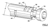
ここで図2を参照すると、本発明のコネクターで用いられるマルチルーメン型の配管の基端部32が示される。基端部は、基端部34で終わりとなる。マルチルーメン型配管は、単一の一体型ボディを有する細長い環状部材を構成する。該一体型ボディを通って、長さ方向に軸作動通路36、38及び40が延在する。作動通路36及び38は、類似の形状及びサイズを持ち、好ましくは、楕円断面を有する。通路36、38は、ほとんどマルチルーメン型配管の全長に沿って延在し、最基端部34で密封される。これらの通路の一つは、空気を供給することが意図され、他方は水を供給することが意図される。第3の軸通路40は、より大きな断面を持ち、円形の断面形態を有する。この通路は、真空を供給するか又は外科手術器具を挿入することが意図されている。第3の通路は、マルチルーメン型配管の全長に沿って延在し、その開放端部が図2に示されている。マルチルーメン型配管の内の通路の位置並びにそれらの断面形態が、マルチルーメン型配管の外側直径dが最小値に維持される態様で選択される。この最小値は、壁厚が通路40を通って供給される真空に起因してつぶれることに耐えるのに十分であることを確実にする。
Referring now to FIG. 2, there is shown a
マルチルーメン型配管の外側周辺部でかつ基端部34の近傍では、2つの接線方向に向けられた切除部42、44が作られ、該切除部は幅wで長さlで画定される。実際には、これらの寸法は、各々の軸通路36及び38との連通を可能にするように選択されている。接線方向に向けられた切除部は、実際には、径方向ポートを提供し、該ポートを通って、流体媒体が、マルチルーメン型配管の各々の軸通路に入ることができる。実際には、マルチルーメン型配管が、例えば、医療グレードのPVC等のエラストマー材料から製造される。マルチルーメン型配管の外径dは、約6.2mmであり、外側周辺部と通路との間の最小の壁厚は、約0.2から約0.3mmであり、幅寸法wは、約1.5から約1.6mmであり、長さ寸法lは約3mmである。
At the outer periphery of the multi-lumen pipe and in the vicinity of the
ここで、図3及び図4を参照してコネクターの構成を説明する。本発明のコネクター30は、エラストマー材料から作られ、該材料は、約60ショアの弾性を持つべきである。実際には、この材料は、シリコンゴム又は他の任意のエラストマープラスチック材料であってもよい。
Here, the configuration of the connector will be described with reference to FIGS. The
コネクターは、主要ボディ部46と、該ボディ部に取り付けられた基端ポート48と、を備える。主要ボディ部は、上側部分52と一体である下側部分50を備えている。下側部分は、U字形状の形態を持ち、該形態は、ハンドル14の横方向ポート上に設けられた適切な突出部に沿って摺動するように構成された長さ方向溝54を画定する。この構成のおかげで、コネクターを、図1に示されるようにハンドルに脱着可能に接続することができる。ハウジングの上側部分内では、長さ方向に延在する貫通ボアが形成される。図4では、該ボアは内径Dを持ち、出口開口部56と、入口開口部58との間に延在していることが示されている。このボアは、マルチルーメン型配管32の基端部を挿入したり取り出したりするように構成され、外科手術器具を挿入したり取り外したりするように構成されている。ボアに関して横方向に向けられ、3つの横方向ポート60、62、64と連通している各々の開口部66、68、70が提供され、図3、図4に示されている。
The connector includes a
これらのポートのうち2つのポート(60及び62)は、管72、74に挿入することが意図されており、該管は、水及び空気をマルチルーメン型配管の通路36、38に各々供給する。第3のポート64は、管76を該ポートに挿入するように構成され、該管を通って真空がマルチルーメン型配管の通路40に供給される。実際には、ポート60、62は、ほぼ同じ直径を持ち、該直径はポート64の直径よりも小さい。
Two of these ports (60 and 62) are intended to be inserted into
貫通ボアの内径Dは、マルチルーメン型配管の基端部34のコネクター内への挿入を可能にするようにマルチルーメン型配管ボアの外径dよりも大きくなるような態様で選択される。実際には、内径Dは6.2から6.3mmである。
The inner diameter D of the through-bore is selected in such a manner as to be larger than the outer diameter d of the multi-lumen piping bore so that the
貫通ボアの周辺部上には、複数の離散環状突起部78、80、82、84が形成される。これらの突起部の全ては、内径を6.2から6.3mmから、マルチルーメン型配管の外径dよりも小さい約5.5mmに減少させる。この構成のおかげで、エラストマー材料の弾性に起因して、環状突起部は、弾性的に変形し、マルチルーメン型配管がコネクターに挿入されるとき該マルチルーメン型配管を高い信頼性で密封する。この構成のおかげで、マルチルーメン型配管とコネクターとの間の空気気密接続は、別個の密封リング又は他の任意の専用密封構成部品を使用する必要無しに提供される。これは、コネクターの構成を非常に簡単にする。突起部は、水及び空気を供給するための横方向ポートと連通する2つの環状ポケットを提供するようにも構成されている。図4には、環状ポケット86が環状突起部80、82の間に形成され、環状ポケット88が環状突起部82、84の間に形成されていることが示されている。横方向ポート60はポケット86と流体連通し、横方向ポート62はポケット88と流体連通していることも示されている。隣接する突起部の間の距離は、横方向ポートがポケット内で直ちに開放し、流体媒体を内部に供給する態様で選択される。
A plurality of discrete
マルチルーメン型配管の基端部が図4に示されるようにコネクターに挿入されるとき、接線方向の切除部は環状ポケット内に位置する流体媒体にさらされ、この流体媒体は、各々の軸通路36、38における接線方向の切除部を通って入ることができることを容易に理解することができる。該流体媒体は、対応するポケットの全環状空間を満たし、かくして、軸通路内の流体媒体の導入は、ボアの周辺部に対するマルチルーメン型配管の角度配置に関係無く常に確実にされる。 When the proximal end of the multi-lumen pipe is inserted into the connector as shown in FIG. 4, the tangential cutout is exposed to a fluid medium located in the annular pocket, which fluid medium is connected to each axial passage. It can easily be seen that it is possible to enter through a tangential cut at 36,38. The fluid medium fills the entire annular space of the corresponding pocket and thus the introduction of the fluid medium in the axial passage is always ensured regardless of the angular arrangement of the multi-lumen piping with respect to the periphery of the bore.
真空がマルチルーメン型配管の通路40の開放端部に入口開口部58を通して供給される。
A vacuum is supplied through the inlet opening 58 to the open end of the
本発明のコネクターのおかげで、コネクターの出口開口部内のマルチルーメン型配管の基端部の挿入だけを必要とするため、内視鏡処置へのマルチルーメン型配管の準備が非常に簡単かつ便利になる一方で、全ての必要となる流体媒体のマルチルーメン型配管への供給が提供される。 Thanks to the connector according to the invention, it is only necessary to insert the proximal end of the multi-lumen pipe in the outlet opening of the connector, so the preparation of the multi-lumen pipe for endoscopic treatment is very simple and convenient. On the other hand, a supply of all required fluid media to the multi-lumen piping is provided.
実際には、コネクターの後部、即ちその基端ポート48は、主要ボディ部と同じエラストマー材料から製造される。基端ポートは、任意の適切な手段、例えば接着剤により接続される。基端ポートが主要ボディ部に接続可能である別個のポートであるときのこの状況は、単なる一例でしかない。基端ポート及び主要ボディ部が一体のものである状況を想定することもできる。
In practice, the rear portion of the connector, ie its
基端ポートは、図5を参照して更に後述されるように、外科手術器具を該ポートを通して挿入したり、該ポートから引き抜くための入口開口部90が設けられている。当該入口は狭窄区分92で終わっている。狭窄通路を開閉し、かくしてボアの入口開口部と基端ポートの入口開口部との間の流体連通を防止したり可能にしたりするように弾性的に折り畳み可能な薄い可撓性仕切り部94が設けられている。図4に示される状況では、マルチルーメン型配管がコネクター内に挿入されるが、外科施術器具は、コネクター内に挿入されない。弾性仕切り部94は、マルチルーメン型配管の各々の通路への流体媒体又は真空の供給を可能にする一方で、入口ポートを高い信頼性で閉じ、貫通ボアとの流体連通を防止するように構成され、寸法が定められている。
The proximal port is provided with an
図5には、外科施術器具、例えば生体検査かんし96が、入口開口部90を通してコネクター内に挿入され、マルチルーメン型配管の通路40に入るように入口開口部58を通して既に前進させられた状況が表されている。当該器具は、内視鏡処置の間に患者の身体内に組織例を採取するため通路40を通って更に前進させられるべきである。この状況では、真空は通路40には供給されないことを理解することができる。器具を入口開口部を通して前進させる初期の工程の間では、当該器具の前側端部は弾性仕切り部94に到達し、該仕切り部を前方に押し、図5に示されるように、折り畳ませる。ここで、当該器具は、マルチルーメン型配管の通路40に沿って更に前進させることができる。当該器具がコネクターから引き抜かれたときの内視鏡処置の終了時では、当該仕切り部は、エラストマー材料の弾性に起因してその閉鎖位置へと戻り、基端ポートは再び閉じられる。
In FIG. 5, a surgical instrument, such as a
上記に開示されたコネクターは、膨張可能スリーブにより推進される内視鏡と接続するときのみに限らず用いることができることが理解されるべきである。本発明のコネクターは、マルチルーメン型配管の通路へ流体媒体を供給し又は外科施術器具を該通路を通して前進させることが要求される他の任意の内視鏡でも使用することができる。 It should be understood that the connector disclosed above can be used not only when connected to an endoscope driven by an inflatable sleeve. The connector of the present invention can also be used in any other endoscope that requires supplying a fluid medium to a passage of a multi-lumen pipe or advancing a surgical instrument through the passage.
本発明は、上述された実施例に限定されるものではなく、添付した請求の範囲に定義されるような本発明の範囲から逸脱することなく、当業者は様々な変更及び変形をなすことができることが理解されるべきである。 The present invention is not limited to the embodiments described above, and various changes and modifications can be made by those skilled in the art without departing from the scope of the invention as defined in the appended claims. It should be understood that it can be done.
例えば、図5に示されるように曲げ可能な弾性仕切り部の代わりに、入口開口部に向って前進させられるとき、外科手術器具により突き刺し可能である薄い弾性壁を使用することもできる。 For example, instead of a bendable elastic partition as shown in FIG. 5, a thin elastic wall that can be pierced by a surgical instrument when advanced toward the entrance opening can be used.
「備える」、「含む」及び「有する」の用語が、請求の範囲で使用されるとき、これらの用語及びそれらの活用形の意味は、該用語に構成続く構成要素を含んでいるが、該構成要素には限定されないということを意味している。 When the terms “comprising”, “including”, and “having” are used in the claims, the meaning of these terms and their conjugations includes the components that follow the term, It means that it is not limited to components.
前述した説明及び/又は添付した請求の範囲及び/又は添付図面に開示された特徴は、各々別個に及びそれらの任意の組み合わせの両方において、本発明を、その多様な形態において理解するための材料となり得る。 The features disclosed in the foregoing description and / or the appended claims and / or the accompanying drawings are each a material for understanding the invention in its various forms, both individually and in any combination. Can be.
Claims (20)
前記コネクターは、該コネクターを通して外科手術器具を挿入したり、引き抜いたりするように構成されている、コネクター。 A connector for establishing fluid communication between a passage of a multi-lumen pipe used in an endoscope apparatus and a pipe for supplying a fluid medium to the multi-lumen pipe during an endoscopic treatment. There,
The connector is configured to insert or withdraw a surgical instrument through the connector.
前記主要ボディ部は、前記ボアと流体連通する複数の横方向ポートを更に備え、
前記ボアは、前記マルチルーメン型配管の基端部を該ボア内に挿入したり該ボアから引き抜いたりすることを可能にし、前記横方向ポートは、前記流体媒体を供給するための前記管を該ポート内に受け入れる、請求項1に記載のコネクター。 Comprising a main body part formed with a through-bore extending in the longitudinal direction between the inlet opening and the outlet opening;
The main body portion further comprises a plurality of lateral ports in fluid communication with the bore;
The bore allows the proximal end of the multi-lumen piping to be inserted into and withdrawn from the bore, and the lateral port provides access to the tube for supplying the fluid medium. The connector of claim 1, wherein the connector is received in a port.
内視鏡と、
前記内視鏡内に挿入可能なマルチルーメンであって、該マルチルーメンには、前記内視鏡に流体媒体を供給するための通路と、外科手術器具の挿入及び引き抜きのための通路とが設けられている、前記マルチルーメンと、
前記マルチルーメンの前記通路に流体媒体を供給するための管と、
前記マルチルーメン型配管の通路と、流体媒体を供給するための前記管との間で流体連通を確立するコネクターであって、該コネクターは、前記外科手術器具を前記マルチルーメン型配管に挿入したり該マルチルーメン型配管から引き抜くことを可能にする、前記コネクターと、
を備える、内視鏡装置。 An endoscopic device,
An endoscope,
A multi-lumen insertable into the endoscope, the multi-lumen being provided with a passage for supplying a fluid medium to the endoscope and a passage for insertion and extraction of a surgical instrument. Said multi-lumen,
A tube for supplying a fluid medium to the passage of the multi-lumen;
A connector for establishing fluid communication between a passage of the multi-lumen pipe and the pipe for supplying a fluid medium, the connector inserting the surgical instrument into the multi-lumen pipe The connector enabling the multi-lumen pipe to be pulled out;
An endoscope apparatus comprising:
前記主要ボディ部は、前記ボアと流体連通する複数の横方向ポートを更に備え、
前記ボアは、前記マルチルーメン型配管の基端部を該ボア内に挿入したり該ボアから取り出したりすることを可能にし、前記横方向ポートは、前記流体媒体を供給するための前記管を該ポート内に受け入れる、請求項18に記載の内視鏡装置。 The connector includes a main body portion formed with a through-bore extending in a length direction between an inlet opening and an outlet opening,
The main body portion further comprises a plurality of lateral ports in fluid communication with the bore;
The bore allows the proximal end of the multi-lumen piping to be inserted into and removed from the bore, and the lateral port connects the tube for supplying the fluid medium. The endoscopic device according to claim 18, which is received in a port.
Applications Claiming Priority (2)
| Application Number | Priority Date | Filing Date | Title |
|---|---|---|---|
| US57061004P | 2004-05-13 | 2004-05-13 | |
| PCT/IL2005/000428 WO2005110200A1 (en) | 2004-05-13 | 2005-04-21 | Connector for use with multilumen tubing |
Publications (2)
| Publication Number | Publication Date |
|---|---|
| JP2007536980A true JP2007536980A (en) | 2007-12-20 |
| JP2007536980A5 JP2007536980A5 (en) | 2009-08-13 |
Family
ID=34970311
Family Applications (1)
| Application Number | Title | Priority Date | Filing Date |
|---|---|---|---|
| JP2007512715A Pending JP2007536980A (en) | 2004-05-13 | 2005-04-21 | Connector for use with multi-lumen piping |
Country Status (8)
| Country | Link |
|---|---|
| EP (1) | EP1755438A1 (en) |
| JP (1) | JP2007536980A (en) |
| CN (1) | CN1988844A (en) |
| AU (1) | AU2005244326A1 (en) |
| BR (1) | BRPI0511076A (en) |
| CA (1) | CA2566402A1 (en) |
| RU (1) | RU2006137466A (en) |
| WO (1) | WO2005110200A1 (en) |
Families Citing this family (8)
| Publication number | Priority date | Publication date | Assignee | Title |
|---|---|---|---|---|
| US20070255101A1 (en) | 2005-03-04 | 2007-11-01 | Sightline Technologies Ltd. | Endoscope with Protective Sleeve |
| WO2008122969A1 (en) | 2007-04-10 | 2008-10-16 | Stryker Gi Ltd. | Versatile control system for supplying fluid medium to endoscope |
| WO2008142672A1 (en) * | 2007-05-21 | 2008-11-27 | Stryker Gi Ltd. | Disposable connector for use with endoscopic apparatus |
| FR2946518B1 (en) * | 2009-06-15 | 2012-09-21 | Assist Publ Hopitaux De Paris | ENDOSCOPE COMPRISING A SINGLE-USE MODULE |
| CN102003587B (en) * | 2010-11-15 | 2012-08-22 | 中国农业大学 | Rotating joint for vacuum device |
| US8777931B2 (en) | 2011-08-19 | 2014-07-15 | Alcon Research, Ltd. | Retractable luer lock fittings |
| US9320867B2 (en) * | 2013-05-22 | 2016-04-26 | Pall Corporation | Connection system |
| US20230346205A1 (en) | 2022-04-27 | 2023-11-02 | Neptune Medical Inc. | Multi-lumen port adapter manifold devices and methods of use |
Citations (4)
| Publication number | Priority date | Publication date | Assignee | Title |
|---|---|---|---|---|
| JPS6354143A (en) * | 1986-08-25 | 1988-03-08 | 株式会社町田製作所 | Guide pipe structure of endoscope |
| JPH01244732A (en) * | 1988-03-28 | 1989-09-29 | Asahi Optical Co Ltd | Endoscope with sheath |
| JPH0329634A (en) * | 1989-06-26 | 1991-02-07 | Asahi Optical Co Ltd | Sheath for endoscope |
| JP2003199704A (en) * | 2002-01-07 | 2003-07-15 | Pentax Corp | Channel tube connecting structure of contamination preventive type endoscope |
Family Cites Families (4)
| Publication number | Priority date | Publication date | Assignee | Title |
|---|---|---|---|---|
| US4979496A (en) * | 1988-04-05 | 1990-12-25 | Fuji Photo Optical Co., Ltd. | Endoscope for bile duct and pancreatic duct |
| US5250038A (en) * | 1992-10-09 | 1993-10-05 | Cook Incorporated | Multiple lumen vascular access introducer sheath |
| US5554098A (en) * | 1993-02-26 | 1996-09-10 | Olympus Optical Co., Ltd. | Endoscope system including endoscope and disposable protection cover |
| US5599324A (en) * | 1995-05-04 | 1997-02-04 | Boston Scientific Corporation | Catheter for administering a liquid agent |
-
2005
- 2005-04-21 EP EP05734776A patent/EP1755438A1/en not_active Withdrawn
- 2005-04-21 RU RU2006137466/14A patent/RU2006137466A/en not_active Application Discontinuation
- 2005-04-21 JP JP2007512715A patent/JP2007536980A/en active Pending
- 2005-04-21 CA CA002566402A patent/CA2566402A1/en not_active Abandoned
- 2005-04-21 WO PCT/IL2005/000428 patent/WO2005110200A1/en active Application Filing
- 2005-04-21 AU AU2005244326A patent/AU2005244326A1/en not_active Abandoned
- 2005-04-21 BR BRPI0511076-9A patent/BRPI0511076A/en not_active IP Right Cessation
- 2005-04-21 CN CN 200580015203 patent/CN1988844A/en active Pending
Patent Citations (4)
| Publication number | Priority date | Publication date | Assignee | Title |
|---|---|---|---|---|
| JPS6354143A (en) * | 1986-08-25 | 1988-03-08 | 株式会社町田製作所 | Guide pipe structure of endoscope |
| JPH01244732A (en) * | 1988-03-28 | 1989-09-29 | Asahi Optical Co Ltd | Endoscope with sheath |
| JPH0329634A (en) * | 1989-06-26 | 1991-02-07 | Asahi Optical Co Ltd | Sheath for endoscope |
| JP2003199704A (en) * | 2002-01-07 | 2003-07-15 | Pentax Corp | Channel tube connecting structure of contamination preventive type endoscope |
Also Published As
| Publication number | Publication date |
|---|---|
| RU2006137466A (en) | 2008-06-20 |
| BRPI0511076A (en) | 2007-12-26 |
| AU2005244326A1 (en) | 2005-11-24 |
| EP1755438A1 (en) | 2007-02-28 |
| WO2005110200A1 (en) | 2005-11-24 |
| CN1988844A (en) | 2007-06-27 |
| CA2566402A1 (en) | 2005-11-24 |
Similar Documents
| Publication | Publication Date | Title |
|---|---|---|
| US20050256376A1 (en) | Connector for use with multilumen tubing | |
| US9480390B2 (en) | Endoscope accessory | |
| EP2712537B1 (en) | Balloon guided endoscopy | |
| US9993137B2 (en) | Endoscope accessory | |
| US9492067B2 (en) | Gastrointestinal lavage system | |
| US20050256373A1 (en) | Disposable set for use with an endoscope | |
| EP2999391B1 (en) | An endoscope accessory | |
| JP2007536980A (en) | Connector for use with multi-lumen piping | |
| US20170065155A1 (en) | Endoscope Accessory | |
| JP5647780B2 (en) | Treatment overtube and treatment system | |
| JP2008511376A (en) | Control system for supplying a fluid medium to an endoscope | |
| JP2008278968A (en) | Insertion assisting tool for endoscope | |
| JP4728047B2 (en) | Endoscope catheter with balloon | |
| US20230397800A1 (en) | Endoscopic accessory | |
| WO2023192330A1 (en) | Endoscopic accessory | |
| KR20070018950A (en) | Connector for use with multilumen tubing | |
| US11925319B2 (en) | Endoscopic accessory |
Legal Events
| Date | Code | Title | Description |
|---|---|---|---|
| A621 | Written request for application examination |
Free format text: JAPANESE INTERMEDIATE CODE: A621 Effective date: 20080404 |
|
| A521 | Request for written amendment filed |
Free format text: JAPANESE INTERMEDIATE CODE: A523 Effective date: 20090626 |
|
| A977 | Report on retrieval |
Free format text: JAPANESE INTERMEDIATE CODE: A971007 Effective date: 20110126 |
|
| A131 | Notification of reasons for refusal |
Free format text: JAPANESE INTERMEDIATE CODE: A131 Effective date: 20110215 |
|
| A601 | Written request for extension of time |
Free format text: JAPANESE INTERMEDIATE CODE: A601 Effective date: 20110516 |
|
| A602 | Written permission of extension of time |
Free format text: JAPANESE INTERMEDIATE CODE: A602 Effective date: 20110523 |
|
| RD04 | Notification of resignation of power of attorney |
Free format text: JAPANESE INTERMEDIATE CODE: A7424 Effective date: 20110905 |
|
| A02 | Decision of refusal |
Free format text: JAPANESE INTERMEDIATE CODE: A02 Effective date: 20111101 |