JP2006522102A - Targeting agent for both photodiagnosis and photodynamic therapy - Google Patents
Targeting agent for both photodiagnosis and photodynamic therapy Download PDFInfo
- Publication number
- JP2006522102A JP2006522102A JP2006507106A JP2006507106A JP2006522102A JP 2006522102 A JP2006522102 A JP 2006522102A JP 2006507106 A JP2006507106 A JP 2006507106A JP 2006507106 A JP2006507106 A JP 2006507106A JP 2006522102 A JP2006522102 A JP 2006522102A
- Authority
- JP
- Japan
- Prior art keywords
- cancer
- imaging
- molecule
- pdt
- molecules
- Prior art date
- Legal status (The legal status is an assumption and is not a legal conclusion. Google has not performed a legal analysis and makes no representation as to the accuracy of the status listed.)
- Pending
Links
- 230000008685 targeting Effects 0.000 title claims abstract description 77
- 238000002428 photodynamic therapy Methods 0.000 title claims abstract description 49
- 239000003795 chemical substances by application Substances 0.000 claims abstract description 58
- 238000000034 method Methods 0.000 claims abstract description 43
- 239000012216 imaging agent Substances 0.000 claims abstract description 18
- 206010028980 Neoplasm Diseases 0.000 claims description 120
- 201000011510 cancer Diseases 0.000 claims description 52
- 238000003384 imaging method Methods 0.000 claims description 48
- 150000004032 porphyrins Chemical group 0.000 claims description 29
- 238000010521 absorption reaction Methods 0.000 claims description 19
- 238000002059 diagnostic imaging Methods 0.000 claims 2
- 239000000203 mixture Substances 0.000 abstract description 22
- 239000002872 contrast media Substances 0.000 abstract description 14
- 230000001588 bifunctional effect Effects 0.000 abstract description 11
- 210000001519 tissue Anatomy 0.000 description 48
- 210000004027 cell Anatomy 0.000 description 40
- 206010006187 Breast cancer Diseases 0.000 description 32
- 208000026310 Breast neoplasm Diseases 0.000 description 31
- 239000000758 substrate Substances 0.000 description 31
- 239000003112 inhibitor Substances 0.000 description 29
- 238000011282 treatment Methods 0.000 description 26
- 108090000765 processed proteins & peptides Proteins 0.000 description 25
- 238000009607 mammography Methods 0.000 description 22
- 229920000642 polymer Polymers 0.000 description 21
- 108090000623 proteins and genes Proteins 0.000 description 20
- 239000003814 drug Substances 0.000 description 19
- 235000018102 proteins Nutrition 0.000 description 19
- 102000004169 proteins and genes Human genes 0.000 description 19
- 102000002274 Matrix Metalloproteinases Human genes 0.000 description 18
- 108010000684 Matrix Metalloproteinases Proteins 0.000 description 18
- 102000005962 receptors Human genes 0.000 description 18
- 108020003175 receptors Proteins 0.000 description 18
- -1 signal transducers Substances 0.000 description 18
- 229940079593 drug Drugs 0.000 description 17
- 150000001875 compounds Chemical class 0.000 description 16
- 125000000524 functional group Chemical group 0.000 description 16
- 238000005516 engineering process Methods 0.000 description 15
- 125000000217 alkyl group Chemical group 0.000 description 14
- 239000003446 ligand Substances 0.000 description 14
- 150000001720 carbohydrates Chemical class 0.000 description 13
- 238000001514 detection method Methods 0.000 description 13
- 150000003839 salts Chemical class 0.000 description 13
- 235000014633 carbohydrates Nutrition 0.000 description 12
- 238000002595 magnetic resonance imaging Methods 0.000 description 12
- 102000004196 processed proteins & peptides Human genes 0.000 description 12
- NHXLMOGPVYXJNR-ATOGVRKGSA-N somatostatin Chemical compound C([C@H]1C(=O)N[C@H](C(N[C@@H](CO)C(=O)N[C@@H](CSSC[C@@H](C(=O)N[C@@H](CCCCN)C(=O)N[C@@H](CC(N)=O)C(=O)N[C@@H](CC=2C=CC=CC=2)C(=O)N[C@@H](CC=2C=CC=CC=2)C(=O)N[C@@H](CC=2C3=CC=CC=C3NC=2)C(=O)N[C@@H](CCCCN)C(=O)N[C@H](C(=O)N1)[C@@H](C)O)NC(=O)CNC(=O)[C@H](C)N)C(O)=O)=O)[C@H](O)C)C1=CC=CC=C1 NHXLMOGPVYXJNR-ATOGVRKGSA-N 0.000 description 12
- 102000005157 Somatostatin Human genes 0.000 description 11
- 108010056088 Somatostatin Proteins 0.000 description 11
- 125000003118 aryl group Chemical group 0.000 description 11
- 230000027455 binding Effects 0.000 description 11
- 210000000481 breast Anatomy 0.000 description 11
- 230000008878 coupling Effects 0.000 description 11
- 238000010168 coupling process Methods 0.000 description 11
- 238000005859 coupling reaction Methods 0.000 description 11
- 238000001727 in vivo Methods 0.000 description 11
- 125000005647 linker group Chemical group 0.000 description 11
- MYMOFIZGZYHOMD-UHFFFAOYSA-N Dioxygen Chemical compound O=O MYMOFIZGZYHOMD-UHFFFAOYSA-N 0.000 description 10
- 238000011161 development Methods 0.000 description 10
- 230000018109 developmental process Effects 0.000 description 10
- 229940088597 hormone Drugs 0.000 description 10
- 239000005556 hormone Substances 0.000 description 10
- 238000012634 optical imaging Methods 0.000 description 10
- 229960000553 somatostatin Drugs 0.000 description 10
- 102000004190 Enzymes Human genes 0.000 description 9
- 108090000790 Enzymes Proteins 0.000 description 9
- 108010060309 Glucuronidase Proteins 0.000 description 9
- 102000053187 Glucuronidase Human genes 0.000 description 9
- 108060003951 Immunoglobulin Proteins 0.000 description 9
- 229940088598 enzyme Drugs 0.000 description 9
- 230000006870 function Effects 0.000 description 9
- 102000018358 immunoglobulin Human genes 0.000 description 9
- 235000013336 milk Nutrition 0.000 description 9
- 239000008267 milk Substances 0.000 description 9
- 210000004080 milk Anatomy 0.000 description 9
- 230000003287 optical effect Effects 0.000 description 9
- 102000000844 Cell Surface Receptors Human genes 0.000 description 8
- 108010001857 Cell Surface Receptors Proteins 0.000 description 8
- 108010035532 Collagen Proteins 0.000 description 8
- 102000008186 Collagen Human genes 0.000 description 8
- 108010010803 Gelatin Proteins 0.000 description 8
- 102000035195 Peptidases Human genes 0.000 description 8
- 108091005804 Peptidases Proteins 0.000 description 8
- 108050001286 Somatostatin Receptor Proteins 0.000 description 8
- 102000011096 Somatostatin receptor Human genes 0.000 description 8
- 230000008901 benefit Effects 0.000 description 8
- 239000003153 chemical reaction reagent Substances 0.000 description 8
- 229920001436 collagen Polymers 0.000 description 8
- 201000010099 disease Diseases 0.000 description 8
- 208000037265 diseases, disorders, signs and symptoms Diseases 0.000 description 8
- 230000000694 effects Effects 0.000 description 8
- 239000008273 gelatin Substances 0.000 description 8
- 229920000159 gelatin Polymers 0.000 description 8
- 235000019322 gelatine Nutrition 0.000 description 8
- 235000011852 gelatine desserts Nutrition 0.000 description 8
- 230000014509 gene expression Effects 0.000 description 8
- 238000002600 positron emission tomography Methods 0.000 description 8
- 239000000126 substance Substances 0.000 description 8
- 210000004881 tumor cell Anatomy 0.000 description 8
- 102000003908 Cathepsin D Human genes 0.000 description 7
- 108090000258 Cathepsin D Proteins 0.000 description 7
- 206010009944 Colon cancer Diseases 0.000 description 7
- 102000010834 Extracellular Matrix Proteins Human genes 0.000 description 7
- 108010037362 Extracellular Matrix Proteins Proteins 0.000 description 7
- 206010027476 Metastases Diseases 0.000 description 7
- 206010061535 Ovarian neoplasm Diseases 0.000 description 7
- 150000001413 amino acids Chemical class 0.000 description 7
- 239000000562 conjugate Substances 0.000 description 7
- 210000002744 extracellular matrix Anatomy 0.000 description 7
- 239000000463 material Substances 0.000 description 7
- 229940121386 matrix metalloproteinase inhibitor Drugs 0.000 description 7
- 239000003771 matrix metalloproteinase inhibitor Substances 0.000 description 7
- 230000009401 metastasis Effects 0.000 description 7
- 239000003504 photosensitizing agent Substances 0.000 description 7
- 230000008569 process Effects 0.000 description 7
- 239000007787 solid Substances 0.000 description 7
- 238000002604 ultrasonography Methods 0.000 description 7
- IJGRMHOSHXDMSA-UHFFFAOYSA-N Atomic nitrogen Chemical compound N#N IJGRMHOSHXDMSA-UHFFFAOYSA-N 0.000 description 6
- 102000004171 Cathepsin K Human genes 0.000 description 6
- 108090000625 Cathepsin K Proteins 0.000 description 6
- 108010047041 Complementarity Determining Regions Proteins 0.000 description 6
- LFQSCWFLJHTTHZ-UHFFFAOYSA-N Ethanol Chemical compound CCO LFQSCWFLJHTTHZ-UHFFFAOYSA-N 0.000 description 6
- 108010077850 Nuclear Localization Signals Proteins 0.000 description 6
- 206010033128 Ovarian cancer Diseases 0.000 description 6
- DNIAPMSPPWPWGF-UHFFFAOYSA-N Propylene glycol Chemical compound CC(O)CO DNIAPMSPPWPWGF-UHFFFAOYSA-N 0.000 description 6
- 235000001014 amino acid Nutrition 0.000 description 6
- QVGXLLKOCUKJST-UHFFFAOYSA-N atomic oxygen Chemical compound [O] QVGXLLKOCUKJST-UHFFFAOYSA-N 0.000 description 6
- 230000015572 biosynthetic process Effects 0.000 description 6
- 210000000988 bone and bone Anatomy 0.000 description 6
- 125000004432 carbon atom Chemical group C* 0.000 description 6
- 230000003197 catalytic effect Effects 0.000 description 6
- 238000003776 cleavage reaction Methods 0.000 description 6
- NOESYZHRGYRDHS-UHFFFAOYSA-N insulin Chemical compound N1C(=O)C(NC(=O)C(CCC(N)=O)NC(=O)C(CCC(O)=O)NC(=O)C(C(C)C)NC(=O)C(NC(=O)CN)C(C)CC)CSSCC(C(NC(CO)C(=O)NC(CC(C)C)C(=O)NC(CC=2C=CC(O)=CC=2)C(=O)NC(CCC(N)=O)C(=O)NC(CC(C)C)C(=O)NC(CCC(O)=O)C(=O)NC(CC(N)=O)C(=O)NC(CC=2C=CC(O)=CC=2)C(=O)NC(CSSCC(NC(=O)C(C(C)C)NC(=O)C(CC(C)C)NC(=O)C(CC=2C=CC(O)=CC=2)NC(=O)C(CC(C)C)NC(=O)C(C)NC(=O)C(CCC(O)=O)NC(=O)C(C(C)C)NC(=O)C(CC(C)C)NC(=O)C(CC=2NC=NC=2)NC(=O)C(CO)NC(=O)CNC2=O)C(=O)NCC(=O)NC(CCC(O)=O)C(=O)NC(CCCNC(N)=N)C(=O)NCC(=O)NC(CC=3C=CC=CC=3)C(=O)NC(CC=3C=CC=CC=3)C(=O)NC(CC=3C=CC(O)=CC=3)C(=O)NC(C(C)O)C(=O)N3C(CCC3)C(=O)NC(CCCCN)C(=O)NC(C)C(O)=O)C(=O)NC(CC(N)=O)C(O)=O)=O)NC(=O)C(C(C)CC)NC(=O)C(CO)NC(=O)C(C(C)O)NC(=O)C1CSSCC2NC(=O)C(CC(C)C)NC(=O)C(NC(=O)C(CCC(N)=O)NC(=O)C(CC(N)=O)NC(=O)C(NC(=O)C(N)CC=1C=CC=CC=1)C(C)C)CC1=CN=CN1 NOESYZHRGYRDHS-UHFFFAOYSA-N 0.000 description 6
- 150000002632 lipids Chemical class 0.000 description 6
- 238000004519 manufacturing process Methods 0.000 description 6
- 229910052760 oxygen Inorganic materials 0.000 description 6
- 239000001301 oxygen Substances 0.000 description 6
- 230000007017 scission Effects 0.000 description 6
- 238000012216 screening Methods 0.000 description 6
- 238000003786 synthesis reaction Methods 0.000 description 6
- 208000001333 Colorectal Neoplasms Diseases 0.000 description 5
- 101001012157 Homo sapiens Receptor tyrosine-protein kinase erbB-2 Proteins 0.000 description 5
- 206010061902 Pancreatic neoplasm Diseases 0.000 description 5
- 239000004365 Protease Substances 0.000 description 5
- 102100030086 Receptor tyrosine-protein kinase erbB-2 Human genes 0.000 description 5
- 102000004338 Transferrin Human genes 0.000 description 5
- 239000000427 antigen Substances 0.000 description 5
- 230000006907 apoptotic process Effects 0.000 description 5
- 125000004429 atom Chemical group 0.000 description 5
- 238000012512 characterization method Methods 0.000 description 5
- 239000000975 dye Substances 0.000 description 5
- NITYDPDXAAFEIT-DYVFJYSZSA-N ilomastat Chemical compound C1=CC=C2C(C[C@@H](C(=O)NC)NC(=O)[C@H](CC(C)C)CC(=O)NO)=CNC2=C1 NITYDPDXAAFEIT-DYVFJYSZSA-N 0.000 description 5
- 108020004999 messenger RNA Proteins 0.000 description 5
- 229910052757 nitrogen Inorganic materials 0.000 description 5
- 201000002528 pancreatic cancer Diseases 0.000 description 5
- 230000005855 radiation Effects 0.000 description 5
- 238000011160 research Methods 0.000 description 5
- 239000000243 solution Substances 0.000 description 5
- 239000012581 transferrin Substances 0.000 description 5
- XLYOFNOQVPJJNP-UHFFFAOYSA-N water Substances O XLYOFNOQVPJJNP-UHFFFAOYSA-N 0.000 description 5
- 206010055113 Breast cancer metastatic Diseases 0.000 description 4
- 201000009030 Carcinoma Diseases 0.000 description 4
- 102000004225 Cathepsin B Human genes 0.000 description 4
- 108090000712 Cathepsin B Proteins 0.000 description 4
- PEDCQBHIVMGVHV-UHFFFAOYSA-N Glycerine Chemical compound OCC(O)CO PEDCQBHIVMGVHV-UHFFFAOYSA-N 0.000 description 4
- 101000829127 Homo sapiens Somatostatin receptor type 2 Proteins 0.000 description 4
- 206010058467 Lung neoplasm malignant Diseases 0.000 description 4
- 108010016165 Matrix Metalloproteinase 2 Proteins 0.000 description 4
- 102000000424 Matrix Metalloproteinase 2 Human genes 0.000 description 4
- 108010030545 N-(2(R)-2-(hydroxamidocarbonylmethyl)-4-methylpentanoyl)-L-tryptophan methylamide Proteins 0.000 description 4
- 102000012404 Orosomucoid Human genes 0.000 description 4
- 108010061952 Orosomucoid Proteins 0.000 description 4
- 241000283973 Oryctolagus cuniculus Species 0.000 description 4
- 102100023832 Prolyl endopeptidase FAP Human genes 0.000 description 4
- 102100023802 Somatostatin receptor type 2 Human genes 0.000 description 4
- NINIDFKCEFEMDL-UHFFFAOYSA-N Sulfur Chemical compound [S] NINIDFKCEFEMDL-UHFFFAOYSA-N 0.000 description 4
- 108090000901 Transferrin Proteins 0.000 description 4
- 230000002378 acidificating effect Effects 0.000 description 4
- 108091007433 antigens Proteins 0.000 description 4
- 102000036639 antigens Human genes 0.000 description 4
- 210000004369 blood Anatomy 0.000 description 4
- 239000008280 blood Substances 0.000 description 4
- 239000002738 chelating agent Substances 0.000 description 4
- 208000029742 colonic neoplasm Diseases 0.000 description 4
- 230000034994 death Effects 0.000 description 4
- 231100000517 death Toxicity 0.000 description 4
- 239000002552 dosage form Substances 0.000 description 4
- 239000003937 drug carrier Substances 0.000 description 4
- 235000019441 ethanol Nutrition 0.000 description 4
- 230000005284 excitation Effects 0.000 description 4
- 238000009472 formulation Methods 0.000 description 4
- 125000000623 heterocyclic group Chemical group 0.000 description 4
- 238000000338 in vitro Methods 0.000 description 4
- 230000005764 inhibitory process Effects 0.000 description 4
- 230000009545 invasion Effects 0.000 description 4
- 230000004807 localization Effects 0.000 description 4
- 201000005202 lung cancer Diseases 0.000 description 4
- 208000020816 lung neoplasm Diseases 0.000 description 4
- 208000015486 malignant pancreatic neoplasm Diseases 0.000 description 4
- 238000012014 optical coherence tomography Methods 0.000 description 4
- 230000002018 overexpression Effects 0.000 description 4
- 208000008443 pancreatic carcinoma Diseases 0.000 description 4
- 229940109328 photofrin Drugs 0.000 description 4
- 229920000656 polylysine Polymers 0.000 description 4
- 229950003608 prinomastat Drugs 0.000 description 4
- YKPYIPVDTNNYCN-INIZCTEOSA-N prinomastat Chemical compound ONC(=O)[C@H]1C(C)(C)SCCN1S(=O)(=O)C(C=C1)=CC=C1OC1=CC=NC=C1 YKPYIPVDTNNYCN-INIZCTEOSA-N 0.000 description 4
- ATHGHQPFGPMSJY-UHFFFAOYSA-N spermidine Chemical compound NCCCCNCCCN ATHGHQPFGPMSJY-UHFFFAOYSA-N 0.000 description 4
- 229910052717 sulfur Inorganic materials 0.000 description 4
- 239000011593 sulfur Substances 0.000 description 4
- 239000012622 synthetic inhibitor Substances 0.000 description 4
- QGKMIGUHVLGJBR-UHFFFAOYSA-M (4z)-1-(3-methylbutyl)-4-[[1-(3-methylbutyl)quinolin-1-ium-4-yl]methylidene]quinoline;iodide Chemical compound [I-].C12=CC=CC=C2N(CCC(C)C)C=CC1=CC1=CC=[N+](CCC(C)C)C2=CC=CC=C12 QGKMIGUHVLGJBR-UHFFFAOYSA-M 0.000 description 3
- 108010076667 Caspases Proteins 0.000 description 3
- 102000011727 Caspases Human genes 0.000 description 3
- 102000029816 Collagenase Human genes 0.000 description 3
- 108060005980 Collagenase Proteins 0.000 description 3
- 102000005927 Cysteine Proteases Human genes 0.000 description 3
- 108010005843 Cysteine Proteases Proteins 0.000 description 3
- 229920002307 Dextran Polymers 0.000 description 3
- 102000001301 EGF receptor Human genes 0.000 description 3
- 108060006698 EGF receptor Proteins 0.000 description 3
- 108010014258 Elastin Proteins 0.000 description 3
- 102000016942 Elastin Human genes 0.000 description 3
- LYCAIKOWRPUZTN-UHFFFAOYSA-N Ethylene glycol Chemical compound OCCO LYCAIKOWRPUZTN-UHFFFAOYSA-N 0.000 description 3
- WQZGKKKJIJFFOK-GASJEMHNSA-N Glucose Natural products OC[C@H]1OC(O)[C@H](O)[C@@H](O)[C@@H]1O WQZGKKKJIJFFOK-GASJEMHNSA-N 0.000 description 3
- 102100024025 Heparanase Human genes 0.000 description 3
- 102000006496 Immunoglobulin Heavy Chains Human genes 0.000 description 3
- 108010019476 Immunoglobulin Heavy Chains Proteins 0.000 description 3
- 102000004877 Insulin Human genes 0.000 description 3
- 108090001061 Insulin Proteins 0.000 description 3
- 208000037396 Intraductal Noninfiltrating Carcinoma Diseases 0.000 description 3
- 108010015302 Matrix metalloproteinase-9 Proteins 0.000 description 3
- 102100039364 Metalloproteinase inhibitor 1 Human genes 0.000 description 3
- 102100026262 Metalloproteinase inhibitor 2 Human genes 0.000 description 3
- QPCDCPDFJACHGM-UHFFFAOYSA-N N,N-bis{2-[bis(carboxymethyl)amino]ethyl}glycine Chemical compound OC(=O)CN(CC(O)=O)CCN(CC(=O)O)CCN(CC(O)=O)CC(O)=O QPCDCPDFJACHGM-UHFFFAOYSA-N 0.000 description 3
- OAICVXFJPJFONN-UHFFFAOYSA-N Phosphorus Chemical compound [P] OAICVXFJPJFONN-UHFFFAOYSA-N 0.000 description 3
- 108010039918 Polylysine Proteins 0.000 description 3
- 206010060862 Prostate cancer Diseases 0.000 description 3
- 208000000236 Prostatic Neoplasms Diseases 0.000 description 3
- 241000283984 Rodentia Species 0.000 description 3
- XUIMIQQOPSSXEZ-UHFFFAOYSA-N Silicon Chemical compound [Si] XUIMIQQOPSSXEZ-UHFFFAOYSA-N 0.000 description 3
- FAPWRFPIFSIZLT-UHFFFAOYSA-M Sodium chloride Chemical compound [Na+].[Cl-] FAPWRFPIFSIZLT-UHFFFAOYSA-M 0.000 description 3
- 210000001744 T-lymphocyte Anatomy 0.000 description 3
- 102000011923 Thyrotropin Human genes 0.000 description 3
- 108010061174 Thyrotropin Proteins 0.000 description 3
- 108010031374 Tissue Inhibitor of Metalloproteinase-1 Proteins 0.000 description 3
- 108010031372 Tissue Inhibitor of Metalloproteinase-2 Proteins 0.000 description 3
- 102000004887 Transforming Growth Factor beta Human genes 0.000 description 3
- 108090001012 Transforming Growth Factor beta Proteins 0.000 description 3
- 206010064390 Tumour invasion Diseases 0.000 description 3
- 108091008605 VEGF receptors Proteins 0.000 description 3
- 102000009484 Vascular Endothelial Growth Factor Receptors Human genes 0.000 description 3
- 230000004913 activation Effects 0.000 description 3
- 239000004480 active ingredient Substances 0.000 description 3
- 150000001412 amines Chemical class 0.000 description 3
- 239000003963 antioxidant agent Substances 0.000 description 3
- 239000002585 base Substances 0.000 description 3
- WQZGKKKJIJFFOK-VFUOTHLCSA-N beta-D-glucose Chemical compound OC[C@H]1O[C@@H](O)[C@H](O)[C@@H](O)[C@@H]1O WQZGKKKJIJFFOK-VFUOTHLCSA-N 0.000 description 3
- 238000001574 biopsy Methods 0.000 description 3
- 230000009400 cancer invasion Effects 0.000 description 3
- 125000003178 carboxy group Chemical group [H]OC(*)=O 0.000 description 3
- 230000001413 cellular effect Effects 0.000 description 3
- 238000007906 compression Methods 0.000 description 3
- 230000006835 compression Effects 0.000 description 3
- 235000018417 cysteine Nutrition 0.000 description 3
- XUJNEKJLAYXESH-UHFFFAOYSA-N cysteine Natural products SCC(N)C(O)=O XUJNEKJLAYXESH-UHFFFAOYSA-N 0.000 description 3
- 239000000412 dendrimer Substances 0.000 description 3
- 229920000736 dendritic polymer Polymers 0.000 description 3
- 208000028715 ductal breast carcinoma in situ Diseases 0.000 description 3
- 229920002549 elastin Polymers 0.000 description 3
- 239000003925 fat Substances 0.000 description 3
- 235000019197 fats Nutrition 0.000 description 3
- 108010072257 fibroblast activation protein alpha Proteins 0.000 description 3
- 239000012634 fragment Substances 0.000 description 3
- 230000004927 fusion Effects 0.000 description 3
- 239000008103 glucose Substances 0.000 description 3
- 150000004676 glycans Chemical class 0.000 description 3
- 230000005283 ground state Effects 0.000 description 3
- 108010037536 heparanase Proteins 0.000 description 3
- 229940125396 insulin Drugs 0.000 description 3
- 238000002955 isolation Methods 0.000 description 3
- 230000007774 longterm Effects 0.000 description 3
- 229920002521 macromolecule Polymers 0.000 description 3
- 210000004379 membrane Anatomy 0.000 description 3
- 239000012528 membrane Substances 0.000 description 3
- 229910052751 metal Inorganic materials 0.000 description 3
- 239000002184 metal Substances 0.000 description 3
- 230000004048 modification Effects 0.000 description 3
- 238000012986 modification Methods 0.000 description 3
- 239000000178 monomer Substances 0.000 description 3
- RQNVIKXOOKXAJQ-UHFFFAOYSA-N naphthazarin Chemical compound O=C1C=CC(=O)C2=C1C(O)=CC=C2O RQNVIKXOOKXAJQ-UHFFFAOYSA-N 0.000 description 3
- 231100000252 nontoxic Toxicity 0.000 description 3
- 230000003000 nontoxic effect Effects 0.000 description 3
- 108020004707 nucleic acids Proteins 0.000 description 3
- 102000039446 nucleic acids Human genes 0.000 description 3
- 150000007523 nucleic acids Chemical class 0.000 description 3
- 229960003330 pentetic acid Drugs 0.000 description 3
- 239000008194 pharmaceutical composition Substances 0.000 description 3
- 229910052698 phosphorus Inorganic materials 0.000 description 3
- 239000011574 phosphorus Substances 0.000 description 3
- 229920001184 polypeptide Polymers 0.000 description 3
- 229920001282 polysaccharide Polymers 0.000 description 3
- 239000005017 polysaccharide Substances 0.000 description 3
- 150000003141 primary amines Chemical class 0.000 description 3
- 235000019833 protease Nutrition 0.000 description 3
- 238000000746 purification Methods 0.000 description 3
- 239000000700 radioactive tracer Substances 0.000 description 3
- 229910052710 silicon Inorganic materials 0.000 description 3
- 239000010703 silicon Substances 0.000 description 3
- 235000000346 sugar Nutrition 0.000 description 3
- 150000008163 sugars Chemical class 0.000 description 3
- 230000004083 survival effect Effects 0.000 description 3
- 239000000725 suspension Substances 0.000 description 3
- ZRKFYGHZFMAOKI-QMGMOQQFSA-N tgfbeta Chemical compound C([C@H](NC(=O)[C@H](C(C)C)NC(=O)CNC(=O)[C@H](CCC(O)=O)NC(=O)[C@H](CCCNC(N)=N)NC(=O)[C@H](CC(N)=O)NC(=O)[C@H](CC(C)C)NC(=O)[C@H]([C@@H](C)O)NC(=O)[C@H](CCC(O)=O)NC(=O)[C@H]([C@@H](C)O)NC(=O)[C@H](CC(C)C)NC(=O)CNC(=O)[C@H](C)NC(=O)[C@H](CO)NC(=O)[C@H](CCC(N)=O)NC(=O)[C@@H](NC(=O)[C@H](C)NC(=O)[C@H](C)NC(=O)[C@@H](NC(=O)[C@H](CC(C)C)NC(=O)[C@@H](N)CCSC)C(C)C)[C@@H](C)CC)C(=O)N[C@@H]([C@@H](C)O)C(=O)N[C@@H](C(C)C)C(=O)N[C@@H](CC=1C=CC=CC=1)C(=O)N[C@@H](C)C(=O)N1[C@@H](CCC1)C(=O)N[C@@H]([C@@H](C)O)C(=O)N[C@@H](CC(N)=O)C(=O)N[C@@H](CCC(O)=O)C(=O)N[C@@H](C)C(=O)N[C@@H](CC=1C=CC=CC=1)C(=O)N[C@@H](CCCNC(N)=N)C(=O)N[C@@H](C)C(=O)N[C@@H](CC(C)C)C(=O)N1[C@@H](CCC1)C(=O)N1[C@@H](CCC1)C(=O)N[C@@H](CCCNC(N)=N)C(=O)N[C@@H](CCC(O)=O)C(=O)N[C@@H](CCCNC(N)=N)C(=O)N[C@@H](CO)C(=O)N[C@@H](CCCNC(N)=N)C(=O)N[C@@H](CC(C)C)C(=O)N[C@@H](CC(C)C)C(O)=O)C1=CC=C(O)C=C1 ZRKFYGHZFMAOKI-QMGMOQQFSA-N 0.000 description 3
- 230000002792 vascular Effects 0.000 description 3
- RGNVSYKVCGAEHK-GUBZILKMSA-N (3s)-3-[[2-[[(2s)-2-[(2-aminoacetyl)amino]-5-(diaminomethylideneamino)pentanoyl]amino]acetyl]amino]-4-[[(1s)-1-carboxy-2-hydroxyethyl]amino]-4-oxobutanoic acid Chemical compound NC(N)=NCCC[C@H](NC(=O)CN)C(=O)NCC(=O)N[C@@H](CC(O)=O)C(=O)N[C@@H](CO)C(O)=O RGNVSYKVCGAEHK-GUBZILKMSA-N 0.000 description 2
- FFTVPQUHLQBXQZ-KVUCHLLUSA-N (4s,4as,5ar,12ar)-4,7-bis(dimethylamino)-1,10,11,12a-tetrahydroxy-3,12-dioxo-4a,5,5a,6-tetrahydro-4h-tetracene-2-carboxamide Chemical compound C1C2=C(N(C)C)C=CC(O)=C2C(O)=C2[C@@H]1C[C@H]1[C@H](N(C)C)C(=O)C(C(N)=O)=C(O)[C@@]1(O)C2=O FFTVPQUHLQBXQZ-KVUCHLLUSA-N 0.000 description 2
- 125000003088 (fluoren-9-ylmethoxy)carbonyl group Chemical group 0.000 description 2
- 125000001917 2,4-dinitrophenyl group Chemical group [H]C1=C([H])C(=C([H])C(=C1*)[N+]([O-])=O)[N+]([O-])=O 0.000 description 2
- HZAXFHJVJLSVMW-UHFFFAOYSA-N 2-Aminoethan-1-ol Chemical compound NCCO HZAXFHJVJLSVMW-UHFFFAOYSA-N 0.000 description 2
- GUBGYTABKSRVRQ-XLOQQCSPSA-N Alpha-Lactose Chemical compound O[C@@H]1[C@@H](O)[C@@H](O)[C@@H](CO)O[C@H]1O[C@@H]1[C@@H](CO)O[C@H](O)[C@H](O)[C@H]1O GUBGYTABKSRVRQ-XLOQQCSPSA-N 0.000 description 2
- QGZKDVFQNNGYKY-UHFFFAOYSA-N Ammonia Chemical compound N QGZKDVFQNNGYKY-UHFFFAOYSA-N 0.000 description 2
- 102000004266 Collagen Type IV Human genes 0.000 description 2
- 108010042086 Collagen Type IV Proteins 0.000 description 2
- 102000012192 Cystatin C Human genes 0.000 description 2
- 108010061642 Cystatin C Proteins 0.000 description 2
- 108020004414 DNA Proteins 0.000 description 2
- LVGKNOAMLMIIKO-UHFFFAOYSA-N Elaidinsaeure-aethylester Natural products CCCCCCCCC=CCCCCCCCC(=O)OCC LVGKNOAMLMIIKO-UHFFFAOYSA-N 0.000 description 2
- QUSNBJAOOMFDIB-UHFFFAOYSA-N Ethylamine Chemical compound CCN QUSNBJAOOMFDIB-UHFFFAOYSA-N 0.000 description 2
- 108090000379 Fibroblast growth factor 2 Proteins 0.000 description 2
- 102100037362 Fibronectin Human genes 0.000 description 2
- 108010067306 Fibronectins Proteins 0.000 description 2
- 206010018338 Glioma Diseases 0.000 description 2
- 102000004269 Granulocyte Colony-Stimulating Factor Human genes 0.000 description 2
- 108010017080 Granulocyte Colony-Stimulating Factor Proteins 0.000 description 2
- 108010054147 Hemoglobins Proteins 0.000 description 2
- 102000001554 Hemoglobins Human genes 0.000 description 2
- HTTJABKRGRZYRN-UHFFFAOYSA-N Heparin Chemical compound OC1C(NC(=O)C)C(O)OC(COS(O)(=O)=O)C1OC1C(OS(O)(=O)=O)C(O)C(OC2C(C(OS(O)(=O)=O)C(OC3C(C(O)C(O)C(O3)C(O)=O)OS(O)(=O)=O)C(CO)O2)NS(O)(=O)=O)C(C(O)=O)O1 HTTJABKRGRZYRN-UHFFFAOYSA-N 0.000 description 2
- 108090000100 Hepatocyte Growth Factor Proteins 0.000 description 2
- 102100021866 Hepatocyte growth factor Human genes 0.000 description 2
- 102000004989 Hepsin Human genes 0.000 description 2
- 108090001101 Hepsin Proteins 0.000 description 2
- 241000282412 Homo Species 0.000 description 2
- 101001062222 Homo sapiens Receptor-binding cancer antigen expressed on SiSo cells Proteins 0.000 description 2
- 101000829138 Homo sapiens Somatostatin receptor type 3 Proteins 0.000 description 2
- 102000008100 Human Serum Albumin Human genes 0.000 description 2
- 108091006905 Human Serum Albumin Proteins 0.000 description 2
- 108010021625 Immunoglobulin Fragments Proteins 0.000 description 2
- 102000008394 Immunoglobulin Fragments Human genes 0.000 description 2
- 102000013463 Immunoglobulin Light Chains Human genes 0.000 description 2
- 108010065825 Immunoglobulin Light Chains Proteins 0.000 description 2
- 108090000723 Insulin-Like Growth Factor I Proteins 0.000 description 2
- 108010002352 Interleukin-1 Proteins 0.000 description 2
- 102100034867 Kallikrein-7 Human genes 0.000 description 2
- 101710176222 Kallikrein-7 Proteins 0.000 description 2
- GUBGYTABKSRVRQ-QKKXKWKRSA-N Lactose Natural products OC[C@H]1O[C@@H](O[C@H]2[C@H](O)[C@@H](O)C(O)O[C@@H]2CO)[C@H](O)[C@@H](O)[C@H]1O GUBGYTABKSRVRQ-QKKXKWKRSA-N 0.000 description 2
- 102000007547 Laminin Human genes 0.000 description 2
- 108010085895 Laminin Proteins 0.000 description 2
- 229940124761 MMP inhibitor Drugs 0.000 description 2
- 239000002616 MRI contrast agent Substances 0.000 description 2
- 102100030417 Matrilysin Human genes 0.000 description 2
- 108090000855 Matrilysin Proteins 0.000 description 2
- 102000000380 Matrix Metalloproteinase 1 Human genes 0.000 description 2
- 108010016113 Matrix Metalloproteinase 1 Proteins 0.000 description 2
- 102100030412 Matrix metalloproteinase-9 Human genes 0.000 description 2
- 102000005741 Metalloproteases Human genes 0.000 description 2
- 108010006035 Metalloproteases Proteins 0.000 description 2
- 241001465754 Metazoa Species 0.000 description 2
- 241000699670 Mus sp. Species 0.000 description 2
- BKAYIFDRRZZKNF-VIFPVBQESA-N N-acetylcarnosine Chemical compound CC(=O)NCCC(=O)N[C@H](C(O)=O)CC1=CN=CN1 BKAYIFDRRZZKNF-VIFPVBQESA-N 0.000 description 2
- 108090000556 Neuregulin-1 Proteins 0.000 description 2
- 108091008606 PDGF receptors Proteins 0.000 description 2
- 229910019142 PO4 Inorganic materials 0.000 description 2
- ISWSIDIOOBJBQZ-UHFFFAOYSA-N Phenol Chemical compound OC1=CC=CC=C1 ISWSIDIOOBJBQZ-UHFFFAOYSA-N 0.000 description 2
- GLUUGHFHXGJENI-UHFFFAOYSA-N Piperazine Chemical compound C1CNCCN1 GLUUGHFHXGJENI-UHFFFAOYSA-N 0.000 description 2
- 102000011653 Platelet-Derived Growth Factor Receptors Human genes 0.000 description 2
- RJKFOVLPORLFTN-LEKSSAKUSA-N Progesterone Chemical compound C1CC2=CC(=O)CC[C@]2(C)[C@@H]2[C@@H]1[C@@H]1CC[C@H](C(=O)C)[C@@]1(C)CC2 RJKFOVLPORLFTN-LEKSSAKUSA-N 0.000 description 2
- 102100029165 Receptor-binding cancer antigen expressed on SiSo cells Human genes 0.000 description 2
- 102000012479 Serine Proteases Human genes 0.000 description 2
- 108010022999 Serine Proteases Proteins 0.000 description 2
- 102100029329 Somatostatin receptor type 1 Human genes 0.000 description 2
- 102100023803 Somatostatin receptor type 3 Human genes 0.000 description 2
- 208000005718 Stomach Neoplasms Diseases 0.000 description 2
- PPBRXRYQALVLMV-UHFFFAOYSA-N Styrene Chemical compound C=CC1=CC=CC=C1 PPBRXRYQALVLMV-UHFFFAOYSA-N 0.000 description 2
- MUMGGOZAMZWBJJ-DYKIIFRCSA-N Testostosterone Chemical compound O=C1CC[C@]2(C)[C@H]3CC[C@](C)([C@H](CC4)O)[C@@H]4[C@@H]3CCC2=C1 MUMGGOZAMZWBJJ-DYKIIFRCSA-N 0.000 description 2
- 239000004098 Tetracycline Substances 0.000 description 2
- WDLRUFUQRNWCPK-UHFFFAOYSA-N Tetraxetan Chemical class OC(=O)CN1CCN(CC(O)=O)CCN(CC(O)=O)CCN(CC(O)=O)CC1 WDLRUFUQRNWCPK-UHFFFAOYSA-N 0.000 description 2
- 108010046722 Thrombospondin 1 Proteins 0.000 description 2
- 102100036034 Thrombospondin-1 Human genes 0.000 description 2
- 108060008682 Tumor Necrosis Factor Proteins 0.000 description 2
- 108010073929 Vascular Endothelial Growth Factor A Proteins 0.000 description 2
- 102100039037 Vascular endothelial growth factor A Human genes 0.000 description 2
- 241000269370 Xenopus <genus> Species 0.000 description 2
- 230000002159 abnormal effect Effects 0.000 description 2
- 230000003213 activating effect Effects 0.000 description 2
- 239000002671 adjuvant Substances 0.000 description 2
- 125000000539 amino acid group Chemical group 0.000 description 2
- 230000033115 angiogenesis Effects 0.000 description 2
- 239000003429 antifungal agent Substances 0.000 description 2
- 229940121375 antifungal agent Drugs 0.000 description 2
- 238000000376 autoradiography Methods 0.000 description 2
- 210000002469 basement membrane Anatomy 0.000 description 2
- 125000001584 benzyloxycarbonyl group Chemical group C(=O)(OCC1=CC=CC=C1)* 0.000 description 2
- MSWZFWKMSRAUBD-UHFFFAOYSA-N beta-D-galactosamine Natural products NC1C(O)OC(CO)C(O)C1O MSWZFWKMSRAUBD-UHFFFAOYSA-N 0.000 description 2
- SQVRNKJHWKZAKO-UHFFFAOYSA-N beta-N-Acetyl-D-neuraminic acid Natural products CC(=O)NC1C(O)CC(O)(C(O)=O)OC1C(O)C(O)CO SQVRNKJHWKZAKO-UHFFFAOYSA-N 0.000 description 2
- 229920002988 biodegradable polymer Polymers 0.000 description 2
- 239000004621 biodegradable polymer Substances 0.000 description 2
- 230000005540 biological transmission Effects 0.000 description 2
- 229910052799 carbon Inorganic materials 0.000 description 2
- SKOLWUPSYHWYAM-UHFFFAOYSA-N carbonodithioic O,S-acid Chemical class SC(S)=O SKOLWUPSYHWYAM-UHFFFAOYSA-N 0.000 description 2
- 239000012876 carrier material Substances 0.000 description 2
- 230000015556 catabolic process Effects 0.000 description 2
- 230000010261 cell growth Effects 0.000 description 2
- 210000003855 cell nucleus Anatomy 0.000 description 2
- 238000007385 chemical modification Methods 0.000 description 2
- 238000006243 chemical reaction Methods 0.000 description 2
- 238000002512 chemotherapy Methods 0.000 description 2
- OSASVXMJTNOKOY-UHFFFAOYSA-N chlorobutanol Chemical compound CC(C)(O)C(Cl)(Cl)Cl OSASVXMJTNOKOY-UHFFFAOYSA-N 0.000 description 2
- HVYWMOMLDIMFJA-DPAQBDIFSA-N cholesterol Chemical compound C1C=C2C[C@@H](O)CC[C@]2(C)[C@@H]2[C@@H]1[C@@H]1CC[C@H]([C@H](C)CCCC(C)C)[C@@]1(C)CC2 HVYWMOMLDIMFJA-DPAQBDIFSA-N 0.000 description 2
- 210000001268 chyle Anatomy 0.000 description 2
- 239000011248 coating agent Substances 0.000 description 2
- 230000011382 collagen catabolic process Effects 0.000 description 2
- 229960002424 collagenase Drugs 0.000 description 2
- 210000004087 cornea Anatomy 0.000 description 2
- 210000000805 cytoplasm Anatomy 0.000 description 2
- 238000006731 degradation reaction Methods 0.000 description 2
- 238000010586 diagram Methods 0.000 description 2
- 235000014113 dietary fatty acids Nutrition 0.000 description 2
- 239000003085 diluting agent Substances 0.000 description 2
- 229910001882 dioxygen Inorganic materials 0.000 description 2
- 239000002270 dispersing agent Substances 0.000 description 2
- 239000006185 dispersion Substances 0.000 description 2
- 239000003995 emulsifying agent Substances 0.000 description 2
- 230000003511 endothelial effect Effects 0.000 description 2
- 150000002148 esters Chemical class 0.000 description 2
- MMXKVMNBHPAILY-UHFFFAOYSA-N ethyl laurate Chemical compound CCCCCCCCCCCC(=O)OCC MMXKVMNBHPAILY-UHFFFAOYSA-N 0.000 description 2
- LVGKNOAMLMIIKO-QXMHVHEDSA-N ethyl oleate Chemical compound CCCCCCCC\C=C/CCCCCCCC(=O)OCC LVGKNOAMLMIIKO-QXMHVHEDSA-N 0.000 description 2
- 229940093471 ethyl oleate Drugs 0.000 description 2
- 229930195729 fatty acid Natural products 0.000 description 2
- 239000000194 fatty acid Substances 0.000 description 2
- 150000004665 fatty acids Chemical class 0.000 description 2
- 239000012530 fluid Substances 0.000 description 2
- 238000000799 fluorescence microscopy Methods 0.000 description 2
- 206010017758 gastric cancer Diseases 0.000 description 2
- 235000011187 glycerol Nutrition 0.000 description 2
- 108010034892 glycyl-arginyl-glycyl-aspartyl-serine Proteins 0.000 description 2
- 230000012010 growth Effects 0.000 description 2
- 239000003102 growth factor Substances 0.000 description 2
- 230000036541 health Effects 0.000 description 2
- 229920000669 heparin Polymers 0.000 description 2
- 229960002897 heparin Drugs 0.000 description 2
- 125000004404 heteroalkyl group Chemical group 0.000 description 2
- 125000005842 heteroatom Chemical group 0.000 description 2
- 229910052739 hydrogen Inorganic materials 0.000 description 2
- 239000001257 hydrogen Substances 0.000 description 2
- 125000004435 hydrogen atom Chemical class [H]* 0.000 description 2
- 238000005286 illumination Methods 0.000 description 2
- 229940072221 immunoglobulins Drugs 0.000 description 2
- 239000007943 implant Substances 0.000 description 2
- 230000008676 import Effects 0.000 description 2
- 238000007901 in situ hybridization Methods 0.000 description 2
- 238000011503 in vivo imaging Methods 0.000 description 2
- 238000011065 in-situ storage Methods 0.000 description 2
- 208000030776 invasive breast carcinoma Diseases 0.000 description 2
- 230000005865 ionizing radiation Effects 0.000 description 2
- 239000008101 lactose Substances 0.000 description 2
- 150000002605 large molecules Chemical class 0.000 description 2
- 239000002502 liposome Substances 0.000 description 2
- 239000007788 liquid Substances 0.000 description 2
- 210000004698 lymphocyte Anatomy 0.000 description 2
- HQKMJHAJHXVSDF-UHFFFAOYSA-L magnesium stearate Chemical compound [Mg+2].CCCCCCCCCCCCCCCCCC([O-])=O.CCCCCCCCCCCCCCCCCC([O-])=O HQKMJHAJHXVSDF-UHFFFAOYSA-L 0.000 description 2
- 238000005259 measurement Methods 0.000 description 2
- 206010061289 metastatic neoplasm Diseases 0.000 description 2
- 230000005012 migration Effects 0.000 description 2
- 238000013508 migration Methods 0.000 description 2
- 150000007522 mineralic acids Chemical class 0.000 description 2
- 210000004940 nucleus Anatomy 0.000 description 2
- 239000003921 oil Substances 0.000 description 2
- 235000019198 oils Nutrition 0.000 description 2
- 239000004006 olive oil Substances 0.000 description 2
- 235000008390 olive oil Nutrition 0.000 description 2
- 210000000056 organ Anatomy 0.000 description 2
- 150000007524 organic acids Chemical class 0.000 description 2
- 230000008520 organization Effects 0.000 description 2
- 230000007170 pathology Effects 0.000 description 2
- 230000035515 penetration Effects 0.000 description 2
- 238000010647 peptide synthesis reaction Methods 0.000 description 2
- 239000000816 peptidomimetic Substances 0.000 description 2
- 239000000546 pharmaceutical excipient Substances 0.000 description 2
- 239000012071 phase Substances 0.000 description 2
- NBIIXXVUZAFLBC-UHFFFAOYSA-K phosphate Chemical compound [O-]P([O-])([O-])=O NBIIXXVUZAFLBC-UHFFFAOYSA-K 0.000 description 2
- 239000010452 phosphate Substances 0.000 description 2
- 150000003904 phospholipids Chemical class 0.000 description 2
- 229920005862 polyol Polymers 0.000 description 2
- 150000003077 polyols Chemical class 0.000 description 2
- 239000000843 powder Substances 0.000 description 2
- 239000003755 preservative agent Substances 0.000 description 2
- 230000035755 proliferation Effects 0.000 description 2
- 108010048738 promatrilysin Proteins 0.000 description 2
- 125000001501 propionyl group Chemical group O=C([*])C([H])([H])C([H])([H])[H] 0.000 description 2
- AQHHHDLHHXJYJD-UHFFFAOYSA-N propranolol Chemical compound C1=CC=C2C(OCC(O)CNC(C)C)=CC=CC2=C1 AQHHHDLHHXJYJD-UHFFFAOYSA-N 0.000 description 2
- 210000002307 prostate Anatomy 0.000 description 2
- 229920005989 resin Polymers 0.000 description 2
- 239000011347 resin Substances 0.000 description 2
- QZAYGJVTTNCVMB-UHFFFAOYSA-N serotonin Chemical compound C1=C(O)C=C2C(CCN)=CNC2=C1 QZAYGJVTTNCVMB-UHFFFAOYSA-N 0.000 description 2
- 108010088197 seryl-isoleucyl-lysyl-valyl-alanyl-valinamide Proteins 0.000 description 2
- SQVRNKJHWKZAKO-OQPLDHBCSA-N sialic acid Chemical compound CC(=O)N[C@@H]1[C@@H](O)C[C@@](O)(C(O)=O)OC1[C@H](O)[C@H](O)CO SQVRNKJHWKZAKO-OQPLDHBCSA-N 0.000 description 2
- 210000003491 skin Anatomy 0.000 description 2
- 208000000587 small cell lung carcinoma Diseases 0.000 description 2
- 239000007790 solid phase Substances 0.000 description 2
- 108010082379 somatostatin receptor type 1 Proteins 0.000 description 2
- 229940063673 spermidine Drugs 0.000 description 2
- PFNFFQXMRSDOHW-UHFFFAOYSA-N spermine Chemical compound NCCCNCCCCNCCCN PFNFFQXMRSDOHW-UHFFFAOYSA-N 0.000 description 2
- 206010041823 squamous cell carcinoma Diseases 0.000 description 2
- 230000001954 sterilising effect Effects 0.000 description 2
- 238000004659 sterilization and disinfection Methods 0.000 description 2
- 150000003431 steroids Chemical class 0.000 description 2
- 201000011549 stomach cancer Diseases 0.000 description 2
- 238000007920 subcutaneous administration Methods 0.000 description 2
- 125000001424 substituent group Chemical group 0.000 description 2
- 125000000547 substituted alkyl group Chemical group 0.000 description 2
- 238000001356 surgical procedure Methods 0.000 description 2
- 235000019364 tetracycline Nutrition 0.000 description 2
- 150000003522 tetracyclines Chemical class 0.000 description 2
- 230000001225 therapeutic effect Effects 0.000 description 2
- 238000003325 tomography Methods 0.000 description 2
- VZCYOOQTPOCHFL-UHFFFAOYSA-N trans-butenedioic acid Natural products OC(=O)C=CC(O)=O VZCYOOQTPOCHFL-UHFFFAOYSA-N 0.000 description 2
- 239000003981 vehicle Substances 0.000 description 2
- 239000001993 wax Substances 0.000 description 2
- 239000000080 wetting agent Substances 0.000 description 2
- 239000011701 zinc Substances 0.000 description 2
- SFLSHLFXELFNJZ-QMMMGPOBSA-N (-)-norepinephrine Chemical compound NC[C@H](O)C1=CC=C(O)C(O)=C1 SFLSHLFXELFNJZ-QMMMGPOBSA-N 0.000 description 1
- JWDFQMWEFLOOED-UHFFFAOYSA-N (2,5-dioxopyrrolidin-1-yl) 3-(pyridin-2-yldisulfanyl)propanoate Chemical compound O=C1CCC(=O)N1OC(=O)CCSSC1=CC=CC=N1 JWDFQMWEFLOOED-UHFFFAOYSA-N 0.000 description 1
- MTCFGRXMJLQNBG-REOHCLBHSA-N (2S)-2-Amino-3-hydroxypropansäure Chemical group OC[C@H](N)C(O)=O MTCFGRXMJLQNBG-REOHCLBHSA-N 0.000 description 1
- OIVAPLYJZKBFMV-KGLIPLIRSA-N (2r)-n'-hydroxy-n-[(3s)-1-methoxy-2-oxo-3,4-dihydroquinolin-3-yl]-2-(2-methylpropyl)butanediamide Chemical compound C1=CC=C2N(OC)C(=O)[C@@H](NC(=O)[C@H](CC(C)C)CC(=O)NO)CC2=C1 OIVAPLYJZKBFMV-KGLIPLIRSA-N 0.000 description 1
- VUYWLCHFKCFCNH-QWRGUYRKSA-N (2s)-1-[(2s)-3-methyl-2-[(2-methylpropan-2-yl)oxycarbonylamino]butanoyl]pyrrolidine-2-carboxylic acid Chemical compound CC(C)(C)OC(=O)N[C@@H](C(C)C)C(=O)N1CCC[C@H]1C(O)=O VUYWLCHFKCFCNH-QWRGUYRKSA-N 0.000 description 1
- JNTASUHAFOHMQK-ZDUSSCGKSA-N (2s)-2-[(2-aminoacetyl)amino]-5-(diaminomethylideneamino)-n-(4-methyl-2-oxochromen-7-yl)pentanamide Chemical compound C1=C(NC(=O)[C@H](CCCN=C(N)N)NC(=O)CN)C=CC2=C1OC(=O)C=C2C JNTASUHAFOHMQK-ZDUSSCGKSA-N 0.000 description 1
- MWOGMBZGFFZBMK-LJZWMIMPSA-N (2s)-2-[[(2s)-2-[[2-[[(2s,3s)-2-[[(2s)-2-amino-3-(4-hydroxyphenyl)propanoyl]amino]-3-methylpentanoyl]amino]acetyl]amino]-3-hydroxypropanoyl]amino]-5-(diaminomethylideneamino)pentanoic acid Chemical compound NC(N)=NCCC[C@@H](C(O)=O)NC(=O)[C@H](CO)NC(=O)CNC(=O)[C@H]([C@@H](C)CC)NC(=O)[C@@H](N)CC1=CC=C(O)C=C1 MWOGMBZGFFZBMK-LJZWMIMPSA-N 0.000 description 1
- ZSQPDAOJXSYJNP-LBPRGKRZSA-N (2s)-2-amino-5-(diaminomethylideneamino)-n-(4-methyl-2-oxochromen-7-yl)pentanamide Chemical compound C1=C(NC(=O)[C@@H](N)CCCN=C(N)N)C=CC2=C1OC(=O)C=C2C ZSQPDAOJXSYJNP-LBPRGKRZSA-N 0.000 description 1
- PIRVHLUVWWMELB-CPDXTSBQSA-N (2s)-6-amino-2-[[(2s,3s)-2-[[(2s)-2-amino-3-hydroxypropanoyl]amino]-3-methylpentanoyl]amino]-n-[(2s)-1-[[(2s)-1-[[(2s)-1-amino-3-methyl-1-oxobutan-2-yl]amino]-1-oxopropan-2-yl]amino]-3-methyl-1-oxobutan-2-yl]hexanamide Chemical compound OC[C@H](N)C(=O)N[C@@H]([C@@H](C)CC)C(=O)N[C@@H](CCCCN)C(=O)N[C@@H](C(C)C)C(=O)N[C@@H](C)C(=O)N[C@@H](C(C)C)C(N)=O PIRVHLUVWWMELB-CPDXTSBQSA-N 0.000 description 1
- FXJYOZKDDSONLX-XADSOVDISA-N (2s,3s,4s,5r,6s)-3,4,5-trihydroxy-6-[4-[1-(4-hydroxyphenyl)-3-oxo-2-benzofuran-1-yl]phenoxy]oxane-2-carboxylic acid Chemical compound O1[C@H](C(O)=O)[C@@H](O)[C@H](O)[C@@H](O)[C@@H]1OC1=CC=C(C2(C3=CC=CC=C3C(=O)O2)C=2C=CC(O)=CC=2)C=C1 FXJYOZKDDSONLX-XADSOVDISA-N 0.000 description 1
- MWUTTXATIMURBN-VSAOOKSHSA-N (4aS,5aS,6S,12aR)-3,6,10,11-tetrahydroxy-6-methyl-1,12-dioxo-4a,5,5a,12a-tetrahydro-4H-tetracene-2-carboxamide Chemical compound C[C@]1(O)[C@H]2C[C@H]3CC(O)=C(C(N)=O)C(=O)[C@H]3C(=O)C2=C(O)c2c(O)cccc12 MWUTTXATIMURBN-VSAOOKSHSA-N 0.000 description 1
- XCCHQGIGHCRZOS-KBKZQPOHSA-N (4as,5as,6s,12ar)-1,6,10,11,12a-pentahydroxy-6-methyl-3,12-dioxo-4,4a,5,5a-tetrahydrotetracene-2-carboxamide Chemical compound C1=CC=C2[C@@](C)(O)[C@@H](C[C@@H]3[C@](C(O)=C(C(N)=O)C(=O)C3)(O)C3=O)C3=C(O)C2=C1O XCCHQGIGHCRZOS-KBKZQPOHSA-N 0.000 description 1
- KPYXMALABCDPGN-HYOZMBHHSA-N (4s)-5-[[(2s)-6-amino-1-[[(2s,3s)-1-[[(2s)-1-[[(2s)-1-[[(2s)-1-[[(2s)-1-[[(2r)-1-[[2-[[2-[[(1s)-3-amino-1-carboxy-3-oxopropyl]amino]-2-oxoethyl]amino]-2-oxoethyl]amino]-1-oxo-3-sulfanylpropan-2-yl]amino]-4-methyl-1-oxopentan-2-yl]amino]-1-oxopropan-2-yl]a Chemical compound NC(=O)C[C@@H](C(O)=O)NC(=O)CNC(=O)CNC(=O)[C@H](CS)NC(=O)[C@H](CC(C)C)NC(=O)[C@H](C)NC(=O)[C@H](C(C)C)NC(=O)[C@H](CC(C)C)NC(=O)[C@H]([C@@H](C)CC)NC(=O)[C@H](CCCCN)NC(=O)[C@H](CCC(O)=O)NC(=O)[C@@H](NC(=O)[C@H](CC(O)=O)NC(=O)CNC(=O)[C@@H](N)CCCCN)CC1=CC=C(O)C=C1 KPYXMALABCDPGN-HYOZMBHHSA-N 0.000 description 1
- SGKRLCUYIXIAHR-AKNGSSGZSA-N (4s,4ar,5s,5ar,6r,12ar)-4-(dimethylamino)-1,5,10,11,12a-pentahydroxy-6-methyl-3,12-dioxo-4a,5,5a,6-tetrahydro-4h-tetracene-2-carboxamide Chemical compound C1=CC=C2[C@H](C)[C@@H]([C@H](O)[C@@H]3[C@](C(O)=C(C(N)=O)C(=O)[C@H]3N(C)C)(O)C3=O)C3=C(O)C2=C1O SGKRLCUYIXIAHR-AKNGSSGZSA-N 0.000 description 1
- UCTWMZQNUQWSLP-VIFPVBQESA-N (R)-adrenaline Chemical compound CNC[C@H](O)C1=CC=C(O)C(O)=C1 UCTWMZQNUQWSLP-VIFPVBQESA-N 0.000 description 1
- 229930182837 (R)-adrenaline Natural products 0.000 description 1
- HTVWFMUWWPKVJJ-UHFFFAOYSA-N 1,1-diaminopropan-2-one Chemical compound CC(=O)C(N)N HTVWFMUWWPKVJJ-UHFFFAOYSA-N 0.000 description 1
- QBPPRVHXOZRESW-UHFFFAOYSA-N 1,4,7,10-tetraazacyclododecane Chemical compound C1CNCCNCCNCCN1 QBPPRVHXOZRESW-UHFFFAOYSA-N 0.000 description 1
- LDMOEFOXLIZJOW-UHFFFAOYSA-N 1-dodecanesulfonic acid Chemical class CCCCCCCCCCCCS(O)(=O)=O LDMOEFOXLIZJOW-UHFFFAOYSA-N 0.000 description 1
- IIZPXYDJLKNOIY-JXPKJXOSSA-N 1-palmitoyl-2-arachidonoyl-sn-glycero-3-phosphocholine Chemical compound CCCCCCCCCCCCCCCC(=O)OC[C@H](COP([O-])(=O)OCC[N+](C)(C)C)OC(=O)CCC\C=C/C\C=C/C\C=C/C\C=C/CCCCC IIZPXYDJLKNOIY-JXPKJXOSSA-N 0.000 description 1
- FUFLCEKSBBHCMO-UHFFFAOYSA-N 11-dehydrocorticosterone Natural products O=C1CCC2(C)C3C(=O)CC(C)(C(CC4)C(=O)CO)C4C3CCC2=C1 FUFLCEKSBBHCMO-UHFFFAOYSA-N 0.000 description 1
- VOXZDWNPVJITMN-ZBRFXRBCSA-N 17β-estradiol Chemical compound OC1=CC=C2[C@H]3CC[C@](C)([C@H](CC4)O)[C@@H]4[C@@H]3CCC2=C1 VOXZDWNPVJITMN-ZBRFXRBCSA-N 0.000 description 1
- DIOSHTLNZVXJOF-UHFFFAOYSA-N 2,5-bis(3-oxobutanoylamino)benzenesulfonic acid Chemical compound CC(=O)CC(=O)NC1=CC=C(NC(=O)CC(C)=O)C(S(O)(=O)=O)=C1 DIOSHTLNZVXJOF-UHFFFAOYSA-N 0.000 description 1
- UEJJHQNACJXSKW-UHFFFAOYSA-N 2-(2,6-dioxopiperidin-3-yl)-1H-isoindole-1,3(2H)-dione Chemical compound O=C1C2=CC=CC=C2C(=O)N1C1CCC(=O)NC1=O UEJJHQNACJXSKW-UHFFFAOYSA-N 0.000 description 1
- MSWZFWKMSRAUBD-GASJEMHNSA-N 2-amino-2-deoxy-D-galactopyranose Chemical compound N[C@H]1C(O)O[C@H](CO)[C@H](O)[C@@H]1O MSWZFWKMSRAUBD-GASJEMHNSA-N 0.000 description 1
- MSWZFWKMSRAUBD-IVMDWMLBSA-N 2-amino-2-deoxy-D-glucopyranose Chemical compound N[C@H]1C(O)O[C@H](CO)[C@@H](O)[C@@H]1O MSWZFWKMSRAUBD-IVMDWMLBSA-N 0.000 description 1
- CQOQDQWUFQDJMK-SSTWWWIQSA-N 2-methoxy-17beta-estradiol Chemical compound C([C@@H]12)C[C@]3(C)[C@@H](O)CC[C@H]3[C@@H]1CCC1=C2C=C(OC)C(O)=C1 CQOQDQWUFQDJMK-SSTWWWIQSA-N 0.000 description 1
- AUUIARVPJHGTSA-UHFFFAOYSA-N 3-(aminomethyl)chromen-2-one Chemical compound C1=CC=C2OC(=O)C(CN)=CC2=C1 AUUIARVPJHGTSA-UHFFFAOYSA-N 0.000 description 1
- UOQHWNPVNXSDDO-UHFFFAOYSA-N 3-bromoimidazo[1,2-a]pyridine-6-carbonitrile Chemical class C1=CC(C#N)=CN2C(Br)=CN=C21 UOQHWNPVNXSDDO-UHFFFAOYSA-N 0.000 description 1
- SATHPVQTSSUFFW-UHFFFAOYSA-N 4-[6-[(3,5-dihydroxy-4-methoxyoxan-2-yl)oxymethyl]-3,5-dihydroxy-4-methoxyoxan-2-yl]oxy-2-(hydroxymethyl)-6-methyloxane-3,5-diol Chemical compound OC1C(OC)C(O)COC1OCC1C(O)C(OC)C(O)C(OC2C(C(CO)OC(C)C2O)O)O1 SATHPVQTSSUFFW-UHFFFAOYSA-N 0.000 description 1
- 125000004042 4-aminobutyl group Chemical group [H]C([*])([H])C([H])([H])C([H])([H])C([H])([H])N([H])[H] 0.000 description 1
- DCPCJFRFCSAZIW-UHFFFAOYSA-N 4-ethenyl-1-ethyl-2h-pyridine Chemical compound CCN1CC=C(C=C)C=C1 DCPCJFRFCSAZIW-UHFFFAOYSA-N 0.000 description 1
- 108700004024 5'-Nucleotidase Proteins 0.000 description 1
- 102000004008 5'-Nucleotidase Human genes 0.000 description 1
- ZJPLGGOKPHISTA-YYLIZZNMSA-N 7-amino-4-methylchromen-2-one (2S)-5-(diaminomethylideneamino)-2-[[(2S)-5-(diaminomethylideneamino)-2-(phenylmethoxycarbonylamino)pentanoyl]amino]pentanoic acid Chemical compound C1=C(N)C=CC2=C1OC(=O)C=C2C.NC(=N)NCCC[C@@H](C(O)=O)NC(=O)[C@H](CCCNC(N)=N)NC(=O)OCC1=CC=CC=C1 ZJPLGGOKPHISTA-YYLIZZNMSA-N 0.000 description 1
- 244000215068 Acacia senegal Species 0.000 description 1
- QTBSBXVTEAMEQO-UHFFFAOYSA-M Acetate Chemical compound CC([O-])=O QTBSBXVTEAMEQO-UHFFFAOYSA-M 0.000 description 1
- HRPVXLWXLXDGHG-UHFFFAOYSA-N Acrylamide Chemical compound NC(=O)C=C HRPVXLWXLXDGHG-UHFFFAOYSA-N 0.000 description 1
- 206010052747 Adenocarcinoma pancreas Diseases 0.000 description 1
- 229920001817 Agar Polymers 0.000 description 1
- WMYJZJRILUVVRG-WDSKDSINSA-N Ala-Gly-Gln Chemical compound C[C@H](N)C(=O)NCC(=O)N[C@H](C(O)=O)CCC(N)=O WMYJZJRILUVVRG-WDSKDSINSA-N 0.000 description 1
- VCSABYLVNWQYQE-SRVKXCTJSA-N Ala-Lys-Lys Chemical compound NCCCC[C@H](NC(=O)[C@@H](N)C)C(=O)N[C@@H](CCCCN)C(O)=O VCSABYLVNWQYQE-SRVKXCTJSA-N 0.000 description 1
- VCSABYLVNWQYQE-UHFFFAOYSA-N Ala-Lys-Lys Natural products NCCCCC(NC(=O)C(N)C)C(=O)NC(CCCCN)C(O)=O VCSABYLVNWQYQE-UHFFFAOYSA-N 0.000 description 1
- SAHQGRZIQVEJPF-JXUBOQSCSA-N Ala-Thr-Lys Chemical compound C[C@H](N)C(=O)N[C@@H]([C@H](O)C)C(=O)N[C@H](C(O)=O)CCCCN SAHQGRZIQVEJPF-JXUBOQSCSA-N 0.000 description 1
- XCIGOVDXZULBBV-DCAQKATOSA-N Ala-Val-Lys Chemical compound CC(C)[C@H](NC(=O)[C@H](C)N)C(=O)N[C@@H](CCCCN)C(O)=O XCIGOVDXZULBBV-DCAQKATOSA-N 0.000 description 1
- 102100022987 Angiogenin Human genes 0.000 description 1
- 102400000068 Angiostatin Human genes 0.000 description 1
- 108010079709 Angiostatins Proteins 0.000 description 1
- 229920000189 Arabinogalactan Polymers 0.000 description 1
- 239000001904 Arabinogalactan Substances 0.000 description 1
- PAPSMOYMQDWIOR-AVGNSLFASA-N Arg-Lys-Val Chemical compound [H]N[C@@H](CCCNC(N)=N)C(=O)N[C@@H](CCCCN)C(=O)N[C@@H](C(C)C)C(O)=O PAPSMOYMQDWIOR-AVGNSLFASA-N 0.000 description 1
- BSYKSCBTTQKOJG-GUBZILKMSA-N Arg-Pro-Ala Chemical compound [H]N[C@@H](CCCNC(N)=N)C(=O)N1CCC[C@H]1C(=O)N[C@@H](C)C(O)=O BSYKSCBTTQKOJG-GUBZILKMSA-N 0.000 description 1
- 239000004475 Arginine Substances 0.000 description 1
- JJQGZGOEDSSHTE-FOHZUACHSA-N Asp-Thr-Gly Chemical compound [H]N[C@@H](CC(O)=O)C(=O)N[C@@H]([C@@H](C)O)C(=O)NCC(O)=O JJQGZGOEDSSHTE-FOHZUACHSA-N 0.000 description 1
- 241000416162 Astragalus gummifer Species 0.000 description 1
- 201000001320 Atherosclerosis Diseases 0.000 description 1
- 208000035404 Autolysis Diseases 0.000 description 1
- 208000035143 Bacterial infection Diseases 0.000 description 1
- BVKZGUZCCUSVTD-UHFFFAOYSA-M Bicarbonate Chemical compound OC([O-])=O BVKZGUZCCUSVTD-UHFFFAOYSA-M 0.000 description 1
- 206010005003 Bladder cancer Diseases 0.000 description 1
- 208000006386 Bone Resorption Diseases 0.000 description 1
- 206010005949 Bone cancer Diseases 0.000 description 1
- 208000018084 Bone neoplasm Diseases 0.000 description 1
- ZOXJGFHDIHLPTG-UHFFFAOYSA-N Boron Chemical compound [B] ZOXJGFHDIHLPTG-UHFFFAOYSA-N 0.000 description 1
- 208000003174 Brain Neoplasms Diseases 0.000 description 1
- HIPSRVYSLRKDDB-LMOVPXPDSA-N C1=C(N)C=CC2=C1OC(=O)C=C2C.C=1C=CC=CC=1N([C@@H](CCCNC(=N)N)C(O)=O)C(=O)OCC1=CC=CC=C1 Chemical compound C1=C(N)C=CC2=C1OC(=O)C=C2C.C=1C=CC=CC=1N([C@@H](CCCNC(=N)N)C(O)=O)C(=O)OCC1=CC=CC=C1 HIPSRVYSLRKDDB-LMOVPXPDSA-N 0.000 description 1
- 108091016585 CD44 antigen Proteins 0.000 description 1
- 101100221122 Caenorhabditis elegans cmt-1 gene Proteins 0.000 description 1
- 208000004434 Calcinosis Diseases 0.000 description 1
- 102400000113 Calcitonin Human genes 0.000 description 1
- 108060001064 Calcitonin Proteins 0.000 description 1
- OYPRJOBELJOOCE-UHFFFAOYSA-N Calcium Chemical compound [Ca] OYPRJOBELJOOCE-UHFFFAOYSA-N 0.000 description 1
- 241000282465 Canis Species 0.000 description 1
- OKTJSMMVPCPJKN-UHFFFAOYSA-N Carbon Chemical compound [C] OKTJSMMVPCPJKN-UHFFFAOYSA-N 0.000 description 1
- 239000004215 Carbon black (E152) Substances 0.000 description 1
- BVKZGUZCCUSVTD-UHFFFAOYSA-L Carbonate Chemical compound [O-]C([O-])=O BVKZGUZCCUSVTD-UHFFFAOYSA-L 0.000 description 1
- 208000024172 Cardiovascular disease Diseases 0.000 description 1
- 102100035904 Caspase-1 Human genes 0.000 description 1
- 108090000426 Caspase-1 Proteins 0.000 description 1
- 102000005600 Cathepsins Human genes 0.000 description 1
- 108010084457 Cathepsins Proteins 0.000 description 1
- 206010057248 Cell death Diseases 0.000 description 1
- 102100025064 Cellular tumor antigen p53 Human genes 0.000 description 1
- 241000282693 Cercopithecidae Species 0.000 description 1
- 206010008342 Cervix carcinoma Diseases 0.000 description 1
- 241000819038 Chichester Species 0.000 description 1
- 102000011022 Chorionic Gonadotropin Human genes 0.000 description 1
- 108010062540 Chorionic Gonadotropin Proteins 0.000 description 1
- 208000032544 Cicatrix Diseases 0.000 description 1
- 108010000063 Ciliary Neurotrophic Factor Receptor Proteins 0.000 description 1
- 102100031615 Ciliary neurotrophic factor receptor subunit alpha Human genes 0.000 description 1
- KRKNYBCHXYNGOX-UHFFFAOYSA-K Citrate Chemical compound [O-]C(=O)CC(O)(CC([O-])=O)C([O-])=O KRKNYBCHXYNGOX-UHFFFAOYSA-K 0.000 description 1
- 102000012432 Collagen Type V Human genes 0.000 description 1
- 108010022514 Collagen Type V Proteins 0.000 description 1
- 102000002734 Collagen Type VI Human genes 0.000 description 1
- 108010043741 Collagen Type VI Proteins 0.000 description 1
- 108091035707 Consensus sequence Proteins 0.000 description 1
- 229920002261 Corn starch Polymers 0.000 description 1
- MFYSYFVPBJMHGN-ZPOLXVRWSA-N Cortisone Chemical compound O=C1CC[C@]2(C)[C@H]3C(=O)C[C@](C)([C@@](CC4)(O)C(=O)CO)[C@@H]4[C@@H]3CCC2=C1 MFYSYFVPBJMHGN-ZPOLXVRWSA-N 0.000 description 1
- MFYSYFVPBJMHGN-UHFFFAOYSA-N Cortisone Natural products O=C1CCC2(C)C3C(=O)CC(C)(C(CC4)(O)C(=O)CO)C4C3CCC2=C1 MFYSYFVPBJMHGN-UHFFFAOYSA-N 0.000 description 1
- 101710081103 Cuticular glutathione peroxidase Proteins 0.000 description 1
- 102000004127 Cytokines Human genes 0.000 description 1
- 108090000695 Cytokines Proteins 0.000 description 1
- FBPFZTCFMRRESA-FSIIMWSLSA-N D-Glucitol Natural products OC[C@H](O)[C@H](O)[C@@H](O)[C@H](O)CO FBPFZTCFMRRESA-FSIIMWSLSA-N 0.000 description 1
- FBPFZTCFMRRESA-JGWLITMVSA-N D-glucitol Chemical compound OC[C@H](O)[C@@H](O)[C@H](O)[C@H](O)CO FBPFZTCFMRRESA-JGWLITMVSA-N 0.000 description 1
- WQZGKKKJIJFFOK-QTVWNMPRSA-N D-mannopyranose Chemical compound OC[C@H]1OC(O)[C@@H](O)[C@@H](O)[C@@H]1O WQZGKKKJIJFFOK-QTVWNMPRSA-N 0.000 description 1
- HMFHBZSHGGEWLO-SOOFDHNKSA-N D-ribofuranose Chemical compound OC[C@H]1OC(O)[C@H](O)[C@@H]1O HMFHBZSHGGEWLO-SOOFDHNKSA-N 0.000 description 1
- XUIIKFGFIJCVMT-GFCCVEGCSA-N D-thyroxine Chemical compound IC1=CC(C[C@@H](N)C(O)=O)=CC(I)=C1OC1=CC(I)=C(O)C(I)=C1 XUIIKFGFIJCVMT-GFCCVEGCSA-N 0.000 description 1
- FEWJPZIEWOKRBE-JCYAYHJZSA-N Dextrotartaric acid Chemical compound OC(=O)[C@H](O)[C@@H](O)C(O)=O FEWJPZIEWOKRBE-JCYAYHJZSA-N 0.000 description 1
- 206010059866 Drug resistance Diseases 0.000 description 1
- 102100031480 Dual specificity mitogen-activated protein kinase kinase 1 Human genes 0.000 description 1
- 101710146526 Dual specificity mitogen-activated protein kinase kinase 1 Proteins 0.000 description 1
- 102400001047 Endostatin Human genes 0.000 description 1
- 108010079505 Endostatins Proteins 0.000 description 1
- 108010062466 Enzyme Precursors Proteins 0.000 description 1
- 102000010911 Enzyme Precursors Human genes 0.000 description 1
- 241000588724 Escherichia coli Species 0.000 description 1
- 208000000461 Esophageal Neoplasms Diseases 0.000 description 1
- 239000001856 Ethyl cellulose Substances 0.000 description 1
- ZZSNKZQZMQGXPY-UHFFFAOYSA-N Ethyl cellulose Chemical compound CCOCC1OC(OC)C(OCC)C(OCC)C1OC1C(O)C(O)C(OC)C(CO)O1 ZZSNKZQZMQGXPY-UHFFFAOYSA-N 0.000 description 1
- PIICEJLVQHRZGT-UHFFFAOYSA-N Ethylenediamine Chemical compound NCCN PIICEJLVQHRZGT-UHFFFAOYSA-N 0.000 description 1
- 108010002388 FC 336 Proteins 0.000 description 1
- 102000018711 Facilitative Glucose Transport Proteins Human genes 0.000 description 1
- 108090000386 Fibroblast Growth Factor 1 Proteins 0.000 description 1
- 102000003971 Fibroblast Growth Factor 1 Human genes 0.000 description 1
- 102100024785 Fibroblast growth factor 2 Human genes 0.000 description 1
- 102000003974 Fibroblast growth factor 2 Human genes 0.000 description 1
- 108090000378 Fibroblast growth factor 3 Proteins 0.000 description 1
- 102100028043 Fibroblast growth factor 3 Human genes 0.000 description 1
- 201000008808 Fibrosarcoma Diseases 0.000 description 1
- 102000012673 Follicle Stimulating Hormone Human genes 0.000 description 1
- 108010079345 Follicle Stimulating Hormone Proteins 0.000 description 1
- 229930091371 Fructose Natural products 0.000 description 1
- 239000005715 Fructose Substances 0.000 description 1
- RFSUNEUAIZKAJO-ARQDHWQXSA-N Fructose Chemical compound OC[C@H]1O[C@](O)(CO)[C@@H](O)[C@@H]1O RFSUNEUAIZKAJO-ARQDHWQXSA-N 0.000 description 1
- VZCYOOQTPOCHFL-OWOJBTEDSA-N Fumaric acid Chemical compound OC(=O)\C=C\C(O)=O VZCYOOQTPOCHFL-OWOJBTEDSA-N 0.000 description 1
- 102000004961 Furin Human genes 0.000 description 1
- 108090001126 Furin Proteins 0.000 description 1
- 229940082999 Furin inhibitor Drugs 0.000 description 1
- 206010061968 Gastric neoplasm Diseases 0.000 description 1
- 206010017993 Gastrointestinal neoplasms Diseases 0.000 description 1
- 102000013382 Gelatinases Human genes 0.000 description 1
- 108010026132 Gelatinases Proteins 0.000 description 1
- 208000032612 Glial tumor Diseases 0.000 description 1
- 102400000321 Glucagon Human genes 0.000 description 1
- 108060003199 Glucagon Proteins 0.000 description 1
- 102000058061 Glucose Transporter Type 4 Human genes 0.000 description 1
- 108091052347 Glucose transporter family Proteins 0.000 description 1
- AEMRFAOFKBGASW-UHFFFAOYSA-N Glycolic acid Polymers OCC(O)=O AEMRFAOFKBGASW-UHFFFAOYSA-N 0.000 description 1
- 229930186217 Glycolipid Natural products 0.000 description 1
- 102000003886 Glycoproteins Human genes 0.000 description 1
- 108090000288 Glycoproteins Proteins 0.000 description 1
- 108010031186 Glycoside Hydrolases Proteins 0.000 description 1
- 102000005744 Glycoside Hydrolases Human genes 0.000 description 1
- 102000009465 Growth Factor Receptors Human genes 0.000 description 1
- 108010009202 Growth Factor Receptors Proteins 0.000 description 1
- 102100020948 Growth hormone receptor Human genes 0.000 description 1
- 229920000084 Gum arabic Polymers 0.000 description 1
- 208000031886 HIV Infections Diseases 0.000 description 1
- 208000037357 HIV infectious disease Diseases 0.000 description 1
- 102000008055 Heparan Sulfate Proteoglycans Human genes 0.000 description 1
- 229920002971 Heparan sulfate Polymers 0.000 description 1
- 102000003745 Hepatocyte Growth Factor Human genes 0.000 description 1
- 229920000209 Hexadimethrine bromide Polymers 0.000 description 1
- 108010033040 Histones Proteins 0.000 description 1
- 101001011887 Homo sapiens Matrix metalloproteinase-17 Proteins 0.000 description 1
- 101001132698 Homo sapiens Retinoic acid receptor beta Proteins 0.000 description 1
- 101000766306 Homo sapiens Serotransferrin Proteins 0.000 description 1
- 101000632994 Homo sapiens Somatostatin Proteins 0.000 description 1
- 241000713772 Human immunodeficiency virus 1 Species 0.000 description 1
- OAKJQQAXSVQMHS-UHFFFAOYSA-N Hydrazine Chemical compound NN OAKJQQAXSVQMHS-UHFFFAOYSA-N 0.000 description 1
- VEXZGXHMUGYJMC-UHFFFAOYSA-N Hydrochloric acid Chemical compound Cl VEXZGXHMUGYJMC-UHFFFAOYSA-N 0.000 description 1
- CPELXLSAUQHCOX-UHFFFAOYSA-N Hydrogen bromide Chemical compound Br CPELXLSAUQHCOX-UHFFFAOYSA-N 0.000 description 1
- 108700012441 IGF2 Proteins 0.000 description 1
- DGAQECJNVWCQMB-PUAWFVPOSA-M Ilexoside XXIX Chemical compound C[C@@H]1CC[C@@]2(CC[C@@]3(C(=CC[C@H]4[C@]3(CC[C@@H]5[C@@]4(CC[C@@H](C5(C)C)OS(=O)(=O)[O-])C)C)[C@@H]2[C@]1(C)O)C)C(=O)O[C@H]6[C@@H]([C@H]([C@@H]([C@H](O6)CO)O)O)O.[Na+] DGAQECJNVWCQMB-PUAWFVPOSA-M 0.000 description 1
- 102000009786 Immunoglobulin Constant Regions Human genes 0.000 description 1
- 108010009817 Immunoglobulin Constant Regions Proteins 0.000 description 1
- 108700005091 Immunoglobulin Genes Proteins 0.000 description 1
- 102000012745 Immunoglobulin Subunits Human genes 0.000 description 1
- 108010079585 Immunoglobulin Subunits Proteins 0.000 description 1
- 206010061218 Inflammation Diseases 0.000 description 1
- 108010001127 Insulin Receptor Proteins 0.000 description 1
- 102100036721 Insulin receptor Human genes 0.000 description 1
- 102000004218 Insulin-Like Growth Factor I Human genes 0.000 description 1
- 108090001117 Insulin-Like Growth Factor II Proteins 0.000 description 1
- 102000048143 Insulin-Like Growth Factor II Human genes 0.000 description 1
- 102000014429 Insulin-like growth factor Human genes 0.000 description 1
- 102100026720 Interferon beta Human genes 0.000 description 1
- 108090000467 Interferon-beta Proteins 0.000 description 1
- 102000014150 Interferons Human genes 0.000 description 1
- 108010050904 Interferons Proteins 0.000 description 1
- 102000004553 Interleukin-11 Receptors Human genes 0.000 description 1
- 108010017521 Interleukin-11 Receptors Proteins 0.000 description 1
- 102000004560 Interleukin-12 Receptors Human genes 0.000 description 1
- 108010017515 Interleukin-12 Receptors Proteins 0.000 description 1
- 102000004559 Interleukin-13 Receptors Human genes 0.000 description 1
- 108010017511 Interleukin-13 Receptors Proteins 0.000 description 1
- 102000004556 Interleukin-15 Receptors Human genes 0.000 description 1
- 108010017535 Interleukin-15 Receptors Proteins 0.000 description 1
- 102000004554 Interleukin-17 Receptors Human genes 0.000 description 1
- 108010017525 Interleukin-17 Receptors Proteins 0.000 description 1
- 102000010789 Interleukin-2 Receptors Human genes 0.000 description 1
- 108010038453 Interleukin-2 Receptors Proteins 0.000 description 1
- 102000010790 Interleukin-3 Receptors Human genes 0.000 description 1
- 108010038452 Interleukin-3 Receptors Proteins 0.000 description 1
- 102000010787 Interleukin-4 Receptors Human genes 0.000 description 1
- 108010038486 Interleukin-4 Receptors Proteins 0.000 description 1
- 102000010786 Interleukin-5 Receptors Human genes 0.000 description 1
- 108010038484 Interleukin-5 Receptors Proteins 0.000 description 1
- 102000010781 Interleukin-6 Receptors Human genes 0.000 description 1
- 108010038501 Interleukin-6 Receptors Proteins 0.000 description 1
- 102000010782 Interleukin-7 Receptors Human genes 0.000 description 1
- 108010038498 Interleukin-7 Receptors Proteins 0.000 description 1
- 108090001007 Interleukin-8 Proteins 0.000 description 1
- 102000004890 Interleukin-8 Human genes 0.000 description 1
- 102000010682 Interleukin-9 Receptors Human genes 0.000 description 1
- 108010038414 Interleukin-9 Receptors Proteins 0.000 description 1
- DICSXQHGGOAUNU-AWEZNQCLSA-N L-arginine 2-naphthylamide Chemical compound C1=CC=CC2=CC(NC(=O)[C@H](CCCN=C(N)N)N)=CC=C21 DICSXQHGGOAUNU-AWEZNQCLSA-N 0.000 description 1
- FFEARJCKVFRZRR-BYPYZUCNSA-N L-methionine Chemical compound CSCC[C@H](N)C(O)=O FFEARJCKVFRZRR-BYPYZUCNSA-N 0.000 description 1
- 108010001831 LDL receptors Proteins 0.000 description 1
- JVTAAEKCZFNVCJ-UHFFFAOYSA-M Lactate Chemical compound CC(O)C([O-])=O JVTAAEKCZFNVCJ-UHFFFAOYSA-M 0.000 description 1
- 101710128836 Large T antigen Proteins 0.000 description 1
- 102000016267 Leptin Human genes 0.000 description 1
- 108010092277 Leptin Proteins 0.000 description 1
- URLZCHNOLZSCCA-VABKMULXSA-N Leu-enkephalin Chemical compound C([C@@H](C(=O)N[C@@H](CC(C)C)C(O)=O)NC(=O)CNC(=O)CNC(=O)[C@@H](N)CC=1C=CC(O)=CC=1)C1=CC=CC=C1 URLZCHNOLZSCCA-VABKMULXSA-N 0.000 description 1
- GDBQQVLCIARPGH-UHFFFAOYSA-N Leupeptin Natural products CC(C)CC(NC(C)=O)C(=O)NC(CC(C)C)C(=O)NC(C=O)CCCN=C(N)N GDBQQVLCIARPGH-UHFFFAOYSA-N 0.000 description 1
- WHXSMMKQMYFTQS-UHFFFAOYSA-N Lithium Chemical compound [Li] WHXSMMKQMYFTQS-UHFFFAOYSA-N 0.000 description 1
- 102100024640 Low-density lipoprotein receptor Human genes 0.000 description 1
- 206010025323 Lymphomas Diseases 0.000 description 1
- HVAUKHLDSDDROB-KKUMJFAQSA-N Lys-Lys-Leu Chemical compound [H]N[C@@H](CCCCN)C(=O)N[C@@H](CCCCN)C(=O)N[C@@H](CC(C)C)C(O)=O HVAUKHLDSDDROB-KKUMJFAQSA-N 0.000 description 1
- 102000043136 MAP kinase family Human genes 0.000 description 1
- 108091054455 MAP kinase family Proteins 0.000 description 1
- 229940124647 MEK inhibitor Drugs 0.000 description 1
- 101150014058 MMP1 gene Proteins 0.000 description 1
- FYYHWMGAXLPEAU-UHFFFAOYSA-N Magnesium Chemical compound [Mg] FYYHWMGAXLPEAU-UHFFFAOYSA-N 0.000 description 1
- 241000124008 Mammalia Species 0.000 description 1
- 229920000057 Mannan Polymers 0.000 description 1
- 108010076557 Matrix Metalloproteinase 14 Proteins 0.000 description 1
- 102100030216 Matrix metalloproteinase-14 Human genes 0.000 description 1
- 239000000637 Melanocyte-Stimulating Hormone Substances 0.000 description 1
- 108010007013 Melanocyte-Stimulating Hormones Proteins 0.000 description 1
- 102000018697 Membrane Proteins Human genes 0.000 description 1
- 108010052285 Membrane Proteins Proteins 0.000 description 1
- 206010027260 Meningitis viral Diseases 0.000 description 1
- FFURXGIJWDIUDN-KBPBESRZSA-N Mercaptoacetyl-Phe-Leu Chemical compound CC(C)C[C@@H](C(O)=O)NC(=O)[C@@H](NC(=O)CS)CC1=CC=CC=C1 FFURXGIJWDIUDN-KBPBESRZSA-N 0.000 description 1
- 206010027406 Mesothelioma Diseases 0.000 description 1
- 102000003843 Metalloendopeptidases Human genes 0.000 description 1
- 108090000131 Metalloendopeptidases Proteins 0.000 description 1
- 206010027480 Metastatic malignant melanoma Diseases 0.000 description 1
- AFVFQIVMOAPDHO-UHFFFAOYSA-N Methanesulfonic acid Chemical compound CS(O)(=O)=O AFVFQIVMOAPDHO-UHFFFAOYSA-N 0.000 description 1
- 102000015728 Mucins Human genes 0.000 description 1
- 108010063954 Mucins Proteins 0.000 description 1
- 241001529936 Murinae Species 0.000 description 1
- 241000699666 Mus <mouse, genus> Species 0.000 description 1
- 241000699660 Mus musculus Species 0.000 description 1
- 101100446513 Mus musculus Fgf4 gene Proteins 0.000 description 1
- 101000686934 Mus musculus Prolactin-7D1 Proteins 0.000 description 1
- SBKRTALNRRAOJP-BWSIXKJUSA-N N-[(2S)-4-amino-1-[[(2S,3R)-1-[[(2S)-4-amino-1-oxo-1-[[(3S,6S,9S,12S,15R,18R,21S)-6,9,18-tris(2-aminoethyl)-15-benzyl-3-[(1R)-1-hydroxyethyl]-12-(2-methylpropyl)-2,5,8,11,14,17,20-heptaoxo-1,4,7,10,13,16,19-heptazacyclotricos-21-yl]amino]butan-2-yl]amino]-3-hydroxy-1-oxobutan-2-yl]amino]-1-oxobutan-2-yl]-6-methylheptanamide (6S)-N-[(2S)-4-amino-1-[[(2S,3R)-1-[[(2S)-4-amino-1-oxo-1-[[(3S,6S,9S,12S,15R,18R,21S)-6,9,18-tris(2-aminoethyl)-15-benzyl-3-[(1R)-1-hydroxyethyl]-12-(2-methylpropyl)-2,5,8,11,14,17,20-heptaoxo-1,4,7,10,13,16,19-heptazacyclotricos-21-yl]amino]butan-2-yl]amino]-3-hydroxy-1-oxobutan-2-yl]amino]-1-oxobutan-2-yl]-6-methyloctanamide sulfuric acid Polymers OS(O)(=O)=O.CC(C)CCCCC(=O)N[C@@H](CCN)C(=O)N[C@@H]([C@@H](C)O)C(=O)N[C@@H](CCN)C(=O)N[C@H]1CCNC(=O)[C@@H](NC(=O)[C@H](CCN)NC(=O)[C@H](CCN)NC(=O)[C@H](CC(C)C)NC(=O)[C@@H](Cc2ccccc2)NC(=O)[C@@H](CCN)NC1=O)[C@@H](C)O.CC[C@H](C)CCCCC(=O)N[C@@H](CCN)C(=O)N[C@@H]([C@@H](C)O)C(=O)N[C@@H](CCN)C(=O)N[C@H]1CCNC(=O)[C@@H](NC(=O)[C@H](CCN)NC(=O)[C@H](CCN)NC(=O)[C@H](CC(C)C)NC(=O)[C@@H](Cc2ccccc2)NC(=O)[C@@H](CCN)NC1=O)[C@@H](C)O SBKRTALNRRAOJP-BWSIXKJUSA-N 0.000 description 1
- GXCLVBGFBYZDAG-UHFFFAOYSA-N N-[2-(1H-indol-3-yl)ethyl]-N-methylprop-2-en-1-amine Chemical compound CN(CCC1=CNC2=C1C=CC=C2)CC=C GXCLVBGFBYZDAG-UHFFFAOYSA-N 0.000 description 1
- OVRNDRQMDRJTHS-UHFFFAOYSA-N N-acelyl-D-glucosamine Natural products CC(=O)NC1C(O)OC(CO)C(O)C1O OVRNDRQMDRJTHS-UHFFFAOYSA-N 0.000 description 1
- OVRNDRQMDRJTHS-FMDGEEDCSA-N N-acetyl-beta-D-glucosamine Chemical compound CC(=O)N[C@H]1[C@H](O)O[C@H](CO)[C@@H](O)[C@@H]1O OVRNDRQMDRJTHS-FMDGEEDCSA-N 0.000 description 1
- MBLBDJOUHNCFQT-LXGUWJNJSA-N N-acetylglucosamine Natural products CC(=O)N[C@@H](C=O)[C@@H](O)[C@H](O)[C@H](O)CO MBLBDJOUHNCFQT-LXGUWJNJSA-N 0.000 description 1
- TZYWCYJVHRLUCT-VABKMULXSA-N N-benzyloxycarbonyl-L-leucyl-L-leucyl-L-leucinal Chemical compound CC(C)C[C@@H](C=O)NC(=O)[C@H](CC(C)C)NC(=O)[C@H](CC(C)C)NC(=O)OCC1=CC=CC=C1 TZYWCYJVHRLUCT-VABKMULXSA-N 0.000 description 1
- RIYLNECMTVNMSO-GOTSBHOMSA-N N-succinyl-Leu-Tyr-7-amido-4-methylcoumarin Chemical compound C([C@H](NC(=O)[C@@H](NC(=O)CCC(O)=O)CC(C)C)C(=O)NC=1C=C2OC(=O)C=C(C)C2=CC=1)C1=CC=C(O)C=C1 RIYLNECMTVNMSO-GOTSBHOMSA-N 0.000 description 1
- 238000004497 NIR spectroscopy Methods 0.000 description 1
- 229910002651 NO3 Inorganic materials 0.000 description 1
- 208000003788 Neoplasm Micrometastasis Diseases 0.000 description 1
- 206010061309 Neoplasm progression Diseases 0.000 description 1
- 102400000058 Neuregulin-1 Human genes 0.000 description 1
- 102000048238 Neuregulin-1 Human genes 0.000 description 1
- 206010029260 Neuroblastoma Diseases 0.000 description 1
- 102000028517 Neuropeptide receptor Human genes 0.000 description 1
- 108070000018 Neuropeptide receptor Proteins 0.000 description 1
- 102100037369 Nidogen-1 Human genes 0.000 description 1
- NHNBFGGVMKEFGY-UHFFFAOYSA-N Nitrate Chemical compound [O-][N+]([O-])=O NHNBFGGVMKEFGY-UHFFFAOYSA-N 0.000 description 1
- 102000002488 Nucleoplasmin Human genes 0.000 description 1
- MSHZHSPISPJWHW-UHFFFAOYSA-N O-(chloroacetylcarbamoyl)fumagillol Chemical compound O1C(CC=C(C)C)C1(C)C1C(OC)C(OC(=O)NC(=O)CCl)CCC21CO2 MSHZHSPISPJWHW-UHFFFAOYSA-N 0.000 description 1
- MSHZHSPISPJWHW-PVDLLORBSA-N O-(chloroacetylcarbamoyl)fumagillol Chemical compound C([C@H]([C@H]([C@@H]1[C@]2(C)[C@H](O2)CC=C(C)C)OC)OC(=O)NC(=O)CCl)C[C@@]21CO2 MSHZHSPISPJWHW-PVDLLORBSA-N 0.000 description 1
- 206010030155 Oesophageal carcinoma Diseases 0.000 description 1
- 108010038807 Oligopeptides Proteins 0.000 description 1
- 102000015636 Oligopeptides Human genes 0.000 description 1
- 208000001132 Osteoporosis Diseases 0.000 description 1
- 102400000050 Oxytocin Human genes 0.000 description 1
- 101800000989 Oxytocin Proteins 0.000 description 1
- XNOPRXBHLZRZKH-UHFFFAOYSA-N Oxytocin Natural products N1C(=O)C(N)CSSCC(C(=O)N2C(CCC2)C(=O)NC(CC(C)C)C(=O)NCC(N)=O)NC(=O)C(CC(N)=O)NC(=O)C(CCC(N)=O)NC(=O)C(C(C)CC)NC(=O)C1CC1=CC=C(O)C=C1 XNOPRXBHLZRZKH-UHFFFAOYSA-N 0.000 description 1
- 102000016387 Pancreatic elastase Human genes 0.000 description 1
- 108010067372 Pancreatic elastase Proteins 0.000 description 1
- 235000019483 Peanut oil Nutrition 0.000 description 1
- 229920002230 Pectic acid Polymers 0.000 description 1
- 102000057297 Pepsin A Human genes 0.000 description 1
- 108090000284 Pepsin A Proteins 0.000 description 1
- 108010033276 Peptide Fragments Proteins 0.000 description 1
- 102000007079 Peptide Fragments Human genes 0.000 description 1
- 102000004211 Platelet factor 4 Human genes 0.000 description 1
- 108090000778 Platelet factor 4 Proteins 0.000 description 1
- 229920002732 Polyanhydride Polymers 0.000 description 1
- 229920000805 Polyaspartic acid Polymers 0.000 description 1
- 239000004698 Polyethylene Substances 0.000 description 1
- 239000002202 Polyethylene glycol Substances 0.000 description 1
- 229920002873 Polyethylenimine Polymers 0.000 description 1
- 108010020346 Polyglutamic Acid Proteins 0.000 description 1
- 229920000954 Polyglycolide Polymers 0.000 description 1
- 108010093965 Polymyxin B Proteins 0.000 description 1
- 229920001710 Polyorthoester Polymers 0.000 description 1
- ZLMJMSJWJFRBEC-UHFFFAOYSA-N Potassium Chemical compound [K] ZLMJMSJWJFRBEC-UHFFFAOYSA-N 0.000 description 1
- DWGFLKQSGRUQTI-IHRRRGAJSA-N Pro-Lys-Lys Chemical compound NCCCC[C@@H](C(O)=O)NC(=O)[C@H](CCCCN)NC(=O)[C@@H]1CCCN1 DWGFLKQSGRUQTI-IHRRRGAJSA-N 0.000 description 1
- 108010002519 Prolactin Receptors Proteins 0.000 description 1
- 102100029000 Prolactin receptor Human genes 0.000 description 1
- 101710129873 Prolyl endopeptidase FAP Proteins 0.000 description 1
- 108010007568 Protamines Proteins 0.000 description 1
- 102000007327 Protamines Human genes 0.000 description 1
- 101710118538 Protease Proteins 0.000 description 1
- 101710149951 Protein Tat Proteins 0.000 description 1
- 102000004022 Protein-Tyrosine Kinases Human genes 0.000 description 1
- 108090000412 Protein-Tyrosine Kinases Proteins 0.000 description 1
- 102000016611 Proteoglycans Human genes 0.000 description 1
- 108010067787 Proteoglycans Proteins 0.000 description 1
- 241000589517 Pseudomonas aeruginosa Species 0.000 description 1
- MUPFEKGTMRGPLJ-RMMQSMQOSA-N Raffinose Natural products O(C[C@H]1[C@@H](O)[C@H](O)[C@@H](O)[C@@H](O[C@@]2(CO)[C@H](O)[C@@H](O)[C@@H](CO)O2)O1)[C@@H]1[C@H](O)[C@@H](O)[C@@H](O)[C@@H](CO)O1 MUPFEKGTMRGPLJ-RMMQSMQOSA-N 0.000 description 1
- 241000700159 Rattus Species 0.000 description 1
- 208000006265 Renal cell carcinoma Diseases 0.000 description 1
- 241000219061 Rheum Species 0.000 description 1
- PYMYPHUHKUWMLA-LMVFSUKVSA-N Ribose Natural products OC[C@@H](O)[C@@H](O)[C@@H](O)C=O PYMYPHUHKUWMLA-LMVFSUKVSA-N 0.000 description 1
- 108010005173 SERPIN-B5 Proteins 0.000 description 1
- 108091006300 SLC2A4 Proteins 0.000 description 1
- 235000019485 Safflower oil Nutrition 0.000 description 1
- RJHJPZQOMKCSTP-CIUDSAMLSA-N Ser-His-Asn Chemical compound [H]N[C@@H](CO)C(=O)N[C@@H](CC1=CNC=N1)C(=O)N[C@@H](CC(N)=O)C(O)=O RJHJPZQOMKCSTP-CIUDSAMLSA-N 0.000 description 1
- XERQKTRGJIKTRB-CIUDSAMLSA-N Ser-His-Asp Chemical compound OC(=O)C[C@@H](C(O)=O)NC(=O)[C@@H](NC(=O)[C@H](CO)N)CC1=CN=CN1 XERQKTRGJIKTRB-CIUDSAMLSA-N 0.000 description 1
- MTCFGRXMJLQNBG-UHFFFAOYSA-N Serine Natural products OCC(N)C(O)=O MTCFGRXMJLQNBG-UHFFFAOYSA-N 0.000 description 1
- 102100030333 Serpin B5 Human genes 0.000 description 1
- 208000000453 Skin Neoplasms Diseases 0.000 description 1
- 206010041067 Small cell lung cancer Diseases 0.000 description 1
- DBMJMQXJHONAFJ-UHFFFAOYSA-M Sodium laurylsulphate Chemical compound [Na+].CCCCCCCCCCCCOS([O-])(=O)=O DBMJMQXJHONAFJ-UHFFFAOYSA-M 0.000 description 1
- 244000061456 Solanum tuberosum Species 0.000 description 1
- 235000002595 Solanum tuberosum Nutrition 0.000 description 1
- 108010068542 Somatotropin Receptors Proteins 0.000 description 1
- 229920002472 Starch Polymers 0.000 description 1
- 229930182558 Sterol Natural products 0.000 description 1
- CZMRCDWAGMRECN-UGDNZRGBSA-N Sucrose Chemical compound O[C@H]1[C@H](O)[C@@H](CO)O[C@@]1(CO)O[C@@H]1[C@H](O)[C@@H](O)[C@H](O)[C@@H](CO)O1 CZMRCDWAGMRECN-UGDNZRGBSA-N 0.000 description 1
- 229930006000 Sucrose Natural products 0.000 description 1
- 108090000054 Syndecan-2 Proteins 0.000 description 1
- 108091008874 T cell receptors Proteins 0.000 description 1
- 102000016266 T-Cell Antigen Receptors Human genes 0.000 description 1
- 101150109894 TGFA gene Proteins 0.000 description 1
- JXAGDPXECXQWBC-LJQANCHMSA-N Tanomastat Chemical compound C([C@H](C(=O)O)CC(=O)C=1C=CC(=CC=1)C=1C=CC(Cl)=CC=1)SC1=CC=CC=C1 JXAGDPXECXQWBC-LJQANCHMSA-N 0.000 description 1
- 102100038126 Tenascin Human genes 0.000 description 1
- 108010008125 Tenascin Proteins 0.000 description 1
- 108090001109 Thermolysin Proteins 0.000 description 1
- AYFVYJQAPQTCCC-UHFFFAOYSA-N Threonine Chemical group CC(O)C(N)C(O)=O AYFVYJQAPQTCCC-UHFFFAOYSA-N 0.000 description 1
- 239000004473 Threonine Chemical group 0.000 description 1
- 208000024770 Thyroid neoplasm Diseases 0.000 description 1
- 102000005876 Tissue Inhibitor of Metalloproteinases Human genes 0.000 description 1
- 108010005246 Tissue Inhibitor of Metalloproteinases Proteins 0.000 description 1
- 229920001615 Tragacanth Polymers 0.000 description 1
- 108010033576 Transferrin Receptors Proteins 0.000 description 1
- 102000007238 Transferrin Receptors Human genes 0.000 description 1
- 108010009583 Transforming Growth Factors Proteins 0.000 description 1
- 102000009618 Transforming Growth Factors Human genes 0.000 description 1
- 108010027252 Trypsinogen Proteins 0.000 description 1
- 102000018690 Trypsinogen Human genes 0.000 description 1
- 102000000852 Tumor Necrosis Factor-alpha Human genes 0.000 description 1
- 102100040247 Tumor necrosis factor Human genes 0.000 description 1
- MUPFEKGTMRGPLJ-UHFFFAOYSA-N UNPD196149 Natural products OC1C(O)C(CO)OC1(CO)OC1C(O)C(O)C(O)C(COC2C(C(O)C(O)C(CO)O2)O)O1 MUPFEKGTMRGPLJ-UHFFFAOYSA-N 0.000 description 1
- 208000025865 Ulcer Diseases 0.000 description 1
- XSQUKJJJFZCRTK-UHFFFAOYSA-N Urea Chemical class NC(N)=O XSQUKJJJFZCRTK-UHFFFAOYSA-N 0.000 description 1
- 208000007097 Urinary Bladder Neoplasms Diseases 0.000 description 1
- 208000006105 Uterine Cervical Neoplasms Diseases 0.000 description 1
- 208000002495 Uterine Neoplasms Diseases 0.000 description 1
- 108010073925 Vascular Endothelial Growth Factor B Proteins 0.000 description 1
- 108010073923 Vascular Endothelial Growth Factor C Proteins 0.000 description 1
- 108010073919 Vascular Endothelial Growth Factor D Proteins 0.000 description 1
- 108010019530 Vascular Endothelial Growth Factors Proteins 0.000 description 1
- 102100038217 Vascular endothelial growth factor B Human genes 0.000 description 1
- 102100038232 Vascular endothelial growth factor C Human genes 0.000 description 1
- 102100038234 Vascular endothelial growth factor D Human genes 0.000 description 1
- GXBMIBRIOWHPDT-UHFFFAOYSA-N Vasopressin Natural products N1C(=O)C(CC=2C=C(O)C=CC=2)NC(=O)C(N)CSSCC(C(=O)N2C(CCC2)C(=O)NC(CCCN=C(N)N)C(=O)NCC(N)=O)NC(=O)C(CC(N)=O)NC(=O)C(CCC(N)=O)NC(=O)C1CC1=CC=CC=C1 GXBMIBRIOWHPDT-UHFFFAOYSA-N 0.000 description 1
- 108010004977 Vasopressins Proteins 0.000 description 1
- 102000002852 Vasopressins Human genes 0.000 description 1
- 241000700605 Viruses Species 0.000 description 1
- HCHKCACWOHOZIP-UHFFFAOYSA-N Zinc Chemical group [Zn] HCHKCACWOHOZIP-UHFFFAOYSA-N 0.000 description 1
- VRGWBRLULZUWAJ-XFFXIZSCSA-N [(2s)-2-[(1r,3z,5s,8z,12z,15s)-5,17-dihydroxy-4,8,12,15-tetramethyl-16-oxo-18-bicyclo[13.3.0]octadeca-3,8,12,17-tetraenyl]propyl] acetate Chemical compound C1\C=C(C)/CC\C=C(C)/CC[C@H](O)\C(C)=C/C[C@@H]2C([C@@H](COC(C)=O)C)=C(O)C(=O)[C@]21C VRGWBRLULZUWAJ-XFFXIZSCSA-N 0.000 description 1
- DGEZNRSVGBDHLK-UHFFFAOYSA-N [1,10]phenanthroline Chemical compound C1=CN=C2C3=NC=CC=C3C=CC2=C1 DGEZNRSVGBDHLK-UHFFFAOYSA-N 0.000 description 1
- BNGRKDJZQIGWQF-UHFFFAOYSA-N [4-benzamido-5-(naphthalen-2-ylamino)-5-oxopentyl]-(diaminomethylidene)azanium;chloride Chemical compound Cl.C=1C=C2C=CC=CC2=CC=1NC(=O)C(CCCNC(=N)N)NC(=O)C1=CC=CC=C1 BNGRKDJZQIGWQF-UHFFFAOYSA-N 0.000 description 1
- MZVQCMJNVPIDEA-UHFFFAOYSA-N [CH2]CN(CC)CC Chemical group [CH2]CN(CC)CC MZVQCMJNVPIDEA-UHFFFAOYSA-N 0.000 description 1
- 238000000862 absorption spectrum Methods 0.000 description 1
- 239000000205 acacia gum Substances 0.000 description 1
- 235000010489 acacia gum Nutrition 0.000 description 1
- 238000009825 accumulation Methods 0.000 description 1
- 229940022663 acetate Drugs 0.000 description 1
- DPXJVFZANSGRMM-UHFFFAOYSA-N acetic acid;2,3,4,5,6-pentahydroxyhexanal;sodium Chemical compound [Na].CC(O)=O.OCC(O)C(O)C(O)C(O)C=O DPXJVFZANSGRMM-UHFFFAOYSA-N 0.000 description 1
- 125000002777 acetyl group Chemical group [H]C([H])([H])C(*)=O 0.000 description 1
- FMYKJLXRRQTBOR-BZSNNMDCSA-N acetylleucyl-leucyl-norleucinal Chemical compound CCCC[C@@H](C=O)NC(=O)[C@H](CC(C)C)NC(=O)[C@H](CC(C)C)NC(C)=O FMYKJLXRRQTBOR-BZSNNMDCSA-N 0.000 description 1
- 239000002253 acid Substances 0.000 description 1
- 208000006336 acinar cell carcinoma Diseases 0.000 description 1
- 230000009471 action Effects 0.000 description 1
- 239000000654 additive Substances 0.000 description 1
- 238000001042 affinity chromatography Methods 0.000 description 1
- 239000008272 agar Substances 0.000 description 1
- 206010064930 age-related macular degeneration Diseases 0.000 description 1
- 150000001298 alcohols Chemical class 0.000 description 1
- 150000001299 aldehydes Chemical class 0.000 description 1
- 239000003513 alkali Substances 0.000 description 1
- 125000003282 alkyl amino group Chemical group 0.000 description 1
- 208000026935 allergic disease Diseases 0.000 description 1
- HMFHBZSHGGEWLO-UHFFFAOYSA-N alpha-D-Furanose-Ribose Natural products OCC1OC(O)C(O)C1O HMFHBZSHGGEWLO-UHFFFAOYSA-N 0.000 description 1
- WQZGKKKJIJFFOK-PHYPRBDBSA-N alpha-D-galactose Chemical compound OC[C@H]1O[C@H](O)[C@H](O)[C@@H](O)[C@H]1O WQZGKKKJIJFFOK-PHYPRBDBSA-N 0.000 description 1
- AZDRQVAHHNSJOQ-UHFFFAOYSA-N alumane Chemical class [AlH3] AZDRQVAHHNSJOQ-UHFFFAOYSA-N 0.000 description 1
- WNROFYMDJYEPJX-UHFFFAOYSA-K aluminium hydroxide Chemical compound [OH-].[OH-].[OH-].[Al+3] WNROFYMDJYEPJX-UHFFFAOYSA-K 0.000 description 1
- 229910021529 ammonia Inorganic materials 0.000 description 1
- 238000004458 analytical method Methods 0.000 description 1
- 230000019552 anatomical structure morphogenesis Effects 0.000 description 1
- 229940121369 angiogenesis inhibitor Drugs 0.000 description 1
- 239000004037 angiogenesis inhibitor Substances 0.000 description 1
- 108010072788 angiogenin Proteins 0.000 description 1
- 230000000844 anti-bacterial effect Effects 0.000 description 1
- 230000001093 anti-cancer Effects 0.000 description 1
- 230000001740 anti-invasion Effects 0.000 description 1
- 230000000259 anti-tumor effect Effects 0.000 description 1
- 239000003146 anticoagulant agent Substances 0.000 description 1
- 229940127219 anticoagulant drug Drugs 0.000 description 1
- 230000003078 antioxidant effect Effects 0.000 description 1
- 230000001640 apoptogenic effect Effects 0.000 description 1
- 238000013459 approach Methods 0.000 description 1
- 239000008365 aqueous carrier Substances 0.000 description 1
- 239000007864 aqueous solution Substances 0.000 description 1
- 235000019312 arabinogalactan Nutrition 0.000 description 1
- ODKSFYDXXFIFQN-UHFFFAOYSA-N arginine Natural products OC(=O)C(N)CCCNC(N)=N ODKSFYDXXFIFQN-UHFFFAOYSA-N 0.000 description 1
- 235000009697 arginine Nutrition 0.000 description 1
- KBZOIRJILGZLEJ-LGYYRGKSSA-N argipressin Chemical compound C([C@H]1C(=O)N[C@@H](CCC(N)=O)C(=O)N[C@@H](CC(N)=O)C(=O)N[C@@H](CSSC[C@@H](C(N[C@@H](CC=2C=CC(O)=CC=2)C(=O)N1)=O)N)C(=O)N1[C@@H](CCC1)C(=O)N[C@@H](CCCN=C(N)N)C(=O)NCC(N)=O)C1=CC=CC=C1 KBZOIRJILGZLEJ-LGYYRGKSSA-N 0.000 description 1
- 206010003246 arthritis Diseases 0.000 description 1
- 150000001502 aryl halides Chemical class 0.000 description 1
- 125000000732 arylene group Chemical group 0.000 description 1
- FZCSTZYAHCUGEM-UHFFFAOYSA-N aspergillomarasmine B Natural products OC(=O)CNC(C(O)=O)CNC(C(O)=O)CC(O)=O FZCSTZYAHCUGEM-UHFFFAOYSA-N 0.000 description 1
- 230000001363 autoimmune Effects 0.000 description 1
- 108010041102 azocasein Proteins 0.000 description 1
- 208000022362 bacterial infectious disease Diseases 0.000 description 1
- BHPNXACHQYJJJS-UHFFFAOYSA-N bacteriochlorin Chemical compound N1C(C=C2N=C(C=C3NC(=C4)C=C3)CC2)=CC=C1C=C1CCC4=N1 BHPNXACHQYJJJS-UHFFFAOYSA-N 0.000 description 1
- 230000004888 barrier function Effects 0.000 description 1
- XFILPEOLDIKJHX-QYZOEREBSA-N batimastat Chemical compound C([C@@H](C(=O)NC)NC(=O)[C@H](CC(C)C)[C@H](CSC=1SC=CC=1)C(=O)NO)C1=CC=CC=C1 XFILPEOLDIKJHX-QYZOEREBSA-N 0.000 description 1
- 229950001858 batimastat Drugs 0.000 description 1
- WPYMKLBDIGXBTP-UHFFFAOYSA-N benzoic acid Chemical compound OC(=O)C1=CC=CC=C1 WPYMKLBDIGXBTP-UHFFFAOYSA-N 0.000 description 1
- ZZGDDBWFXDMARY-SVBPBHIXSA-N benzyl n-[(2s)-1-[[(2s)-5-(diaminomethylideneamino)-1-[(4-methyl-2-oxochromen-7-yl)amino]-1-oxopentan-2-yl]amino]-1-oxo-3-phenylpropan-2-yl]carbamate Chemical compound C([C@@H](C(=O)N[C@@H](CCCN=C(N)N)C(=O)NC1=CC=2OC(=O)C=C(C=2C=C1)C)NC(=O)OCC=1C=CC=CC=1)C1=CC=CC=C1 ZZGDDBWFXDMARY-SVBPBHIXSA-N 0.000 description 1
- XDHCDHWCFOQOCP-ZEQRLZLVSA-N benzyl n-[(2s)-1-[[(2s)-5-(diaminomethylideneamino)-1-[(4-methyl-2-oxochromen-7-yl)amino]-1-oxopentan-2-yl]amino]-4-methyl-1-oxopentan-2-yl]carbamate Chemical compound N([C@@H](CC(C)C)C(=O)N[C@@H](CCCN=C(N)N)C(=O)NC=1C=C2OC(=O)C=C(C)C2=CC=1)C(=O)OCC1=CC=CC=C1 XDHCDHWCFOQOCP-ZEQRLZLVSA-N 0.000 description 1
- NBXTVHFPBZSIFU-QHCPKHFHSA-N benzyl n-[(2s)-5-(diaminomethylideneamino)-1-(4-nitroanilino)-1-oxopentan-2-yl]-n-phenylcarbamate Chemical compound C=1C=CC=CC=1N([C@@H](CCCN=C(N)N)C(=O)NC=1C=CC(=CC=1)[N+]([O-])=O)C(=O)OCC1=CC=CC=C1 NBXTVHFPBZSIFU-QHCPKHFHSA-N 0.000 description 1
- MPTXQKPPFWMRPT-GOTSBHOMSA-N benzyl n-[(2s)-5-(diaminomethylideneamino)-1-[[(2s)-5-(diaminomethylideneamino)-2-[(4-methyl-2-oxochromen-7-yl)amino]pentanoyl]amino]-1-oxopentan-2-yl]carbamate Chemical compound N([C@@H](CCCN=C(N)N)C(=O)NC(=O)[C@H](CCCN=C(N)N)NC1=CC=2OC(=O)C=C(C=2C=C1)C)C(=O)OCC1=CC=CC=C1 MPTXQKPPFWMRPT-GOTSBHOMSA-N 0.000 description 1
- SXTGIAYWYXVNLT-NRFANRHFSA-N benzyl n-[2-[[2-[[(2s)-5-(diaminomethylideneamino)-1-[(4-methyl-2-oxochromen-7-yl)amino]-1-oxopentan-2-yl]amino]-2-oxoethyl]amino]-2-oxoethyl]carbamate Chemical compound N([C@@H](CCCNC(N)=N)C(=O)NC1=CC=2OC(=O)C=C(C=2C=C1)C)C(=O)CNC(=O)CNC(=O)OCC1=CC=CC=C1 SXTGIAYWYXVNLT-NRFANRHFSA-N 0.000 description 1
- 108010020562 benzyloxycarbonylarginyl-arginine 4-methylcoumarin-7-ylamide Proteins 0.000 description 1
- 108010042362 beta-Lipotropin Proteins 0.000 description 1
- 239000003613 bile acid Substances 0.000 description 1
- 210000001772 blood platelet Anatomy 0.000 description 1
- 230000008468 bone growth Effects 0.000 description 1
- 230000010072 bone remodeling Effects 0.000 description 1
- 230000024279 bone resorption Effects 0.000 description 1
- 229910052796 boron Inorganic materials 0.000 description 1
- 210000004556 brain Anatomy 0.000 description 1
- 239000000872 buffer Substances 0.000 description 1
- 239000006172 buffering agent Substances 0.000 description 1
- 244000309464 bull Species 0.000 description 1
- BBBFJLBPOGFECG-VJVYQDLKSA-N calcitonin Chemical compound N([C@H](C(=O)N[C@@H](CC(C)C)C(=O)NCC(=O)N[C@@H](CCCCN)C(=O)N[C@@H](CC(C)C)C(=O)N[C@@H](CO)C(=O)N[C@@H](CCC(N)=O)C(=O)N[C@@H](CCC(O)=O)C(=O)N[C@@H](CC(C)C)C(=O)N[C@@H](CC=1NC=NC=1)C(=O)N[C@@H](CCCCN)C(=O)N[C@@H](CC(C)C)C(=O)N[C@@H](CCC(N)=O)C(=O)N[C@@H]([C@@H](C)O)C(=O)N[C@@H](CC=1C=CC(O)=CC=1)C(=O)N1[C@@H](CCC1)C(=O)N[C@@H](CCCNC(N)=N)C(=O)N[C@@H]([C@@H](C)O)C(=O)N[C@@H](CC(N)=O)C(=O)N[C@@H]([C@@H](C)O)C(=O)NCC(=O)N[C@@H](CO)C(=O)NCC(=O)N[C@@H]([C@@H](C)O)C(=O)N1[C@@H](CCC1)C(N)=O)C(C)C)C(=O)[C@@H]1CSSC[C@H](N)C(=O)N[C@@H](CO)C(=O)N[C@@H](CC(N)=O)C(=O)N[C@@H](CC(C)C)C(=O)N[C@@H](CO)C(=O)N[C@@H]([C@@H](C)O)C(=O)N1 BBBFJLBPOGFECG-VJVYQDLKSA-N 0.000 description 1
- 229960004015 calcitonin Drugs 0.000 description 1
- 239000011575 calcium Substances 0.000 description 1
- 229910052791 calcium Inorganic materials 0.000 description 1
- 230000005907 cancer growth Effects 0.000 description 1
- 108010035919 carbobenzoxy-leucyl-leucyl-leucine Proteins 0.000 description 1
- 150000001718 carbodiimides Chemical class 0.000 description 1
- 239000001768 carboxy methyl cellulose Substances 0.000 description 1
- 150000001735 carboxylic acids Chemical class 0.000 description 1
- 231100000504 carcinogenesis Toxicity 0.000 description 1
- 210000000845 cartilage Anatomy 0.000 description 1
- 230000008355 cartilage degradation Effects 0.000 description 1
- 230000030833 cell death Effects 0.000 description 1
- 230000004709 cell invasion Effects 0.000 description 1
- 210000000170 cell membrane Anatomy 0.000 description 1
- 239000002458 cell surface marker Substances 0.000 description 1
- 239000001913 cellulose Substances 0.000 description 1
- 229920002678 cellulose Polymers 0.000 description 1
- 229920002301 cellulose acetate Polymers 0.000 description 1
- 210000003169 central nervous system Anatomy 0.000 description 1
- 201000010881 cervical cancer Diseases 0.000 description 1
- 230000035605 chemotaxis Effects 0.000 description 1
- 210000000038 chest Anatomy 0.000 description 1
- SURLGNKAQXKNSP-DBLYXWCISA-N chlorin Chemical compound C\1=C/2\N/C(=C\C3=N/C(=C\C=4NC(/C=C\5/C=CC/1=N/5)=CC=4)/C=C3)/CC\2 SURLGNKAQXKNSP-DBLYXWCISA-N 0.000 description 1
- 229960004926 chlorobutanol Drugs 0.000 description 1
- ZPEIMTDSQAKGNT-UHFFFAOYSA-N chlorpromazine Chemical compound C1=C(Cl)C=C2N(CCCN(C)C)C3=CC=CC=C3SC2=C1 ZPEIMTDSQAKGNT-UHFFFAOYSA-N 0.000 description 1
- 235000012000 cholesterol Nutrition 0.000 description 1
- 229940107161 cholesterol Drugs 0.000 description 1
- 229940015047 chorionic gonadotropin Drugs 0.000 description 1
- BVFDLIAWTKFZQD-JXVDNWKRSA-N cmt-8 Chemical compound O=C1C2=C(O)C=CC=C2C(C)[C@@H]2C1=C(O)[C@]1(O)C(=O)C(C(N)=O)=C(O)C[C@@H]1C2O BVFDLIAWTKFZQD-JXVDNWKRSA-N 0.000 description 1
- 230000004186 co-expression Effects 0.000 description 1
- 238000000576 coating method Methods 0.000 description 1
- 229940110456 cocoa butter Drugs 0.000 description 1
- 235000019868 cocoa butter Nutrition 0.000 description 1
- 230000003366 colagenolytic effect Effects 0.000 description 1
- 108700004333 collagenase 1 Proteins 0.000 description 1
- 201000010897 colon adenocarcinoma Diseases 0.000 description 1
- 239000003086 colorant Substances 0.000 description 1
- 230000002301 combined effect Effects 0.000 description 1
- 230000001010 compromised effect Effects 0.000 description 1
- 238000002591 computed tomography Methods 0.000 description 1
- 238000012790 confirmation Methods 0.000 description 1
- 238000010276 construction Methods 0.000 description 1
- 229940039231 contrast media Drugs 0.000 description 1
- 235000005687 corn oil Nutrition 0.000 description 1
- 239000002285 corn oil Substances 0.000 description 1
- 239000008120 corn starch Substances 0.000 description 1
- 230000001054 cortical effect Effects 0.000 description 1
- 229960004544 cortisone Drugs 0.000 description 1
- 235000012343 cottonseed oil Nutrition 0.000 description 1
- 239000002385 cottonseed oil Substances 0.000 description 1
- 239000003431 cross linking reagent Substances 0.000 description 1
- 239000013078 crystal Substances 0.000 description 1
- 125000000753 cycloalkyl group Chemical group 0.000 description 1
- 208000031513 cyst Diseases 0.000 description 1
- 125000000151 cysteine group Chemical group N[C@@H](CS)C(=O)* 0.000 description 1
- 208000012106 cystic neoplasm Diseases 0.000 description 1
- 230000003111 delayed effect Effects 0.000 description 1
- 108010005905 delta-hGHR Proteins 0.000 description 1
- 210000004207 dermis Anatomy 0.000 description 1
- 238000013461 design Methods 0.000 description 1
- 230000001687 destabilization Effects 0.000 description 1
- 125000005077 diacylhydrazine group Chemical group 0.000 description 1
- ZBCBWPMODOFKDW-UHFFFAOYSA-N diethanolamine Chemical compound OCCNCCO ZBCBWPMODOFKDW-UHFFFAOYSA-N 0.000 description 1
- HPNMFZURTQLUMO-UHFFFAOYSA-N diethylamine Chemical compound CCNCC HPNMFZURTQLUMO-UHFFFAOYSA-N 0.000 description 1
- UGMCXQCYOVCMTB-UHFFFAOYSA-K dihydroxy(stearato)aluminium Chemical compound CCCCCCCCCCCCCCCCCC(=O)O[Al](O)O UGMCXQCYOVCMTB-UHFFFAOYSA-K 0.000 description 1
- 239000000539 dimer Substances 0.000 description 1
- GQOKIYDTHHZSCJ-UHFFFAOYSA-M dimethyl-bis(prop-2-enyl)azanium;chloride Chemical compound [Cl-].C=CC[N+](C)(C)CC=C GQOKIYDTHHZSCJ-UHFFFAOYSA-M 0.000 description 1
- ZWWCURLKEXEFQT-UHFFFAOYSA-N dinitrogen pentaoxide Chemical compound [O-][N+](=O)O[N+]([O-])=O ZWWCURLKEXEFQT-UHFFFAOYSA-N 0.000 description 1
- 150000002016 disaccharides Chemical class 0.000 description 1
- 238000004090 dissolution Methods 0.000 description 1
- 125000002228 disulfide group Chemical group 0.000 description 1
- POULHZVOKOAJMA-UHFFFAOYSA-N dodecanoic acid Chemical class CCCCCCCCCCCC(O)=O POULHZVOKOAJMA-UHFFFAOYSA-N 0.000 description 1
- 229960003722 doxycycline Drugs 0.000 description 1
- 230000009977 dual effect Effects 0.000 description 1
- 150000002066 eicosanoids Chemical class 0.000 description 1
- 230000003246 elastolytic effect Effects 0.000 description 1
- 230000005611 electricity Effects 0.000 description 1
- 239000000839 emulsion Substances 0.000 description 1
- 238000009261 endocrine therapy Methods 0.000 description 1
- 229940034984 endocrine therapy antineoplastic and immunomodulating agent Drugs 0.000 description 1
- 210000002889 endothelial cell Anatomy 0.000 description 1
- 229960005139 epinephrine Drugs 0.000 description 1
- 210000002919 epithelial cell Anatomy 0.000 description 1
- 108010062797 equistatin Proteins 0.000 description 1
- 201000004101 esophageal cancer Diseases 0.000 description 1
- 229960005309 estradiol Drugs 0.000 description 1
- 229930182833 estradiol Natural products 0.000 description 1
- 229940011871 estrogen Drugs 0.000 description 1
- 239000000262 estrogen Substances 0.000 description 1
- 102000015694 estrogen receptors Human genes 0.000 description 1
- 108010038795 estrogen receptors Proteins 0.000 description 1
- BEFDCLMNVWHSGT-UHFFFAOYSA-N ethenylcyclopentane Chemical compound C=CC1CCCC1 BEFDCLMNVWHSGT-UHFFFAOYSA-N 0.000 description 1
- YQDHCCVUYCIGSW-LBPRGKRZSA-N ethyl (2s)-2-benzamido-5-(diaminomethylideneamino)pentanoate Chemical compound NC(=N)NCCC[C@@H](C(=O)OCC)NC(=O)C1=CC=CC=C1 YQDHCCVUYCIGSW-LBPRGKRZSA-N 0.000 description 1
- 235000019325 ethyl cellulose Nutrition 0.000 description 1
- 229920001249 ethyl cellulose Polymers 0.000 description 1
- 238000011156 evaluation Methods 0.000 description 1
- 230000005281 excited state Effects 0.000 description 1
- 238000002474 experimental method Methods 0.000 description 1
- 239000013604 expression vector Substances 0.000 description 1
- 239000010685 fatty oil Substances 0.000 description 1
- 239000000835 fiber Substances 0.000 description 1
- 210000002950 fibroblast Anatomy 0.000 description 1
- 230000003352 fibrogenic effect Effects 0.000 description 1
- 239000000945 filler Substances 0.000 description 1
- 238000001506 fluorescence spectroscopy Methods 0.000 description 1
- 239000007850 fluorescent dye Substances 0.000 description 1
- 229940028334 follicle stimulating hormone Drugs 0.000 description 1
- 235000003599 food sweetener Nutrition 0.000 description 1
- 239000003205 fragrance Substances 0.000 description 1
- 239000012458 free base Substances 0.000 description 1
- 150000002243 furanoses Chemical class 0.000 description 1
- 125000002541 furyl group Chemical group 0.000 description 1
- VRGWBRLULZUWAJ-UHFFFAOYSA-N fusaproliferin Natural products C1C=C(C)CCC=C(C)CCC(O)C(C)=CCC2C(C(COC(C)=O)C)=C(O)C(=O)C21C VRGWBRLULZUWAJ-UHFFFAOYSA-N 0.000 description 1
- 229930182830 galactose Natural products 0.000 description 1
- 238000001415 gene therapy Methods 0.000 description 1
- 210000001703 glandular epithelial cell Anatomy 0.000 description 1
- MASNOZXLGMXCHN-ZLPAWPGGSA-N glucagon Chemical compound C([C@@H](C(=O)N[C@H](C(=O)N[C@@H](CCC(N)=O)C(=O)N[C@@H](CC=1C2=CC=CC=C2NC=1)C(=O)N[C@@H](CC(C)C)C(=O)N[C@@H](CCSC)C(=O)N[C@@H](CC(N)=O)C(=O)N[C@@H]([C@@H](C)O)C(O)=O)C(C)C)NC(=O)[C@H](CC(O)=O)NC(=O)[C@H](CCC(N)=O)NC(=O)[C@H](C)NC(=O)[C@H](CCCNC(N)=N)NC(=O)[C@H](CCCNC(N)=N)NC(=O)[C@H](CO)NC(=O)[C@H](CC(O)=O)NC(=O)[C@H](CC(C)C)NC(=O)[C@H](CC=1C=CC(O)=CC=1)NC(=O)[C@H](CCCCN)NC(=O)[C@H](CO)NC(=O)[C@H](CC=1C=CC(O)=CC=1)NC(=O)[C@H](CC(O)=O)NC(=O)[C@H](CO)NC(=O)[C@@H](NC(=O)[C@H](CC=1C=CC=CC=1)NC(=O)[C@@H](NC(=O)CNC(=O)[C@H](CCC(N)=O)NC(=O)[C@H](CO)NC(=O)[C@@H](N)CC=1NC=NC=1)[C@@H](C)O)[C@@H](C)O)C1=CC=CC=C1 MASNOZXLGMXCHN-ZLPAWPGGSA-N 0.000 description 1
- 229960004666 glucagon Drugs 0.000 description 1
- 239000003862 glucocorticoid Substances 0.000 description 1
- 229960002442 glucosamine Drugs 0.000 description 1
- 125000002791 glucosyl group Chemical group C1([C@H](O)[C@@H](O)[C@H](O)[C@H](O1)CO)* 0.000 description 1
- 229930182480 glucuronide Natural products 0.000 description 1
- 230000036252 glycation Effects 0.000 description 1
- 125000005456 glyceride group Chemical group 0.000 description 1
- 150000002334 glycols Chemical class 0.000 description 1
- 102000035122 glycosylated proteins Human genes 0.000 description 1
- 108091005608 glycosylated proteins Proteins 0.000 description 1
- 210000003714 granulocyte Anatomy 0.000 description 1
- 239000003966 growth inhibitor Substances 0.000 description 1
- 229910052736 halogen Inorganic materials 0.000 description 1
- 150000002367 halogens Chemical class 0.000 description 1
- 210000003128 head Anatomy 0.000 description 1
- 208000019622 heart disease Diseases 0.000 description 1
- 206010073071 hepatocellular carcinoma Diseases 0.000 description 1
- 125000001072 heteroaryl group Chemical group 0.000 description 1
- IPCSVZSSVZVIGE-UHFFFAOYSA-M hexadecanoate Chemical compound CCCCCCCCCCCCCCCC([O-])=O IPCSVZSSVZVIGE-UHFFFAOYSA-M 0.000 description 1
- 108010064060 high density lipoprotein receptors Proteins 0.000 description 1
- 208000029824 high grade glioma Diseases 0.000 description 1
- 210000003630 histaminocyte Anatomy 0.000 description 1
- 102000053208 human MMP17 Human genes 0.000 description 1
- 102000045305 human SST Human genes 0.000 description 1
- 208000033519 human immunodeficiency virus infectious disease Diseases 0.000 description 1
- 210000004408 hybridoma Anatomy 0.000 description 1
- 229930195733 hydrocarbon Natural products 0.000 description 1
- QAOWNCQODCNURD-UHFFFAOYSA-M hydrogensulfate Chemical compound OS([O-])(=O)=O QAOWNCQODCNURD-UHFFFAOYSA-M 0.000 description 1
- 229960003696 ilomastat Drugs 0.000 description 1
- 125000002883 imidazolyl group Chemical group 0.000 description 1
- BCGWQEUPMDMJNV-UHFFFAOYSA-N imipramine Chemical compound C1CC2=CC=CC=C2N(CCCN(C)C)C2=CC=CC=C21 BCGWQEUPMDMJNV-UHFFFAOYSA-N 0.000 description 1
- 229960004801 imipramine Drugs 0.000 description 1
- 230000008105 immune reaction Effects 0.000 description 1
- 210000000987 immune system Anatomy 0.000 description 1
- 230000002055 immunohistochemical effect Effects 0.000 description 1
- 230000002637 immunotoxin Effects 0.000 description 1
- 229940051026 immunotoxin Drugs 0.000 description 1
- 239000002596 immunotoxin Substances 0.000 description 1
- 231100000608 immunotoxin Toxicity 0.000 description 1
- 125000001041 indolyl group Chemical group 0.000 description 1
- 230000008595 infiltration Effects 0.000 description 1
- 238000001764 infiltration Methods 0.000 description 1
- 230000002757 inflammatory effect Effects 0.000 description 1
- 230000004054 inflammatory process Effects 0.000 description 1
- 239000004615 ingredient Substances 0.000 description 1
- 230000002401 inhibitory effect Effects 0.000 description 1
- 239000007972 injectable composition Substances 0.000 description 1
- 238000002347 injection Methods 0.000 description 1
- 239000007924 injection Substances 0.000 description 1
- 230000000266 injurious effect Effects 0.000 description 1
- 150000007529 inorganic bases Chemical class 0.000 description 1
- 238000007689 inspection Methods 0.000 description 1
- 230000003993 interaction Effects 0.000 description 1
- 229940079322 interferon Drugs 0.000 description 1
- 102000002467 interleukin receptors Human genes 0.000 description 1
- 108010093036 interleukin receptors Proteins 0.000 description 1
- 102000010681 interleukin-8 receptors Human genes 0.000 description 1
- 108010038415 interleukin-8 receptors Proteins 0.000 description 1
- 238000007918 intramuscular administration Methods 0.000 description 1
- 238000010255 intramuscular injection Methods 0.000 description 1
- 239000007927 intramuscular injection Substances 0.000 description 1
- 150000002500 ions Chemical class 0.000 description 1
- 230000007794 irritation Effects 0.000 description 1
- 125000005956 isoquinolyl group Chemical group 0.000 description 1
- 239000007951 isotonicity adjuster Substances 0.000 description 1
- 210000002510 keratinocyte Anatomy 0.000 description 1
- 210000001985 kidney epithelial cell Anatomy 0.000 description 1
- TWYRZWVFWYSNBU-JTQLQIEISA-N l-arg p-nitroanilide Chemical compound NC(N)=NCCC[C@H](N)C(=O)NC1=CC=C([N+]([O-])=O)C=C1 TWYRZWVFWYSNBU-JTQLQIEISA-N 0.000 description 1
- 238000002372 labelling Methods 0.000 description 1
- 239000000787 lecithin Substances 0.000 description 1
- 235000010445 lecithin Nutrition 0.000 description 1
- 229940067606 lecithin Drugs 0.000 description 1
- 229940039781 leptin Drugs 0.000 description 1
- NRYBAZVQPHGZNS-ZSOCWYAHSA-N leptin Chemical compound O=C([C@H](CO)NC(=O)[C@H](CC(C)C)NC(=O)[C@H](CC(O)=O)NC(=O)[C@H](CC(C)C)NC(=O)[C@H](CCC(N)=O)NC(=O)[C@H](CC=1C2=CC=CC=C2NC=1)NC(=O)[C@H](CC(C)C)NC(=O)[C@@H](NC(=O)[C@H](CC(O)=O)NC(=O)[C@H](CCC(N)=O)NC(=O)[C@H](CC(C)C)NC(=O)[C@H](CO)NC(=O)CNC(=O)[C@H](CCC(N)=O)NC(=O)[C@@H](N)CC(C)C)CCSC)N1CCC[C@H]1C(=O)NCC(=O)N[C@@H](CS)C(O)=O NRYBAZVQPHGZNS-ZSOCWYAHSA-N 0.000 description 1
- 102000005861 leptin receptors Human genes 0.000 description 1
- 108010019813 leptin receptors Proteins 0.000 description 1
- GDBQQVLCIARPGH-ULQDDVLXSA-N leupeptin Chemical compound CC(C)C[C@H](NC(C)=O)C(=O)N[C@@H](CC(C)C)C(=O)N[C@H](C=O)CCCN=C(N)N GDBQQVLCIARPGH-ULQDDVLXSA-N 0.000 description 1
- 108010052968 leupeptin Proteins 0.000 description 1
- 239000006194 liquid suspension Substances 0.000 description 1
- 229910052744 lithium Inorganic materials 0.000 description 1
- 210000004185 liver Anatomy 0.000 description 1
- 208000014018 liver neoplasm Diseases 0.000 description 1
- 239000000314 lubricant Substances 0.000 description 1
- 210000004072 lung Anatomy 0.000 description 1
- 208000037841 lung tumor Diseases 0.000 description 1
- 210000003712 lysosome Anatomy 0.000 description 1
- 230000001868 lysosomic effect Effects 0.000 description 1
- 210000002540 macrophage Anatomy 0.000 description 1
- 208000002780 macular degeneration Diseases 0.000 description 1
- 239000011777 magnesium Substances 0.000 description 1
- 229910052749 magnesium Inorganic materials 0.000 description 1
- VTHJTEIRLNZDEV-UHFFFAOYSA-L magnesium dihydroxide Chemical compound [OH-].[OH-].[Mg+2] VTHJTEIRLNZDEV-UHFFFAOYSA-L 0.000 description 1
- 239000000347 magnesium hydroxide Substances 0.000 description 1
- 229910001862 magnesium hydroxide Inorganic materials 0.000 description 1
- 235000019359 magnesium stearate Nutrition 0.000 description 1
- 230000005291 magnetic effect Effects 0.000 description 1
- 238000012423 maintenance Methods 0.000 description 1
- VZCYOOQTPOCHFL-UPHRSURJSA-N maleic acid Chemical compound OC(=O)\C=C/C(O)=O VZCYOOQTPOCHFL-UPHRSURJSA-N 0.000 description 1
- 125000005439 maleimidyl group Chemical class C1(C=CC(N1*)=O)=O 0.000 description 1
- 230000003211 malignant effect Effects 0.000 description 1
- 201000011614 malignant glioma Diseases 0.000 description 1
- 229950008959 marimastat Drugs 0.000 description 1
- OCSMOTCMPXTDND-OUAUKWLOSA-N marimastat Chemical compound CNC(=O)[C@H](C(C)(C)C)NC(=O)[C@H](CC(C)C)[C@H](O)C(=O)NO OCSMOTCMPXTDND-OUAUKWLOSA-N 0.000 description 1
- 239000003550 marker Substances 0.000 description 1
- 230000035800 maturation Effects 0.000 description 1
- 230000007246 mechanism Effects 0.000 description 1
- 230000001404 mediated effect Effects 0.000 description 1
- 201000001441 melanoma Diseases 0.000 description 1
- 230000001394 metastastic effect Effects 0.000 description 1
- 208000021039 metastatic melanoma Diseases 0.000 description 1
- 229930182817 methionine Natural products 0.000 description 1
- DFTAZNAEBRBBKP-UHFFFAOYSA-N methyl 4-sulfanylbutanimidate Chemical compound COC(=N)CCCS DFTAZNAEBRBBKP-UHFFFAOYSA-N 0.000 description 1
- 239000004530 micro-emulsion Substances 0.000 description 1
- 244000005700 microbiome Species 0.000 description 1
- 229960004023 minocycline Drugs 0.000 description 1
- 238000002625 monoclonal antibody therapy Methods 0.000 description 1
- 210000001616 monocyte Anatomy 0.000 description 1
- 150000002772 monosaccharides Chemical class 0.000 description 1
- 229940051875 mucins Drugs 0.000 description 1
- JDQOXPIFVWUPNN-UHFFFAOYSA-N n-[2-oxo-3-(prop-2-enoylamino)propyl]prop-2-enamide Chemical compound C=CC(=O)NCC(=O)CNC(=O)C=C JDQOXPIFVWUPNN-UHFFFAOYSA-N 0.000 description 1
- 229950006780 n-acetylglucosamine Drugs 0.000 description 1
- 210000000822 natural killer cell Anatomy 0.000 description 1
- 239000005445 natural material Substances 0.000 description 1
- 230000001338 necrotic effect Effects 0.000 description 1
- 238000013188 needle biopsy Methods 0.000 description 1
- 230000009826 neoplastic cell growth Effects 0.000 description 1
- 230000001613 neoplastic effect Effects 0.000 description 1
- 230000007935 neutral effect Effects 0.000 description 1
- 210000000440 neutrophil Anatomy 0.000 description 1
- 239000002547 new drug Substances 0.000 description 1
- 108010008217 nidogen Proteins 0.000 description 1
- 238000012148 non-surgical treatment Methods 0.000 description 1
- 239000012457 nonaqueous media Substances 0.000 description 1
- 229960002748 norepinephrine Drugs 0.000 description 1
- SFLSHLFXELFNJZ-UHFFFAOYSA-N norepinephrine Natural products NCC(O)C1=CC=C(O)C(O)=C1 SFLSHLFXELFNJZ-UHFFFAOYSA-N 0.000 description 1
- 239000012038 nucleophile Substances 0.000 description 1
- 230000000269 nucleophilic effect Effects 0.000 description 1
- 108060005597 nucleoplasmin Proteins 0.000 description 1
- 238000011580 nude mouse model Methods 0.000 description 1
- QIQXTHQIDYTFRH-UHFFFAOYSA-N octadecanoic acid Chemical compound CCCCCCCCCCCCCCCCCC(O)=O QIQXTHQIDYTFRH-UHFFFAOYSA-N 0.000 description 1
- 229940049964 oleate Drugs 0.000 description 1
- ZQPPMHVWECSIRJ-KTKRTIGZSA-N oleic acid Chemical compound CCCCCCCC\C=C/CCCCCCCC(O)=O ZQPPMHVWECSIRJ-KTKRTIGZSA-N 0.000 description 1
- 229920001542 oligosaccharide Polymers 0.000 description 1
- 150000002482 oligosaccharides Chemical class 0.000 description 1
- 230000000771 oncological effect Effects 0.000 description 1
- 238000011275 oncology therapy Methods 0.000 description 1
- 229940127240 opiate Drugs 0.000 description 1
- 239000013307 optical fiber Substances 0.000 description 1
- 150000007530 organic bases Chemical class 0.000 description 1
- 150000002895 organic esters Chemical class 0.000 description 1
- 230000003349 osteoarthritic effect Effects 0.000 description 1
- 210000002997 osteoclast Anatomy 0.000 description 1
- 230000002611 ovarian Effects 0.000 description 1
- 201000003733 ovarian melanoma Diseases 0.000 description 1
- 125000003431 oxalo group Chemical group 0.000 description 1
- 229940094443 oxytocics prostaglandins Drugs 0.000 description 1
- XNOPRXBHLZRZKH-DSZYJQQASA-N oxytocin Chemical compound C([C@H]1C(=O)N[C@H](C(N[C@@H](CCC(N)=O)C(=O)N[C@@H](CC(N)=O)C(=O)N[C@@H](CSSC[C@H](N)C(=O)N1)C(=O)N1[C@@H](CCC1)C(=O)N[C@@H](CC(C)C)C(=O)NCC(N)=O)=O)[C@@H](C)CC)C1=CC=C(O)C=C1 XNOPRXBHLZRZKH-DSZYJQQASA-N 0.000 description 1
- 229960001723 oxytocin Drugs 0.000 description 1
- 201000002094 pancreatic adenocarcinoma Diseases 0.000 description 1
- 208000002820 pancreatoblastoma Diseases 0.000 description 1
- 230000005298 paramagnetic effect Effects 0.000 description 1
- 210000002990 parathyroid gland Anatomy 0.000 description 1
- 238000007911 parenteral administration Methods 0.000 description 1
- 239000002245 particle Substances 0.000 description 1
- 230000001575 pathological effect Effects 0.000 description 1
- 239000000312 peanut oil Substances 0.000 description 1
- 229940111202 pepsin Drugs 0.000 description 1
- FAXGPCHRFPCXOO-LXTPJMTPSA-N pepstatin A Chemical compound OC(=O)C[C@H](O)[C@H](CC(C)C)NC(=O)[C@H](C)NC(=O)C[C@H](O)[C@H](CC(C)C)NC(=O)[C@H](C(C)C)NC(=O)[C@H](C(C)C)NC(=O)CC(C)C FAXGPCHRFPCXOO-LXTPJMTPSA-N 0.000 description 1
- 239000000863 peptide conjugate Substances 0.000 description 1
- 102000014187 peptide receptors Human genes 0.000 description 1
- 108010011903 peptide receptors Proteins 0.000 description 1
- 230000002093 peripheral effect Effects 0.000 description 1
- 238000002823 phage display Methods 0.000 description 1
- FXJYOZKDDSONLX-UHFFFAOYSA-N phenolphthalein-mono-beta-glucuronic acid Natural products O1C(C(O)=O)C(O)C(O)C(O)C1OC1=CC=C(C2(C3=CC=CC=C3C(=O)O2)C=2C=CC(O)=CC=2)C=C1 FXJYOZKDDSONLX-UHFFFAOYSA-N 0.000 description 1
- UYWQUFXKFGHYNT-UHFFFAOYSA-N phenylmethyl ester of formic acid Natural products O=COCC1=CC=CC=C1 UYWQUFXKFGHYNT-UHFFFAOYSA-N 0.000 description 1
- 239000008055 phosphate buffer solution Substances 0.000 description 1
- 235000011007 phosphoric acid Nutrition 0.000 description 1
- 150000003016 phosphoric acids Chemical class 0.000 description 1
- 230000026731 phosphorylation Effects 0.000 description 1
- 238000006366 phosphorylation reaction Methods 0.000 description 1
- 230000001817 pituitary effect Effects 0.000 description 1
- 229920000724 poly(L-arginine) polymer Polymers 0.000 description 1
- 229920000233 poly(alkylene oxides) Polymers 0.000 description 1
- 229920000083 poly(allylamine) Polymers 0.000 description 1
- 229920000447 polyanionic polymer Polymers 0.000 description 1
- 108010011110 polyarginine Proteins 0.000 description 1
- 108010064470 polyaspartate Proteins 0.000 description 1
- 125000003367 polycyclic group Chemical group 0.000 description 1
- 229920000573 polyethylene Polymers 0.000 description 1
- 229920001223 polyethylene glycol Polymers 0.000 description 1
- 239000010318 polygalacturonic acid Substances 0.000 description 1
- 229960003548 polymyxin b sulfate Drugs 0.000 description 1
- 108010055896 polyornithine Proteins 0.000 description 1
- 229920002714 polyornithine Polymers 0.000 description 1
- 238000010837 poor prognosis Methods 0.000 description 1
- 239000011591 potassium Substances 0.000 description 1
- 229910052700 potassium Inorganic materials 0.000 description 1
- 235000012015 potatoes Nutrition 0.000 description 1
- 230000003389 potentiating effect Effects 0.000 description 1
- 239000002243 precursor Substances 0.000 description 1
- 230000002265 prevention Effects 0.000 description 1
- 125000002924 primary amino group Chemical group [H]N([H])* 0.000 description 1
- 108010028067 procathepsin D Proteins 0.000 description 1
- 229960003387 progesterone Drugs 0.000 description 1
- 239000000186 progesterone Substances 0.000 description 1
- 238000004393 prognosis Methods 0.000 description 1
- 230000002250 progressing effect Effects 0.000 description 1
- 230000002062 proliferating effect Effects 0.000 description 1
- 229930185346 proliferin Natural products 0.000 description 1
- 229960003712 propranolol Drugs 0.000 description 1
- 150000003180 prostaglandins Chemical class 0.000 description 1
- 208000023958 prostate neoplasm Diseases 0.000 description 1
- 229940048914 protamine Drugs 0.000 description 1
- 235000019419 proteases Nutrition 0.000 description 1
- 230000017854 proteolysis Effects 0.000 description 1
- 125000000561 purinyl group Chemical group N1=C(N=C2N=CNC2=C1)* 0.000 description 1
- 125000000714 pyrimidinyl group Chemical group 0.000 description 1
- 125000000168 pyrrolyl group Chemical group 0.000 description 1
- 125000005493 quinolyl group Chemical group 0.000 description 1
- 238000001959 radiotherapy Methods 0.000 description 1
- MUPFEKGTMRGPLJ-ZQSKZDJDSA-N raffinose Chemical compound O[C@H]1[C@H](O)[C@@H](CO)O[C@@]1(CO)O[C@@H]1[C@H](O)[C@@H](O)[C@H](O)[C@@H](CO[C@@H]2[C@@H]([C@@H](O)[C@@H](O)[C@@H](CO)O2)O)O1 MUPFEKGTMRGPLJ-ZQSKZDJDSA-N 0.000 description 1
- 238000011552 rat model Methods 0.000 description 1
- 230000009257 reactivity Effects 0.000 description 1
- 230000008707 rearrangement Effects 0.000 description 1
- 210000003370 receptor cell Anatomy 0.000 description 1
- 238000007634 remodeling Methods 0.000 description 1
- 208000015347 renal cell adenocarcinoma Diseases 0.000 description 1
- 230000004044 response Effects 0.000 description 1
- 238000012552 review Methods 0.000 description 1
- 235000005713 safflower oil Nutrition 0.000 description 1
- 239000003813 safflower oil Substances 0.000 description 1
- 231100000241 scar Toxicity 0.000 description 1
- 230000037387 scars Effects 0.000 description 1
- 150000003335 secondary amines Chemical class 0.000 description 1
- 230000028327 secretion Effects 0.000 description 1
- 230000028043 self proteolysis Effects 0.000 description 1
- 238000001338 self-assembly Methods 0.000 description 1
- 230000035945 sensitivity Effects 0.000 description 1
- 239000003001 serine protease inhibitor Substances 0.000 description 1
- 229940076279 serotonin Drugs 0.000 description 1
- 210000002966 serum Anatomy 0.000 description 1
- 239000008159 sesame oil Substances 0.000 description 1
- 235000011803 sesame oil Nutrition 0.000 description 1
- 108091006024 signal transducing proteins Proteins 0.000 description 1
- 102000034285 signal transducing proteins Human genes 0.000 description 1
- 230000011664 signaling Effects 0.000 description 1
- 201000000849 skin cancer Diseases 0.000 description 1
- 150000003384 small molecules Chemical class 0.000 description 1
- 210000000329 smooth muscle myocyte Anatomy 0.000 description 1
- 239000011734 sodium Substances 0.000 description 1
- 229910052708 sodium Inorganic materials 0.000 description 1
- 235000019812 sodium carboxymethyl cellulose Nutrition 0.000 description 1
- 229920001027 sodium carboxymethylcellulose Polymers 0.000 description 1
- 239000011780 sodium chloride Substances 0.000 description 1
- 235000019333 sodium laurylsulphate Nutrition 0.000 description 1
- 210000004872 soft tissue Anatomy 0.000 description 1
- 239000002904 solvent Substances 0.000 description 1
- 229940075582 sorbic acid Drugs 0.000 description 1
- 235000010199 sorbic acid Nutrition 0.000 description 1
- 239000004334 sorbic acid Substances 0.000 description 1
- 239000000600 sorbitol Substances 0.000 description 1
- 239000003549 soybean oil Substances 0.000 description 1
- 235000012424 soybean oil Nutrition 0.000 description 1
- 125000006850 spacer group Chemical group 0.000 description 1
- 241000894007 species Species 0.000 description 1
- 230000003595 spectral effect Effects 0.000 description 1
- 229940063675 spermine Drugs 0.000 description 1
- 210000000278 spinal cord Anatomy 0.000 description 1
- 235000019698 starch Nutrition 0.000 description 1
- 239000003270 steroid hormone Substances 0.000 description 1
- 150000003432 sterols Chemical class 0.000 description 1
- 235000003702 sterols Nutrition 0.000 description 1
- 210000002784 stomach Anatomy 0.000 description 1
- 210000002536 stromal cell Anatomy 0.000 description 1
- 238000010254 subcutaneous injection Methods 0.000 description 1
- 239000007929 subcutaneous injection Substances 0.000 description 1
- KDYFGRWQOYBRFD-UHFFFAOYSA-L succinate(2-) Chemical compound [O-]C(=O)CCC([O-])=O KDYFGRWQOYBRFD-UHFFFAOYSA-L 0.000 description 1
- 239000005720 sucrose Substances 0.000 description 1
- 125000001174 sulfone group Chemical group 0.000 description 1
- 150000003460 sulfonic acids Chemical class 0.000 description 1
- 125000000472 sulfonyl group Chemical group *S(*)(=O)=O 0.000 description 1
- 239000000829 suppository Substances 0.000 description 1
- 230000001629 suppression Effects 0.000 description 1
- 239000004094 surface-active agent Substances 0.000 description 1
- 239000003765 sweetening agent Substances 0.000 description 1
- 229920002994 synthetic fiber Polymers 0.000 description 1
- 229920001059 synthetic polymer Polymers 0.000 description 1
- 239000000454 talc Substances 0.000 description 1
- 229910052623 talc Inorganic materials 0.000 description 1
- 239000013077 target material Substances 0.000 description 1
- 229940095064 tartrate Drugs 0.000 description 1
- 150000003505 terpenes Chemical class 0.000 description 1
- 235000007586 terpenes Nutrition 0.000 description 1
- KVLPXRKIIFWZLM-DPZBCOQUSA-N tert-butyl n-[(2s)-1-[[(2s)-1-[[(2s)-6-amino-1-[(4-methyl-2-oxochromen-7-yl)amino]-1-oxohexan-2-yl]amino]-4-methyl-1-oxopentan-2-yl]amino]-3-methyl-1-oxobutan-2-yl]carbamate Chemical compound CC1=CC(=O)OC2=CC(NC(=O)[C@H](CCCCN)NC(=O)[C@@H](NC(=O)[C@@H](NC(=O)OC(C)(C)C)C(C)C)CC(C)C)=CC=C21 KVLPXRKIIFWZLM-DPZBCOQUSA-N 0.000 description 1
- 150000003512 tertiary amines Chemical class 0.000 description 1
- 229960003604 testosterone Drugs 0.000 description 1
- 229960002180 tetracycline Drugs 0.000 description 1
- 229930101283 tetracycline Natural products 0.000 description 1
- 229940040944 tetracyclines Drugs 0.000 description 1
- 229960003433 thalidomide Drugs 0.000 description 1
- 229940126585 therapeutic drug Drugs 0.000 description 1
- 125000000335 thiazolyl group Chemical group 0.000 description 1
- 239000002562 thickening agent Substances 0.000 description 1
- 125000001544 thienyl group Chemical group 0.000 description 1
- 150000003573 thiols Chemical class 0.000 description 1
- 125000000341 threoninyl group Chemical group [H]OC([H])(C([H])([H])[H])C([H])(N([H])[H])C(*)=O 0.000 description 1
- 208000013076 thyroid tumor Diseases 0.000 description 1
- 229940034208 thyroxine Drugs 0.000 description 1
- XUIIKFGFIJCVMT-UHFFFAOYSA-N thyroxine-binding globulin Natural products IC1=CC(CC([NH3+])C([O-])=O)=CC(I)=C1OC1=CC(I)=C(O)C(I)=C1 XUIIKFGFIJCVMT-UHFFFAOYSA-N 0.000 description 1
- 230000000451 tissue damage Effects 0.000 description 1
- 231100000827 tissue damage Toxicity 0.000 description 1
- JOXIMZWYDAKGHI-UHFFFAOYSA-N toluene-4-sulfonic acid Chemical compound CC1=CC=C(S(O)(=O)=O)C=C1 JOXIMZWYDAKGHI-UHFFFAOYSA-N 0.000 description 1
- 231100000419 toxicity Toxicity 0.000 description 1
- 230000001988 toxicity Effects 0.000 description 1
- 239000000196 tragacanth Substances 0.000 description 1
- 235000010487 tragacanth Nutrition 0.000 description 1
- 229940116362 tragacanth Drugs 0.000 description 1
- 230000009261 transgenic effect Effects 0.000 description 1
- 230000007704 transition Effects 0.000 description 1
- 230000032258 transport Effects 0.000 description 1
- 150000003626 triacylglycerols Chemical class 0.000 description 1
- 230000001960 triggered effect Effects 0.000 description 1
- 230000004614 tumor growth Effects 0.000 description 1
- 230000005751 tumor progression Effects 0.000 description 1
- 108010052768 tyrosyl-isoleucyl-glycyl-seryl-arginine Proteins 0.000 description 1
- 230000036269 ulceration Effects 0.000 description 1
- 238000012285 ultrasound imaging Methods 0.000 description 1
- 241001515965 unidentified phage Species 0.000 description 1
- 230000003827 upregulation Effects 0.000 description 1
- 201000005112 urinary bladder cancer Diseases 0.000 description 1
- 210000002700 urine Anatomy 0.000 description 1
- 206010046766 uterine cancer Diseases 0.000 description 1
- 208000024719 uterine cervix neoplasm Diseases 0.000 description 1
- 229940070710 valerate Drugs 0.000 description 1
- NQPDZGIKBAWPEJ-UHFFFAOYSA-N valeric acid Chemical compound CCCCC(O)=O NQPDZGIKBAWPEJ-UHFFFAOYSA-N 0.000 description 1
- 229960003726 vasopressin Drugs 0.000 description 1
- 239000013598 vector Substances 0.000 description 1
- 235000015112 vegetable and seed oil Nutrition 0.000 description 1
- 239000008158 vegetable oil Substances 0.000 description 1
- 210000003462 vein Anatomy 0.000 description 1
- 201000010044 viral meningitis Diseases 0.000 description 1
- 210000001835 viscera Anatomy 0.000 description 1
- 238000001429 visible spectrum Methods 0.000 description 1
- 229910052725 zinc Inorganic materials 0.000 description 1
- 230000004572 zinc-binding Effects 0.000 description 1
Images
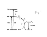


Classifications
-
- A—HUMAN NECESSITIES
- A61—MEDICAL OR VETERINARY SCIENCE; HYGIENE
- A61K—PREPARATIONS FOR MEDICAL, DENTAL OR TOILETRY PURPOSES
- A61K49/00—Preparations for testing in vivo
- A61K49/001—Preparation for luminescence or biological staining
- A61K49/0013—Luminescence
- A61K49/0017—Fluorescence in vivo
- A61K49/0019—Fluorescence in vivo characterised by the fluorescent group, e.g. oligomeric, polymeric or dendritic molecules
- A61K49/0021—Fluorescence in vivo characterised by the fluorescent group, e.g. oligomeric, polymeric or dendritic molecules the fluorescent group being a small organic molecule
- A61K49/0032—Methine dyes, e.g. cyanine dyes
-
- A—HUMAN NECESSITIES
- A61—MEDICAL OR VETERINARY SCIENCE; HYGIENE
- A61K—PREPARATIONS FOR MEDICAL, DENTAL OR TOILETRY PURPOSES
- A61K41/00—Medicinal preparations obtained by treating materials with wave energy or particle radiation ; Therapies using these preparations
- A61K41/0057—Photodynamic therapy with a photosensitizer, i.e. agent able to produce reactive oxygen species upon exposure to light or radiation, e.g. UV or visible light; photocleavage of nucleic acids with an agent
- A61K41/0071—PDT with porphyrins having exactly 20 ring atoms, i.e. based on the non-expanded tetrapyrrolic ring system, e.g. bacteriochlorin, chlorin-e6, or phthalocyanines
-
- A—HUMAN NECESSITIES
- A61—MEDICAL OR VETERINARY SCIENCE; HYGIENE
- A61K—PREPARATIONS FOR MEDICAL, DENTAL OR TOILETRY PURPOSES
- A61K49/00—Preparations for testing in vivo
- A61K49/001—Preparation for luminescence or biological staining
- A61K49/0013—Luminescence
- A61K49/0017—Fluorescence in vivo
- A61K49/005—Fluorescence in vivo characterised by the carrier molecule carrying the fluorescent agent
- A61K49/0052—Small organic molecules
-
- A—HUMAN NECESSITIES
- A61—MEDICAL OR VETERINARY SCIENCE; HYGIENE
- A61P—SPECIFIC THERAPEUTIC ACTIVITY OF CHEMICAL COMPOUNDS OR MEDICINAL PREPARATIONS
- A61P35/00—Antineoplastic agents
-
- A—HUMAN NECESSITIES
- A61—MEDICAL OR VETERINARY SCIENCE; HYGIENE
- A61P—SPECIFIC THERAPEUTIC ACTIVITY OF CHEMICAL COMPOUNDS OR MEDICINAL PREPARATIONS
- A61P35/00—Antineoplastic agents
- A61P35/04—Antineoplastic agents specific for metastasis
-
- A—HUMAN NECESSITIES
- A61—MEDICAL OR VETERINARY SCIENCE; HYGIENE
- A61P—SPECIFIC THERAPEUTIC ACTIVITY OF CHEMICAL COMPOUNDS OR MEDICINAL PREPARATIONS
- A61P43/00—Drugs for specific purposes, not provided for in groups A61P1/00-A61P41/00
Abstract
本発明は、ターゲティング分子、光線力学的療法(PDT)分子(1または2個の光子のいずれか)、および任意のイメージング剤(例えば、発色団、造影剤など)を含む、多機能性(通常、二機能性または三機能性)剤を含有する、方法並びに組成物に関する。 The present invention is multifunctional (usually comprising targeting molecules, photodynamic therapy (PDT) molecules (either one or two photons), and any imaging agent (eg, chromophore, contrast agent, etc.). , Bifunctional or trifunctional) agents and methods and compositions.
Description
(関連出願のクロスリファレンス)
本出願は、USSN 60/453,618(2003年3月10日出願)の優先権の利益を主張する一部継続出願(これは、本明細書の一部を構成する)である。
(Cross-reference of related applications)
This application is a continuation-in-part application (which forms part of this specification) that claims the benefit of the priority of USSN 60 / 453,618 (filed 10 March 2003).
(技術分野)
本発明は、ターゲティング分子(targeting moiety)、光線力学的療法(photo dynamic therapy)(PDT)分子(1個または2個のいずれか)、および任意のイメージング(imaging)剤(例えば、発色団、造影剤など)を含む、多機能性(通常、二機能性または三機能性)剤を含有する、方法並びに組成物に関する。
(Technical field)
The present invention relates to targeting moieties, photodynamic therapy (PDT) molecules (either one or two), and any imaging agent (eg, chromophore, imaging). The present invention relates to methods and compositions containing multifunctional (usually bifunctional or trifunctional) agents, including
(背景技術)
過去5年にわたって、癌性腫瘍の早期検出のための新規なイメージングおよび処置の技術の開発における進行中のルネサンスが存在する。過去数十年間にわたる多数の癌研究者の努力にもかかわらず、癌は未だ米国において2番目の主要な死因であり、これを超えるのは心臓疾患のみである。2002年には米国単独で120万以上の新規な癌の症例が診断されるであろう。これらの内の約70%は、現在活発な開発中の様々なインビボイメージング技術によって早期の検出を受け易い固形癌性腫瘍であろう。患者および保険医療のコストの展望から、特に経過観察の確証的な外科的生検(follow-up, confirmatory surgical biopsies)を排除することができるならば、終夜の病院の滞在を必要としない非侵襲性のイメージング技術が非常に所望されている。これらの新規なイメージング技術がまた同じ患者のセッションにおいて非侵襲的な処置方法と組み合わせることができる場合には、完全な外来患者のイメージング/検出/処置のプロトコールを設計することができ、このものは、現在の診断/外科手術/化学療法/電離放射線療法のプロトコール(これは、過去20年間、処置の標準であった)に置き代わり得る。
(Background technology)
Over the past five years there has been an ongoing renaissance in the development of new imaging and treatment techniques for the early detection of cancerous tumors. Despite the efforts of many cancer researchers over the past few decades, cancer is still the second leading cause of death in the United States, and only heart disease exceeds it. In 2002, more than 1.2 million new cancer cases will be diagnosed in the United States alone. About 70% of these will be solid cancerous tumors that are susceptible to early detection by various in vivo imaging technologies currently under active development. Non-invasive that does not require overnight hospital stay if the cost of patient and insurance care can be eliminated, especially follow-up, confirmatory surgical biopsies Sex imaging techniques are highly desired. If these new imaging techniques can also be combined with non-invasive treatment methods in the same patient session, a complete outpatient imaging / detection / treatment protocol can be designed, It can replace current diagnostic / surgical / chemotherapy / ionizing radiation therapy protocols, which have been the standard of treatment for the past 20 years.
世界保健機関は、2002年には120万以上の新規な癌の症例が診断され、そしてこのうち、全米癌学会は約203,500が侵襲性の乳癌(I〜IV期と定義される)を見積もっており、これにより、概算で40,000の死亡となる(引用文献9(これは、本明細書の一部を構成する)を参照)。早期の検出は、長期間に成功にした乳癌の場合にとって決定的であり、そして生存率を増大する(表1に例示する通り)。

明らかに、早期の検出は生存率の主要な因子であるが、乳癌の20〜40%は毎年のルーチンなマンモグラムの検診段階(検出することができる最小の腫瘍は0.5〜1.0cmである)では検出されない。加えて、多数の女性が、典型的なマンモグラム方法の間、激しい不快(特に、乳の圧迫)を経験している。40歳を超える女性は毎年の検診を受けていると通常考えられるが、この分類にあるわずか62%の女性だけが実際には過去1年の間にマンモグラムを受けていると、全米癌学会によって見積もられている。これらの見積もりは、早期の癌を検出しそして不快因子を排除する新規な検診方法に対する必要性をさし示している。 Obviously, early detection is a major factor in survival, but 20-40% of breast cancers occur at the routine mammogram screening stage each year (the smallest tumor that can be detected is 0.5-1.0 cm). Is not detected. In addition, many women experience severe discomfort (especially breast compression) during typical mammogram methods. Women over the age of 40 are usually considered to have annual screening, but only 62% of women in this category have actually received mammograms over the past year, according to the National Cancer Society. Estimated. These estimates point to the need for new screening methods that detect early cancers and eliminate discomfort.
ごく最近、主として健康な女性にとっての主な検診デバイスとしてマンモグラフィーを支持してきた何年ものデータの解釈に最近問題が投げかけられていると公表された研究に基づいて、ルーチンなマンモグラフィーの有効性が世間一般の報道機関およびマスコミ媒体(例えば、Time Magazine、2/4および2/18(2002年発行))において再考されている。ほとんどの乳癌が乳菅を起源とし、そして最終的に早期癌(これは、非浸潤性乳管癌またはDCISと呼ばれる)、前侵襲性の限局期(これは、乳菅の外側には未だ進行していない)にまで発展する。典型的なマンモグラフィーは、この期での偽陽性によって悩まされ得て、このことは、経過観察マンモグラフィーが針生検によって探求される可能な別の確認と合わせて実施されることを示し得る。最小のDCISは時々、切除のみによって処置することができるが、女性が不必要な生検および外科手術からの多数の瘢痕を示すことは珍しくなく、このことは、切除した腫瘍の周囲にある広い癌のない縁は経過化学療法および電離放射線処置を回避する必要があるとの警告である。 Most recently, the effectiveness of routine mammography has been based on research that has recently been published as a problem with the interpretation of years of data that has supported mammography as the primary screening device for primarily healthy women. It has been reconsidered in general news media and media (eg, Time Magazine, 2/4 and 2/18 (issued in 2002)). Most breast cancers originate from chyle and ultimately early cancer (which is called non-invasive ductal cancer or DCIS), pre-invasive focal stage (which is still progressing outside the chyle Not developed). Typical mammography can be plagued by false positives at this stage, which can indicate that follow-up mammography is performed in conjunction with another possible confirmation sought by needle biopsy. Although minimal DCIS can sometimes be treated by excision alone, it is not uncommon for women to show numerous scars from unnecessary biopsies and surgery, which is a wide area around the resected tumor A margin free of cancer is a warning that follow-up chemotherapy and ionizing radiation treatment should be avoided.
真に非侵襲であってそして(a)最も早期の可能な段階でDCISsを検出し、そして(b)非外科的な処置を許容し得る、別のイメージングおよび処置のプロトコールの開発は、高い危険状態にある女性によって最も歓迎されるであろう。 The development of alternative imaging and treatment protocols that are truly non-invasive and (a) detect DCISs at the earliest possible stage, and (b) allow non-surgical treatment, is a high risk Will be most welcomed by women in state.
過去10年にわたって、癌性腫瘍のインビボ光学イメージング(特に、乳癌のイメージング)に関係する可能性および問題の両方について議論された、多数の優れた総説が存在する(引用文献17(これは、本明細書の一部を構成する)を参照)。これらの筆者の数人は、腫瘍に関係するマーカーをターゲットとする高アフィニティーのベクター分子についての必要性、および周囲の健康な組織に対する腫瘍中への造影剤の取り込みの増大についての必要性を指摘している。モノクローナル抗体が、この観点で使用されており(引用文献21−23(これらは、本明細書の一部を構成する)を参照)、そしてBeckerおよび共同研究者は最近、マクロ分子(例えば、トランスフェリン、およびインドカルボシアニン(ITCC)とのヒト血清アルブミン接合体)が、腫瘍の光学的なイメージングのための有効な造影剤であることを示した(引用文献24(これは、本明細書の一部を構成する)を参照)。抗体または他の大分子を用いるいくつかの欠点は、それらが肝臓によって取り込まれ、ヒトにおいては有害な免疫学的な反応を誘発し、そして血液系中で非常に長い残留時間を有することである。加えて、大分子は、正の内部圧力が原因で腫瘍に深くまで容易に浸透することができない。 Over the past decade, there are a number of excellent reviews that discuss both the possibilities and problems associated with in vivo optical imaging of cancerous tumors, particularly breast cancer imaging (reference 17 (this is a book) Part of the description). Several of these authors point to the need for high-affinity vector molecules that target tumor-related markers, and the need for increased uptake of contrast media into the tumor for surrounding healthy tissue is doing. Monoclonal antibodies have been used in this regard (see refs. 21-23, which form part of this specification), and Becker and co-workers have recently developed macromolecules (eg, transferrin And human serum albumin conjugates with indocarbocyanine (ITCC) have been shown to be effective contrast agents for optical imaging of tumors (cited document 24, which is Part))). Some drawbacks with using antibodies or other large molecules are that they are taken up by the liver, elicit harmful immunological reactions in humans, and have very long residence times in the blood system . In addition, large molecules cannot easily penetrate deep into tumors due to positive internal pressure.
大分子−造影剤の接合体に関係する問題に対する可能な解決法は、小分子(例えば、小ペプチド)を使用して、造影剤をターゲット腫瘍に向けさせることである。多数の腫瘍は、ソマトスタチン(SST)および他のペプチドに対する受容体を過剰発現すること(引用文献25−28(これらは、本明細書の一部を構成する)を参照)、および胃腸−膵臓の腫瘍に対する受容体シンチグラフィーがルーチンな臨床的使用であることを示した。ソマトスタチンアナログ−蛍光接合体を用いる腫瘍のターゲティングおよびイメージングは、癌性腫瘍の光学的なイメージングの魅力的な代替物である。Beckerらは最近、オクトレオエート(octreoate)と結合するNIR蛍光リガンド、安定なソマトスタチン小ペプチドアナログをベースとする、受容体をターゲットとする腫瘍の光学的イメージング(引用文献29(これは、本明細書の一部を構成する)を参照)を提案している。彼らの研究において、インドシアニン色素(例えば、インドジカルボシアニン(indodicarbocyanine)(IDCC)およびインドトリカルボシアニン(indotricarbocyanine)(ITCC)は、Fmoc固相ペプチド合成方法論を用いて、オクトオレエートとカップリングした。直鎖のアナログである、メチオニンをシステインで置換した修飾オクトレオエートをコントロールとして使用した。ITCC−オクトレオエートは、RIN38/SSTR2腫瘍を有するマウスの異種移植片中に蓄積した。腫瘍および正常な組織の間の蛍光造影は、直ぐに増大し(約1分)、そして3〜24時間で、腫瘍の蛍光強度は周囲の正常な組織よりも3倍以上高くなった。従って、該小ペプチドソマトスタチンアナログは腫瘍中に直ぐに蓄積することができ、そしてコンパニオン(companion)実験はまた、それらが24時間後には直ちに該システムから排除されることを示した。該直鎖のオクトレオエート−ITCC接合体は腫瘍中には蓄積しないが、このものは該ソマトスタチン接合体が過剰発現した腫瘍受容体部位と注意深く適合するのに必要である。高アフィニティーのSST受容体はまた、大部分の乳癌腫中でも過剰発現する(引用文献(30(これは、本明細書の一部を構成する)を参照)。Reubiおよび共同研究者は(引用文献31−32(これらは、本明細書の一部を構成する)を参照)は、ソマトスタチン受容体である、SSTR1、SSTR2、およびSSTR3のメッセンジャーRNAs、並びにSSオートラジオグラフィーおよびmRNAインシチュハイブリダイゼーションの発現および局在化に関して、多数のヒト腫瘍を調べている。SS受容体は、全ての乳癌腫中に存在し、SSTR2が優位であって、そしてこのものは、オクトレオエートに対する高アフィニティーを示した。SST2は、ヒトソマトスタチン受容体サブタイプであり、このものは商業的に入手可能な合成アナログに対して最大のアフィニティーを有する。 A possible solution to the problem associated with large molecule-contrast agent conjugates is to use a small molecule (eg, a small peptide) to direct the contrast agent to the target tumor. Many tumors overexpress receptors for somatostatin (SST) and other peptides (see references 25-28, which form part of this specification), and gastrointestinal-pancreatic Receptor scintigraphy for tumors has been shown to be a routine clinical use. Tumor targeting and imaging using somatostatin analog-fluorescent conjugates is an attractive alternative to optical imaging of cancerous tumors. Recently, Becker et al., Optical Imaging of Receptor-Targeted Tumors Based on NIR Fluorescent Ligands that Bind Octreoate, Stable Somatostatin Small Peptide Analogues (Reference 29 (this document That is part of the book)). In their study, indocyanine dyes (eg, indodicarbocyanine (IDCC) and indotricarbocyanine (ITCC) are coupled to octoleate using Fmoc solid phase peptide synthesis methodology. A straight-chain analog, modified octreoate with methionine replaced with cysteine was used as a control, and ITCC-octreoate accumulated in xenografts of mice bearing RIN38 / SSTR2 tumors. Fluorescence imaging between normal tissues immediately increased (about 1 minute), and at 3-24 hours, the fluorescence intensity of the tumor was more than 3 times higher than the surrounding normal tissue, thus the small peptide. Somatostatin analogs can quickly accumulate in tumors and Companion experiments also showed that they were cleared from the system immediately after 24 hours, although the linear octreoate-ITCC conjugate does not accumulate in the tumor, It is necessary for the somatostatin conjugate to be carefully matched to the overexpressed tumor receptor site.The high affinity SST receptor is also overexpressed in most breast cancers (cited (30) Reubi and co-workers (see refs. 31-32, which form part of this specification) are SSTR1, a somatostatin receptor. , SSTR2 and SSTR3 messenger RNAs, and SS autoradiography and mRNA in situ hybridization A number of human tumors are being investigated, the S S receptor is present in all breast cancers, with SSTR2 predominating, which showed high affinity for octreoate. The human somatostatin receptor subtype, which has the greatest affinity for commercially available synthetic analogs.
HawryszおよびSevick-Muraca(引用文献33(これは、本明細書の一部を構成する)を参照)は、新規なイメージング剤(ここで、吸収/蛍光は、NIRの方向に赤色シフト(red shift)し、より深い組織への浸透を達成することができる)の開発に関する、優れた総説を指摘している。 Hawrysz and Sevick-Muraca (see ref. 33, which forms part of this specification), a novel imaging agent (where absorption / fluorescence is red shifted in the direction of the NIR). ) And can achieve a deeper penetration into the organization).
最近の研究は、2光子断面積(cross-section)を非常に増大したポルフィリンについての新規なデザイン方法に関し、そして我々は実際に、これら新規なポルフィリンの構造モチーフが、一重項酸素のインビトロ発生で誘発される2光子、1光子光線力学的療法における癌細胞アポトーシスの原因であると通常認められている剤を非常に有効なものとすることができることを証明した(引用文献1〜4(これらは、本明細書の一部を構成する)を参照)。 Recent work has focused on novel design methods for porphyrins that have greatly increased two-photon cross-sections, and we have indeed demonstrated that these new porphyrin structural motifs are in vitro generation of singlet oxygen. It has been proved that agents that are normally recognized as the cause of cancer cell apoptosis in induced two-photon, one-photon photodynamic therapy can be very effective (cited references 1-4 (these are , Which constitutes part of this specification).
しかしながら、真に非侵襲的であってそして1外来患者のセッションにおけるイメージングおよび処置を達成することができる、処置および様式に対する必要性が存在する。 However, there is a need for treatments and modalities that are truly non-invasive and capable of achieving imaging and treatment in one outpatient session.
(発明の概要)
上記の目的に従って、本発明は、ターゲティング分子および少なくとも光線力学的療法(PDT)分子(2光子PDT分子(2PM)が好ましい)を含有する、二機能性および三機能性剤を提供する。該薬剤は場合によりイメージング剤を含み、光学的なイメージング剤(例えば、発色団(chromophore)または発発色団(fluorophore)が好ましい)が好ましく、1光子発色団が特に好ましい。加えて、該薬剤は場合によるが通常、リンカーを含み、このことにより、該薬剤の成分の共有結合が可能となる。
(Summary of Invention)
In accordance with the above objectives, the present invention provides bifunctional and trifunctional agents containing a targeting molecule and at least a photodynamic therapy (PDT) molecule, preferably a two-photon PDT molecule (2PM). The agent optionally includes an imaging agent, preferably an optical imaging agent (eg, preferably a chromophore or fluorophore), and more preferably a one-photon chromophore. In addition, the drug will optionally include a linker, which allows for covalent attachment of the drug components.
本発明は、更に該分子を活性化するのに適当な波長の光を用いるPDT分子の活性化によって、疾患(とりわけ最も、癌)を検出しおよび/または処置する方法を提供する。該方法はまた、他のイメージング様式と組み合わせることができる。 The present invention further provides a method of detecting and / or treating a disease (especially most cancer) by activating a PDT molecule using light of an appropriate wavelength to activate the molecule. The method can also be combined with other imaging modalities.
(発明の詳細な記載)
本発明は、疾患の検出および処置のために、1以上の外来患者セッションにおいて使用することができる単一試薬に腫瘍(または他の疾患)のイメージングおよび処置のいくつかの面を組み合わせる、多機能性化合物に関する。内視鏡の使用は他のタイプの腫瘍(例えば、固形腫瘍を含む)の検出および処置を可能とすると理解するが、通常皮下癌性腫瘍は、以下に記載する通り、2光子薬剤の波長要件に適当な能力のために良好な候補である。
(Detailed description of the invention)
The present invention combines multiple aspects of tumor (or other disease) imaging and treatment into a single reagent that can be used in one or more outpatient sessions for disease detection and treatment It relates to a sex compound. Although it is understood that the use of an endoscope allows the detection and treatment of other types of tumors (eg, including solid tumors), usually subcutaneous cancerous tumors are wavelength requirements for two-photon drugs, as described below It is a good candidate for its appropriate ability.
多機能性剤は、二機能性または三機能性であり得る。二機能性剤としては、2光子光線学的分子(2PM)と通常はリンカーによって連結したターゲティング分子を含む。ターゲティング分子は、該共有結合した2PMが選択した組織(例えば、腫瘍)中に素早く蓄積することを可能とし、そして周囲のおよび/または健康な組織中にはいずれの実質的な程度にも蓄積しない。該2PMは、NIR光子の2光子吸収によって活性化されて、患部細胞(例えば、癌細胞)の死を開始することが可能となる。 Multifunctional agents can be bifunctional or trifunctional. Bifunctional agents include two-photon photological molecules (2PM) and usually targeting molecules linked by a linker. Targeting molecules allow the covalently bound 2PM to accumulate rapidly in selected tissues (eg, tumors) and do not accumulate to any substantial extent in surrounding and / or healthy tissues . The 2PM is activated by two-photon absorption of NIR photons, and can start the death of affected cells (for example, cancer cells).
三機能性剤は、ターゲティング分子、イメージング剤およびPDT分子(PM)(これは、1光子PMまたは2PMのいずれかであり得る)を含む。以下に記載する通り、該イメージング剤により、患部組織(例えば、癌性腫瘍)の素早い3次元イメージングが可能となる。イメージング剤を2PMと組み合わせる場合に、得られる剤は、組織透明度ウィンドウ(800〜1000nm)内でのNIRパルスレーザー放射によって活性化することができる。従って、この共有結合アンサンブルは二重機能性を包含し;そのものは、低レーザー出力でのイメージング様式で使用して1光子イメージング剤のみを活性化することができ、またはそのものは、レーザー焦点を変えそして出力を増大することによって光線力学的療法試薬として操作することができる。該2光子プロセスは、該腫瘍部位でのレーザー光線の焦点で活性化されるのみで、周囲の健康な組織上にはほとんどまたは全く影響を及ぼさない。2光子光線力学的療法は長い間、いくつかの学術的な研究者および小企業の目標であったが(引用文献5〜7(これらは、本明細書の一部を構成する)を参照)、しかし、癌処置に対するこの研究の発展は、天然ポルフィリンまたは商業的な試薬(例えば、フォトフリン(Photofrin)(引用文献8(これは、本明細書の一部を構成する)を参照)の非常に小さい2光子断面積が原因で制限されてきた。しかしながら、2光子断面積の非常な増大を有する合成ポルフィリン物質の最近の開発により、今では1光子PDTの実用的な代替物である正に2光子PDTを製造している。特に、2PM構造に関しては、米国公開番号2003/0105070(これは、本明細書の一部を構成する)を参照。 Trifunctional agents include targeting molecules, imaging agents and PDT molecules (PM), which can be either one-photon PM or 2PM. As described below, the imaging agent enables rapid three-dimensional imaging of affected tissue (for example, a cancerous tumor). When the imaging agent is combined with 2PM, the resulting agent can be activated by NIR pulsed laser radiation within the tissue transparency window (800-1000 nm). Thus, this covalent ensemble includes dual functionality; it can be used in an imaging mode at low laser power to activate only a one-photon imaging agent, or it can alter the laser focus. It can then be operated as a photodynamic therapy reagent by increasing the output. The two-photon process is only activated at the focal point of the laser beam at the tumor site and has little or no effect on the surrounding healthy tissue. Two-photon photodynamic therapy has long been the goal of several academic researchers and small businesses (see references 5-7, which form part of this specification) However, the development of this study for cancer treatment has been largely based on natural porphyrins or commercial reagents such as Photofrin (see ref. 8 (which forms part of this specification)). However, due to the recent development of synthetic porphyrin materials with a significant increase in the two-photon cross-section, it is now a practical alternative to the one-photon PDT. Manufactures two-photon PDT, especially for the 2PM structure, see US Publication No. 2003/0105070, which forms part of this specification.
従来のイメージング/造影発色団および2光子PDT発色団の同じ試薬中での組み合わせは、イメージングプロセスで発見されたいずれかの潜在的な癌性増殖の素早い光線力学的療法処置の可能性をも含む外来患者の検診(screening)および検出システムに対する新規な方法を与える。次いで、癌の直接的な処置はルーチン(例えば、毎年)な検診と直接的に関連し、このことにより、早期の検出および非外科的な外来患者の処置の双方の利点を与える。この方法はまた、現在使用中または開発中の他のイメージング技術(例えば、従来およびデジタルのマンモグラフィー)への別個の補助としての可能性をも与える。従って、これらの剤は、早期癌性腫瘍のインビボでの検出および処置のための強力な新規パラダイムとして機能することができる。この方法の利点は、早期の乳癌腫瘍の処置においていかに使用できるかという以下の議論で例示されるが、しかしながら、該処置プロトコールはいずれかの発生段階での癌性組織(固形腫瘍を含む)に使用することができる。 The combination of a conventional imaging / contrast chromophore and a two-photon PDT chromophore in the same reagent also includes the potential for rapid photodynamic therapy treatment of any potential cancerous growth discovered in the imaging process. A novel method for outpatient screening and detection system is given. The direct treatment of cancer is then directly associated with routine (eg, annual) screening, which provides the benefits of both early detection and non-surgical outpatient treatment. This method also offers the potential as a separate aid to other imaging technologies currently in use or in development (eg, conventional and digital mammography). Thus, these agents can function as a powerful new paradigm for in vivo detection and treatment of early cancerous tumors. The advantages of this method are illustrated in the following discussion of how it can be used in the treatment of early breast cancer tumors, however, the treatment protocol applies to cancerous tissues (including solid tumors) at any stage of development. Can be used.
従って、本発明は本明細書中に記載する多機能性剤を提供する。好ましい実施態様において、該剤は、3個の異なる構成成分:ターゲティング分子、イメージング分子、およびPDT分子を含有する三機能性または三連分子構造(triad)の組成物である。以下に概説するとおり、該3個の構成成分と共有結合するように機能するリンカー分子が、使用されることが多い。 Accordingly, the present invention provides the multifunctional agents described herein. In a preferred embodiment, the agent is a trifunctional or triad composition containing three different components: a targeting molecule, an imaging molecule, and a PDT molecule. As outlined below, linker molecules that function to covalently link the three components are often used.
本明細書中で使用する「ターゲティング(targeting)分子」または文法上の均等物は、官能基(このものは、ターゲットに機能するか、または該複合体を特定の位置、細胞タイプ、患部組織もしくは結合に方向付ける)を意味する。通常、ターゲティング分子はターゲット(target)分子を指向する。当業者によって認められている通り、本発明の剤は通常、静脈内注射され;従って、好ましいターゲティング分子とは、体内腔(例えば、脊髄、関節の間質性腔など)中への直接的な注射もまた可能であるが、特に血管システムへ近づくことができる特定の局在での該剤の濃縮が可能であるものである。好ましい実施態様において、該剤は、1:1でない比率で、該位置に分配される。従って、例えば抗体、細胞表面受容体リガンド、およびホルモン、脂質、糖類およびデキストラン、アルコール、胆汁酸、脂肪酸、アミノ酸、タンパク質(ペプチドを含む)、および核酸は全て結合して、該造影剤を特定の部位に局在化したりまたはターゲットとすることができる。 As used herein, a “targeting molecule” or grammatical equivalent refers to a functional group (which either functions as a target or makes the complex a specific location, cell type, affected tissue or Direction). Usually, the targeting molecule is directed to the target molecule. As will be appreciated by those skilled in the art, the agents of the invention are usually injected intravenously; thus, preferred targeting molecules are directly into the body lumen (eg, spinal cord, interstitial space of joints, etc.) Injection is also possible, but is particularly capable of concentrating the agent at a specific location that allows access to the vascular system. In a preferred embodiment, the agent is dispensed to the location in a ratio that is not 1: 1. Thus, for example, antibodies, cell surface receptor ligands, and hormones, lipids, sugars and dextrans, alcohols, bile acids, fatty acids, amino acids, proteins (including peptides), and nucleic acids all bind to identify the contrast agent. It can be localized to a site or targeted.
好ましい実施態様において、ターゲティング分子は、細胞の特定の組織または表面に対して本発明の剤をターゲティングすることができる。すなわち、好ましい実施態様において、本発明の剤は、有用な細胞の細胞質中に摂取(taken up)される必要はない。加えて、好ましいターゲティング分子は、癌ターゲットに対するものである。「癌ターゲット」とは、癌の細胞、組織および/または腫瘍中で優先的に発現したりまたは合成されるものである。例えば、適当な癌ターゲット物質としては例えば、酵素およびタンパク質(例えば、ペプチドを含む)、例えば細胞表面受容体;核酸;脂質およびリン脂質を含むが、これらに限定されない。好ましい実施態様は、固形腫瘍の表面上に存在する癌ターゲット(例えば、上記のソマトスタチン(SST)受容体、下記のHER2受容体など)を使用する。 In a preferred embodiment, the targeting molecule can target the agent of the present invention to a particular tissue or surface of the cell. That is, in a preferred embodiment, the agent of the present invention need not be taken up into the cytoplasm of useful cells. In addition, preferred targeting molecules are for cancer targets. A “cancer target” is one that is preferentially expressed or synthesized in cancer cells, tissues and / or tumors. For example, suitable cancer target materials include, but are not limited to, enzymes and proteins (including peptides, for example) such as cell surface receptors; nucleic acids; lipids and phospholipids. A preferred embodiment uses a cancer target (eg, the somatostatin (SST) receptor described above, the HER2 receptor described below, etc.) present on the surface of a solid tumor.
好ましい実施態様において、ターゲティング分子はタンパク質である。本明細書中の「タンパク質」または文法上の均等物は、タンパク質、オリゴペプチドおよびペプチド、誘導体およびアナログ(例えば、非天然アミノ酸およびアミノ酸アナログを含有するタンパク質を含む)、およびペプチド模倣(peptidomimetic)構造を意味する。側鎖は、(R)または(S)立体配置のいずれかであり得る。好ましい実施態様において、アミノ酸は(S)またはL−立体配置である。下記の通り、タンパク質をターゲティング分子として使用する場合には、そのものは、プロテアーゼによるインビボ分解を遅延する(retard)ためにタンパク質アナログを使用することが所望され得る。 In a preferred embodiment, the targeting molecule is a protein. As used herein, “protein” or grammatical equivalents include proteins, oligopeptides and peptides, derivatives and analogs (including, for example, proteins containing unnatural amino acids and amino acid analogs), and peptidomimetic structures. Means. The side chain can be either in the (R) or (S) configuration. In a preferred embodiment, the amino acid is in (S) or L-configuration. As described below, when using proteins as targeting molecules, it may be desirable to use protein analogs to retard in vivo degradation by proteases.
好ましい実施態様において、タンパク質は、細胞表面受容体の結合性パートナー(リガンド)、特に疾患と関連するもの、例えば癌性組織に特異的であるかまたは差次的に発現するかのいずれかである癌細胞表面受容体である。ターゲティング分子の疾患組織に対する高い特異性は好ましいが、放射はターゲットされ得るという理由で、完全な特異性(例えば、健康な組織とは全く結合しない)は存在する必要がないことに注目するのは重要である。細胞表面のリガンドおよび/またはアナログおよび誘導体(例えば、フラグメントを含む)が好ましく、例えば酵素基質またはインヒビター、特に細胞表面結合酵素が挙げられる。 In a preferred embodiment, the protein is a binding partner (ligand) of a cell surface receptor, particularly one associated with a disease, such as either cancerous tissue or differentially expressed It is a cancer cell surface receptor. Note that the high specificity of the targeting molecule for diseased tissue is preferred, but full specificity (eg, does not bind to healthy tissue at all) need to be present because radiation can be targeted is important. Cell surface ligands and / or analogs and derivatives (eg, including fragments) are preferred, including, for example, enzyme substrates or inhibitors, particularly cell surface-bound enzymes.
好ましい実施態様において、ターゲティング分子は、細胞表面受容体に対するリガンドの全てまたは一部(例えば、結合部分)である。適当なリガンドとしては例えば、細胞表面受容体と結合するリガンドの全てまたは官能性部分を含むが、これらに限定されない。該受容体としては、インスリン受容体(インスリン)、インスリン様成長因子(IGF−1およびIGF−2の両方を含む)、成長ホルモン受容体、グルコーストランスポーター(特に、GLUT 4受容体)、トランスフェリン受容体(トランスフェリン)、上皮細胞成長因子受容体(EGF)、低密度リポタンパク質受容体、高密度リポタンパク質受容体、レプチン受容体、エストロゲン受容体(エストロゲン);インターロイキン受容体(例えば、IL−1、IL−2、IL−3、IL−4、IL−5、IL−6、IL−7、IL−8、IL−9、IL−11、IL−12、IL−13、IL−15およびIL−17受容体を含む)、ヒト成長ホルモン受容体、VEGF受容体(VEGF)、PDGF受容体(PDGF)、トランスフォーミング成長因子受容体(例えば、TGF−aおよびTGF−βを含む)、EPO受容体(EPO)、TPO受容体(TPO)、繊毛様神経栄養因子受容体、プロラクチン受容体、およびT−細胞受容体からなる群から選ばれる。特に、ホルモンリガンドが好ましい。ホルモンとしてはステロイドホルモンおよびタンパク質性ホルモンの両方を含む。例えば、エピネフリン、チロキシン、オキシトシン、インスリン、甲状腺−刺激ホルモン、カルシトニン、絨毛性ゴナドトロピン、コルチコトロピン(cortictropin)、卵胞刺激ホルモン、グルカゴン、黄体形成(leuteinizing)ホルモン、リポトロピン、メラニン細胞刺激ホルモン、ノルエピネフリン、副甲状腺(parathryroid)ホルモン、甲状腺刺激ホルモン(TSH)、バソプレッシン、エンケファリン、セロトニン(seratonin)、エストラジオール、プロゲステロン、テストステロン、コルチゾンおよびグルココルチコイド、並びに以下に例示するホルモンを含むが、これらに限定されない。受容体リガンドとしては、受容体(例えば、細胞表面受容体)と結合するリガンドを含む。例えば、ホルモン、脂質、タンパク質、糖タンパク質、シグナグトランスデューサー、成長因子、サイトカイン、およびその他を含む。ソマトスタチンおよびトランスフェリンが、特に好ましい。 In preferred embodiments, the targeting molecule is all or part of a ligand for a cell surface receptor (eg, a binding moiety). Suitable ligands include, but are not limited to, for example, all or functional moieties of ligands that bind to cell surface receptors. Such receptors include insulin receptor (insulin), insulin-like growth factor (including both IGF-1 and IGF-2), growth hormone receptor, glucose transporter (especially GLUT 4 receptor), transferrin receptor Body (transferrin), epidermal growth factor receptor (EGF), low density lipoprotein receptor, high density lipoprotein receptor, leptin receptor, estrogen receptor (estrogen); interleukin receptor (eg IL-1 IL-2, IL-3, IL-4, IL-5, IL-6, IL-7, IL-8, IL-9, IL-11, IL-12, IL-13, IL-15 and IL -17 receptor), human growth hormone receptor, VEGF receptor (VEGF), PDGF receptor (PDGF), Blooming growth factor receptors (including, for example, TGF-a and TGF-β), EPO receptors (EPO), TPO receptors (TPO), ciliary neurotrophic factor receptors, prolactin receptors, and T-cell receptors Selected from the group consisting of bodies. In particular, hormone ligands are preferred. Hormones include both steroid hormones and proteinaceous hormones. E.g. epinephrine, thyroxine, oxytocin, insulin, thyroid-stimulating hormone, calcitonin, chorionic gonadotropin, cortictropin, follicle stimulating hormone, glucagon, leuteinizing hormone, lipotropin, melanocyte stimulating hormone, norepinephrine, parathyroid gland (parathryroid) hormones, thyroid stimulating hormone (TSH), vasopressin, enkephalin, serotonin, estradiol, progesterone, testosterone, cortisone and glucocorticoid, and hormones exemplified below, but are not limited thereto. Receptor ligands include ligands that bind to receptors (eg, cell surface receptors). For example, hormones, lipids, proteins, glycoproteins, signal transducers, growth factors, cytokines, and others. Somatostatin and transferrin are particularly preferred.
従って、好ましい実施態様において、該タンパク質はペプチドであり、特に癌特異的な細胞表面状態と結合することが知られるものが挙げられる。ソマトスタチン、トランスフェリンおよびそれらの機能的な誘導体が特に好ましい。その上、化学走性ペプチドを用いて、組織の損傷および炎症(特に、細菌感染によるもの)をイメージする;WO 97/14443(これは、本明細書の一部を構成する)を参照。加えて、ペプチドと関係する癌に関与する広範囲な酵素が存在し、該ペプチドは、これらの酵素と物質またはインヒビターのいずれかとして結合し、ターゲティング分子として付随的に使用することができる。 Thus, in a preferred embodiment, the protein is a peptide, particularly those known to bind to cancer specific cell surface conditions. Somatostatin, transferrin and their functional derivatives are particularly preferred. Moreover, chemotaxis peptides are used to image tissue damage and inflammation, particularly due to bacterial infections; see WO 97/14443, which forms part of this specification. In addition, there are a wide range of enzymes involved in cancers associated with peptides, which can bind to these enzymes as either substances or inhibitors and can be used incidentally as targeting molecules.
カテプシンBは、腫瘍の侵襲および進行に関与する。細胞からのカテプシンBの分泌は、生理学的なpHで機能するが、媒質の酸性pHによって誘発され得る。そのものは、プロテアーゼカスケードを分解する細胞外基質(ECM)中のタンパク質であり、そしてこのものは基質の非存在下で自己分解を受ける。カテプシンBは、乳、頚部、卵巣、胃、肺、脳、結腸直腸、前立腺および甲状腺の腫瘍に関与する。そのものは、局所的な侵襲期で活性であり、IV期の腫瘍はより低い進展期の腫瘍よりも有意な高濃度を示す。そのものは、腫瘍細胞表面、接着斑、および浸潤突起(invadopodia)(ここで、該腫瘍細胞は基底膜およびECMと接触する)で活性であることが分かった。そのものは、細胞内および細胞外の両方でECMを分解し、例えば天然物質としてラミニン、フィブロネクチン、およびコラーゲンIVを含む。本発明における使用のための適当な更なるおよび合成の物質としては以下のものを含むが、これらに限定されない。エデスチン、ゼラチン、アゾカゼイン、ベンジルオキシカルボニルアルギニルアルギニン4−メチルクマリン−7−イルアミン(Z−Arg−Arg−NH−Mec);トリプシノーゲン;ベンジルオキシカルボニルフェニルアルギニン4−メチルクマリン−7−イルアミン(Z−Phe−Arg−NH−Mec);N−a−ベンジルオキシカルボニル−L−アルギニル−L−アルギニン2−ナフチルアミド(Z−Arg−Arg−NNap);セトフィン(setfin)A;ベンジルオキシカルボニルアルギニルアルギニンp−ニトロアニリド(Z−Arg−Arg−p−NA);インスリンの酸化β鎖;ベンジルオキシカルボニルフェニルアルギニンp−ニトロアニリド(Z−Phe−Arg−p−NA);a−N−ベンゾイル−L−アルギニンアミド(BAA);a−N−ベンゾイル−L−アルギニンエチルエステル(BAEE);a−N−ベンゾイル−D,L−アルギニン2−ナフチルアミド(BANA);a−N−ベンゾイル−D,L−アルギニンp−ニトロアニリド(BAPA);a−N−ベンゾイル−L−リシンアミド(BLA);a−N−ベンジルオキシカルボニルグリシンp−ニトロフェニルエステル(CGN);および、a−N−ベンジルオキシカルボニル−L−リシンp−ニトロフェニルエステル(CLN)。Buckらによる, Biochem. J. 282 (Pt 1), 273-278 (1992);Moinらによる, Biochem. J. 285 (Pt 2), 427-434 (1992);Hasnainらによる, Biol. Chem. Hoppe Seyler 373, 413-418 (1992);Willenbrockらによる, Biochem. J. 227, 521-528 (1985);Otto, K.による, in Tissue Proteinases(Barrett, A. J.およびDingle, J. T.編))1頁, North-Holland, Amsterdam;Bajkowskiらによる, Anal. Biochem 68, 119-127 (1975)、およびそれらの中の引用文献(これらは全て、本明細書の一部を構成する)を参照。これらの全ては、ターゲティング分子として使用することができる。 Cathepsin B is involved in tumor invasion and progression. Secretion of cathepsin B from cells functions at physiological pH but can be triggered by the acidic pH of the medium. It is a protein in the extracellular matrix (ECM) that degrades the protease cascade, and it undergoes autolysis in the absence of the substrate. Cathepsin B is involved in breast, cervical, ovarian, stomach, lung, brain, colorectal, prostate and thyroid tumors. As such, it is active in the locally invasive phase, with stage IV tumors showing significantly higher concentrations than lower stage tumors. As such, it was found to be active on the tumor cell surface, adhesion spots, and invadopodia, where the tumor cells contact the basement membrane and the ECM. It degrades ECM both intracellularly and extracellularly and includes, for example, laminin, fibronectin, and collagen IV as natural substances. Suitable additional and synthetic materials for use in the present invention include, but are not limited to: Edestine, gelatin, azocasein, benzyloxycarbonylarginylarginine 4-methylcoumarin-7-ylamine (Z-Arg-Arg-NH-Mec); trypsinogen; benzyloxycarbonylphenylarginine 4-methylcoumarin-7-ylamine (Z- Phe-Arg-NH-Mec); Na-benzyloxycarbonyl-L-arginyl-L-arginine 2-naphthylamide (Z-Arg-Arg-NNap); cetofin A; benzyloxycarbonylarginyl arginine p-nitroanilide (Z-Arg-Arg-p-NA); oxidized β-chain of insulin; benzyloxycarbonylphenylarginine p-nitroanilide (Z-Phe-Arg-p-NA); aN-benzoyl-L -Arginine AN-benzoyl-L-arginine ethyl ester (BAEE); a-N-benzoyl-D, L-arginine 2-naphthylamide (BANA); a-N-benzoyl-D, L-arginine p-nitroanilide (BAPA); aN-benzoyl-L-lysine amide (BLA); aN-benzyloxycarbonylglycine p-nitrophenyl ester (CGN); and aN-benzyloxycarbonyl-L- Lysine p-nitrophenyl ester (CLN). Buck et al., Biochem. J. 282 (Pt 1), 273-278 (1992); Moin et al., Biochem. J. 285 (Pt 2), 427-434 (1992); Hasnain et al., Biol. Chem. Hoppe Seyler 373, 413-418 (1992); by Willenbrock et al., Biochem. J. 227, 521-528 (1985); Otto, K., in Tissue Proteinases (Barrett, AJ and Dingle, JT)) 1 , North-Holland, Amsterdam; by Bajkowski et al., Anal. Biochem 68, 119-127 (1975), and references cited therein, all of which form part of this specification. All of these can be used as targeting molecules.
加えて、様々な公知のインヒビター(例えば、シスタチンC,1−(L−トランスエポキシスクシニルロイシルアミノ)−4−グアニジノブタン(これは、E−64または(N−[N−(L−3−トランス−カルボキシオキシラン−2−カルボニル)−L−ロイシル]−アグマチン)とも呼ばれる)が存在する。Yanらによる, (1998) Biol. Chem. 379: 113;Kepplerらによる, (1994) Biochem. Soc. Trans. 22: 43;Hughesらによる, PNAS USA 95: 12410 (1998);Abdollahiらによる, J. Soc. Gynecol. Invest. 6: 32 (1999);Varugheseらによる, Biochemistry 31, 5172-5176 (1992);Hasnainらによる, J. Biol. Chem. 267, 4713-4721 (1992)(これらは全て、本明細書の一部を構成する)を参照)。これらの全ては、ターゲティング分子として使用することができる。 In addition, various known inhibitors (eg, cystatin C, 1- (L-trans epoxysuccinyl leucylamino) -4-guanidinobutane (which can be E-64 or (N- [N- (L-3- (Also referred to as trans-carboxyoxirane-2-carbonyl) -L-leucyl] -agmatine)) by Yan et al., (1998) Biol. Chem. 379: 113; by Keppler et al., (1994) Biochem. Soc. Trans. 22: 43; Hughes et al., PNAS USA 95: 12410 (1998); Abdollahi et al., J. Soc. Gynecol. Invest. 6: 32 (1999); Varughese et al., Biochemistry 31, 5172-5176 (1992) ); Hasnain et al., J. Biol. Chem. 267, 4713-4721 (1992), all of which are part of this specification. All of these can be used as targeting molecules.
好ましい実施態様において、ターゲティング分子は、カテプシンDについての基質またはインヒビターである。カテプシンDは、典型的なAsp−Thr−Gly活性部位を有する48kDaのアスパルチルエンドプロテアーゼである。様々な他のカテプシンと同様に、そのものは、52kDaの前駆体であるプロカテプシンDとして産生される。そのものは、リソソーム中に遍在的に分配される。カテプシンDは、乳癌、腎細胞癌、卵巣癌およびメラノーマ癌に関係し、そして臨床的な転移へのミクロ転移の増殖に関与すると考えられる。腫瘍細胞において、カテプシンDは周囲の媒質中に分泌され、その結果、原形質膜にまで運搬される。カテプシンBと同様に、カテプシンDは、プロテアーゼのECM分解性カスケードの一部である。加えて、カテプシンDは、最適な活性のために酸性pH(4.5〜5.0)を必要とする。Rochefortらによる, APMIS 107: 86 (1999);Xingらによる, Mol. Endo. 12 (9): 1310 (1998);Yaziovitskayaらによる, Proc. Am. Assoc. Cancer Res. 37: #3553 519 (1996);(これらは、本明細書の一部を構成する)を参照)。それらの全てはターゲティング分子として使用することができる。 In a preferred embodiment, the targeting molecule is a substrate or inhibitor for cathepsin D. Cathepsin D is a 48 kDa aspartyl endoprotease with a typical Asp-Thr-Gly active site. Like a variety of other cathepsins, it is produced as procathepsin D, a 52 kDa precursor. It is ubiquitously distributed throughout the lysosome. Cathepsin D is implicated in breast cancer, renal cell cancer, ovarian cancer and melanoma cancer, and is thought to be involved in the growth of micrometastasis to clinical metastasis. In tumor cells, cathepsin D is secreted into the surrounding medium and is consequently transported to the plasma membrane. Like cathepsin B, cathepsin D is part of the ECM degradable cascade of proteases. In addition, cathepsin D requires an acidic pH (4.5-5.0) for optimal activity. Rochefort et al., APMIS 107: 86 (1999); Xing et al., Mol. Endo. 12 (9): 1310 (1998); Yaziovitskaya et al., Proc. Am. Assoc. Cancer Res. 37: # 3553 519 (1996) ); (These form part of this specification)). All of them can be used as targeting molecules.
公知のカテプシンDの基質およびインヒビターとしては例えば、基質:gp−120およびナフタラジン(naphthazarin)(5,8−ジヒドロキシル−1,4−ナフトキノン);およびインヒビター:ペプスタチン(pepstatine)およびエクイスタチン(equistatin)を含むが、これらに限定されない。Ollingerによる, Archives of Biochemistry & Biophysics. 373 (2): 346-51, 2000;EI Messaoudiらによる, Journal of Virology. 74 (2): 1004-7, 2000;Bessodesらによる, Biochemical Pharmacology, 58 (2): 329-33, 1999;および、Lenarcicらによる, Journal of Biological Chemistry. 274 (2): 563-6, 1999(これらは、本明細書の一部を構成する)を参照。それらの全ては、ターゲティング分子として使用することができる。 Known substrates and inhibitors of cathepsin D include, for example, substrates: gp-120 and naphthazarin (5,8-dihydroxyl-1,4-naphthoquinone); and inhibitors: pepstatine and equistatin Including, but not limited to. Ollinger, Archives of Biochemistry & Biophysics. 373 (2): 346-51, 2000; EI Messaoudi et al., Journal of Virology. 74 (2): 1004-7, 2000; Bessodes et al., Biochemical Pharmacology, 58 (2 ): 329-33, 1999; and Lenarcic et al., Journal of Biological Chemistry. 274 (2): 563-6, 1999, which form part of this specification. All of them can be used as targeting molecules.
好ましい実施態様において、ターゲティング分子は、カテプシンKにとっての基質またはインヒビターである。カテプシンKはまた、弾性組織分解性(elastolytic)システインプロテアーゼでもあり、そしてこのものは最も強力な哺乳動物エラスターゼであると考えられ、そしてまたコラーゲン分解性活性を有する。カテプシンKは、そのコラーゲン分解性活性が他のシステインプロテアーゼと対比してコラーゲンの三重らせん体の不安定化に寄与せず、そして間質性コラーゲナーゼよりもより多くの部位で未変性分子を切断する点で、哺乳動物プロテイナーゼの間で特異的であると考えられる。従って、カテプシンKは、他のプロテアーゼの不在下で成体の皮質骨の不溶性コラーゲンを完全に分解することができる。そのものは、破骨細胞中で非常に発現する。そのものは、骨再吸収において重要な役割を果たしており、そして正常な骨の成長および再構築にとって必須である。そのものは、骨粗鬆症、多発異骨症(pycnodysotosis)、骨癌、並びに乳癌に関係する。乳癌は通常、骨に転移し、そしてカテプシンKは乳癌細胞(このものは、骨に広がりそして侵襲する)中での存在によって乳癌に関連すると最初に同定された。その基質としては例えば、エラスチンおよびコラーゲンを含むが、これらに限定されない。そのインヒビターとしては、例えば以下のものを含むが、これらに限定されない:Cbz−Gly−Arg−AMC;Cbz−Arg−Arg−AMC;Cbz−Gly−Gly−Arg−AMC;Cbz−Ala−Lys−Arg−AMC;Cbz−Ala−Arg−Arg−AMC;Cbz−d−Phe−Arg−AMC;Boc−Leu−Gly−Arg−AMC;H−Gly−Arg−AMC;H−Ala−Arg−AMC;Cbz−Leu−Leu−Leu−AMC;Cbz−Leu−Leu−AMC;Cbz−Phe−Gly−AMC;Cbz−Gly−Gly−Leu−AMC;Suc−Ala−Ala−Val−AMC;Cbz−Gly−Ala−Met−AMC;E−64;ロイペプチン(Ac−Leu−Leu−Arg−CHO);N−アセチル−Leu−Leu−メチオナール(methional);Ac−Leu Leu−Met−CHO;Ac−Leu−Val−Lys−CHO;Ac−Leu−Leu−Nle−CHO;Cbz−Lys−Leu−Leu−CHO;Cbz−Leu−Leu Leu−CHO;Cbz−Arg−Leu−Leu−CHO;1,3−ビス(アクリルアミノ)−2−プロパノンの群;1,3ジアミノケトンの群;および、1,5−ジアシルカルボヒドラジドの群。適当なカテプシンK基質としては、例えば以下のものを含むが、これらに限定されない:Cbz−Leu−Arg−AMC;Cbz−Val−Arg−AMC;Cbz−Phe−Arg−AMC;Cbz−Leu−Leu−Arg−AMC;Tos Gly−Pro−Arg−AMC;Bz−;Phe−Val−Arg−AMC;H−Pro−Phe−Arg−AMC;Cbz−Val−Val−Arg−AMC;Boc−Val−Pro Arg−AMC;Cbz−Glu−Arg−AMC;Bz−Arg−AMC;Ac−Phe−Arg−AMC;Boc−Val−Leu−Lys−AMC;Suc−Leu−Tyr AMC;Boc−Ala−Gly−Pro−Arg−AMC;Cbz−Gly−Pro−Arg−AMC;Z−Leu−Arg−4−メトキシ−b−ナフチルアミド(ここで、Cbzはベンジルオキシカルボニルであり、そして、AMCはアミノメチルクマリンである);ジアミノプロパノン、ジアシルヒドラジンおよびシスタチンC。Bossard, M. J.らによる, J. Biol. Chem. 271, 12517-12524 (1996);Aibe, K.らによる, Biol. Pharm. Bull. 19, 1026-1031 (1996);Votta, B. J.らによる, J. Bone Miner. Res. 12, 1396 1406 (1997);Yamshita, D. S.らによる, J. Am. Chem. Soc. 119, 11351-11352 (1997);DesJarlais, R. L.らによる, J. Am. Chem. Soc. 120, 9114-9115 (1998);Marquis, R. W.らによる, J. Med. Chem. 41, 3563-3567 (1998);Thompsonらによる, J. Med. Chem. 41, 3923-3927 (1998);Thompsonらによる, Bioorg. Med. Chem. 7, 599 605 (1999);Kamiya, T.らによる, J. Biochem. (Tokyo) 123, 752-759 (1998);Shiらによる, J. Clin. Invest. 104: 1191 (1999);および、Sukhovaらによる, J. Clin. Invest. 102: 576 (1998)(これらは全て、本明細書の一部を構成する)を参照)。これらの全ては、ターゲティング分子として使用することができる。 In a preferred embodiment, the targeting molecule is a substrate or inhibitor for cathepsin K. Cathepsin K is also an elastic elastolytic cysteine protease and is considered to be the most potent mammalian elastase and also has collagenolytic activity. Cathepsin K does not contribute to destabilization of the collagen triple helix in contrast to other cysteine proteases, and cleaves native molecules at more sites than interstitial collagenase. In that respect, it is considered to be specific among mammalian proteinases. Thus, cathepsin K can completely degrade insoluble collagen in adult cortical bone in the absence of other proteases. It is highly expressed in osteoclasts. As such, they play an important role in bone resorption and are essential for normal bone growth and remodeling. As such, it is related to osteoporosis, pycnodysotosis, bone cancer, and breast cancer. Breast cancer usually metastasizes to bone, and cathepsin K was first identified as being associated with breast cancer by its presence in breast cancer cells, which spread and invade bone. Examples of the substrate include, but are not limited to, elastin and collagen. The inhibitors include, but are not limited to, for example: Cbz-Gly-Arg-AMC; Cbz-Arg-Arg-AMC; Cbz-Gly-Gly-Arg-AMC; Cbz-Ala-Lys- Cgz-Ala-Arg-Arg-AMC; Cbz-d-Phe-Arg-AMC; Boc-Leu-Gly-Arg-AMC; H-Gly-Arg-AMC; H-Ala-Arg-AMC; Cbz-Leu-Leu-Leu-AMC; Cbz-Leu-Leu-AMC; Cbz-Phe-Gly-AMC; Cbz-Gly-Gly-Leu-AMC; Suc-Ala-Ala-Val-AMC; Cbz-Gly- Ala-Met-AMC; E-64; Leupeptin (Ac-Leu-Leu-Arg) N-acetyl-Leu-Leu-methional; Ac-Leu-Leu-Met-CHO; Ac-Leu-Val-Lys-CHO; Ac-Leu-Leu-Nle-CHO; Cbz-Lys-Leu -Leu-CHO; Cbz-Leu-Leu Leu-CHO; Cbz-Arg-Leu-Leu-CHO; group of 1,3-bis (acrylamino) -2-propanone; group of 1,3 diamino ketones; and A group of 1,5-diacylcarbohydrazides. Suitable cathepsin K substrates include, for example, but are not limited to: Cbz-Leu-Arg-AMC; Cbz-Val-Arg-AMC; Cbz-Phe-Arg-AMC; Cbz-Leu-Leu-Leu -Arg-AMC; Tos Gly-Pro-Arg-AMC; Bz-; Phe-Val-Arg-AMC; H-Pro-Phe-Arg-AMC; Cbz-Val-Val-Arg-AMC; Boc-Val-Pro Arg-AMC; Cbz-Glu-Arg-AMC; Bz-Arg-AMC; Ac-Phe-Arg-AMC; Boc-Val-Leu-Lys-AMC; Suc-Leu-Tyr AMC; Boc-Ala-Gly-Pro -Arg-AMC; Cbz-Gly-Pro-Arg-AMC; Z-Leu-A g-4-methoxy -b- naphthylamide (here, Cbz is benzyloxycarbonyl, and, AMC is aminomethylcoumarin); diamino propanone, diacyl hydrazine and cystatin C. Bossard, MJ et al., J. Biol. Chem. 271, 12517-12524 (1996); Aibe, K. et al., Biol. Pharm. Bull. 19, 1026-1031 (1996); Votta, BJ et al., J Bone Miner. Res. 12, 1396 1406 (1997); Yamshita, DS et al., J. Am. Chem. Soc. 119, 11351-11352 (1997); DesJarlais, RL et al., J. Am. Chem. Soc. 120, 9114-9115 (1998); Marquis, RW et al., J. Med. Chem. 41, 3563-3567 (1998); Thompson et al., J. Med. Chem. 41, 3923-3927 (1998); Thompson et al., Bioorg. Med. Chem. 7, 599 605 (1999); Kamiya, T. et al., J. Biochem. (Tokyo) 123, 752-759 (1998); Shi et al., J. Clin. Invest 104: 1191 (1999); and Sukhova et al., J. Clin. Invest. 102: 576 (1998), all of which form part of this specification. All of these can be used as targeting molecules.
好ましい実施態様において、ターゲティング分子は、β−グルクロニダーゼについての基質またはインヒビターである。β−グルクロニダーゼは、乳癌、結腸直腸癌、および小細胞肺癌腫に関係する。β−グルクロニダーゼは、グリサミノ炭水化物の非還元性終端でグルクロニド結合を加水分解する。様々な基質がβ−グルクロニダーゼによって切断され、例えばフェノールフタレイングルクロニド、5−ブロモ−4−クロロ−3−インドリル−β−グルクロニドなどを含むが、これらに限定されない。β−グルクロニダーゼの濃度は、十分に分化したセルライン中では低く、そして分化が乏しい(癌腫)セルライン中では高いことが分かった。加えて、β−グルクロニダーゼ活性は、腫瘍を貫通する間質細胞中および固形腫瘍の壊死領域中で検出され、ここでは、そのものは宿主炎症性成分、主に単球および顆粒球によって遊離される。癌性組織由来の酵素は、セリンおよびトレオニンの位置で炭水化物およびタンパク質をリン酸化することが分かった。β−グルクロニダーゼは、エキソグリシダーゼ(exoglycosidase)(これは、332kDaのホモ四量体である)である。そのものは、ヒト−6−P/IGFII受容体(ここでは、そのものは、酸性基質によって放出される)によってリポソームに輸送される。Fengらによる, Chin. Med. J. 112 (9): 854 (1999);Fujitaらによる, I., GANN 75: 598 (1984);Mintonらによる, Br. Canc. Res. Treat. 8: 217 (1986);Pearsonらによる, Cancer 64: 911 (1989);Bossletらによる, Canc. Res. 58: 1195 (1998);Jainらによる, Nat. Struc. Bio. 3: 375 (1998);Onoらによる, J. Biol. Chem. 263: 5884 (1988)(これらは全て、本明細書の一部を構成する)を参照。 In a preferred embodiment, the targeting molecule is a substrate or inhibitor for β-glucuronidase. β-glucuronidase is implicated in breast cancer, colorectal cancer, and small cell lung carcinoma. β-glucuronidase hydrolyzes the glucuronide bond at the non-reducing end of the glycamino carbohydrate. Various substrates are cleaved by β-glucuronidase, including but not limited to phenolphthalein glucuronide, 5-bromo-4-chloro-3-indolyl-β-glucuronide, and the like. The concentration of β-glucuronidase was found to be low in well-differentiated cell lines and high in poorly differentiated (carcinoma) cell lines. In addition, β-glucuronidase activity is detected in stromal cells that penetrate the tumor and in necrotic areas of solid tumors, where it is released by host inflammatory components, primarily monocytes and granulocytes. Enzymes from cancerous tissues have been found to phosphorylate carbohydrates and proteins at serine and threonine positions. β-glucuronidase is an exoglycosidase (which is a 332 kDa homotetramer). It is transported to the liposomes by the human-6-P / IGFII receptor, where it is released by the acidic substrate. Feng et al., Chin. Med. J. 112 (9): 854 (1999); Fujita et al., I., GANN 75: 598 (1984); Minton et al., Br. Canc. Res. Treat. 8: 217 (1986); Pearson et al., Cancer 64: 911 (1989); Bosslet et al., Canc. Res. 58: 1195 (1998); Jain et al., Nat. Struc. Bio. 3: 375 (1998); , J. Biol. Chem. 263: 5884 (1988), all of which are part of this specification.
好ましい実施態様において、ターゲティング分子は、ヘパラナーゼについての基質またはインヒビターである。ヘパラナーゼは、乳癌、膀胱癌、前立腺癌、大腸癌、肝細胞癌および頚部癌、転移性メラノーマ、神経芽細胞種、中皮腫、および内皮腫中に関係する。そのものは、不活性な65KDa形態を有する、50kDAのエンドグルクロニダーゼ(これは、時々プロテオグリカナーゼ(proteoglycanase)と呼ばれる)である。そのものは、非常に転移性の腫瘍細胞、活性化T−リンパ球、肥満細胞、血小板、および好中球によって分泌され、そして腫瘍細胞の侵襲および転移に関与すると考えられる。ヘパラナーゼの発現は、リンパ腫、線維肉腫、およびメラノーマセルラインの転移性の可能性と相関し、そして腫瘍を持つ患者の尿中で検出されている。その基質は、ヘパラン硫酸プロテオグリカンであって、このものは細胞外基質の自己組織化および不溶性において必須である。様々な公知のインヒビター(例えば、ヘパリンおよびポリ硫酸化多糖類の他の抗凝固分子(例えば、ホスホマノ−硫酸ペントース))が存在する。Vlodasvskyらによる, Nature Med. 5: 793 (1999);Hulettらによる, Nature Med. 5: 803 (1999)(これらは共に、本明細書の一部を構成する)を参照。これらの全ては、ターゲティング分子として使用することができる。 In a preferred embodiment, the targeting molecule is a substrate or inhibitor for heparanase. Heparanase is implicated in breast cancer, bladder cancer, prostate cancer, colon cancer, hepatocellular and cervical cancer, metastatic melanoma, neuroblastoma, mesothelioma, and endothelial tumor. It is a 50 kDa endoglucuronidase (sometimes called proteoglycanase), which has an inactive 65 KDa form. It is secreted by highly metastatic tumor cells, activated T-lymphocytes, mast cells, platelets, and neutrophils, and is thought to be involved in tumor cell invasion and metastasis. Heparanase expression correlates with the metastatic potential of lymphoma, fibrosarcoma, and melanoma cell lines, and has been detected in the urine of patients with tumors. The substrate is heparan sulfate proteoglycan, which is essential in the self-assembly and insolubility of the extracellular matrix. There are a variety of known inhibitors such as heparin and other anticoagulant molecules of polysulfated polysaccharides such as phosphomano-sulfate pentose. See Vlodasvsky et al., Nature Med. 5: 793 (1999); Hulett et al., Nature Med. 5: 803 (1999), both of which are incorporated herein. All of these can be used as targeting molecules.
好ましい実施態様において、ターゲティング分子はヘプシンについての基質またはインヒビターである。ヘプシンは卵巣癌に関係し、そしてこのものは近接細胞のインプラントおよび侵襲を可能とすることによって、腫瘍の侵襲および転移に関与すると考えられる。そのものは、典型的な触媒的な三連分子構造(ser−his−asn)を有するセリンプロテアーゼであって、そしてマトリックスメタロプロテイナーゼ(MMP)を活性化し得る。そのものは、ペプチド結合の切断によってECMを分解し、そしてこのものは細胞外中に存在する。Tantimotoらによる, Proc. Am. Assoc. Cancer Res. 38: (#2765) : 413 (1997)を参照。 In a preferred embodiment, the targeting molecule is a substrate or inhibitor for hepsin. Hepsin is implicated in ovarian cancer, and it is believed to be involved in tumor invasion and metastasis by allowing proximal cell implants and invasion. It is a serine protease with a typical catalytic tri-molecular structure (ser-his-asn) and can activate matrix metalloproteinases (MMPs). As such, it breaks down the ECM by cleaving peptide bonds, and it is present outside the cell. See Tantimoto et al., Proc. Am. Assoc. Cancer Res. 38: (# 2765): 413 (1997).
好ましい実施態様において、ターゲティング分子は、マトリックスメタロプロテイナーゼ(MMP)(このうちの様々なものが知られる)についての基質またはインヒビターである。通常、MMPsの公知のインヒビターは、化学的に改変されたテトラサイクリン(CMTs)であり、このものの多数を以下に例示する。CMTsは、例えば以下のものを含むが、これらに限定されない:4−ジメチルアミノ−TC(これは、CMT−1としても知られる);テトラサイクリノニトリル(tetracycinonitrile)(CMT−2);6−デメチル,6−デオキシ,4−デジメチルアミノ−TC(CMT−3);7−クロロ,4−デジメチルアミノ−TC(CMT−4);4−ヒドロキシ,4−デジメチルアミノ−TC(CMT−6);12a−デオキシ,5−ヒドロキシ−4−デジメチルアミノ−TC(CMT−7);6a−デオキシ,5ヒドロキシ−4−デジメチルアミノ−TC(CMT−8);12a,4a−アンヒドロ,4−デジメチルアミノ−TC(CMT−9);7−ジメチルアミノ,4−デジメチルアミノ−TC(CMT−10)。CMTsに加えて、MMPsの他の公知のインヒビターとしては、MPs−1およびMPs−2の組織インヒビター(それぞれ、TIMP−1およびTIMP−2)、並びにミノサイクリン(Min)およびドキシサイクリン(Dox)を含む。MMPsについてのペプチド基質を含有する適当なターゲティング分子としては例えば、ペプチド配列:Pro−Met−Ala−Leu−Trp−Met−Arg(Netzel-Arnett, S.らによる, 1993, Biochem., 32: 6427-6432)を含む。MMPによるペプチド配列の認識は、ペプチド配列:Pro−Met−Ala−Leu−Trp−Met−Argの切断を生じて、2個のペプチドフラグメント:−Pro−Met−Ala−、および−Leu−Trp−Met−Argを与えることができる。好ましいペプチド基質としては、−Ala−Leu−を含む。他のMMPインヒビター、およびターゲティング分子として使用することができる基質が多数存在する。該基質は、配位部位バリヤーの使用の場合に、癌切断部位として特に有用である。これらのMMPインヒビターおよび基質としては、以下のものを含むが、これらに限定されない:1,10−フェナントロリン;CT 1847;AG3319、AG3340(これは、プリノマスタット(Prinomastat)とも呼ばれる)、AG3287、AG3293、AG3294、AG3296;2−メルカプトアセチルL−フェニル−アラニル−L−ロイシン;HSCH2CH[CH2CH(CH3)2]CO−Phe−Ala−NH2;OPB−3206;フリン(Furin)インヒビター;3,4−ジヒドロ−1−オキソ−1,2,3−ベンゾトリアジン−3−(3−テトラヒドロフラニル)カルボネート(IW−1);1,2−ジヒドロ−3,6−ジオキソ−2−フェニル−ピリダジン−1−メチルカルボネート(LW−2);3,4−ジヒドロ−1−オキソ−1,2,3−ベンゾトリアジン−3−(2−メトキシ)エチルカルボネート(LW−3);1,2−ジヒドロ−2−エトキシカルボニル−(1−オキソ−イソチノリン(isochinolin)−5−イル)エチルカルボネート(LW−4);1(2H)−フタラジノン(phtalazinone)−2−(4−メトキシフェニル)カルボネート(LW−5);N−[2(R)−2−(ヒドロキサミドカルボニルメチル)−4−メチルペンタノイル]−L−トリプトファンメチルアミド(これはまた、GM6001とも呼ばれる)、ガラルディン(Galardin)およびイロマスタット(ilomastat));BAY 12−9566;ネオバスタット(Neovastat)(AE−941);BB−1101;G1129471;Ph(CH2NH−D−RrevCO−CH2CH2−D)2(これはまた、FC−336とも呼ばれる);Mca−Pro−Leu−Gly−Leu−Dpa−Ala−Arg−NH2(切断は、GlyおよびLeuの間で起こる);DNP−Pro−Leu−Gly−lle−Ala−Gly−Arg−OOOH(切断は、GlyおよびLeuの間で起こる);アルボキシ(arboxy)メチルトランスフェリン(Cm−Tf);(7−メトキシクマリン-4−イル)アセチル−PLGP−[3−(2,4−ジニトロフェニル)−L−2,3−ジアミノプロピオニル]−AR−NH2;(7−メトキシクマリン−4−イル)アセチル−PLAQAV−[3−(2,4−ジニトロフェニル)−L−2,3−ジアミノプロピオニル]−RSSSR−NH2;Ac−PLG−[2−メルカプト−4−メチルペンタノイル]−LG−OEt;ペプチドI;GPLGLRSW;およびペプチドII:GPLPLRSW。通常、Greenwald, R. A.らによる, In vitro sensitivity of the three mammal collagenases to tetracycline inhibition: relationship to bone and cartilage degradation. Bone 22, 33-38 (1998);Kolb, S. A.らによる, Matrix metalloproteinase and tissue inhibitor of metalloproteinase in viral meningitis: upregulation of MMP-9 and TIMP-1 incerebrospinal fluid., J. Neuroimmunol. 84, 143-150 (1998);Charoenrat, P.らによる, Overexpression of epidermal growth factor receptor in human head and neck squamous carcinoma cell lines correlates with metalloproteinase-9 expression and in vitro invasion. Int. J. Cancer 86, 307-317 (2000);Uzui, H., Lee, J. D., Shimizu, H., Tsutani, H. & Ueda, T.による, The role of protein-tyrosine phosphorylation and gelatinase production in the migration and proliferation of smooth muscle cell., Atherosclerosis 149, 51-59 (2000);Montesano, R., Soriano, J. V., Hosseini, G., Pepper, M. S. & Schramek, H.による, Constitutively active mitogen-activated protein kinase kinase MEK1 disrupts morphogenesis and induces an invasive phenotype in Madin-Darby canine kidney epithelial cell., Cell Growth Differ. 10, 317-332 (1999);Yip, D., Ahmad, A., Karapetis, C. S., Hawkins, C. A. & Harper, P. G.による, Matrix metalloproteinase inhibitors: applications in oncology., Invest New Drugs 17, 387-399 (1999);Price, A.らによる, Marked inhibition of tumor growth in a malignant glioma tumor model by a novel syntetic matrix metalloproteinase inhibitor AG3340., Clin. Cancer Res. 5, 845-854 (1999);Santos, O., McDermott, C. D., Daniels, R. G. & Appelt, K.による, Rodent pharmacokinetic and anti-tumor efficacy studies with a series of synthetic inhibitors of matrix metalloproteinases., Clin. Exp. Metastasis 15, 499-508 (1997);Barletta, J. P.らによる, Inhibition of pseudomonal ulceration in rabbit corneas by a synthetic matrix metalloproteinase inhibitor., Invest Ophthalmol. Vis. Sci. 37, 20-28 (1996);Maquoi, E.らによる, Inhibition of matrix metalloproteinase 2 maturation and HT1080 invasiveness by a synthetic furin inhibitor., FEBS Lett. 424, 262-266 (1998);Makela, M.らによる, Matrix metalloproteinase 2 (gelatinase A) is related to migration of keratinocytes., Exp. Cell Res. 251, 67-78 (1999);Hao, J. L.らによる, Effect of galardin on collagen degradation by Pseudomonas aeruginosa., Exp. Eye Res. 69, 595-601 (1999);Hao, J. L.らによる, Galardin inhibits collagen degradation by rabbit keratocytes by inhibiting the activation of pro-matrix metalloproteinase., Exp. Eye Res. 68, 565-572 (1999); Wallace, G. R.らによる, The matrix metalloproteinase inhibitor BB-1 101 prevents experimental autoimmune uveoretinitis (EAU)., Clin. Exp. Immunol. 118, 364-370 (1999);Maquoi, E.らによる, Membrane type 1 metalloproteinase-associated degradation of tissue inhibitor of metalloproteinase 2 in human tumor cell line: J. Biol. Chem. 275, 11368-11378 (2000);Ikeda, T.らによる, Anti-invasive activity of synthetic serine protease inhibitors and its combined effect with a matrix metalloproteinase inhibitor., Anticancer Res. 18, 4259-4265 (1998);Schutz, S.らによる, Treatment of alkali-injured rabbit corneas with a synthetic inhibitor of matrix metalloproteinases., Invest Ophthalmol. Vis. Sci. 33, 3325-3331 (1992);Buchardt, J.らによる, Phosphinic Peptide Matrix Metalloproteinase-9 inhibitors by Solid Synthesis Using a Building Block Approach., Chem. Eur. J. 5, 2877-2884 (2000);Dahlberg, L.らによる, Selective enhancement of collagenase-mediated cleavage of resident type 11 collagen in cultured osteoarthritic cartilage and arrest with a synthetic inhibitor that spares collagenase 1 (matrix metalloproteinase 1)., Arthritis Rheum. 43, 673-682 (2000);Lombard, M. A.らによる, Synthetic matrix metalloproteinase inhibitors and tissu inhibitor of metalloproteinase(TIMP)-2, but not TIMP-1, inhibit shedding of tumor necrosis factor-alpha receptors in a human colon adenocarcinoma (Colo 205) cell line., Cancer Res. 58, 4001-4007 (1998); Lein, M.らによる, Synthetic inhibitor of matrix metalloproteinases (batimastat) reduces prostate cancer growth in an orthotopic rat model., Prostate 43, 77-82 (2000);Brown, P. D.による, Matrix metalloproteinase inhibitors in the treatment of Cancer. Med. Oncol. 14, 1-10 (1997);Garbett, E. A., Reed, M. W. & Brown, N. J.による, Proteolysis in colorectal cancer., Mol. Pathol. 52, 140-145 (1999);Itoh, M.らによる, Purification and refolding of recombinant human proMMP-7 (pro-matrilysin) expressed in Escherichia coli and its characterization., J. Biochem. (Tokyo) 119, 667-673(1996);Wang, Y., Johnson, A. R., Ye, Q. Z. & Dyer, R. D.による, Catalytic activities and substrate specificity of the human membrane type 4 matrix metalloproteinase catalytic domain., J. Biol. Chem. 274, 33043 33049 (1999); Ohkubo, S.らによる, Identification of substrate sequences for membrane type-1 matrix metalloproteinase using bacteriophage peptide display library., Biochem. Biophys. Res. Commun. 266, 308-313 (1999)(これらは全て、本明細書の一部を構成する)を参照。これらの全ては、ターゲティング分子として使用することができる。
In a preferred embodiment, the targeting molecule is a substrate or inhibitor for matrix metalloproteinase (MMP), various of which are known. Usually, known inhibitors of MMPs are chemically modified tetracyclines (CMTs), many of which are exemplified below. CMTs include, for example, but are not limited to: 4-dimethylamino-TC (also known as CMT-1); tetracycinonitrile (CMT-2); 6- Demethyl, 6-deoxy, 4-dedimethylamino-TC (CMT-3); 7-chloro, 4-dedimethylamino-TC (CMT-4); 4-hydroxy, 4-dedimethylamino-TC (CMT-) 6); 12a-deoxy, 5-hydroxy-4-dedimethylamino-TC (CMT-7); 6a-deoxy, 5hydroxy-4-dedimethylamino-TC (CMT-8); 12a, 4a-anhydro, 4-dedimethylamino-TC (CMT-9); 7-dimethylamino, 4-dedimethylamino-TC (CMT-10). In addition to CMTs, other known inhibitors of MMPs include tissue inhibitors of MPs-1 and MPs-2 (TIMP-1 and TIMP-2, respectively), and minocycline (Min) and doxycycline (Dox). Suitable targeting molecules containing peptide substrates for MMPs include, for example, the peptide sequence: Pro-Met-Ala-Leu-Trp-Met-Arg (by Netzel-Arnett, S. et al., 1993, Biochem., 32: 6427 -6432). Recognition of the peptide sequence by MMP results in cleavage of the peptide sequence: Pro-Met-Ala-Leu-Trp-Met-Arg, resulting in two peptide fragments: -Pro-Met-Ala- and -Leu-Trp- Met-Arg can be given. Preferred peptide substrates include -Ala-Leu-. There are many other MMP inhibitors and substrates that can be used as targeting molecules. The substrate is particularly useful as a cancer cleavage site when using a coordination site barrier. These MMP inhibitors and substrates include, but are not limited to: 1,10-phenanthroline; CT 1847; AG3319, AG3340 (also referred to as Prinomastat), AG3287, AG3293 , AG3294, AG3296; 2- mercaptoacetyl L- phenyl - alanyl -L- leucine; HSCH 2 CH [CH 2 CH (CH 3) 2] CO-Phe-Ala-NH 2; OPB-3206; Flynn (furin) inhibitors 3,4-dihydro-1-oxo-1,2,3-benzotriazine-3- (3-tetrahydrofuranyl) carbonate (IW-1); 1,2-dihydro-3,6-dioxo-2-phenyl -Pyridazine-1-methyl carbonate (LW-2); 3,4-dihydro-1-oxo 1,2,3-benzotriazine-3- (2-methoxy) ethyl carbonate (LW-3); 1,2-dihydro-2-ethoxycarbonyl- (1-oxo-isochinolin-5-yl) Ethyl carbonate (LW-4); 1 (2H) -phthalazinone-2- (4-methoxyphenyl) carbonate (LW-5); N- [2 (R) -2- (hydroxamidocarbonylmethyl) ) -4-methylpentanoyl] -L-tryptophan methylamide (also referred to as GM6001), Galardin and ilomastat); BAY 12-9566; Neovastat (AE-941) ; BB-1101; G1129471; Ph (CH 2 NH-D-RrevCO-CH 2 CH 2 -D) 2 ( which is also referred to as FC-336); ca-Pro-Leu-Gly- Leu-Dpa-Ala-Arg-NH 2 ( cleavage occurs between the Gly and Leu); DNP-Pro-Leu -Gly-lle-Ala-Gly-Arg-OOOH ( cleavage Occurs between Gly and Leu); arboxy methyltransferrin (Cm-Tf); (7-methoxycoumarin-4-yl) acetyl-PLGP- [3- (2,4-dinitrophenyl) -L 2,3 diamino propionyl] -AR-NH 2; (7- methoxy coumarin-4-yl) acetyl -PLAQAV- [3- (2,4- dinitrophenyl) -L-2,3-diamino propionyl] - RSSSR-NH 2; Ac-PLG- [2- mercapto-4-methyl-pentanoyl] -LG-OEt; peptide I; GPLGLRSW; and peptidase De II: GPLPLRSW. Usually, Greenwald, RA et al., In vitro sensitivity of the three mammal collagenases to tetracycline inhibition: relationship to bone and cartilage degradation. Bone 22, 33-38 (1998); Kolb, SA et al., Matrix metalloproteinase and tissue inhibitor of metalloproteinase in viral meningitis: upregulation of MMP-9 and TIMP-1 incerebrospinal fluid., J. Neuroimmunol. 84, 143-150 (1998); Charoenrat, P. et al., Overexpression of epidermal growth factor receptor in human head and neck squamous carcinoma. cell lines correlates with metalloproteinase-9 expression and in vitro invasion. Int. J. Cancer 86, 307-317 (2000); Uzui, H., Lee, JD, Shimizu, H., Tsutani, H. & Ueda, T. By, the role of protein-tyrosine phosphorylation and gelatinase production in the migration and proliferation of smooth muscle cell., Atherosclerosis 149, 51-59 (2000); Montesano, R., Soriano, JV, Hosseini, G., Pepper, MS & Constitutively active mitogen-activated protein kinase kin by & Schramek, H. ase MEK1 disrupts morphogenesis and induces an invasive phenotype in Madin-Darby canine kidney epithelial cell., Cell Growth Differ. 10, 317-332 (1999); Yip, D., Ahmad, A., Karapetis, CS, Hawkins, CA & Harper, PG, Matrix metalloproteinase inhibitors: applications in oncology., Invest New Drugs 17, 387-399 (1999); Price, A. et al., Marked inhibition of tumor growth in a malignant glioma tumor model by a novel syntetic matrix metalloproteinase inhibitor AG3340., Clin. Cancer Res. 5, 845-854 (1999); by Santos, O., McDermott, CD, Daniels, RG & Appelt, K., Rodent pharmacokinetic and anti-tumor efficacy studies with a series of synthetic inhibitors of matrix metalloproteinases., Clin. Exp. Metastasis 15, 499-508 (1997); Barletta, JP et al., Inhibition of pseudomonal ulceration in rabbit corneas by a synthetic matrix metalloproteinase inhibitor., Invest Ophthalmol. Vis. Sci. 37, 20-28 (1996); Maquoi, E. et al., Inhibition of
好ましい実施態様において、該ターゲティング分子は、マトリリシン(これは、文献中でpump−1およびMMP−7と呼ばれることがある)についての基質またはインヒビターである。そのものは、胃癌、大腸癌、乳癌および前立腺癌に関係し、そしてこれは転移、並びに潜在的には増殖および侵襲に関係することは明らかである。そのものは、サーモリシンタイプのZn結合領域を有する亜鉛金属結合酵素であり、そしてシステインスイッチによって活性化される。そのものは、他のMMPsと違って、腫瘍細胞と専ら結合し、そしてそのmRNAの発現はIL−βによって誘発される。そのものは、腺性組織の上皮細胞から分泌する。その基質としては例えば、プロテオグリカン(proteglycans)、ラミニン、フィブロネクチン、ゲラチン、コラーゲンIV、エラスチン、エンタクチン、およびテネイシンを含むが、これらに限定されない。そのインヒビターとしては例えば、様々な金属キレーターおよび組織インヒビター(TIMPs)を含む。MacDougallらによる, Cancer and Metastasis Rev. 14:351 (1995);Stetler-Stevensonらによる, FASEB 7:1434 (1993);Mirelle Gaireらによる, J. Biol. Chem. 269: 2032 (1994)(これらは全て、本明細書の一部を構成する)を参照。これらの全ては、ターゲティング分子として使用することができる。 In a preferred embodiment, the targeting molecule is a substrate or inhibitor for matrilysin (sometimes referred to in the literature as pump-1 and MMP-7). As such, it is related to stomach cancer, colon cancer, breast cancer and prostate cancer, and it is clear that this is related to metastasis and potentially proliferation and invasion. It is a zinc metal binding enzyme with a thermolysin type Zn binding region and is activated by a cysteine switch. As such, unlike other MMPs, it binds exclusively to tumor cells and its mRNA expression is induced by IL-β. It is secreted from glandular epithelial cells. Examples of the substrate include, but are not limited to, proteoglycans, laminin, fibronectin, gelatin, collagen IV, elastin, entactin, and tenascin. Inhibitors include, for example, various metal chelators and tissue inhibitors (TIMPs). MacDougall et al., Cancer and Metastasis Rev. 14: 351 (1995); Stetler-Stevenson et al., FASEB 7: 1434 (1993); Mirelle Gaire et al., J. Biol. Chem. 269: 2032 (1994) (these are All of which are incorporated herein by reference. All of these can be used as targeting molecules.
好ましい実施態様において、該ターゲティング分子は、細胞外の角質層キモトリプシン酵素(statum corneum chymotryptic enzyme)(SCCE)(これは、卵巣癌に関係する)についての基質またはインヒビターである。この酵素は、近接細胞のインプラントおよび侵襲を可能とすることによって、腫瘍の侵襲および転移に関与する。そのものは、その活性部位において標準的な触媒的三連分子構造(ser−his−asp)を有するセリンプロテアーゼであり、そしてそのものはMMPsを活性し得る。その基質としては例えば、ゲラチンおよびコラーゲンを含み、そしてこのものはD43mAbによって阻害される。Tantimotoらによる, 上述;Hanssonらによる, J. Biol. Com. 269: 19420 (1994)(これらは共に、本明細書の一部を構成する)を参照。これらの全ては、ターゲティング分子として使用することができる。 In a preferred embodiment, the targeting molecule is a substrate or inhibitor for extracellular stratum corneum chymotryptic enzyme (SCCE), which is associated with ovarian cancer. This enzyme is involved in tumor invasion and metastasis by allowing adjacent cell implants and invasion. It is a serine protease that has a standard catalytic tri-molecular structure (ser-his-asp) in its active site, and can itself activate MMPs. Its substrates include, for example, gelatin and collagen, which are inhibited by D43 mAb. See Tantimoto et al., Supra; Hansson et al., J. Biol. Com. 269: 19420 (1994), both of which form part of this specification. All of these can be used as targeting molecules.
好ましい実施態様において、該ターゲティング分子は、セプラーゼ(seprase)についての基質またはインヒビターである。セプラーゼは乳癌に関係し、そして非侵襲性前癌性表現型から侵襲性悪性表現型への進行における早期の事象に関与する。そのものは、170kDaの二量体であって、そしてゲラチチナーゼ(gelanitinase)活性を有するセリン内在膜プロテアーゼ(これは、推定上の標準的な触媒的三連分子構造である)である。そのモノマー97kDa形態は、不活性である。触媒ドメインは、細胞外環境に曝露される。セプラーゼは、新生物侵襲性乳菅癌(IDC)細胞中で過剰発現し、そして良性の増殖性組織または正常な乳細胞中での低レベルの発現を示す。そのものはまた、MMPsを活性化し得る。そのものは、ゲラチンおよびコラーゲンを分解する。Kellyらによる, Mod. Path. 11(9): 855 (1998)(これは、本明細書の一部を構成する)を参照。 In a preferred embodiment, the targeting molecule is a substrate or inhibitor for seprase. Seprase is involved in breast cancer and is involved in early events in the progression from a non-invasive precancerous phenotype to an invasive malignant phenotype. It is a 170 kDa dimer and is a serine integral membrane protease with gelanitinase activity (this is a putative standard catalytic triad structure). The monomer 97 kDa form is inactive. The catalytic domain is exposed to the extracellular environment. Seprase is overexpressed in neoplastic invasive breast cancer (IDC) cells and exhibits low levels of expression in benign proliferating tissue or normal breast cells. It can also activate MMPs. It itself degrades gelatin and collagen. See, Kelly et al., Mod. Path. 11 (9): 855 (1998), which forms part of this specification.
好ましい実施態様において、該ターゲティング分子は、タイプIVコラゲナーゼ(collegenase)(これはまた、NNP−2およびゲラチナーゼAを意味することがある)についての基質またはインヒビターである。該酵素は、乳癌、大腸癌、および胃癌に関係し、そして膜物質の浸透および間質の浸潤に関与する。そのものは、72kDaの中性のZnメタロエンドペプチダーゼ(これは、ペプシン耐性ドメイン中で基底膜タイプIVコラーゲンおよびゼラチンを分解する)である。そのものは、システインスイッチによって活性化され、そして膜タイプIのMMPである。そのものは、上皮細胞、線維芽細胞、内皮細胞、およびマクロファージによって不活性形態として、細胞外に分泌される。その基質としては例えば、タイプIVコラーゲン、ゼラチン、線維芽細胞腫、タイプVコラーゲン、タイプVIコラーゲン、プロMMP−9、およびエラスチンを含むが、これらに限定されない。そのインヒビターとしては例えば、TIMP−2を含む。Poulsomらによる, Am. J. Path. 141: 389 (1992);Stearnsらによる, Cancer Res. 53: 878 (1993);Nakaharaらによる, PNAS USA94: 7959 (1997);および、Johnsonらによる, Curr. Opin. Chem. Biol. 2: 466 (1999)(これらは全て、本明細書の一部を構成する)を参照。これらの全ては、ターゲティング分子として使用することができる。 In a preferred embodiment, the targeting molecule is a substrate or inhibitor for type IV collagenase (which may also mean NNP-2 and gelatinase A). The enzyme is involved in breast cancer, colon cancer, and gastric cancer and is involved in membrane material penetration and interstitial infiltration. As such, it is a 72 kDa neutral Zn metalloendopeptidase, which degrades basement membrane type IV collagen and gelatin in the pepsin resistance domain. It is activated by a cysteine switch and is a membrane type I MMP. It is secreted extracellularly as an inactive form by epithelial cells, fibroblasts, endothelial cells, and macrophages. Examples of the substrate include, but are not limited to, type IV collagen, gelatin, fibroblastoma, type V collagen, type VI collagen, pro-MMP-9, and elastin. Examples of the inhibitor include TIMP-2. Poulsom et al., Am. J. Path. 141: 389 (1992); Stearns et al., Cancer Res. 53: 878 (1993); Nakahara et al., PNAS USA 94: 7959 (1997); and Johnson et al., Curr. Opin. Chem. Biol. 2: 466 (1999), all of which are part of this specification. All of these can be used as targeting molecules.
好ましい実施態様において、ターゲティング分子は、HER−2/neu−タンパク質(これは、erb−B−2を意味することがある)の基質またはインヒビターである。HER−2/neuは、乳癌、卵巣癌、および非小細胞(NDC)肺癌中に存在するチロシンキナーゼ活性を有する185kDaの膜貫通リン糖タンパク質である。高血清レベルは、乏しい予後および内分泌療法に対する耐性の増大と相関することが分かり、そして全ての乳癌の25〜30%で同定された。そのリガンドは、NDF/ヘレグリンおよびgp30(これは、TGFaに関連する)である。Codony-Seratらによる, Cancer Res. 59: 1196 (1999);Earpらによる, Breast Cane. Res. Treat. 35: 115 (1995);Depowskiらによる, Am. J. Clin. Pathol. 112: 459 (1999)(これらの全ては、本明細書の一部を構成する)を参照。これらの全ては、ターゲティング分子として使用することができる。 In a preferred embodiment, the targeting molecule is a substrate or inhibitor of HER-2 / neu-protein, which may mean erb-B-2. HER-2 / neu is a 185 kDa transmembrane phosphoglycoprotein with tyrosine kinase activity present in breast cancer, ovarian cancer, and non-small cell (NDC) lung cancer. High serum levels were found to correlate with poor prognosis and increased resistance to endocrine therapy and were identified in 25-30% of all breast cancers. The ligands are NDF / Heregulin and gp30 (which is related to TGFa). Codony-Serat et al., Cancer Res. 59: 1196 (1999); Earp et al., Breast Cane. Res. Treat. 35: 115 (1995); Depowski et al., Am. J. Clin. Pathol. 112: 459 ( 1999), all of which are part of this specification. All of these can be used as targeting molecules.
好ましい実施態様において、ターゲティング分子は、rasと結合しおよび/または阻害し、そしてNSC肺癌に関係する。Rasは、必須のシグナル形質導入タンパク質であり、それにより、HER2/neuタンパク質の過剰発現を生じ、そしてまた、p53過剰発現に関連する。rasの下方調節発現は非制御性の細胞増殖および癌を生じ、過剰発現は薬物耐性と相関する。そのものは、抗体およびT−細胞によって認識される表面抗原として機能する。Shackneyらによる, J. Thorac. Cadio. Surg 118: 259 (1999)(これは本明細書の一部を構成する)を参照。これらの全ては、ターゲティング分子として使用することができる。 In a preferred embodiment, the targeting molecule binds and / or inhibits ras and is associated with NSC lung cancer. Ras is an essential signal transducing protein that results in overexpression of the HER2 / neu protein and is also associated with p53 overexpression. Downregulated expression of ras results in unregulated cell growth and cancer, and overexpression correlates with drug resistance. As such, it functions as a surface antigen recognized by antibodies and T-cells. See Shackney et al., J. Thorac. Cadio. Surg 118: 259 (1999), which forms part of this specification. All of these can be used as targeting molecules.
好ましい実施態様において、ターゲティング分子は、RCAS1と結合する。RCAS1は、子宮癌、卵巣癌、食道癌、小細胞肺癌、胃腸癌、肺癌、および膵臓癌に関係する。そのものは、タイプ11膜タンパク質であり、そして正常な末梢性リンパ球(例えば、T細胞およびNK細胞)上の受容体に対するリガンドとして作用し、その後に、受容体細胞の抑制および細胞死を生じる。そのものは、リンパ球による免疫保護を中和する。そのものは、癌細胞表面上および細胞外媒質(medium)中で発現するが、正常な細胞中では検出されない。Nakashimaらによる, Nature Med. 5: 938 (1999)およびVillungerらによる, Nature Medicine 5: 874 (1999)(これらは、本明細書の一部を構成する)を参照。 In a preferred embodiment, the targeting molecule binds to RCAS1. RCAS1 is implicated in uterine cancer, ovarian cancer, esophageal cancer, small cell lung cancer, gastrointestinal cancer, lung cancer, and pancreatic cancer. It is a type 11 membrane protein and acts as a ligand for receptors on normal peripheral lymphocytes (eg, T cells and NK cells), followed by receptor cell suppression and cell death. It neutralizes immune protection by lymphocytes. It is expressed on the surface of cancer cells and in the extracellular medium, but is not detected in normal cells. See Nakashima et al., Nature Med. 5: 938 (1999) and Villunger et al., Nature Medicine 5: 874 (1999), which form part of this specification.
好ましい実施態様において、ターゲティング分子は、regタンパク質(これは、reg 1a、reg 1β、およびpapを含む)と結合する。Regは、膵臓癌、結腸直腸癌、および肺癌に関係し、そして、腺房細胞癌腫、膵芽腫、固形種、嚢胞性腫瘍、および乳菅癌腫に存在する。Rechrecheらによる, Int. J. Cancer 81: 688 (1999)およびKimuraらによる, Cancer 70: 1857 (1992)(これらは、本明細書の一部を構成する)を参照。 In a preferred embodiment, the targeting molecule binds to a reg protein, which includes reg 1a, reg 1β, and pap. Reg is associated with pancreatic cancer, colorectal cancer, and lung cancer, and is present in acinar cell carcinoma, pancreatoblastoma, solid species, cystic tumor, and breast cancer. See Rechreche et al., Int. J. Cancer 81: 688 (1999) and Kimura et al., Cancer 70: 1857 (1992), which form part of this specification.
好ましい実施態様において、ターゲティング分子は、膵臓腺癌に関係するトロンボスポンジン−1と結合する。そのものは、TGF−β(これは、線維形成(desmoplasia)を生じる重要な線維形成因子である)を活性化する。Cramerらによる, Gastrent. 166 (4 pt 2): pA1116 (G4840) (1999)(これは、本明細書の一部を構成する)を参照。 In a preferred embodiment, the targeting molecule binds to thrombospondin-1 associated with pancreatic adenocarcinoma. As such, it activates TGF-β, which is an important fibrogenic factor that produces desmoplasia. See Cramer et al., Gastrent. 166 (4 pt 2): pA1116 (G4840) (1999), which forms part of this specification.
好ましい実施態様において、ターゲティング分子は、カスパーゼ酵素(これは例えば、カスパーゼ−1(これは、IL−1βをも意味する)、−3、−8、−9などを含む)についての基質またはインヒビターである。カスパーゼはまた、アポトーシスカスケードに関与すると推定されるシステインプロテアーゼである。カスパーゼの多数は、通常30〜50kDaのプロ酵素である。それらは、切断部位のN−側上に4個のアミノ酸の認識を有するasp残基の後で、切断する。 In a preferred embodiment, the targeting molecule is a substrate or inhibitor for a caspase enzyme (which includes, for example, caspase-1 (which also means IL-1β), -3, -8, -9, etc.). is there. Caspases are also cysteine proteases that are presumed to be involved in the apoptotic cascade. The majority of caspases are usually 30-50 kDa proenzymes. They cleave after an asp residue with a recognition of 4 amino acids on the N-side of the cleavage site.
好ましい実施態様において、ターゲティング分子は、アルファ1−酸性糖タンパク質(AAG)と結合する。AAGは、神経膠腫、転移性乳癌、および他の癌腫に対する予後の助けとして示唆されている。AAGは非常に可溶性であり、そしてこれは、1本の183アミノ酸のポリペプチド鎖である。そのものは、高炭水化物(45%)およびシアル酸(12%)の含有量、並びに低い等電点(pH2.7)を特徴とする。そのものは、多数の薬物(例えば、プロプラノロール、イミプラミン、およびクロロプロマジンを含む)(これらは全て、ガード(guarding)分子として使用することができる)の結合に関係する。 In a preferred embodiment, the targeting molecule binds to alpha 1-acid glycoprotein (AAG). AAG has been suggested as a prognostic aid for glioma, metastatic breast cancer, and other carcinomas. AAG is very soluble and is a single 183 amino acid polypeptide chain. As such, it is characterized by a high carbohydrate (45%) and sialic acid (12%) content, and a low isoelectric point (pH 2.7). As such, it involves the binding of a number of drugs, including propranolol, imipramine, and chloropromazine, all of which can be used as guarding molecules.
好ましい実施態様において、ターゲティング分子は、血管形成に関与する。血管形成に関与することが知られる広範囲の分子が存在する。例えば、血管内皮増殖因子(VEGF;例えば、VEGF−A、VEGF−B、VEGF−C、およびVEGF−Dを含む)、FGF−1(aFGF)、FGF−2(bFGF)、FGF−3、FGF−4、肝細胞増殖因子(HGF、分散因子(scatter factor))、チミジンリン酸化酵素、アンジオゲニン、IL−8、TNF−a、レプチン、トランスフォーミング成長因子(TGF−a、TGF−β)、血小板由来成長因子、プロリフェリン(proliferin)、および顆粒球コロニー刺激因子(G−CSF)を含むが、これらに限定されない。公知の血管形成インヒビターとしては以下のものを含むが、これらに限定されない:血小板因子4、トロンボスポンジン−1、インターフェロン(IFN−a、IFN−β、IFN−?)、IL−1、IL−2、血管内皮増殖インヒビター(VEGI)、2−メトキシエストラジオール、MMPsの組織インヒビター(TIMPs)、プロリフェリン関連タンパク質、アンジオスタチン、エンドスタチン、u−PAのアミオン末端フラグメント(ATF)、サリドマイド、TNP−470/AGM−1470、カルボキシアミドトリアゾール、マスピン(maspin)、AG3340、マリマスタット(marimastat)、BAY9566、CSG−27023A、gly−arg−gly−asp−ser(GRGDS)、tyr−ile−gly−ser−arg(YIGSR)、およびser−ile−lys−val−ala−val(SIKVAV)が挙げられる。van Hinsberghらによる, Annals of Oncology 10 Supp. 4: 60 (1999)およびその中の引用文献;Liらによる, Human Gene Therapy 10 (18): 3045 (1999);Duenasらによる, Investigative Ophthalmology, 1999;Bauerらによる, J. Pharmacology & Experimental Therapeutics 292(1): 31 (2000);Zhangらによる, Nature Medicine 6 (2): 196 (2000);Siposeらによる, Annal of the New York Academy of Sciences 732: 263 (1994およびその中の引用文献);Niresiaらによる, Am. J. Pathology 138 (4): 829 (1991);Yamamuraらによる, Seminars in Cancer Biology 4 (4): 259 (1993)を参照。従って、これらの因子と結合する分子は、本発明においてターゲティング分子として有用である。 In a preferred embodiment, the targeting molecule is involved in angiogenesis. There is a wide range of molecules known to be involved in angiogenesis. For example, vascular endothelial growth factor (VEGF; including, for example, VEGF-A, VEGF-B, VEGF-C, and VEGF-D), FGF-1 (aFGF), FGF-2 (bFGF), FGF-3, FGF -4, hepatocyte growth factor (HGF, scatter factor), thymidine phosphatase, angiogenin, IL-8, TNF-a, leptin, transforming growth factor (TGF-a, TGF-β), platelet derived Including but not limited to growth factors, proliferin, and granulocyte colony stimulating factor (G-CSF). Known angiogenesis inhibitors include, but are not limited to: platelet factor 4, thrombospondin-1, interferon (IFN-a, IFN-β, IFN-?), IL-1, IL- 2. Vascular endothelial growth inhibitor (VEGI), 2-methoxyestradiol, tissue inhibitors of MMPs (TIMPs), proliferin-related proteins, angiostatin, endostatin, amion-terminal fragment (ATF) of u-PA, thalidomide, TNP-470 / AGM-1470, carboxamidotriazole, maspin, AG3340, marimastat, BAY9566, CSG-27023A, gly-arg-gly-asp-ser (GRGDS), tyr-ile-gly-ser-a g (YIGSR), and ser-ile-lys-val-ala-val (SIKVAV) and the like. Van Hinsbergh et al., Annals of Oncology 10 Supp. 4: 60 (1999) and references cited therein; Li et al., Human Gene Therapy 10 (18): 3045 (1999); Duenas et al., Investigative Ophthalmology, 1999; Bauer et al., J. Pharmacology & Experimental Therapeutics 292 (1): 31 (2000); Zhang et al., Nature Medicine 6 (2): 196 (2000); Sipose et al., Annal of the New York Academy of Sciences 732: 263 (1994 and references cited therein); see Niresia et al., Am. J. Pathology 138 (4): 829 (1991); Yamamura et al., Seminars in Cancer Biology 4 (4): 259 (1993). Therefore, molecules that bind to these factors are useful as targeting molecules in the present invention.
ある実施態様において、ターゲティング分子は抗体である。用語「抗体」とは、当該分野において知られる抗体フラグメントを含み、例えば、Fab Fab2、一本鎖抗体(例えば、Fv)、キメラ抗体など(ホール(whole)抗体の改変によって産生するもの、または組み換えDNA技術もしくは他の技術を用いて新規に製造したもののいずれか)を含む。 In certain embodiments, the targeting molecule is an antibody. The term “antibody” includes antibody fragments known in the art, such as Fab Fab2, single chain antibodies (eg, Fv), chimeric antibodies, etc. (those produced by modification of whole antibodies, or recombinant Either newly produced using DNA technology or other technology).
ある実施態様において、本発明の抗体ターゲティング分子は、ヒト化抗体またはヒト抗体である。非ヒト(例えば、マウス)抗体のヒト化形態は、キメラ免疫グロブリン、免疫グロブリン鎖、またはそれらのフラグメント(例えば、Fv、Fab、Fab'、F(ab')2、または抗体の他の抗原結合サブ配列)(このものは、非ヒト免疫グロブリン由来の最少配列を含む)である。ヒト化抗体は、該レシピエントの相補性決定領域(CDR)由来の残基が非ヒト種(ドナー抗体)(例えば、所望する特異性、アフィニティー、および能力を有するマウス、ラット、またはウサギ)のCDR由来の残基によって置換された、ヒト免疫グロブリン(レシピエント抗体)を含む。例えば、ヒト免疫グロブリンのFvフレームワーク残基は、対応する非ヒト残基によって置換される。ヒト化抗体はまた、レシピエント抗体または移入(imported)CDR配列もしくはフレームワーク配列のいずれにおいても存在しない残基をも含み得る。通常、ヒト化抗体は、少なくとも1つであって典型的には2つの可変ドメインの全てを実質的に含み、ここで、該CDR領域の全てまたは実質的に全てが非ヒト免疫グロブリンのものに相当し、そしてFR領域の全てもしくは実質的に全てがヒト免疫グロブリン共通配列のものである。該ヒト化抗体は最適には、免疫グロブリン定常部(Fc)(典型的には、ヒト免疫グロブリンのもの)の少なくとも1部分を含む[Jonesらによる, Nature 321: 522-525 (1986);Riechmannらによる, Nature 332: 323-329 (1988);および、Prestaによる, Curr. Op. Struct. Biol. 2: 593-596 (1992)]。 In certain embodiments, the antibody targeting molecules of the invention are humanized antibodies or human antibodies. Humanized forms of non-human (eg, murine) antibodies can be chimeric immunoglobulins, immunoglobulin chains, or fragments thereof (eg, Fv, Fab, Fab ′ , F (ab ′ ) 2 , or other antigen binding antibodies). Subsequence), which contains a minimal sequence derived from non-human immunoglobulin. A humanized antibody is a residue of the recipient's complementarity determining region (CDR) derived from a non-human species (donor antibody) (eg, a mouse, rat, or rabbit) that has the desired specificity, affinity, and ability. Includes human immunoglobulins (recipient antibodies) replaced by residues from CDRs. For example, Fv framework residues of human immunoglobulin are replaced by corresponding non-human residues. A humanized antibody may also comprise residues that are not present in either the recipient antibody or the imported CDR or framework sequences. Usually, a humanized antibody will comprise at least one and typically substantially all of the two variable domains, wherein all or substantially all of the CDR regions are of non-human immunoglobulin. And all or substantially all of the FR region is of human immunoglobulin consensus sequence. The humanized antibody optimally comprises at least a portion of an immunoglobulin constant region (Fc), typically that of a human immunoglobulin [Jones et al., Nature 321: 522-525 (1986); Riechmann Et al., Nature 332: 323-329 (1988); and Presta, Curr. Op. Struct. Biol. 2: 593-596 (1992)].
非ヒト抗体をヒト化する方法は、当該分野においてよく知られる。通常、ヒト化抗体は、非ヒトである供給源からその中に導入される1個以上のアミノ酸残基を有する。これらの非ヒトアミノ酸残基は、「移入(import)」残基と呼ばれることが多く、このものは典型的に「移入」可変ドメインから採られる。ヒト化は、Winterおよび共同研究者による[Jonesらによる, Nature 321: 522-525 (1986);Riechmannらによる, Nature 332: 323-327 (1988);Verhoeyenらによる, Science 239: 1534-1536 (1988)]の方法に従って、ヒト抗体の対応する配列の代わりにげっ歯類CDRsまたはCDRの配列で置換することによって、本質的に行なうことができる。従って、該「ヒト化」抗体は、キメラ抗体であり(米国特許第4,816,567号)、ここでは、実質的に少ない無傷のヒト可変ドメインが非ヒト種由来の対応する配列によって置換される。実際には、ヒト化抗体は典型的には、いくつかのCDR残基および可能ならばいくつかのFR残基がげっ歯類の抗体中のアナログ部位由来の残基によって置換された、ヒト抗体である。 Methods for humanizing non-human antibodies are well known in the art. Usually, a humanized antibody has one or more amino acid residues introduced into it from a source which is non-human. These non-human amino acid residues are often referred to as “import” residues, which are typically taken from an “import” variable domain. Humanization is performed by Winter and co-workers [Jones et al., Nature 321: 522-525 (1986); Riechmann et al., Nature 332: 323-327 (1988); Verhoeyen et al., Science 239: 1534-1536 ( 1988)] can be performed essentially by substituting rodent CDRs or CDR sequences for the corresponding sequences of human antibodies. Thus, the “humanized” antibody is a chimeric antibody (US Pat. No. 4,816,567) wherein substantially fewer intact human variable domains are replaced by corresponding sequences from non-human species. In practice, humanized antibodies typically have human antibodies in which some CDR residues and possibly some FR residues are replaced by residues from analog sites in rodent antibodies. It is.
ヒト抗体はまた、当該分野において知られる様々な技術を用いて製造することができ、例えばファージディスプレーライブラリー[HoogenboomおよびWinterによる, J. Mol. Biol. 227: 381 (1991);Marksらによる, J. Mol. Biol. 222: 581 (1991)]を含む。ColeらおよびBoernerらによる技術はまた、ヒトモノクローナル抗体の製造に利用することができる(Coleらによる, Monoclonal Antibody and Cancer Therapy, Alan R. Liss, 77頁 (1985)、およびBoernerらによる, J. Immunol. 147 (1): 86-95 (1991)]。同様に、ヒト抗体は、トランスジェニック動物(例えば、マウス)(ここでは、内因性の免疫グロブリン遺伝子は部分的にまたは完全に失活されている)中にヒト免疫グロブリン座位を導入することによって製造することができる。抗原投与の際に、ヒト抗体の産生が観察され、このことは、全ての面(例えば、遺伝子の再構成(rearrangement)、構築、および抗体レパートリーを含む)でヒトにおいて観察されるのと非常に似ている。この方法は、例えば米国特許第5,545,807号;第5,545,806号;第5,569,825号;第5,625,126号;第5,633,425号;第5,661,016号、および以下の科学的な刊行物(Marksらによる, Bio/Technology 10: 779-783 (1992);Lonbergらによる, Nature 368: 856-859 (1994);Morrisonによる, Nature 368: 812-13 (1994);Fishwildらによる, Nature Biotechnology 14: 845-51 (1996);Neubergerによる, Nature Biotechnology, 14: 826 (1996);LonbergおよびHuszarによる, Intern. Rev. Immunol. 13: 65-93 (1995))中に記載されている。 Human antibodies can also be produced using various techniques known in the art, such as phage display libraries [by Hoogenboom and Winter, J. Mol. Biol. 227: 381 (1991); Marks et al., J. Mol. Biol. 222: 581 (1991)]. The techniques by Cole et al. And Boerner et al. Can also be used to produce human monoclonal antibodies (Cole et al., Monoclonal Antibody and Cancer Therapy, Alan R. Liss, p. 77 (1985), and Boerner et al., J. Chem. 147 (1): 86-95 (1991)] Similarly, human antibodies are used in transgenic animals (eg, mice), where endogenous immunoglobulin genes are partially or completely inactivated. The production of human antibodies is observed upon challenge with all aspects (eg, rearrangement of genes). ), Construction, and antibody repertoire), which are very similar to those observed in humans, such as US Patent Nos. 5,545,807, 5,545,806, 5,569,825, 5,625,126, and 5,633,425. ;number 5, 661,016 and the following scientific publications (by Marks et al., Bio / Technology 10: 779-783 (1992); by Lonberg et al., Nature 368: 856-859 (1994); by Morrison, Nature 368: 812- 13 (1994); Fishwild et al., Nature Biotechnology 14: 845-51 (1996); Neuberger, Nature Biotechnology, 14: 826 (1996); Lonberg and Huszar, Intern. Rev. Immunol. 13: 65-93 ( 1995)).
二重特異性抗体は、モノクローナル抗体であり、ヒト抗体もしくはヒト化抗体(これらは、少なくとも2個の異なる抗原に対する結合特異性を有する)が好ましい。本発明において、該結合特異性の1つは、第1ターゲット分子に対するものであり、そして他方は第2ターゲット分子に対するものである。 Bispecific antibodies are monoclonal antibodies, preferably human antibodies or humanized antibodies (which have binding specificities for at least two different antigens). In the present invention, one of the binding specificities is for the first target molecule and the other is for the second target molecule.
二重特異性抗体の製造方法は、当該分野において知られる。典型的には、二重特異性抗体の組み換え産生は、2個の免疫グロブリンの重鎖/軽鎖の対(ここで、該2個の重鎖は異なる特異性を有する)の同時発現に基づく[MilsteinおよびCuelloによる, Nature 305: 537-539 (1983)]。免疫グロブリン重鎖および軽鎖のランダムな各種取り合わせ(assortment)の理由で、これらのハイブリドーマ(クアドローマ)は、10個の異なる抗体分子の可能な混合物(この内、1つだけが正確な二重特異的な構造を有する)を与える。該精密分子の精製は通常、アフィニティークロマトグラフィー工程によって達成する。同様な方法は、WO 93/08829(1993年5月13日公開)、およびTrauneckerらによる, EMBO J. 10: 3655-3659 (1991)中に開示されている。 Methods for producing bispecific antibodies are known in the art. Typically, recombinant production of bispecific antibodies is based on the co-expression of two immunoglobulin heavy chain / light chain pairs, where the two heavy chains have different specificities. [Milstein and Cuello, Nature 305: 537-539 (1983)]. Because of the random assortment of immunoglobulin heavy and light chains, these hybridomas (quadromas) are a possible mixture of 10 different antibody molecules, of which only one is accurate bispecific Having a typical structure). Purification of the precision molecule is usually accomplished by an affinity chromatography step. Similar methods are disclosed in WO 93/08829 (published May 13, 1993) and in Traunecker et al., EMBO J. 10: 3655-3659 (1991).
所望する結合特異性(抗体−抗原結合部位)を有する抗体可変ドメインは、免疫グロブリン定常ドメイン配列と融合することができる。免疫グロブリン重鎖定常ドメイン(これは、ヒンジ、CH2、およびCH3領域の少なくとも1部分を含有する)との融合が好ましい。第1の重鎖定常部(CH1)(これは、該融合の少なくとも1つに存在する軽鎖との結合にとって必要な部位を含有する)を有することが好ましい。該免疫グロブリン重鎖融合(望むならば、免疫グロブリン軽鎖)をコードするDNAを、別々の発現ベクター中にインサートし、そしてこれらを適当な宿主生物中に同時形質移入する。二重特異的な抗体を得るための更なる詳細については、例えばSureshらによる, Methods in Enzymology 121: 210 (1986)を参照。 Antibody variable domains with the desired binding specificities (antibody-antigen combining sites) can be fused to immunoglobulin constant domain sequences. (This includes the hinge, CH 2, and CH 3 containing at least a portion of the region) an immunoglobulin heavy chain constant domain fusion with are preferred. Preferably, it has a first heavy chain constant region (CH1), which contains the site necessary for binding to the light chain present in at least one of the fusions. DNAs encoding the immunoglobulin heavy chain fusions (if desired, the immunoglobulin light chain) are inserted into separate expression vectors and co-transfected into a suitable host organism. For further details on obtaining bispecific antibodies see, for example, Suresh et al., Methods in Enzymology 121: 210 (1986).
ヘテロ接合(Heteroconjugate)抗体もまた、本発明の範囲内である。ヘテロ接合抗体は、2個の共有結合した抗体から構成される。該抗体は例えば、免疫系細胞を望まない細胞にターゲット化し[米国特第4,676,980号]、およびHIV感染の処置のために[WO 91/00360;WO 92/200373;EP03089]、提案されている。該抗体は合成タンパク質化学における公知の方法(例えば、架橋剤が関与する方法を含む)を用いてインビトロで製造することができると、企図する。例えば、免疫毒素は、ジスルフィド交換反応を用いるかまたはチオエーテル結合を形成させることによって、構築することができる。この目的に適当な試薬としては例えば、イミノチオエートおよびメチル4−メルカプトブチルイミデート、および例えば米国特許第4,676,980号に開示されているものを含む。 Heteroconjugate antibodies are also within the scope of the present invention. Heterozygous antibodies are composed of two covalently bound antibodies. Such antibodies have been proposed, for example, targeting immune system cells to unwanted cells [US Pat. No. 4,676,980] and for the treatment of HIV infection [WO 91/00360; WO 92/200373; EP03089]. It is contemplated that the antibodies can be produced in vitro using known methods in synthetic protein chemistry, including methods involving cross-linking agents. For example, immunotoxins can be constructed using a disulfide exchange reaction or by forming a thioether bond. Suitable reagents for this purpose include, for example, iminothioate and methyl 4-mercaptobutyrimidate, and those disclosed, for example, in US Pat. No. 4,676,980.
好ましい実施態様において、抗体は癌細胞上の細胞表面マーカーを指向し、すなわち、ターゲット分子は細胞表面分子である。当該分野において知られる通り、腫瘍細胞(例えば、HER2を含むが、これらに限定されない)上で差次的に発現することが知られる様々な抗体が存在する。 In a preferred embodiment, the antibody is directed to a cell surface marker on the cancer cell, i.e. the target molecule is a cell surface molecule. As is known in the art, there are a variety of antibodies known to be differentially expressed on tumor cells, including but not limited to HER2.
加えて、生理学的に関連する炭水化物に対する抗体を使用することができ、例えば、乳癌(CA15−3、CA 549、CA 27.29)、ムチン様癌腫関連抗原(MCA)、卵巣癌(CA125)、膵臓癌(DE−PAN−2)、並びに結腸直腸癌および膵臓癌(CA 19、CA 50、CA242)についてのマーカーに対する抗体を含むが、これらに限定されない。特に好ましい炭水化物ターゲティング分子は、上で概説する酵素β−グルクロニダーゼと結合する。 In addition, antibodies against physiologically relevant carbohydrates can be used, such as breast cancer (CA15-3, CA549, CA27.29), mucin-like carcinoma associated antigen (MCA), ovarian cancer (CA125), Including, but not limited to, antibodies to markers for pancreatic cancer (DE-PAN-2), and colorectal and pancreatic cancer (CA 19, CA 50, CA242). A particularly preferred carbohydrate targeting molecule binds to the enzyme β-glucuronidase outlined above.
好ましい実施態様において、ターゲティング分子は炭水化物である。本明細書中で使用する「炭水化物」とは、一般式Cx(H2O)yを有する化合物を意味する。単糖類、二糖類、およびオリゴ糖類または多糖類は全て該定義中に包含され、そしてグリコシド結合によって結合した様々な糖類分子のポリマーを含む。特に好ましい炭水化物は、グリコシル化タンパク質の炭水化物成分の全てまたは一部を含むものであり、例えば、ガラクトース、マンノース、フラノース、ガラクトサミン(特に、N−アセチルグルコサミン)、グルコサミン、グルコース、およびシアル酸、特にグルコシル化成分(これは、ある受容体(例えば、細胞表面受容体)と結合することができるグルコシル化成分)のモノマーおよびオリゴマーを含む。他の炭水化物は、グルコース、リボース、ラクトース、ラフィノース、フルクトース、および他の生物学的に重要な炭水化物のモノマーおよびポリマーを含む。特に、多糖類(例えば、アラビノガラクタン(arabinogalactan)、アラビアガム、マンナンなどを含むが、これらに限定されない)は、MRI剤を細胞中に運搬するのに使用され(米国特許第5,554,386号(これは、本明細書の一部を構成する)を参照);そして、同様に本発明の三連分子構造組成物に使用することができる。 In a preferred embodiment, the targeting molecule is a carbohydrate. As used herein, “carbohydrate” means a compound having the general formula Cx (H 2 O) y. Monosaccharides, disaccharides, and oligosaccharides or polysaccharides are all encompassed within the definition and include polymers of various saccharide molecules linked by glycosidic bonds. Particularly preferred carbohydrates are those that contain all or part of the carbohydrate component of a glycosylated protein, such as galactose, mannose, furanose, galactosamine (particularly N-acetylglucosamine), glucosamine, glucose, and sialic acid, particularly glucosyl. Monomers and oligomers of glycation components, which are glucosylated components that can bind to certain receptors (eg, cell surface receptors). Other carbohydrates include glucose, ribose, lactose, raffinose, fructose, and other biologically important carbohydrate monomers and polymers. In particular, polysaccharides (eg, including but not limited to arabinogalactan, gum arabic, mannan, etc.) are used to carry MRI agents into cells (US Pat. No. 5,554,386). Is a part of this specification)); and can also be used in the triple molecular structure composition of the present invention.
加えて、炭水化物ターゲティング分子の使用により、異なる組織中への差次的な取り込み、または該化合物の半減期の改変が可能となる。 In addition, the use of carbohydrate targeting molecules allows for differential uptake into different tissues or modification of the compound's half-life.
好ましい実施態様において、ターゲティング分子は脂質である。本明細書中で使用する用語「脂質」とは例えば、脂肪、脂肪油、ワックス、リン脂質、糖脂質、テルペン、脂肪酸およびグリセリド(特に、トリグリセリド)を含む。脂質の定義中には、エイコサノイド、ステロイド、およびステロールをも含み、そのいくつかはまた、ホルモン(例えば、プロスタグランジン、オピエート、およびコレステロール)である。 In a preferred embodiment, the targeting molecule is a lipid. As used herein, the term “lipid” includes, for example, fats, fatty oils, waxes, phospholipids, glycolipids, terpenes, fatty acids and glycerides (particularly triglycerides). The definition of lipid also includes eicosanoids, steroids, and sterols, some of which are also hormones (eg, prostaglandins, opiates, and cholesterol).
好ましい実施態様において、ターゲティング分子を用いて、該三連分子構造剤を細胞質へ内部移行したりまたはそのものを特定の細胞コンパートメント(例えば、核)に局在化することができる。 In preferred embodiments, targeting molecules can be used to internalize the triplicate structuring agent into the cytoplasm or to localize it to a particular cellular compartment (eg, the nucleus).
好ましい実施態様において、ターゲティング分子は、HIV?1TaTタンパク質アナログ、および関連タンパク質の全てまたは一部であり、これにより、ターゲット細胞中への非常に高い取り込みが可能となる。例えば、Fawellらによる, PNAS USA 91: 664 (1994);Frankelらによる, Cell 55: 1189 (1988);Savionらによる, J. Biol. Chem. 256: 1149 (1981);Derossiらによる, J. Biol. Chem. 269: 10444 (1994);Baldinらによる, EMBO J. 9: 1511 (1990);Watsonらによる, Biochem. Pharmcol. 58: 1521 (1999)(これらは全て、本明細書の一部を構成する)を参照。 In preferred embodiments, the targeting molecule is all or part of an HIV-1 TaT protein analog and related proteins, which allows for very high uptake into target cells. See, for example, Fawell et al., PNAS USA 91: 664 (1994); Frankel et al., Cell 55: 1189 (1988); Savion et al., J. Biol. Chem. 256: 1149 (1981); Biol. Chem. 269: 10444 (1994); Baldin et al., EMBO J. 9: 1511 (1990); Watson et al., Biochem. Pharmcol. 58: 1521 (1999) (all of which are part of this specification). To configure).
好ましい実施態様において、ターゲティング分子は、核局在化シグナル(NLS)である。NLSsは通常、短く正に荷電した(塩基性)ドメインであって、このものは該分子をそれらが細胞核と結合するように指向するよう機能する。多数のNLSアミノ酸配列が報告されており、例えば単一塩基性NLS'sを含む。例えば、SV40(サルウイルス)ラージT抗原(Pro Lys Lys Lys Arg Lys Val)(Kalderon (1984)らによる, Cell, 39: 499-509);ヒトレチノイン酸受容体−β核局在化シグナル(ARRRRP);NF?B p50(EEVQRKRQKL;Ghoshらによる, Cell 62: 1019 (1990);NF?B p65(EEKRKRTYE;Nolanらによる, Cell 64: 961 (1991);および、その他(例えば、Boulikasによる, J. Cell. Biochem. 55 (1): 32-58 (1994)(これは、本明細書の一部を構成する)を参照)、並びに二重塩基性NLS's(これは、アフリカツメガエル(Xenopus)(African clawed toad)タンパク質、ヌクレオプラスミン(Ala Val Lys Arg Pro Ala Ala Thr Lys Lys Ala Gly Gln Ala Lys Lys Lys Lys Leu Asp)によって例示される)(Dingwallらによる, Cell, 30: 449-458, 1982;および、Dingwallらによる, J. Cell Biol.,107: 641-849; 1988)が挙げられる。多数の局在化研究が、NLSs(これは、合成ペプチド中に取り込まれたりまたはリポータータンパク質上に移植されるが、通常細胞核にターゲットされない)により、これらのペプチドおよびリポータータンパク質の核中での濃縮が生じることを報告している。例えば、DingwallおよびLaskeyによる, Ann, Rev. Cell Biol., 2: 367-390, 1986;Bonnerotらによる, Proc. Natl. Acad. Sci. USA, 84: 6795-6799, 1987;Galileoらによる, Proc. Nati. Acad. Sci. USA, 87: 458-462, 1990を参照。 In a preferred embodiment, the targeting molecule is a nuclear localization signal (NLS). NLSs are usually short, positively charged (basic) domains that function to direct the molecules to bind them to the cell nucleus. Many have been reported NLS amino acid sequences have, for example comprise a single basic NLS 's. For example, SV40 (monkey virus) large T antigen (Pro Lys Lys Arg Lys Val) (Kalderon (1984) et al., Cell, 39: 499-509); human retinoic acid receptor-β nuclear localization signal (ARRRRP) NF; B p50 (EEVQRKRQKL; by Ghosh et al., Cell 62: 1019 (1990); NF? B p65 (EEKRKRTYE; Nolan et al., Cell 64: 961 (1991)); and others (eg, by Bourikas, J .. Cell Biochem 55 (1) :. 32-58 (1994) ( which is part of the constituting of the present specification) above), as well as double basic NLS 's (this is Xenopus (Xenopus ) (African clawed toad) protein, nucleoplasmin (Ala Val Lys Arg Pro Ala Ala Thr Lys Ala Gly Gln Ala Lys Lys Lys Lys Leu Asp) (Dingwall et al., Cell, 30: 449-458, 1982; and Dingwall et al., J. Cell Biol., 107: 641-849; 1988). Report that NLSs (which are either incorporated into synthetic peptides or transplanted onto reporter proteins but are not normally targeted to the cell nucleus) result in enrichment of these peptides and reporter proteins in the nucleus. For example, Dingwall and Laskey, Ann, Rev. Cell Biol., 2: 367-390, 1986; Bonnero et al., Proc. Natl. Acad. Sci. USA, 84: 6795-6799, 1987; , Proc. Nati. Acad. Sci. USA, 87: 458-462, 1990.
好ましい実施態様において、肝胆汁性システムについてのターゲティング分子を使用する;米国特許第5,573,752号および第5,582,814号(これらは共に、本明細書の一部を構成する)を参照。 In a preferred embodiment, targeting molecules for the hepatobiliary system are used; see US Pat. Nos. 5,573,752 and 5,582,814, both of which are hereby incorporated by reference.
ターゲティング分子およびイメージング分子に加えて、PDT処置分子が、本発明の多機能性剤に含まれる。光線力学的療法は、腫瘍、並びに加齢関連黄斑変性症の承認された処置である。PDTは、光増感剤を被験者の血流中に導入することによって開始する。適当な時間間隔後に(これは通常、数十時間(tens of hours)であり、これにより、該剤の適当な部位での蓄積が可能となる)、可視光(通常、赤色レーザー光線)をドナー位置で照射する(shining)ことによって、該光増感剤を活性化する。ターゲット剤の場合(例えば、本明細書中に記載する場合)、蓄積の期間を短縮することができ、これにより、処置時間をより短縮できることに注目すべきである。 In addition to targeting and imaging molecules, PDT treatment molecules are included in the multifunctional agents of the present invention. Photodynamic therapy is an approved treatment for tumors as well as age-related macular degeneration. PDT begins by introducing a photosensitizer into the subject's bloodstream. After a suitable time interval (which is usually tens of hours, which allows the agent to accumulate at the appropriate site), visible light (usually a red laser beam) is delivered to the donor site. The photosensitizer is activated by shining with. It should be noted that in the case of targeted agents (eg as described herein), the period of accumulation can be shortened, thereby further reducing the treatment time.
PDTは、いくつかのポルフィリンおよびポルフィリン様光増感剤の特別の能力を用いて、病理学的な細胞中に蓄積し、そして放射時もしくはその後に吸収される光子エネルギーを血液および組織中の天然酸素分子にまで移動させる。ポルフィリン剤を用いてPDTを構築する光物理学プロセスを、図1に示すエネルギーレベル図中に要約する。 PDT uses the special ability of some porphyrins and porphyrin-like photosensitizers to accumulate photon energy in the pathological cells and absorbs photon energy that is absorbed or emitted when it is naturally occurring in blood and tissues. Move to oxygen molecules. The photophysical process of constructing PDT with porphyrin agents is summarized in the energy level diagram shown in FIG.
その典型的な実行において、可視波長の1光子の吸収により、光増感剤分子は短寿命励起状態S1(これは、170〜190kJmolのエネルギーを有する)となり、これは、約620〜690nmの照明波長に相当する。数ナノ秒を変えると、該ポルフィリンは、項間交差(TSC)機構によって三重項状態T1(これは、110〜130kJmol−1のエネルギーおよびより長い寿命を有する)にミリ秒の次数で変換される。三重項基底状態3Σgから励起一重項状態1Δg(これは、94kJmol−1の励起エネルギーを有する)へスイッチすることによって、この三重項状態から、エネルギーをどこにでも存在する酸素分子に伝達する。一旦、励起一重項状態となると、該酸素は極端な活性種を供し、このものは、周囲の細胞物質と化学的に反応し、そして腫瘍のアポトーシスを生じる。 In its typical implementation, the absorption of one photon at visible wavelengths puts the photosensitizer molecule into a short-lived excited state S1, which has an energy of 170-190 kJmol, which is about 620-690 nm illumination. Corresponds to wavelength. When changing a few nanoseconds, the porphyrin is converted to the triplet state T1 (which has an energy of 110-130 kJmol-1 and a longer lifetime) by the intersystem crossing (TSC) mechanism in millisecond order. . By switching from the triplet ground state 3Σg to the excited singlet state 1Δg (which has an excitation energy of 94 kJmol −1), energy is transferred from this triplet state to oxygen molecules present anywhere. Once in an excited singlet state, the oxygen provides an extremely active species that chemically reacts with surrounding cellular material and causes tumor apoptosis.
より長波長の使用の場合としては、より長波長光の2光子の吸収を生じる近赤外光が、乳癌および他の癌を処置するために開発されている。米国特許第5,829,448号、第5,832,931号、第5,998,597号、および第6,042,603号を参照。この2光子技術は、モード同期(locked)Ti:サファイアレーザーを用いて、近赤外光を有するPDTを与える。1光子PDTと対比して、Ti:サファイアレーザーによって産出される近赤外光は、使用する光反応性剤の特徴的な1光子吸収波長帯よりも実質的に長い波長である。通常の光線力学的な反応に関与する1(single)光子吸収プロセスの代わりに、2光子プロセスが、700〜1300nm光のパルスを有する放射の際に生じ得る。そのかなり長い波長のために、Ti:サファイアレーザーによって放射される近赤外光は、8センチメートル以上にまで組織中に透過することができ、それにより、腫瘍(これは、被験者の身体内のかなり深くにあり、真皮層の真下にある)を処置することが可能となる。加えて、適当な領域中で光を放出し/受けるのに適合する内視鏡の使用を、当該分野によって認められている他のタイプのより深い組織について使用することができる。 For the use of longer wavelengths, near-infrared light that results in the absorption of two photons of longer wavelength light has been developed to treat breast cancer and other cancers. See U.S. Pat. Nos. 5,829,448, 5,832,931, 5,998,597, and 6,042,603. This two-photon technology uses a mode-locked Ti: sapphire laser to provide PDT with near infrared light. In contrast to the one-photon PDT, the near-infrared light produced by the Ti: sapphire laser is substantially longer than the characteristic one-photon absorption wavelength band of the photoreactive agent used. Instead of the single photon absorption process involved in normal photodynamic reactions, a two-photon process can occur during radiation with 700-1300 nm light pulses. Because of its fairly long wavelength, near-infrared light emitted by a Ti: sapphire laser can penetrate into tissues up to 8 centimeters or more, thereby allowing tumors (which are within the subject's body) It is possible to treat (which is quite deep and just under the dermis layer). In addition, the use of an endoscope that is adapted to emit / receive light in the appropriate area can be used for other types of deeper tissue recognized by the art.
特に有効である光増感剤分子の場合には、それらは腫瘍組織中に選択的に蓄積する。ポルフィリンベースの分子はこの特徴を有することが知られる。今日まで、連邦食品医薬品局(U. S. Food and Drug Administration)は、少なくとも2個のポルフィリンベースのPDT剤:フォトフリン(Photofrin)(登録商標)およびベルテポルフリン(Verteporfrin)(登録商標)を承認している。フォトフリン(登録商標)は天然のポルフィリンであり、このものは、可視スペクトルのレンジ(X<690nn)で光を吸収する。しかしながら、これらの化合物のいずれも700〜1300nmの放射の近赤外領域内での有意な吸収スペクトルを有せず、また、それらは効率的な多光子吸収をも示さない。 In the case of photosensitizer molecules that are particularly effective, they accumulate selectively in tumor tissue. Porphyrin-based molecules are known to have this feature. To date, the US Food and Drug Administration has approved at least two porphyrin-based PDT agents: Photofrin® and Verteporfrin®. Photofrin® is a natural porphyrin that absorbs light in the visible spectrum range (X <690 nn). However, none of these compounds has a significant absorption spectrum in the near-infrared region of radiation of 700-1300 nm, nor do they show efficient multiphoton absorption.
しかしながら、ある場合には、1(single)光子PDT剤をターゲティング分子とカップリングさせて、該剤の特異性を増大させ、所望する位置で蓄積させ、そして下記の通りイメージング剤とカップリングさせて、三機能性剤を得ることができる。好ましい実施態様は、ターゲティング分子を有する二機能性剤またはイメージング剤の添加を伴う三機能性剤のいずれかとして、2光子PDT剤を使用する。 However, in some cases, a single photon PDT agent can be coupled with a targeting molecule to increase the specificity of the agent, accumulate at a desired location, and coupled with an imaging agent as described below. A trifunctional agent can be obtained. Preferred embodiments use a two-photon PDT agent as either a bifunctional agent having a targeting molecule or a trifunctional agent with the addition of an imaging agent.
1光子吸収帯をより長い波長にシフトするための、ポルフィリン構造(例えば、クロリン(chlorin)またはバクテリオクロリン(bacteriochlorin))の化学的な改変は、T1状態のエネルギーが一重項酸素の励起エネルギーよりも高いという基礎的な要件によって制限される。その上、ポルフィリン構造のそれら構造的な改変は、安定性が劣る化合物を与え得る。 The chemical modification of porphyrin structures (eg chlorin or bacteriochlorin) to shift the one-photon absorption band to longer wavelengths, the energy of the T1 state is greater than the excitation energy of singlet oxygen Limited by the basic requirement of high. Moreover, those structural modifications of the porphyrin structure can give compounds with poor stability.
非ポルフィリンベースの物質は、TPA断面積を増大し得るが、典型的には一重項酸素を生成する能力を欠くかあるいは生物学的組織との未知のまたは有害ないずれかの相互作用の性質を有するかのいずれかであり得る。 Non-porphyrin-based materials can increase the TPA cross-section, but typically lack the ability to generate singlet oxygen or exhibit the nature of either unknown or harmful interactions with biological tissues. Can have either.
現時点では、FDAは、いくつかのタイプの皮膚癌、転移性乳癌および特定の内視鏡が利用可能な癌を処置することだけが可能であるとPDA治療剤を承認している。その理由は、これらの試薬にとっての活性化波長は700nm以下であるために、皮膚および表面組織を通過する光の透過の不足のためである。PDTをより一般的に利用可能とするためには、組織中により深くまで光を運搬することが重大である。このことは、2光子吸収(TPA)の非線形光学効果を利用することによって達成することができる。この場合に、照明は、組織がよりずっと透明であるNIR波長で実行する。しかしながら、最も公知のポルフィリンのTPAは非効率で悪名高く、深い腫瘍のPDT処置は非実用的となっている。我々は最近、TPA断面積を増大した新規なポルフィリン増感剤(引用文献16(これは、本明細書の一部を構成する)を参照)の合成を報告し、そしてこのものがNIR光を用いた照明時に一重項酸素を生成する能力を示した。ポルフィリンによるTPAおよび一重項酸素の生成に関与するプロセスを、図1に示す。TPA後に、励起一重項から三重項状態への項間交差が生じ、引き続いて溶液中(または、腫瘍の場合には血液中)に溶解した一重項酸素が得られることに注目すべきである。 At present, the FDA has approved PDA therapeutics that can only treat several types of skin cancer, metastatic breast cancer, and cancers for which specific endoscopes are available. The reason for this is that the activation wavelength for these reagents is 700 nm or less, and thus the lack of transmission of light through the skin and surface tissue. In order to make PDT more generally available, it is critical to carry light deeper into the tissue. This can be achieved by taking advantage of the nonlinear optical effect of two-photon absorption (TPA). In this case, illumination is performed at NIR wavelengths where the tissue is much more transparent. However, the most known porphyrin TPA is inefficient and notorious, and deep tumor PDT treatment has become impractical. We recently reported the synthesis of a novel porphyrin sensitizer with an increased TPA cross-section (see ref. 16 (which forms part of this specification)), which produces NIR light. It showed the ability to generate singlet oxygen when used in lighting. The process involved in the production of TPA and singlet oxygen by porphyrin is shown in FIG. It should be noted that after TPA, an intersystem crossing from the excited singlet to the triplet state occurs, followed by singlet oxygen dissolved in solution (or blood in the case of a tumor).
FDA承認の光線力学的な利用法において現在使用中のポルフィリンは、組織透明ウィンドウ(800〜1000nm)でそれらの吸収を有するという欠点を持つ。その理由は、それらのS0からS1の遷移は通常、620〜690nm領域内であり、ここで、皮膚を通過する有効な透過はわずか数ミリメートルであるためである。残念なことに、1光子吸収帯をポルフィリン構造の化学的な改変によってより高波長(赤色シフト)へシフトする試みは、一重項酸素の励起エネルギーがT1状態のエネルギーよりも低いという基礎的な要件と矛盾する。加えて、ポルフィリンエネルギーレベルの長波長シフトは、ポルフィリンの光安定性を低下させることによってその状況を悪化させることが多い。1および2個の光子PDT化合物の両方(後者が好ましい)が、本発明における使用において見出した。特に、PCT US02/26626(2002年8月22日出願)(これはまた、米国公開番号2003/0105070(これは、本明細書の一部を構成する))中に記載されている2光子分子が好ましく(該図面中に示されている2PM剤が特に好ましい)、少なくとも1個のTPA発色団を用いて改変したポルフィリン分子(これは、2PM分子を与える)が特に好ましい。通常、米国公開番号2003/0105070中の構造は通常、以下に記載するいずれかの番号の位置での結合によって使用することができることに注目すべきである。ポルフィリン環の炭素を用いるリンカー(従って、本明細書中の剤の他の構成成分)との結合が好ましい。図4に示す通り、別のリンカーを用いて、Cに示すコアリンカーと結合することができる。ポルフィリンとカップリングさせた場合に2PMを得るために使用する同じTPA発色団がイメージング剤として使用することができることは、更に注目すべきである。あるいは、1個のTPA発色団を用いて、ポルフィリンおよびイメージング分子としての別のものを有する2PMを得る。 Porphyrins currently in use in FDA approved photodynamic applications have the disadvantage of having their absorption in a tissue transparent window (800-1000 nm). The reason is that their S0 to S1 transition is usually in the 620-690 nm region, where the effective transmission through the skin is only a few millimeters. Unfortunately, the attempt to shift the one-photon absorption band to a higher wavelength (red shift) by chemical modification of the porphyrin structure is a fundamental requirement that the excitation energy of singlet oxygen is lower than the energy of the T1 state. Contradicts. In addition, the long wavelength shift of the porphyrin energy level often exacerbates the situation by reducing the photostability of the porphyrin. Both 1 and 2 photon PDT compounds (the latter being preferred) have been found for use in the present invention. In particular, the two-photon molecule described in PCT US02 / 26626 (filed Aug. 22, 2002), which is also US publication number 2003/0105070, which forms part of this specification. (2PM agents shown in the figure are particularly preferred) and porphyrin molecules modified with at least one TPA chromophore (which gives 2PM molecules) are particularly preferred. It should be noted that the structure in US Publication No. 2003/0105070 can usually be used by bonding at any of the number positions described below. Coupling with a linker using the carbon of the porphyrin ring (and thus other components of the agents herein) is preferred. As shown in FIG. 4, another linker can be used to join with the core linker shown in C. It should be further noted that the same TPA chromophore used to obtain 2PM when coupled with porphyrin can be used as an imaging agent. Alternatively, one TPA chromophore is used to obtain 2PM with porphyrin and another as the imaging molecule.
ターゲティング分子に加えて、好ましい実施態様は、イメージング分子を利用する。使用することができる様々な適当なイメージング分子が存在する。例えば、光学的なイメージング剤(例えば、発色団および発発色団を含む)、並びに他の技術に基づくイメージング剤(例えば、MRIおよびPETの造影剤)を含むが、これらに限定されない。 In addition to targeting molecules, preferred embodiments utilize imaging molecules. There are a variety of suitable imaging molecules that can be used. Examples include, but are not limited to, optical imaging agents (including, for example, chromophores and chromophores), and imaging agents based on other techniques (eg, MRI and PET contrast agents).
好ましい実施態様は、1光子発色団(これは、当該分野において知られ、このもののいくつかは図5に示す)を利用する。 A preferred embodiment utilizes a one-photon chromophore, which is known in the art, some of which are shown in FIG.
本明細書中に概説する方法に加えて、本発明の剤は、他のイメージング様式を用いてカップリングすることができる。これらの技術のいくつかの評価は、実際には現時点ではNIH(NCI)によって資金を供給されている。これは、Johns Hopkins Medicine Department of Radiology(これは、NCIによって資金を供給されており、そして米国放射線学会議(the American College of Radiology Imaging Network)と呼ばれる)によって管理されている2千5百万ドルの研究を含む。これにより、従来のおよびデジタルのマンモグラフィーの相対的な利点と比較するために、米国およびカナダ国において49,500人の女性を審査する。以下の簡単なリストは、より有望なイメージング技術のいくつかを含む。以下の議論は乳癌を強調しているが、同じ考えが他のタイプの固形癌性腫瘍、およびある場合には多数の他の疾患状態において保たれることは、再度強調されるべきである。これらのイメージング様式の全ては、本出願中に記載されている技術に付加することができる有効な剤を有する。 In addition to the methods outlined herein, the agents of the present invention can be coupled using other imaging modalities. Some evaluations of these technologies are actually funded by NIH (NCI) at the present time. This is $ 25 million managed by the Johns Hopkins Medicine Department of Radiology, which is funded by the NCI and is called the American College of Radiology Imaging Network Including research. This screens 49,500 women in the United States and Canada to compare with the relative benefits of traditional and digital mammography. The following simple list includes some of the more promising imaging technologies. The following discussion emphasizes breast cancer, but it should be emphasized again that the same idea is maintained in other types of solid cancerous tumors, and in some cases many other disease states. All of these imaging modalities have effective agents that can be added to the techniques described in this application.
デジタルマンモグラフィー:
従来のマンモグラフィーと比較して、デジタルマンモグラフィーは、X−線フィルムの代わりにコンピューターコードでX−線データを記録する。デジタルマンモグラムについての方法は通常のマンモグラフィーのものと同じであり、そしてイメージは電子工学的に保存されるので、長い距離の診察が可能である。しかしながら、デジタルマンモグラフィーが従来のマンモグラフィーよりも癌を検出する際により有効であるとの研究は未だ結論を示していないと報告されている(引用文献10(これは、本明細書の一部を構成する)を参照)。
Digital mammography:
Compared to conventional mammography, digital mammography records X-ray data with computer code instead of X-ray film. The method for digital mammograms is the same as that for normal mammography, and the images are stored electronically, allowing long distance examinations. However, studies have shown that digital mammography is more effective in detecting cancer than conventional mammography has been reported with no conclusions (cited reference 10 (which forms part of this specification). To see)).
コンピュータ支援診断(Computer-Aided Detection):
本技術は、通常のマンモグラフィーによって既に知られる疑わしい領域を放射線科医の目に留めさせるための、コンピューターの使用を含む。これは、置換技術ではなく、むしろ増大である。CADは、放射線科医がより密に診察したいことがあり得る乳の領域をマークする。乳のイメージングのためのCAD技術は、1998年にFDAによって承認され、そしてR2社(R2 Technology, Inc.)はImageCheckerと呼ばれる診断システムを市販し、そして世界中で約200ユニットを売った。
Computer-Aided Detection:
The technique involves the use of a computer to cause the radiologist to see suspicious areas already known by normal mammography. This is not a replacement technique, but rather an increase. CAD marks areas of milk that a radiologist may wish to see more closely. CAD technology for milk imaging was approved by the FDA in 1998, and R2 Technology, Inc. marketed a diagnostic system called ImageChecker and sold about 200 units worldwide.
磁気共鳴画像法(MRI):
磁気共鳴画像法(引用文献10(これは、本明細書の一部を構成する)を参照)は、無線高周波放射をX−線の代わりに使用する点で、化合物の構造を測定するために広範囲に使用されている核磁気画像システムと同じである。該プロセスは、軟組織の詳細な写真を得る際に非常に正確であるが、しかし、長い患者のセッション(1時間まで)(ここでは、患者はそのままでなければならない)を必要とし、そしてある機械は非常に密室恐怖である。MRIは必ずしも癌性および良性の組織を区別することができるとは限らず、そしてそのものは、微小石灰化を検出することができて、多数の偽陽性を減少させ得る。MRI造影剤は、通常のマンモグラフィーから得られるものよりもずっと鮮明であるイメージを与えることができ、そして、MRIシグナルは脂肪沈着物由来のシグナルによっては損なわれない。Siemens社、Marconi社、Philips Medical Systems社およびGE社は全て開発中のシステムを有し、そしてNIHは、乳癌のための診断用ツールとしてMRIを評価する、14の大学および研究センターの総括コンソーシアムである。上記の通り、MRI造影剤(例えば、DOTAおよびDTPAの誘導体)はイメージング剤として使用することができ、またはMRI(造影剤のあるなし)はアジュバントイメージング工程として使用することができる。
Magnetic resonance imaging (MRI):
Magnetic resonance imaging (see reference 10 (which forms part of this specification)) is used to measure the structure of a compound in that it uses wireless radio frequency radiation instead of X-rays. It is the same as a widely used nuclear magnetic imaging system. The process is very accurate in obtaining detailed pictures of soft tissue, but requires a long patient session (up to 1 hour) (here the patient must remain) and some machine Is a very closed room fear. MRI does not always be able to distinguish between cancerous and benign tissues, and as such can detect microcalcifications and reduce the number of false positives. MRI contrast agents can give images that are much sharper than those obtained from normal mammography, and the MRI signal is not compromised by signals from fat deposits. Siemens, Marconi, Philips Medical Systems and GE all have systems under development, and NIH is a consortium of 14 universities and research centers that evaluate MRI as a diagnostic tool for breast cancer. is there. As described above, MRI contrast agents (eg, derivatives of DOTA and DTPA) can be used as imaging agents, or MRI (with or without contrast agents) can be used as an adjuvant imaging step.
今日まで、MRIにおける造影の基礎を形成する常磁性イオンについての多数のキレーターが使用されており、例えばジエチレントリアミン五酢酸(DTPA)、1,4,7,10−テトラアザシクロドデカン−'N,N'N'',N''’−四酢酸(DOTA)、およびそれらの誘導体を含む。米国特許第5,155,215号、第5,087,440号、第5,219,553、第5,188,816号、第4,885,363号、第5,358,704号、第5,262,532号、およびMeyerらによる, Invest. Radiol. 25: S53 (1990)を参照。 To date, and a number of chelators have been used for the paramagnetic ions that form the basis of a contrast in MRI, such as diethylene triamine pentaacetic acid (DTPA), 1,4,7,10 tetraazacyclododecane - 'N, N ' N "' , N '" -tetraacetic acid (DOTA), and derivatives thereof. See U.S. Patent Nos. 5,155,215, 5,087,440, 5,219,553, 5,188,816, 4,885,363, 5,358,704, 5,262,532, and Meyer et al., Invest. Radiol. 25: S53 (1990).
超音波(超音波検査):
超音波画像技術は、組織および内臓の外に音波をはね返らせ(bounce)、そしてソノグラムと呼ばれるエコー写真を与える。超音波を用いて、マンモグラムにおいては見ることが困難な乳内の塊を評価することができ、そして固形腫瘍および体液充填嚢腫(fluid-filled cysts)の間の区別をすることができる。3D超音波技術(引用文献12を参照)は、腫瘍に関係する乳内の異常な血管の活動を検出することができ、そしてこのものは深さが2インチにまでイメージすることができる。超音波は、癌の早期の徴候を一貫して検出することはできない。
Ultrasound (ultrasound inspection):
Ultrasound imaging technology bounces sound waves out of the tissues and internal organs and gives an echograph called a sonogram. Ultrasound can be used to assess milk masses that are difficult to see in mammograms and to distinguish between solid tumors and fluid-filled cysts. 3D ultrasound technology (see reference 12) can detect abnormal vascular activity in the breast associated with the tumor, and it can be imaged to a depth of 2 inches. Ultrasound cannot consistently detect early signs of cancer.
ポジトロン放出断層撮影(PET):
PETスキャンは、低用量の放射性トレーサーを患者に注射することによって、組織内での化学的な変化のコンピュータ化されたイメージを作ることができる。該トレーサーを経口摂取後に、該患者を約45分間そのままにしておかなければならず、その後にPETスキャナーを用いて更に45分間イメージをとり、そして放射性核種の位置および濃度を定量化して、高分解能イメージを得る。PETスキャンは、大きくて且つより高悪性度の腫瘍を検出する際には非常に正確であるが、しかし、8mmよりも小さい腫瘍または高悪性ではない腫瘍を検出する際には良好ではない。PETトレーサーを本発明におけるイメージング分子として使用することができ、またはPETスキャンを本発明のイメージングの方法にとっての補助として使用することができる。
Positron emission tomography (PET):
A PET scan can create a computerized image of chemical changes in tissue by injecting a patient with a low dose of radioactive tracer. After the oral intake of the tracer, the patient must be left for about 45 minutes, after which an image is taken for an additional 45 minutes using a PET scanner, and the location and concentration of the radionuclide is quantified for high resolution Get an image. PET scans are very accurate in detecting large and higher grade tumors, but not good in detecting tumors smaller than 8 mm or non-high grade tumors. A PET tracer can be used as an imaging molecule in the present invention, or a PET scan can be used as an aid to the imaging method of the present invention.
電気インピーダンススキャニング法(Electrical Impedance Scanning):
EISは、電気が物質を通って移動するスピードを測定する。乳癌組織は、正常な組織よりもずっと低い電気インピーダンスを有する。これらのデバイスは、典型的なマンモグラフィーと組み合わせて使用され、そして該マンモグラフィーによっては検出されない異常な領域を検出することができる。そのものは、乳癌についての独立型(stand-alone)デバイスとしては承認されておらず、また利用されていない。
Electrical Impedance Scanning:
EIS measures the speed at which electricity travels through a material. Breast cancer tissue has a much lower electrical impedance than normal tissue. These devices are used in combination with typical mammography and can detect abnormal areas that are not detected by the mammography. As such, it has not been approved or used as a stand-alone device for breast cancer.
光干渉断層撮影(Optical Coherence Tomography)(OCT)
共に組織の外に波長をはね返すことによるが、音よりもむしろ光を用いることによってイメージを作る点で、OCTは超音波に似ている。そのものは、伝導性媒質(conducting medium)を必要とせず、従って水および空気を通ってイメージングすることができる。該技術は、2次元および3次元の高分解能イメージに翻訳することができる干渉パターンを作るために、2個のNIR光線を使用する。アドバンスド社(Advanced Research Technologies, Inc.)は、臨床治験においてソフトスキャン(SoftScan)(ここでは、光学的なイメージは、典型的なマンモグラフィーおよびバイプシーと比較される)と呼ばれるシステムを有する。ベックマンレーザー学会(Beckman Laser Institute)(カリフォルニア大学アーバイン校(U.Cal.-Irvine))での研究者(B. Tromberg)は、乳圧迫なしで、深さが数センチメートルにまで約30秒で600〜1000nmの完全なスペクトル写真を捕獲することができるレーザーベースの乳組織スキャナーを開発した。該技術は、酸素化および脱酸素化したヘモグロビン、水および脂肪の濃度、並びに全ヘモグロビン含有量を定量化する。該スキャナー(これは、10NIRレーザーおよび乳組織を通じて照射するために広いバンド域の光源から構成される)は、レーザー光源をMHzからGHzに及ぶ周波数で調節し、ある位相速度で組織を通って伝導する拡散光子密度波(diffuse photon density wave)を作ることによって、吸収および散乱の効果を分離する。初期の研究において、該スキャナーは、年齢の差違、組織密度およびホルモンレベルの変化に関係する乳組織における正常な変化を検出することができた。通常のマンモグラフィーおよび生検との比較を、計画する。クレモン大学(Clemson University)の同様な研究者(H. Jiang)は、16〜3mmの光ファイバー束を通る785nmの光を用いて、5mmよりも小さい癌腫を検出することを可能とした(引用文献14(これは、本明細書の一部を構成する)を参照)。ダートマス大学(Dartmouth College)の研究者(T. McBride)は、16〜3mmの光ファイバー束およびTi:サファイアレーザー(600〜1100nmの領域で操作する)を用いて同様な結果を得ている(引用文献15(これは、本明細書の一部を構成する)を参照)。光学的な断層撮影法は、10cmの直径領域内に包埋されたサブセンチメーター対象物を検出しおよび確認することができることを示した。
Optical Coherence Tomography (OCT)
OCT is similar to ultrasound in that both repel the wavelength out of the tissue but create an image by using light rather than sound. As such, it does not require a conducting medium and can therefore be imaged through water and air. The technique uses two NIR rays to create an interference pattern that can be translated into 2D and 3D high resolution images. Advanced Research Technologies, Inc. has a system called SoftScan (where the optical image is compared to typical mammography and vipsy) in clinical trials. A researcher (B. Tromberg) at the Beckman Laser Institute (U.Cal.-Irvine) is about 30 seconds deep to several centimeters without milk compression. A laser-based breast tissue scanner has been developed that can capture full spectral photographs from 600-1000 nm. The technique quantifies oxygenated and deoxygenated hemoglobin, water and fat concentrations, and total hemoglobin content. The scanner, which consists of a 10 NIR laser and a wide band light source to illuminate through milk tissue, adjusts the laser light source at a frequency ranging from MHz to GHz and conducts through the tissue at a certain phase velocity. Separate the effects of absorption and scattering by creating a diffuse photon density wave. In early studies, the scanner was able to detect normal changes in breast tissue related to age differences, tissue density and changes in hormone levels. Plan comparisons with conventional mammography and biopsy. A similar researcher (H. Jiang) at Clemson University made it possible to detect carcinomas smaller than 5 mm using 785 nm light passing through a 16-3 mm optical fiber bundle (reference 14). (See, which forms part of this specification.)). A Dartmouth College researcher (T. McBride) has obtained similar results using a 16-3 mm fiber optic bundle and a Ti: sapphire laser (operating in the 600-1100 nm region). 15 (see this forms part of this specification). Optical tomography showed that sub-centimeter objects embedded within a 10 cm diameter region can be detected and confirmed.
イメージング社(Imaging Diagnostic Systems, Inc.)(Plantation, FL)は、コンピュータ断層レーザーマンモグラフィー(CTLM)(これは、現在ではFDAによって評価されており、そして欧州では市販されている)と呼ばれるシステムを開発している。米国においては、ウーマンズセンター・オブ・ラジオロジー(Women's Center of Radiology)(Orlando, FL)、エリザベス・ウェンデ・ブレスト・クリニック(the Elizabeth Wende Breast Clinic)(Rochester, NY)に設置されており、そしてIDEプログラム下、米国において総計10個のCTLMシステムを置くとのFDA承認が得られている。該システムは、最先端のレーザー技術および専売のアルゴリズムを使用して、乳圧迫を用いることなく、乳(4mm毎)の近接する断面積イメージを作る。それらはまた、CTLMシステムの開発を助けるために、乳組織と同様な光学的な性質を有するファントムをも開発した。マーカーとしてのNIR発発色団の局在化は、ファントムにおいて実証することに成功した。このシステムは、いずれの角度からも見ることができる乳の3−D投影を与え、そして完全なイメージを、患者がスキャンベッド上に腹臥位で横たわったまま15〜20分で得ることができる。 Imaging Diagnostic Systems, Inc. (Plantation, FL) has developed a system called Computed Tomography Laser Mammography (CTLM), which is now evaluated by the FDA and is commercially available in Europe is doing. In the United States, the Women's Center of Radiology (Orlando, FL), the Elizabeth Wende Breast Clinic (Rochester, NY) and IDE Under the program, FDA approval has been obtained for a total of 10 CTLM systems in the United States. The system uses state-of-the-art laser technology and proprietary algorithms to produce close-up cross-sectional images of milk (every 4 mm) without using milk compression. They have also developed phantoms with optical properties similar to milk tissue to aid in the development of CTLM systems. The localization of the NIR chromophore as a marker was successfully demonstrated in the phantom. This system provides a 3-D projection of milk that can be viewed from any angle, and a complete image can be obtained in 15-20 minutes while the patient is lying prone on the scan bed. .
従って、好ましい実施態様を図2および3に示し、これは以下のもののいずれかまたは全てを含有する二連分子構造(dyads)(二機能性剤)を示す:(1)ターゲティング分子を有する1光子PDT分子(これは、ソマトスタチン−14、オクトレオエート、またはそれらの誘導体として該図に示すが、しかし、上記のターゲティング分子のいずれかを含み、ペプチドが特に好ましい);(2)ターゲティング分子を有する2光子PDT分子:(3)1光子PDT分子、ターゲティング分子、およびイメージング分子;または、(4)2光子PDT分子、ターゲティング分子、およびイメージング分子。 Thus, a preferred embodiment is shown in FIGS. 2 and 3, which show dyads (bifunctional agents) containing any or all of the following: (1) One photon with a targeting molecule PDT molecule (shown in the figure as somatostatin-14, octreoate, or a derivative thereof, but includes any of the targeting molecules described above, peptides are particularly preferred); (2) has a targeting molecule Two-photon PDT molecule: (3) One-photon PDT molecule, targeting molecule, and imaging molecule; or (4) Two-photon PDT molecule, targeting molecule, and imaging molecule.
通常、該三連分子構造組成物の3成分は、共有結合する。これは、多数の様式で達成することができる。図1中に例示するAおよびB成分、並びにヨードトリカルボシアニン−ペプチド接合体としてのそれらの組み合わせの製造は、既に記載されている。Becker, A., Hessenius, C., Licha, K.らによる, 「Receptor-targeted Optical Imaging of Tumor with Newar-infrared Fluorescent Ligands」., Nature Biotech. 19: 327 (2001);Achilefu, A., Dorshow, R. B., Bugai, J. E., Rajagopalan, R.による, 「Novel Receptor-targeted Fluorescent Contrast Agents for In Vivo Tumor Imaging」, Investig. Radiology 35: 479 (2000), 36.;Licha, K., Riefke, B., Ntziachristos, V., Becker, A., Chance, B., Semmler, W.による, 「Hydrophilic Cyanine Dyes as Contrast Agents for Near-infrared Tumor Imaging: Synthesis, Photophysical Properties and Spectroscopic In Vivo Characterization」, Photochem. Photobiol. 72: 392 (2000)。 Usually, the three components of the triple molecular structure composition are covalently bonded. This can be accomplished in a number of ways. The manufacture of the A and B components illustrated in FIG. 1 and their combination as iodotricarbocyanine-peptide conjugates has already been described. Becker, A., Hessenius, C., Licha, K. et al., "Receptor-targeted Optical Imaging of Tumor with Newar-infrared Fluorescent Ligands"., Nature Biotech. 19: 327 (2001); Achilefu, A., Dorshow , RB, Bugai, JE, Rajagopalan, R., "Novel Receptor-targeted Fluorescent Contrast Agents for In Vivo Tumor Imaging", Investig. Radiology 35: 479 (2000), 36 .; Licha, K., Riefke, B. , Ntziachristos, V., Becker, A., Chance, B., Semmler, W., `` Hydrophilic Cyanine Dyes as Contrast Agents for Near-infrared Tumor Imaging: Synthesis, Photophysical Properties and Spectroscopic In Vivo Characterization, '' Photochem. Photobiol. 72: 392 (2000).
ソマトスタチン受容体に特異的なペプチドは、Fmoc固相ペプチド合成によって製造され、そして最後の工程において、該色素は通常、該ペプチドのN−末端で結合し、続いて該樹脂から切断される。我々の新規な三連分子構造アンサンブルにおいては、1光子NIRイメージング剤(例えば、ITTC)および2光子PDTポルフィリンを、Frechetデンドリマー方法論(引用文献37(これは、本明細書の一部を構成する)を参照)と同様な様式でAB2デンドロンの一部として組み合わせて、次いでこのものをオクトレオエートのN−末端と反応させ、続いて該樹脂から切断することができる。この方法を、反応式1に概説する。結合および組み合わせの様式により、ターゲティング試薬、イメージング試薬、およびPDT試薬のいずれかの組み合わせが可能となり、従って、この方法はいずれの腫瘍のタイプに対しても改変することができる。標準的な有機合成の方法を用いて該3成分を連結する多数の他の様式を、想到することができる。 A peptide specific for the somatostatin receptor is produced by Fmoc solid phase peptide synthesis, and in the last step, the dye is usually attached at the N-terminus of the peptide and subsequently cleaved from the resin. In our new triple molecular structure ensemble, a one-photon NIR imaging agent (eg, ITTC) and a two-photon PDT porphyrin are combined with Frechet dendrimer methodology (reference 37, which forms part of this specification). Can be combined as part of an AB2 dendron in a manner similar to that described above and then reacted with the N-terminus of octreoate and subsequently cleaved from the resin. This method is outlined in Scheme 1. The mode of binding and combination allows for any combination of targeting, imaging, and PDT reagents, and thus the method can be modified for any tumor type. Many other ways of connecting the three components using standard organic synthesis methods can be envisaged.
1実施態様において、該構成成分を、各成分上の少なくとも1個の官能基を用いて共に直結する。この実施態様において、本発明の該成分としては、化学結合において官能基として機能する1個以上の置換基を含む。適当な官能基としては例えば、アミン(第1級アミンが好ましい)、カルボキシ基、およびチオール(例えば、SPDP、アルキルおよびアリールのハライド、マレイミド、a−ハロアセチル、およびピリジルジスルフィドを含む)(これらは、結合することができる官能基として有用である)を含むが、これらに限定されない。 In one embodiment, the components are directly linked together using at least one functional group on each component. In this embodiment, the component of the present invention includes one or more substituents that function as functional groups in chemical bonds. Suitable functional groups include, for example, amines (preferably primary amines), carboxy groups, and thiols (including, for example, SPDP, alkyl and aryl halides, maleimides, a-haloacetyls, and pyridyl disulfides). Useful functional groups that can be attached), but are not limited thereto.
これは、当該分野においてよく知られるいずれかの数の安定な二機能性基を用いて達成することができ、例えばホモ二機能性およびヘテロ二機能性のリンカー(Pierce Catalog and Handbook, 1994, 頁T155-T200(これは、本明細書の一部を構成する)を参照)を含む。これは、例えば1キレーターが官能基として第1級アミンを含み、第2のものが官能基としてカルボキシ基を含み、そしてカルボジイミドを求核性アミンによる結合のためにカルボキシを活性化するための剤として使用する場合には、直結で得ることができる(Torchilinらによる, Critical Rev. Therapeutic Drug Carrier Systems, 7(4): 275-308 (1991))。別法として、当該分野によって認められている通り、いくつかの二機能性リンカーの使用により、該構造中に存在する短いカップリング分子またはリンカーを得る。「カップリング分子」または「リンカー」は、2個以上のものと共有結合することができる。該カップリング分子の官能基は通常、別の原子(例えば、アルキル基またはアリール基(例えば、ヘテロアルキルおよびアリール、並びに置換誘導体を含む))と結合して、カップリング分子を生成する。オキソリンカーがまた好ましい。当該分野の当業者によって認められている通り、広範囲のカップリング分子が可能であり、そしてこのものは通常、該分子を製造する能力および該官能基の反応性によってのみ制限される。通常、該カップリング分子は、合成要件のために少なくとも1個の炭素原子を含む;しかしながら、ある実施態様において、該カップリング分子は該官能基を正に含み得る。 This can be achieved using any number of stable bifunctional groups well known in the art, such as homobifunctional and heterobifunctional linkers (Pierce Catalog and Handbook, 1994, p. T155-T200 (see which forms part of this specification). For example, one chelator contains a primary amine as a functional group, the second contains a carboxy group as a functional group, and an agent for activating the carbodiimide for attachment by a nucleophilic amine. Can be obtained directly (Torchilin et al., Critical Rev. Therapeutic Drug Carrier Systems, 7 (4): 275-308 (1991)). Alternatively, as recognized by the art, the use of several bifunctional linkers results in short coupling molecules or linkers present in the structure. A “coupling molecule” or “linker” can be covalently linked to more than one. The functional group of the coupling molecule is usually attached to another atom (eg, an alkyl or aryl group (including heteroalkyl and aryl, and substituted derivatives)) to form a coupling molecule. Oxo linkers are also preferred. As will be appreciated by those skilled in the art, a wide range of coupling molecules are possible and are usually limited only by the ability to produce the molecule and the reactivity of the functional group. Usually, the coupling molecule contains at least one carbon atom due to synthetic requirements; however, in certain embodiments, the coupling molecule can contain the functional group positively.
好ましい実施態様において、該カップリング分子は、スペーサーとして更なる原子を含む。当該分野の当業者によって認められている通り、広範囲の基を使用することができる。例えば、カップリング分子は、1個以上の官能基で置換されたアルキル基またはアリール基を含み得る。従って、1実施態様において、多数の成分の結合のための官能基の多重度を含有するカップリング分子を、下記のポリマー実施態様と同様に使用することができる。例えば、多数の官能基を含有する分枝アルキル基が、いくつかの実施態様において所望され得る。 In a preferred embodiment, the coupling molecule contains further atoms as spacers. A wide range of groups can be used, as recognized by those skilled in the art. For example, a coupling molecule can include an alkyl group or an aryl group substituted with one or more functional groups. Thus, in one embodiment, a coupling molecule containing a multiplicity of functional groups for attachment of multiple components can be used as in the polymer embodiment described below. For example, branched alkyl groups containing multiple functional groups may be desired in some embodiments.
本明細書中の「アルキル基」または文法上の均等物は、直鎖または分枝のアルキル基を意味するが、直鎖アルキル基が好ましい。分枝の場合には、そのものは特に断らなければいずれかの位置で、1個以上の位置で分枝であり得る。ある実施態様においてはアルキル基がよりずっと大きい場合もあり得るが、該アルキル基は、炭素数が約1〜約30個(C1?C30)の範囲であり得る。好ましい実施態様においては、炭素数が約1〜約20個(C1?C20)を利用し、約C1から約C12〜約C15が好ましく、そしてC1〜C5が特に好ましい。アルキル基の定義内には、シクロアルキル基(例えば、C5およびC6環)、および窒素、酸素、硫黄またはリンを有するヘテロ環を含む。アルキルはまた、ヘテロアルキルを含み、ここで、ヘテロ原子は硫黄、酸素、窒素およびシリコンが好ましい。アルキルは、置換アルキル基を含む。本明細書中の「置換アルキル基」は、更に上記の1個以上の置換分子「R」を含有するアルキル基を意味する。 An “alkyl group” or grammatical equivalent in this specification means a linear or branched alkyl group, with a linear alkyl group being preferred. In the case of branching, it can be branched at any position and at one or more positions unless otherwise noted. In some embodiments, the alkyl group can be much larger, but the alkyl group can range from about 1 to about 30 carbon atoms (C1-C30). In preferred embodiments, from about 1 to about 20 carbon atoms (C1-C20) are utilized, from about C1 to about C12 to about C15 are preferred, and C1 to C5 are particularly preferred. Within the definition of alkyl groups are included cycloalkyl groups (eg, C5 and C6 rings) and heterocycles having nitrogen, oxygen, sulfur or phosphorus. Alkyl also includes heteroalkyl, where the heteroatoms are preferably sulfur, oxygen, nitrogen and silicon. Alkyl includes substituted alkyl groups. The “substituted alkyl group” in the present specification means an alkyl group further containing one or more substituted molecules “R” described above.
本明細書中の「芳香族基」、「アリール基」または文法上の均等物は通常、炭素数が5〜14を含有する芳香族の単環式または多環式の炭化水素分子(より大きな多環式構造を製造することができるが)、およびそれらのいずれかの炭環式ケトンもしくはチオケトン誘導体(ここで、自由原子価を有する炭素原子は芳香族環の要素である)を意味する。芳香族基としては、2個以上の原子が除去されたアリレン基および芳香族基を含む。この利用目的のために、芳香族はヘテロ環を含む。「ヘテロ環」または「ヘテロアリール」とは、示す炭素原子の1〜5個がヘテロ原子(窒素、酸素、硫黄、リン、ホウ素およびケイ素から選ばれる)によって置換され、そして該自由原子価を有する原子が芳香族環並びにそれらのいずれかのヘテロ環のケトンおよびチオケトン誘導体の要素である、芳香族基を意味する。従って、ヘテロ環としては、チエニル、フリル、ピロリル、ピリミジニル、オキサリル、インドリル、プリニル、キノリル、イソキノリル、チアゾリル、イミダゾリルなどを含む。 “Aromatic groups”, “aryl groups” or grammatical equivalents herein are usually aromatic monocyclic or polycyclic hydrocarbon molecules containing 5 to 14 carbon atoms (larger Polycyclic structures can be made), and any of their carbocyclic ketones or thioketone derivatives, where the carbon atoms with free valences are elements of the aromatic ring. Aromatic groups include arylene groups and aromatic groups from which two or more atoms have been removed. For this purpose of use, aromatics include heterocycles. "Heterocycle" or "heteroaryl" has 1 to 5 of the indicated carbon atoms replaced by a heteroatom (chosen from nitrogen, oxygen, sulfur, phosphorus, boron and silicon) and has the free valence By aromatic group is meant an atom whose atoms are elements of aromatic rings and any of their heterocyclic ketone and thioketone derivatives. Thus, heterocycles include thienyl, furyl, pyrrolyl, pyrimidinyl, oxalyl, indolyl, purinyl, quinolyl, isoquinolyl, thiazolyl, imidazolyl and the like.
適当なR基としては例えば、水素、アルキル、アルコール、芳香族、アミノ、アミド、ニトロ、エーテル、エステル、アルデヒド、スルホニル、シリコン分子、ハロゲン、硫黄含有分子、リン含有分子、およびエチレングリコールを含むが、これらに限定されない。本明細書中に示す構造において、位置が無置換である場合には、Rは水素である。いくつかの位置が2個の置換基RおよびR’を許容し得て、この場合には、R基およびR’基は同じかまたは異なるかのいずれかであり得ることに注意すべきである。 Suitable R groups include, for example, hydrogen, alkyl, alcohol, aromatic, amino, amide, nitro, ether, ester, aldehyde, sulfonyl, silicon molecule, halogen, sulfur-containing molecule, phosphorus-containing molecule, and ethylene glycol. However, it is not limited to these. In the structures shown herein, R is hydrogen when the position is unsubstituted. It should be noted that some positions can allow for two substituents R and R ′, in which case the R and R ′ groups can be either the same or different. .
更なる実施態様において、リンカーはポリマーである。この実施態様において、本発明の少なくとも1個の三連分子構造を含有するポリマーを使用する。ターゲティング分子は、個々の三連分子構造、該三連分子構造の多量体または該ポリマーに加えることができる。好ましい実施態様は、ポリマー当たり多数の三連分子構造を使用する。ポリマー当たりの三連分子構造の数は、ポリマーの単位長さ当たりの三連分子構造剤の密度およびポリマーの長さに依存する。 In a further embodiment, the linker is a polymer. In this embodiment, a polymer containing at least one triple molecular structure of the present invention is used. Targeting molecules can be added to individual triads, multimers of the triads or polymers. A preferred embodiment uses a large number of triple molecular structures per polymer. The number of triple molecular structures per polymer depends on the density of the triple molecular structuring agent per unit length of the polymer and the length of the polymer.
該ポリマーの性質は変わるが、重要なことは、該ポリマーが本発明の剤の結合のための官能基を含んだりあるいは含むように改変できるかのいずれかである。適当なポリマーとしては例えば、官能化デキストラン、スチレンポリマー、ポリエチレンおよび誘導体、ポリアニオン(例えば、ヘパリンのポリマー、ポリガラクツロン酸、ムチン、核酸およびそれらのアナログを含むが、これらに限定されない)(例えば、改変されたリボース−リン酸塩の骨格を有するものを含む)、ポリペプチドポリグルタミン酸およびポリアスパラギン酸、並びに合成ポリマーのカルボン酸、リン酸、およびスルホン酸;ポリカチオン(例えば、アクリルアミドおよび2−アクリルアミド−2−メチルプロパントリメチルアミン、ポリ(N−エチル−4−ビニルピリジン)または同様な第4級化ポリピリジン、ジエチルアミノエチルポリマー、およびデキストラン接合体、ポリミキシンB硫酸塩、リポポリアミン、ポリ(アリルアミン)(例えば、強ポリカチオンポリ(ジメチルジアリルアンモニウムクロリド)、ポリエチレンイミン、ポリブレン(polybrene)、スペルミン、スペルミジンおよびポリペプチド(例えば、プロタミン、ヒストンポリペプチド、ポリリシン、ポリアルギニン、およびポリオルニチン)を含むが、これらに限定されない);並びに、これらの混合物および誘導体、を含むが、これらに限定されない。特に好ましいポリカチオンは、ポリリシンおよびスペルミジンであって、前者が特に好ましい。ポリリシンの両方の光学異性体を使用することができる。該D異性体は、細胞プロテアーゼに対する長期間の耐性を有するという利点を有する。該L異性体は、被験者からより速く排除されるという利点を有する。当該分野の当業者によって認められている通り、直鎖および分枝のポリマーを使用することができる。ポリ(アルキレンオキシド)を含有する好ましいポリマーはまた、米国特許第5,817,292号(これは、本明細書の一部を構成する)に記載されている。 While the nature of the polymer varies, what is important is that the polymer either contains or can be modified to contain functional groups for attachment of the agents of the invention. Suitable polymers include, for example, functionalized dextrans, styrene polymers, polyethylene and derivatives, polyanions (eg, including but not limited to polymers of heparin, polygalacturonic acid, mucins, nucleic acids and analogs thereof) (eg, modified ), Polypeptides polyglutamic acid and polyaspartic acid, and synthetic polymers carboxylic acids, phosphoric acids, and sulfonic acids; polycations (eg, acrylamide and 2-acrylamide-) 2-methylpropanetrimethylamine, poly (N-ethyl-4-vinylpyridine) or similar quaternized polypyridine, diethylaminoethyl polymer, and dextran conjugate, polymyxin B sulfate, lipopolyamine, poly (Allylamine) (eg strong polycation poly (dimethyldiallylammonium chloride), polyethylenimine, polybrene, spermine, spermidine and polypeptides (eg protamine, histone polypeptide, polylysine, polyarginine, and polyornithine) Including, but not limited to, mixtures and derivatives thereof, particularly preferred polycations are polylysine and spermidine, the former being particularly preferred, the optical isomerism of both polylysines. The D isomer has the advantage of having long-term resistance to cellular proteases, and the L isomer has the advantage of being eliminated more quickly from the subject. Approved by the contractor Linear and branched polymers can be used, as is preferred Polymers containing poly (alkylene oxides) are also described in US Patent No. 5,817,292, which forms part of this specification. It is described in.
高いpHでリシン側鎖のNH2基が活性化キレーターの多重結合のための強い求核体として機能するために、好ましいポリマーはポリリシンである。 The preferred polymer is polylysine because the NH 2 group of the lysine side chain functions as a strong nucleophile for activated chelator multiple bonds at high pH.
該化合物の合成は、本明細書中に概説する通り行なうことができ、そしてこれは通常当該分野において知られる。 The synthesis of the compounds can be performed as outlined herein and is generally known in the art.
一旦製造されると、該三連分子構造組成物は、様々な利用法において使用することができ、そして通常このものとしては、疾患(例えば、癌、循環器病(例えば、プラークなど))および他の関連疾患のイメージングおよび処置を含む。本明細書中に記載する通り、該剤は、二機能性(これは、ターゲティング分子およびPDT分子(発色団または発発色団が好ましい)を含有し、特に好ましい実施態様は2光子発色団である)、または三機能性(これは、ターゲティング分子、イメージング分子、およびPDT分子(好ましい実施態様は、1光子発色団または発発色団のイメージング分子を利用することが好ましく、そして2光子PDT発色団がPDT剤としては特に好ましい)を含有する)であり得る。加えて、該剤は、光学的なイメージングシステム(外部システムまたは内部システムのいずれか)において使用することができ、そして単独で(適当なイメージング様式を用いて)または他のイメージング様式(例えば、デジタルマンモグラフィー、EIS<OCT、MRI、PETなど)と組み合わせることによって使用することができる。 Once manufactured, the triple molecular structure composition can be used in a variety of applications, and usually includes diseases (eg, cancer, cardiovascular diseases (eg, plaques, etc.)) and Includes imaging and treatment of other related diseases. As described herein, the agent contains bifunctionality, which contains a targeting molecule and a PDT molecule (preferably a chromophore or chromophore), and a particularly preferred embodiment is a two-photon chromophore. ), Or trifunctional (which is a targeting molecule, an imaging molecule, and a PDT molecule (preferred embodiments preferably utilize a one-photon or chromophore imaging molecule, and a two-photon PDT chromophore is It is particularly preferable as a PDT agent). In addition, the agent can be used in an optical imaging system (either an external system or an internal system) and alone (using an appropriate imaging modality) or other imaging modality (eg, digital It can be used in combination with mammography, EIS <OCT, MRI, PET, etc.).
従って、本発明の1態様は、医薬的に許容し得る組成物を提供し、このものは治療学的に有効な量の該三連分子構造組成物(例えば、下記の通り、1個以上の医薬的に許容し得る担体(添加物)および/または希釈物と一緒に製剤化する)を含む。以下に詳細に記載する通り、本発明の医薬組成物は、固体または液体の形態での投与のために具体的に製剤化することができ、例えば非経口投与(例えば、皮下、筋肉内、または静脈内注射による)に適合するもの(例えば、減菌の液剤または懸濁剤)を含む。 Accordingly, one aspect of the present invention provides a pharmaceutically acceptable composition, which comprises a therapeutically effective amount of the triple molecular structure composition (eg, one or more Pharmaceutically acceptable carriers (additives) and / or formulated with diluents). As described in detail below, the pharmaceutical compositions of the invention can be specifically formulated for administration in solid or liquid form, for example parenteral (eg, subcutaneous, intramuscular, or Including those that are compatible (for example, by sterilization solution or suspension).
本明細書中で使用する用語「治療学的に有効な量」とは、いくつかの所望する治療学的な効果を与えるのに有効な、本発明に係わる三連分子構造の量を意味する。 As used herein, the term “therapeutically effective amount” means an amount of the triplicate molecular structure according to the present invention that is effective to provide some desired therapeutic effect. .
本明細書中で使用する用語「医薬的に許容し得る」は、健全な医学的な判断の範囲内で、過剰な毒性、刺激、アレルギー応答、または他の問題もしくは合併症を有しないヒトおよび動物の組織との接触に使用するのに適当であって、合理的な利点/危険の比率が釣り合った、化合物、物質、組成物および/または剤形を意味する。 As used herein, the term “pharmaceutically acceptable” refers to a human who does not have excessive toxicity, irritation, allergic responses, or other problems or complications within the scope of sound medical judgment. Means a compound, substance, composition and / or dosage form that is suitable for use in contact with animal tissue and has a reasonable ratio of benefits / dangers.
本明細書中に使用する用語「医薬的に許容し得る担体」とは、医薬的に許容し得る物質、組成物、またはビヒクル(例えば、液体または固体の増量剤(filler)、希釈剤、賦形剤、溶媒、またはカプセル化物質)(このものは、1つの臓器または身体の一部から別の臓器または身体の一部にまで、抗酸化剤または抗真菌剤を被験者に運んだりまたは輸送したりするのに関与する)を意味する。各担体は、製剤の他の成分と適合し得てそして患者にとって有害でないという意味で、「許容し得る」ものでなければいけない。医薬的に許容し得る担体として機能し得るいくつかの物質としては例えば、以下のものを含む:(1)糖類(例えば、ラクトース、グルコース、およびスクロース);(2)デンプン(例えば、コーンスターチおよびポテトスターチ);(3)セルロースおよびその誘導体(例えば、カルボキシメチルセルロースナトリウム、エチルセルロースおよび酢酸セルロース);(4)トラガカント粉末;(5)麦芽;(6)ゼラチン;(7)タルク;(8)賦形剤(例えば、ココアバターおよび坐剤ワックス);(9)油(例えば、ピーナッツ油、綿実油、ベニバナ油、ゴマ油、オリーブ油、コーン油、および大豆油);(10)グリコール(例えば、プロピレングリコール);(11)ポリオール(例えば、グリセリン、ソルビトール、マンニトール、およびポリエチレングリコール);(12)エステル(例えば、オレイン酸エチルおよびラウリン酸エチル);(13)寒天;(14)緩衝化剤(例えば、水酸化マグネシウムおよび水酸化アルミニウム);(15)アルギニン酸;(16)発熱物質(pyrogen)なしの水;(17)等張性生理食塩水;(18)リンガー溶液;(19)エチルアルコール;(20)リン酸緩衝溶液;および(21)医薬的な製剤に使用する他の非毒性の適合し得る物質。 As used herein, the term “pharmaceutically acceptable carrier” refers to a pharmaceutically acceptable substance, composition, or vehicle (eg, a liquid or solid filler, diluent, excipient). Form, solvent, or encapsulating substance), which carries or transports an antioxidant or antifungal agent to a subject from one organ or body part to another organ or body part. Is involved). Each carrier must be “acceptable” in the sense of being compatible with the other ingredients of the formulation and not injurious to the patient. Some substances that can function as pharmaceutically acceptable carriers include, for example: (1) sugars (eg, lactose, glucose, and sucrose); (2) starches (eg, corn starch and potatoes) (3) cellulose and its derivatives (eg sodium carboxymethylcellulose, ethylcellulose and cellulose acetate); (4) tragacanth powder; (5) malt; (6) gelatin; (7) talc; (8) excipients (Eg, cocoa butter and suppository waxes); (9) oils (eg, peanut oil, cottonseed oil, safflower oil, sesame oil, olive oil, corn oil, and soybean oil); (10) glycols (eg, propylene glycol); 11) Polyol (eg glycerin, sorbitol, mannito) (12) esters (eg, ethyl oleate and ethyl laurate); (13) agar; (14) buffering agents (eg, magnesium hydroxide and aluminum hydroxide); (15) arginine (16) pyrogen-free water; (17) isotonic saline; (18) Ringer's solution; (19) ethyl alcohol; (20) phosphate buffer solution; and (21) pharmaceutical Other non-toxic compatible substances used in various formulations.
本発明の組成物のある実施態様は、塩基性官能基(例えば、アミノまたはアルキルアミノ)を含み得て、従ってこのものは医薬的に許容し得る酸と合わせて医薬的に許容し得る塩を生成することができる。この観点で用語「医薬的に許容し得る塩」とは、本発明の化合物の比較的に非毒性の無機および有機の酸付加塩を意味する。これらの塩は、本発明の化合物の最終的な単離および精製の間にインシチュで製造することができ、あるいは遊離塩基形態の本発明の精製化合物を適当な有機または無機の酸と別個に反応させ、そしてその結果生成する塩を単離することによって製造することができる。代表的な塩としては例えば、臭化水素塩、塩酸塩、硫酸塩、硫酸水素塩、リン酸塩、硝酸塩、酢酸塩、吉草酸塩、オレイン酸塩、パルミチン酸塩、ステアリン酸塩、ラウリン酸塩、安息香酸塩、乳酸塩、リン酸塩、トシレート、クエン酸塩、マレイン酸塩、フマル酸塩、コハク酸塩、酒石酸塩、ナフチル酸塩(napthylate)、メシレート、グリコヘプタン酸塩、ラクトビオン酸塩、およびラウリルスルホン酸塩など(例えば、Bergeらによる, (1977)「Pharmaceutical Salts」, J. Pharm. Sci. 66: 1-19を参照)を含む。 Certain embodiments of the compositions of the present invention may contain a basic functional group (eg, amino or alkylamino) so that it combines a pharmaceutically acceptable salt with a pharmaceutically acceptable acid. Can be generated. The term “pharmaceutically acceptable salts” in this regard refers to the relatively non-toxic inorganic and organic acid addition salts of the compounds of the present invention. These salts can be prepared in situ during the final isolation and purification of the compounds of the invention, or the purified compounds of the invention in free base form can be reacted separately with a suitable organic or inorganic acid. And the resulting salt can be isolated by isolation. Typical salts include, for example, hydrobromide, hydrochloride, sulfate, hydrogen sulfate, phosphate, nitrate, acetate, valerate, oleate, palmitate, stearate, lauric acid Salt, benzoate, lactate, phosphate, tosylate, citrate, maleate, fumarate, succinate, tartrate, napthylate, mesylate, glycoheptanoate, lactobionic acid Salts, and lauryl sulfonates (see, eg, Berge et al. (1977) “Pharmaceutical Salts”, J. Pharm. Sci. 66: 1-19).
他の場合には、本発明の化合物は、1個以上の酸性官能基を含み得て、従って、このものは医薬的に許容し得る塩基と一緒に医薬的に許容し得る塩を生成することができる。これらの場合に、用語「医薬的に許容し得る塩」とは、本明細書中の化合物の比較的に非毒性の無機および有機の塩基付加塩を意味する。これらの塩は同様に、該化合物の最終的な単離および生成の間にインシチュで製造することができ、あるいはカルボキシル基またはスルホン基を含有する誘導体を適当な塩基(例えば、医薬的に許容し得る金属カチオンの水酸化物、炭酸塩、または炭酸水素塩)、アンモニア、または医薬的に許容し得る第1級、第2級もしくは第3級アミンと別個に反応させることによって製造することができる。代表的なアルカリまたはアルカリ土類の塩としては例えば、リチウム、ナトリウム、カリウム、カルシウム、マグネシウム、およびアルミニウムの塩などを含む。塩基付加塩の生成に有用な代表的な有機アミンとしては例えば、エチルアミン、ジエチルアミン、エチレンジアミン、エタノールアミン、ジエタノールアミン、ピペラジンなど(例えば、Bergeらによる上記を参照)を含む。 In other cases, the compounds of the present invention may contain one or more acidic functional groups, so that they form a pharmaceutically acceptable salt with a pharmaceutically acceptable base. Can do. In these cases, the term “pharmaceutically acceptable salts” refers to the relatively non-toxic inorganic and organic base addition salts of the compounds herein. These salts can also be prepared in situ during the final isolation and production of the compound, or derivatives containing carboxyl or sulfone groups can be prepared with suitable bases (eg pharmaceutically acceptable). Can be prepared by reacting separately with a metal cation hydroxide, carbonate, or bicarbonate), ammonia, or a pharmaceutically acceptable primary, secondary or tertiary amine. . Representative alkali or alkaline earth salts include, for example, lithium, sodium, potassium, calcium, magnesium, and aluminum salts. Representative organic amines useful for the formation of base addition salts include, for example, ethylamine, diethylamine, ethylenediamine, ethanolamine, diethanolamine, piperazine and the like (see, eg, Berge et al. Above).
湿潤剤、乳化剤および滑沢剤(例えば、ラウリル硫酸ナトリウムおよびステアリン酸マグネシウム)、並びに着色剤、放出剤、コーティング剤、甘味剤、芳香剤、香味剤(perfuming)、保存剤、および抗酸化剤もまた、該組成物中に存在することができる。 Wetting agents, emulsifiers and lubricants (eg, sodium lauryl sulfate and magnesium stearate) as well as colorants, release agents, coating agents, sweeteners, fragrances, perfuming, preservatives, and antioxidants It can also be present in the composition.
該製剤は、容易に単位用量形態で供することができ、そして薬学の分野においてよく知られるいずれかの方法によって製造することができる。単位用量形態を得るために担体物質と組み合わせることができる活性成分の量は、処置する宿主、投与の様式に依存して変わる。単位用量形態を得るために担体物質と組み合わせることができる活性成分の量は通常、治療学的な効果を与える該三連分子構造化合物の量である。通常、100%の内、この量は、活性成分の約0.1〜約99.5%(約5%〜約70%が好ましく、約10%〜約30%が最も好ましい)の範囲である。 The formulation can be readily provided in unit dosage form and can be prepared by any method well known in the pharmaceutical arts. The amount of active ingredient that can be combined with the carrier materials to produce a unit dosage form will vary depending upon the host treated, the mode of administration. The amount of active ingredient that can be combined with a carrier material to obtain a unit dosage form is usually that amount of the triple molecular structure compound that provides a therapeutic effect. Usually in 100% this amount ranges from about 0.1 to about 99.5% of the active ingredient (preferably from about 5% to about 70%, most preferably from about 10% to about 30%). .
非経口投与に適当な本発明の医薬組成物は、1個以上の三連分子構造組成物を、1個以上の医薬的に許容し得る減菌の等張性の水性もしくは非水性の液剤、分散剤、懸濁剤もしくは乳剤、または減菌散剤と組み合わせて含む。このものは、使用する直前に減菌注射可能な液剤および分散剤中で再構築することができ、そして抗酸化剤、緩衝剤、静菌剤、溶質(このものは、製剤を意図するレシピエントの血液と等張とする)、懸濁剤、または増粘剤を含み得る。 The pharmaceutical composition of the present invention suitable for parenteral administration comprises one or more triple molecular structure compositions, one or more pharmaceutically acceptable sterile isotonic aqueous or non-aqueous solutions, Contains in combination with dispersants, suspensions or emulsions, or sterilization powders. This can be reconstituted in sterile injectable solutions and dispersions just prior to use, and can contain antioxidants, buffers, bacteriostats, solutes (which are recipients intended for the formulation) Or isotonic with blood), suspensions, or thickeners.
本発明の医薬組成物において使用することができる適当な水性および非水性の担体としては例えば、水、エタノール、ポリオール(例えば、グリセリン、プロピレングリコール、ポリエチレングリコールなど)、およびそれらの適当な混合物、植物油(例えば、オリーブ油)、並びに注射可能な有機エステル(例えば、オレイン酸エチル)を含む。適当な流動性は、例えばコーティング物質(例えば、レシチン)を使用することによって、分散剤の場合には必要とされる粒子サイズを保持することによって、および界面活性剤の使用によって、保持することができる。 Suitable aqueous and non-aqueous carriers that can be used in the pharmaceutical compositions of the present invention include, for example, water, ethanol, polyols (eg, glycerin, propylene glycol, polyethylene glycol, etc.), and suitable mixtures thereof, vegetable oils (Eg olive oil), as well as injectable organic esters (eg ethyl oleate). The proper fluidity can be maintained, for example, by the use of a coating material (eg lecithin), by the maintenance of the required particle size in the case of dispersion and by the use of surfactants. it can.
これらの組成物はまた、アジュバント(例えば、保存剤、湿潤剤、乳化剤、および分散剤)を含み得る。微生物の作用の防止は、様々な抗菌剤および他の抗真菌剤(例えば、パラベン、クロロブタノール、フェノールソルビン酸など)の含有によって確実とすることができる。等張性剤(例えば、糖類、塩化ナトリウムなど)を該組成物中に含むことが所望されることもあり得る。加えて、注射可能な医薬形態の長期間の吸収は、吸収を遅延する剤(例えば、モノステアリン酸アルミニウムおよびゼラチン)の含有によって、達成し得る。 These compositions can also contain adjuvants such as preservatives, wetting agents, emulsifying agents, and dispersing agents. Prevention of the action of microorganisms can be ensured by the inclusion of various antibacterial and other antifungal agents (eg, parabens, chlorobutanol, phenol sorbic acid, etc.). It may be desirable to include isotonic agents (eg, sugars, sodium chloride, etc.) in the composition. In addition, long-term absorption of injectable pharmaceutical forms can be achieved by the inclusion of agents that delay absorption such as aluminum monostearate and gelatin.
ある場合に、薬物の効果を延長させるために、皮下または筋肉内の注射からの薬物の吸収を遅らせることが望まれる。このことは、水への溶解度が乏しい結晶性またはアモルファスの物質の液体懸濁液を使用することによって達成し得る。次いで、該薬物の吸収の速度は、その溶解の速度に依存し、順に(in turn)結晶のサイズおよび結晶性形態に依存し得る。あるいは、非経口投与の薬物形態の吸収の遅延は、該薬物を油状ビヒクル中に溶解するかまたは懸濁することによって達成される。 In some cases it may be desirable to delay the absorption of a drug from subcutaneous or intramuscular injection in order to prolong the effect of the drug. This can be achieved by using a liquid suspension of crystalline or amorphous material with poor water solubility. The rate of absorption of the drug will then depend on its rate of dissolution and may in turn depend on the crystal size and crystalline form. Alternatively, delayed absorption of a parenterally administered drug form is accomplished by dissolving or suspending the drug in an oil vehicle.
注射可能なデポー形態は、生分解性ポリマー(例えば、ポリアクチド−ポリグリコリド)中での目的のペプチドまたはペプチド模倣体のマイクロカプセル化マトリックスを生成することによって製造する。薬物のポリマーに対する比率および使用するポリマーの性質に依存して、薬物の放出の速度を制御することができる。他の生分解性ポリマーの例としては、ポリ(オルトエステル)およびポリ(アンヒドリド)を含む。デポー注射可能な製剤はまた、薬物をリポソームまたはマイクロエマルジョン(これらは、身体組織と適合し得る)中に封入することによって製造する。 Injectable depot forms are made by forming microencapsulated matrices of the peptide or peptidomimetics of interest in biodegradable polymers such as polyactide-polyglycolide. Depending on the ratio of drug to polymer and the nature of the polymer used, the rate of drug release can be controlled. Examples of other biodegradable polymers include poly (orthoesters) and poly (anhydrides). Depot injectable formulations are also prepared by entrapping the drug in liposomes or microemulsions, which can be compatible with body tissues.
(引用文献)
1. Goodbar, D. E.による「Light and Death: Photons and Apoptosis」, J. Investigative Dermatology Symp. Proc., 11: 17 (1999)。
2. Oleinick, N. L., He, J., Xue, L., Separovic, D.による「Stress-activated Signaling Responses Leading to Apoptosis Following Photodynamic Therapy」, Prog. Biomed. Optics, Proc. SPIE, 3247: 82 (1998)。
3. Kessel, D., Luo, Y., Kim, H.-R. C.による「Determinants of PDT-Induced Apoptosis」, Prog. Biomed. Optics, Proc. SPIE, 3903: 76 (2000)。
4. Oleinick, N. L., Belichenko, I., Chiu, S., Lam, M. C., Morris, R. L., Xue, L.による「PDT-induced Apoptosis: What are the Critical Molecular Targets」, Prog. Biomed. Optics, Proc. SPIE, 4248: 132 (2001)。
5. Fisher, W. G., Partridge, W. P., Dees, C., Wachter, E.による「Simultaneous Two-Photon Activation of Type-1 Photodynamic Therapy Agents」, Photochem. Photobiol. 66: 141 (1997)。
6. Wachter, E. A., Partridge, W. P., Fisher, W. G., Dees, H. C., Peterse, M. G.による「Simultaneous Two-Photon Excitation of Photodynamic Therapy Agents」, Proc. SPIE, 3269: 68 (1998)。
7. Bhawalkar, J. D., Kumar, N. D., Zhao, C.-F., Prasad, P. N.による「Two-Photon Photodynamic Therapy」, J. Clin. Laser Medicine and Surgery 66: 201 (1997)。
8. フォトフリンは、QLT, Inc. Vancouver, B. C.によって提供された、登録商標:ポルフィリンベースのPDT試薬である。
9. 癌の割合および死亡率を報告している全てのデータは、NCIおよびACSのウェブサイトから入手した。
10. NCI Cancer Facts「Improving Methods for Breast Cancer Detection and Diagnosis-Digital Mammography」(6/12/2001)。
11. Reiss, S. M.による「Tomorrow's MRI: It's Not Your Father's MRI」, Biophotonics Int., p.50-55, Nov. (2000)。
12. Rowell, N. D.による「Biophotonics News: 3-D Ultrasound Makes Breast Cancer Detection Easier」, Biophotonics Int., p.32-33, Nov. (2000)。
13. Hogan, H.による「Laser Imaging Beats Mamography for Detail」, Biophotonics News, p.26-29, June (2000)。
14. Rowell, N. D.による「Laser Imaging Beats Mamography for Detail」, Biophotonics Int., p.32, Dec. (2000)。
15. Robinson, K.による「Biophotonics Research: Optical Tomography Images Breast Tissue」, Biophotonics Int., p.48-49, March (2000)。
16. Karotki, A., Kruck, M., Drobizhev, M., Rebane, A., Spangler, C. W.による「Efficient Singlet Oxygen Generation Upon Two-photon Exitation of new Porphyrin with Enhanced Nnlinear Absorption」, IEEE J. Select. Top. Quant. Electronics 7: 961 (2001)。
17. Reubi, J. C.による「Neuropeptide Receptors in Health and Disease: The Molecular Basis for In Vivo Imaging」, J. Nucl. Med. 36: 1825 (1995)。
18. Troy, T. L., Page, D. L., Sevick-Muraca, E. M.による「Optical Properties of Normal and Diseased Breast Tissues: Prognosis for Optical Mammography」, J. Biomed. Opt. 1: 342 (1996)。
19. Heusmann, H., Koizer, J., Mitic, G.による「Characterization of Female Breasts In Vivo by Time resolved and Spectroscopic Measurements in Near Infrared Spectroscopy」, J. Biomed. Opt. 1: 425 (1996)。
20. Wagnieres, G. A., Star, W. M., Wilson, B. C.による「In Vivo Fluorescence Spectroscopy and Imaging for Oncological Applications」, Photochem. Photobiol. 68: 603 (1998)。
21. Baum, R. P., Brummendorf, T. H.による「Radioimmunolocalization of Primary and Metastatic Breast Cancer」, J. Nucl. Med. 42: 33 (1998)。
22. Ballou, B., Fisher, G. W., Waggoner, A. S.らによる「Tumor Labeling In Vivo Using Cyanine-conjugated Monoclonal Antibodies」, Cancer immunol. Immunother. 41: 257 (1995)。
23. Folli, S., Westermann, P., Braichiotte, D.らによる「Antibody-indocyanin Conjugates for Immunodetection of Human Squamous Cell Carcinoma in Nude Mice」, Cancer Res. 54: 2643 (1994)。
24.Becker, A., Reifke, B., Ebert, B. らによる「Macromolecular Contrast Agents for Optical Imaging of Tumors: Comparison of Indotricarbocyanin-labeled Human Serum Albumin and Transferrin」, Photochem. Photobiol. 72: 234 (2000)。
25. Reubi, J. C.による「Relevance of Somatostatin Receptors and Other Peptide Receptors in Pathology」, Endocr. Pathol. 8: 11 (1997)。
26. Reubi, J. C., Lang, W., Maurer, R., Koper, J. W., Lamberts, S. W. J.による「Distribution and Biochemical Characterization of Somatostatin Receptors in Tumors of the Human Central Nervous System」, Cancer Res. 47: 5758 (1987)。
27. Schaer, J. C., Waser, B., Mengood, G., Reubi, J. R.による「Somatostatin Receptor Subtytpes sst1, sst2, sst3, and sst5 Expression in Human Pituitary Gasteroentero, Pancreatic and Mammary Tumors: Comparison of mRNA Analysis with Receptor Autoradiography」, Int. J. Cancer 70: 530 (1997)。
28. Lamberts, S. W. J., Bakker, W. H., Reubi, J. C., Krenning, E. P.による「Somatostatin Receptor Imaging. In vivo Localization of Tumors with a Radiolabeled Somatostatin Analog」, J. Steroid Biochem. Mol. Biol. 37: 1079 (1990)。
29. Becker, A., Hessenius, C., Licha, K.らによる「Receptor-targeted Optical Imaging of Tumors with Newar-infrared Fluorescent Ligands」, Nature Biotech. 19: 327 (2001)。
30. Denzler, B., Reubi, J. C.による「Expression of Somatostatin Receptors in Peritumoral Veins of Human Cancer」, Cancer 85: 188 (1999)。
31. Reubi, J. C., Schaer, J. C., Wasser, B., Mengood, G.による「Expression and Localization of Somatostatin Receptor SSTR1, SSTR2, and SSTR3 Messenger RNAs in Primary Human Tumors Using in situ Hybridization」, Cancer Res. 54: 3455 (1994)。
32. Reubi, J. C., Kappeler, A., Wasser, B., Laissure, J., Hipkin, R. W. , Schonbrunn, A.による「Immunohistochemical Localization of Somatostatin Receptors ss2A in HumanTumors」, Am. J. Pathol. 153: 233 (1998)。
33. Hawrysz, D. J., Sevick-Muraca, E. M.による「Developments Toward Diagnostic Breast Cancer Imaging Using Near-Infrared Optical Measurements and Fluorescent Contrast Agents」, Neoplasia 2: 388 (2000)。
34. Flanagan, J. H., Khan, S. H., Menchen, S., Soper, S. A., Hammer, R. P.による「Functionalized Tricarbocyanine Dyes as Near-infrared Fluorescent Probes for Biomolecules」, Bioconjugate Chem. 8: 751 (1997)。
35. Achilefu, A., Dorshow, R. B., Bugai, J. E., Rajagopalan, R.による「Novel Receptor-targeted Fluorescent Contrast Agents for In Vivo Tumor Imaging」, Investig. Radiology 35: 479 (2000)。
36. Licha, K., Riefke, B., Ntziachristos, V., Becker, A., Chance, B., Semmler, W.による「Hydrophilic Cyanine Dyes as Contrast Agents for Near-infrared Tumor Imaging: Synthesis, Photophysical Properties and Spectroscopic In Vivo Characterization」, Photochem. Photobiol. 72: 392 (2000)。
37. Frechet, J. M. J., Tomalia, D. A.による「Dendrimers and Other Dendritic Polymers」, Chichester: John Wiley & Sons, Ltd., (2001)。
(Cited document)
1. “Light and Death: Photons and Apoptosis” by Goodbar, DE, J. Investigative Dermatology Symp. Proc., 11: 17 (1999).
2. “Stress-activated Signaling Responses Leading to Apoptosis Following Photodynamic Therapy” by Oleinick, NL, He, J., Xue, L., Separovic, D., Prog. Biomed. Optics, Proc. SPIE, 3247: 82 (1998 ).
3. "Determinants of PDT-Induced Apoptosis" by Kessel, D., Luo, Y., Kim, H.-RC, Prog. Biomed. Optics, Proc. SPIE, 3903: 76 (2000).
4. “PDT-induced Apoptosis: What are the Critical Molecular Targets” by Oleinick, NL, Belichenko, I., Chiu, S., Lam, MC, Morris, RL, Xue, L., Prog. Biomed. Optics, Proc SPIE, 4248: 132 (2001).
5. “Simultaneous Two-Photon Activation of Type-1 Photodynamic Therapy Agents” by Fisher, WG, Partridge, WP, Dees, C., Wachter, E., Photochem. Photobiol. 66: 141 (1997).
6. "Simultaneous Two-Photon Excitation of Photodynamic Therapy Agents" by Wachter, EA, Partridge, WP, Fisher, WG, Dees, HC, Peterse, MG, Proc. SPIE, 3269: 68 (1998).
7. "Two-Photon Photodynamic Therapy" by Bhawalkar, JD, Kumar, ND, Zhao, C.-F., Prasad, PN, J. Clin. Laser Medicine and Surgery 66: 201 (1997).
8. Photofrin is a registered trademark: porphyrin-based PDT reagent provided by QLT, Inc. Vancouver, BC.
9. All data reporting cancer rates and mortality were obtained from the NCI and ACS websites.
10. NCI Cancer Facts “Improving Methods for Breast Cancer Detection and Diagnosis-Digital Mammography” (6/12/2001).
11. “Tomorrow's MRI: It's Not Your Father's MRI” by Reiss, SM, Biophotonics Int., P.50-55, Nov. (2000).
12. “Biophotonics News: 3-D Ultrasound Makes Breast Cancer Detection Easier” by Rowell, ND, Biophotonics Int., P.32-33, Nov. (2000).
13. “Laser Imaging Beats Mamography for Detail” by Hogan, H., Biophotonics News, p.26-29, June (2000).
14. “Laser Imaging Beats Mamography for Detail” by Rowell, ND, Biophotonics Int., P.32, Dec. (2000).
15. “Biophotonics Research: Optical Tomography Images Breast Tissue” by Robinson, K., Biophotonics Int., P.48-49, March (2000).
16. `` Efficient Singlet Oxygen Generation Upon Two-photon Exitation of new Porphyrin with Enhanced Nnlinear Absorption '' by Karotki, A., Kruck, M., Drobizhev, M., Rebane, A., Spangler, CW, IEEE J. Select. Top. Quant. Electronics 7: 961 (2001).
17. “Neuropeptide Receptors in Health and Disease: The Molecular Basis for In Vivo Imaging” by Reubi, JC, J. Nucl. Med. 36: 1825 (1995).
18. "Optical Properties of Normal and Diseased Breast Tissues: Prognosis for Optical Mammography" by Troy, TL, Page, DL, Sevick-Muraca, EM, J. Biomed. Opt. 1: 342 (1996).
19. "Characterization of Female Breasts In Vivo by Time resolved and Spectroscopic Measurements in Near Infrared Spectroscopy" by Heusmann, H., Koizer, J., Mitic, G., J. Biomed. Opt. 1: 425 (1996).
20. “In Vivo Fluorescence Spectroscopy and Imaging for Oncological Applications” by Wagnieres, GA, Star, WM, Wilson, BC, Photochem. Photobiol. 68: 603 (1998).
21. “Radioimmunolocalization of Primary and Metastatic Breast Cancer” by Baum, RP, Brummendorf, TH, J. Nucl. Med. 42: 33 (1998).
22. “Tumor Labeling In Vivo Using Cyanine-conjugated Monoclonal Antibodies” by Ballou, B., Fisher, GW, Waggoner, AS et al., Cancer immunol. Immunother. 41: 257 (1995).
23. “Antibody-indocyanin Conjugates for Immunodetection of Human Squamous Cell Carcinoma in Nude Mice” by Folli, S., Westermann, P., Braichiotte, D. et al., Cancer Res. 54: 2643 (1994).
24.``Macromolecular Contrast Agents for Optical Imaging of Tumors: Comparison of Indotricarbocyanin-labeled Human Serum Albumin and Transferrin '' by Becker, A., Reifke, B., Ebert, B. et al., Photochem. Photobiol. 72: 234 (2000) .
25. “Relevance of Somatostatin Receptors and Other Peptide Receptors in Pathology” by Reubi, JC, Endocr. Pathol. 8: 11 (1997).
26.``Distribution and Biochemical Characterization of Somatostatin Receptors in Tumors of the Human Central Nervous System '' by Reubi, JC, Lang, W., Maurer, R., Koper, JW, Lamberts, SWJ, Cancer Res. 47: 5758 (1987 ).
27. `` Somatostatin Receptor Subtytpes sst1, sst2, sst3, and sst5 Expression in Human Pituitary Gasteroentero, Pancreatic and Mammary Tumors: Comparison of mRNA Analysis with Receptor Autoradiography by Schaer, JC, Waser, B., Mengood, G., Reubi, JR ", Int. J. Cancer 70: 530 (1997).
28. `` Somatostatin Receptor Imaging. In vivo Localization of Tumors with a Radiolabeled Somatostatin Analog '' by Lamberts, SWJ, Bakker, WH, Reubi, JC, Krenning, EP, J. Steroid Biochem. Mol. Biol. 37: 1079 (1990) .
29. “Receptor-targeted Optical Imaging of Tumors with Newar-infrared Fluorescent Ligands” by Becker, A., Hessenius, C., Licha, K. et al., Nature Biotech. 19: 327 (2001).
30. “Expression of Somatostatin Receptors in Peritumoral Veins of Human Cancer” by Denzler, B., Reubi, JC, Cancer 85: 188 (1999).
31. `` Expression and Localization of Somatostatin Receptor SSTR1, SSTR2, and SSTR3 Messenger RNAs in Primary Human Tumors Using in situ Hybridization '' by Reubi, JC, Schaer, JC, Wasser, B., Mengood, G., Cancer Res. 54: 3455 (1994).
32. "Immunohistochemical Localization of Somatostatin Receptors ss2A in HumanTumors", Am. J. Pathol. 153: 233 by Reubi, JC, Kappeler, A., Wasser, B., Laissure, J., Hipkin, RW, Schonbrunn, A. (1998).
33. “Developments Toward Diagnostic Breast Cancer Imaging Using Near-Infrared Optical Measurements and Fluorescent Contrast Agents” by Hawrysz, DJ, Sevick-Muraca, EM, Neoplasia 2: 388 (2000).
34. “Functionalized Tricarbocyanine Dyes as Near-infrared Fluorescent Probes for Biomolecules” by Flanagan, JH, Khan, SH, Menchen, S., Soper, SA, Hammer, RP, Bioconjugate Chem. 8: 751 (1997).
35. “Novel Receptor-targeted Fluorescent Contrast Agents for In Vivo Tumor Imaging” by Achilefu, A., Dorshow, RB, Bugai, JE, Rajagopalan, R., Investig. Radiology 35: 479 (2000).
36. `` Hydrophilic Cyanine Dyes as Contrast Agents for Near-infrared Tumor Imaging: Synthesis, Photophysical Properties by Licha, K., Riefke, B., Ntziachristos, V., Becker, A., Chance, B., Semmler, W. and Spectroscopic In Vivo Characterization ", Photochem. Photobiol. 72: 392 (2000).
37. “Dendrimers and Other Dendritic Polymers” by Frechet, JMJ, Tomalia, DA, Chichester: John Wiley & Sons, Ltd., (2001).
Claims (7)
b)医学的なイメージング剤;および、
c)光線力学的療法(PDT)分子
を含有する三機能性剤。 a) targeting molecules;
b) a medical imaging agent; and
c) A trifunctional agent containing a photodynamic therapy (PDT) molecule.
Applications Claiming Priority (2)
| Application Number | Priority Date | Filing Date | Title |
|---|---|---|---|
| US45361803P | 2003-03-10 | 2003-03-10 | |
| PCT/US2004/007525 WO2004080483A1 (en) | 2003-03-10 | 2004-03-10 | Targeted agents for both photodiagnosis and photodynamic therapy |
Publications (2)
| Publication Number | Publication Date |
|---|---|
| JP2006522102A true JP2006522102A (en) | 2006-09-28 |
| JP2006522102A5 JP2006522102A5 (en) | 2007-04-26 |
Family
ID=32990795
Family Applications (1)
| Application Number | Title | Priority Date | Filing Date |
|---|---|---|---|
| JP2006507106A Pending JP2006522102A (en) | 2003-03-10 | 2004-03-10 | Targeting agent for both photodiagnosis and photodynamic therapy |
Country Status (6)
| Country | Link |
|---|---|
| US (1) | US20040247527A1 (en) |
| EP (1) | EP1613351A1 (en) |
| JP (1) | JP2006522102A (en) |
| AU (1) | AU2004220530A1 (en) |
| CA (1) | CA2523605A1 (en) |
| WO (1) | WO2004080483A1 (en) |
Cited By (1)
| Publication number | Priority date | Publication date | Assignee | Title |
|---|---|---|---|---|
| JP2012512184A (en) * | 2008-12-16 | 2012-05-31 | キュー エル ティー インク. | Photodynamic therapy for eye condition |
Families Citing this family (16)
| Publication number | Priority date | Publication date | Assignee | Title |
|---|---|---|---|---|
| US20110040295A1 (en) * | 2003-02-28 | 2011-02-17 | Photometics, Inc. | Cancer treatment using selective photo-apoptosis |
| US7354433B2 (en) * | 2003-02-28 | 2008-04-08 | Advanced Light Technologies, Llc | Disinfection, destruction of neoplastic growth, and sterilization by differential absorption of electromagnetic energy |
| CA2591942A1 (en) | 2005-01-13 | 2006-07-20 | Cinvention Ag | Composite materials containing carbon nanoparticles |
| US20080268013A1 (en) * | 2005-02-10 | 2008-10-30 | Emory University | Polyethylene Oxide Polymers Including Anti-Inflammatory Glycodendrons |
| US20080070802A1 (en) * | 2006-08-23 | 2008-03-20 | Moerschell Richard P | Directed heterobifunctional linkers |
| US20080233051A1 (en) * | 2006-09-08 | 2008-09-25 | Prasad Paras N | Nanoparticles for two-photon activated photodynamic therapy and imaging |
| WO2008030624A2 (en) * | 2006-09-08 | 2008-03-13 | The Research Foundation Of State University Of New York | Nanoparticles for two-photon activated photodynamic therapy and imaging |
| MX2009008560A (en) * | 2007-02-28 | 2009-08-18 | Sanofi Aventis | Imaging probes. |
| US20090069673A1 (en) * | 2007-03-16 | 2009-03-12 | The Charles Stark Draper Laboratory, Inc. | Spinal needle optical sensor |
| WO2011037622A1 (en) * | 2009-09-22 | 2011-03-31 | Photometics | Cancer treatment using selective photo-apoptosis |
| US8492339B2 (en) * | 2009-10-26 | 2013-07-23 | Empire Technology Development Llc | Angiogenesis promoted by caged growth factors |
| US8883503B2 (en) | 2011-06-23 | 2014-11-11 | Indian Institute Of Technology Kanpur | Hydrogel scaffolds for tissue engineering |
| RU2695220C2 (en) | 2014-06-30 | 2019-07-22 | Тарведа Терапьютикс, Инк. | Targeted conjugates and particles and compositions thereof |
| US9459201B2 (en) | 2014-09-29 | 2016-10-04 | Zyomed Corp. | Systems and methods for noninvasive blood glucose and other analyte detection and measurement using collision computing |
| CN108473538B (en) | 2015-10-28 | 2022-01-28 | 塔弗达治疗有限公司 | SSTR targeting conjugates and particles and formulations thereof |
| US9554738B1 (en) | 2016-03-30 | 2017-01-31 | Zyomed Corp. | Spectroscopic tomography systems and methods for noninvasive detection and measurement of analytes using collision computing |
Citations (5)
| Publication number | Priority date | Publication date | Assignee | Title |
|---|---|---|---|---|
| WO2001052746A1 (en) * | 2000-01-18 | 2001-07-26 | Mallinckrodt Inc. | Versatile hydrophilic dyes |
| US20020156117A1 (en) * | 2000-01-18 | 2002-10-24 | Mallinckrodt Inc. | Receptor-avid exogenous optical contrast and therapeutic agents |
| WO2003003806A2 (en) * | 2001-07-03 | 2003-01-16 | Mallinckrodt, Inc. | Dye-azide compounds for dual phototherapy |
| WO2003004466A2 (en) * | 2001-07-03 | 2003-01-16 | Mallinckrodt Inc. | Dye-sulfenates for dual phototherapy |
| WO2003011106A2 (en) * | 2001-07-31 | 2003-02-13 | Mallinckrodt Inc. | Internal image antibodies for optical imaging and therapy |
Family Cites Families (8)
| Publication number | Priority date | Publication date | Assignee | Title |
|---|---|---|---|---|
| US5832931A (en) * | 1996-10-30 | 1998-11-10 | Photogen, Inc. | Method for improved selectivity in photo-activation and detection of molecular diagnostic agents |
| US6493570B1 (en) * | 1998-11-02 | 2002-12-10 | Photogen, Inc. | Method for improved imaging and photodynamic therapy |
| US6217848B1 (en) * | 1999-05-20 | 2001-04-17 | Mallinckrodt Inc. | Cyanine and indocyanine dye bioconjugates for biomedical applications |
| CA2457589A1 (en) * | 2001-08-22 | 2003-03-06 | Eric Nickel | Porphyrins with enhanced multi-photon absorption cross-sections for photodynamic therapy |
| US20030105300A1 (en) * | 2001-10-17 | 2003-06-05 | Mallinckrodt Inc. | Tumor targeted photodiagnostic-phototherapeutic agents |
| US6761878B2 (en) * | 2001-10-17 | 2004-07-13 | Mallinckrodt, Inc. | Pathological tissue detection and treatment employing targeted benzoindole optical agents |
| US20030105299A1 (en) * | 2001-10-17 | 2003-06-05 | Mallinckrodt Inc. | Carbocyanine dyes for tandem, photodiagnostic and therapeutic applications |
| US20030152577A1 (en) * | 2002-02-07 | 2003-08-14 | Mallinckrodt Inc. | Dye-bioconjugates for simultaneous optical diagnostic and therapeutic applications |
-
2004
- 2004-03-10 CA CA002523605A patent/CA2523605A1/en not_active Abandoned
- 2004-03-10 WO PCT/US2004/007525 patent/WO2004080483A1/en active Application Filing
- 2004-03-10 AU AU2004220530A patent/AU2004220530A1/en not_active Abandoned
- 2004-03-10 JP JP2006507106A patent/JP2006522102A/en active Pending
- 2004-03-10 US US10/805,683 patent/US20040247527A1/en not_active Abandoned
- 2004-03-10 EP EP04719315A patent/EP1613351A1/en not_active Withdrawn
Patent Citations (5)
| Publication number | Priority date | Publication date | Assignee | Title |
|---|---|---|---|---|
| WO2001052746A1 (en) * | 2000-01-18 | 2001-07-26 | Mallinckrodt Inc. | Versatile hydrophilic dyes |
| US20020156117A1 (en) * | 2000-01-18 | 2002-10-24 | Mallinckrodt Inc. | Receptor-avid exogenous optical contrast and therapeutic agents |
| WO2003003806A2 (en) * | 2001-07-03 | 2003-01-16 | Mallinckrodt, Inc. | Dye-azide compounds for dual phototherapy |
| WO2003004466A2 (en) * | 2001-07-03 | 2003-01-16 | Mallinckrodt Inc. | Dye-sulfenates for dual phototherapy |
| WO2003011106A2 (en) * | 2001-07-31 | 2003-02-13 | Mallinckrodt Inc. | Internal image antibodies for optical imaging and therapy |
Cited By (1)
| Publication number | Priority date | Publication date | Assignee | Title |
|---|---|---|---|---|
| JP2012512184A (en) * | 2008-12-16 | 2012-05-31 | キュー エル ティー インク. | Photodynamic therapy for eye condition |
Also Published As
| Publication number | Publication date |
|---|---|
| EP1613351A1 (en) | 2006-01-11 |
| CA2523605A1 (en) | 2004-09-23 |
| WO2004080483A1 (en) | 2004-09-23 |
| US20040247527A1 (en) | 2004-12-09 |
| AU2004220530A1 (en) | 2004-09-23 |
Similar Documents
| Publication | Publication Date | Title |
|---|---|---|
| JP2006522102A (en) | Targeting agent for both photodiagnosis and photodynamic therapy | |
| AU2016318949B2 (en) | PSMA-targeted NIR dyes and their uses | |
| Zhang et al. | Beyond the margins: real-time detection of cancer using targeted fluorophores | |
| Zeng et al. | Near‐infrared II dye‐protein complex for biomedical imaging and imaging‐guided photothermal therapy | |
| Choi et al. | Selective antitumor effect of novel protease-mediated photodynamic agent | |
| Jaffer et al. | Optical visualization of cathepsin K activity in atherosclerosis with a novel, protease-activatable fluorescence sensor | |
| Mallidi et al. | Optical imaging, photodynamic therapy and optically triggered combination treatments | |
| Sonn et al. | Fluorescent image–guided surgery with an anti-prostate stem cell antigen (PSCA) diabody enables targeted resection of mouse prostate cancer xenografts in real time | |
| JP2022153469A (en) | FLUORESCENT COMPOUND COMPRISING FLUOROPHORE CONJUGATED TO pH-TRIGGERED POLYPEPTIDE | |
| CN101687040A (en) | Integrated photoactive small molecules and uses thereof | |
| JP2014523907A (en) | Photosensitized antibody-fluorophore conjugate | |
| JP2012520856A (en) | Optical imaging agent | |
| US20110177007A1 (en) | Dithienopyrrole Dyes for Imaging and Therapy | |
| JP2011051900A (en) | Integrated photoactive peptide and use thereof | |
| US20110264026A1 (en) | Azo Derivatives and Uses Thereof in Phototherapy | |
| Hentzen et al. | Molecular fluorescence‐guided surgery of peritoneal carcinomatosis of colorectal origin: A narrative review | |
| Onda et al. | In vivo imaging of tissue-remodeling activity involving infiltration of macrophages by a systemically administered protease-activatable probe in colon cancer tissues | |
| Saad et al. | Dual function antibody conjugates for multimodal imaging and photoimmunotherapy of cancer cells | |
| Citrin et al. | In vivo tumor imaging using a near-infrared–labeled endostatin molecule | |
| US8731655B2 (en) | Compounds containing acyclic N-N bonds for phototherapy | |
| Wang et al. | Early diagnosis of heterotopic ossification with a NIR fluorescent probe by targeting type II collagen | |
| Ottobrini et al. | Optical Imaging Agents | |
| Allémann et al. | Pharmaceutical technology at the service of targeted drug delivery | |
| Whitley | Preclinical and Clinical Studies of an Investigational Protease-Activated Fluorescent Probe for Cancer Theranostics | |
| van der Plas Bsc | Molecular fluorescence-guided surgery of peritoneal carcinomatosis of colorectal origin: A narrative review |
Legal Events
| Date | Code | Title | Description |
|---|---|---|---|
| A521 | Request for written amendment filed |
Free format text: JAPANESE INTERMEDIATE CODE: A523 Effective date: 20070307 |
|
| A621 | Written request for application examination |
Free format text: JAPANESE INTERMEDIATE CODE: A621 Effective date: 20070307 |
|
| RD04 | Notification of resignation of power of attorney |
Free format text: JAPANESE INTERMEDIATE CODE: A7424 Effective date: 20090917 |
|
| A131 | Notification of reasons for refusal |
Free format text: JAPANESE INTERMEDIATE CODE: A131 Effective date: 20100406 |
|
| A601 | Written request for extension of time |
Free format text: JAPANESE INTERMEDIATE CODE: A601 Effective date: 20100706 |
|
| A602 | Written permission of extension of time |
Free format text: JAPANESE INTERMEDIATE CODE: A602 Effective date: 20100713 |
|
| A02 | Decision of refusal |
Free format text: JAPANESE INTERMEDIATE CODE: A02 Effective date: 20101019 |