JP2006509043A - Cell-based VEGF delivery - Google Patents
Cell-based VEGF delivery Download PDFInfo
- Publication number
- JP2006509043A JP2006509043A JP2005502632A JP2005502632A JP2006509043A JP 2006509043 A JP2006509043 A JP 2006509043A JP 2005502632 A JP2005502632 A JP 2005502632A JP 2005502632 A JP2005502632 A JP 2005502632A JP 2006509043 A JP2006509043 A JP 2006509043A
- Authority
- JP
- Japan
- Prior art keywords
- vegf
- cells
- sdf
- concentration
- damaged tissue
- Prior art date
- Legal status (The legal status is an assumption and is not a legal conclusion. Google has not performed a legal analysis and makes no representation as to the accuracy of the status listed.)
- Pending
Links
Images


Classifications
-
- A—HUMAN NECESSITIES
- A61—MEDICAL OR VETERINARY SCIENCE; HYGIENE
- A61K—PREPARATIONS FOR MEDICAL, DENTAL OR TOILETRY PURPOSES
- A61K35/00—Medicinal preparations containing materials or reaction products thereof with undetermined constitution
- A61K35/12—Materials from mammals; Compositions comprising non-specified tissues or cells; Compositions comprising non-embryonic stem cells; Genetically modified cells
- A61K35/28—Bone marrow; Haematopoietic stem cells; Mesenchymal stem cells of any origin, e.g. adipose-derived stem cells
-
- A—HUMAN NECESSITIES
- A61—MEDICAL OR VETERINARY SCIENCE; HYGIENE
- A61K—PREPARATIONS FOR MEDICAL, DENTAL OR TOILETRY PURPOSES
- A61K35/00—Medicinal preparations containing materials or reaction products thereof with undetermined constitution
- A61K35/12—Materials from mammals; Compositions comprising non-specified tissues or cells; Compositions comprising non-embryonic stem cells; Genetically modified cells
- A61K35/34—Muscles; Smooth muscle cells; Heart; Cardiac stem cells; Myoblasts; Myocytes; Cardiomyocytes
-
- A—HUMAN NECESSITIES
- A61—MEDICAL OR VETERINARY SCIENCE; HYGIENE
- A61K—PREPARATIONS FOR MEDICAL, DENTAL OR TOILETRY PURPOSES
- A61K38/00—Medicinal preparations containing peptides
- A61K38/16—Peptides having more than 20 amino acids; Gastrins; Somatostatins; Melanotropins; Derivatives thereof
- A61K38/17—Peptides having more than 20 amino acids; Gastrins; Somatostatins; Melanotropins; Derivatives thereof from animals; from humans
- A61K38/18—Growth factors; Growth regulators
- A61K38/1858—Platelet-derived growth factor [PDGF]
- A61K38/1866—Vascular endothelial growth factor [VEGF]
-
- A—HUMAN NECESSITIES
- A61—MEDICAL OR VETERINARY SCIENCE; HYGIENE
- A61K—PREPARATIONS FOR MEDICAL, DENTAL OR TOILETRY PURPOSES
- A61K38/00—Medicinal preparations containing peptides
- A61K38/16—Peptides having more than 20 amino acids; Gastrins; Somatostatins; Melanotropins; Derivatives thereof
- A61K38/17—Peptides having more than 20 amino acids; Gastrins; Somatostatins; Melanotropins; Derivatives thereof from animals; from humans
- A61K38/19—Cytokines; Lymphokines; Interferons
- A61K38/193—Colony stimulating factors [CSF]
-
- A—HUMAN NECESSITIES
- A61—MEDICAL OR VETERINARY SCIENCE; HYGIENE
- A61K—PREPARATIONS FOR MEDICAL, DENTAL OR TOILETRY PURPOSES
- A61K38/00—Medicinal preparations containing peptides
- A61K38/16—Peptides having more than 20 amino acids; Gastrins; Somatostatins; Melanotropins; Derivatives thereof
- A61K38/17—Peptides having more than 20 amino acids; Gastrins; Somatostatins; Melanotropins; Derivatives thereof from animals; from humans
- A61K38/19—Cytokines; Lymphokines; Interferons
- A61K38/195—Chemokines, e.g. RANTES
-
- A—HUMAN NECESSITIES
- A61—MEDICAL OR VETERINARY SCIENCE; HYGIENE
- A61P—SPECIFIC THERAPEUTIC ACTIVITY OF CHEMICAL COMPOUNDS OR MEDICINAL PREPARATIONS
- A61P9/00—Drugs for disorders of the cardiovascular system
- A61P9/10—Drugs for disorders of the cardiovascular system for treating ischaemic or atherosclerotic diseases, e.g. antianginal drugs, coronary vasodilators, drugs for myocardial infarction, retinopathy, cerebrovascula insufficiency, renal arteriosclerosis
-
- A—HUMAN NECESSITIES
- A61—MEDICAL OR VETERINARY SCIENCE; HYGIENE
- A61K—PREPARATIONS FOR MEDICAL, DENTAL OR TOILETRY PURPOSES
- A61K39/00—Medicinal preparations containing antigens or antibodies
- A61K2039/51—Medicinal preparations containing antigens or antibodies comprising whole cells, viruses or DNA/RNA
- A61K2039/53—DNA (RNA) vaccination
-
- C—CHEMISTRY; METALLURGY
- C12—BIOCHEMISTRY; BEER; SPIRITS; WINE; VINEGAR; MICROBIOLOGY; ENZYMOLOGY; MUTATION OR GENETIC ENGINEERING
- C12N—MICROORGANISMS OR ENZYMES; COMPOSITIONS THEREOF; PROPAGATING, PRESERVING, OR MAINTAINING MICROORGANISMS; MUTATION OR GENETIC ENGINEERING; CULTURE MEDIA
- C12N2799/00—Uses of viruses
- C12N2799/02—Uses of viruses as vector
- C12N2799/021—Uses of viruses as vector for the expression of a heterologous nucleic acid
- C12N2799/022—Uses of viruses as vector for the expression of a heterologous nucleic acid where the vector is derived from an adenovirus
Abstract
虚血損傷組織の幹細胞分化を刺激する方法は、虚血損傷組織でVEGFの濃度を高めるステップと、虚血損傷組織のVEGFの濃度を高める一方で、虚血損傷組織での幹細胞の濃度を高めるステップとを有する。A method of stimulating stem cell differentiation of ischemic damaged tissue includes increasing VEGF concentration in ischemic damaged tissue and increasing VEGF concentration in ischemic damaged tissue while increasing stem cell concentration in ischemic damaged tissue Steps.
Description
本発明は、幹細胞分化を刺激する方法に関し、特に細胞に基づくVEGF送達を用いて幹細胞分化を刺激する方法に関する。 The present invention relates to methods for stimulating stem cell differentiation, and in particular to methods for stimulating stem cell differentiation using cell-based VEGF delivery.
治療的血管新生によって、虚心型心筋症および他の虚血性症候群の治療に対する可能性が得られる。虚血性心疾患を処置するための治療的血管新生は、いくつかの動物モデルと冠動脈血管再生術に不適な患者でのヒト試行治験とで有効性が示された。ヒト治験で試験される血管新生因子として、FGF-1、FGF-2、およびFGF-4と、VEGFのいくつかの多様なイソ型とが挙げられる(非特許文献1)。血管新生因子処置のための最適送達方法は、まだ決定されていない。持続的かつ無秩序な血管内皮増殖因子(VEGF)の生産は、動物モデルでは望ましくない悪影響があるため、局所かつ一時的な発現は、全身効果を最小化する試みとして好ましい(非特許文献2)。 Therapeutic angiogenesis offers the potential for the treatment of heart failure cardiomyopathy and other ischemic syndromes. Therapeutic angiogenesis to treat ischemic heart disease has been shown to be effective in several animal models and human trials in patients who are unsuitable for coronary revascularization. Angiogenic factors to be tested in human clinical trials include FGF-1, FGF-2, and FGF-4 and several various isoforms of VEGF (Non-patent Document 1). The optimal delivery method for angiogenic factor treatment has not yet been determined. Since persistent and unregulated production of vascular endothelial growth factor (VEGF) has undesirable adverse effects in animal models, local and transient expression is preferred as an attempt to minimize systemic effects (2).
血管新生因子の送達のために臨床集団で検討される戦略として、静脈内または冠内注射を通してのタンパク質の送達、アデノウイルスの冠内注射、あるいはタンパク質、裸のDNA、または血管新生遺伝子産物をコードするアデノウイルスの心筋内直接注射が挙げられた(非特許文献3−8)。 Strategies to be considered in the clinical population for the delivery of angiogenic factors include protein delivery through intravenous or intracoronary injections, adenoviral intracoronary injections, or encoding proteins, naked DNA, or angiogenic gene products Intramyocardial direct injection of adenovirus was mentioned (Non-patent Documents 3-8).
VEGFタンパク質の冠内送達戦略は、充分な心筋血管再生を得る上で要求される高用量に反応して生ずる低血圧等の全身毒性によって制限される(非特許文献9−10)。裸のDNAは、循環ヌクレアーゼによる急速な分解のため、心筋への直接注射を必要とする。
本発明の一態様は、虚血損傷組織の幹細胞分化を刺激する方法に関する。虚血損傷組織は、第1の濃度の幹細胞と、第1の濃度のVEGFとを含む。この方法では、虚血損傷組織中のVEGFの濃度を第1の濃度から第2の濃度へ高めることができる。虚血損傷組織中の幹細胞の濃度を第1の濃度から第2の濃度へ高めることができる。虚血損傷組織中のVEGFの濃度を増加させながら、幹細胞の濃度を増加させることができる。 One aspect of the present invention relates to a method of stimulating stem cell differentiation of ischemic damaged tissue. The ischemic damaged tissue includes a first concentration of stem cells and a first concentration of VEGF. In this method, the concentration of VEGF in the ischemic damaged tissue can be increased from the first concentration to the second concentration. The concentration of stem cells in the ischemic damaged tissue can be increased from the first concentration to the second concentration. The concentration of stem cells can be increased while increasing the concentration of VEGF in the ischemic damaged tissue.
本発明の別の態様によれば、幹細胞数の増加は、骨髄から虚血損傷組織の末梢血への幹細胞の動員を引き起こす薬剤を投与することによって、あるいは末梢血に幹細胞を注射することによって、おこなわれる。本発明の好ましい態様では、骨髄から虚血損傷組織の末梢血への幹細胞の動員を引き起こす薬剤を、サイトカイン、ケモカイン、および化学療法薬からなる群から選択することができる。本発明のより好ましい態様では、上記薬剤は、顆粒球コロニー刺激因子(G-CSF)を含む。 According to another aspect of the invention, the increase in the number of stem cells is achieved by administering an agent that causes mobilization of stem cells from the bone marrow to the peripheral blood of ischemic damaged tissue or by injecting stem cells into the peripheral blood. It is carried out. In a preferred embodiment of the invention, the agent that causes mobilization of stem cells from the bone marrow to the peripheral blood of ischemic damaged tissue can be selected from the group consisting of cytokines, chemokines, and chemotherapeutic agents. In a more preferred embodiment of the invention, the agent comprises granulocyte colony stimulating factor (G-CSF).
本発明の別の態様では、VEGFの濃度を高めるステップは、細胞に影響を及ぼして虚血損傷組織でVEGFを発現させることを含む。遺伝子治療を用いることで、細胞に影響を与えてVEGFを発現させることができる。遺伝子治療の好ましい方法は、発現ベクターを細胞に形質移入することを含む。発現ベクターは、VEGFをコードしている核酸を含むことができる。 In another aspect of the invention, increasing the concentration of VEGF comprises affecting cells to express VEGF in ischemic damaged tissue. By using gene therapy, VEGF can be expressed by affecting cells. A preferred method of gene therapy involves transfecting cells with an expression vector. The expression vector can include a nucleic acid encoding VEGF.
本発明のさらにもう一つの態様によれば、VEGFを発現するために影響を受ける細胞は、生体外で培養されて虚血損傷組織に導入された細胞を含む。培養細胞は、培養に先立って、処置される被験体から収集された自己由来の細胞を含むことができる。もう一つの態様において、VEGFを発現するために影響を受ける細胞は、虚血損傷組織由来の自生細胞を含むことができる。 According to yet another aspect of the invention, the cells affected to express VEGF include cells that have been cultured in vitro and introduced into ischemic damaged tissue. Cultured cells can include autologous cells collected from the subject to be treated prior to culture. In another embodiment, the cells affected to express VEGF can include autologous cells derived from ischemic damaged tissue.
本発明のもう一つの態様によれば、虚血損傷組織は第1の濃度のSDF-1を含むことができる。虚血損傷組織中のVEGFの濃度を増加させながら、虚血損傷組織のSDF-1の濃度を第1の濃度から第2の濃度まで高めることができる。虚血損傷組織中のSDF-1濃度は、虚血損傷組織内の細胞に影響を及ぼしてSDF-1を発現させることで、高めることができる。 According to another aspect of the present invention, the ischemic damaged tissue can comprise a first concentration of SDF-1. While increasing the concentration of VEGF in the ischemic damaged tissue, the concentration of SDF-1 in the ischemic damaged tissue can be increased from the first concentration to the second concentration. The SDF-1 concentration in the ischemic damaged tissue can be increased by influencing cells in the ischemic damaged tissue and expressing SDF-1.
本発明のもう一つの態様によれば、発現ベクターを細胞に導入することによって細胞に影響を及ぼしてSDF-1を発現させることができる。発現ベクターは、SDF-1をコードする核酸を含むことができる。SDF-1を発現するために影響を受ける細胞は、生体外で培養されて虚血損傷組織に導入された細胞を含むことができる。SDF-1を発現するために影響を受ける細胞は、培養に先だって、処置される被験体から収集された自己由来の細胞をさらに含むことができる。あるいは、SDF-1を発現するために影響を受ける細胞は、虚血損傷組織由来の自生細胞を含むことができる。 According to another embodiment of the present invention, SDF-1 can be expressed by affecting cells by introducing an expression vector into the cells. The expression vector can include a nucleic acid encoding SDF-1. Cells that are affected to express SDF-1 can include cells that have been cultured in vitro and introduced into an ischemic damaged tissue. The cells affected to express SDF-1 can further include autologous cells collected from the subject to be treated prior to culture. Alternatively, the cells affected to express SDF-1 can include autologous cells derived from ischemic damaged tissue.
本発明のさらにもう一つの態様によれば、虚血損傷組織でVEGFを発現するために影響を受ける細胞は、虚血損傷組織でSDF-1を発現するために、さらに影響を受けることができる。細胞は、SDF-1を発現するために発現ベクターを細胞に導入することによって影響を受けることができる。発現ベクターは、VEGFをコードする核酸を含むことができる。 According to yet another aspect of the invention, cells affected to express VEGF in ischemic damaged tissue can be further affected to express SDF-1 in ischemic damaged tissue. . A cell can be affected by introducing an expression vector into the cell to express SDF-1. The expression vector can include a nucleic acid encoding VEGF.
本発明のもう一つの態様は、梗塞を起こした心筋層で幹細胞分化を刺激する方法に関する。この方法では、梗塞を起こした心筋層に細胞を導入することができる。細胞に影響を及ぼすことで、梗塞を起こした心筋層でVEGFを発現させることができる。骨髄から梗塞を起こした心筋層の末梢血まで幹細胞を動員する薬剤を、投与することができる。梗塞を起こした心筋層でVEGFが発現されている間、幹細胞は骨髄から末梢血まで移動することができる。 Another aspect of the invention relates to a method of stimulating stem cell differentiation in an infarcted myocardium. In this method, cells can be introduced into the infarcted myocardium. By affecting cells, VEGF can be expressed in the infarcted myocardium. Agents that mobilize stem cells from the bone marrow to the peripheral blood of the infarcted myocardium can be administered. While VEGF is expressed in the infarcted myocardium, stem cells can migrate from the bone marrow to the peripheral blood.
本発明の別の態様では、骨髄から梗塞を起こした心筋層の末梢血への幹細胞の動員を生じうる薬剤は、サイトカイン、ケモカイン、および化学療法薬からなる群から選択できる。好ましくは、上記薬剤は、顆粒球コロニー刺激因子 (G-CSF)を含むことができる。 In another aspect of the invention, the agent capable of causing mobilization of stem cells from the bone marrow to the peripheral blood of the infarcted myocardium can be selected from the group consisting of cytokines, chemokines, and chemotherapeutic agents. Preferably, the agent can include granulocyte colony stimulating factor (G-CSF).
本発明の別の態様によれば、梗塞を起こした心筋層でVEGFを発現させるために、遺伝子治療を使用して細胞が影響を受けることができる。遺伝子治療は、VEGFを発現させるために発現ベクターで細胞に影響を与えることを含むことができる。発現ベクターは、VEGFをコードする核酸配列を含むことができる。 According to another aspect of the present invention, cells can be affected using gene therapy to express VEGF in the infarcted myocardium. Gene therapy can include influencing cells with an expression vector to express VEGF. The expression vector can include a nucleic acid sequence encoding VEGF.
本発明のさらに別の態様によれば、梗塞を起こした心筋層でVEGFを発現する細胞は、その梗塞を起こした心筋層への導入に先立って、生体外で培養された細胞を含む。さらなる態様では、VEGFを発現するために影響を受ける細胞は、培養に先立って、処置される被験体から収集した自己由来の細胞を含むことができる。 According to yet another aspect of the invention, the cells expressing VEGF in the infarcted myocardium include cells cultured in vitro prior to introduction into the infarcted myocardium. In a further aspect, cells affected to express VEGF can include autologous cells collected from the subject to be treated prior to culture.
本発明の別の態様では、梗塞を起こした心筋層は、第1の濃度のVEGFを含むことができる。VEGFを発現するために影響を受けた細胞は、虚血損傷組織中のVEGFの濃度を第1の濃度から第2の濃度に高めることがきる。 In another aspect of the invention, the infarcted myocardium can comprise a first concentration of VEGF. Cells affected to express VEGF can increase the concentration of VEGF in the ischemic damaged tissue from a first concentration to a second concentration.
本発明のさらにもう一つの態様によれば、梗塞を起こした心筋層でVEGFを発現するために影響を受けた細胞は、梗塞を起こした心筋層でSDF-1を発現するためにさらに影響を受けることができる。細胞に発現ベクターを導入することで、細胞が影響を受けてSDF-1を発現することができる。発現ベクターは、SDF-1をコードする核酸を含むことができる。 According to yet another aspect of the present invention, cells affected to express VEGF in the infarcted myocardium are further affected to express SDF-1 in the infarcted myocardium. Can receive. By introducing an expression vector into a cell, the cell is affected and can express SDF-1. The expression vector can include a nucleic acid encoding SDF-1.
本発明のさらなる態様は、梗塞を起こした心筋層の組織再生を促進するために、梗塞を起こした心筋層の幹細胞分化を刺激する方法に関する。本方法では、梗塞を起こした心筋層に骨格筋芽細胞を導入することができる。骨格筋芽細胞に発現ベクターを形質移入することで、梗塞を起こした心筋層でVEGFを発現させることができる。骨髄から梗塞を起こした心筋層の末梢血まで前記幹細胞を動員するコロニー刺激因子を、投与することができる。VEGFが梗塞を起こした心筋層で発現されている間、幹細胞を骨髄から末梢血に動員することができる。骨髄から動員される幹細胞は、心筋細胞に分化しうる。 A further aspect of the invention relates to a method of stimulating stem cell differentiation of an infarcted myocardium to promote tissue regeneration of the infarcted myocardium. In this method, skeletal myoblasts can be introduced into the infarcted myocardium. By transfecting skeletal myoblasts with an expression vector, VEGF can be expressed in the infarcted myocardium. A colony stimulating factor that mobilizes the stem cells from the bone marrow to the peripheral blood of the infarcted myocardium can be administered. Stem cells can be mobilized from the bone marrow to peripheral blood while VEGF is expressed in the infarcted myocardium. Stem cells recruited from the bone marrow can differentiate into cardiomyocytes.
本発明のさらなる特徴は、添付する図面を参照にして以下の説明を読むことによって、本発明に関連する当業者にとって、明らかになる。 Further features of the present invention will become apparent to those skilled in the art to which the present invention relates upon reading the following description with reference to the accompanying drawings.
特に定義しない限り、本明細書中に使用される全ての専門用語は、この発明が属する技術分野の当業者によって一般的に理解されているものと同じ意味を持つ。分子生物学の一般によく理解されている定義を、例えば、Rieger et al., Glossary of Genetics: Classical and Molecular, 5th edition, Springer-Verlag : New York, 1991; およびLewin, Genes V, Oxford University Press: New York, 1994に見いだすことができる。 Unless defined otherwise, all technical terms used herein have the same meaning as commonly understood by one of ordinary skill in the art to which this invention belongs. Commonly understood definitions of molecular biology include, for example, Rieger et al., Glossary of Genetics: Classical and Molecular, 5th edition, Springer-Verlag: New York, 1991; and Lewin, Genes V, Oxford University Press: New York, 1994.
従来の分子生物学技術に関連する方法が、本明細書中に記載されている。そのような技術は周知であって、方法論に関する学術論文に詳述されており、そのような学術論文として、例えば、Molecular Cloning : A Laboratory Manual, 2nd ed., vol. 1-3, ed. Sambrook et al., Cold Spring Harbor Laboratory Press, Cold Spring Harbor, N. Y. , 1989; およびCurrent Protocols in Molecular Biology, ed. Ausubel et al., Greene Publishing and Wiley-interscience, New York, 1992 (定期的改訂あり)が挙げられる。核酸の化学合成法が、例えばBeaucage and Carruthers, Tetra. Letts. 22: 1859-1862,1981, およびMatteucci et al., J. Am. Chem. Soc. 103: 3185, 1981で議論されている。核酸の化学合成は、例えば、市販の自動オリゴヌクレオチド合成装置でおこなうことができる。免疫学的方法(例えば、抗原特異的抗体、免疫沈降、およびイムノブロッティング)は、例えば、Current Protocols in Immunology, ed. Coligan et al., John Wiley & Sons, New York, 1991; および Methods of Immunological Analysis, ed. Masseyeff et al., John Wiley & Sons, New York, 1992に記載されている。従来の遺伝子導入法および遺伝子治療法もまた、本発明での使用に適用することができる。例えば、Gene Therapy: Principles and Applications, ed. T. Blackenstein, Springer Verlag, 1999; Gene Therapy Protocols (Methods in Molecular Medicine), ed. P. D. Robbins, Humana Press, 1997; およびRetro-vector for Human Gene Therapy, ed. C. P. Hodgson, Springer Verlag, 1996を参照すること。 Methods related to conventional molecular biology techniques are described herein. Such techniques are well known and are described in detail in academic papers on methodologies, such as Molecular Cloning: A Laboratory Manual, 2nd ed., Vol. 1-3, ed. Sambrook. et al., Cold Spring Harbor Laboratory Press, Cold Spring Harbor, NY, 1989; and Current Protocols in Molecular Biology, ed. Ausubel et al., Greene Publishing and Wiley-interscience, New York, 1992 (with periodic revisions) Can be mentioned. Methods for chemical synthesis of nucleic acids are discussed, for example, in Beaucage and Carruthers, Tetra. Letts. 22: 1859-1862, 1981, and Matteucci et al., J. Am. Chem. Soc. 103: 3185, 1981. The chemical synthesis of nucleic acid can be performed, for example, with a commercially available automatic oligonucleotide synthesizer. Immunological methods (eg, antigen-specific antibodies, immunoprecipitation, and immunoblotting) are described, for example, in Current Protocols in Immunology, ed. Coligan et al., John Wiley & Sons, New York, 1991; and Methods of Immunological Analysis , ed. Masseyeff et al., John Wiley & Sons, New York, 1992. Conventional gene transfer methods and gene therapy methods can also be applied for use in the present invention. For example, Gene Therapy: Principles and Applications, ed.T. Blackenstein, Springer Verlag, 1999; Gene Therapy Protocols (Methods in Molecular Medicine), ed.PD Robbins, Humana Press, 1997; and Retro-vector for Human Gene Therapy, ed. See CP Hodgson, Springer Verlag, 1996.
本発明は、虚血損傷組織を再生させるために虚血損傷組織の幹細胞分化を刺激する方法に関する。本発明の方法は、虚血から(例えば、数週間)時間の経過した時点で虚血損傷組織を処置するのに用いることができる。 The present invention relates to a method of stimulating stem cell differentiation of an ischemic damaged tissue to regenerate the ischemic damaged tissue. The methods of the invention can be used to treat ischemic damaged tissue at a point in time after ischemia (eg, several weeks).
上記方法は、哺乳類被験体内の虚血損傷組織に対して多能性幹細胞の遊走を動員することを含む。本発明に記載される多能性幹細胞は、本発明の方法によって刺激されて別の細胞に分化しうる任意の細胞である。多能性幹細胞の一例として、心筋細胞に分化しうる造血幹細胞が挙げられる。 The method includes mobilizing migration of pluripotent stem cells against ischemic damaged tissue in a mammalian subject. The pluripotent stem cell described in the present invention is any cell that can be stimulated by the method of the present invention to differentiate into another cell. An example of a pluripotent stem cell is a hematopoietic stem cell that can differentiate into cardiomyocytes.
哺乳類被験体として、ヒト、ラット、マウス、ネコ、イヌ、ヤギ、ヒツジ、ウマ、サル、類人猿、ウサギ、牛等の任意の哺乳類が挙げられる。哺乳類被験体は、成体、幼若動物、および新生仔(児)を含む発生の任意の段階にあり得る。また、哺乳類被験体は発生の胎児段階にあるものも含むことができる。 Mammalian subjects include any mammal such as a human, rat, mouse, cat, dog, goat, sheep, horse, monkey, ape, rabbit, cow and the like. A mammalian subject can be at any stage of development, including adults, juvenile animals, and newborns (children). Mammalian subjects can also include those in the fetal stage of development.
虚血損傷組織として、組織への血液供給が不足することで損傷を受けたいずれかの組織を挙げることができる。血液供給の不足は、血液を供給している動脈の閉塞もしくは狭窄に、または組織から還流する静脈の閉塞もしくは狭窄に起因する可能性がある。 As an ischemic damaged tissue, any tissue damaged by lack of blood supply to the tissue can be exemplified. The lack of blood supply can be due to an occlusion or stenosis of the artery supplying the blood, or an occlusion or stenosis of the vein returning from the tissue.
本発明の一態様では、虚血損傷組織として、梗塞を起こした心筋層が挙げられる。本発明で用いられる梗塞を起こした心筋層とは、梗塞性の心筋組織、梗塞性の心筋組織の末梢組織(例えば、末梢血管組織の場合、骨格筋組織)、ならびに梗塞性の心筋組織および梗塞性の心筋組織の末梢組織の両方をいう。 In one embodiment of the present invention, the infarcted myocardium can be used as the ischemic damaged tissue. The infarcted myocardium used in the present invention includes infarcted myocardial tissue, peripheral tissue of infarcted myocardial tissue (for example, skeletal muscle tissue in the case of peripheral vascular tissue), and infarcted myocardial tissue and infarct. It refers to both peripheral tissues of sex myocardial tissue.
虚血から時間が経過した時点で、第1の数の多能性幹細胞は虚血損傷組織を通行する。より多くの幹細胞が虚血傷害を通行するように、幹細胞のこの第の一数を増加させることができる。虚血損傷組織を通行する幹細胞数を増加させることによって、虚血損傷組織は再生することができる。なぜなら、虚血損傷組織にあるより多くの多能性幹細胞が、虚血損傷組織を再び埋めて(すなわち、移植)部分的にまたは完全に正常機能を修復することができる細胞に分化することができるからである。 When time has passed since ischemia, the first number of pluripotent stem cells pass through the ischemic damaged tissue. This first number of stem cells can be increased so that more stem cells pass through the ischemic injury. By increasing the number of stem cells that pass through the ischemic damaged tissue, the ischemic damaged tissue can be regenerated. Because more pluripotent stem cells in the ischemic damaged tissue can differentiate into cells that can refill the ischemic damaged tissue (ie, transplant) and partially or fully restore normal function Because it can.
本発明の別の態様では、上記方法は、虚血損傷組織内の血管内皮増殖因子(VEGF)の濃度を、第1の濃度から該第1の濃度よりも十分に高い第2の濃度へ高めるステップを含む。虚血損傷組織内でのVEGFの第1の濃度は、虚血から時間の経過した時点(すなわち、数週間)での虚血性傷害組織で一般に認められるVEGF濃度である。VEGF濃度は、虚血から時間の経過した時点に虚血傷害組織で一般に発現されるVEGFの量から、虚血損傷組織でのVEGFの発現を上方制御することによって、虚血性傷害組織で増加させることができる。 In another aspect of the invention, the method increases the concentration of vascular endothelial growth factor (VEGF) in the ischemic damaged tissue from a first concentration to a second concentration that is sufficiently higher than the first concentration. Includes steps. The first concentration of VEGF in the ischemic damaged tissue is the VEGF concentration commonly found in ischemic injured tissue at a point in time after ischemia (ie, several weeks). VEGF concentration is increased in ischemic injured tissue by up-regulating VEGF expression in ischemic injured tissue from the amount of VEGF generally expressed in ischemic injured tissue over time after ischemia be able to.
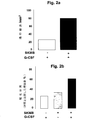
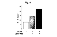
虚血損傷組織での血管内皮増殖因子の濃度を増加させると、生理的食塩水を用いた対照と比較して虚血損傷組織中の血管密度が増加する。虚血損傷組織で発現されるVEGFが、虚血損傷組織の幹細胞分化および再生を刺激するという知見も得られた。例えば、梗塞を起こした心筋層で発現したVEGFが、動員された幹細胞を心筋細胞に分化させるという知見が得られた。 Increasing the concentration of vascular endothelial growth factor in the ischemic damaged tissue increases the vascular density in the ischemic damaged tissue as compared to a control using saline. It was also found that VEGF expressed in ischemic damaged tissue stimulates stem cell differentiation and regeneration of ischemic damaged tissue. For example, it was found that VEGF expressed in the infarcted myocardium differentiates mobilized stem cells into cardiomyocytes.
本発明の方法は、虚血損傷組織の末梢血での多能性幹細胞の濃度(すなわち、数)が第1の濃度から第1の濃度より十分に大きな第2の濃度まで増加するように、虚血損傷組織に幹細胞を動員するステップをさらに含む。第1の濃度の幹細胞は、虚血から時間が経過した時点で末梢血で典型的に見いだされる幹細胞の濃度となりうる。虚血損傷組織中のVEGFの濃度が増加する間に末梢血での幹細胞の濃度が増加する限りにおいて、VEGFの濃度が虚血損傷組織で上方制御される前に、または、その後に、末梢血での幹細胞の濃度を増加させることができる。 The method of the present invention is such that the concentration (ie, number) of pluripotent stem cells in the peripheral blood of ischemic damaged tissue is increased from a first concentration to a second concentration sufficiently greater than the first concentration. The method further includes mobilizing stem cells to the ischemic damaged tissue. The first concentration of stem cells can be the concentration of stem cells typically found in peripheral blood when time has passed since ischemia. Peripheral blood before or after VEGF concentration is up-regulated in ischemic damaged tissue, as long as the concentration of stem cells in peripheral blood increases while the concentration of VEGF in ischemic damaged tissue increases The concentration of stem cells in can be increased.
本発明のもう一つの態様によれば、本方法は虚血損傷組織に向かうように多能性幹細胞を誘発することを含むことができる。虚血損傷組織でのSDF-1タンパク質の濃度を、第1の濃度から該第1の濃度よりも十分に高い第2の濃度へ増加させることによって、多能性幹細胞を虚血損傷組織へ向かわすことができる。SDF-1タンパク質の第1の濃度は、虚血(例えば、心筋梗塞)から時間の経過した時点(すなわち、数週間)で、虚血損傷組織(例えば、梗塞を起こした心筋層)で典型的に見られるSDF-1タンパク質の濃度である。SDF-1タンパク質の第2の濃度は、第1の濃度のSDF-1タンパク質より十分に大きくなりうる。 According to another embodiment of the present invention, the method can include inducing pluripotent stem cells to go to the ischemic damaged tissue. By increasing the concentration of SDF-1 protein in the ischemic damaged tissue from the first concentration to a second concentration sufficiently higher than the first concentration, pluripotent stem cells are directed to the ischemic damaged tissue. Can dodge. The first concentration of SDF-1 protein is typical in ischemic damaged tissue (eg, infarcted myocardium) at a point in time (ie, several weeks) after ischemia (eg, myocardial infarction). Is the concentration of SDF-1 protein found in The second concentration of SDF-1 protein can be sufficiently greater than the first concentration of SDF-1 protein.
SDF-1タンパク質の濃度は、虚血損傷組織でのSDF-1タンパク質の発現を上方制御することにより、心筋梗塞から時間の経過した時点で虚血損傷組織にて典型的に発現されるSDF-1タンパク質の量から増加させることができる。虚血損傷組織のVEGF-1の濃度が増加し、その一方で虚血損傷組織中のSDF-1濃度が増加するとともに末梢血での幹細胞の濃度が増加する。
The concentration of SDF-1 protein upregulates the expression of SDF-1 protein in ischemic damaged tissue, so that SDF- typically expressed in ischemic damaged tissue over time after
VEGF
本発明の1つの態様によれば、虚血損傷組織で発現されるVEGFは、新しい側副血管の成長を誘発することができる血管内皮増殖因子のファミリーのうちの1つである。VEGFは、血管透過性活性を持つ特異的血管新生成長因子であり、表皮細胞をほぼ排他的に標的にする。
VEGF
According to one embodiment of the present invention, VEGF expressed in ischemic damaged tissue is one of a family of vascular endothelial growth factors that can induce the growth of new collateral vessels. VEGF is a specific angiogenic growth factor with vascular permeability activity that targets epidermal cells almost exclusively.
虚血損傷組織で発現されるVEGFは、VEGF遺伝子の発現産物である。本発明にもとづいて使用される好ましいVEGFとして、VEGF-1(別名VEGF-A)および他の構造的に相同なVEGF、例えばVEGF-2(VEGF-C)、VEGF-3(VEGF-B)、VEGF-D、VEGF-E、および胎盤成長因子が挙げられる。VEGF-1の既知のイソ型は、例えば、121、138、162、165、182,189、および206アミノ酸を含むことができる。これらのイソ型は、それぞれ、VEGF-121、VEGF-165、VEGF-162、VEGF-182、VEGF-189、およびVEGF-206と同定されている。これらのイソ型の分裂促進およびヘパリン結合活性は異なる。本発明にもとづいて使用されるVEGF-1の好ましいイソ型は、VEGF-165である。列挙されていないVEGF-1の他のイソ型およびVEGFの他のホモログもまた、本発明にもとづいて使用することができる。 VEGF expressed in ischemic damaged tissue is an expression product of the VEGF gene. Preferred VEGFs used in accordance with the present invention include VEGF-1 (also known as VEGF-A) and other structurally homologous VEGFs such as VEGF-2 (VEGF-C), VEGF-3 (VEGF-B), VEGF-D, VEGF-E, and placental growth factor. Known isoforms of VEGF-1 can include, for example, 121, 138, 162, 165, 182,189, and 206 amino acids. These isoforms have been identified as VEGF-121, VEGF-165, VEGF-162, VEGF-182, VEGF-189, and VEGF-206, respectively. These isoforms have different mitogenic and heparin binding activities. The preferred isoform of VEGF-1 used in accordance with the present invention is VEGF-165. Other isoforms of VEGF-1 and other homologs of VEGF not listed can also be used in accordance with the present invention.
本発明のVEGFは、上記VEGFのうちの1つと同一のアミノ酸配列を持つことができる。本発明のVEGFは、上記VEGF(例えばVEGFのフラグメント、類似体、および誘導体)のうちの1つの変異体であってもよい。そのような変異体として、例えば天然VEGF遺伝子(すなわち、天然に生ずる哺乳類VEGFをコードする天然に生ずる核酸)から天然に生ずる対立遺伝子変異体によってコードされるポリペプチド、VEGF遺伝子の選択的スプライシング型によってコードされるポリペプチド、および天然VEGF遺伝子の非自然的に生ずる変異体によってコードされるポリペプチドが挙げられる。 The VEGF of the present invention can have the same amino acid sequence as one of the above VEGFs. The VEGF of the present invention may be a variant of one of the above VEGFs (eg, fragments, analogs and derivatives of VEGF). Such variants include, for example, polypeptides encoded by naturally occurring allelic variants from naturally occurring VEGF genes (ie, naturally occurring nucleic acids encoding naturally occurring mammalian VEGF), by alternative splicing forms of the VEGF gene The encoded polypeptide and the polypeptide encoded by a non-naturally occurring variant of the native VEGF gene.
VEGF変異体は、天然VEGFと1つ以上のアミノ酸において異なるペプチド配列を持つ。そのような変異体のペプチド配列は、天然VEGFの1つ以上のアミノ酸の欠失、付加、または置換を特徴とすることができる。アミノ酸の挿入は、好ましくは約1ないし4個の連続したアミノ酸からなり、欠失は好ましくは約1ないし10個の連続したアミノ酸からなる。本発明にもとづく変異体VEGFは、実質的に天然VEGFの機能的作用を維持している。好ましいVEGFタンパク質変異体は、サイレントまたは保存的変異を特徴とする本発明の範囲内で核酸分子を発現することによって作られることができる。 VEGF variants have peptide sequences that differ from native VEGF in one or more amino acids. Such variant peptide sequences can be characterized by the deletion, addition or substitution of one or more amino acids of native VEGF. The amino acid insertion preferably consists of about 1 to 4 consecutive amino acids, and the deletion preferably consists of about 1 to 10 consecutive amino acids. Mutant VEGF according to the present invention substantially maintains the functional action of native VEGF. Preferred VEGF protein variants can be made by expressing nucleic acid molecules within the scope of the invention characterized by silent or conservative mutations.
1種類以上の特定のモチーフおよび/またはドメイン、または任意のサイズに対応しているVEGFフラグメントは、本発明の対象とする範囲内である。VEGFの単離ペプチジル部分は、そのようなペプチドをコードしている核酸の対応するフラグメントから組換えによって生産されるペプチドをスクリーニングすることによって得られる。加えて、フラグメントは従来のメリーフィールド固相f-Mocまたはt-Bocケミストリーのような公知技術を使用して、化学的に合成することができる。 VEGF fragments corresponding to one or more particular motifs and / or domains, or any size are within the scope of the present invention. The isolated peptidyl portion of VEGF is obtained by screening peptides produced recombinantly from corresponding fragments of nucleic acids encoding such peptides. In addition, fragments can be chemically synthesized using known techniques such as conventional Maryfield solid phase f-Moc or t-Boc chemistry.
VEGFの変異体は、VEGFの組換え型を含むこともできる。本発明によって好まれる組換えポリペプチドは、VEGFに加えて、哺乳類VEGFをコードする遺伝子の核酸配列と少なくとも85%の配列同一性を持つ核酸によってコードされる。 Variants of VEGF can also include recombinant forms of VEGF. The recombinant polypeptide preferred by the present invention is encoded by a nucleic acid having at least 85% sequence identity with the nucleic acid sequence of the gene encoding mammalian VEGF, in addition to VEGF.
VEGF変異体は、天然VEGFの機能的作用を恒常的に発現するタンパク質のアゴニスト型を含むことができる。他のVEGF変異体として、例えば、プロテアーゼ標的配列を変える突然変異によるタンパク質分解的切断に対して耐性を有するものを挙げることができる。ペプチドのアミノ酸配列の変化が、VEGFの1種類以上の機能的作用を持っている変異体に帰着するかどうかは、変異体をVEGF機能的な活性用に試験することによって、直ちに決定することができる。 VEGF variants can include agonistic forms of proteins that constitutively express the functional effects of native VEGF. Other VEGF variants can include, for example, those that are resistant to proteolytic cleavage by mutations that alter the protease target sequence. Whether a change in the amino acid sequence of a peptide results in a variant having one or more functional effects of VEGF can be readily determined by testing the variant for VEGF functional activity. it can.
VEGF核酸
本発明のもう一つの態様は、VEGFをコードする核酸分子と哺乳類のVEGFをコードする非天然の核酸に関する。そのような核酸分子は、RNAの形でまたはDNA(例えば、相補DNA、ゲノムDNA、および合成DNA)の形であってもよい。DNAは、二重鎖または単鎖であってもよく、単鎖である場合、コード(センス)鎖または非コード(アンチセンス)鎖であってもよい。
VEGF Nucleic Acid Another aspect of the present invention relates to nucleic acid molecules encoding VEGF and non-natural nucleic acids encoding mammalian VEGF. Such nucleic acid molecules may be in the form of RNA or in the form of DNA (eg, complementary DNA, genomic DNA, and synthetic DNA). DNA may be double-stranded or single-stranded, and if single-stranded, may be a coding (sense) strand or a non-coding (antisense) strand.
本発明の範囲内の他の核酸分子は、天然VEGF遺伝子の変異体(例えば、天然VEGFのフラグメント、類似体、および誘導体をコードするもの)である。そのような変異体は、例えば、VEGF遺伝子の天然に生ずる対立遺伝子変異体、天然VEGF遺伝子のホモログ、または天然VEGF遺伝子の非天然に生じる変異体であってもよい。これらの変異体は、天然VEGF遺伝子と1種類以上の塩基が異なる核酸配列を持つ。例えば、そのような変異体の核酸配列は、天然VEGF遺伝子の1つ以上のヌクレオチドの欠失、付加、または置換を特徴とすることができる。核酸の挿入は、好ましくは約1ないし10個の連続したヌクレオチドからなり、欠失は、好ましくは約1ないし30個の連続したヌクレオチドからなる。 Other nucleic acid molecules within the scope of the present invention are variants of the natural VEGF gene (eg, those encoding fragments, analogs, and derivatives of natural VEGF). Such a variant may be, for example, a naturally occurring allelic variant of the VEGF gene, a homologue of the natural VEGF gene, or a non-naturally occurring variant of the natural VEGF gene. These variants have nucleic acid sequences that differ from the natural VEGF gene by one or more types of bases. For example, the nucleic acid sequence of such a variant can be characterized by a deletion, addition, or substitution of one or more nucleotides of the native VEGF gene. The nucleic acid insertion preferably consists of about 1 to 10 consecutive nucleotides, and the deletion preferably consists of about 1 to 30 consecutive nucleotides.
他の適用では、コードされたポリペプチドに保存的とはいえないほどの変化を引き起こすヌクレオチド置換を作ることで、実質的な構造変化を示す変異体VEGF を生成することができる。そのようなヌクレオチド置換の例として、(a)ポリペプチド主鎖構造;(b)ポリペプチドの電荷または疎水性;あるいは(c)アミノ酸側鎖の大部分(バルク)、における変化を引き起こすようなものが挙げられる。通常、タンパク質特性で最も大きな変化を生ずると思われるヌクレオチド置換は、コドンで非保存的変化を引き起こす。タンパク質構造の大きな変化が起きる可能性があるコドン変化の実施例は、(a)疎水性残基(例えば、ロイシン、イソロイシン、フェニルアラニン、バリン、またはアラニン)に対して(またはそれによって)、親水性残基(例えば、セリンまたはスレオニン);(b)任意の他の残基に対して(またはそれによって)、システインまたはプロリン;(c)負に帯電した残基(例えば、グルタミンまたはアスパラギン)に対する(またはそれによって)、正に帯電した側鎖を持つ残基(例えば、リジン、アルギニン、またはヒスチジン);あるいは(d)側鎖を持たない残基(例えば、グリシン)に対して(またはそれによって)かさばった側鎖を持つ残基(例えば、フェニルアラニン)の、それぞれ置換を引き起こすものである。 In other applications, mutant VEGFs that exhibit substantial structural changes can be generated by making nucleotide substitutions that cause non-conservative changes in the encoded polypeptide. Examples of such nucleotide substitutions are those that cause changes in (a) the polypeptide backbone structure; (b) the charge or hydrophobicity of the polypeptide; or (c) the majority (bulk) of amino acid side chains. Is mentioned. Usually, nucleotide substitutions that appear to produce the greatest changes in protein properties cause non-conservative changes in codons. Examples of codon changes that can result in large changes in protein structure are (a) hydrophilic (or thereby) to hydrophobic residues (eg, leucine, isoleucine, phenylalanine, valine, or alanine) Residue (eg, serine or threonine); (b) to (or by) any other residue, cysteine or proline; (c) to a negatively charged residue (eg, glutamine or asparagine) ( Or) by residues with positively charged side chains (eg, lysine, arginine, or histidine); or (d) against (or by) residues that do not have side chains (eg, glycine). Each of the residues with bulky side chains (eg, phenylalanine) causes substitution.
本発明の範囲内のVEGF遺伝子の天然に生ずる対立遺伝子変異体は、天然VEGF遺伝子と少なくとも75%配列同一性を持つ哺乳類組織から単離された核酸であって、天然VEGFに構造類似性があるポリペプチドをコードする。本発明の範囲内の天然VEGFのホモログは、天然の遺伝子と少なくとも75%の配列同一性を持つ他の種から単離される核酸であって、天然VEGFに構造類似性があるポリペプチドをコードする。公的な、および/または私有の核酸データベースを、天然VEGF遺伝子に高いパーセント配列同一性を持つ他の核酸分子を同定するために、検索することができる。 Naturally occurring allelic variants of the VEGF gene within the scope of the present invention are nucleic acids isolated from mammalian tissue having at least 75% sequence identity with the native VEGF gene and have structural similarity to native VEGF Encodes a polypeptide. A native VEGF homolog within the scope of the present invention is a nucleic acid isolated from another species that has at least 75% sequence identity with the native gene and encodes a polypeptide that is structurally similar to native VEGF . Public and / or private nucleic acid databases can be searched to identify other nucleic acid molecules with a high percent sequence identity to the native VEGF gene.
非自然的に生ずるVEGF変異体は、自然界で生ずることのない核酸(例えば、ヒトの手によって作られたもの)であり、天然VEGF遺伝子と少なくとも75%の配列同一性を持ち、天然VEGFに構造類似性があるポリペプチドをコードする。非自然的に生ずるVEGF遺伝子変異体の例として、天然VEGFのフラグメントをコードするもの、厳密な条件下で天然VEGF遺伝子または天然VEGF遺伝子の相補体にハイブリダイズするもの、天然VEGF遺伝子または該天然VEGF遺伝子の相補体と少なくとも65%の配列同一性を共有するもの、さらにVEGFをコードするものが挙げられる。 Non-naturally occurring VEGF variants are non-naturally occurring nucleic acids (eg, those made by human hands) that have at least 75% sequence identity with the native VEGF gene and are structured in native VEGF It encodes polypeptides with similarity. Examples of non-naturally occurring VEGF gene variants include those that encode a fragment of natural VEGF, those that hybridize under stringent conditions to the natural VEGF gene or the complement of the natural VEGF gene, the natural VEGF gene or the natural VEGF Those that share at least 65% sequence identity with the complement of the gene, and those that encode VEGF.
本発明の範囲内のVEGFのフラグメントをコードする核酸は、天然VEGFのアミノ酸残基をコードする。天然VEGFのフラグメントをコードする核酸をコードまたは該核酸とハイブリダイズする短いオリゴヌクレオチドを、プローブ、プライマー、またはアンチセンス分子として使用することができる。天然VEGFのフラグメントをコードする核酸または該核酸とハイブリダイズする長いポリヌクレオチドもまた、本発明の種々の態様で用いることができる。天然VEGFのフラグメントをコードする核酸は、完全長の天然VEGF遺伝子またはその変異体の酵素的消化(例えば、制限酵素を用いて)または化学的分解によって、作製することができる。 A nucleic acid encoding a fragment of VEGF within the scope of the present invention encodes an amino acid residue of native VEGF. Short oligonucleotides that encode or hybridize with nucleic acids encoding fragments of native VEGF can be used as probes, primers, or antisense molecules. Nucleic acids encoding fragments of native VEGF or long polynucleotides that hybridize to the nucleic acids can also be used in various aspects of the invention. Nucleic acids encoding fragments of native VEGF can be made by enzymatic digestion (eg, using restriction enzymes) or chemical degradation of a full-length native VEGF gene or a variant thereof.
上記の核酸のうちの1つに厳密な条件下でハイブリダイズする核酸を、本発明で用いることもできる。例えば、そのような核酸は、厳密さが低い条件、厳密さが中程度の条件、または厳密さが高い条件の下で、上記の核酸のうちの1つにハイブリダイズすることができる。 Nucleic acids that hybridize under stringent conditions to one of the above nucleic acids can also be used in the present invention. For example, such a nucleic acid can hybridize to one of the above nucleic acids under conditions of low stringency, moderate stringency, or high stringency.
VEGF融合タンパク質をコードしている核酸分子を、本発明で使うことも可能である。そのような核酸は、適当な標的細胞に導入された時にVEGF融合タンパク質を発現するような構築物(例えば、発現ベクター)を調製することによって作られる。例えばそのような構築物は、適当な発現系での構築物の発現が融合タンパク質を産生するように、VEGFをコードする第1のポリヌクレオチドをもう一つのタンパク質をコードしている第2のポリヌクレオチドとインフレームで連結することによって作られる。 Nucleic acid molecules encoding VEGF fusion proteins can also be used in the present invention. Such nucleic acids are made by preparing a construct (eg, an expression vector) that expresses a VEGF fusion protein when introduced into a suitable target cell. For example, such a construct may include a first polynucleotide encoding VEGF and a second polynucleotide encoding another protein, such that expression of the construct in an appropriate expression system produces a fusion protein. Made by connecting in-frame.
本発明のオリゴヌクレオチドは、DNAもしくはRNA、またはそれらのキメラ混合物、誘導体、または修飾変異体であってよく、単鎖または二重鎖であってよい。そのようなオリゴヌクレオチドを、塩基部分、糖部分、またはリン酸骨格において修飾することができ、それによって、例えば、分子の安定性やハイブリダイゼーション等を改善する。本発明に包含されるオリゴヌクレオチドは、ペプチド(例えば、生体内で細胞受容体を標的にするため)等の他の付加基、または細胞膜を越える輸送を促進する作用物質を、付加的に含むものであってもよい。このために、オリゴヌクレオチドを、もう一つの分子(例えば、ペプチド、ハイブリダイゼーション誘発架橋因子、輸送因子、またはハイブリダイゼーション誘発切断因子)と結合させることが可能である。 The oligonucleotides of the invention may be DNA or RNA, or chimeric mixtures, derivatives or modified variants thereof, and may be single-stranded or double-stranded. Such oligonucleotides can be modified at the base moiety, sugar moiety, or phosphate backbone, thereby improving, for example, molecular stability, hybridization, and the like. Oligonucleotides encompassed by the present invention additionally include other additional groups such as peptides (eg to target cell receptors in vivo) or agents that facilitate transport across the cell membrane It may be. To this end, the oligonucleotide can be conjugated to another molecule (eg, a peptide, a hybridization-induced crosslinking factor, a transport factor, or a hybridization-induced cleavage factor).
VEGF発現
本発明の別の態様によれば、標的細胞へVEGFの発現を高める作用因子を導入することで、虚血損傷組織でのVEGFの発現を増加させることができる。標的細胞として、虚血損傷組織にあるそれらの細胞または作用因子の導入後に虚血損傷組織に移植される生体外細胞が挙げられる。
VEGF expression According to another aspect of the present invention, VEGF expression in an ischemic damaged tissue can be increased by introducing an agent that enhances VEGF expression into a target cell. Target cells include ex vivo cells that are transplanted into the ischemic damaged tissue after introduction of those cells or agents in the ischemic damaged tissue.
生体外細胞は、該細胞が移植されるべき組織と生体適合性がある任意の細胞である。これらの細胞は好ましくは、処置すべき被験体から収集される細胞(すなわち、自己由来細胞)であり、移植に先立って培養される。自己由来細胞が好ましい理由は、移植に対する細胞の生体適合性を増加させて、拒絶反応の可能性を最小化することにある。 An in vitro cell is any cell that is biocompatible with the tissue into which the cell is to be transplanted. These cells are preferably cells collected from the subject to be treated (ie, autologous cells) and cultured prior to transplantation. The reason why autologous cells are preferred is to increase the biocompatibility of the cells for transplantation and minimize the possibility of rejection.
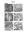
虚血損傷組織が梗塞を起こした心筋層を含む場合、梗塞を起こした心筋層に移植される細胞として、例えば、自己由来細胞、培養骨格筋芽細胞、繊維芽細胞、平滑筋細胞、および骨髄由来細胞が挙げられる。梗塞を起こした心筋層への移植にとって好ましい細胞は、骨格筋芽細胞である。筋芽細胞は、骨格筋の再生能を維持し、ストレス期間中に増殖して筋管に分化することで、最終的に収縮可能な筋繊維を形成する。心筋に移植される筋芽細胞は、筋管形成を起こし、細胞周期から脱して生き残る。機能的な研究によって、心筋に筋芽細胞を移植した後での局所収縮性および伸展性の改善が示された。骨格筋芽細胞は、筋繊維基底膜下で容易に収集でき、培養して細胞系を増大させた後、梗塞を起こした心筋層に移植することができる。例えば、マウスを被験体として用いた場合、該被験体の後脚から骨格筋芽細胞を収集して培養した後、その被験体の梗塞を起こした心筋層へ移植することができる。 When the ischemic damaged tissue includes an infarcted myocardium, examples of cells to be transplanted into the infarcted myocardium include, for example, autologous cells, cultured skeletal myoblasts, fibroblasts, smooth muscle cells, and bone marrow Derived cells. Preferred cells for transplantation into the infarcted myocardium are skeletal myoblasts. Myoblasts maintain the regenerative capacity of skeletal muscles and proliferate and differentiate into myotubes during the stress period, and finally form contractile muscle fibers. Myoblasts transplanted into the myocardium undergo myotube formation and survive the cell cycle. Functional studies have shown improved local contractility and extensibility after transplanting myoblasts into the myocardium. Skeletal myoblasts can be easily collected under the myofiber basement membrane, cultured to expand the cell line, and then transplanted into the infarcted myocardium. For example, when a mouse is used as a subject, skeletal myoblasts are collected from the hind legs of the subject and cultured, and then transplanted to the infarcted myocardium of the subject.
VEGFの発現を増加させるために標的細胞に導入される作用因子は、細胞への導入と該細胞での複製が可能で、かつ組換え核酸構築物(概してDNA構築物)に組み込み可能な天然または合成のVEGF核酸を含むことができる。そのような構築物は、好ましくは、所定の標的細胞でポリペプチドをコードする配列の転写および翻訳ができる複製系および配列を含む。 Agents introduced into target cells to increase VEGF expression can be natural or synthetic that can be introduced into and replicated in cells and can be incorporated into recombinant nucleic acid constructs (generally DNA constructs). VEGF nucleic acids can be included. Such constructs preferably include a replication system and sequences capable of transcription and translation of sequences encoding the polypeptide in a given target cell.
他の作用因子を標的細胞に導入にして、標的細胞のVEGFレベルを高めることもできる。例えば、VEGFをコードしている遺伝子の転写を増加させる作用因子、VEGFをコードしているmRNAの翻訳を増加させる作用因子、および/またはVEGFをコードしているmRNAの分解を減少させる作用因子を使用して、VEGFレベルを増加させることが可能である。細胞内の遺伝子からの転写率の増大は、VEGFをコードしている遺伝子の上流に外来のプロモータを導入することによって達成される。異種遺伝子の発現を促進するエンハンサの使用も可能である。 Other agents can be introduced into the target cell to increase the VEGF level in the target cell. For example, an agent that increases transcription of a gene encoding VEGF, an agent that increases translation of mRNA encoding VEGF, and / or an agent that decreases degradation of mRNA encoding VEGF. It can be used to increase VEGF levels. Increasing the rate of transcription from intracellular genes is achieved by introducing a foreign promoter upstream of the gene encoding VEGF. It is also possible to use enhancers that promote the expression of heterologous genes.
作用因子を所定の標的細胞に導入する好ましい方法として、遺伝子治療が包含される。遺伝子治療とは、生体内またはインビトロで細胞から治療用産物を発現するための遺伝子移入をいう。本発明にもとづく遺伝子治療は、生体内またはインビトロで所定の標的細胞からVEGFを発現させるために用いられる。 A preferred method for introducing an agent into a given target cell includes gene therapy. Gene therapy refers to gene transfer for expressing therapeutic products from cells in vivo or in vitro. The gene therapy based on the present invention is used to express VEGF from a predetermined target cell in vivo or in vitro.
遺伝子治療の1つの方法は、VEGFをコードしているヌクレオチドを含むベクターを使用する。「ベクター」(時に遺伝子送達または遺伝子移入用「運搬体」と呼ばれる」)とは、インビトロまたは生体内で、所定の標的細胞に送達されるポリヌクレオチドを含んでいる巨大分子または分子の複合体のことをいう。送達されるポリヌクレオチドは、遺伝子治療で重要なコード配列を含むことができる。ベクターとして、例えば、ウイルス・ベクター(例えば、アデノウイルス(「Ad」)、アデノ随伴ウイルス(AAV)、およびレトロウイルス)、リポソームおよび他の脂質を含有する複合体、さらに所定の標的細胞へのポリヌクレオチド送達の媒介となることができるその他の巨大分子複合体が挙げられる。 One method of gene therapy uses a vector containing nucleotides encoding VEGF. A “vector” (sometimes referred to as a “carrier” for gene delivery or gene transfer) is a macromolecule or complex of molecules that contains a polynucleotide that is delivered in vitro or in vivo to a given target cell. That means. The polynucleotide to be delivered can include coding sequences that are important in gene therapy. Vectors include, for example, viral vectors (eg, adenovirus (“Ad”), adeno-associated virus (AAV), and retrovirus), complexes containing liposomes and other lipids, and poly to specific target cells. Other macromolecular complexes that can mediate nucleotide delivery include.
ベクターは、さらに遺伝子送達および/または遺伝子発現を調整、さもなければ標的細胞に有益な性質を提供する他の構成要素または機能性を含むこともできる。他の構成要素として、例えば、細胞への結合または標的化に影響を及ぼす構成要素(細胞型または組織特異的結合を調節する構成要素を含む);細胞によるベクター核酸の取り込みに影響を及ぼす構成要素;取り込みの後、細胞内でポリヌクレオチドの局在化に影響する構成要素(例えば核移行を調節する作用因子);およびポリヌクレオチドの発現に影響する構成要素、が挙げられる。そのような構成要素は、マーカー(例えば、ベクターによって送達された核酸を取り込んで発現している細胞の検出または選択に使用しうる検出可能および/または選択可能なマーカー)を含む場合もあり得る。そのような構成要素は、ベクターの本来の特徴(例えば、結合および取り込みを媒介する構成要素または機能性を持つ一定のウイルス性ベクターの使用)として提供されることができ、またはベクターを修飾することでそのような機能性を提供することができる。 The vector may further include other components or functionalities that modulate gene delivery and / or gene expression or otherwise provide beneficial properties to the target cells. Other components, for example, components that affect binding or targeting to cells (including components that modulate cell type or tissue specific binding); components that affect uptake of vector nucleic acids by cells Components that affect localization of the polynucleotide within the cell after uptake (eg, agents that modulate nuclear translocation); and components that affect expression of the polynucleotide. Such a component may also include a marker (eg, a detectable and / or selectable marker that can be used to detect or select cells that have taken up and are expressing nucleic acids delivered by the vector). Such components can be provided as the original features of the vector (eg, the use of certain viral vectors with components or functionality that mediate binding and uptake) or to modify the vector Can provide such functionality.
選択マーカーは、ポジティブ(正)の作用性、ネガティブ(負)の作用性、または二作用性である。ポジティブ選択マーカーは、マーカーを担持する細胞を選択可能とし、一方ネガティブ選択マーカーは、マーカーを担持する細胞を選択的に除去することを可能にする。二作用性(すなわち、ポジティブ/ネガティブ)を含むそのような様々なマーカー遺伝子が記述されている(例えば、Lupton, S., WO 92/08796(1992年5月29日公開); およびLupton, S., WO 94/28143(1994年12月8日公開)を参照すること)。そのようなマーカー遺伝子は、遺伝子治療の状況で有利でありえる付加的な制御の手段を提供することができる。そのようなベクターは多くの種類が公知であり、一般に利用可能である。 Selectable markers are positive (positive), negative (negative), or dual. A positive selectable marker allows selection of cells carrying the marker, while a negative selection marker allows selective removal of cells carrying the marker. Various such marker genes have been described, including dual acting (ie positive / negative) (eg, Lupton, S., WO 92/08796 (published 29 May 1992); and Lupton, S ., WO 94/28143 (published December 8, 1994)). Such marker genes can provide additional means of control that can be advantageous in gene therapy situations. Many types of such vectors are known and generally available.
本発明で用いるベクターとして、ウイルス・ベクター、脂質をベースとしたベクター、および標的細胞に対して本発明におけるヌクレオチドを送達することができる他のベクターが挙げられる。上記ベクターは、標的ベクター、特に標的細胞(例えば、心筋細胞)に優先して結合する標的ベクターである。本発明で使用するための好ましいベクターは、所定の標的細胞に対する毒性が低く、組織または細胞特異的なかたちで治療に有効な量からなるVEGFの産生を誘導するウイルス・ベクターである。 Vectors used in the present invention include viral vectors, lipid-based vectors, and other vectors that can deliver the nucleotides of the present invention to target cells. The vector is a target vector that binds preferentially to a target vector, particularly a target cell (eg, cardiomyocyte). A preferred vector for use in the present invention is a viral vector that induces the production of VEGF consisting of a therapeutically effective amount in a tissue or cell specific manner with low toxicity to a given target cell.
現在好ましいウイルス・ベクターは、アデノウイルス(Ad)またはアデノ随伴ウイルス(AAV)に由来する。ヒトおよび非ヒト・ウイルス・ベクターが使われてはいるが、好ましくは組換え型のウイルス・ベクターはヒトで複製不能である。ベクターがアデノウイルスである場合、好ましくはVEGFをコードしている遺伝子に操作可能な状態で連結されたプロモータを有するポリヌクレオチドを含み、かつヒトで複製不能である。 Presently preferred viral vectors are derived from adenovirus (Ad) or adeno-associated virus (AAV). Although human and non-human viral vectors are used, preferably the recombinant viral vector is not replicable in humans. When the vector is an adenovirus, it preferably comprises a polynucleotide having a promoter operably linked to a gene encoding VEGF and is non-replicatable in humans.
アデノウイルス・ベクターは、本発明での使用にとって好ましい。なぜなら、アデノウイルス・ベクターは(1)標的細胞で効率の高い遺伝子発現をおこなう能力があること、(2)比較的多量の異種(非ウイルス)DNAに対応することができるからである。組換えアデノウイルスの好ましい形態は、「無気力な(gutless)」、「高容量の」、および「ヘルパー依存性の」アデノウイルス・ベクターである。そのようなベクターの特徴は、例えば、(1)ウイルスをコードしているすべてまたはほとんどの配列(ウイルスタンパク質をコードしている配列)の欠失、(2)ウイルスのDNA複製に必要なウイルス逆方向末端反復(ITR)、(3)最大で28〜32kbの「外来性」または「異種」の配列(例えば、SDF-1タンパク質をコードする配列)、および(4)感染性カプシドにウイルス・ゲノムをパッケージングするのに必要であるウイルスDNAパッケージング配列、である。特に心筋細胞に関しては、そのような組換えアデノウイルス・ベクターの好ましい変異体は、組織(例えば心筋細胞)特異的エンハンサを含み、またプロモータがVEGF遺伝子に操作可能な状態で連結されている。 Adenoviral vectors are preferred for use in the present invention. This is because adenoviral vectors are (1) capable of high-efficiency gene expression in target cells, and (2) can handle relatively large amounts of heterologous (non-viral) DNA. Preferred forms of recombinant adenovirus are “gutless”, “high capacity”, and “helper-dependent” adenoviral vectors. Such vectors are characterized, for example, by (1) deletion of all or most sequences encoding the virus (sequences encoding viral proteins), (2) viral reverse necessary for viral DNA replication. Directional terminal repeats (ITRs), (3) up to 28-32 kb "foreign" or "heterologous" sequences (eg, sequences encoding SDF-1 protein), and (4) viral genomes on infectious capsids Viral DNA packaging sequences, which are necessary to package Particularly for cardiomyocytes, preferred variants of such recombinant adenoviral vectors include a tissue (eg, cardiomyocyte) specific enhancer and a promoter is operably linked to the VEGF gene.
AAVをベースとするベクターは、所定の標的細胞に対する高い形質導入効率を示し、部位特異的なかたちで標的ゲノムに統合されることから、有利である。組換えAAVベクターの使用については、Tal, J. , J. Biomed. Sci. 7: 279-291,2000 ならびにMonahan and Samulski, 遺伝子治療 7: 24-30,2000に、詳しく記載されている。好ましいAAVベクターは、VEGF核酸に操作可能な状態で連結された組織(例えば、心筋)または細胞(例えば、心筋細胞)特異的プロモータを含む少なくとも1つのカセットに隣接する一対のAAV逆方向末端反復を含む。ITR、プロモータ、およびVEGF遺伝子を含むAAVベクターのDNA配列は、標的ゲノムに組み込まれることが可能である。 AAV-based vectors are advantageous because they exhibit high transduction efficiency for a given target cell and integrate into the target genome in a site-specific manner. The use of recombinant AAV vectors is described in detail in Tal, J., J. Biomed. Sci. 7: 279-291, 2000 and Monahan and Samulski, Gene Therapy 7: 24-30, 2000. Preferred AAV vectors comprise a pair of AAV inverted terminal repeats flanked by at least one cassette containing a tissue (eg, myocardial) or cell (eg, cardiomyocyte) specific promoter operably linked to a VEGF nucleic acid. Including. The DNA sequence of the AAV vector including the ITR, promoter, and VEGF gene can be integrated into the target genome.
本発明にもとづいて使用しうる他のウイルス・ベクターとして、単純ヘルペスウイルス(HSV)をベースとしたベクターが挙げられる。1種類以上の即初期遺伝子(IE)が欠失したHSVベクターが有利である。なぜなら、HSVベクターは一般に無細胞毒性であり、標的細胞での潜伏期に類似した状態を持続し、所定標的細胞の効率的な形質導入をもたらすからである。組換えHSVベクターは、約30kbの異種核酸を組み込むことができる。好ましいHSVベクターは、(1)HSV1型から設計され、(2)IE遺伝子が欠失し、(3)VEGF核酸に操作可能な状態で連結した組織(例えば、心筋)特異的プロモータを含む、HSVベクターである。HSV単位複製配列ベクターも、本発明の種々の方法で有用であると思われる。典型的には、HSV単位複製配列ベクターは長さが約15kbであり、ウイルス複製開始点およびパッケージング配列を有する。 Other viral vectors that can be used in accordance with the present invention include herpes simplex virus (HSV) based vectors. HSV vectors lacking one or more immediate early genes (IE) are advantageous. This is because HSV vectors are generally non-cytotoxic and persist in a state similar to the latent period in the target cells, resulting in efficient transduction of the given target cells. Recombinant HSV vectors can incorporate about 30 kb of heterologous nucleic acid. A preferred HSV vector is an HSV comprising a tissue (eg, myocardium) specific promoter (1) designed from HSV1 type, (2) lacking the IE gene, and (3) operably linked to a VEGF nucleic acid. It is a vector. HSV amplicon vectors may also be useful in the various methods of the present invention. Typically, HSV amplicon vectors are about 15 kb in length and have a viral origin of replication and packaging sequences.
レトロウイルス(例えばC型レトロウイルスおよびレンチウイルス)も本発明でも使用可能であると考えられる。例えば、レトロウイルス・ベクターがマウス白血病ウイルス(MLV)に基づくものであってもよい。例えば、Hu and Pathak, Pharmacol. Rev. 52: 493-511, 2000 およびFong et al., Crit. Rev. Ther. Drug Carrier Syst. 17: 1-60,2000を参照すること。MLVをベースとするベクターは、ウイルス遺伝子の代わりに最大で8kbの異種(治療用)DNAを含むことが可能である。異種DNAは、組織特異的プロモータとSDF-1核酸とを含むことが可能である。梗塞を起こした心筋層に対する送達の方法では、それが心筋特異的受容体に対するリガンドをコードすることも可能である。 It is contemplated that retroviruses (eg, type C retroviruses and lentiviruses) can be used in the present invention. For example, the retroviral vector may be based on murine leukemia virus (MLV). See, for example, Hu and Pathak, Pharmacol. Rev. 52: 493-511, 2000 and Fong et al., Crit. Rev. Ther. Drug Carrier Syst. 17: 1-60,2000. MLV-based vectors can contain up to 8 kb of heterologous (therapeutic) DNA instead of viral genes. The heterologous DNA can include a tissue-specific promoter and an SDF-1 nucleic acid. In the method of delivery to the infarcted myocardium, it is also possible to encode a ligand for a myocardial specific receptor.
使用する可能性のある追加のレトロウイルス・ベクターは、ヒト免疫不全ウイルス(HIV)をベースとしたベクターを含む複製不能なレンチウイルスをベースとしたベクターである。例えば、Vigna and Naldini, J. Gene Med. 5: 308-316,2000 およびMiyoshi et al., J. Virol. 72: 8150-8157,1998を参照すること。 レンチウイルス・ベクターは、活発に分裂している細胞と分裂していない細胞との両方に感染する能力を有する点で、有利である。また、レンチウイルス・ベクターによって、ヒト上皮細胞も効率高く形質導入される。 Additional retroviral vectors that may be used are non-replicating lentiviral based vectors, including human immunodeficiency virus (HIV) based vectors. See, for example, Vigna and Naldini, J. Gene Med. 5: 308-316, 2000 and Miyoshi et al., J. Virol. 72: 8150-8157, 1998. Lentiviral vectors are advantageous in that they have the ability to infect both actively dividing and non-dividing cells. In addition, human epithelial cells are efficiently transduced by a lentiviral vector.
本発明で用いるレンチウイルス・ベクターは、ヒトおよび非ヒト(SIVを含む)レンチウイルスに由来するものであってもよい。好ましいレンチウイルス・ベクターは、VEGF遺伝子に操作可能な状態で連結した組織特異的プロモータ(例えば、心筋)と同様にベクター増殖のために必要な核酸配列を含む。これらの元のベクターは、ウイルス末端反復配列、プライマー結合部位、ポリプリン配列、att部位、およびキャプシド形成部位を含むものであってもよい。 Lentiviral vectors used in the present invention may be derived from human and non-human (including SIV) lentiviruses. Preferred lentiviral vectors contain the nucleic acid sequences necessary for vector propagation as well as a tissue specific promoter (eg, myocardium) operably linked to the VEGF gene. These original vectors may include viral terminal repeats, primer binding sites, polypurine sequences, att sites, and encapsidation sites.
レンチウイルス・ベクターを任意の適当なレンチウイルス・カプシドにパッケージングすることが可能である。一つの粒状タンパク質を、異なるウイルス由来の別のものに置き換えることを、「シュードタイピング(pseudotyping)」という。ベクター・カプシドは、マウス白血病ウイルス(MLV)または水疱性口炎ウイルス(VSV)を含む他のウイルスに由来するウイルスエンベロープ・タンパク質を含むものであってもよい。VSVのGTP結合タンパク質を使用することで、高いベクター力価が得られ、ベクター・ウイルス粒子の安定性がより高くなる。 The lentiviral vector can be packaged into any suitable lentiviral capsid. Replacing one granular protein with another from a different virus is called “pseudotyping”. Vector capsids may include viral envelope proteins from other viruses including murine leukemia virus (MLV) or vesicular stomatitis virus (VSV). By using the VSV GTP-binding protein, a high vector titer is obtained and the stability of the vector virus particle is higher.
アルファ・ウイルスをベースとするベクター(例えば、セムリキ森林ウイルス(SFV)とシンドビス・ウイルス(SIN)とから作られるベクター)が、本発明でも使用することが可能であると考えられる。アルファ・ウイルスの使用については、Lundstrom, K., Intervirology 43: 247-257,2000 and Perri et al., Journal of Virology 74: 9802-9807,2000に記載されている。アルファウイルス・ベクターは、一般にレプリコンとして知られている形態で構築される。レプリコンは、(1)RNA複製のために必要なアルファ・ウイルス遺伝因子と(2)異種核酸(例えば、VEGF核酸をコードしているもの)とを含むものであってもよい。アルファウィルス・レプリコン内で、異種核酸と組織特異的(例えば、心筋)プロモータまたはエンハンサとを操作可能な状態で連結することが可能である。 It is contemplated that alpha virus-based vectors (eg, vectors made from Semliki Forest virus (SFV) and Sindbis virus (SIN)) can be used in the present invention. The use of alphaviruses is described in Lundstrom, K., Intervirology 43: 247-257, 2000 and Perri et al., Journal of Virology 74: 9802-9807, 2000. Alphavirus vectors are constructed in a form commonly known as a replicon. The replicon may include (1) an alphavirus genetic factor required for RNA replication and (2) a heterologous nucleic acid (eg, one encoding a VEGF nucleic acid). Within an alphavirus replicon, it is possible to operably link a heterologous nucleic acid and a tissue-specific (eg, myocardial) promoter or enhancer.
組換え体複製不能アルファウイルス・ベクターは、有利である。なぜなら、それらは高レベルの異種(治療用)遺伝子発現の能力を有し、また幅広い範囲の所定の標的細胞に感染することができるからである。アルファウイルス・レプリコンは、ビリオン表面に、同族の結合相手を発現する所定の標的細胞に選択的に結する機能的異種リガンドまたは結合ドメインを提示することにより、特定の細胞型(例えば、心筋細胞)を標的とすることが可能である。アルファウイルス・レプリコンは、潜伏期を確立することで、所定の標的細胞において長期にわたる異種核酸発現を確立することが可能である。レプリコンは、標的細胞で一時的な異種核酸発現を示すことも可能である。好ましいアルファウイルス・ベクターまたはレプリコンは、非細胞壊死性である。 Recombinant non-replicating alphavirus vectors are advantageous. This is because they have the ability to express high levels of heterologous (therapeutic) genes and can infect a wide range of predetermined target cells. Alphavirus replicons display specific cell types (eg, cardiomyocytes) on the virion surface by presenting functional heterologous ligands or binding domains that selectively bind to a given target cell that expresses a cognate binding partner. Can be targeted. Alphavirus replicons can establish long term heterologous nucleic acid expression in a given target cell by establishing a latency period. The replicon can also show transient heterologous nucleic acid expression in the target cell. Preferred alphavirus vectors or replicons are non-cell necrotic.
本発明の方法と互換性を持つウイルス・ベクターの多くにおいて、複数のプロモータをベクターに取り込むことで、該ベクターによって複数の異種遺伝子を発現させられることができる。さらに、ベクターは標的細胞からVEGF遺伝子産物の発現を容易にするシグナル・ペプチドまたは他の部分をコードする配列を含むことができる。 In many of the viral vectors compatible with the method of the present invention, by incorporating a plurality of promoters into the vector, a plurality of heterologous genes can be expressed by the vector. In addition, the vector can include sequences encoding a signal peptide or other moiety that facilitates expression of the VEGF gene product from the target cell.
2つのウイルス・ベクター系の有利な性質を組合せるために、ハイブリッド・ウイルス・ベクターを用いてVEGF核酸を標的組織(例えば、心筋)へ送達させることが可能である。ハイブリッド・ベクターを構築するための標準的な技術は、当業者に周知である。そのような技術は、例えば、Sambrook, et al., In Molecular Cloning : A laboratory manual. Cold Spring Harbor, N. Y. または組換え体DNA技術について述べている多くの実験マニュアルに見いだすことができる。AAVとアデノウイルスITRとの組合せを含むアデノウイルス・カプシド内の二重鎖AAVゲノムを用いて、細胞に形質導入をおこなうことが可能である。別の変異体では、AAVベクターを「無気力な」、「ヘルパー依存性」、または「高容量」型アデノウイルス・ベクターに入れることが可能である。アデノウイルス/AAVハイブリッド・ベクターは、Lieber et al., J. Virol. 73: 9314- 9324,1999で議論されている。レトロウイルス/アデノウイルス・ハイブリッド・ベクターは、Zheng et al., Nature Biotechnol. 18: 176-186,2000で議論されている。アデノウイルスに含まれるレトロウイルス・ゲノムを、標的細胞ゲノムに組み込んで、安定なVEGF遺伝子発現をもたらすことが可能である。 To combine the advantageous properties of the two viral vector systems, hybrid viral vectors can be used to deliver VEGF nucleic acid to the target tissue (eg, myocardium). Standard techniques for constructing hybrid vectors are well known to those of skill in the art. Such techniques can be found, for example, in Sambrook, et al., In Molecular Cloning: A laboratory manual. Cold Spring Harbor, N. Y. or many laboratory manuals that describe recombinant DNA technology. It is possible to transduce cells using a double-stranded AAV genome within an adenovirus capsid containing a combination of AAV and adenovirus ITR. In another variant, the AAV vector can be placed in an “apathetic”, “helper-dependent”, or “high capacity” type adenoviral vector. Adenovirus / AAV hybrid vectors are discussed in Lieber et al., J. Virol. 73: 9314-9324, 1999. Retrovirus / adenovirus hybrid vectors are discussed in Zheng et al., Nature Biotechnol. 18: 176-186,2000. The retroviral genome contained in the adenovirus can be integrated into the target cell genome, resulting in stable VEGF gene expression.
VEGF遺伝子の発現とベクターのクローン化とを容易にする他の核酸配列要素について、さらに考察する。例えば、プロモータの上流にエンハンサが存在すること、またはコード領域の下流にターミネーターが存在することで、発現を促進することができる。 Other nucleic acid sequence elements that facilitate VEGF gene expression and vector cloning are further discussed. For example, expression can be promoted by the presence of an enhancer upstream of the promoter or the presence of a terminator downstream of the coding region.
本発明は、細胞標的化のために組織特異的プロモータを用いることも検討する。例えば、虚血損傷組織が梗塞を起こした心筋層を含む場合、左室ミオシンL鎖-2(MLC2v)またはミオシン重鎖(MHC)の組織特異的転写制御配列を導入遺伝子(例えばアデノウイルス構築物内のVEGF-165遺伝子)に融合させることができる。導入遺伝子にそのような組織特異的転写制御配列を融合させることによって、導入遺伝子発現を心室性心筋細胞に限定することができる。MLCzvまたはMHCプロモータを使用して、生体内で導入遺伝子を送達することによって、心筋細胞(心臓内での内皮細胞、平滑筋細胞、および繊維芽細胞で付随する発現無し)単独が血管新生タンパク質(例えば血管形成を促進するVEGF-165)の十分な発現を提供すると考えられている。 The present invention also contemplates the use of tissue specific promoters for cell targeting. For example, if the ischemic damaged tissue contains an infarcted myocardium, a transgene (eg, an adenovirus construct) is inserted with a tissue-specific transcription control sequence of left ventricular myosin L chain-2 (MLC 2v ) or myosin heavy chain (MHC). In the VEGF-165 gene). By fusing such a tissue-specific transcriptional control sequence to the transgene, transgene expression can be limited to ventricular cardiomyocytes. By using MLC zv or MHC promoter to deliver transgenes in vivo, cardiomyocytes (without associated expression in endothelial cells, smooth muscle cells, and fibroblasts in the heart) alone are angiogenic proteins It is believed to provide sufficient expression of (eg, VEGF-165 that promotes angiogenesis).
発現を心筋細胞に限定することもまた、うっ血性心不全を処置するための遺伝子移入の有用性に関して、利点がある。発現を心臓に限定することによって、非心臓性の組織での血管形成の潜在的に有害な効果を回避する。加えて、心臓の細胞のうち筋細胞は、急激な代謝回転を受けないことから、最も長い導入遺伝子発現をおこなう可能性がある。それ故に、内皮細胞で起こるように、細胞分裂および細胞死により発現が減少することはないだろう。内皮特異的なプロモータは、この目的のためにすでに利用できる。 Limiting expression to cardiomyocytes is also advantageous with respect to the usefulness of gene transfer to treat congestive heart failure. By limiting expression to the heart, the potentially detrimental effects of angiogenesis in noncardiac tissue are avoided. In addition, muscle cells of the heart cells are not subject to rapid turnover and thus may have the longest transgene expression. Therefore, expression will not be reduced by cell division and cell death, as occurs in endothelial cells. Endothelial specific promoters are already available for this purpose.
ウイルス・ベクターをベースとする方法に加えて、非ウイルス性の方法はVEGF遺伝子を所定の標的細胞に導入する際に用いられることが可能である。本発明にもとづく好ましい非ウイルス性遺伝子送達法は、VEGF核酸を細胞に導入するためにプラスミドDNAを使用する。プラスミドをベースとした遺伝子送達方法は、周知である。 In addition to viral vector based methods, non-viral methods can be used in introducing the VEGF gene into a given target cell. A preferred non-viral gene delivery method according to the present invention uses plasmid DNA to introduce VEGF nucleic acid into cells. Plasmid-based gene delivery methods are well known.
合成遺伝子導入分子を、プラスミドDNA(たとえば心筋特異的プロモータに操作可能な状態で連結したVEGFコード配列)とともに多分子凝集体が形成されるように、設計することができる。これらの凝集体は、所定の標的細胞(例えば、心筋細胞)と結合するように設計される。 Synthetic gene transfer molecules can be designed such that multimolecular aggregates are formed with plasmid DNA (eg, a VEGF coding sequence operably linked to a myocardial specific promoter). These aggregates are designed to bind to certain target cells (eg, cardiomyocytes).
陽イオン両親媒性物質(リポ・ポリアミンおよび陽イオン脂質を含む)は、所定の標的細胞(例えば、心筋細胞)への受容体非依存的なVEGF核酸導入を与えるために用いられてもよい。また、事前に形成された陽イオン・リポソームまたは陽イオン脂質をプラスミドDNAと混合して、細胞形質移入複合体を形成することもできる。陽イオン脂質形成を伴う方法は、Felgner et al., Ann. N. Y. Acad. Sci. 772: 126-139,1995 およびLasic and Templeton, Adv. Drug Delivery Rev. 20: 221-266,1996に概説されている。遺伝子送達のために、 DNAは、両親媒性陽イオン・ペプチドに連結することもできる(Fominaya et al., J. Gene Med. 2: 455-464,2000)。 Cationic amphiphiles (including lipopolyamines and cationic lipids) may be used to provide receptor-independent VEGF nucleic acid introduction into a given target cell (eg, cardiomyocytes). Alternatively, pre-formed cationic liposomes or cationic lipids can be mixed with plasmid DNA to form a cell transfection complex. Methods involving cationic lipid formation are reviewed in Felgner et al., Ann. NY Acad. Sci. 772: 126-139, 1995 and Lasic and Templeton, Adv. Drug Delivery Rev. 20: 221-266, 1996. Yes. For gene delivery, DNA can also be linked to an amphiphilic cationic peptide (Fominaya et al., J. Gene Med. 2: 455-464, 2000).
ウイルスをベースにした構成要素および非ウイルスをベースとした構成要素の両方を伴う方法を、本発明にもとづいて用いることも可能である。例えば、治療用遺伝子送達のためのエプスタイン・バー・ウイルス(EBV)をベースとしたプラスミドが、Cui et al., Gene Therapy 8: 1508-1513,2001に記載されている。その上、アデノウイルスに連結するDNA/リガンド/ポリカチオン添加剤を伴う方法が、Curiel, D. T., Nat. Immun. 13: 141-164,1994に記載されている。 Methods involving both virus-based components and non-virus-based components can also be used in accordance with the present invention. For example, Epstein-Barr virus (EBV) based plasmids for therapeutic gene delivery are described in Cui et al., Gene Therapy 8: 1508-1513,2001. Moreover, methods involving DNA / ligand / polycation additives linked to adenovirus are described in Curiel, D. T., Nat. Immun. 13: 141-164,1994.
VEGFの発現をコードするベクターを、必要に応じて医薬的に許容される担体(例えば生理食塩水)を含む注射可能な製剤の形で、標的細胞に送達することができる。他の医薬用担体、製剤形態、および投薬量を本発明にもとづいて用いることもできる。 A vector encoding the expression of VEGF can be delivered to target cells in the form of an injectable formulation optionally containing a pharmaceutically acceptable carrier (eg, saline). Other pharmaceutical carriers, formulation forms, and dosages can also be used in accordance with the present invention.
標的細胞が虚血損傷組織の細胞を含む場合、ベクターの送達は、幹細胞分化の効果的刺激を可能にする程度のVEGFの発現に充分な量で、蛍光透視による誘導下、ツベルクリン・シリンジを用いた直接注射によって、おこなわれる。ベクターを直接、虚血損傷組織に注射することによって、むしろ効果的に遺伝子を標的とすることができ、また組換え体ベクターの損失を最小にすることが可能である。 If the target cells include cells from ischemic damaged tissue, vector delivery should be done using a tuberculin syringe under fluoroscopic guidance, in an amount sufficient for VEGF expression to allow effective stimulation of stem cell differentiation. It was done by direct injection. By directly injecting the vector into the ischemic damaged tissue, it is possible to target the gene rather effectively and to minimize loss of the recombinant vector.
この種の注射は、所望の数の細胞(特に梗塞を起こした心筋層の心筋細胞)の局所的形質移入を可能にすることで、遺伝子導入の治療効力を最大にし、またウイルスタンパク質に対する炎症反応の可能性を最小化する。虚血損傷組織は梗塞を起こした心筋層を含む場合、例えば心筋細胞特異的プロモータを用いることで、心筋細胞に限局した発現を安全に可能にする。したがって、このことでの導入遺伝子の送達は、例えば、左心室(LV)の細胞で、標的遺伝子発現を結果として生ずることができる。周知の他の技術を、梗塞を起こした心筋層の標的細胞にベクターを移植するために、用いることもできる。 This type of injection maximizes the therapeutic efficacy of gene transfer by allowing local transfection of the desired number of cells (especially infarcted myocardial cardiomyocytes) and also the inflammatory response to viral proteins Minimize the possibility of When the ischemic damaged tissue includes an infarcted myocardium, for example, by using a cardiomyocyte-specific promoter, expression restricted to cardiomyocytes can be safely performed. Thus, transgene delivery in this way can result in target gene expression in, for example, left ventricular (LV) cells. Other well known techniques can also be used to transplant the vector into target cells of the infarcted myocardium.
標的細胞が、虚血損傷組織に後で移植される培養細胞である場合、培地に対して直接注射することでベクターを送達することができる。細胞に形質移入されたVEGFをコードする核酸は、組織特異的プロモータとエンハンサを含む任意の適当な調節配列に対して操作可能状態で、連結することができる。続いて形質移入された所定の標的細胞を、周知の移植技術、例えばツベルクリン・シリンジを用いて直接的に冠内注射することで、虚血損傷組織に移植することができる。 If the target cells are cultured cells that are later transplanted into ischemic damaged tissue, the vector can be delivered by direct injection into the medium. Nucleic acid encoding VEGF transfected into a cell can be operably linked to any suitable regulatory sequence, including tissue specific promoters and enhancers. Subsequently, the transfected target cells can be transplanted directly into the ischemic damaged tissue by direct intracoronary injection using a well-known transplantation technique such as a tuberculin syringe.
虚血損傷組織が梗塞を起こした心筋層を含む場合、標的細胞は、好ましくは、処置される被験体から収集されて、生体外で培養される自己由来細胞である。梗塞を起こした心筋層に対して、最初に標的細胞を生体外で形質移入し、続いて形質移入した所定の標的細胞を梗塞を起こした心筋層に対して移植することで、梗塞を起こした心筋層の炎症反応の可能性が最小化され、また左心系性の機能が、梗塞を起こした心筋層にベクターを直接注射する方法と比較して、改良されたことが判明した。この改善がアデノウイルス注射と典型的に関係している炎症反応の欠如から生じると考えられている。 Where the ischemic damaged tissue comprises an infarcted myocardium, the target cells are preferably autologous cells that are collected from the subject to be treated and cultured in vitro. The infarcted myocardium was first transfected in vitro with target cells, and then the given target cells transfected were transplanted into the infarcted myocardium. It has been found that the possibility of myocardial inflammatory response is minimized and that the left ventricular function is improved compared to direct injection of the vector into the infarcted myocardium. This improvement is believed to result from the lack of an inflammatory response typically associated with adenovirus injection.
本発明のVEGFは、任意の適当な長さの時間にわたって標的細胞内で発現することが可能であり、このような発現には一時的な発現と、安定で長期にわたる発現とが含まれる。好ましい実施形態では、VEGF核酸が適当かつ所定の時間にわたって治療量で発現される。 The VEGF of the present invention can be expressed in target cells for any suitable length of time, such expression includes transient expression and stable and long-term expression. In a preferred embodiment, the VEGF nucleic acid is expressed in a therapeutic amount for an appropriate and predetermined time.
治療量とは、医学的に望ましい結果を、治療をうけている動物またはヒトで得ることができる量である。医学分野で周知のように、任意の一匹の動物または1人のヒトに対する投薬量は、被験体のサイズ、体表面積、年齢、投与される特定の組成物、性、投与の時間および経路、健康状態、および並列して投与される他の薬物等、多くの因子に依存する。タンパク質、核酸、または小分子の特異的な投薬量は、後述する実験的方法を使用している当業者によって容易に決定される。 A therapeutic amount is an amount that can achieve a medically desirable result in the animal or human being treated. As is well known in the medical field, the dosage for any one animal or human is the subject's size, body surface area, age, the particular composition being administered, sex, time and route of administration, It depends on many factors, including health status and other drugs administered in parallel. Specific dosages for proteins, nucleic acids, or small molecules are readily determined by those skilled in the art using the experimental methods described below.
VEGF発現が一時的または長期であるとなかろうと、虚血損傷組織で発現されたVEGFの濃度を制限して、血管腫または血管内皮細胞から派生した血管内腫瘍の形成を予防することが求められる。 Whether VEGF expression is transient or long-term, it is required to limit the concentration of VEGF expressed in ischemic damaged tissues to prevent the formation of hemangiomas or intravascular tumors derived from vascular endothelial cells .
幹細胞の動員
本発明のもう一つの態様によれば、被験体の虚血損傷組織の末梢血にある幹細胞の濃度は、末梢血への幹細胞の動員を誘導する薬剤を投与することによって増加させることができる。幹細胞は、末梢血で幹細胞濃度を増加させるために、多数の作用因子を用いて被験体の末梢血に動員させることができる。例えば、哺乳類被験体の末梢血で幹細胞数を増加させるために、多能性幹細胞を骨髄から動員させる薬剤を、被験体に投与することができる。そのような作用因子は数多く知られており、例として、サイトカイン、例えば顆粒球コロニー刺激因子(G-CSF)、顆粒球−マクロファージ・コロニー刺激因子(GM-CSF)、インターロイキン(IL)-7、IL-3、IL-12、幹細胞因子(SCF)、およびflt-3リガンド;ケモカイン、例えばIL-8、IL-10、Mip-1a、およびGroβ、ならびにサイクロホスアミド(Cy)およびパクリタキセルの化学療法薬が挙げられる。これらの作用因子は、幹細胞動員を達成する時間枠、動員された幹細胞のタイプ、および効率が異なる。
Stem Cell Recruitment According to another aspect of the invention, the concentration of stem cells in the peripheral blood of a subject's ischemic injury tissue is increased by administering an agent that induces the mobilization of stem cells to the peripheral blood. Can do. Stem cells can be mobilized to the peripheral blood of a subject using a number of agents to increase the stem cell concentration in the peripheral blood. For example, to increase the number of stem cells in the peripheral blood of a mammalian subject, an agent that mobilizes pluripotent stem cells from the bone marrow can be administered to the subject. Many such agents are known, including cytokines such as granulocyte colony stimulating factor (G-CSF), granulocyte-macrophage colony stimulating factor (GM-CSF), interleukin (IL) -7 Chemistry of IL-3, IL-12, stem cell factor (SCF), and flt-3 ligands; chemokines such as IL-8, IL-10, Mip-1a, and Groβ, and cyclophosamide (Cy) and paclitaxel Therapeutic drugs. These agents differ in the time frame for achieving stem cell recruitment, the type of stem cell recruited, and the efficiency.
動員因子は、被験体に動員因子を直接注射することによって投与される。好ましくは、VEGF発現が虚血損傷組織で上方制御された後、動員因子が投与される。しかし、VEGF発現が上方制御される前に、動員因子を投与することができる。 The recruitment factor is administered by directly injecting the recruitment factor into the subject. Preferably, the mobilization factor is administered after VEGF expression is upregulated in ischemic damaged tissue. However, mobilization factors can be administered before VEGF expression is upregulated.
好ましい動員因子は、コロニー刺激因子(例えばG-CSF)である。マウス被験体のG-CSFの典型的投薬量は、約5日ないし約10日間にわたって1日あたり約125μg/kgである。VEGF発現が上方制御された後、G-CSF因子を投与することができる。 A preferred mobilization factor is a colony stimulating factor (eg, G-CSF). A typical dosage of G-CSF in a mouse subject is about 125 μg / kg per day for about 5 days to about 10 days. After VEGF expression is upregulated, G-CSF factor can be administered.
あるいは、当技術分野で周知であるように、末梢血に特定幹細胞(すなわち、MAPC)を注射することによって、末梢血での幹細胞の濃度を増加させることができる。 Alternatively, as is well known in the art, the concentration of stem cells in peripheral blood can be increased by injecting specific stem cells (ie, MAPC) into peripheral blood.
SDF−1タンパク質
本発明のもう一つの態様によれば、虚血損傷組織で発現されるSDF-1タンパク質(またはSDF-1ポリペプチド)は、SDF-1遺伝子の発現産物である。いくつかの異なる哺乳類(例えば、ヒト、ラット、マウス、およびネコ)のSDF-1タンパク質のアミノ酸配列が知られている。SDF-1タンパク質は、上記した天然の哺乳類SDF-1タンパク質のうちの1つと同一のアミノ酸配列を持つことができる。
SDF-1 protein According to another embodiment of the present invention, the SDF-1 protein (or SDF-1 polypeptide) expressed in ischemic damaged tissue is the expression product of the SDF-1 gene. The amino acid sequences of SDF-1 protein of several different mammals (eg, human, rat, mouse, and cat) are known. The SDF-1 protein can have the same amino acid sequence as one of the natural mammalian SDF-1 proteins described above.
本発明のSDF-1タンパク質を、哺乳類SDF-1タンパク質の変異体(例えば哺乳類SDF-1遺伝子のフラグメント、アナログ、および誘導体)とすることもできる。そのような変異体として、例えば、天然SDF-1遺伝子(すなわち、天然に生ずる哺乳類SDF-1タンパク質をコードする天然に生ずる核酸)の天然に生ずる対立遺伝子変異体によってコードされるポリペプチド、天然SDF-1遺伝子の選択的スプライス型によってコードされるポリペプチド、天然SDF-1遺伝子のホモログによってコードされるポリペプチド、および天然SDF-1遺伝子の非天然の変異体によってコードされるポリペプチド、が挙げられる。 The SDF-1 protein of the present invention can also be a mutant of a mammalian SDF-1 protein (eg, a fragment, analog, and derivative of a mammalian SDF-1 gene). Such variants include, for example, a polypeptide encoded by a naturally occurring allelic variant of a naturally occurring SDF-1 gene (ie, a naturally occurring nucleic acid encoding a naturally occurring mammalian SDF-1 protein), a native SDF A polypeptide encoded by an alternative splice form of the -1 gene, a polypeptide encoded by a homologue of the natural SDF-1 gene, and a polypeptide encoded by a non-natural variant of the natural SDF-1 gene It is done.
SDF-1タンパク質変異体は、天然SDF-1タンパク質と1つ以上のアミノ酸において異なるペプチド配列を持つ。そのような変異体のペプチド配列は、SDF-1タンパク質の1つ以上のアミノ酸の欠失、付加、または置換を特徴とすることができる。アミノ酸挿入は、好ましくは約1ないし4個の連続したアミノ酸からなり、欠失は好ましくは約1ないし10個の連続したアミノ酸からなる。変異体DF-1タンパク質は、実質的に天然SDF-1タンパク質機能的作用を維持する。好ましいSDF-1タンパク質変異体は、サイレントまたは保存的変化を特徴とする本発明の範囲内で核酸分子を発現することによって作られる。 SDF-1 protein variants have a peptide sequence that differs from the native SDF-1 protein in one or more amino acids. Such variant peptide sequences can be characterized by deletions, additions or substitutions of one or more amino acids of the SDF-1 protein. The amino acid insertion preferably consists of about 1 to 4 consecutive amino acids and the deletion preferably consists of about 1 to 10 consecutive amino acids. The mutant DF-1 protein substantially maintains the functional action of the native SDF-1 protein. Preferred SDF-1 protein variants are made by expressing nucleic acid molecules within the scope of the invention characterized by silent or conservative changes.
1種類以上の特定のモチーフおよび/またはドメインにまたは任意のサイズに対応しているSDF-1タンパク質フラグメントは、本発明の範囲内である。SDF-1タンパク質の単離されたペプチジル部分は、そのようなペプチドをコードしている核酸の対応するフラグメントから組換え的に生産されるペプチドをスクリーニングすることによって得られる。また、従来のメリーフィールド固相f-Mocまたはt-Bocケミストリーのような公知技術を使用して、そのようなフラグメントを化学的に合成することができる。例えば、本発明のSDF-1タンパク質は、フラグメントの重なりなしで所望の長さのフラグメントに任意に分けられるか、あるいは好ましくは所望の長さの重なり合うフラグメントに分けることが可能である。フラグメントを組換え的に生産することができ、また試験することで、天然SDF-1タンパク質のアゴニストとして機能することができるそれらのペプチジル・フラグメントを同定することができる。 SDF-1 protein fragments corresponding to one or more specific motifs and / or domains or to any size are within the scope of the present invention. An isolated peptidyl portion of the SDF-1 protein is obtained by screening peptides that are produced recombinantly from corresponding fragments of nucleic acids encoding such peptides. Also, such fragments can be chemically synthesized using known techniques such as conventional Maryfield solid phase f-Moc or t-Boc chemistry. For example, the SDF-1 protein of the present invention can be arbitrarily divided into fragments of a desired length without overlapping fragments, or preferably can be divided into overlapping fragments of a desired length. Fragments can be produced recombinantly and tested can identify those peptidyl fragments that can function as agonists of the native SDF-1 protein.
SDF-1タンパク質の変異体は、SDF-1タンパク質の組換え型を含むこともできる。本発明によって好まれる組換えポリペプチドは、SDF-1タンパク質に加えて、哺乳類SDF-1をコードする遺伝子の核酸配列と少なくとも85%の配列同一性を持つことができる核酸によってコードされる。 Variants of SDF-1 protein can also include recombinant forms of SDF-1 protein. The recombinant polypeptides preferred by the present invention are encoded by nucleic acids that can have at least 85% sequence identity with the nucleic acid sequence of the gene encoding mammalian SDF-1 in addition to the SDF-1 protein.
SDF-1タンパク質変異体は、天然SDF-1タンパク質の機能的作用を恒常的に発現するタンパク質のアゴニスト型を含むことができる。他のSDF-1タンパク質変異体は、例えば、プロテアーゼ標的配列を変える突然変異によってタンパク質分解的切断に対して抵抗性を持つものを含むことができる。ペプチドのアミノ酸配列の変化が天然SDF-1タンパク質の1種類以上の機能的作用を持っている変異体に帰着するかどうかは、変異体を天然SDF-1タンパク質機能的作用について試験することによって、容易に決定することができる。 SDF-1 protein variants can include agonistic forms of proteins that constitutively express the functional effects of native SDF-1 protein. Other SDF-1 protein variants can include, for example, those that are resistant to proteolytic cleavage by mutations that alter the protease target sequence. Whether a change in the amino acid sequence of a peptide results in a variant having one or more functional actions of the native SDF-1 protein can be determined by testing the mutant for the functional action of the native SDF-1 protein, Can be easily determined.
SDF−1核酸
本発明のもう一つの態様は、SDF-1タンパク質をコードする核酸分子とSDF-1タンパク質をコードする非天然核酸とに関する。そのような核酸分子が、RNAの形またはDNA(例えば相補DNA、ゲノムDNAと合成DNA)の形をとることができる。DNAは二重鎖または一本鎖でよく、もしも一本鎖である場合はコード(センス)鎖または非コード(アンチセンス)鎖でよい。SDF-1タンパク質をコードするコード配列は、ジェンバンク寄託番号(GenBank Accession No.)AF189724、ジェンバンク寄託番号AF209976、ジェンバンク寄託番号L120029、およびジェンバンク寄託番号NM022177に示される核酸配列と同一であると考えられる。それは、遺伝暗号(遺伝コード)の冗長性または縮退の結果として、そのようなポリヌクレオチドと同じポリペプチドをコードする、異なるコード配列であってもよい。
SDF-1 Nucleic Acid Another aspect of the present invention relates to a nucleic acid molecule encoding SDF-1 protein and a non-natural nucleic acid encoding SDF-1 protein. Such nucleic acid molecules can take the form of RNA or DNA (eg, complementary DNA, genomic DNA and synthetic DNA). DNA may be double-stranded or single-stranded, and if single-stranded, may be a coding (sense) strand or a non-coding (antisense) strand. The coding sequence encoding the SDF-1 protein is identical to the nucleic acid sequence shown in GenBank Accession No. AF189724, Genbank Accession Number AF209976, Genbank Accession Number L120029, and Genbank Accession Number NM022177 it is conceivable that. It may be a different coding sequence that encodes the same polypeptide as such a polynucleotide as a result of redundancy or degeneracy of the genetic code (genetic code).
本発明の範囲内で、SDF-1をコードする他の核酸分子は、天然SDF-1の変異体(例えば天然SDF-1タンパク質のフラグメント、アナログ、および誘導体をコードするもの)である。そのような変異体を、例えば、天然SDF-1遺伝子の天然に生ずる対立遺伝子変異体、天然SDF-1遺伝子のホモログ、または天然SDF-1遺伝子の人工的な変異体とすることができる。これらの変異体は、天然SDF-1遺伝子と1つ以上の塩基において異なるヌクレオチド配列とを持つ。例えば、そのような変異体のヌクレオチド配列は、天然SDF-1遺伝子の1つ以上のヌクレオチドの欠失、付加、または置換を特徴とすることができる。核酸挿入は、好ましくは約1ないし10個の連続したヌクレオチドからなり、欠失は好ましくは約1ないし10個の連続したヌクレオチドからなる。 Within the scope of the present invention, other nucleic acid molecules encoding SDF-1 are variants of native SDF-1 (eg, those encoding fragments, analogs, and derivatives of natural SDF-1 protein). Such a variant can be, for example, a naturally occurring allelic variant of the natural SDF-1 gene, a homologue of the natural SDF-1 gene, or an artificial variant of the natural SDF-1 gene. These variants have a nucleotide sequence that differs from the native SDF-1 gene in one or more bases. For example, the nucleotide sequence of such a variant can be characterized by a deletion, addition, or substitution of one or more nucleotides of the native SDF-1 gene. The nucleic acid insertion preferably consists of about 1 to 10 consecutive nucleotides, and the deletion preferably consists of about 1 to 10 consecutive nucleotides.
他のアプリケーションでは、コードされたポリペプチドに保存的とはいえないほどのヌクレオチド置換を作ることで、実質的な構造変化を示す変異体SDF-1タンパク質を生成することができる。そのようなヌクレオチド置換の例として、(a)ポリペプチド主鎖構造;(b)ポリペプチドの電荷または疎水性;あるいは(c)アミノ酸側鎖の大部分(バルク)、での変化を引き起こすヌクレオチド置換が挙げられる。通常、タンパク質特性で最も大きな変化を生ずると思われるヌクレオチド置換は、コドンで非保存的変化を引き起こす。タンパク質構造の大きな変化が起きる可能性があるコドン変化の実施例は、(a)疎水性残基(例えば、ロイシン、イソロイシン、フェニルアラニン、バリン、またはアラニン)に対して(またはそれによって)、親水性残基(例えば、セリンまたはスレオニン);(b)任意の他の残基に対して(またはそれによって)、システインまたはプロリン;(c)負に帯電した残基(例えば、グルタミンまたはアスパラギン)に対する(またはそれによって)、正に帯電した側鎖を持つ残基(例えば、リジン、アルギニン、またはヒスチジン);あるいは(d)側鎖を持たない残基(例えば、グリシン)に対して(またはそれによって)かさばった側鎖を持つ残基(例えば、フェニルアラニン)の、それぞれ置換を引き起こすものである。 In other applications, non-conservative nucleotide substitutions in the encoded polypeptide can be made to generate mutant SDF-1 proteins that exhibit substantial structural changes. Examples of such nucleotide substitutions include nucleotide substitutions that cause changes in (a) the polypeptide backbone structure; (b) the charge or hydrophobicity of the polypeptide; or (c) the majority (bulk) of amino acid side chains. Is mentioned. Usually, nucleotide substitutions that appear to produce the greatest changes in protein properties cause non-conservative changes in codons. Examples of codon changes that can result in large changes in protein structure are (a) hydrophilic (or thereby) to hydrophobic residues (eg, leucine, isoleucine, phenylalanine, valine, or alanine) Residue (eg, serine or threonine); (b) to (or by) any other residue, cysteine or proline; (c) to a negatively charged residue (eg, glutamine or asparagine) ( Or) by residues with positively charged side chains (eg, lysine, arginine, or histidine); or (d) against (or by) residues that do not have side chains (eg, glycine). Each of the residues with bulky side chains (eg, phenylalanine) causes substitution.
本発明の範囲内の天然SDF-1遺伝子の天然に生ずる対立遺伝子変異体は、天然SDF-1遺伝子と少なくとも75%配列同一性を持つ哺乳類組織から単離された核酸であって、天然SDF-1タンパク質に構造類似性があるポリペプチドをコードする。本発明の範囲内の天然SDF-1遺伝子のホモログは、天然の遺伝子と少なくとも75%の配列同一性を持つ他の種から単離される核酸であって、天然SDF-1タンパク質に構造類似性があるポリペプチドをコードする。公的な、および/または私有の核酸データベースを、天然SDF-1遺伝子に高いパーセント配列同一性(例えば、70%またはそれ以上)を持つ他の核酸分子を同定するために、検索することができる。 Naturally occurring allelic variants of the native SDF-1 gene within the scope of the present invention are nucleic acids isolated from mammalian tissue having at least 75% sequence identity with the native SDF-1 gene, wherein the native SDF- 1 encodes a polypeptide with structural similarity to the protein. A homologue of a native SDF-1 gene within the scope of the present invention is a nucleic acid isolated from another species having at least 75% sequence identity with the native gene and having structural similarity to the native SDF-1 protein. Encodes a polypeptide. Public and / or private nucleic acid databases can be searched to identify other nucleic acid molecules having a high percent sequence identity (eg, 70% or more) to the native SDF-1 gene .
非自然的に生ずるSDF-1変異体は、自然界で生ずることのない核酸(例えば、ヒトの手によって作られたもの)であり、天然SDF-1遺伝子と少なくとも75%の配列同一性を持ち、天然SDF-1タンパク質に構造類似性があるポリペプチドをコードする。非自然的に生ずるSDF-1遺伝子変異体の例として、天然SDF-1タンパク質のフラグメントをコードするもの、厳密な条件下で天然SDF-1遺伝子または天然SDF-1遺伝子の相補体にハイブリダイズするもの、天然SDF-1遺伝子または該天然SDF-1遺伝子の相補体と少なくとも65%の配列同一性を共有するもの、さらにSDF-1融合タンパク質をコードするものが挙げられる。 Non-naturally occurring SDF-1 variants are nucleic acids that do not occur in nature (eg, those made by human hands), have at least 75% sequence identity with the native SDF-1 gene, It encodes a polypeptide with structural similarity to the native SDF-1 protein. Examples of non-naturally occurring SDF-1 gene variants that encode fragments of the native SDF-1 protein, which hybridize to the natural SDF-1 gene or the complement of the natural SDF-1 gene under stringent conditions And those that share at least 65% sequence identity with the native SDF-1 gene or the complement of the natural SDF-1 gene, and those that encode an SDF-1 fusion protein.
本発明の範囲内の天然SDF-1タンパク質のフラグメントをコードする核酸は、天然SDF-1タンパク質のアミノ酸残基をコードする。天然SDF-1タンパク質のフラグメントをコードする核酸またはより短いオリゴヌクレオチドを、プローブ、プライマー、またはアンチセンス分子として、使用することができる。天然SDF-1タンパク質のフラグメントをコードする核酸または該核酸とハイブリダイズする長いポリヌクレオチドもまた、本発明の種々の態様で用いることができる。天然SDF-1のフラグメントをコードする核酸の作製を、完全長の天然SDF-1遺伝子またはその変異体の酵素的消化(例えば、制限酵素を用いて)または化学的分解によって、おこなうことができる。 A nucleic acid encoding a fragment of a native SDF-1 protein within the scope of the present invention encodes an amino acid residue of the native SDF-1 protein. Nucleic acids or shorter oligonucleotides that encode fragments of the native SDF-1 protein can be used as probes, primers, or antisense molecules. Nucleic acids encoding fragments of native SDF-1 protein or long polynucleotides that hybridize to the nucleic acids can also be used in various embodiments of the invention. Generation of nucleic acids encoding fragments of native SDF-1 can be accomplished by enzymatic digestion (eg, using restriction enzymes) or chemical degradation of the full length native SDF-1 gene or a variant thereof.
上記の核酸のうちの1つに厳密な条件下でハイブリダイズする核酸を、本発明で用いることもできる。例えば、そのような核酸は、厳密さが低い条件、厳密さが中程度の条件、または厳密さが高い条件の下で、上記の核酸のうちの1つにハイブリダイズすることができ、それらは本発明の範囲内である。 Nucleic acids that hybridize under stringent conditions to one of the above nucleic acids can also be used in the present invention. For example, such a nucleic acid can hybridize to one of the above nucleic acids under low stringency, moderate stringency, or high stringency conditions, which are It is within the scope of the present invention.
SDF-1融合タンパク質をコードしている核酸分子を、本発明で使うことも可能である。そのような核酸は、適当な標的細胞に導入された時にSDF-1融合タンパク質を発現するような構築物(例えば、発現ベクター)を調製することによって作られる。例えばそのような構築物は、適当な発現系での構築物の発現が融合タンパク質を産生するように、SDF-1タンパク質をコードする第1のポリヌクレオチドをもう一つのタンパク質をコードしている第2のポリヌクレオチドとインフレームで連結することによって作られる。 Nucleic acid molecules encoding SDF-1 fusion proteins can also be used in the present invention. Such nucleic acids are made by preparing a construct (eg, an expression vector) that expresses an SDF-1 fusion protein when introduced into a suitable target cell. For example, such a construct may include a first polynucleotide encoding an SDF-1 protein and a second protein encoding another protein, such that expression of the construct in an appropriate expression system produces a fusion protein. It is made by linking with a polynucleotide in frame.
本発明のオリゴヌクレオチドは、DNAもしくはRNA、またはそれらのキメラ混合物、誘導体、または修飾変異体であってもよく、単鎖または二重鎖であってよい。そのようなオリゴヌクレオチドを、塩基部分、糖部分、またはリン酸骨格の部分で修飾することができ、それによって、例えば、分子の安定性やハイブリダイゼーション等を改善する。本発明に包含されるオリゴヌクレオチドは、ペプチド(例えば、生体内で細胞受容体を標的にするため)等の他の付加基、または細胞膜を越える輸送を促進する作用物質を、付加的に含むものであってもよい。このために、オリゴヌクレオチドを、もう一つの分子(例えば、ペプチド、ハイブリダイゼーション誘発架橋因子、輸送因子、またはハイブリダイゼーション誘発切断因子)と結合させることが可能である。 The oligonucleotides of the present invention may be DNA or RNA, or chimeric mixtures, derivatives or modified variants thereof, and may be single-stranded or double-stranded. Such oligonucleotides can be modified with a base moiety, a sugar moiety, or a moiety of the phosphate backbone, thereby improving, for example, molecular stability, hybridization, and the like. Oligonucleotides encompassed by the present invention additionally include other additional groups such as peptides (eg to target cell receptors in vivo) or agents that facilitate transport across the cell membrane It may be. To this end, the oligonucleotide can be conjugated to another molecule (eg, a peptide, a hybridization-induced crosslinking factor, a transport factor, or a hybridization-induced cleavage factor).
SDF−1発現
本発明のもう一つの態様によれば、虚血損傷組織のSDF-1の発現を、虚血損傷組織への導入細胞によって、上方制御することができる。虚血損傷組織への細胞の導入は、虚血損傷組織でのSDF-1タンパク質の発現を上方制御する。例えば、マウス被験体の梗塞を起こした心筋層に移植された骨格筋芽細胞は、梗塞を起こした心筋層への骨格筋芽細胞移植後の約1時間から移植後約7日未満までに、梗塞を起こした心筋層でSDF-1タンパク質の発現を上方制御する。
SDF-1 Expression According to another embodiment of the present invention, the expression of SDF-1 in ischemic damaged tissue can be upregulated by cells introduced into the ischemic damaged tissue. Introduction of cells into the ischemic damaged tissue up-regulates the expression of SDF-1 protein in the ischemic damaged tissue. For example, skeletal myoblasts transplanted into an infarcted myocardium of a mouse subject may be about 1 hour after transplantation of skeletal myoblasts into the infarcted myocardium to less than about 7 days after transplantation, Up-regulate SDF-1 protein expression in the infarcted myocardium.
虚血損傷組織に移植することができる細胞の種類は、細胞が移植されることになっている組織で生体適合性を示す任意の細胞である。これらの細胞は、好ましくは処置を受ける被験体から収集されて(すなわち、自己由来細胞)、移植に先だって培養される。自己由来細胞は、移植に際して細胞の生体適合性を増加させて拒絶反応の可能性を最小化するために好ましい。 The type of cell that can be transplanted into an ischemic damaged tissue is any cell that is biocompatible with the tissue into which the cell is to be transplanted. These cells are preferably collected from the subject undergoing treatment (ie, autologous cells) and cultured prior to transplantation. Autologous cells are preferred because they increase the biocompatibility of the cells upon transplantation to minimize the possibility of rejection.
虚血損傷組織が梗塞を起こした心筋層を含む場合、梗塞を起こした心筋層に移植される細胞として、例えば、自己由来細胞、培養骨格筋芽細胞、繊維芽細胞、平滑筋細胞、および骨髄由来細胞が挙げられる。梗塞を起こした心筋層への移植にとって好ましい細胞は、骨格筋芽細胞である。 When the ischemic damaged tissue includes an infarcted myocardium, examples of cells to be transplanted into the infarcted myocardium include, for example, autologous cells, cultured skeletal myoblasts, fibroblasts, smooth muscle cells, and bone marrow Derived cells. Preferred cells for transplantation into the infarcted myocardium are skeletal myoblasts.
SDF-1タンパク質の発現を上方制御するために虚血損傷組織に導入される細胞は、VEGFを発現するために虚血損傷組織に導入される細胞と同一であるか、または異なる細胞であってよい。VEGFを発現する虚血損傷組織細胞に導入されることでVEGF濃度が上昇する場合、VEGFを発現する細胞は、好ましくは虚血損傷組織でのSDF-1タンパク質発現を上方制御するのに用いられる細胞と同一である。例えば、マウスの梗塞を起こした心筋層に移植された場合にVEGFを発現する移植された骨格筋細胞は、梗塞を起こした心筋層への骨格筋芽細胞の移植後約1時間から移植後約7日未満までに、梗塞を起こした心筋層でSDF-1タンパク質の一時的発現を生ずる。 The cells introduced into the ischemic damaged tissue to upregulate SDF-1 protein expression may be the same or different from the cells introduced into the ischemic damaged tissue to express VEGF. Good. When introduced into ischemic damaged tissue cells that express VEGF, VEGF levels are preferably used to upregulate SDF-1 protein expression in ischemic damaged tissue Identical to the cell. For example, transplanted skeletal muscle cells that express VEGF when transplanted into an infarcted myocardium of mice are about 1 hour after transplantation of skeletal myoblasts into the infarcted myocardium. By less than 7 days, transient expression of SDF-1 protein occurs in the infarcted myocardium.
本発明の別の態様において、標的細胞でSDF-1タンパク質の発現を増加させる作用因子を所定の標的細胞に導入することによって、SDF-1タンパク質の発現は上方制御される。標的細胞として、虚血損傷組織内の細胞、または被験体から収集して培養した自己由来細胞といった生体外細胞を挙げることができる。例えば、生体外細胞を、VEGFを発現するために虚血損傷組織に導入される細胞および/またはSDF-1の一時的発現を生ずるために虚血損傷組織に導入される細胞とすることができる。 In another embodiment of the invention, the expression of SDF-1 protein is upregulated by introducing into the given target cell an agent that increases the expression of SDF-1 protein in the target cell. Target cells include ex vivo cells such as cells in ischemic damaged tissue or autologous cells collected and cultured from a subject. For example, an in vitro cell can be a cell that is introduced into an ischemic damaged tissue to express VEGF and / or a cell that is introduced into an ischemic damaged tissue to cause transient expression of SDF-1. .
本発明および上記にもとづいて、作用因子は、細胞内への導入および複製をおこなうことが可能な組換え核酸構築物(概してDNA構築物)に組み込まれる天然の核酸または合成された核酸を含むことができる。そのような構築物は、好ましくは、所定の標的細胞内でポリペプチドをコードする配列の転写および翻訳がでる複製系と配列とを有する。 Based on the present invention and above, the agent can include a natural or synthesized nucleic acid that is incorporated into a recombinant nucleic acid construct (generally a DNA construct) that can be introduced and replicated into a cell. . Such constructs preferably have a replication system and sequence capable of transcription and translation of the sequence encoding the polypeptide within a given target cell.
標的組織でSDF-1タンパク質レベルを増加させるために、他の作用因子を標的細胞に導入することもできる。例えば、SDF-1タンパク質をコードする遺伝子の転写を増加させる作用因子は、SDF-1タンパク質をコードするmRNAの翻訳を増加させ、および/またはSDF-1タンパク質をコードするmRNAの分解を減少させる作用因子は、SDF-1タンパク質レベルを増加させるために用いることが可能である。細胞内での遺伝子からの転写率を増やすことは、SDF-1タンパク質をコードする遺伝子の上流に外来性のプロモータを導入することによって達成することができる。異種遺伝子の発現を促進するエンハンサを用いることも可能である。 Other agents can also be introduced into the target cells to increase SDF-1 protein levels in the target tissue. For example, an agent that increases transcription of a gene encoding SDF-1 protein increases translation of mRNA encoding SDF-1 protein and / or decreases degradation of mRNA encoding SDF-1 protein Factors can be used to increase SDF-1 protein levels. Increasing the transcription rate from the gene in the cell can be achieved by introducing an exogenous promoter upstream of the gene encoding SDF-1 protein. It is also possible to use an enhancer that promotes the expression of a heterologous gene.
作用因子を所定の標的細胞に導入する好ましい方法は、遺伝子治療の使用を伴う。本発明にもとづく遺伝子治療を用いて、生体内または生体外で所定の標的細胞からSDF-1タンパク質を発現することができる。好ましい遺伝子治療方法は、SDF-1をコードするヌクレオチドを含むベクターの使用を伴う。使用することができるベクターの実施例として、ウイルス・ベクター(例えばアデノウイルス(Ad)、アデノ随伴ウイルス(AAV)、およびレトロウイルス)、リポソーム、ならびに他の脂質含有複合体、さらに所定の標的細胞へのポリヌクレオチドの送達の媒介となることができるその他の高分子複合体が挙げられる。 A preferred method of introducing an agent into a given target cell involves the use of gene therapy. Using gene therapy based on the present invention, SDF-1 protein can be expressed from a predetermined target cell in vivo or in vitro. A preferred gene therapy method involves the use of a vector comprising a nucleotide encoding SDF-1. Examples of vectors that can be used include viral vectors (eg, adenovirus (Ad), adeno-associated virus (AAV), and retrovirus), liposomes, and other lipid-containing complexes, as well as to predetermined target cells. Other macromolecular complexes that can mediate delivery of the polynucleotides.
ベクターは、さらに遺伝子送達および/または遺伝子発現を調整する構成要素または機能性、またはさもなければ標的細胞に有益な性質を提供する他の構成要素または機能性を含むこともできる。そういった他の構成要素は、上で記載される。 The vector may further include components or functionality that modulate gene delivery and / or gene expression, or other components or functionality that otherwise provide beneficial properties to the target cells. Such other components are described above.
SDF-1タンパク質を発現するためのベクターとして、本発明にもとづくヌクレオチドを標的細胞に送達することができるウイルス・ベクター、脂質ベースのベクター、および他のベクターが挙げられる。ベクターは、標的ベクター、特に優先して特異的な細胞種と結合する標的ベクターであってよい。本発明の好ましいウイルス・ベクターは、低い毒性を標的細胞に示して、組織特異的方法でSDF-1タンパク質の治療的に有用な量の生産を誘導するものである。 Vectors for expressing the SDF-1 protein include viral vectors, lipid-based vectors, and other vectors that can deliver nucleotides according to the present invention to target cells. The vector may be a target vector, particularly a target vector that binds preferentially to a specific cell type. Preferred viral vectors of the invention are those that exhibit low toxicity to target cells and induce the production of therapeutically useful amounts of SDF-1 protein in a tissue specific manner.
現在のところ、好ましいウイルス・ベクターは、アデノウイルス(Ad)またはアデノ随伴ウイルス(AAV)に由来する。ヒトおよび非ヒト・ウイルス・ベクターの両方を使用することができる、しかし、好ましくは、組換え体ウイルス・ベクターはヒトで複製欠損である。ベクターがアデノウイルスである場合、該ベクターは好ましくは、SDF-1タンパク質をコードしている遺伝子にプロモータを操作可能な状態で連結させたポリヌクレオチドを含むとともに、ヒトでは複製欠損である。当技術分野で周知の、上記したウイルスおよび非ウイルス・ベクターを含む他のベクターもまた、用いることができる。 Currently, preferred viral vectors are derived from adenovirus (Ad) or adeno-associated virus (AAV). Both human and non-human viral vectors can be used, but preferably the recombinant viral vector is replication defective in humans. When the vector is an adenovirus, the vector preferably comprises a polynucleotide having a promoter operably linked to a gene encoding SDF-1 protein and is replication-defective in humans. Other vectors well known in the art, including the viral and non-viral vectors described above, can also be used.
本発明の方法と互換性を持つ多くのウイルス・ベクターで、2つ以上のプロモータをベクターが含むことで、複数の異種遺伝子をベクターによって発現させることができる。さらに、ベクターはシグナル・ペプチドまたは標的細胞からSDF-1遺伝子産物の分泌を促進する他の部分をコードする配列を含むことができる。 In many viral vectors that are compatible with the method of the present invention, the vector contains two or more promoters so that multiple heterologous genes can be expressed by the vector. In addition, the vector can include a sequence encoding a signal peptide or other moiety that facilitates secretion of the SDF-1 gene product from the target cell.
2つのウイルス・ベクター系の有利な特性を組合せるために、ハイブリッド・ウイルス・ベクターを用いて標的組織(例えば、心筋)にSDF-1核酸を送達してもよい。ハイブリッド・ベクターを構築するための標準技術は、当業者に周知である。例えば、プロモータの上流にエンハンサが、または翻訳領域の下流にターミネーターが存在することで、発現を促進することができる。 To combine the advantageous properties of the two viral vector systems, a hybrid viral vector may be used to deliver SDF-1 nucleic acid to the target tissue (eg, myocardium). Standard techniques for constructing hybrid vectors are well known to those of skill in the art. For example, expression can be promoted by the presence of an enhancer upstream of the promoter or a terminator downstream of the translation region.
本発明の別の態様によれば、組織特異的または薬物調節可能なプロモータは、SDF-1遺伝子に融合することができる。そのような組織特異的プロモータをアデノウイルス・構築物に融合させることで、導入遺伝子の発現が特定の細胞種に限定されるか(例えば、心室性心筋細胞)、または特定の薬物(例えば、テトラサイクリン)に反応する。遺伝子発現の有効性と組織特異的プロモータとによって提供される特異性の程度を、本発明の組換えアデノウイルス系を用いて判断することができる。 According to another aspect of the invention, a tissue-specific or drug-regulatable promoter can be fused to the SDF-1 gene. By fusing such tissue-specific promoters to adenovirus constructs, transgene expression is restricted to specific cell types (eg, ventricular cardiomyocytes) or specific drugs (eg, tetracycline) To react. The degree of specificity provided by the effectiveness of gene expression and the tissue specific promoter can be determined using the recombinant adenovirus system of the present invention.
虚血損傷組織が梗塞を起こした心筋層である場合、SDF-1遺伝子の生体内送達の際に、心筋細胞にのみに向けられた(すなわち、心臓内の血管内皮細胞、平滑筋細胞、および繊維芽細胞の同時発現無し)組織特異的プロモータの使用は、治療的な処置のためのSDF-1タンパク質の十分な発現が与えられる。発現を心筋細胞に限定することはまた、CHFの治療のための遺伝子導入の有用性に関しても、利点を有する。加えて、心筋細胞は急速な代謝回転を受けないことから、心筋細胞によって最も長い導入遺伝子発現が与えられる可能性があり、従って血管内皮細胞で起こるような細胞分裂および細胞死による発現の減少は起こらないと思われる。 If the ischemic damaged tissue is an infarcted myocardium, it is directed only to cardiomyocytes during the in vivo delivery of the SDF-1 gene (ie, vascular endothelial cells in the heart, smooth muscle cells, and The use of tissue specific promoters (without co-expression of fibroblasts) provides sufficient expression of SDF-1 protein for therapeutic treatment. Limiting expression to cardiomyocytes also has advantages with regard to the utility of gene transfer for the treatment of CHF. In addition, since cardiomyocytes are not subject to rapid turnover, cardiomyocytes may provide the longest transgene expression, thus reducing expression due to cell division and cell death as occurs in vascular endothelial cells. It seems not to happen.
ウイルス・ベクターをベースとする方法に加えて、SDF-1遺伝子を所定の標的細胞に導入する際に、ウイルスに基づかない方法を用いることが可能である。本発明にもとづくウイルスに基づかない好ましい遺伝子送達法では、SDF-1核酸を細胞に導入するために、プラスミドDNAが使用される。プラスミド・ベースの遺伝子送達方法は、周知である。 In addition to methods based on viral vectors, methods that are not based on viruses can be used when introducing the SDF-1 gene into a given target cell. In a preferred gene delivery method that is not based on viruses according to the present invention, plasmid DNA is used to introduce SDF-1 nucleic acid into cells. Plasmid-based gene delivery methods are well known.
ウイルス・ベースおよび非ウイルス・ベースの構成要素の両方を含む方法を、本発明にもとづいて使用することが可能である。例えば、治療的な遺伝子送達のためのエプスタイン・バー・ウイルス(EBV)をベースとしたプラスミドが、Cui et al., Gene Therapy 8: 1508-1513,2001に記載されている。加えて、アデノウイルスに連結するDNA/リガンド/ポリカチオン添加剤を必要とする方法が、Curiel, D. T. , Nat. Immun. 13: 141-164,1994に記載されている。 Methods that include both virus-based and non-virus-based components can be used in accordance with the present invention. For example, Epstein-Barr virus (EBV) based plasmids for therapeutic gene delivery are described in Cui et al., Gene Therapy 8: 1508-1513,2001. In addition, methods that require DNA / ligand / polycation additives linked to adenovirus are described in Curiel, D. T., Nat. Immun. 13: 141-164,1994.
SDF-1の発現をコードするベクターを、必要に応じて医薬的に許容される担体(例えば生理食塩水)を含む注射可能な製剤の形で、標的細胞に送達することができる。他の医薬用担体、製剤形態、および投薬量を本発明にもとづいて用いることもできる。 A vector encoding expression of SDF-1 can be delivered to target cells in the form of an injectable formulation optionally containing a pharmaceutically acceptable carrier (eg, saline). Other pharmaceutical carriers, formulation forms, and dosages can also be used in accordance with the present invention.
ベクターの送達は、有効性の高い治療を可能とする程度のSDF-1タンパク質の発現に充分な量で、蛍光透視による誘導下、ツベルクリン・シリンジを用いた直接注射によって、おこなわれる。虚血損傷組織にベクターを直接注射することによって、むしろ効果的に遺伝子を標的とすることができ、組換え体ベクターの損失を最小化することが可能である。この種の注射も、所望の数の細胞の局所的形質移入を可能にすることで、遺伝子導入の治療効力を最大にし、またウイルスタンパク質に対する炎症反応の可能性を最小化する。 Delivery of the vector is performed by direct injection using a tuberculin syringe under fluoroscopic guidance, in an amount sufficient for expression of SDF-1 protein to an extent that allows highly effective therapy. By directly injecting the vector into the ischemic damaged tissue, the gene can rather be effectively targeted and the loss of the recombinant vector can be minimized. This type of injection also maximizes the therapeutic efficacy of gene transfer by allowing local transfection of the desired number of cells, and also minimizes the possibility of an inflammatory response to viral proteins.
虚血損傷組織は梗塞を起こした心筋層を含む場合、心筋細胞特異的プロモータを用いて、例えば、心筋細胞に限られている発現を安全に可能にすることが可能である。したがって、例えば、このことでの導入遺伝子の送達は、左心室の細胞で、標的遺伝子発現を結果として生ずることができる。周知の他の技術を、梗塞を起こした心筋層の標的細胞にベクターを移植するために、用いることもできる。 When the ischemic damaged tissue includes an infarcted myocardium, it is possible to safely enable expression limited to, for example, cardiomyocytes using a cardiomyocyte specific promoter. Thus, for example, transgene delivery in this way can result in target gene expression in left ventricular cells. Other well known techniques can also be used to transplant the vector into target cells of the infarcted myocardium.
標的細胞が、虚血損傷組織に後で移植される培養細胞である場合、培地に対して直接注射することでベクターを送達することができる。細胞に形質移入されるSDF-1をコードする核酸を、心筋特異的プロモータとエンハンサとを含む任意の適当な調節配列に対して操作可能状態で、連結することができる。 If the target cells are cultured cells that are later transplanted into ischemic damaged tissue, the vector can be delivered by direct injection into the medium. A nucleic acid encoding SDF-1 that is transfected into a cell can be operably linked to any suitable regulatory sequence, including a myocardial specific promoter and enhancer.
続いて形質移入された所定の標的細胞を周知の移植技術、例えばツベルクリン・シリンジを用いた直接注射で、虚血損傷組織に移植することができる。インビトロで標的細胞を最初に形質移入し、続いて形質移入された所定の標的細胞を、梗塞を起こした心筋層に移植することより、虚血損傷組織にベクターを直接注射した場合と比較して、梗塞を起こした心筋層の炎症反応の可能性が最小化される。 Subsequently, the transfected target cells can be transplanted into an ischemic damaged tissue by a well-known transplantation technique, for example, direct injection using a tuberculin syringe. Compared to direct injection of vector into ischemic damaged tissue by first transfecting the target cells in vitro and then transplanting the transfected target cells into the infarcted myocardium The likelihood of an inflammatory response in the infarcted myocardium is minimized.
本発明のSDF-1核酸は、任意の適当な長さの時間にわたって標的細胞内で発現することが可能であり、このような発現には一時的な発現と、安定で長期にわたる発現とが含まれる。好ましい実施形態では、SDF-1核酸が適当かつ所定の時間にわたって治療量で発現される。タンパク質、核酸、または小分子の特異的な投薬量を、以下に説明する実験方法を用いて当業者により、決定することができる。 The SDF-1 nucleic acids of the invention can be expressed in target cells for any suitable length of time, and such expression includes transient and stable long-term expression. It is. In preferred embodiments, the SDF-1 nucleic acid is expressed in therapeutic amounts for an appropriate and predetermined time. Specific dosages of proteins, nucleic acids, or small molecules can be determined by those skilled in the art using the experimental methods described below.
SDF-1タンパク質をコードするベクターによる形質移入がなされていない細胞が、梗塞を起こした心筋層に移植される場合と同様に、SDF-1発現は一時的であってもよい。一方、梗塞を起こした心筋層がSDF-1タンパク質コード化ベクターによって形質移入されるか、あるいはSDF-1タンパク質コード化ベクターにより形質移入された細胞が梗塞を起こした心筋層に移植される場合と同様に、SDF-1タンパク質発現が長期であってもよい。 Similar to the case where cells that have not been transfected with a vector encoding SDF-1 protein are transplanted into the infarcted myocardium, SDF-1 expression may be transient. On the other hand, when the infarcted myocardium is transfected with the SDF-1 protein-encoding vector, or when cells transfected with the SDF-1 protein-encoding vector are transplanted into the infarcted myocardium Similarly, SDF-1 protein expression may be long term.
長期のSDF-1発現は有利である。その理由は、G-CSFまたは他のいくつかの動員作用因子により、細胞を移植した手術または処置から離れた時点で、幹細胞の濃度の増加を可能とすることである。G-CSFが動員因子である場合、好中球数の著しい増加が起こり、数日または数週間後ではないが、手術の前後の期間では負の効果を生ずる。さらに、長期または慢性的な上方制御SDF-1タンパク質発現は、幹細胞動員で多数の試みを可能にさせると思われる。また、SDF-1タンパク質での慢性的上方制御によって、幹細胞の動員を必要とすることなく、末梢血から虚血損傷組織に対して幹細胞が長期にわたって誘導(ホーミング)される。 Long-term SDF-1 expression is advantageous. The reason is that G-CSF or some other mobilizing agent allows for an increase in the concentration of stem cells at a point away from the operation or treatment in which the cells were transplanted. When G-CSF is a mobilization factor, there is a significant increase in neutrophil count and negative effects in the period before and after surgery, but not days or weeks later. Furthermore, long-term or chronic upregulated SDF-1 protein expression appears to allow numerous attempts at stem cell mobilization. In addition, chronic up-regulation with SDF-1 protein induces (homing) stem cells from peripheral blood to ischemia-damaged tissues over a long period of time without the need for stem cell mobilization.
[実施例]
以下の一連の実施例によって、本発明をさらに例証する。これらの実施例は、例証するために提供されるものであって、いかなる形であれ本発明の範囲または内容を制限するものとして解釈されることはない。
[Example]
The invention is further illustrated by the following series of examples. These examples are provided for the purpose of illustration and are not to be construed as limiting the scope or content of the invention in any way.
第1の一連の実施例
−−MI後の8週間のG-CSFによる幹細胞動員の効果−−
確立された虚心型心筋症のラットでG-CSFによる幹細胞動員が心筋再生を導くかどうか確認するために、MI後8週目のラットに対して、組換えヒトG-CSF(腹腔内注射を介して125μg/kg/日を5日間)または生理食塩水のいずれかをランダムに与えた。骨髄刺激を検査するために、G-CSF療法を開始して5日後に採血したところ、生理食塩水(11.8+4.0細胞/μl)療法と比較して、G-CSF(37.3+5.3細胞/μl)による白血球数が3倍になった。5-ブロモ2'-デオキシウリジン(BrdU)を、心筋内の任意の増殖細胞を標識するためにG-CSF投与後の最終日から初めて全体で14日間にわたって投与した。
First series of examples --effects of stem cell mobilization by G-CSF for 8 weeks after MI--
To confirm whether stem cell mobilization by G-CSF leads to myocardial regeneration in rats with established cardiomyopathy, recombinant human G-CSF (intraperitoneal injection was administered to rats 8 weeks after MI. Either 125 μg / kg / day for 5 days) or saline at random. In order to examine bone marrow stimulation, blood was collected 5 days after the start of G-CSF therapy, and compared to saline (11.8 + 4.0 cells / μl) therapy, G-CSF (37.3 + 5. The leukocyte count by 3 cells / μl) tripled. 5-Bromo 2'-deoxyuridine (BrdU) was administered for a total of 14 days, starting from the last day after G-CSF administration, to label any proliferating cells in the myocardium.
図1(aおよびb)は、それぞれ、生理食塩水またはG-CSF(LADライゲーション後の12週間)投与4週間後の梗塞領域(a)と短化分画(b)とにおけるBrU陽性細胞の数を示す。細胞数は、1mm2あたりの細胞である。データは、平均値±標準偏差(s.d) n=6〜8/群を表す。 FIG. 1 (a and b) shows BrU positive cells in infarcted area (a) and shortened fraction (b) 4 weeks after administration of saline or G-CSF (12 weeks after LAD ligation), respectively. Indicates a number. The number of cells is cells per mm 2 . Data represent mean ± standard deviation (sd) n = 6-8 / group.
LADライゲーション後2ヶ月間のG-CSF投与は、梗塞領域(図1a)内でのBrdU陽性細胞数の増加には至らないか、または梗塞領域(データ不図示)中での血管形成の欠如または心筋ミオシン陽性細胞によって決定される有意な心筋層の再生をもたらすことはなかった。LADライゲーション後12週間、これらの動物は約10%未満(標準>60%)の有意の対照動物での短絡分画による顕著な心筋疾患を示した。LADライゲーション後8週間、G-CSFに応答した著しい心筋再生の組織学的根拠の欠如に合致して、G-CSFによる収縮期機能の有意性のある回復は、見られなかった(図1b)。 G-CSF administration for 2 months after LAD ligation does not lead to an increase in the number of BrdU positive cells in the infarct area (FIG. 1a) or lack of angiogenesis in the infarct area (data not shown) or It did not result in significant myocardial regeneration as determined by myocardial myosin positive cells. Twelve weeks after LAD ligation, these animals showed significant myocardial disease due to shunt fractionation in less than about 10% (standard> 60%) significant control animals. Eight weeks after LAD ligation, no significant restoration of systolic function was observed with G-CSF, consistent with the lack of histological evidence of significant myocardial regeneration in response to G-CSF (FIG. 1b). .
−−幹細胞動員に先立つSKMB移植の虚血損傷組織に対する効果−−
心筋梗塞時と時間的にかけ離れた心筋層は、幹細胞動員に応じて心筋再生のために最適化されるという仮説を検証するために、骨格筋芽細胞移植をLADライゲーション後8週で実行した。動物に対して、梗塞境界領域内に200,000SKMB/注射を投与した。初期の仮説は、幹細胞誘導(ホーミング)に関与する遺伝子発現に対する戦略として、移植されたSKMBを用いることが可能であるというものであったので、対照として、SKMBに対して、ルシフェラーゼをコードするアデノウイルスを形質移入した。
--- Effect of SKMB transplantation on ischemic damaged tissue prior to stem cell mobilization ---
Skeletal myoblast transplantation was performed 8 weeks after LAD ligation to test the hypothesis that myocardium, which is distant from the time of myocardial infarction, is optimized for myocardial regeneration in response to stem cell mobilization. Animals received 200,000 SKMB / injection within the infarct border area. The initial hypothesis was that transplanted SKMB could be used as a strategy for gene expression involved in stem cell induction (homing), so as a control, an adeno encoding luciferase versus SKMB. Virus was transfected.
図2(aおよびb)は、細胞移植後4週間(LADライゲーション後12週)での梗塞領域におけるBrdU+細胞数に対する骨格筋芽細胞(SKMB)移植の効果を示す。データは、平均値±標準偏差(s.d)n=6〜8/群を表す。 FIG. 2 (a and b) shows the effect of skeletal myoblast (SKMB) transplantation on the number of BrdU + cells in the infarct region 4 weeks after cell transplantation (12 weeks after LAD ligation). Data represent mean ± standard deviation (s.d) n = 6-8 / group.
G-CSFが存在しない場合、梗塞性の心臓へのSKMBの導入は、梗塞領域へのBrdU-陽性細胞の取り込みを有意に増加させるものではなかった。しかし、SKMB移植とG-CSFとを組合せることで、4週後の梗塞領域におけるBrdU陽性細胞の著しい増加をもたらした(図2a)。また、G-CSFまたはSKMB移植のいずれか一方のみを受けた動物と比較して、併用療法を受けた動物は、生理的食塩水の対照に比べて短化分画(図2b)において著しい増加を経験した。 In the absence of G-CSF, introduction of SKMB into the infarcted heart did not significantly increase the uptake of BrdU-positive cells into the infarct area. However, the combination of SKMB transplantation and G-CSF resulted in a significant increase in BrdU positive cells in the infarct region after 4 weeks (FIG. 2a). Also, animals receiving combination therapy significantly increased in shortened fractions (FIG. 2b) compared to saline controls compared to animals receiving only either G-CSF or SKMB transplantation. experienced.
梗塞領域のBrdU-陽性細胞が骨髄から生じたか、心筋層からの分裂した内生細胞であったかどうか決定するために、LADライゲーション後8週で、SKMBの移植およびG-CSFの開始に先立つ6日前から始めて5日間にわたってBrDUが投与された。 To determine if the infarcted BrdU-positive cells originated from the bone marrow or were divided endogenous cells from the myocardium, 8 weeks after LAD ligation, 6 days prior to SKMB transplantation and G-CSF initiation BrDU was administered over 5 days starting from
図3aは、BrdUで染色された骨髄(BM)が、複数の動物から培養されたBM細胞のほぼ100%染色であることが認められたことを示している。図3bでは、BrdU投与後5日目に未処理の心筋層で、なんら有意なBrdU+細胞は見られないことを示している。データは、各動物(1群あたりn=6〜8)の身元を隠された2人の観測者によって、定量化された陽性細胞の平均値±標準偏差(s.d)を表している。スケール・バーは、25μMを表す。これによって、心筋層になんらかの有意のBrdU標識を行うこと無しに、骨髄内の細胞に対する標識をもたらした(17.5±2.9陽性細胞/mm2)。
FIG. 3a shows that bone marrow (BM) stained with BrdU was found to be almost 100% stained of BM cells cultured from multiple animals. FIG. 3b shows that no significant BrdU + cells are seen in the
図3cは、本発明にもとづく治療法で評価される梗塞領域内で増加したBrdU+細胞を示す。
これらの実験において、SKMBとG-CSFを受けたそれらの動物だけは、梗塞領域内でのBrdU陽性細胞数が著しく増加した。これらのデータは、BrdU陽性細胞が骨髄に起因して、併用療法の後で梗塞領域に戻った(ホーミング)という概念と整合している。また、データはSKMB移植が心筋層への幹細胞のホーミングのために必要なシグナルを再確立することもサポートする。
FIG. 3c shows BrdU + cells increased within the infarct area evaluated with the therapy according to the invention.
In these experiments, only those animals that received SKMB and G-CSF had a marked increase in the number of BrdU positive cells within the infarct region. These data are consistent with the notion that BrdU positive cells have returned to the infarct area after combination therapy due to bone marrow (homing). The data also supports SKMB transplantation to re-establish the signals necessary for stem cell homing to the myocardium.
−−梗塞組織に誘導される幹細胞にかかわるシグナル伝達分子−−
SKMB移植によって組織の「刺激」によるMIの8週間後に循環細胞が梗塞組織に移植されるという観察は、幹細胞誘導(ホーミング)の潜在的な媒介因子の評価を促した。間質細胞由来因子1(SDF-1)が骨髄における造血性輸送および幹細胞誘導を媒介し、骨髄で帰っている造血輸送と幹細胞の媒介となることが知られている。したがって、MIでの幹細胞移植およびSKMB移植に応じる潜在的シグナル分子としてのその役割が評価された。
--Signal transduction molecules related to stem cells induced in infarcted tissue--
The observation that SKMB transplantation transplants circulating cells into infarcted tissue 8 weeks after MI due to tissue “stimulation” has prompted the evaluation of potential mediators of stem cell induction (homing). It is known that stromal cell-derived factor 1 (SDF-1) mediates hematopoietic transport and stem cell induction in the bone marrow, and mediates hematopoietic transport and stem cells returning in the bone marrow. Therefore, its role as a potential signaling molecule in response to stem cell transplantation and SKMB transplantation in MI was evaluated.
図4は、心筋梗塞後での時間の関数として間質細胞由来因子1(SDF-1)発現を現しているRT-PCRを示している写真である。基線ではSDF-1発現がなく、MIの後で1ないし24時間目で増加した。SDF-1発現は、MI後24時間ないし7日間の間にその存在が認められない状態に戻り、MI後30日、存在しないままである。SDF-1発現は、MI後30日(30+)で実行されるSKMB移植の72時間後に繰り返される。 FIG. 4 is a photograph showing RT-PCR showing stromal cell-derived factor 1 (SDF-1) expression as a function of time after myocardial infarction. At baseline, there was no SDF-1 expression and increased 1-24 hours after MI. SDF-1 expression reverts to the absence of presence between 24 hours and 7 days after MI and remains absent 30 days after MI. SDF-1 expression is repeated 72 hours after SKMB transplantation performed 30 days after MI (30+).
したがって、RT-PCRによって、SDF-1発現が1ないし24時間観察されたが、LADライゲーション後、0、7または30日では認められなかった。SDF-1発現はSKMBによって誘発されて、移植の72時間後に観察されたが、偽の手術を施された動物では観察されなかった(データ示さず)。同じ試料のGAPDHのためのPCRは、相補DNAが全ての試料で完全なことを証明した(データ示さず)。SKMB移植に応じてのSDF-1発現の増加は、リアルタイムPCR(データ示さず)によって確認された。リアルタイムPCRは、GAPDHレベルが群間で同程度のことを明らかにした。 Therefore, SDF-1 expression was observed by RT-PCR for 1 to 24 hours, but was not observed at 0, 7 or 30 days after LAD ligation. SDF-1 expression was induced by SKMB and was observed 72 hours after transplantation but not in animals subjected to sham surgery (data not shown). PCR for GAPDH of the same sample proved that the complementary DNA was complete in all samples (data not shown). Increased SDF-1 expression in response to SKMB transplantation was confirmed by real-time PCR (data not shown). Real-time PCR revealed that GAPDH levels were similar between groups.
SDF-1が梗塞領域へのBrdU陽性細胞の移植の媒介となったかどうか評価するために、対照心臓繊維芽細胞またはSDF-1発現ベクターによって安定して形質移入された心臓繊維芽細胞がLADライゲーションの後4週目に心筋層に移植された。10日後、内生SDF-1の遺伝子発現の下方制御を可能とするために、G-SCF投与の最終日に開始して、G-CSFを5日間投与し、同様にBrdUを5日間投与した。 To assess whether SDF-1 mediated transplantation of BrdU positive cells into the infarcted area, control cardiac fibroblasts or cardiac fibroblasts stably transfected with an SDF-1 expression vector were LAD ligated After 4 weeks, it was transplanted into the myocardium. Ten days later, in order to allow down-regulation of endogenous SDF-1 gene expression, G-CSF was administered for 5 days, similarly BrdU was administered for 5 days, starting on the last day of G-SCF administration. .
図5(aおよびb)は、心臓繊維芽細胞移植後5日間にわたるG-CSF投与の存在下または非存在下でSDF-1発現ベクターの存在または非存在下で安定して形質移入された心臓繊維芽細胞の移植後4週間、梗塞領域内の(a)BrdU+細胞および(b)CD117+細胞の数を示すものである。データは、各動物の身元を隠された2人の観測者によって、定量化された陽性細胞の平均値±標準偏差(s.d)を表している。1群あたりn=3〜5。 FIG. 5 (a and b) shows stably transfected hearts in the presence or absence of SDF-1 expression vector in the presence or absence of G-CSF administration for 5 days after cardiac fibroblast transplantation. 4 shows the number of (a) BrdU + cells and (b) CD117 + cells in the infarct region for 4 weeks after fibroblast transplantation. The data represent the mean ± standard deviation (s.d) of positive cells quantified by two observers with the identity of each animal hidden. N = 3-5 per group.
図5cは、SDF-1/G-CSF処理してCD117+で染色した動物の写真である。スケール・バーは、25μMを示す。 FIG. 5c is a photograph of an animal treated with SDF-1 / G-CSF and stained with CD117 +. The scale bar indicates 25 μM.
SDF-1がG-CSFに応答して傷ついた心筋層に誘導される幹細胞を誘発するのに十分であることと整合して、心臓繊維芽細胞を発現するSDF-1が移植された心臓は、梗塞領域全体に及ぶBrdU-陽性細胞の数の3倍を超える増加があることを明らかにした。対照心臓繊維芽細胞を受けた動物は、対照との違いがないBrdUシグナルを示した。 Consistent with SDF-1 being sufficient to induce stem cells induced in the damaged myocardium in response to G-CSF, the heart transplanted with SDF-1 expressing cardiac fibroblasts Revealed an increase of more than three times the number of BrdU-positive cells throughout the infarct area. Animals that received control cardiac fibroblasts showed a BrdU signal that was not different from the control.
−−BrdU陽性細胞の同定−−
我々は、梗塞領域の中でBrdU細胞のアイデンティティを決定するために、免疫蛍光法を実行した。CD45の抗体染色は、細胞移植およびG-CSF投与に応答して、BrdU陽性細胞の5%未満が白血球であることをそれぞれ示している。心臓ミオシンBrdU陽性細胞は、SKMB存在下または非存在下でG-CSFにより処理、または心臓繊維芽細胞移植によって処理された動物の梗塞領域で観察されなかった。
--Identification of BrdU positive cells--
We performed immunofluorescence to determine the identity of BrdU cells within the infarct region. Antibody staining for CD45 shows that less than 5% of BrdU positive cells are leukocytes, respectively, in response to cell transplantation and G-CSF administration. Cardiac myosin BrdU positive cells were not observed in infarcted areas of animals treated with G-CSF in the presence or absence of SKMB or by cardiac fibroblast transplantation.
−−MIに続く心血管新生およびLV機能遅延に対するVEGF発現SKMBおよびG-CSFの効果−−
SKMB移植およびG-CSFによる骨髄刺激の組合せを受け取った動物でのBrdU-陽性細胞の増加数にもかかわらず、梗塞領域にある血管密度の増加または心筋細胞数の増加は、観察されなかった。したがって、我々はSKMB移植およびG-CSF投与へのVEGF過剰発現の付加的効果を検討した。
-Effects of VEGF-expressing SKMB and G-CSF on cardiovascularization and LV function delay following MI--
Despite the increased number of BrdU-positive cells in animals receiving a combination of SKMB transplantation and bone marrow stimulation with G-CSF, no increase in vascular density or number of cardiomyocytes in the infarct region was observed. Therefore, we investigated the additional effects of VEGF overexpression on SKMB transplantation and G-CSF administration.
図6(aおよびb)は、(a)SKMBまたは(b)VEGF発現SKMBの細胞移植によるLADライゲーションとそれに続くG-CSFを用いた幹細胞の動員後、12週目に、梗塞領域の免疫組織化学がBrdU+細胞(白抜き矢印)と心筋ミオシン発現細胞(ベタ矢印)との両方を明らかにしたことを示す。 FIG. 6 (a and b) shows the immune system of the infarct region at 12 weeks after LAD ligation by cell transplantation of (a) SKMB or (b) VEGF-expressing SKMB followed by stem cell mobilization using G-CSF. It shows that chemistry revealed both BrdU + cells (open arrows) and myocardial myosin expressing cells (solid arrows).
図6cは、非処置対照に対比してLV機能の改善を示す。データは、平均値±標準偏差(s.d) n=6〜8/群を表す。スケール・バーは10μMを表す。 FIG. 6c shows an improvement in LV function relative to the untreated control. Data represent mean ± standard deviation (s.d) n = 6-8 / group. The scale bar represents 10 μM.
VEGF165発現SKMBの移植は、MI後8週目におこなわれ、生理食塩水対照と比較した梗塞領域での血管密度の増加をもたらした(それぞれ44.1± 5.2対17.7±2.8血管/mm2;VEGF-165 対 生理食塩水)。さらにまた、VEGF-165の発現とG-CSFによる幹細胞動員の組合せは、心筋再生と整合した、心臓ミオシン発現細胞による梗塞領域の再増殖を導いた(図6a)。短化分画で測定されるように、VEGF-165発現のSKMBへの追加は有意にLV機能も増加させた(図6b)。VEGF-165発現SKMBの移植と、G-CSFの投与と組み合わせたSKMB移植という、処置戦略の間の短化分画での有意差は認められなかった(データ示さず)。 Transplantation of VEGF165-expressing SKMB occurred 8 weeks after MI and resulted in an increase in vascular density in the infarct area compared to saline controls (44.1 ± 5.2 vs. 17.7 ± 2. 8 vessels / mm 2 ; VEGF-165 vs. saline). Furthermore, the combination of VEGF-165 expression and G-CSF stem cell recruitment led to repopulation of infarcted areas with cardiac myosin-expressing cells consistent with myocardial regeneration (FIG. 6a). Addition of VEGF-165 expression to SKMB significantly increased LV function as measured by the shortened fraction (Figure 6b). There was no significant difference in the shortened fraction between treatment strategies between transplantation of VEGF-165 expressing SKMB and SKMB transplantation combined with administration of G-CSF (data not shown).
[方 法]
−−LADライゲーション−−
全ての動物のプロトコルは、動物研究委員会の承認を得て、全ての動物をクリーブランド・クリニック財団のAAALAC動物施設に収容した。動物をペントバルビタールナトリウム(50mg/kg)で麻酔し、挿管し、圧力循環された齧歯動物換気装置(ケント・サイエンティフィック社、RSP1002)を使用して、1分につき80の呼吸で、大気で通気した。前壁MIの誘発は、外科用顕微鏡(ライカM500)を用いて、左前下行枝(LAD)動脈のライゲーションによって、ルイス・ラット(150-175gのオス)でおこなった。
[Method]
--- LAD ligation ---
All animal protocols were approved by the Animal Research Committee and all animals were housed in the AAALAC animal facility of the Cleveland Clinic Foundation. Animals are anesthetized with sodium pentobarbital (50 mg / kg), intubated, and pressure-circulated rodent ventilator (Kent Scientific, RSP1002) at 80 breaths per minute at atmospheric pressure Aerated. Induction of the anterior wall MI was performed in Lewis rats (150-175 g males) by ligation of the left anterior descending branch (LAD) artery using a surgical microscope (Leica M500).
−−二次元超音波心臓検査−−
二次元超音波心臓検査は、LADライゲーション後、5〜7日、および8週目、またSKMB移植後4週目に、セコイア(Sequoia)C256(アキューソン(Acuson))を入出力として15MHzの一次元配列トランスデューサーを用いておこなった。ラットを、各々の心エコー図のためにケタミン(50mg/kg)で、軽く鎮静させた。LV寸法と肉厚の定量化のために、我々は異なるラットで同じ解剖学的位置から一貫した測定ができるよう、乳頭筋の直下で、中央LVから短軸表示でデジタル的に二次元クリップとm-モード画像とを記録した。測定は、オフラインでプロソルブ(ProSolve)を使用し、目隠しした2人の観察者を別々にしておこなった。各々の動物に対して各々の測定を6回繰り返し、無作為に選択された3つから、mモードが、処置腕に対して目隠しされた観察者によって記録された5つを切り取る。LV機能の測定として、短化分画をM-Mode記録から算出した。短化分画(%)=(LVEDD-LVESD)/LVEDD*100、式中、LVEDDは左室拡張末期径、LVESDは左室収縮末期径である。寸法は、乳頭筋の直下のレベルで、短軸表示から前壁と後壁との間で測定された。加えて、前壁肉厚は、拡張終期で測定された。
-Two-dimensional echocardiography-
Two-dimensional echocardiography is a one-dimensional 15 MHz input and output from Sequoia C256 (Acuson) at 5-7 days, 8 weeks after LAD ligation, and 4 weeks after SKMB transplantation. This was done using an array transducer. Rats were lightly sedated with ketamine (50 mg / kg) for each echocardiogram. For the quantification of LV dimensions and wall thickness, we have digitally visualized two-dimensional clips from the central LV with a short axis display directly under the papillary muscle so that consistent measurements can be made from the same anatomical location in different rats. An m-mode image was recorded. The measurement was performed offline using ProSolve with two blinded observers separately. Each measurement is repeated 6 times for each animal, and from the 3 randomly selected, the m mode cuts out the 5 recorded by the observer blinded to the treatment arm. As a measure of LV function, shortened fractions were calculated from M-Mode recordings. Shortened fraction (%) = (LVEDD-LVESD) / LVEDD * 100, where LVEDD is the left ventricular end-diastolic diameter and LVESD is the left ventricular end-systolic diameter. Dimensions were measured between the anterior and posterior walls from the short axis view at the level just below the papillary muscle. In addition, anterior wall thickness was measured at end diastole.
−−細胞調製ならびに細胞およびウイルス送達−−
骨格筋芽細胞を数匹のルイス・ラット(ハーラン・ラボ)の後脚から収集し、175ml培養瓶(ファルコン)で平板培養し、10%のウシ胎仔血清、300mg/lの ECGS、ならびに抗生物質であるペニシリン、ストレプトマイシンおよびオフロキサシンを含むDMEM中で増殖させた。一旦75%の集密が達成されると、分化を回避するために細胞の継代をおこなった。細胞移植より前の日に、精製された筋芽細胞は、サイトメガロウイルス・プロモータの制御下のVEGF-165またはルシフェラーゼ(対照)を発現している複製欠損のアデノウイルスによって108pfu/mlで形質移入された。移植当日に、筋芽細胞をトリプシンで収集し、遊離しているウイルス粒子のすべてを除去するためしっかりとPBSで洗浄し、移植直前に再構成された。つぎに、動物を麻酔し、通気し、梗塞領域を直接視覚化するために、側方胸壁切開を施した。約1x106細胞が一匹の動物あたり、5カ所に注射された。
-Cell preparation and cell and virus delivery-
Skeletal myoblasts are collected from the hind legs of several Lewis rats (Harlan Labs), plated in 175 ml culture bottles (Falcon), 10% fetal bovine serum, 300 mg / l ECGS, and antibiotics Were grown in DMEM containing penicillin, streptomycin and ofloxacin. Once 75% confluence was achieved, cells were passaged to avoid differentiation. The day before cell transplantation, purified myoblasts were transfected at 108 pfu / ml with replication-defective adenovirus expressing VEGF-165 or luciferase (control) under the control of the cytomegalovirus promoter. It was done. On the day of transplantation, myoblasts were harvested with trypsin, washed thoroughly with PBS to remove all free virus particles, and reconstituted immediately prior to transplantation. The animals were then anesthetized, ventilated and a lateral chest wall incision was made to directly visualize the infarct area. Approximately 1 × 10 6 cells were injected at 5 sites per animal.
心臓繊維芽細胞を、数匹の成体ラット心臓から収集して、SKMBと同様に平板培養した。心筋梗塞の24時間後に心臓から取り出された全RNAからSDF-1を、発現ベクターPCDNA3.1にクローニングした(順方向-(NOT-1)-AATAAGAAATGCGGCCGCATGGACGCCAAGGTCGTCGCTGTGCTGGCC;逆方向-(Xba-1)-TCTAGACTTGTTTAAGGCTTTGTCCAGGTACTCTTGGA。SDF-1 PCDNA3.1発現ベクターで安定に形質移入された心臓繊維芽細胞は、ネオマイシンで選択された。 Cardiac fibroblasts were collected from several adult rat hearts and plated as with SKMB. SDF-1 was cloned into the expression vector PCDNA3.1 from the total RNA extracted from the heart 24 hours after myocardial infarction (forward- (NOT-1) -AATAAGAAATGCGGCCGCATGGACGCCAAGGTCGTCGCTGTGCTGGCC; reverse-(Xba-1) -TCTAGACTTGTTTAAGGCTTTGTCCAGGTACTCTTGGA. Cardiac fibroblasts stably transfected with the SDF-1 PCDNA3.1 expression vector were selected with neomycin.
−−幹細胞動員−−
組換え体ヒトG-CSF(125ug/kg)の投与は、骨格筋芽細胞移植日から開始して5日間、腹膜内(i. p.)注射によっておこなった。完全血球算定と差分データ(バイエル、ADVIA)は、移植後の第0日、5日、14日、および21日目に得られた。細胞増殖の累積的な範囲を測定するために、第5日目から開始して14日間、50mg/kgのBrdUを腹膜内(i. p.)注射し、G-CSFによって誘発されるいかなる増殖性幹細胞のBrdU標識をも可能にした。
-Stem cell mobilization-
Recombinant human G-CSF (125 ug / kg) was administered by intraperitoneal (ip) injection for 5 days starting from the day of skeletal myoblast transplantation. Complete blood counts and differential data (Bayer, ADVIA) were obtained on
−−組織学的分析−−
細胞移植をおこなった4週間にラットを安楽死させて該ラットの心臓を収集して、ヒスト・チョイス (Amresco Inc., Solon, OH)による潅流固定法の後にLV長軸に対して垂直な3つの等分画に区分した。中央心室および先端部分をパラフィンに包埋し、いくつかの6μm厚切片は免疫組織化学のために利用された。心筋ミオシン(ケミコン)、CD45(ケミコン)、Factor VIll(ケミコン)、CD117(サンタクルス・バイオテクノロジー)、およびBrdU(ベクター・ラボ)に対するモノクローナル抗体を利用した。FITCまたはビオチンによる標識をつけた二次抗体が、使われた。定量化のために、梗塞領域内の5つの切片は、陽性細胞および血管密度ついて分析した。BrdU抗原提示についての我々のプロトコルのHCI処置が3つの異なるCD117抗体が認識する核抗原決定基の発現に帰着したので、我々はCD117とBrdUとの二重標識を実行することができなかった。
--Histological analysis--
At 4 weeks after cell transplantation, the rats were euthanized and the heart of the rat was collected and 3% perpendicular to the LV long axis after perfusion fixation with Histochoice (Amresco Inc., Solon, OH). Divided into two equal fractions. The central ventricle and tip were embedded in paraffin and several 6 μm thick sections were used for immunohistochemistry. Monoclonal antibodies against cardiac myosin (Chemicon), CD45 (Chemicon), Factor VIll (Chemicon), CD117 (Santa Cruz Biotechnology), and BrdU (Vector Lab) were utilized. Secondary antibodies labeled with FITC or biotin were used. For quantification, 5 sections within the infarct area were analyzed for positive cells and vessel density. Since HCI treatment of our protocol for BrdU antigen presentation resulted in the expression of nuclear antigenic determinants recognized by three different CD117 antibodies, we were unable to perform dual labeling with CD117 and BrdU.
−−PCR分析−−
RT-PCR分析は、時間の関数としてLADライゲーション後およびSKMB移植後に、ラット心臓から単離された全RNAに対しておこなった。全RNAを、グアニジン・イソチオシアネート-セシウム・クロライド法によって、組織から抽出した。ラットSDF-1に対して特異的なプライマー(順方:TTGCCAGCACAAAGACACTCC;逆方向:CTCCAAAGCAAACCGAA TACAG、予想される産物243塩基対、40サイクル)を用い、GAPDH(順方向:CCCCTGGCCAAGGTCATCCA;逆方向:CGGAAGGCCATGCCAGTGAG、予想される産物238塩基対、20サイクル)。リアルタイムPCR(パーキン・エルマー、ABIプリズム7700)を用いて、PCR産物SDF-1(順方向:ATGCCCCTGCCGATTCTTTG; 逆方向:TGTTGTTGCTTTTCAGCCTTGC(予想される産物116塩基対))および上記のGAPDHへのSYBR-グリーンの取り込みを用いて、梗塞および移植心臓内での発現増加を確認した。
--PCR analysis--
RT-PCR analysis was performed on total RNA isolated from rat hearts after LAD ligation and after SKMB transplantation as a function of time. Total RNA was extracted from tissues by the guanidine isothiocyanate-cesium chloride method. Using a primer specific for rat SDF-1 (forward: TTGCCAGCACAAAGACACTCC; reverse: CTCCAAAGCAAACCGAA TACAG, expected
−−アデノウイルス構築物−−
VEGF-165をコードするアデノウイルス構築物は、ゲン・ベック社(Gen Vec(Inc)(Gaithersburg博士))から寛容にいただいた。手短に言うと、293細胞をアメリカン・タイプ・カルチャー・コレクション(ATCC CRL. 1573)から入手し、10%のウシ血清を加えたダルベッコの改質イーグル培地(DMEM)で維持した。E1-およびE3-アデノウイルス・ベクターAdVEGF-165は、左端逆転末端反復(ITR)に隣接したユニーク制限部位でシャトルベクター・プラスミドを直線化することによって得られ、Clal消化されたH5dl324 DNAと同時に293細胞に形質移入させた。2度の連続したプラーク精製の後、ベクター・ストックを293細胞上で増殖させ、塩化セシウム勾配上で3度の連続したバンド形成を通して精製した。精製ウイルスを、10mMのTris、pH 7.8、150mMのNaCl、10mMのMgC12、および3%のスクロースを含む緩衝液に対して透析し、使用まで−80℃で保存した。導入遺伝子発現は、サイトメガロウイルス最初期プロモータの制御下にある。
--Adenovirus construct--
The adenoviral construct encoding VEGF-165 was generously received from Gen Vec (Inc) (Dr. Gaithersburg). Briefly, 293 cells were obtained from American Type Culture Collection (ATCC CRL. 1573) and maintained in Dulbecco's Modified Eagle Medium (DMEM) supplemented with 10% bovine serum. The E1- and E3-adenovirus vectors AdVEGF-165 were obtained by linearizing the shuttle vector plasmid with a unique restriction site flanking the left inverted terminal repeat (ITR) and simultaneously with Clal digested H5dl324 DNA. Cells were transfected. After two successive plaque purifications, the vector stock was grown on 293 cells and purified through three successive band formations on a cesium chloride gradient. The purified virus was dialyzed against a buffer containing 10 mM Tris, pH 7.8, 150 mM NaCl, 10 mM MgCl 2 , and 3% sucrose and stored at −80 ° C. until use. Transgene expression is under the control of the cytomegalovirus immediate early promoter.
−−統計的解析−−
データは、平均値±標準誤差(SEM)として示される。群の間の比較は、Student t検定によってなされた。
-Statistical analysis-
Data are presented as mean ± standard error (SEM). Comparisons between groups were made by Student t test.
第2の一連の実施例
−−左心室機能に対する骨格筋芽細胞移植の効果−−
血管密度とLV機能へのSKMB自家移植の効果は、SKMB発現VEGF-165が梗塞領域の有意な新血管新生と左心室機能の改良に導いたかどうか決定するために検討された。
Second series of examples --effects of skeletal myoblast transplantation on left ventricular function--
The effect of SKMB autograft on vessel density and LV function was examined to determine whether SKMB-expressing VEGF-165 led to significant neovascularization and improved left ventricular function in the infarct region.
LADライゲーションによって誘発される心筋梗塞後8週で、周囲梗塞領域に対して、100万SKMB(n=6)または生理食塩水(n=7)のいずれかを均等に5回に分けて注射した。4週後に、LV機能を心エコー図によって短化分画として定量化し、心臓を収集した。脈管構造を第VIII因子免疫組織化学によって同定し、血管密度を梗塞領域の全体を通じて定量化した。 Eight weeks after myocardial infarction induced by LAD ligation, either 1 million SKMB (n = 6) or physiological saline (n = 7) was injected equally into the surrounding infarct area in five divided doses. . After 4 weeks, LV function was quantified as a shortened fraction by echocardiography and the heart was collected. Vascular structure was identified by Factor VIII immunohistochemistry and vascular density was quantified throughout the infarct area.
図7(aおよびb)は、この実験から得たデータをグラフ表示したものである。データは、平均値±標準偏差(s.d.)を表す。*P < 0.01. FIG. 7 (a and b) is a graphical representation of the data obtained from this experiment. Data represent mean ± standard deviation (s.d.). * P <0.01.
データは、LADライゲーションによって誘発される心筋梗塞の8週間後に周囲梗塞領域へのSKMBの移植が梗塞領域の新血管新生を結果として生じなかったことを示している(7a)。しかし、SKMB移植の結果として、、LV機能の、僅かではあるが(〜20%)統計学的に有意の増加が、乳頭筋のレベルでの短化分画として測定された(6.8±1.0% 対8.1±1. 1%, P < 0.01) (7b)。 The data show that transplantation of SKMB into the surrounding infarct region 8 weeks after myocardial infarction induced by LAD ligation did not result in neovascularization of the infarct region (7a). However, as a result of SKMB transplantation, a small but statistically significant increase in LV function (~ 20%) was measured as a shortened fraction at the papillary muscle level (6.8 ± 1.0% Vs. 8.1 ± 1.1%, P <0.01) (7b).
−−細胞をベースとしたおよび直接的なアデノウイルスVEGF送達に応じた新血管新生−−
直接的なウイルス注射間の血管新生反応を、ウイルスによる形質移入がおこなわれたSKMBの移植と比較した。各々の事例(LADライゲーションによって誘発される心筋梗塞8週後)で周囲梗塞領域に対して、1x10⁷ pfuのAdVEGF-165 (n=4)、もしくは、AdLuc(n=3)またはAdVEGF-165(n=6)で形質移入された100万のSKMBのいずれかを均等に5回に分けて注射した。脈管構造は第VIII因子免疫組織化学によって同定され、血管密度の定量は、乳頭筋のレベル以下でちょうど得られる組織切片の染色された第VIII因子染色血管を計数することによっておこなった。
-Neovascularization in response to cell-based and direct adenoviral VEGF delivery-
The angiogenic response between direct virus injections was compared to SKMB transplants that had been transfected with the virus. In each case (8 weeks after myocardial infarction induced by LAD ligation), 1x10 7 pfu of AdVEGF-165 (n = 4) or AdLuc (n = 3) or AdVEGF-165 ( One of the 1 million SKMB transfected in n = 6) was injected equally in 5 doses. The vasculature was identified by factor VIII immunohistochemistry, and vascular density quantification was performed by counting stained factor VIII stained vessels in tissue sections just obtained below the level of papillary muscle.
図8は、VEGF-165を発現する細胞の移植による血管密度と比較して、直接アデノウイルス注射による梗塞領域内の血管密度を表す。データは、平均値±標準偏差(s.d.)を表す。 *P < 0. 05。 FIG. 8 represents the blood vessel density in the infarct region by direct adenovirus injection compared to the blood vessel density by transplantation of cells expressing VEGF-165. Data represent mean ± standard deviation (s.d.). * P <0. 05.
血管密度の著しい増加は、VEGF-165のアデノウイルス注射またはVEGF-165発現SKMBによる細胞移植のいずれかを受けた動物で観察された。血管腫形成について、いずれの処置戦略によっても著しい肉眼的または組織学的所見がなかった。直接ウイルス注射と比較して、細胞移植で治療された動物の血管密度の有意により大きな増加も観察された。 A significant increase in blood vessel density was observed in animals that received either adenoviral injection of VEGF-165 or cell transplantation with VEGF-165 expressing SKMB. There were no significant gross or histological findings with either treatment strategy for hemangioma formation. A significantly greater increase in blood vessel density was also observed in animals treated with cell transplantation compared to direct virus injection.
図9(a〜f)は、AdLUCで形質移入された100万SKMB(A、D)、1x10⁷pfu AdVEGF-165(B、E)、またはAdVEGF-165で形質移入された100万SKMB(C、F)を周囲梗塞領域に均等に5回に分けて注射した4週後の、梗塞領域の代表的な切片の写真である。(A-C) H & E 染色および (D-F) 第VIII因子の免疫組織学的化学。 FIG. 9 (a-f) shows 1 million SKMB (A, D), 1 × 10 7 pfu AdVEGF-165 (B, E), or 1 million SKMB (C, transfected with AdVEGF-165 transfected with AdLUC. , F) is a photograph of a representative section of the infarcted area 4 weeks after injection of 5 equal doses into the surrounding infarcted area. (AC) H & E staining and (DF) Factor VIII immunohistochemistry.
図9(a〜f)の中の写真は、SKMB移植後の梗塞領域が比較的に無血管性であることを示す(図9(aおよびd))。増加した血管密度の発現のいずれのモダリティー結果にもよるVEGF-165治療後の新血管新生は、毛細管と小さい細動脈の数の増加によって特徴づけられた(図9(b、c、e、およびf))。 The photographs in FIG. 9 (af) show that the infarct area after SKMB transplantation is relatively avascular (FIGS. 9 (a and d)). Neovascularization after VEGF-165 treatment with either modality outcome of increased vascular density expression was characterized by an increase in the number of capillaries and small arterioles (FIGS. 9 (b, c, e, and f)).
図10(aおよびb)は、 (a) 1x10⁷ pfu AdVEGF-165または(b)1x10⁷ pfu AdVEGF-165 で形質移入された100万SKMBを等しく5回に分けて注射した4週後の、周囲梗塞領域の代表的なH&E染色片を表す。 FIG. 10 (a and b) shows that 4 weeks after injection of 1 million SKMB transfected with (a) 1 × 10 7 pfu AdVEGF-165 or (b) 1 × 10 7 pfu AdVEGF-165 equally in 5 doses. Represents a representative H & E stain of the surrounding infarct area.
処置4週後、アデノウイルスを注射された動物の周囲梗塞領域は、一貫して炎症性浸潤(図10a)であることが示された。このような炎症は、VEGF-165発現SKMBによる移植を受けたいずれの動物でも認められなかった(図10b)。 Four weeks after treatment, the surrounding infarct area of animals injected with adenovirus was consistently shown to be inflammatory infiltrate (FIG. 10a). Such inflammation was not observed in any animal that received transplantation with VEGF-165 expressing SKMB (FIG. 10b).
−−改良された左心室機能をもたらすVEGFの細胞をベースとする送達−−
直接的なウイルス注射または細胞をベースとするVEGF-165の発現が、改良されたLV機能を導くかどうか決定するために、心筋梗塞8週後、生理食塩水、1x10⁷pfuのAdVEGF-165(n=4)、AdLuc(n=3)またはAdVEGF-165(n=6)で形質移入された100万のSKMBのいずれかを周囲梗塞領域に均等に5回に分けて注射した。LV機能を、心エコー図によって4週後に定量化した。
--Cell-based delivery of VEGF resulting in improved left ventricular function--
To determine whether direct viral injection or cell-based VEGF-165 expression leads to improved LV function, 8 weeks after myocardial infarction, saline, 1 × 10 7 pfu AdVEGF-165 ( n = 4), one million SKMB transfected with AdLuc (n = 3) or AdVEGF-165 (n = 6) were injected equally into the infarcted area in five divided doses. LV function was quantified after 4 weeks by echocardiography.
図11(aおよびb)は、(A)短化分画(%)または(B)生理食塩水対照との対比として表したLV機能を表す。データは、平均値±標準偏差(s.d.)を現す。 *P<0.01。 FIG. 11 (a and b) represents LV function expressed as a comparison with (A) shortened fraction (%) or (B) saline control. Data represent mean ± standard deviation (s.d.). * P <0.01.
本研究のために使用されるLADライゲーション・モデルは、短化分画で著しい減少をもたらした。VEGF-165をコードするアデノウイルスの直接注射をで受けた心臓と比較して、VEGF-165を発現する細胞による移植を受けた心臓で、LV機能の著しい増加が認められた(図11a)。さらにまた、VEGF-165発現SKMBの移植で見られるLV機能の改善は、SKMB単独の移植で見られるより、有意に大きいかった(図11b)。VEGF-165コード化アデノウイルスの直接注射による血管密度の著しい増加にもかかわらず、生理食塩水注射単独と比較したLV機能の改善は、この処置戦略では認められなかった(図11b)。 The LAD ligation model used for this study resulted in a significant decrease in the shortened fraction. A significant increase in LV function was observed in hearts that were transplanted with cells expressing VEGF-165 as compared to hearts that received direct injection of an adenovirus encoding VEGF-165 (FIG. 11a). Furthermore, the improvement in LV function seen with transplantation of VEGF-165 expressing SKMB was significantly greater than that seen with transplantation of SKMB alone (FIG. 11b). Despite a significant increase in vascular density with direct injection of VEGF-165-encoded adenovirus, no improvement in LV function was observed with this treatment strategy compared to saline injection alone (FIG. 11b).
これらの第2シリーズの実験からのデータは、アデノウイルスおよび細胞をベースとするVEGF-165送達の両方が梗塞領域内で新血管新生(それぞれのSKMB単独と比較した血管密度の増加が50±7%および145±29%である)を誘発することを証明する。細胞をベースとするVEGF-165送達では、たとえSKMB送達単独(19.1±10.7%)と比較しても、心機能の顕著な増加が得られたが、アデノウイルスによる送達ではそうではなかった(生理的食塩水からなる対照と比較して短化分画でそれぞれ69.1±8.2%および1.5±5.8%の増加)。これらの第2シリーズの実験からのデータは、さらにVEGFの細胞をベースとする送達が直接的なアデノウイルス注射よりも改善された処置効果を導くことを証明しており、SKMB移植を考慮するとき、すでに発達したアデノウイルス・ベクターが補助的療法として潜在的に使われることができたことを示唆する。 Data from these second series of experiments show that both adenovirus and cell-based VEGF-165 delivery are neovascularized within the infarct region (50 ± 7 increase in vascular density compared to the respective SKMB alone). % And 145 ± 29%). Cell-based VEGF-165 delivery resulted in a marked increase in cardiac function even when compared to SKMB delivery alone (19.1 ± 10.7%), but adenoviral delivery did not. None (an increase of 69.1 ± 8.2% and 1.5 ± 5.8% in the shortened fraction compared to the control consisting of saline, respectively). Data from these second series of experiments further demonstrate that cell-based delivery of VEGF leads to an improved treatment effect over direct adenovirus injection, when considering SKMB transplantation , Suggesting that already developed adenoviral vectors could potentially be used as an adjunct therapy.
これらのアプローチの両方ともVEGFの局所一時的発現をもたらし、本研究の全ての動物はVEGFコード化アデノウイルスを1x10⁷pfu用いることで最終的に治療された。 Both of these approaches resulted in local transient expression of VEGF, and all animals in this study were finally treated with 1 × 10 7 pfu of VEGF-encoded adenovirus.
著しい新血管新生が、両方のVEGF送達戦略で梗塞領域の全体を通じて見られ、左心室機能のわずかな増加は、SKMB単独で治療した動物に観察された。VEGF-165発現SKMBの移植は、有意により大きな血管密度の増加と、短化分画によって定量化されたLV機能の〜70%増加をもたらした。一方では、アデノウイルス・コード化VEGF-165の注射により、梗塞領域の範囲内の血管密度が増加したにもかかわらず、この治療は、LV機能の改善をもたらすことはなかった。 Significant neovascularization was seen throughout the infarct area with both VEGF delivery strategies, and a slight increase in left ventricular function was observed in animals treated with SKMB alone. Transplantation of VEGF-165 expressing SKMB resulted in a significantly greater increase in vessel density and a ˜70% increase in LV function quantified by the shortened fraction. On the other hand, despite the increase in vascular density within the infarct region with injection of adenovirus-encoded VEGF-165, this treatment did not result in improved LV function.
並列的にSKMBとVEGFとを使用することで観察されるLV機能の相乗的な改善のための潜在的機構は、骨髄からの血液幹細胞(HSC)放出を誘導するVEGFの能力に関するものであると考えられる。血管内皮増殖因子(VEGF)投与は、マウスでCD34+造血性幹細胞を動員することが示され、新血管新生の増強をもたらした。両方の送達戦略がHSC放出に類似している可能性がある。しかし、アデノウイルス注射に典型的に伴う(そしてVEGF-165発現SKMBの移植には伴わない)炎症反応がない場合、心筋層に入るHSCは、心臓筋細胞に分化する可能性が高い。 A potential mechanism for the synergistic improvement in LV function observed using SKMB and VEGF in parallel is related to the ability of VEGF to induce blood stem cell (HSC) release from the bone marrow Conceivable. Vascular endothelial growth factor (VEGF) administration has been shown to mobilize CD34 + hematopoietic stem cells in mice, resulting in enhanced neovascularization. Both delivery strategies may be similar to HSC release. However, in the absence of the inflammatory response typically associated with adenovirus injection (and not associated with transplantation of VEGF-165 expressing SKMB), HSCs that enter the myocardium are likely to differentiate into cardiac myocytes.
[方 法]
−−LADライゲーション−−
全ての動物のプロトコルは、動物研究委員会の承認を得て、全ての動物をクリーブランド・クリニック財団のAAALAC動物施設に収容した。動物をペントバルビタールナトリウム(50mg/kg)で麻酔し、挿管し、圧力循環された齧歯動物換気装置(ケント・サイエンティフィック社、RSP1002)を使用して、1分につき80の呼吸で、大気で通気した。前壁MIの誘発は、外科用顕微鏡(ライカM500)を用いて、左前下行枝(LAD)動脈のライゲーションによって、ルイス・ラット(150-175gのオス)でおこなった。
[Method]
--- LAD ligation ---
All animal protocols were approved by the Animal Research Committee and all animals were housed in the AAALAC animal facility of the Cleveland Clinic Foundation. Animals are anesthetized with sodium pentobarbital (50 mg / kg), intubated, and pressure-circulated rodent ventilator (Kent Scientific, RSP1002) at 80 breaths per minute at atmospheric pressure Aerated. Induction of the anterior wall MI was performed in Lewis rats (150-175 g males) by ligation of the left anterior descending branch (LAD) artery using a surgical microscope (Leica M500).
−−二次元超音波心臓検査−−
二次元超音波心臓検査は、LADライゲーション後、5〜7日、および8週目、またSKMB移植後、4週目に、またセコイア(Sequoia)C256(アキューソン(Acuson))を入出力として15MHzの一次元配列トランスデューサーを用いておこなった。ラットを、各々の心エコー図のためにケタミン(50mg/kg)で、軽く鎮静させた。LV寸法と肉厚の定量化のために、我々は異なるラットで同じ解剖学的位置から一貫した測定ができるよう、乳頭筋の直下で、中央LVから短軸表示でデジタル的に二次元クリップとm-モード画像とを記録した。測定は、オフラインでプロソルブ(ProSolve)を使用し、目隠しした2人の観察者を別々にしておこなった。各々の動物に対して各々の測定を6回繰り返し、無作為に選択された3つから、mモードが、処置腕に対して目隠しされた観察者によって記録された5つを切り取る。LV機能の測定として、短化分画をM-Mode記録から算出した。短化分画(%)=(LVEDD-LVESD)/LVEDD*100、式中、LVEDDは左室拡張末期径、LVESDは左室収縮末期径である。乳頭筋の直下のレベルで、短軸表示から前壁と後壁との間で寸法が測定された。加えて、前壁肉厚は、拡張終期で測定された。
-Two-dimensional echocardiography-
Two-dimensional echocardiography is performed at 5-7 days and 8 weeks after LAD ligation, and at 4 weeks after SKMB transplantation, and at 15 MHz with Sequoia C256 (Acuson) as input and output. This was done using a one-dimensional array transducer. Rats were lightly sedated with ketamine (50 mg / kg) for each echocardiogram. For the quantification of LV dimensions and wall thickness, we have digitally visualized two-dimensional clips from the central LV with a short axis display directly under the papillary muscle so that consistent measurements can be made from the same anatomical location in different rats. An m-mode image was recorded. The measurement was performed offline using ProSolve with two blinded observers separately. Each measurement is repeated 6 times for each animal, and from the 3 randomly selected, the m mode cuts out the 5 recorded by the observer blinded to the treatment arm. As a measure of LV function, shortened fractions were calculated from M-Mode recordings. Shortened fraction (%) = (LVEDD-LVESD) / LVEDD * 100, where LVEDD is the left ventricular end-diastolic diameter and LVESD is the left ventricular end-systolic diameter. Dimensions were measured between the anterior and posterior walls from the short axis view at a level just below the papillary muscle. In addition, anterior wall thickness was measured at end diastole.
−−細胞調製ならびに細胞およびウイルス送達−−
骨格筋芽細胞を数匹のルイス・ラット(ハーラン・ラボ)の後脚から収集し、175ml培養瓶(ファルコン)で平板培養し、10%のウシ胎仔血清、300mg/i ECGS、ならびに抗生物質であるペニシリン、ストレプトマイシンおよびオフロキサシンを含むDMEM中で増殖させた。一旦75%の集密が達成されると、分化を回避するために細胞の継代をおこなった。
-Cell preparation and cell and virus delivery-
Skeletal myoblasts are collected from the hind legs of several Lewis rats (Harlan Labs), plated in 175 ml culture bottles (Falcon), with 10% fetal bovine serum, 300 mg / i ECGS, and antibiotics Grown in DMEM with some penicillin, streptomycin and ofloxacin. Once 75% confluence was achieved, cells were passaged to avoid differentiation.
細胞移植前日に、精製された筋芽細胞を、CMVプロモータによる制御下でVEGF-165またはルシフェラーゼ(対照)を発現する1x10⁷ pfu/mlの複製欠損、E1、E3欠失アデノウイルスで形質移入した。移植当日に、筋芽細胞をトリプシンで収集し、いっさいの遊離ウイルス粒子を除去するためにしっかりとPBSで洗浄して、移植直前に再構成した。つぎに、動物を麻酔し、通気し、梗塞領域を直接視覚化するために、側方胸壁切開を施した。約1x106細胞が一匹の動物あたり、5カ所に注射された。同様に、周囲梗塞への直接的なウイルス注射は、5回に分けて各々0.2x107pfu注射することによって達成した。各々の注射のボリュームは、100μlであった。全ての実験では、周囲梗塞領域の左側の境界に沿って注射を2回、右側の境界に沿って2回おこなった。周囲梗塞領域のLV頂点で5回目の注射がおこなわれた。 The day before cell transplantation, purified myoblasts were transfected with 1x10 7 pfu / ml replication-defective, E1, E3-deficient adenovirus expressing VEGF-165 or luciferase (control) under the control of a CMV promoter. . On the day of transplantation, myoblasts were harvested with trypsin, washed thoroughly with PBS to remove any free virus particles, and reconstituted immediately prior to transplantation. The animals were then anesthetized, ventilated and a lateral chest wall incision was made to directly visualize the infarct area. Approximately 1 × 10 6 cells were injected at 5 sites per animal. Similarly, direct virus injection into the surrounding infarction was achieved by injecting 0.2 × 10 7 pfu each in 5 divided doses. The volume of each injection was 100 μl. In all experiments, two injections were made along the left border of the surrounding infarct region and twice along the right border. A fifth injection was made at the LV apex in the surrounding infarct area.
−−アデノウイルス構築物−−
VEGF-165をコードするアデノウイルス構築物は、ゲン・ベック社(Gen Vec(Inc)(Gaithersburg医学博士))から寛容にもいただいた。手短に言うと、293細胞をアメリカン・タイプ・カルチャー・コレクション(ATCC CRL. 1573)から入手し、10%のウシ血清を加えたダルベッコの改質イーグル培地(DMEM)で維持した。E1-およびE3-欠失アデノウイルス・ベクターAdVEGF-165は、左端逆転末端反復(ITR)に隣接したユニーク制限部位でシャトルベクター・プラスミドを直線化することによって得られ、Clal消化されたH5dl324 DNAと同時に293細胞に形質移入させた。2度の連続したプラーク精製の後、ベクター・ストックを293細胞上で増殖させ、塩化セシウム勾配上で3度の連続したバンド形成を通して精製した。精製ウイルスを、10mMのTris、pH 7.8、150mMのNaCl、10mMのMgC12、および3%のスクロースを含む緩衝液に対して透析し、使用まで−80℃で保存した。導入遺伝子発現は、サイトメガロウイルス最初期プロモータの制御下にある。
--Adenovirus construct--
The adenovirus construct encoding VEGF-165 was generously received from Gen Vec (Inc) (Gaithersburg Medical Doctor). Briefly, 293 cells were obtained from American Type Culture Collection (ATCC CRL. 1573) and maintained in Dulbecco's Modified Eagle Medium (DMEM) supplemented with 10% bovine serum. The E1- and E3-deleted adenoviral vector AdVEGF-165 is obtained by linearizing the shuttle vector plasmid with a unique restriction site flanked by the left inverted terminal repeat (ITR), and the Clal digested H5dl324 DNA. At the same time, 293 cells were transfected. After two successive plaque purifications, the vector stock was grown on 293 cells and purified through three successive band formations on a cesium chloride gradient. The purified virus was dialyzed against a buffer containing 10 mM Tris, pH 7.8, 150 mM NaCl, 10 mM MgCl 2 , and 3% sucrose and stored at −80 ° C. until use. Transgene expression is under the control of the cytomegalovirus immediate early promoter.
−−組織学的分析−−
細胞移植の4週間後に、ラットを安楽死させて該ラットの心臓を収集して、ヒスト・チョイス (Amresco Inc., Solon, OH)による潅流固定法の後に、LV長軸に対して垂直な3つの等分画に区分した。中央心室の部分をパラフィンに包埋し、乳頭筋の直下で6μm厚の切片を分析のために得た。切片を、組織学分析のためにヘモトキシリンおよびエオシンで染色した。血管識別を助けるために、切片を第VIII因子(サンタクルス・バイオテクノロジー)に対する抗体およびHRP標識ヤギ抗マウス二次抗体を用いて染色した。これらの切片を、ヘマトキシリンで対比染色した。血管は、各々の動物の身元を隠された熟練した観察者によって、各々の動物の梗塞領域の全体を通じて計数された。
--Histological analysis--
Four weeks after cell transplantation, the rats were euthanized and the heart of the rat was collected, and after perfusion fixation with Hist Choice (Amresco Inc., Solon, OH), 3 perpendicular to the LV long axis. Divided into two equal fractions. A portion of the central ventricle was embedded in paraffin, and a 6 μm thick section was obtained for analysis directly under the papillary muscle. Sections were stained with hemotoxylin and eosin for histological analysis. To aid vessel discrimination, sections were stained with an antibody against Factor VIII (Santa Cruz Biotechnology) and an HRP-labeled goat anti-mouse secondary antibody. These sections were counterstained with hematoxylin. Blood vessels were counted throughout the infarct area of each animal by a trained observer concealed from each animal.
−−統計分析−−
データは、平均値±標準偏差(s.d.)で表した。群間の比較は、Student t−検定によっておこなった。
-Statistical analysis-
Data were expressed as mean ± standard deviation (sd). Comparison between groups was performed by Student t-test.
Claims (29)
前記虚血損傷組織は、第1の濃度の幹細胞と第1の濃度のVEGFとを含み、前記方法は、
前記第1の濃度から第2の濃度へ前記虚血損傷組織内のVEGF濃度を増加させるステップと、
前記第1の濃度から第2の濃度へ前記虚血損傷組織内の幹細胞の濃度を増加させるステップとを有し、前記虚血損傷組織内の前記VEGFの濃度が高くなる間に、前記幹細胞の濃度が高くなることを特徴とする方法。 A method of stimulating stem cell differentiation of ischemic damaged tissue,
The ischemic damaged tissue comprises a first concentration of stem cells and a first concentration of VEGF, the method comprising:
Increasing the VEGF concentration in the ischemic damaged tissue from the first concentration to a second concentration;
Increasing the concentration of stem cells in the ischemic damaged tissue from the first concentration to a second concentration, while the concentration of the VEGF in the ischemic damaged tissue is increased. A method characterized by increasing the concentration.
虚血損傷組織中のVEGFの濃度が増加する間、第2の濃度に虚血損傷組織中のSDF-1濃度を第1の濃度から増やすステップを更に含む、請求項6に記載の方法。 The ischemic damaged tissue includes a first concentration of SDF-1, and
7. The method of claim 6, further comprising increasing the SDF-1 concentration in the ischemic damaged tissue from the first concentration to a second concentration while the concentration of VEGF in the ischemic damaged tissue is increased.
前記梗塞を起こした心筋層に、前記梗塞を起こした心筋層内でVEGFを発現するために影響を受けた細胞を導入するステップと、
前記幹細胞を骨髄から前記梗塞を起こした心筋層の末梢血に動員する作用因子を投与し、前記VEGFが前記梗塞を起こした心筋層で発現される間、前記骨髄から前記末梢血へ前記幹細胞が動員されるステップとを有することを特徴とする方法。 A method of stimulating stem cell differentiation in an infarcted myocardium,
Introducing into the infarcted myocardium cells affected to express VEGF in the infarcted myocardium; and
Administering an agent that recruits the stem cells from the bone marrow to the peripheral blood of the infarcted myocardium, and the stem cells are transferred from the bone marrow to the peripheral blood while the VEGF is expressed in the infarcted myocardium And mobilized step.
て前記梗塞を起こした心筋層でVEGFを発現するために発現ベクターによって形質移入された骨格筋芽細胞を、前記梗塞を起こした心筋層に導入するステップと、
前記幹細胞を骨髄から前記梗塞を起こした心筋層の末梢血へ動員するコロニー刺激因子を投与し、前記VEGFが前記梗塞を起こした心筋層で発現されている間、前記幹細胞が前記骨髄から前記末梢血へ動員されるステップとを有することを特徴とする方法。 A method of stimulating stem cell differentiation in an infarcted myocardium,
Introducing skeletal myoblasts transfected with an expression vector to express VEGF in the infarcted myocardium, into the infarcted myocardium;
Administering a colony-stimulating factor that recruits the stem cells from the bone marrow to the peripheral blood of the infarcted myocardium, and the stem cells are expressed from the bone marrow to the periphery while the VEGF is expressed in the infarcted myocardium Mobilizing into the blood.
28. The method of claim 27, wherein the stem cell differentiation promotes tissue regeneration in the infarcted myocardium.
Applications Claiming Priority (3)
| Application Number | Priority Date | Filing Date | Title |
|---|---|---|---|
| US42406502P | 2002-11-06 | 2002-11-06 | |
| US10/426,769 US20040161412A1 (en) | 2002-08-22 | 2003-04-30 | Cell-based VEGF delivery |
| PCT/US2003/034411 WO2004056186A1 (en) | 2002-11-06 | 2003-10-29 | Cell-based vegf delivery |
Publications (1)
| Publication Number | Publication Date |
|---|---|
| JP2006509043A true JP2006509043A (en) | 2006-03-16 |
Family
ID=32685147
Family Applications (1)
| Application Number | Title | Priority Date | Filing Date |
|---|---|---|---|
| JP2005502632A Pending JP2006509043A (en) | 2002-11-06 | 2003-10-29 | Cell-based VEGF delivery |
Country Status (7)
| Country | Link |
|---|---|
| US (1) | US20040161412A1 (en) |
| EP (1) | EP1578200A1 (en) |
| JP (1) | JP2006509043A (en) |
| AU (1) | AU2003287243A1 (en) |
| BR (1) | BR0315435A (en) |
| CA (1) | CA2505213A1 (en) |
| WO (1) | WO2004056186A1 (en) |
Cited By (2)
| Publication number | Priority date | Publication date | Assignee | Title |
|---|---|---|---|---|
| JP2010214019A (en) * | 2009-03-18 | 2010-09-30 | Chiba Univ | Ischemic tissue regeneration method using notch signal |
| JP2013503202A (en) * | 2009-08-28 | 2013-01-31 | ザ クリーブランド クリニック ファウンデーション | SDF-1 delivery for treating ischemic tissue |
Families Citing this family (36)
| Publication number | Priority date | Publication date | Assignee | Title |
|---|---|---|---|---|
| US10281478B2 (en) | 2000-04-06 | 2019-05-07 | Wayne P. Franco | Combination growth factor therapy and cell therapy for treatment of acute and chronic diseases of the organs |
| US9694038B2 (en) | 2000-04-06 | 2017-07-04 | Wayne P. Franco | Combination growth factor therapy and cell therapy for treatment of acute and chronic diseases of the organs |
| US20050277576A1 (en) * | 2000-04-06 | 2005-12-15 | Franco Wayne P | Combination growth factor therapy and cell therapy for treatment of acute and chronic diseases of the organs |
| US20070111935A1 (en) * | 2000-04-06 | 2007-05-17 | Franco Wayne P | Combination growth factor therapy and cell therapy for treatment of acute and chronic diseases of the organs |
| US7291597B2 (en) | 2000-04-06 | 2007-11-06 | Franco Wayne P | Growth factor therapy mobilization of stem cells into the peripheral blood |
| CA2495112A1 (en) * | 2002-08-09 | 2004-02-19 | Peter K. Law | Mechanisms of myoblast transfer in treating heart failure |
| US8940292B2 (en) * | 2003-01-28 | 2015-01-27 | Wake Forest University Health Sciences | Enhancement of angiogenesis to grafts using cells engineered to produce growth factors |
| JP2008514188A (en) * | 2004-09-24 | 2008-05-08 | アンジオブラスト システムズ,インコーポレーテッド | Methods for enhancing proliferation and / or survival of mesenchymal progenitor cells (MPC) |
| WO2007047963A2 (en) * | 2005-10-20 | 2007-04-26 | Caritas St. Elizabeth Medical Center Of Boston, Inc. | Use of bone-marrow derived stem cells to treat ischemia |
| SI2007416T1 (en) | 2006-04-19 | 2018-02-28 | Ludwig-Maximilians-Universitaet Muenchen | Paratyroid hormone (pth) for use in the treatment of ischemia |
| US8455435B2 (en) | 2006-04-19 | 2013-06-04 | Ludwig-Maximilians-Universitat Munchen | Remedies for ischemia |
| US11051733B2 (en) * | 2008-01-18 | 2021-07-06 | Wake Forest University Health Sciences | Isolating and purifying cells for therapy |
| WO2010045659A1 (en) | 2008-10-17 | 2010-04-22 | American Gene Technologies International Inc. | Safe lentiviral vectors for targeted delivery of multiple therapeutic molecules |
| SI2332422T1 (en) | 2009-12-11 | 2012-12-31 | Creta Farm Societe Anonyme Industrial And Commercial Trading As Creta Farm S.A. | Cheese alternative product and method for producing the same |
| KR101812820B1 (en) * | 2010-01-08 | 2017-12-27 | 웨이크 포리스트 유니버시티 헬스 사이언시즈 | Delivery system |
| EP2625264B1 (en) | 2010-10-08 | 2022-12-07 | Terumo BCT, Inc. | Methods and systems of growing and harvesting cells in a hollow fiber bioreactor system with control conditions |
| KR101983402B1 (en) | 2011-03-07 | 2019-05-28 | 웨이크 포리스트 유니버시티 헬스 사이언시즈 | Delivery system |
| JP6612227B2 (en) | 2013-11-16 | 2019-11-27 | テルモ ビーシーティー、インコーポレーテッド | Cell growth in bioreactors |
| EP3613841B1 (en) | 2014-03-25 | 2022-04-20 | Terumo BCT, Inc. | Passive replacement of media |
| WO2016049421A1 (en) | 2014-09-26 | 2016-03-31 | Terumo Bct, Inc. | Scheduled feed |
| CN104774806B (en) * | 2015-04-09 | 2019-02-15 | 广州赛莱拉干细胞科技股份有限公司 | A kind of preparation method of placental hematopoietic stem cell |
| WO2017004592A1 (en) | 2015-07-02 | 2017-01-05 | Terumo Bct, Inc. | Cell growth with mechanical stimuli |
| EP3402483B1 (en) | 2016-01-15 | 2023-11-08 | American Gene Technologies International Inc. | Methods and compositons for the activation of gamma-delta t-cells |
| US10137144B2 (en) | 2016-01-15 | 2018-11-27 | American Gene Technologies International Inc. | Methods and compositions for the activation of gamma-delta T-cells |
| JP7153332B2 (en) | 2016-02-08 | 2022-10-14 | アメリカン ジーン テクノロジーズ インターナショナル インコーポレイテッド | HIV vaccination and immunotherapy |
| WO2017156311A2 (en) | 2016-03-09 | 2017-09-14 | American Gene Technologies International Inc. | Combination vectors and methods for treating cancer |
| US11965175B2 (en) | 2016-05-25 | 2024-04-23 | Terumo Bct, Inc. | Cell expansion |
| US11685883B2 (en) | 2016-06-07 | 2023-06-27 | Terumo Bct, Inc. | Methods and systems for coating a cell growth surface |
| US11104874B2 (en) | 2016-06-07 | 2021-08-31 | Terumo Bct, Inc. | Coating a bioreactor |
| EP3468617A4 (en) * | 2016-06-08 | 2020-01-22 | American Gene Technologies International Inc. | Non-integrating viral delivery system and methods related thereto |
| IL300730A (en) | 2016-07-08 | 2023-04-01 | American Gene Tech Int Inc | Hiv pre-immunization and immunotherapy |
| WO2018017882A1 (en) | 2016-07-21 | 2018-01-25 | American Gene Technologies International Inc. | Viral vectors for treating parkinson's disease |
| DK3476395T3 (en) * | 2017-02-24 | 2021-08-16 | Metcela Inc | INJECTIBLE COMPOSITION INCLUDING FIBROBLASTS FOR USE IN THE TREATMENT OF HEART DISEASES |
| US11624046B2 (en) | 2017-03-31 | 2023-04-11 | Terumo Bct, Inc. | Cell expansion |
| JP7393945B2 (en) | 2017-03-31 | 2023-12-07 | テルモ ビーシーティー、インコーポレーテッド | cell proliferation |
| EP3607072A4 (en) | 2017-04-03 | 2021-01-06 | American Gene Technologies International Inc. | Compositions and methods for treating phenylketonuria |
Family Cites Families (5)
| Publication number | Priority date | Publication date | Assignee | Title |
|---|---|---|---|---|
| CA2188575A1 (en) * | 1995-02-28 | 1996-09-06 | H. Kirk Hammond | Gene transfer-mediated angiogenesis therapy |
| EP1053326A2 (en) * | 1998-02-06 | 2000-11-22 | Collateral Therapeutics | Variants of the angiogenic factor vascular endothelial cell growth factor: vegf |
| US6358697B2 (en) * | 1999-04-21 | 2002-03-19 | Children's Hospital Medical Center | Intracellular pharmaceutical targeting |
| AU2001284695A1 (en) * | 2000-07-31 | 2002-02-13 | New York Medical College | Methods and compositions for the repair and/or regeneration of damaged myocardium |
| WO2002036078A2 (en) * | 2000-11-05 | 2002-05-10 | University Of Florida | Targeting pluripotent stem cells to tissues |
-
2003
- 2003-04-30 US US10/426,769 patent/US20040161412A1/en not_active Abandoned
- 2003-10-29 CA CA002505213A patent/CA2505213A1/en not_active Abandoned
- 2003-10-29 BR BR0315435-1A patent/BR0315435A/en not_active IP Right Cessation
- 2003-10-29 EP EP03781477A patent/EP1578200A1/en not_active Ceased
- 2003-10-29 JP JP2005502632A patent/JP2006509043A/en active Pending
- 2003-10-29 WO PCT/US2003/034411 patent/WO2004056186A1/en active Application Filing
- 2003-10-29 AU AU2003287243A patent/AU2003287243A1/en not_active Abandoned
Cited By (2)
| Publication number | Priority date | Publication date | Assignee | Title |
|---|---|---|---|---|
| JP2010214019A (en) * | 2009-03-18 | 2010-09-30 | Chiba Univ | Ischemic tissue regeneration method using notch signal |
| JP2013503202A (en) * | 2009-08-28 | 2013-01-31 | ザ クリーブランド クリニック ファウンデーション | SDF-1 delivery for treating ischemic tissue |
Also Published As
| Publication number | Publication date |
|---|---|
| CA2505213A1 (en) | 2004-07-08 |
| WO2004056186A1 (en) | 2004-07-08 |
| AU2003287243A1 (en) | 2004-07-14 |
| US20040161412A1 (en) | 2004-08-19 |
| EP1578200A1 (en) | 2005-09-28 |
| BR0315435A (en) | 2005-09-27 |
Similar Documents
| Publication | Publication Date | Title |
|---|---|---|
| JP2006509043A (en) | Cell-based VEGF delivery | |
| JP4280708B2 (en) | Stromal cell-derived factor-1 (SDF-1) mediates homing and tissue regeneration of stem cells in ischemic cardiomyopathy | |
| JP5095403B2 (en) | Genetically modified cells for therapeutic applications | |
| US9226978B2 (en) | Method of treating ischemic disorders | |
| US8513007B2 (en) | SDF-1 delivery for treating ischemic tissue | |
| JP5269411B2 (en) | CCR ligand for homing stem cells | |
| US20120058086A1 (en) | Compositions, kits, and methods for promoting ischemic and diabetic wound healing | |
| AU2010241483B2 (en) | Stromal cell-derived factor-1 mediates stem cell homing and tissue regeneration | |
| CN100366257C (en) | Stromal cell-derived factor-1 mediates stem cell homing and tissue regeneration in ischemic cardiomyopathy | |
| JP6461085B2 (en) | SDF-1 delivery for treating ischemic tissue | |
| by Chimeric et al. | GENE THERAPY OR THE NERVOUS SYSTEM I |
Legal Events
| Date | Code | Title | Description |
|---|---|---|---|
| A131 | Notification of reasons for refusal |
Free format text: JAPANESE INTERMEDIATE CODE: A131 Effective date: 20080916 |
|
| A02 | Decision of refusal |
Free format text: JAPANESE INTERMEDIATE CODE: A02 Effective date: 20090303 |