JP2006506101A - Methods for designing inhibitors of influenza virus nonstructural protein 1 - Google Patents
Methods for designing inhibitors of influenza virus nonstructural protein 1 Download PDFInfo
- Publication number
- JP2006506101A JP2006506101A JP2005507162A JP2005507162A JP2006506101A JP 2006506101 A JP2006506101 A JP 2006506101A JP 2005507162 A JP2005507162 A JP 2005507162A JP 2005507162 A JP2005507162 A JP 2005507162A JP 2006506101 A JP2006506101 A JP 2006506101A
- Authority
- JP
- Japan
- Prior art keywords
- dsrna
- protein
- ns1a
- influenza virus
- compound
- Prior art date
- Legal status (The legal status is an assumption and is not a legal conclusion. Google has not performed a legal analysis and makes no representation as to the accuracy of the status listed.)
- Pending
Links
Images


Classifications
-
- C—CHEMISTRY; METALLURGY
- C07—ORGANIC CHEMISTRY
- C07K—PEPTIDES
- C07K14/00—Peptides having more than 20 amino acids; Gastrins; Somatostatins; Melanotropins; Derivatives thereof
- C07K14/005—Peptides having more than 20 amino acids; Gastrins; Somatostatins; Melanotropins; Derivatives thereof from viruses
-
- A—HUMAN NECESSITIES
- A61—MEDICAL OR VETERINARY SCIENCE; HYGIENE
- A61K—PREPARATIONS FOR MEDICAL, DENTAL OR TOILETRY PURPOSES
- A61K38/00—Medicinal preparations containing peptides
-
- C—CHEMISTRY; METALLURGY
- C12—BIOCHEMISTRY; BEER; SPIRITS; WINE; VINEGAR; MICROBIOLOGY; ENZYMOLOGY; MUTATION OR GENETIC ENGINEERING
- C12N—MICROORGANISMS OR ENZYMES; COMPOSITIONS THEREOF; PROPAGATING, PRESERVING, OR MAINTAINING MICROORGANISMS; MUTATION OR GENETIC ENGINEERING; CULTURE MEDIA
- C12N2760/00—MICROORGANISMS OR ENZYMES; COMPOSITIONS THEREOF; PROPAGATING, PRESERVING, OR MAINTAINING MICROORGANISMS; MUTATION OR GENETIC ENGINEERING; CULTURE MEDIA ssRNA viruses negative-sense
- C12N2760/00011—Details
- C12N2760/16011—Orthomyxoviridae
- C12N2760/16111—Influenzavirus A, i.e. influenza A virus
- C12N2760/16122—New viral proteins or individual genes, new structural or functional aspects of known viral proteins or genes
-
- C—CHEMISTRY; METALLURGY
- C12—BIOCHEMISTRY; BEER; SPIRITS; WINE; VINEGAR; MICROBIOLOGY; ENZYMOLOGY; MUTATION OR GENETIC ENGINEERING
- C12N—MICROORGANISMS OR ENZYMES; COMPOSITIONS THEREOF; PROPAGATING, PRESERVING, OR MAINTAINING MICROORGANISMS; MUTATION OR GENETIC ENGINEERING; CULTURE MEDIA
- C12N2760/00—MICROORGANISMS OR ENZYMES; COMPOSITIONS THEREOF; PROPAGATING, PRESERVING, OR MAINTAINING MICROORGANISMS; MUTATION OR GENETIC ENGINEERING; CULTURE MEDIA ssRNA viruses negative-sense
- C12N2760/00011—Details
- C12N2760/16011—Orthomyxoviridae
- C12N2760/16211—Influenzavirus B, i.e. influenza B virus
- C12N2760/16222—New viral proteins or individual genes, new structural or functional aspects of known viral proteins or genes
Landscapes
- Chemical & Material Sciences (AREA)
- Organic Chemistry (AREA)
- Health & Medical Sciences (AREA)
- Life Sciences & Earth Sciences (AREA)
- Proteomics, Peptides & Aminoacids (AREA)
- Molecular Biology (AREA)
- Biophysics (AREA)
- General Health & Medical Sciences (AREA)
- Genetics & Genomics (AREA)
- Virology (AREA)
- Medicinal Chemistry (AREA)
- Biochemistry (AREA)
- Gastroenterology & Hepatology (AREA)
- Peptides Or Proteins (AREA)
- Medicines That Contain Protein Lipid Enzymes And Other Medicines (AREA)
- Measuring Or Testing Involving Enzymes Or Micro-Organisms (AREA)
- Investigating Or Analysing Biological Materials (AREA)
- Pharmaceuticals Containing Other Organic And Inorganic Compounds (AREA)
- Medicines Containing Antibodies Or Antigens For Use As Internal Diagnostic Agents (AREA)
Abstract
インフルエンザA型およびB型ウイルスのようなインフルエンザウイルスの阻害剤を同定するのに有用な方法および組成物を開示する。また、インフルエンザウイルスに感染したヒトを含めた動物に投与するための、またはインフルエンザウイルスに対して保護するための組成物を調製する方法も開示される。Disclosed are methods and compositions useful for identifying inhibitors of influenza viruses such as influenza A and B viruses. Also disclosed are methods of preparing compositions for administration to animals, including humans infected with influenza virus, or for protection against influenza virus.
Description
[関連出願の相互参照]
本出願は、ここに引用してその内容を援用する、2002年11月13日に出願された仮出願第60/425,661号;および2003年6月10日に出願された第60/477,453号に対する優先権を主張する。
[Cross-reference of related applications]
This application is related to provisional application 60 / 425,661 filed on 13 November 2002; and 60 / 477,453 filed 10 June 2003, the contents of which are incorporated herein by reference. Claim priority.
[政府の支援]
研究のための基金は、契約番号GM47014およびAI11772下でThe National Institutes of Healthによって部分的に支援された。
[Government support]
The fund for the study was partially supported by The National Institutes of Health under contract numbers GM47014 and AI11772.
インフルエンザウイルスは主なヒトの健康問題である。それは、インフルエンザとして知られた高度に感染性の急性呼吸器病を引き起こす。「スペインインフルエンザ」の1918年〜1919年の汎発性流行は約5億の症例を引き起こし、その結果、世界中で2千万人が死亡したと見積もられている(Robbins,1986)。1918年の当該ウイルスの病原性の遺伝的決定基は依然として同定されておらず、そのような再出現に対して効果的であろう特異的臨床的予防または治療も決定されていない。Tumpey,et al.,PNAS USA 99(15):13849-54(2002)参照。驚くべきことではないが、天然の原因を介するか、またはバイオテロリズムの結果としてかを問わず、再出現1918または1918様インフルエンザウイルスの潜在的インパクトのかなりの関心がある。非汎発性年においてさえ、インフルエンザウイルスの感染は、合衆国単独においても1年当たり約20,000〜30,000死亡を引き起こす(Wright & Webster,(2001)Orthomyxoviruses.In"Fields Virology,4th Edition"(D.M.Knipe,and P.M.Howley,Eds.)pp.1533-1579.Lippincott Williams & Wilkins,Philadelphia,PA)。加えて、ほんの数日または数週間のうちに病気の温和な症例を克服する人々についても、生産性および生活の質双方において計り知れない損失がある。もう1つの複雑な因子は、インフルエンザA型ウイルスが継続的抗原の変化を受けつつあり、その結果、毎年新しい株が単離されることである。率直に述べると、インフルエンザ抗ウイルス剤の新しいクラスに対する継続的要望が存在する。 Influenza virus is a major human health problem. It causes a highly infectious acute respiratory disease known as influenza. The 1918-1919 pandemic of "Spanish influenza" caused about 500 million cases, resulting in an estimated 20 million deaths worldwide (Robbins, 1986). The genetic determinants of the virulence of the virus in 1918 have not yet been identified, and no specific clinical prevention or treatment that would be effective against such reappearance has been determined. See Tumpey, et al., PNAS USA 99 (15): 13849-54 (2002). Not surprisingly, there is considerable interest in the potential impact of the re-emerging 1918 or 1918-like influenza virus, whether through natural causes or as a result of bioterrorism. Even in non-generic years, influenza virus infection causes about 20,000-30,000 deaths per year in the United States alone (Wright & Webster, (2001) Orthomyxoviruses. In "Fields Virology, 4th Edition" (DMKnipe, and PMHowley, Eds.) pp.1533-1579. Lippincott Williams & Wilkins, Philadelphia, PA). In addition, there are immeasurable losses in both productivity and quality of life for people who overcome mild cases of illness in just a few days or weeks. Another complex factor is that influenza A virus is undergoing continuous antigenic changes, resulting in the isolation of new strains each year. To be honest, there is a continuing need for a new class of influenza antiviral agents.
インフルエンザウイルスはオルソミクソウイルス科のメンバーのみであって、それらの核タンパク質(NP)およびマトリックス(M)タンパク質の間の抗原性の差に基づいて3つの区別されるタイプ(A、BおよびC)に分類される(Pereira,(1969)Progr.Molec.Virol.11:46)。前記オルソミクソウイルスはほぼ直径が100nmの包膜動物ウイルスである。インフルエンザビリオンは一本鎖RNAゲノムを含有する内部リボヌクレオタンパク質コア(ラセン状ヌクレオキャプシド)、およびマトリックスタンパク質(M)による外側リポタンパク質エンベロープ被覆内部からなる。インフルエンザA型ウイルスのセグメント化ゲノムは、ヌクレオキャプシドを形成するRNA指向性RNAポリメラーゼタンパク質(PB2、PB1およびPA)およびヌクレオタンパク質(NP);マトリックスタンパク質(M1、MS);リポタンパク質エンベロープから突出する2つの表面糖タンパク質:ヘマグルチミン(HA)およびノイラミニダーゼ(NA);およびその機能が以下に明らかにされている非構造タンパク質(NS1およびNS2)を含めた、10ポリペプチドをコードする直線状の負極性の一本鎖RNAの8つの分子(インフルエンザC型ウイルスでは7つ)からなる。ゲノムの転写および複製は核で起こり、組立ては、原形質膜上の出芽を介して起こる。ウイルスは混合感染の間に遺伝子を再度分類することができる。
Influenza viruses are only members of the Orthomyxoviridae family and are distinguished by three distinct types (A, B, and C) based on antigenic differences between their nucleoprotein (NP) and matrix (M) proteins. (Pereira, (1969) Progr. Molec. Virol. 11:46). The orthomyxovirus is a enveloped virus with a diameter of approximately 100 nm. Influenza virions consist of an inner ribonucleoprotein core (helical nucleocapsid) containing a single-stranded RNA genome, and an outer lipoprotein envelope coating by a matrix protein (M). The segmented genomes of influenza A virus are RNA-directed RNA polymerase proteins (PB2, PB1 and PA) and nucleoproteins (NP) that form nucleocapsids; matrix proteins (M1, MS); protruding from the
インフルエンザウイルスRNAの複製および転写は4つのウイルス−コードタンパク質:NP、およびウイルスRNA依存性RNAポリメラーゼの3つの成分、PB1、PB2およびPAを必要とする(Huang,et al.,1990,J.Virol.64:5669-5673)。NPは、ビリオンの主要な構造成分であり、ゲノムRNAと相互作用し、RNA合成の間に抗終止に必要である(Beaton & Krug,1986,Proc.Natl.Acad.Sci.USA 83:6282-6286)。NPはRNA鎖の延長でも必要であり(Shapiro & Krug,1988,J.Virol.62:2285-2290)、しかし開始では必要ではない(Honda,et al.,1988,J.Biochem.104:1021-1026)。 Influenza virus RNA replication and transcription requires four virus-encoded proteins: NP, and three components of viral RNA-dependent RNA polymerase, PB1, PB2, and PA (Huang, et al., 1990, J. Virol). .64: 5669-5673). NP is a major structural component of virions, interacts with genomic RNA and is required for anti-termination during RNA synthesis (Beaton & Krug, 1986, Proc. Natl. Acad. Sci. USA 83: 6282- 6286). NP is also required for RNA strand extension (Shapiro & Krug, 1988, J. Virol. 62: 2285-2290) but not required for initiation (Honda, et al., 1988, J. Biochem. 104: 1021) -1026).
インフルエンザウイルスは、細胞膜糖タンパク質および糖脂質中のシアリルオリゴ糖へHAを介して吸着される。ビリオンのエンドサイトーシスに続き、HA分子の立体配座の変化が細胞エンドソーム内で起こり、これは、膜融合を促進し、従って、未コーティングをトリガーする。核キャプシドは核まで移動し、そこで、ウイルスmRNAは感染における必須の開始事象として転写される。ウイルスmRNAはユニークなメカニズムによって転写され、そこでは、ウイルスエンドヌクレアーゼが、ウイルス転写酵素によってウイルスRNA鋳型の転写用のプライマーとして働く細胞異種mRNAからのキャップド5’末端を切断する。転写物はその鋳型の末端から部位15〜22塩基で終了し、そこでは、オリゴ(U)配列はポリ(A)トラクトの鋳型−非依存性付加用のシグナルとして作用する。そのように生産された8つのウイルスmRNA分子のうち、6つは、HA、NA、NPおよびウイルスポリメラーゼタンパク質PB2、PB1およびPAを表すタンパク質に直接的に翻訳されるモノシストロニックメッセージである(インフルエンザウイルスはヒト、哺乳動物および鳥類から単離されており、それらの表面糖タンパク質、ヘマグルチニン(HA)およびノイラミニダーゼ(NA)に従って分類される)。 Influenza viruses are adsorbed via HA to sialyl oligosaccharides in cell membrane glycoproteins and glycolipids. Following virion endocytosis, a conformational change of the HA molecule occurs within the cell endosome, which promotes membrane fusion and thus triggers uncoating. The nuclear capsid moves to the nucleus where viral mRNA is transcribed as an essential initiation event in infection. Viral mRNA is transcribed by a unique mechanism, where the viral endonuclease cleaves the capped 5 'end from cellular heterologous mRNA that acts as a primer for transcription of the viral RNA template by viral transcriptase. The transcript ends at site 15-22 bases from the end of the template, where the oligo (U) sequence acts as a signal for template-independent addition of the poly (A) tract. Of the eight viral mRNA molecules so produced, six are monocistronic messages that are translated directly into proteins representing HA, NA, NP and viral polymerase proteins PB2, PB1 and PA (influenza Viruses have been isolated from humans, mammals and birds and are classified according to their surface glycoproteins, hemagglutinin (HA) and neuraminidase (NA)).
他の2つの転写物はスプライシングを受け、各々は2つのmRNAを生じ、これは異なるリーディングフレームにて翻訳されて、M1、M2、非構造タンパク質−1(NS1)および非構造タンパク質−2(NS2)を生じる。真核生物細胞は、タンパク質のバッテリー、とりわけ、インターフェロンを生産することによってウイルス感染に対して防御する。NS1タンパク質は、宿主細胞におけるインターフェロン生産を阻害することによってインフルエンザウイルスの複製および感染を促進する。インフルエンザA型ウイルスのNS1タンパク質は長さが可変であり(Parvin et al.,(1983)Virology 128:512-517)、その機能的一体性に影響することなくカルボキシル末端において大きな欠失を許容することができる(Norton et al.,(1987)156(2):204-213)。NS1タンパク質は2つの機能的ドメイン、すなわち、二本鎖RNA(dsRNA)に結合するドメイン、およびエフェクタードメインを含む。エフェクタードメインはタンパク質のC末端ドメインに位置する。その機能は比較的よく確立されている。具体的には、エフェクタードメインは、宿主核タンパク質と相互作用して、核RNA輸出機能を行うことによって機能する(Qian et al.,(1994)J.Virol.68(4):2433-2441)。 The other two transcripts are spliced, each resulting in two mRNAs that are translated in different reading frames, M1, M2, nonstructural protein-1 (NS1) and nonstructural protein-2 (NS2 ) Is generated. Eukaryotic cells protect against viral infection by producing a battery of proteins, especially interferon. NS1 protein promotes influenza virus replication and infection by inhibiting interferon production in host cells. The NS1 protein of influenza A virus is variable in length (Parvin et al., (1983) Virology 128: 512-517) and allows large deletions at the carboxyl terminus without affecting its functional integrity (Norton et al., (1987) 156 (2): 204-213). The NS1 protein contains two functional domains, a domain that binds to double-stranded RNA (dsRNA) and an effector domain. The effector domain is located in the C-terminal domain of the protein. Its function is relatively well established. Specifically, effector domains function by interacting with host nucleoproteins to perform nuclear RNA export functions (Qian et al., (1994) J. Virol. 68 (4): 2433-2441) .
NS1Aタンパク質のdsRNA結合ドメインはそのアミノ末端に位置する(Qian et al.,1994)。第一の73アミノ末端アミノ酸[NS1A(1〜73)]からなるアミノ末端断片は、全長タンパク質の全てのdsRNA結合特性を保有する(Qian et al.,(1995)RNA 1:948-956)。NS1A(1〜73)のNMR解析およびX線結晶構造は、溶液中では、それがユニークな6つのラセン状鎖折りたたみを有する対称ホモダイマーを形成することを示している(Chien et al.,(1997)Nature Struct.Biol.4:891-895;Liu et al.,(1997)Nature Struct.Biol.4:896-899)。NS1A(1〜73)ドメインの各ポリペプチド鎖はセグメントAsn4−Asp24(ラセン1)、Pro31−Leu50(ラセン2)、およびIle54−Lys70(ラセン3)に対応する3つのアルファ−ラセンからなる。NS1A(1〜73)表面特徴の予備的分析は、2つの可能な核酸結合部位を示唆し、1つはほとんどが塩基性側鎖からなるラセン2および2’の溶媒暴露ストレッチを含み、他方は、ラセン3および3’のいくつかのリシン残基を含む分子の反対側にある(Chien et al.,1997)。その後の、部位特異的突然変異誘発実験は、第二のアルファラセンにおける2つの塩基性アミノ酸(Arg38およびLys41)の側鎖のみが、無傷ダイマータンパク質のdsRNA結合活性に必要であるアミノ酸側鎖であることを示した(Wang et al.,1999 RNA 5:195-205)。これらの研究は、NS1A(1〜73)ドメインのダイマー化がdsRNA結合に必要であることを示した。しかしながら、結合dsRNAとは別に(例えば、Hatada & Futada,(1992)J.Gen.Virol.,vol.73(12):3325-3329;Lu et al.,(1995)Virology,214:222-228;Wang et al.,(1999))、dsRNA結合ドメインの正確な機能は確立されていない。
The dsRNA binding domain of NS1A protein is located at its amino terminus (Qian et al., 1994). The amino terminal fragment consisting of the first 73 amino terminal amino acid [NS1A (1-73)] possesses all the dsRNA binding properties of the full-length protein (Qian et al., (1995) RNA 1: 948-956). NMR analysis and X-ray crystal structure of NS1A (1-73) show that in solution it forms a symmetric homodimer with six unique helical chain folds (Chien et al., (1997). ) Nature Struct. Biol. 4: 891-895; Liu et al., (1997) Nature Struct. Biol. 4: 896-899). Each polypeptide chain of the NS1A (1-73) domain has three alphas corresponding to the segments Asn 4 -Asp 24 (Lacene 1), Pro 31 -Leu 50 (Lathene 2), and Ile 54 -Lys 70 (Lathene 3). -Made of spiral. Preliminary analysis of NS1A (1-73) surface features suggests two possible nucleic acid binding sites, one containing a helical 2 and 2 'solvent exposed stretch consisting mostly of basic side chains, the other , On the opposite side of the molecule containing several lysine residues in
本発明は、正確にどのようにしてNS1タンパク質および、特に、前記タンパク質のN末端部分におけるdsRNA結合ドメインがインフルエンザウイルスの感染プロセスに関わっているかに関する出願人の発見を利用する。出願人らは、NS1Aタンパク質のRNA結合ドメインがインフルエンザA型ウイルスの複製および病原性にとって臨界的であることを見出した。出願人らは、NS1Aの結合ドメインが宿主細胞におけるdsRNAに結合する場合、前記細胞は、ウイルスタンパク質の生産を阻害するその抗ウイルス防御系のタンパク質を活性化することができないことを見出した。NS1AのdsRNAへの結合により、酵素、二本鎖RNA活性化タンパク質キナーゼ(「PKR」)が、翻訳開始因子eIF2αのリン酸化を触媒することができないように、不活化されたままとなる(さもなければウイルスタンパク質の合成および複製を阻害することができる。)。他の者による以前の報告は、PKRの阻害に関与するアミノ酸は、dsRNA結合に必要なものを含まないことを示した。これらの報告とは対照的に、また、出願人らは、RNA結合の点で鍵となる残基である、インフルエンザA型およびB双方のウイルスについてのNS1タンパク質における2つのアミノ酸残基(すなわち、NS1A:アルギニン38(R38)、およびリシン41(K41);NS1B:アルギニン50(R50)、およびアルギニン53(R53))もまた、このようにして、宿主細胞を縮小するdsRNA結合ドメインの能力に関与することも見出した。出願人らは、NS1AまたはNS1BのdsRNAとの構造接点を発見し、前記に基づき、薬物設計のための標的であるこの接点の構造的特徴を規定した。出願人らは、この相互作用の阻害剤についての、小規模および/または高スループットスクリーニングで用いることができる、NS1AまたはNS1B、およびdsRNAの間の相互作用を特徴付けるためのアッセイの組を発明した。また、出願人らは、第一の93アミノ末端アミノ酸[NS1B(1−93)]からなるアミノ末端断片が、インフルエンザB型ウイルスの全長NS1タンパク質の全てのdsRNA結合特性を保有することを見出した。 The present invention takes advantage of Applicants' discovery regarding exactly how the NS1 protein and, in particular, the dsRNA binding domain in the N-terminal portion of said protein is involved in the influenza virus infection process. Applicants have found that the RNA binding domain of NS1A protein is critical for influenza A virus replication and pathogenicity. Applicants have found that when the NS1A binding domain binds to dsRNA in a host cell, the cell is unable to activate its antiviral defense system proteins that inhibit the production of viral proteins. NS1A binding to dsRNA leaves the enzyme, double-stranded RNA-activated protein kinase (“PKR”) inactivated so that it cannot catalyze phosphorylation of the translation initiation factor eIF2α. Otherwise, the synthesis and replication of viral proteins can be inhibited.) Previous reports by others have shown that the amino acids involved in the inhibition of PKR do not include what is necessary for dsRNA binding. In contrast to these reports, Applicants have also identified two amino acid residues in the NS1 protein for both influenza A and B viruses, ie key residues in terms of RNA binding (ie, NS1A: arginine 38 (R 38 ), and lysine 41 (K 41 ); NS1B: arginine 50 (R 50 ), and arginine 53 (R 53 ) are also dsRNA binding domains that thus shrink host cells. I also found that I was involved in the ability of Applicants have discovered a structural contact with NS1A or NS1B dsRNA and, based on the above, have defined the structural features of this contact that are targets for drug design. Applicants have invented a set of assays for characterizing the interaction between NS1A or NS1B and dsRNA that can be used in small-scale and / or high-throughput screening for inhibitors of this interaction. Applicants have also found that the amino terminal fragment consisting of the first 93 amino terminal amino acid [NS1B (1-93)] possesses all the dsRNA binding properties of the full-length NS1 protein of influenza B virus. .
本発明の1つの態様は、
a)インフルエンザウイルスのNS1タンパク質またはそのdsRNA結合ドメイン、前記タンパク質またはその結合ドメインに結合するdsRNA、および候補化合物を含む反応系を準備するステップと;
b)前記NS1タンパク質および前記dsRNAの間の結合の程度を検出するステップと、対照と比べて、化合物の存在下における前記NS1タンパク質および前記dsRNAの間の低下した結合はインフルエンザウイルスに対する化合物の阻害活性を示す、ステップと;を含む、インフルエンザウイルスに対する阻害活性を有する化合物を同定する方法に指向される。インフルエンザウイルスに対する阻害活性を有するものとして同定された化合物を、さらにテストして、それらが薬物として適当であるか否かを判断することができる。このようにして、インフルエンザウイルス複製の最も効果的な阻害剤を、その後の動物実験で用いるために、ならびにヒトを含めた動物におけるインフルエンザウイルス感染の治療(予防またはその他)のために同定することができる。
One aspect of the present invention is:
a) providing a reaction system comprising an NS1 protein of influenza virus or a dsRNA binding domain thereof, a dsRNA that binds to the protein or the binding domain thereof, and a candidate compound;
b) detecting the degree of binding between the NS1 protein and the dsRNA, and compared to the control, the reduced binding between the NS1 protein and the dsRNA in the presence of the compound is an inhibitory activity of the compound against influenza virus Is directed to a method for identifying a compound having inhibitory activity against influenza virus. Compounds identified as having inhibitory activity against influenza virus can be further tested to determine whether they are suitable as drugs. In this way, the most effective inhibitors of influenza virus replication can be identified for use in subsequent animal experiments, as well as for the treatment (prevention or other) of influenza virus infection in animals, including humans. it can.
従って、本発明のもう1つの態様は、
a)インフルエンザウイルスのNS1タンパク質またはそのdsRNA結合ドメイン、前記タンパク質またはその結合ドメインに結合するdsRNA、および候補化合物を含む反応系を準備するステップと;
b)前記NS1タンパク質および前記dsRNAの間の結合の程度を検出するステップであって、対照と比べて、前記化合物の存在下における前記NS1タンパク質および前記dsRNAの間の低下した結合は、インフルエンザウイルスに対する化合物の阻害活性を示す、ステップと;
c)阻害活性を有するものとしてb)で同定された化合物が、インビトロにてインフルエンザの増殖を阻害する程度を決定するステップと;
を含む、インフルエンザウイルスに対する阻害活性を有する化合物を同定する方法に指向される。
Thus, another aspect of the present invention is:
a) providing a reaction system comprising an NS1 protein of influenza virus or a dsRNA binding domain thereof, a dsRNA that binds to the protein or the binding domain thereof, and a candidate compound;
b) detecting the degree of binding between the NS1 protein and the dsRNA, wherein compared to the control, the reduced binding between the NS1 protein and the dsRNA in the presence of the compound is against influenza virus Showing the inhibitory activity of the compound; and
c) determining the extent to which the compound identified in b) as having inhibitory activity inhibits influenza growth in vitro;
To a method for identifying compounds having inhibitory activity against influenza viruses.
いくつかの実施形態において、前記方法は、さらに、d)インビトロにてインフルエンザウイルスの増殖を阻害するものとしてc)で同定された化合物が、非ヒト動物においてインフルエンザウイルスの複製を阻害する程度を決定するステップを含む。 In some embodiments, the method further determines the extent to which d) the compound identified in c) as inhibiting the growth of influenza virus in vitro inhibits influenza virus replication in a non-human animal. Including the steps of:
本発明のさらなる態様は、
a)インフルエンザウイルスのNS1タンパク質またはそのdsRNA結合ドメイン、前記タンパク質またはその結合ドメインに結合するdsRNA、および候補化合物を含む反応系を準備するステップと;
b)前記NS1タンパク質および前記dsRNAの間の結合の程度を検出するステップであって、対照と比べて、前記化合物の存在下における前記NS1タンパク質および前記dsRNAの間の低下した結合は、インフルエンザウイルスに対する化合物の阻害活性を示す、ステップと;
c)阻害活性を有するものとしてb)で同定された化合物が、インビトロにてインフルエンザウイルスの増殖を阻害する程度を決定するステップと;
d)インビトロでインフルエンザウイルスの増殖を阻害するものとしてc)で同定された化合物が、非ヒト動物においてインフルエンザウイルスの複製を阻害する程度を決定するステップと;
e)非ヒト動物においてインフルエンザウイルスの複製を阻害するものとしてd)で同定された化合物の阻害有効量を、担体とともに、処方することによって組成物を調製するステップと;
を含む、インビトロまたはインビボにてインフルエンザウイルスの複製を阻害するための組成物の製法に指向される。
A further aspect of the invention is:
a) providing a reaction system comprising an NS1 protein of influenza virus or a dsRNA binding domain thereof, a dsRNA that binds to the protein or the binding domain thereof, and a candidate compound;
b) detecting the degree of binding between the NS1 protein and the dsRNA, wherein compared to the control, the reduced binding between the NS1 protein and the dsRNA in the presence of the compound is against influenza virus Showing the inhibitory activity of the compound; and
c) determining the extent to which the compound identified in b) as having inhibitory activity inhibits influenza virus growth in vitro;
d) determining the extent to which the compound identified in c) as inhibiting the growth of influenza virus in vitro inhibits influenza virus replication in a non-human animal;
e) preparing a composition by formulating, together with a carrier, an inhibitory effective amount of the compound identified in d) as inhibiting influenza virus replication in a non-human animal;
Directed to a method of making a composition for inhibiting influenza virus replication in vitro or in vivo.
本発明の前記態様の各々において、いくつかの実施形態は、NS1タンパク質またはdsRNAを蛍光分子で標識するステップと、蛍光共鳴エネルギー移動または蛍光偏光を介して結合の程度を測定するステップを含む。他の実施形態においては、対照は、前記dsRNAと、前記NS1タンパク質またはアミノ酸残基R38および/またはK41を欠くdsRNA結合ドメインとの間の結合の程度である。他の実施形態は、インフルエンザウイルスNS1タンパク質/dsRNA複合体形成につきアッセイする方法を含む。なおさらなる他の実施形態は、阻害剤につきスクリーニングする、またはそれを最適化する際に、インフルエンザウイルスNS1タンパク質/dsRNA複合体の形成を用いる方法を含む。これらの実施形態は、NS1タンパク質のNMR化学シフト摂動(NMR chemical shift perturbation)、またはNS1タンパク質の構造またはNS1−RNA複合体のモデルを用いるRNAゲル濾過沈降平衡およびウイルススクリーニングを含む。 In each of the above aspects of the invention, some embodiments include labeling NS1 protein or dsRNA with a fluorescent molecule and measuring the extent of binding via fluorescence resonance energy transfer or fluorescence polarization. In other embodiments, the control is the a dsRNA, the degree of coupling between the dsRNA binding domains lacking the NS1 protein or amino acid residues R 38 and / or K 41. Other embodiments include methods for assaying for influenza virus NS1 protein / dsRNA complex formation. Yet still other embodiments include methods that use the formation of influenza virus NS1 protein / dsRNA complexes in screening for or optimizing for inhibitors. These embodiments include NMR chemical shift perturbation of NS1 protein, or RNA gel filtration precipitation equilibration and virus screening using NS1 protein structure or NS1-RNA complex model.
本発明のさらなる態様は、インフルエンザウイルスのNS1タンパク質、またはそのdsRNA結合断片、および前記タンパク質に結合するdsRNAの複合体を含む反応混合物を含む組成物に指向される。いくつかの実施形態において、前記NS1タンパク質はNS1Aタンパク質、またはそのdsRNA結合断片、前記タンパク質の73N末端アミノ酸残基である。他の実施形態において、前記NS1タンパク質はNS1Bタンパク質、またはそのdsRNA結合断片、前記タンパク質の93N末端アミノ酸残基である。他の実施形態において、前記組成物は、さらに、インフルエンザウイルスに対する阻害活性についてテストすべき候補またはテスト化合物を含む。 A further aspect of the invention is directed to a composition comprising a reaction mixture comprising an NS1 protein of influenza virus, or a dsRNA binding fragment thereof, and a complex of dsRNA that binds to said protein. In some embodiments, the NS1 protein is NS1A protein, or a dsRNA binding fragment thereof, the 73 N-terminal amino acid residue of the protein. In another embodiment, the NS1 protein is NS1B protein, or a dsRNA binding fragment thereof, the 93N-terminal amino acid residue of the protein. In another embodiment, the composition further comprises a candidate or test compound to be tested for inhibitory activity against influenza virus.
本発明のなおさらなる態様は、NS1タンパク質またはそのdsRNA結合ドメイン、NS1A(1〜73)またはNS1B(1〜93)、および薬物スクリーニングアッセイにおけるNS1−RNA複合体のモデルの3次元座標の構造を用いることを含む、インフルエンザウイルス感染を治療するのに用いることができる化合物を同定する方法に指向される。 A still further aspect of the invention uses the structure of the NS1 protein or its dsRNA binding domain, NS1A (1-73) or NS1B (1-93), and a model of the NS1-RNA complex in a drug screening assay in three dimensional coordinates. Is directed to methods of identifying compounds that can be used to treat influenza virus infections.
本発明のこれらのおよび他の態様は、以下の図面および詳細な記載を参照することによって良好に認識されるであろう。 These and other aspects of the invention will be better appreciated by reference to the following drawings and detailed description.
この特許のファイルは、少なくとも1つの色彩で仕上げられた図面を含む。色彩図面を含むこの特許のコピーは、要求および必要な費用の支払いに際して、特許および商標局によって提供されるであろう。 The file of this patent contains drawings finished with at least one color. Copies of this patent, including color drawings, will be provided by the Patent and Trademark Office upon request and payment of the necessary costs.
本発明は、インフルエンザA型およびB型ウイルス双方からのNS1タンパク質のdsRNA結合ドメインの特異的阻害剤を設計する方法を提供する。インフルエンザA型のNS1タンパク質のdsRNA結合ドメインのアミノ酸配列、特にR38およびK41アミノ酸残基は実質的に保存されている。インフルエンザA型ウイルスの種々の株のNS1タンパク質についての複数配列の整列を表1に記載する。 The present invention provides a method for designing specific inhibitors of the dsRNA binding domain of NS1 protein from both influenza A and B viruses. The amino acid sequence of the dsRNA binding domain of the influenza A NS1 protein, in particular R 38 and K 41 amino acid residues are substantially preserved. Multiple sequence alignments for NS1 proteins of various strains of influenza A virus are listed in Table 1.
加えて、例としてのみ、インフルエンザA型ウイルスの種々の株のNS1タンパク質のアミノ酸配列は以下に記載する。 In addition, by way of example only, the amino acid sequences of NS1 proteins of various strains of influenza A virus are listed below.
インフルエンザA型ウイルス、A/Udorn/72のNS1タンパク質のアミノ酸配列:

インフルエンザB型ウイルス株もまた同様なdsRNA結合ドメインを保有する。インフルエンザB型ウイルスの種々の株のNS1タンパク質についての複数の配列の整列を表2に記載する。 Influenza B virus strains also possess a similar dsRNA binding domain. Multiple sequence alignments for NS1 proteins of various strains of influenza B virus are listed in Table 2.
加えて、例としてのみ、インフルエンザB型ウイルスの種々の株のNS1タンパク質のアミノ酸配列を以下に記載する。
インフルエンザB型ウイルス、(B/Lee/40)のNS1タンパク質のアミノ酸配列:
Amino acid sequence of NS1 protein of influenza B virus, (B / Lee / 40):
従って、開示された発明である、dsRNAに結合する(かつNS1Aにあっては、無傷R38、K41残基、およびNS1BにあってはR50、R53残基を有する)NS1タンパク質またはその断片のいずれか1つの使用は、インフルエンザA型ウイルスの株、およびインフルエンザB型ウイルスの株のそれぞれに対する阻害活性を有する化合物を同定するのに有用である。 Accordingly, the disclosed invention, NS1 protein that binds to dsRNA (and has intact R 38 , K 41 residues in NS1A, and R 50 , R 53 residues in NS1B) or its Use of any one of the fragments is useful for identifying compounds having inhibitory activity against each of influenza A virus strains and influenza B virus strains.
本発明は、タンパク質が天然に生じたものであることを必要としない。天然に生じたタンパク質のdsRNA結合特異性を保有する点で機能的に同等なNS1タンパク質のアナログも用いることができる。代表的なアナログは、タンパク質の断片、例えば、dsRNA結合ドメインを含む。NS1タンパク質の断片以外では、アナログは1以上のアミノ酸置換、欠失または付加の点で天然に生じるタンパク質とは異なり得る。例えば、機能的に
同等なアミノ酸残基は、配列の変化をもたらす配列内の残基と置換することができる。そのような置換基はアミノ酸が属するクラスの他のメンバーから選択することができ;例えば非極性(疎水性)アミノ酸はアラニン、ロイシン、イソロイシン、バリン、プロリン、フェニルアラニン、トリプトファンおよびメチオニンを含み;極性中性アミノ酸はグリシン、セリン、スレオニン、システイン、チオシン、アスパラギンおよびグルタミンを含み;正に荷電した(塩基性)アミノ酸はアルギニン、リシンおよびヒスチジンを含み;負に荷電した(酸性)アミノ酸はアスパラギン酸およびグルタミン酸を含む。NS1AについてのR38およびK41残基は変化することができるが、制限がある。例えば、出願人らは、R38のリシン残基での置換は、RNA結合に対する有害な効果を有さないが、他方、アラニン残基での置換はこの活性をなくすると判断し、これは、この位置(すなわち、リシンまたはアルギニン)における正に荷電した塩基性側鎖がこれらのdsRNAタンパク質相互作用に必要なことを示し;残りの17の天然の通常のアミノ酸残基のいずれかでの置換が、アラニン置換と同様にこの活性をなくすると予測される。しかしながら、好ましい実施形態においては、R38およびK41残基は無傷(intact)なままである。前記で述べたことは、NS1BのR50およびR53残基にも等しく適用できる。本発明の目的では用語「dsRNA結合ドメイン」は、dsRNAへの結合の点で、天然に生じるタンパク質と機能的に同等なNS1タンパク質のアナログを含める意図である。
The present invention does not require that the protein be naturally occurring. An analog of NS1 protein that is functionally equivalent in that it retains the dsRNA binding specificity of a naturally occurring protein can also be used. Exemplary analogs include a fragment of a protein, eg, a dsRNA binding domain. Other than a fragment of the NS1 protein, the analog may differ from the naturally occurring protein in one or more amino acid substitutions, deletions or additions. For example, functionally equivalent amino acid residues can be substituted for residues in the sequence that result in a change in the sequence. Such substituents can be selected from other members of the class to which the amino acid belongs; for example, nonpolar (hydrophobic) amino acids include alanine, leucine, isoleucine, valine, proline, phenylalanine, tryptophan and methionine; Sexual amino acids include glycine, serine, threonine, cysteine, thiocin, asparagine and glutamine; positively charged (basic) amino acids include arginine, lysine and histidine; negatively charged (acidic) amino acids are aspartic acid and glutamic acid including. R 38 and K 41 residues for NS1A can vary, but is limited. For example, Applicants have determined that substitution of R 38 with a lysine residue has no deleterious effect on RNA binding, whereas substitution with an alanine residue abolishes this activity, Indicates that a positively charged basic side chain at this position (ie lysine or arginine) is required for these dsRNA protein interactions; substitution with any of the remaining 17 natural normal amino acid residues Like the alanine substitution, it is expected to eliminate this activity. However, in a preferred embodiment, R 38 and K 41 residues remain intact (intact). That stated above is equally applicable to R 50 and R 53 residues NS1B. For the purposes of the present invention, the term “dsRNA binding domain” is intended to include an analog of NS1 protein that is functionally equivalent to a naturally occurring protein in terms of binding to dsRNA.
本発明のNS1タンパク質は確立されたプロトコルに従って調製することができる。インフルエンザウイルスのNS1タンパク質、またはそのdsRNA結合ドメインは天然源に由来することができ、例えば、当分野で周知のタンパク質分離技術を用いて、各々、インフルエンザウイルス感染細胞およびウイルスから精製することができ;当分野で知られた技術を用いる組換えDNA技術によって製造することができ(例えば、Sambrook et al.,1989,Molecular Cloning:A Laboratory Manual,Cold Spring Harbor Laboratoris Press,Cold Spring Harbor N.Y.参照);および/または当分野で知られた技術を用いて全部または一部を化学的に合成することができ;例えば、ペプチドは固相技術によって合成し、樹脂から切断し、分取用高速液体クロマトグラフィーによって精製することができる(例えば、Creighton,1983,Proteins:Structures and Molecular Principles,W.H.Freeman & Co,N.Y.,pp,50-60参照)。NMR分析に適した同位体標識の有りまたは無しにて、NS1Aのアミノ酸残基1〜73によって規定されたペプチドの生合成のためのプロトコルはQian,et al,.RNA 1(9):948-56(1995)およびChien et al.,(1997)に報告されている。合成ペプチドの組成物は、例えば、エドマン分解手法を用い、アミノ酸分析または配列決定によって確認することができる(例えば、Creighton,1983,supra at pp.34-49参照)。 NS1 proteins of the invention can be prepared according to established protocols. The NS1 protein of influenza virus, or its dsRNA binding domain, can be derived from a natural source, for example, purified from influenza virus infected cells and viruses, respectively, using protein separation techniques well known in the art; Can be produced by recombinant DNA technology using techniques known in the art (see, eg, Sambrook et al., 1989, Molecular Cloning: A Laboratory Manual, Cold Spring Harbor Laboratoris Press, Cold Spring Harbor NY); And / or can be chemically synthesized in whole or in part using techniques known in the art; for example, peptides are synthesized by solid phase techniques, cleaved from the resin, and by preparative high performance liquid chromatography. Can be purified (eg, Creighton, 1983, Proteins: Structures and Molecular Principles, WHFreeman & Co, NY, pp, 5 0-60). The protocol for biosynthesis of peptides defined by amino acid residues 1-73 of NS1A with or without isotope labeling suitable for NMR analysis is Qian, et al, RNA 1 (9): 948- 56 (1995) and Chien et al., (1997). The composition of a synthetic peptide can be confirmed by amino acid analysis or sequencing using, for example, Edman degradation techniques (see, eg, Creighton, 1983, supra at pp. 34-49).
出願人らによってなされたもう1つの発見は、インフルエンザウイルス非構造タンパク質1のNS1A(1−73)dsRNA結合ドメインが、非常に多数の真核生物および原核生物タンパク質で見出される、dsRBMと呼ばれる、dsRNA結合ドメインの支配的なクラスとは異なるということである。dsRBMドメインを含むタンパク質は真核生物タンパク質キナーゼR(PKR)(Nanduri et al.,1998)、細胞抗ウイルス応答で鍵となる役割を演じるキナーゼ、Drosophila melonogaster Staufen(Ramos et al.,2000)およびEscherichia coli Rnase III(Kharrat et al.,1995)を含む。dsRBMドメインはモノマーα−β−β−β−α折りたたみを含む。構造解析は、このドメインが2つの従たる溝およびdsRNA標的の介在する主たる溝にわたることを確立した(Ryter & Schultz,1998)。dsRBNドメインのいくつかのアミノ酸は、ホスホジエステル骨格、リボース2’−OH基、および少数の塩基との直接的および水媒介相互作用に関与する。この結合の結果、カノニカルA形態のdsRNAデュプレックスは複合体形成について乱れる。この結合は比較的強く、Kdはほぼ1ナノモラーである。従って、本発明の方法は、感染した真核生物細胞に存在するウイルスタンパク質およびdsRNAの間で専ら起こる現象を利用する。従って、本発明の方法によって同定された化合物は、そうでなければ、正常の細胞機能に影響しないであろう。
Another discovery made by the applicants is that the NS1A (1-73) dsRNA binding domain of influenza virus
また、出願人らは、NS1Aタンパク質のRNA結合ドメインの細胞内機能の1つは、結合するdsRNAによるPKRの活性化を妨げることであるのを発見した。出願人らは、その唯一の欠陥がRNA結合にあるNS1Aタンパク質をコードする組換えA/Udorn/72ウイルスを作り出した。位置38におけるR(R38)および位置41におけるウイルスK(K41)は、RNA結合に唯一必要である唯一のアミノ酸であるので、我々は、これらのアミノ酸の一方または双方のいずれかに代えてAで置換した。3つの突然変異体ウイルスは大いに弱毒化される。前記R38およびK41突然変異体ウイルスはピンポイントプラークを形成し、二重突然変異体(R38/K41)は目に見えるプラークを形成しない。これらの突然変異体ウイルスのいずれかでのA549細胞の高多重度感染の間に、PKRが活性化され、eIF2aはリン酸化され、ウイルスタンパク質合成は阻害される。驚くべきことに、その活性化の後に、PKRは分解される。R38/K41二重突然変異体は、PKR活性化を誘導するのに最も効果的である。 Applicants have also discovered that one of the intracellular functions of the RNA binding domain of NS1A protein is to prevent activation of PKR by the binding dsRNA. Applicants have created a recombinant A / Udorn / 72 virus that encodes an NS1A protein whose only defect is in RNA binding. Since R (R 38 ) at position 38 and virus K (K 41 ) at position 41 are the only amino acids that are required for RNA binding, we can replace either or both of these amino acids. Substituted with A. The three mutant viruses are greatly attenuated. Wherein R 38 and K 41 mutant viruses form a pinpoint plaques, the double mutant (R38 / K41) does not form plaques visible. During high multiplicity infection of A549 cells with any of these mutant viruses, PKR is activated, eIF2a is phosphorylated, and viral protein synthesis is inhibited. Surprisingly, after its activation, PKR is degraded. The R38 / K41 double mutant is most effective in inducing PKR activation.
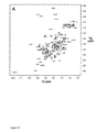
NS1A(1〜73)はdsRNAに結合するが、dsDNAまたはRNA/DNAハイブリッドには結合しない。NS1A(1〜73)および全長NS1Aタンパク質は、配列特異性なしで二本鎖RNA(dsRNA)に結合することが示されている)Lu et al.,(1995)Virology 214,222-228,Qian et al.,(1995)RNA 1,948-956,Wang et al.,1999)が、本発明までは、NS1A(1〜73)またはNS1Aタンパク質はRNA−DNAハイブリッドまたはdsDNAに結合するか否かは決定されていなかった。出願人らはNS1A(1〜73)を4つの32P標識デュプレックス:16bpのdsRNA(RR)、dsDNA(DD)および2つのRNA−DNAハイブリッドデュプレックス(RDおよびDR)とともにインキュベートした。次いで、これらの混合物を天然15%ポリアクリルアミドゲルで分析する(図1)。他の者によって報告されているように(Roberts and Crothers(1992)Science 258,1463-1466;Ratmeyer et al.,(1994)Biochemistry 33,5298-5304;Lesnik and Freier(1995)Biochemistry 34,10807-10815)、出願人らは天然ゲル上で遊離デュプレックスにつき以下の移動パターンを観察した(最も早いから最も遅い):DD>DR/RD>RR(各々レーン1、3、5および7)。より重要なことは、dsRNAのみがNS1A(1〜73)とで複合体を形成し、30%ゲルシフトを生じ(レーン2)、他方、全ての他のデュプレックスはタンパク質に結合しない(レーン4、6および8)ことが判明する。これらのデータは、NS1A(1〜73)が、これらの条件下では、dsDNA(B形態立体配座)またはRNA/DNAハイブリッド(中間体A/B立体配座)とは区別されるdsRNAの立体配座および/または構造的特徴(A形態立体配座)を特異的に認識することを示す。
NS1A (1-73) binds to dsRNA but not to dsDNA or RNA / DNA hybrids. NS1A (1-73) and full-length NS1A protein have been shown to bind to double-stranded RNA (dsRNA) without sequence specificity) Lu et al., (1995) Virology 214, 222-228, Qian et al (1995) RNA 1,948-956, Wang et al., 1999) until the present invention, it has been determined whether NS1A (1-73) or NS1A protein binds to RNA-DNA hybrids or dsDNA. There wasn't. Applicants incubated NS1A (1-73) with four 32 P-labeled duplexes: 16 bp dsRNA (RR), dsDNA (DD) and two RNA-DNA hybrid duplexes (RD and DR). These mixtures are then analyzed on a natural 15% polyacrylamide gel (FIG. 1). As reported by others (Roberts and Crothers (1992) Science 258,1463-1466; Ratmeyer et al., (1994) Biochemistry 33,5298-5304; Lesnik and Freier (1995) Biochemistry 34,10807- 10815), Applicants observed the following migration pattern per free duplex on natural gels (fastest to slowest): DD> DR / RD> RR (
dsRNAの長さおよびリボヌクレオチド配列は臨界的ではない。本明細書中でのいくつかの実施例に記載したように、本発明の方法は、タンパク質RNAの相互作用の形態の鍵となる特徴を同定する短い合成16塩基対(bp)のdsRNAを用いて行うことができる。このdsRNA分子は、pGEM1プラスミドのポリリンカーのセンスおよびアンチセンス転写物をアニールすることによって少量を生じさせることができる通常に用いられる29塩基対dsRNA結合基質に由来する配列を有する(Qian et al.,1995)。沈降平衡測定に基づき、溶液中でのこの合成16bpのdsRNAデュプレックスへのNS1A(1〜73)の結合の化学量論は、ほぼ1:1であり(1つのdsRNAデュプレックス分子に1つのタンパク質ダイマー)、二分子解離定数(Kd)はマイクロモラーの範囲である。出願人らは、これは、高スループット結合アッセイで用いるための適当なdsRNA基質分子であると提唱する。NMR化学シフト摂動実験は、NS1A(1〜73)のdsRNA結合エピトープは、部位特異的突然変異誘発実験によって以前に示されているように(Wang et al.,1999)、反平行ラセン2および2’と会合することを示す。精製されたNS1A(1〜73)−dsRNA複合体の円二色(CD)スペクトルは、遊離dsRNAおよびNS1A(1〜73)のCDスペクトルの和に非常に似ており、これは、結合の結果として、前記タンパク質またはそのA形態dsRNA標的いずれかの立体配座の変化はほとんどまたは全く起こらないことを示す。さらに、NS1A(1〜73)は対応するDNA−DNAデュプレックスまたはDNA−RNAハイブリッドデュプレックスに結合しないことが示されているので、NS1A(1〜73)はカノニカルA形態RNA特異的立体配座特徴を認識するようであり、従って、本発明の方法がインフルエンザのNS1タンパク質およびその宿主の間の相互作用をかなり模倣するさらにもう1つの方法を強調する。 The length of dsRNA and the ribonucleotide sequence are not critical. As described in several examples herein, the methods of the invention use short synthetic 16 base pair (bp) dsRNAs that identify key features of the form of protein RNA interaction. Can be done. This dsRNA molecule has a sequence derived from a commonly used 29 base pair dsRNA binding substrate that can be produced in small quantities by annealing the sense and antisense transcripts of the polylinker of the pGEM1 plasmid (Qian et al. 1995). Based on sedimentation equilibrium measurements, the stoichiometry of NS1A (1-73) binding to this synthetic 16 bp dsRNA duplex in solution is approximately 1: 1 (one protein dimer per dsRNA duplex molecule). The bimolecular dissociation constant (K d ) is in the micromolar range. Applicants propose that this is a suitable dsRNA substrate molecule for use in high throughput binding assays. NMR chemical shift perturbation experiments show that the dsRNA binding epitope of NS1A (1-73) is as shown previously by site-directed mutagenesis experiments (Wang et al., 1999), antiparallel helical 2 and 2 'Meeting with. The circular dichroic (CD) spectrum of the purified NS1A (1-73) -dsRNA complex is very similar to the sum of the free dsRNA and the CD spectra of NS1A (1-73), which is the result of binding. As shown, there is little or no conformational change of either the protein or its A-form dsRNA target. Furthermore, NS1A (1-73) has been shown not to bind to the corresponding DNA-DNA duplex or DNA-RNA hybrid duplex, so NS1A (1-73) exhibits canonical A-form RNA specific conformational features. It seems to be appreciated, therefore, highlights yet another way in which the method of the invention considerably mimics the interaction between the influenza NS1 protein and its host.
本発明の方法は、有利には、高スループットインビトロアッセイの意味で実行される。本発明のこの実施形態において、アッセイシステムは蛍光共鳴エネルギー移動または標識されたdsRNA分子、NS1AもしくはNS1A(1〜73)、またはNS1BもしくはNS1B(1〜93)分子いずれかでの蛍光偏光の標準的な方法のいずれかまたは双方を用いて、これらのタンパク質標的および種々のdsRNAデュプレックスの間の相互作用をモニターし、結合親和性を測定することができよう。これらのアッセイを用いて、化合物をスクリーニングして、NS1タンパク質の前記開示の構造に基づきNS1標的およびRNA基質の間の相互作用を阻害する分子を同定する。 The method of the invention is advantageously carried out in the sense of a high throughput in vitro assay. In this embodiment of the invention, the assay system is a standard for fluorescence polarization of either fluorescence resonance energy transfer or labeled dsRNA molecules, NS1A or NS1A (1-73), or NS1B or NS1B (1-93) molecules. Either or both of these methods can be used to monitor the interaction between these protein targets and the various dsRNA duplexes and determine the binding affinity. Using these assays, compounds are screened to identify molecules that inhibit the interaction between NS1 target and RNA substrate based on the previously disclosed structure of NS1 protein.
広く種々の化合物を、ランダムおよびバイアスド化合物ライブラリーを含めた、本発明によるインフルエンザウイルスに対する阻害活性につきテストすることができる。バイアスド化合物ライブラリーは、例えば、公表された結果に基づいて推定された、NS1標的RNA基質相互作用部位の特定の構造的特徴を用いて設計することができる。例えば、Chien,et al.,Nature Struct.Biol.4:891-95(1997);Liu,et al.,Nature Struct.Biol.4:896-899(1997);およびWang,et al.,RNA 5:195-205(1999)参照。 A wide variety of compounds can be tested for inhibitory activity against influenza viruses according to the present invention, including random and biased compound libraries. A biased compound library can be designed, for example, using specific structural features of the NS1 target RNA substrate interaction site deduced based on published results. For example, Chien, et al., Nature Struct. Biol. 4: 891-95 (1997); Liu, et al., Nature Struct. Biol. 4: 896-899 (1997); and Wang, et al., RNA 5: 195-205 (1999).
[ウイルス複製に必要なNS1Aタンパク質およびdsRNAの相互作用に干渉する化合物についてのスクリーニングアッセイ]
インフルエンザウイルスのNS1タンパク質1またはそのdsRNA結合ドメイン、および相互作用および結合するdsRNAは、時々、本明細書中では「結合パートナー」という。多数のアッセイシステムのいずれかを利用して、結合パートナーの相互作用に干渉する能力について化合物をテストすることができる。しかしながら、限定されるものではないが、リガンド(天然または合成)、ペプチドまたは小さな有機分子を含めた、多数の化合物をスクリーニングするための迅速な高スループットアッセイが好ましい。結合パートナーの相互作用に干渉すると同定される化合物は、さらに、細胞ベースのアッセイ、動物モデル系および患者において、本明細書中に記載したように抗ウイルス活性につき評価すべきである。インフルエンザウイルスのNS1タンパク質またはそのdsRNA結合ドメイン、およびdsRNAの間の相互作用に干渉する化合物を同定するために用いるアッセイシステムの基本的な原理は、2つの結合パートナーが相互作用し、結合し、従って、複合体を形成するのを可能とする条件下にて、かつそれに十分な時間の間で、インフルエンザウイルスのNS1タンパク質またはdsRNA結合ドメイン、およびdsRNAを含有する反応混合物を準備することを含む。阻害活性につき化合物をテストするために、テスト化合物の存在下および非存在下で反応を行い、すなわち、テスト化合物をまず反応混合物に含ませるか、あるいはインフルエンザウイルスのNS1タンパク質またはそのdsRNA結合ドメイン、およびdsRNAの添加の後の時点で加えることができ;対照をテスト化合物なしで、またはプラセボと共にインキュベートする。インフルエンザウイルスのNS1タンパク質またはそのdsRNA結合ドメインおよびdsRNAの間のいずれかの複合体の形成を検出する。テスト化合物を含有する反応混合物ではなく、対照反応における複合体の形成は、前記化合物が、インフルエンザウイルスのNS1タンパク質またはそのdsRNA結合ドメインおよびdsRNAの相互作用に干渉することを示す。
[Screening Assay for Compounds Interfering with NS1A Protein and dsRNA Interaction Required for Virus Replication]
The
本発明のさらにもう1つの態様は、NS1タンパク質またはそのdsRNA結合ドメインNS1A(1〜73)またはNS1B(1〜93)の構造、および薬物スクリーニングアッセイにおけるNS1−RNA複合体のモデルの3次元座標を用いることを含む、インフルエンザウイルス感染を治療するのに用いることができる化合物について現実にスクリーニングする方法を含む。 Yet another aspect of the present invention relates to the structure of NS1 protein or its dsRNA binding domain NS1A (1-73) or NS1B (1-93) and the three-dimensional coordinates of the model of NS1-RNA complex in drug screening assays. Methods of actually screening for compounds that can be used to treat influenza virus infection, including using.
本発明のもう1つの態様は、スクリーニング用の化合物ライブラリーを設計するために、複合体のモデルの3次元座標を用いる方法を含む。 Another aspect of the invention includes a method that uses the three-dimensional coordinates of a model of a complex to design a compound library for screening.
従って、本発明は、インフルエンザウイルス感染を治療するのに用いることができる化合物または薬物を同定する方法を提供する。1つのそのような実施形態は、インフルエンザウイルスのNS1タンパク質またはそのdsRNA結合ドメインの阻害剤としての使用のための化合物を同定するための方法、およびインフルエンザA型またはB型ウイルスのNS1タンパク質またはそのdsRNA結合ドメインから得られた3次元座標を含むデータ組を含む。好ましくは、選択は、コンピュータモデリングと組み合わせて行う。 Thus, the present invention provides a method of identifying compounds or drugs that can be used to treat influenza virus infection. One such embodiment is a method for identifying a compound for use as an inhibitor of the influenza virus NS1 protein or its dsRNA binding domain, and the influenza A or B virus NS1 protein or its dsRNA. It includes a data set that includes three-dimensional coordinates obtained from the binding domain. Preferably, the selection is done in combination with computer modeling.
1つの実施形態において、インフルエンザウイルスのNS1タンパク質、またはそのdsRNA結合ドメインについて決定された3次元座標で合理的薬物設計(rational drug design)を行うことによって、潜在的化合物を選択する。前記したように、好ましくは、選択はコンピュータモデリングと組み合わせて行われる。潜在的化合物をインフルエンザウイルスのNS1タンパク質またはそのdsRNA結合ドメイン、およびdsRNAと接触させ、その結合に干渉させ、結合の阻害を決定する(例えば、測定する)。潜在的化合物は、結合の減少がある場合に、インフルエンザウイルスのNS1タンパク質またはそのdsRNA結合ドメインの結合を阻害する化合物として同定される。あるいは、潜在的化合物をインフルエンザウイルス感染細胞培養と接触させおよび/またはそれに添加し、ウイルス培養の増殖を決定する。潜在的化合物は、ウイルス培養の増殖の減少がある場合にウイルス増殖を阻害する化合物として同定される。 In one embodiment, potential compounds are selected by performing a rational drug design with three-dimensional coordinates determined for the NS1 protein of influenza virus, or its dsRNA binding domain. As mentioned above, preferably the selection is done in combination with computer modeling. A potential compound is contacted with the influenza virus NS1 protein or its dsRNA binding domain and dsRNA to interfere with its binding and determine (eg, measure) inhibition of binding. A potential compound is identified as a compound that inhibits binding of the influenza virus NS1 protein or its dsRNA binding domain when there is a decrease in binding. Alternatively, a potential compound is contacted with and / or added to the influenza virus infected cell culture to determine the growth of the virus culture. A potential compound is identified as a compound that inhibits virus growth when there is a decrease in growth of the virus culture.
好ましい実施形態において、前記方法は、さらに、分子置き換え分析、および薬物につき決定される3次元座標での合理的薬物設計を行うことによって選択された第二世代の候補薬物の設計を含む。好ましくは、選択はコンピュータモデリングと組み合わせて行われる。候補薬物を本明細書中に例示する標準生化学方法を用いて非常に多数の薬物スクリーニングアッセイでテストすることができる。本発明のこれらの実施形態において、NS1Aタンパク質の3次元座標およびNS1A−dsRNA複合体のモデルまたはNS1B−dsRNA複合体のモデルは、(a)スクリーニングのための阻害剤ライブラリーを設計する、(b)リード化合物を合理的に最適化する、および(c)潜在的阻害剤を現実にスクリーニングするための方法を提供する。 In a preferred embodiment, the method further comprises the design of a second generation candidate drug selected by performing a molecular replacement analysis and a rational drug design with three-dimensional coordinates determined for the drug. Preferably, the selection is made in combination with computer modeling. Candidate drugs can be tested in a large number of drug screening assays using standard biochemical methods exemplified herein. In these embodiments of the invention, the three-dimensional coordinates of the NS1A protein and the NS1A-dsRNA complex model or NS1B-dsRNA complex model (a) design an inhibitor library for screening, (b) It provides a method for rationally optimizing lead compounds, and (c) actually screening potential inhibitors.
本発明の方法を行うことができる他のアッセイ成分およびフォーマットを以下のサブセクションに記載する。 Other assay components and formats in which the methods of the invention can be performed are described in the following subsections.
[アッセイ成分]
アッセイシステムで用いる結合パートナーの1つを、直接的にまたは間接的に標識して、NS1タンパク質またはdsRNA結合部分、およびdsRNAの間の結合の程度を測定することができる。以下で詳細に記載するアッセイフォーマットに応じて、結合の程度を、インフルエンザウイルスのNS1タンパク質、またはそのdsRNA結合ドメイン、およびdsRNAの間の複合体化、または候補化合物の存在下における、予め形成された複合体の解離の程度の項目につき測定することができる。限定されるものではないが、125Iのような放射性同位体;基質に暴露した場合に検出可能な色シグナルまたは光を生じる酵素標識系;および蛍光標識を含む、種々の適当な標識系を用いることができる。
[Assay components]
One of the binding partners used in the assay system can be directly or indirectly labeled to measure the extent of binding between the NS1 protein or dsRNA binding moiety and the dsRNA. Depending on the assay format described in detail below, the extent of binding was pre-formed in the presence of a complex between influenza virus NS1 protein, or its dsRNA binding domain, and dsRNA, or in the presence of a candidate compound. It can be measured per item of the degree of dissociation of the complex. A variety of suitable labeling systems are used, including but not limited to radioisotopes such as 125 I; enzyme labeling systems that produce a detectable color signal or light when exposed to a substrate; and fluorescent labels be able to.
組換えDNA技術を用いてインフルエンザウイルスのNS1タンパク質、またはそのdsRNA結合ドメイン、および前記アッセイのdsRNA結合パートナーを生産する場合、標識、固定化および/または検出を促進することができる融合タンパク質を作成するのが有利であろう。例えば、インフルエンザウイルスのNS1タンパク質、またはそのdsRNA結合ドメインのコーディング配列を、酵素活性を有するか、あるいは酵素基質として働く異種タンパク質のそれに融合させて、標識および検出を促進することができる。融合構築体は、融合産物の異種成分が、インフルエンザウイルスのNS1タンパク質またはそのdsRNA結合ドメインおよび、dsRNAの結合に干渉しないように設計すべきである。 When using recombinant DNA technology to produce the influenza virus NS1 protein, or its dsRNA binding domain, and the dsRNA binding partner of the assay, create a fusion protein that can facilitate labeling, immobilization and / or detection. Would be advantageous. For example, the NS1 protein of influenza virus, or the coding sequence of its dsRNA binding domain, can be fused to a heterologous protein that has enzymatic activity or serves as an enzyme substrate to facilitate labeling and detection. The fusion construct should be designed so that the heterologous components of the fusion product do not interfere with the binding of influenza virus NS1 protein or its dsRNA binding domain and dsRNA.
間接的標識は、インフルエンザウイルスのNS1タンパク質、またはそのdsRNA結合ドメインに特異的に結合する標識された抗体のような第三のタンパク質の使用を含む。そのような抗体は、限定されるものではないが、ポリクローナル、モノクローナル、キメラ、一本鎖、Fab断片、およびFab発現ライブラリーによって生産された断片を含む。 Indirect labeling involves the use of a third protein, such as a labeled antibody that specifically binds to the NS1 protein of influenza virus, or its dsRNA binding domain. Such antibodies include, but are not limited to, polyclonal, monoclonal, chimeric, single chain, Fab fragments, and fragments produced by Fab expression libraries.
抗体の生産では、インフルエンザウイルスのNS1タンパク質、またはそのdsRNA結合ドメインでの注射によって種々の宿主動物を免疫化することができる。そのような宿主動物は、限定されるものではないが、少数を述べると、ウサギ、マウスおよびラットを含む。限定されるものではないが、フロイントの(完全および不完全)アジュバント、水酸化アルミニウムのようなミネラルゲル、リソレクチン、プルロニックポリオール、ポリアニオン、ペプチド、油エマルジョン、キーホールリンペットヘモシアニン、ジニトロフェノールのような表面活性物質、およびBCG(バチルスCalmette-Guerin)およびCorynebacterium parvumのような潜在的に有用なヒトアジュバントを含めた種々のアジュバントを用いて、宿主種に応じて免疫学的応答を増加させることができる。 For the production of antibodies, various host animals can be immunized by injection with the NS1 protein of influenza virus, or its dsRNA binding domain. Such host animals include, but are not limited to, rabbits, mice and rats, to name a few. Such as, but not limited to, Freund's (complete and incomplete) adjuvants, mineral gels such as aluminum hydroxide, lysolectin, pluronic polyols, polyanions, peptides, oil emulsions, keyhole limpet hemocyanin, dinitrophenol Various adjuvants, including surface active substances and potentially useful human adjuvants such as BCG (Bacillus Calmette-Guerin) and Corynebacterium parvum, can be used to increase the immunological response depending on the host species .
モノクローナル抗体は、培養中の連続的細胞系統による抗体分子の生産を提供するいずれの技術を用いて調製することもできる。それらは、限定されるものではないが、KohlerおよびMilstein(Nature,1975,256:495-497)によって元来は記載されたハイブリドーマ技術、ヒトB細胞ハイブリドーマ技術(Kosbor et al.,1983,Immunology Today,4:72,Cote et al.,1983,Proc.Natl.Acad.Sci.,80:2026-2030)およびEBV−ハイブリドーマ技術(Cole et al.,1985,Monoclonal Antibodies and Cancer Therapy,Alan R.Liss,Inc.,pp.77-96)を含む。加えて、適当な生物学的活性のヒト抗体分子からの遺伝子と共に適当な抗原特異性のマウス抗体分子からの遺伝子をスプライシングすることによる「キメラ抗体」の生産用に開発された技術(Morrison et al.,1984,Proc.Natl.Acad.Sci.,81:6851-6855;Neuberger et al.,1984,Nature,312:604-608;Takeda et al.,1985,Nature,314:452-454)を用いることができる。あるいは、一本鎖抗体の生産のために記載された技術(米国特許第4,946,778号)を適合させて、インフルエンザウイルスのNS1タンパク質またはそのdsRNA結合ドメインに対して特異的な一本鎖抗体を生産することができる。 Monoclonal antibodies can be prepared using any technique that provides for the production of antibody molecules by continuous cell lines in culture. They include, but are not limited to, the hybridoma technology originally described by Kohler and Milstein (Nature, 1975, 256: 495-497), the human B cell hybridoma technology (Kosbor et al., 1983, Immunology Today). , 4:72, Cote et al., 1983, Proc. Natl. Acad. Sci., 80: 2026-2030) and EBV-hybridoma technology (Cole et al., 1985, Monoclonal Antibodies and Cancer Therapy, Alan R. Liss. , Inc., pp. 77-96). In addition, a technique developed for the production of “chimeric antibodies” by splicing genes from mouse antibody molecules of appropriate antigen specificity together with genes from human antibody molecules of appropriate biological activity (Morrison et al , 1984, Proc. Natl. Acad. Sci., 81: 6851-6855; Neuberger et al., 1984, Nature, 312: 604-608; Takeda et al., 1985, Nature, 314: 452-454). Can be used. Alternatively, techniques described for the production of single chain antibodies (US Pat. No. 4,946,778) are adapted to produce single chain antibodies specific for the influenza virus NS1 protein or its dsRNA binding domain. be able to.
特異的エピトープを認識する抗体断片は公知の技術によって作り出すことができる。例えば、そのような断片は、限定されるものではないが、抗体分子のペプシン消化によって生産することができるF(ab’)2断片および前記F(ab’)2断片のジスルフィドブリッジを還元することによって作り出すことができるFab断片を含む。あるいは、Fab発現ライブラリーを構築して(Huse et al.,1989,Science,246:1275-1281)、所望の特異性を持つモノクローナルFab断片の迅速かつ容易な同定を可能とすることができる。 Antibody fragments that recognize specific epitopes can be generated by known techniques. For example, such fragments include, but are not limited to, reducing F (ab ′) 2 fragments and disulfide bridges of said F (ab ′) 2 fragments that can be produced by pepsin digestion of antibody molecules. Fab fragments that can be produced by Alternatively, Fab expression libraries can be constructed (Huse et al., 1989, Science, 246: 1275-1281) to allow rapid and easy identification of monoclonal Fab fragments with the desired specificity.
[アッセイフォーマット]
前記アッセイは、異種または同種フォーマットで行うことができる。異種アッセイは、結合パートナーの一方を固相に係留させ、反応の最後に固相に係留させた複合体を検出することを含む。同種アッセイでは、全反応を液相で行う。いずれのアプローチにおいても、反応体の添加の順序を変化させて、テストすべき化合物についての異なる情報を得ることができる。例えば、競合によって、結合パートナー間の相互作用に干渉するテスト化合物は、例えば、テスト物質の存在下で反応を行うことによって;すなわち、インフルエンザウイルスのNS1タンパク質、またはそのdsRNA結合ドメイン、およびdsRNAに先立って、またはそれと同時に、テスト物質を反応混合物に添加することによって同定することができる。他方、予め形成された複合体を破壊するテスト化合物、例えば、複合体からの結合パートナーの一方を置き換えるより高い結合定数を持つ化合物は、複合体が形成された後に、テスト化合物を反応混合物に添加することによってテストすることができる。種々のフォーマットを以下に簡単に記載する。
[Assay format]
The assay can be performed in a heterogeneous or homogeneous format. Heterogeneous assays involve anchoring one of the binding partners to the solid phase and detecting the complex anchored to the solid phase at the end of the reaction. In homogeneous assays, all reactions are performed in the liquid phase. In either approach, the order of addition of the reactants can be varied to obtain different information about the compound to be tested. For example, test compounds that interfere with the interaction between binding partners by competition, for example, by reacting in the presence of the test substance; ie, prior to the influenza virus NS1 protein, or its dsRNA binding domain, and dsRNA. Or simultaneously, by adding a test substance to the reaction mixture. On the other hand, test compounds that break the preformed complex, such as compounds with higher binding constants that replace one of the binding partners from the complex, add the test compound to the reaction mixture after the complex is formed. You can test by doing. Various formats are briefly described below.
異種アッセイ系においては、1つの結合パートナー、例えば、インフルエンザウイルスのNS1タンパク質、またはそのdsRNA結合ドメイン、あるいはdsRNAのいずれかを固体表面に係留し、係留されていないその結合パートナーを直接的または間接的に標識する。実行においては、マイクロタイタープレートを便宜には利用する。係留された種は、非共有結合または共有結合によって固定化することができる。あるいは、インフルエンザウイルスのNS1タンパク質、またはそのdsRNA結合ドメインに特異的な固定化された抗体を用いて、インフルエンザウイルスのNS1タンパク質、またはそのdsRNA結合ドメインを固体表面に係留させることができる。前記表面は、予め調製し、保存することができる。 In a heterogeneous assay system, one binding partner, for example, the NS1 protein of influenza virus, or its dsRNA binding domain, or dsRNA is anchored to a solid surface and its unbound anchoring partner is directly or indirectly Labeled. In practice, microtiter plates are conveniently used. The anchored species can be immobilized by non-covalent or covalent bonds. Alternatively, an immobilized antibody specific for the influenza virus NS1 protein, or dsRNA binding domain thereof, can be used to anchor the influenza virus NS1 protein, or dsRNA binding domain, to a solid surface. The surface can be prepared and stored in advance.
前記アッセイを行うためには、固定化された種の結合パートナーを、テスト化合物と共に、またはそれ無くして、被覆された表面に添加する。反応が完了した後、未反応成分を(例えば、洗浄によって)除去し、形成されたいずれの複合体も固体表面に固定化されたままである。固体表面に係留された複合体の検出は、多数の方法で達成することができる。結合パートナーが予め標識された場合、表面に固定された標識の検出は、複合体が形成されたことを示す。結合パートナーが予め標識されない場合、間接的標識を用いて、例えば、結合パートナーに対して特異的な標識された抗体(前記抗体は、今度は、標識された抗−Ig抗体で直接的または間接的に標識することができる)を用い、表面に係留された複合体を検出することができる。反応成分の添加の順序に応じて、複合体の形成を阻害するか、あるいは予め形成された複合体を破壊するテスト化合物を検出することができる。 To perform the assay, an immobilized species binding partner is added to the coated surface with or without the test compound. After the reaction is complete, unreacted components are removed (eg, by washing) and any complexes formed remain immobilized on the solid surface. Detection of complexes anchored on a solid surface can be accomplished in a number of ways. When the binding partner is pre-labeled, detection of the label immobilized on the surface indicates that a complex has formed. If the binding partner is not pre-labeled, indirect labeling can be used, for example, a labeled antibody specific for the binding partner (which in turn is directly or indirectly with a labeled anti-Ig antibody. Can be used to detect complexes anchored on the surface. Depending on the order of addition of reaction components, a test compound that inhibits complex formation or destroys the preformed complex can be detected.
あるいは、反応は、テスト化合物の存在下または非存在下にて液相で行うことができ、反応生成物を未反応成分から分離し、例えば、溶液中で形成されたいずれの複合体も係留するためにインフルエンザウイルスのNS1タンパク質またはそのdsRNA結合ドメインに対して特異的な固定化された抗体を用い、複合体を検出することができる。再度、液相への反応体の添加の順序に応じて、複合体を阻害する、あるいは予め形成された複合体を破壊するテスト化合物を同定することができる。 Alternatively, the reaction can be performed in the liquid phase in the presence or absence of the test compound, separating the reaction product from unreacted components, eg, tethering any complexes formed in solution. Therefore, the complex can be detected using an immobilized antibody specific for the NS1 protein of influenza virus or its dsRNA binding domain. Again, depending on the order of addition of the reactants to the liquid phase, test compounds that inhibit the complex or destroy the preformed complex can be identified.
本発明の他の実施形態においては、同種アッセイを用いることができる。このアプローチにおいては、インフルエンザウイルスNS1タンパク質またはそのdsRNA結合ドメインおよびdsRNAの予め形成された複合体を調製し、そこでは、結合パートナーの一方が標識されるが、前記標識によって生じたシグナルは、複合体形成のためクエンチされる(例えば、免疫アッセイのためにこのアプローチを利用するRubensteinによる米国特許第4,109,496号参照)。予め形成された複合体からの結合パートナーの一方と競合し、それを破壊するテスト物質の添加の結果、バックグラウンドを超えるシグナルが生成する。このようにして、インフルエンザウイルスのNS1タンパク質、またはそのdsRNA結合ドメイン、およびdsRNA相互作用を破壊するテスト物質を同定することができる。 In other embodiments of the invention, homogeneous assays can be used. In this approach, a pre-formed complex of influenza virus NS1 protein or its dsRNA binding domain and dsRNA is prepared, where one of the binding partners is labeled, but the signal generated by the label is complex Quenched for formation (see, eg, US Pat. No. 4,109,496 by Rubenstein, which utilizes this approach for immunoassays). Addition of a test substance that competes with and destroys one of the binding partners from the preformed complex results in a signal that exceeds background. In this way, the NS1 protein of influenza virus, or its dsRNA binding domain, and test substances that disrupt dsRNA interactions can be identified.
例えば、特別な実施形態においては、インフルエンザウイルスのNS1タンパク質、またはそのdsRNA結合ドメインは、前記した組換えDNA技術を用いて固定化のために調製することができる。その結合活性が得られた融合タンパク質で維持されるように、融合ベクターpGEX−5X−1を用い、そのコーディング領域をグルタチオン−S−トランスフェラーゼ(GST)遺伝子に融合させることができる。NS1タンパク質またはそのdsRNA結合ドメインを精製し、それを用いて、当分野でルーチン的に実行され、かつ前記した方法を用いてNS1またはNS1断片に特異的なモノクローナル抗体を生起させることができる。この抗体は、例えば、当分野でルーチン的に実行される方法によって、放射性同位体125Iで標識することができる。異種アッセイでは、例えば、GST−NS1融合タンパク質をグルタチオン−アガロースビーズに係留させることができる。dsRNAを、dsRNAが融合タンパク質のNS1部分と相互作用し、それに結合できるように、テスト化合物の存在下または非存在下で添加することができる。テスト化合物が添加された後、未結合物質を洗浄して除去することができ、NS1−特異的標識モノクローナル抗体を系に添加し、複合体化された結合パートナーに結合させることができる。NS1およびdsRNAの間の相互作用は、グルタチオン−アガロースビーズと会合したままである放射能の量を測定することによって検出することができる。テスト化合物による相互作用の成功した阻害の結果、測定された放射能は減少する。 For example, in a special embodiment, the influenza virus NS1 protein, or its dsRNA binding domain, can be prepared for immobilization using the recombinant DNA techniques described above. The coding region can be fused to the glutathione-S-transferase (GST) gene using the fusion vector pGEX-5X-1 so that the binding activity is maintained with the resulting fusion protein. NS1 protein or its dsRNA binding domain can be purified and used to raise monoclonal antibodies that are routinely practiced in the art and that are specific for NS1 or NS1 fragments using the methods described above. The antibody can be labeled with the radioisotope 125 I, for example, by methods routinely practiced in the art. In heterogeneous assays, for example, GST-NS1 fusion protein can be tethered to glutathione-agarose beads. The dsRNA can be added in the presence or absence of the test compound so that the dsRNA can interact with and bind to the NS1 portion of the fusion protein. After the test compound is added, unbound material can be washed away and NS1-specifically labeled monoclonal antibody can be added to the system and allowed to bind to the complexed binding partner. The interaction between NS1 and dsRNA can be detected by measuring the amount of radioactivity that remains associated with glutathione-agarose beads. As a result of successful inhibition of the interaction by the test compound, the measured radioactivity is reduced.
あるいは、GST−NS1融合タンパク質、およびdsRNAは固体グルタチオン−アガロースビーズの存在下にて液体中で一緒に混合することができる。テスト化合物は、結合パートナーを相互作用させる間に、またはその後のいずれかに添加することができる。この混合物をグルタチオン−アガロースビーズに添加することができ、未結合物質を洗浄して除去する。再度、結合パートナーの相互作用の阻害の程度は、ビーズに会合した放射能を測定することによって検出することができる。 Alternatively, the GST-NS1 fusion protein and dsRNA can be mixed together in a liquid in the presence of solid glutathione-agarose beads. The test compound can be added either during or after the binding partner interacts. This mixture can be added to the glutathione-agarose beads and unbound material is washed away. Again, the degree of inhibition of binding partner interaction can be detected by measuring the radioactivity associated with the beads.
本発明に従って、1つのウイルスを阻害することが判明している所与の化合物を、広い範囲の異なるインフルエンザウイルスに対する一般的抗ウイルス活性につきテストすることができる。例えば、限定されるものではないが、インフルエンザA型ウイルスNS1とdsRNAとの相互作用を、NS1結合部位への結合によって阻害する化合物は、インフルエンザA型ウイルスの異なる株ならびにインフルエンザB型ウイルスの株に対して、前記したアッセイに従ってテストすることができる。 In accordance with the present invention, a given compound known to inhibit one virus can be tested for general antiviral activity against a wide range of different influenza viruses. For example, but not limited to, compounds that inhibit the interaction between influenza A virus NS1 and dsRNA by binding to the NS1 binding site have been found in different strains of influenza A virus as well as strains of influenza B virus. In contrast, it can be tested according to the assay described above.
薬物開発のために潜在的リード化合物を選択するためには、NS1標的およびRNA物質の間の相互作用の同定された阻害剤を、さらに、まず、組織培養中の、動物モデル実験において、インフルエンザウイルスの複製を阻害するそれらの能力についてテストすることができる。インフルエンザウイルス複製を効果的に阻害する各阻害剤の最低濃度は、感染の高および低多重度を用いて決定することができる。 In order to select potential lead compounds for drug development, identified inhibitors of the interaction between the NS1 target and the RNA agent can be further combined with an influenza virus, first in an animal model experiment in tissue culture. Can be tested for their ability to inhibit replication. The minimum concentration of each inhibitor that effectively inhibits influenza virus replication can be determined using the high and low multiplicity of infection.
[ウイルス増殖アッセイ]
ウイルスの増殖を阻害する、前記アッセイ系で同定された阻害剤の能力は、プラーク形成によって、あるいはTCID50またはヒヨコ胚の尿膜における成長のようなウイルス増殖の他の指標によってアッセイすることができる。これらのアッセイにおいて、適当な細胞系統または胚形成卵を野生型インフルエンザウイルスで感染させ、感染時、またはその後のいずれかに、テスト化合物を組織培養基に添加する。テスト化合物の効果は、ウイルスプラークの存在によって;あるいはプラーク表現型が存在しない場合mには、TCID50、またはヒヨコ胚の尿膜中の成長のような指標によって、あるいはヘマグルチニン化アッセイで、感染された細胞の上清中で、あるいは感染された胚形成卵の尿膜流体中で測定されたヘマグルチニン(HA)力価によって示されるウイルス粒子形成の定量によってスコア取りされる。阻害剤は、HA力価またはプラーク形成を抑制する、あるいはウイルス感染細胞またはヒヨコ胚の尿膜における細胞障害効果を低下させるテスト化合物の能力によって、あるいはヘマグルチニン化アッセイで測定されたウイルス粒子形成を低下させるその能力によって、スコア取りすることができる。
[Virus proliferation assay]
The ability of the inhibitors identified in the assay system to inhibit viral growth can be assayed by plaque formation or by other indicators of viral proliferation such as TCID 50 or growth in the allantoic membrane of chick embryos. . In these assays, an appropriate cell line or embryogenic egg is infected with wild-type influenza virus and a test compound is added to the tissue culture medium either at the time of infection or thereafter. The effect of the test compound is infected by the presence of a viral plaque; or in the absence of a plaque phenotype, by an indicator such as TCID 50 , or growth in the allantoic membrane of a chick embryo, or in a hemagglutination assay. Scored by quantification of virus particle formation as indicated by hemagglutinin (HA) titer measured in the supernatant of cultured cells or in the allantoic fluid of infected embryogenic eggs. Inhibitors reduce HA titer or plaque formation, or reduce viral particle formation as measured by the ability of test compounds to reduce cytotoxic effects in the allantoic membrane of virus-infected cells or chick embryos, or in hemagglutination assays It is possible to score according to the ability.
[動物モデルアッセイ]
本発明のプロセスによって同定されたウイルス複製の最も効果的な阻害剤を、後の動物実験で用いることができる。インフルエンザウイルスの複製を妨げる阻害剤の能力は、天然の、あるいはインフルエンザに対して適合させた宿主である動物モデルでアッセイすることができる。そのような動物はブタ、フェレット、マウス、サル、ウマ、および霊長類のような哺乳動物、または鳥類を含むことができる。本明細書中に詳細に記載するように、そのような動物モデルを用いて、動物対象においてLD50およびED50を測定することができ、そのようなデータを用いて、NS1A(1〜73)またはNS1B(1〜93)およびdsRNA相互作用の阻害剤に対する治療指数を求めることができる。
[Animal model assay]
The most effective inhibitors of viral replication identified by the process of the present invention can be used in subsequent animal experiments. The ability of an inhibitor to prevent influenza virus replication can be assayed in animal models that are natural or influenza-adapted hosts. Such animals can include mammals such as pigs, ferrets, mice, monkeys, horses, and primates, or birds. As described in detail herein, such animal models can be used to measure LD 50 and ED 50 in animal subjects, and such data can be used to NS1A (1-73) Alternatively, therapeutic indices for NS1B (1-93) and inhibitors of dsRNA interaction can be determined.
また、リード化合物の設計の最適化は、高スループットスクリーニングによって同定された阻害剤によるNS1タンパク質またはそのdsRNA結合ドメインの表面の特徴的な結合部位によって助けることができる。そのような特徴付けは、NMR共鳴帰属と共に化学シフト摂動NMRを用いて行うことができる。NMRは、RNAに対する小分子阻害剤の結合部位を決定することができる。これらの結合部位の位置の決定は、複数の初期阻害剤リードを一緒に連結するための、およびリード設計を最適化するためのデータを提供するであろう。 Also, optimization of lead compound design can be aided by the characteristic binding sites on the surface of NS1 protein or its dsRNA binding domain by inhibitors identified by high-throughput screening. Such characterization can be performed using chemical shift perturbation NMR with NMR resonance assignments. NMR can determine the binding sites of small molecule inhibitors for RNA. Determination of the location of these binding sites will provide data for linking multiple early inhibitor leads together and for optimizing lead design.
[医薬製剤および投与方法]
ウイルス複製を阻害する同定された化合物は、ウイルス感染を治療するために治療上有効量にて患者に投与することができる。治療上有効量とは、ウイルス感染の兆候の軽減をもたらすのに十分な化合物の量をいう。
[Pharmaceutical preparation and administration method]
Identified compounds that inhibit viral replication can be administered to a patient in a therapeutically effective amount to treat a viral infection. A therapeutically effective amount refers to the amount of the compound sufficient to provide relief from the symptoms of viral infection.
そのような化合物の毒性および治療効果は、例えば、LD50(集団の50%に対して致死的な用量)およびED50(集団の50%で治療的に有効な用量)を決定するために、細胞培養または実験動物における標準的な医薬手法によって測定することができる。毒性および治療効果の間の用量比は治療指標であり、それは、比率LD50/ED50として表すことができる。大きな治療指標を呈する化合物が好ましい。毒性副作用を呈する化合物を用いることができるが、そのような化合物を感染の部位に標的化して、未感染細胞に対する損傷を最小化し、副作用を低下させる送達システムを設計するよう注意を払うべきである。 The toxic and therapeutic effects of such compounds are, for example, to determine the LD 50 (a lethal dose to 50% of the population) and ED 50 (the therapeutically effective dose in 50% of the population) It can be measured by standard pharmaceutical techniques in cell culture or laboratory animals. The dose ratio between toxic and therapeutic effects is the therapeutic index and it can be expressed as the ratio LD 50 / ED 50 . Compounds that exhibit large therapeutic indices are preferred. Although compounds that exhibit toxic side effects can be used, care should be taken to design such delivery systems that target such compounds to the site of infection to minimize damage to uninfected cells and reduce side effects .
細胞培養アッセイおよび動物実験から得られたデータは、ヒトで用いるためのある範囲の用量を処方する際に用いることができる。そのような化合物の用量は、好ましくは、毒性をほとんどまたは全く伴わないED50を含むある範囲の循環濃度内にある。前記用量は、使用する投与形態および利用する投与経路に応じてこの範囲内で変化させることができる。本発明の方法で用いられるいずれの化合物についても、治療上有効量は細胞培養アッセイから最初に見積もることができる。細胞培養で測定されるIC50(すなわち、半−最大感染、または半−最大阻害を達成するテスト化合物の濃度)を含む循環血漿濃度の範囲を達成するために、用量を動物モデルで処方することができる。そのような情報を用いて、人における有用な用量をより正確に決定することができる。血漿中でのレベルは、例えば、高性能液体クロマトグラフィーによって測定することができる。 Data obtained from cell culture assays and animal studies can be used in formulating a range of doses for use in humans. The dosage of such compounds lies preferably within a range of circulating concentrations that include the ED 50 with little or no toxicity. The dosage may vary within this range depending on the dosage form used and the route of administration utilized. For any compound used in the method of the invention, the therapeutically effective dose can be estimated initially from cell culture assays. Formulating a dose in an animal model to achieve a range of circulating plasma concentrations that include an IC 50 (ie, the concentration of test compound that achieves half-maximal infection, or half-maximal inhibition) as measured in cell culture Can do. Such information can be used to more accurately determine useful doses in humans. Levels in plasma can be measured, for example, by high performance liquid chromatography.
本発明に従って用いるための医薬組成物は、1以上の生理学的に許容される担体または賦形剤を用いて慣用的な方法で処方することができる。 A pharmaceutical composition for use in accordance with the present invention can be formulated in a conventional manner using one or more physiologically acceptable carriers or excipients.
従って、化合物およびそれらの生理学的に許容される塩および溶媒和物は、(口または鼻いずれかを介する)吸入または吹込み、あるいは経口、バッカル、非経口または直腸投与による投与用に処方することができる。 Thus, the compounds and their physiologically acceptable salts and solvates should be formulated for administration by inhalation or insufflation (either through the mouth or nose) or oral, buccal, parenteral or rectal administration. Can do.
吸入による投与では、本発明に従って用いられる化合物は、便宜には、適当なプロペラント、例えば、ジクロロジフルオロメタン、トリクロロフルオロメタン、ジクロロテトラフルオロエタン、二酸化炭素または他の適当なガスを使用し、圧縮パックまたはネビュライザーからエアロゾルスプレー提示の形態で送達される。圧縮エアロゾルの場合には、投与単位は、計量された量を送達するためにバルブを設けることによって決定することができる。例えば、インヘーラーまたはインサフレーターで用いられるゼラチンのカプセルおよびカートリッジは、化合物およびラクトースまたは澱粉のような適当な基材の粉末ミックスを含有するように処方することができる。 For administration by inhalation, the compounds used in accordance with the present invention are conveniently compressed using a suitable propellant, such as dichlorodifluoromethane, trichlorofluoromethane, dichlorotetrafluoroethane, carbon dioxide or other suitable gas. Delivered in the form of an aerosol spray presentation from a pack or nebulizer. In the case of a compressed aerosol, the dosage unit can be determined by providing a valve to deliver a metered amount. For example, gelatin capsules and cartridges used in inhalers or insufflators can be formulated to contain a powder mix of the compound and a suitable substrate such as lactose or starch.
経口投与では、医薬組成物は、例えば、結合剤(例えば、予めα化されたトウモロコシ澱粉、ポリビニルピロリドンまたはヒドロキシプロピルメチルセルロース);充填剤(例えば、ラクトース、マイクロクリスタリンセルロースまたはリン酸水素カルシウム);滑沢剤(例えば、ステアリン酸マグネシウム、タルクまたはシリカ);崩壊剤(例えば、ジャガイモ澱粉または澱粉グリコール酸ナトリウム);または湿潤剤(例えば、ラウリル硫酸ナトリウム)のような医薬上許容される賦形剤を用いて従来の手段によって調製された錠剤またはカプセル剤の形態を取ることができる。錠剤は当分野で周知の方法によって被覆することができる。経口投与用の液体製剤は、例えば、溶液、シロップまたは懸濁液の形態を取ることができるか、あるいはそれらは、使用前に、水または他の適当な溶剤での復元のための乾燥生成物として呈することができる。そのような液体製剤は懸濁化剤(例えば、ソルビトールシロップ、セルロース誘導体または水素食用脂肪);乳化剤(例えば、レシチンまたはアカシア);非水性溶剤(例えば、アーモンド油、油状エステル、エチルアルコールまたは分別植物油);および保存剤(例えば、メチルもしくはプロピル−p−ヒドロキシベンゾエートまたはソルビン酸)のような医薬上許容される添加剤で従来の手段によって調製することができる。前記製剤は、適当であれば、緩衝液塩、フレーバー剤、着色剤および甘味剤を含有することもできる。 For oral administration, the pharmaceutical composition comprises, for example, a binder (eg pre-gelatinized corn starch, polyvinyl pyrrolidone or hydroxypropyl methylcellulose); a filler (eg lactose, microcrystalline cellulose or calcium hydrogen phosphate); A pharmaceutically acceptable excipient such as a bulking agent (eg, magnesium stearate, talc or silica); a disintegrant (eg, potato starch or sodium starch glycolate); or a wetting agent (eg, sodium lauryl sulfate). Can be used in the form of tablets or capsules prepared by conventional means. The tablets can be coated by methods well known in the art. Liquid preparations for oral administration can take the form of, for example, solutions, syrups or suspensions, or they can be dried products for reconstitution with water or other suitable solvent before use. Can be presented as Such liquid formulations include suspending agents (eg, sorbitol syrup, cellulose derivatives or hydrogen edible fat); emulsifiers (eg, lecithin or acacia); non-aqueous solvents (eg, almond oil, oily esters, ethyl alcohol or fractionated vegetable oils). ); And pharmaceutically acceptable additives such as preservatives (eg, methyl or propyl-p-hydroxybenzoate or sorbic acid) can be prepared by conventional means. The formulations may also contain buffer salts, flavoring agents, coloring agents and sweetening agents as appropriate.
経口投与用の製剤は、適当には、活性化合物の制御された放出を与えるように処方することができる。 Preparations for oral administration may be suitably formulated to give controlled release of the active compound.
バッカル投与では、組成物は、慣用的な手法で処方された錠剤またはロゼンジの形態を取ることができる。 For buccal administration, the composition can take the form of tablets or lozenges formulated in conventional manner.
化合物は注射による、例えば、ボーラス注射または継続的注入による非経口投与用に処方することができる。注射用の処方は、例えば、保存剤を添加したアンプルまたは多用量容器にて、単位投与形態で供することができる。組成物は油性または水性溶剤中の懸濁液、溶液またはエマルジョンのような形態を取ることができ、懸濁化剤、安定化剤および/または分散剤のような処方剤を含有することができる。あるいは、有効成分は、適当な溶剤、例えば、滅菌発熱物質なしの水での使用に先立っての復元用の粉末形態とすることができる。 The compounds can be formulated for parenteral administration by injection, eg, by bolus injection or continuous infusion. Formulations for injection can be presented in unit dosage forms, for example, in ampoules or multi-dose containers with a preservative added. The composition can take the form of a suspension, solution or emulsion in an oily or aqueous solvent and can contain formulating agents such as suspending, stabilizing and / or dispersing agents. . Alternatively, the active ingredient can be in powder form for reconstitution prior to use in a suitable solvent, eg, sterile pyrogen-free water.
また、化合物は、例えば、カカオバターまたは他のグリセリドのような慣用的な坐薬基剤を含有する坐薬または滞留浣腸のような直腸組成物に処方することもできる。 The compounds can also be formulated in rectal compositions such as suppositories or retention enemas, eg containing conventional suppository bases such as cocoa butter or other glycerides.
前記した処方に加え、化合物はデポ製剤として処方することもできる。そのような長期作用処方は移植(例えば、皮下または筋肉内)によって、または筋肉内注射によって投与することができる。従って、例えば、化合物は、(例えば、許容される油中のエマルジョンとして)適当なポリマーまたは疎水性物質、またはイオン交換樹脂にて、あるいは貧溶解性誘導体として、例えば、貧溶性塩として処方することができる。 In addition to the formulations described above, the compounds can also be formulated as a depot preparation. Such long acting formulations can be administered by implantation (for example subcutaneously or intramuscularly) or by intramuscular injection. Thus, for example, the compound may be formulated in a suitable polymer or hydrophobic material (eg, as an acceptable emulsion in oil), or an ion exchange resin, or as a poorly soluble derivative, eg, as a poorly soluble salt. Can do.
組成物は、所望であれば、有効成分を含有する1以上の単位投与形態を含有することができるパックまたはディスペンサーデバイスで供することができる。パックは、例えば、ブリスターパックのような金属またはプラスチックホイルを含むことができる。パックまたはディスペンサーデバイスには投与用の指令書を伴うことができる。 The composition can be provided in a pack or dispenser device that can contain one or more unit dosage forms containing the active ingredients, if desired. The pack can include a metal or plastic foil, such as a blister pack. The pack or dispenser device can be accompanied by instructions for administration.
本発明は本明細書中に記載された実施形態に限定されるものではなく、本発明の範囲を逸脱することなく修飾し、または変形することができる。 The invention is not limited to the embodiments described herein, but can be modified or varied without departing from the scope of the invention.
[タンパク質試料の調製]
E.coli BL21(DE3)細胞培養を、NS1A(1〜73)をコードするpET11a発現ベクターで形質転換し、37℃で増殖させ、次いで、各々、唯一の窒素および炭素源としての均一に豊富化された15NH4Clおよび13C6−グルコースを含有するMJ最小培地(Jansson et al.,(1996)J.Biomol.NMR 7,131-141)中、1mMのIPTGでOD600=0.6にて5時間誘導した。細胞を音波処理によって破壊し、続いて、100,000×gにて4℃で1時間遠心した。次いで、他の箇所に記載された手法に従い、Pharmacia FPLCシステムを用いるイオン交換およびゲル濾過クロマトグラフィーによって、タンパク質を上清から精製した(Qian et al.,(1995)RNA 1,948-956)。精製されたNS1A(1〜73)の全収率は培養基1リットル当たり約5mgであった。タンパク質の濃度は、5750M-1cm-1のモノマーについてのモル吸光係数(ε280)を用いて280nmの吸光度(A280)によって決定した。
[Preparation of protein sample]
E. coli BL21 (DE3) cell culture was transformed with the pET11a expression vector encoding NS1A (1-73), grown at 37 ° C., and then uniformly enriched as the only nitrogen and carbon source, respectively. In MJ minimal medium (Jansson et al., (1996) J. Biomol. NMR 7,131-141) containing 15 NH 4 Cl and 13 C 6 -glucose at OD 600 = 0.6 with 1 mM IPTG Induced for 5 hours. Cells were disrupted by sonication followed by centrifugation at 100,000 xg for 1 hour at 4 ° C. The protein was then purified from the supernatant by ion exchange and gel filtration chromatography using a Pharmacia FPLC system according to procedures described elsewhere (Qian et al., (1995) RNA 1,948-956). The overall yield of purified NS1A (1-73) was about 5 mg per liter of culture medium. Protein concentration was determined by absorbance at 280 nm (A 280 ) using the molar extinction coefficient (ε 280 ) for the monomer at 5750 M −1 cm −1 .
[RNAオリゴマーの合成および精製]
二本鎖(ss)16−ヌクレオチド(16−nt)RNA、CCAUCCUCUACAGGCG(センス)およびCGCCUGUAGAGGAUGG(アンチセンス)は標準的なホスホルアミダイト化学(Wincott et al.,(1995)Nucleic Acids Res.23,2677-2684)を用い、DNA/RNAシンセサイザー型式392(Applied Biosystems,Inc.)にて化学的に合成した。次いで、双方のRNAオリゴマーをBio-Rad Econo-Pac 10DGカラムで脱塩し、20%(w/v)アクリルアミド、7M尿素変性ゲルでの分取用ゲル電気泳動によって精製した。UVシャドーイングによって可視化した適当な生成物のバンドを切り出し、潰し、ゆっくりと一晩揺らすことによって90mMトリス−ホウ酸塩、2mMのEDTA、pH8.0緩衝液に抽出した。得られた溶液を凍結乾燥によって濃縮し、Econo-Pac 10DGカラムを用いて再度脱塩した。次いで、精製されたRNAオリゴマーを凍結乾燥し、−20°で貯蔵する。同一配列を含む類似の16−ntセンスおよびアンチセンスDNAストランドは、Genosys Biotechnologies,Inc.から購入することができる。核酸試料の濃度は、260nm(A260)における吸光度に基づいて、以下のモル吸光係数(20℃におけるε260、M-1cm-1):(+)RNA、151 530;(-)RNA、165 530;(+)DNA、147 300;(-)DNA、161 440;dsRNA、262 580;RNA/DNA、260 060;DNA/RNA、273 330;dsDNA、275 080を用いて計算した。一本鎖についての吸光係数は、モル吸光度が最隣接特性であり、オリゴヌクレオチドが20℃にて一本鎖であると仮定して(Hung et al.,(1994),Nucleic Acids Res.22,4326-4334)、20℃におけるモノマーおよびダイマーの吸光係数(Cantor et al.,(1965)J.Mol.Biol.13,65-77)から計算した。デュプレックスについてのモル吸光係数は、以下の表現:ε(260,20°)=[A(260,20°)/A(260,90°)]×ε(260,90°,計算)(式中、ε(260,90°,計算)はこの温度におけるデュプレックスの完全な解離を仮定して一本鎖の合計から得られた90℃におけるモル吸光係数である)を用いて、20および90℃におけるA260値から計算した。
[Synthesis and purification of RNA oligomers]
Double stranded (ss) 16-nucleotide (16-nt) RNA, CCAUCCUCUACAGGCG (sense) and CGCCUGUAGAGGAUGG (antisense) are standard phosphoramidite chemistry (Wincott et al., (1995) Nucleic Acids Res. 23, 2677. -2684) and chemically synthesized using a DNA / RNA synthesizer model 392 (Applied Biosystems, Inc.). Both RNA oligomers were then desalted on a Bio-Rad Econo-Pac 10DG column and purified by preparative gel electrophoresis on a 20% (w / v) acrylamide, 7M urea denaturing gel. Appropriate product bands visualized by UV shadowing were excised, crushed and extracted into 90 mM Tris-borate, 2 mM EDTA, pH 8.0 buffer by gently rocking overnight. The resulting solution was concentrated by lyophilization and desalted again using an Econo-Pac 10DG column. The purified RNA oligomer is then lyophilized and stored at -20 °. Similar 16-nt sense and antisense DNA strands containing identical sequences can be purchased from Genosys Biotechnologies, Inc. The concentration of the nucleic acid sample is based on the absorbance at 260 nm (A 260 ) based on the following molar extinction coefficient (ε 260 , M −1 cm −1 at 20 ° C.): (+) RNA, 151 530; (−) RNA, 165 530; (+) DNA, 147 300; (−) DNA, 161 440; dsRNA, 262 580; RNA / DNA, 260 060; DNA / RNA, 273 330; dsDNA, 275 080. The extinction coefficient for a single strand assumes that molar absorbance is the nearest neighbor property and that the oligonucleotide is single stranded at 20 ° C. (Hung et al., (1994), Nucleic Acids Res. 22, 4326-4334), calculated from the extinction coefficients of monomers and dimers at 20 ° C. (Cantor et al., (1965) J. Mol. Biol. 13, 65-77). The molar extinction coefficient for duplex is expressed as follows: ε (260,20 ° ) = [A (260,20 ° ) / A (260,90 ° ) ] × ε (260,90 ° , calculation ) , Ε (260,90 ° , calculated ) is the molar extinction coefficient at 90 ° C. obtained from the sum of the single strands assuming complete dissociation of the duplex at this temperature), at 20 and 90 ° C. Calculated from the A 260 value.
[ポリアクリルアミドゲルシフト結合アッセイ]
T4ポリヌクレオチドキナーゼを用い、一本鎖16−nt合成RNAおよびDNAオリゴヌクレオチドを[γ32P]ATPでそれらの5’末端を標識し、変性尿素PAGEによって精製した。ほぼ1:1モル比の一本鎖(ss)センスRNA(またはDNA)およびアンチセンスRNA(またはDNA)を50mMトリス、100mMのNaCl、pH8.0緩衝液中で混合した。溶液を2分間で90℃まで加熱し、次いで、ゆっくりと室温まで冷却して、デュプレックスをアニールした。NS1A(1〜73)、(0.4μMの最終濃度)を、20μlの結合緩衝液(50mMトリス−グリシン、8%グリセロール、1mMジチオスレイトール、50ng/μlのtRNA、40ユニットのRNasin、pH8.8)中の4種の二本鎖(ds)核酸(dsRNA)(RR)、RNA−DNA(RD)およびDNA−RNA(DR)ハイブリッド、およびdsDNA(DD)、10,000cpm、最終濃度〜1nM)の各々に添加した。反応混合物を氷上で30分間インキュベートした。タンパク質−核酸複合体を、4℃の50mMトリス−ホウ酸塩、1mMのEDTA、pH8.0中で、150Vにて6時間、15%非変性PAGEによって遊離dsまたはssオリゴマーから分解した。次いで、ゲルを乾燥し、オートラジオグラフィーによって分析した。
[Polyacrylamide gel shift binding assay]
Using T4 polynucleotide kinase, single-stranded 16-nt synthetic RNA and DNA oligonucleotides were labeled at their 5 ′ ends with [γ 32 P] ATP and purified by denaturing urea PAGE. Single-stranded (ss) sense RNA (or DNA) and antisense RNA (or DNA) in an approximately 1: 1 molar ratio were mixed in 50 mM Tris, 100 mM NaCl, pH 8.0 buffer. The solution was heated to 90 ° C. for 2 minutes and then slowly cooled to room temperature to anneal the duplex. NS1A (1-73), (0.4 μM final concentration) was added to 20 μl of binding buffer (50 mM Tris-glycine, 8% glycerol, 1 mM dithiothreitol, 50 ng / μl tRNA, 40 units of RNasin,
[分析ゲル濾過クロマトグラフィー]
4種の16−ntデュプレックス(RR、RD、DR、およびDD)のマイクロモラー溶液を10mMリン酸カリウム、100mMのKCl、50μMのEDTA、pH7.0緩衝液中で調製し、前記したようにアニールした。次いで、これらのデュプレックスを、Superdex-75 HR 10/30ゲル濾過カラム(Pharmacia)を用い、未アニールまたは過剰ss種から精製し、4μMのデュプレックス濃度に調整する。次いで、各ds核酸を1.5mMのNS1A(1〜73)(モノマー濃度)と合わせて、1:1モル比のデュプレックスに対するタンパク質を得た。ゲル濾過クロマトグラフィーは、Superdex-75 HR 10/30カラム(Pharmacia)で行うことができる。4種の標準タンパク質:アルブミン(67kDa)、オバルブミン(43kDa)、キモトリプシノーゲンA(25kDa)、およびリボヌクレアーゼ(13.7kDa)を用いてこのカラムをキャリブレートする。クロマトグラフィーは、0.5ml/分の流速を用いて、20℃にて、10mMリン酸カリウムおよび100mMのKCl、50μMのEDTA、pH7.0中で行う。1:1のモル比のタンパク質−デュプレックスの試料をカラムに適用し、画分をそれらのA260によって核酸の存在につきモニターし;NS1A(1〜73)からのUV吸光度に対する寄与は、核酸デュプレックスと比較したその比較的小さなε260のため無視することができる。
[Analysis gel filtration chromatography]
Four 16-nt duplex (RR, RD, DR, and DD) micromolar solutions were prepared in 10 mM potassium phosphate, 100 mM KCl, 50 μM EDTA, pH 7.0 buffer and annealed as described above. did. These duplexes are then purified from unannealed or excess ss species using a Superdex-75
[NS1A(1〜73)−dsRNA複合体の精製]
1:1のモル比のNS1A(1〜73)ダイマーおよびdsRNA混合物のゲル濾過クロマトグラフィーで示された第一のピークに対応する画分を収集し、Centriconコンセントレーター(Amicon,Inc.)を用いて1ml未満まで濃縮した。次いで、この濃縮された試料を再度同一ゲル濾過カラムに負荷し、主な画分を再度収集する。この精製されたNS1A(1〜73)−dsRNA複合体の濃度は、260nmにおけるUV吸光度を測定することによって決定した。また、この複合体の純度および安定性は、調製直後、および1ヶ月後に、4μMにて100μlの試料を負荷することによって、分析ゲル濾過を用いて調べた。
[Purification of NS1A (1-73) -dsRNA Complex]
Fractions corresponding to the first peak indicated by gel filtration chromatography of a 1: 1 molar ratio of NS1A (1-73) dimer and dsRNA mixture were collected and used with a Centricon concentrator (Amicon, Inc.). And concentrated to less than 1 ml. This concentrated sample is then loaded again onto the same gel filtration column and the main fraction is collected again. The concentration of this purified NS1A (1-73) -dsRNA complex was determined by measuring the UV absorbance at 260 nm. The purity and stability of this complex was also examined using analytical gel filtration by loading 100 μl of sample at 4 μM immediately after preparation and one month later.
[沈降平衡]
沈降平衡実験は、25℃にてBeckman XL-I機器を用いて行った。スピード30K〜48KrpmでBeckman8-チャネル12mm経路チャコール-Eponセルを用いる短カラム実行は、各々、0.2〜2mg/mlおよび0.2〜0.6mg/mlのNS1A(1〜73)およびdsRNA負荷濃度で行って、これらの遊離成分の挙動を独立して評価した。データは、ライレー干渉光学系を用いて獲得した。NS1A(1〜73)ダイマーおよびdsRNAの会合挙動を調べるために、ゲル濾過クロマトグラフィーによって精製された複合体の試料を用い、16K〜38Krpmのスピードで、Beckman6-チャネル(1.2cm経路)チャコール-Eponセルを用いて長カラム実行を行った。これらのデータは、260nmにおけるUV吸光度光学系、および0.3、0.5および0.6吸光度単位の負荷濃度を用いて獲得した。試料の平衡を確実とするために、測定は短カラムでは4時間の間0.5時間毎に、および長カラムでは8〜28時間の間、1〜6時間毎に測定を行った。プログラムWINMACH(Yphantis,D.A.およびLarry,J.によって開発され、The University of ConnecticutのNational Analytical Ultracentrifugation Facilityによって配給)を用いて計算して、1時間離して採取した2つのスキャンの間の差が、レイリー干渉オプティックスで、0.005〜0.008干渉縞内、または吸光度オプティックスで約0.005ODである場合に、平衡は確立されたと判断された。
[Sedimentation equilibrium]
Sedimentation equilibrium experiments were performed using a Beckman XL-I instrument at 25 ° C. Short column runs using Beckman8-channel 12mm path charcoal-Epon cells at speeds of 30K-48K rpm are 0.2-2 mg / ml and 0.2-0.6 mg / ml NS1A (1-73) and dsRNA loading, respectively. Performed in concentration, the behavior of these free components was evaluated independently. Data was acquired using a Railay interferometric optical system. To examine the association behavior of NS1A (1-73) dimer and dsRNA, a sample of the complex purified by gel filtration chromatography was used at a speed of 16K-38K rpm and a Beckman6-channel (1.2 cm pathway) charcoal-Epon A long column run was performed using the cell. These data were acquired using UV absorbance optics at 260 nm and loading concentrations of 0.3, 0.5 and 0.6 absorbance units. To ensure sample equilibration, measurements were taken every 0.5 hours for 4 hours for short columns and every 1 to 6 hours for 8 to 28 hours for long columns. The difference between two scans taken 1 hour apart, calculated using the program WINMACH (developed by Yphantis, DA and Larry, J. and distributed by the National Analytical Ultracentrifugation Facility at The University of Connecticut) Equilibrium was determined to be established when the interference optics were within 0.005-0.008 interference fringes, or about 0.005 OD with absorbance optics.
データ解析は、プログラムWINNL106、オリジナルの非線形最小二乗プログラムNONLIN(Johnson et al.,1981)に基づくWindows(登録商標)95バージョンを用いて行った。データは特異的負荷濃度およびスピードにて、あるいはいくつかのデータの組を異なる負荷濃度および/またはスピードと一緒に組み合わせることによって、各データ組につき別々にフィットさせた。全体的フィットとは、全てのデータ組を用いることによって、かつ通常のパラメーターとして処理される会合定数lnKにて行われるフィッティングをいう。ベアー則からの偏差によって引き起こされる複雑性を回避するために、吸光度のデータは、特に断りのない限り、ベース領域からのOD≦1.0のカットオフ値にて編集した。 Data analysis was performed using the Windows 95 version based on the program WINNL106, the original nonlinear least squares program NONLIN (Johnson et al., 1981). Data were fit separately for each data set at specific load concentrations and speeds or by combining several data sets with different load concentrations and / or speeds. Overall fit refers to a fitting performed by using all data sets and with the association constant lnK processed as a normal parameter. In order to avoid the complexity caused by deviations from the Bear's law, the absorbance data were compiled with a cut-off value of OD ≦ 1.0 from the base region unless otherwise noted.
NS1A(1〜73)の部分比容(partial specific volume)
![]()
![]()
[解離定数の計算]
1:1 NS1A(1〜73)−dsRNA複合体の解離定数の計算は、オリジナルの溶液中には等しいモル量の遊離NS1A(1〜73)タンパク質および遊離dsRNAがあるという仮定に基づくものであった。この仮定は、もしこれらの実験で用いた複合体のゲル−濾過精製試料が事実1:1の化学量論であるならば有効である。この場合には、遊離dsRNAおよび遊離NS1A(1〜73)の量は、1:1複合体からの解離を有するものに対応する。加えて、NS1A(1〜73)ダイマーおよびdsRNAの低下した分子量(方程式2で以下に規定される)は3%異なるに過ぎないので、2つの遊離高分子は沈降の間に同一の水動力学種として扱う。沈降平衡における理想的な系のi番目の種の濃度分布は、
![]()
The calculation of the dissociation constant of the 1: 1 NS1A (1-73) -dsRNA complex was based on the assumption that there are equal molar amounts of free NS1A (1-73) protein and free dsRNA in the original solution. It was. This assumption is valid if the complex gel-filtration purified sample used in these experiments is in fact 1: 1 stoichiometry. In this case, the amount of free dsRNA and free NS1A (1-73) corresponds to that having dissociation from the 1: 1 complex. In addition, since the reduced molecular weight of NS1A (1-73) dimer and dsRNA (defined below in Equation 2) is only 3% different, the two free macromolecules have the same hydrodynamics during sedimentation. Treat as a seed. The concentration distribution of the i th species of the ideal system in sedimentation equilibrium is
![]()
方程式2中の前記Miおよび
![]()
![]()
質量の保存の原理に基づき(Van Holde & Baldwin,1958)、dsRNAは
量m0とはオリジナルの溶液の濃度をいい、他方、m(r)とは、沈降平衡における半径Rでの濃度をいう。添字「RNA,t」、「RNA,free」および「RNA,x」とは、各々、dsRNA、遊離dsRNAおよびNS1A(1〜73)−dsRNA複合体中のdsRNAの全量をいい;rmおよびrbは、各々、メニスカスおよび溶液カラムのベースにおける半径値である。続いての結果を単純化するために、r’はrmの位置におけるものに設定する。次いで、方程式3を積分して:
この特定のタンパク質:RNA複合体につきσRNA≡σNS1の事実を利用し、方程式5は、参照位置においてm(r’)RNA,free=m(r’)NS1,free、従って、いずれかの半径rにおいてはm(r)RNA,free=m(r)NS1,freeであることを示す。
Utilizing the fact that σ RNA ≡ σ NS1 for this particular protein: RNA complex,
最後に、沈降平衡における半径rの吸光度は、
前記方程式においては、Ex=(εRNA+εNS1)lであり、εは吸光係数であって、lは光学路長である。Kaはモル濃度スケールにおける会合定数であって、条件mRNA=mNS1下では、mxおよびmRNAの関数として表される(方程式7)。
Ka=mx/mRNA2 (Eq.7)
In the above equation, Ex = (ε RNA + ε NS1 ) l, ε is the extinction coefficient, and l is the optical path length. K a is an association constant of molar scale, under conditions m RNA = m NS1, is expressed as a function of m x and m RNA (equation 7).
Ka = mx / mRNA2 (Eq.7)
従って、NS1A(1〜73)およびdsRNAの会合系は、沈降の間には2つの成分の単純な系に還元される。それは、フィットパラメーターK2=Ka/ExにてNONLINの理想的なモノマー−ダイマー自己−会合モデルにて容易にフィットすることができ、NS1A(1〜73)−dsRNA複合体の解離定数Kdは以下の方程式から計算される。
KD=1/(ExK2) (Eq.8)
Thus, the NS1A (1-73) and dsRNA association system is reduced to a simple two-component system during precipitation. It can be easily fitted in NONLIN's ideal monomer-dimer self-association model with the fit parameter K 2 = K a / E x , and the dissociation constant K of NS1A (1-73) -dsRNA complex d is calculated from the following equation:
K D = 1 / (E x K 2 ) (Eq.8)
[NMR分光分析]
全てのNMRデータは、4つのチャネルを備えたVarian INOVA500および600NMRスペクトロメーターシステムにて20℃で収集した。プログラムVNMR(Varian Associates)、NMRCompass(Molecular Simulations,Inc.)、およびAUTOASSIGN(Zimmerman et al.,(1997)J.Mol.Biol.269,592-610)を、データの処理および分析で用いた。プロトン化学シフトは内部2,2−ジメチル−2−シラペンタン−5−スルホン酸を参照し;13Cおよび15N化学シフトは、間接的に、各磁気回転比、13C:1H(0.251449530)および15N:1H(0.101329118)を用いて参照した(Wishart et al.,(1995)J.Biomol.NMR 6,135-140)。
[NMR spectroscopy]
All NMR data was collected at 20 ° C. on a Varian INOVA 500 and 600 NMR spectrometer system with 4 channels. The programs VNMR (Varian Associates), NMRCompass (Molecular Simulations, Inc.), and AUTOASSIGN (Zimmerman et al., (1997) J. Mol. Biol. 269, 592-610) were used for data processing and analysis. Proton chemical shifts refer to internal 2,2-dimethyl-2-silapentane-5-sulfonic acid; 13 C and 15 N chemical shifts are indirectly related to the respective gyromagnetic ratios, 13 C: 1 H (0.251449530) and Reference was made using 15 N: 1 H (0.101329118) (Wishart et al., (1995) J. Biomol. NMR 6,135-140).
[NS1A(1〜73)の配列特異的帰属]
帰属で用いる遊離13C,15N−NS1A(1〜73)のNMR試料は、Shigemi感受性−適合NMRチューブ中にて、pH6.0で、50mM酢酸アンモニウムおよび1mMのNaN3を含有する270μlの95%H2O/5%D2O溶液中で1.0〜1.25mMのダイマータンパク質濃度で調製した。骨格1H、13C、15N、および13C共鳴帰属は、コンピュータープログラムAUTOASSIGN(Zimmerman et al.,(1997)J.Mol.Biol.269,592-610)を用い、13C,15N−豊富化タンパク質の三重−共鳴NMRスペクトルの自動分析によって決定した。AUTOASSIGNについての入力は、3つの内部残基[HNCA、CBCANH、およびHA(CA)NH]および3つの間残基[CA(CO)NH、CBCA(CO)NH、およびHA(CA)(CO)NH]実験からのピークリストと共に2D 1H−15N HSQCおよび3D HNCOスペクトルからのピークリストを含む。これらのパルス配列および最適化パラメーターの詳細は他の箇所でレビューした(Montelione et al.,(1999),Berliner,L.J.,and Krishna、N.R.,Eds,Vol.17,pp 81-130,Kluwer,Academic/Plenum Publishers,New York)。AUTOASSIGNについてのピークリストは、NMRCompassを用いる自動化ピーク−ピッキングによって作成し、手動で編集して、明らかなノイズピークおよびスペクトル人工物を除いた。(芳香族側鎖の13C帰属を除き)側鎖共鳴帰属は、3D HCC(CO)NH TOCSY(Montelione et al.,(1992)J.Am.Chem.Soc.114,10974-10975)、HCCH−COSY(Ikura et al.,(1991)J.Biomol.NMR 1,299-304)および15N−編集TOCSY(Fesik et al.,(1988)J.Magn.Reson.78:588-593)実験および32、53および75msの混合時間で記録された2D TOCSYスペクトル(Celda and Montelione(1993)J.Magn.Reson.Ser.B 101,189-193)の手動分析によって得られた。
[Sequence-specific assignment of NS1A (1-73)]
NMR samples of free 13 C, 15 N-NS1A (1-73) used in the assignment were 270 μl of 95 containing 50 mM ammonium acetate and 1 mM NaN 3 at pH 6.0 in a Shigemi sensitive-compatible NMR tube. It was prepared by the dimeric protein concentration 1.0~1.25mM at% H 2 O / 5% D 2 O solution. Skeletal 1 H, 13 C, 15 N, and 13 C resonance assignments were performed using the computer program AUTOASSIGN (Zimmerman et al., (1997) J. Mol. Biol. 269, 592-610), 13 C, 15 N-enriched. Determined by automated analysis of the triple-resonance NMR spectrum of the protein. The inputs for AUTOASSIGN are three internal residues [HNCA, CBCANH, and HA (CA) NH] and three interstitial residues [CA (CO) NH, CBCA (CO) NH, and HA (CA) (CO). NH] includes a peak list from the 2D 1 H- 15 N HSQC and 3D HNCO spectra along with a peak list from the experiment. Details of these pulse sequences and optimization parameters were reviewed elsewhere (Montelione et al., (1999), Berliner, LJ, and Krishna, NR, Eds, Vol. 17, pp 81-130, Kluwer, Academic / Plenum Publishers, New York). The peak list for AUTOASSIGN was generated by automated peak-picking using NMRCompass and manually edited to remove obvious noise peaks and spectral artifacts. Side chain resonance assignments (except for the 13 C assignment of aromatic side chains) are 3D HCC (CO) NH TOCSY (Montelione et al., (1992) J. Am. Chem. Soc. 114, 10974-10975), HCCH -COSY (Ikura et al., (1991) J. Biomol. NMR 1,299-304) and 15 N-edited TOCSY (Fesik et al., (1988) J. Magn. Reson. 78: 588-593) experiments and 32 Obtained by manual analysis of 2D TOCSY spectra (Celda and Montelione (1993) J. Magn. Reson. Ser. B 101, 189-193) recorded with mixing times of 53 and 75 ms.
[NMR化学シフト摂動実験]
前記したように、15N−豊富化NS1A(1〜73)を精製し、調製した。まず、50mM酢酸アンモニウム、1mMのNaN3、5%D2O、pH6.0中の15N−豊富化NS1A(1〜73)、0.1mMダイマーの250μl溶液を、遊離タンパク質の1HN−15N HSQCスペクトルを収集するために用いた。1:1モル比の16−ntセンスおよびアンチセンスRNAストランドを200mM酢酸アンモニウム、pH7.0中でアニールし、3回凍結乾燥し、10mMの最終RNAデュプレックス濃度のために、同一NMR試料緩衝液に溶解させた。この高度に濃縮されたdsRNA溶液を用いて、遊離15N−豊富化NS1A(1〜73)のNMR試料を滴定し、2:1、1:1、1:1.5、および1:2としての[dsRNA]に対する[ダイマータンパク質]の比率を持つタンパク質−dsRNA試料を作成した。NS1A(1〜73)の沈殿を妨げるために、これらの試料は、ゆっくりと遊離タンパク質溶液を濃縮されたdsRNAに添加することによって調製した。遊離15N−豊富化NS1A(1〜73)のHSQCスペクトルは、増分当たり80スキャン、および200×2048複合体データポイントにて獲得し、t1寸法におけるゼロ−充填後に1024×2048ポイントに変換した。dsRNA滴定実験のためのHSQCスペクトルを、増分当たり320スキャンを用いて同一デジタル分解能で収集した。
[NMR chemical shift perturbation experiment]
As described above, 15 N-enriched NS1A (1-73) was purified and prepared. First, a 250 μl solution of 15 N-enriched NS1A (1-73), 0.1 mM dimer in 50 mM ammonium acetate, 1 mM NaN 3 , 5% D 2 O, pH 6.0, 1 H N − Used to collect 15 N HSQC spectra. A 1: 1 molar ratio of 16-nt sense and antisense RNA strands were annealed in 200 mM ammonium acetate, pH 7.0, lyophilized three times, and added to the same NMR sample buffer for a final RNA duplex concentration of 10 mM. Dissolved. This highly concentrated dsRNA solution was used to titrate NMR samples of free 15 N-enriched NS1A (1-73) as 2: 1, 1: 1, 1: 1.5, and 1: 2. A protein-dsRNA sample having a ratio of [dimer protein] to [dsRNA] was prepared. In order to prevent precipitation of NS1A (1-73), these samples were prepared by slowly adding free protein solution to the concentrated dsRNA. HSQC spectra of free 15 N-enriched NS1A (1-73) were acquired at 80 scans per increment, and 200 × 2048 complex data points, and converted to 1024 × 2048 points after zero-fill in the t1 dimension. HSQC spectra for dsRNA titration experiments were collected with the same digital resolution using 320 scans per increment.
[CD測定]
CDスペクトルは、1cm経路長セルを備えたAviv型式62−DS分光ポラライザーを用いて20℃で200〜350nmの領域で記録した。4種の核酸デュプレックス(RR、RD、DR、DD)についてのCDスペクトルは、前記したリン酸緩衝液中の1.1ml、4μM試料で得た。次いで、各デュプレックスを1.5mMのNS1A(1〜73)(モノマー濃度)と合わせて、デュプレックスに対するタンパク質の1:1モル比を形成した。これらのタンパク質−デュプレックス混合物のCDスペクトルは、全デュプレックス濃度が各試料について4μMのままであると仮定して、同一条下で収集した。共に同一リン酸緩衝液中の4μMにて、遊離NS1A(1〜73)およびカラム精製NS1A(1〜73)−dsRNA複合体の1.1ml試料のCDスペクトルも獲得した。タンパク質−デュプレックス混合物の計算されたCDスペクトルは、遊離NS1A(1〜73)からの、および各二本鎖核酸単独からのCDデータの合計を用いて得られた。CDスペクトルは、モルヌクレオチド当たりのM-1cm-1のユニットにてεL−εRとして報告した。
[CD measurement]
CD spectra were recorded in the 200-350 nm region at 20 ° C. using an Aviv type 62-DS spectropolarizer equipped with a 1 cm path length cell. CD spectra for four nucleic acid duplexes (RR, RD, DR, DD) were obtained with 1.1 ml, 4 μM samples in the phosphate buffer described above. Each duplex was then combined with 1.5 mM NS1A (1-73) (monomer concentration) to form a 1: 1 molar ratio of protein to duplex. CD spectra of these protein-duplex mixtures were collected under the same conditions, assuming that the total duplex concentration remained 4 μM for each sample. CD spectra of 1.1 ml samples of free NS1A (1-73) and column purified NS1A (1-73) -dsRNA complexes were also acquired, both at 4 μM in the same phosphate buffer. The calculated CD spectrum of the protein-duplex mixture was obtained using the sum of CD data from free NS1A (1-73) and from each double-stranded nucleic acid alone. CD spectra were reported as ε L -ε R in units of M −1 cm −1 per mole nucleotide.
[ゲル濾過クロマトグラフィーによるNS1A(1〜73)−dsRNA複合体の特徴付けおよび精製]
4つのNS1A(1〜73)−前記した核酸デュプレックス混合物を、さらに、分析ゲル濾過クロマトグラフィーを用いて複合体形成につき分析した。NS1A(1〜73)−dsRNA混合物は、260nmでモニターされたクロマトグラフィープロフィールにおいて2つの主なピークを示し(図2A)、他方、dsDNAおよびRNA/DNAを含有する混合物は単一ピークとして溶出された(図2B、C、D)。クロマトグラフィー溶出物は260nmにおける吸光度によって検出されたので、これらのクロマトグラムは、これらの試料における核酸の状態を反映する。dsRNAの場合(図2A)では、より速くおよびより遅く溶出するピークは、各々、NS1A(1〜73)−dsRNA複合体および未結合dsRNAデュプレックスに対応する。より迅速に溶出するピークについての溶出時間および対応する分子量(〜26kDa)は、1:1化学量論(dsRNAに対するタンパク質ダイマー)を持つ複合体に合致した。クロマトグラフィー条件下で複合体画分にあるRNAおよびタンパク質の約70%を用いた。複合体形成に対応するピークは他の試料では観察されなかった。これらの結果は、NS1A(1〜73)が専らdsRNAに結合し、調べたdsDNAまたはRNA/DNAハイブリッドに対してはそうではないというさらなる証拠を提供する。また、ゲル濾過クロマトグラフィーを分取的に用いて、引き続いての実験(すなわち、沈降平衡およびCD)に先立ってNS1A(1〜73)−dsRNA複合体を精製し、複合体の長期安定性を評価した(図3)。新たに精製されたNS1A(1〜73)−dsRNA複合体の再クロマトグラフィー分析により、比較的安定かつ純粋な複合体に合致する単一ピークを得た(図3A)。しかしながら、4℃における一ヶ月の貯蔵の後に遊離dsRNAの増加が観察され(図3B)、これは、複合体が長時間にわたってゆっくりとかつ不可逆的に解離することを示唆する。
Characterization and purification of NS1A (1-73) -dsRNA complex by gel filtration chromatography
The four NS1A (1-73) -nucleic acid duplex mixtures described above were further analyzed for complex formation using analytical gel filtration chromatography. The NS1A (1-73) -dsRNA mixture shows two major peaks in the chromatographic profile monitored at 260 nm (FIG. 2A), while the mixture containing dsDNA and RNA / DNA is eluted as a single peak. (FIG. 2B, C, D). Since chromatographic eluates were detected by absorbance at 260 nm, these chromatograms reflect the state of the nucleic acids in these samples. In the case of dsRNA (FIG. 2A), the faster and slower eluting peaks correspond to NS1A (1-73) -dsRNA complex and unbound dsRNA duplex, respectively. The elution time and corresponding molecular weight (˜26 kDa) for the more rapidly eluting peak matched the complex with 1: 1 stoichiometry (protein dimer versus dsRNA). Approximately 70% of the RNA and protein in the complex fraction under chromatographic conditions was used. No peak corresponding to complex formation was observed in the other samples. These results provide further evidence that NS1A (1-73) binds exclusively to dsRNA and not for the dsDNA or RNA / DNA hybrids examined. In addition, gel filtration chromatography was used preparatively to purify the NS1A (1-73) -dsRNA complex prior to subsequent experiments (ie, sedimentation equilibria and CD) to increase the long-term stability of the complex. Evaluation was made (FIG. 3). Rechromatographic analysis of the newly purified NS1A (1-73) -dsRNA complex gave a single peak that matched the relatively stable and pure complex (FIG. 3A). However, an increase in free dsRNA was observed after 1 month storage at 4 ° C. (FIG. 3B), suggesting that the complex dissociates slowly and irreversibly over time.
[沈降平衡]
遊離NS1A(1〜73)およびdsRNA:沈降平衡技術を用いて、NS1A(1〜73)および16−bp dsRNAデュプレックスの間の複合体形成の化学量論および解離定数を測定する。まず、短カラム平衡実行を、複数負荷濃度および複数スピードにて、精製されたNS1A(1〜73)タンパク質および精製されたdsRNA試料で行う。NS1A(1〜73)タンパク質は16,851g/モルの分子量を持ち、解離の明らかな兆候がない溶液中でダイマーとして存在する(データは示さず)。いくつかの例において、これらの沈降実験で用いるNS1A(1〜73)試料は、大きな非特異的分離体の存在を含む。分離体形成の全量は各試料で変化し得、高スピードでダイマー種から分離される。これは、遅い試料依存性分離プロセスを示す。結果として、dsRNAとの複合体におけるタンパク質の試料は、沈降平衡測定を行う直前にゲル濾過によって精製する(図3参照)。精製されたdsRNA試料は、沈降の間に単一成分を含む理想的な溶液として挙動する。データをNONLINの単一成分モデルにフィットさせることによって得られた、見積もられた低下した分子量は負荷濃度および/またはスピードによっては変化しない。これは、方程式2を用いる見積もられた低下分子量に基づくdsRNAの比容量の計算を可能とする(前記参照)。得られた値、
![]()
![]()
Free NS1A (1-73) and dsRNA: A sedimentation equilibrium technique is used to measure the stoichiometry and dissociation constant of complex formation between NS1A (1-73) and 16-bp dsRNA duplex. First, a short column equilibration run is performed with purified NS1A (1-73) protein and purified dsRNA sample at multiple loading concentrations and multiple speeds. NS1A (1-73) protein has a molecular weight of 16,851 g / mol and exists as a dimer in solution with no obvious signs of dissociation (data not shown). In some examples, the NS1A (1-73) sample used in these sedimentation experiments includes the presence of large non-specific isolates. The total amount of separator formation can vary from sample to sample and is separated from the dimer species at high speed. This indicates a slow sample dependent separation process. As a result, the protein sample in the complex with dsRNA is purified by gel filtration immediately before the sedimentation equilibrium measurement is performed (see FIG. 3). A purified dsRNA sample behaves as an ideal solution containing a single component during sedimentation. The estimated reduced molecular weight obtained by fitting the data to the NONLIN single component model does not vary with loading concentration and / or speed. This allows calculation of the specific capacity of dsRNA based on the estimated reduced molecular weight using Equation 2 (see above). The value obtained,
![]()
![]()
[沈降平衡に基づく複合体形成の化学量論および熱力学]
NS1A(1〜73)タンパク質とdsRNAとの会合は、前記したように調製され、分析ゲル濾過によって均質なものとして有効化された精製されたNS1A(1〜73)−dsRNA複合体の試料を用いて調べた(図3A)。複合体の化学量論は、16000rpmで収集されたデータに基づいて決定された(図4A)。この低スピードにおいては、遊離dsRNAおよびNS1A(1〜73)タンパク質は0.5未満のσi値を有する(方程式2)。これらのゆっくりとしたスピードの条件下では、2つのより低い分子量の種(すなわち、遊離NS1A(1〜73)および遊離dsRNA)は、有意には再分布せず、従って、吸光度プロフィールに対するベースライン寄与を有した。したがって、これらのデータは、NONLINを用いて理想的な単一成分モデルに適合させた(図4Aおよび表3)。?24.4kDaの見積もられた見掛けの分子量(Mapp)は、対応するアミノ酸および核酸配列から計算された1:1 NS1A(1〜73)−dsRNA複合体のそれと非常に近かった。比較的低いRMS値およびランダムな残存プロット(図4Aのインサート)は、1:1化学量論に対して良好なフィットを示した。データを溶液カラムのベースからの0.8のOD260カットオフ値で編集すると、前記フィットの質はさらに改良される(表3)。26,100g/モルの見積もられた平均分子量は、1:1 NS1A(1〜73)−dsRNA複合体の式分子量の?3%内であった。これは、この精製されたNS1A(1〜73)−dsRNA複合体が1:1の化学量論を有することを示す。1:1の化学量論に基づき、3つの異なる負荷濃度および3つのスピードにおけるデータをNONLINの平衡モノマー−ダイマーモデルに適合させて、解離定数Kdを見積もった(図4B)。このモデルを用い、小さなRMS値およびランダムな残存プロットによって判断して、データに対する優れたフィットが得られた。フィッティングモデルが正しいことを確認するために、個々のデータ組もまた、3つの異なるスピードにおける単一負荷濃度のデータ、または1つのスピードなどにおける異なる負荷濃度のデータのような異なる組合せを用いて別々に、または一緒にフィットさせた。各フィットでは、数個の異なるモデルを比較した。全ての場合において、モノマー−ダイマーモデルは最良なものとして出現した。1つの例外は16Krpmで得られたデータであり、これは、単一成分システムおよびモノマー−ダイマーモデル双方によく等しくフィットする。また、セルのベースにおける異なるカットオフ値でデータを編集することもでき;これは、0.8〜1.5の吸光度単位の間のカットオフとは比較的独立している最終フィッティング結果に導く。方程式8を用いて計算されたKd値は、行った特異的フィッティングに応じて、比較的狭い範囲Kd=0.4〜1.4μM内にある。
[Stoichiometry and thermodynamics of complex formation based on sedimentation equilibrium]
The association of NS1A (1-73) protein and dsRNA was prepared using a sample of purified NS1A (1-73) -dsRNA complex prepared as described above and validated as homogeneous by analytical gel filtration. (FIG. 3A). The stoichiometry of the complex was determined based on data collected at 16000 rpm (FIG. 4A). At this low speed, free dsRNA and NS1A (1-73) protein have σ i values less than 0.5 (Equation 2). Under these slow speed conditions, the two lower molecular weight species (ie, free NS1A (1-73) and free dsRNA) do not significantly redistribute, and therefore the baseline contribution to the absorbance profile. Had. Therefore, these data were fitted to an ideal single component model using NONLIN (FIG. 4A and Table 3). ? The estimated apparent molecular weight (M app ) of 24.4 kDa was very close to that of the 1: 1 NS1A (1-73) -dsRNA complex calculated from the corresponding amino acid and nucleic acid sequence. The relatively low RMS value and random residual plot (insert in FIG. 4A) showed a good fit for 1: 1 stoichiometry. Editing the data with an OD 260 cutoff value of 0.8 from the base of the solution column further improves the quality of the fit (Table 3). Is the estimated average molecular weight of 26,100 g / mole the formula molecular weight of the 1: 1 NS1A (1-73) -dsRNA complex? Within 3%. This indicates that this purified NS1A (1-73) -dsRNA complex has a 1: 1 stoichiometry. Based on 1: 1 stoichiometry, data at three different loading concentrations and three speeds were fitted to the NONLIN equilibrium monomer-dimer model to estimate the dissociation constant K d (FIG. 4B). Using this model, an excellent fit to the data was obtained as judged by small RMS values and random residual plots. To ensure that the fitting model is correct, the individual data sets are also separated using different combinations such as single load concentration data at three different speeds, or different load concentration data at one speed, etc. Fit together or together. For each fit, several different models were compared. In all cases, the monomer-dimer model appeared as the best. One exception is data obtained at 16K rpm, which fits equally well in both single component systems and monomer-dimer models. It is also possible to edit the data with different cutoff values at the base of the cell; this leads to a final fitting result that is relatively independent of the cutoff between 0.8 and 1.5 absorbance units. . The K d value calculated using
遊離NS1A(1〜73)についての1H、15Nおよび13C共鳴帰属:NMRによるdsRNAとのその複合体の分析に必要な、遊離NS1A(1〜73)タンパク質についての本質的に完全なNMR共鳴帰属を決定した。全てにおいて、合計65/71(92%)帰属可能15N−1HN部位がAUTOASSIGN(Zimmerman et al.(1997)J.Mol.Biol.269,592-610)を用いて自動的に帰属された。この自動分析は、残基内および/または順次の結合を介する71/78Hα、68/73Cα、64/71C、および44/68Cβ共鳴帰属を提供した。同一三重−共鳴データの引き続いての手動での分析によりAUTOASSIGNのこれらの結果が確認され、また、残りの骨格原子および60/68Cβ原子についての共鳴帰属を完成させた。全ての骨格共鳴は、Met1NH2、Pro31N、およびC末端残基Ser73およびPro−先行残基Ala30のC’を除いて帰属された。非交換可能プロトンおよびプロトン化炭素(芳香族炭素は含まれない)の完全な側鎖帰属が、全ての残基について得られた。交換可能側鎖基に関しては、全てのArg NεH、Gln Nε2H、Asp Nδ2H、およびTrp Nε1H共鳴もまた帰属されたが、SerおよびThrのArg NηHまたはヒドロキシルプロトンはこれらのスペクトルでは観察されなかった。pH6.0および20℃におけるNS1A(1〜73)についてのこれらの1H、13C、15N化学シフトデータはBioMagResBank( HYPERLINK "http://www.bmrb.wisc.edu" http://www.bmrb.wisc.edu;受入番号4317)に寄託した。 1 H, 15 N and 13 C resonance assignments for free NS1A (1-73): essentially complete NMR for free NS1A (1-73) protein required for analysis of its complex with dsRNA by NMR Resonance assignment was determined. In all, a total of 65/71 (92%) assignable 15 N- 1 H N sites were automatically assigned using AUTOASSIGN (Zimmerman et al. (1997) J. Mol. Biol. 269, 592-610). This automated analysis provided 71 / 78Hα, 68 / 73Cα, 64 / 71C, and 44 / 68Cβ resonance assignments via intraresidue and / or sequential binding. Subsequent manual analysis of the same triple-resonance data confirmed these results of AUTOASSIGN and completed the resonance assignments for the remaining skeletal and 60/68 Cβ atoms. All skeletal resonances were assigned except for Cet of Met 1 NH 2 , Pro 31 N, and C-terminal residue Ser 73 and Pro-leading residue Ala 30 . Full side chain assignments of non-exchangeable protons and protonated carbons (not including aromatic carbons) were obtained for all residues. For exchangeable side groups, all Arg NεH, Gln Nε 2 H, Asp Nδ 2 H, and Trp Nε 1 H resonances were also assigned, whereas Ser and Thr Arg NηH or hydroxyl protons are not in these spectra. Not observed. These 1 H, 13 C, 15 N chemical shift data for NS1A (1-73) at pH 6.0 and 20 ° C. are available from BioMagResBank (HYPERLINK “http://www.bmrb.wisc.edu” http: // www .bmrb.wisc.edu; accession number 4317).
pH6.0および20℃における15N−豊富化NS1A(1〜73)についての1H−15N HSQCスペクトルを図5に示す。Arg NεH、Gln Nε2H、Asp Nδ2H、およびTrp Nε1Hの側鎖共鳴と同様に、全ての骨格アミドピーク(Pro31およびN末端Met1を除く)を標識した。総じて、少数の縮重15N−1HN交差ピークはあったが、スペクトルは合理的に良好な化学シフト分散を呈した。例えば、残基Arg37およびArg38はHN、N、C’、Cα、Hα、およびCβ共鳴についてのほとんど同一の化学シフトを有した。 The 1 H- 15 N HSQC spectrum of the 15 N-enriched NS1A in pH6.0 and 20 ° C. (1-73) shown in FIG. All backbone amide peaks (except Pro 31 and N-terminal Met 1 ) were labeled as well as side chain resonances of Arg NεH, Gln Nε 2 H, Asp Nδ 2 H, and Trp Nε 1 H. Overall, there were a small number of degenerate 15 N- 1 H N cross peaks, but the spectrum exhibited reasonably good chemical shift dispersion. For example, residues Arg 37 and Arg 38 had almost identical chemical shifts for the H N , N, C ′, Cα, Hα, and Cβ resonances.
[化学シフト摂動によるエピトープマッピング]
15N−豊富化NS1A(1〜73)の滴定のモニタリングは、一連の1HN−15N HSQCスペクトルを収集することによって16bp dsRNAで達成された。1Hおよび15N核双方の化学シフトはそれらの局所的電子的環境に対して感受性であり、従って、標識されたタンパク質および未標識RNAの間の相互作用についてのプローブとして用いられる。電子的環境の最強の摂動は、直接的にRNAと接触するようになるか、あるいはRNAへの結合に際して主な立体配座の変化に関与する残基で観察される。
[Epitope mapping by chemical shift perturbation]
Monitoring of titration of 15 N-enriched NS1A (1-73), a series of IH N - was achieved by the 16 bp dsRNA by collecting 15 N HSQC spectra. Chemical shifts of both 1 H and 15 N nuclei are sensitive to their local electronic environment and are therefore used as probes for the interaction between labeled proteins and unlabeled RNA. The strongest perturbations of the electronic environment are observed at residues that come into direct contact with RNA or are responsible for major conformational changes upon binding to RNA.
4つのHSQCスペクトルを、2:1、1:1、1:1.5、および1:2としてのdsRNAに対するダイマータンパク質の減少するモル比にて0.1mMダイマー濃度のNS1A(1〜73)を含有する試料について記録した。この比率が5:1を超えると、タンパク質は沈殿するように誘導された。2:1比率の試料のHSQCスペクトルにおいて、1HN−15N交差ピークは非常に広く、分析するのが困難であり、これは、タンパク質がdsRNAとでより大きな分子量の複合体を形成し得ることを示唆する。1:1化学量論と等しいかまたはそれ未満のスペクトルは、より多いdsRNAが導入された場合における感度の改良にもかかわらず、ピークの1つの組を呈したに過ぎない。NS1A(1〜73)−dsRNA複合体の大きなサイズのため、複合体におけるNS1A(1〜73)に対する新規の骨格帰属は完了しなかった。しかしながら、遊離およびdsRNA−結合NS1A(1〜73)についてのHSQCスペクトルの比較によって(図5Bおよび前記した滴定実験で得られたデータ)、ラセン3および3’における骨格−アミド化学シフトは複合体形成によって影響されなかったが、ラセン2および2’におけるほとんどの全ての残基は複合体形成に際して15Nおよび1Hシフト摂動を示した。加えて、ラセン1および1’におけるいくつかの残基もまた複合体形成に際して化学的シフト摂動を呈した。15Nおよび1H化学シフトの変化は、結合に際して、図6における遊離NS1A(1〜73)の三次元構造にマッピングされた。(シアンで表される)複合体形成に際して観察された有意な化学シフト摂動の全ては、多数のアルギニンおよびリシンを含むラセン2および2’にある、あるいは、ラセン2および2’と密に接触したラセン1および1’にあるNS1A(1〜73)骨格原子に対応した(図7B)。しかしながら、その骨格NHが、(ピンク色で表される)ほとんどまたは全く構造的改変を示さない有意な化学シフト変化を受けなかった残基は、見掛けの結合エピトープから距離がある傾向にあった。これらの結果により、部位−特異的突然変異誘発研究(Wang et al.,(1999)RNA,5:195-205)によって従前に示されているように、反平行−ラセン2および2’における、またはその周りの領域におけるds−RNA結合エピトープの同定が確認され、さらに、複合体形成によって結合エピトープから離れたアミドの化学シフトが摂動されなかったように、NS1A(1〜73)の総じての構造はdsRNA結合によって酷くは乱されなかったことを示した。
Four HSQC spectra were obtained with 0.1 mM dimer concentration NS1A (1-73) at decreasing molar ratios of dimer protein to dsRNA as 2: 1, 1: 1, 1: 1.5, and 1: 2. The contained sample was recorded. When this ratio exceeded 5: 1, the protein was induced to precipitate. 2: In 1 ratio HSQC spectrum of the sample, 1 H N - 15 N cross peaks are very broad and difficult to analyze, this protein can form a larger molecular weight complex with dsRNA I suggest that. A spectrum equal to or less than 1: 1 stoichiometry only presented one set of peaks, despite the improved sensitivity when more dsRNA was introduced. Due to the large size of the NS1A (1-73) -dsRNA complex, a new backbone assignment for NS1A (1-73) in the complex was not completed. However, by comparison of HSQC spectra for free and dsRNA-bound NS1A (1-73) (FIG. 5B and data obtained in the titration experiment described above), the backbone-amide chemical shifts in helical 3 and 3 ′ are complex formation. Most of the residues in
[円二色(CD)分光分析]
円二色は、核酸およびタンパク質の二次構造エレメントおよび全体的立体配座特性の有用なプローブを提供する。タンパク質では、CDスペクトルの180〜240nm領域は、主として、骨格立体配座のクラスを反映する(Johnson,W.C.,Jr.(1990)Proteins:7:205-214)。タンパク質−核酸複合体の形成に際して250nmを超えて観察されるCDスペクトルの変化は、主として、核酸の二次構造の変化から生起する(Gray,D.M.(1996)Circular Dichroism and the Conformational Analysis of Biomolecules,Plenum Press,New York,469-501)。4つの16bpデュプレックス(RR、RD、DRおよびDD)のCDプロフィールは区別され、それらの各デュプレックスのタイプに特徴的である(図7、赤色トレース)(Gray and Ratliff(1975)Biopolymers 14:487-498;Wells and Yang(1974)Biochemistry 13:1317-1321;Gray et al.,(1978)Nucleic Acids Res.5:3679-3695)。RRデュプレックスは、A形態のdsRNA立体配座に特徴的な、295nmにおけるわずかに陰性のバンド、210nmにおける強力な陰性のバンド、および260nm近くの陽性のバンドを特徴とした(図7A)(Hung et al.,(1994)Nucleic Acids Res.22:4326-4334;Clark et al.,(1997)Nucleic Acids Res.25:4098-4105)。DDデュプレックスは220nmを超えておおまか等しい陽性および陰性のバンドを有し、B−DNAに典型的な260nmにおける陽性バンドをもたらす交差を持つものであった(図7D)(Id.,Gray et al.,(1992)Methods Enzymol.211:389-406)。2つのハイブリッド、RDおよびDRは、相互に区別される特性を呈したが、双方はA形態のdsRNAおよびB形態のdsDNA構造の間のおおまか中間体であった(図7B、C)((Hung et al.,(1994),Nucleic Acids Res.22:4326-4334);Roberts and Crothers(1992)Science 258:1463-1466;Ratmeyer et al.,(1994)Biochemistry 33:5298-5304;Lesnik and Freier(1995)Biochemistry 34:10807-10815);Clark et al.,(1997)Nucleic Acids Res.25:4098-4095)。加えて、260nmにおける陽性バンドの強度はハイブリッドデュプレックスのA様特徴に対して最も感受性のように見えた(Clark et al.,(1997)Nucleic Acids Res.25:4098-4105)。等モル量のRR、RD、DR、またはDDデュプレックスの存在下におけるNS1A(1〜73)のCDスペクトルは図7に示す(オレンジ色トレース)。
[Circular two-color (CD) spectroscopy]
Circular dichroism provides useful probes of secondary structural elements and overall conformational properties of nucleic acids and proteins. For proteins, the 180-240 nm region of the CD spectrum primarily reflects the class of skeletal conformation (Johnson, WC, Jr. (1990) Proteins: 7: 205-214). Changes in the CD spectrum observed beyond 250 nm upon formation of protein-nucleic acid complexes mainly arise from changes in the secondary structure of nucleic acids (Gray, DM (1996) Circular Dichroism and the Conformational Analysis of Biomolecules, Plenum Press, New York, 469-501). The four 16 bp duplex (RR, RD, DR and DD) CD profiles are distinct and are characteristic for their respective duplex type (FIG. 7, red trace) (Gray and Ratliff (1975) Biopolymers 14: 487- 498; Wells and Yang (1974) Biochemistry 13: 1317-1321; Gray et al., (1978) Nucleic Acids Res. 5: 3679-3695). The RR duplex was characterized by a slightly negative band at 295 nm, a strong negative band at 210 nm, and a positive band near 260 nm, characteristic of the A form of dsRNA conformation (FIG. 7A) (Hung et al.). al., (1994) Nucleic Acids Res. 22: 4326-4334; Clark et al., (1997) Nucleic Acids Res. 25: 4098-4105). The DD duplex had roughly positive and negative bands above 220 nm with crossings resulting in a positive band at 260 nm typical of B-DNA (FIG. 7D) (Id., Gray et al. (1992) Methods Enzymol. 211: 389-406). The two hybrids, RD and DR, exhibited distinct characteristics, but both were rough intermediates between the A form dsRNA and the B form dsDNA structure (FIGS. 7B, C) ((Hung et al., (1994), Nucleic Acids Res. 22: 4326-4334); Roberts and Crothers (1992) Science 258: 1463-1466; Ratmeyer et al., (1994) Biochemistry 33: 5298-5304; Lesnik and Freier (1995) Biochemistry 34: 10807-10815); Clark et al., (1997) Nucleic Acids Res. 25: 4098-4095). In addition, the intensity of the positive band at 260 nm appeared to be most sensitive to the A-like features of the hybrid duplex (Clark et al., (1997) Nucleic Acids Res. 25: 4098-4105). The CD spectrum of NS1A (1-73) in the presence of equimolar amounts of RR, RD, DR, or DD duplex is shown in FIG. 7 (orange trace).
dsRNAの場合には(図7A)、ゲル−濾過精製NS1A(1〜73)−dsRNA複合体を用いて、遊離dsRNAの存在による干渉を回避した(図2および3参照)。各場合には、遊離NS1A(1〜73)のスペクトルも示された(青色トレース)。NS1A(1〜73)は200〜240nmの範囲ではCDスペクトルで支配的であり(Qian et al.,(1995)RNA 1:948-956)、他方、核酸デュプレックスについての構造的情報は250〜320nm領域で支配的であった。前記したゲルシフトアッセイおよびゲル濾過データは、dsRNA基質のみがNS1A(1〜73)とで複合体を形成することを示した。しかしながら、図8Aに示すように、複合体形成(黄色トレース)は、核酸デュプレックスの立体配座に対して最も感受性であるCDスペクトルの250〜320nm領域に対する有意な変化をもたらさなかった。これらのデータは、RNAデュプレックスが、一般的には、タンパク質−dsRNA複合体においてそのA形態立体配座を保持することを示した。さらに、dsRNA−NS1A(1〜73)(黄色)のCDスペクトルおよび遊離NS1A(1〜73)および遊離dsRNA(緑色)のスペクトルを単純に加えることによって計算されたスペクトルもまた200〜240nm領域においてかなり似ており、これは、NS1A(1〜73)骨格構造が複合体形成によって広く改変されないことを示す。NS1A(1〜73)は他のデュプレックスに結合しないが、等モル量のNS1A(1〜73)と混合された各RD、DRおよびDDについてのCDスペクトルは対照として得られた(図7B、C、D)。これらのデータは、これらの混合物の検出されたCDスペクトルが、これらの分子の構造が変化しない場合、別々のデュプレックスおよびタンパク質スペクトルの合計と等しいことを確認した。 In the case of dsRNA (FIG. 7A), gel-filtered purified NS1A (1-73) -dsRNA complex was used to avoid interference due to the presence of free dsRNA (see FIGS. 2 and 3). In each case, the spectrum of free NS1A (1-73) was also shown (blue trace). NS1A (1-73) is dominant in the CD spectrum in the 200-240 nm range (Qian et al., (1995) RNA 1: 948-956), while structural information about the nucleic acid duplex is 250-320 nm. Was dominant in the territory. The gel shift assay and gel filtration data described above showed that only the dsRNA substrate forms a complex with NS1A (1-73). However, as shown in FIG. 8A, complex formation (yellow trace) did not result in a significant change to the 250-320 nm region of the CD spectrum that was most sensitive to the nucleic acid duplex conformation. These data indicated that the RNA duplex generally retains its A-form conformation in the protein-dsRNA complex. In addition, the spectrum calculated by simply adding the CD spectrum of dsRNA-NS1A (1-73) (yellow) and the spectrum of free NS1A (1-73) and free dsRNA (green) is also quite significant in the 200-240 nm region. This is similar, indicating that the NS1A (1-73) backbone structure is not widely modified by complex formation. NS1A (1-73) does not bind to other duplexes, but CD spectra for each RD, DR and DD mixed with equimolar amounts of NS1A (1-73) were obtained as controls (FIG. 7B, C , D). These data confirmed that the detected CD spectra of these mixtures were equal to the sum of the separate duplex and protein spectra if the structure of these molecules did not change.
インフルエンザA型ウイルスからのNS1タンパク質のN末端ドメインと2つの合成オリゴヌクレオチドから形成された16−bp dsRNAとの相互作用から、i)NS1A(1〜73)はdsRNAに結合するが、dsDNAまたは対応するヘテロデュプレックスには結合せず;ii)NS1A(1〜73)−dsRNA複合体は1:1の化学量論および〜1μモラーの解離定数を呈し、iii)対称性−関連反平行ラセン2および2’はdsRNA標的への結合において中枢的な役割を演じ;iv)dsRNAおよびNS1A(1〜73)骨格構造の構造は、それらが対応する未結合分子中にあるよりもそれらの複合体形態において有意に異ならないことが確立された。総じて、この情報は、この新規なdsRNA結合モチーフおよびデュプレックスRNAの間の複合体の実際的な仮説モデルに対する重要な生物物理学的証拠を提供する。加えて、この情報は、NS1A(1〜73)および16bp dsRNAの間の複合体は将来の三次元構造分析のための適切な試薬であり、すなわち、それは均一な1:1複合体であることを確立した。
From the interaction of the N-terminal domain of the NS1 protein from influenza A virus and the 16-bp dsRNA formed from two synthetic oligonucleotides, i) NS1A (1-73) binds to dsRNA, but dsDNA or the corresponding Ii) the NS1A (1-73) -dsRNA complex exhibits a 1: 1 stoichiometry and a dissociation constant of ˜1 μmolar, iii) symmetry-related
[NS1A(1−73)]
dsRNA複合体の生物物理学的特徴付け:ゲルシフトポリアクリルアミドゲル電気泳動、ゲル濾過クロマトグラフィー、およびCDスペクトロポラリメトリーは、全て、NS1A(1〜73)が専らdsRNAに結合し、イソ連続dsDNAおよびハイブリッドデュプレックスに対する検出可能な親和性を呈しないことを示した。NMR、X線、CDおよびラーマン分光分析研究を含めた、文献における膨大な分光分析的証拠は、dsDNAがC2’−エンド糖プッカリングを持つB−タイプの立体配座によって特徴付けられ、dsRNAがC3’−エンド糖を特徴とするA形態の構造を採用し、およびDNA/RNAハイブリッドがA−およびB−モチーフの間の中間的立体配座を呈することを確立した(Hung et al.,(1994)Nucleic Acids Res.22:4326-4334;Lesnik and Freier(1995)Biochemistry 34:10807-10815;Dickerson et al.,(1982)Science 216:75-85;Chou et al.,(1989)Biochemistry 28:2435-2443;Lane et al.,(1991)Biochem.J.279:269-81;Arnott et al.,(1968)Nature 220:561-564;Egli et al.,(1993)Biochemistry 32:3221-3237;Benevides et al.,(1986)Biochemistry 25:41-50;Gyi et al.,(1996),Biochemistry 35:12538-12548;Nishizaki et al.,(1996)Biochemistry 35:4016-4025;Salazar et al.,(1996)Biochemistry 35:8126-8135;Rice and Gao(1997)Biochemistry 36:399-411;Hashem et al.,(1998)Biochemistry 37:61-72;Gray et al.,(1995)Methods Enzymol.246:19-34)。
[NS1A (1-73)]
Biophysical characterization of dsRNA complexes: Gel shift polyacrylamide gel electrophoresis, gel filtration chromatography, and CD spectropolarimetry all bind NS1A (1-73) exclusively to dsRNA, and iso-continuous dsDNA and It showed no detectable affinity for the hybrid duplex. The vast spectroscopic evidence in the literature, including NMR, X-ray, CD and Raman spectroscopic studies, is characterized by a B-type conformation in which dsDNA has a C2′-endo sugar puckering, and dsRNA Adopting an A-form structure characterized by a C3′-endosaccharide, and established that DNA / RNA hybrids exhibit an intermediate conformation between the A- and B-motifs (Hung et al., ( 1994) Nucleic Acids Res. 22: 4326-4334; Lesnik and Freier (1995) Biochemistry 34: 10807-10815; Dickerson et al., (1982) Science 216: 75-85; Chou et al., (1989) Biochemistry 28 : 2435-2443; Lane et al., (1991) Biochem. J. 279: 269-81; Arnott et al., (1968) Nature 220: 561-564; Egli et al., (1993) Biochemistry 32: 3221 -3237; Benevides et al., (1986) Biochemistry 25: 41-50; Gyi et al., (1996), Biochemistry 35: 12538-12548; Nishizaki et al., (1996) Biochemistry 35: 4016-4025; Salazar et al., (1996) Biochemistry 35: 8126-8135; Ri ce and Gao (1997) Biochemistry 36: 399-411; Hashem et al., (1998) Biochemistry 37: 61-72; Gray et al., (1995) Methods Enzymol. 246: 19-34).
加えて、カノニカルデュプレックスのトポロジーは異なり、A形態は広く浅い従たる溝を特徴とし、他方、B形態は狭く深い主な溝によって特徴付けられる。NS1A(1〜73)は、配列特異性なしで、dsRNAに明らかに結合するに過ぎないので、このタンパク質は、デュプレックスの立体配座(すなわち、A形態の立体配座)に大いに基づいてこれらの核酸ラセンの間を区別することは明らかである。しかしながら、分子認識プロセスは、デュプレックスの各ストランド上の2’−OH基の存在にも依存することは排除することができない。これらの結果は、全長NS1Aタンパク質およびNS1A(1〜73)のもう1つのRNA標的、スプライセオソーマル小核RNAの1つにおける特異的ステム−バルジ、U6 snRNAへの結合についての説明を提供する(Qian et al,(1994)J.Virol.68:2433-2441;Wang and Krug,(1996)Virology 223:41-50)。U6 snRNAのこのステム−バルジは溶液中でdsRNAのようなA形態構造を形成し、NS1A(1〜73)および16−bp dsRNA断片の間のこの仕事によって特徴付けられるのと同様に、NS1A(1〜73)がU6 snRNAとで複合体を形成するのを可能とすると仮定される。 In addition, the topology of the canonical duplex is different: the A form is characterized by a shallow shallow secondary groove, while the B form is characterized by a narrow and deep main groove. Since NS1A (1-73) only apparently binds to dsRNA without sequence specificity, the protein is based on these duplex conformations (ie, A-form conformations). It is clear to distinguish between nucleic acid spirals. However, it cannot be excluded that the molecular recognition process also depends on the presence of 2'-OH groups on each strand of the duplex. These results provide an explanation for binding to the full-length NS1A protein and another stem target of NS1A (1-73), a specific stem-bulge, one of the spliceosomal micronuclear RNAs, U6 snRNA (Qian et al, (1994) J. Virol. 68: 2433-2441; Wang and Krug, (1996) Virology 223: 41-50). This stem-bulge of U6 snRNA forms a dsRNA-like A-form structure in solution and is characterized by NS1A (1-73) and 16-bp dsRNA fragments as well as this work between NS1A ( 1-73) are assumed to be able to form complexes with U6 snRNA.
前記した沈降平衡実験は、NS1A(1〜73)ダイマーが、?1μMの解離定数Kdにて、1:1様式でdsRNAデュプレックスに結合することを確立した。興味深いことには、dsRNAの約30%が、1:1のデュプレックスに対するダイマーのモル比でのサイズ排除実験で複合体化されず(図2A)、より多くの遊離dsRNAでさえゲルシフトアッセイで検出された(図1)。未結合dsRNAの分率は、1つのNS1A(1〜73)調製からもう1つの調製で変化することが判明し、複合体の新たに形成された試料のゲル濾過クロマトグラフィーでは観察されなかった(図3A)。さらに、複合体は延長された貯蔵の間にゆっくりと解離することが観察された(図3B)。従って、NS1A(1〜73)は、これらの実験で用いた条件下ではゆっくりとした不可逆的自己凝集を呈すると仮定された。この仮説は、検出の方法としてレーザー光散乱を用いた場合に、沈降平衡実験におけるより大きな分子の観察によっても支持される。加えて、遊離NS1A(1〜73)試料のゲル濾過泳動のいくつかにおいて、リーディングピークがNS1A(1〜73)のダイマーの溶出前に観察され、これは、可能な凝集を示す。しかしながら、精製した場合、NS1A(1〜73)−dsRNA複合体がゲル濾過カラムに再度付加された場合、過剰な遊離dsRNAは観察されなかった。試料はμM範囲のKdを持つ密な複合体のように挙動し、これは、沈降平衡実験からの見積もりと合致する。複合体形成それ自体は、ある意味では、活性なNS1A(1〜73)ダイマー−活性dsRNA複合体を、試料に存在する「不活性物質」から単離する精製メカニズムを提供した。従って、汚染物、凝集体および/または能力のない種の性質にかかわらず、そのような因子のいずれも、精製されたNS1A(1〜73)−dsRNA複合体を用いる沈降平衡実験に基づいて、化学量論および解離定数の見積もりに影響すべきでない。さらに、ゲル精製複合体が密な均質複合体として挙動することの証明は、これらの複合体がX線結晶学またはNMRによる構造解析に使用できることを示した。 In the sedimentation equilibrium experiment described above, NS1A (1-73) dimer is It was established to bind to dsRNA duplexes in a 1: 1 manner with a dissociation constant Kd of 1 μM. Interestingly, about 30% of the dsRNA is not complexed in size exclusion experiments at a molar ratio of dimer to 1: 1 duplex (FIG. 2A), and even more free dsRNA is detected in the gel shift assay. (FIG. 1). The fraction of unbound dsRNA was found to vary from one NS1A (1-73) preparation to another and was not observed in gel filtration chromatography of newly formed samples of the complex ( FIG. 3A). Furthermore, the complex was observed to slowly dissociate during extended storage (FIG. 3B). Thus, NS1A (1-73) was hypothesized to exhibit slow irreversible self-aggregation under the conditions used in these experiments. This hypothesis is also supported by the observation of larger molecules in sedimentation equilibrium experiments when laser light scattering is used as the method of detection. In addition, in some gel filtration runs of free NS1A (1-73) samples, a leading peak is observed before elution of NS1A (1-73) dimers, indicating possible aggregation. However, when purified, no excess free dsRNA was observed when the NS1A (1-73) -dsRNA complex was added again to the gel filtration column. The sample behaves like a dense complex with a Kd in the μM range, which is consistent with estimates from sedimentation equilibrium experiments. Complex formation itself provided, in a sense, a purification mechanism that isolates active NS1A (1-73) dimer-active dsRNA complexes from “inactive material” present in the sample. Thus, regardless of the nature of the contaminants, aggregates and / or incapable species, any such factors are based on sedimentation equilibrium experiments using purified NS1A (1-73) -dsRNA complexes. Stoichiometry and dissociation constant estimates should not be affected. Furthermore, proof that gel purified complexes behave as dense homogeneous complexes indicated that these complexes could be used for structural analysis by X-ray crystallography or NMR.
[NS1A(1〜73):dsRNA親和性および化学量論の別の見積もりとの比較]
ゲルシフト測定を用いるNS1A(1〜73):dsRNA親和性の以前の見積もりは、20〜200nMの範囲の見掛けの解離定数(KD)の報告された値を有する(Qian et al.,1995;Wang et al.,1999)。これらの研究は、前記した生物物理的測定で用いられた基質よりも異なった配列を有する少量のより長いdsRNA基質で全て行われた。この以前の研究では、(ゲルシフトのサイズに基づく)NS1A(1〜73):dsRNA結合の化学量論は、dsRNA基質の長さに依存し、結合は半−協働的であることが観察された(Wang et al.,1999)。同様な半協働的結合の結果が全長NS1Aで報告されている(Lu et al.,(1995)Virology 214,222-228)。本出願で記載されたNS1A(1〜73)および16−bp dsRNAデュプレックス分子の間の複合体は、インビボで起こると考えられているように、複数NS1A RNA−結合ドメインがより長い長さのdsRNAに沿って結合する場合に起こる相互作用の完全な組の一部のモデルである。出願人らの発明で観察された1:1化学量論は、より大きなシステムの多重−結合形態で起こり得る、可能なタンパク質−タンパク質相互作用および他の協働的効果を排除する。NS1Aタンパク質のより大きなdsRNAへの結合において、見掛けの親和性は、非特異的結合のための多くの可能な部位がある場合に、立体配置エントロピー効果によって変調される(Wang et al.,(1999)RNA 5,195-205)。例えば、Wang et al.(1999)は、NS1A(1〜73)が、同様な55−bp dsRNA基質に対するよりも140−bp dsRNA基質に対する10倍高い親和性を有することを報告した。これらのいくつかの理由で、NS1A(1〜73)ダイマーとdsRNAの16−bpセグメントとの単純な1:1複合体に対して本出願で報告された親和性定数は、より大きな協働的システムで以前に報告された見掛けの親和性よりも小さい。しかしながら、本研究で記載されたモデル複合体は完全な多重−結合協働システムの全構造情報の一部のみを捉えるが、本研究で記載された複合体はよく特徴付けられており、容易に創製させ、NS1A−RNA分子認識プロセスの基礎となるタンパク質−dsRNA相互作用の詳細な構造的研究で最も適している。
[NS1A (1-73): Comparison with dsRNA affinity and other estimates of stoichiometry]
NS1A (1-73) using gel shift measurements: Previous estimates of dsRNA affinity have reported values of apparent dissociation constant (KD) in the range of 20-200 nM (Qian et al., 1995; Wang et al., 1999). These studies were all performed with small amounts of longer dsRNA substrates having different sequences than the substrates used in the biophysical measurements described above. In this previous study, it was observed that NS1A (1-73): dsRNA binding stoichiometry (based on the size of the gel shift) depends on the length of the dsRNA substrate, and the binding is semi-cooperative. (Wang et al., 1999). Similar semi-cooperative binding results have been reported for full-length NS1A (Lu et al., (1995) Virology 214, 222-228). The complex between NS1A (1-73) and 16-bp dsRNA duplex molecules described in this application is a dsRNA with a longer length of multiple NS1A RNA-binding domains, as is believed to occur in vivo. Is a model of part of the complete set of interactions that occur when binding along. The 1: 1 stoichiometry observed in Applicants' invention eliminates possible protein-protein interactions and other cooperative effects that can occur in the multi-bound form of larger systems. In binding NS1A protein to larger dsRNA, the apparent affinity is modulated by configurational entropy effects when there are many possible sites for non-specific binding (Wang et al., (1999 ) RNA 5,195-205). For example, Wang et al. (1999) reported that NS1A (1-73) has a 10-fold higher affinity for a 140-bp dsRNA substrate than for a similar 55-bp dsRNA substrate. For some of these reasons, the affinity constants reported in this application for a simple 1: 1 complex of NS1A (1-73) dimer and the 16-bp segment of dsRNA are more cooperative. Less than the apparent affinity previously reported in the system. However, while the model complex described in this study captures only a portion of the total structural information of a complete multi-coupled cooperative system, the complex described in this study is well characterized and easily It is best suited for detailed structural studies of protein-dsRNA interactions that are created and underlie the NS1A-RNA molecule recognition process.
[NS1A(1〜73)のRNA結合部位]
NS1A(1〜73)についての最近のアラニンスキャンニング突然変異誘発研究(Wang et al.,1999)は、より大きなdsRNA断片ならびにU6 snRNAへの結合が、i)タンパク質はその標的に結合するためにはダイマーでなければならず;およびii)K41も重要な役割を演じるが、R38のみがRNA結合で絶対的に必要であることを確立したことを明らかにした。前記した15N−1H HSQC共鳴の化学的シフト摂動によって同定されたNS1A(1〜73)のRNA−結合エピトープは、これらの突然変異誘発データを指示し、それを拡大する。ラセン2および2’内の骨格アミド共鳴の実質的に全ての化学シフトは、dsRNAへの結合に際して改変された。これは、Arg38およびLys41を含めたラセン2および2’における残基の溶媒−暴露塩基性側鎖の1以上(図6B)が、dsRNAとの直接的接触に関与するモデルと合致する。また、Arg37およびArg44の溶媒−暴露塩基性側鎖、ならびに(分子内および分子間塩ブリッジに参画する)Arg35およびArg46の部分的に埋もれた側鎖(Chien et al.,(1997),Nature Struct.Biol.4:891-895;Liu et al.,(1997)Nature Struct.Biol.4:896-89917)もまたdsRNAと直接的に相互作用する可能性もある。さらに、化学シフト摂動もまたラセン3および3’上の提案された潜在的RNA結合部位の関与を排除する(Chien et al.,(1997))。というのは、第3のラセン上の残基の骨格1HN、15N原子のほとんどは複合体形成に際して化学シフトのいずれの変化も示さなかったからであり、これは、結合エピトープがラセン3および3’から離れており、NS1A(1〜73)の総じての骨格立体配座がRNA結合によって影響されないことを示す。タンパク質のコア領域におけるラセン1および1’上のいくつかの残基についての化学シフトの差は、RNA相互作用によって誘導される局所的環境の変化に帰すことができる。総じて、これらのNMRデータは、NS1A(1〜73)の6つの−ラセン鎖折畳み立体配座が、dsRNAに結合しつつ無傷のままであることを示す。この結論は、CD研究からのNS1A(1〜73)またはdsRNAも複合体形成に際して広範な骨格構造変化を呈しないという結論と良好に合致する。
[RNA binding site of NS1A (1-73)]
A recent alanine scanning mutagenesis study on NS1A (1-73) (Wang et al., 1999) found that binding to a larger dsRNA fragment as well as U6 snRNA, i) because the protein binds to its target It must be dimers; and ii) K 41 also plays an important role, but revealed that it has established that only R 38 is absolutely necessary in RNA binding. The RNA-binding epitope of NS1A (1-73) identified by the chemical shift perturbation of the 15N-1H HSQC resonance described above directs and expands these mutagenesis data. Virtually all chemical shifts of the backbone amide resonances in
[NS1A(1〜73)−dsRNA複合体の3Dモデル]
NS1A(1〜73)−dsRNA複合体についての本明細書中で提示した全てのデータの分析は、非特異的dsRNA結合機能をコードする新規な構造的特徴を明らかにした。NS1A(1〜73)の結合部位は、Arg−リッチな表面を持つ反平行ラセン2および2’からなる。NS1A(1〜73)のdsRNA結合特性の我々の累積知識と合致する仮定的モデルは、カノニカルA形態のRNAの従たる溝にわたるタンパク質の結合表面を持つ対称構造をその特徴とする(図8)。この仮説的モデルでは、反平行ラセン2および2’の外側に向いたアルギニンおよびリシン側鎖は、主な溝の形でエッジを形成する反平行リン酸骨格と対称に相互作用し、他方、ラセン2および2’の間の表面イオン対は、従たる溝中の塩基とで水素−結合した相互作用を形成する。NS1A(1〜73)の2および2’ラセンの軸の間の驚くべき同様な間隔(〜16.5Å)および従たる溝を横切ってのリン酸間距離(〜16.8Å)は、さらに、NS1A(1〜73)がA形態のRNAの従たる溝に「座り」、適切なドッキングにはA形態の立体配座を必要とするというモデルに対して信頼性を加える。さらに、これらのタンパク質−RNA相互作用はほとんどまたは全く配列特異性を必要とせず、これは、NS1AとdsRNAとの相互作用における特徴付けられた配列−特異性の欠如と合致する(Hatada and Fukuda(1992)J.Gen.Virol.73:3325-3329;Lu et al.,(1995)Virology 214,222-228;Qian et al.,(1995)RNA 1,948-956)。
[3D model of NS1A (1-73) -dsRNA complex]
Analysis of all data presented herein for the NS1A (1-73) -dsRNA complex revealed a novel structural feature encoding a non-specific dsRNA binding function. The binding site of NS1A (1-73) consists of
[他のタンパク質]
[dsRNA複合体との比較]
公知のRNAタンパク質相互作用の関係に入れた場合、本出願によって主張される推定NS1A(1〜73):dsRNAモデルはタンパク質−dsRNA複合体の形成の新規な形態を構成する。HIV−1 Revタンパク質に由来するもののようなアルギニン−リッチなα−ラセンペプチドは、主な溝中での特異的相互作用を介してdsRNAに結合することが知られている(Battiste et al.,(1996),Science 273:1547-1551)。しかしながら、カノニカルA形態デュプレックスにおける主な溝は、単一α−ラセンでさえ収容するのに余りにも狭く、かつ深い。その結果、Rev−タンパク質−RNA複合体においては、Arg−リッチなラセンの結合の結果、核酸の構造に対して酷い歪みが生じる。Id。よって、NS1A(1〜73)のラセン2/2’およびそのdsRNA標的の主な溝の間の類似の相互作用は排除できる。というのは、タンパク質および核酸は共に複合体形成に際してそれらの自由な状態の立体配座を保持するからである。dsRNA結合タンパク質のほとんど大部分は、典型的には、dsRNA結合ドメイン(dsRBD)と呼ばれる普遍的約70アミノ酸のα1−β1−β2−β3−α2モジュールの1を超えるコピーを含む(Fierro-Monti & Matthews,2000)。dsRNAとの複合体中のXelopus laevis RNA−結合タンパク質AからのdsRBDのX線結晶構造は、2つのα−ラセン+2つのストランドの間のループが、デュプレックスの1つの面での16−bpウインドウ−2つの従たる溝および介在主溝に集合的にわたる相互作用を形成することを明らかにした(Ryter & Schultz,1998)。これらのタンパク質−RNA接触の実質的に全ては、従たる溝における2’−OH部位およびホスホジエステル骨格中の非ブリッジング酸素を含む。同様な見解が、最近、Drosophila staufenタンパク質とdsRNAからのdsRBDの間の複合体のNMR構造において報告されている(Ramos et al.,2000)。NS1A(1〜73)の場合のように、双方のシステムにおけるタンパク質−dsRNA相互作用は大いに非配列特異的であり、デュプレックスおよび遊離タンパク質双方の構造に対する比較的些細な摂動をもたらす(Kharrat et al.,1995;Bycroft et al.,1995;Nanduri et al.,1998)。しかしながら、本モデルとは異なり、dsRDBの非ラセン領域は、核酸とで臨界的な接触を形成する。NS1A(1〜73)に存在せず、複合体形成に際してNS1A(1〜73)において形成されないような、核酸認識に必須である非ラセン立体配座を含めることに加えて、これらのdsRBMモジュールは、恐らくは、分子認識プロセスで利用されるNS1A(1〜73)の対称特徴を欠く。
[Other proteins]
[Comparison with dsRNA complex]
When put into the relationship of known RNA protein interactions, the putative NS1A (1-73): dsRNA model claimed by this application constitutes a novel form of protein-dsRNA complex formation. Arginine-rich α-helical peptides, such as those derived from HIV-1 Rev protein, are known to bind to dsRNA through specific interactions in the major groove (Battiste et al., (1996), Science 273: 1547-1551). However, the main groove in the canonical A-form duplex is too narrow and deep to accommodate even a single α-helix. As a result, in the Rev-protein-RNA complex, the Arg-rich helix binding results in severe distortion of the nucleic acid structure. Id. Thus, a similar interaction between the helical 2/2 ′ of NS1A (1-73) and the main groove of its dsRNA target can be eliminated. This is because both proteins and nucleic acids retain their free conformation upon complex formation. The vast majority of dsRNA binding proteins typically contain more than one copy of the universal approximately 70 amino acid α 1 -β 1 -β 2 -β 3 -α 2 module called the dsRNA binding domain (dsRBD) (Fierro-Monti & Matthews, 2000). The X-ray crystal structure of dsRBD from Xelopus laevis RNA-binding protein A in complex with dsRNA shows that the loop between two α-helene + two strands is a 16-bp window on one face of the duplex— It was revealed that a collective interaction was formed in two secondary grooves and an intervening main groove (Ryter & Schultz, 1998). Substantially all of these protein-RNA contacts contain a 2'-OH site in the secondary groove and a non-bridging oxygen in the phosphodiester backbone. A similar view has recently been reported in the NMR structure of the complex between Drosophila staufen protein and dsRBD from dsRNA (Ramos et al., 2000). As in NS1A (1-73), protein-dsRNA interactions in both systems are highly non-sequence specific, resulting in relatively minor perturbations to both duplex and free protein structures (Kharrat et al. 1995; Bycroft et al., 1995; Nanduri et al., 1998). However, unlike this model, the non-helical regions of dsRDB form critical contacts with nucleic acids. In addition to including non-helical conformations essential for nucleic acid recognition that are not present in NS1A (1-73) and not formed in NS1A (1-73) upon complex formation, these dsRBM modules are Perhaps lacks the symmetrical features of NS1A (1-73) utilized in the molecular recognition process.
[産業上の利用可能性]
本発明は、インフルエンザウイルス増殖の制御、インフルエンザウイルスの化学、および抗ウイルス療法において適用性を有する。
[Industrial applicability]
The present invention has applicability in the control of influenza virus growth, influenza virus chemistry, and antiviral therapy.
本明細書中の本発明を特定の実施形態を参照して記載してきたが、これらの実施形態は本発明の原理および適用を単に説明するものと理解されるべきである。従って、添付の請求の範囲に従って規定される本発明の精神および範囲を逸脱することなく、多数の修飾を例示的実施形態に対してなすことができ、他の配置も工夫することができるのは理解されるべきである。 Although the invention herein has been described with reference to particular embodiments, it is to be understood that these embodiments are merely illustrative of the principles and applications of the present invention. Accordingly, numerous modifications can be made to the exemplary embodiments and other arrangements can be devised without departing from the spirit and scope of the invention as defined by the appended claims. Should be understood.
本明細書中で引用された全ての刊行物は、本発明が関する分野における当業者のレベルを示す。全てのこれらの刊行物は、あたかも各個々の刊行物が、参照して、組み込まれるように具体的かつ個々に示されるように、同一程度に参照して本明細書に組み込む。 All publications cited herein are indicative of the level of those skilled in the art to which this invention pertains. All these publications are incorporated herein by reference to the same extent as if each individual publication was specifically and individually indicated to be incorporated by reference.
Claims (42)
a)インフルエンザウイルスのNS1タンパク質またはそのdsRNA結合ドメイン、前記タンパク質またはその結合ドメインに結合するdsRNA、および候補化合物を含む反応系を調製準備するステップと;
b)前記NS1タンパク質と前記dsRNAとの間の結合の程度を検出するステップであって、対照と比べて、前記化合物の存在下における前記NS1タンパク質と前記dsRNAとの間の低下した結合は、インフルエンザウイルスに対する前記化合物の阻害活性を示す、ステップと;
を含む、方法。 A method for identifying a compound having inhibitory activity against influenza virus, comprising:
a) preparing to prepare a reaction system comprising an NS1 protein of influenza virus or a dsRNA binding domain thereof, a dsRNA that binds to the protein or the binding domain thereof, and a candidate compound;
b) detecting the degree of binding between the NS1 protein and the dsRNA, wherein compared to the control, the reduced binding between the NS1 protein and the dsRNA in the presence of the compound is Showing the inhibitory activity of said compound against viruses;
Including a method.
a)インフルエンザウイルスのNS1タンパク質またはそのdsRNA結合ドメイン、前記タンパク質またはその結合ドメインに結合するdsRNA、および候補化合物を含む反応系を準備するステップと;
b)前記NS1タンパク質と前記dsRNAとの間の結合の程度を検出するステップであって、対照と比べて、前記化合物の存在下における前記NS1タンパク質と前記dsRNAとの間の低下した結合は、インフルエンザウイルスに対する前記化合物の阻害活性を示す、ステップと;
c)阻害活性を有するものとしてb)で同定された化合物が、インビトロにてインフルエンザウイルスの増殖を阻害する程度を決定するステップと;
を含む、方法。 A method for identifying a compound having inhibitory activity against influenza virus, comprising:
a) providing a reaction system comprising an NS1 protein of influenza virus or a dsRNA binding domain thereof, a dsRNA that binds to the protein or the binding domain thereof, and a candidate compound;
b) detecting the degree of binding between the NS1 protein and the dsRNA, wherein compared to the control, the reduced binding between the NS1 protein and the dsRNA in the presence of the compound is Showing the inhibitory activity of said compound against viruses;
c) determining the extent to which the compound identified in b) as having inhibitory activity inhibits influenza virus growth in vitro;
Including a method.
a)インフルエンザウイルスのNS1タンパク質またはそのdsRNA結合ドメイン、前記タンパク質またはその結合ドメインに結合するdsRNA、および候補化合物を含む反応系を準備するステップと;
b)前記NS1タンパク質と前記dsRNAとの間の結合の程度を検出するステップであって、対照と比べて、前記化合物の存在下での前記NS1タンパク質と前記dsRNAとの間の低下した結合は、インフルエンザウイルスに対する前記化合物の阻害活性を示す、ステップと;
c)阻害活性を有するものとしてb)で同定された化合物が、インビトロでインフルエンザウイルスの増殖を阻害する程度を決定するステップと;
d)インビトロにてインフルエンザウイルスの増殖を阻害するものとしてc)で同定された化合物が、非ヒト動物においてインフルエンザウイルスの複製を阻害する程度を決定するステップと;
e)非ヒト動物においてインフルエンザウイルスの複製を阻害するものとしてd)で同定された化合物の阻害有効量を、担体と共に、処方することによって組成物を調製するステップと;
を含む、方法。 A method of preparing a composition for inhibiting influenza virus replication in vitro or in vivo comprising the steps of:
a) providing a reaction system comprising an NS1 protein of influenza virus or a dsRNA binding domain thereof, a dsRNA that binds to the protein or the binding domain thereof, and a candidate compound;
b) detecting the degree of binding between the NS1 protein and the dsRNA, wherein compared to a control, the reduced binding between the NS1 protein and the dsRNA in the presence of the compound is: Showing the inhibitory activity of said compound against influenza virus;
c) determining the extent to which the compound identified in b) as having inhibitory activity inhibits influenza virus growth in vitro;
d) determining the degree to which the compound identified in c) as inhibiting the growth of influenza virus in vitro inhibits influenza virus replication in a non-human animal;
e) preparing a composition by formulating with a carrier an inhibitory effective amount of the compound identified in d) as inhibiting influenza virus replication in a non-human animal;
Including a method.
(a)インフルエンザウイルスNS1タンパク質の三次元構造についての座標を得るステップと;
(b)ステップ(a)で得られた三次元構造についての前記座標により合理的薬物設計を行うことによって潜在的化合物を選択するステップであって、NS1−dsRNA複合体のコンピューターモデリングと組み合わせて行われる、ステップと;
(c)前記潜在的化合物をインフルエンザウイルスと接触させるステップと;
(d)インフルエンザウイルスの活性を測定するステップであって、潜在的化合物は、その非存在下と比べて、前記化合物の存在下においてインフルエンザウイルスの活性の減少がある場合に、インフルエンザウイルスを阻害する化合物として同定される、ステップと;
を含む、方法。 A method for identifying a compound used as an inhibitor of influenza virus, comprising:
(A) obtaining coordinates for the three-dimensional structure of the influenza virus NS1 protein;
(B) selecting potential compounds by rational drug design with the coordinates for the three-dimensional structure obtained in step (a), which is performed in combination with computer modeling of NS1-dsRNA complex. Step,
(C) contacting the potential compound with an influenza virus;
(D) measuring the activity of the influenza virus, wherein the potential compound inhibits the influenza virus when there is a decrease in the activity of the influenza virus in the presence of said compound compared to its absence. Identified as a compound, step;
Including a method.
Applications Claiming Priority (3)
| Application Number | Priority Date | Filing Date | Title |
|---|---|---|---|
| US42566102P | 2002-11-13 | 2002-11-13 | |
| US47745303P | 2003-06-10 | 2003-06-10 | |
| PCT/US2003/036292 WO2004043404A2 (en) | 2002-11-13 | 2003-11-13 | Process for designing inhibitors of influenza virus non-structural protein 1 |
Publications (2)
| Publication Number | Publication Date |
|---|---|
| JP2006506101A true JP2006506101A (en) | 2006-02-23 |
| JP2006506101A5 JP2006506101A5 (en) | 2007-02-01 |
Family
ID=32314599
Family Applications (1)
| Application Number | Title | Priority Date | Filing Date |
|---|---|---|---|
| JP2005507162A Pending JP2006506101A (en) | 2002-11-13 | 2003-11-13 | Methods for designing inhibitors of influenza virus nonstructural protein 1 |
Country Status (5)
| Country | Link |
|---|---|
| US (2) | US20080234175A1 (en) |
| JP (1) | JP2006506101A (en) |
| AU (1) | AU2003290842A1 (en) |
| CA (1) | CA2505949A1 (en) |
| WO (1) | WO2004043404A2 (en) |
Cited By (1)
| Publication number | Priority date | Publication date | Assignee | Title |
|---|---|---|---|---|
| KR101471245B1 (en) * | 2012-05-31 | 2014-12-10 | 충북대학교 산학협력단 | Composition for prevention and treatment of influenza A viral diseases |
Families Citing this family (12)
| Publication number | Priority date | Publication date | Assignee | Title |
|---|---|---|---|---|
| NZ509054A (en) * | 1998-06-12 | 2004-01-30 | Sinai School Medicine | Attenuated negative strand viruses with altered Interferon antagonist activity for use as vaccines and pharmaceuticals |
| US6635416B2 (en) | 2000-04-10 | 2003-10-21 | Mount Sinai School Of Medicine Of New York University | Screening methods for identifying viral proteins with interferon antagonizing functions and potential antiviral agents |
| BR122015032743B1 (en) | 2004-06-01 | 2018-04-03 | Mount Sinai School Of Medicine Of New York University | Attenuated Influenza Virus of Genetically Engineered Pigs, Immunogenic Formulation, Pharmaceutical Formulation, Uses, and Methods for Producing a Vaccine |
| US8137676B2 (en) * | 2005-02-15 | 2012-03-20 | Mount Sinai School Of Medicine | Genetically engineered equine influenza virus and uses thereof |
| US20090221808A1 (en) * | 2005-11-07 | 2009-09-03 | Fazio Michael J | Process for the Preparation of Nucleic Acid Duplexes |
| WO2007061969A2 (en) | 2005-11-18 | 2007-05-31 | Rutgers, The State University | Vaccines against influenza a and influenza b |
| US7601490B2 (en) | 2005-12-02 | 2009-10-13 | Board Of Regents, The University Of Texas System | Development of influenza A antivirals |
| US7709190B2 (en) | 2005-12-02 | 2010-05-04 | Board Of Regents, The University Of Texas System | Influenza A virus vaccines and inhibitors |
| CN102002489B (en) * | 2009-09-02 | 2013-06-12 | 中国科学院微生物研究所 | MicroRNA for inhibiting multiplication of H1N1 influenza virus and application thereof |
| WO2011147199A1 (en) * | 2010-05-28 | 2011-12-01 | Versitech Limited | Compounds and methods for treating viral infections |
| MX2017010908A (en) | 2015-02-26 | 2017-12-07 | Boehringer Ingelheim Vetmedica Gmbh | Bivalent swine influenza virus vaccine. |
| JP2024536360A (en) * | 2021-10-06 | 2024-10-04 | セキラス インコーポレイテッド | Lipid nanoparticles containing nucleic acid-binding proteins |
Family Cites Families (3)
| Publication number | Priority date | Publication date | Assignee | Title |
|---|---|---|---|---|
| US5750394A (en) * | 1994-05-20 | 1998-05-12 | The Mount Sinai Medical Center | Identification and use of antiviral compounds that inhibit interaction of host cell proteins and viral proteins required for viral replication |
| US5843724A (en) * | 1995-04-27 | 1998-12-01 | Rutgers University | Chimeric nucleic acids and proteins for inhibiting HIV-1 expression |
| EP1015889A1 (en) * | 1997-09-12 | 2000-07-05 | Genelabs Technologies, Inc. | dsRNA/dsRNA-BINDING PROTEIN METHODS AND COMPOSITIONS |
-
2003
- 2003-11-13 CA CA002505949A patent/CA2505949A1/en not_active Abandoned
- 2003-11-13 JP JP2005507162A patent/JP2006506101A/en active Pending
- 2003-11-13 WO PCT/US2003/036292 patent/WO2004043404A2/en active Application Filing
- 2003-11-13 AU AU2003290842A patent/AU2003290842A1/en not_active Abandoned
- 2003-11-13 US US10/534,782 patent/US20080234175A1/en not_active Abandoned
-
2009
- 2009-09-11 US US12/557,927 patent/US20100081126A1/en not_active Abandoned
Non-Patent Citations (8)
| Title |
|---|
| JPN6009051271, Nat. Struct. Biol., 1997, vol.4, no.11, p.896−899 * |
| JPN6009051274, Nat. Struct. Biol., 1997, vol.4, no.11, p.891−895 * |
| JPN6009051276, Virology, 1996, vol.223, no.1, p.41−50 * |
| JPN6009051278, Virology, 1995, vol.214, no.1, p.222−228 * |
| JPN6009051280, RNA, 1995, vol.1, no.5, p.948−956 * |
| JPN6009051281, J. Virol., 1994, vol.68, no.4, p.2433−2441 * |
| JPN6009051283, J. Gen. Virol., 1992, vol.73, no.12, p.3325−3329 * |
| JPN6009051285, RNA, 1999, vol.5, no.2, p.195−205 * |
Cited By (1)
| Publication number | Priority date | Publication date | Assignee | Title |
|---|---|---|---|---|
| KR101471245B1 (en) * | 2012-05-31 | 2014-12-10 | 충북대학교 산학협력단 | Composition for prevention and treatment of influenza A viral diseases |
Also Published As
| Publication number | Publication date |
|---|---|
| AU2003290842A1 (en) | 2004-06-03 |
| US20080234175A1 (en) | 2008-09-25 |
| US20100081126A1 (en) | 2010-04-01 |
| WO2004043404A3 (en) | 2004-09-23 |
| CA2505949A1 (en) | 2004-05-27 |
| WO2004043404A2 (en) | 2004-05-27 |
| AU2003290842A8 (en) | 2004-06-03 |
Similar Documents
| Publication | Publication Date | Title |
|---|---|---|
| US20100081126A1 (en) | Process for designing inhibitors of influenza virus structural protein-1 | |
| Zhao et al. | Influenza virus infection causes global RNAPII termination defects | |
| US8357789B2 (en) | Nucleic acid molecules, polypeptides, antibodies and compositions for treating and detecting influenza virus infection | |
| Serrano et al. | Nuclear magnetic resonance structure of the N-terminal domain of nonstructural protein 3 from the severe acute respiratory syndrome coronavirus | |
| Chang et al. | Modular organization of SARS coronavirus nucleocapsid protein | |
| Houben et al. | Interaction of the C-terminal domains of sendai virus N and P proteins: comparison of polymerase-nucleocapsid interactions within the paramyxovirus family | |
| US5750394A (en) | Identification and use of antiviral compounds that inhibit interaction of host cell proteins and viral proteins required for viral replication | |
| Wang et al. | Nuclear factor 90 negatively regulates influenza virus replication by interacting with viral nucleoprotein | |
| Bruns et al. | Structural characterization and oligomerization of PB1-F2, a proapoptotic influenza A virus protein | |
| Zhirnov et al. | Histones as a target for influenza virus matrix protein M1 | |
| Kakisaka et al. | A novel antiviral target structure involved in the RNA binding, dimerization, and nuclear export functions of the influenza A virus nucleoprotein | |
| Cox et al. | Orally efficacious broad-spectrum allosteric inhibitor of paramyxovirus polymerase | |
| Wang et al. | Guanidine modifications enhance the anti‐herpes simplex virus activity of (E, E)‐4, 6‐bis (styryl)‐pyrimidine derivatives in vitro and in vivo | |
| Borin et al. | Murine norovirus protein NS1/2 aspartate to glutamate mutation, sufficient for persistence, reorients side chain of surface exposed tryptophan within a novel structured domain | |
| Li et al. | Characterization of RNA G-quadruplexes in porcine epidemic diarrhea virus genome and the antiviral activity of G-quadruplex ligands | |
| Johnson et al. | NMR structure of the SARS-CoV nonstructural protein 7 in solution at pH 6.5 | |
| Lan et al. | Screening and identification of Lassa virus endonuclease-targeting inhibitors from a fragment-based drug discovery library | |
| Diefenbacher et al. | Interactions between influenza A virus nucleoprotein and gene segment untranslated regions facilitate selective modulation of viral gene expression | |
| Yang et al. | SARS‐CoV‐2 NSP12 utilizes various host splicing factors for replication and splicing regulation | |
| WO2022023734A1 (en) | Coronaviral packaging signals | |
| Zhang et al. | PROTAR Vaccine 2.0 generates influenza vaccines by degrading multiple viral proteins | |
| JP6566368B2 (en) | Hepatitis B virus secretion inhibitor | |
| Ferrié et al. | The AP-1 adaptor complex is essential for intracellular trafficking of the ORF2 capsid protein and assembly of Hepatitis E virus | |
| Werner | Identification of lead molecules for the development of antivirals targeting the Ebola virus matrix protein VP40 | |
| Chia et al. | Structural analysis of peptides that interact with Newcastle disease virus |
Legal Events
| Date | Code | Title | Description |
|---|---|---|---|
| A521 | Request for written amendment filed |
Free format text: JAPANESE INTERMEDIATE CODE: A523 Effective date: 20061110 |
|
| A621 | Written request for application examination |
Free format text: JAPANESE INTERMEDIATE CODE: A621 Effective date: 20061110 |
|
| A131 | Notification of reasons for refusal |
Free format text: JAPANESE INTERMEDIATE CODE: A131 Effective date: 20091002 |
|
| A601 | Written request for extension of time |
Free format text: JAPANESE INTERMEDIATE CODE: A601 Effective date: 20091228 |
|
| A602 | Written permission of extension of time |
Free format text: JAPANESE INTERMEDIATE CODE: A602 Effective date: 20100108 |
|
| A02 | Decision of refusal |
Free format text: JAPANESE INTERMEDIATE CODE: A02 Effective date: 20100330 |