JP2006504636A - Unobstructed microencapsulation for structurally fragile drugs and application of stable aqueous-aqueous emulsions - Google Patents
Unobstructed microencapsulation for structurally fragile drugs and application of stable aqueous-aqueous emulsions Download PDFInfo
- Publication number
- JP2006504636A JP2006504636A JP2004508943A JP2004508943A JP2006504636A JP 2006504636 A JP2006504636 A JP 2006504636A JP 2004508943 A JP2004508943 A JP 2004508943A JP 2004508943 A JP2004508943 A JP 2004508943A JP 2006504636 A JP2006504636 A JP 2006504636A
- Authority
- JP
- Japan
- Prior art keywords
- aqueous
- protein
- polysaccharide
- composition
- polymer
- Prior art date
- Legal status (The legal status is an assumption and is not a legal conclusion. Google has not performed a legal analysis and makes no representation as to the accuracy of the status listed.)
- Pending
Links
Images


Classifications
-
- A—HUMAN NECESSITIES
- A61—MEDICAL OR VETERINARY SCIENCE; HYGIENE
- A61K—PREPARATIONS FOR MEDICAL, DENTAL OR TOILETRY PURPOSES
- A61K9/00—Medicinal preparations characterised by special physical form
- A61K9/0012—Galenical forms characterised by the site of application
- A61K9/0043—Nose
-
- A—HUMAN NECESSITIES
- A61—MEDICAL OR VETERINARY SCIENCE; HYGIENE
- A61K—PREPARATIONS FOR MEDICAL, DENTAL OR TOILETRY PURPOSES
- A61K9/00—Medicinal preparations characterised by special physical form
- A61K9/0012—Galenical forms characterised by the site of application
- A61K9/007—Pulmonary tract; Aromatherapy
- A61K9/0073—Sprays or powders for inhalation; Aerolised or nebulised preparations generated by other means than thermal energy
-
- A—HUMAN NECESSITIES
- A61—MEDICAL OR VETERINARY SCIENCE; HYGIENE
- A61K—PREPARATIONS FOR MEDICAL, DENTAL OR TOILETRY PURPOSES
- A61K9/00—Medicinal preparations characterised by special physical form
- A61K9/10—Dispersions; Emulsions
- A61K9/107—Emulsions ; Emulsion preconcentrates; Micelles
- A61K9/1075—Microemulsions or submicron emulsions; Preconcentrates or solids thereof; Micelles, e.g. made of phospholipids or block copolymers
-
- A—HUMAN NECESSITIES
- A61—MEDICAL OR VETERINARY SCIENCE; HYGIENE
- A61K—PREPARATIONS FOR MEDICAL, DENTAL OR TOILETRY PURPOSES
- A61K9/00—Medicinal preparations characterised by special physical form
- A61K9/10—Dispersions; Emulsions
- A61K9/107—Emulsions ; Emulsion preconcentrates; Micelles
- A61K9/113—Multiple emulsions, e.g. oil-in-water-in-oil
-
- A—HUMAN NECESSITIES
- A61—MEDICAL OR VETERINARY SCIENCE; HYGIENE
- A61K—PREPARATIONS FOR MEDICAL, DENTAL OR TOILETRY PURPOSES
- A61K9/00—Medicinal preparations characterised by special physical form
- A61K9/14—Particulate form, e.g. powders, Processes for size reducing of pure drugs or the resulting products, Pure drug nanoparticles
- A61K9/19—Particulate form, e.g. powders, Processes for size reducing of pure drugs or the resulting products, Pure drug nanoparticles lyophilised, i.e. freeze-dried, solutions or dispersions
-
- A—HUMAN NECESSITIES
- A61—MEDICAL OR VETERINARY SCIENCE; HYGIENE
- A61K—PREPARATIONS FOR MEDICAL, DENTAL OR TOILETRY PURPOSES
- A61K9/00—Medicinal preparations characterised by special physical form
- A61K9/48—Preparations in capsules, e.g. of gelatin, of chocolate
- A61K9/50—Microcapsules having a gas, liquid or semi-solid filling; Solid microparticles or pellets surrounded by a distinct coating layer, e.g. coated microspheres, coated drug crystals
- A61K9/5005—Wall or coating material
- A61K9/5021—Organic macromolecular compounds
- A61K9/5031—Organic macromolecular compounds obtained otherwise than by reactions only involving carbon-to-carbon unsaturated bonds, e.g. polyethylene glycol, poly(lactide-co-glycolide)
-
- A—HUMAN NECESSITIES
- A61—MEDICAL OR VETERINARY SCIENCE; HYGIENE
- A61K—PREPARATIONS FOR MEDICAL, DENTAL OR TOILETRY PURPOSES
- A61K9/00—Medicinal preparations characterised by special physical form
- A61K9/48—Preparations in capsules, e.g. of gelatin, of chocolate
- A61K9/50—Microcapsules having a gas, liquid or semi-solid filling; Solid microparticles or pellets surrounded by a distinct coating layer, e.g. coated microspheres, coated drug crystals
- A61K9/5005—Wall or coating material
- A61K9/5021—Organic macromolecular compounds
- A61K9/5036—Polysaccharides, e.g. gums, alginate; Cyclodextrin
-
- A—HUMAN NECESSITIES
- A61—MEDICAL OR VETERINARY SCIENCE; HYGIENE
- A61K—PREPARATIONS FOR MEDICAL, DENTAL OR TOILETRY PURPOSES
- A61K9/00—Medicinal preparations characterised by special physical form
- A61K9/48—Preparations in capsules, e.g. of gelatin, of chocolate
- A61K9/50—Microcapsules having a gas, liquid or semi-solid filling; Solid microparticles or pellets surrounded by a distinct coating layer, e.g. coated microspheres, coated drug crystals
- A61K9/5073—Microcapsules having a gas, liquid or semi-solid filling; Solid microparticles or pellets surrounded by a distinct coating layer, e.g. coated microspheres, coated drug crystals having two or more different coatings optionally including drug-containing subcoatings
-
- A—HUMAN NECESSITIES
- A61—MEDICAL OR VETERINARY SCIENCE; HYGIENE
- A61K—PREPARATIONS FOR MEDICAL, DENTAL OR TOILETRY PURPOSES
- A61K38/00—Medicinal preparations containing peptides
-
- A—HUMAN NECESSITIES
- A61—MEDICAL OR VETERINARY SCIENCE; HYGIENE
- A61K—PREPARATIONS FOR MEDICAL, DENTAL OR TOILETRY PURPOSES
- A61K9/00—Medicinal preparations characterised by special physical form
- A61K9/0012—Galenical forms characterised by the site of application
- A61K9/0019—Injectable compositions; Intramuscular, intravenous, arterial, subcutaneous administration; Compositions to be administered through the skin in an invasive manner
- A61K9/0024—Solid, semi-solid or solidifying implants, which are implanted or injected in body tissue
-
- A—HUMAN NECESSITIES
- A61—MEDICAL OR VETERINARY SCIENCE; HYGIENE
- A61K—PREPARATIONS FOR MEDICAL, DENTAL OR TOILETRY PURPOSES
- A61K9/00—Medicinal preparations characterised by special physical form
- A61K9/14—Particulate form, e.g. powders, Processes for size reducing of pure drugs or the resulting products, Pure drug nanoparticles
- A61K9/16—Agglomerates; Granulates; Microbeadlets ; Microspheres; Pellets; Solid products obtained by spray drying, spray freeze drying, spray congealing,(multiple) emulsion solvent evaporation or extraction
- A61K9/1605—Excipients; Inactive ingredients
- A61K9/1629—Organic macromolecular compounds
- A61K9/1652—Polysaccharides, e.g. alginate, cellulose derivatives; Cyclodextrin
Abstract
本発明は、タンパク質及びペプチド等の構造的に脆弱な作用物質の徐放送給のための方法を提供する。独自の乳剤系(安定高分子水性−水性乳剤)を使用することで、タンパク質及びペプチドは、有機溶媒、強い界面張力、強い剪断、高温、多量の界面活性剤、及び架橋剤といった何らかの化学的又は物理的障害の存在しない条件下で、多糖ガラス質粒子においてマイクロカプセル化できる。こうしたガラス質粒子に充填されたタンパク質は、分解性高分子マイクロスフェアに更に充填される時、有機溶媒に対する強い耐性と、水和状態での長期に渡る活性と、バースト及び不完全放出が最小限である優れた徐放性プロフィールとを示した。したがって、本発明は、タンパク質の徐放送給において生じる全ての技術的課題に対処する単純かつ効果的なアプローチを提供する。The present invention provides a method for slow broadcast delivery of structurally fragile agents such as proteins and peptides. By using a unique emulsion system (stable polymer aqueous-aqueous emulsion), proteins and peptides can be converted to any chemical or organic solvent, strong interfacial tension, strong shear, high temperature, large amounts of surfactants, and crosslinkers. It can be microencapsulated in polysaccharide vitreous particles under conditions without physical obstacles. Proteins loaded into these glassy particles have minimal resistance to organic solvents, long-term activity in hydrated state, minimal burst and incomplete release when further loaded into degradable polymer microspheres. With an excellent sustained release profile. Thus, the present invention provides a simple and effective approach that addresses all the technical challenges that arise in the slow delivery of proteins.
Description
本発明は、製剤プロセス、保存、及び投与中に、蛋白質、ペプチド、DNA、リポソーム、生ウイルスの構造/活性を効果的に維持する新規な粒子ガラス質系を調製するための新規な方法を実証する。
関連出願
本願は、出典を明示することによりその開示内容全体を本願明細書の一部とする2002年6月3日提出の米国出願第60/384,971号及び2002年10月11日提出の米国出願第60/418,100号に基く優先権を主張する。本願の全体では、様々な参考文献を参照する。こうした刊行物の開示は、本発明に関連する最新技術をより完全に説明するために、その全体を参照により本願に組み込むものとする。
The present invention demonstrates a novel method for preparing a novel particulate vitreous system that effectively maintains the structure / activity of proteins, peptides, DNA, liposomes, and live viruses during formulation process, storage, and administration. To do.
RELATED APPLICATIONS This application is a U.S.
不浸透性と短い半減期とから、殆どの蛋白質治療法では、頻繁な注射が必要となる。注射の頻度を減らすために、徐放性投薬形態は、1970年代以降、長年に渡って研究の焦点となってきた(1)。広範な研究努力にもかかわらず(2)、現在に至るまで、徐放製剤技術は、一つの蛋白質薬物、即ち、組み換えヒト成長ホルモン(rhGH)のみでしか成功を収めていない。大きな障害となっているのは、常に、製剤プロセス及び放出部位における蛋白質の不安定性(3、4)と、初期バースト及び不完全放出とである。 Due to its impermeability and short half-life, most protein treatments require frequent injections. To reduce the frequency of injection, sustained release dosage forms have been the focus of research for many years since the 1970s (1). Despite extensive research efforts (2), to date, sustained release formulation technology has been successful only with one protein drug, namely recombinant human growth hormone (rhGH). The major obstacles are always protein instability at the formulation process and site of release (3, 4), initial burst and incomplete release.
過去30年間には、マイクロカプセル化において蛋白質安定性を改良するための様々な戦略が報告されている(3、5、6)。しかしながら、こうしたアプローチの多くは、一つ又は一部の問題に対処しているに過ぎず、他の未解決問題が残されており、或いは、新たな問題すら生み出されている。一部の方法は、特定の蛋白質のみで実行可能であり、一部の報告は、研究者の焦点が異なることから、互いに矛盾している。例えば、唯一市販されている有用な長期活性蛋白質である徐放性rhGHでは、蛋白質は、天然hGHが分泌顆粒において亜鉛と錯体を形成することに基づいて(8)、亜鉛イオンとの錯体を形成することで安定化された(7)。亜鉛が別の蛋白質、例えばエリスロポエチン(EPO)と共にカプセル化された時には、放出された蛋白質の40%までが凝集し、不要な免疫原性を発生させる場合があった。マイクロカプセル化において使用される有機溶媒から蛋白質を守るために、solid−in−oil−water(S−O−W)乳化プロセスによって蛋白質を分解性高分子マイクロスフェアへとマイクロカプセル化する前に、糖、無機塩類、又はその他の考えられる賦形剤を使用して、蛋白質を固体粒子へと予備的に製剤する(7、9、10)。こうした賦形剤は、その高い溶解性(11)と急速な溶解(12)とによって形成される強い浸透圧のため、多大なバースト放出を発生させる場合が多かった。高溶解性の硫酸アンモニアを使用して、EPOをマイクロカプセル化において安定させた時には、バースト放出は、全薬物の55%までを占めた(9)。 In the past 30 years, various strategies have been reported to improve protein stability in microencapsulation (3, 5, 6). However, many of these approaches only address one or some issues, leaving other open issues or even creating new ones. Some methods are feasible with only specific proteins, and some reports are contradictory to each other due to the different focus of researchers. For example, in sustained-release rhGH, the only commercially available long-term active protein, the protein forms a complex with zinc ions based on the natural hGH forming a complex with zinc in the secretory granule (8). (7). When zinc was encapsulated with another protein, such as erythropoietin (EPO), up to 40% of the released protein could aggregate and generate unwanted immunogenicity. To protect proteins from organic solvents used in microencapsulation, before microencapsulating proteins into degradable polymeric microspheres by a solid-in-oil-water (SOW) emulsification process, Proteins are preliminarily formulated into solid particles using sugars, inorganic salts, or other possible excipients (7, 9, 10). Such excipients often generated large burst releases due to the strong osmotic pressure formed by their high solubility (11) and rapid dissolution (12). When highly soluble ammonia sulfate was used to stabilize EPO in microencapsulation, burst release accounted for up to 55% of the total drug (9).
Cleland及びJonesは、water−in−oil−in−water(W−O−W)及びS−O−Wカプセル化プロセスにおけるrhGH及びインタフェロン(IFN−)に対する様々な賦形剤の影響を研究し、マイクロカプセル化プロセス中に蛋白質の凝集を防止するのに、マンニトール又はトレハロースが最良であることを発見した(6)。Sanchezらは、別の蛋白質、破傷風トキソイドに対する同様の賦形剤の保護効果を調査し、Cleland及びJonesの報告ではrhGH及びIFN−γの回収に対して効果がなかったデキストランが水和条件下での放出相において(ELISAに基づいて)最良の保護を示すことを発見した(10)。小さな糖は脱水ステップ(乾燥)において優れた保護を提供するように見える一方で、多糖類は、水和ステップ(放出)において、より効果的である(13)。Sanchezらが調製したデキストラン含有PLGAマイクロスフェアでは、総充填量の60%のバースト放出が観察された。このバースト放出は、共に凍結乾燥させた蛋白質賦形剤粒子の粒子サイズが原因であり得る(14、15)。 Cleland and Jones studied the effect of various excipients on rhGH and interferon (IFN-) in the water-in-oil-in-water (W-O-W) and S-O-W encapsulation processes. We have found that mannitol or trehalose is best for preventing protein aggregation during the microencapsulation process (6). Sanchez et al. Investigated the protective effect of similar excipients against another protein, tetanus toxoid, and Cleland and Jones reported that dextran had no effect on the recovery of rhGH and IFN-γ under hydrated conditions. Was found to show the best protection (based on ELISA) in the release phase of (10). While small sugars appear to provide excellent protection in the dehydration step (drying), polysaccharides are more effective in the hydration step (release) (13). In dextran-containing PLGA microspheres prepared by Sanchez et al., A burst release of 60% of the total loading was observed. This burst release can be attributed to the particle size of the protein excipient particles, both lyophilized (14, 15).
予備形成された蛋白質粒子のサイズは、S−O−Wプロセスにおいて重要な役割を果たす。Moritaらは、固体蛋白質粒子の平均径が5μmから20μmに増加すると、初期放出がほぼ二倍となり、カプセル化効率が80%から20%に低下することを実証した(15)。Clelandらは、S−O−Wプロセス用に蛋白質粒子を低減するための様々なアプローチについて説明している(6)。凍結乾燥させた蛋白質賦形剤粉末を有機溶媒中で均質化しても、直径10μmを超える粒子が生成できるに過ぎず、一方、粉末をより小さなサイズにミルで製粉すると、発生する剪断及び熱により、蛋白質を変性させ得る(6)。噴霧乾燥は、望ましいサイズで蛋白質粒子を生産し得るが、噴霧化時の剪断及び熱と、空気液体界面とにより、変性が生じ得る(6、16)。更に、噴霧乾燥及び噴霧凍結乾燥では、蛋白質とジクロロメタン(マイクロカプセル化において最も頻繁に使用される溶媒)との接触を促進する界面活性剤を使用する必要がある(6)。Maaらは、噴霧乾燥前のrhGHの亜鉛との錯体化により、蛋白質の凝集を効果的に防止できることを報告した(16)。ここでも、亜鉛錯体化は、rhGH以外の蛋白質を変性させる可能性がある(9)。Moritaらは、蛋白質とPEGとの共溶解による凍結誘導型沈殿で微細蛋白質粒子を調製した(15、17)。しかしながら、蛋白質粒子は、マイクロカプセル化中に、依然として、有機溶媒に直接的に晒す必要がある。保護されていない蛋白質がPLGAに直接的に接触すると、高分子マトリクスの内部表面での蛋白質の強い吸着により、不完全な放出が生じる(18)。親水性−疎水性界面を回避するために、多糖粒子の調製には水性二相系が使用される(19、20)。しかしながら、分散相は、蛋白質変性の別の原因となる可能性がある共有又はイオン架橋によって安定化する必要がある。 The size of the preformed protein particles plays an important role in the S—O—W process. Morita et al. Demonstrated that increasing the average diameter of solid protein particles from 5 μm to 20 μm almost doubled the initial release and reduced the encapsulation efficiency from 80% to 20% (15). Cleland et al. Describe various approaches to reducing protein particles for S-O-W processes (6). Even if the lyophilized protein excipient powder is homogenized in an organic solvent, only particles having a diameter of more than 10 μm can be produced. On the other hand, when the powder is milled to a smaller size, the generated shear and heat are generated. The protein can be denatured (6). Spray drying can produce protein particles at the desired size, but denaturation can occur due to shear and heat during nebulization and the air-liquid interface (6, 16). Furthermore, spray drying and spray lyophilization require the use of a surfactant that promotes contact between the protein and dichloromethane (the solvent most frequently used in microencapsulation) (6). Maa et al. Reported that protein aggregation can be effectively prevented by complexing rhGH with zinc before spray drying (16). Again, zinc complexation may denature proteins other than rhGH (9). Morita et al. Prepared fine protein particles by freeze-induced precipitation by co-dissolution of protein and PEG (15, 17). However, protein particles still need to be directly exposed to organic solvents during microencapsulation. When unprotected protein comes into direct contact with PLGA, incomplete release occurs due to strong adsorption of the protein on the inner surface of the polymer matrix (18). In order to avoid a hydrophilic-hydrophobic interface, an aqueous two-phase system is used for the preparation of polysaccharide particles (19, 20). However, the dispersed phase needs to be stabilized by covalent or ionic crosslinking, which can be another source of protein denaturation.
脆弱な蛋白質の徐放のためには、こうした全ての重要な問題に対処できるアプローチが強く望まれる。上で説明した長年に渡る困難から、この課題を既存のアプローチで達成できる可能性は低いと思われる。新しい科学的概念に基づいたマイクロカプセル化戦略が必要となる。 For sustained release of fragile proteins, an approach that can address all these important issues is highly desirable. Due to the long-standing difficulties described above, it is unlikely that this challenge can be achieved with existing approaches. A microencapsulation strategy based on new scientific concepts is needed.
我々の以前の特許出願の一つでは、独自のマイクロカプセル化系である安定高分子水性−水性乳剤を(我々の知る限りにおいて初めて)報告した(24)。この系は、分散相及び連続相の両方が水性である点において、従来の乳剤とは異なる。この系は、連続混合のない状態で二つの遮断相を形成する、いわゆる高分子水性二相系とも異なる。この乳剤は、何らかの(共有又はイオン)架橋処理なしで、一週間まで安定している。こうした独自の特徴のため、蛋白質、リポソーム、又は生ウイルスといった脆弱な治療物を、有機溶媒、濃縮塩類、極端なpH、架橋剤、高剪断応力、高界面張力、及び高圧といった化学的又は物理的障害のない条件下で、この乳剤の小滴に充填できる。凍結乾燥又はその他の乾燥法により、乳剤の分散相は、様々な送給の目的(吸入又は徐放)で、定められた形状及び均一なサイズのガラス質粒子を形成できる。我々の以前の作業では、蛋白質充填、乾燥、保存、及び放出といった蛋白質マイクロカプセル化において発生するあらゆる安定化の問題(3)に、この独自の系を使用して対処できる原理の証明を確立した。加えて、使用した原料は、全て人間への注射に関して検証済みのものである。 One of our previous patent applications reported (for the first time to our knowledge) a stable polymeric aqueous-aqueous emulsion, a unique microencapsulation system (24). This system differs from conventional emulsions in that both the dispersed and continuous phases are aqueous. This system is also different from so-called polymeric aqueous two-phase systems that form two blocking phases in the absence of continuous mixing. The emulsion is stable for up to a week without any (covalent or ionic) crosslinking treatment. Because of these unique features, fragile therapeutics such as proteins, liposomes, or live viruses can be chemically or physically treated with organic solvents, concentrated salts, extreme pH, crosslinkers, high shear stress, high interfacial tension, and high pressure. The emulsion droplets can be filled under unobstructed conditions. By lyophilization or other drying methods, the dispersed phase of the emulsion can form glassy particles of defined shape and uniform size for various delivery purposes (inhalation or sustained release). In our previous work, we established a proof of principle that this unique system can be used to address any stabilization issues (3) that occur in protein microencapsulation, such as protein filling, drying, storage, and release. . In addition, all raw materials used have been validated for human injection.
本願では、蛋白質薬物の送給において、この安定水性−水性乳剤系の応用を更に実証する。蛋白質は、水性−水性乳剤系の分散相へ充填し、その後の乾燥凍結によりガラス質粒子を形成できる。調製プロセスの全体では、いかなる化学的物理的障害も存在しない。蛋白質活性は、この調製プロセス中に十分に維持される。この乳剤系によって調製されるガラス質粒子(以降、アクアスフェア(AqueSphere)(群)と呼ばれる)に充填された蛋白質は、有機溶媒に対する強い耐性と、37℃での水和状態における長期的な活性とを示し、更に、分解性高分子マイクロスフェアに更に充填された時、最小限のバースト及び不完全放出を有する直線的放出プロフィールを示す。 The present application further demonstrates the application of this stable aqueous-aqueous emulsion system in the delivery of protein drugs. The protein can be filled into a dispersed phase of an aqueous-aqueous emulsion system, and glassy particles can be formed by subsequent dry freezing. There are no chemical or physical obstacles throughout the preparation process. Protein activity is well maintained during this preparation process. Proteins loaded into glassy grains (hereinafter referred to as AquaSphere (s)) prepared by this emulsion system have strong resistance to organic solvents and long-term activity in a hydrated state at 37 ° C. And shows a linear release profile with minimal burst and incomplete release when further loaded into degradable polymeric microspheres.
本発明の目的は、蛋白質治療物の徐放のための高分子マイクロスフェアを調製する方法を提供することである。方法とは、我々の以前の特許出願(24)において説明した、安定高分子水性−水性乳剤という材料系と、アクアスフェア(AqueSphere、乳剤系の固化によって調製された多糖ガラス質粒子)との応用である。方法は、1)安定水性−水性乳剤系の小滴に蛋白質を充填することと、2)吸入蛋白質送給用の1乃至5ミクロンの範囲の直径を有するアクアスフェアの調製と、3)PLGA及びその他の分解性高分子マイクロスフェア及び注射可能移植物でのアクアスフェアのカプセル化と、4)吸入、鼻内噴霧、及びその他の治療的使用のための、蛋白質以外の構造的に脆弱な物質(リポソーム及び生ウイルス等)を充填したアクアスフェアの調製と、を備える。 The object of the present invention is to provide a method for preparing polymeric microspheres for the sustained release of protein therapeutics. The method is the application of the stable polymer aqueous-aqueous emulsion material system described in our previous patent application (24) and Aquasphere (polysaccharide glassy particles prepared by solidification of the emulsion system). It is. The methods include: 1) filling the stable aqueous-aqueous emulsion system droplets with protein, 2) preparing an aquasphere having a diameter in the range of 1-5 microns for inhaled protein delivery, 3) PLGA and Encapsulation of aquaspheres in other degradable polymeric microspheres and injectable implants, and 4) structurally fragile substances other than proteins for inhalation, nasal spraying, and other therapeutic uses ( Aquaspheres filled with liposomes and live viruses).
徐放性又は非侵襲性蛋白質製剤の開発を遅らせた大きな難点は、蛋白質が製剤プロセス中に変性することである。蛋白質の変性を防止するために、製剤プロセスには、上で説明した化学的物理的障害が存在してはならない(或いは蛋白質が保護されなくてはならない)。しかしながら、この目的の達成においては、粒子サイズ及び形状、放出プロフィール、充填効率、放出部位での長期的活性、及びその他といった、最終生産物の特性及び機能を低下させるべきではない。更に、製造プロセスを、容易で、再現可能で、環境に優しいものにできることが好ましい。 A major difficulty delaying the development of sustained release or non-invasive protein formulations is that proteins are denatured during the formulation process. In order to prevent protein denaturation, the formulation process must not have the chemical and physical obstacles described above (or the protein must be protected). However, in achieving this goal, the properties and functions of the final product should not be reduced, such as particle size and shape, release profile, packing efficiency, long-term activity at the release site, and others. Furthermore, it is preferred that the manufacturing process can be made easy, reproducible and environmentally friendly.
本発明では、こうした上の全ての目的のための単純な解決策を実証している。 The present invention demonstrates a simple solution for all these above purposes.
第一に、蛋白質のような壊れやすい生物剤は、何らかの化学的又は物理的障害のない条件下で安定高分子水性−水性乳剤系(24)の分散相に充填できる。小滴の均一なサイズ分布は、従来の乳化プロセスにより、(水性−水性の性質のため)適切な剪断応力及び低界面張力の下で達成できる。その後、この系を凍結乾燥し、乾燥粉末にすることが可能であり、高分子小滴は、均一なサイズ(直径1乃至5μm)のガラス質粒子へと転換される。ガラス質粒子が形成されると、充填物の構造は、維持及び保護される。その親水性及び高いガラス転移温度のため、この系は、有機溶媒に対する強い耐性を提供すると共に、(蛋白質活性保持に関して)周囲温度及び湿度に対する耐性を提供する。生物剤充填アクアスフェアは、したがって、(そのサイズ範囲に基づいて)吸入薬物送給に使用可能であり、或いは、徐放のための生分解性の疎水性高分子による更なる製剤プロセスを施すことができる。 First, fragile biological agents such as proteins can be loaded into the dispersed phase of a stable polymeric aqueous-aqueous emulsion system (24) under conditions without any chemical or physical obstacles. A uniform size distribution of the droplets can be achieved under appropriate shear stress and low interfacial tension (due to the aqueous-aqueous nature) by conventional emulsification processes. The system can then be lyophilized to a dry powder, and the polymer droplets are converted to glassy particles of uniform size (1-5 μm diameter). Once the vitreous particles are formed, the structure of the packing is maintained and protected. Due to its hydrophilicity and high glass transition temperature, this system provides strong resistance to organic solvents and (with respect to protein activity retention) resistance to ambient temperature and humidity. Bioagent-filled aquaspheres can therefore be used for inhalation drug delivery (based on their size range) or subject to further formulation processes with biodegradable hydrophobic polymers for sustained release. Can do.
徐放性マイクロスフェアの調製のために、アクアスフェアは、従来のsolid−in−oil−in−water(S−O−W)又はsolid−in−oil−in−oil(S−O−O)乳化法によって、PLGA(又はその他の分解性高分子)マイクロスフェアに充填できる。PLGAマイクロスフェアからの回収実験は、アクアスフェアがマイクロスフェアの内部で原形を保つことを示した(実施例4)。 For the preparation of sustained release microspheres, aquaspheres can be prepared using conventional solid-in-oil-in-water (SO-W) or solid-in-oil-in-oil (SO-O). PLGA (or other degradable polymer) microspheres can be filled by an emulsification method. Recovery experiments from PLGA microspheres showed that aquaspheres remain intact within the microsphere (Example 4).
アクアスフェアに充填された蛋白質の生物活性は、細胞増殖の測定において、有機溶媒との接触後、及びマイクロカプセル化プロセス後にも維持されており(実施例5、6、及び7)、多糖類のガラス質マトリクス内で蛋白質の構造が十分に保護されたことを示した。加えて、PLGAマイクロスフェア内でのマイクロカプセル化後の蛋白質の活性保持(実施例7)は、高いカプセル化効率を示唆している。 The biological activity of the protein loaded in the aquasphere is maintained in cell proliferation measurements after contact with organic solvents and after the microencapsulation process (Examples 5, 6 and 7). It was shown that the protein structure was well protected within the glassy matrix. In addition, retention of protein activity after microencapsulation in PLGA microspheres (Example 7) suggests high encapsulation efficiency.
徐放性蛋白質投与形態の開発において最も困難な課題は、生理的温度の水和状態において蛋白質の活性を確保することである(21)。水和及び温度上昇は、蛋白質の可動性を高め、蛋白質の加水分解、凝集、及び構造変化のためのエネルギ障壁を低下させる。本技術により、アクアスフェアに充填された蛋白質は、37℃の水和状態において長期的な活性を示した(実施例8)。8.5時間のin vitro半減期及び一日のin vitro半減期を有する組み換えヒトエリスロポエチン(rhEPO)は、アクアスフェアに充填された時、水和条件下で一週間の半減期を示した(実施例8)。アクアスフェアマトリクスは、蛋白質を取り囲む粘性相を形成したため、蛋白質の可動性と、蛋白質が互いに接触する機会及び他の種(分解性高分子及び酵素)に接触する機会とを限定した。 The most difficult task in developing sustained release protein dosage forms is to ensure protein activity in the hydrated state at physiological temperature (21). Hydration and temperature increase increase protein mobility and lower the energy barrier for protein hydrolysis, aggregation, and structural changes. The protein filled in the aquasphere by this technique showed long-term activity in a hydrated state at 37 ° C. (Example 8). Recombinant human erythropoietin (rhEPO) with an in vitro half-life of 8.5 hours and a daily in-vitro half-life showed a half-life under hydration conditions when loaded into aquaspheres. Example 8). Since the aquasphere matrix formed a viscous phase surrounding the protein, it limited the mobility of the protein and the opportunities for the proteins to contact each other and to contact other species (degradable polymers and enzymes).
投与の初期期間における多大な量の充填物の急速な放出として定義されるバースト効果は、蛋白質薬物の徐放投与形態を開発する上で、別の共通する問題である。バースト効果は、注射可能移植物とマイクロスフェア製剤との両方で見られるが、原因は異なる場合がある。バースト効果には、充填された蛋白質の一部が高分子マトリクスと強く相互作用し、必要な期間に放出できない不完全放出が付随する。分解性高分子への充填前にアクアスフェアにおいてカプセル化された蛋白質は、バースト効果を効果的に防止可能であり、同時に、不完全放出の部分を低減できる(実施例9)。 The burst effect, defined as the rapid release of a large amount of filler during the initial period of administration, is another common problem in developing sustained release dosage forms of protein drugs. Burst effects are seen in both injectable implants and microsphere formulations, but the cause may be different. The burst effect is accompanied by incomplete release, where some of the packed protein interacts strongly with the polymer matrix and cannot be released in the required period. Proteins encapsulated in aquaspheres before filling into the degradable polymer can effectively prevent the burst effect and at the same time reduce the part of incomplete release (Example 9).
更に、アクアスフェアは、高分子の分解によって発生する局所酸性を低減するのに役立つ。局所酸性は、放出期間中の蛋白質の変性の別の原因と考えられる。アクアスフェアは、水和されている際に、分解性高分子マトリクスにおいて相互接続チャネルを形成し、相互接続チャネルは、その粘着性で巨大分子蛋白質の拡散を制限するが、小分子緩衝剤には浸透可能となる。この性質により、徐放プロセスにおいて、局所酸性を緩衝できる。加えて、表面改質剤(アルギン酸ナトリウム)自体が、大きな緩衝効果を有する。 Furthermore, aquaspheres help to reduce local acidity generated by polymer degradation. Local acidity is thought to be another cause of protein denaturation during the release period. When aquaspheres are hydrated, they form interconnecting channels in the degradable polymer matrix, and the interconnecting channels limit macromolecular protein diffusion due to their stickiness, but for small molecule buffers Penetration is possible. This property can buffer local acidity in the sustained release process. In addition, the surface modifier (sodium alginate) itself has a great buffering effect.
本発明は、徐放性蛋白質マイクロスフェアの開発における、長年に渡るあらゆる技術的困難(3乃至5)に単純かつ効果的な解決策を提供する。 The present invention provides a simple and effective solution to all the technical difficulties (3-5) over the years in the development of sustained release protein microspheres.
本発明は、高分子水性−水性乳剤系[24]を使用して、蛋白質及びその他の生物剤を徐放性投薬形態で送給する方法を提供する。生物剤を多糖溶液に加え、その後、乳化及び凍結乾燥する際に、その構造は、親水性ガラス質マトリクスにおいて「固定」される。こうしたガラス質粒子(アクアスフェア)は、報告済みの他のいかなる方法でも全てを達成することはできない一連の利点を提供する。 The present invention provides a method of delivering proteins and other biological agents in sustained release dosage forms using a polymeric aqueous-aqueous emulsion system [24]. When the biological agent is added to the polysaccharide solution and then emulsified and lyophilized, the structure is “fixed” in the hydrophilic glassy matrix. These glassy particles (aquaspheres) offer a range of advantages that cannot be achieved in any other way reported.
予備的に保護された蛋白質の小さく均一な粒子サイズは、バースト放出の制御と、S−O−W又はS−O−Oマイクロカプセル化プロセスにおけるカプセル化効率の向上とにおいて重要な役割を果たす(6、13)。本発明は、有機溶媒と、強い界面張力と、強い剪断と、高温と、多量の界面活性剤と、(共有又はイオン)架橋剤との存在しない条件下で、定められた形状及び均一なサイズ(1乃至3μm、実施例1及び2)の蛋白質充填多糖ガラス質粒子を調製する方法を提供する。こうした要素は、マイクロカプセル化プロセスの一つ又はいくつかのステップにおいて、蛋白質を変性させることが知られている(3、6、21)。しかしながら、上で説明したように、上記の障害による劣化なしで、蛋白質粒子を調製するために使用可能な(乳化、噴霧乾燥、噴霧凍結乾燥、凍結乾燥、製粉、in situ架橋を行わない)方法は、これまでに知られていない。 The small and uniform particle size of the pre-protected protein plays an important role in controlling burst release and improving the encapsulation efficiency in the S-O-W or S-O-O microencapsulation process ( 6, 13). The present invention provides a defined shape and uniform size under the absence of organic solvents, strong interfacial tension, strong shear, high temperature, large amounts of surfactants and (covalent or ionic) crosslinkers. Provided is a method for preparing protein-filled polysaccharide vitreous particles (1 to 3 μm, Examples 1 and 2). Such elements are known to denature proteins in one or several steps of the microencapsulation process (3, 6, 21). However, as explained above, a method that can be used to prepare protein particles without degradation due to the above obstacles (without emulsification, spray drying, spray freeze drying, freeze drying, milling, in situ crosslinking) Is not known so far.
加えて、多糖溶液は粘性が高く噴霧できないため、噴霧乾燥及び噴霧凍結乾燥は、低分子量の糖又は塩類を蛋白質安定剤とした粒子の調製にしか使用できない。本発明の安定乳化方法では、粘性水溶液を容易に分散させることができる。下で説明するように、多糖安定剤は、蛋白質の安定化及び動態について多数の利点を保持している。 In addition, because polysaccharide solutions are highly viscous and cannot be sprayed, spray drying and spray freeze drying can only be used to prepare particles with low molecular weight sugars or salts as protein stabilizers. In the stable emulsification method of the present invention, a viscous aqueous solution can be easily dispersed. As explained below, polysaccharide stabilizers retain numerous advantages for protein stabilization and kinetics.
多糖粒子に充填すると、脆弱な蛋白質は、マイクロカプセル化プロセス中に、有機溶媒との接触から保護される。β−ガラクトシダーゼと、組み換えヒトエリスロポエチン(rhEPO)と、組み換えヒト顆粒求マクロファージコロニー刺激因子(rhGM−CSF)を、アクアスフェアに充填し、ジクロロメタン(DCM)による洗浄、及び/又はDCMを溶媒としたPLGAマイクロスフェアでのカプセル化を施した。こうした蛋白質の生物活性は、調製処理後の活性測定により決定されるものとして、十分に保持できる(実施例5、6、及び7)。有機溶媒との接触は、分解性高分子を使用したマイクロカプセル化プロセスにおける主要な化学的障害として考えられる(3)。 When packed into polysaccharide particles, fragile proteins are protected from contact with organic solvents during the microencapsulation process. β-galactosidase, recombinant human erythropoietin (rhEPO), and recombinant human granulophilic macrophage colony stimulating factor (rhGM-CSF) are loaded into aquaspheres, washed with dichloromethane (DCM), and / or PLGA with DCM as solvent. Encapsulated with microspheres. The biological activity of such proteins can be sufficiently retained as determined by activity measurements after the preparation process (Examples 5, 6 and 7). Contact with organic solvents is considered as a major chemical obstacle in the microencapsulation process using degradable polymers (3).
有機溶媒に対する耐性に加え、アクアスフェアは、生理的温度の水和状態における凝集及び構造変化から蛋白質を保護できる。こうした条件下で脆弱な蛋白質を保護することは、徐放性放出において最も困難な技術的ハードルとみなされている(21)。我々は、rhEPO、rhGM−CSF、及びβ−ガラクトシダーゼをそれぞれ充填した水和アクアスフェアを37℃で培養し、蛋白質活性が十分に維持されることを発見した(実施例8)。rhEPOでは、水和アクアスフェアにおける半減期が、BPS緩衝液におけるものの七倍の長さになった(図8及び実施例8)。rhGM−CSFでは、九日間の培養後、生物活性における大幅な低下が存在しなかった(図9及び実施例8)。β−ガラクトシダーゼでは、アクアスフェアとトレハロース(同じ培養条件下で強く推奨される蛋白質安定剤(6)マトリクス)との間での比較を行った。アクアスフェアで保護された蛋白質は、一週間の培養後、トレハロースで保護されたものの五倍の活性を示した(図10及び実施例8)。 In addition to resistance to organic solvents, aquaspheres can protect proteins from aggregation and structural changes in the hydrated state at physiological temperatures. Protecting fragile proteins under these conditions is regarded as the most difficult technical hurdle in sustained release (21). We cultured hydrated aquaspheres loaded with rhEPO, rhGM-CSF, and β-galactosidase, respectively, at 37 ° C., and found that protein activity was sufficiently maintained (Example 8). With rhEPO, the half-life in hydrated aquaspheres was seven times longer than in BPS buffer (FIG. 8 and Example 8). In rhGM-CSF, there was no significant reduction in biological activity after nine days of culture (Figure 9 and Example 8). For β-galactosidase, a comparison was made between aquasphere and trehalose (a protein stabilizer (6) matrix highly recommended under the same culture conditions). The protein protected with aquasphere showed an activity five times higher than that protected with trehalose after one week of culture (FIG. 10 and Example 8).
アクアスフェアは、分解性高分子マイクロスフェアにおいてカプセル化される時、長期の線形動態を有し、バーストの存在しない、理想的な放出プロフィールを提供する。ポリ乳酸−グリコール酸(PLGA)マイクロスフェアは、充填された巨大分子を三相で放出することが知られており(22)、これらは即ち、マイクロスフェアの表面領域(25)又は水が充満した内部の微細孔(14)に位置する分子の急速な拡散による初期バーストと、初期バースト後のラグ相と、高分子の大量の分解による加速放出とである。投与後の一日目に充填物の50%を超えた放出が起こり得るバースト効果は、多くの治療剤にとって危険となり得る。小さく均一なサイズのため、本方法によって調製された粒子は、分解性高分子のマトリクスにおいて均一に分布しており(実施例3)、表面での高い蛋白質分布は存在しない。加えて、容易に溶解し(高い浸透圧を引き起こし(11))、高分子マトリクスの外部へ急速に拡散する小分子量の蛋白質安定剤とは異なり、アクアスフェアは、水和時に拡散チャネルを満たす粘性相を形成する。多糖自体の分子が高分子マトリクスから徐々に拡散するため(23)、蛋白質のバーストは、粘性安定剤によって抑制可能である(実施例9)。更に、拡散プロセスは、分解プロセスと重複するように延長し、単相の放出動態を提供し得る(実施例9)。 Aquaspheres have long-term linear kinetics when encapsulated in degradable polymeric microspheres and provide an ideal release profile with no burst. Polylactic acid-glycolic acid (PLGA) microspheres are known to release packed macromolecules in three phases (22), ie they are filled with microsphere surface area (25) or water. The initial burst due to the rapid diffusion of molecules located in the internal micropores (14), the lag phase after the initial burst, and the accelerated release due to the massive decomposition of the polymer. Burst effects that can occur in more than 50% of the filling on the first day after administration can be dangerous for many therapeutic agents. Due to the small and uniform size, the particles prepared by this method are uniformly distributed in the matrix of degradable polymer (Example 3) and there is no high protein distribution on the surface. In addition, unlike small molecular weight protein stabilizers that dissolve easily (cause high osmotic pressure (11)) and diffuse rapidly out of the polymeric matrix, aquaspheres are viscous to fill diffusion channels when hydrated. Form a phase. Since the polysaccharide itself gradually diffuses from the polymer matrix (23), protein bursts can be suppressed by a viscosity stabilizer (Example 9). Furthermore, the diffusion process can be extended to overlap with the degradation process to provide single phase release kinetics (Example 9).
充填蛋白質と分解性高分子との間の相互作用は、不完全な放出と不溶性の蛋白質凝集とを発生させる、もう一つの問題である(18)。本発明において、蛋白質分子は、放出期間全体で水和マイクロスフェアの側において粘性多糖類に囲まれており(23)、蛋白質−高分子接触の期間は低減される。図11では、PLGAミクロスフェア内で直接的にカプセル化されたミオグロビンと、アクアスフェアを介してカプセル化されたものとの間で、放出プロフィールを比較している(実施例9)。直接的マイクロカプセル化では、45日間に20%未満の充填蛋白質が放出された。一方、アクアスフェアを介したカプセル化では、同じ期間で、充填物の70%が放出された。 The interaction between packed protein and degradable polymer is another problem that generates incomplete release and insoluble protein aggregation (18). In the present invention, protein molecules are surrounded by viscous polysaccharides on the side of hydrated microspheres throughout the release period (23), and the duration of protein-polymer contact is reduced. In FIG. 11, the release profile is compared between myoglobin directly encapsulated in PLGA microspheres and encapsulated via aquaspheres (Example 9). Direct microencapsulation released less than 20% of packed protein in 45 days. On the other hand, encapsulation via aquaspheres released 70% of the filling during the same period.
PGLAマトリクスにおける局所酸性は、蛋白質変性のもう一つの原因となる(3)。高分子が分解する時、分解産物(乳酸、グリコール酸、及びそのオリゴマ)は、高分子マトリクスの内部に捕らえられ、局所pHの低下を発生させ得る。我々の系において、アクアスフェアは、水和時に、相互接続粘性相を形成する。こうした粘性チャネルは、巨大分子への浸透性は低いものの、小分子緩衝剤には浸透可能であるため、酸性分解断片を緩衝し得る。加えて、水性−水性乳剤の表面改質剤として使用されるアルギン酸塩(実施例1)は、緩衝効果を有する。滴定試験において、0.1NのHCl100μlを150mM(モノマに基づく)のアルギン酸溶液0.9mlに追加した時、pHは、約5で安定した。同量の水では、同じ酸10μlで、pHは1まで低下した。 Local acidity in the PGLA matrix is another cause of protein denaturation (3). When the polymer is degraded, degradation products (lactic acid, glycolic acid, and oligomers thereof) can be trapped inside the polymer matrix and cause a decrease in local pH. In our system, aquaspheres form an interconnected viscous phase upon hydration. Such viscous channels are less permeable to macromolecules, but are permeable to small molecule buffers and can buffer acidic degradation fragments. In addition, the alginate used as a surface modifier for aqueous-aqueous emulsions (Example 1) has a buffering effect. In the titration test, the pH stabilized at about 5 when 100 μl of 0.1 N HCl was added to 0.9 ml of 150 mM (monomer based) alginate solution. With the same amount of water, the pH dropped to 1 with 10 μl of the same acid.
本発明は、徐放性蛋白質送給における技術的課題の全てに対処可能な、単純かつ包括的解決策を初めて提供する。この方法により、脆弱な蛋白質は、製剤及び投与の両方のステップにおいて保護可能であり、最小のバースト及び不完全放出により、ほぼ絶え間なく放出できる。この系は、脆弱な治療物の送給のための広範な用途を有すると予想される。 The present invention provides for the first time a simple and comprehensive solution that can address all of the technical challenges in sustained release protein delivery. In this way, fragile proteins can be protected at both the formulation and administration steps and can be released almost continuously with minimal bursts and incomplete release. This system is expected to have a wide range of applications for delivery of fragile therapeutics.
本発明は下の実施例を参照することで更に良く理解されるが、具体的な例が単なる例示的なものであり、後の請求項によって定義される本明細書記載の本発明の制限を意図しないことを、当業者は容易に理解するであろう。 While the present invention may be better understood with reference to the following examples, the specific examples are illustrative only and are intended to limit the invention described herein as defined by the claims that follow. Those skilled in the art will readily understand that this is not intended.
実施例
実施例1.高分子水性−水性乳剤の安定性
高分子水性−水性乳剤の安定性は、分散相の融合(サイズ変化)を顕微鏡下で観察し、着色した分散相の遮断相の形成を、時間の関数として直接的に目で観察することにより調査した。分散相は、デキストラン溶液によって形成した。5、20、及び40w/w%という三種類の濃度のデキストラン溶液を実験に使用しており、結果には大きな差異がなく、即ち、いずれの濃度においても、安定水性−水性乳剤が形成された。デキストランの平均分子量については、<M>w=10,000、67,000、及び500,000が試験され、結果には大きな差異がなかった。連続相は、濃度5、20、及び40w/w%のPEGを異なる試験において含有し、その全てについて、安定した乳剤が形成された。使用したPEGの平均分子量は、8000及び22,000である。乳剤安定剤としては、アルギン酸ナトリウム、カルボキシメチルデキストラン、及びカルボキシメチルセルロースを試験した。これら全ての安定剤は、水性−水性乳剤の安定化に有効性を示した。アルギン酸ナトリウム(<M>wは、低、中、又は高粘性によって表現した)は、ソースが豊富であるため、殆どの実験に使用した。乳剤安定剤の濃度は、それぞれ0.2、1、及び5w/w%を実験に使用した。乳剤安定剤は、それぞれ分散相及び連続相と共溶解した。乳剤安定剤における大きな差異は観察されなかった。直接観察のため、着色分子、青色デキストラン(<M>w=50,000及び1,000,000)又はミオグロビンを、インジケータとして分散相に追加した。
Examples Example 1 Stability of Polymer Aqueous-Aqueous Emulsion The stability of the polymer aqueous-aqueous emulsion is determined by observing the fusion (size change) of the dispersed phase under a microscope and the formation of the colored dispersed phase as a function of time It was investigated by direct visual observation. The dispersed phase was formed by a dextran solution. Three concentrations of dextran solutions of 5, 20, and 40 w / w% were used in the experiment and the results were not significantly different, i.e. a stable aqueous-aqueous emulsion was formed at any concentration. . For the average molecular weight of dextran, <M> w = 10,000, 67,000, and 500,000 were tested and the results were not significantly different. The continuous phase contained concentrations of 5, 20, and 40 w / w% PEG in different tests, for which stable emulsions were formed. The average molecular weight of the PEG used is 8000 and 22,000. As emulsion stabilizers, sodium alginate, carboxymethyl dextran, and carboxymethyl cellulose were tested. All these stabilizers have shown effectiveness in stabilizing aqueous-aqueous emulsions. Sodium alginate (<M> w was expressed by low, medium, or high viscosity) was used in most experiments due to its rich source. Emulsion stabilizer concentrations of 0.2, 1, and 5 w / w% were used in the experiments, respectively. The emulsion stabilizer was co-dissolved with the dispersed phase and the continuous phase, respectively. No major difference in emulsion stabilizer was observed. For direct observation, colored molecules, blue dextran (<M> w = 50,000 and 1,000,000) or myoglobin were added as an indicator to the dispersed phase.
デキストラン溶液をPEG溶液に追加し、その後、機械式ホモジナイザにより均質化することで、様々な塩化ナトリウム濃度の乳剤を調製した。デキストランのPEGに対する比は、1:5乃至1:20とした。乳剤の調製後、一滴の試料を顕微鏡にセットし、顕微鏡写真を撮影した。その後、継続的な観察のため、試料を瓶に残しておいた。 Emulsions of various sodium chloride concentrations were prepared by adding the dextran solution to the PEG solution and then homogenizing with a mechanical homogenizer. The ratio of dextran to PEG was 1: 5 to 1:20. After preparation of the emulsion, a drop of the sample was set in a microscope and a photomicrograph was taken. The sample was then left in the bottle for continued observation.
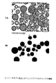
図1は、混合を停止し、乳剤を室温で一週間保管した後の高分子水性−水性乳剤の写真を示している。ミオグロビンが、分散相に充填するモデル蛋白質として使用されており、錆色を示している。六つの試料のうち、試料4は、アルギン酸ナトリウムなしで調製した。試料6は、塩化ナトリウムを(100mMに達するまで)追加したことを除き、試料1と同じとした。これら二試料では、撹拌を停止した直後に、分散相の融合が発生し、数時間のうちに二つの遮断相の形成につながった。図1の他の四試料では、小滴の直径は、一週間に渡って3乃至7μmの範囲を維持した(図2A)。この結果は、荷電ポリマ分子が小滴の表面に吸着され、拡散二重層を形成するという我々の仮説を支持した。ナトリウムイオンの濃度を高めると、アルギン酸塩の対イオンでは、表面電荷が奪われ、ゼータ電位の大きさが低下し、したがって、小滴の合体が生じる。デキストラン/PEG比を1:15に減らすことで、二週間に渡って安定状態の乳剤が発生した。
FIG. 1 shows a photograph of a polymeric aqueous-aqueous emulsion after mixing is stopped and the emulsion is stored at room temperature for a week. Myoglobin is used as a model protein that fills the dispersed phase and shows a rust color. Of the six samples,
この実験において、連続相と分散相との間でのミオグロビンの分配係数は、410nmでの吸光度によって決定されるものとして、1:50となり、ミオグロビンの大部分がデキストラン相にあることを示した。ミオグロビンに加えて、組み換えヒト顆粒求マクロファージコロニー刺激因子(rhGM−CSF)及びアンフォテリシンB(AmB)を運ぶリポソームも、この系に充填し、図2Bのものと同様のガラス質粒子を形成した。rhGM−CSFの約93%及びAmB/リポソームの95%が、分散相に分配されたことが、それぞれ、活性測定(下の説明を参照)及び408nmでのUV吸光度によって検出された。 In this experiment, the myoglobin partition coefficient between the continuous phase and the dispersed phase was 1:50, as determined by absorbance at 410 nm, indicating that the majority of myoglobin is in the dextran phase. In addition to myoglobin, liposomes carrying recombinant human granulophilic macrophage colony stimulating factor (rhGM-CSF) and amphotericin B (AmB) were also loaded into this system to form glassy particles similar to those of FIG. 2B. It was detected by activity measurements (see below) and UV absorbance at 408 nm that approximately 93% of rhGM-CSF and 95% of AmB / liposomes were partitioned in the dispersed phase, respectively.
図2Aは、上で説明した塩化ナトリウムなしの溶液によって調製された高分子水性−水性乳剤の顕微鏡画像を図示している。直径3乃至8ミクロンの均一なサイズ分布の乳剤小滴が観察された。 FIG. 2A illustrates a microscopic image of a polymeric aqueous-aqueous emulsion prepared with the above-described solution without sodium chloride. Emulsion droplets with a uniform size distribution of 3-8 microns in diameter were observed.
実施例2.アクアスフェアの調製
アクアスフェアは、単純に上の安定乳剤を凍結乾燥することで調製した。凍結乾燥後、デキストラン小滴は、固体粒子に転換された。しかしながら、殆どのデキストラン粒子は、連続相のPEGによって形成された固体マトリクス内に分散した。PEGは、凍結乾燥粉末を塩化メチレン又はアセトニトリルによって洗浄することで除去できる。こうした溶媒は、乾燥デキストラン相を溶解又は膨張させない。図2A及び2Bは、異なる調製段階での分散相の顕微鏡画像を図示しており、それぞれ、乳化後、凍結乾燥し(PEG除去のため)ジクロロメタンにより洗浄した後、及びPLGAコーティングからの回収後である。凍結乾燥後、分散相の直径は、依然として均一だったが、3乃至7μmから1乃至3μmへ低下し、水分の喪失による妥当なサイズとなった。これらの画像は、凍結乾燥中に小滴の融合が発生していないことを示した。この乾燥粒子のサイズ範囲(1乃至3μm)は、治療物の吸入送給に理想的であり、二重カプセル化(S−O−W)(5、13)による分解性高分子コーティングミクロスフェアの調製にも適している。
Example 2 Aquasphere preparation Aquaspheres were prepared by simply lyophilizing the above stable emulsion. After lyophilization, the dextran droplets were converted to solid particles. However, most dextran particles were dispersed within a solid matrix formed by continuous phase PEG. PEG can be removed by washing the lyophilized powder with methylene chloride or acetonitrile. Such solvents do not dissolve or swell the dried dextran phase. 2A and 2B illustrate microscopic images of the dispersed phase at different stages of preparation, after emulsification, lyophilized (to remove PEG), washed with dichloromethane, and after recovery from the PLGA coating, respectively. is there. After lyophilization, the diameter of the dispersed phase was still uniform, but decreased from 3-7 μm to 1-3 μm and became a reasonable size due to loss of moisture. These images indicated that no droplet fusion occurred during lyophilization. This dry particle size range (1-3 μm) is ideal for inhalation delivery of therapeutics, and the degradable polymer-coated microspheres with double encapsulation (SOW) (5, 13). Also suitable for preparation.
実施例3.アクアスフェアのPLGAマイクロスフェアへのマイクロカプセル化
アクアスフェアは、「solid−in−oil−in−water」乳化プロセスにより、PLGA及びその他の生分解性高分子のマトリクスにおいて更にマイクロカプセル化できる。本研究では、乳酸:グリコール酸の比が50:50及び75:25のPLGAを使用した。実施例2のように調製したアクアスフェアを、最初に、アクアスフェア/PLGAの比を1:2乃至1:20でPLGA/ジクロロメタン溶液(10乃至20%)中で懸濁し、次に、0.1乃至10%の塩化ナトリウム及び0.1乃至4%のポリビニルアルコール(PVA)又はPEG又はポリビニルピロリドンを含む水溶液に撹拌しながら追加した。二つの溶液の体積比は、1:2乃至1:10とした。乳剤形成後、この系に大量の冷水(乳剤の10倍)を撹拌しながら流し込んで、有機溶媒を抽出した。図3A及び3Bは、それぞれ溶媒抽出前のPLGA小滴及び溶媒抽出後のPLGA粒子の顕微鏡画像を図示している。溶媒抽出前、PLGA小滴は、透明であり、内部にはアクアスフェアが均等に分布していた。溶媒の除去により固めた後、PLGA粒子は、透明性を失った。
Example 3 Microencapsulation of Aquaspheres into PLGA Microspheres Aquaspheres can be further microencapsulated in a matrix of PLGA and other biodegradable polymers by a “solid-in-oil-in-water” emulsification process. In this study, PLGA with lactic acid: glycolic acid ratios of 50:50 and 75:25 was used. The aquaspheres prepared as in Example 2 are first suspended in a PLGA / dichloromethane solution (10-20%) with an aquasphere / PLGA ratio of 1: 2 to 1:20 and then 0. It was added with stirring to an aqueous solution containing 1 to 10% sodium chloride and 0.1 to 4% polyvinyl alcohol (PVA) or PEG or polyvinylpyrrolidone. The volume ratio of the two solutions was 1: 2 to 1:10. After forming the emulsion, a large amount of cold water (10 times the emulsion) was poured into the system with stirring to extract the organic solvent. 3A and 3B illustrate microscopic images of PLGA droplets before solvent extraction and PLGA particles after solvent extraction, respectively. Prior to solvent extraction, the PLGA droplets were transparent and the aquasphere was evenly distributed inside. After solidifying by solvent removal, the PLGA particles lost transparency.
実施例4.PLGA粒子からのアクアスフェアの回収
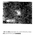
アクアスフェアは、実施例3のように調製したPLGAマイクロスフェアから回収できる。アクアスフェア充填PLGA粒子を、ジクロロメタン又はアセトニトリルに再溶解し、その後、遠心分離した。この手順を、四乃至六回、繰り返した。図4は、上記の手順によりPLGAマイクロスフェアから回収したアクアスフェアを図示している。アクアスフェアの粒子サイズ及び形状は、PLGAマイクロスフェアにカプセル化する前と同じものを保っている。この結果は、マイクロカプセル化プロセス中のアクアスフェアの水和が顕著ではないことを示唆している。
Example 4 Recovery of Aquaspheres from PLGA Particles Aquaspheres can be recovered from PLGA microspheres prepared as in Example 3. Aquasphere-filled PLGA particles were redissolved in dichloromethane or acetonitrile and then centrifuged. This procedure was repeated four to six times. FIG. 4 illustrates aquaspheres recovered from PLGA microspheres by the above procedure. The aquasphere particle size and shape remain the same as before encapsulating in PLGA microspheres. This result suggests that the hydration of the aquasphere during the microencapsulation process is not significant.
PLGAマイクロスフェアによるアクアスフェアのカプセル化効率を調べるために重量測定を実行した。PLGAに対するデキストランの重量比は、マイクロカプセル化の前(1:19)と後(1.06:19)で、比較的一定のものが得られ、高いカプセル化効率を示唆した。この結論は、我々のカプセル化前後での蛋白質活性測定の結果と一致する。 Weighing was performed to investigate the encapsulation efficiency of aquaspheres by PLGA microspheres. The weight ratio of dextran to PLGA was relatively constant before (1:19) and after (1.06: 19) before microencapsulation, suggesting high encapsulation efficiency. This conclusion is consistent with our protein activity measurements before and after encapsulation.
実施例5.アクアスフェアによる有機溶媒に対するβ−ガラクトシダーゼの保護
有機溶媒からの脆弱な蛋白質の保護におけるアクアスフェアの有効性を調べるため、四量体構造で分子量434KDの酵素であるβ−ガラクトシダーゼを、アクアスフェアに充填した。蛋白質を、10乃至100単位/mlの比でデキストラン溶液(MW=10乃至500KD、濃度5乃至25%)に溶解し、実施例1のようにPEG溶液で乳化した。凍結乾燥後、ジクロロメタン(PLGAマイクロスフェアの調製において使用される一般的な溶液)により実施例4のように数回洗浄することで、PEG相を除去した。その後、得られた蛋白質充填アクアスフェアを、緩衝液に再溶解し、o−ニトロフェニル−β−D−ガラクトピラノシド(ONPG)の加水分解により測定した。図5に示すように、酵素の触媒活性は、(乳化、凍結乾燥、ジクロロメタンによる洗浄を含む)実施例1から実施例2の手順後に、10%未満しか減少しなかった。この結果は、三回の実施で再現できた。この10%の活性低下は、乳化プロセス及び洗浄プロセスによるデキストラン相及びPEG相の間での分配による蛋白質の損失と、凍結乾燥及び洗浄プロセスにおいて変性した蛋白質、及び洗浄プロセス中に失われた蛋白質とを含む。この結果は、アクアスフェア内部の脆弱な蛋白質を、マイクロカプセルプロセス中に有機溶媒から十分に保護できることを示唆している。
Embodiment 5 FIG. Protection of β-galactosidase against organic solvents by aquaspheres In order to examine the effectiveness of aquaspheres in protecting vulnerable proteins from organic solvents, β-galactosidase, an enzyme with a tetrameric structure and a molecular weight of 434 KD, is packed into aquaspheres. did. The protein was dissolved in a dextran solution (MW = 10 to 500 KD, concentration 5 to 25%) at a ratio of 10 to 100 units / ml, and emulsified with a PEG solution as in Example 1. After lyophilization, the PEG phase was removed by washing several times as in Example 4 with dichloromethane (a common solution used in the preparation of PLGA microspheres). Subsequently, the obtained protein-filled aquasphere was redissolved in a buffer solution and measured by hydrolysis of o-nitrophenyl-β-D-galactopyranoside (ONPG). As shown in FIG. 5, the catalytic activity of the enzyme decreased by less than 10% after the procedure of Examples 1 to 2 (including emulsification, lyophilization, washing with dichloromethane). This result was reproducible in three runs. This 10% decrease in activity is due to the loss of protein due to partitioning between the dextran and PEG phases by the emulsification and washing processes, the protein that has been denatured in the lyophilization and washing processes, and the proteins that are lost during the washing process. including. This result suggests that fragile proteins within the aquasphere can be well protected from organic solvents during the microcapsule process.
実施例6.水性−水性乳剤の分散相及び連続相におけるrhEPO及びrhGM−CSFの分配
乳剤系の分散相に充填された蛋白質の効率を判定するために分配実験を行った。組み換えヒトエリスロポエチン(rhEPO)又は組み換えヒト顆粒求マクロファージコロニー刺激因子(rhGM−CSF)を含有する水性−水性乳剤を遠心分離し、その後、TF1細胞株を使用して細胞増殖測定を行った。蛋白質活性は、顕微鏡下でウェル当たりの細胞数を計数して測定した。分配実験により、rhEPOの94%及びrhGM−CSFの93%がデキストラン相において発見された。
Example 6 Distribution of rhEPO and rhGM-CSF in Disperse Phase and Continuous Phase of Aqueous-Aqueous Emulsion Distribution experiments were performed to determine the efficiency of the protein loaded in the dispersed phase of the emulsion system. Aqueous-aqueous emulsions containing recombinant human erythropoietin (rhEPO) or recombinant human granulophilic macrophage colony stimulating factor (rhGM-CSF) were centrifuged and then cell proliferation measurements were performed using the TF1 cell line. Protein activity was measured by counting the number of cells per well under a microscope. Partitioning experiments found 94% of rhEPO and 93% of rhGM-CSF in the dextran phase.
実施例7.アクアスフェアによる有機溶媒に対するrhEPO及びrhGM−CSFの保護
アクアスフェアによる蛋白質保護を、二種類の蛋白質rhEPO及びrhGM−CSFにより更に調べた。蛋白質をアクアスフェアに充填し、実施例5と同一の手順に従って処理した。蛋白質の生物活性は、分配に関するもの(実施例6)と同じ細胞増殖方法により測定した。乳化前の蛋白質と(ジクロロメタンによる洗浄後)アクアスフェアから回収した蛋白質とを、それぞれ同じ細胞懸濁液に追加した。rhEPOの結果は、図6に図示している。凍結乾燥後、rhEPOでの活性保持は、ウェル当たりの細胞数の27800から23700への低下によって示されるように、85%だった。凍結乾燥粉末の洗浄(したがって、PEG相が除去される)により、細胞数は、更に22600まで低下し、活性保持が95%であることを示した。有機溶媒による洗浄後、蛋白質の94%のみがデキストラン層に留まることから(実施例6)、有機溶媒との接触後の活性保持は、100%となった。
Example 7 Protection of rhEPO and rhGM-CSF against organic solvents by aquaspheres Protein protection by aquaspheres was further investigated by two proteins rhEPO and rhGM-CSF. Proteins were loaded into aquaspheres and processed according to the same procedure as in Example 5. The biological activity of the protein was measured by the same cell growth method as that for partitioning (Example 6). The protein before emulsification and the protein recovered from the aquasphere (after washing with dichloromethane) were each added to the same cell suspension. The rhEPO results are illustrated in FIG. After lyophilization, retention of activity with rhEPO was 85%, as indicated by a decrease in the number of cells per well from 27800 to 23700. Washing the lyophilized powder (thus removing the PEG phase) further reduced the cell number to 22600, indicating a 95% retention of activity. Since only 94% of the protein remained in the dextran layer after washing with the organic solvent (Example 6), the activity retention after contact with the organic solvent was 100%.
図7は、画策性ステップ後のrhGM−CSFの活性測定の結果を図示している。蛋白質充填乳剤の乾燥粉末への凍結乾燥により、ウェル当たりの平均細胞数は、130900から122600へ僅かに減少し、約94%の活性保持を示した。残存PEGを除去するためのジクロロメタンによる凍結乾燥粉末の洗浄後、細胞数は、ウェル当たり111100に減少し、更に9%が低減された。しかしながら、この9%の低減の多くは、PEGと一緒に洗い落とされた連続相におけるrhGM−CSFの分配によって生じたものだった(全rhGM−CSFの約7%、実施例6)。蛋白質充填デキストラン粒子のPLGAマイクロスフェアへのカプセル化では、ウェル当たり118900の平均細胞数が示すように、更なる活性の減少は生じなかった。高い活性保持は、更に、重量測定(実施例4)二よって示唆された高いカプセル化効率を示した。 FIG. 7 illustrates the results of rhGM-CSF activity measurement after the strategy step. By freeze-drying the protein-filled emulsion to a dry powder, the average number of cells per well decreased slightly from 130900 to 122600, indicating about 94% activity retention. After washing the lyophilized powder with dichloromethane to remove residual PEG, the cell number was reduced to 111100 per well, a further 9% reduction. However, much of this 9% reduction was caused by the distribution of rhGM-CSF in the continuous phase washed away with PEG (about 7% of total rhGM-CSF, Example 6). Encapsulation of protein loaded dextran particles into PLGA microspheres did not result in further activity reduction, as shown by an average cell number of 118900 per well. The high activity retention further showed the high encapsulation efficiency suggested by gravimetry (Example 4).
実施例8.生理的温度の水和状態におけるアクアスフェアによるrhEPO、rhGM−CSF、及びβ−ガラクトシダーゼの活性保持
蛋白質薬物の徐放性投薬形態を開発する上で最も困難な課題は、生理的温度の水和状態において蛋白質の活性を確保することであると広く考えられてきた(18)。徐放中、分解性高分子マイクロスフェアは、水を吸収して膨張し、カプセル化した蛋白質分子は、体温での水和条件に晒される。水和及び温度上昇は、蛋白質分子の可動性を高め、蛋白質の化学的又は物理的変化の機会を増加させる(19)。生理的条件下での蛋白質の安定性を調べるために、rhEPO又はrhGM−CSFを充填したデキストラン粒子に水を追加し(粘性30w/w%デキストラン溶液を形成し)、37℃で培養した。TF1細胞増殖における蛋白質活性を、培養時間の関数として、図8及び9に図示した。
Example 8 FIG. Retaining activity of rhEPO, rhGM-CSF, and β-galactosidase by aquaspheres at hydrated state at physiological temperature The most difficult task in developing a sustained release dosage form of a protein drug is the hydrated state at physiological temperature It has been widely considered to ensure the activity of proteins in (18). During sustained release, degradable polymer microspheres absorb water and swell, and encapsulated protein molecules are exposed to hydration conditions at body temperature. Hydration and temperature increase increase the mobility of protein molecules and increase the chance of chemical or physical changes in the protein (19). In order to examine the stability of the protein under physiological conditions, water was added to the dextran particles filled with rhEPO or rhGM-CSF (to form a viscous 30 w / w% dextran solution) and cultured at 37 ° C. Protein activity in TF1 cell growth is illustrated in FIGS. 8 and 9 as a function of culture time.
rhEPOでは、アクアスフェアによって保護されたものの活性は、一週間で約50%まで徐々に低下した(図8)。しかしながら、保護されていないrhEPOでは、同量の活性低下に、僅か一日しか要しなかった。rhEPOの半減期は、体内の酵素触媒作用により、in vivoで8.5時間である。明らかに、アクアスフェアの水和によって形成された粘性多糖相は、生理的条件において、大幅な期間に渡って蛋白質活性を延長できる。 With rhEPO, the activity of those protected by aquasphere gradually decreased to about 50% in one week (FIG. 8). However, unprotected rhEPO required only one day to reduce the same amount of activity. The half-life of rhEPO is 8.5 hours in vivo due to enzyme catalysis in the body. Clearly, the viscous polysaccharide phase formed by the hydration of aquaspheres can prolong protein activity over a significant period in physiological conditions.
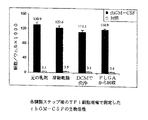
同様の結果は、rhGM−CSFでも得られた(図9)。保護されたrhGM−CSFでは、活性保持は、培養十日後で85%となった。保護されていないrhGM−CSFでは、同じ培養期間で56%だった。 Similar results were obtained with rhGM-CSF (FIG. 9). With protected rhGM-CSF, the retention of activity was 85% after 10 days of culture. With unprotected rhGM-CSF, it was 56% over the same culture period.
水和状態におけるβ−ガラクトシダーゼでの多糖安定剤の保護効果を、トレハロースのものと比較した。活性測定は、実施例5と同じように実施した。37℃での培養七日後、多糖類によって安定化した蛋白質の活性は、89%に下降したが、トレハロースによって安定化したものは、17%まで下降した。培養時間を二週間に延長すると、水和アクアスフェアでは活性が更に48%まで低下したが、トレハロース溶液での培養では0%となった。 The protective effect of the polysaccharide stabilizer with β-galactosidase in the hydrated state was compared with that of trehalose. The activity measurement was performed in the same manner as in Example 5. After 7 days of culturing at 37 ° C., the activity of the protein stabilized by the polysaccharide dropped to 89%, whereas that stabilized by trehalose fell to 17%. When the culture time was extended to 2 weeks, the activity was further reduced to 48% in the hydrated aquasphere, but 0% in the culture with the trehalose solution.
実施例9.PLGAマイクロスフェアからの最小限のバースト及び不完全放出を有する蛋白質放出プロフィール
バースト効果及び不完全放出は、蛋白質薬物の徐放投薬形態の開発における別の共通問題である。バースト効果により、充填された蛋白質の30乃至70%が、投与直後に放出され得る。不完全放出は、充填物の20乃至40%が不溶残分として残ることを指す。こうした望ましくない放出は、アクアスフェア内に蛋白質を予備充填することで防止できる。蛋白質を、最初に水性−水性乳化プロセスにより、アクアスフェア(0.1乃至20%)に充填した。次に、S−O−W手法を使用して、蛋白質充填アクアスフェアを、PLGAマイクロスフェア(1乃至20%)においてカプセル化した。PLGA内のミオグロビンの充填容量は、0.25乃至5%とした。PVA、PEG、及びPVPを、水相(0.1乃至5%)において、界面活性剤として溶解させた。図11は、アクアスフェアによる保護が存在する状態と存在しない状態とでの、(末端器をブロックした)PLGAマイクロスフェアでカプセル化したミオグロビンの放出プロフィールを図示している。純粋な蛋白質粒子として、ミオグロビンがエステル末端PLGAで調製されたマイクロスフェアにカプセル化された時、45日間で充填蛋白質の17%のみが放出された。アクアスフェアに予備充填した後でカプセル化されたミオグロビンでは、充填蛋白質の75%までが、開始時のバースト放出なしで、45日間に渡って直線的に放出された。こうしたバーストのない線形放出は、ミオグロビン−デキストラン粒子を、相対的に親水性の酸末端PLGAのマイクロスフェアにおいてカプセル化した時にも達成される(図12)。
Example 9 Protein release profile with minimal burst and incomplete release from PLGA microspheres Burst effect and incomplete release are another common problem in the development of sustained release dosage forms of protein drugs. Due to the burst effect, 30-70% of the loaded protein can be released immediately after administration. Incomplete release refers to 20-40% of the filling remaining as an insoluble residue. Such undesirable release can be prevented by pre-filling the aquasphere with protein. The protein was initially loaded into aquaspheres (0.1-20%) by an aqueous-aqueous emulsification process. Next, protein filled aquaspheres were encapsulated in PLGA microspheres (1-20%) using the S-O-W technique. The filling capacity of myoglobin in PLGA was 0.25 to 5%. PVA, PEG, and PVP were dissolved as surfactants in the aqueous phase (0.1-5%). FIG. 11 illustrates the release profile of myoglobin encapsulated with PLGA microspheres (with blocked end organs) with and without aquasphere protection. As pure protein particles, when myoglobin was encapsulated in microspheres prepared with ester-terminated PLGA, only 17% of the packed protein was released in 45 days. In myoglobin encapsulated after pre-filling the aquasphere, up to 75% of the filled protein was released linearly over 45 days without an initial burst release. Such burst-free linear release is also achieved when myoglobin-dextran particles are encapsulated in relatively hydrophilic acid-terminated PLGA microspheres (FIG. 12).
図12は、乳酸:グリコール酸の比がそれぞれ50:50、65:35、及び75:25である酸末端PLGA(分子量=12K)で調製されたマイクロスフェアからのミオグロビン放出プロフィールを図示している。これら全ての試料において、ミオグロビンは、PLGAマイクロスフェアでのカプセル化の前に、予備製剤してアクアスフェアとした。充填物の約7乃至12%が一日目に放出され、その後、線形動態で放出された。L/Gが50/50及び65/35で、MWが12KであるPLGAで調製されたマイクロスフェアでは、蛋白質放出が、50日間で90%を上回り、ほぼ完全となった。L/G比を65/35から75/25に増加させることで、放出の割合は僅かに低下し、同じ期間に充填物の80%が放出された。分子量(MW)を12Kから20Kに増加させることで、放出の割合は、更に下降した。L/G比が65/35のPLGAでは、カプセル化したミオグロビンの65%が、50日の間に放出された。いずれのケースにおいても、放出プロフィールは、ほぼ線形となった。この方法によるミオグロビンのPLGAマイクロスフェアへのカプセル化効率は、調製プロセス後の上澄みにおける蛋白質含有量の分析に基づいて、約90%となった。
FIG. 12 illustrates myoglobin release profiles from microspheres prepared with acid-terminated PLGA (molecular weight = 12K) with lactic acid: glycolic acid ratios of 50:50, 65:35, and 75:25, respectively. . In all these samples, myoglobin was pre-formulated into aquaspheres prior to encapsulation with PLGA microspheres. About 7-12% of the filling was released on the first day and then released with linear kinetics. In microspheres prepared with PLGA with L /
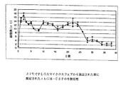
実施例10.PLGAマイクロスフェアから放出されたGM−CSFの生物活性
蛋白質rhGM−CSFを、実施例1、2、及び3において説明した方法に従って、アクアスフェアを介してPLGAマイクロスフェアに充填した。デキストランに対する蛋白質の比は、1:500とし、PLGAに対するアクアスフェアの比は、1:5とした。rhGM−CSF充填PLGAマイクロスフェアを、緩衝溶液に懸濁し、37℃で培養した。上澄みを毎日収集し、新鮮な緩衝液に置き換えた。収集した上澄みを、20倍に希釈し、実施例7のように測定した。測定された活性は、図13において、試料抽出の日付に対してプロットしている。活性は、培養後24日目まで、ほぼ一定となり、その後、32日目に陰性対照のレベルまで低下した。
Example 10 Bioactivity of GM-CSF released from PLGA microspheres Protein rhGM-CSF was loaded into PLGA microspheres via aquaspheres according to the methods described in Examples 1, 2, and 3. The ratio of protein to dextran was 1: 500, and the ratio of aquasphere to PLGA was 1: 5. rhGM-CSF loaded PLGA microspheres were suspended in buffer solution and cultured at 37 ° C. The supernatant was collected daily and replaced with fresh buffer. The collected supernatant was diluted 20 times and measured as in Example 7. The measured activity is plotted against the date of sample extraction in FIG. The activity was almost constant until day 24 after culture and then decreased to the level of the negative control on day 32.
高分子の分解によりPLGAマイクロスフェア内部で生成される局所酸性は、放出期間中の蛋白質変性の主な原因の一つとして広く認識されている(26)。rhGM−CSFの活性に対する酸性の影響を調べるために、この蛋白質を、活性測定の一日前に、デキストラン溶液中において、pH1、2、3、4、5、及び6で、それぞれ培養した。pH6で培養した試料と比較して、活性は、pH4で75%に減少し、pHga2を下回る時、45%に減少した。このpH依存性の活性低下は、PLGAマイクロスフェアから放出された蛋白質では観察されなかった(図13)。この結果は、PLGAマイクロスフェアのマトリクスにおいて、局所酸性が蓄積されなかったことを示唆している。おそらく、アクアスフェアは、巨大分子の作用物質に対する透過性は低いが、小分子の緩衝剤に対して透過性が非常に高い粘性チャネルを水和時に形成するため、PLGAの分解によって生成された酸性基が蛋白質放出期間中に緩衝されるようになる。
参考文献
1.R. Langer, Folkman, J., Nature 263, 793-800 (1976).
2.CAS, 「Chemical Abstructの検索の結果、分解性高分子に基づく蛋白質の徐放を主題とする962件の研究論文及び特許が存在した」(2002).
3.M. V. Weert, Hennink, W. E., Jiskoot, W., Pharm. Res. 17, 11591167 (2000).
4.R. T. Bartus, Tracy, M. A., Emerich, D. F., Zale, S. E., Science 281, 1161-1162 (1998).
5.P. A. Burke, 薬剤制御放出技術ハンドブック, Marcel Dekker, 661-692 (2000).
6.J. L. Cleland, Jones J. S., Pharm. Res. 13, 1465-1475 (1996).
7.O. L. Jonson, Pharmaceutical Research 14, 730-735 (1997).
8.B. C. Cunningham, Mulkerrin, M. G., Wells, J. A., Science 253, 545-548 (1991).
9.S. E. Zale, Burke, P. A., Berstein, H., Brickner, A., 米国特許第5,716,644号内(米国、1998).
10.A. sanchez, Villamayor, B., Guo, Y., Mclver, J., Alonso, M. j., Intern. J. Pharm. 185, 255-266 (1999).
11.S. P. Schwendeman, Tobio, M., Jaworowicz, M., Alonso, M. J., Langer, R., J. Microencapsulation 15, 299-318 (1998).
12.M. Morlock, Koll, H., Winter, G., Kissel, T., European Journal of pharmaceutics and biopharmaceutics 43, 29-36 (1997).
13.S. Yoshioka, Aso, Y., Kojima, S., Pharmaceutical Research 14, 736-741 (1997).
14.M. v. d. Weert2. Hof, R. V., Weerd, J. v. d., Heeren, M. A., Posthuma, G., Hennink, W. E., Crommelin D. J. A., J. Controlled Release 68, 31-40 (2000).
15.T. Morita, Horikiri. Y., Suzuki, T., Yoshino, H., International Journal of Pharmaceutics 219, 127-137 (2001).
16.Y. -F. Maa, Nguyen, P-A., Hsu, S. W., J. Pharm. Sci., 87, 152-159 (1997).
17.T. Morita, Horikiri. Y., Yamahara, H., Suzuki, T., Yoshino, H., Pharm. Res. 17, 1367-1373 (2000).
18.T. G. Park, Lee, H. Y., Nam Y. S., J. Controlled Release 55, 181-191 (1998).
19.O. Franssen, W. E. Hennink, Intern. J. Pharm., 168, 1-7 (1998).
20.F. Lamberti, WO96/40071内(1996).
21.S. P. Schwendeman, Cardamone, M., Brandon, M. R., Klibanov, A., Langer, R., 蛋白質の安定性と生分解性高分子マイクロスフェアからの送給、S. C. H. Bernstein編、蛋白質及びワクチンの送給のための微粒子系 (Mercel Dekker, New York, 1996), vol. 77.
22.W. R. Liu, Langer, R., Klibanov, A. M., Biotech Bioeng. 37, 177-184 (1991).
23.B. Bittner, Morlock, M., Koll, H., Winter, G., Kissel, T., Eur. J. Pharm. Bipharm. 45, 295-305 (1998).
24.T. Jin, L. Chen, H. Zhu, 米国特許出願第09/886,555号(2001).
25.H. Takahata, Lavelle, E. C., Coombes, A. G. A., Davis, S. S., J. Controlled Release 50, 237-246 (1998).
26.G. Zhu, S. R. Mallery, S. P. Schwendemen, Nature Biotech., 18, 52-57 (2000).
Local acidity generated inside PLGA microspheres due to polymer degradation is widely recognized as one of the main causes of protein denaturation during the release period (26). In order to examine the effect of acidity on the activity of rhGM-CSF, this protein was cultured in dextran solution at
2. CAS, “As a result of the search for Chemical Abstract, there were 962 research papers and patents on the subject of sustained release of proteins based on degradable polymers” (2002).
3. MV Weert, Hennink, WE, Jiskoot, W., Pharm. Res. 17, 11591167 (2000).
4). RT Bartus, Tracy, MA, Emerich, DF, Zale, SE, Science 281, 1161-1162 (1998).
5. PA Burke, Drug Controlled Release Technology Handbook, Marcel Dekker, 661-692 (2000).
6). JL Cleland, Jones JS, Pharm. Res. 13, 1465-1475 (1996).
7). OL Jonson, Pharmaceutical Research 14, 730-735 (1997).
8). BC Cunningham, Mulkerrin, MG, Wells, JA, Science 253, 545-548 (1991).
9. SE Zale, Burke, PA, Berstein, H., Brickner, A., in US Pat. No. 5,716,644 (US, 1998).
10. A. sanchez, Villamayor, B., Guo, Y., Mclver, J., Alonso, M. j., Intern. J. Pharm. 185, 255-266 (1999).
11. SP Schwendeman, Tobio, M., Jaworowicz, M., Alonso, MJ, Langer, R.,
12 M. Morlock, Koll, H., Winter, G., Kissel, T., European Journal of pharmaceutics and biopharmaceutics 43, 29-36 (1997).
13. S. Yoshioka, Aso, Y., Kojima, S., Pharmaceutical Research 14, 736-741 (1997).
14 M. vd Weert2.Hof, RV, Weerd, J. vd, Heeren, MA, Posthuma, G., Hennink, WE, Crommelin DJA, J. Controlled Release 68, 31-40 (2000).
15. T. Morita, Horikiri. Y., Suzuki, T., Yoshino, H., International Journal of Pharmaceutics 219, 127-137 (2001).
16. Y. -F.Maa, Nguyen, PA., Hsu, SW, J. Pharm. Sci., 87, 152-159 (1997).
17. T. Morita, Horikiri. Y., Yamahara, H., Suzuki, T., Yoshino, H., Pharm. Res. 17, 1367-1373 (2000).
18. TG Park, Lee, HY, Nam YS, J. Controlled Release 55, 181-191 (1998).
19. O. Franssen, WE Hennink, Intern. J. Pharm., 168, 1-7 (1998).
20. F. Lamberti, in WO 96/40071 (1996).
21. SP Schwendeman, Cardamone, M., Brandon, MR, Klibanov, A., Langer, R., Protein stability and delivery from biodegradable polymer microspheres, SCH Bernstein, Protein and vaccine delivery For fine particles (Mercel Dekker, New York, 1996), vol. 77.
22. WR Liu, Langer, R., Klibanov, AM, Biotech Bioeng. 37, 177-184 (1991).
23. B. Bittner, Morlock, M., Koll, H., Winter, G., Kissel, T., Eur. J. Pharm. Bipharm. 45, 295-305 (1998).
24. T. Jin, L. Chen, H. Zhu, US patent application Ser. No. 09 / 886,555 (2001).
25. H. Takahata, Lavelle, EC, Coombes, AGA, Davis, SS, J. Controlled
26. G. Zhu, SR Mallery, SP Schwendemen, Nature Biotech., 18, 52-57 (2000).
Claims (22)
a.作用物質を多糖分散相においてカプセル化することが可能な安定高分子水性−水性乳剤を提供するために、多糖類を水性−水性乳化の前記分散相として選択し、水性高分子を連続相として選択し、水性−水性乳化のための安定剤及びその濃度を選択するステップと、
b.少なくとも一つの作用物質を提供するステップと、
c.前記作用物質充填多糖粒子のサイズ及び形状を適切なサイズ範囲に制御するステップと、
d.前記乳剤を乾燥させるステップと、
e.前記乾燥分散相に浸透せず、前記充填済みの脆弱な作用物質(群)に影響を与えない溶媒(群)により前記試料を洗浄することで乾燥させた後、前記連続相を除去するステップと、を備える方法。 A method of encapsulating an agent in particles via aqueous-aqueous emulsification comprising:
a. In order to provide a stable polymer aqueous-aqueous emulsion capable of encapsulating the active substance in the polysaccharide dispersed phase, the polysaccharide is selected as the dispersed phase of the aqueous-aqueous emulsion and the aqueous polymer is selected as the continuous phase. Selecting a stabilizer for aqueous-aqueous emulsification and its concentration;
b. Providing at least one agent;
c. Controlling the size and shape of the agent-filled polysaccharide particles to an appropriate size range;
d. Drying the emulsion;
e. Removing the continuous phase after drying the sample by washing with a solvent (s) that does not penetrate the dry dispersed phase and does not affect the filled fragile agent (s); A method comprising:
a)前記乾燥多糖分散相を固相として、solid−in−oil−in−water(S−O−W)又はsolid−in−oil−in−oil(S−O−O)乳化プロセスを利用するステップと、
b)生分解性高分子を選択し、前記高分子を有機溶媒に溶解し、前記乾燥多糖分散相を前記高分子溶液に懸濁させるステップと、
c)小分子の塩の水溶液において前記生分解性高分子の前記溶液を分散させるために高分子界面活性剤(群)を選択するステップと、
d)前記塩溶液の濃度が0.5%乃至50%の範囲であるステップと、
e)抽出又は蒸発により前記有機溶媒を除去するステップと、を備える方法。 A method of encapsulating a dried polysaccharide dispersed phase in biodegradable polymer microspheres for controlled release of bioactive agent (s), comprising:
a) Solid-in-oil-in-water (SO-O-W) or solid-in-oil-in-oil (SO-O) emulsification process using the dried polysaccharide dispersed phase as a solid phase Steps,
b) selecting a biodegradable polymer, dissolving the polymer in an organic solvent, and suspending the dried polysaccharide dispersion phase in the polymer solution;
c) selecting a polymeric surfactant (s) to disperse the solution of the biodegradable polymer in an aqueous solution of a small molecule salt;
d) a step in which the concentration of the salt solution is in the range of 0.5% to 50%;
e) removing the organic solvent by extraction or evaporation.
Applications Claiming Priority (4)
| Application Number | Priority Date | Filing Date | Title |
|---|---|---|---|
| US38497102P | 2002-06-03 | 2002-06-03 | |
| US41810002P | 2002-10-11 | 2002-10-11 | |
| US10/291,327 US6998393B2 (en) | 2000-06-23 | 2002-11-08 | Aquespheres, their preparation and uses thereof |
| PCT/CN2003/000431 WO2003101600A2 (en) | 2002-06-03 | 2003-06-03 | Hazard-free microencapsulation for structurally delicate agents, an application of stable aqueous-aqueous emulsion |
Publications (2)
| Publication Number | Publication Date |
|---|---|
| JP2006504636A true JP2006504636A (en) | 2006-02-09 |
| JP2006504636A5 JP2006504636A5 (en) | 2006-07-20 |
Family
ID=27404043
Family Applications (1)
| Application Number | Title | Priority Date | Filing Date |
|---|---|---|---|
| JP2004508943A Pending JP2006504636A (en) | 2002-06-03 | 2003-06-03 | Unobstructed microencapsulation for structurally fragile drugs and application of stable aqueous-aqueous emulsions |
Country Status (7)
| Country | Link |
|---|---|
| US (2) | US6998393B2 (en) |
| EP (1) | EP1572341A4 (en) |
| JP (1) | JP2006504636A (en) |
| CN (1) | CN102247327A (en) |
| AU (1) | AU2003245804A1 (en) |
| CA (1) | CA2487867A1 (en) |
| WO (1) | WO2003101600A2 (en) |
Cited By (1)
| Publication number | Priority date | Publication date | Assignee | Title |
|---|---|---|---|---|
| WO2011004552A1 (en) * | 2009-07-09 | 2011-01-13 | 国立大学法人九州大学 | Water-soluble drug carrier and process for production thereof |
Families Citing this family (35)
| Publication number | Priority date | Publication date | Assignee | Title |
|---|---|---|---|---|
| US6998393B2 (en) * | 2000-06-23 | 2006-02-14 | Biopharm Solutions, Inc. | Aquespheres, their preparation and uses thereof |
| US9186322B2 (en) * | 2002-08-02 | 2015-11-17 | Insmed Incorporated | Platinum aggregates and process for producing the same |
| US8728525B2 (en) * | 2004-05-12 | 2014-05-20 | Baxter International Inc. | Protein microspheres retaining pharmacokinetic and pharmacodynamic properties |
| BRPI0404983A (en) * | 2004-09-03 | 2006-05-02 | Nanocore Biotecnologia Ltda | immunogenic compositions |
| CA2604225A1 (en) * | 2005-04-27 | 2006-11-02 | Baxter International Inc. | Surface-modified microparticles and methods of forming and using the same |
| US20070015689A1 (en) * | 2005-06-23 | 2007-01-18 | Alza Corporation | Complexation of metal ions with polypeptides |
| WO2007025441A1 (en) * | 2005-08-29 | 2007-03-08 | Tuo Jin | Polysaccharide microparticles containing biological agents: there preparation and applications |
| US9107824B2 (en) | 2005-11-08 | 2015-08-18 | Insmed Incorporated | Methods of treating cancer with high potency lipid-based platinum compound formulations administered intraperitoneally |
| MX2009003661A (en) * | 2006-10-06 | 2009-04-22 | Baxter Int | Microencapsules containing surface-modified microparticles and methods of forming and using the same. |
| US9475974B2 (en) * | 2007-07-17 | 2016-10-25 | Schlumberger Technology Corporation | Controlling the stability of water in water emulsions |
| US8044000B2 (en) | 2007-07-17 | 2011-10-25 | Schlumberger Technology Corporation | Polymer delivery in well treatment applications |
| US8043999B2 (en) * | 2007-07-17 | 2011-10-25 | Schlumberger Technology Corporation | Stabilizing biphasic concentrates through the addition of small amounts of high molecular weight polyelectrolytes |
| US7703527B2 (en) * | 2007-11-26 | 2010-04-27 | Schlumberger Technology Corporation | Aqueous two-phase emulsion gel systems for zone isolation |
| US7703521B2 (en) * | 2008-02-19 | 2010-04-27 | Schlumberger Technology Corporation | Polymeric microspheres as degradable fluid loss additives in oilfield applications |
| EP2169042B1 (en) | 2008-09-30 | 2012-04-18 | The Procter & Gamble Company | Composition comprising microcapsules |
| US7950459B2 (en) * | 2009-01-15 | 2011-05-31 | Schlumberger Technology Corporation | Using a biphasic solution as a recyclable coiled tubing cleanout fluid |
| US20100179076A1 (en) * | 2009-01-15 | 2010-07-15 | Sullivan Philip F | Filled Systems From Biphasic Fluids |
| US20100184630A1 (en) * | 2009-01-16 | 2010-07-22 | Sullivan Philip F | Breaking the rheology of a wellbore fluid by creating phase separation |
| US20100184631A1 (en) * | 2009-01-16 | 2010-07-22 | Schlumberger Technology Corporation | Provision of viscous compositions below ground |
| WO2010102065A1 (en) | 2009-03-05 | 2010-09-10 | Bend Research, Inc. | Pharmaceutical compositions of dextran polymer derivatives |
| WO2010111132A2 (en) | 2009-03-27 | 2010-09-30 | Bend Research, Inc. | Spray-drying process |
| PT2611529T (en) | 2010-09-03 | 2019-05-09 | Bend Res Inc | Spray-drying method |
| US8815294B2 (en) | 2010-09-03 | 2014-08-26 | Bend Research, Inc. | Pharmaceutical compositions of dextran polymer derivatives and a carrier material |
| US9084944B2 (en) | 2010-09-03 | 2015-07-21 | Bend Research, Inc. | Spray-drying apparatus and methods of using the same |
| EP2618924A1 (en) | 2010-09-24 | 2013-07-31 | Bend Research, Inc. | High-temperature spray drying process and apparatus |
| US9084727B2 (en) | 2011-05-10 | 2015-07-21 | Bend Research, Inc. | Methods and compositions for maintaining active agents in intra-articular spaces |
| DK2892524T3 (en) | 2012-09-04 | 2021-01-25 | Eleison Pharmaceuticals LLC | PREVENTION OF PULMONAL CANCER RECYCLING WITH LIPID-COMPLEXED CISPLATIN |
| US20160303242A1 (en) | 2013-12-09 | 2016-10-20 | Durect Corporation | Pharmaceutically Active Agent Complexes, Polymer Complexes, and Compositions and Methods Involving the Same |
| WO2016046845A1 (en) * | 2014-09-25 | 2016-03-31 | Manu Chaudhary | Stealth, targeted nanoparticles (stn) for oral drug delivery |
| CA2962719A1 (en) | 2014-10-31 | 2016-05-06 | Bend Research Inc. | Process for forming active domains dispersed in a matrix |
| CN105509836B (en) * | 2015-12-11 | 2019-01-18 | 大连民族大学 | A kind of rapid assay methods of liposomal encapsulated volume |
| CN105571673B (en) * | 2015-12-11 | 2019-01-18 | 大连民族大学 | A kind of measuring method of liposomal encapsulated volume |
| US10039801B2 (en) | 2016-02-16 | 2018-08-07 | Strongbridge Ireland Limited | Pharmaceutical compositions of water soluble peptides with poor solubility in isotonic conditions and methods for their use |
| CA3031097A1 (en) * | 2016-07-18 | 2018-01-25 | Tissuegen, Inc. | Methods and compositions for maintaining the conformation and structural integrity of biomolecules |
| CA3140681A1 (en) * | 2019-06-07 | 2020-12-10 | President And Fellows Of Harvard College | Compositions and methods relating to erythrocytes with adhered particles |
Citations (1)
| Publication number | Priority date | Publication date | Assignee | Title |
|---|---|---|---|---|
| US20020055461A1 (en) * | 2000-06-23 | 2002-05-09 | Tuo Jin | Stable polymer aqueous/aqueous emulsion system and uses thereof |
Family Cites Families (7)
| Publication number | Priority date | Publication date | Assignee | Title |
|---|---|---|---|---|
| JP2653255B2 (en) * | 1990-02-13 | 1997-09-17 | 武田薬品工業株式会社 | Long-term sustained release microcapsules |
| US5716644A (en) | 1992-06-11 | 1998-02-10 | Alkermes, Inc. | Composition for sustained release of non-aggregated erythropoietin |
| US5827707A (en) | 1995-06-07 | 1998-10-27 | Neocrin Company | Method for manufacturing minimal volume capsules containing biological materials |
| US6395302B1 (en) * | 1996-11-19 | 2002-05-28 | Octoplus B.V. | Method for the preparation of microspheres which contain colloidal systems |
| BR0004152A (en) * | 1999-01-18 | 2000-11-21 | Lg Chemichal Ltd | Lipophilic microparticles containing a protein or antigen drug and prepared comprising the same |
| US6998393B2 (en) * | 2000-06-23 | 2006-02-14 | Biopharm Solutions, Inc. | Aquespheres, their preparation and uses thereof |
| SE0201599D0 (en) * | 2002-03-21 | 2002-05-30 | Skyepharma Ab | microparticles |
-
2002
- 2002-11-08 US US10/291,327 patent/US6998393B2/en not_active Expired - Fee Related
-
2003
- 2003-06-03 WO PCT/CN2003/000431 patent/WO2003101600A2/en active Application Filing
- 2003-06-03 CN CN2011101074278A patent/CN102247327A/en active Pending
- 2003-06-03 CA CA002487867A patent/CA2487867A1/en not_active Abandoned
- 2003-06-03 EP EP03737838A patent/EP1572341A4/en not_active Withdrawn
- 2003-06-03 JP JP2004508943A patent/JP2006504636A/en active Pending
- 2003-06-03 US US10/517,122 patent/US20060121121A1/en not_active Abandoned
- 2003-06-03 AU AU2003245804A patent/AU2003245804A1/en not_active Abandoned
Patent Citations (1)
| Publication number | Priority date | Publication date | Assignee | Title |
|---|---|---|---|---|
| US20020055461A1 (en) * | 2000-06-23 | 2002-05-09 | Tuo Jin | Stable polymer aqueous/aqueous emulsion system and uses thereof |
Cited By (2)
| Publication number | Priority date | Publication date | Assignee | Title |
|---|---|---|---|---|
| WO2011004552A1 (en) * | 2009-07-09 | 2011-01-13 | 国立大学法人九州大学 | Water-soluble drug carrier and process for production thereof |
| US8568745B2 (en) | 2009-07-09 | 2013-10-29 | Kyushu University, National University Corporation | Water-soluble drug carrier and process for producing the same |
Also Published As
| Publication number | Publication date |
|---|---|
| EP1572341A2 (en) | 2005-09-14 |
| WO2003101600A2 (en) | 2003-12-11 |
| EP1572341A4 (en) | 2008-11-12 |
| US20060121121A1 (en) | 2006-06-08 |
| US6998393B2 (en) | 2006-02-14 |
| US20030059402A1 (en) | 2003-03-27 |
| CA2487867A1 (en) | 2003-12-11 |
| AU2003245804A8 (en) | 2003-12-19 |
| AU2003245804A1 (en) | 2003-12-19 |
| CN102247327A (en) | 2011-11-23 |
| WO2003101600A3 (en) | 2007-12-13 |
Similar Documents
| Publication | Publication Date | Title |
|---|---|---|
| JP2006504636A (en) | Unobstructed microencapsulation for structurally fragile drugs and application of stable aqueous-aqueous emulsions | |
| Molavi et al. | Polyester based polymeric nano and microparticles for pharmaceutical purposes: A review on formulation approaches | |
| ES2283394T3 (en) | IMPLEMENTATION KIT CONTAINING A SUPPORT PHASE AND A SOLVENT. | |
| ES2292634T3 (en) | Induced phase transition procedure for the production of microparticles containing active hydrophilic agents. | |
| US6692770B2 (en) | Starch microparticles | |
| ES2204837T3 (en) | METHOD FOR THE PREPARATION OF MICROSPHERES CONTAINING COLLOIDAL SYSTEMS. | |
| US8932633B2 (en) | Polysaccharide microparticles containing biological agents: their preparation and applications | |
| KR101354521B1 (en) | Controlled release compositions for interferon based on pegt/pbt block copolymers | |
| IL163218A (en) | Polymer-based compositions for sustained release of fsh | |
| US7087246B2 (en) | Controlled release preparation of insulin and its method | |
| US7105181B2 (en) | Microparticles | |
| JP5297994B2 (en) | Biocompatible alginate microparticle composition for controlled release of active ingredients by intravenous administration | |
| Yuan et al. | Preparation of dextran glassy particles through freezing-induced phase separation | |
| Yeo et al. | Characterization of reservoir-type microcapsules made by the solvent exchange method | |
| TW201714610A (en) | Soluble microneedle for protein or peptide | |
| JP2004513914A (en) | Parenterally administrable microparticles | |
| KR100342746B1 (en) | Controlled release preparation of insulin and its method | |
| Li et al. | Microparticles | |
| Yuan et al. | Microencapsulation of protein‐loaded polysaccharide particles within poly (D, L‐lactic‐co‐glycolic acid) microspheres using S/O/W: characterization and release studies | |
| TAMANNA | 5. ADVANCES IN LONG ACTING DRUG PREPARATIONS IN VETERINARY PRACTICE by SOLANKI TAMANNA, H1., GONDALIYA, VAISHALI1, TANVI D. MANAT2, PATEL, SA 3 AND PANDYA, SHAILEE4 | |
| Jin et al. | Granulation Approaches in Biotech Industry | |
| WO2002039986A1 (en) | Process for producing microparticles |
Legal Events
| Date | Code | Title | Description |
|---|---|---|---|
| A521 | Request for written amendment filed |
Free format text: JAPANESE INTERMEDIATE CODE: A523 Effective date: 20060530 |
|
| A621 | Written request for application examination |
Free format text: JAPANESE INTERMEDIATE CODE: A621 Effective date: 20060530 |
|
| A131 | Notification of reasons for refusal |
Free format text: JAPANESE INTERMEDIATE CODE: A131 Effective date: 20100105 |
|
| A601 | Written request for extension of time |
Free format text: JAPANESE INTERMEDIATE CODE: A601 Effective date: 20100402 |
|
| A601 | Written request for extension of time |
Free format text: JAPANESE INTERMEDIATE CODE: A601 Effective date: 20100427 |
|
| A602 | Written permission of extension of time |
Free format text: JAPANESE INTERMEDIATE CODE: A602 Effective date: 20100427 |
|
| A602 | Written permission of extension of time |
Free format text: JAPANESE INTERMEDIATE CODE: A602 Effective date: 20100526 |
|
| A521 | Request for written amendment filed |
Free format text: JAPANESE INTERMEDIATE CODE: A523 Effective date: 20100601 |
|
| A02 | Decision of refusal |
Free format text: JAPANESE INTERMEDIATE CODE: A02 Effective date: 20110531 |
|
| A521 | Request for written amendment filed |
Free format text: JAPANESE INTERMEDIATE CODE: A523 Effective date: 20110930 |