JP2005537886A - Cardiac pacing method and apparatus for pacing after defibrillation - Google Patents
Cardiac pacing method and apparatus for pacing after defibrillation Download PDFInfo
- Publication number
- JP2005537886A JP2005537886A JP2004535983A JP2004535983A JP2005537886A JP 2005537886 A JP2005537886 A JP 2005537886A JP 2004535983 A JP2004535983 A JP 2004535983A JP 2004535983 A JP2004535983 A JP 2004535983A JP 2005537886 A JP2005537886 A JP 2005537886A
- Authority
- JP
- Japan
- Prior art keywords
- pacing
- cardiac
- heart
- patient
- applying
- Prior art date
- Legal status (The legal status is an assumption and is not a legal conclusion. Google has not performed a legal analysis and makes no representation as to the accuracy of the status listed.)
- Pending
Links
Images


Classifications
-
- A—HUMAN NECESSITIES
- A61—MEDICAL OR VETERINARY SCIENCE; HYGIENE
- A61N—ELECTROTHERAPY; MAGNETOTHERAPY; RADIATION THERAPY; ULTRASOUND THERAPY
- A61N1/00—Electrotherapy; Circuits therefor
- A61N1/18—Applying electric currents by contact electrodes
- A61N1/32—Applying electric currents by contact electrodes alternating or intermittent currents
- A61N1/36—Applying electric currents by contact electrodes alternating or intermittent currents for stimulation
- A61N1/362—Heart stimulators
- A61N1/3621—Heart stimulators for treating or preventing abnormally high heart rate
- A61N1/3622—Heart stimulators for treating or preventing abnormally high heart rate comprising two or more electrodes co-operating with different heart regions
-
- A—HUMAN NECESSITIES
- A61—MEDICAL OR VETERINARY SCIENCE; HYGIENE
- A61N—ELECTROTHERAPY; MAGNETOTHERAPY; RADIATION THERAPY; ULTRASOUND THERAPY
- A61N1/00—Electrotherapy; Circuits therefor
- A61N1/18—Applying electric currents by contact electrodes
- A61N1/32—Applying electric currents by contact electrodes alternating or intermittent currents
- A61N1/36—Applying electric currents by contact electrodes alternating or intermittent currents for stimulation
- A61N1/362—Heart stimulators
- A61N1/365—Heart stimulators controlled by a physiological parameter, e.g. heart potential
- A61N1/368—Heart stimulators controlled by a physiological parameter, e.g. heart potential comprising more than one electrode co-operating with different heart regions
- A61N1/3688—Heart stimulators controlled by a physiological parameter, e.g. heart potential comprising more than one electrode co-operating with different heart regions configured for switching the pacing mode, e.g. from AAI to DDD
-
- A—HUMAN NECESSITIES
- A61—MEDICAL OR VETERINARY SCIENCE; HYGIENE
- A61N—ELECTROTHERAPY; MAGNETOTHERAPY; RADIATION THERAPY; ULTRASOUND THERAPY
- A61N1/00—Electrotherapy; Circuits therefor
- A61N1/18—Applying electric currents by contact electrodes
- A61N1/32—Applying electric currents by contact electrodes alternating or intermittent currents
- A61N1/38—Applying electric currents by contact electrodes alternating or intermittent currents for producing shock effects
- A61N1/39—Heart defibrillators
- A61N1/3956—Implantable devices for applying electric shocks to the heart, e.g. for cardioversion
- A61N1/3962—Implantable devices for applying electric shocks to the heart, e.g. for cardioversion in combination with another heart therapy
- A61N1/39622—Pacing therapy
Landscapes
- Health & Medical Sciences (AREA)
- Cardiology (AREA)
- Heart & Thoracic Surgery (AREA)
- Life Sciences & Earth Sciences (AREA)
- Animal Behavior & Ethology (AREA)
- Nuclear Medicine, Radiotherapy & Molecular Imaging (AREA)
- Radiology & Medical Imaging (AREA)
- Biomedical Technology (AREA)
- Engineering & Computer Science (AREA)
- General Health & Medical Sciences (AREA)
- Public Health (AREA)
- Veterinary Medicine (AREA)
- Biophysics (AREA)
- Physiology (AREA)
- Electrotherapy Devices (AREA)
- Measurement Of The Respiration, Hearing Ability, Form, And Blood Characteristics Of Living Organisms (AREA)
- Measuring Pulse, Heart Rate, Blood Pressure Or Blood Flow (AREA)
Abstract
心臓ぺーシングを実施するための方法、システムおよびコンピュータプログラム製品が提供される。除細動ショックが患者の心臓に印加され、除細動ショックの印加が停止した後にぺーシング刺激信号が患者の心臓に自動的に印加される。ぺーシング刺激は除細動ショックが停止してから約2秒以内に患者の心臓に印加されるようにする。ぺーシング刺激信号は心臓で検出される電気活動の特性に関係なく除細動ショックが停止した後に患者の心臓に印加される場合がある。このぺーシング刺激は単一のぺーシング刺激および/または対のぺーシング刺激を含む場合がある。Methods, systems and computer program products are provided for performing cardiac pacing. A defibrillation shock is applied to the patient's heart and a pacing stimulus signal is automatically applied to the patient's heart after the defibrillation shock is stopped. The pacing stimulus is applied to the patient's heart within about 2 seconds after the defibrillation shock stops. The pacing stimulus signal may be applied to the patient's heart after the defibrillation shock has stopped, regardless of the nature of the electrical activity detected in the heart. This pacing stimulus may include a single pacing stimulus and / or a pair of pacing stimuli.
Description
本発明は患者の心臓機能(cardiac function)を改善するための方法および装置に関する。 The present invention relates to a method and apparatus for improving a patient's cardiac function.
心臓は心膜として知られる繊維嚢で覆われた筋肉性の器官である。心膜と筋肉性の器官との間の空間は心膜腔と称される。心臓壁は骨格筋または平滑筋のいずれとも異なる筋肉(心筋)から実質的に形成される。心臓は心房と心室を備え、心房と心室はそれぞれ血液で満たされた腔(chanmber)をすっぽり包み込むように形成された心筋層から構成される。動作に関しては、腔壁が収縮すると、それらは全体に握りこぶしのようになる。この心筋の収縮は筋膜の脱分極によって引き起こされる。心臓として正しく働くためには、心筋収縮は協調して起こらねばならない。 The heart is a muscular organ covered with a fibrous sac known as the pericardium. The space between the pericardium and the muscular organ is called the pericardial space. The heart wall is substantially formed from a muscle (myocardium) different from either skeletal muscle or smooth muscle. The heart includes an atrium and a ventricle, and the atrium and the ventricle are each composed of a myocardium formed so as to completely wrap a chamber filled with blood. In terms of movement, when the cavity walls contract, they become a fist overall. This myocardial contraction is caused by the depolarization of the fascia. In order to work properly as a heart, myocardial contractions must occur in concert.
心筋収縮が心室内で協調して起こらない場合、血液は大動脈と肺動脈に流れ込む代わりに心室空洞内をあちこち動き回ることがある。このため、心室ポンプを形成する複合した心筋の塊は効率よくポンピングするためにほぼ同時に収縮しなければならない。 If myocardial contractions do not occur in concert within the ventricle, blood can move around in the ventricular cavity instead of flowing into the aorta and pulmonary artery. Thus, the complex myocardial mass that forms the ventricular pump must contract almost simultaneously to pump efficiently.
心臓は(a)隣接する心臓繊維間がタイトに接合され(繊維は介在板として知られる構造部分で端と端が接合され接合部または接点を形成する)、それにより活動電位が或る心筋細胞から別の心筋細胞に伝わることが可能となり、さらに(b)心臓のある決まったエリア内にある特殊筋繊維が心臓の適切な興奮を引き起こす刺激伝導系を形成するので、心臓はこうした協調動作を実現することが可能である。特殊筋繊維は心筋繊維と接触して細隙結合(gap junctions)を形成し、それにより活動電位が或る細胞から別の細胞に移動することが可能となる。この特殊化した伝導系は通常の動作において興奮の迅速且つ協調した広がりを実現するように構成されている。 The heart is (a) tightly joined between adjacent heart fibers (fibers are joined at ends in a structural part known as an intervening plate to form a joint or contact), whereby cardiomyocytes with an action potential Can be transmitted from one to another cardiomyocytes, and (b) the special muscle fibers in a certain area of the heart form a stimulation conduction system that triggers the proper excitation of the heart, so the heart It is possible to realize. Special muscle fibers come into contact with myocardial fibers to form gap junctions, which allow action potentials to move from one cell to another. This specialized conduction system is configured to achieve a rapid and coordinated spread of excitement in normal operation.
心筋細胞は自動律動的、つまり自発的な律動的自励が可能である。SA結節(sinoatrial node;洞結節)は心臓全体または平滑筋の通常のペースメーカであり、この領域から興奮波が発せられ、興奮波は心筋の残りの部分を同期的に移動または伝搬する。心臓のSA結節領域は、上大静脈入口近くの右心房壁内にある速い固有のリズムを刻んでSA結節が通常のペースメーカたり得ることを可能にする特殊な心筋細胞の小さな塊を含む。異常な状況では、心臓の他の領域がより興奮しやすく、より速く自発的なリズムを刻む可能性がある。この状況では、この他の領域がペースメーカとなって心臓全体のリズムを作り出すことがあり得る。 Cardiomyocytes are capable of automatic rhythm, that is, spontaneous rhythmic self-excitation. The SA node (sinoatrial node) is a normal pacemaker of the whole heart or smooth muscle, and excitatory waves are emitted from this region, and the excitatory waves move or propagate synchronously in the rest of the myocardium. The SA nodal region of the heart contains a small mass of specialized cardiomyocytes that allow the SA node to become a normal pacemaker by slicing the fast intrinsic rhythm in the right atrial wall near the superior vena cava entrance. In abnormal situations, other areas of the heart are more likely to excite and can swiftly spontaneously rhythm. In this situation, other areas can become pacemakers and create the rhythm of the entire heart.
正常な動作では、SA結節の細胞は周囲を取り巻く心房心筋繊維と接触する。このため、SA結節から興奮波が細隙結合を介して心房心筋細胞を伝って右心房全体に拡がる。さらに、心房組織はSA結節からのパルスを左心房に直接導き、左右両方の心房を同時に収縮させる。 In normal operation, cells in the SA node come into contact with the surrounding atrial myocardial fibers. For this reason, excitatory waves from the SA node propagate through the atrial cardiomyocytes via the slit junction and spread throughout the right atrium. In addition, the atrial tissue directs pulses from the SA node directly to the left atrium, causing both the left and right atria to contract simultaneously.
その後、興奮波は心室間の隔壁近くにある右心房基部に位置する特殊細胞の第2の小さな塊(AV(atrioventricular node;房室結節))によって心室に拡がる。AV結節は活動電位(波面)の伝搬を約0.1秒遅らせるよう設定されており、心房が収縮して心室収縮が起こる前に心室に血液を送って心房が空になるようになっている。波面は迅速に特殊伝導繊維に沿って(心室中隔を下方に向かって心室自由壁まで)伝わり、さらに残りの心筋内の特殊ではない(一般的な)心筋繊維に分散する。 Thereafter, the excitement wave spreads into the ventricle by a second small mass of special cells (AV (atrioventricular node)) located at the right atrial base near the septum between the ventricles. The AV node is set to delay the propagation of action potential (wavefront) by about 0.1 second, and the atrium is emptied by sending blood to the ventricle before the atrium contracts and ventricular contraction occurs. . The wavefront quickly travels along special conduction fibers (downward from the ventricular septum to the ventricular free wall) and further disperses into non-special (general) myocardial fibers in the remaining myocardium.
血液のポンピングには収縮と弛緩を交互に繰り返すことが含まれる。心筋は比較的長い不応期(約250ms程度)を有する。不応期というのは膜が刺激を感じない(興奮波を伝搬させることが全く不可能な、あるいは或る刺激レベルを超えたときのみ興奮波を伝搬させることが可能な)期間である。 Blood pumping involves alternating contraction and relaxation. The myocardium has a relatively long refractory period (about 250 ms). The refractory period is a period in which the membrane does not feel a stimulus (it is impossible to propagate an excitement wave, or an excitement wave can be propagated only when a certain stimulus level is exceeded).
心室細動(VF;ventricular fibrillation)の間は、多数の独立な活性化波面(activation wavefronts)が心筋を同時に伝搬して通り抜ける。これらの波面が伝搬した結果、心臓活動の協調性が失われて心臓機能が低下もしくは損なわれる場合がある。VFが原因の心停止に対する蘇生法としては除細動ショック(療法)が含まれる。除細動ショックは独立した活性化波面の伝搬を断ち切って正常な活動を可能にする。 During ventricular fibrillation (VF), a number of independent activation wavefronts propagate through the myocardium simultaneously. As a result of the propagation of these wavefronts, the coordination of cardiac activity is lost and cardiac function may be reduced or impaired. Defibrillation shock (therapy) is included as resuscitation for cardiac arrest caused by VF. Defibrillation shocks interrupt normal activation wavefront propagation and allow normal activity.
一般的に、除細動ショックを行っても正常な電気的および/または機械的な活動に戻らない場合には4つの問題のある結果が生じる可能性がある。第1に、除細動ショックは細動の停止に失敗する。第2に、除細動ショックは細動を停止させるが次の2,3分後かには細動が再発してしまう。第3に、除細動ショックは細動を止めたはよいが、心臓機能は自然には快方に進まず、つまり心静止または無脈の状態が続く。第4に、除細動ショックが成功して心臓の電気活動が除細動ショック後に回復するが、心臓の機械的機能は失われるか大きく低下するかのいずれかとなる。この第4番目の状態は無脈性電気活動(PEA;pulseless electrical activity)と称される場合がある。こうしたことから、これら問題となる結果の少なくとも1つの発生率を減らすことができるようにVF治療を改良する必要がある。特に、細動および/またはPEAに戻る率を減らすような改良が行われる必要がある。 In general, four detrimental consequences can occur if a defibrillation shock does not return to normal electrical and / or mechanical activity. First, defibrillation shocks fail to stop fibrillation. Second, defibrillation shock stops fibrillation, but fibrillation recurs after the next few minutes. Thirdly, defibrillation shocks should stop fibrillation, but heart function does not naturally progress in favor, that is, a state of asystole or no pulse. Fourth, although the defibrillation shock is successful and the heart's electrical activity recovers after the defibrillation shock, the heart's mechanical function is either lost or greatly reduced. This fourth state may be referred to as pulseless electrical activity (PEA). Thus, there is a need to improve VF treatment so that the incidence of at least one of these problematic outcomes can be reduced. In particular, improvements need to be made to reduce the rate of fibrillation and / or return to PEA.
上記課題を解決するため手段として、患者に対して心臓ぺーシングを実施するための方法、システム、およびコンピュータプログラム製品が提供される。除細動ショックが患者の心臓に印加され、除細動ショックが停止した後にぺーシング刺激信号が患者の心臓に自動的に印加される。本発明のある具体的な形態では、ぺーシング刺激は除細動ショックが停止してから約2秒以内に患者の心臓に印加されることがある。さらに、ぺーシング刺激信号は心臓で検出される電気活動の特性に関係なく除細動ショックが停止した後に印加されることがある。除細動ショックはVF(心室細動)、AF(心房細動)および/または他の状態に対して実施される場合がある。 As means for solving the above problems, methods, systems, and computer program products for performing cardiac pacing on a patient are provided. A defibrillation shock is applied to the patient's heart and a pacing stimulus signal is automatically applied to the patient's heart after the defibrillation shock has ceased. In one specific form of the invention, the pacing stimulus may be applied to the patient's heart within about 2 seconds after the defibrillation shock stops. In addition, the pacing stimulus signal may be applied after the defibrillation shock has ceased regardless of the characteristics of the electrical activity detected in the heart. Defibrillation shocks may be performed for VF (ventricular fibrillation), AF (atrial fibrillation) and / or other conditions.
本発明の特定の形態では、ぺーシング刺激信号は単一のぺーシング刺激(single pacing stimulation)を与える。他の形態では、ぺーシング刺激信号は対のぺーシング刺激(paired pacing stimulation)を与える。心臓活動が検出されて、検出された心臓活動に基づいて対のぺーシング刺激が選択的に印加されることがある。検出される心臓活動は単一のぺーシング刺激の印加に伴ったものである場合がある。対のぺーシング刺激の印加を指示する信号が検出されて、検出された信号に基づいて対のぺーシング刺激が選択的に印加されるようにすることもできる場合がある。 In a particular form of the invention, the pacing stimulation signal provides a single pacing stimulation. In other forms, the pacing stimulation signal provides a paired pacing stimulation. Heart activity may be detected and a pair of pacing stimuli may be selectively applied based on the detected heart activity. The detected cardiac activity may be with the application of a single pacing stimulus. A signal indicating application of a pair of pacing stimuli may be detected and the pair of pacing stimuli may be selectively applied based on the detected signal.
本発明のなお更なる形態では、除細動ショックは第1の少なくとも1セットの電極を使用して患者の心臓に印加され、ぺーシング刺激信号は第2の少なくとも1セットの電極を使用して患者の心臓に印加される。第1のセットの電極と第2のセットの電極は同じセットの電極または異なるセットの電極である場合がある。 In yet a further aspect of the invention, the defibrillation shock is applied to the patient's heart using a first at least one set of electrodes and the pacing stimulus signal is used using a second at least one set of electrodes. Applied to the patient's heart. The first set of electrodes and the second set of electrodes may be the same set of electrodes or different sets of electrodes.
本発明の他の形態では、対のぺーシング刺激は外部からの指示の受信および/または心臓活動に関連する被検知変数(sensed variables)に基づいて患者の心臓に選択的に印加される。心臓活動に関連する被検知変数としては、所定の閾値未満の脈圧、インピーダンスの変化、電極間距離の変化、電極間距離の変化率および/または運動検出が含まれる。また外部からの指示はヘルスケア提供者から入力される作業指示の場合がある。 In another form of the invention, the paired pacing stimuli are selectively applied to the patient's heart based on the receipt of external instructions and / or sensed variables associated with cardiac activity. Detected variables related to cardiac activity include pulse pressure below a predetermined threshold, impedance change, interelectrode distance change, interelectrode distance change rate and / or motion detection. In addition, an instruction from the outside may be a work instruction input from a health care provider.
本発明の追加の形態として、心臓ぺーシングシステムは除細動ショックが停止した直後に患者の心臓にぺーシング刺激信号を自動的に印加するように構成されたコントローラ回路を備える。このコントローラ回路は除細動ショックが停止してから約2秒以内に患者の心臓にぺーシング刺激を印加するように更に構成される。加えて、コントローラ回路は患者の心臓で正常な電気活動が検出されたかどうかに関係なく患者の心臓にぺーシング刺激を自動的に印加するように構成されることがある。 As an additional aspect of the present invention, the cardiac pacing system includes a controller circuit configured to automatically apply a pacing stimulus signal to the patient's heart immediately after the defibrillation shock stops. The controller circuit is further configured to apply a pacing stimulus to the patient's heart within about 2 seconds after the defibrillation shock stops. In addition, the controller circuit may be configured to automatically apply pacing stimuli to the patient's heart regardless of whether normal electrical activity has been detected in the patient's heart.
本発明の特定の形態において、ぺーシングコントローラは単一のぺーシング刺激を印加するように構成される。また他の形態では、ぺーシングコントローラは対のぺーシング刺激を印加するように構成される。ぺーシングコントローラは心臓活動を検出して、検出された心臓活動に基づいて対のぺーシング刺激を選択的に印加するように構成されることもある。検出される心臓活動は単一のぺーシング刺激の印加に伴ったものである場合がある。対のぺーシング刺激の印加を指示する信号が検出され、検出された信号に基づいて対のぺーシング刺激が選択に印加されるようにすることもできる。 In a particular form of the invention, the pacing controller is configured to apply a single pacing stimulus. In yet another form, the pacing controller is configured to apply a pair of pacing stimuli. The pacing controller may be configured to detect cardiac activity and selectively apply a pair of pacing stimuli based on the detected cardiac activity. The detected cardiac activity may be with the application of a single pacing stimulus. A signal indicating application of a pair of pacing stimuli may be detected, and a pair of pacing stimuli may be applied to the selection based on the detected signal.
本発明のなお更なる形態において、除細動ショックは第1の少なくとも1セットの電極を使用して患者の心臓に印加され、コントローラ回路は除細動ショックが停止した後に第2の少なくとも1セットの電極を使用して患者の心臓にぺーシング刺激信号を印加するように更に構成される。第1のセットの電極と第2のセットの電極は同じセットの電極または異なるセットの電極である場合がある。電極は患者の体内の他の場所あるいは患者の体外に例えば皮膚に取り付けるなどして配置される場合がある。 In yet a further aspect of the invention, the defibrillation shock is applied to the patient's heart using the first at least one set of electrodes, and the controller circuit has a second at least one set after the defibrillation shock has ceased. Are further configured to apply a pacing stimulus signal to the patient's heart. The first set of electrodes and the second set of electrodes may be the same set of electrodes or different sets of electrodes. The electrodes may be placed elsewhere in the patient's body or outside the patient's body, for example, attached to the skin.
コントローラ回路は外部からの指示の受信および/または心臓活動に関連する被検知変数の少なくとも一方に基づいて患者の心臓に対のぺーシング刺激を選択的に印加するように構成されることもある。心臓活動に関連する被検知変数は所定の閾値未満の脈圧を含むことがある。また外部の指示はヘルスケア提供者からの指示の場合がある。 The controller circuit may be configured to selectively apply a pair of pacing stimuli to the patient's heart based on at least one of a sensed variable associated with receiving an external indication and / or cardiac activity. A sensed variable associated with cardiac activity may include a pulse pressure below a predetermined threshold. The external instruction may be an instruction from a health care provider.
コントローラ回路は患者の体内に植込み可能な筐体内に組み込まれるように構成されることもある。コントローラ回路は患者の体外に配置することもできる。 The controller circuit may be configured to be incorporated into a housing that can be implanted in the patient's body. The controller circuit can also be located outside the patient's body.
本発明のなお更に他の形態において、心臓ぺーシングシステムは患者の心臓に除細動ショックを印加するように構成された除細動回路を含む。除細動回路はコントローラ回路に除細動ショックの停止を指示するように更に構成されることがある。除細動回路とコントローラ回路は患者の体内に植込み可能な筐体内部に組み込まれるように構成されることがある。除細動回路とコントローラ回路は別個の装置内に組み込まれる場合もある。例えば、除細動回路とコントローラ回路の一方は植込み可能な筐体内に組み込まれるように構成され、除細動回路とコントローラ回路の他方は患者の体外に配置されることがある。 In yet another aspect of the invention, the cardiac pacing system includes a defibrillation circuit configured to apply a defibrillation shock to the patient's heart. The defibrillation circuit may be further configured to instruct the controller circuit to stop the defibrillation shock. The defibrillation circuit and the controller circuit may be configured to be incorporated within a housing that can be implanted in a patient's body. The defibrillation circuit and the controller circuit may be incorporated in separate devices. For example, one of the defibrillation circuit and the controller circuit may be configured to be incorporated within an implantable housing, and the other of the defibrillation circuit and the controller circuit may be located outside the patient's body.
本発明の更なる形態に基づく心臓ぺーシングシステムは患者の心臓にぺーシング刺激信号を印加するための少なくとも1セットの電極を含むこともある。 A cardiac pacing system according to a further aspect of the present invention may include at least one set of electrodes for applying a pacing stimulation signal to the patient's heart.
本発明の更なる形態において、心臓ぺーシングシステムは心臓活動および/または心臓機能を検出して、検出された心臓活動および/または心臓機能に基づいて除細動ショックが停止した後に患者の心臓に印加するぺーシング刺激のタイプを選択する。選択されたタイプのぺーシング刺激は患者の心臓に自動的に印加されることがある。心臓ぺーシングシステムは患者の心臓に除細動ショックを印加する前または後に心臓活動および/または心臓機能を検出するように構成されることがある。選択されるタイプのぺーシング刺激は、単一のぺーシング刺激、対のぺーシング刺激、および/またはその2つの刺激の組み合わせを含む場合がある。 In a further aspect of the invention, the cardiac pacing system detects cardiac activity and / or cardiac function and applies to the patient's heart after defibrillation shock ceases based on the detected cardiac activity and / or cardiac function. Select the type of pacing stimulus to be applied. The selected type of pacing stimulus may be automatically applied to the patient's heart. The cardiac pacing system may be configured to detect cardiac activity and / or cardiac function before or after applying a defibrillation shock to the patient's heart. The type of pacing stimulus selected may include a single pacing stimulus, a pair of pacing stimuli, and / or a combination of the two stimuli.
本発明の更なる形態において、心臓ぺーシングシステムは心臓活動の検出に基づいてぺーシング刺激の印加を抑制する。検出される心臓活動は血圧および/または自発的な電気活動の少なくとも一方を含むことがある。 In a further aspect of the invention, the cardiac pacing system suppresses the application of pacing stimuli based on detection of cardiac activity. The detected cardiac activity may include at least one of blood pressure and / or spontaneous electrical activity.
本開示内容から当業者に明らかであるように、本発明はシステム、方法および/またはコンピュータプログラム製品として具現化されることができる。 As will be apparent to those skilled in the art from this disclosure, the present invention may be embodied as a system, method and / or computer program product.
以下、添付図面を参照しながら本発明の実施の最良の形態について詳細に説明する。しかしながら本発明は多くの形態で実施することが可能で、ここに述べる実施形態に限定されないものとする。また全体を通して、同一または類似の構成要素には同一または類似の符号が付与される。図面では、一部の特徴的な外観、構成要素および/または部品は分かりやすくするために誇張して描かれている場合ある。 The best mode for carrying out the present invention will be described below in detail with reference to the accompanying drawings. However, the present invention can be implemented in many forms, and is not limited to the embodiments described herein. Throughout, the same or similar components are given the same or similar reference numerals. In the drawings, some characteristic appearances, components, and / or parts may be exaggerated for clarity.
本発明は除細動ショックを印加した後に心臓に対してぺーシングを実施するために使用することができる。本発明は任意の動物を対象とすることができるが、好ましくは哺乳類(例えば、ヒト科、イヌ科、ネコ科、ウシ科、ヤギ科、ヒツジ科、ウマ科、リス科、ブタ科、および/またはウサギ科)、より好ましくはヒトを対象とする。 The present invention can be used to perform pacing on the heart after applying a defibrillation shock. The present invention can be directed to any animal, but preferably is a mammal (eg, humane, canine, feline, bovine, goatous, ovine, equine, squirrel, porcine, and / or Or rabbits), more preferably humans.
本発明の実施形態は除細動ショックが停止したときにぺーシング刺激を自動的に印加する技術を提供する。ここに使用される「自動的(automatic)」とか「自動的に(automatically)」という表現は検出される心臓活動に関係なくぺーシング刺激を印加することを指しており、つまりは除細動ショックが成功裏に終わったか終わらなかったかあるいは心臓が正常に戻ったかどうかが検出されたことに関係なく除細動ショックが印加されるということである。除細動ショックは心室細動ショックと/または心房細動ショックの場合がある。ぺーシング刺激は除細動ショックを印加する一部または全ての同じ電極を使って、あるいは異なる電極を使って印加される場合がある。さらに、ぺーシング刺激は単一のぺーシング刺激と/または対のぺーシング刺激の場合がある。 Embodiments of the present invention provide a technique for automatically applying a pacing stimulus when a defibrillation shock stops. The expressions “automatic” or “automatically” as used herein refer to the application of pacing stimuli regardless of the detected heart activity, ie defibrillation shock. A defibrillation shock is applied regardless of whether it was successful or unsuccessful or whether the heart returned to normal. The defibrillation shock may be a ventricular fibrillation shock and / or an atrial fibrillation shock. Pacing stimuli may be applied using some or all of the same electrodes that apply a defibrillation shock, or using different electrodes. Further, the pacing stimulus may be a single pacing stimulus and / or a pair of pacing stimuli.
1セット以上の電極が一カ所以上の部位に配置されることがある。ここで電極と言った場合には刺激部位に関係のある1つ以上の電極を指すことがある。従って、電極の刺激あるいは刺激信号の印加と言った場合には刺激部位または刺激路に付随した1つ以上の電極の刺激を指すことがある。利用される様々な刺激部位は特定の患者および/または特定のぺーシング方式(pacing regime)に依存する場合がある。こうした部位については例えば米国特許第4,929,688号および第6,285,907号明細書に記述されており、これらの特許文献の全ての開示内容は本願に援用される。同様に、異なる電極の配置と場所が本発明の実施形態に利用されることもある。例えば、電極の配置とタイプは2000年12月21日に出願された米国特許出願公開第09/742,651号明細書「心臓の不整脈および細動を治療するためのぺーシング方法および装置(PACING METHODS AND DEVICES FOR TREATING CARDIAC ARRHYTHMIAS AND FIBRILLATION)」に開示されており、その開示内容は全て本願に援用される。適切な市販されている電極としては当業者に周知の除細動用電極および/またはぺーシング用電極が存在する。実施形態によっては対象者の心臓の静脈に留置されるのに適した電極が特に適していることがある。詳しくは米国特許第5,107,834号、第5,224,476号、第5,978,704号、および第6,002,962号明細書を参照されたい。またこれらの特許文献の全ての開示内容は本願に援用される。 One or more sets of electrodes may be placed at one or more locations. Here, the term “electrode” may refer to one or more electrodes related to the stimulation site. Accordingly, reference to electrode stimulation or stimulation signal application may refer to stimulation of one or more electrodes associated with the stimulation site or path. The various stimulation sites utilized may depend on the particular patient and / or the particular pacing regime. Such sites are described, for example, in US Pat. Nos. 4,929,688 and 6,285,907, the entire disclosures of which are hereby incorporated by reference. Similarly, different electrode placements and locations may be utilized in embodiments of the present invention. For example, electrode placement and types can be found in US patent application Ser. No. 09 / 742,651, filed Dec. 21, 2000, “PACING METHODS AND PACING METHODS AND DEVICES FOR TREATING HEART arrhythmias and fibrillation. DEVICES FOR TREATING CARDIAC ARRHYTHMIAS AND FIBRILLATION), all of which are incorporated herein by reference. Suitable commercially available electrodes include defibrillation electrodes and / or pacing electrodes well known to those skilled in the art. In some embodiments, an electrode suitable for placement in a vein of a subject's heart may be particularly suitable. For details, see US Pat. Nos. 5,107,834, 5,224,476, 5,978,704, and 6,002,962. The entire disclosure of these patent documents is incorporated herein by reference.
カテーテルまたは電極は心臓機能を測定するためのセンサを含むこともある。例えば、カテーテルは1つ以上の刺激電極および/または治療コンディションの発現または固有心臓拍動のうち少なくとも1つを検知するためのセンサを含むことがある。詳しくは米国特許第5,978,704号明細書「心臓不整脈を治療するための方法および装置(Method and Apparatus for Treating Cardiac Arrhythmia)」を参照されたい。この特許文献の開示内容は本願に援用される。さらに、本発明の実施形態によれば、センサとしては、心臓機能の指標を検出するためのセンサ、例えば、インピーダンスの変化を測定したり、電極間の距離の変化および/または距離の変化率を測定したり、更に/または例えば加速度計を使用して運動を検出するためのセンサを含むこともある。ここに使用される運動とは、加速度、速度、変位、加速度や変位および/もしくは速度の積分、並びに/または加速度や変位および/もしくは速度の微分を指す。さらに、心臓の機能および/または特性の検出の実施については、同時出願された同一出願人による米国特許出願公開第・・・号明細書「除細動治療を施した後の細動の存在に基づく患者の細動を治療するための方法、システムおよびコンピュータプログラム製品(METHODS, SYSTEMS AND COMPUTER PROGRAM PRODUCTS FOR TREATING FIBRILLATION IN A PATIENT BASED ON THE PRESENCE OF FIBRILLATION FOLLOWING ADMINISTRATION OF DEFIBRILLATION THERAPY)」(米国代理人番号5656-24)、または米国特許出願公開第・・・号明細書「除細動治療を施した後の心臓活動の存在を検出するための装置(DEVICES FOR DETECTING THE PRESENCE OF CARDIAC ACTIVITY FOLLOWING ADMINISTRATION OF DEFIBRILLATION THERAPY)」(米国代理人番号5656-25)を参照されたい。これらの特許文献の開示内容は全て本願に援用される。 The catheter or electrode may include a sensor for measuring cardiac function. For example, the catheter may include one or more stimulation electrodes and / or sensors for detecting at least one of onset of treatment condition or intrinsic heart beat. For details, see US Pat. No. 5,978,704 “Method and Apparatus for Treating Cardiac Arrhythmia”. The disclosure of this patent document is incorporated herein by reference. Furthermore, according to the embodiment of the present invention, the sensor may be a sensor for detecting an index of cardiac function, for example, a change in impedance, a change in distance between electrodes and / or a change rate in distance. It may also include a sensor for measuring and / or detecting motion using, for example, an accelerometer. As used herein, motion refers to acceleration, velocity, displacement, acceleration or displacement and / or velocity integration, and / or acceleration, displacement and / or velocity derivative. Further, regarding the implementation of the detection of cardiac function and / or characteristics, the co-filed US Patent Application Publication No. ... “the presence of fibrillation after defibrillation treatment” Methods, Systems and Computer Program Products for TREATING FIBRILLATION IN A PATIENT BASED ON THE PRESENCE OF FIBRILLATION FOLLOWING ADMINISTRATION OF DEFIBRILLATION THERAPY ”(US agent number 5656) -24), or US Patent Application Publication No .... "Devices for detecting the presence of cardiac activity after defibrillation therapy (DEVICES FOR DETECTING THE PRESENCE OF CARDIAC ACTIVITY FOLLOWING ADMINISTRATION OF DEFIBRILLATION THERAPY) ) "(US agent number 5656-25). The disclosures of these patent documents are all incorporated herein.
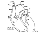
解剖学的に言えば、心臓は、線維性骨格、弁、大動脈幹、肺静脈、および心室(すなわち、右心房、左心房、右心室、左心室)の筋肉塊(心筋)から成る。図1または図2の一方または両方に概略的に示された心臓10は、右心室「RV(right ventricle)」20、左心室「LV(left ventricle)」30、右心房「RA(right atrium)」40(ここで言う「右心房」は上大静脈と腕頭静脈を含む)、「LA(left atrium)」50(とその部分)、上大静脈48、冠状静脈洞「CS(coronary sinus)」42、大心臓静脈44、左肺動脈45(ここで言う「左肺動脈」というのは主肺動脈と右心室出路を含む)と、冠状静脈洞口「OS(ostium)」41といった部分を含む。図1は心臓壁(心筋)と心膜腔58を取り囲む心外膜55(点線)も示している。図2は肺静脈52とその近隣領域も示している。関心のある他の領域としては心房中隔と右および左心耳と三尖弁環を含むことがある。図1は伝導系60、SA(sinoatrial)結節(洞結節)62、AV(atrioventricular)結節(房室結節)64も示している。
Anatomically speaking, the heart consists of a muscular mass (myocardium) of the fibrous skeleton, valves, aortic trunk, pulmonary veins, and ventricles (ie, right atrium, left atrium, right ventricle, left ventricle). The
先に言及したように、本発明の実施形態においては、電極の配置、刺激部位、心臓に対する除細動および/またはぺーシングのために選ばれる望ましい部位または局所領域は、患者の生理または疾患および/または採用される特定の選ばれたぺーシング手順(プロトコル)に応じて変わる場合がある。このため、電極は、それらの電極が心筋の望ましい局所領域または他の関心のある部位に近接かつ/または接触するように、患者の体内および/または体外の多数の領域内に多数の異なる技術を用いて配置される場合がある。例えば、電極は患者の胸部の表面上に直に配置される場合がある。更なる例として、1つ以上の電極が心臓の自然内腔(心房、心室、静脈、動脈など)内、または心膜腔内、心臓壁の外側と内側の表面上、または厚みのある心筋壁内に配置されることがある。電極は対象者の体内に外科的手法によってあるいは電極の付いた位置決めカテーテルなどを使ってそれらを埋め込むことによって配置することができる。実施形態によっては、一部の電極は心臓が鼓動する間にその各電極がそれぞれの刺激部位または検知部位で組織と接触することができるような構成とサイズを有する。ここで言う「局所(localized)」という表現は電気刺激が心臓全体ではなく心臓の限られた一部分に与えられること指す。 As previously mentioned, in embodiments of the present invention, the desired site or local region chosen for electrode placement, stimulation site, defibrillation and / or pacing to the heart is the patient's physiology or disease and It may vary depending on the particular selected pacing procedure (protocol) employed. For this reason, the electrodes employ a number of different techniques within and / or outside of the patient's body so that they are in close proximity and / or in contact with the desired local region of the myocardium or other site of interest. It may be arranged using. For example, the electrodes may be placed directly on the surface of the patient's chest. As a further example, one or more electrodes may be in the heart's natural lumen (atrium, ventricle, vein, artery, etc.) or in the pericardial space, on the outer and inner surfaces of the heart wall, or thick myocardial wall May be placed inside. Electrodes can be placed in a subject's body by surgical techniques or by implanting them using a positioning catheter or the like with electrodes. In some embodiments, some electrodes are configured and sized such that each electrode can contact tissue at the respective stimulation or sensing site while the heart is beating. As used herein, the expression “localized” refers to electrical stimulation being applied to a limited portion of the heart, not the entire heart.
こうして、既に指摘されたように、ぺーシング電極は心膜腔内または心臓の他の局所領域内に配置されることがある。例えば、ぺーシング電極はカテーテルに付けて心内膜内に挿入されたり、あるいは心臓を縫うようにして進んで心臓の静脈内に挿入される(OS(冠状静脈洞口)を縫うようにして進んで静脈内で輪にする)ことがある。実施形態によっては、左心房のぺーシングは電極を左心房の一部と肺静脈内に拡がるように配置することによってこの領域の細動発生を根絶または制御するのを助けるために実施されることがある。1つ以上の電極を肺静脈内に配置することは心房細動の治療に特に適する場合がある。他の配置の例については後述される。 Thus, as already pointed out, the pacing electrodes may be placed in the pericardial space or other local regions of the heart. For example, a pacing electrode can be inserted into the endocardium attached to a catheter, or can be advanced as a heart is sewn and inserted into a heart vein (as an OS (coronary sinus ostium) is sewn). Loop in veins). In some embodiments, left atrial pacing is performed to help eradicate or control the occurrence of fibrillation in this region by positioning electrodes to extend into a portion of the left atrium and into the pulmonary veins. There is. Placing one or more electrodes in the pulmonary veins may be particularly suitable for the treatment of atrial fibrillation. Examples of other arrangements will be described later.
上述したように、心臓における血流の駆動力は心筋のアクティブな収縮によるものである。この収縮は電気信号として検出することができる。心臓の収縮は、SA結節とそれを取り巻く心房心筋繊維の細胞から始まって心房に伝わり、続いてAV結節を通過して、やや遅れてから心室に入るといった伝搬パタンで伝播する電気パルスによって引き起こされる。ぺーシングする間に心臓の起動または収縮を検知することによってデータをぺーシングシステム(制御装置または心臓モニタ)に提供することができる。そのデータは必要に応じて多数の動作パラメータ、例えば(a)ぺーシング刺激を停止する時、(b)ぺーシング刺激のスピードまたはレート(ぺーシングレートの増大または減少)、刺激パルスの持続時間または強度、(c)組織が成功裏に捕捉されているかどうか、(d)局所領域に中継されるパルスおよび/またはパルス列の数、を決定かつ調節するために利用することができる。 As described above, the driving force for blood flow in the heart is due to active contraction of the myocardium. This contraction can be detected as an electrical signal. The heart contraction is caused by an electrical pulse that propagates in a propagation pattern that begins with cells in the SA node and the surrounding atrial myocardial fibers, propagates into the atria, then passes through the AV node and then enters the ventricle after a short delay. . Data can be provided to a pacing system (controller or heart monitor) by sensing heart activation or contraction during pacing. The data may include a number of operating parameters as needed, for example: (a) when pacing stimulation is stopped, (b) pacing stimulation speed or rate (pacing rate increase or decrease), stimulation pulse duration or It can be used to determine and adjust the intensity, (c) whether the tissue has been successfully captured, (d) the number of pulses and / or pulse trains relayed to the local region.
本発明の実施形態によれば、ぺーシングは除細動ショックを印加した後に患者の心臓に対して自動的に実施される。ここに使われる「ぺーシング(pacing)」という表現は任意のサイクル長または複数のサイクル長の組み合わせを有する刺激を含む。例えば、そのようなぺーシングとしては、単一のぺーシング、対のぺーシング、またはこれらの2つの組み合わせが含まれる。こうしたぺーシング刺激は不整脈の再発の可能性を小さくするため且つ/または心臓の機械的機能を改善するために印加されることがある。さらに、ぺーシング刺激のタイミングはフィードバック制御されることがある。このやり方については2002年7月31日に出願された米国特許出願公開第10/210,587号明細書「フィードバック制御されたタイミングを使用する心臓ぺーシング方法および装置(Pacing Methods and Devices Using Feedback Controlled Timing)」を参照されたい。またその開示内容は全て本願に援用される。 In accordance with an embodiment of the present invention, pacing is performed automatically on the patient's heart after applying a defibrillation shock. As used herein, the term “pacing” includes stimuli having any cycle length or combinations of cycle lengths. For example, such pacing includes single pacing, paired pacing, or a combination of the two. Such pacing stimuli may be applied to reduce the likelihood of arrhythmia recurrence and / or improve the mechanical function of the heart. Further, the timing of the pacing stimulus may be feedback controlled. This method is described in US patent application Ser. No. 10 / 210,587 filed Jul. 31, 2002, “Pacing Methods and Devices Using Feedback Controlled Timing”. Please refer to. Moreover, all the content of an indication is used for this application.
本発明の一部の実施形態に基づくぺーシングの自動印加を実施するためのシステムを図3に示す。図3に示されているように、一部の実施形態に基づく除細動および/またはぺーシングシステム100はぺーシングコントローラ回路110と除細動回路112とを具備する。ぺーシングコントローラ回路110と除細動回路112は上述したように心臓10の特定の場所または領域(図示された部位は単に例示的なものである)に配置された1セット以上の電極115と118を有する。電極が配置される特定の場所は除細動および/またはぺーシングの特定の印加によって異なる場合がある。配置(もしくは留置)場所は本開示を踏まえれば当業者には明かであるのでここではこれ以上詳しくは述べない。上述したように、ぺーシングに使用される除細動に対して同じ電極が利用されることがある。代わりに、ぺーシングに使用される除細動に対して異なる電極または異なるセットの電極が利用されることがある。最後に、共通の電極と異なる電極の組み合わせがぺーシングと除細動に使用される場合がある。図3に示されているように、ぺーシングコントローラ回路110がここに述べられるようにぺーシング刺激の印加を制御することができるように、心臓機能を検知するためのセンサ120も提供されることがある。さらに、ぺーシングコントローラ回路110は後述されるようにぺーシング刺激を更に制御するために例えばヘルスケア提供者からの外部からの入力を受信する場合がある。ぺーシングコントローラ回路110は、除細動ショック後にぺーシング刺激を自動的に印加する自動ぺーシング回路111を具備する。
A system for performing automatic pacing application according to some embodiments of the present invention is shown in FIG. As shown in FIG. 3, a defibrillation and / or
ぺーシングコントローラ回路110および/または除細動回路112は外部装置(例えば動作部品をその中に保持する遠隔筐体)の一部を成すこと、または密閉された本体内に動作回路を保持する生体適合性を有する単一(シングル)もしくは二重(デュアル)の植込み可能な筐体内に組み込まれることがある。同様に、ぺーシングコントローラ回路110または除細動回路112の一方は外部装置内に組み込まれる一方で、ぺーシングコントローラ回路110および除細動回路112の他方は患者の体内に植込み可能な筐体内に組み込まれることがあり得る。ぺーシングコントローラ110および/または除細動回路112は、検知された心臓信号を増幅し且つ/または電極115と/または電極118に刺激を与えるための1つ以上の増幅器(図示されていない)を含む電子回路を含むことができる。ぺーシングコントローラ110および/または除細動回路112は、増幅された信号を解析して心房および/または心室の不整脈状態または細動状態の存在もしくは発現を検出し、心室細動(あるいは、装置がそれ用に構成された特定の治療によっては他の不整脈)が起こっている時または起こっているかどうかを特定するための従来の回路も備えることがある。さらに、除細動回路112は従来の除細動回路でよい。しかしながらより詳しく後述されるように、本発明の一部の実施形態では、除細動回路112は自動ぺーシング回路111に提供される信号をぺーシングコントローラ回路110に送ったり、あるいは自動ぺーシング回路111は除細動回路112の動作を監視して除細動ショックが停止したかどうかを自動ぺーシング回路111が決定する。
The pacing
一部の実施形態では、ぺーシングコントローラ110および/または除細動回路112は、心室細動を誘発する可能性を減らすために関連する除細動ショックパルスが心周期の感じやすい部分で与えられる可能性を小さくするために除細動ショックパルスのタイミングを調節するために使用することができる。また心房除細動に対するショックのタイミングを調節するための心室検知(ventricular sensing)が当業者に周知の除細動装置に使用されるようなRVおよび/またはLV電極を使って実行されることがある。詳しくは米国特許第5,978,704号明細書に記載されている。この特許文書の開示内容は本願に援用される。
In some embodiments, the pacing
動作に関して言えば、本発明の一部の実施形態によれば、除細動回路112は電極115を通じて心臓10に除細動ショックを印加する。除細動回路112は除細動ショックが停止したらそのことをぺーシングコントローラ回路110に通知し、自動ぺーシング回路111は電極118を通じて心臓10にぺーシング刺激を自動的に印加する。代わりに、ぺーシングコントローラ回路110が除細動ショックの停止を検知し、あるいは除細動ショックの開始が通知されたあと所定期間だけ待機するか、または他の類似のテクニックを使って除細動ショックが停止したことを決定するようにすることが可能である。ぺーシングコントローラ回路110はここに述べられたぺーシング刺激のタイミングまたは有効な印加を制御するためにセンサ120を利用して心臓機能を検知することもある。除細動ショックが印加された直ぐ後に心臓機能の検知が行われる場合、こうした検知は除細動パルスが印加されている間は例えばリレー(relay)を使うなどして検知回路からのセンサリード線の接続を切断して行われることがある。除細動パルスが印加された直ぐ後に心臓機能および/または特性を検知するためのシステムおよび動作については米国特許出願公開第・・・号明細書(米国代理人番号5656-25)を参照されたい。この特許文書の全ての開示内容は本願に援用される。
In operation, according to some embodiments of the present invention,
本発明の実施形態はここでは図3に示された機能および/またはアーキテクチャの特定の分割に関連して説明されているが、当業者であれば本開示を踏まえて分かるように本発明の教えから機能および/またはアーキテクチャの他の分割が利用される場合がある。例えば、除細動回路112とぺーシングコントローラ回路110は別々もしくは単一の装置に組み込まれる場合がある。さらに、ぺーシングコントローラ回路110は除細動回路112に組み込まれる場合がある。このように図3に示されたアーキテクチャは説明目的だけで提示されたもので、本発明の範囲がその態様に限定されるものと解されてはならない。
While embodiments of the present invention are described herein with reference to a particular division of functionality and / or architecture shown in FIG. 3, the teachings of the present invention will be apparent to those of skill in the art in light of this disclosure. Other divisions of functionality and / or architecture may be utilized. For example,
本発明の実施態様に基づく作業の流れを図4に示す。図4に見られるように、除細動ショックは例えば除細動回路112によって心臓10に印加される(ブロック300)。除細動ショックの停止は、例えば、除細動回路112がぺーシングコントローラ回路110に除細動ショックの停止を通知することによって、あるいはぺーシングコントローラ回路110が除細動ショックの停止を検出することによって、あるいはぺーシングコントローラ回路110が除細動ショックが停止したと確実に言えるのに十分な時間だけ待機することによって、決定される(ブロック302)。除細動ショックが停止した後(ブロック302)、ぺーシング刺激は例えば自動ぺーシング回路111によって心臓10に自動的に印加される(ブロック304)。このように、ぺーシング刺激の印加は正常な心臓機能が検出されたかどうか、あるいは心臓の正常な電気活動が検出されたかどうかに関係なく自動的に実行されることがある。
A work flow according to an embodiment of the present invention is shown in FIG. As seen in FIG. 4, a defibrillation shock is applied to the
本発明の一部の実施形態では、ぺーシング刺激は除細動ショックが停止した後直ぐに(直後に)印加される。ここに使用される「直ぐに」(immediately)という表現は、従来の心電図で心臓活動を検出して除細動ショックが成功裏に細動を止めたかどうかを判定することができる以前にぺーシング刺激を印加することを指す。詰まり例えば、除細動ショックが停止してから約2乃至4秒以内に実行されるぺーシング刺激の印加は除細動ショックが停止した直後と見なされる。本発明の特定の実施形態では、ぺーシング刺激は除細動ショックが停止してから約2秒以内に印加され、更なる実施形態では、ぺーシング刺激は除細動ショックが停止してから約1秒以内に印加され、なお更なる実施形態では、ぺーシング刺激は除細動ショックが停止してから約0.5秒以内に印加される。 In some embodiments of the present invention, the pacing stimulus is applied immediately (immediately after) the defibrillation shock stops. The term “immediately” as used herein is a pacing stimulus that can be used to detect cardiac activity on a conventional electrocardiogram and determine whether the defibrillation shock has successfully stopped fibrillation. Is applied. Clogging For example, the application of a pacing stimulus performed within about 2 to 4 seconds after the defibrillation shock stops is considered immediately after the defibrillation shock stops. In a particular embodiment of the invention, the pacing stimulus is applied within about 2 seconds after the defibrillation shock stops, and in a further embodiment, the pacing stimulus is applied about once after the defibrillation shock stops. Applied within 1 second, and in yet further embodiments, the pacing stimulus is applied within about 0.5 seconds after the defibrillation shock stops.
既に手短に述べたように、ぺーシング刺激は単一のぺーシング刺激および/または対のぺーシング刺激である場合がある。単一のぺーシング刺激に関して言えば、ぺーシング刺激は従来のタイミング関係を利用して印加されることがある。さらに、従来の対のぺーシングも本発明の一部の実施形態に基づいて利用されることがある。例えば、刺激パルスの各ペア間のタイミングは一定の場合があり、また各ペア毎のパルス間のタイミングも一定の場合がある。単一および/または対のぺーシング刺激のぺーシングレートは事前に定めらたものか、あるいは当業者には周知のように除細動ショックの前もしくは後の心臓の電気活動を含む被検知変数(sensed variables)に基づく場合がある。ぺーシング刺激の強度は事前に定められたものか、あるいは当業者には周知のオートキャプチャ(autocapture)技術を利用して動的に設定される場合がある。対のぺーシング刺激は、作業仕様(operator specification)に基づいて、あるいは所定の閾値未満の脈圧、心拍数、少なくとも1回の固有心室拍動のタイミングおよび/または形態(morphology)、インピーダンスの変化、2カ所の間の距離の変化および/もしくは変位および/もしくは変化率、並びに/または心臓に関連した場所の運動といった、被検知変数に基づいて選択的に利用することもできる。上述した電極の配置場所は単一のぺーシングおよび/または対のぺーシングに利用される場合がある。 As already briefly mentioned, the pacing stimulus may be a single pacing stimulus and / or a pair of pacing stimuli. With respect to a single pacing stimulus, the pacing stimulus may be applied using a conventional timing relationship. In addition, conventional pairs of pacing may also be utilized in accordance with some embodiments of the present invention. For example, the timing between each pair of stimulation pulses may be constant, and the timing between pulses for each pair may also be constant. The pacing rate of single and / or paired pacing stimuli is predetermined or, as is well known to those skilled in the art, the variable to be detected, including the electrical activity of the heart before or after the defibrillation shock May be based on (sensed variables). The intensity of the pacing stimulus may be predetermined or may be set dynamically using an autocapture technique well known to those skilled in the art. Paired pacing stimuli may be based on operator specifications or below a predetermined threshold of pulse pressure, heart rate, timing and / or morphology of at least one intrinsic ventricular beat, changes in impedance It can also be used selectively based on a sensed variable, such as a change in distance and / or displacement and / or rate of change between two locations and / or movement of a location relative to the heart. The electrode placement locations described above may be used for single pacing and / or paired pacing.
本発明の更なる実施態様に基づく作業の流れを図5に示す。図5に見られるように、心臓活動および/または心臓機能が心臓において検出される(ブロック500)。図5では心臓活動および/または心臓機能の検出は除細動ショックが印加される前に行われるものとして示されているが、本発明はその設定に限定されるものではない。例えば、本発明の教えから逸脱することなく、心臓活動は除細動ショックが印加された後に検出されることがある。次に、検出された心臓活動および/または心臓機能に基づいてぺーシング刺激のタイプが選択される(ブロック502)。例えば、一連の検出された電気信号(心臓活動)から患者が単一のぺーシング刺激に反応しそうにないことが示される場合があるが、従ってその場合にはその電気信号に基づいてこの患者に対して対のぺーシング刺激が選択されることがある。同様に、低い脈圧から心臓機能が低下していることが示される場合があるが、それは対のぺーシングによって改善されることがある。選択されるぺーシング刺激のタイプとしては、単一のぺーシング刺激、対のぺーシング刺激、および/またはその2つのタイプの刺激の組み合わせが含まれることがある。 A work flow according to a further embodiment of the invention is shown in FIG. As seen in FIG. 5, cardiac activity and / or function is detected in the heart (block 500). Although the detection of cardiac activity and / or cardiac function is shown in FIG. 5 as being performed before the defibrillation shock is applied, the present invention is not limited to that setting. For example, cardiac activity may be detected after a defibrillation shock is applied without departing from the teachings of the present invention. A pacing stimulus type is then selected based on the detected cardiac activity and / or cardiac function (block 502). For example, a series of detected electrical signals (cardiac activity) may indicate that the patient is unlikely to respond to a single pacing stimulus, so that in that case, the patient is informed based on that electrical signal. In contrast, a pair of pacing stimuli may be selected. Similarly, low pulse pressure may indicate that cardiac function is decreasing, which may be improved by paired pacing. The type of pacing stimulus selected may include a single pacing stimulus, a pair of pacing stimuli, and / or a combination of the two types of stimuli.
除細動ショックは例えば除細動回路112によって心臓に印加されることがある(ブロック300)。除細動ショックの停止は、例えば、除細動回路112がぺーシングコントローラ回路110に除細動ショックの停止を通知することによって、あるいはぺーシングコントローラ回路110が除細動ショックの停止を検出することによって、あるいはぺーシングコントローラ回路110が除細動ショックが停止したと確実に言えるのに十分な時間だけ待機することによって、決定される(ブロック302)。除細動ショックが停止した後(ブロック302)、選択されたぺーシング刺激(ブロック502)は例えば自動ぺーシング回路111によって心臓10に自動的に印加される(ブロック304)。
A defibrillation shock may be applied to the heart, for example, by the defibrillation circuit 112 (block 300). The defibrillation shock is stopped by, for example, the
本発明の更なる実施態様に基づく作業の流れを図6に示す。図6に見られるように、図4に関連して述べたように除細動ショックの印加が開始され(ブロック300)、除細動ショックが停止された(ブロック302)後、ぺーシング刺激信号が心臓10に印加される(ブロック304)。図6ではさらに心臓活動が存在するかどうかが判定される(ブロック310)。心臓活動は例えば血圧や自発的な電気手活動などを検出することによって検出されることがある。心臓活動は存在すると判定された場合(ブロック310)、ぺーシングは抑制される(ブロック315)。ぺーシングの抑制は例えばぺーシングを停止することおよび/またはぺーシングのペースを落とすことにより行われることがある。他方、心臓活動は存在しないと判定された場合(ブロック310)、ぺーシングは中断されることなく継続される(ブロック313)。 A work flow according to a further embodiment of the invention is shown in FIG. As seen in FIG. 6, the pacing stimulus signal is applied after defibrillation shock application is started (block 300) and defibrillation shock is stopped (block 302) as described in connection with FIG. Is applied to the heart 10 (block 304). In FIG. 6, it is further determined whether there is cardiac activity (block 310). Cardiac activity may be detected, for example, by detecting blood pressure or spontaneous electrical manual activity. If it is determined that cardiac activity is present (block 310), pacing is suppressed (block 315). Pacing suppression may be done, for example, by stopping pacing and / or slowing down the pacing. On the other hand, if it is determined that there is no cardiac activity (block 310), pacing continues without interruption (block 313).
対のぺーシングも利用される本発明の更なる実施態様に基づく作業の流れを図7に示す。図7に見られるように、図4に関連して述べたように除細動ショックの印加が開始され(ブロック300)、除細動ショックが停止された(ブロック302)後、第1のぺーシング刺激信号が心臓10に印加される(ブロック320)。さらに図7では、第1のぺーシング刺激信号の印加の結果もたらされる心臓活動が検出される(ブロック322)。第1のぺーシング刺激信号の印加に伴ったまたはそれに反応した検出された心臓活動に基づいて(ブロック324)、対のぺーシングをその検出された心臓活動に基づいて選択的に実施するために第2のぺーシング刺激信号が心臓10に選択的に印加される(ブロック326)。対のぺーシングを実施するための第2の刺激信号の選択的な印加は検知された心臓活動の性質に基づいて行われることがある。こうして、検出された心臓活動が低い心臓機能を示している場合、第2の刺激信号は心臓の機械的機能を改善するために対のぺーシングを実施するために印加されることがある。例えば、被検知心臓機能には、低い脈圧のインピーダンス信号、心拍数、少なくとも1回の固有心室拍動のタイミングおよび/または形態(morphology)、インピーダンスの変化、2カ所の間の距離の変化および/もしくは変位および/もしくは変化率、並びに/または心臓に関連した場所の運動を検知することが含まれることがある。このように図7に示された実施形態では、第2の刺激は第1の刺激に伴う心臓活動に基づいて対のぺーシングを施すために印加されることがある。 A work flow according to a further embodiment of the present invention in which pair pacing is also utilized is shown in FIG. As can be seen in FIG. 7, the application of defibrillation shock is started as described in connection with FIG. 4 (block 300), and after the defibrillation shock is stopped (block 302), the first page. A singing stimulus signal is applied to the heart 10 (block 320). Further, in FIG. 7, cardiac activity resulting from the application of the first pacing stimulus signal is detected (block 322). Based on the detected cardiac activity with or in response to application of the first pacing stimulus signal (block 324), to selectively perform paired pacing based on the detected cardiac activity. A second pacing stimulus signal is selectively applied to the heart 10 (block 326). The selective application of the second stimulus signal to perform paired pacing may be based on the nature of the sensed cardiac activity. Thus, if the detected cardiac activity is indicative of low cardiac function, the second stimulation signal may be applied to perform paired pacing to improve the mechanical function of the heart. For example, the sensed cardiac function includes low pulse pressure impedance signals, heart rate, timing and / or morphology of at least one intrinsic ventricular beat, changes in impedance, changes in distance between two locations, and Sensing displacement and / or rate of change, and / or movement of a location associated with the heart may be included. Thus, in the embodiment shown in FIG. 7, the second stimulus may be applied to provide paired pacing based on cardiac activity associated with the first stimulus.
本発明の更なる実施形態として、対のぺーシングは例えばヘルスケア提供者といった外源からの信号であって対のぺーシングを選択的に実施するために利用される信号を受信したことを契機に開始されることがある。このような場合、図7の作業は外源からの信号が検出されたかどうかを判定するようにブロック322を変更すればよい。外源からの信号が検出された場合、ブロック326の第2の刺激が印加されることになる。また植込み可能な装置では、外源からの信号は無線周波数信号または植込み込み可能な装置と通信するための他の技術でよい。同様に、ソフトウェアスイッチが外源からの信号を提供するためにセッティングされる場合がある。外部の装置では、スイッチのセッティング(ハードウェアまたはソフトウェアのどちらか)は単一のぺーシングと対のぺーシングの間で選択するために利用されることがある。
As a further embodiment of the present invention, the pair pacing is triggered by receiving a signal from an external source such as a health care provider that is used to selectively perform pair pacing. May be started. In such a case, the operation of FIG. 7 may change block 322 to determine whether a signal from an external source has been detected. If a signal from an external source is detected, the second stimulus of
本発明のなお更なる実施形態として、検出される心臓活動は第1の刺激信号の印加に反応したものである必要はない。こうした態様では、図7は、心臓活動が第1の刺激信号の印加の結果もたらされたかどうかには関係なく心臓活動が検出される(ブロック322)ように変更されることが可能である。検出された心臓活動に基づいて(ブロック324)、対のぺーシング刺激を検出された心臓活動に基づいて選択的に印加するために第2の刺激信号が心臓10に選択的に印加される(ブロック326)。
As a still further embodiment of the invention, the detected cardiac activity need not be responsive to the application of the first stimulus signal. In such an aspect, FIG. 7 can be modified such that cardiac activity is detected (block 322) regardless of whether the cardiac activity resulted from the application of the first stimulation signal. Based on the detected cardiac activity (block 324), a second stimulation signal is selectively applied to the
当業者なら分かるように、本発明は、方法、データ処理システム、またはコンピュータプログラム製品として実施することができる。従って、本発明は、全体にハードウェアの形態、全体にソフトウェアの形態、あるいはソフトウェアとハードウェアの組み合わせの形態を採る場合があるが、ここでは全て総称的に「回路」と称される。さらに、本発明はコンピュータが利用可能なプログラムコード手段がその中に具現化されたコンピュータ利用可能な記憶媒体上のコンピュータプログラム製品の形態を採る場合がある。メモリ装置、ハードディスク、CD−ROM、光学記憶装置、無線伝送媒体および/もしくはインターネットまたはイントラネットをサポートする伝送媒体といった伝送媒体、または磁気記憶媒体を含む、任意の適切なコンピュータ読取り可能な媒体が利用されることがある。 As will be appreciated by one skilled in the art, the present invention may be implemented as a method, data processing system, or computer program product. Therefore, the present invention may take the form of hardware as a whole, the form of software as a whole, or the form of a combination of software and hardware, but here they are generically referred to as “circuits”. Further, the present invention may take the form of a computer program product on a computer-usable storage medium having computer-usable program code means embodied therein. Any suitable computer readable medium may be utilized including memory devices, hard disks, CD-ROMs, optical storage devices, wireless transmission media and / or transmission media such as transmission media supporting the Internet or Intranet, or magnetic storage media. Sometimes.
本発明はここでは本発明の実施形態に基づく方法、装置(システム)、およびコンピュータプログラム製品のフローチャートおよび/またはブロック図および/または流れ図を参照して説明される。流れ図および/またはブロック図の各ブロック、並びに流れ図および/またはブロック図のブロックの組み合わせは、コンピュータプログラム命令によって実行が可能であることは理解されよう。これらのコンピュータプログラム命令は、汎用コンピュータ、専用コンピュータ、またはマシンを構成する他のプログラマブルなデータ処理装置のプロセッサに送られることがあり、それによりこれらの命令はコンピュータまたは他のプログラマブルなデータ処理装置のプロセッサを介して実行されてフローチャートおよび/またはブロック図および/または流れ図に指定された機能を実現するための手段が生成される。 The present invention is described herein with reference to flowchart illustrations and / or block diagrams and / or flowchart illustrations of methods, apparatus (systems) and computer program products according to embodiments of the invention. It will be understood that each block of the flowchart illustrations and / or block diagrams, and combinations of blocks in the flowchart illustrations and / or block diagrams, can be implemented by computer program instructions. These computer program instructions may be sent to a processor of a general purpose computer, special purpose computer, or other programmable data processing device comprising a machine, whereby these instructions are transmitted to the computer or other programmable data processing device. Means are generated for performing the functions specified in the flowcharts and / or block diagrams and / or flowcharts, executed through the processor.
コンピュータまたは他のプログラマブルなデータ処理装置を特定の仕方で機能させることができるこれらのコンピュータプログラム命令はコンピュータ読取り可能なメモリに保存されることもある。それによりコンピュータ読取り可能なメモリに保存された命令がフローチャートおよび/またはブロック図および/または流れ図の指定された機能を実現する命令手段を含む製品が作り出される。 These computer program instructions, which allow a computer or other programmable data processing device to function in a particular manner, may be stored in computer readable memory. Thereby, a product is created that includes instruction means in which instructions stored in a computer readable memory implement specified functions of flowcharts and / or block diagrams and / or flowcharts.
コンピュータプログラム命令はコンピュータまたは他のプログラマブルなデータ処理装置にロードされることがあり、コンピュータまたは他のプログラマブルなデータ処理装置上で実行される命令がフローチャートおよび/またはブロック図および/または流れ図に指定された機能を実行する段階を提供するようになっており、一連のそうした作業段階がそれらのコンピュータまたは他のプログラマブルなデータ処理装置上で実行されてコンピュータ実行プロセスが作り出される。 Computer program instructions may be loaded into a computer or other programmable data processing device, and instructions executed on the computer or other programmable data processing device are specified in flowcharts and / or block diagrams and / or flow diagrams. And a series of such work steps are executed on those computers or other programmable data processing devices to create a computer-executed process.
本発明の実施形態は機能の特定のアーキテクチャおよび/または分担に関連して説明されてきたが、本発明はこうしたアーキテクチャおよび/または分担に限定されるものと解されるべきではない。つまり、ここで述べた作業を実行することが可能な機能の他のアーキテクチャおよび/または分担が本発明の教えから逸脱することなく利用されることがある。さらに、本発明の実施形態は特定の回路に関連して説明されてきたが、こうした回路は別々のコンポーネント、マイクロプロセッサおよび/または信号プロセッサといったプロセッサ、アナログ回路、デジタル回路および/またはそれらの組み合わせを含む場合がある。さらに、本発明の実施形態は全体にハードウェア、全体にソフトウェア、あるいはハードウェアとソフトウェアの組み合わせとして実施される場合がある。 Although embodiments of the present invention have been described with reference to particular architectures and / or sharing of functions, the present invention should not be construed as limited to such architectures and / or sharing. That is, other architectures and / or sharing of functions capable of performing the tasks described herein may be utilized without departing from the teachings of the present invention. Moreover, although embodiments of the invention have been described with reference to specific circuits, such circuits may include separate components, processors such as microprocessors and / or signal processors, analog circuits, digital circuits, and / or combinations thereof. May include. Furthermore, embodiments of the present invention may be implemented as entirely hardware, entirely software, or a combination of hardware and software.
本開示を踏まえて当業者であれば分かるように、上述したフローチャートに示された作業に関して、本発明の実施態様はここに述べた特定の手順又は作業手順に限定されない。例えば、フローチャートの作業は順次または同時に実行されることがある。同様に、本発明の実施形態に基づくフィードバック調節をなお実施する他の一連の作業が利用されることがある。従って、本発明はフローチャートに示された特定の作業または特定の手順の作業に限定されると解されるべきではない。 As will be appreciated by one of ordinary skill in the art in light of the present disclosure, embodiments of the present invention are not limited to the specific procedures or work procedures described herein with respect to the work illustrated in the flowcharts described above. For example, flowchart operations may be performed sequentially or simultaneously. Similarly, another series of operations that still implement feedback adjustments according to embodiments of the present invention may be utilized. Accordingly, the present invention should not be construed as limited to the specific operations shown in the flowcharts or the operations of specific procedures.
以上の記述は本発明を説明するためのもので、それに限定されるものと解してはならない。本発明のいくつかの例示的な実施形態が説明されてきたが、当業者であれば本発明の新規な教えと利点から大きく逸脱することなくこれら例示的な実施形態について多くの変更が可能であることは容易に理解するであろう。従って、全てのこうした変更は特許請求の範囲の請求項に定められる本発明の範囲内に含まれることが意図される。特許請求の範囲の請求項において、ミーンズ・プラス・ファンクション表現はそれが使用されるところでは列挙された機能を実行するものとしてここで述べられた構造、および構造的均等物だけでなく均等な構造もカバーすることが意図されている。故に、以上の記述は本発明を説明するためのもので開示された特定の実施形態に限定されるものと解してはならないこと、また開示された実施形態の変更、変形は他の実施形態と並んで特許請求の範囲内に含まれることが意図されていることは理解されよう。本発明は特許請求の範囲の各請求項に係る発明とその均等物を包含する。 The above description is for explaining the present invention and should not be construed as being limited thereto. While several exemplary embodiments of the present invention have been described, those skilled in the art will be able to make many changes to these exemplary embodiments without significantly departing from the novel teachings and advantages of the present invention. It will be easy to understand. Accordingly, all such modifications are intended to be included within the scope of this invention as defined in the claims. In the claims, a means plus function representation is the structure described herein as performing the recited function, and equivalent structures as well as structural equivalents, where it is used. Is also intended to cover. Therefore, the above description should not be construed as being limited to the specific embodiments disclosed for purposes of illustrating the present invention, and modifications and variations of the disclosed embodiments may be considered as other embodiments. It is to be understood that both are intended to be included within the scope of the claims. The present invention includes the inventions according to the respective claims and equivalents thereof.
Claims (104)
患者の心臓に除細動ショックを印加する段階と、
除細動ショックが停止した後に患者の心臓にぺーシング刺激信号を自動的に印加する段階と、を具備する心臓ぺーシング方法。 In a method of performing cardiac pacing on a patient,
Applying a defibrillation shock to the patient's heart;
Automatically applying a pacing stimulation signal to the patient's heart after the defibrillation shock has ceased.
検出された心臓活動に基づいて対のぺーシング刺激を選択的に印加する段階と、を更に具備する請求項4に記載の心臓ぺーシング方法。 Detecting cardiac activity associated with the application of a single pacing stimulus;
5. The cardiac pacing method according to claim 4, further comprising selectively applying a pair of pacing stimuli based on the detected cardiac activity.
検出された心臓活動に基づいて対のぺーシング刺激を選択的に印加する段階と、を更に具備する請求項4に記載の心臓ぺーシング方法。 Detecting cardiac activity; and
5. The cardiac pacing method according to claim 4, further comprising selectively applying a pair of pacing stimuli based on the detected cardiac activity.
検出された信号に基づいて対のぺーシング刺激を選択的に印加する段階と、を更に具備する請求項4に記載の心臓ぺーシング方法。 Detecting a signal indicating application of a pair of pacing stimuli;
5. The cardiac pacing method of claim 4, further comprising selectively applying a pair of pacing stimuli based on the detected signal.
前記患者の心臓にぺーシング刺激信号を自動的に印加する段階は、除細動ショックが停止した後に第2の少なくとも1セットの電極を使用して患者の心臓にぺーシング刺激信号を自動的に印加することを含み、
前記第1のセットの電極と前記第2のセットの電極は異なるセットの電極である、請求項1に記載の心臓ぺーシング方法。 Applying a defibrillation shock to the patient's heart comprises applying a defibrillation shock to the patient's heart using a first at least one set of electrodes;
The step of automatically applying a pacing stimulus signal to the patient's heart automatically applies the pacing stimulus signal to the patient's heart using a second at least one set of electrodes after the defibrillation shock has ceased. Including applying,
The cardiac pacing method according to claim 1, wherein the first set of electrodes and the second set of electrodes are different sets of electrodes.
前記患者の心臓にぺーシング刺激信号を自動的に印加する段階は、除細動ショックを印加した後に第2の少なくとも1セットの電極を使用して患者の心臓にぺーシング刺激信号を自動的に印加することを含み、
前記第1のセットの電極と前記第2のセットの電極は同じセットの電極である、請求項1に記載の心臓ぺーシング方法。 Applying a defibrillation shock to the patient's heart comprises applying a defibrillation shock to the patient's heart using a first at least one set of electrodes;
The step of automatically applying a pacing stimulus signal to the patient's heart automatically applies a pacing stimulus signal to the patient's heart using a second at least one set of electrodes after applying a defibrillation shock. Including applying,
The cardiac pacing method according to claim 1, wherein the first set of electrodes and the second set of electrodes are the same set of electrodes.
除細動ショックが停止した直後に前記第1のセットの電極を利用して患者の心臓にぺーシング刺激信号を自動的に印加するための手段と、を具備する心臓ぺーシングシステム。 A first set of electrodes for applying pacing stimuli to the patient's heart;
Means for automatically applying a pacing stimulation signal to the patient's heart utilizing the first set of electrodes immediately after the defibrillation shock has ceased.
検出された心臓活動に基づいて対のぺーシング刺激を選択的に印加するための手段と、を更に具備する請求項58に記載の心臓ぺーシングシステム。 Means for detecting cardiac activity associated with the application of a single pacing stimulus;
59. The cardiac pacing system of claim 58, further comprising means for selectively applying a pair of pacing stimuli based on detected cardiac activity.
検出された心臓活動に基づいて対のぺーシング刺激を選択的に印加するための手段と、を更に具備する請求項58に記載の心臓ぺーシングシステム。 Means for detecting cardiac activity;
59. The cardiac pacing system of claim 58, further comprising means for selectively applying a pair of pacing stimuli based on detected cardiac activity.
検出された信号に基づいて対のぺーシング刺激を選択的に印加するための手段と、を更に具備する請求項58に記載の心臓ぺーシングシステム。 Means for detecting a signal indicating application of a pair of pacing stimuli;
59. The cardiac pacing system of claim 58, further comprising means for selectively applying a pair of pacing stimuli based on the detected signal.
検出された心臓活動および/または心臓機能に基づいて除細動ショックが停止した後に患者の心臓に印加するぺーシング刺激のタイプを選択するための手段とを具備し、
前記ぺーシング刺激信号を自動的に印加するための手段は、選択されたタイプのぺーシング刺激を自動的に印加するための手段を含む、請求項55に記載の心臓ぺーシングシステム。 Means for detecting cardiac activity and / or cardiac function;
Means for selecting a type of pacing stimulus to be applied to the patient's heart after defibrillation shock ceases based on detected cardiac activity and / or cardiac function;
56. The cardiac pacing system of claim 55, wherein the means for automatically applying a pacing stimulus signal includes means for automatically applying a selected type of pacing stimulus.
前記コンピュータ読取り可能なプログラムコードは、除細動ショックが停止した直後に第1のセットの電極を利用して患者の心臓にぺーシング刺激信号を自動的に印加するコンピュータ読取り可能なプログラムコードを具備する、コンピュータプログラム製品。 A computer program product for controlling cardiac pacing, comprising a computer readable medium having computer readable program code embodied therein,
The computer readable program code comprises computer readable program code that automatically applies a pacing stimulus signal to the patient's heart using the first set of electrodes immediately after the defibrillation shock is stopped. A computer program product.
検出された心臓活動に基づいて対のぺーシング刺激を選択的に印加するコンピュータ読取り可能なプログラムコードと、を更に具備する請求項86に記載のコンピュータプログラム製品。 Computer readable program code for detecting cardiac activity associated with the application of a single pacing stimulus;
90. The computer program product of claim 86, further comprising computer readable program code for selectively applying a pair of pacing stimuli based on detected cardiac activity.
検出された心臓活動に基づいて対のぺーシング刺激を選択的に印加するコンピュータ読取り可能なプログラムコードと、を更に具備する請求項86に記載のコンピュータプログラム製品。 Computer readable program code for detecting cardiac activity;
90. The computer program product of claim 86, further comprising computer readable program code for selectively applying a pair of pacing stimuli based on detected cardiac activity.
検出された信号に基づいて対のぺーシング刺激を選択的に印加するコンピュータ読取り可能なプログラムコードと、を更に具備する請求項86に記載のコンピュータプログラム製品。 Computer readable program code for detecting a signal indicating application of a pair of pacing stimuli;
90. The computer program product of claim 86, further comprising computer readable program code for selectively applying a pair of pacing stimuli based on the detected signal.
検出された心臓活動および/または心臓機能に基づいて除細動ショックが停止した後に患者の心臓に印加するぺーシング刺激のタイプを選択するコンピュータ読取り可能なプログラムコードとを更に具備し、
前記ぺーシング刺激信号を自動的に印加するコンピュータ読取り可能なプログラムコードは選択されたタイプのぺーシング刺激を自動的に印加するコンピュータ読取り可能なプログラムコードを更に含む、請求項83に記載のコンピュータプログラム製品。 Computer readable program code for detecting cardiac activity and / or cardiac function;
Computer readable program code for selecting a type of pacing stimulus to be applied to the patient's heart after the defibrillation shock ceases based on detected cardiac activity and / or cardiac function;
84. The computer program of claim 83, wherein the computer readable program code for automatically applying a pacing stimulus signal further comprises computer readable program code for automatically applying a selected type of pacing stimulus. Product.
104. The computer program product of claim 102, wherein the detected cardiac activity comprises spontaneous electrical activity.
Applications Claiming Priority (2)
| Application Number | Priority Date | Filing Date | Title |
|---|---|---|---|
| US10/238,343 US8560063B2 (en) | 2002-09-10 | 2002-09-10 | Post-defibrillation pacing methods and devices |
| PCT/US2003/022263 WO2004024231A1 (en) | 2002-09-10 | 2003-07-16 | Post-defribillation pacing methods and devices |
Publications (2)
| Publication Number | Publication Date |
|---|---|
| JP2005537886A true JP2005537886A (en) | 2005-12-15 |
| JP2005537886A5 JP2005537886A5 (en) | 2006-08-31 |
Family
ID=31990957
Family Applications (1)
| Application Number | Title | Priority Date | Filing Date |
|---|---|---|---|
| JP2004535983A Pending JP2005537886A (en) | 2002-09-10 | 2003-07-16 | Cardiac pacing method and apparatus for pacing after defibrillation |
Country Status (5)
| Country | Link |
|---|---|
| US (1) | US8560063B2 (en) |
| EP (1) | EP1556137A1 (en) |
| JP (1) | JP2005537886A (en) |
| AU (1) | AU2003251965A1 (en) |
| WO (1) | WO2004024231A1 (en) |
Cited By (1)
| Publication number | Priority date | Publication date | Assignee | Title |
|---|---|---|---|---|
| JP2012500050A (en) * | 2008-08-14 | 2012-01-05 | カーディアック ペースメイカーズ, インコーポレイテッド | System and method for increased pacing output after external high energy electric shock |
Families Citing this family (23)
| Publication number | Priority date | Publication date | Assignee | Title |
|---|---|---|---|---|
| US7139608B2 (en) | 2002-07-31 | 2006-11-21 | Uab Research Foundation | Pacing methods and devices using feedback controlled timing |
| US7113822B1 (en) * | 2003-02-25 | 2006-09-26 | Pacesetter, Inc. | System and method for providing cardioversion therapy and overdrive pacing using an implantable cardiac stimulation device |
| US7079891B1 (en) * | 2003-02-25 | 2006-07-18 | Pacesetter | System and method for providing cardioversion therapy and overdrive pacing using an implantable cardiac stimulation device |
| GB2413082A (en) * | 2004-04-14 | 2005-10-19 | Patrick Schauerte | Implantable cardioverter-defibrillator for treating tachycardia |
| US7634316B2 (en) * | 2005-04-28 | 2009-12-15 | Imperception, Inc. | Method and apparatus for validating a pacing train associated with T-shock delivery |
| US8858441B2 (en) * | 2005-05-12 | 2014-10-14 | The Trustees Of Columbia University In The City Of New York | System and method for electromechanical wave imaging of body structures |
| US10687785B2 (en) | 2005-05-12 | 2020-06-23 | The Trustees Of Columbia Univeristy In The City Of New York | System and method for electromechanical activation of arrhythmias |
| US7650181B2 (en) * | 2005-09-14 | 2010-01-19 | Zoll Medical Corporation | Synchronization of repetitive therapeutic interventions |
| US7714701B2 (en) * | 2005-11-16 | 2010-05-11 | Gm Global Technology Operations, Inc. | Active material based haptic alert system |
| WO2011035312A1 (en) | 2009-09-21 | 2011-03-24 | The Trustees Of Culumbia University In The City Of New York | Systems and methods for opening of a tissue barrier |
| US8565865B2 (en) | 2008-07-24 | 2013-10-22 | Medtronic, Inc. | Methods for the determination of T-shock vulnerable window from far-field electrograms in implantable cardioverter defibrillators |
| US8644923B2 (en) | 2008-07-24 | 2014-02-04 | Medtronic, Inc. | Determination of upper limit of vulnerability using a variable number of shocks |
| WO2012162664A1 (en) | 2011-05-26 | 2012-11-29 | The Trustees Of Columbia University In The City Of New York | Systems and methods for opening of a tissue barrier in primates |
| US9701245B2 (en) | 2012-06-22 | 2017-07-11 | GM Global Technology Operations LLC | Alert systems and methods for a vehicle |
| US9349263B2 (en) | 2012-06-22 | 2016-05-24 | GM Global Technology Operations LLC | Alert systems and methods for a vehicle |
| US9493116B2 (en) | 2012-06-22 | 2016-11-15 | GM Global Technology Operations LLC | Alert systems and methods for a vehicle |
| US9123215B2 (en) | 2012-06-22 | 2015-09-01 | GM Global Technology Operations LLC | Alert systems and methods for a vehicle |
| US9153108B2 (en) | 2012-06-22 | 2015-10-06 | GM Global Technology Operations LLC | Alert systems and methods for a vehicle |
| US9266451B2 (en) * | 2012-06-22 | 2016-02-23 | GM Global Technology Operations LLC | Alert systems and methods for a vehicle |
| US9132774B2 (en) | 2012-06-22 | 2015-09-15 | GM Global Technology Operations LLC | Alert systems and methods for a vehicle |
| WO2014059170A1 (en) | 2012-10-10 | 2014-04-17 | The Trustees Of Columbia University In The City Of New York | Systems and methods for mechanical mapping of cardiac rhythm |
| US9604070B2 (en) * | 2012-10-10 | 2017-03-28 | West Affum Holdings Corp. | External defibrillation with automatic post-shock anti-tachycardia (APSAT) pacing |
| US10232182B2 (en) * | 2016-04-28 | 2019-03-19 | Medtronic, Inc. | Detecting and responding to anti-tachyarrhythmia shocks |
Citations (1)
| Publication number | Priority date | Publication date | Assignee | Title |
|---|---|---|---|---|
| WO2002022207A1 (en) * | 2000-09-14 | 2002-03-21 | Cardiac Pacemakers, Inc. | Pulse generator system and method |
Family Cites Families (105)
| Publication number | Priority date | Publication date | Assignee | Title |
|---|---|---|---|---|
| US3825015A (en) * | 1972-12-14 | 1974-07-23 | American Optical Corp | Single catheter for atrial and ventricular stimulation |
| US3995623A (en) * | 1974-12-23 | 1976-12-07 | American Hospital Supply Corporation | Multipurpose flow-directed catheter |
| US4365639A (en) * | 1980-02-07 | 1982-12-28 | Applied Cardiac Electrophysiology | Catheter, cardiac pacemaker and method of pacing |
| US4355646A (en) * | 1980-11-26 | 1982-10-26 | Medtronic, Inc. | Transvenous defibrillating lead |
| US4643201A (en) * | 1981-02-02 | 1987-02-17 | Medtronic, Inc. | Single-pass A-V lead |
| US4366486A (en) * | 1981-03-16 | 1982-12-28 | Northern Illinois Gas Company | Low profile antenna for data transponders |
| US4693253A (en) * | 1981-03-23 | 1987-09-15 | Medtronic, Inc. | Automatic implantable defibrillator and pacer |
| US4444195A (en) * | 1981-11-02 | 1984-04-24 | Cordis Corporation | Cardiac lead having multiple ring electrodes |
| US4708145A (en) * | 1982-06-01 | 1987-11-24 | Medtronic, Inc. | Sequential-pulse, multiple pathway defibrillation method |
| EP0095726B1 (en) | 1982-06-01 | 1988-11-02 | Medtronic, Inc. | Apparatus for controlling cardiac ventricular tachyarrhythmias |
| US4559946A (en) * | 1982-06-18 | 1985-12-24 | Mieczyslaw Mirowski | Method and apparatus for correcting abnormal cardiac activity by low energy shocks |
| US4499907A (en) * | 1982-11-15 | 1985-02-19 | Medtronic, Inc. | Energy limiting cardioversion lead |
| US4567901A (en) * | 1983-12-15 | 1986-02-04 | Cordis Corporation | Prebent ventricular/atrial cardiac pacing lead |
| US4637397A (en) * | 1985-05-30 | 1987-01-20 | Case Western Reserve University | Triphasic wave defibrillation |
| US4800883A (en) * | 1986-04-02 | 1989-01-31 | Intermedics, Inc. | Apparatus for generating multiphasic defibrillation pulse waveform |
| US5554176A (en) * | 1986-05-15 | 1996-09-10 | Telectronics Pacing Systems, Inc. | Implantable electrode and sensor lead apparatus |
| US4735206A (en) * | 1986-07-28 | 1988-04-05 | Brunswick Manufacturing Co., Inc. | Method and apparatus for defibrillating and pacing the heart |
| US4850357A (en) * | 1988-01-12 | 1989-07-25 | Cardiac Pacemakers, Inc. | Biphasic pulse generator for an implantable defibrillator |
| US4901725A (en) * | 1988-01-29 | 1990-02-20 | Telectronics N.V. | Minute volume rate-responsive pacemaker |
| US4928688A (en) * | 1989-01-23 | 1990-05-29 | Mieczyslaw Mirowski | Method and apparatus for treating hemodynamic disfunction |
| US5411527A (en) * | 1989-05-03 | 1995-05-02 | Intermedics, Inc. | Difibrillation electrodes and implantation |
| US5433730A (en) * | 1989-05-03 | 1995-07-18 | Intermedics, Inc. | Conductive pouch electrode for defibrillation |
| US5269319A (en) * | 1989-12-08 | 1993-12-14 | Cardiac Pacemakers, Inc. | Unitary intravascular defibrillating catheter with bipolar sensing |
| US5230337A (en) * | 1990-06-06 | 1993-07-27 | Cardiac Pacemakers, Inc. | Process for implanting subcutaneous defibrillation electrodes |
| US5113869A (en) | 1990-08-21 | 1992-05-19 | Telectronics Pacing Systems, Inc. | Implantable ambulatory electrocardiogram monitor |
| FR2671013B1 (en) * | 1990-12-27 | 1996-09-13 | Ela Medical Sa | HEART STIMULATOR WITH SERVO STIMULATION FREQUENCY. |
| US5107834A (en) * | 1991-01-30 | 1992-04-28 | Cardiac Pacemakers, Inc. | Low energy multiple shock defibrillation/cardioversion discharge technique and electrode configuration |
| US5165403A (en) * | 1991-02-26 | 1992-11-24 | Medtronic, Inc. | Difibrillation lead system and method of use |
| DE69210395T2 (en) * | 1991-04-05 | 1997-01-09 | Medtronic Inc | DETECTION SYSTEM WITH SUBCUTANEOUS MULTIPLE ELECTRODES |
| US5282837A (en) * | 1991-04-12 | 1994-02-01 | Incontrol, Inc. | Atrial defibrillator and method |
| US5433729A (en) * | 1991-04-12 | 1995-07-18 | Incontrol, Inc. | Atrial defibrillator, lead systems, and method |
| US5209229A (en) * | 1991-05-20 | 1993-05-11 | Telectronics Pacing Systems, Inc. | Apparatus and method employing plural electrode configurations for cardioversion of atrial fibrillation in an arrhythmia control system |
| US5184616A (en) * | 1991-10-21 | 1993-02-09 | Telectronics Pacing Systems, Inc. | Apparatus and method for generation of varying waveforms in arrhythmia control system |
| US5314448A (en) * | 1991-10-28 | 1994-05-24 | Angeion Corporation | Process for defibrillation pretreatment of a heart |
| CA2082015C (en) * | 1991-11-04 | 2001-03-20 | Rodney W. Salo | Implantable cardiac function monitor and stimulator for diagnosis and therapy delivery |
| US5224475A (en) * | 1991-11-20 | 1993-07-06 | Medtronic, Inc. | Method and apparatus for termination of ventricular tachycardia and ventricular fibrillation |
| EP0547256A1 (en) * | 1991-12-16 | 1993-06-23 | M.E.D.I. CO. ITALIA S.r.L. | Improved three-pole electrocatheter cardiac stimulation |
| US5313953A (en) | 1992-01-14 | 1994-05-24 | Incontrol, Inc. | Implantable cardiac patient monitor |
| US5201808A (en) * | 1992-02-10 | 1993-04-13 | Telectronics Pacing Systems, Inc. | Minute volume rate-responsive pacemaker employing impedance sensing on a unipolar lead |
| US5224476A (en) * | 1992-02-24 | 1993-07-06 | Duke University | Method and apparatus for controlling fibrillation or tachycardia |
| EP0559933A1 (en) * | 1992-03-10 | 1993-09-15 | Pacesetter AB | Electrode assembly for an implantable defibrillator/cardioverter |
| EP0559932A1 (en) * | 1992-03-10 | 1993-09-15 | Pacesetter AB | Implantable assembly for defibrillating or cardioverting a heart |
| US5531764A (en) * | 1992-03-24 | 1996-07-02 | Angeion Corporation | Implantable defibrillator system and method having successive changeable defibrillation waveforms |
| US5348021A (en) * | 1992-03-31 | 1994-09-20 | Incontrol, Inc. | Apparatus and method for reliably detecting a depolarization activation wave of the heart and atrial defibrillator utilizing same |
| US5304218A (en) * | 1992-06-02 | 1994-04-19 | Incontrol, Inc. | Cardiac lead implanting arrangement and method |
| US5251624A (en) * | 1992-06-22 | 1993-10-12 | Incontrol, Inc. | Pulse generator for use in an implantable atrial defibrillator |
| US5366486A (en) | 1992-06-25 | 1994-11-22 | Indiana University Foundation | Automatic fibrillation detector and defibrillator apparatus and method |
| US5292338A (en) * | 1992-07-30 | 1994-03-08 | Medtronic, Inc. | Atrial defibrillator employing transvenous and subcutaneous electrodes and method of use |
| SE9202521D0 (en) * | 1992-09-02 | 1992-09-02 | Siemens Elema Ab | DEVICE FOR STIMULATION OF LIVING WEAVEN |
| SE9202662D0 (en) * | 1992-09-16 | 1992-09-16 | Siemens Elema Ab | DEVICE FOR CREATING STIMULATION PULSES AND DEFIBRILLATION SHOCKS CREATED HEART DEFIBRILLATION SEQUENCES |
| SE9202663D0 (en) * | 1992-09-16 | 1992-09-16 | Siemens Elema Ab | IMPLANTABLE HEART DEFIBRILLATOR |
| US5324309A (en) * | 1992-09-25 | 1994-06-28 | Medtronic, Inc. | Overlapping pulse cardioversion or defibrillation |
| US5269298A (en) * | 1992-10-23 | 1993-12-14 | Incontrol, Inc. | Atrial defibrillator and method for providing synchronized delayed cardioversion |
| US5267559A (en) * | 1992-10-23 | 1993-12-07 | Incontrol, Inc. | Atrial defibrillator and method for providing atrial sensing |
| US5282836A (en) * | 1992-10-23 | 1994-02-01 | Incontrol, Inc. | Atrial defibrillator and method for providing pre-cardioversion pacing |
| US5522853A (en) * | 1992-10-27 | 1996-06-04 | Angeion Corporation | Method and apparatus for progressive recruitment of cardiac fibrillation |
| US5328442A (en) * | 1992-11-20 | 1994-07-12 | Siemens Pacesetter, Inc. | System and method for stimulating a heart having undergone cardiac myoplasty using a single-chamber pacemaker |
| AU5205493A (en) * | 1992-12-01 | 1994-06-16 | Siemens Aktiengesellschaft | Cardiac event detection in implantable medical devices |
| SE9203735D0 (en) * | 1992-12-11 | 1992-12-11 | Siemens Elema Ab | ELECTRIC SYSTEM FOR DEFIBRILLATOR |
| US5332400A (en) * | 1992-12-24 | 1994-07-26 | Incontrol, Inc. | Atrial defibrillator and method for providing pre-cardioversion warning |
| US5395373A (en) * | 1993-01-07 | 1995-03-07 | Incontrol, Inc. | Atrial defibrillator and method for setting energy threshold values |
| US5387233A (en) * | 1993-01-11 | 1995-02-07 | Incontrol, Inc. | Intravenous cardiac lead with improved fixation and method |
| US5403351A (en) * | 1993-01-11 | 1995-04-04 | Saksena; Sanjeev | Method of transvenous defibrillation/cardioversion employing an endocardial lead system |
| US5697953A (en) * | 1993-03-13 | 1997-12-16 | Angeion Corporation | Implantable cardioverter defibrillator having a smaller displacement volume |
| US5350402A (en) * | 1993-05-26 | 1994-09-27 | Incontrol, Inc. | Atrial defibrillator and method for providing T wave detection and interval timing prior to cardioversion |
| US5314430A (en) * | 1993-06-24 | 1994-05-24 | Medtronic, Inc. | Atrial defibrillator employing transvenous and subcutaneous electrodes and method of use |
| US5423772A (en) * | 1993-08-13 | 1995-06-13 | Daig Corporation | Coronary sinus catheter |
| US5489293A (en) * | 1993-08-31 | 1996-02-06 | Ventritex, Inc. | Method and apparatus for treating cardiac tachyarrhythmia |
| US5718718A (en) * | 1993-09-13 | 1998-02-17 | Angeion Corporation | Method and apparatus for polarity reversal of consecutive defibrillation countershocks having back biasing precharge pulses |
| US5431681A (en) * | 1993-09-22 | 1995-07-11 | Pacesetter, Inc. | Combination pacing and defibrillating lead having sensing capability |
| SE9303119D0 (en) * | 1993-09-24 | 1993-09-24 | Siemens Elema Ab | defibrillator |
| US5531781A (en) | 1993-11-02 | 1996-07-02 | Alferness; Clifton A. | Implantable lead having a steering distal guide tip |
| SE9304031D0 (en) * | 1993-12-03 | 1993-12-03 | Siemens Elema Ab | electrode System |
| US5456706A (en) * | 1994-01-04 | 1995-10-10 | Ventritex, Inc. | Cardiac defibrillation lead having defibrillation and atrial sensing electrodes |
| US5800470A (en) * | 1994-01-07 | 1998-09-01 | Medtronic, Inc. | Respiratory muscle electromyographic rate responsive pacemaker |
| US5405375A (en) * | 1994-01-21 | 1995-04-11 | Incontrol, Inc. | Combined mapping, pacing, and defibrillating catheter |
| US5447519A (en) * | 1994-03-19 | 1995-09-05 | Medtronic, Inc. | Method and apparatus for discrimination of monomorphic and polymorphic arrhythmias and for treatment thereof |
| US5464432A (en) * | 1994-04-25 | 1995-11-07 | Incontrol, Inc. | Implantable atrial defibrillator having an intermittently activated fibrillation detector |
| US5486199A (en) * | 1994-07-20 | 1996-01-23 | Kim; Jaeho | System and method for reducing false positives in atrial fibrillation detection |
| US5578064A (en) * | 1994-08-11 | 1996-11-26 | Intermedics, Inc. | Rate responsive cardiac pacemaker with impedance sensing |
| US5476498A (en) * | 1994-08-15 | 1995-12-19 | Incontrol, Inc. | Coronary sinus channel lead and method |
| US5601615A (en) * | 1994-08-16 | 1997-02-11 | Medtronic, Inc. | Atrial and ventricular capture detection and threshold-seeking pacemaker |
| US5441519A (en) * | 1995-02-06 | 1995-08-15 | Incontrol, Inc. | Implantable atrial defibrillator having delayed intervention therapy |
| US5487753A (en) * | 1995-03-16 | 1996-01-30 | Telectronics Pacing Systems, Inc. | Rate-responsive pacemaker with anaerobic threshold adaptation and method |
| US5620471A (en) * | 1995-06-16 | 1997-04-15 | Pacesetter, Inc. | System and method for discriminating between atrial and ventricular arrhythmias and for applying cardiac therapy therefor |
| US5609621A (en) * | 1995-08-04 | 1997-03-11 | Medtronic, Inc. | Right ventricular outflow tract defibrillation lead |
| EP0804938B1 (en) | 1996-04-29 | 2004-07-07 | Pacesetter, Inc. | Pacemaker with anaerobic threshold determination |
| US5683429A (en) * | 1996-04-30 | 1997-11-04 | Medtronic, Inc. | Method and apparatus for cardiac pacing to prevent atrial fibrillation |
| US5645569A (en) * | 1996-06-04 | 1997-07-08 | Incontrol, Inc. | Post atrial cardioversion atrial pacing and method |
| US5987354A (en) * | 1996-08-13 | 1999-11-16 | Uab Research Foundation | Dual shock atrial defibrillation apparatus |
| US6006131A (en) * | 1996-08-13 | 1999-12-21 | Uab Research Foundation | Dual current pathway atrial defibrillation apparatus |
| US6275730B1 (en) * | 1997-03-14 | 2001-08-14 | Uab Research Foundation | Method and apparatus for treating cardiac arrythmia |
| US5978705A (en) * | 1997-03-14 | 1999-11-02 | Uab Research Foundation | Method and apparatus for treating cardiac arrhythmia using auxiliary pulse |
| ATE290417T1 (en) * | 1997-04-14 | 2005-03-15 | Univ Alabama Res Found | IMPLANTABLE THREE PHASE WAVEFORM DEFIBRILLATOR |
| US5800469A (en) * | 1997-04-16 | 1998-09-01 | Nappholz; Tibor A. | Pacemaker with anaerobic threshold determination |
| EP0988088A1 (en) * | 1997-05-27 | 2000-03-29 | Cardiac Pacemakers, Inc. | System and method for ventricular defibrillation |
| US5978704A (en) | 1997-06-03 | 1999-11-02 | Uab Research Foundation | Method and apparatus for treating cardiac arrhythmia |
| US6148230A (en) * | 1998-01-30 | 2000-11-14 | Uab Research Foundation | Method for the monitoring and treatment of spontaneous cardiac arrhythmias |
| US6122545A (en) * | 1998-04-28 | 2000-09-19 | Medtronic, Inc. | Multiple channel sequential cardiac pacing method |
| US6263241B1 (en) * | 1999-04-30 | 2001-07-17 | Intermedics, Inc. | Method and apparatus for treatment of cardiac electromechanical dissociation |
| US6285907B1 (en) * | 1999-05-21 | 2001-09-04 | Cardiac Pacemakers, Inc. | System providing ventricular pacing and biventricular coordination |
| US6772005B2 (en) * | 2000-12-21 | 2004-08-03 | Medtronic, Inc. | Preferred ADI/R: a permanent pacing mode to eliminate ventricular pacing while maintaining backup support |
| US6484057B2 (en) * | 2000-12-21 | 2002-11-19 | Uab Research Foundation | Pacing methods and devices for treating cardiac arrhythmias and fibrillation |
| DE10132612A1 (en) * | 2001-07-04 | 2003-01-16 | Biotronik Mess & Therapieg | Electrical therapy device for regulating cardiovascular capacity, has electrostimulation device that delivers signals suitable for defibrillation of heart based on peripheral hemodynamic undersupply |
| US7139608B2 (en) | 2002-07-31 | 2006-11-21 | Uab Research Foundation | Pacing methods and devices using feedback controlled timing |
-
2002
- 2002-09-10 US US10/238,343 patent/US8560063B2/en active Active
-
2003
- 2003-07-16 EP EP03795572A patent/EP1556137A1/en not_active Withdrawn
- 2003-07-16 AU AU2003251965A patent/AU2003251965A1/en not_active Abandoned
- 2003-07-16 JP JP2004535983A patent/JP2005537886A/en active Pending
- 2003-07-16 WO PCT/US2003/022263 patent/WO2004024231A1/en active Application Filing
Patent Citations (1)
| Publication number | Priority date | Publication date | Assignee | Title |
|---|---|---|---|---|
| WO2002022207A1 (en) * | 2000-09-14 | 2002-03-21 | Cardiac Pacemakers, Inc. | Pulse generator system and method |
Cited By (1)
| Publication number | Priority date | Publication date | Assignee | Title |
|---|---|---|---|---|
| JP2012500050A (en) * | 2008-08-14 | 2012-01-05 | カーディアック ペースメイカーズ, インコーポレイテッド | System and method for increased pacing output after external high energy electric shock |
Also Published As
| Publication number | Publication date |
|---|---|
| AU2003251965A1 (en) | 2004-04-30 |
| US8560063B2 (en) | 2013-10-15 |
| US20040049232A1 (en) | 2004-03-11 |
| WO2004024231A1 (en) | 2004-03-25 |
| EP1556137A1 (en) | 2005-07-27 |
Similar Documents
| Publication | Publication Date | Title |
|---|---|---|
| JP2005537886A (en) | Cardiac pacing method and apparatus for pacing after defibrillation | |
| JP6542257B2 (en) | Pacing system having an independent anti-tachycardia pacing function | |
| US6567697B1 (en) | External defibrillator with electrical CPR assist | |
| US6484057B2 (en) | Pacing methods and devices for treating cardiac arrhythmias and fibrillation | |
| US6292693B1 (en) | Contractility enhancement using excitable tissue control and multi-site pacing | |
| JP4995191B2 (en) | Method and apparatus for cardioprotective pacing | |
| US7440800B2 (en) | System and method for managing detrimental cardiac remodeling | |
| JP3631437B2 (en) | Apparatus and method for multi-site biphasic stimulation to reverse ventricular arrhythmia | |
| US20150073494A1 (en) | Pacing Methods, Systems and Computer Program Products for Using Feedback Controlled Timing | |
| US20220088384A1 (en) | Method and apparatus for intrachamber resynchronization | |
| US7162298B2 (en) | Devices for detecting the presence of cardiac activity following administration of defibrillation therapy | |
| JP2010509987A (en) | Myocardial ischemia treatment device | |
| US20040049118A1 (en) | Methods, systems and computer program products for treating fibrillation in a patient based on the presence of fibrillation following administration of defibrillation therapy | |
| JP4629673B2 (en) | Electrically effective neural excitatory stimulation | |
| Albatat et al. | Technological and clinical challenges in lead placement for cardiac rhythm management devices | |
| US8014858B1 (en) | Shockless defibrillation | |
| JPH11313898A (en) | Multi-region type active embedded medical device | |
| JP2008500883A (en) | System and method for managing harmful cardiac remodeling | |
| US6853859B1 (en) | Electrical cardiac output forcer | |
| US20140350627A1 (en) | System and method for conditioning the heart using sub-threshold anodal stimulation | |
| WO2001076691A1 (en) | Contractility enhancement using excitable tissue control and multi-site pacing | |
| JP2023022120A (en) | Implant type medical device for heart defibrillation by detecting mechanical vibrations |
Legal Events
| Date | Code | Title | Description |
|---|---|---|---|
| A521 | Written amendment |
Free format text: JAPANESE INTERMEDIATE CODE: A523 Effective date: 20060711 |
|
| A621 | Written request for application examination |
Free format text: JAPANESE INTERMEDIATE CODE: A621 Effective date: 20060711 |
|
| A131 | Notification of reasons for refusal |
Free format text: JAPANESE INTERMEDIATE CODE: A131 Effective date: 20090529 |
|
| A601 | Written request for extension of time |
Free format text: JAPANESE INTERMEDIATE CODE: A601 Effective date: 20090827 |
|
| A602 | Written permission of extension of time |
Free format text: JAPANESE INTERMEDIATE CODE: A602 Effective date: 20090903 |
|
| A521 | Written amendment |
Free format text: JAPANESE INTERMEDIATE CODE: A523 Effective date: 20091130 |
|
| A02 | Decision of refusal |
Free format text: JAPANESE INTERMEDIATE CODE: A02 Effective date: 20100423 |