JP2005524711A - Medium useful for contrast enhancement or for ultrasound, endoscopes and other medical examinations - Google Patents
Medium useful for contrast enhancement or for ultrasound, endoscopes and other medical examinations Download PDFInfo
- Publication number
- JP2005524711A JP2005524711A JP2004502810A JP2004502810A JP2005524711A JP 2005524711 A JP2005524711 A JP 2005524711A JP 2004502810 A JP2004502810 A JP 2004502810A JP 2004502810 A JP2004502810 A JP 2004502810A JP 2005524711 A JP2005524711 A JP 2005524711A
- Authority
- JP
- Japan
- Prior art keywords
- composition
- medium
- cavity
- viscosity
- organ
- Prior art date
- Legal status (The legal status is an assumption and is not a legal conclusion. Google has not performed a legal analysis and makes no representation as to the accuracy of the status listed.)
- Pending
Links
- 238000002604 ultrasonography Methods 0.000 title claims abstract description 98
- 238000000034 method Methods 0.000 claims abstract description 86
- 239000000203 mixture Substances 0.000 claims abstract description 84
- 210000000056 organ Anatomy 0.000 claims abstract description 48
- 238000003384 imaging method Methods 0.000 claims abstract description 31
- 239000007787 solid Substances 0.000 claims abstract description 31
- 230000008859 change Effects 0.000 claims abstract description 10
- 210000004291 uterus Anatomy 0.000 claims description 48
- 238000002059 diagnostic imaging Methods 0.000 claims description 28
- 239000003795 chemical substances by application Substances 0.000 claims description 13
- 239000003814 drug Substances 0.000 claims description 9
- 230000007423 decrease Effects 0.000 claims description 8
- 229920000642 polymer Polymers 0.000 claims description 7
- 239000002671 adjuvant Substances 0.000 claims description 5
- 229940079593 drug Drugs 0.000 claims description 4
- 238000004519 manufacturing process Methods 0.000 claims description 4
- 230000008569 process Effects 0.000 claims description 4
- 229940124597 therapeutic agent Drugs 0.000 claims description 4
- 229960005475 antiinfective agent Drugs 0.000 claims description 3
- 239000004599 antimicrobial Substances 0.000 claims description 3
- 230000004044 response Effects 0.000 claims description 3
- 230000006903 response to temperature Effects 0.000 claims 1
- 239000007788 liquid Substances 0.000 abstract description 26
- 238000001839 endoscopy Methods 0.000 abstract description 13
- 238000012800 visualization Methods 0.000 abstract description 12
- 238000002591 computed tomography Methods 0.000 abstract description 11
- 238000002595 magnetic resonance imaging Methods 0.000 abstract description 9
- 238000002601 radiography Methods 0.000 abstract description 6
- 239000000126 substance Substances 0.000 abstract description 5
- 239000000499 gel Substances 0.000 description 28
- FAPWRFPIFSIZLT-UHFFFAOYSA-M Sodium chloride Chemical compound [Na+].[Cl-] FAPWRFPIFSIZLT-UHFFFAOYSA-M 0.000 description 27
- 239000000243 solution Substances 0.000 description 25
- 210000003101 oviduct Anatomy 0.000 description 23
- 238000012360 testing method Methods 0.000 description 21
- 239000011780 sodium chloride Substances 0.000 description 17
- 206010046798 Uterine leiomyoma Diseases 0.000 description 15
- 201000010260 leiomyoma Diseases 0.000 description 15
- 208000037062 Polyps Diseases 0.000 description 14
- 238000002347 injection Methods 0.000 description 14
- 239000007924 injection Substances 0.000 description 14
- 208000002193 Pain Diseases 0.000 description 13
- 239000002872 contrast media Substances 0.000 description 13
- 239000007789 gas Substances 0.000 description 13
- 230000036407 pain Effects 0.000 description 13
- 230000008901 benefit Effects 0.000 description 12
- 238000001802 infusion Methods 0.000 description 12
- 230000007170 pathology Effects 0.000 description 12
- 239000003623 enhancer Substances 0.000 description 11
- 206010046811 uterine polyp Diseases 0.000 description 11
- 208000016018 endometrial polyp Diseases 0.000 description 9
- 230000010363 phase shift Effects 0.000 description 9
- 230000010339 dilation Effects 0.000 description 8
- 206010013908 Dysfunctional uterine bleeding Diseases 0.000 description 7
- 206010027514 Metrorrhagia Diseases 0.000 description 7
- 238000003745 diagnosis Methods 0.000 description 7
- XLYOFNOQVPJJNP-UHFFFAOYSA-N water Substances O XLYOFNOQVPJJNP-UHFFFAOYSA-N 0.000 description 7
- RJKFOVLPORLFTN-LEKSSAKUSA-N Progesterone Chemical compound C1CC2=CC(=O)CC[C@]2(C)[C@@H]2[C@@H]1[C@@H]1CC[C@H](C(=O)C)[C@@]1(C)CC2 RJKFOVLPORLFTN-LEKSSAKUSA-N 0.000 description 6
- HEMHJVSKTPXQMS-UHFFFAOYSA-M Sodium hydroxide Chemical compound [OH-].[Na+] HEMHJVSKTPXQMS-UHFFFAOYSA-M 0.000 description 6
- 210000003679 cervix uteri Anatomy 0.000 description 6
- 230000008602 contraction Effects 0.000 description 6
- 230000002708 enhancing effect Effects 0.000 description 6
- 239000000523 sample Substances 0.000 description 6
- 229920002125 Sokalan® Polymers 0.000 description 5
- 230000002159 abnormal effect Effects 0.000 description 5
- 210000004381 amniotic fluid Anatomy 0.000 description 5
- 230000036760 body temperature Effects 0.000 description 5
- 238000011161 development Methods 0.000 description 5
- 230000018109 developmental process Effects 0.000 description 5
- 230000002357 endometrial effect Effects 0.000 description 5
- 230000029142 excretion Effects 0.000 description 5
- 238000009472 formulation Methods 0.000 description 5
- 208000000509 infertility Diseases 0.000 description 5
- 210000001161 mammalian embryo Anatomy 0.000 description 5
- 239000000463 material Substances 0.000 description 5
- 210000004877 mucosa Anatomy 0.000 description 5
- 230000032258 transport Effects 0.000 description 5
- 208000032843 Hemorrhage Diseases 0.000 description 4
- 230000005856 abnormality Effects 0.000 description 4
- 208000034158 bleeding Diseases 0.000 description 4
- 230000000740 bleeding effect Effects 0.000 description 4
- 210000004696 endometrium Anatomy 0.000 description 4
- 239000012530 fluid Substances 0.000 description 4
- 230000006870 function Effects 0.000 description 4
- 230000036512 infertility Effects 0.000 description 4
- 231100000535 infertility Toxicity 0.000 description 4
- 229920003023 plastic Polymers 0.000 description 4
- 239000004033 plastic Substances 0.000 description 4
- 230000000717 retained effect Effects 0.000 description 4
- 210000001215 vagina Anatomy 0.000 description 4
- NIXOWILDQLNWCW-UHFFFAOYSA-N Acrylic acid Chemical compound OC(=O)C=C NIXOWILDQLNWCW-UHFFFAOYSA-N 0.000 description 3
- 206010002091 Anaesthesia Diseases 0.000 description 3
- 230000037005 anaesthesia Effects 0.000 description 3
- 239000008280 blood Substances 0.000 description 3
- 210000004369 blood Anatomy 0.000 description 3
- 229960001631 carbomer Drugs 0.000 description 3
- 238000012790 confirmation Methods 0.000 description 3
- 230000001779 embryotoxic effect Effects 0.000 description 3
- 231100000238 embryotoxicity Toxicity 0.000 description 3
- 238000011156 evaluation Methods 0.000 description 3
- 239000004615 ingredient Substances 0.000 description 3
- 230000036244 malformation Effects 0.000 description 3
- 230000007246 mechanism Effects 0.000 description 3
- 230000002175 menstrual effect Effects 0.000 description 3
- 230000000704 physical effect Effects 0.000 description 3
- 230000035935 pregnancy Effects 0.000 description 3
- 239000003755 preservative agent Substances 0.000 description 3
- 231100000241 scar Toxicity 0.000 description 3
- 238000012546 transfer Methods 0.000 description 3
- 238000011282 treatment Methods 0.000 description 3
- ZCYVEMRRCGMTRW-UHFFFAOYSA-N 7553-56-2 Chemical compound [I] ZCYVEMRRCGMTRW-UHFFFAOYSA-N 0.000 description 2
- FBPFZTCFMRRESA-FSIIMWSLSA-N D-Glucitol Natural products OC[C@H](O)[C@H](O)[C@@H](O)[C@H](O)CO FBPFZTCFMRRESA-FSIIMWSLSA-N 0.000 description 2
- 201000009273 Endometriosis Diseases 0.000 description 2
- 206010016717 Fistula Diseases 0.000 description 2
- DHMQDGOQFOQNFH-UHFFFAOYSA-N Glycine Chemical compound NCC(O)=O DHMQDGOQFOQNFH-UHFFFAOYSA-N 0.000 description 2
- 229920002472 Starch Polymers 0.000 description 2
- 208000036029 Uterine contractions during pregnancy Diseases 0.000 description 2
- 206010046788 Uterine haemorrhage Diseases 0.000 description 2
- 230000002378 acidificating effect Effects 0.000 description 2
- 230000000747 cardiac effect Effects 0.000 description 2
- 238000006243 chemical reaction Methods 0.000 description 2
- 229940039231 contrast media Drugs 0.000 description 2
- 230000003890 fistula Effects 0.000 description 2
- 230000006872 improvement Effects 0.000 description 2
- 208000021267 infertility disease Diseases 0.000 description 2
- 230000009027 insemination Effects 0.000 description 2
- 238000003780 insertion Methods 0.000 description 2
- 230000037431 insertion Effects 0.000 description 2
- 238000007689 inspection Methods 0.000 description 2
- 229910052740 iodine Inorganic materials 0.000 description 2
- 239000011630 iodine Substances 0.000 description 2
- 230000005906 menstruation Effects 0.000 description 2
- 238000012986 modification Methods 0.000 description 2
- 230000004048 modification Effects 0.000 description 2
- 230000003287 optical effect Effects 0.000 description 2
- 239000013307 optical fiber Substances 0.000 description 2
- 239000002245 particle Substances 0.000 description 2
- 230000000737 periodic effect Effects 0.000 description 2
- 230000008855 peristalsis Effects 0.000 description 2
- 230000035479 physiological effects, processes and functions Effects 0.000 description 2
- 229960003387 progesterone Drugs 0.000 description 2
- 239000000186 progesterone Substances 0.000 description 2
- 239000000583 progesterone congener Substances 0.000 description 2
- 230000001850 reproductive effect Effects 0.000 description 2
- 150000003839 salts Chemical class 0.000 description 2
- 239000002904 solvent Substances 0.000 description 2
- 239000000600 sorbitol Substances 0.000 description 2
- 239000008107 starch Substances 0.000 description 2
- 235000019698 starch Nutrition 0.000 description 2
- 238000001356 surgical procedure Methods 0.000 description 2
- 208000024891 symptom Diseases 0.000 description 2
- 210000001519 tissue Anatomy 0.000 description 2
- 208000004998 Abdominal Pain Diseases 0.000 description 1
- 108010056388 Albunex Proteins 0.000 description 1
- 241000416162 Astragalus gummifer Species 0.000 description 1
- 206010003694 Atrophy Diseases 0.000 description 1
- 208000032544 Cicatrix Diseases 0.000 description 1
- FBPFZTCFMRRESA-JGWLITMVSA-N D-glucitol Chemical compound OC[C@H](O)[C@@H](O)[C@H](O)[C@H](O)CO FBPFZTCFMRRESA-JGWLITMVSA-N 0.000 description 1
- 208000005171 Dysmenorrhea Diseases 0.000 description 1
- 206010013935 Dysmenorrhoea Diseases 0.000 description 1
- 239000004606 Fillers/Extenders Substances 0.000 description 1
- 108010010803 Gelatin Proteins 0.000 description 1
- 239000004471 Glycine Substances 0.000 description 1
- 239000004705 High-molecular-weight polyethylene Substances 0.000 description 1
- 229920000715 Mucilage Polymers 0.000 description 1
- 201000004458 Myoma Diseases 0.000 description 1
- 206010028980 Neoplasm Diseases 0.000 description 1
- DYAHQFWOVKZOOW-UHFFFAOYSA-N Sarin Chemical compound CC(C)OP(C)(F)=O DYAHQFWOVKZOOW-UHFFFAOYSA-N 0.000 description 1
- 206010057040 Temperature intolerance Diseases 0.000 description 1
- 229920001615 Tragacanth Polymers 0.000 description 1
- 230000003187 abdominal effect Effects 0.000 description 1
- 239000000853 adhesive Substances 0.000 description 1
- 230000002411 adverse Effects 0.000 description 1
- 230000003321 amplification Effects 0.000 description 1
- 210000003484 anatomy Anatomy 0.000 description 1
- 238000013459 approach Methods 0.000 description 1
- 230000037444 atrophy Effects 0.000 description 1
- 239000000440 bentonite Substances 0.000 description 1
- 229910000278 bentonite Inorganic materials 0.000 description 1
- SVPXDRXYRYOSEX-UHFFFAOYSA-N bentoquatam Chemical compound O.O=[Si]=O.O=[Al]O[Al]=O SVPXDRXYRYOSEX-UHFFFAOYSA-N 0.000 description 1
- 229960000686 benzalkonium chloride Drugs 0.000 description 1
- CADWTSSKOVRVJC-UHFFFAOYSA-N benzyl(dimethyl)azanium;chloride Chemical compound [Cl-].C[NH+](C)CC1=CC=CC=C1 CADWTSSKOVRVJC-UHFFFAOYSA-N 0.000 description 1
- 201000011510 cancer Diseases 0.000 description 1
- 229940031663 carbomer-974p Drugs 0.000 description 1
- 210000003169 central nervous system Anatomy 0.000 description 1
- 238000004140 cleaning Methods 0.000 description 1
- 230000015271 coagulation Effects 0.000 description 1
- 238000005345 coagulation Methods 0.000 description 1
- 238000002607 contrast-enhanced ultrasound Methods 0.000 description 1
- 230000007547 defect Effects 0.000 description 1
- 230000001419 dependent effect Effects 0.000 description 1
- 230000000916 dilatatory effect Effects 0.000 description 1
- 230000009429 distress Effects 0.000 description 1
- 230000000694 effects Effects 0.000 description 1
- 238000009297 electrocoagulation Methods 0.000 description 1
- 238000002674 endoscopic surgery Methods 0.000 description 1
- 238000005516 engineering process Methods 0.000 description 1
- 230000004720 fertilization Effects 0.000 description 1
- 210000003754 fetus Anatomy 0.000 description 1
- 239000006260 foam Substances 0.000 description 1
- -1 for example Substances 0.000 description 1
- 239000008273 gelatin Substances 0.000 description 1
- 229920000159 gelatin Polymers 0.000 description 1
- 235000019322 gelatine Nutrition 0.000 description 1
- 235000011852 gelatine desserts Nutrition 0.000 description 1
- 238000002695 general anesthesia Methods 0.000 description 1
- 239000011521 glass Substances 0.000 description 1
- 238000005469 granulation Methods 0.000 description 1
- 230000003179 granulation Effects 0.000 description 1
- 230000008543 heat sensitivity Effects 0.000 description 1
- 238000005286 illumination Methods 0.000 description 1
- 238000000338 in vitro Methods 0.000 description 1
- 229910010272 inorganic material Inorganic materials 0.000 description 1
- 239000011147 inorganic material Substances 0.000 description 1
- 230000016507 interphase Effects 0.000 description 1
- 239000012669 liquid formulation Substances 0.000 description 1
- 238000002690 local anesthesia Methods 0.000 description 1
- 230000029849 luteinization Effects 0.000 description 1
- 238000010339 medical test Methods 0.000 description 1
- 239000013028 medium composition Substances 0.000 description 1
- 229920000609 methyl cellulose Polymers 0.000 description 1
- 239000001923 methylcellulose Substances 0.000 description 1
- 235000010981 methylcellulose Nutrition 0.000 description 1
- 239000011859 microparticle Substances 0.000 description 1
- 239000004005 microsphere Substances 0.000 description 1
- 238000002156 mixing Methods 0.000 description 1
- 230000004660 morphological change Effects 0.000 description 1
- 230000004899 motility Effects 0.000 description 1
- 210000000754 myometrium Anatomy 0.000 description 1
- 231100000252 nontoxic Toxicity 0.000 description 1
- 230000003000 nontoxic effect Effects 0.000 description 1
- 238000003199 nucleic acid amplification method Methods 0.000 description 1
- 229940100655 ophthalmic gel Drugs 0.000 description 1
- 230000011599 ovarian follicle development Effects 0.000 description 1
- 230000016087 ovulation Effects 0.000 description 1
- 239000006174 pH buffer Substances 0.000 description 1
- 230000001575 pathological effect Effects 0.000 description 1
- 230000007310 pathophysiology Effects 0.000 description 1
- 239000001814 pectin Substances 0.000 description 1
- 235000010987 pectin Nutrition 0.000 description 1
- 229920001277 pectin Polymers 0.000 description 1
- 230000002572 peristaltic effect Effects 0.000 description 1
- 239000008194 pharmaceutical composition Substances 0.000 description 1
- 239000013308 plastic optical fiber Substances 0.000 description 1
- 229920001223 polyethylene glycol Polymers 0.000 description 1
- 238000002360 preparation method Methods 0.000 description 1
- 230000002335 preservative effect Effects 0.000 description 1
- 230000002265 prevention Effects 0.000 description 1
- 230000002035 prolonged effect Effects 0.000 description 1
- 230000009467 reduction Effects 0.000 description 1
- 230000003252 repetitive effect Effects 0.000 description 1
- 230000007441 retrograde transport Effects 0.000 description 1
- 230000037387 scars Effects 0.000 description 1
- 238000012216 screening Methods 0.000 description 1
- 238000007789 sealing Methods 0.000 description 1
- 230000001568 sexual effect Effects 0.000 description 1
- 238000004904 shortening Methods 0.000 description 1
- 238000002693 spinal anesthesia Methods 0.000 description 1
- 238000003756 stirring Methods 0.000 description 1
- 235000000346 sugar Nutrition 0.000 description 1
- 230000009974 thixotropic effect Effects 0.000 description 1
- 238000012285 ultrasound imaging Methods 0.000 description 1
- 238000007794 visualization technique Methods 0.000 description 1
Images


Classifications
-
- A—HUMAN NECESSITIES
- A61—MEDICAL OR VETERINARY SCIENCE; HYGIENE
- A61B—DIAGNOSIS; SURGERY; IDENTIFICATION
- A61B8/00—Diagnosis using ultrasonic, sonic or infrasonic waves
-
- A—HUMAN NECESSITIES
- A61—MEDICAL OR VETERINARY SCIENCE; HYGIENE
- A61K—PREPARATIONS FOR MEDICAL, DENTAL OR TOILETRY PURPOSES
- A61K49/00—Preparations for testing in vivo
- A61K49/22—Echographic preparations; Ultrasound imaging preparations ; Optoacoustic imaging preparations
- A61K49/222—Echographic preparations; Ultrasound imaging preparations ; Optoacoustic imaging preparations characterised by a special physical form, e.g. emulsions, liposomes
- A61K49/226—Solutes, emulsions, suspensions, dispersions, semi-solid forms, e.g. hydrogels
Abstract
本発明は、超音波、内視鏡検査、MRI、X-線、ヒステロサルピンゴグラム、CTスキャン、および同様の方法を含む画像化、放射線撮影、可視化、または他の同様の医学的検査におけるコントラストの強調および/または対象の身体または器官の腔の拡張に特に便利な媒質に関する。本発明は、継続的な媒質の漏れが無く、また更なる媒質を継続的にまたは繰り返して注入する必要もなく、コントラストを強調し、および/または身体または器官の腔を拡張する。該媒質は、固体、半固体、ゲル、粘性液体またはその他の何れであっても、最初は十分な粘性または粘稠性を有し、方法が完了するのに一般的に必要な時間、身体または器官の腔中に維持され、次いで、好ましくは液状化または粘性の喪失により該腔から容易に除去または排泄されるように設計されている。画像化および放射線撮影検査の場合、該媒質はコントラストを強調し、該腔を拡張し得る。該相または粘性の変化は、時間経過、温度またはpHの変化、他の物質の添加、または他の誘因のような種々の要因により生じ、組成物を液状化するか、でなければ該腔から該組成物を容易に除去または排泄し得る。The present invention provides contrast in imaging, radiography, visualization, or other similar medical examinations including ultrasound, endoscopy, MRI, X-rays, hysterosalpingograms, CT scans, and similar methods. To a medium that is particularly useful for emphasizing and / or expanding the cavity of the body or organ of interest. The present invention enhances contrast and / or dilates body or organ cavities without continuous medium leakage and without the need to inject additional medium continuously or repeatedly. The medium, whether solid, semi-solid, gel, viscous liquid, or any other, initially has sufficient viscosity or consistency and is generally necessary for the time, body or It is designed to be maintained in an organ cavity and then easily removed or excreted from the cavity, preferably by liquefaction or loss of viscosity. For imaging and radiographic examinations, the medium can enhance contrast and expand the cavity. The phase or viscosity change may be caused by various factors such as time, temperature or pH changes, the addition of other substances, or other triggers to liquefy the composition or otherwise from the cavity. The composition can be easily removed or excreted.
Description
本発明は、超音波、内視鏡検査、MRI、X-線、CTスキャン、および同様の方法(「医用画像法」と総称)を含む画像化、放射線撮影、可視化、またはその他の同様の医学的な試験および手法におけるコントラスト強化または拡張(身体または器官の「仮想化された」腔に用いる)に特に有用な媒質に関する。本発明は、コントラストを強調し、および/または身体または器官の腔を拡張し(通常は仮想的に)、かつ、該媒質は継続的に漏れることがないか、または継続的、反復的、または一定頻度で、漏れたコントラスト組成物を充填する必要もない。該媒質は、該手法を為すために一般的に十分な時間、身体または器官の腔中に保持され、次いで、液状化しまたは粘性低下し該腔から除去または排泄(expulsion)を容易にするのに十分な初発濃度(固体、半固体、ゲル、または粘性液の何れであっても)となるように設計される。画像化および放射線撮影試験では、該媒質は、コントラストを強調し、および該腔を拡張し得る。可視化試験では、該媒質は、典型的に、光学的に透明であり得、内視鏡映像に必要な拡張を供する。 The present invention includes imaging, radiography, visualization, or other similar medicine, including ultrasound, endoscopy, MRI, X-ray, CT scan, and similar methods (collectively referred to as “medical imaging”) Relates to a medium that is particularly useful for contrast enhancement or dilation (used in “virtualized” cavities of the body or organ) in typical tests and procedures. The present invention enhances contrast and / or dilates the body or organ cavity (usually virtually) and the medium does not leak continuously or is continuous, repetitive, or There is also no need to fill the leaked contrast composition with a certain frequency. The medium is generally held in the body or organ cavity for a sufficient time to perform the procedure, and then liquefies or decreases in viscosity to facilitate removal or expulsion from the cavity. It is designed to have a sufficient initial concentration (whether solid, semi-solid, gel, or viscous liquid). In imaging and radiography studies, the medium can enhance contrast and dilate the cavity. In visualization tests, the medium typically can be optically transparent, providing the necessary extension to the endoscopic image.
本発明はまた、その有用な媒質を用いる、画像化、放射線撮影、可視化、または他の同様の医学的な試験を構成する方法に関する。 The invention also relates to a method of constructing imaging, radiography, visualization, or other similar medical tests using the useful medium.
超音波および内視鏡検査は、種々の体腔内またはその近辺に位置する種々の器官の医療検査にしばしば用いられている。例えば、子宮の超音波画像は容易に撮ることができる。体外受精(IVF)での卵回収を容易にするように本来的に設計した新規経腟プローブにより、子宮超音波画像の画質および有用性が更に向上すれた。この改良点は、膣に挿入した該プローブと分析対象の該器官、この場合は子宮、とが近接することから可能となる高周波-高解像度の超音波プローブより直接得られるものである。 Ultrasound and endoscopy are often used for medical examination of various organs located in or near various body cavities. For example, an ultrasound image of the uterus can be easily taken. The quality and usefulness of uterine ultrasound images was further improved by a novel vaginal probe that was originally designed to facilitate egg collection in in vitro fertilization (IVF). This improvement is obtained directly from the high frequency-high resolution ultrasound probe that is made possible by the proximity of the probe inserted into the vagina and the organ to be analyzed, in this case the uterus.
子宮の超音波画像が用いられる臨床上の状況は主に2つのカテゴリーに別けられる。妊娠中、超音波は胎児の成長および健在か否かを調べるのに非常によく使用される。そして、妊娠していない女性では、超音波は、特に、出血異常(機能性子宮出血(dysfunctional uterine bleeding)、または「DUB」)、腹痛、および不妊のような発症した症状を調べる際の子宮筋腫またはポリープなどの子宮の病状の確認に用いられる。 The clinical situations in which uterine ultrasound images are used can be divided into two main categories. During pregnancy, ultrasound is very often used to determine whether a fetus is growing and healthy. And in women who are not pregnant, ultrasound is especially useful when examining onset symptoms such as bleeding abnormalities (dysfunctional uterine bleeding, or “DUB”), abdominal pain, and infertility. It is also used to confirm uterine pathologies such as polyps.
妊娠中の子宮および妊娠していない子宮のそれぞれの超音波画像で顕著な違いは、妊娠中の子宮内に存在する羊水の有無である。妊娠中、子宮内の液体(羊水)は、超音波画像の解像度を顕著に向上する、際立ったコントラスト強調物として機能する。羊水により得られるコントラスト強調の影響を最も受ける構造は、中枢神経系構造のような、液体内相のすぐ近くに位置する該構造である。 A significant difference between the ultrasound images of the pregnant and non-pregnant uterus is the presence or absence of amniotic fluid present in the pregnant uterus. During pregnancy, the fluid in the uterus (amniotic fluid) functions as an outstanding contrast enhancer that significantly improves the resolution of the ultrasound image. The structure most affected by contrast enhancement obtained by amniotic fluid is the structure located in the immediate vicinity of the liquid internal phase, such as the central nervous system structure.
妊娠中の超音波画像に特有の利点およびコントラスト強調物としての羊水の役割が判明したことから、複数の研究者は、妊娠していない子宮の検査にもこれらの利点を見出そうとした。それ故、妊娠していない子宮の超音波画像の解像度についても同様に向上するために、研究者らは、音波透過性または音波非透過性の液体を子宮腔に注入し、画像において陰性(黒)または陽性(白)のコントラストの強調をそれぞれ得ようとした。 Given the unique advantages of ultrasound images during pregnancy and the role of amniotic fluid as a contrast enhancer, several researchers have sought to find these benefits in examinations of non-pregnant uterus. Therefore, in order to improve the resolution of the ultrasound image of a non-pregnant uterus as well, investigators inject sonic or non-transparent liquid into the uterine cavity and negative (black) ) Or positive (white) contrast enhancement, respectively.
最も一般的には、超音波検査の間、子宮が拡張するように適当な圧力を維持するべく、0.9%塩化ナトリウム(NaCl、または生理食塩水)溶液(陰性コントラストを供する音波透過性溶液) 10-40ミリリッターをゆっくり、そして継続的にまたは反復して子宮腔に注入する。これは、通常、胚移植用に設計したプラスチックカテーテル、または種々の他のカテーテルモデルで為し得る。その液体は継続的にまたは反復して注入されなければならない。なぜなら、蓋なし容器(open container)中の液体に圧力を幾分かけたときのように、この場合には子宮頚および/またはファロピウス管から該液体が継続的に漏れ出すからである。カテーテルに、該管ではなく子宮頸端での漏れを防止するために膨張可能なバルーンまたは他のシーリング機構が備え付けられ場合もある。 Most commonly, a 0.9% sodium chloride (NaCl or saline) solution (a sound permeable solution that provides a negative contrast) to maintain the proper pressure to dilate the uterus during ultrasonography 10 -40 milliliters are slowly and continuously or repeatedly injected into the uterine cavity. This can usually be done with plastic catheters designed for embryo transfer, or various other catheter models. The liquid must be injected continuously or repeatedly. This is because the liquid continuously leaks from the cervix and / or the fallopian tube, as if some pressure was applied to the liquid in the open container. The catheter may be equipped with an inflatable balloon or other sealing mechanism to prevent leakage at the cervical end rather than the tube.
今日、子宮超音波手法において液体注入を含む手法は、最も一般的には「ソノヒステログラフィー」として称せられるが、他の名称も知られており、その名前にはヒステロサルピンゴ-コントラストソノグラフィー(HyCoSY)、サリンヒステロソノグラフィーなどがる。 Today, the technique involving fluid injection in the uterine ultrasound technique is most commonly referred to as “Sonohysterography”, but other names are also known, and the name is Hysterosarpingo-Contrast Sonography. (HyCoSY), sarin hysterosonography, etc.
原理は単純であるが、超音波コントラスト強調物としての子宮内注入物NaClまたは他の溶液の使用は複雑であり、実際に煩雑である。検鏡を所定の位置に置き、NaClシリンジと連結した子宮内カテーテルを、子宮頚管を介し子宮に最初に導入しなければならない。次いで、該カテーテルを子宮中に保持しつつ該検鏡を取り除く必要がある。腟内超音波プローブ(intra-vaginal ultrasound probe)を入れるためである。最終的に、該プローブを片手で膣に挿入する。このとき該カテーテルがはずれないように注意する。一方、反対の手で該カテーテルおよび該シリンジを持ち、プランジャーを押し子宮内に液体を流し始める。それ故、超音波発生機(ultrasound machine)を調節し、そして画像文書を保存するための「第三の手」が必要となる。 Although the principle is simple, the use of intrauterine injection NaCl or other solutions as an ultrasound contrast enhancer is complex and actually cumbersome. The speculum is in place and an intrauterine catheter connected to a NaCl syringe must first be introduced into the uterus via the cervix. The microscope should then be removed while holding the catheter in the uterus. This is to insert an intra-vaginal ultrasound probe. Finally, the probe is inserted into the vagina with one hand. At this time, care is taken so that the catheter does not come off. On the other hand, hold the catheter and the syringe with the opposite hand and press the plunger to begin flowing fluid into the uterus. Therefore, a “third hand” is needed to adjust the ultrasound machine and store the image document.
超音波検査を行う間に子宮腔を経てコントラスト液を継続的に送り込む必要があることから(該液が継続的に漏れるため)、検査領域(検診台を含む)において便利が悪く面倒な該液のたまりが生じ、これもまた患者の苦痛の要因となり得る。言うまでもなく、ソノヒステログラフィーは、殆どの婦人科医にとっては時間のかかる手法であるといえ、非常に具合が悪く苦痛となることが確実な場合にのみ仕方なく使用される傾向がある。これらの事実上の困難性にもかかわらず、ソノヒステログラフィーにより得られる超音波画像の画質を向上することは重要であり、それによって子宮筋腫およびポリープのような子宮の病状、他には、通常の超音波では検査不可能または困難である病状を正確に診断することが可能となる。 Since it is necessary to continuously feed contrast liquid through the uterine cavity during ultrasound examination (because the liquid leaks continuously), the liquid is inconvenient and troublesome in the examination area (including the examination table). A buildup occurs, which can also be a source of patient pain. Needless to say, sonohysterography is a time consuming technique for most gynecologists, but it tends to be used unavoidably only when it is certain that it will be very ill and painful. Despite these practical difficulties, it is important to improve the quality of the ultrasound images obtained by sonohysterography, so that uterine pathologies such as fibroids and polyps, It is possible to accurately diagnose a medical condition that is impossible or difficult to examine with normal ultrasound.
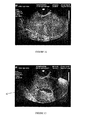
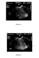
コントラスト強調超音波により子宮内の病状のより良い画像が提供される。例えば、図1から4参照。図1A、2A、3Aおよび4Aは、4人の患者それぞれにおける、子宮における単なる超音波の結果を示す。図1B、2B、3Bおよび4Bは、同一の4人の患者それぞれにおいて、本発明の媒質を用い超音波のコントラストを強調した結果を示す。本発明により成し遂げられるコントラストおよび視認性の実質的な強調は、以下に述べる本発明の利便性および利点を得ることのできない典型的なソノヒステログラフィーによって得られる程度と同程度である。 Contrast-enhanced ultrasound provides a better image of the intrauterine pathology. For example, see FIGS. Figures 1A, 2A, 3A and 4A show simple ultrasound results in the uterus in each of four patients. 1B, 2B, 3B and 4B show the results of enhancing the contrast of ultrasound using the medium of the present invention in each of the same four patients. The substantial enhancement of contrast and visibility achieved by the present invention is comparable to that obtained by typical sonohysterography where the convenience and advantages of the present invention described below cannot be obtained.
多くの研究において、子宮内コントラスト媒質の添加により通常の子宮ソノグラフィーを超える改良が為されている。特に、豊富なデータから、子宮の通常の超音波の結果および能力と比べると、子宮内病状の確認にサリンソノヒステログラフィーの陽性および陰性の予測値が優れていることが判った。ソノヒステログラフィーによって容易に確認される該病状には、粘膜下筋腫および子宮内膜ポリープが含まれ、その二つに共通するのは機能性子宮出血の要因となることである。また、多くの学術的研究により判明した多くの臨床的価値にもかかわらず、ソノヒステログラフィーの使用は実際のところ、比較的限定されており、それは、該手順は困難であり時間がかかるという特性によるものである。これら現実的な理由により、ソノヒステログラフィーは、通常、日々、婦人科で使用されているというわけではない。 In many studies, the addition of an intrauterine contrast medium has improved over normal uterine sonography. In particular, a wealth of data showed that the positive and negative predictive values of sarinsonohisterography were superior in confirming intrauterine pathology compared to normal uterine ultrasound results and capabilities. The pathologies readily identified by sonohysterography include submucosal fibroids and endometrial polyps, two of which are common causes of functional uterine bleeding. Also, despite the many clinical values found by many academic studies, the use of sonohysterography is actually relatively limited, indicating that the procedure is difficult and time consuming. It depends on the characteristics. For these practical reasons, sonohysterography is not usually used daily in gynecology.
サリンソノヒステログラフィーの変形型では、子宮腔で低音波発生(hypoechogenic)(「黒色」)ではなく高音波発生(hyperechogenic)(「白色」)をさせるため、通常の生理食塩水のような音波透過性(sono-transparent)溶液の代わりに、Echovist(登録商標)-200、Albunex(登録商標)、または同様な製品のような音波非透過性(sono-opaque)溶液が使用される。生理食塩水のような「黒色」の低音波発生性製品を超える価値を有する「白色」の高音波発生性コントラストが検討された。 In a variant of sarinsononohysterography, the uterine cavity is hyperechogenic (“white”) rather than hypoechogenic (“black”), and so Instead of a sono-transparent solution, a sono-opaque solution such as Echovist®-200, Albunex®, or similar product is used. A “white” hypersonic contrast with a value exceeding that of a “black” hyposonic product such as saline was investigated.
高音波発生(「白色」)媒質を使用するヒステロソノグラフィーでは、使用製品(Echovistおよび同様の製品)は、血管内注射用および心臓超音波のコントラスト強調用に設計された。そして、産科および婦人科での使用は追加的なものであった。それらは全て液体であり、ちょうどNaClまたは他の「黒色」の高音波発生性製品のように該手順を通して注入する必要がある。高音波発生(または陽性コントラスト)の手法において要求される1つの利点は、ファロピウス管、特にそれに近接する部分での画像化に陰性コントラストソノヒステログラフィー(生理食塩水など)を使用するとコントラストは相対的に消失するのに対して、該部分でのコントラスト画像が強調されるという点である。 In hysterosonography using hypersonic generation (“white”) media, the products used (Echovist and similar products) were designed for intravascular injection and contrast enhancement of cardiac ultrasound. And the use in obstetrics and gynecology was additional. They are all liquid and need to be injected through the procedure just like NaCl or other “black” sonogenic products. One advantage required in the technique of hypersonic generation (or positive contrast) is that the contrast is relative when using negative contrast sonohysterography (such as saline) for imaging in the fallopian tube, especially in close proximity to it. The contrast image at this portion is enhanced while the image disappears.
しかし、陽性コントラストソノヒステログラフィーは卵管の開通性を確認するのに使用し得ると今日の医師の多くが認識しているものの、従来のヒステロサルピンゴグラフィー(HSG)(X線を使用する検査)が未だに卵管の構造を研究する上での主だった方法となっている。得られる卵管画像の画質が優れているからである。卵管の開通性の検査では陽性コントラストヒステロソノグラフィーよりもHSGのほうが一般的に優れていることは認めるが、陽性コントラストヒステロソノグラフィーは卵管機能性(蠕動性萎縮)の研究で重要な役割をすると我々は考える。 However, while many of today's physicians recognize that positive contrast sonohysterography can be used to confirm tubal patency, conventional hysterosalpingography (HSG) (using X-rays) Inspection) is still the main method for studying the structure of the fallopian tube. This is because the image quality of the obtained oviduct image is excellent. Although we find that HSG is generally superior to positive contrast hysterosonography in tubal patency testing, positive contrast hysterosonography is important in studies of fallopian tube functionality (peristaltic atrophy) We think that it plays a role.
その関心の高さにもかかわらず、陽性コントラストソノヒステログラフィーは、陰性ソノヒステログラフィーと同様、煩わしい「第三の手」を必要とする手法であり、コントラスト液体の注入、超音波プローブの保持、および超音波発生機の必要な調節を同時に行う必要が多分にある。Echovist-200のような陽性コントラスト製剤は、注入可能な液体製剤であるため、その製品は、典型的にまた、検査の間、継続的にまたは定期的に注入される必要がある。それ故、これらの方法でもまた子宮腔中にカテーテルを保持して、媒質を継続的にまたは頻繁に再充填する必要があり、通常、好ましくない水のようなものを検診台に漏洩する結果となる。 Despite its high interest, positive contrast sonohysterography, like negative sonohysterography, is a technique that requires a cumbersome “third hand”. There is probably a need to hold and make the necessary adjustments of the ultrasonic generator at the same time. Since positive contrast formulations such as Echovist-200 are injectable liquid formulations, the product typically also needs to be infused continuously or periodically during the test. Therefore, these methods also require holding the catheter in the uterine cavity and refilling the medium continuously or frequently, usually resulting in the leakage of undesired water or something into the examination table. Become.
超音波法とは異なり子宮鏡検査法は、コントラスト画像というより直接可視化することを目的とする、子宮の内視鏡検査である。従って、その方法では、典型的に、コントラスト強調剤を使用する必要はないが、それでも、子宮(でなければ仮想腔、すなわち、「潰れた」、そしてそれ故、可視化困難であるもの)を拡張する「画像容易化」媒質として使用される。このタイプの方法は有用ではあるが重大な欠点が有り、それによって、その使用が広まっていない。これらの欠点とは、該検査を行っている間、患者に痛みと苦痛を与えること、検査の度毎に高価な装置を再滅菌する必要があることである。 Unlike ultrasonography, hysteroscopy is an endoscopic examination of the uterus aimed at direct visualization rather than contrast images. Thus, the method typically does not require the use of contrast-enhancing agents, but still dilates the uterus (otherwise the virtual cavity, ie “collapsed” and therefore difficult to visualize). Used as an “image facilitating” medium. While this type of method is useful, it has significant drawbacks, and its use is not widespread. These drawbacks are pain and distress to the patient during the test and the need to re-sterilize expensive equipment with each test.
子宮鏡検査の間、子宮鏡は子宮口を通し子宮腔に導入される。子宮鏡検査において手技は麻酔下で行われ、その麻酔は通常、全身麻酔であるが、脊髄麻酔または局所麻酔(子宮頸部分)でもよい。該方法では、比較的大きな装置、外科手術用子宮鏡を挿入するため約7から9mmの子宮頸部の拡張が必要である。NaCl溶液、リンゲル液、またはグリシン溶液のような溶液(モノポーラー凝固を考える場合)、またはCO2のような気体の継続的な注入が、継続的な腔の拡張に必要であり、出血の洗浄に必要である。 During hysteroscopy, the hysteroscope is introduced through the uterine ostium into the uterine cavity. In hysteroscopy, the procedure is performed under anesthesia, which is usually general anesthesia, but may be spinal or local anesthesia (cervical part). The method requires a cervical dilatation of about 7 to 9 mm to insert a relatively large device, a surgical hysteroscope. Solutions such as NaCl solution, Ringer's solution, or glycine solution (if considering monopolar coagulation) or continuous infusion of a gas such as CO 2 are required for continuous cavity dilation and can be used to clean bleeding is necessary.
ある状況では、子宮鏡使用手技においてHyscon(商標)のような濃度の高い溶液を注入する。粘性があり、生理食塩水よりも僅かに濃度が高いものの、Hysconはそれ自体、圧力なしに該腔を拡張するわけではない。そして、Hysconは非常に漏れ出しやすく、そのため、その方法でも継続的または定期的な注入が必要となる。 In certain situations, a highly concentrated solution such as Hyscon ™ is injected in a hysteroscopic procedure. Although viscous and slightly more concentrated than saline, Hyscon does not itself expand the cavity without pressure. And Hyscon is very easy to leak out, so that method also requires continuous or periodic injection.
子宮鏡は、必要ならば、小規模の外科的装置を該子宮鏡を介し挿入しポリープ、子宮内膜下子宮筋腫および瘢痕組織(癒着症)のような異常構造を切除および除去するか、または電気凝固またはレーザービームを用いそれらを焼き払う方法に典型的に用いられる。従って、子宮鏡手術は大がかりになりやすい。 A hysteroscope, if necessary, inserts a small surgical device through the hysteroscope to resect and remove abnormal structures such as polyps, subendometrial myomas and scar tissue (adhesion), or Typically used in methods of burning them using electrocoagulation or a laser beam. Therefore, hysteroscopic surgery tends to be large.
オフィスヒステロスコピー(office hysteroscopy)は、診断目的にのみ用いられる簡単なものである。オフィスヒステロスコピーに使用する装置のサイズは比較的小さい(直径3-5mm)ため、医師は子宮頸管の拡張および麻酔を避けることができる。にもかかわらず、その方法には痛みおよび苦痛が伴い、そのため、この形式の方法は未だに幾分制限される。子宮内視鏡手術ににおいての場合と同様、子宮腔を拡張させそれにより可視化するため、溶液または気体を検査の間を通して注入しなければならない。もちろん、注入自体(圧力により行われる)、および漏れた液体により典型的に生じた混乱は、患者の苦痛の更なる原因となる。 Office hysteroscopy is a simple one used only for diagnostic purposes. The size of the device used for office hysteroscopy is relatively small (3-5 mm in diameter), so doctors can avoid cervical dilation and anesthesia. Nevertheless, the method is painful and painful, so this type of method is still somewhat limited. As in uterine endoscopic surgery, a solution or gas must be infused throughout the examination in order to expand and visualize the uterine cavity. Of course, the infusion itself (performed by pressure) and the confusion typically caused by leaked liquid are a further source of patient pain.
そのため、(1)陰性または陽性子宮内コントラスト強調物の超音波コントラスト強調特性を提供する製品であって、子宮腔を僅かに拡張することにより、超音波検査の間に継続的に液体を灌流することが必要となる漏れを生じさせずにコントラスト中間相(contrast interphase)が得られるもの、または(2)光学的に透明であり、かつ、子宮鏡検査法に必要な拡張を提供する(該媒質の継続的な灌流に大きな装置は必要ではない)製品、またはその両方を提供する製品は、本分野の両態様において非常に大きな利点となるだろう。かかる製品があれば、内視鏡による方法は厄介なものでも、不快でもなく、面倒でもなく、そしてより便利で効率的なものとなるだろう。 Therefore, (1) products that provide the ultrasound contrast enhancement properties of negative or positive intrauterine contrast enhancements, with a slight dilation of the uterine cavity to continuously perfuse fluid during ultrasonography Providing a contrast interphase without causing the necessary leakage, or (2) optically transparent and providing the necessary extension for hysteroscopy (the medium Products that provide a product, or both, would be a huge advantage in both aspects of the field. With such a product, the endoscopic method would be cumbersome, not uncomfortable, not cumbersome, and more convenient and efficient.
かかる製品は、ゲルのような粘性を有するか、そうでなくとも検査中、子宮内注入後に子宮腔に保持され得るようなものであるべきである。これにより、医師は、注入後に検鏡および子宮内カテーテルを取り除くことができ、該検査中に該製品を継続的に注入することなく自由に超音波または子宮鏡検査法を行うことができる。更なる利点は、得られる画像には子宮内カテーテルにより生ずる画像の乱れがないという点である。理想的な製品は次いで、適当な時間後液状化、あるいは他の態様にて除去が容易となり、それによって、永続的に残存するわけでなく、むしろ超音波検査の完了後子宮腔から排泄されるべきである。粘性ゲルの粘性を残す物質が子宮内に残存するのは、生殖の妨げとなる可能性があり、好ましくない。 Such a product should have a gel-like viscosity, or otherwise be able to be retained in the uterine cavity after intrauterine injection during testing. This allows the physician to remove the speculum and intrauterine catheter after injection, and to perform ultrasound or hysteroscopy freely without continuously injecting the product during the examination. A further advantage is that the resulting image is free of image distortion caused by the intrauterine catheter. The ideal product is then easily liquefied after an appropriate time, or otherwise easily removed, so that it does not remain permanently, but rather is excreted from the uterine cavity after completion of the ultrasound examination Should. Remaining in the uterus of a substance that retains the viscosity of a viscous gel is not preferable because it may interfere with reproduction.
本明細書で使用する「画像を強調する」媒質とは、例えば、超音波または内視鏡検査のような方法の際に身体または器官の腔を拡張させることにより、および/または超音波検査のような画像化方法の際にコントラストを改善することにより、医療検査の際にコントラストおよび/または直接的な可視化を向上または容易にする媒質を意味する。従って、コントラスト強調も本明細書では、コントラスト画像を単に向上または改善するという純粋な意味よりも広い意味で使用する。代わりに、我々は、該コントラストを文字通り強調することにより、または検査する表面の拡張を容易にすることにより、それら何れかによるかまたはそれら両方により、一般的に画像化、放射線撮影、可視化、または他の同様の技術を向上または容易にすることをいうのに該用語を文章中で適宜使用する。 As used herein, an “image enhancing” medium is, for example, by expanding a body or organ cavity during a method such as ultrasound or endoscopy, and / or By improving contrast during such an imaging method is meant a medium that enhances or facilitates contrast and / or direct visualization during medical examination. Accordingly, contrast enhancement is also used herein in a broader sense than the pure meaning of simply enhancing or improving a contrast image. Instead, we generally do imaging, radiography, visualization, or either by literally enhancing the contrast or by facilitating the expansion of the surface to be inspected, either by them or both The term is used where appropriate in the text to refer to improving or facilitating other similar techniques.
本明細書で使用するとき、「相変化する」または「相シフトする」媒質とは、医療検査の際に身体または器官の腔内では最初は多くが固体または半固体であるが、その後、多くが液体に変化またはシフトし該腔からの除去または排泄しやすくなる(好ましくは該方法が完了した後)ものである。対照的に、「粘性変化する」または「粘性シフトする」媒質とは、医療検査の際、身体または器官の腔に使用するときには、より粘性があるが、その後、粘性は低くなり、該腔から除去または排泄しやすくなる(好ましくは該方法が完了した後)ものである。 As used herein, a “phase-changing” or “phase-shifting” medium is initially mostly solid or semi-solid within a body or organ cavity during a medical examination, but then many Changes or shifts to a liquid and is easily removed or excreted from the cavity (preferably after the method is complete). In contrast, a “viscous changing” or “viscous shifting” medium is more viscous when used in a body or organ cavity during a medical examination, but then becomes less viscous, It is easy to remove or excrete (preferably after the method is completed).
本明細書で使用するとき、「固体」、「半固体」または「ゲル」状態または相である媒質とは、物理的に十分に安定(すなわち、液体または気体状というよりもむしろ比較的、固体であるかまたは粘性がある)であり、医療検査の際には継続的または頻繁に充填する必要が典型的になく身体または器官の腔内に実質的に残存するものである。 As used herein, a medium that is in a “solid”, “semi-solid” or “gel” state or phase is physically sufficiently stable (ie, relatively solid rather than liquid or gaseous). Or are viscous) and remain substantially in the body or organ cavity without the need for continuous or frequent filling during medical examinations.
本明細書で使用するとき、医療検査を行うのに「十分な時間」とは、典型的には、少なくとも数分、一般的には約3から10分、および好ましくは約5から7分のオーダーである。その十分な時間、本発明の組成物が所定のところにある間に、方法は通常完了する。液状化または粘性が減少するかどうか、次いで該組成物が身体または器官の腔から漏れるか否か、または迅速におよび容易に除かれるか否か。一般的に、該腔では、該腔内に最初に置いてから約7から20分以内、好ましくは約10から15分以内で該組成物は殆どなくなる。 As used herein, “sufficient time” to perform a medical examination is typically at least a few minutes, generally about 3 to 10 minutes, and preferably about 5 to 7 minutes. It is an order. The process is usually complete while the composition of the present invention is in place for that sufficient time. Whether liquefaction or viscosity decreases, then whether the composition leaks from the body or organ cavity, or is quickly and easily removed. Generally, the cavity is nearly depleted of the composition within about 7 to 20 minutes, preferably within about 10 to 15 minutes of first placement in the cavity.
本明細書で使用するとき、「医用画像」とは、評価、診断、観察、治療などを含む種々の目的にて、特定の関心事または症状のため、または単に予防のために用いられる、身体もしくは器官の腔または隣接組織を検査する画像化、放射線撮影、可視化、または他の同様の方法をいう。その方法には、例えば、超音波、内視鏡検査、MRI、X-線、ヒステロサルピンゴグラム(HSG)、CT(コンピューター断層撮影)スキャン(何れも「医用画像法」)が含まれる。同様に、医用画像組成物または媒質とは、その医用画像法での使用を目的とするものである。 As used herein, a “medical image” is a body that is used for a specific interest or symptom, or simply for prevention, for a variety of purposes, including evaluation, diagnosis, observation, treatment, etc. Or imaging, radiography, visualization, or other similar method of examining an organ cavity or adjacent tissue. The methods include, for example, ultrasound, endoscopy, MRI, X-ray, hysterosalpingogram (HSG), CT (computed tomography) scan (all “medical imaging”). Similarly, a medical imaging composition or medium is intended for use in the medical imaging method.
本発明は、身体または器官の腔に用いる医用画像法で使用する医用画像組成物に関する。該組成物には、画像強調、粘性シフト媒質が含まれ、それは、身体または器官の腔への導入時または導入後画像法が完了するのに十分な時間、該腔内の所定の位置に実質的に保持されるのに十分な初期粘性または粘稠性を有する。有利なことに、該組成物ではまた、その後、該身体または器官の腔から容易に該媒質が排泄または除去されるように粘性または粘稠性が十分に減少する。 The present invention relates to a medical imaging composition for use in medical imaging methods for use in body or organ cavities. The composition includes an image enhancement, viscosity shifting medium, which is substantially in place in the cavity during or after introduction into the body or organ cavity for a time sufficient to complete the imaging. Sufficient initial viscosity or consistency to be retained. Advantageously, the composition also has a sufficiently reduced viscosity or consistency so that the medium is then easily excreted or removed from the body or organ cavity.
本発明の1つの実施態様では、媒質は身体または器官の腔への導入時または導入後最初は固体、半固体、またはゲルである。そのため、該媒質を、画像法の間、継続的にまたは頻繁に投与する必要はない。この実施態様では、該媒質は、一定時間後、典型的には液状化するかまたは粘性が低下し、それによって腔から容易に排泄または除去し得る。 In one embodiment of the invention, the medium is initially a solid, semi-solid, or gel upon or after introduction into the body or organ cavity. As such, the medium need not be administered continuously or frequently during imaging. In this embodiment, the medium typically liquefies or decreases in viscosity after a period of time, so that it can be easily excreted or removed from the cavity.
本発明の特定の実施態様では、媒質は特有の性質を有するように設計されており、その性質によって該媒質は膣の超音波検査での使用に適した組成物となる。 In certain embodiments of the present invention, the medium is designed to have unique properties that make it a composition suitable for use in vaginal ultrasonography.
更なる実施態様では、媒質は、組成物の特定の温度変化に応じて液状化するか粘性が減少するように設計されている。 In further embodiments, the medium is designed to liquefy or decrease in viscosity in response to a specific temperature change of the composition.
媒質には、1以上のポリマーおよび所望により1以上のアジュバントまたは治療剤が含まれ得る。使用する該アジュバントまたは治療剤には、典型的に抗感染性剤または放射線不透過性剤が含まれる。 The medium can include one or more polymers and optionally one or more adjuvants or therapeutic agents. The adjuvant or therapeutic agent used typically includes an anti-infective agent or a radiopaque agent.
本発明はまた、身体または器官の腔に医用画像法を行う方法に関係する。ある実施態様では、該方法は、(a)本発明の組成物を子宮のような腔中に入れること、(b)該画像法を行うこと、そして(c)該腔から該組成物を除去または排泄させることを含む。 The invention also relates to a method for performing medical imaging on a body or organ cavity. In certain embodiments, the method comprises (a) placing the composition of the invention in a cavity such as the uterus, (b) performing the imaging, and (c) removing the composition from the cavity. Or including excretion.
本発明は、更に、身体または器官の腔に用いる医用画像法に使用する画像強調組成物の製造における本発明の媒質の使用に関係する。使用する該媒質は、典型的に十分な粘性を有しており、そのため、該方法の間、継続的にまたは頻繁に投与する必要がないが、該方法を正常に完了するのに十分な時間経過後に該腔から容易に排泄または除去される。 The present invention further relates to the use of the media of the present invention in the manufacture of image enhancement compositions for use in medical imaging methods used in body or organ cavities. The medium used typically has sufficient viscosity so that it does not need to be administered continuously or frequently during the method, but sufficient time to successfully complete the method. It is easily excreted or removed from the cavity after the passage.
これらの実施態様のうちの何れかにおいて、最初の粘性または固体、固体状、またはゲル状態は、媒質のポリマーまたは他の成分を使用することにより部分的または全体的に生じ得る。 In any of these embodiments, the initial viscous or solid, solid, or gel state can be generated in part or in whole by using a polymer or other component of the medium.
本発明の媒質は、僅かな圧力で身体または器官の腔を拡張させ、医用画像化に付随する、更なる該媒質の継続的注入を行うことなく該腔を医用画像化し得る。良好な画質が、該媒質が液状化するまで(それは、画像が悪くなるか該媒質が失われることにより検査状態が悪くなる時点である)、典型的に得ることが可能である。該媒質は、継続的な注入の必要が全くなく子宮腔を全体的に検査するのに十分な時間を得ることを目的とするが、より長時間、該検査を行うことが必要ならば、第二または更なる追加的な適用を一般的に行い得る。 The medium of the present invention can expand a body or organ cavity with little pressure and medically image the cavity without further continuous infusion of the medium associated with medical imaging. Good image quality can typically be obtained until the medium liquefies, which is the point at which the image becomes worse or the inspection condition worsens due to the loss of the medium. The medium is intended to allow sufficient time to examine the uterine cavity globally without any need for continuous infusion, but if it is necessary to perform the examination for a longer period of time, Two or further additional applications can generally be made.
図面の簡単な説明
図1Aおよび1Bは、それぞれ、患者#1に行った通常の超音波検査および本発明を使用した超音波検査を示す。図1Bは、図1Aでは見られない子宮内膜ポリープ(10)の陽性結果を示す。組織学的にポリープの存在を確認した。
BRIEF DESCRIPTION OF THE DRAWINGS FIGS. 1A and 1B show a normal ultrasound examination performed on
図2Aおよび2Bは、それぞれ、患者#2に行った通常の超音波検査および本発明を使用した超音波検査を示す。図2Bは、図2Aでは見られない子宮内膜ポリープまたは子宮筋腫(11)を示す陽性結果を示す。組織学的にポリープの存在を確認した。 2A and 2B show a normal ultrasonography performed on patient # 2 and an ultrasonography using the present invention, respectively. FIG. 2B shows a positive result indicating endometrial polyps or uterine fibroids (11) not seen in FIG. 2A. Histologically, the presence of polyps was confirmed.
図3Aおよび3Bは、それぞれ、患者#3に行った通常の超音波検査および本発明を使用した超音波検査を示す。図3Bは、図3Aでは見られない子宮内膜ポリープ(10)を示唆する陽性結果を示す。組織学的にポリープの存在を確認した。
FIGS. 3A and 3B show a normal ultrasound examination performed on
図4Aおよび4Bは、それぞれ、患者#4に行った通常の超音波検査および本発明を使用した超音波検査を示す。図4Bは、図4Aでは見られない子宮内膜ポリープまたは子宮筋腫(11)を示唆する陽性結果を示す。組織学的にポリープの存在を確認した。
4A and 4B show a normal ultrasonography performed on
図5および6は、本発明を使用した他の2人の患者の子宮鏡検査の結果を示す。図5は子宮ポリープ(12)を示し、図6は子宮の癒着または瘢痕(13)を示す。両方の結果とも、その後に外科的子宮鏡検査法により確認した。 Figures 5 and 6 show the results of hysteroscopy of two other patients using the present invention. FIG. 5 shows a uterine polyp (12) and FIG. 6 shows a uterine adhesion or scar (13). Both results were subsequently confirmed by surgical hysteroscopy.
本発明の詳細な説明
本発明は、身体または器官の腔の、検査、評価、診断、治療、観察、および検診などの目的の医用画像法で媒質として使用する便利の良いコントラスト強調組成物を提供する。該腔が、空っぽであるが拡張および開いてはおらず、空っぽのバルーンと同様に潰れ、折り込まれる傾向にあるという点で典型的に「仮想的な腔」である。
DETAILED DESCRIPTION OF THE INVENTION The present invention provides a convenient contrast enhancement composition for use as a medium in medical imaging for purposes such as examination, evaluation, diagnosis, treatment, observation, and screening of body or organ cavities. To do. The cavity is typically a “virtual cavity” in that it is empty but not expanded and open, and tends to collapse and fold like an empty balloon.
その方法には、例えば、超音波検査、内視鏡検査、MRI、X-線、ヒステロサルピンゴグラム(HSG)、CTスキャンが含まれる。特定の方法を確実に行え、該方法に適当となるように、媒質は、生理食塩水または同様の注入物のコントラスト強調特性を供し得る。該コントラストを強調する効果は、例えば生理食塩水または同様の溶液により生ずる「黒色」の中間相、またはEchovistのような高音波発生溶液により生ずる「白色」の中間相から得ることができる。何れの場合においても、コントラスト剤により生ずる中間相は、様々な濃さの灰色で表示される周囲の構造とは異なる。コントラスト強調が望ましいか否かにかかわらず、該媒質はまた、該方法の間に著しい漏れがなくまたは継続的または定期的な再充填の必要がなく所定の位置にとどまる能力を有する一方、該方法が完了するのに十分な時間経過後に相または粘性が変化するか、またはその他の態様にて完了時に身体または器官の腔から簡単にそして容易に排泄されまたは除去される能力を有し得る。 The methods include, for example, ultrasonography, endoscopy, MRI, X-ray, hysterosalpingogram (HSG), CT scan. To ensure that a particular method can be performed and is suitable for the method, the medium can provide the contrast enhancement properties of saline or similar infusion. The contrast enhancing effect can be obtained, for example, from a “black” mesophase produced by saline or similar solution, or from a “white” mesophase produced by a hypersonic solution such as Echovist. In any case, the mesophase generated by the contrast agent is different from the surrounding structure displayed in various shades of gray. Regardless of whether contrast enhancement is desired, the medium also has the ability to remain in place without significant leakage during the method or without the need for continuous or periodic refilling The phase or viscosity may change after a sufficient amount of time to complete, or may otherwise have the ability to be easily and easily excreted or removed from the body or organ cavity upon completion.
媒質を注入するための圧力が下がると患者の苦痛は和らぐ。本発明の媒質は、子宮頸部およびチューブからの継続的な漏れに打ち勝つ必要がないため、典型的により低圧でおよび従来技術の媒質よりも圧力を掛ける時間が短くて注入され得る一方、身体または器官の腔は十分に拡張し得る。この圧力の低下および加圧時間の短縮化は患者の苦痛を最少限にするのに役立つ。更に、該方法において該チューブを介して骨盤腔中へ大量に漏れる(spillage)ことがなくなり、更に、本発明を使用する方法によって苦痛は除かれることになる。 The patient's pain is eased when the pressure to inject the medium decreases. The medium of the present invention does not need to overcome continuous leakage from the cervix and tube, so it can be infused while typically applying a lower pressure and less time to apply pressure than prior art media, The organ cavity can expand sufficiently. This reduction in pressure and shortening of the pressurization time helps to minimize patient pain. In addition, the method does not spillage into the pelvic cavity through the tube, and pain is eliminated by the method using the present invention.
本発明は、観察、診断、治療または他の目的の何れかを目的とする、以下に限らないが、x線、超音波、CTスキャンまたはMRIのような従来の「画像化」法、HSGまたはCTスキャンのような放射線撮影検査、または内視鏡のような直接の可視化技術を含む種々のタイプの医用画像法に有用となる。 The present invention is intended for any of the following purposes, including but not limited to conventional “imaging” methods such as x-ray, ultrasound, CT scan or MRI, HSG or It is useful for various types of medical imaging methods including radiographic examinations such as CT scans, or direct visualization techniques such as endoscopes.
上記考察のように、妊婦に行う超音波検査の場合、羊水が子宮内コントラスト強調物としての役割をし、陰性または「黒色の」音波面領域(sonic interface)を供することとなる。妊娠していない子宮では、子宮腔にはコントラスト面領域(contrasting interface)が無く〜潰れることにより生じた子宮の折り目、および小さな空隙により容易に可視化することができない〜詳細な画像を得ることができない。それ故、正常および病態画像(ポリープ、または粘膜下筋腫など)がしばしば不鮮明となり、最終的な診断ができない。 As discussed above, in the case of ultrasound performed on pregnant women, amniotic fluid serves as an intrauterine contrast enhancer and provides a negative or “black” sonic interface. In a non-pregnant uterus, the uterine cavity has no contrasting interface (cannot be easily visualized due to folds of the uterus caused by collapse and small voids)-detailed images cannot be obtained . Therefore, normal and pathological images (such as polyps or submucosal fibroids) are often blurred and cannot be finally diagnosed.
ヒステロソノグラフィーは妊婦における超音波検査と似ており、良好な画質が得られる。しかし、ヒステロソノグラフィーは煩わしく、時間がかかり、そして不便であり、典型的に2人以上で実施する必要がある。本発明では、ヒステロソノグラフィーにおいて鍵となる利点を供し、これら主たる問題点をさけるべく創意工夫した。 Hysterosography is similar to ultrasonography in pregnant women and provides good image quality. However, hysterosonography is cumbersome, time consuming and inconvenient and typically needs to be performed by two or more people. The present invention provides key advantages in hysterosonography and has been devised to avoid these major problems.
高音波発生(「白色」)媒質を用いるヒステロソノグラフィーは、子宮の病状および卵管の構造(開通性)の検査に推奨されている。使用する製品(Echovist、および同様の製品)は、腔内注射および心臓超音波検査でのコントラスト強調用に設計されたものである。産科および婦人科でのその使用は追加的なものであった。これらの製品は液体であり、ちょうどNaCl溶液のようにその手順により注入する必要がある。 Hysterosography using hypersonic ("white") media is recommended for examination of uterine pathology and fallopian tube structure (patency). The products used (Echovist and similar products) are designed for contrast enhancement in intracavitary injection and cardiac ultrasonography. Its use in obstetrics and gynecology was additional. These products are liquid and need to be injected by the procedure just like a NaCl solution.
経験上、これらの製品は、ポリープおよび粘膜下筋腫のような子宮内の病状の確認ではNaClより優れているわけではないことがわかった。医師の中には、シャドーイング効果が生じやすいため生理食塩水のほうが望ましいと考えるものもいる。しかし、卵管検査では、白色コントラストは、生理食塩水溶液のような陰性(「黒色」)媒質よりも幾分優れていた。そうは言うものの、卵管解剖学的検査でのこれらの製品の価値には疑問が残る。未だHSG(x線)のほうが優れているためである。我々は、「白色」製品を卵管機能の試験に使用し得ることを証明し得ると考える。 Experience has shown that these products are not superior to NaCl in confirming intrauterine conditions such as polyps and submucosal fibroids. Some doctors consider that saline is preferable because it is prone to shadowing effects. However, in oviduct testing, white contrast was somewhat better than negative (“black”) media such as saline solution. That being said, the value of these products in tubal anatomy remains questionable. This is because HSG (x-ray) is still superior. We believe that a “white” product may prove to be used for testing oviduct function.
本発明の特定の実施態様では、相シフト子宮コントラスト媒質(PSCM)を用いており、ヒステロソノグラフィーで得られる子宮画像の画質に匹敵する一方、通常の膣超音波検査の使用は簡単となり、非常に容易となる。例えば、以下の実施例1参照。通常の卵移植カテーテルを子宮内に挿入するとき、該媒質は濃厚であり、適度に腔を拡張する。使用する特定のPSCM(水の音波特性を有する)は、陰性または「黒色」のコントラストを供し、子宮内膜ポリープおよび/または粘膜下筋腫のような腔内で突き出た構造を鮮明に可視化する。相シフト特性を有していることから、該PSCMは後にそれ自体の温度が上昇し体温に到達し液状化する。一旦、液体になると、子宮の拡張は終了し、該PSCMは排泄される。 Certain embodiments of the present invention use a phase shift uterine contrast medium (PSCM), which is comparable to the image quality of uterine images obtained with hysterosonography, while simplifying the use of normal vaginal ultrasonography, It will be very easy. See, for example, Example 1 below. When a normal egg transplant catheter is inserted into the uterus, the medium is thick and moderately dilates the cavity. The particular PSCM used (having sonic properties of water) provides a negative or “black” contrast and clearly visualizes protruding structures within the cavity such as endometrial polyps and / or submucosal fibroids. Since the PSCM has a phase shift characteristic, the temperature of the PSCM increases later, reaches the body temperature, and liquefies. Once in liquid form, uterine dilation ends and the PSCM is excreted.
それ故、本発明の特定の実施態様は、「白色」または「黒色」コントラスト強調特性を有する、子宮超音波検査法用の水性ゲルであり得る。該ゲルは、超音波検査の間、子宮腔にとどまることができる。所定の時間、一般的に約3-10分で、該ゲルは液状化し、子宮の蠕動により該子宮腔から排泄される。この排泄は、ヒステロソノグラフィーのような、従来技術において生理食塩水の注入によって、子宮からまだ漏れ出していなかった残りを排泄させるのに似ている。 Thus, certain embodiments of the present invention may be aqueous gels for uterine ultrasonography, having “white” or “black” contrast enhancement properties. The gel can remain in the uterine cavity during ultrasonography. In a given time, typically about 3-10 minutes, the gel liquefies and is excreted from the uterine cavity by uterine peristalsis. This excretion is similar to excreting the remainder that has not yet leaked out of the uterus by injection of saline in the prior art, such as hysterosonography.
ある実施態様において、時限的な粘性は温度に依存する。例えば、室温またはそれより低い温度のその組成物は、子宮に挿入されたとき、体温(粘性を喪失する温度)に到達するまで初期の高い粘性を有しており、その温度によって液体となり、容易に排泄される。これにより、通常の子宮内人工授精(intrauterine insemination)(IUI)に必要な清浄手順および予防措置および全ての子宮内手順を用い、該IUIで受精卵輸送に使用する簡単なプラスチックカテーテルに似たものを用い子宮内膜腔(endometrial cavity)中に約2-7ccの子宮ゲルを注入することができる。 In some embodiments, the timed viscosity is temperature dependent. For example, a composition at or below room temperature has an initial high viscosity when inserted into the uterus until it reaches body temperature (the temperature at which it loses viscosity), which makes it liquid and easily Is excreted. This is similar to the simple plastic catheter used for fertilized egg transport in the IUI, using the cleaning procedures and precautions required for normal intrauterine insemination (IUI) and all intrauterine procedures. Can be used to inject about 2-7cc of uterine gel into the endometrial cavity.
超音波検査法では、カテーテルおよび検鏡を取り除き、そして超音波検査を膣および腹部超音波検査の全てにおいて通常通りに行う。子宮ゲルは、十分な時間、超音波検査が完了するまで粘性を有しており、その一方、従来、複雑な「第三の手」が必要なヒステロソノグラフィー法のみによって得られていたコントラスト強調と同様の強調が利点となる。 In ultrasonography, the catheter and speculum are removed and ultrasound is performed as usual in all vaginal and abdominal ultrasound. Uterine gels are viscous enough for ultrasonography to complete, while contrasting conventionally obtained only by the hysterography method, which requires a complex "third hand" Emphasis similar to emphasis is an advantage.
子宮鏡検査法では、同一タイプの媒質が典型的に使用され得るが、一般的にコントラスト強調を供する必要はない。代わりに、その媒質は通常光学的に透明であり得る。 In hysteroscopy, the same type of medium can typically be used, but generally it is not necessary to provide contrast enhancement. Instead, the medium can usually be optically transparent.
子宮鏡検査法では、子宮腔の内部の様子を直接可視化する。該方法では、子宮は人為的に拡張させる必要がある。子宮の潰れた、折りたたまれた壁ではなく全体像を得るためである。使用される装置(子宮鏡)は、照明を提供し子宮腔内を可視化する光ファイバーシステムと、該腔を拡張するのに十分な圧力の溶液または気体を注入する機構とが組み合わされており、それにより子宮壁を見ることができる。子宮頚管への挿入に伴う苦痛を最小限にするため、診断目的にのみに制限されるオフィスヒステロスコピーを用い装置を小型化する。しかし、小型化は、現在の子宮鏡の複雑さ故に、特に注入装置と光学システムとを組み合わせる必要があるために制限される。これら工学的制限が要因となって、オフィスヒステロスコピーでは未だに相当に痛みを伴い、そのため、その通常のおよび頻繁な使用の障害となっている。 Hysteroscopy directly visualizes the interior of the uterine cavity. In this method, the uterus needs to be artificially expanded. This is to get a full picture, not a collapsed, folded wall of the uterus. The device used (hysteroscope) combines an optical fiber system that provides illumination and visualizes the interior of the uterine cavity with a mechanism that injects a solution or gas at a pressure sufficient to expand the cavity. You can see the uterine wall. To minimize the pain associated with insertion into the cervix, the device is miniaturized using office hysteroscopy, which is limited to diagnostic purposes only. However, miniaturization is limited because of the complexity of current hysteroscopy, especially because it is necessary to combine an injection device with an optical system. Due to these engineering limitations, office hysteroscopy is still quite painful and thus hinders its normal and frequent use.
子宮鏡を小型化し該方法による痛みを和らげようとする試みよって、コンタクトヒステロスコピー(contact hysteroscopy)が開発された。コンタクトヒステロスコピーは、子宮腔を拡張させることなく頚管内および子宮内膜の粘膜を直接可視化することからなり、そのため、該腔の全体像が得られるということはない。この場合、単一のガラスカラム(注入装置を有する鞘はない)を搭載した硬質なスコープは、頚管内および子宮内膜の粘膜を直接可視化する。光学増幅システムを用いて観察した場合、該粘膜(頚管内および子宮内膜)の組織学的診断がある程度可能となる。このため、コンタクトヒステロスコピーは、癌、出血の組織学的要因のスクリーニングに用いられ、また子宮内膜日付診断(functional dating)に用いられる可能性もある。しかし、子宮腔は拡張していないため、コンタクトヒステロスコピーでは、子宮内膜ポリープまたは粘膜下筋腫(外科的な除去を必要とする機能不全性子宮出血(DUB)で最もよくある解剖学的要因)に必要な全体像は得られない。 Contact hysteroscopy has been developed in an attempt to reduce the size of the hysteroscope and relieve the pain associated with the method. Contact hysteroscopy consists of directly visualizing the mucosa of the cervical and endometrial tracts without dilating the uterine cavity, so that an overall picture of the cavity is not obtained. In this case, a rigid scope with a single glass column (no sheath with infusion device) directly visualizes the mucosa of the cervical canal and endometrium. When observed using an optical amplification system, histological diagnosis of the mucosa (intracervical and endometrium) is possible to some extent. For this reason, contact hysteroscopy is used to screen for histological factors of cancer and bleeding, and may also be used for functional dating of endometrium. However, because the uterine cavity is not dilated, contact hysteroscopy shows endometrial polyps or submucosal fibroids (the most common anatomical factor in dysfunctional uterine bleeding (DUB) that requires surgical removal) The overall picture required for) cannot be obtained.
子宮腔の内視鏡検査において、拡張させるための液体または気体を注入する必要性を除くべく、光学的に透明な、相シフト媒質(PSM)を使用し得る。その新規媒質は例えば室温では十分粘性があり、内視鏡検査において子宮腔を拡張する特性を有する。数分の後、体温に到達すると液状化し、子宮腔から自然に排泄される。 In endoscopic examination of the uterine cavity, an optically transparent, phase shift medium (PSM) may be used to eliminate the need to inject liquid or gas to expand. The new medium is sufficiently viscous at room temperature, for example, and has the property of expanding the uterine cavity in endoscopy. A few minutes later, when it reaches body temperature, it liquefies and is naturally excreted from the uterine cavity.
かかる一時的なゲル相、または他の有用媒質の粘性は上に説明したように熱感受性であるか、別の態様にて一定時間後に液体組成物に変化し得る。好ましくは、該媒質は、半固体または固体(ゲルのような)から液体に変化する相または粘性シフト特性を有している。該媒質の物理的特性のうちの該変化の特性(および該変化のタイミングおよびきっかけ)は1以上の要因に依存し得る。例えば、形態変化のきっかけとなり得る要因は、pH、温度、剪断力(shear)または圧力の変化、更なる物質(例えば、塩、溶媒、酸性物質または塩基性物質)の存在、電位の適用、または単なる時間の経過のような種々の状態に基づき得る。一旦、該媒質が液状化すると、その変化した特性により、相シフト過程の完了によって任意の液体のように排泄される。 The viscosity of such a temporary gel phase, or other useful medium, can be heat sensitive as described above, or in other embodiments can change to a liquid composition after a period of time. Preferably, the medium has a phase or viscosity shift characteristic that changes from a semi-solid or solid (such as a gel) to a liquid. Of the physical properties of the medium, the nature of the change (and the timing and trigger of the change) may depend on one or more factors. For example, factors that can trigger morphological changes include changes in pH, temperature, shear or pressure, the presence of additional materials (e.g., salts, solvents, acidic or basic materials), potential application, or It can be based on various conditions such as just the passage of time. Once the medium is liquefied, due to its altered characteristics, it is excreted like any liquid upon completion of the phase shift process.
媒質の特定組成物に依存して、その媒質は、超音波画像における陰性(黒色)または陽性(白色)の何れかのコントラスト付加物を提供し得る。該媒質の相シフトまたは粘性シフト特性は、子宮のような身体または器官の仮想腔の超音波画像または内視鏡検査用の画像強調物としての使用に好ましい。 Depending on the specific composition of the medium, the medium can provide either negative (black) or positive (white) contrast adducts in the ultrasound image. The phase shift or viscosity shift characteristic of the medium is preferred for use as an ultrasound image of a virtual cavity of a body or organ such as the uterus or an image enhancer for endoscopy.
本発明の熱感受性または他の一時的な粘性は、医薬組成物に有用な特定成分の特定のゲル形成特性に由来し得るものである。例えば、カルボマー934P、またはカルボマー974Pのような特定のポリマーは、「一時的な」粘性を供する本発明の方法に有用となり得る。これらポリマーまたは他の成分は、例えば、低温では粘性のある組成物中において一時的に粘性があるが体温またはそれに近い温度になると液状化する製剤、または僅かな時間粘性でありその後液状化する製剤の提供に有用であることが判る。更に、望ましいならば、例えば、抗感染性剤、放射線不透過性剤、粒状性を低下する薬剤または他に該組成物の見た目をよくする薬剤、または保存剤のような、他の製剤またはアジュバントもまた該組成物に加えられ得る。 The heat sensitivity or other temporary viscosity of the present invention can be derived from the specific gel-forming properties of the specific ingredients useful in the pharmaceutical composition. For example, certain polymers such as Carbomer 934P, or Carbomer 974P may be useful in the methods of the invention that provide “temporary” viscosity. These polymers or other ingredients are, for example, preparations that are temporarily viscous in a composition that is viscous at low temperatures but become liquefied at or near body temperature, or that become viscous for a short time and then liquefy It turns out that it is useful for providing. In addition, if desired, other formulations or adjuvants such as, for example, anti-infective agents, radiopaque agents, agents that reduce granularity or other agents that improve the appearance of the composition, or preservatives Can also be added to the composition.
もちろん、本発明は、当業者に容易に理解できるようにより広く応用可能である。例えば、他の身体または器官(膣または子宮腔以外)の超音波または内視鏡検査もまた、本発明の使用によって容易になるだろう。同様に、X線またはMRIのような他の医用画像法もまた、本発明の使用によって利益が得られ得る。 Of course, the present invention can be more widely applied to those skilled in the art. For example, ultrasound or endoscopy of other bodies or organs (other than the vagina or uterine cavity) will also be facilitated by use of the present invention. Similarly, other medical imaging methods such as X-rays or MRI may also benefit from the use of the present invention.
加えて、一時的な粘性または固体性は本発明の殆どの実施態様で重要となるが、該組成物の液状化または更には変換のための厳密な機構または手段自体は重要ではない。該媒質は投与時当初は粘性があり、次いで比較的短時間および予測可能な時間内に液状化するか、でなければ該方法が完了するのに十分な時間経過後容易に除去される限りにおいて、液状化または他の相もしくは粘稠度への変換は、例えば、上記考察したように温度変化によって、または単に一定時間経過後に、または他の薬剤の添加によって生じ得る。もちろん、本発明は、要求通り所定の場所にとどまるのに十分な粘性がある組成物であれば、該組成物の粘性または粘稠性が変化することなく排泄または除去され得る組成物を用いても実施可能である。 In addition, although temporary viscosity or solidity is important in most embodiments of the present invention, the exact mechanism or means for liquefaction or even conversion of the composition itself is not important. The medium is initially viscous at the time of administration and then liquefies within a relatively short and predictable time, or as long as it is easily removed after sufficient time to complete the process. Liquefaction or conversion to other phases or consistency can occur, for example, by changes in temperature as discussed above, or simply after a period of time, or by the addition of other agents. Of course, the present invention uses a composition that can be excreted or removed without change in the viscosity or consistency of the composition, provided that the composition is sufficiently viscous to remain in place as required. Can also be implemented.
本発明が子宮鏡検査法による使用を提供または支持する利点としては、特に以下のものが含まれる:
1.溶液または気体の継続的な注入の必要のない子宮鏡検査法;
2.注入装置(チューブ、さや等)がない、より小さな直径の子宮鏡装置の開発、プラスチックまたは他の物質の使用が可能となり、使い捨て装置の開発が容易とされ得る。これら新規な簡単なオフィスヒステロスコープ(office hysteroscope)(使い捨てかまたは使い捨てではない)は、ビューイングカメラ(viewing camera)に連結し、その後、インスタントビューイング(instant viewing)し、そして画像または連続画像を記録し得る。
Among the advantages that the present invention provides or supports for use by hysteroscopy include, among others:
1. Hysteroscopy without the need for continuous infusion of solution or gas;
2. The development of smaller diameter hysteroscopic devices without infusion devices (tubes, sheaths, etc.), the use of plastics or other materials can be made, and the development of disposable devices can be facilitated. These new simple office hysteroscopes (disposable or non-disposable) are connected to a viewing camera, then instant viewing, and images or continuous images Can be recorded.
該媒質はまた、体腔中への導入前に固体または半固体であるものでも、または体腔中へ導入されることにより固体または半固体にシフトし得るものであってもよい。該媒質はまた、導入前またはその後は半固体または固体であるが、相シフトを生じ該媒質を液状化する要因となる他の物質(例えば、塩、酸性または塩基性の物質、または溶媒)の導入によって相シフトし得るものであってもよい。 The medium may also be solid or semi-solid prior to introduction into the body cavity, or may be shifted to solid or semi-solid upon introduction into the body cavity. The medium is also semi-solid or solid before or after introduction, but other substances (e.g. salts, acidic or basic substances, or solvents) that cause a phase shift and cause the medium to liquefy. It may be capable of phase shifting by introduction.
望ましい相または粘性の特性を供する該媒質組成物に有用な成分には、以下に限らないが、ポリマー(カルボマー、メチルセルロース、ゼラチン、糖、ペクチン、デンプン、高分子量ポリエチレングリコールなど)、およびコロイドクレイ(colloidal clays)(ベントナイトまたはトラガカントゴム)が含まれる。揺変性混合物はまた、該媒質と同じぐらいに有用となり得ると考えられる。その無機物質の混合物は拡張剤として使用し得、可視化しやすくする。 Ingredients useful in the media composition that provide desirable phase or viscosity properties include, but are not limited to, polymers (carbomers, methylcellulose, gelatin, sugar, pectin, starch, high molecular weight polyethylene glycols, etc.), and colloidal clays ( colloidal clays) (bentonite or gum tragacanth). It is believed that the thixotropic mixture can also be as useful as the medium. The mixture of inorganic materials can be used as an extender to facilitate visualization.
理想的には、画像強調製品は、子宮粘膜および生殖体にとって可能な限り非毒性のものである。該製品の変形型は、超音波検査法のような画像法での使用のため、「陰性」および「陽性」コントラスト強調特性を有するよう開発されるべきである。 Ideally, the image enhancement product is as non-toxic as possible to the uterine mucosa and reproductive body. Variants of the product should be developed to have “negative” and “positive” contrast enhancement properties for use in imaging methods such as ultrasonography.
陰性コントラスト強調物は、典型的に、子宮内膜下子宮筋腫、子宮内膜ポリープ、および子宮癒着および奇形のような子宮腔中で突き出る、子宮の病状(しばしば機能不全性子宮出血または不妊症の要因となるようである)を確認するのに最も適している。陽性コントラスト強調物は典型的に画像化、例えば、ファロピウス管の画像化に有用となる。陰性および陽性コントラスト強調物の使用は、特定の状況下で行い得るならば、連続的に、または組み合わせて、二重コントラスト画像化に役立ち得る。 Negative contrast enhancers are typically uterine pathologies (often dysfunctional uterine bleeding or infertility) that protrude in the uterine cavity, such as subendometrial fibroids, endometrial polyps, and uterine adhesions and malformations. It seems to be a factor). Positive contrast enhancers are typically useful for imaging, for example, imaging of Fallopian tubes. The use of negative and positive contrast enhancers can be useful for double-contrast imaging, either continuously or in combination, if it can be done under certain circumstances.
陽性コントラスト製品の臨床適用の可能性は、Echovistのような従来の陽性超音波コントラスト強調物において提案されている使用よりも広範となる。Echovistは卵管の解剖学的所見を可視化するのに使用されるのであるが、我々の陽性超音波コントラスト強調物はまた、卵管の生理の研究、例えば、性周期中期における初期の逆行収縮(retrograde contractility)(すなわち、膣、子宮頸から基底部/卵管へ)による精子輸送の研究に有用となるだろうと考えている。または、陽性コントラスト強調物を使用すれば、子宮蠕動による子宮内容物の排泄が正常であり本来的な順方向(子宮頚方向)であるべき月経時の子宮収縮の研究に用いられるだろう。子宮超音波コントラスト強調物の他の変形型は、性周期の中期における子宮およびファロピウス管方向への精子の正常な移動を検査する際の膣から卵管への輸送の研究用として設計され得る。もちろん、別の用途もまた当業者にとっては自明である。 The potential clinical application of positive contrast products is broader than the proposed use in conventional positive ultrasound contrast enhancements such as Echovist. While Echovist is used to visualize the anatomical findings of the fallopian tube, our positive ultrasound contrast enhancer is also used for studies of fallopian tube physiology, such as early retrograde contraction ( We believe it will be useful for studies of sperm transport by retrograde contractility (ie, from the vagina, cervix to base / tubal). Or, if a positive contrast enhancer is used, it will be used to study menstrual uterine contractions, where excretion of uterine contents by uterine peristalsis is normal and should be in the normal forward direction (cervical direction). Other variants of uterine ultrasound contrast enhancers can be designed for studies of vaginal to fallopian tube transport in examining normal movement of sperm in the direction of the uterus and fallopian tube during the middle of the sexual cycle. Of course, other uses will be apparent to those skilled in the art.
同様に、溶液または気体を継続的に注入する必要のないオフィスヒステロスコピーを行える可能性により、現在のものよりも遙かに小さい装置の開発のような、当分野における新しい可能性が開かれた。その方法の際に伴う痛みおよび苦痛が、検査件数が増えない主な理由であるため、非常に小さい装置が使えるかもしれないという可能性は重要である。現在、約半分の大きさの装置(直径)を使用して、注入システムと注入管と光ファイバーを分離する同軸鞘を収容するものを作成している。 Similarly, the ability to perform office hysteroscopy without the need to continuously inject solutions or gases opens new possibilities in the field, such as the development of much smaller devices than the current one. It was. The pain and pain associated with the method is the main reason why the number of tests does not increase, so the possibility that very small devices may be used is important. Currently, about half the size of the device (diameter) is used to make a housing that houses a coaxial sheath that separates the injection system, the injection tube, and the optical fiber.
子宮鏡検査法が連続的な注入システム(溶液または気体)を必要としないならば、子宮腔を可視化する革新的な新規技術オプションの開発ははるかに容易となる。システムは、プラスチックの光ファイバーのみを装置(患者から別患者に使う際に再度滅菌する必要のある高価で使い捨てできない装置ではなく、安価で使い捨てできる子宮内装置または付加装置となり得る)に使用することにより開発され得る。これらのより小さい装置は、子宮内人工授精に使用される現在のプラスチックカテーテルの大きさにまで近づけ得、子宮腔を該媒質で満たした後に該装置を該子宮腔中に痛みなしに導入できる。その利点により、例えば、ポリープまたは子宮内膜下子宮筋腫のような、よくある子宮内の病状を検査するか、または不妊性の女性において可能性ある子宮内膜異形(ポリープなど)の検査をするための診断・診査で、より日常的にその方法を用いることができる。 If hysteroscopy does not require a continuous infusion system (solution or gas), the development of innovative new technology options to visualize the uterine cavity is much easier. The system uses only plastic optical fibers in the device (which can be an inexpensive or disposable intrauterine device or additional device, not an expensive and non-disposable device that needs to be sterilized again from patient to patient) Can be developed. These smaller devices can approach the size of current plastic catheters used for intrauterine insemination, and the device can be introduced into the uterine cavity without pain after the uterine cavity is filled with the medium. Because of its benefits, test common intrauterine conditions, such as polyps or subendometrial uterine fibroids, or test for possible endometrial malformations (such as polyps) in infertile women The method can be used more routinely for diagnosis and examination.
故に、拡張させるための溶液または気体を継続的にまたは頻繁に注入する必要もなく子宮鏡検査法を行える発明は、実に痛みを緩和しそれ故日常的に使用されることから、子宮鏡検査法を根底から変えてしまう可能性がある。更には、新規で、かなり簡単でより小さく、使い捨て可能な装置であって、更なる便利および利点が得られる該装置を開発できる可能性がある。 Therefore, the invention that allows hysteroscopy without the need for continuous or frequent infusion of the solution or gas to be expanded really relieves pain and is therefore routinely used, so hysteroscopy There is a possibility of changing from the root. Furthermore, it is possible to develop a new, fairly simple, smaller, disposable device that provides additional convenience and advantages.
図1から6は、超音波および子宮鏡検査法において、本発明を使用した結果である。図1A、2A、3A、および4Aは、4人の患者に行った通常の超音波(コントラスト媒質なし)の画像である。図1B、2B、3B、および4Bは、同一患者の画像であって、本発明を使用したヒステロソノグラフィーで得られた画像である。それぞれの場合において、本発明の使用により、従来のヒステロソノグラフィーの困難性および不便さもなく、通常の超音波検査では観察されなかったポリープまたは子宮筋腫が見られた。 Figures 1 to 6 are the results of using the present invention in ultrasound and hysteroscopy. 1A, 2A, 3A, and 4A are images of normal ultrasound (no contrast medium) performed on 4 patients. FIGS. 1B, 2B, 3B, and 4B are images of the same patient, obtained with hysterosonography using the present invention. In each case, the use of the present invention resulted in polyps or uterine fibroids that were not observed by routine ultrasonography, without the difficulty and inconvenience of conventional hysterosonography.
そして図5および6は、本発明を使用した子宮鏡検査法を2人の患者に行った際に得られた画像である。これらの方法により、通常、子宮鏡検査法が原因となる困難性、苦痛および不便さもなく、従来の子宮鏡検査と同一の鮮明さで重篤な生理的異常が見られた。 5 and 6 are images obtained when the hysteroscopy using the present invention was performed on two patients. With these methods, there was no difficulty, pain and inconvenience usually caused by hysteroscopy, and the same vividness and serious physiological abnormalities were seen as in conventional hysteroscopy.
サンプル製剤は以下のように調製し得る。カルボマー934P 3gを室温で純水900ml中に加え、その間、その水は撹拌する。全ての該カルボマーが該水中に混ざると、ソルビトール10gを添加し、それによって粒状化を抑制し得る。適当量の純水(総体積1000mlとなるように)および塩化ナトリウム(得られた生成物を等張性および等浸透性(isosmolar)とするため)を混合物に加える。次いで、生じる粘質物を、15−30分間、低い剪断力で迅速に混合する。該混合物を、泡が割れるような撹拌はせずに、30分間静置させる。水酸化ナトリウムでpH7.4となるように調整する。該混合物を脱気する。 Sample formulations can be prepared as follows. 3 g of Carbomer 934P is added to 900 ml of pure water at room temperature, while the water is stirred. When all the carbomers are mixed in the water, 10 g sorbitol can be added, thereby suppressing granulation. Appropriate amounts of pure water (to a total volume of 1000 ml) and sodium chloride (to make the resulting product isotonic and isosmolar) are added to the mixture. The resulting mucilage is then rapidly mixed with low shear for 15-30 minutes. The mixture is allowed to stand for 30 minutes without stirring to break the foam. Adjust to pH 7.4 with sodium hydroxide. The mixture is degassed.
以下の実施例1および2は、市場で入手可能な類似の組成物を使用した試験である。Lacryvisc(商標)は、Alconにより製造される眼用のゲルであり、0.3% カルボマー934P、ソルビトール、水酸化ナトリウム(pH緩衝液)、塩化ベンザルコニウム(保存料)および純水が含まれる。Lacryviscを華氏約35-40度に冷却し、その後、これらの手順を行うべく子宮に注入した。しかし、一般的に、(1)使用前に冷却する必要が無く、(2)最後に滅菌され、それ故、保存料が不要である製剤を使用するのが好ましいであろう。 Examples 1 and 2 below are tests using similar compositions available on the market. Lacryvisc (TM) is an ophthalmic gel manufactured by Alcon and contains 0.3% carbomer 934P, sorbitol, sodium hydroxide (pH buffer), benzalkonium chloride (preservative) and pure water . Lacryvisc was cooled to about 35-40 degrees Fahrenheit and then injected into the uterus to perform these procedures. However, it will generally be preferred to use formulations that (1) do not need to be cooled prior to use, and (2) are sterilized at the end and therefore do not require preservatives.
実施例1:コントラスト超音波−患者53名
設計−実施可能性の予備的研究を不妊症の女性および機能性子宮出血を伴う女性に行った。
Example 1: Contrast ultrasound-53 patients Design-Preliminary feasibility studies were conducted on women with infertility and women with functional uterine bleeding.
材料および方法−不妊性(n=27)またはDUB(n=26)であるとして医者にかかっている53名の女性について調査した。従来の膣超音波検査を行った後、全員にコントラスト超音波検査を受けてもらった。このために、陰性(黒色)の相シフト子宮コントラスト媒質(PSCM)3-7ccを、3.5Frydman胎芽輸送カテーテルを用い子宮腔に注入した。次いで、子宮カテーテルを取り除き、更なる膣超音波検査を行った。 Materials and Methods-53 women who were attending a doctor as being infertile (n = 27) or DUB (n = 26) were investigated. After performing a conventional vaginal ultrasonography, everyone had a contrast ultrasonography. For this, negative (black) phase-shifted uterine contrast medium (PSCM) 3-7 cc was injected into the uterine cavity using a 3.5Frydman embryo transfer catheter. The uterine catheter was then removed and further vaginal ultrasonography was performed.
結果−該女性全てにおいて、子宮腔中の「黒色」面領域(interface)は3.5mm厚以上となったまま、子宮腔は3分以上拡張した。患者全てにおいて、子宮の拡張および媒質は10分後に消失した。DUBの女性26名のうち21名がコントラスト超音波により子宮内の病状が見られ、その後、子宮鏡検査法および組織学的方法により確認された。そのうち14名が、従来の膣超音波検査では決定的な診断ができなかった。 Results-In all of the women, the uterine cavity dilated for more than 3 minutes while the "black" interface in the uterine cavity was 3.5 mm thick or more. In all patients, uterine dilation and media disappeared after 10 minutes. Of the 26 women with DUB, 21 showed intrauterine pathology with contrast ultrasound, which was subsequently confirmed by hysteroscopy and histological methods. 14 of them failed to make a definitive diagnosis with conventional vaginal ultrasonography.
考察−本発明のゲルを用いる検査は痛みを伴わなかった。患者は苦痛を訴えることはなく、痛みを伴う薬物療法の必要もなかった。 Discussion—Testing with the gel of the present invention was not painful. The patient did not complain and there was no need for painful medication.
結論−これにより、一定時間、子宮腔を適度に拡張する新規PSCMの開発によって音波-透過性(「黒色」)中間相が得られることがわかる。これによって、「コントラスト超音波」映像が得られ、ポリープおよび子宮筋腫のような子宮内構造の陽性診断が非常に向上する。PSCMを使用する子宮のコントラスト超音波により子宮超音波検査法が根底から変わり得る。使用の容易性および一般的な経腟超音波検査の容易性が維持されつつ、ヒステロソノグラフィーと同一の画質が得られるからである。 Conclusion—This shows that the development of a new PSCM that moderately expands the uterine cavity over a period of time provides a sound-permeable (“black”) mesophase. This provides a “contrast ultrasound” image and greatly improves the positive diagnosis of intrauterine structures such as polyps and uterine fibroids. Uterine ultrasonography can be fundamentally changed by contrast ultrasound of the uterus using PSCM. This is because the same image quality as that of hysterosonography can be obtained while maintaining ease of use and general transvaginal ultrasound.
実施例2:子宮腔の内視鏡映像−患者7名
設計−光学的に透明な相シフト媒質(「PSM」)を用いる実施可能性の予備的研究を、外科的子宮鏡検査を受ける予定のある女性で行う。
Example 2: Endoscopic imaging of the uterine cavity-7 patients Design-Preliminary study of feasibility using optically transparent phase shift medium ("PSM") will undergo surgical hysteroscopy Performed by a woman.
材料および方法−PSM試験は、7名の女性に、予定している外科的子宮鏡検査の直前に行った。患者の子宮頚が拡張した後、通常の胎芽輸送カテーテルを用い、子宮腔にPSM 7-10ccを注入した。該胎芽輸送カテーテルを除いた後、従来の子宮鏡を導入し、注入システムを連結することなく内視鏡検査を行った。直接の可視化が完了した後、拡張溶液の注入を用い、従来の子宮鏡検査法を行った。 Materials and Methods—PSM testing was performed on 7 women immediately prior to scheduled surgical hysteroscopy. After the patient's cervix was dilated, PSM 7-10 cc was injected into the uterine cavity using a normal embryo transfer catheter. After removing the embryo delivery catheter, a conventional hysteroscope was introduced, and endoscopy was performed without connecting the injection system. After direct visualization was completed, conventional hysteroscopy was performed using an infusion of expansion solution.
結果−挿入時にPSMの粘性により僅かに拡張した子宮腔を、全体的内視鏡検査法で容易に検査できた。PSMの粘性によってまた、子宮粘膜から出血した血液(映像をぼやけさせる)とは混ざらない。ポリープおよび突き出た粘膜下筋腫が、1人および3人、それぞれ確認された。思いもよらず子宮癒着が1人確認された。残る2人は、開通性の病状は見られなかった。徐々にPSMの温度が上昇し、通常体温にまで到達すると(3分を超えた後)、粘性および子宮腔の拡張は無くなり、それ故、内視鏡による該腔の更なる映像化はできなくなった。その後、全ての場合において、従来の子宮鏡検査法によって内視鏡検査の調査結果を確認した。 Results—The uterine cavity, which was slightly dilated due to the viscosity of the PSM at the time of insertion, could easily be examined with global endoscopy. The viscosity of the PSM also does not mix with blood that bleeds from the uterine mucosa (which blurs the image). Polyps and protruding submucosal fibroids were identified in 1 and 3 respectively. Unexpectedly, one uterine adhesion was confirmed. The remaining two did not have a patency condition. When the temperature of the PSM gradually increases and reaches normal body temperature (after more than 3 minutes), there is no viscosity and dilation of the uterine cavity, and therefore no further imaging of the cavity by the endoscope is possible. It was. Thereafter, in all cases, endoscopic examination results were confirmed by conventional hysteroscopy.
考察−全体的な子宮腔の映像が検査を通して良好であり、病状は全ての場合で陽性であった。調査結果を記録するため全ての症例の写真を撮った。直後に行った後の子宮鏡検査法により確認した。我々が知る限りでは、これは、溶液または気体の注入または拡張を行うことなく、全体的(コンタクトではない)子宮鏡検査法を行った最初の例である。 Discussion—Overall uterine cavity images were good throughout the examination and the condition was positive in all cases. Photos of all cases were taken to record the findings. It was confirmed by hysteroscopy after a short time. To the best of our knowledge, this is the first example of a global (non-contact) hysteroscopy without infusion or expansion of solution or gas.
結論−これらの結果により、拡張する液体または気体の注入を行う必要なしに子宮腔の内視鏡映像を得るためのPSMの使用可能性が確認される。この革新には、子宮のオフィス内視鏡検査(office endoscopy)の根底を変えてしまう可能性がある。 Conclusion—These results confirm the feasibility of using the PSM to obtain endoscopic images of the uterine cavity without the need for inflating liquid or gas injections. This innovation may change the foundation of uterine office endoscopy.
原型型媒質は、子宮頚拡張(該媒質の望ましくない排泄を最小化する)および麻酔なしで行うオフィスヒステロスコピーで試験する。 Prototype media are tested with office hysteroscopy performed without cervical dilation (minimizing unwanted excretion of the media) and without anesthesia.
実施例3:陽性および陰性コントラスト子宮ゲル
陰性コントラストゲルにより、子宮超音波のコントラストの強調の使用に特に好適である。その陰性媒質は、粘膜下筋腫、子宮内膜ポリープ、子宮内膜癒着(瘢痕)、子宮奇形、および子宮内膜に反映される(endometrial repercussion)および/または超音波検査にて現れる全ての子宮の病状の診断が容易となる。該陰性ゲルにより、一方または両方のファロピウス管の一部または全てを画像化し得ることも可能である。
Example 3: Positive and Negative Contrast Uterine Gels Negative contrast gels are particularly suitable for use in contrast enhancement of uterine ultrasound. Its negative media are submucosal fibroids, endometrial polyps, endometrial adhesions (scars), uterine malformations, and endometrial repercussion and / or all uterine manifestations that appear on ultrasonography Diagnosis of the medical condition becomes easy. It is also possible with the negative gel to image part or all of one or both Fallopian tubes.
経口避妊などにより子宮がプロゲステロンまたはプロゲスチンにさらされると、プロゲステロンおよびプロゲスチンによる子宮弛緩性のため、ゲルの存在は長引くようである。そして子宮および卵管の他の型の画像化(例えば、MRIおよびCTスキャンによるものを含む)の陰性コントラストの強調について、更なる可能性ある適用が探索されるだろう。 When the uterus is exposed to progesterone or progestin, such as by oral contraception, the presence of the gel appears to be prolonged due to uterine laxity due to progesterone and progestin. And further potential applications will be explored for the enhancement of negative contrast in other types of imaging of the uterus and fallopian tubes, including for example by MRI and CT scans.
陽性コントラスト媒質もまた有用である。陽性コントラスト製剤では、その陽性超音波コントラスト特性は、校正した気体バブルまたはマイクロスフェア、マクロポリサッカライド/デンプンなどのような音波屈折性粒子から生ずるようである。 Positive contrast media are also useful. For positive contrast formulations, the positive ultrasonic contrast properties appear to arise from sonic refractive particles such as calibrated gas bubbles or microspheres, macropolysaccharide / starch and the like.
Echovistのような他の陽性超音波コントラスト製品を用いる場合、本発明の陽性コントラスト熱感受性ゲルは、ファロピウス管の近隣部分の可視化に特に有用となるだろう。また、今ある製品とは対照的に、我々の陽性コントラスト子宮コントラストゲルによってまた、卵管構成単位での収縮性の生理および病態生理の評価が容易となるだろう。例えば、胎芽輸送カテーテルを用い子宮腔に入れた陽性ゲルは、ゲルのエコー発生性を供するために保持されている空気の粒子(air particle)または他の物質の卵管通過を評価することにより、ファロピウス管方向への実際の移動をモニターし得る。種々の試験を行い、精子輸送を評価するために、空気泡または微小粒子の最適な大きさを容易に決定し得る。 When using other positive ultrasound contrast products such as Echovist, the positive contrast heat-sensitive gel of the present invention will be particularly useful for visualization of the neighboring parts of the fallopian tube. Also, in contrast to existing products, our positive contrast uterine contrast gel will also facilitate the evaluation of contractile physiology and pathophysiology in the oviductal unit. For example, a positive gel placed in the uterine cavity using an embryo delivery catheter can be evaluated by assessing the passage of air particles or other substances retained in the uterus to provide the echogenicity of the gel, The actual movement in the direction of the Fallopian tube can be monitored. Various tests can be performed to easily determine the optimal size of air bubbles or microparticles to assess sperm transport.
陽性コントラストゲルに関する他の可能性ある応用は明かであろう。その適用には、例えば子宮内容物の逆行輸送の可視化を改善するための種々の陰性コントラストゲル関連型が含まれる。陽性強調コントラストを用いる超音波画像化の可能な応用には、子宮内膜症または月経困難症のような特定の子宮運動異常、およびまだ解明されていない生殖の種々の問題と関連する病状の検査および治療が含まれる。 Other possible applications for positive contrast gels will be apparent. Its applications include various negative contrast gel related types to improve visualization of retrograde transport of uterine contents, for example. Possible applications of ultrasound imaging with positive enhancement contrast include examination of certain uterine motility abnormalities such as endometriosis or dysmenorrhea, and pathologies associated with various unresolved reproductive problems And treatment.
診断用の子宮ゲル(陽性または陰性コントラスト)は、胎芽毒性(embryotoxicity)のないように設計されるであろう。pH、重量オスモル濃度および他の関連の物理的性質を変えた後、媒質の胎芽毒性について、マウスの胎芽系統、必要と考えられるならばヒトの胎芽系統(Swedenで利用可能なもの等)の細胞2つにおいて検査されるだろう。胎芽毒性が無ければ、医師は、子宮収縮機能の検査のような、卵管機能を機能的に検査するために、該子宮ゲルを使用することができる。これには、排卵時の逆行収縮、黄体期の子宮休止(utero quiescence)、および卵胞期初期(月経)に通常多い排泄性順行収縮の検査が含まれる。該ゲルが妊婦にとって絶対的に安全であるとは言えないならば、生殖に悪影響を与えることのないように妊娠していない女性で使用の安全性を確認する試験が行われるであろう。そのゲルは、何れの方法の使用であっても簡単に安全に製剤化し得ると我々は考える。 A diagnostic uterine gel (positive or negative contrast) would be designed to be free of embryotoxicity. After changing pH, osmolality and other related physical properties, cells of mouse embryonic line, human embryonic line if necessary (such as those available in Sweden) for medium embryotoxicity Will be examined in two. If there is no embryo toxicity, the physician can use the uterine gel to functionally test fallopian tube function, such as testing for uterine contractility function. This includes testing for retrograde contractions during ovulation, utero quiescence in the luteal phase, and excretory anterograde contractions that are common in the early follicular phase (menstruation). If the gel is not absolutely safe for pregnant women, a test will be conducted to confirm safety of use in non-pregnant women so as not to adversely affect reproduction. We believe that the gel can be easily and safely formulated using either method.
実施例4:子宮超音波検査における本発明の使用に係る最初の所見
媒質の物理的特性(固体、半固体、またはゲル状)により、子宮腔(通常「仮想」腔のみである)が相対的に拡張し、それより、その表面の画像が改善される。該媒質の特性については、実際にはどのように使用するかが重要となる。該固体/半固体の特性により、超音波検査の完了に必要な十分な時間、子宮腔の相対的な拡張が維持される。その後の、該媒質の液状化または他の放出により、その媒質は、月経時の血液のような、なんらかの他の液体のように子宮から自然に排泄される。
Example 4: Initial findings of the use of the present invention in uterine ultrasonography Depending on the physical properties of the medium (solid, semi-solid, or gel), the uterine cavity (usually only a “virtual” cavity) is relative And the surface image is improved. As for the characteristics of the medium, how it is actually used is important. The solid / semi-solid properties maintain the relative expansion of the uterine cavity for a sufficient time required to complete the ultrasound examination. Subsequent liquefaction or other release of the medium causes it to be naturally excreted from the uterus like any other liquid, such as menstrual blood.
該媒質の超音波検査の特徴(エコー発生性または「陰性コントラスト」を減少させるか、またはエコー発生性または「陽性コントラスト」を強調するか何れか)によって、超音波により様々な濃さの灰色で示される周囲構造(子宮内膜および子宮筋層)とのコントラストが強調される。 Depending on the ultrasound characteristics of the medium (either reducing the echogenicity or “negative contrast” or enhancing the echogenicity or “positive contrast”), the ultrasound can produce various shades of gray. The contrast with the surrounding structures shown (endometrium and myometrium) is emphasized.
そのため、卵管の画質の向上が以下により得られる:
1.子宮腔の拡張、その一方、検査に必要な時間、コントラスト媒質は十分に保持される、
2.子宮腔中において陰性コントラスト媒質(黒色)または陽性コントラスト媒質(白色)を用いた、該コントラストの強調。
Therefore, an improvement in oviduct image quality is obtained by:
1. Expansion of the uterine cavity, while the time required for the examination, the contrast medium is sufficiently retained,
2. Enhancement of the contrast using negative contrast medium (black) or positive contrast medium (white) in the uterine cavity.
例えば以下の状況において、子宮検査に本発明を特に適当に使用し得る:
1.子宮ポリープおよび/または粘膜下筋腫のような、子宮腔内に存在する解剖病理学の確認
2.卵管の解剖学的病状の確認およびその開通性の検査(特に、「陽性」子宮相シフトコントラスト媒質を用いる)
3.以下を含む、収縮および子宮内容物排泄の異常パターンに関係する子宮および卵管の機能的な病状の確認
a)精子輸送の欠失および不妊性/低妊性(sub-fertility)と関係する卵胞期後期での逆行収縮の異常
b)子宮内容物(月経による血液)の減少において子宮収縮が重要な役割をする場合の月経時の収縮異常、逆流性出血(retrograde bleeding)および子宮内膜症を生ずるか、またはそれらを伴う可能性がある
4.子宮腔を拡張させるための液体または気体状溶液(gaseous solution)を継続的または散発的に注入する必要のない子宮鏡検査診断の実施、気体または液体を注入しない真正の非コンタクトヒステロスコピーが行える
5.超音波以外の子宮画像化、例えば、ヒステロサルピンゴグラム(HSG)、CT(コンピューター断層撮影)スキャンおよびMRIによる画像化の実施。HSGの場合、相シフトコントラスト媒質とX線コントラスト溶液(ヨウ素ベースまたは他のもの)との混合により、医師は、該溶液を吹き込む必要が無くなり、代わりに該溶液を子宮腔に直接入れ得る。
For example, the present invention may be particularly suitably used for uterine tests in the following situations:
1. 1. confirmation of anatomical pathology present in the uterine cavity, such as uterine polyps and / or submucosal fibroids Confirmation of the anatomical pathology of the fallopian tube and examination of its patency (especially with a “positive” uterine phase shift contrast medium)
3. Confirmation of functional pathology of the uterus and fallopian tubes related to abnormal patterns of contraction and uterine content excretion, including: a) Defects in sperm transport and follicles related to infertility / sub-fertility Abnormal retrograde contraction in late stage b) Menstrual contraction abnormalities, retrograde bleeding and endometriosis when uterine contraction plays an important role in reducing uterine contents (blood by menstruation) 3. may occur or be accompanied by them Perform hysteroscopy without the need for continuous or sporadic injection of liquid or gaseous solutions to dilate the uterine cavity, and authentic non-contact hysteroscopy without injecting gas or liquid 5. Perform uterine imaging other than ultrasound, eg, hysterosalpingogram (HSG), CT (computed tomography) scan and MRI. In the case of HSG, mixing the phase shift contrast medium with an X-ray contrast solution (iodine-based or other) eliminates the need for the physician to infuse the solution and instead can place the solution directly into the uterine cavity.
実施例5:子宮以外の器官における本発明の使用
例えば以下の状況において、子宮以外の器官の検査に本発明を適当に使用し得る:
1.子宮およびファロピウス管以外の器官の画像化用のコントラスト強調物(陽性または陰性)として、検査の間、コントラスト強調特性を持続すること
2.洞のような正常な体腔、または瘻管のような異常なものおよび/または他の集合体(collections)の画像化のための一時的な拡張の提供
3.超音波で他の体腔を調査するためのコントラスト強調物、およびCTスキャン、MRI、およびX線のような他の形態の医用画像法として
4.正常な体腔または瘻管のような異常な体腔内で行う内視鏡検査。
Example 5: Use of the invention in organs other than the uterus The invention may suitably be used for examination of organs other than the uterus, for example in the following situations:
1. 1. Contrast enhancement properties during examination as contrast enhancement (positive or negative) for imaging of organs other than uterus and fallopian tubes 2. Provision of temporary expansion for imaging normal body cavities such as sinuses, or abnormal ones such as fistulas and / or other collections. 3. Contrast enhancement for examining other body cavities with ultrasound and other forms of medical imaging such as CT scans, MRI, and X-rays. Endoscopy performed in a normal body cavity or an abnormal body cavity such as a fistula.
実施例6:本発明の媒質の他の可能性ある使用
本発明はまた、他の形態のコントラスト媒質と組み合わせて使用し得る。例えば、該媒質は、ヨウ素および他の形態のX線コントラスト剤と組合せ得る。その組合せによって、子宮腔およびファロピウス管のような、身体または器官の腔中での存在が長くなり、外圧源を存続させる必要が無く、気体または液体を注入することも無くその腔は拡張する。
Example 6: Other possible uses of the medium of the invention The invention may also be used in combination with other forms of contrast media. For example, the medium can be combined with iodine and other forms of X-ray contrast agents. The combination prolongs the presence in body or organ cavities, such as the uterine cavity and the fallopian tube, expanding the cavity without the need to persist an external pressure source and without injecting gas or liquid.
本明細書において記載した何れかおよび全ての刊行物および特許出願は、本発明属する分野の当業者の技術レベルを示している。それにより、本明細書に記載の全ての刊行物および特許出願は、引用により本明細書に組み込まれるものとする。 Any and all publications and patent applications mentioned in this specification are indicative of the level of skill of those skilled in the art to which this invention pertains. Accordingly, all publications and patent applications mentioned in this specification are hereby incorporated by reference.
本発明は、本明細書に開示および記載の通りの構成に限られるわけではないと理解すべきである。従って、適当な修飾は全て、当業者ならば本明細書で示した開示によって、またはそれらに記載の通常の実験によって容易に行うことができ、該修飾は添付の特許請求の範囲によって決定される本発明の精神および範囲内にあると考える。 It should be understood that the invention is not limited to the configurations as disclosed and described herein. Accordingly, all suitable modifications can be readily made by those skilled in the art according to the disclosure provided herein or by the routine experimentation described therein, which modifications are determined by the appended claims. It is considered to be within the spirit and scope of the present invention.
Claims (30)
(a)身体または器官の腔に導入時または導入後、最初は固体、半固体、またはゲルであり、それ故、該媒質は、該画像法の間、継続的にまたは頻繁に投与される必要はなく、
(b)次いで、一定時間後、液状化するかまたは粘性が低下し、それによって該腔から容易に排泄または除去される、
該組成物。 A medical imaging composition (including an image-enhanced viscosity shifting medium) for use in medical imaging for use in a body or organ cavity, wherein the medium is
(a) At the time of or after introduction into the body or organ cavity, it is initially solid, semi-solid, or gel, and therefore the medium needs to be administered continuously or frequently during the imaging Not,
(b) then, after a certain time, it liquefies or becomes less viscous, thereby being easily excreted or removed from the cavity,
The composition.
(a)十分な粘性を有しており、それ故、該媒質は、該画像法の間、継続的または頻回投与の必要はなく、そして
(b)画像法が完了するのに十分な時間の後、該腔から容易に排泄または除去される、
該組成物。 A medical imaging composition for use in medical imaging for use in a body or organ cavity comprising an image enhancement medium, the medium comprising:
(a) has sufficient viscosity, so the medium does not need to be administered continuously or frequently during the imaging, and
(b) Easily excreted or removed from the cavity after a sufficient time for the imaging method to complete,
The composition.
(a)身体または器官の腔への導入時または導入後は初期粘性を有しており、それ故、該媒質は、継続的または頻回投与される必要はなく、そして
(b)次いで、画像法の遂行に十分な時間の経過後、液状化するかまたは粘性が低下し、該腔から排泄または容易に除去される、
該画像強調媒質の使用。 Use of an image enhancement medium in the manufacture of a composition for use in medical imaging for use in a body or organ cavity, the medium comprising:
(a) has an initial viscosity upon or after introduction into the cavity of the body or organ, so the medium need not be administered continuously or frequently, and
(b) Then, after a sufficient amount of time to perform the imaging method, it liquefies or becomes less viscous and is excreted or easily removed from the cavity.
Use of the image enhancement medium.
(a)身体または器官の腔に導入時または導入後、最初は固体、半固体、またはゲルであり、それ故、該媒質は、継続的にまたは頻繁に投与される必要はなく、そして
(b)次いで、一定時間後、液状化するかまたは粘性が低下し、該腔から排泄または容易に除去される、
該画像強調媒質の使用。 Use of an image enhancement medium in the manufacture of a composition for use in medical imaging for use in a body or organ cavity, the medium comprising:
(a) upon introduction or after introduction into the body or organ cavity, it is initially a solid, semi-solid, or gel, so the medium need not be administered continuously or frequently, and
(b) Then, after a certain time, it liquefies or loses its viscosity and is excreted or easily removed from the cavity.
Use of the image enhancement medium.
(a)最初は十分な粘性があり、それ故、その方法の間に継続的または頻繁に該媒質を投与する必要はなく、
(b)次いで、該方法の正常な完了に必要な十分な時間後、該腔から容易に排泄または除去される、
該画像強調媒質の使用。 Use of an image enhancement medium in the manufacture of an image enhancement composition for use in medical imaging for use in a body or organ cavity, the medium comprising:
(a) Initially sufficiently viscous, so there is no need to administer the medium continuously or frequently during the method,
(b) is then easily excreted or removed from the cavity after sufficient time necessary for the successful completion of the method;
Use of the image enhancement medium.
(a)身体または器官の腔に導入時または導入後、最初は粘性があり、それ故、その方法の間に継続的または頻繁に該媒質を投与する必要はなく、
(b)次いで、該方法の実施に必要な十分な時間の経過後、液状化するかまたは粘性が低下し、該腔から排泄または容易に除去される、
該薬剤。 An agent for use in medical imaging for use in a body or organ cavity, the agent being an image enhancement medium, the medium comprising:
(a) initially viscous after or after introduction into the body or organ cavity, so there is no need to administer the medium continuously or frequently during the method,
(b) then after a sufficient time necessary to carry out the method, it liquefies or decreases in viscosity and is excreted or easily removed from the cavity;
The drug.
(a)身体または器官の腔に導入時または導入後、最初は固体、半固体、またはゲルであり、それ故、該媒質は、その方法の間に継続的にまたは頻繁に投与される必要はなく、そして
(b)次いで、一定時間後、液状化するかまたは粘性が低下し、該腔から排泄または容易に除去される、
該薬剤。 An agent for use in medical imaging for use in a body or organ cavity, the agent being an image enhancement medium, the medium comprising:
(a) At the time of or after introduction into the body or organ cavity, it is initially a solid, semi-solid, or gel, so the medium need not be administered continuously or frequently during the method Not and
(b) Then, after a certain time, it liquefies or loses its viscosity and is excreted or easily removed from the cavity.
The drug.
(a)十分な粘性があり、それ故、継続的または頻繁に該媒質を投与する必要はなく、
(b)その方法を正常に完了するのに必要な十分な時間後、該腔から容易に排泄または除去される、
該薬剤。
An agent for use in medical imaging for use in a body or organ cavity, the agent being an image enhancement medium, the medium comprising:
(a) sufficiently viscous so that it is not necessary to administer the medium continuously or frequently;
(b) is easily excreted or removed from the cavity after sufficient time necessary to successfully complete the method;
The drug.
Applications Claiming Priority (3)
| Application Number | Priority Date | Filing Date | Title |
|---|---|---|---|
| US37864002P | 2002-05-09 | 2002-05-09 | |
| US42814502P | 2002-11-21 | 2002-11-21 | |
| PCT/EP2003/004834 WO2003094710A2 (en) | 2002-05-09 | 2003-05-08 | Medium for contrast enhancement use for ultrasonic, endoscopic and other medical examinations |
Publications (2)
| Publication Number | Publication Date |
|---|---|
| JP2005524711A true JP2005524711A (en) | 2005-08-18 |
| JP2005524711A5 JP2005524711A5 (en) | 2010-09-02 |
Family
ID=29423640
Family Applications (1)
| Application Number | Title | Priority Date | Filing Date |
|---|---|---|---|
| JP2004502810A Pending JP2005524711A (en) | 2002-05-09 | 2003-05-08 | Medium useful for contrast enhancement or for ultrasound, endoscopes and other medical examinations |
Country Status (12)
| Country | Link |
|---|---|
| US (1) | US7727155B2 (en) |
| EP (2) | EP2275144A3 (en) |
| JP (1) | JP2005524711A (en) |
| KR (1) | KR20050010792A (en) |
| AU (1) | AU2003239850B2 (en) |
| CA (1) | CA2484869C (en) |
| IL (1) | IL165052A (en) |
| MX (1) | MXPA04011061A (en) |
| NZ (1) | NZ536456A (en) |
| PL (1) | PL374188A1 (en) |
| WO (1) | WO2003094710A2 (en) |
| ZA (1) | ZA200409059B (en) |
Families Citing this family (15)
| Publication number | Priority date | Publication date | Assignee | Title |
|---|---|---|---|---|
| US8048086B2 (en) * | 2004-02-25 | 2011-11-01 | Femasys Inc. | Methods and devices for conduit occlusion |
| ES2331195T3 (en) * | 2004-07-15 | 2009-12-23 | Gynaecologiq B.V. | COMPOSITION AND METHOD FOR MEDICAL IMAGINARY OF BODY CAVITIES. |
| US8123693B2 (en) * | 2005-06-20 | 2012-02-28 | Conceptus, Inc. | Methods and devices for determining lumen occlusion |
| US8043604B2 (en) * | 2007-04-27 | 2011-10-25 | Pluromed, Inc. | Ultrasonography using time- and temperature-sensitive variable adhesion coupling gels |
| WO2009137446A2 (en) * | 2008-05-05 | 2009-11-12 | Pluromed, Inc. | Methods of preventing fluid leaks from body cavities during medical imaging procedures |
| US8237784B2 (en) * | 2008-11-10 | 2012-08-07 | Medison Co., Ltd. | Method of forming virtual endoscope image of uterus |
| US8585616B2 (en) * | 2009-10-09 | 2013-11-19 | Conceptus, Inc. | Methods and apparatus for determining fallopian tube occlusion |
| NL2003660C2 (en) | 2009-10-16 | 2011-04-19 | Giskit B V | Composition and method for medical imaging of body cavities. |
| AU2013362794B2 (en) * | 2012-12-20 | 2018-02-22 | Smilesonica Inc. | Internal ultrasound gel |
| US10034986B2 (en) | 2013-11-11 | 2018-07-31 | Crossbay Medical, Inc. | Method and apparatus of tubal patency catheter and delivery systems |
| US10245074B2 (en) | 2013-11-11 | 2019-04-02 | Crossbay Medical, Inc. | Apparatus and methods for accessing and sealing bodily vessels and cavities |
| US9101391B2 (en) | 2013-11-11 | 2015-08-11 | Cross Bay Medical, Inc. | Apparatus and methods for accessing and sealing bodily vessels and cavities |
| EP3142718A4 (en) * | 2014-05-16 | 2017-11-29 | Ultrast, Inc. | Phase-shifting formulations |
| AU2018225819B2 (en) | 2017-02-22 | 2023-08-10 | Smilesonica Inc. | Artificial saliva, related methods, and uses |
| US11318041B2 (en) | 2019-10-09 | 2022-05-03 | Crossbay Medical, Inc. | Apparatus and method for everting catheter for IUD delivery and placement in the uterine cavity |
Citations (13)
| Publication number | Priority date | Publication date | Assignee | Title |
|---|---|---|---|---|
| JPS61149128A (en) * | 1984-12-21 | 1986-07-07 | カネボウ株式会社 | Transparent viscous composition for ultrasonic diagnosis |
| JPH0424024A (en) * | 1990-05-21 | 1992-01-28 | Kanebo Ltd | Transparent viscous composition for ultrasonic diagnosis |
| JPH04253849A (en) * | 1991-02-06 | 1992-09-09 | Fuji Ratetsukusu Kk | Cover for probe of ultrasonic diagnostic apparatus and use thereof |
| JPH0574504U (en) * | 1992-03-11 | 1993-10-12 | オリンパス光学工業株式会社 | Intracorporeal ultrasound diagnostic device |
| JPH07145063A (en) * | 1993-07-28 | 1995-06-06 | Johnson & Johnson Consumer Prod Inc | Spermatocidal antiviral lubricant composition and its use |
| JPH09289968A (en) * | 1996-04-26 | 1997-11-11 | Olympus Optical Co Ltd | Intra-celom ultrasonic diagnostic system |
| JPH11235337A (en) * | 1998-02-24 | 1999-08-31 | Nippon Ran Well Kk | Coating composition for ultrasonography |
| JPH11318898A (en) * | 1998-05-14 | 1999-11-24 | Pola Chem Ind Inc | Gel composition for ultrasonic diagnosis |
| JP2001500469A (en) * | 1990-06-01 | 2001-01-16 | イマアーレクス・フアーマシユーチカル・コーポレーシヨン | Contrast-imparting media for ultrasonic imaging |
| JP2001097847A (en) * | 1999-09-29 | 2001-04-10 | Pola Chem Ind Inc | Production of gel composition |
| JP2001299757A (en) * | 2000-04-26 | 2001-10-30 | Olympus Optical Co Ltd | Ultrasonic probe |
| JP2001327503A (en) * | 2000-05-22 | 2001-11-27 | Olympus Optical Co Ltd | Ultrasonic endoscope |
| JP2001340338A (en) * | 2000-06-05 | 2001-12-11 | Olympus Optical Co Ltd | Ultrasonic diagnosing device |
Family Cites Families (10)
| Publication number | Priority date | Publication date | Assignee | Title |
|---|---|---|---|---|
| DE858457C (en) * | 1949-05-24 | 1952-12-08 | Werner Dr Haase | Process for the production of contrast media for diagnostic radiology |
| US5252318A (en) * | 1990-06-15 | 1993-10-12 | Allergan, Inc. | Reversible gelation compositions and methods of use |
| US5107842A (en) * | 1991-02-22 | 1992-04-28 | Molecular Biosystems, Inc. | Method of ultrasound imaging of the gastrointestinal tract |
| EP0669823A4 (en) * | 1992-10-06 | 1996-03-13 | Molecular Biosystems Inc | Method of ultrasonic imaging of body cavities. |
| US5385147A (en) * | 1993-09-22 | 1995-01-31 | Molecular Biosystems, Inc. | Method of ultrasonic imaging of the gastrointestinal tract and upper abdominal organs using an orally administered negative contrast medium |
| NL9400762A (en) * | 1994-05-09 | 1995-12-01 | T G Wiersma | Vaginal opaque medium for X-ray diagnostic examinations and method for the preparation thereof |
| ATE198979T1 (en) * | 1994-10-12 | 2001-02-15 | Focal Inc | TARGETED DISHES ADMINISTERED USING BIODEGRADABLE POLYMERS |
| US6033645A (en) * | 1996-06-19 | 2000-03-07 | Unger; Evan C. | Methods for diagnostic imaging by regulating the administration rate of a contrast agent |
| KR20000076368A (en) * | 1997-03-18 | 2000-12-26 | 아카이케 도시히로 | Mri contrast media recognizing minor environmental changes |
| JP4024024B2 (en) | 2001-09-28 | 2007-12-19 | Kpe株式会社 | Slot machine |
-
2003
- 2003-05-08 WO PCT/EP2003/004834 patent/WO2003094710A2/en not_active Application Discontinuation
- 2003-05-08 PL PL03374188A patent/PL374188A1/en not_active Application Discontinuation
- 2003-05-08 AU AU2003239850A patent/AU2003239850B2/en not_active Ceased
- 2003-05-08 EP EP10011849A patent/EP2275144A3/en not_active Withdrawn
- 2003-05-08 EP EP03732330A patent/EP1506022A2/en not_active Withdrawn
- 2003-05-08 CA CA2484869A patent/CA2484869C/en not_active Expired - Lifetime
- 2003-05-08 MX MXPA04011061A patent/MXPA04011061A/en unknown
- 2003-05-08 NZ NZ536456A patent/NZ536456A/en not_active IP Right Cessation
- 2003-05-08 KR KR10-2004-7018090A patent/KR20050010792A/en not_active Application Discontinuation
- 2003-05-08 JP JP2004502810A patent/JP2005524711A/en active Pending
-
2004
- 2004-11-04 IL IL165052A patent/IL165052A/en active IP Right Grant
- 2004-11-08 US US10/982,879 patent/US7727155B2/en active Active
- 2004-11-09 ZA ZA2004/09059A patent/ZA200409059B/en unknown
Patent Citations (13)
| Publication number | Priority date | Publication date | Assignee | Title |
|---|---|---|---|---|
| JPS61149128A (en) * | 1984-12-21 | 1986-07-07 | カネボウ株式会社 | Transparent viscous composition for ultrasonic diagnosis |
| JPH0424024A (en) * | 1990-05-21 | 1992-01-28 | Kanebo Ltd | Transparent viscous composition for ultrasonic diagnosis |
| JP2001500469A (en) * | 1990-06-01 | 2001-01-16 | イマアーレクス・フアーマシユーチカル・コーポレーシヨン | Contrast-imparting media for ultrasonic imaging |
| JPH04253849A (en) * | 1991-02-06 | 1992-09-09 | Fuji Ratetsukusu Kk | Cover for probe of ultrasonic diagnostic apparatus and use thereof |
| JPH0574504U (en) * | 1992-03-11 | 1993-10-12 | オリンパス光学工業株式会社 | Intracorporeal ultrasound diagnostic device |
| JPH07145063A (en) * | 1993-07-28 | 1995-06-06 | Johnson & Johnson Consumer Prod Inc | Spermatocidal antiviral lubricant composition and its use |
| JPH09289968A (en) * | 1996-04-26 | 1997-11-11 | Olympus Optical Co Ltd | Intra-celom ultrasonic diagnostic system |
| JPH11235337A (en) * | 1998-02-24 | 1999-08-31 | Nippon Ran Well Kk | Coating composition for ultrasonography |
| JPH11318898A (en) * | 1998-05-14 | 1999-11-24 | Pola Chem Ind Inc | Gel composition for ultrasonic diagnosis |
| JP2001097847A (en) * | 1999-09-29 | 2001-04-10 | Pola Chem Ind Inc | Production of gel composition |
| JP2001299757A (en) * | 2000-04-26 | 2001-10-30 | Olympus Optical Co Ltd | Ultrasonic probe |
| JP2001327503A (en) * | 2000-05-22 | 2001-11-27 | Olympus Optical Co Ltd | Ultrasonic endoscope |
| JP2001340338A (en) * | 2000-06-05 | 2001-12-11 | Olympus Optical Co Ltd | Ultrasonic diagnosing device |
Also Published As
| Publication number | Publication date |
|---|---|
| EP2275144A3 (en) | 2011-06-22 |
| US7727155B2 (en) | 2010-06-01 |
| IL165052A (en) | 2014-06-30 |
| IL165052A0 (en) | 2005-12-18 |
| ZA200409059B (en) | 2005-08-31 |
| CA2484869A1 (en) | 2003-11-20 |
| WO2003094710A2 (en) | 2003-11-20 |
| EP2275144A2 (en) | 2011-01-19 |
| US20050171419A1 (en) | 2005-08-04 |
| EP1506022A2 (en) | 2005-02-16 |
| CA2484869C (en) | 2012-01-03 |
| MXPA04011061A (en) | 2005-07-14 |
| NZ536456A (en) | 2007-09-28 |
| WO2003094710A3 (en) | 2004-06-03 |
| PL374188A1 (en) | 2005-10-03 |
| AU2003239850B2 (en) | 2008-08-28 |
| AU2003239850A1 (en) | 2003-11-11 |
| KR20050010792A (en) | 2005-01-28 |
Similar Documents
| Publication | Publication Date | Title |
|---|---|---|
| Campo et al. | Office mini-hysteroscopy | |
| Deichert et al. | Transvaginal hysterosalpingo-contrast-sonography (Hy-Co-Sy) compared with conventional tubal diagnostics | |
| Goldberg et al. | Sonohysterographic evaluation of uterine abnormalities noted on hysterosalpingography. | |
| Savelli et al. | Tolerability, side effects, and complications of hysterosalpingocontrast sonography (HyCoSy) | |
| CA2484869C (en) | Medium for contrast enhancement or convenience for ultrasonic, endoscopic, and other medical examinations | |
| Alborzi et al. | Sonohysterosalpingographic screening for infertile patients | |
| Watrelot et al. | Advances in the assessment of the uterus and fallopian tube function | |
| US20150182639A1 (en) | Composition and method for medical imaging or body cavities | |
| Chen et al. | Combined real-time three-dimensional hysterosalpingo-contrast sonography with B mode hysterosalpingo-contrast sonography in the evaluation of fallopian tube patency in patients undergoing infertility investigations | |
| Valle | Development of hysteroscopy: from a dream to a reality, and its linkage to the present and future | |
| Cicinelli | Diagnostic minihysteroscopy with vaginoscopic approach: rationale and advantages | |
| Tanawattanacharoen et al. | Transvaginal hysterosalpingo‐contrast sonography (HyCoSy) compared with chromolaparoscopy | |
| Maguiness et al. | Assessment of the fallopian tube | |
| Campo et al. | Minimally invasive exploration of the female reproductive tract in infertility | |
| CN100467067C (en) | Medium for contrast enhancement or convenience for ultrasonic, endoscopic, and other medical examinations | |
| Kennedy | RADIOGRAPHY OF CLOSED FALLOPIAN TUBES* TO DETERMINE THE LOCATION OF OBSTRUCTIONS | |
| Meyer et al. | Combined abdominal-perineal sonography to assist in diagnosis of transverse vaginal septum | |
| de Crespigny et al. | Saline infusion sonohysterosalpingography, an underutilized technique | |
| Hamou | Microhysteroscopy | |
| Kohlenberg et al. | Transabdominal ultrasound as an aid to advanced hysteroscopic surgery | |
| EP1878447A2 (en) | Medium for contrast enhancement or convenience for ultrasonic, endoscopic and other medical examinations | |
| Allison et al. | Pearls and pitfalls in sonohysterography | |
| Sunitha | A clinical study of diagnostic hysteroscopy in abnormal uterine bleeding and its histopathological correlation | |
| Sona Tejaswini | The Efficacy of Transvaginal Ultrasonography and Office Hysteroscopy in Evaluation of Abnormal Uterine Bleeding (Aub') | |
| Tsonis et al. | The History of Hysteroscopy as an Endoscopic Method |
Legal Events
| Date | Code | Title | Description |
|---|---|---|---|
| A621 | Written request for application examination |
Free format text: JAPANESE INTERMEDIATE CODE: A621 Effective date: 20060425 |
|
| A131 | Notification of reasons for refusal |
Free format text: JAPANESE INTERMEDIATE CODE: A131 Effective date: 20090616 |
|
| A601 | Written request for extension of time |
Free format text: JAPANESE INTERMEDIATE CODE: A601 Effective date: 20090916 |
|
| A602 | Written permission of extension of time |
Free format text: JAPANESE INTERMEDIATE CODE: A602 Effective date: 20090928 |
|
| A601 | Written request for extension of time |
Free format text: JAPANESE INTERMEDIATE CODE: A601 Effective date: 20091014 |
|
| A602 | Written permission of extension of time |
Free format text: JAPANESE INTERMEDIATE CODE: A602 Effective date: 20091021 |
|
| A521 | Request for written amendment filed |
Free format text: JAPANESE INTERMEDIATE CODE: A523 Effective date: 20091216 |
|
| A521 | Request for written amendment filed |
Free format text: JAPANESE INTERMEDIATE CODE: A821 Effective date: 20091216 |
|
| A02 | Decision of refusal |
Free format text: JAPANESE INTERMEDIATE CODE: A02 Effective date: 20100209 |
|
| A521 | Request for written amendment filed |
Free format text: JAPANESE INTERMEDIATE CODE: A523 Effective date: 20100609 |
|
| A524 | Written submission of copy of amendment under article 19 pct |
Free format text: JAPANESE INTERMEDIATE CODE: A524 Effective date: 20100609 |
|
| A521 | Request for written amendment filed |
Free format text: JAPANESE INTERMEDIATE CODE: A821 Effective date: 20100819 |
|
| A911 | Transfer to examiner for re-examination before appeal (zenchi) |
Free format text: JAPANESE INTERMEDIATE CODE: A911 Effective date: 20100910 |
|
| A912 | Re-examination (zenchi) completed and case transferred to appeal board |
Free format text: JAPANESE INTERMEDIATE CODE: A912 Effective date: 20101001 |