JP2005524436A - System and method for tissue pH monitoring - Google Patents
System and method for tissue pH monitoring Download PDFInfo
- Publication number
- JP2005524436A JP2005524436A JP2004500681A JP2004500681A JP2005524436A JP 2005524436 A JP2005524436 A JP 2005524436A JP 2004500681 A JP2004500681 A JP 2004500681A JP 2004500681 A JP2004500681 A JP 2004500681A JP 2005524436 A JP2005524436 A JP 2005524436A
- Authority
- JP
- Japan
- Prior art keywords
- tissue
- electrode
- acidosis
- preservation solution
- heart
- Prior art date
- Legal status (The legal status is an assumption and is not a legal conclusion. Google has not performed a legal analysis and makes no representation as to the accuracy of the status listed.)
- Pending
Links
Images


Classifications
-
- A—HUMAN NECESSITIES
- A61—MEDICAL OR VETERINARY SCIENCE; HYGIENE
- A61B—DIAGNOSIS; SURGERY; IDENTIFICATION
- A61B5/00—Measuring for diagnostic purposes; Identification of persons
- A61B5/145—Measuring characteristics of blood in vivo, e.g. gas concentration, pH value; Measuring characteristics of body fluids or tissues, e.g. interstitial fluid, cerebral tissue
- A61B5/14539—Measuring characteristics of blood in vivo, e.g. gas concentration, pH value; Measuring characteristics of body fluids or tissues, e.g. interstitial fluid, cerebral tissue for measuring pH
-
- A—HUMAN NECESSITIES
- A61—MEDICAL OR VETERINARY SCIENCE; HYGIENE
- A61B—DIAGNOSIS; SURGERY; IDENTIFICATION
- A61B5/00—Measuring for diagnostic purposes; Identification of persons
- A61B5/41—Detecting, measuring or recording for evaluating the immune or lymphatic systems
- A61B5/413—Monitoring transplanted tissue or organ, e.g. for possible rejection reactions after a transplant
Abstract
本発明は診断法および/もしくは外科手術法を制御するためのシステムとしての組織のpH測定値の使用に関する。本発明はまた、組織pH測定を実施するために使用される装置に関する。リアルタイムの組織pH測定値が組織の虚血部分を決定し、ユーザーに外科手術中およびその後の処置法の経過を提供するための方法をとして使用することができる。組織に虚血が存在することが発見される時は、ユーザーは目的部位への保存流体の最適な送達を実施し、そして/もしくはその部位のpHを上昇させるために手術の実施法の変更を実施することができる。好ましい態様には、患者の組織を左心室の前壁内に配置された第1のpH電極と接触させることおよび更に、患者の組織を左心室の後壁中に配置された第2のpH電極と接触させることの段階を含んで成る、組織中のアシドーシスを検出する方法が含まれる。The present invention relates to the use of tissue pH measurements as a system for controlling diagnostic and / or surgical procedures. The present invention also relates to a device used to perform tissue pH measurements. Real-time tissue pH measurements can be used as a method for determining ischemic portions of tissue and providing the user with a course of treatment during and after surgery. When it is discovered that ischemia is present in the tissue, the user can perform optimal delivery of the storage fluid to the target site and / or change the surgical procedure to increase the pH of the site. Can be implemented. Preferred embodiments include contacting the patient's tissue with a first pH electrode disposed in the anterior wall of the left ventricle, and further, a second pH electrode disposed in the posterior wall of the left ventricle. A method of detecting acidosis in a tissue comprising the step of contacting with the tissue.
Description
本出願は2002年5月2日出願の米国特許出願第10/137,709号の継続出願であり、それは2000年5月26日出願の米国特許出願第09/580,809号の部分継続出願であり、それは1999年6月23日出願の米国特許出願第09/339/081号の部分継続出願であり、それは1999年5月28日出願の米国特許仮出願第60/136,502号に優先権を主張し、前記の出願の全教示を引用により本明細書に取り入れている。 This application is a continuation of US Patent Application No. 10 / 137,709 filed May 2, 2002, which is a continuation-in-part of US Patent Application No. 09 / 580,809 filed May 26, 2000. Which is a continuation-in-part of US patent application Ser. No. 09/339/081, filed Jun. 23, 1999, which is incorporated by reference in US Provisional Application No. 60 / 136,502, filed May 28, 1999. Priority is claimed and the entire teachings of the above applications are incorporated herein by reference.
電極セルアセンブリを使用し、体液の試料中に測定電極を浸漬することにより体液中のpHを決定することは当該技術分野で周知である。pHはH+イオン濃度のマイナス対数の記号であることは知られている。血液のpH値は血液の酸性度を表わす。低いpHにより示される高い血液酸性度は、身体の器官が十分な酸素を供給されていないことを示し、最終的には有害であることが判明する可能性がある。 It is well known in the art to use an electrode cell assembly to determine the pH in a body fluid by immersing a measurement electrode in a sample of body fluid. It is known that pH is a minus logarithmic symbol of H + ion concentration. The pH value of blood represents the acidity of blood. The high blood acidity indicated by the low pH indicates that the body's organs are not being supplied with sufficient oxygen and may eventually prove to be harmful.
心筋組織中の組織pHを測定することも当該技術分野で周知である。心筋組織中のpHの測定は、pHの減少により示される組織アシドーシスにより示されるような心筋虚血の存在を決定するために使用されてきた。心臓手術中に、大動脈は交差結紮され、心筋はその血液および栄養の供給を断たれ、それにより虚血から心臓に対する損傷の可能性を生じる。虚血は、虚血中に著しく低下し、アシドーシスになる心筋のpHをモニターすることにより診断することができる。 Measuring tissue pH in myocardial tissue is also well known in the art. Measurement of pH in myocardial tissue has been used to determine the presence of myocardial ischemia as indicated by tissue acidosis as indicated by a decrease in pH. During cardiac surgery, the aorta is cross-ligated and the myocardium is cut off its blood and nutrient supply, thereby creating the potential for damage to the heart from ischemia. Ischemia can be diagnosed by monitoring the pH of the myocardium, which decreases significantly during ischemia and becomes acidosis.
心筋組織の水素イオン[H+]もしくはpHの測定に対して多数の方法が示唆されてきた。1970年代の初期には心外膜(表面)電極が使用されたが再現性の欠乏および心筋のより深部の、より脆弱な層中のpHを測定することができないために却下された。ガラスもしくはポリマー先端をもつ突き刺し電極並びに光ファイバーのプローブが使用され、様々な結果をもたらしてきた。 A number of methods have been suggested for the measurement of hydrogen ion [H + ] or pH in myocardial tissue. In the early 1970s, epicardial (surface) electrodes were used but were rejected due to lack of reproducibility and inability to measure pH in deeper, more fragile layers of the myocardium. Puncture electrodes with glass or polymer tips and fiber optic probes have been used with various results.
更に、リン−31の核磁気共鳴(NMR)分光法および蛍光画像法は細胞内心筋pHの測定を可能にし、様々な細胞、器官および動物のプレパラートに一般的に使用される。しかしこれらの方法は手術室環境にはいまだ適用することができない。 In addition, nuclear magnetic resonance (NMR) spectroscopy and fluorescence imaging of phosphorus-31 allows measurement of intracellular myocardial pH and is commonly used for various cell, organ and animal preparations. However, these methods are not yet applicable to operating room environments.
心臓手術はその複雑さ、手術を受ける患者の疾患の重篤度の広い変動および手術経過中の心臓への血液供給を遮断する必要、のために、傷害の主要な可能な原因である。安全な手術を決定するための典型的な規準は患者の術後の予後である。しかし、心臓への不都合な虚血障害を診断し、予測し、予防するために手術中に使用することができる方法には引き続き需要が存在する。 Cardiac surgery is a major possible cause of injury because of its complexity, wide variation in the severity of disease in patients undergoing surgery, and the need to block blood supply to the heart during the course of surgery. A typical criterion for determining safe surgery is the patient's postoperative prognosis. However, there continues to be a need for methods that can be used during surgery to diagnose, predict and prevent adverse ischemic damage to the heart.
心臓組織中の虚血もしくは組織アシドーシスは測定されてきたが、特に組織温度の広範な変動に直面する時に、組織およびとりわけ心臓のアシドーシスを予防そして/もしくは回復させるシステムおよび方法は未知であった。外科医は温度変化に直面して組織アシドーシスをいかに測定するか、そして一旦発見された組織アシドーシスをいかにして回復するかを知らなかった。本発明は組織の虚血/アシドーシスを予防そして/もしくは回復するために、これらのpH測定値に基づいて、虚血を診断し、手術の実施法を評価するために組織pH測定値を使用するシステムおよび/もしくは方法に関する。本発明は、一旦発見された組織アシドーシスをそれにより是正することができる方法を提供する。 Although ischemia or tissue acidosis in heart tissue has been measured, systems and methods for preventing and / or ameliorating tissue and especially heart acidosis have been unknown, especially when faced with wide variations in tissue temperature. The surgeon did not know how to measure tissue acidosis in the face of temperature changes and how to recover the tissue acidosis once discovered. The present invention uses tissue pH measurements to diagnose ischemia and evaluate surgical performance based on these pH measurements to prevent and / or ameliorate tissue ischemia / acidosis. It relates to a system and / or method. The present invention provides a method by which tissue acidosis once discovered can thereby be corrected.
本発明は診断法および/もしくは外科手術法を制御するためのシステムとして、組織虚血のpH−指導(pH−guided)管理法もしくは、温度に対して補正された組織のpH測定値の使用に関する。本発明の好ましい態様は具体的には、心臓手術を受けている患者に適用できる装置および方法に関する。それは組織電極(pHおよび温度双方を測定する)およびモニターを使用し、好ましい態様においては、大動脈結紮中に、心停止剤溶液の均一で有効な分配を達成し、そして心筋の虚血部分の適切な血行再開術を確保することを目的とする一連の段階を含んで成る。pH指導心筋管理法を使用する当該方法は、手術の実施法の指針を与え、心臓への損傷を予防し、酸素遮断の安全な期間を延長し、そして心臓手術を受けている患者の予後を改善させる。 The present invention relates to the use of tissue-ischemic pH-guided management methods or temperature-corrected tissue pH measurements as a system for controlling diagnostic and / or surgical procedures. . Preferred embodiments of the present invention relate specifically to devices and methods applicable to patients undergoing cardiac surgery. It uses a tissue electrode (measuring both pH and temperature) and a monitor, and in a preferred embodiment, during aortic ligation, achieves a uniform and effective distribution of the cardioplegic agent solution, and the appropriate ischemic portion of the myocardium It consists of a series of steps aimed at ensuring successful revascularization. The method of using pH-guided myocardial management provides guidance on how to perform surgery, prevents damage to the heart, extends the safe period of oxygen blockage, and increases the prognosis of patients undergoing cardiac surgery. Improve.
心筋の虚血部分を確定するためのpH指導心筋管理システムの使用は、外科手術中および手術後双方の実施法の具体的な方針に対するオプションをユーザーに提供することができる。これらのオプションには:虚血を減少させるために心臓に保存液(preservation solution)の最適な送達を実施すること、心臓外科手術後の冠血行再開術の妥当性を判定すること、血行再開術から利益を得ると考えられる生存可能なしかし機能していない心筋を確定すること、外科手術の実施法の変更を促すこと、術後心筋のpHをモニターすることおよび、より新しい心筋保護剤の効果を評価すること、が含まれる。 The use of a pH-guided myocardial management system to determine the ischemic portion of the myocardium can provide the user with options for specific strategies for performing both during and after surgery. These options include: optimal delivery of preservation solution to the heart to reduce ischemia, determining the relevance of coronary revascularization after cardiac surgery, revascularization Establishing viable but nonfunctional myocardium that may benefit from, promoting changes in surgical practice, monitoring postoperative myocardial pH, and the effects of newer myocardial protectants Evaluating.
目的部位へのpH指導心筋管理法で使用されるpH電極の送達にはいくつかの方法がある。電極はユーザーにより手動で送達することができる。電極はまた、経皮的切開部をとおしてカテーテルにより送達することができる。電極はまた、目的部位に内視鏡、結腸鏡もしくは腹腔鏡により送達することができる。従って、本発明の好ましい態様において、本方法は脳組織、骨格筋、皮下組織、腎臓および他の固形器官の組織、組織、筋肉−皮膚弁、または小腸もしくは大腸のような他の組織の測定に適用することができる。もう1つの態様において、アシドーシスは拒絶反応の初期の兆候であるので、拒絶反応の診断および/もしくは処置を補助するために、肝臓もしくは腎臓のような移植された臓器のpHを測定することができる。 There are several ways to deliver the pH electrode used in the pH-guided myocardial management method to the target site. The electrodes can be delivered manually by the user. The electrode can also be delivered by a catheter through a percutaneous incision. The electrodes can also be delivered to the target site by an endoscope, colonoscope or laparoscope. Thus, in a preferred embodiment of the invention, the method is for the measurement of brain tissue, skeletal muscle, subcutaneous tissue, kidney and other solid organ tissues, tissue, muscle-skin flap, or other tissues such as the small or large intestine. Can be applied. In another embodiment, acidosis is an early sign of rejection, so the pH of a transplanted organ such as the liver or kidney can be measured to aid in the diagnosis and / or treatment of rejection. .
特定の適用において、表面pHの測定値、磁気共鳴測定値もしくは、光ファイバープローブもしくは内視鏡を使用する光学的方法を含む他のシステムおよび方法もpHを測定するために使用することができる。 In certain applications, other systems and methods may be used to measure pH, including surface pH measurements, magnetic resonance measurements, or optical methods using fiber optic probes or endoscopes.
ユーザーが目的部位に組織のアシドーシスが存在することを発見した時には、ユーザーはその部位のpHを上昇させるために、心臓に保存流体もしくは心停止剤流体の最適な送達を実施することができる。その部位に心停止剤溶液の最適な送達をもたらす幾つかのシステムおよび方法がユーザーに利用可能である。これらには:保存流体の流量を変更すること、流体の温度を変更すること、送達部位を変更すること、カテーテルの先端を再配置すること、保存流体をマニホルドをとおして選択的に誘動すること、動脈の近位部分上に直接的冠動脈圧をかけること、バルーンカテーテルで左主冠動脈を栓塞すること、逆行性冠静脈洞カテーテルのバルーンを膨張させる/空気を抜くこと、右冠動脈口をとおして心停止剤のボーラスを投与することおよび、具体的な前以て形成された移植片をとおして心停止剤を誘動すること、が含まれる。 When the user discovers that there is tissue acidosis at the site of interest, the user can perform an optimal delivery of storage fluid or cardioplegia fluid to the heart to raise the pH at that site. Several systems and methods are available to the user that provide optimal delivery of the cardioplegia solution to the site. These include: changing the flow rate of the storage fluid, changing the temperature of the fluid, changing the delivery site, repositioning the tip of the catheter, selectively attracting the storage fluid through the manifold Applying direct coronary pressure on the proximal part of the artery, plugging the left main coronary artery with a balloon catheter, inflating / deflating the balloon of the retrograde coronary sinus catheter, This includes administering a bolus of cardioplegic agent and inducing the cardioplegic agent through a specific preformed implant.
組織のアシドーシスが目的部位に存在することをユーザーが発見した場合は、ユーザーはまた、その部位のpHを上昇させるために外科手術の実施法に対する変更を促すこともできる。外科手術法の変更の幾つかの代替法がユーザーに利用できる。これらには:心筋の具体的な部分の血行再開術の必要を決定すること、血行再開術の順序を変更すること、更なる血行再開術を提供すること、虚血時間を短縮するために手術もしくは外科医を変更すること、手術を中止することおよび、心肺バイパスからの患者の離脱を遅らせること、が含まれる。 If the user finds that tissue acidosis is present at the site of interest, the user can also prompt changes to the surgical procedure to increase the pH of the site. Several alternatives to changing surgical procedures are available to the user. These include: determining the need for revascularization of specific parts of the myocardium, changing the order of revascularization, providing further revascularization, and surgery to reduce ischemia time Or changing the surgeon, stopping the surgery, and delaying the patient's withdrawal from cardiopulmonary bypass.
pH電極自体はそこで、銀のワイヤがAg/AgCl(銀/塩化銀)のワイヤである銀ワイヤに接続されたケーブルをもつことができる。ケーブルおよびワイヤは収縮配管中に包まれたハウジングに収納されている。電極は銀のワイヤ、サーミスター(すなわち温度センサー)、pHセンサーおよび、ゲル化電解質を収納するガラスのステムを有する。電極は、ユーザーに使用前もしくは使用中に電極の配置を調整させ、長引く挿入後の電極の取り出しを容易にさせる湾曲可能な接合体(joint)を有する。ガラスのステムは組織中への直接の挿入を可能にするために尖っている。好ましい態様において、ガラスのステムは鉛ガラスから製造されている。 The pH electrode itself can then have a cable connected to a silver wire where the silver wire is an Ag / AgCl (silver / silver chloride) wire. Cables and wires are housed in a housing wrapped in shrink tubing. The electrode has a silver wire, a thermistor (ie, a temperature sensor), a pH sensor, and a glass stem that houses the gelled electrolyte. The electrode has a bendable joint that allows the user to adjust the placement of the electrode before or during use and facilitate removal of the electrode after prolonged insertion. The glass stem is pointed to allow direct insertion into the tissue. In a preferred embodiment, the glass stem is made from lead glass.
電極はカテーテルおよび/もしくは内視鏡を使用してヒトの体内の1部位に送達することができるプローブ中に使用することができる。センサーは、データを記録し、処理するために使用することができるパーソナルコンピューターのようなデータ処理システムに接続することができる。コンピューターは温度制御システム運用のためにpHを補正し、ユーザーに患者の状態およびシステム状態および運用の変更を示すようにソフトウェアモジュールを使用してプログラムすることができる。システムはまた、進行中の外科手術における示された変更に関して外科医を促すことができる。コンピューターは流体送達システムを運用する制御装置に接続することができ、様々な温度および圧力センサーが患者の状態に関するモニターシステムにデータを提供することができる。 The electrodes can be used in probes that can be delivered to a site in the human body using a catheter and / or endoscope. The sensor can be connected to a data processing system such as a personal computer that can be used to record and process data. The computer can be programmed using a software module to correct the pH for temperature control system operation and indicate to the user patient status and system status and operational changes. The system can also prompt the surgeon regarding the indicated changes in ongoing surgery. The computer can be connected to a controller operating the fluid delivery system, and various temperature and pressure sensors can provide data to the monitoring system regarding the patient's condition.
本発明のもう1つの好ましい態様には、心筋組織のpHのオンラインの、リアルタイムの測定およびモニターが含まれる。とりわけ、心筋組織のpHは心臓手術を受けている患者の左心室前壁および後壁中でモニターされる。本発明の1アスペクトに従うと、心臓外科手術の経過中にもたらされる心筋pHデータを説明するために、組織中のpHと温度間およびpHと水素イオン[H+]間の相関が使用される。心筋pHの術中のモニターが外科医に対する心筋保護の妥当性のオンラインの判定および改善のための重要な方法(modality)である。心筋保護は心筋組織のアシドーシスからの保護と関連付けられる。アシドーシスのこの測定はまた、冠血行再開術の妥当性およびオフポンプの冠動脈バイパス移植の予後を評価するためのオンラインの手段を提供する。 Another preferred embodiment of the present invention includes online, real-time measurement and monitoring of myocardial tissue pH. In particular, the pH of the myocardial tissue is monitored in the anterior and posterior walls of the left ventricle of a patient undergoing cardiac surgery. According to one aspect of the present invention, correlations between pH and temperature in tissue and between pH and hydrogen ion [H + ] are used to describe myocardial pH data produced during the course of cardiac surgery. Intraoperative monitoring of myocardial pH is an important modality for online determination and improvement of the validity of myocardial protection for surgeons. Myocardial protection is associated with protection from myocardial tissue acidosis. This measurement of acidosis also provides an online tool to assess the validity of revascularization and the prognosis of off-pump coronary artery bypass grafting.
好ましい態様において、大動脈結紮期間中の積算(integrated)平均pHが左心室前壁および後壁双方でpH6.8以上に維持される時に、30−日および長期術後の改善された予後を達成する。好ましい態様において、左心室前壁および後壁で同時に測定すると、心筋pHは左心室心筋の全体的な酸−塩基平衡の信頼できる提示をもたらす。 In a preferred embodiment, an improved prognosis of 30-days and long-term surgery is achieved when the integrated average pH during the aortic ligation period is maintained above pH 6.8 in both the left ventricular and posterior walls. . In a preferred embodiment, the myocardial pH provides a reliable representation of the overall acid-base balance of the left ventricular myocardium when measured simultaneously on the left ventricular anterior and posterior walls.
好ましい態様には、左心室前壁に配置された第1のpH電極と患者の組織を接触させること、および更に左心室後壁に配置された第2のpH電極と患者の組織を接触させることの段階を含んで成る、組織中のアシドーシスを検出する方法が含まれる。その方法には、心臓外科手術中に第1および第2の電極で組織のpHを測定すること、心臓外科手術中に第1および第2の電極で組織のpHをモニターすること、組織pHが、第1および第2の電極でアシドーシスを示す閾値レベルより下に低下するか否か決定すること、および閾値レベル並びに第1および第2の電極で測定された組織pHに基づいて術後の予後を決定すること、が含まれる。好ましい態様において、通常のおよび温度補正された組織pH範囲は7.10〜7.30であり、やや軽度のアシドーシスは6.69〜6.50の範囲にあり、重篤なアシドーシスは6.49〜6.20の範囲にあり、非常に重篤なアシドーシスはpH≦6.19である。 Preferred embodiments include contacting a patient's tissue with a first pH electrode disposed on the left ventricular anterior wall, and further contacting a patient's tissue with a second pH electrode disposed on the left ventricular posterior wall. A method for detecting acidosis in a tissue comprising the steps of: The method includes measuring tissue pH with first and second electrodes during cardiac surgery, monitoring tissue pH with first and second electrodes during cardiac surgery, Determining whether the first and second electrodes fall below a threshold level indicative of acidosis, and a postoperative prognosis based on the threshold level and the tissue pH measured at the first and second electrodes Determining. In a preferred embodiment, the normal and temperature corrected tissue pH range is 7.10 to 7.30, the mild acidosis is in the range 6.69 to 6.50, and the severe acidosis is 6.49. In the range of ~ 6.20, a very severe acidosis is pH ≤ 6.19.
本発明の以上およびその他の本発明の目的、特徴物および利点は、そこで類似の参照文字が異なる図面全体にわたる同様な部品を表わす付記の図面に示されるように、本発明の好ましい態様の、以下のより具体的な説明から明白であろう。図面は必ずしも実測に基づかず、本発明の原理を説明することを強調されている。 The above and other objects, features and advantages of the present invention will be described below in terms of preferred embodiments of the present invention, as shown in the accompanying drawings, wherein like reference characters represent like parts throughout the different views. It will be clear from a more specific explanation of. The drawings are not necessarily based on actual measurements, but are emphasized to explain the principles of the present invention.
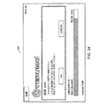
図1は、適切な血液および栄養供給を受けていない心筋の領域である心臓の虚血部分を確定するために組織pHを使用する方法および、この情報を巧みに利用し、適当な行動方針を追求するためにユーザーに利用できるオプションを表わす。ユーザーは最初に、患者の心臓にpH電極を送達する10。次にユーザーはモニター上に表示されるように組織pHを測定し12、組織中にアシドーシスが存在するか否か決定する14。組織アシドーシスが存在しない場合は16、pHを再度測定する12。好ましい態様において、pHはモニター上にpH測定値を表示されながら、電極により連続的に測定される。しかしアシドーシスが組織中に存在した場合は18、ユーザーはそれらに限定はされないが、以下の行動のような適当な行動を採るためにこの情報を利用する。
FIG. 1 shows how to use tissue pH to determine the ischemic portion of the heart, which is the region of the heart muscle that is not receiving adequate blood and nutrients, and how to use this information to Represents the options available to the user to pursue. The user first delivers a
ユーザーは1種もしくは複数の具体的な概要の介入法(compendium of specific interventions)により心臓に保存液の最適な送達を実施することができる20。開胸心臓手術を実施するためには大動脈を結紮して、それにより心筋からその血液、栄養および酸素補給を断たねばならない。しばしば心停止剤溶液と呼ばれる保存液を通常、心臓およびその血管中に潅流して、時間に依存した虚血性損傷を予防する。一部は代謝過程によりもたらされる水素イオンの流失を示す組織pHの測定が保存液の部分的分配の良好な指標であることが示された。この分配は著しく不均一で、予測不可能であり、心停止剤溶液がこれらの部分に到達しないために、心筋壁のその部分がアシドーシスを患うことも示された。pH指導心筋管理法の主な目的は開胸心臓手術の経過中心筋の全部分において組織アシドーシスを予防することである。これは心停止剤溶液の適当で均一な送達および心臓の虚血部分の適当な血行再開を確保することにより達成される。これらは心筋のpHをできるだけ正常に近く維持することにより達成され、ここで正常なpHはとりわけ7.1〜7.3の間にある。 The user can perform optimal delivery of the preservative solution to the heart through one or more specific summary interventions20. In order to perform open heart surgery, the aorta must be ligated, thereby severing its blood, nutrition and oxygen supply from the heart muscle. A preservative solution, often called a cardioplegia solution, is usually perfused into the heart and its blood vessels to prevent time-dependent ischemic damage. It has been shown that the measurement of tissue pH, which in part indicates the loss of hydrogen ions caused by metabolic processes, is a good indicator of partial partitioning of the preservation solution. This distribution was extremely heterogeneous and unpredictable, and it was also shown that that part of the myocardial wall suffered from acidosis because the cardioplegia solution did not reach these parts. The main purpose of pH-guided myocardial management is to prevent tissue acidosis in all parts of the myocardium during the course of open heart surgery. This is accomplished by ensuring proper and uniform delivery of the cardioplegia solution and proper recirculation of the ischemic portion of the heart. These are achieved by maintaining the myocardial pH as close to normal as possible, where the normal pH is especially between 7.1 and 7.3.
ユーザーはまた冠動脈バイパス移植、バルーン拡張もしくは冠血管内ステント挿入後の冠血行再開術の妥当性を評価する22。この機能性は組織血流の規模の指標として虚血期間中に組織中に蓄積する水素イオンの流失速度を利用する。新規に形成された大動脈−冠血管バイパス移植片をとおる流れの回復後にその移植片が存在する心筋部分のpHに変化がないことは不適切な血行再開を示す。他方、0.1pH単位を越えるpHの上昇は虚血性心筋への有効な組織流の回復を示す。
The user also assesses the adequacy of coronary revascularization after coronary artery bypass grafting, balloon dilation or
ユーザーはまた、適当な冠血行再開術によりその機能を改善する、冬眠心筋として知られる、生存しているが機能していない心筋を確定することができる24。pH指導心筋管理法は、非収縮性心筋壁部分の、酸を生成する、すなわち組織のアシドーシスを示す能力がこの部分における生存可能性および機能不全の回復性の指標であることを示した。従ってこの方法は非収縮心筋部分の生存可能性を判定することができる手段を提供する。
The user can also identify a living but not functioning myocardium known as hibernating myocardium that improves its function by appropriate
ユーザーはまた、組織pHに関する情報を得た後に手術の実施法の具体的な変更を促すことができる26。外科手術法のこれらの変更は図4に更に詳細に概説される。
The user can also encourage specific changes in the performance of the surgery after obtaining information regarding
ユーザーはまた、術後期間中の心筋の酸−塩基状態をモニターし28、関連する問題を確定することができる。この機能性は術後の最初の72時間以内の集中治療ユニットにおける虚血発作の描写を可能にする。この方法は、術後の患者の部分的組織代謝および酸塩基平衡の連続的モニターが可能である。安定な血液pHに対して、0.1pH単位を越える心筋pHの低下は心筋のアシドーシスを示す。pHの低下が激しいほど、虚血程度をより著しく示す。この機能性は手術時に心筋中に電極を移植し、特別な胸管をとおしてそれらを体外に出すことにより達成される。電極はそれらを収納する胸管とともにそれらを単に引き抜くことによりモニターを終結した後に、外科集中治療ユニット(SICU)において引き出される。 The user can also monitor 28 myocardial acid-base status during the post-operative period to determine related problems. This functionality allows the depiction of ischemic strokes in the intensive care unit within the first 72 hours after surgery. This method allows continuous monitoring of the patient's partial tissue metabolism and acid-base balance after surgery. For stable blood pH, a decrease in myocardial pH exceeding 0.1 pH units indicates myocardial acidosis. The more severe the pH drop, the more marked the degree of ischemia. This functionality is achieved by implanting electrodes into the myocardium during surgery and bringing them out of the body through a special thoracic duct. The electrodes are withdrawn in a surgical intensive care unit (SICU) after terminating the monitor by simply pulling them together with the thoracic duct that houses them.
ユーザーはまた、組織アシドーシスの予防および患者の予後の改善における、より最近の心筋保護剤および保護法の効果を評価することができる30。心筋保護を改善するために、多数の薬剤が心停止剤溶液に対する添加剤として提唱されており、心停止剤の投与の新規の方法が探求されている。pH指導心筋管理法はこれらの新薬の効果の比較評価を可能にすることができる代謝マーカーおよび、その品質証明が大動脈結紮および再潅流期間中のアシドーシスの予防度であることができる手術内保護の度合いを改善する方法を提供する。心筋保護のこれらの方法を比較するために使用される変化物は大動脈結紮期間中もしくは再潅流の具体的な期間中の積算平均心筋pHである。これらの期間中の積算平均pHが高いほど、心筋保護の程度はより良い。 Users can also evaluate the effectiveness of more recent cardioprotectants and protection methods in preventing tissue acidosis and improving patient prognosis30. In order to improve myocardial protection, a number of drugs have been proposed as additives to cardioplegia solutions and new methods of administration of cardioplegic drugs are being sought. pH-guided myocardial management methods can enable comparative assessment of the effects of these new drugs, as well as intraoperative protection whose proof of quality can be the degree of prevention of acidosis during aortic ligation and reperfusion Provide a way to improve the degree. The variation used to compare these methods of myocardial protection is the cumulative average myocardial pH during aortic ligation or during the specific period of reperfusion. The higher the integrated average pH during these periods, the better the degree of myocardial protection.
図2は心臓組織へのpH電極の送達の様々な方法を表わす。ユーザーは直接の挿入を使用してpH電極を移植することができる40。これには心臓手術法期間中に患者の胸腔を開胸することおよび患者の心臓組織中に手で電極を配置することが含まれる。ユーザーはまた経皮的切開を使用してカテーテルによりpH電極を挿入することができる42。ユーザーはまた内視鏡、結腸鏡もしくは腹腔鏡を使用してpH電極を挿入することができる44。次にユーザーは組織のpHを測定し46、組織にアシドーシスが存在するか否か決定することができる48。アシドーシスが認められない場合は50、組織のpHを再度測定する46。組織にアシドーシスを認める場合は52、ユーザーは図1に概説されたような適切な行動方針を採ることができる54。
FIG. 2 represents various methods of delivering pH electrodes to heart tissue. The user can implant the pH electrode using
図3は外科手術中、心臓への保存液の最適な送達を提供する方法を表わす。この方法において、ユーザーは最初に、心臓組織のpHを測定し60、組織にアシドーシスがあるか否か決定する62。アシドーシスが認められない場合は64、組織のpHを再度測定することができる62。好ましい態様において、pHは連続的に測定し、モニターされる。アシドーシスが組織に認める場合は66、ユーザーは1種もしくは複数の具体的な概要の介入法により心臓への保存液の最適な送達を実施することができる。心停止剤溶液の適切で均一な送達を実施するために使用することができる介入法はそれらに限定はされないが以下の操作(maneuvers)が含まれる。ユーザーは心停止剤溶液の最適な送達をもたらすために保護液の流量を変更することができる68。潅流担当者(perfusionist)は投与される心停止剤溶液の流量を制御する。pH指導心筋管理法は、アシドーシスを予防するために必要な流量が患者と心筋部分では異なることを示した。従って、心停止剤溶液の流量を変更することは組織pHを変更し、改善することができる。
FIG. 3 represents a method that provides optimal delivery of a preservative solution to the heart during surgery. In this method, the user first measures the pH of the
ユーザーはまた、溶液の送達を最適化するために保存液の温度を変更することができる70。心臓手術の経過中に広範にわたる可能性がある心筋温度の変更は様々な度合いの冠血管の収縮および拡張をもたらす。これが順次、心停止剤溶液の分配および更に組織アシドーシスのレベルに影響を与える。組織アシドーシスの回避は、心停止剤溶液の部分的分配に対する温度の効果に応じて、心停止剤溶液を冷却もしくは再度暖めることにより達成することができる。pH指導心筋管理法は、心停止剤溶液の部分的分配に対する温度の効果が極めて予測不能であることを示し、従って心筋の組織pHの連続的モニターが、心筋アシドーシスをもっとも予防しそうな心筋温度の決定を可能にする。冷却および再暖めの双方により、患者によって心筋pHに対する相反する効果が認められた。しかし、概して暖かい心停止剤を投与することが大部分の患者の組織pHの改善をもたらした。
The user can also change the temperature of the stock solution to optimize
溶液の最適な送達をもたらすために、ユーザーはまた、心停止剤溶液の送達部位を変更することができる72。心停止剤溶液は幾つかの部位をとおして:大動脈根をとおり順行性に、右および/もしくは左主冠動脈口をとおり順行性に、新規に形成された移植片の近位末端をとおり順行性に、そして冠静脈洞をとおり逆行性に、送達することができる。pH指導心筋管理法は外科医に部分的アシドーシスをもっとも良く回避することができる投与部位もしくは部位の組み合わせを選択させる。
To provide optimal delivery of the solution, the user can also change the delivery site of the
ユーザーは送達を最適にするために心停止剤溶液を送達するカテーテルの先端を再配置することができる74。これは心停止剤が左主冠動脈口をとおして投与される時に非常に短い左主冠動脈をもつ患者に実施する必要があるかも知れない。それはまた、冠静脈洞中にあまりに深く挿入された逆行性カテーテル上で引き戻す際に役立つことができる。
The user can reposition the tip of the catheter that delivers the cardioplegia solution to optimize
ユーザーはまた、溶液のスチール(steal)を減少させるためにマニホルドをとおして心停止剤溶液を選択的に誘動することができる76。心停止剤溶液は単一源から放射する数本のカテーテルを有するマニホルドをとおして送達することができる。マニホルドのこの配列は「七面鳥の足」として知られている。心停止剤溶液が2本以上のこれらのカテーテルをとおして同時に投与される時に、これらのカテーテルにより供給される様々な心筋部分への溶液の分配に著明な不均一性が存在する。溶液はしばしば、もっとも抵抗の弱い心筋部分、通常、冠動脈疾患のもっとも少ない心筋部分に供給するカテーテル中に優先的に移動する。これが「スチール現象(steal phenomenon)」と呼ばれるものである。組織の水素イオンの流失速度が組織流の大きさを示す事実を利用する心筋pHのモニターは、心筋のどの部分が心停止剤溶液を受け、どの部分が「スチール」現象のために心停止剤が欠乏するかを決定することができる。スチールに遭遇する時は、心停止剤溶液の分配の均一性は、スチールの原因のカテーテルを栓塞し、アシドーシスを示す領域のみに流れを具体的に誘動することにより達成することができる。
The user can also selectively trigger the cardioplegia solution through the manifold to reduce
ユーザーはまた、新規に形成された移植片をとおして心停止剤流を遠位に誘動するために動脈の近位部分に直接の冠動脈圧をかけることができる78。この圧力は溶液が優先的に近位に流れることを防止することができ、心停止剤溶液を低いpHをもつ領域に遠位に押し出して、その領域の組織アシドーシスを低下させる。
The user can also apply direct coronary pressure to the proximal portion of the
ユーザーは冠静脈洞をとおす、もしくは最近形成された伏在静脈移植片の近位末端をとおす逆行性心停止剤の送達中に左主冠動脈口のバルーンカテーテル栓塞を実施することができる80。左主冠動脈のバルーンカテーテル栓塞は、溶液がもっとも抵抗の少ない経路をとおって、心停止剤溶液を低いpH領域に押しやるスチール現象を防止する。この方法は低pHを示す領域のアシドーシスを回復させることができる。
The user can perform a balloon catheter embolization of the left main coronary artery orifice during delivery of retrograde cardioplegia through the coronary sinus or through the proximal end of a recently formed
ユーザーはまた、心停止溶液が順行性に投与されている間に逆行性冠静脈洞カテーテルのバルーンを膨張させることができる82。通常、心停止剤が順行性そして逆行性に同時に送達されている場合は、冠静脈洞中のバルーンは収縮したままで維持される。心停止剤が順行性そして逆行性に同時に送達される間、冠静脈洞中のバルーンが膨張して維持される場合には、心停止剤溶液のより均一な分配を達成することができる。
The user can also inflate the retrograde coronary
ユーザーはまた、右冠動脈が主要な非塞栓血管である時に右冠動脈口をとおして心停止剤のボーラスを投与することができる84。大動脈根が開放している開胸心臓手術の経過中は、心停止剤を左冠動脈口に加えて右冠動脈口をとおして投与することができる。しかしこれは面倒で時間のかかる可能性があるので、一般的な方法ではない。pH指導心筋管理法は、右冠動脈が主要で、それをとおして心停止剤が投与されない場合には、左心室後壁が難治性心筋アシドーシスをより受け易いことを示した。従って、pH指導心筋管理法の経過中に後壁に難治性アシドーシスが遭遇された場合には、右冠動脈が主要である場合には右冠動脈口をとおして心停止剤のボーラスを投与することが後壁への心停止剤溶液の適切な送達を確保させることができ、アシドーシスを回復させることができる。
The user can also administer a cardioplegia bolus through the right
ユーザーはまた、組織アシドーシスが存在する時に、外科手術を促すことができる86。組織アシドーシスをモニターすることにより、ユーザーはその時間をむだに使用することまたは標準的でないもしくは有効でない可能性のある外科手術法を試みることを回避することができる。更に、5%未満の僅かな患者においては組織アシドーシスを防止する知られた方法がなく、外科手術を急がなければならない。手術法を急ぐことにより、手術中に結紮される大動脈が計画より早期に緩められ、従って酸素の多い血液を心筋に到達させ、それによりアシドーシスを回復させる。
The user can also prompt
前記のオプション68〜86の1つがpHモニター上の組織pHレベルの表示により証明されるような虚血状態を緩和させない場合には、ユーザーは組織pHを上昇させることを試みるために記載された他のオプションのいずれかを使用することができる。 If one of the above options 68-86 does not relieve the ischemic condition as evidenced by the display of the tissue pH level on the pH monitor, the user has described other attempts to increase the tissue pH. Any of the options can be used.
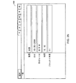
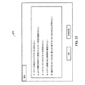
図4は組織アシドーシスが存在することを決定後に、手術の実施法の具体的な変更を促進するためにpH電極を使用する方法を表わす。この方法においては、ユーザーは最初に、心臓組織のpHを測定し90、組織中にアシドーシスが存在するかどうか決定する92。アシドーシスが認められない場合には94、組織のpHを再度連続的もしくは定期的に測定する90。組織にアシドーシスを認める場合は96、ユーザーは手術の実施法を変更することができる98。
FIG. 4 represents a method of using a pH electrode to facilitate specific changes in the procedure of surgery after it has been determined that tissue acidosis is present. In this method, the user first measures 90 the pH of the heart tissue and determines 92 whether acidosis is present in the tissue. If no acidosis is observed 94, the tissue pH is again measured continuously or periodically 90. If the tissue has
これらの変更にはそれらに限定はされないが、以下の方法が含まれる。第1に、心筋の具体的な部分の血行再開術の必要性を決定する100。どれが具体的に血行再開術を要する心筋の部分であるかを確定する能力は人命に拘わる。血行再開術を要する部分は手術経過中の部分的アシドーシスの開始もしくは心房拍動に対する心筋のpHの応答を調べることにより決定することができる。心房拍動に対する応答は手術中に、術後にSICUにおいて、そして心臓カテーテル挿入実験室において利用することができる。心房拍動に応答する組織部分中の0.1pH以上の低下は、その部分に内在する冠動脈内に生理学的に重大な栓塞、および従ってその冠動脈を血行再開術する必要を示す。
These modifications include, but are not limited to, the following methods. First, determine the need for revascularization of a specific portion of the
ユーザーはまた、血行再開術の順序を変更することができる102。pH指導心筋管理法は、大動脈結紮経過中に遭遇されるアシドーシスの程度を最少にするために、外科医に、最初に心筋のもっとも虚血性の部分を血行再開させ、それらをとおして心停止剤溶液を潅流させる。
The user can also change the order of
ユーザーはまた、冠動脈中への具体的な物質の栓塞に続発して起る可能性があるような、手術の経過中に遭遇される不測のアシドーシスに基づいて、心臓の更なる血行再開術をもたらすことにより手術手順を変更することができる104。pH指導心筋管理法はしばしば、術前には予測されなかった血行再開術を要する左心室壁の虚血部分の確定を伴なう。 Users can also initiate further resuscitation of the heart based on unforeseen acidosis encountered during the course of surgery, which may occur secondary to embolization of specific substances into the coronary arteries. The surgical procedure can be altered 104 by bringing about. pH-guided myocardial management is often accompanied by the determination of the ischemic portion of the left ventricular wall requiring revascularization that was not anticipated before surgery.
ユーザーはまた、虚血時間の期間を短縮するために手術計画を変更するかもしくは外科医を変更することができる106。pH指導心筋管理法は幾つかの方法で計画された手術の規模を縮小させる。pHモニターが是正することができない有意な量の心筋アシドーシスを表わす時は、更なる移植片の形成のような省略することができる手術の特定の部分の可能な利益よりも、虚血時間を短縮する必要性が重要になる。pHモニターはまた、この部分に真の必要を示さないために、外科医に手術の計画部分を棄却させる。これに関しては、pH指導心筋管理法はまた、特に初期の研修医はこれらの手術の実施においてかなり時間がかかる可能性があるので、主治医の外科医に、手術のどの部分を彼/彼女が研修医に受け持たせることができるかそして、どの部分を主治医の外科医が自身で実行することができるかの情報を提供するために、研修医の教育に主要な役目を果たす。ユーザーはまた、pH測定値に基づき手順の危険性が利益を越えることが認められる場合には、手術を中止することもできる108。
The user can also change the surgical plan or change the
最後に、ユーザーは再潅流中の残留アシドーシスにより表わされる酸素欠乏が十分に満たされるまで心肺バイパスからの離脱を遅らせることができる110。心筋アシドーシスの存在下での心肺バイパスからの離脱は術後に血行動態を悪化させ、しばしば心肺バイパスの再実施を急がせる可能性がある。心臓が大動脈結紮もしくは再潅流期間中に重篤な虚血にさらされる時には、虚血が正常レベルに回復するまでにはかなりの時間を要する可能性がある。 Finally, the user can delay 110 withdrawal from cardiopulmonary bypass until the oxygen deficiency represented by residual acidosis during reperfusion is fully satisfied 110. Withdrawal from cardiopulmonary bypass in the presence of myocardial acidosis may exacerbate hemodynamics postoperatively and often urge the re-execution of cardiopulmonary bypass. When the heart is exposed to severe ischemia during aortic ligation or reperfusion, it can take a considerable amount of time for the ischemia to return to normal levels.
前記のオプション、100〜110の1つがpHモニター上の組織pHレベルの表示により表わされるような虚血状態を緩和することができない場合には、ユーザーは組織pHを上昇させることを試みるためのこれらの記載された他のオプションのいずれかを使用することができる。 If one of the above options, one of 100-110, is unable to alleviate the ischemic condition as represented by the display of the tissue pH level on the pH monitor, the user may attempt to increase the tissue pH. Any of the other options described in can be used.
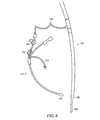
図5は本発明に従う、組織アシドーシスをモニターするために使用されるpH電極136の好ましい態様を表わす。電極136は銀のワイヤ114に接続されたケーブル112を有することができる。好ましい態様において、銀のワイヤ114はAg/AgCl(銀/塩化銀)のワイヤである。もう1つの好ましい態様において、ケーブル112はガラスのシール118中を通過する白金のワイヤ116により銀のワイヤ114に接続されている。ケーブル112とワイヤ114、116は収縮配管122中に収納されるハウジング120中に収納される。電極136は銀のワイヤ114、サーミスター126、pHセンサー128およびゲル化電極130を収納するガラスのステム124を有する。電極136はまた、それが使用される部位に電極136を固定させるための縫合溝132を有することができる。電極136はまた、使用前もしくは使用中にユーザーに電極136の配置を調整させる湾曲可能な接合体134を有することができる。ガラスのステム124は組織中への直接の挿入を可能にするように尖っている。好ましい態様において、ガラスのステム124は鉛ガラスから製造されている。電極は酸化エチレンもしくはガンマ照射により滅菌することができる。本発明による使用に適するpH電極はVascular Technology Inc.,Lowell,MassachusettsおよびTokyo,JapanのTerumo Corporationから購入できる。この具体的な電極は10mmまでの深さに組織中に挿入することができ、1mmの直径を有し、そしてプローブの遠位4mmにおいてpHセンサーを使用する。
FIG. 5 represents a preferred embodiment of the
組織pHは重要な臨床的測定値である。pHの著明な低下として測定することができる局所的アシドーシスは虚血と関連づけられてきた。目盛定めおよび組織pH測定値の温度補正を可能にするために、温度を好ましくはpHと同時に測定する。pHの温度補正は特に十分な冷却を要する開胸心臓手術のような手術において重要である。pH電極はそれぞれがpH感知センサー内に固定された温度感知素子を含む組み合わせpH/温度センサーを使用する。 Tissue pH is an important clinical measurement. Local acidosis, which can be measured as a marked decrease in pH, has been associated with ischemia. The temperature is preferably measured simultaneously with the pH to allow calibration and temperature correction of the tissue pH measurement. Temperature correction of pH is particularly important in surgery such as open heart surgery that requires sufficient cooling. Each pH electrode uses a combined pH / temperature sensor that includes a temperature sensing element fixed within the pH sensing sensor.
ガラスのpH電極は正確な臨床的pH測定値を得るためにもっとも一般に使用されている。それらは順次、内部の照合ワイヤと接触している電解質で充填された中空のガラスセンサーから成る。使用されるガラスの本質により、ガラス上に電位が生じる。この電位はガラスの外面と接触する非分析物溶液のpHと、内部のバッファー溶液の本質的に一定のpH間の差に比例する。 Glass pH electrodes are most commonly used to obtain accurate clinical pH measurements. They in turn consist of a hollow glass sensor filled with an electrolyte in contact with an internal reference wire. Due to the nature of the glass used, an electric potential is created on the glass. This potential is proportional to the difference between the pH of the non-analyte solution in contact with the outer surface of the glass and the essentially constant pH of the internal buffer solution.
電気的測定を実施するためには、完全な電気回路を形成しなければならない。従って、非分析物溶液との第2の電気的接触を形成しなければならない。これは針の照合電極の使用により実施される。それは一定のモル数の塩溶液と接触する塩化銀の針から成る。塩溶液は適当な隔離機構を使用して、この場合にはその開放末端が患者と接して配置される柔軟な管中に配置されたゲル化塩溶液の使用により、非分析物溶液、すなわち患者の組織と接して配置される。 In order to perform electrical measurements, a complete electrical circuit must be formed. Therefore, a second electrical contact with the non-analyte solution must be made. This is accomplished by the use of a needle reference electrode. It consists of a silver chloride needle in contact with a fixed number of moles of salt solution. The salt solution uses a suitable isolation mechanism, in this case a non-analyte solution, i.e. patient, by using a gelled salt solution placed in a flexible tube whose open end is placed in contact with the patient. Placed in contact with the organization.
Nernstの式は一定の環境条件下では、ガラスpH電極の出力はpHと直線関係にあることを予測する。従って、センサーの電気的出力は簡単な直線曲線−フィットの使用によりpHに変換することができる。これは、それらから直線等式の勾配および補正定数を計算することができる2種の異なるpH値における電極の電気的出力を決定することを要する。pH電極の補正に一般に利用できる規準バッファーは4、7および10のpH値を有する。4および7pHのバッファーは本システムの好ましい態様による使用に好ましい。電極のゼロ−電位地点がpH7に近いために7−pHのバッファーが好ましい。もっとも問題の大きいpH値がpH7より僅かに下にあるために、4−バッファーが好ましい。 The Nernst equation predicts that, under certain environmental conditions, the output of the glass pH electrode is linearly related to pH. Thus, the electrical output of the sensor can be converted to pH by using a simple linear curve-fit. This entails determining the electrical power of the electrode at two different pH values from which the linear equation slope and correction constant can be calculated. Commonly available reference buffers for pH electrode correction have pH values of 4, 7, and 10. 4 and 7 pH buffers are preferred for use according to preferred embodiments of the system. A 7-pH buffer is preferred because the zero-potential point of the electrode is close to pH 7. 4-buffer is preferred because the most problematic pH value is slightly below pH 7.
電極のこの種類の理論的感受性−勾配は25℃において59.16mV/pHである。実際の電極に対しては、それは僅かに低い傾向があり、その値は電極毎に僅かに異なり、特定の電極に対してはその有効寿命全体にわたり変動する。 This kind of theoretical sensitivity-gradient of the electrode is 59.16 mV / pH at 25 ° C. For a real electrode, it tends to be slightly lower, its value varies slightly from electrode to electrode, and for a particular electrode it varies throughout its useful life.
ゼロ電位点は、内部溶液および照合溶液の塩濃度のあらゆる差に対して補正後に、測定された出力電圧がゼロである非分析物のpH値と定義される。従ってゼロ電位点は非分析物pH値がpHセンサーの内部バッファーのpH値と同一である時に起る。しかしこれらの条件下で測定が実際に実施される場合、概して非ゼロの電位が測定される。これは、センサーの内部照合ワイヤが露出されるClの濃度が、照合針が露出される濃度と異なる時、もしくは双方の照合ワイヤが同一材料から製造されていない場合に起る。本システムの好ましい態様において、照合針は飽和KClゲル中に浸漬され、他方センサーの内部照合ワイヤは内部バッファー中0.87M濃度のKClに露出される。非分析物が内部バッファーと同様なpH値、公称25℃で6.33pHを有する時は、この差が25℃で約+30mVの測定電位をもたらす。従って、真のゼロ電位点を測定するためにはそれから30mVを差し引くことにより測定電圧を補正する必要がある。ゼロ点補正のための目盛定め中に7pHバッファーが使用され、それが6.33にもっとも近い利用できるバッファー値である。 The zero potential point is defined as the non-analyte pH value at which the measured output voltage is zero after correction for any difference in salt concentration between the internal solution and the reference solution. Thus, the zero potential point occurs when the non-analyte pH value is the same as the pH value of the internal buffer of the pH sensor. However, when measurements are actually performed under these conditions, generally non-zero potentials are measured. This occurs when the concentration of Cl at which the internal verification wire of the sensor is exposed is different from the concentration at which the verification needle is exposed, or when both verification wires are not manufactured from the same material. In a preferred embodiment of the system, the verification needle is immersed in a saturated KCl gel, while the internal verification wire of the sensor is exposed to 0.87M concentration of KCl in the internal buffer. When the non-analyte has a pH value similar to the internal buffer, nominally 6.33 pH at 25 ° C., this difference results in a measured potential of about +30 mV at 25 ° C. Therefore, in order to measure the true zero potential point, it is necessary to correct the measurement voltage by subtracting 30 mV from it. A 7 pH buffer is used during calibration for zero point correction, which is the closest available buffer value to 6.33.
前記のように、センサー毎に、そして同一センサーに対して長期間の間に出力には理想的な値から、いくらかの変動が存在するために、pHセンサーは各使用前に補正しなければならない。これは最初にセンサーをスロープバッファー(slope buffer)(4.00pH)中に、次にゼロ電位点バッファー(7.00pH)に入れることによる補正手順中に自動的に実施される。マイクロプロセッサーがmVのセンサーの出力を読み、塩の差(salt differential)に対して補正し、読みの値が安定する時を決定し、次に各センサーの勾配および補正因子を計算する。スロープおよびゼロ電位点双方は温度とともに変動し、モニターのソフトウェアにより補正される。 As mentioned above, the pH sensor must be corrected before each use because there is some variation from ideal to output for each sensor and over the long term for the same sensor. . This is done automatically during the correction procedure by first placing the sensor in the slope buffer (4.00 pH) and then in the zero potential point buffer (7.00 pH). A microprocessor reads the mV sensor output, corrects for salt differential, determines when the reading stabilizes, and then calculates the slope and correction factor for each sensor. Both the slope and zero potential points vary with temperature and are corrected by the monitor software.
pH電極のpH/温度センサー組み合わせ物は温度を測定するために精密なサーミスター素子を使用する。サーミスターは使用されているもっとも一般的な温度測定装置の1つである。それは金属酸化物の半導体セラミックの小ビーズから成る。物質の電気抵抗は非直線状に温度に逆比例して変動する。 The pH electrode's pH / temperature sensor combination uses a precision thermistor element to measure temperature. The thermistor is one of the most common temperature measuring devices in use. It consists of small beads of metal oxide semiconductor ceramic. The electrical resistance of a substance fluctuates in a non-linear manner and inversely proportional to temperature.
温度を測定するためには、正確に知られた抵抗を有するモニター中に固定抵抗と電気的に直列に配置される。直列の組み合わせ上に電圧をかけ、サーミスターと抵抗器の接合部の電圧を測定する。固定抵抗器と、かけた電圧の既知の値と一緒にこの測定値を使用して、サーミスターの抵抗を計算する。次にマイクロプロセッサープログラム中に保存された参照表により温度を決定する。本システムの好ましい態様とともに使用されるサーミスターセンサーはシステムのユーザーによる個別の補正を不要にさせるレベルの精度まで製造されている。 To measure the temperature, it is placed in electrical series with a fixed resistance in a monitor having a precisely known resistance. A voltage is applied on the series combination and the voltage at the junction of the thermistor and resistor is measured. Using this measurement along with a fixed resistor and a known value of the applied voltage, the resistance of the thermistor is calculated. The temperature is then determined by a lookup table stored in the microprocessor program. The thermistor sensor used with the preferred embodiment of the system is manufactured to a level of accuracy that eliminates the need for individual corrections by the user of the system.
pH電極は前以て補正されて、電極の先端がpH4.0のバッファーを含むスリーブもしくはスリーブポケット内に封入されるように包装することができる。スリーブポケットはプラスチック材料から形成することができ、3mmの内径を有することができる。患者の体内へのその挿入前に、スリーブのポケットを取り出して、電極の先端を例えばガーゼで乾燥払拭し、pH7.0バッファーを含むビーカー中に電極を挿入する。この時点で補正は完了する。pH4.0のバッファー内に電極を包装することが、適当な補正に必要な一因子である、電極をその保存中湿って維持させ、電極の補正に要する段階を1段階に減少させる。電極モニター中のソフトウェアは1段階の補正を示すように修飾することができる。 The pH electrode can be pre-corrected and packaged so that the tip of the electrode is enclosed in a sleeve or sleeve pocket containing a pH 4.0 buffer. The sleeve pocket can be formed from a plastic material and can have an inner diameter of 3 mm. Prior to its insertion into the patient's body, the sleeve pocket is removed, the tip of the electrode is wiped dry, for example with gauze, and the electrode is inserted into a beaker containing pH 7.0 buffer. At this point, the correction is complete. Packaging the electrode in a pH 4.0 buffer is one factor necessary for proper correction, keeping the electrode moist during its storage and reducing the steps required for electrode correction to one step. The software in the electrode monitor can be modified to show one level of correction.
pH電極、照合電極およびサーミスターが取り付けられている処理ユニットおよびモニターは、信号を処理し、20秒以下の間隔で次のデータを連続的に記録し、表示する:1)pH単位における組織pH、2)nモルの組織水素イオン濃度[H+]、3)℃の組織温度、4)37℃に補正されたpH、および5)pHの逆数logとして計算された組織の水素イオン濃度[H+]。37℃に対する補正は一連の測定値に基づいて決定される0.017pH単位/℃の因子に基づく。更に、モニターは特定期間の開始時と終結時に合図を与えることによる特定期間にわたる積算平均pH、[H+]および温度の計算を可能にする。スレーブモニターをユニットに取り付けて外科医の正面に配置して、データの注文に応じた連続的な表示を提供する。データの連続的なリアルタイムの表示が心筋組織のアシドーシスを予防もしくは回復するためのpH指導心筋管理法の早急な実行を可能にする。 A processing unit and monitor fitted with a pH electrode, reference electrode and thermistor process the signal and continuously record and display the following data at intervals of 20 seconds or less: 1) Tissue pH in pH units 2) nmole tissue hydrogen ion concentration [H + ], 3) tissue temperature at 0 ° C., 4) pH corrected to 37 ° C., and 5) tissue hydrogen ion concentration [H calculated as reciprocal log of pH] + ]. The correction for 37 ° C. is based on a factor of 0.017 pH units / ° C. determined based on a series of measurements. In addition, the monitor allows calculation of the integrated average pH, [H + ] and temperature over a specified period by giving a signal at the start and end of the specified period. A slave monitor is attached to the unit and placed in front of the surgeon to provide a continuous display as data is ordered. Continuous real-time display of data allows for rapid implementation of pH-guided myocardial management methods to prevent or ameliorate myocardial tissue acidosis.
心臓手術中のpH指導心筋管理法および心筋生存可能性の判定に、幾つかの装置もしくは用具を使用することができる。心臓手術中の具体的な心筋部分への心停止剤溶液の維持および分配は、幾つかの異なる装置および方法を使用して達成することができる。 Several devices or tools can be used to determine pH-guided myocardial management during cardiac surgery and to determine myocardial viability. Maintenance and distribution of cardioplegia solution to specific myocardial segments during cardiac surgery can be accomplished using a number of different devices and methods.
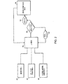
図6はGrand Rapids,MichiganのMedtronicにより供給される心停止剤送達システム140の「七面鳥の脚」形態を表わす。電極と一緒の送達システム140が心筋管理システムを形成することができる。システム140にはまた、コンピューターおよび制御装置158のようなデータ処理システム160が含まれる。データ処理システム160は患者の状態およびシステム状態の変更のような測定データ162を受け取るようにプログラムすることができる。データ処理システム160は流体送達システムの流体源144に取り付けることができる。データ処理システム160はまた、制御装置158を介して流体源に取り付けることもできる。制御装置158は流体送達システムを操作することができる。制御装置158は手術部位に送達される保存流体もしくは心停止剤流体の流量を制御することができる。制御装置158はまた、保存液の温度および保存液の送達部位を制御することもできる。システム140は様々な心臓の取り付け部位に源144から送達された心停止剤溶液の量を調整し、選択的に投与するために使用することができる複数の制御装置142を有する。システム140は心停止剤溶液の流量を制御する閉塞装置(occluder)もしくは弁146を含むことができる。
FIG. 6 represents a “turkey leg” configuration of a cardiac
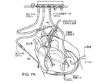
システム140には心停止剤源144と様々な心臓部位間に取り付けられた幾つかの送達装置が含まれる。これらの装置は心停止剤溶液のそれらそれぞれの心臓部位への送達を可能にする。1つの装置は大動脈根に挿入することができるカニューレ148(Sarns Inc.,Ann Arbor,Michigan)である。もう1つの装置は左主冠動脈口156内に挿入することができるSpencerカニューレ(Research Medical,Inc.,Midvale,Utah)である。左主冠動脈口156中へのこの挿入は図7Aおよび7Bに示される。もう1種の装置は右主冠動脈口内に挿入することができる融通が効く金属カテーテル152(Medtronic,Grand Rapids,Michigan)である。カテーテル152はまた挿入されない状態で図7Aに示されている。もう1種の装置は心停止剤の送達のために伏在静脈移植片の近位端に取り付けることができる14ゲージのビード状の(beaded)針(Randall Faichney Corp.,Avon,Massachusetts)である。静脈移植片への取り付けはまた図7Aに示されている。
心臓手術中に心停止剤溶液を再分配するために、140の他の部位をとおって心停止剤を提供しながら、Spencerカニューレ150(Research Medical,Inc.,Midvale Utah)のような球状カテーテルもしくは#3FFogerty Catheter(Ideas For Medicine,St.Petersburg,Florida)のようなバルーン先端カテーテルによる左主冠静脈洞口をブロックすることを使用することもできる。更に、心臓手術中に心停止剤流体を再誘動するために、移植片の近位端をとおって心停止剤溶液を潅流しながら、新規静脈移植片の挿入部位に近位の冠動脈に一過性の閉塞圧をかけることを利用することもできる。閉塞圧はKelly鉗子(Allegiance Healthcare Corp.,McGaw Park,Illinois)の先端でガーゼの「ピーナツ」で維持することができる。
A spherical catheter, such as a Spencer cannula 150 (Research Medical, Inc., Midvale Utah), or providing a cardiac arrest agent through
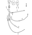
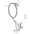
Guntrieバルーンを先端にもつカニューレ(Medtronic,Grand Rapids,Michigan)を、逆行性の方法で、心停止剤の選択的投与のためにシステム140に取り付けて冠静脈洞に挿入することもできる。カニューレ170は図8に示される。この図において、それは静脈カニューレ178に配管176を介して取り付けられているように示されている。これは、pH指導心筋管理法の一部として組織への心停止剤の送達を改善するために、冠静脈洞内の圧力を操作することを可能にする。圧力は冠静脈洞カテーテルの流体開口部を閉鎖して、冠静脈洞のバルーン172を膨張させ、そして心停止剤を順行性に送達することにより操作することができる。170を178に接続する1mmの配管176が背圧を形成し、それが適当な順行性心停止剤の流れを妨げずに送達を改善する。冠静脈洞カテーテル170の流体開口部の開放もしくは閉鎖は弁184により制御することができる。静脈カニューレ178は通常、下大静脈孔中および右心房のより近位の開口部180中にその先端182を伴なって心肺バイパスの経路に挿入される。
A cannula with a Guntrie balloon tip (Medtronic, Grand Rapids, Michigan) can also be attached to the
Sarns,Ann Arbor,Michiganにより製造されるような水ヒーター/クーラーを使用して心停止剤溶液の温度を操作することにより組織温度を変化させることは心臓手術中の心筋pHを管理する補助をすることができる。更に、HE30 Gold心停止システム(Baxter Corporation,Irvine,California)のような心停止システムを使用して心停止剤流の流量を変更することにより心停止剤溶液の潅流圧を変更することも心臓手術中の心筋pH管理の補助をすることができる。 Changing tissue temperature by manipulating the temperature of the cardioplegia solution using a water heater / cooler such as that manufactured by Sanns, Ann Arbor, Michigan helps to manage myocardial pH during cardiac surgery. be able to. Furthermore, altering the perfusion pressure of the cardioplegia solution by altering the flow rate of the cardioplegic agent flow using a cardioplegia system such as the HE30 Gold cardioplegia system (Baxter Corporation, Irvine, California) can also be used for cardiac surgery. Can assist in controlling myocardial pH.
心筋の生存性の判定および冠動脈狭窄の生理学的重要性の決定のための器具も使用することができる。器具は手術室もしくは心臓カテーテル挿入実験室のどちらでも使用することができる。 Instruments for determining myocardial viability and determining the physiological significance of coronary stenosis can also be used. The instrument can be used in either the operating room or the cardiac catheterization laboratory.
手術室では、例えば、Somerville,New JerseyのEthiconにより製造された拍動ワイヤを右心房上に配置し、Grand Rapids,MichiganのMedtronicにより製造された外部ペースメーカーに接続することができる。pH電極も心筋中に挿入することができる。5分間の急速心房拍動に応答する心筋pHの低下は組織虚血を表わすことができ、更に電極が配置されている心筋部分が生存可能であることを示すこともできる。 In the operating room, for example, a pulsation wire manufactured by Ethicon of Somerville, New Jersey can be placed on the right atrium and connected to an external pacemaker manufactured by Medtronic of Grand Rapids, Michigan. A pH electrode can also be inserted into the myocardium. A decrease in myocardial pH in response to a 5 minute rapid atrial beat can indicate tissue ischemia and can also indicate that the portion of the myocardium where the electrode is located is viable.
心臓カテーテル挿入実験室においては、pH電極は長い0.014ゲージのワイヤの先端に固定して、Miami,FloridaのCordisにより製造されるような通常の6フレンチの心臓カテーテル挿入用カテーテルをとおして挿入することができる。カテーテルの先端は、研究されている冠動脈により内在される部分の心室壁に対して垂直に配置し、pH電極は心内膜下中に侵入するように押し込むことができる。好ましくは電極は心内膜下中に約5mm侵入するように押し込まれる。拍動は右心室中に前進され、外部ペースメーカー(Medtronic,Grand Rapid,Michigan)に取り付けられた拍動ワイヤを介して達成される。再度、5分間の急速心房拍動に応答する心筋pHの低下が組織虚血を表わすことができる。 In the cardiac catheterization laboratory, the pH electrode is secured to the end of a long 0.014 gauge wire and inserted through a conventional 6 French heart catheter insertion catheter such as that manufactured by Cordis of Miami, Florida. can do. The tip of the catheter can be placed perpendicular to the part of the ventricular wall inherent by the coronary artery being studied, and the pH electrode can be pushed to penetrate into the subendocardium. Preferably the electrode is pushed to penetrate about 5 mm into the subendocardium. The beat is advanced into the right ventricle and is achieved via a beat wire attached to an external pacemaker (Medtronic, Grand Rapid, Michigan). Again, a decrease in myocardial pH in response to a 5 minute rapid atrial beat can be indicative of tissue ischemia.
pH電極およびモニターシステムは心臓組織の虚血を決定する際の使用に対して説明されてきたが、pHシステムおよび方法は他の種類の組織にも同様に使用することができる。pHシステムは臓器移植における拒絶反応をモニターするため、腸間膜動脈虚血を判定するため、脳血流をモニターし、評価するため、そして整形外科手術におけるフラップをモニターするために使用することができる。 Although pH electrodes and monitoring systems have been described for use in determining cardiac tissue ischemia, the pH system and method can be used with other types of tissues as well. The pH system can be used to monitor rejection in organ transplants, to determine mesenteric artery ischemia, to monitor and evaluate cerebral blood flow, and to monitor flaps in orthopedic surgery it can.
pH電極は心臓移植過程において患者の切開、輸送および挿入中のドナー心臓の酸塩基状態および保存の妥当性をモニターするために使用することができる。 The pH electrode can be used to monitor the acid-base status and preservation validity of the donor heart during patient incision, transport and insertion during the heart transplantation process.
pH電極は腎臓移植過程中およびその後の腎臓をモニターするために使用することができる。pH電極は大手術の経過中、そしてとりわけ腎臓移植中に腎臓への組織潅流のモニターに使用することができる。電極は心臓と同様な方法で腎臓に容易に移植可能で、7.2以上の組織pHレベルが適当な組織潅流を示す。特にドナー関連の心臓移植の目的のための腎臓の切開中の腎臓への損傷を検出し、回避することができ、従ってドナー関連腎臓移植のよりよい予後を確保することができる。移植前の輸送中の腎臓の保存もまた正常レベルのpHをモニターし、維持することにより確保することができる。これは臓器潅流用に特別に設計された装置内で血液で腎臓を一定に潅流することにより達成することができる。 The pH electrode can be used to monitor the kidney during and after the kidney transplantation process. The pH electrode can be used to monitor tissue perfusion to the kidney during the course of major surgery and especially during kidney transplantation. The electrode can be easily implanted into the kidney in a manner similar to that of the heart, with tissue pH levels above 7.2 indicating adequate tissue perfusion. Damage to the kidney during kidney incision, particularly for donor-related heart transplantation purposes, can be detected and avoided, thus ensuring a better prognosis of donor-related kidney transplantation. Preservation of the kidney during transport prior to transplantation can also be ensured by monitoring and maintaining normal levels of pH. This can be accomplished by constantly perfusing the kidney with blood in a device specially designed for organ perfusion.
腎臓移植後、術後の直後48時間、腎臓内に電極を維持することは初期の虚血をモニターさせ、手術的介入によりこの虚血を回復させることができる。この期間の虚血は重大な悪い予後を予告することができる。移植された腎臓、機能の評価、およびその拒絶反応の検出もまた、カテーテル上に電極を配置して、腎杯中に逆行性に通過させることにより実施することができる。心臓につき前記に説明されたものと同様に、腎臓の実質組織とともに腎杯を穿刺することが、切迫したもしくは現実の拒絶反応を示すことができ、従って不都合な予後を示す。アシドーシスの早期検出は拒絶反応の主要な処置を促し、従って腎臓移植の予後を改善することができる。 Maintaining an electrode in the kidney for 48 hours immediately after surgery after kidney transplantation allows early ischemia to be monitored and can be restored by surgical intervention. This period of ischemia can predict a serious bad prognosis. Implanted kidneys, evaluation of function, and detection of their rejection can also be performed by placing electrodes on the catheter and passing retrogradely through the renal cup. Similar to that described above for the heart, puncturing the renal cup with the renal parenchyma can indicate an impending or real rejection and thus an unfavorable prognosis. Early detection of acidosis can facilitate the main treatment of rejection and thus improve the prognosis of kidney transplantation.
各電極はまた腎臓動脈再開術過程中の腎臓の血行再開術の妥当性の判定に使用することができる。生命に係わる狭窄腎動脈の血行再開術の効果は冠動脈の血行再開術の効果と類似の方法で手術中に決定することができる。血行再開術によりアシドーシスを回復しない場合は、アシドーシスを回復するための更なる手術中の手段を促し、従って血行再開術の不都合な予後を回避する。心臓におけるように、血行再開術によりアシドーシスを回復しないことは血行再開術法の不適切さを示し、状態を改善し、血行再開術の予後を改善するための更なる手術中管理の指針を提供する。 Each electrode can also be used to determine the adequacy of renal revascularization during the renal artery resuscitation process. The effects of life-threatening renal artery revascularization can be determined during surgery in a manner similar to that of coronary revascularization. If resuscitation does not restore acidosis, it encourages further intraoperative measures to restore acidosis, thus avoiding the adverse prognosis of revascularization. Not reversing acidosis by resuscitation, as in the heart, indicates inadequacy of resuscitation, provides additional in-operative management guidelines to improve conditions and improve prognosis of resuscitation To do.
pH電極はまた、肝臓移植時およびその後の肝臓をモニターするために使用することができる。pH電極は前記の腎臓のものに類似の重要なデータを提供するために肝臓中に挿入することができる。腎臓中の電極の使用の説明が手術中の経過のモニター、早期の拒絶反応の確定および拒絶反応過程を抑制するための手段を実施する際のpH電極の使用に関して肝臓に適用できる。 The pH electrode can also be used to monitor the liver during and after liver transplantation. A pH electrode can be inserted into the liver to provide important data similar to that of the kidney. A description of the use of electrodes in the kidney can be applied to the liver with regard to the use of pH electrodes in monitoring the progress during surgery, establishing early rejection and implementing measures to suppress the rejection process.
電極はまた、主要末梢血行再開術中およびその後の末梢器官をモニターする際、そして救命手当において使用することができる。末梢器官の皮下組織中への電極の挿入は組織潅流の妥当性の情報を提供する。主として下肢の遠位半分の皮下組織中のこれらの部位で測定されたアシドーシスは末梢動脈閉塞もしくは不適切な心臓拍出を示し、心臓拍出を改善するための手段もしくは組織潅流の実施を急がせることができる。これらの手段には、直接的血行再開術手術もしくは薬理学的操作および/もしくは例えば、下行大動脈中への例えば、Reading,PennsylvaniaのArrow Internationalにより製造されるような大動脈内バルーンの挿入を含むことができる。最近は、低い心臓拍出症候を判定し、処置するためには中心の(central)血行動態の手段のみが使用される。末梢器官のpHを測定することは、それが「適切な」心臓拍出量の維持における最終的目標である組織潅流の真の手段を提供するために、より優れた代替法を提供する。 Electrodes can also be used in monitoring peripheral organs during and after major peripheral revascularization and in lifesaving. The insertion of electrodes into the subcutaneous tissue of peripheral organs provides information on the validity of tissue perfusion. Acidosis, measured primarily at these sites in the subcutaneous tissue of the distal half of the lower limb, indicates peripheral arterial occlusion or inadequate cardiac output and hastened the implementation of means or tissue perfusion to improve cardiac output. Can be made. These means may include direct revascularization surgery or pharmacological manipulation and / or insertion of an intra-aortic balloon, such as, for example, manufactured by Arrow International of Reading, Pennsylvania into the descending aorta. it can. Recently, only central hemodynamic means are used to determine and treat low cardiac output symptoms. Measuring the pH of peripheral organs provides a better alternative because it provides a true means of tissue perfusion, which is the ultimate goal in maintaining “appropriate” cardiac output.
好ましい態様のpH電極はまた、整形外科手術において筋肉内およびフラップの皮下組織内に使用することができる。pH電極による組織アシドーシスは皮膚および皮下フラップの損なわれた(compromeside)生存可能性を示すことが示された。電極は術後にフラップの縁内に配置され、術後3〜4日までpHをモニターされる。pHの低下はその後の失敗を予防するために手術中の介入法およびフラップの更新を急がせる。 The pH electrode of the preferred embodiment can also be used in intramuscular and flap subcutaneous tissue in orthopedic surgery. Tissue acidosis with a pH electrode has been shown to exhibit compromised survival of skin and subcutaneous flaps. The electrode is placed within the flap edge after surgery and the pH is monitored until 3-4 days after surgery. Lowering the pH urges renewal of interventions and flaps during surgery to prevent subsequent failures.
pH電極はまた、腸の虚血の判定および処置において結腸に使用することができる。腸の虚血を判定し、回復させるために、pH電極を以前に心臓カテーテル挿入法中に心臓に記載されたものと類似の方法でワイヤ上に配置することができる。pH電極を先端にもつワイヤを末梢回腸中への通常の結腸鏡法中にSeattle,WashingtonのOlympus Medicalにより製造されたもののような結腸鏡をとおして挿入することができる。回腸の管腔内pHは潅流の妥当性の信頼できる指標である。回腸の管腔内のアシドーシスは腸の虚血を表わし、虚血を回復するかもしくはその不都合な予後を予防するためのいずれかのための手段を急がせることができる。回腸の管腔内pHの知識は、例えば腸の可能な切除を伴なう腹部の探索のような手術的介入法並びに心臓拍出および組織潅流を改善するための薬理学的介入法の開始を可能にする。 The pH electrode can also be used for the colon in the determination and treatment of intestinal ischemia. In order to determine and restore intestinal ischemia, a pH electrode can be placed on the wire in a manner similar to that previously described for the heart during cardiac catheterization. A wire with a pH electrode tip can be inserted through a colonoscope, such as that manufactured by Olympus Medical, Seattle, Washington, during routine colonoscopy into the peripheral ileum. The luminal pH of the ileum is a reliable indicator of perfusion validity. Acidosis in the lumen of the ileum represents intestinal ischemia and can expedite measures to either recover the ischemia or prevent its adverse prognosis. Knowledge of the ileal intraluminal pH can be used to initiate surgical interventions such as exploring the abdomen with possible resection of the intestine and pharmacological interventions to improve cardiac output and tissue perfusion. to enable.
好ましい態様に従うpH電極は他の器官に使用することができる。前記の器官に加えて、pHモニターシステムに取り付けたpH電極を脳、膀胱、横隔膜および小腸のような器官中に挿入することにより、組織アシドーシスを測定し、操作し、そして回復させることができる。 The pH electrode according to the preferred embodiment can be used for other organs. In addition to the above organs, tissue acidosis can be measured, manipulated and recovered by inserting pH electrodes attached to a pH monitoring system into organs such as the brain, bladder, diaphragm and small intestine.
好ましい態様に従う心筋pHモニターシステムは冠血流の遮断中の心筋保護の妥当性および大動脈冠動脈バイパス移植片形成後の血行再開術の妥当性の双方を判定するための感受性の高いオンラインの手段を提供する。pH指導心筋管理法はすべての相の心臓外科手術中の心筋組織アシドーシスを予防し、回復させる。心筋組織アシドーシスは心筋の虚血の大きさの信頼できる指標であることが示された。部分的心筋アシドーシスはまた、しばしば、ヒトの心臓手術の経過中に予測できない方法で遭遇される。最近の研究により、手術中に遭遇される部分的心筋アシドーシスの規模と不都合な術後患者の予後の発生率の間に直接的相関が示された。手術中の重篤な心筋アシドーシスはまた、心臓手術後の長期間生存を減少させることが示された。これらの観察は虚血および再潅流の条件下の筋細胞のアポトーシスの主要な引き金として、そして後の心臓不全の可能な原因としてアシドーシスを示唆した最近の実験的研究を考慮すると重要性を増す。 A myocardial pH monitoring system according to a preferred embodiment provides a sensitive online means to determine both the validity of myocardial protection during blockage of coronary blood flow and the validity of revascularization after aortic coronary artery bypass graft formation To do. pH-guided myocardial management prevents and reverses myocardial tissue acidosis during all phases of cardiac surgery. Myocardial tissue acidosis has been shown to be a reliable indicator of the magnitude of myocardial ischemia. Partial myocardial acidosis is also often encountered in an unpredictable way during the course of human heart surgery. Recent studies have shown a direct correlation between the magnitude of partial myocardial acidosis encountered during surgery and the incidence of adverse postoperative patient prognosis. Severe myocardial acidosis during surgery has also been shown to reduce long-term survival after cardiac surgery. These observations are of increasing importance in light of recent experimental studies that have suggested acidosis as a major trigger of myocyte apoptosis under conditions of ischemia and reperfusion and as a possible cause of later heart failure.
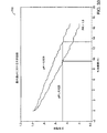
図9は本発明に従う左心室前壁および後壁でモニターされたpHを図式により表わす。左心室前壁および後壁双方において心筋pH(37℃に補正)を測定し記録した。電極を0分に挿入し、5分後に大動脈を交差結紮(XC)した。大動脈根をとおして投与された温血心停止剤の最初のボーラスは双方の壁のpHの増加をもたらした。次に心停止剤の送達を中断し、伏在静脈の部分を左前方下行冠動脈(LAD)に縫合する。この期間中にpHは前壁で6.2に、後壁で6.4に低下した。 FIG. 9 graphically represents the pH monitored at the anterior and posterior walls of the left ventricle according to the present invention. Myocardial pH (corrected to 37 ° C.) was measured and recorded on both the left ventricular anterior and posterior walls. The electrodes were inserted at 0 minutes and the aorta was cross-ligated (XC) after 5 minutes. The first bolus of warm blood cardioplegia administered through the aortic root resulted in an increase in the pH of both walls. Cardiac delivery is then interrupted and the saphenous vein portion is sutured to the left anterior descending coronary artery (LAD). During this period, the pH dropped to 6.2 on the front wall and 6.4 on the rear wall.
LADに対する吻合を完了後、移植片の近位端(矢印A)をとおして連続的血液心停止剤を送達し、手術の残りの期間中維持した。それは前壁pHの急速な上昇をもたらし、前壁への適切な心停止剤の送達および移植片の吻合の技術的な妥当性を示した。数分後、大動脈が切開されて、大動脈弁交換が開始された時、LAD移植片に加えて冠静脈洞をとおる、連続的な温かい血液心停止剤を逆行性に投与された。しかし、次の30分間には、LAD移植片および冠静脈洞をとおる心停止剤の同時投与にも拘わらず、そして冠静脈洞のみをとおる心停止剤溶液を送達すること、双方の部位をとおるその流量を二倍にすることおよびそれを15℃に冷却することを含む他の手段にも拘わらず、後壁のpHは低酸性レベルまで降下し続けた。左主冠動脈口を通って来るように認められた血液の過剰な復帰量に基づくと、スチール現象が起っている可能性があることが疑われた。矢印Bにより示された時点において、Spencerカニューレを左主冠動脈口中に配置し、結紮し、従って近位方向の代わりに遠位方向にLAD移植片をとおって心停止剤溶液を駆動させた。この方法は後壁のpHの即座の劇的な上昇をもたらし、以前に疑われたスチール現象を確証した。 After completing the anastomosis to the LAD, continuous blood cardioplegia was delivered through the proximal end of the graft (arrow A) and maintained for the remainder of the surgery. It resulted in a rapid increase in the anterior wall pH, indicating the technical validity of proper cardioplegic agent delivery to the anterior wall and graft anastomosis. A few minutes later, when the aorta was dissected and aortic valve replacement was initiated, a continuous warm blood cardioplegia was administered retrogradely through the coronary sinus in addition to the LAD graft. However, in the next 30 minutes, despite the simultaneous administration of the cardioplegic agent through the LAD graft and the coronary sinus and delivering the cardioplegic agent solution through the coronary sinus only, both sites Despite other means including doubling the flow rate and cooling it to 15 ° C., the backwall pH continued to drop to low acid levels. It was suspected that a steel phenomenon may have occurred based on the excessive return of blood allowed to come through the left main coronary artery ostium. At the time indicated by arrow B, the Spencer cannula was placed in the left main coronary ostium and ligated, thus driving the cardioplegia solution through the LAD graft in the distal direction instead of the proximal direction. This method resulted in an immediate and dramatic increase in backwall pH, confirming the previously suspected steel phenomenon.
公差結紮の最後の30分間にわたり、LAD移植片のみをとおる心停止剤溶液の送達は前壁および後壁双方中に正常なpHを維持するのに十分であった。逆行性心停止剤は後壁を保護するのに有効ではなかったので、この期間にはそれを中止した。大動脈結紮の解放後(XCオフ)、患者は自然に脱細動し、彼の術後経過中、筋収縮剤の支援なしに心肺バイパスから離脱した。近位吻合を完了後、LAD移植片をとおる流量は回復し(矢印C)、双方の壁でpHを正常にした(正常の心筋pH範囲は7.1〜7.3である)。大動脈結紮期間中の積算平均pH(予後を予測することが示された)は前壁で7.12であり、後壁で6.60であった。心筋pHがモニターされない場合は、後壁のアシドーシスはもっとずっと重篤であり、損なわれた患者の予後の可能性が著しく増加されたであろう。 Over the last 30 minutes of tolerance ligation, delivery of the cardioplegia solution through the LAD graft alone was sufficient to maintain a normal pH in both the anterior and posterior walls. Since retrograde cardioplegia was not effective in protecting the posterior wall, it was discontinued during this period. After the release of the aortic ligation (XC off), the patient spontaneously defibrillated and withdrawn from cardiopulmonary bypass without the help of muscle contractors during his postoperative course. After completing the proximal anastomosis, the flow through the LAD graft was restored (arrow C) and the pH was normalized on both walls (the normal myocardial pH range is 7.1-7.3). The cumulative average pH (shown to predict prognosis) during the aortic ligation period was 7.12 for the anterior wall and 6.60 for the posterior wall. If myocardial pH was not monitored, posterior wall acidosis would be much more severe and the prognostic potential of the impaired patient would have been significantly increased.
アシドーシスは細胞のアポトーシスもしくはプログラムに組まれた細胞死の引き金を早期に引き、加速させる可能性がある。心臓において、アポトーシスは後の悪い予後、主として進行性心不全に現れる可能性がある。開胸心臓手術の過程中に、50%を越える患者において左心室の少なくとも1部分において中等度から重篤なアシドーシスに遭遇する。開胸心臓手術の過程におけるpH指導心筋管理法による心筋組織のアシドーシスの開始の予防はアポトーシスの引き金を引く可能性を減少もしくは回避し、従って後の不都合な術後の予後の可能性を減少もしくは回避する。 Acidosis can trigger and accelerate early cell apoptosis or programmed cell death triggers. In the heart, apoptosis can appear later with a poor prognosis, mainly progressive heart failure. During the course of open heart surgery, moderate to severe acidosis is encountered in at least part of the left ventricle in over 50% of patients. Prevention of initiation of myocardial tissue acidosis by pH-guided myocardial management in the course of open heart surgery reduces or avoids the possibility of triggering apoptosis and thus reduces the likelihood of later adverse postoperative prognosis or To avoid.
好ましい態様のもう1つのアスペクトに従うと、本システムは下記に考察されるように心臓手術が患者の傷害の主要な可能な原因であるという多数の指摘が存在するという認識を含む。第1に、概括的に心臓手術後の比較的低い30−日後の死亡率にも拘わらず、その死亡率および疾病率は心臓手術を受けている患者の比較的大きいサブセットにおいて、受け入れ難いほど高いままである。近年、特定の危険性の高い患者人口中で、ほとんど1/3の患者が冠動脈バイパス手術後に死亡もしくは心筋梗塞を患うようである。これらの比較的高い術後疾病率および死亡率は、これらの患者群におけるすべての患者の安全性は今日ですら理解しにくいままであることを示す。 In accordance with another aspect of the preferred embodiment, the system includes the recognition that there are numerous indications that cardiac surgery is a major possible cause of patient injury, as discussed below. First, despite the generally low 30-day mortality after cardiac surgery, the mortality and morbidity is unacceptably high in a relatively large subset of patients undergoing cardiac surgery. Until now. In recent years, it appears that almost one third of patients in certain high-risk patient populations suffer from death or myocardial infarction after coronary artery bypass surgery. These relatively high post-operative morbidity and mortality indicate that the safety of all patients in these patient groups remains difficult to understand even today.
第2に、手術の死亡率の受け入れ難いほど高い危険性として認められるもののために、心臓手術を拒否される多数の患者がまだ存在する。これは、これらの患者が手術中もしくは手術後に重篤な傷害を被るかも知れないと外科医が心配することを示す。 Second, there are still a large number of patients who are denied cardiac surgery because of the perceived high risk of surgical mortality. This indicates that the surgeon is worried that these patients may suffer severe injury during or after surgery.
第3に、低い術後30−日の死亡率は心臓手術の安全性のそれほど良好な指標ではない。心臓手術は30−日死亡率が低い時ですら、増加した30−日疾病率、貧しい長期の予後およびかさむ経費を伴ない続ける。心臓外科医が、心臓手術後の病院の経過が全く問題なかった患者で、後になって術後心不全に遭遇することは珍しくない。アシドーシスはアポトーシスの主要な引き金と見なされており、同時に心臓アポトーシスが心不全をもたらすことができるという証明が、心臓手術の経過中にアポトーシスに係わる変化が引き金を引かれ、従って術後経過の、後になって現れる不都合な臨床症例の傷害的カスケードをもたらす可能性があることを示唆している。 Third, low post-operative 30-day mortality is not a very good indicator of cardiac surgery safety. Cardiac surgery continues with increased 30-day morbidity, poor long-term prognosis and cumbersome costs even when 30-day mortality is low. It is not uncommon for a cardiac surgeon to encounter postoperative heart failure later in a patient who has had no problems with the hospital after the heart surgery. Acidosis is regarded as a major trigger for apoptosis, and at the same time, the proof that heart apoptosis can lead to heart failure is triggered by changes related to apoptosis during the course of cardiac surgery, and therefore later in the postoperative course. This suggests that it may lead to a damaging cascade of adverse clinical cases.
第4に、術後30−日の死亡率はすべての施設で常に低いわけではない。術前の危険性に適当に調整されると、30−日死亡率は様々な施設間の手術治療の質の、信頼できる比較指標である。この率は様々な施設間で4の因子だけ偏差をもつことが示された。これらの考察により、心臓手術における患者の安全性が脅かされていることが示される。心臓手術における患者の傷害の可能な原因の理解は安全性および従って心臓手術の患者の長期予後の改善に対して最大のものである。 Fourth, post-operative 30-day mortality is not always low at all institutions. When appropriately adjusted for preoperative risk, the 30-day mortality rate is a reliable comparative indicator of the quality of surgical treatment between various institutions. This rate was shown to vary by a factor of 4 between various facilities. These considerations indicate that patient safety in cardiac surgery is compromised. Understanding the possible causes of patient injury in cardiac surgery is greatest for improving safety and thus the long-term prognosis of patients undergoing cardiac surgery.
患者の傷害の1つの重要な原因は前記に考察したように心臓への血液供給の妨害である。ほとんどすべての心臓手術において、心臓は部分的もしくは全体的にその血液供給を一時的に遮断される。その血流の遮断後数分以内に心筋に進行性の病理学的虚血変化が起り始ることを考慮すると、心臓手術を受けているすべての患者に心筋傷害の時間依存性の危険性が存在する。これらの方法は心臓手術を受けているすべての患者においては心臓の完全な保護を達成するそれらの能力において限定されるが、心筋保護法として前記に説明されてきた概要の方法は主としてこの種類の傷害の予防を目的とする。この種類の傷害の時間依存性は特に長時間の大動脈結紮を必要とする複雑な手術において、最近の心筋保護法の保護効果を限定する。心臓手術の予後の決定因子を確定するために試みられてきたすべての研究において、大動脈結紮期間の長さおよび心肺バイパスの期間双方は術後の予後の主要な決定因子であることが常に示されてきた。 One important cause of patient injury is obstruction of the blood supply to the heart as discussed above. In almost all heart surgeries, the heart is temporarily or partially blocked from its blood supply. Given that progressive pathological ischemic changes begin to occur in the myocardium within minutes after the blockage of blood flow, all patients undergoing cardiac surgery have a time-dependent risk of myocardial injury. Exists. Although these methods are limited in their ability to achieve complete protection of the heart in all patients undergoing cardiac surgery, the general method described above for myocardial protection is primarily of this type. The purpose is to prevent injury. The time dependence of this type of injury limits the protective effect of recent myocardial protection methods, especially in complex surgery requiring long-term aortic ligation. All studies that have been attempted to determine the prognostic determinants of cardiac surgery have always shown that both the length of the aortic ligation period and the duration of cardiopulmonary bypass are the main determinants of postoperative prognosis. I came.
心筋組織のpHのオンライン測定および、リアルタイムの心筋組織のアシドーシスの開始を予防し、もしくは逆転させる手段を指導するためのその可能性は本発明の重要な功績である。後の術後の予後および長期生存は術中の心筋管理の妥当性を示す。 The on-line measurement of myocardial tissue pH and its potential for teaching the means to prevent or reverse the onset of real-time myocardial tissue acidosis are important achievements of the present invention. Later postoperative prognosis and long-term survival indicate the validity of intraoperative myocardial management.
2種の重要な変数:大動脈結紮期間中の心筋組織アシドーシスの度合いおよび早期術後左心室駆出分画(ijection fraction)が心臓手術後の長期予後および生存の決定因子である。複雑な心臓手術後平均10年の追跡による研究において、一連の方法から、大動脈結紮期間中およびその後の双方に記録された最低平均心筋pHと、長期患者生存の間の直接的相関が認められる。手術経過中の様々な期間中にアシドーシスを経験した患者は経験しなかった患者に比較して、減少した生存を有した。心筋アシドーシスは本発明の好ましい態様に対する属性であり、心筋虚血および乏しい心筋保護の双方を反映しているために、長期予後に対する術中心筋保護の妥当性が好ましい態様において決定され、術中アシドーシスの予防が心臓手術後の長期生存を改善することを示す。 Two important variables: the degree of myocardial tissue acidosis during the period of aortic ligation and the early postoperative left ventricular ejection fraction are determinants of long-term prognosis and survival after cardiac surgery. In a study with an average 10-year follow-up after complex cardiac surgery, a series of methods reveals a direct correlation between the lowest mean myocardial pH recorded both during and after aortic ligation and long-term patient survival. Patients who experienced acidosis during various periods during the course of surgery had reduced survival compared to those who did not. Myocardial acidosis is an attribute to the preferred embodiment of the present invention and reflects both myocardial ischemia and poor myocardial protection, so the relevance of intraoperative myocardial protection against long-term prognosis is determined in the preferred embodiment, preventing intraoperative acidosis. Show improved long-term survival after cardiac surgery.
本発明の好ましい態様により認められるように、心筋の不適切な保存および心筋虚血の開始のもっとも敏感な指標の1つが心筋組織アシドーシスの発達である。心筋は約70%の率の、身体のあらゆる器官のうちで最高の部分酸素抽出能を有する。心筋代謝はほとんど完全に好気性である。正常な代謝および適切な心筋潅流の条件下では水素イオンの生成および流失は平衡にあり、正常な組織の酸−塩基平衡をもたらす。解糖、グリコーゲン分解、アデノシン3リン酸(ATP)の加水分解、トリグリセリドの加水分解およびパルミテートからのトリグリセリドの合成はすべて、心筋細胞中の水素イオン生成源である。心筋がほとんど完全にその酸素供給を断たれた時の、全体的虚血条件下では、ATPの主要生成源は嫌気性解糖である。この状態においては、高エネルギーのリン酸塩の細胞内の減少および無機リン酸塩の増加が存在する。虚血期間の増加に従って、乳酸塩および水素イオンのレベル増加により解糖が阻害される。従って90分後に嫌気性解糖が終結する。この状態における水素イオンの蓄積は血流の減少もしくは中断のいずれかによる流失減少と合わせて、嫌気性解糖、グリコーゲン分解およびATP加水分解からの生成増加による。部分的虚血期間には、減少した濃度の酸素が筋細胞に利用可能である。酸素の供給が組織の需要を満たさない時は、水素イオンの細胞内生成が増加する。次に水素イオンの生成速度が部分的心筋血流によるその流失速度を超えると、水素イオンは心筋組織中に蓄積する。全身的および部分的心筋虚血双方の条件下で、水素イオンの蓄積はその生成速度およびその流失速度の双方に依存することを強調することが重要である。 As will be appreciated by the preferred embodiments of the present invention, one of the most sensitive indicators of inadequate preservation of the myocardium and the onset of myocardial ischemia is the development of myocardial tissue acidosis. The myocardium has the highest partial oxygen extraction capacity of any organ of the body at a rate of about 70%. Myocardial metabolism is almost completely aerobic. Under conditions of normal metabolism and proper myocardial perfusion, hydrogen ion production and loss is in equilibrium, resulting in normal tissue acid-base equilibrium. Glycolysis, glycogenolysis, adenosine triphosphate (ATP) hydrolysis, triglyceride hydrolysis and triglyceride synthesis from palmitate are all sources of hydrogen ions in cardiomyocytes. Under global ischemic conditions, when the myocardium is almost completely cut off its oxygen supply, the main source of ATP is anaerobic glycolysis. In this state, there is an intracellular decrease in high energy phosphate and an increase in inorganic phosphate. As the ischemic period increases, glycolysis is inhibited by increased levels of lactate and hydrogen ions. Therefore, anaerobic glycolysis is terminated after 90 minutes. Accumulation of hydrogen ions in this state is due to increased production from anaerobic glycolysis, glycogenolysis and ATP hydrolysis, combined with decreased loss due to either reduced or interrupted blood flow. During partial ischemia, a reduced concentration of oxygen is available to muscle cells. When the supply of oxygen does not meet tissue demand, the intracellular production of hydrogen ions increases. The hydrogen ions then accumulate in the myocardial tissue when the rate of hydrogen ion production exceeds that lost by partial myocardial blood flow. It is important to emphasize that under both systemic and partial myocardial ischemia conditions, hydrogen ion accumulation depends on both its production rate and its flux rate.
心筋部分への冠血流の遮断はその部分中の組織水素イオンおよびCO2双方の急速な蓄積をもたらす。組織のアシドーシスは炭酸脱水素酵素の反応により、増加した水素イオンおよびCO2生成をもたらす。これらの代謝物のピーク濃度は流れの遮断の30〜45分後に到達される。これらの代謝物の蓄積の最大速度および到達したピークの組織濃度は、血流の遮断により課された虚血損傷の大きさに比例する。ピークに到達後、水素イオンおよびCO2双方の濃度は徐々に低下する。この低下は進行性の虚血性細胞機能不全を示し、水素イオンおよびCO2を生成する細胞の能力がその生存可能性のインデックスであることを示す。代謝的に、機能不全の、および死亡した心筋組織はこれらの組織への血液供給の遮断に応答して水素イオンおよびCO2の上昇を示さない。 Blocking coronary blood flow to the myocardial region results in a rapid accumulation of both tissue hydrogen ions and CO 2 in that region. Tissue acidosis results in increased hydrogen ion and CO 2 production by the reaction of carbonic dehydrogenase. The peak concentrations of these metabolites are reached 30-45 minutes after flow interruption. The maximum rate of accumulation of these metabolites and the peak tissue concentration reached is proportional to the magnitude of ischemic damage imposed by blockage of blood flow. After reaching the peak, the concentration of both hydrogen ions and CO 2 gradually decreases. This decrease indicates the progressive ischemic cell dysfunction, indicating that the ability of cells to produce hydrogen ions and CO 2 is the index of its viability. Metabolically, dysfunctional and dead myocardial tissue does not show an increase in hydrogen ions and CO 2 in response to a blockage of blood supply to these tissues.
心筋の収縮および機能の低下はアシドーシスの環境で起ることが知られている。水素イオンの蓄積は筋細胞に対する直接作用により、そして細胞内カルシウムとの相互作用により心筋の収縮を抑制する。心筋のPCO2も同様に収縮を減少させることが示されている。全身的虚血の条件下では、大動脈結紮の期間全体に被った組織アシドーシスの規模および再潅流の最初の10分間中の組織[H+]の上昇率が、虚血後心臓機能不全の重要な予測指標である。全身的虚血および再潅流期間中の組織アシドーシスの規模を減少すると虚血後心臓機能不全を減少させる。 Myocardial contraction and loss of function are known to occur in an acidosis environment. Accumulation of hydrogen ions suppresses myocardial contraction by direct action on muscle cells and by interaction with intracellular calcium. Myocardial PCO2 has also been shown to reduce contraction as well. Under conditions of systemic ischemia, the magnitude of tissue acidosis experienced during the entire period of aortic ligation and the rate of tissue [H + ] increase during the first 10 minutes of reperfusion are important for post-ischemic cardiac dysfunction. It is a predictive index. Reducing the magnitude of tissue acidosis during systemic ischemia and reperfusion reduces postischemic cardiac dysfunction.
心筋組織のアシドーシスは組織PCO2もしくは水素イオン[H+]濃度の測定により定量化することができる。心筋組織PCO2のオンライン測定には質量分析法が採用されてきた。実験的ラボラトリーにおいてはこの方法により信頼できるデータを得ることができるが、比較的長い安定化および応答時間のような固有の制約のために、手術室にはもたらすことができない。 Myocardial tissue acidosis can be quantified by measuring tissue PCO2 or hydrogen ion [H + ] concentration. Mass spectrometry has been employed for on-line measurement of myocardial tissue PC02 . In experimental laboratories, this method can provide reliable data, but due to inherent constraints such as relatively long stabilization and response time, it cannot be brought into the operating room.
好ましい態様におけるpHおよび水素イオン検出システムは検出電極、照合電極および、記録能力を伴なう連続的オンライン出力を提供するモニターから成る。図5において前記のように、電極は鉛ガラスの、銀/塩化銀検出表面をもつ、約10mmの長さ、そして1mmの直径の突き刺し型プローブである。電極はまた、心筋温度の同時測定のためのサーミスターを含む。照合電極は室温に維持され、塩橋(salt bridge)により皮下組織に接続される。電極の補正後、コンピューター化されたモニターシステムは心筋温度、pHおよび[H+]をリアルタイムで記録、表示する。心筋のpHおよび[H+]の値は温度の効果に対して、そして37℃に補正される。pH電極は好ましい態様において5〜15秒の95%応答時間を有する。それは6時間の間中、0.1pH単位未満の偏りを伴なって安定である。 The pH and hydrogen ion detection system in a preferred embodiment consists of a detection electrode, a reference electrode, and a monitor that provides continuous on-line output with recording capability. As described above in FIG. 5, the electrodes are lead glass, pierced probes of approximately 10 mm length and 1 mm diameter with a silver / silver chloride sensing surface. The electrode also includes a thermistor for simultaneous measurement of myocardial temperature. The reference electrode is maintained at room temperature and connected to the subcutaneous tissue by a salt bridge. After electrode correction, the computerized monitoring system records and displays myocardial temperature, pH and [H + ] in real time. Myocardial pH and [H + ] values are corrected for the effect of temperature and to 37 ° C. The pH electrode has a 95% response time of 5 to 15 seconds in a preferred embodiment. It is stable for 6 hours with a bias of less than 0.1 pH unit.
虚血条件下では、心筋のpH測定値は心筋組織PCO2、局所的腸壁内ST−部分(ST−segment)の変化、部分的心筋血流、収縮機能および細胞内高エネルギーリン酸塩貯蔵物を含む、虚血の他の確立されたマーカーと相関する。 Under ischemic conditions, myocardial pH measurements include myocardial tissue P CO2 , changes in local ST-segment (ST-segment), partial myocardial blood flow, contractile function and intracellular high energy phosphate storage. Correlates with other established markers of ischemia, including
好ましい態様に従う電極は意識のある長期にわたるイヌモデルにおける部分的虚血症例を表わす際、そして心肺バイパスを付けたイヌ被検体における全身虚血変化を測定する際に感受性である。部分的虚血を被ったイヌ心臓において電極および核磁気共鳴(NMR)分光法で実施された同時測定は本発明の電極−由来pHとNMR−由来のpH間の高度の相関を示す。これらの方法における電極−由来のpHはまた、図10Aおよび10Bに示されるように、心筋組織ATPの変化と良好に相関する。 The electrodes according to preferred embodiments are sensitive in representing partial ischemic cases in a conscious long-term canine model and in measuring systemic ischemic changes in dog subjects with cardiopulmonary bypass. Simultaneous measurements performed with electrode and nuclear magnetic resonance (NMR) spectroscopy in canine hearts undergoing partial ischemia show a high degree of correlation between the electrode-derived pH of the present invention and the NMR-derived pH. The electrode-derived pH in these methods also correlates well with changes in myocardial tissue ATP, as shown in FIGS. 10A and 10B.
血液もしくは組織の温度と、酸の生成および流失と独立のpH間には直接の相関が存在する。低体温症単独は組織pHの上昇をもたらす。1℃毎の組織温度変化に対し0.017単位のpHの対応する変化が存在することが、様々な動物の種類において示された。従って低体温症の存在においてpH上昇のないことは相対的アシドーシスを示す。この補正因子およびその決定因子が本発明の好ましい態様のpHモニター中に構築される。冷却心停止剤の送達中に遭遇するような心筋温度の広範な絶え間無い変化に直面すると、pHモニターは37℃に連続的に補正されたpHもしくは[H+]を表示する。これは外科医に、pHに対する温度の効果を制御し、図11に示した酸−塩基代謝のみによる変化を決定させる。 There is a direct correlation between blood or tissue temperature and pH independent of acid production and loss. Hypothermia alone results in an increase in tissue pH. It has been shown in various animal types that there is a corresponding change in pH of 0.017 units for changes in tissue temperature every 1 ° C. Thus, the absence of an increase in pH in the presence of hypothermia indicates relative acidosis. This correction factor and its determinants are constructed in the pH monitor of the preferred embodiment of the present invention. In the face of a wide and constant change in myocardial temperature as encountered during the delivery of cold cardioplegic agents, the pH monitor displays a pH or [H + ] continuously corrected to 37 ° C. This allows the surgeon to control the effect of temperature on pH and determine changes due to acid-base metabolism alone as shown in FIG.
水素イオンに対するpHの相関は対数的である:pH=log[H+]。従ってpH目盛に沿った等しい数字の変化が水素イオン濃度の対応する等しい変化を反映しない。単位におけるpHとナノモルにおける対応する[H+]間の相関を表わす図12に示すように、7.0から6.9へのpHの低下は30nモル/Lの[H+]の増加を反映する。6.1から6.0への0.1pH単位の同様な低下は[H+]の206nモル/Lの増加を反映する。虚血期間中の心筋pHの変化を評価する時には、心筋pHの進行的降下は生成されている指数的に高い量の酸を反映することに注目しなければならない。 The correlation of pH to hydrogen ion is logarithmic: pH = log [H + ]. Thus, an equal number change along the pH scale does not reflect a corresponding equal change in hydrogen ion concentration. The decrease in pH from 7.0 to 6.9 reflects an increase in [H + ] of 30 nmol / L, as shown in FIG. 12, which shows the correlation between pH in units and the corresponding [H + ] in nanomolar. To do. A similar decrease of 0.1 pH units from 6.1 to 6.0 reflects a 206 nmol / L increase in [H + ]. When assessing changes in myocardial pH during the ischemic period, it should be noted that the progressive drop in myocardial pH reflects the exponentially high amount of acid being produced.
大動脈結紮期間中の積算平均pHは心筋保護の妥当性の決定因子である。この期間の低い積算平均pHレベルは乏しい心筋保護と直接関連する。心筋保護は術中および術後の強心剤の支援の必要性、クレアチンキナーゼイソ酵素レベル、心電図変化および放射線核種心室造影法の結果に基づいて考案される臨床的評点により評価される。記録された低い心筋温度にも拘わらず長期間の虚血と関連したアシドーシスの悪化した臨床効果が認められる。 The cumulative average pH during the aortic ligation is a determinant of the validity of myocardial protection. Low integrated average pH levels during this period are directly associated with poor myocardial protection. Myocardial protection is assessed by clinical scores devised based on the need for support of intracardiac and intraoperative cardiotonic agents, creatine kinase isoenzyme levels, electrocardiographic changes and radionuclide ventricular imaging results. Despite the recorded low myocardial temperature, there is an exacerbated clinical effect of acidosis associated with long-term ischemia.
図13は51歳男性の、37℃に補正され、大動脈弁交換および下行冠動脈(LAD)左前壁へのCABGの経過中にオンラインで得られた心筋pH曲線を示す。図は血液心停止剤溶液の送達部位、期間および速度を示すことによる、血液心停止剤送達法の記録を提供する。図はまた、前壁および後壁双方の適切な心筋保護の1例を提供する。2本の縦の破線の間に示した交差結紮期間は2時間20分であり、その間、心筋温度は約20℃に維持される。温度補正心筋pHは37℃pHとして縦軸上に記入され、すなわちpHは心筋温度が手術期間中37℃で一定に維持された場合のものとして表わされる。心停止剤の送達部位は横軸上に示した送達期間および縦軸上に示した心停止剤送達速度に従ってプロットした記号により示される。遠位伏在静脈移植片がLADに縫合されている期間を除き、血液心停止剤は最初は移植片をとおってLADへ、その後冠静脈洞をとおって逆行性に連続的に送達される。大動脈結紮の期間全体の平均積算pHは前壁で7.05、そして後壁で6.97であり、主として逆行性送達により適切な心筋保護が達成されたことを示す。適切な代謝保護は大動脈結紮中、6.8以上の平均pHと釣り合う。結紮の解放後、後壁のpHは低下したが、近位の吻合の終結後は、移植片をとおる血流の確立により回復した。この患者は大動脈結紮のほぼ2.5時間後に自然脱細動を経験し、離脱時もしくは術後のあらゆる他の時間に強心剤の支援を要しなかった。(Ant=前壁、CP=心停止剤、Est’d=移植片をとおって確立、LAD=LAD移植片の近位端をとおる心停止剤送達、LM Ostium=左主冠動脈口をとおる心停止剤送達、Post=後壁、Root=大動脈根をとおる心停止剤送達、Retro=冠静脈洞をとおる心停止剤の逆行性送達、XC=交差結紮)。 FIG. 13 shows the myocardial pH curve of a 51 year old male, corrected to 37 ° C. and obtained online during the course of aortic valve replacement and CABG to the descending coronary artery (LAD) left anterior wall. The figure provides a record of the blood cardioplegia delivery method by showing the site, duration and rate of delivery of the blood cardioplegia solution. The figure also provides an example of appropriate myocardial protection for both the anterior and posterior walls. The cross ligation period shown between the two vertical dashed lines is 2 hours and 20 minutes, during which the myocardial temperature is maintained at about 20 ° C. The temperature corrected myocardial pH is entered on the vertical axis as 37 ° C. pH, ie, the pH is expressed as if the myocardial temperature was kept constant at 37 ° C. during the surgery. The site of delivery of the cardiac arrest agent is indicated by a symbol plotted according to the delivery period indicated on the horizontal axis and the cardiac arrest agent delivery rate indicated on the vertical axis. Except for the period when the distal saphenous vein graft is sutured to the LAD, the blood cardioplegia is continuously delivered retrogradely initially through the graft to the LAD and then through the coronary sinus. The average integrated pH over the entire period of aortic ligation is 7.05 for the anterior wall and 6.97 for the posterior wall, indicating that adequate myocardial protection was achieved primarily by retrograde delivery. Proper metabolic protection is balanced with an average pH of 6.8 or higher during aortic ligation. After release of the ligature, the pH of the posterior wall decreased, but was restored by the establishment of blood flow through the graft after termination of the proximal anastomosis. The patient experienced spontaneous defibrillation approximately 2.5 hours after aortic ligation and did not require cardiotonic support at withdrawal or any other time after surgery. (Ant = anterior wall, CP = cardioplegic agent, Est'd = established through graft, LAD = cardioplegic agent delivery through proximal end of LAD graft, LM Ostium = cardiac arrest through the left main coronary ostium Agent delivery, Post = posterior wall, Root = cardioplegia delivery through the aortic root, Retro = retrograde delivery of cardioplegia through the coronary sinus, XC = cross ligation).
それに対し、図14は前壁が適切に保護されているが、後壁が保護されていない臨床状況を例示する。温度補正pHは左心室(LV)後壁で6.0に達し、右心室(RVでは5.8の低さに達した。これは、大部分、冠静脈洞および大動脈根をとおる、高速の血液心停止剤の連続的送達に際して発生した前壁および後壁pH間の違いを表わした。その当時、アシドーシスを回復させると考えられるpH指導手段は知られておらず、心停止剤は順行性および逆行性双方で送達されていた。大動脈結紮期間中の積算平均pHはLV前壁で7.30、LV後壁で6.25、そしてRV前壁で6.05であり、LV後壁とRV前壁の乏しい保護を示した。心臓は3回脱細動され、患者は心肺バイパスから離脱するために有意な強心剤の支援を必要とし、術後24時間強心剤の支援を継続して必要とした。記号▲=冠静脈洞心停止剤、□=逆行性心停止剤、●=順行性および逆行性心停止剤に注意されたい。700人を超える患者における臨床経験により、前壁および後壁双方の積算平均pHが6.8以上に維持される患者は大部分、大動脈結紮の期間が3時間を超えた時ですら、有意な強心剤支援を伴なわずに心肺バイパスから離脱するようであることが示された。
In contrast, FIG. 14 illustrates a clinical situation where the front wall is properly protected but the back wall is not protected. The temperature-corrected pH reached 6.0 in the left ventricular (LV) posterior wall and reached as low as 5.8 in the right ventricle (RV. This is a high-speed, mostly through coronary sinus and aortic root. It represents the difference between the anterior and posterior wall pH that occurred during the continuous delivery of blood cardioplegic agents, at which time there is no known pH guidance means that would restore acidosis, The cumulative average pH during the aortic ligation period was 7.30 for the LV anterior wall, 6.25 for the LV posterior wall, and 6.05 for the RV anterior wall, and the LV posterior wall And showed poor protection of the anterior wall of the RV, the heart was defibrillated 3 times, patient needed significant cardiotonic support to withdraw from cardiopulmonary bypass, and continued to have
過去20年間にわたり、心筋保護の新規の方法が開発されてきた。これらの方法は心筋に対する心停止剤の組成、温度および投与経路を中心に展開された。心筋pHモニターはこれらの様々な方法の効果の臨床評価に対する新規の手段を提供した。血液心停止剤溶液と晶質(crystalloid)を比較する研究において、大動脈結紮および連続的な冷却血液心停止剤による再潅流期間中には低レベルの組織アシドーシスが記録される。これは術後のより低い強心剤および機械的支援と相関し、術中の心筋アシドーシスの予防における晶質心停止に対する連続的な冷却血の優秀性を確立する。もう1種の分析は複雑な心臓手術の経過における140件の心筋部分における部分的アシドーシスの決定因子を検査した。それはその部分における心筋アシドーシスの開始の唯一の有意な決定因子として具体的な心筋部分への心停止剤溶液の送達の欠乏を強調した。投与された心停止剤溶液の容量、その投与法、もしくはその温度のどれも心筋アシドーシスの開始に影響を与えなかった。 Over the past 20 years, new methods of myocardial protection have been developed. These methods revolved around the composition, temperature and route of administration of cardiac arrest agents for the myocardium. Myocardial pH monitoring provided a novel means for clinical evaluation of the effectiveness of these various methods. In studies comparing blood cardioplegia solutions and crystalloids, low levels of tissue acidosis are recorded during reperfusion with aortic ligation and continuous cold blood cardioplegia. This correlates with lower postoperative cardiotonic and mechanical support and establishes continuous cold blood excellence against crystalline cardiac arrest in the prevention of intraoperative myocardial acidosis. Another analysis examined determinants of partial acidosis in 140 myocardial segments in the course of complex cardiac surgery. It emphasized the lack of delivery of cardioplegia solution to the specific myocardial segment as the only significant determinant of the onset of myocardial acidosis in that segment. None of the volume of cardioplegia solution administered, its method of administration, or its temperature affected the onset of myocardial acidosis.
図15は急速な心房拍動に対する前壁および後側壁のpHの応答を表わす。上部のバーは心拍率を示す。120ビート/分における5分間の拍動は前壁pHの低下をもたらした。後側壁のpHは不変であった。比較応答は前壁の虚血を示し、後壁には示さなかった。この応答に基づくと、前壁への冠動脈供給は血行再開術されなかった。 FIG. 15 represents the response of the anterior and posterior sidewall pH to rapid atrial beats. The upper bar shows the heart rate. A 5 minute beat at 120 beats / minute resulted in a decrease in front wall pH. The pH of the rear side wall was unchanged. The comparative response showed anterior wall ischemia and no posterior wall. Based on this response, coronary artery supply to the anterior wall was not revascularized.
好ましい態様のpH電極はまた、危険な冠動脈狭窄の生理学的重要性を評価するために使用されてきた。冠血管造影法は概括的に冠動脈疾患の解剖学的程度の適切な判定を提供するが、問題の血管造影病巣の生理学的重大性を確証する必要がある状況が存在する。手術中に、血管造影法が利用可能でないか適さない状況にも遭遇する。これらの状況下で、心筋pHを連続的に測定しながら心房拍動による増加した要求を心臓にかけることは、冠血管栓塞が生理学的に重大な場合に心筋pHが低下する「ストレステスト」を形成する。適切な血行再開術法後に回復される0.01単位以上の心筋pHの拍動誘発の低下は、この狭窄が血管造影法により容易に確証されない場合ですら、生理学的に重大な狭窄を示す。本発明のこの方法は、外科医が手術に関して、バイパスしなければならない生理学的に重要な病巣を確定する補助を与え、それは更に不適切な血行再開術が起った場合を検出するための手段を提供する。図15は悪性の心室不整脈を示した男性における左心室の前および後側壁のpHを示す。前記の拍動に対する応答は前壁の、疑念をもたれなかった虚血を表わす補助を与えた。 Preferred embodiments of pH electrodes have also been used to assess the physiological significance of dangerous coronary stenosis. Although coronary angiography generally provides an adequate determination of the anatomical extent of coronary artery disease, there are situations where the physiological significance of the angiographic lesion in question needs to be confirmed. During surgery, you will also encounter situations where angiography is not available or suitable. Under these circumstances, applying the increased demand of atrial beats to the heart while continuously measuring the myocardial pH is a “stress test” that lowers the myocardial pH when coronary occlusion is physiologically significant. Form. A pulsatile reduction in myocardial pH greater than 0.01 units that is recovered after appropriate revascularization indicates a physiologically significant stenosis, even if this stenosis is not readily established by angiography. This method of the present invention provides assistance to the surgeon to identify physiologically important lesions that must be bypassed with respect to surgery, which further provides a means for detecting when inappropriate revascularization occurs. provide. FIG. 15 shows the pH of the anterior and posterior sidewalls of the left ventricle in men with malignant ventricular arrhythmias. The response to the pulsation provided an anterior support of the anterior wall, representing unsuspected ischemia.
新規に形成された移植片をとおる血液心筋保護剤の投与および新規に吻合された左内部乳腺動脈の茎状移植片をとおる血流の回復に対するpHの応答は、新規に形成された移植片の効果および遠位吻合の技術的妥協性の良好な指標である。心肺バイパスからの離脱前の左心室の前壁および後壁の組織pHの正常レベルへの回復は、適切な血行再開術の現れである。 The response of pH to the administration of blood myocardial protective agent through the newly formed graft and the recovery of blood flow through the newly anastomosed left internal mammary artery stalk graft is the result of the newly formed graft It is a good indicator of efficacy and technical compromise of distal anastomosis. Restoration of left ventricular anterior and posterior tissue pH to normal levels prior to withdrawal from cardiopulmonary bypass is an indication of proper revascularization.
心肺バイパスおよび心筋保護下の心停止を使用する標準法に対する実行可能な代替法としてオフポンプ冠動脈バイパス手術が過去数年間にわたり浮上してきた。冠血管の露出に役立つ多数の方法とともに、多数の安定化プラットフォームが外科医に利用可能である。幾つかのプラットフォームは遠位吻合の形成中に冠血管内シャントの使用に基づき、他方、その他は予備調整を伴ないもしくは伴なわずに冠血管栓塞法を使用する。選択的遠位潅流法は急性の虚血心筋を救出する。連続的心拍力および混合静脈モニターの使用に加えて標準の血行動態および心電図モニターが、心臓の適切でない(gross)取り扱いおよび配置により起る可能性がある部分的および全身的虚血の開始を検出するために近年利用できる唯一の手段である。好ましい態様の心筋pHモニターシステムの特徴は、拍動している心臓手術を実施している外科医に対する有用な付属物である。本発明のプローブサイズはそれを外科手術分野に得難いものにさせる。異なる心筋部分中でそれを移動し、再挿入できる能力は具体的な血管分布に沿ったpHの部分的決定を可能にする。急速な応答時間を伴なって部分的虚血変化を定量する能力は外科医に虚血発作の進展をモニターさせ、重大な虚血発作の結果を回避するために手術手順を変更させる。手術手順の変更には、近位吻合を実施する前に、冠血管内シャントの配置もしくは動脈の流入源をとおる静脈移植片の潅流、あるいはまた、最初に近位吻合を実施することが含まれる。一過性冠血管栓塞を伴なう虚血を検出する能力は遠位吻合を実施する順序に影響を与える。LVの前壁および後壁双方の進行性アシドーシス変化により評価される全身的虚血に遭遇する場合は、外科医は望ましくない結果が起る十分前に「オフポンプ」法を中断することを決定することができる。 Off-pump coronary artery bypass surgery has emerged over the past few years as a viable alternative to standard methods using cardiopulmonary bypass and cardiac arrest under myocardial protection. A number of stabilization platforms are available to the surgeon, along with a number of methods to aid in coronary vessel exposure. Some platforms are based on the use of an intracoronary shunt during the formation of a distal anastomosis, while others use coronary embolization with or without preconditioning. Selective distal perfusion rescues acute ischemic myocardium. Standard hemodynamic and electrocardiographic monitors in addition to the use of continuous heart rate and mixed venous monitors detect the onset of partial and systemic ischemia that can occur due to gross handling and placement of the heart Is the only means available in recent years. A feature of the preferred embodiment myocardial pH monitoring system is a useful appendage to the surgeon performing the beating heart surgery. The probe size of the present invention makes it difficult to obtain in the surgical field. The ability to move and reinsert it in different myocardial parts allows for a partial determination of pH along the specific vascular distribution. The ability to quantify partial ischemic changes with rapid response times allows surgeons to monitor the development of ischemic strokes and alter the surgical procedure to avoid serious ischemic stroke results. Modifications to the surgical procedure include placement of an intracoronary shunt or perfusion of a vein graft through the arterial inflow prior to performing a proximal anastomosis, or alternatively performing a proximal anastomosis first . The ability to detect ischemia with transient coronary embolism affects the order in which distal anastomoses are performed. In the event of systemic ischemia assessed by progressive acidosis changes in both the anterior and posterior walls of the LV, the surgeon decides to suspend the “off-pump” procedure well before undesirable results occur Can do.
従って、好ましい態様の組織pH電極/モニターシステムを使用する、左心室の前壁および後壁の心筋組織pHのオンラインモニターは、心臓手術の経過中に維持される心筋虚血の規模のリアルタイムの代謝的評価のための有効な臨床的手段を提供する。 Thus, online monitoring of myocardial tissue pH in the left and right ventricular walls using the preferred embodiment tissue pH electrode / monitoring system provides real-time metabolism of the magnitude of myocardial ischemia maintained during the course of cardiac surgery. Provide an effective clinical tool for clinical evaluation.
大動脈結紮期間中の積算平均pHがpH6.8以上に維持される時には、改善された術後30−日および長期の予後を認めた。左心室の前壁および後壁で自然に測定される時、心筋pHは左心室の心筋の全体的酸−塩基平衡の良好な提示をもたらす。 When the cumulative average pH during the aortic ligation period was maintained above pH 6.8, improved post-operative 30-days and a long prognosis were observed. Myocardial pH results in a good presentation of the overall acid-base balance of the left ventricular myocardium, as measured naturally at the anterior and posterior walls of the left ventricle.
具体的な態様において、心筋pHを変化させるように介入せずに、心筋pHがただモニターされる時に、50%の患者において、大動脈結紮期間中の平均pHは前壁もしくは後壁のいずれかにおいて6.3未満であることを認める。それに対し、もう1種の具体的な態様において、pH指導心筋管理法を適用すると、60%を超える患者が大動脈結紮期間中、6.8以上の平均pHを有し、より良い長期術後予後を有する。 In a specific embodiment, when the myocardial pH is only monitored without intervention to change the myocardial pH, in 50% of patients, the average pH during the aortic ligation period is either in the anterior or posterior wall We recognize that it is less than 6.3. In contrast, in another specific embodiment, when pH-guided myocardial management is applied, more than 60% of patients have an average pH of 6.8 or higher during the aortic ligation period, and a better long-term postoperative prognosis Have
臨床的には、最適な代謝的保護は、好ましい術後の予後を予言する、6.8以上の温度補正積算平均pHにより定量化されるような大動脈塞栓期間中の重篤な心筋組織のアシドーシスの不在と定義することができる。 Clinically, optimal metabolic protection predicts a favorable postoperative prognosis of severe myocardial tissue acidosis during the period of aortic embolization as quantified by a temperature corrected integrated mean pH of 6.8 or higher Can be defined as absence.
心筋組織pH測定値は2種の方法で温度に対して補正される。pH電極の勾配に対する温度の物理的影響はpH電極の補正経過中Nernstの式の使用により補正される。温度によるpHの生理学的変化を説明するために、37℃に補正されたpHのプロットを作成するために0.017pH単位/℃の補正因子が使用され、それにより心筋温度の変化の影響に独立して、心筋の酸/塩基状態の比較的評価を可能にする。積算平均心筋組織pHおよび心筋温度値は前記のように、その期間中の心筋組織pHと心筋温度プロットの面積測定により大動脈塞栓期間に対して計算される。 Myocardial tissue pH measurements are corrected for temperature in two ways. The physical effect of temperature on the pH electrode gradient is corrected by using the Nernst equation during the pH electrode correction process. To account for the physiological change in pH with temperature, a correction factor of 0.017 pH units / ° C. was used to create a pH plot corrected to 37 ° C., thereby independent of the effect of changes in myocardial temperature. Thus, it is possible to relatively evaluate the acid / base state of the myocardium. As described above, the integrated average myocardial tissue pH and myocardial temperature value are calculated for the aortic embolization period by measuring the area of myocardial tissue pH and myocardial temperature plots during that period.
好ましい態様に従うと、オンラインpHモニターシステムは2基の測定プローブおよび、モニターに取り付けられた接続箱中に差し込む共通コネクターをもつ照合電極を伴なう、交流(AC)−電力のマイクロプロセッサーに基づくモニターである。2基のpHプローブおよび照合電極はpHセンサーと呼ばれる。1基もしくは双方のプローブから連続的なpHおよび温度測定値が得られ、モニター上に表示される。センサーはガンマ線殺菌により殺菌製造される。 According to a preferred embodiment, the on-line pH monitoring system is an alternating current (AC) -powered microprocessor based monitor with two measuring probes and a reference electrode having a common connector that plugs into a junction box attached to the monitor. It is. The two pH probes and the reference electrode are called pH sensors. Continuous pH and temperature measurements are obtained from one or both probes and displayed on the monitor. The sensor is sterilized by gamma sterilization.
前記に説明されたように、pHプローブはpH感受性ガラスから製造された閉鎖末端ガラス管から成る。管にはその中に塩化銀でコートされた銀化ワイヤが挿入される電解質(KCl)溶液で充填されている。ワイヤは電気絶縁されたシース内に収納されるケーブルに取り付けられ、モニターに固定されている。ガラスの先端は使用中に心筋組織中に容易に挿入できるように、尖っている。サーミスターはガラス管の後部を囲むプラスチック中に埋められる金属酸化物セラミックの先端を有する。 As explained above, the pH probe consists of a closed-end glass tube made from pH-sensitive glass. The tube is filled with an electrolyte (KCl) solution into which silver chloride coated with silver chloride is inserted. The wire is attached to a cable housed in an electrically insulated sheath and secured to the monitor. The tip of the glass is pointed so that it can be easily inserted into myocardial tissue during use. The thermistor has a metal oxide ceramic tip embedded in plastic surrounding the rear of the glass tube.
照合電極は回路を完結するために使用される。照合電極はKCl電解質液の、柔軟なプラスチック管中に挿入されたAg/AgClワイヤから成る。管の正面末端は使用中に組織(通常胸骨の近位)中への挿入を容易にするために小さい直径にテーパーされている。それはpH測定中に流体の大量の漏洩を防止するが、患者と電気的接触を維持する半透性物質が詰まっている。ワイヤは管のシールされた後方末端から突き出し、モニターに接続するケーブルに取り付けられる。 The reference electrode is used to complete the circuit. The reference electrode consists of an Ag / AgCl wire inserted in a flexible plastic tube of KCl electrolyte. The front end of the tube is tapered to a small diameter to facilitate insertion into tissue (usually proximal to the sternum) during use. It prevents large fluid leaks during pH measurements, but is clogged with semi-permeable material that maintains electrical contact with the patient. The wire protrudes from the sealed rear end of the tube and is attached to a cable that connects to the monitor.
プローブからのアナログ電圧信号は接続箱中に供給され、そこでアナログ信号を増幅して、あらゆる邪魔なノイズを除去するために調整する。次にアナログ信号をアナログからディジタルへの変換装置中でディジタル情報に変換される。このディジタル信号を次に、接続箱からモニターに供給し、そこで、モニター中で変換演算法を使用して、ディジタル情報をpH単位に変換する。次に値をモニター画面上に表示する。 The analog voltage signal from the probe is fed into the junction box where it is amplified and adjusted to remove any disturbing noise. The analog signal is then converted to digital information in an analog to digital converter. This digital signal is then fed from the junction box to the monitor where the digital information is converted to pH units using a conversion algorithm in the monitor. The value is then displayed on the monitor screen.
pHモニターシステムとともに使用のためのモニターはシングルボードコンピュータおよび、センサーに接続する接続箱によりインターフェースするためのディジタル回路を含む専用回線、から成る。モニターはモニターのモードおよび動作を制御するユーザーインターフェースボタンをもつ液晶表示(LCD)のフラットタッチの画面表示を有する。pHシステムは回線動力であり(line powered)、その症例からのデータのみがメモリーに保存される。症例の結果をユーザーに印刷させることができる印刷機が存在する。モニターは心臓ポンプのポール、静脈(IV)用ポール上に固定するかもしくは平面上に配置することができる。 The monitor for use with the pH monitor system consists of a single board computer and a dedicated line containing digital circuitry for interfacing with a junction box connected to the sensor. The monitor has a liquid crystal display (LCD) flat touch screen display with user interface buttons that control the mode and operation of the monitor. The pH system is line powered and only data from the case is stored in memory. There are printing machines that allow a user to print the results of a case. The monitor can be fixed on a heart pump pole, a vein (IV) pole, or placed on a flat surface.
システムおよびソフトウェアはAnn Arbor,Michiganに位置するTerumo Cardiovascular Systemsと共同で開発されている。図16は本発明の好ましい態様に従う、画面上に数値を伴なうユーザーインターフェースモニターの正面パネルを表わす。表面画面が値および警告を映すことに注目されたい。ストライプなしのプローブの値351の表示はpHプローブ上にストライプを伴なわないケーブルと関連する画面上に値を示す。状態バー352は印刷機の状態、プローブの状態、補正状態、フラッシュカードの状態および警告状態に関するユーザーメッセージを表示する。
The system and software have been developed in collaboration with Termo Cardiovascular Systems located in Ann Arbor, Michigan. FIG. 16 represents the front panel of a user interface monitor with numerical values on the screen according to a preferred embodiment of the present invention. Note that the front screen displays values and warnings. The display of the
警告インディケーター353は、警告を始動する、ユーザーが規定した限界外にある数値を示し、テキストボックス内に「HI」もしくは「LOW」のインディケーターが示される。ストライプを付けたプローブの値355はpHプローブ上のケーブルのストライプと相関する。値のラベル354はpH、 ’、〜およびTempである。プローブのアイコン356はモニターが接続箱およびソフトウェアに接続されていることを示す。警告アイコン357は警告の表示状態を示す(オン/オフ)。状態バーにおける心拍数アイコン358はソフトウェアが操作可能であることをユーザーに示す。非アクティヴな(inactive)値359もまたユーザーインターフェース画面上に表示される。システムモードのボタン360は異なるモード(使用可能(active)、補正、待機および動作)の間を選択するために、そしてラベルは操作モードの表示選択(数値および図式)のために使用される。ソフトのボタン362はモードに特異的で、ソフトウェア駆動機能ボタンである。それらの目的は画面毎に変動することができる。
The warning indicator 353 indicates a numerical value that is outside a user-defined limit that triggers the warning, and a “HI” or “LOW” indicator is shown in the text box. The
図17は本発明のpHモニターシステムの好ましい態様に含まれるモニターの背面図を表わす。データ出力ポート372は外部コンピューターもしくはデータ獲得装置へのpH値の連続転送(serial transmission)を可能にする。システムの電力スチッチ374はモニターへの電力をオンもしくはオフにする。モニターが切断される時は、もっとも新しい補正値および設定パラメーターがメモリーに保存される。ユーザーがモニターを再度オンに戻した後に、これらの値が自動的に呼び出される。接続箱コネクター376に対するモニターはモニターを接続箱に接続する円形コネクターである。電源コードコネクター378はAC電力供給体に接続する電源コードのコンセントである。電源コードは病院等級のACコードによりモニターシステムに供給する。印刷機カバー380は印刷機および紙を流失物から保護する。更に、モニターポールクランプ382はモニターポールクランプトレーへのモニターの容易な固定(および取り外し)を可能にする。
FIG. 17 represents a rear view of a monitor included in a preferred embodiment of the pH monitor system of the present invention. The
図18は本発明の好ましい態様に従う接続箱400を表わす。接続箱センサーコネクター402はセンサーケーブルコネクターのための容器である。ドレープクリップ404は接続箱にドレープ布上にクリップさせる。モニターコネクター406に対する接続箱は接続箱接続に対するモニターのための円形のコネクターである。
FIG. 18 represents a
好ましい態様に従うセンサーは2基のpH測定プローブおよび、モニターに取り付けられている接続箱中に差し込まれる共通のジャックを伴なう照合電極から成る。測定プローブは最少の抵抗を伴なう組織中への挿入のための尖った先端および、心筋組織中への過剰突入を防止するために先端から約5/8”に直角の湾曲部、を伴なって設計されている。両pHプローブは局所組織温度を測定するためのサーミスターを含む。温度測定は、測定されたpH値を対応する37℃に補正された値に変換する、プローブに対するあらゆる温度補正を計算するために使用される。照合電極は銀化(silvered)ワイヤと堅く接触する固定バッファーから成る。照合電極上にはサーミスターは存在しない。プローブはケーブルに沿ったストライプにより相互から区別される。モニターに2基のプローブおよび照合電極を取り付けるこのケーブルは6−フィートの長さの電気絶縁された柔軟なケーブルである。 The sensor according to a preferred embodiment consists of two pH measuring probes and a reference electrode with a common jack plugged into a junction box attached to the monitor. The measurement probe has a pointed tip for insertion into tissue with minimal resistance and a bend approximately 5/8 "perpendicular to the tip to prevent excessive entry into the myocardial tissue. Both pH probes include a thermistor for measuring local tissue temperature, which can be used to convert any measured pH value to the corresponding 37 ° C corrected value for any probe. Used to calculate temperature corrections, the reference electrode consists of a fixed buffer in tight contact with the silvered wire, there is no thermistor on the reference electrode, the probes are separated from each other by stripes along the cable This cable, which attaches two probes and a reference electrode to the monitor, is a 6-foot long, electrically insulated flexible cable.
両pH測定プローブは補正バッファー液中に浸漬されて包装することができる。この溶液は保存中にマイクロセンサーを安定化し、2点補正期間中に予測可能なpH値を確立するために使用される。 Both pH measurement probes can be immersed in a correction buffer solution and packaged. This solution is used to stabilize the microsensor during storage and to establish a predictable pH value during the two-point correction period.
各センサーは唯一回の使用を意図される。汚染から保護するために殺菌法が使用される。センサーは包装物が開封されず、損傷されない限り滅菌されている。各センサーは内側のフォイルのパウチ中そして次に透明な外側のパウチ中に個別に包装され、それぞれの上に印刷されたロット番号の使用期限の日時に示された推奨貯蔵寿命を有する。 Each sensor is intended for single use. Sterilization methods are used to protect against contamination. The sensor is sterilized as long as the package is not opened and damaged. Each sensor is individually packaged in an inner foil pouch and then in a transparent outer pouch and has a recommended shelf life as indicated by the expiration date of the lot number printed on each.
図19は本発明に従うpHモニターシステムに使用されるセンサーの検出末端420を表わす。照合電極422および少なくとも2基のpHプローブ424が存在する。
FIG. 19 represents the
図20はpHモニターシステムの好ましい態様に従う、ポールクランプとユーザーインターフェースモニターの取り付けを表わす。システムとともに使用のためのポールクランプは標準の心−肺機械のポール、静脈(IV)ポールに取り付けられるかもしくは平坦な表面上に配置することができる。ポールクランプは容易な固定および取り外しのための整列コーンを伴なうアームおよびトレーから成る。モニターがトレー上に配置された後に、モニターの方向に固定ねじを押し上げ、モニターのポールクランプにモニターを固定するために時計廻りの方向に締め付ける。 FIG. 20 represents the installation of the pole clamp and user interface monitor according to a preferred embodiment of the pH monitor system. Pole clamps for use with the system can be attached to standard cardiopulmonary machine poles, intravenous (IV) poles, or placed on a flat surface. The pole clamp consists of an arm and tray with an alignment cone for easy fixing and removal. After the monitor is placed on the tray, push up the fixing screw in the direction of the monitor and tighten it clockwise to secure the monitor to the monitor pole clamp.
好ましい態様におけるセンサーの2点補正には、接続箱中へのセンサーコネクターの接続、照合電極からのキャップの取り外しおよび操作使用指導書により補正キュベット中への挿入、センサープローブの濯ぎおよび生理食塩溶液中での保存の段階が含まれる。更にその段階には、接続箱へセンサーアセンブリーコネクターを接続することおよびモニターを補正モードに設定することが含まれる。 In the preferred embodiment, the sensor has two point corrections: connection of the sensor connector into the junction box, removal of the cap from the reference electrode and insertion into the correction cuvette according to the operating instructions, rinsing of the sensor probe and in saline solution A stage of storage at Further steps include connecting the sensor assembly connector to the junction box and setting the monitor to the correction mode.
好ましい態様におけるセンサーの挿入には、濯ぎ液/貯蔵液からセンサープローブを取り出すことおよび心筋組織中へのそれらの挿入が含まれる。更に、照合電極を濯ぎ液/貯蔵液から取り出して、組織、例えば胸腔中に小さい切開をとおして挿入される。 Insertion of the sensor in a preferred embodiment includes removing the sensor probes from the rinsing / stock solution and inserting them into myocardial tissue. In addition, the reference electrode is removed from the rinsing / stock solution and inserted through a small incision into the tissue, eg, the thoracic cavity.
モニター458はモニターを「Operate(動作)」モードに設定することにより動作可能にされる。以下の表示オプションが選択される:データを観察するために表示モードを変更(選択は「数値」もしくは「図式」)、データをマークもしくは印刷、患者の温度モードを変更、および停止もしくは’iIntegration停止。モニターを設定するために、標準の心−肺機械のポールもしくはIVポールにモニターポールクランプを堅く取り付けること、およびそれが固定されるまでポールクランプを締め付けること、を含む段階を使用する。更に、モニターをポールクランプのトレーに取り付け、整列コーン上に正確に配置されたトレー上にモニターを配置する。トレーの底部上の締め付けねじを使用してトレーを固定する。
The
図21は本発明の好ましい態様に従うユーザーインターフェースモニターの表示画面470を表わす。モニターシステムが操作可能になると、システムの診断が自動的に走行試験をする。これらの試験はモニターおよび接続箱の電子機器の機能を確認し、フラッシュカードおよびフラッシュカードメモリーをチェックし、接続箱の補正データを確認する。起動系列が開始し、好ましい態様においてシステム診断試験は約40秒を要する。起動メッセージ下の時間バーが試験の進行を表わす。システム診断中にエラーが検出されると、図24の表示画面540に示すようなエラーメッセージが画面の中心に現れる。
FIG. 21 represents a
図22は以前の補正の使用を問いただす、本発明に従うユーザーインターフェースモニターの表示画面500を表わす。とりわけ、センサーが例えば、過去8時間以内に補正された場合に、システムは以前の補正を使用することができるかどうかユーザーにプロンプトする。
FIG. 22 shows a
図23は好ましい態様に従う交信のチェックについて、ユーザーに知らせるユーザーインターフェースモニターの表示画面520を表わす。画面520は接続箱がモニターに適切に接続されなかった場合に表示される。修復可能なエラーはOKボタンを押すことにより継続するためのオプションを提供する。エラーにも拘わらず継続のあらゆる結果がエラーメッセージ中に記載される。
FIG. 23 shows a
好ましい態様に従うと、システムの設定は注文に応じて構成される。以下はユーザーがインターフェースできる設定画面のリストである:ユーザーがいかにそしていつ、pHおよび温度値を印刷することを望むかに対するオプションを選択するための印刷オプション画面、患者のpHおよび温度のパラメーター値がユーザーが規定する限界外に出る時にユーザーに警告するための高いおよび低いパラメーターの閾値を設定するための警告画面、手術中に図式のフォーマットに表示されたどの値を見るべきかを選択するための図式による表示画面、一般的画面パラメーター:モニターがどの言語を表示することを必要とするか、現在の日付、時間、日付フォーマット、を選択するための一般的画面、並びに更に、パラメーターを含まない計算画面およびコンピューターもしくはデータ獲得システムにデータを送るためのパラメーターを設定するためのシリアルポート構成画面。 According to a preferred embodiment, the system settings are configured according to the order. The following is a list of settings screens that the user can interface with: a print options screen for selecting options for how and when the user wishes to print pH and temperature values, patient pH and temperature parameter values A warning screen to set thresholds for high and low parameters to alert the user when the user is outside specified limits, to select which values should be seen in the graphical format during surgery Graphical display screen, general screen parameters: general screen for selecting which language the monitor needs to display, the current date, time, date format, and further calculations without parameters Send data to screen and computer or data acquisition system Serial port configuration screen for setting the parameters of the eye.
設定画面上のオプションを選択するためには、画面の上部のタブボタンを軽く押して6個の個別の設定画面の1つを選択する。タブボタンを押すと、古いタブボタンのハイライトが消えて、新規のボタンがハイライトになって、現在のタブを示す。図25はユーザーインターフェースモニターの表示画面550を表わし、本発明の好ましい態様に従う一般的画面を示す。図26は印刷機のオプションを詳細に説明する、好ましい態様に従うユーザーインターフェースモニターの表示画面570を表わす。
To select an option on the setting screen, lightly press the tab button at the top of the screen to select one of six individual setting screens. When the tab button is pressed, the old tab button highlight disappears and the new button highlights to indicate the current tab. FIG. 25 represents a
図27は好ましい態様に従う警告の設定を示すユーザーインターフェースモニターの表示画面580を表わす。設定モードの第2の画面は手術中の可能な問題についてユーザーに告知する警告をユーザーに設定させる警告画面である。この画面上には、患者のpHおよび温度パラメーターの許容できる範囲並びに患者の値が許容範囲外に出る時に音を出す警報の音量が設定される。
FIG. 27 shows a
図28は本発明の好ましい態様に従う図式表示を選択するためのユーザーインターフェースの表示画面600を表わす。操作モードにおいて、モニターpHシステムは図式フォーマットで患者のpHもしくは〜値を表示することができる。グラフは2種の選択可能な期間にわたる値を示し、従って患者の値の変化を一目で認めることができる。設定モードの第3の画面である図式表示画面は、図式フォーマットで表示するためにユーザーにpHもしくは〜値の間で切り替えさせる。図式表示設定画面上では、編集をタブし、値のボタンをタブし、そしてpHもしくは〜のいずれかを選択し、その選択を受け入れるために再度編集をタブするように、選択および編集キーが表示される。
FIG. 28 depicts a user
図29は本発明の好ましい態様に従う、外部装置と交信するためのパラメーターの設定のためのユーザーインターフェースの表示画面620を表わす。この画面、シリアルポート構成画面は外部のシリアルプリンター、コンピューターもしくはデータ獲得システムと交信するためのパラメーターを設定するために使用される。外部装置に取り付けられると、システムは指示された振動数でもしくは要求に応じて、手術の情報およびpH、’、〜もしくは温度の値を送ることができる。
FIG. 29 shows a
図30は好ましい態様に従って補正されているモニターpHセンサーの図を表わす。pHモニターシステムに使用されるセンサーの2点補正のために、それは最初に、接続箱ケーブルに対してセンサーとモニターが堅く固定されていることが確認される。使い捨てセンサーアセンブリー650には補正キュベット654中に含まれた2個の補正物質(calibrants)の1個中に埋めた2基のpHプローブ656が供給される。照合電極658は乾燥保存されて供給され、ユーザーは照合電極を補正キュベット中に挿入する。図31は好ましい態様の補正法を更に詳述しているユーザーインターフェースの表示画面670を表わす。
FIG. 30 represents a diagram of a monitor pH sensor that has been corrected according to a preferred embodiment. Due to the two-point correction of the sensor used in the pH monitoring system, it is first confirmed that the sensor and the monitor are firmly fixed to the junction box cable.
この補正キュベット654は2種のシールされた部分(compartment)から成り、各部分は2種の異なるpHのバッファー溶液を含む。一方の部分はpHプローブの貯蔵/水和媒質として使用される。他方の部分はpHプローブおよび照合電極を第2の補正点に移す時に容易に穿刺することができる3枚の薄膜を有する。好ましい態様における補正は第1の補正バッファーと接しているpHプローブと照合電極により電圧を測定することから成る。次にユーザーは2基のpHプローブおよび照合電極を第2の補正バッファー部分に移す。補正後、プローブを使用前に第2の補正バッファー中に保存することができる。プローブを補正後3時間以内に使用することができる。
The
待機モードにある間に、設定および補正ボタンは使用可能であり、プローブが補正された場合は操作ボタンが使用可能である。これらのモード中いつでもユーザーは待機モードにアクセスできる。待機モードにおいては、「停止」ボタンを押してすべての申し込みを閉鎖し、抜け出し(exit)ウィンドーおよびプロセッサーは低電力モードにある。「履歴」ボタンを押すと、フラッシュカード上に保存されたすべてのデータファイルを発見する。ユーザーはまた、症例のグラフを見たり、もしくは選択されたファイルを印刷するために履歴症例ファイルをリストから選択することができる。待機モードの「症例印刷」を押して、規定された時間間隔における、最後の症例を印刷機/シリアルポートもしくは両方に送る(「要約送達」および「印刷頻度」の設定モードの印刷機オプションの選択に応じて両方)。システム情報ボタンは以下の統計を表示する:モニターの一連番号、接続箱の一連番号、Windows CEバージョン、mpHソフトウェアバージョン、mpH構成番号およびブート番号。 While in the standby mode, the set and correct buttons can be used, and the operation buttons can be used if the probe is corrected. The user can access the standby mode at any time during these modes. In standby mode, the “Stop” button is pressed to close all applications and the exit window and processor are in low power mode. Press the “History” button to find all the data files stored on the flash card. The user can also select a historical case file from the list to view the case graph or print the selected file. Press “Case Print” in standby mode to send the last case to the printer / serial port or both at the specified time interval (to select the printer option in the “summary delivery” and “print frequency” setting modes Both according). The system information button displays the following statistics: monitor serial number, connection box serial number, Windows CE version, mpH software version, mpH configuration number and boot number.
プローブを挿入する方法には濯ぎ液/貯蔵液からの2基のpHプローブの取り出しおよびプローブ先端の直角湾曲部まで約5/8”の深さの心筋組織中への挿入が含まれる。挿入位置は、プローブが心臓腔中に侵入して、血液と直接接触することを防止するのに十分に厚い心臓の領域で実施しなければならない。 Methods of inserting the probe include removal of the two pH probes from the rinsing / stock solution and insertion into myocardial tissue about 5/8 "deep to the right angle bend at the probe tip. Must be performed in an area of the heart that is thick enough to prevent the probe from entering the heart chamber and coming into direct contact with blood.
プローブおよび照合電極はその場に縫合することができる。好ましい態様において、プローブおよび照合電極を取り出して、システムを再補正せずに再挿入することができる。 The probe and reference electrode can be sutured in place. In a preferred embodiment, the probe and reference electrode can be removed and reinserted without recorrecting the system.
図32に示した表示画面700は好ましい態様の生存モードを表わす。数値表示は交互の(alternate)好ましい態様において警告を与えないように設定することができる。「マーク」ボタンは各新規の症例に対して「1」で開始するマーク計数装置を増加する。このボタンを最初に押した後に、マーク計数装置の値が次の形態のボタンテキストに表示される:Mark(x),ここでx=マーク計数装置の値。マーク計数装置はその症例のための履歴ファイル中に保存される。ボタンは値がファイルに保存されるまで使用不能である。「供給」ボタンは印刷機の紙を前進させる。「オン/オフ」ボタンは「積算開始」と「積算停止」間を切り替える。積算期間中(開始ボタンを押した後)、画面は開始ボタンを押した時からの値を積算することにより「」をアップデートする。停止ボタンを押すと、「’」領域が最後の値で固定され、その数字がアップデートされていないことを示すために色を変える。手続き中、ユーザーは2種の表示モード間−−−数字および図式間で切り替えることができる。各表示モードにおいて、連続的pHおよび温度(37度の計算)値が示される。図式表示上には(左)および(右)矢印のキーがグラフ上のカーソルを動かし、カーソルの位置により参照される履歴ウィンドーにデータの値を表示する。あるいはまた、ユーザーはグラフのある領域に触れることができ、するとカーソルがその点にジャンプする(選択された地点にデータが存在する限りにおいて)。
A
図式モードにおいては2種のグラフが示される。上のグラフは設定画面中に設定されたユーザーが規定した選択物である。下のグラフは両プローブからの温度データである。データは最古の値はグラフの左側に、最新の値は右側に、左から右に加えられる。グラフが満杯になると、グラフ全体が左側に巻き取られ、グラフ上のもっとも右側の位置に新規のデータ点のための空間を形成する。現在の値は画面の左側に表示され、履歴データ速度(10Hz)でアップデートされる。履歴値はグラフの上部に表示され、カーソルが配置されている値を示す。範囲外の数字はダッシュ記号を付けられ、警告を引き起こす値は背景を有する。 In graphical mode, two types of graphs are shown. The upper graph is the user-defined selection set in the settings screen. The lower graph is the temperature data from both probes. The oldest values are added to the left side of the graph, the latest values are added to the right side, and from left to right. When the graph is full, the entire graph is rolled up to the left, creating a space for new data points at the rightmost position on the graph. The current value is displayed on the left side of the screen and is updated at the historical data rate (10 Hz). The history value is displayed at the top of the graph and indicates the value where the cursor is located. Numbers outside the range are marked with a dash, and values that cause a warning have a background.
図33は本発明の好ましい態様に従う、大動脈鉗子の挿入直前に測定されたpHとして規定される、基本線pHに対する生存曲線750を図式で表わす。本発明の好ましい態様に従う、心臓手術後の長期生存に対する手術中のアシドーシスの影響が3〜17年の範囲で平均10年間にわたりモニターされた。好ましい態様に従う統計的分析には、連続的変数として生存を含む従属変数を提供するCox比例ハザード回帰分析が含まれる。更なる統計的分析には、2項(dichotomous)変数として生存および、更なる統計的パラメーターとして告発された(censured)術後30−日の死亡を含む自動的相互作用検出分析(automatic interaction detection analysis)が含まれた。
FIG. 33 graphically represents a
図34は本発明の好ましい態様に従う、再潅流終末のpHに基づく患者の生存曲線770を図式で表わす。図35は異なる基本線および再潅流終末pHカテゴリーに対する生存曲線800を図式で表わす。Cox回帰分析によりもっとも有意な変数は再潅流終末時の前壁もしくは後壁pHのより低い方(lower)である。これらの曲線は、6.735未満の再潅流終末pHが長期生存のほぼ50%の減少をもたらし、6.628未満の基本線から>6.735の再潅流終末pHに手術の経過中のpHを上昇することが2桁だけ長期生存を改善したことを示した。
FIG. 34 graphically represents a
本発明はその好ましい態様について具体的に示し、説明されてきたが、付記の請求の範囲により包含される本発明の範囲から逸脱することなしに、そこに形態および詳細の様々な変更を実施することができることは当業者により理解されるであろう。 While the invention has been particularly shown and described with respect to preferred embodiments thereof, various changes in form and detail may be made therein without departing from the scope of the invention as encompassed by the appended claims. It will be appreciated by those skilled in the art that this can be done.
Claims (89)
複数の部位への流体流量を制御するために流体源に接続されたマニホルドを有する流体送達システム、
モニター操作を実施するようにプログラムされたデータ処理システム、および
モニター操作を表示する表示装置、
を含んで成る組織モニターシステム。 A probe of a first pH electrode and a probe of a second pH electrode for insertion into a site of tissue;
A fluid delivery system having a manifold connected to a fluid source to control fluid flow to multiple sites;
A data processing system programmed to perform the monitoring operation, and a display device for displaying the monitoring operation,
Comprising an organization monitoring system.
組織の温度を測定する温度センサー、
pH電極測定値および温度測定値に応答する組織pHを測定するプロセッサー、
モニター操作を表示する、プロセッサーに接続された表示装置、および
複数の部位への流体送達を制御するための流体源に接続されたマニホルドを有し、少なくとも1部位中に挿入することができるプローブを有する流体送達システム、
を含んで成る組織モニターシステム。 A pH electrode for insertion into one site of tissue,
A temperature sensor that measures the temperature of the tissue,
a processor for measuring tissue pH in response to pH electrode measurements and temperature measurements;
A probe having a display connected to a processor for displaying monitoring operations and a manifold connected to a fluid source for controlling fluid delivery to a plurality of sites and capable of being inserted into at least one site Having a fluid delivery system,
Comprising an organization monitoring system.
左心室の後壁内に配置された第2のpH電極
を更に含んで成り、
ここで、プロセッサーが、第1および第2の電極により組織pHがアシドーシスを示す閾値レベルより下に低下するか否かを決定し、第1および第2の電極により測定された閾値レベルおよび組織pHに基づいて術後の予後を決定するように、心臓手術中に電極が組織pHを測定する、
請求項15のシステム。 A first pH electrode disposed in the anterior wall of the left ventricle,
Further comprising a second pH electrode disposed within the posterior wall of the left ventricle;
Here, the processor determines whether the tissue pH drops below a threshold level indicative of acidosis with the first and second electrodes, and the threshold level and tissue pH measured by the first and second electrodes. The electrode measures tissue pH during cardiac surgery so as to determine the postoperative prognosis based on
The system of claim 15.
組織pHが組織pHデータからアシドーシスを示す閾値レベルより下に低下するか否か決定すること、および
閾値レベルおよび組織pHに基づいて術後の予後を決定すること、
の段階を含んで成る組織アシドーシスデータの処理方法。 Obtaining tissue pH data from the first electrode and the second electrode;
Determining whether tissue pH falls below a threshold level indicative of acidosis from tissue pH data, and determining a postoperative prognosis based on the threshold level and tissue pH;
A method for processing tissue acidosis data comprising the steps of:
患者の組織を左心室後壁中に配置された第2のpH電極と接触させること、
心臓手術中に第1および第2の電極で組織のpHを測定すること、
心臓手術中に第1および第2の電極で組織のpHをモニターすること、および
複数の部位の心臓に保存液を送達することによりアシドーシスを制御すること、
の段階を更に含んで成る請求項40の組織アシドーシスデータの処理方法。 Contacting the patient's tissue with a first pH electrode disposed in the anterior left ventricular wall;
Contacting the patient's tissue with a second pH electrode disposed in the posterior wall of the left ventricle;
Measuring the pH of the tissue at the first and second electrodes during cardiac surgery;
Monitoring the pH of the tissue at the first and second electrodes during cardiac surgery and controlling acidosis by delivering a preservation solution to the heart at multiple sites;
41. The method of processing tissue acidosis data of claim 40, further comprising:
前壁および後壁のpHおよび温度を測定すること、
前壁および後壁のpHがアシドーシスを示す閾値レベルより下に低下するか否かを決定すること、および
pHがアシドーシスを示す閾値レベルより下に低下する場合に心筋組織中の前壁および後壁のpHを上昇させるために心臓に保存液を送達すること、
の段階を含んで成る心筋組織の目的部位のアシドーシスを是正することを更に含んで成る請求項40の方法。 During cardiac surgery, the first pH electrode disposed in the anterior wall and the second pH electrode and temperature sensor disposed in the posterior wall with the anterior wall in the patient's myocardial tissue,
Measuring the pH and temperature of the front and rear walls,
Determining whether the pH of the anterior and posterior walls falls below a threshold level indicative of acidosis, and the anterior and posterior walls in myocardial tissue when the pH falls below a threshold level indicative of acidosis Delivering a preservation solution to the heart to raise the pH of
41. The method of claim 40, further comprising correcting acidosis at a target site in the myocardial tissue comprising the steps of:
組織の温度を測定する温度センサー、
少なくとも1個のpH電極により発生される第1の信号および、温度信号により発生される第2の信号を処理するためのデータ処理ユニット、および
データ処理ユニットによりもたらされる複数の測定値を表示するための図式によるユーザー表示ユニット、
を含んで成る、組織モニターシステム。 At least one pH electrode for insertion into a site of tissue;
A temperature sensor that measures the temperature of the tissue,
A data processing unit for processing a first signal generated by at least one pH electrode and a second signal generated by a temperature signal, and for displaying a plurality of measurements provided by the data processing unit User display unit with the following diagram,
An organization monitoring system comprising:
複数のpH測定値から計算された時間的に変化する生存確率を示す測定値を表示する第2のウィンドー、
を含んで成る、組織のモニターおよび診断システム中の図式によるユーザー表示システム。 A first window having a first portion for displaying a plurality of operating modes including setting, correction, standby, operation and numerical values, wherein the first window is a first tissue pH measurement and a second tissue pH measurement; A second window for displaying a measured value indicating a time-varying survival probability calculated from a plurality of pH measurements, and a second window for displaying a comparison of values)
A graphical user display system in an organization monitoring and diagnostic system comprising:
Applications Claiming Priority (2)
| Application Number | Priority Date | Filing Date | Title |
|---|---|---|---|
| US10/137,709 US20030040665A1 (en) | 1999-05-28 | 2002-05-02 | Systems and methods of pH tissue monitoring |
| PCT/US2003/015328 WO2003092489A1 (en) | 1999-05-28 | 2003-05-02 | Systems and methods of ph tissue monitoring |
Publications (2)
| Publication Number | Publication Date |
|---|---|
| JP2005524436A true JP2005524436A (en) | 2005-08-18 |
| JP2005524436A5 JP2005524436A5 (en) | 2006-06-29 |
Family
ID=33550847
Family Applications (1)
| Application Number | Title | Priority Date | Filing Date |
|---|---|---|---|
| JP2004500681A Pending JP2005524436A (en) | 2002-05-02 | 2003-05-02 | System and method for tissue pH monitoring |
Country Status (5)
| Country | Link |
|---|---|
| EP (1) | EP1503661B1 (en) |
| JP (1) | JP2005524436A (en) |
| AT (1) | ATE408999T1 (en) |
| AU (1) | AU2003239469A1 (en) |
| DE (1) | DE60323746D1 (en) |
Cited By (3)
| Publication number | Priority date | Publication date | Assignee | Title |
|---|---|---|---|---|
| JP2013214317A (en) * | 2013-06-05 | 2013-10-17 | Toshiba Tec Corp | Settlement terminal and control program thereof |
| JP2014503235A (en) * | 2010-11-11 | 2014-02-13 | ゾール メディカル コーポレイション | Instrument panel of acute care treatment system |
| JP2017512588A (en) * | 2014-03-31 | 2017-05-25 | ガンブロ・ルンディア・エービーGambro Lundia Ab | Alarm docking for extracorporeal blood treatment |
Families Citing this family (1)
| Publication number | Priority date | Publication date | Assignee | Title |
|---|---|---|---|---|
| EP2992927B1 (en) * | 2014-09-04 | 2017-04-05 | BIOTRONIK SE & Co. KG | Activity monitor and rate-adaptive implantable leadless pacemaker |
-
2003
- 2003-05-02 EP EP03734037A patent/EP1503661B1/en not_active Expired - Lifetime
- 2003-05-02 AT AT03734037T patent/ATE408999T1/en not_active IP Right Cessation
- 2003-05-02 DE DE60323746T patent/DE60323746D1/en not_active Expired - Fee Related
- 2003-05-02 JP JP2004500681A patent/JP2005524436A/en active Pending
- 2003-05-02 AU AU2003239469A patent/AU2003239469A1/en not_active Abandoned
Cited By (8)
| Publication number | Priority date | Publication date | Assignee | Title |
|---|---|---|---|---|
| JP2014503235A (en) * | 2010-11-11 | 2014-02-13 | ゾール メディカル コーポレイション | Instrument panel of acute care treatment system |
| JP2016187562A (en) * | 2010-11-11 | 2016-11-04 | ゾール メディカル コーポレイションZOLL Medical Corporation | Acute care treatment system dashboard |
| US10485490B2 (en) | 2010-11-11 | 2019-11-26 | Zoll Medical Corporation | Acute care treatment systems dashboard |
| US10959683B2 (en) | 2010-11-11 | 2021-03-30 | Zoll Medical Corporation | Acute care treatment systems dashboard |
| US11759152B2 (en) | 2010-11-11 | 2023-09-19 | Zoll Medical Corporation | Acute care treatment systems dashboard |
| US11826181B2 (en) | 2010-11-11 | 2023-11-28 | Zoll Medical Corporation | Acute care treatment systems dashboard |
| JP2013214317A (en) * | 2013-06-05 | 2013-10-17 | Toshiba Tec Corp | Settlement terminal and control program thereof |
| JP2017512588A (en) * | 2014-03-31 | 2017-05-25 | ガンブロ・ルンディア・エービーGambro Lundia Ab | Alarm docking for extracorporeal blood treatment |
Also Published As
| Publication number | Publication date |
|---|---|
| EP1503661B1 (en) | 2008-09-24 |
| ATE408999T1 (en) | 2008-10-15 |
| DE60323746D1 (en) | 2008-11-06 |
| EP1503661A1 (en) | 2005-02-09 |
| AU2003239469A1 (en) | 2003-11-17 |
Similar Documents
| Publication | Publication Date | Title |
|---|---|---|
| US6600941B1 (en) | Systems and methods of pH tissue monitoring | |
| US20030040665A1 (en) | Systems and methods of pH tissue monitoring | |
| US6351667B1 (en) | Device for detecting pericardial effusion | |
| US11058354B2 (en) | Access needle with direct visualization and related methods | |
| JP4625808B2 (en) | Embedded multi-parameter detection system and manufacturing method thereof | |
| US8075490B2 (en) | Vascular resistance gauge | |
| CN113490452A (en) | Implantable cardiac sensor | |
| WO2010027957A2 (en) | Systems for characterizing physiologic parameters and methods for use therewith | |
| BRPI0513428B1 (en) | physiological detection device for pco2 measurement | |
| US20100106140A1 (en) | Assay catheter with pressure monitoring | |
| Khabbaz et al. | Intraoperative metabolic monitoring of the heart: II. Online measurement of myocardial tissue pH | |
| JP2010284532A (en) | Device for deciding patient's physiological parameter, and method for the same | |
| EP1503661B1 (en) | Systems of ph tissue monitoring | |
| Matsumine et al. | Improvement of the salvage‐rate of flap after venous thrombosis with intraparenchymatous venous pressure monitoring | |
| JP4227519B2 (en) | Method for measurement of posterior arteriole pressure | |
| JP2005524436A5 (en) | ||
| Khan et al. | Clinical application of proximal arch cannulation in the surgical treatment of acute type I aortic dissection | |
| JP7308853B2 (en) | Equipment and methods | |
| GB2279253A (en) | Method and apparatus for haemodyamic assessment | |
| US20200164133A1 (en) | Method of sensing and sensing cannula for use during cardiac surgery | |
| Smith et al. | Postoperative Monitoring in Reconstructive Microsurgery, With Special Respect to Transcutaneous Oxygen Measurements | |
| Lajos et al. | A permanent experimental model for reversible myocardial anoxia | |
| SIMPSON | Monitoring During Anesthesia for Cardiac Surgery | |
| Filipović-Grčić et al. | Pulmonary Artery Catheter | |
| Stokes et al. | Haemodynamic monitoring with the Swan-Ganz catheter |
Legal Events
| Date | Code | Title | Description |
|---|---|---|---|
| A521 | Request for written amendment filed |
Free format text: JAPANESE INTERMEDIATE CODE: A523 Effective date: 20060501 |
|
| A621 | Written request for application examination |
Free format text: JAPANESE INTERMEDIATE CODE: A621 Effective date: 20060501 |
|
| A711 | Notification of change in applicant |
Free format text: JAPANESE INTERMEDIATE CODE: A711 Effective date: 20080909 |
|
| A131 | Notification of reasons for refusal |
Free format text: JAPANESE INTERMEDIATE CODE: A131 Effective date: 20090519 |
|
| A601 | Written request for extension of time |
Free format text: JAPANESE INTERMEDIATE CODE: A601 Effective date: 20090818 |
|
| A602 | Written permission of extension of time |
Free format text: JAPANESE INTERMEDIATE CODE: A602 Effective date: 20090825 |
|
| A02 | Decision of refusal |
Free format text: JAPANESE INTERMEDIATE CODE: A02 Effective date: 20100216 |