JP2005521632A - Production of transduced hematopoietic progenitor cells - Google Patents
Production of transduced hematopoietic progenitor cells Download PDFInfo
- Publication number
- JP2005521632A JP2005521632A JP2003512371A JP2003512371A JP2005521632A JP 2005521632 A JP2005521632 A JP 2005521632A JP 2003512371 A JP2003512371 A JP 2003512371A JP 2003512371 A JP2003512371 A JP 2003512371A JP 2005521632 A JP2005521632 A JP 2005521632A
- Authority
- JP
- Japan
- Prior art keywords
- cells
- nucleic acid
- patient
- foreign nucleic
- treatment
- Prior art date
- Legal status (The legal status is an assumption and is not a legal conclusion. Google has not performed a legal analysis and makes no representation as to the accuracy of the status listed.)
- Withdrawn
Links
Images


Classifications
-
- C—CHEMISTRY; METALLURGY
- C12—BIOCHEMISTRY; BEER; SPIRITS; WINE; VINEGAR; MICROBIOLOGY; ENZYMOLOGY; MUTATION OR GENETIC ENGINEERING
- C12N—MICROORGANISMS OR ENZYMES; COMPOSITIONS THEREOF; PROPAGATING, PRESERVING, OR MAINTAINING MICROORGANISMS; MUTATION OR GENETIC ENGINEERING; CULTURE MEDIA
- C12N5/00—Undifferentiated human, animal or plant cells, e.g. cell lines; Tissues; Cultivation or maintenance thereof; Culture media therefor
- C12N5/10—Cells modified by introduction of foreign genetic material
-
- C—CHEMISTRY; METALLURGY
- C12—BIOCHEMISTRY; BEER; SPIRITS; WINE; VINEGAR; MICROBIOLOGY; ENZYMOLOGY; MUTATION OR GENETIC ENGINEERING
- C12N—MICROORGANISMS OR ENZYMES; COMPOSITIONS THEREOF; PROPAGATING, PRESERVING, OR MAINTAINING MICROORGANISMS; MUTATION OR GENETIC ENGINEERING; CULTURE MEDIA
- C12N5/00—Undifferentiated human, animal or plant cells, e.g. cell lines; Tissues; Cultivation or maintenance thereof; Culture media therefor
- C12N5/06—Animal cells or tissues; Human cells or tissues
- C12N5/0602—Vertebrate cells
- C12N5/0634—Cells from the blood or the immune system
- C12N5/0647—Haematopoietic stem cells; Uncommitted or multipotent progenitors
-
- A—HUMAN NECESSITIES
- A61—MEDICAL OR VETERINARY SCIENCE; HYGIENE
- A61P—SPECIFIC THERAPEUTIC ACTIVITY OF CHEMICAL COMPOUNDS OR MEDICINAL PREPARATIONS
- A61P31/00—Antiinfectives, i.e. antibiotics, antiseptics, chemotherapeutics
- A61P31/12—Antivirals
-
- A—HUMAN NECESSITIES
- A61—MEDICAL OR VETERINARY SCIENCE; HYGIENE
- A61P—SPECIFIC THERAPEUTIC ACTIVITY OF CHEMICAL COMPOUNDS OR MEDICINAL PREPARATIONS
- A61P31/00—Antiinfectives, i.e. antibiotics, antiseptics, chemotherapeutics
- A61P31/12—Antivirals
- A61P31/14—Antivirals for RNA viruses
- A61P31/18—Antivirals for RNA viruses for HIV
-
- A—HUMAN NECESSITIES
- A61—MEDICAL OR VETERINARY SCIENCE; HYGIENE
- A61P—SPECIFIC THERAPEUTIC ACTIVITY OF CHEMICAL COMPOUNDS OR MEDICINAL PREPARATIONS
- A61P43/00—Drugs for specific purposes, not provided for in groups A61P1/00-A61P41/00
-
- A—HUMAN NECESSITIES
- A61—MEDICAL OR VETERINARY SCIENCE; HYGIENE
- A61P—SPECIFIC THERAPEUTIC ACTIVITY OF CHEMICAL COMPOUNDS OR MEDICINAL PREPARATIONS
- A61P7/00—Drugs for disorders of the blood or the extracellular fluid
-
- C—CHEMISTRY; METALLURGY
- C12—BIOCHEMISTRY; BEER; SPIRITS; WINE; VINEGAR; MICROBIOLOGY; ENZYMOLOGY; MUTATION OR GENETIC ENGINEERING
- C12N—MICROORGANISMS OR ENZYMES; COMPOSITIONS THEREOF; PROPAGATING, PRESERVING, OR MAINTAINING MICROORGANISMS; MUTATION OR GENETIC ENGINEERING; CULTURE MEDIA
- C12N5/00—Undifferentiated human, animal or plant cells, e.g. cell lines; Tissues; Cultivation or maintenance thereof; Culture media therefor
- C12N5/06—Animal cells or tissues; Human cells or tissues
- C12N5/0602—Vertebrate cells
- C12N5/0634—Cells from the blood or the immune system
-
- A—HUMAN NECESSITIES
- A61—MEDICAL OR VETERINARY SCIENCE; HYGIENE
- A61K—PREPARATIONS FOR MEDICAL, DENTAL OR TOILETRY PURPOSES
- A61K35/00—Medicinal preparations containing materials or reaction products thereof with undetermined constitution
- A61K35/12—Materials from mammals; Compositions comprising non-specified tissues or cells; Compositions comprising non-embryonic stem cells; Genetically modified cells
- A61K2035/124—Materials from mammals; Compositions comprising non-specified tissues or cells; Compositions comprising non-embryonic stem cells; Genetically modified cells the cells being hematopoietic, bone marrow derived or blood cells
-
- A—HUMAN NECESSITIES
- A61—MEDICAL OR VETERINARY SCIENCE; HYGIENE
- A61K—PREPARATIONS FOR MEDICAL, DENTAL OR TOILETRY PURPOSES
- A61K48/00—Medicinal preparations containing genetic material which is inserted into cells of the living body to treat genetic diseases; Gene therapy
-
- C—CHEMISTRY; METALLURGY
- C12—BIOCHEMISTRY; BEER; SPIRITS; WINE; VINEGAR; MICROBIOLOGY; ENZYMOLOGY; MUTATION OR GENETIC ENGINEERING
- C12N—MICROORGANISMS OR ENZYMES; COMPOSITIONS THEREOF; PROPAGATING, PRESERVING, OR MAINTAINING MICROORGANISMS; MUTATION OR GENETIC ENGINEERING; CULTURE MEDIA
- C12N2510/00—Genetically modified cells
Abstract
本発明は、外来核酸含有造血前駆細胞を患者に導入するための方法に関する。The present invention relates to a method for introducing foreign nucleic acid-containing hematopoietic progenitor cells into a patient.
Description
発明の分野
本発明は遺伝子治療に関し、特に、造血前駆(HP)細胞に関する。詳細には、本発明は、所望の治療効果を得るために患者に送達する形質導入HP細胞の濃縮プールの生成、並びにこの形質導入HP細胞の濃縮プールの製造方法及び使用方法に関する。
The present invention relates to gene therapy, and in particular to hematopoietic progenitor (HP) cells. In particular, the present invention relates to the generation of a concentrated pool of transduced HP cells that are delivered to a patient to obtain a desired therapeutic effect, and to methods for making and using the transduced HP cell concentrated pool.
発明の背景
本発明において、遺伝子治療とは、治療効果を得るために特定の種類の細胞に組換えDNA配列を計画的に導入することである。遺伝子治療には、異常に発現する遺伝子を不活化するために必要な遺伝子の導入、または他の核酸作製物の使用が含まれ得る。遺伝子治療の目的は、例えば遺伝子異常による様々な疾患を治療することにある。
Background of the Invention In the present invention, gene therapy is the planned introduction of a recombinant DNA sequence into a specific type of cell in order to obtain a therapeutic effect. Gene therapy can include the introduction of genes necessary to inactivate abnormally expressed genes, or the use of other nucleic acid products. The purpose of gene therapy is to treat various diseases caused by genetic abnormalities, for example.
多数の研究者が、組織培養に新規な抗ヒト免疫不全ウイルス剤を用いる様々な遺伝子治療法を提案し研究してきた。このような治療法には、トランスドミナントタンパク質(transdominant proteins)の細胞内発現(スマイス(Smythe)他、1994年)、細胞内抗体(マラスコ(Marasco)他、1998年)、アンチセンス・リボ核酸(RNA)(シュザキエル(Sczakiel)他、1991年)、ウイルスデコイ(viral decoys)(コーン(Kohn)他、1999年)、及び触媒リボザイム(サーバー(Sarver)他、1990年、サン(Sun)他、1994年、サン(Sun)他、1996年)が含まれる。 A number of researchers have proposed and studied various gene therapy methods using novel anti-human immunodeficiency virus agents in tissue culture. Such therapies include intracellular expression of transdominant proteins (Smythe et al., 1994), intracellular antibodies (Marasco et al., 1998), antisense ribonucleic acid ( RNA) (Sczakiel et al., 1991), viral decoys (Kohn et al., 1999), and catalytic ribozymes (Sarver et al., 1990, Sun et al., 1994) Year, Sun et al., 1996).
リボザイムは、例えばHIV−1や他のHIV株を含め、特定のRNA標的分子を切断可能な小さな触媒RNA部分である。例えば、HIV−1に対するリボザイムは、HIV−1のライフサイクルにおける複数のステップを阻害してHIV−1の複製を阻害することができる。HIV−1のライフサイクルには、(i)逆転写の前に最近感染した細胞でゲノムウイルスRNAを産生すること、及び(ii)翻訳またはゲノムRNAパッケージングの前にプロウイルスから転写されたウイルスNRAを産生することが含まれる(サーバー(Sarver)他(1990年)、サン(Sun)他(1994年)、サン(Sun)他(1996年)、サン(Sun)他(1998年))。一般に、リボザイムはアンチセンス系の治療法よりも効果的であると考えられている。なぜなら、リボザイムは、1つの触媒リボザイムが細胞内の複数のRNA基質分子に結合して切断することができる触媒分子であるからである(サーバー(Sarver)他(1990年)、サン(Sun)他(1994年)、サン(Sun)他(1996年))。 Ribozymes are small catalytic RNA moieties that can cleave specific RNA target molecules, including, for example, HIV-1 and other HIV strains. For example, ribozymes against HIV-1 can inhibit HIV-1 replication by inhibiting multiple steps in the HIV-1 life cycle. The life cycle of HIV-1 includes (i) production of genomic viral RNA in recently infected cells prior to reverse transcription, and (ii) virus transcribed from a provirus prior to translation or genomic RNA packaging. NRA production is included (Sarver et al. (1990), Sun et al. (1994), Sun et al. (1996), Sun et al. (1998)). In general, ribozymes are believed to be more effective than antisense therapies. This is because a ribozyme is a catalytic molecule in which one catalytic ribozyme can bind to and cleave a plurality of RNA substrate molecules in the cell (Sarver et al. (1990), Sun et al. (1994), Sun et al. (1996)).
リボザイムによる切断は、ハンマーヘッド型リボザイムの場合にはアクセス可能なRNA領域が必要であり、NUXが十分であろう場合には(ここで、Nは任意のリボヌクレオチド、XはA、C、またはUのリボヌクレオチドである)、GUX標的モチーフが必要である(ここで、Gはグアノシン、XはA、C、またはUのリボヌクレオチドである)。他の抗ウイルス治療とは異なり、触媒RNAは免疫反応を誘発する可能性が低い。触媒RNAを発現する不所望の免疫反応は、外来遺伝子を含む細胞を除去することができる。 Ribozyme cleavage requires an accessible RNA region in the case of hammerhead ribozymes, where NUX would be sufficient (where N is any ribonucleotide, X is A, C, or UUX ribonucleotides), a GUX target motif is required (where G is a guanosine and X is an A, C, or U ribonucleotide). Unlike other antiviral therapies, catalytic RNA is unlikely to elicit an immune response. Undesirable immune reactions that express catalytic RNA can eliminate cells containing foreign genes.
多数の研究で、試験管反応におけるリボザイムの切断活性、並びにHIV−1の実験室分離株及び臨床分離株に対する組織培養系での保護効果が実証された(サーバー(Sarver)他(1990年)、サン(Sun)他(1994年)、サン(Sun)他(1996年)、サン(Sun)他(1998年)、ワン(Wang)他(1998年))。ハンマーヘッド型リボザイムまたはヘアピン型リボザイムを用いたこれらの研究では、例えば、Rz2と呼ばれるハンマーヘッド型リボザイムは、tat遺伝子の高度に保存された領域に対して行われた(図1を参照)。tat遺伝子はHIV−1の複製に必須であり、組み込まれたHIVプロウイルスの転写アクティベータであるTatタンパク質をコードし産生する。Rz2リボザイムは、相補的にハイブリダイズする標的配列を有する。この標的配列は、基準株HIV−HXB2(Genbankアクセッション番号:K03455)の5833番目〜5849番目のヌクレオチド(配列番号1)を含み、HIV IIIB(Genbankアクセッション番号:X01762)の5865番目〜5881番目のヌクレオチド(配列番号1)も同様に標的配列である。このような研究では、新規のウイルスRRz2を作製するべく、Rz2リボザイム配列(配列番号2)をDNAとしてプラスミドpLNL6内のneoR 遺伝子の3’非翻訳領域に導入した。プラスミドpLNL6は、複製不能なレトロウイルスベクターLNL6(ベンダー(Bender)他(1987年)、Genbankアクセッション番号:M63653、以下の定義を参照されたい)を含む。このリボザイム配列は、RRz2のマウス・モロニー白血病ウイルス(Moloney Murine Leukemia Virus:MoMLV)の長い末端反復配列(LTR)からneoR −リボザイム融合転写物として発現した。このウイルスを用いて、生体外で(in vitro)HIV感染細胞を切断するのに成功した。 Numerous studies have demonstrated ribozyme cleavage activity in tube reactions and protective effects in tissue culture systems against laboratory and clinical isolates of HIV-1 (Sarver et al. (1990), Sun et al. (1994), Sun et al. (1996), Sun et al. (1998), Wang et al. (1998)). In these studies using hammerhead or hairpin ribozymes, for example, a hammerhead ribozyme called Rz2 was performed on a highly conserved region of the tat gene (see FIG. 1). The tat gene is essential for HIV-1 replication and encodes and produces the Tat protein, which is a transcriptional activator of the integrated HIV provirus. The Rz2 ribozyme has a target sequence that hybridizes in a complementary manner. This target sequence contains nucleotides 5833 to 5849 (SEQ ID NO: 1) of the reference strain HIV-HXB2 (Genbank accession number: K03455) and 5865 to 5881 of HIV IIIB (Genbank accession number: X01762). The nucleotide (SEQ ID NO: 1) is also a target sequence. In such studies, the Rz2 ribozyme sequence (SEQ ID NO: 2) was introduced as DNA into the 3 ′ untranslated region of the neo R gene in plasmid pLNL6 in order to produce a new virus RRz2. Plasmid pLNL6 contains a non-replicatable retroviral vector LNL6 (Bender et al. (1987), Genbank accession number: M63653, see definition below). This ribozyme sequence was expressed as a neo R -ribozyme fusion transcript from the long terminal repeat (LTR) of RRz2 mouse Moloney Murine Leukemia Virus (MoMLV). This virus has been used to successfully cleave HIV-infected cells in vitro.
体外(ex vivo)での治療用遺伝子のCD34+多能性造血前駆細胞内への導入は、HIV−1感染の治療にとって魅力的である。なぜなら、このような前駆細胞は、より成熟した造血細胞(CD34+抗原が115Kdの膜結合分子であって、細胞を形成する多系統コロニーを発生させることができる細胞の表面に存在するが、より成熟した造血細胞表面には存在しないことに留意されたい(ボーム(Baum)他、1992年))から容易に分離でき、比較的迅速にリンパ系(CD4+Tリンパ球、CD8+Tリンパ球)及び骨髄系(単球/マクロファージ)の血球生成を再構築することができるからである(レビンスキー(Levinsky)(1989年)、シュワルツバーグ(Schwartzberg)他(1992年))。造血前駆(HP)細胞は分化して成熟し、中間前駆細胞を経る様々な系統の細胞の成熟を促す。HIV/AIDS感染で重要な細胞は、CD4+Tリンパ球及び単球/マクロファージである。再構築に時間が必要であるが、1つのCD34+HP細胞は、様々な系統の様々な成熟段階の細胞からなる全ての造血細胞系を理論的に再構築することができる。とは言うものの、実用的な見地からは、遺伝子改変細胞集団を含む造血系を効率的に再定着し、これにより疾患に影響を与えることができる形質導入HP細胞の最適な数は分かっていなかった。 In vitro (ex vivo) introduction of therapeutic genes into CD34 + pluripotent hematopoietic progenitor cells is attractive for the treatment of HIV-1 infection. This is because such progenitor cells are present on the surface of more mature hematopoietic cells (CD34 + antigen is a 115 Kd membrane-bound molecule and can generate multilineage colonies that form cells, but more mature Note that it is not easily present on the surface of the hematopoietic cells (Baum et al., 1992)) and is relatively quickly separated from the lymphatic system (CD4 + T lymphocytes, CD8 + T lymphocytes) and myeloid system (single This is because the hematopoiesis of spheres / macrophages can be reconstructed (Levinsky (1989), Schwartzberg et al. (1992)). Hematopoietic progenitor (HP) cells differentiate and mature, facilitating the maturation of various lineages of cells through intermediate progenitor cells. The important cells in HIV / AIDS infection are CD4 + T lymphocytes and monocytes / macrophages. Although time is required for reconstitution, one CD34 + HP cell can theoretically reconstruct all hematopoietic cell lines consisting of cells of various maturity stages of various lineages. That said, from a practical standpoint, the optimal number of transduced HP cells that can efficiently repopulate the hematopoietic system, including genetically modified cell populations, and thereby affect disease, is unknown. It was.
2つのフェーズIの臨床試験を、RRz2を用いてCD4+細胞(クーパー(Cooper)他、1999年)またはCD34+細胞(アマド(Amado)他、1999年)の何れかに作製物を導入して行った。それぞれの臨床試験では、ぞれぞれの適切な細胞集団(CD4+またはCD34+)の約半分をLNL6で形質導入し、残りの半分をRRz2で形質導入し、次いで、ここに開示する方法とは異なった方法で細胞を混合して再注入した。CD4+の臨床試験では、RRz2を含むリンパ球をHIV陰性のドナーから採取し、体外(ex vivo)で形質導入し、遺伝子的に同一の双子に導入した(クーパー(Cooper)他、1999年)。 Two Phase I clinical trials were conducted using RRz2 to introduce constructs into either CD4 + cells (Cooper et al., 1999) or CD34 + cells (Amado et al., 1999). . In each clinical trial, about half of each appropriate cell population (CD4 + or CD34 +) is transduced with LNL6 and the other half is transduced with RRz2, and then differs from the method disclosed herein. The cells were mixed and reinjected by the same method. In a CD4 + clinical trial, lymphocytes containing RRz2 were collected from HIV negative donors, transduced ex vivo, and introduced into genetically identical twins (Cooper et al., 1999).
CD34+臨床試験では、体外(ex vivo)でのCD34+細胞へのRRz2の導入及び同一患者へのこれらの細胞の注入は、技術的に可能で安全であることが示され、抹消リンパ球及び骨髄細胞にリボザイム作製物の存在及び発現が確認された。この研究の目的は少なくとも、HIV感染患者の細胞をHIV−1感染及びHIV−1の細胞内複製から少なくとも部分的に保護することにある。実際にこの研究により、CD34+臨床試験において、LNL6含有リンパ球よりもRRz2含有リンパ球の方が優先的に生存することが実証された。本発明とは異なり、フェーズI HP臨床試験は、本発明の提案よりも各患者に戻す平均細胞数を少なくし、形質導入効率を低くして行われた。その代わりに、フェーズI臨床試験は、体外(ex vivo)法の可能性及び安全性を評価するため、並びに患者におけるこれらの形質導入細胞の造血細胞の子孫の存在(持続)の長さを決定するために確立された。CD34+細胞が大きな再構成及び再増殖の可能性を有していることが知られており、CD34+細胞がHIVによって直接感染するのではない証拠が存在する。ある意味では、本発明は、患者の体内の保護された細胞の継続中の供給源となり、疾患の進行を阻害する治療的に適切なレベル形質導入されたCD34+細胞を確認することに関する。 CD34 + clinical trials have shown that introduction of RRz2 into CD34 + cells ex vivo and infusion of these cells into the same patient is technically possible and safe, with peripheral lymphocytes and bone marrow cells In addition, the presence and expression of the ribozyme product was confirmed. The aim of this study is at least to protect the cells of HIV-infected patients at least partially from HIV-1 infection and HIV-1 intracellular replication. In fact, this study demonstrated that RRz2-containing lymphocytes preferentially survived over LNL6-containing lymphocytes in CD34 + clinical trials. Unlike the present invention, the Phase I HP clinical trial was conducted with a lower number of average cells returned to each patient and lower transduction efficiency than the proposal of the present invention. Instead, Phase I clinical trials will assess the feasibility and safety of ex vivo methods and determine the length of the presence (duration) of hematopoietic cell progeny of these transduced cells in the patient. Established to do. It is known that CD34 + cells have great reorganization and regrowth potential, and there is evidence that CD34 + cells are not directly infected by HIV. In a sense, the present invention relates to identifying therapeutically relevant levels of transduced CD34 + cells that are a continuous source of protected cells in a patient's body and inhibit disease progression.
HIV感染のみならず、造血系の細胞に関連する他の疾患を含む疾患の進行に影響を与えるには、適当な時間内で遺伝子組換え成熟リンパ細胞及び骨髄細胞を生産する十分な数の遺伝子組換え前駆リンパ細胞及び骨髄細胞を生産するために、形質導入されたHP細胞数の意味を明確にし細胞数を最大化する必要がある。 A sufficient number of genes to produce genetically modified mature lymphocytes and bone marrow cells in an appropriate amount of time to affect disease progression, including not only HIV infection but also other diseases associated with hematopoietic cells In order to produce recombinant progenitor lymphocytes and bone marrow cells, it is necessary to clarify the meaning of the number of transduced HP cells and maximize the number of cells.
体外(ex vivo)で形質導入された遺伝子組換えHP細胞で患者に恩恵を与えるには、ウイルス感染及び/または疾患の進行を阻害するリボザイム含有成熟リンパ細胞(CD4+Tリンパ球及びCD34+Tリンパ球)及び骨髄細胞(単球/マクロファージ)を十分に産生するキメラ造血系を生成するべく、レシピエント患者に十分な数の形質導入HP細胞を導入する必要があると本発明者達は考えた。また、造血系の遺伝子改変HP細胞またはより成熟した前駆細胞が必要なウイルス性または非ウイルス性の全ての疾患についても同様のことが言える。このような疾患では、遺伝子含有子孫を産生するようにHP細胞を全く同様に単離、処理、及び形質導入して、これを患者に再導入してその患者の骨髄に樹立すなわち移植することができる。従って、本発明は、患者に送達するために、治療用遺伝子が形質導入されたHP細胞の濃縮プールの必要な治療量を定義、入手、及び準備することに関する。この濃縮プールは、CD34+細胞を含むHP細胞の集団を起源とする。原理としては、これらの形質導入HP細胞が治療用遺伝子を含む成熟したリンパ細胞及び骨髄細胞を産生する。
発明の要約
本発明の一態様では、本発明は、形質導入され再注入された後の遺伝子操作したHP細胞数の最小閾値を数学的モデル化により決定することに関する。このような最小閾値の決定は、全ての造血系ではないにしても造血系の有意な割合に、様々な血液細胞系統の遺伝子改変細胞が定着して遺伝子改変細胞が治療効果を有するのを確認するのに有用である。
SUMMARY OF THE INVENTION In one aspect of the invention, the invention relates to determining a minimum threshold for the number of genetically engineered HP cells after being transduced and reinjected by mathematical modeling. The determination of such minimum thresholds confirms that genetically modified cells of various blood cell lineages are established and have a therapeutic effect in a significant proportion of hematopoietic systems, if not all hematopoietic systems. Useful to do.
更に、本発明は、治療用遺伝子を含む造血前駆(HP)細胞数の最小閾値に達成させる方法に関する。この方法には、細胞洗浄ステップに加えて、患者の骨髄から抹消血区画へのHP細胞の動員と、抹消血から単核細胞成分を得るためのアフェレーシスと、CD34抗原または造血−枯渇抗原を用いたHP細胞集団の精製と、HP細胞を組織培養に移すこと、サイトカイン/成長因子の活性化及び培養と、レトロウイルスによる形質導入と、続く細胞培養と、回収と、患者への再注入とが含まれる。本発明は更に、形質導入HP細胞の量的測定及び患者内の遺伝子含有子孫細胞の量的測定のためのモデルを利用する。後者のモデルは、有望な治療効果のインジケーターとして、造血系の遺伝子改変キメラ化の程度をモニターするための手段を提供する。 Furthermore, the present invention relates to a method of achieving a minimum threshold for the number of hematopoietic progenitor (HP) cells containing a therapeutic gene. In addition to the cell washing step, this method uses mobilization of HP cells from the patient's bone marrow to the peripheral blood compartment, apheresis to obtain mononuclear cell components from the peripheral blood, and CD34 antigen or hematopoietic-depleted antigen. Purification of the existing HP cell population, transfer of HP cells to tissue culture, cytokine / growth factor activation and culture, retroviral transduction, subsequent cell culture, recovery, and reinfusion into the patient. included. The present invention further utilizes models for quantitative measurement of transduced HP cells and quantitative measurement of gene-containing progeny cells within a patient. The latter model provides a means for monitoring the extent of hematopoietic genetically modified chimerization as an indicator of promising therapeutic effects.
発明の詳細な説明
図1は、HIV−1ゲノム内のリボザイム標的部位の位置を例示する模式図である。図1Aは、複製遺伝子、制御遺伝子、及びアクセサリー遺伝子の位置を示すHIV−1ゲノムを模式的な線図である。図1Bは、好適なリボザイム配列、そのリボザイムが相補的にハイブリダイズするtat遺伝子内の標的配列を示している。標的GUA切断部位は円形である。図1Cは、Tatタンパク質及びVprタンパク質をコードする各遺伝子内のGUA標的配列の位置を示している。
Detailed Description of the Invention FIG. 1 is a schematic diagram illustrating the location of a ribozyme target site in the HIV-1 genome. FIG. 1A is a schematic diagram of the HIV-1 genome showing the location of replication genes, regulatory genes, and accessory genes. FIG. 1B shows a preferred ribozyme sequence, the target sequence within the tat gene to which the ribozyme hybridizes complementarily. The target GUA cleavage site is circular. FIG. 1C shows the location of the GUA target sequence within each gene encoding the Tat protein and Vpr protein.
図2は、本発明の例示的なステップのフローチャートである。この例の各ステップは、図示されている順番で実施するのが好ましい。 FIG. 2 is a flowchart of exemplary steps of the present invention. The steps in this example are preferably performed in the order shown.
図3は、遺伝子を含有し遺伝子を発現する細胞のパーセンテージを決定するための、この場合はDzyNA PCRであるリアルタイム量的PCRの原理を例示する。図3Aは、DzyNA PCR検出法の模式図である。図3Bは定量の手段を示す図である。 FIG. 3 illustrates the principle of real-time quantitative PCR, in this case DzyNA PCR, to determine the percentage of cells that contain and express the gene. FIG. 3A is a schematic diagram of the DzyNA PCR detection method. FIG. 3B is a diagram showing a means for quantitative determination.
図4は、CD34+HP細胞からCD4+Tリンパ球が生産される数学的モデルを例示する。考慮したパラメータは、Tリンパ球前駆体が骨髄を離れて胸腺内の選択機構を経て、ナイーブ細胞(N)として抹消血内に放出される割合である。これには、年齢に応じた患者の胸腺放出の推定が必要である。なぜなら、年齢と共に胸腺が退縮し、それにつれてCD4+T細胞が放出される割合が低下することが分かっているためである(センポウスキー(Sempowski)ら、2000年)。抹消血中でナイーブ細胞として樹立したら、遺伝子含有Tリンパ球の生存及び増殖は自然の恒常性機構に依存する。T細胞の数を調節するこの恒常性機構は図4に示されている。恒常性機構は、CD4+Tリンパ球が発達する過程は次の4つである。(1)胸腺から新しいナイーブ細胞が放出される。(2)ナイーブ細胞が活性化される。(3)メモリー細胞が活性化細胞を発生させ、その中にはメモリー表現型に転換するものがある。(4)メモリー細胞がナイーブ表現型に転換する。 FIG. 4 illustrates a mathematical model in which CD4 + T lymphocytes are produced from CD34 + HP cells. The parameter considered is the rate at which T lymphocyte progenitors leave the bone marrow and go through a selective mechanism in the thymus and are released into peripheral blood as naïve cells (N). This requires an estimation of the patient's thymic release as a function of age. This is because it is known that the thymus regresses with age, and the rate at which CD4 + T cells are released decreases accordingly (Sempowski et al., 2000). Once established as naive cells in peripheral blood, the survival and proliferation of gene-containing T lymphocytes depends on natural homeostatic mechanisms. This homeostatic mechanism that regulates the number of T cells is shown in FIG. The homeostatic mechanism is the following four processes in which CD4 + T lymphocytes develop. (1) New naive cells are released from the thymus. (2) Naive cells are activated. (3) Memory cells generate activated cells, some of which convert to a memory phenotype. (4) Memory cells convert to a naive phenotype.
図5は、骨髄のCD34+HP細胞からマクロファージが生産される数学的モデルを例示する。 FIG. 5 illustrates a mathematical model in which macrophages are produced from bone marrow CD34 + HP cells.
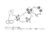
図6は、HIV−1感染の存在下でCD34+HP細胞からCD4+Tリンパ球が生産される数学的モデルを例示する。考慮したパラメータは、RRz2(または他の抗HIV遺伝子)含有Tリンパ球前駆体が骨髄を離れて、胸腺内での選択機構を経て、抹消血中にナイーブ細胞(N)として放出される割合である。これには、年齢に応じた患者の胸腺放出の推定が必要である。なぜなら、年齢と共に胸腺が退縮し、それにつれてCD4+T細胞が放出される割合が低下することが分かっているためである(センポウスキー(Sempowski)ら、2000年)。抹消血中でナイーブ細胞として樹立したら、RRz2含有Tリンパ球の生存及び増殖は、自然の恒常性機構、並びにHIVがこの恒常性機構を変えた場合はRz2−CD4+Tリンパ球に対する潜在的な選択優位性に依存する。T細胞の数を調節するこの恒常性機構は図6の左側に示されている。恒常性機構は、図3の説明で詳述したCD4+Tリンパ球が発達する過程(1)〜過程(4)を含む。
胸腺からの放出に加えて、RRz2含有CD4+Tリンパ球の数が、抗原によって活性化されてメモリーTリンパ球に分化することにより増大する。本発明のこのモデルは、このメモリーTリンパ球が増大する程度を推定することを含む。HIV感染により、図の右側の構成要素(過程(5)〜過程(7))が機能するようになる。活性化細胞がウイルス感染し(過程(5)と過程(7))、新しいウイルスを産生し(過程(6))、感染サイクルを完全にする。これらの機構がこのモデルに含まれている。また、このモデルは、HIVの存在によるナイーブ細胞(過程(2))及びメモリー細胞(過程(3))の活性の上昇などのある種の自然のプロセスの変更が可能である。
FIG. 6 illustrates a mathematical model in which CD4 + T lymphocytes are produced from CD34 + HP cells in the presence of HIV-1 infection. The parameter considered was the rate at which RRz2 (or other anti-HIV gene) -containing T lymphocyte precursors leave the bone marrow, undergo a selective mechanism within the thymus, and are released as naive cells (N) during peripheral blood. is there. This requires an estimation of the patient's thymic release as a function of age. This is because it is known that the thymus regresses with age, and the rate at which CD4 + T cells are released decreases accordingly (Sempowski et al., 2000). Once established as naive cells in peripheral blood, the survival and proliferation of RRz2-containing T lymphocytes is a natural homeostatic mechanism, and a potential selective advantage over Rz2-CD4 + T lymphocytes if HIV alters this homeostatic mechanism. Depends on gender. This homeostatic mechanism that regulates the number of T cells is shown on the left side of FIG. The homeostatic mechanism includes processes (1) to (4) in which CD4 + T lymphocytes develop in detail in the explanation of FIG.
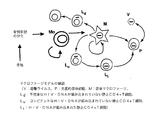
In addition to release from the thymus, the number of RRz2-containing CD4 + T lymphocytes increases by being activated by the antigen and differentiating into memory T lymphocytes. This model of the present invention involves estimating the extent to which this memory T lymphocyte increases. Due to HIV infection, the components on the right side of the figure (process (5) to process (7)) become functional. Activated cells become virally infected (process (5) and process (7)), produce new virus (process (6)), and complete the infection cycle. These mechanisms are included in this model. This model can also alter certain natural processes such as increased activity of naive cells (process (2)) and memory cells (process (3)) due to the presence of HIV.
図7は、HIV−1感染下でCD34+HP細胞からマクロファージが生産される数学的モデルを例示する。このモデルは、感染した単球及びマクロファージが抗レトロウイルス治療中の感染を維持するのに重要な役割を果たし、かつこれらの細胞が抗ウイルス治療を用いないときに感染の進行に大きな役割を果たすという確信に基づいている。このような感染した細胞の役割を評価するモデルは、ここで開発したものであって、ザック(Zack)他(1990年)及びマーレイ(Murray)(2001年)の仮説を含む。このモデルは、未処置のセロコンバーター(seroconverter)及び処置したセロコンバーターの両方におけるHIV-1 RNA及びHIV-1 DNAの動態を検査する。このモデルはまた、組み込まれていない(非組み込み)HIV-1 DNAの不安定な性質を考慮し、潜伏感染CD4+Tリンパ球及び感染マクロファージを含む。このモデルは、長命感染マクロファージMとの相互作用による、不完全な組み込まれていない形Ld 及びコンピテントな組み込まれていない形Lu の両方の感染を含む。コンピテントなHIV-1 DNAが組み込まれていない感染細胞Lu にHIV-1 DNA分子が組み込まれたときに、HIV-1 DNAが組み込まれた潜伏的感染細胞Li が生じる。遊離ウイルスVによって感染した細胞が活性化され、HIV-1 DNAが組み込まれた潜伏的感染細胞が活性化され、感染マクロファージとの相互作用のプロセス中に細胞が活性化されて生産的感染細胞Pが生じる。マクロファージは感染マクロファージと接触して感染する。 FIG. 7 illustrates a mathematical model in which macrophages are produced from CD34 + HP cells under HIV-1 infection. This model plays an important role in infected monocytes and macrophages in maintaining infection during antiretroviral therapy, and plays a major role in the progression of infection when these cells do not use antiviral therapy Is based on the belief. A model for assessing the role of such infected cells was developed here and includes the hypothesis of Zack et al. (1990) and Murray (2001). This model examines the kinetics of HIV-1 RNA and HIV-1 DNA in both untreated seroconverters and treated seroconverters. This model also considers the unstable nature of unintegrated (non-integrated) HIV-1 DNA and includes latently infected CD4 + T lymphocytes and infected macrophages. This model includes infection of both long-lived macrophages infected by interaction with M, unincorporated incomplete form L d and competent unincorporated form L u. When competent HIV-1 DNA is the HIV-1 DNA molecules in infected cells L u unincorporated incorporated, the latently infected cells L i of HIV-1 DNA is incorporated occur. Cells infected by free virus V are activated, latently infected cells incorporating HIV-1 DNA are activated, and cells are activated during the process of interaction with infected macrophages to produce productive infected cells P Occurs. Macrophages are infected in contact with infected macrophages.
用語「造血前駆(HP)細胞」は、多能性であって、生体内(in vivo)で造血系の様々な系統の全てに連続的に分化できる造血細胞を指す。 The term “hematopoietic progenitor (HP) cell” refers to a hematopoietic cell that is pluripotent and can differentiate continuously into all of the various lineages of the hematopoietic system in vivo.
用語「CD34+細胞」は、表面にCD34+抗原を有する細胞を指し、造血前駆細胞のサブセットである。 The term “CD34 + cells” refers to cells with CD34 + antigen on the surface and is a subset of hematopoietic progenitor cells.
句「CD34+細胞の純度」は、任意の集団におけるCD34抗原を有する細胞のパーセンテージを指す。 The phrase “CD34 + cell purity” refers to the percentage of cells with CD34 antigen in any population.
用語「外来核酸産物」は、細胞に導入される発現可能な核酸断片であって、好ましくは、細胞内に導入されたときに、好ましくは抗ウイルス効果である治療効果を有する断片を指す。また、断片は、細胞にとって非天然構造(non-native)であるのが好ましい。この産物は、限定するものではないが、抗体、アンチセンス分子、リボザイム、または細胞内環境で転写または転写と翻訳により産生され得る他の産物を含むタンパク質をコードする遺伝子を含むことができる。 The term “foreign nucleic acid product” refers to an expressible nucleic acid fragment that is introduced into a cell, preferably having a therapeutic effect, preferably an antiviral effect when introduced into the cell. Also, the fragment is preferably non-native to the cell. This product can include a gene encoding a protein, including but not limited to antibodies, antisense molecules, ribozymes, or other products that can be produced by transcription or transcription and translation in an intracellular environment.
用語「LNL6」は、複製遺伝子が除去されネオマイシン・ホスホトランスフェラーゼ(neor )遺伝子が挿入されたマウス・モロニー白血病ウイルスを起源とするマウス・レトロウイルスベクターを指す(ベンダー(Bender)ら、1987年)。このベクターは、複製不能レトロウイルスベクターLNL6(Genbankアクセッション番号:M63653)を含むレトロウイルスプラスミドであるpLNL6に基づいている。 The term "LNL6" refers to mouse retroviral vector to replicate genes removed neomycin phosphotransferase the (neo r) murine Moloney leukemia virus gene inserted originate (vendor (Bender) et al., 1987) . This vector is based on pLNL6, a retroviral plasmid containing the non-replicating retroviral vector LNL6 (Genbank accession number: M63653).
用語「Rz2」は、tat遺伝子の高度に保存された領域を標的とする抗HIVハンマーヘッド型リボザイムを指す。Rz2リボザイムのDNA形の配列は添付の配列表に示めされている配列番号3であり、RNA形の配列は配列番号4である。 The term “Rz2” refers to an anti-HIV hammerhead ribozyme that targets a highly conserved region of the tat gene. The sequence of the DNA form of Rz2 ribozyme is SEQ ID NO: 3 shown in the attached sequence listing, and the sequence of the RNA form is SEQ ID NO: 4.
用語「RRz2」は、Rz2がneor の3’非翻訳領域内に挿入されたLNL6からなるレトロウイルスベクターを指す。 The term “RRz2” refers to a retroviral vector consisting of LNL6 with Rz2 inserted into the 3 ′ untranslated region of neo r .
用語「DzyNA」は、米国特許第6,140,055号及び同第6,201,113号に開示されているようなDNAまたはRNAのリアルタイム量的PCR検出及び定量のための方法を指す。 The term “DzyNA” refers to a method for real-time quantitative PCR detection and quantification of DNA or RNA as disclosed in US Pat. Nos. 6,140,055 and 6,201,113.
用語「形質導入」は、ある細胞内にある遺伝子を導入して、後にその細胞がその遺伝子を発現することを指す。 The term “transduction” refers to the introduction of a gene that is in a cell that is later expressed by that cell.
第1の態様では、本発明は、患者に送達するべく、外来治療用遺伝子を含む細胞の用量の決定及びその細胞を準備する方法を提供する。患者に対するHP細胞の用量を増量する効果を決定するために、ユニークな数学的シミュレーションを作成した。このシミュレーションを用いて、遺伝子が形質導入されたHP細胞の手法が、成熟したTリンパ球及び単球/マクロファージ子孫細胞の集団に対して臨床的に適切な効果を上げることができるか否かを推定した。 In a first aspect, the present invention provides a method for determining a dose of cells containing an exogenous therapeutic gene and preparing the cells for delivery to a patient. In order to determine the effect of increasing the dose of HP cells on the patient, a unique mathematical simulation was created. Using this simulation, we can determine whether the gene-transduced HP cell approach can have a clinically relevant effect on a population of mature T lymphocytes and monocyte / macrophage progeny. Estimated.
数学的モデル化を用いて、(i)CD4+リンパ球と(ii)単球及びその子孫組織マクロファージの2つの細胞集団の動態を調べた。それぞれの細胞の種類のモデル化は、それぞれが異なった増殖、成熟、及び死のパラメータの特徴を有するため別々に行った。例えば、Rz2を含む集団によるウイルスの減少量を決定する。CD4+Tリンパ球の場合は、HIVの存在下でCD4+Tリンパ球集団が維持/増大する程度を評価する。 Mathematical modeling was used to examine the dynamics of two cell populations: (i) CD4 + lymphocytes and (ii) monocytes and their progeny tissue macrophages. Each cell type was modeled separately because each has characteristics of different growth, maturation, and death parameters. For example, the amount of virus reduction by a population containing Rz2 is determined. In the case of CD4 + T lymphocytes, the extent to which the CD4 + T lymphocyte population is maintained / expanded in the presence of HIV is assessed.
数学的シミュレーションは、公開されている微分方程式(マーレイ(Murray)他、1998年、ハース(Haase)、1996年)に少なくとも部分的に基づいている。この文献には、(i)患者の年齢及び胸腺の質量の関数である長期に亘るTリンパ球細胞の産生、(ii)抗原に応答したナイーブTリンパ球の活性化及び増殖、及び(iii)単球/マクロファージの産生が記載されている。 Mathematical simulations are based at least in part on published differential equations (Murray et al., 1998, Haase, 1996). This document includes (i) long-term T lymphocyte cell production as a function of patient age and thymus mass, (ii) activation and proliferation of naive T lymphocytes in response to antigen, and (iii) Monocyte / macrophage production has been described.
これらのシミュレーションにより、HP細胞の用量を最小閾値レベルに増大すると、遺伝子を含むCD4+Tリンパ球及び単球/マクロファージの数が増大することが示された。シミュレーションの結果から、形質導入CD34+細胞のパーセンテージが常在HP細胞の10%、より好ましくは20%を超えると、ウイルス負荷及びCD4+細胞数に影響を与えることが分かった。更に、単にリンパ球及びマクロファージのそれぞれで別々にモデル化したため、モデルは、マクロファージにおける抗ウイルス効果が見られる条件を予想した。マクロファージにおける抗ウイルス効果は、患者体内のHIVウイルスの貯蔵を減少させるのに重要である。 These simulations showed that increasing the HP cell dose to the minimum threshold level increased the number of CD4 + T lymphocytes and monocytes / macrophages containing the gene. The results of the simulation showed that the viral load and the number of CD4 + cells were affected when the percentage of transduced CD34 + cells exceeded 10%, more preferably 20% of resident HP cells. In addition, the model predicted conditions under which antiviral effects were seen in macrophages, simply modeled separately for each of lymphocytes and macrophages. Antiviral effects in macrophages are important in reducing HIV virus storage in the patient's body.
第2の態様では、本発明は、この同じパーセンテージの治療用遺伝子含有細胞を生産及び送達する方法を提供する。この方法は、(a)CD34+HP細胞を含む細胞集団を患者から得るステップと、(b)このHP細胞を濃縮して少なくとも40%HP細胞を含む細胞集団を得るステップと、(c)HP細胞で発現する能力をもった遺伝子を含むベクターをHP細胞集団に導入し、この細胞を生体外(in vitro)で更に培養するステップと、(d)このような遺伝子含有HP細胞と遺伝子非含有HP細胞の数を決定して、同じまたは別の患者に送達したときに、その患者が、体重1kg当たり、遺伝子含有HP細胞を少なくとも0.52×10⁶ 、1.63×10⁶ の総細胞数の投与を受けるようにするステップとを含む。 In a second aspect, the present invention provides a method for producing and delivering this same percentage of therapeutic gene-containing cells. The method comprises (a) obtaining a cell population comprising CD34 + HP cells from a patient; (b) enriching the HP cells to obtain a cell population comprising at least 40% HP cells; and (c) with HP cells. Introducing a vector containing a gene capable of expression into a population of HP cells and further culturing the cells in vitro; (d) such gene-containing and non-gene-containing HP cells; Of the total number of cells containing the gene-containing HP cells per kg body weight of at least 0.52 × 10 6 and 1.63 × 10 6 when delivered to the same or another patient. Receiving the dose.
導入後約1ヶ月〜3ヶ月の間に患者の骨髄で少なくとも10%の遺伝子含有HP細胞を観察できるような遺伝子含有HP細胞の数が好ましい。モデル化では、この程度の量の遺伝子含有HP細胞が骨髄で観察できれば、抗ウイルス効果などの治療効果が見られると予想する。より好ましくは、遺伝子含有CD34+HP細胞は、導入ステップの後少なくとも1年間、患者の体内で検出できる遺伝子含有子孫リンパ細胞及び子孫骨髄細胞を産生する。更に好ましくは、この方法の結果として得られるキメラ造血系は、導入ステップの後4年間はあらゆる種類の抹消血細胞に少なくとも0.01%、0.1%、1.0%、10%、より好ましくは20%、更に好ましくは50%の遺伝子含有細胞を含む。加えて、この方法の結果により得られるキメラ造血系は、導入ステップの後4年間は採取した骨髄サンプルに少なくとも0.01%、0.1%、1.0%、10%、より好ましくは20%、更に好ましくは50%の遺伝子含有細胞を含む。 The number of gene-containing HP cells is preferred so that at least 10% of the gene-containing HP cells can be observed in the bone marrow of the patient between about 1 to 3 months after introduction. In modeling, if such a quantity of gene-containing HP cells can be observed in the bone marrow, it is expected that therapeutic effects such as antiviral effects can be seen. More preferably, the gene-containing CD34 + HP cells produce gene-containing progeny lymphocytes and progeny bone marrow cells that can be detected in the patient's body for at least one year after the introduction step. More preferably, the resulting chimeric hematopoietic system is at least 0.01%, 0.1%, 1.0%, 10%, more preferably on all types of peripheral blood cells for 4 years after the introduction step. Contains 20%, more preferably 50% gene-containing cells. In addition, the chimeric hematopoietic system resulting from this method is at least 0.01%, 0.1%, 1.0%, 10%, more preferably 20% of bone marrow samples collected for 4 years after the introduction step. %, More preferably 50% gene-containing cells.
従って、本発明は、患者におけるウイルス負荷の低減を観察できるか否かを推定する方法にも関連する。この方法は、先述した方法のステップを含み、移植すなわち骨髄で細胞が定着した後で、少なくとも10%の遺伝子含有HP細胞が骨髄に存在する。更に、本発明の別の好適な実施形態では、遺伝子含有HP細胞及び/または遺伝子非含有HP細胞の数が、患者に導入するには好適でない場合は、この細胞を凍結し、プールしたHP細胞(遺伝子含有及び非遺伝子含有)の数が少なくとも前記したステップ(d)と同量になるまで、1または複数回の追加の動員及びアフェレーシスを繰り返す。 Accordingly, the present invention also relates to a method for estimating whether a reduction in viral load in a patient can be observed. This method includes the steps of the method described above, wherein at least 10% of the gene-containing HP cells are present in the bone marrow after transplantation, ie, the cells have established in the bone marrow. Furthermore, in another preferred embodiment of the invention, if the number of gene-containing and / or gene-free HP cells is not suitable for introduction into a patient, the cells are frozen and pooled HP cells One or more additional mobilizations and apheresis are repeated until the number of (gene-containing and non-gene-containing) is at least as large as in step (d) above.
得られる細胞のプールは、十分な遺伝子含有CD34+HP細胞を含むのが好ましい。具体的には、患者に投与する場合、その患者の体重1kg当たり、少なくとも5×10⁶ 、より好ましくは1×10⁷ または2×10⁷ よりも多く、更に好ましくは4×10⁷ または5×10⁷ よりも多く、更に好ましくは8×10⁷ を超えるか少なくとも10×107 の遺伝子含有CD34+HP細胞の投与を患者が受けるようにするのが好ましい。 The resulting pool of cells preferably contains sufficient gene-containing CD34 + HP cells. Specifically, when administered to a patient, at least 5 × 10 6 , more preferably more than 1 × 10 7 or 2 × 10 7, more preferably 4 × 10 7 or 5 × per kg body weight of the patient. 10 7 more than, more preferably preferred to the administration of at least 10 × 10 7 gene-containing CD34 + HP cells or more than 8 × 10 7 to patient receives.
得られる細胞のプールは、患者に投与する場合、その患者の体重1kg当たり、少なくとも1×10⁷ 、最大10×107 の総細胞数(すなわち、得られる細胞のプールに存在する治療遺伝子含有HP細胞と他の全ての細胞の総数)の投与を患者が受けるようにするのが好ましい。 The resulting pool of cells, when administered to a patient, has a total cell count of at least 1 × 10 7 and up to 10 × 10 7 per kg body weight of the patient (ie, a therapeutic gene-containing HP present in the resulting pool of cells). Preferably, the patient receives a dose of the total number of cells and all other cells.
患者から収集する細胞集団は、当分野で周知のあらゆる方法で得ることができる。例えば、患者に処置を施して、HP細胞が骨髄から抹消血に動員されるようにする。その方法は、例えば、限定するものではないが、ペグ化顆粒球コロニー刺激因子(pegG-CSF)、顆粒球マクロファージコロニー刺激因子(GM-CSF)、及びより好ましくはG−CSFを含むサイトカインを適量投与し、次いでアフェレーシス濾過を行う。別法では、HP細胞は、当分野で周知の方法に従って骨髄または臍帯血から吸引することができる。 The cell population collected from the patient can be obtained by any method known in the art. For example, the patient is treated so that HP cells are mobilized from the bone marrow to peripheral blood. The method includes, but is not limited to, an appropriate amount of a cytokine comprising pegylated granulocyte colony stimulating factor (pegG-CSF), granulocyte macrophage colony stimulating factor (GM-CSF), and more preferably G-CSF. Dosing followed by apheresis filtration. Alternatively, HP cells can be aspirated from bone marrow or umbilical cord blood according to methods well known in the art.
収集した細胞集団の処置には、1回または複数回の洗浄ステップ(例えば、遠心分離器または細胞洗浄装置を用いる)、及び/または容量減少ステップ(すなわち、余分な赤血球、顆粒球、血小板、Tリンパ球などの除去)が含まれるのが好ましい。容量減少ステップは、ノースカロライナ州ウインストンセーラムに所在のチャーターメディカル社(Charter Medical, Winston Salem, NC)が販売するデンドレオンDACSシステム(Dendreon DACS System)などの装置を用いて行うのが好ましく、更にHP細胞選択ステップを含むのが好ましい。HP細胞の選択は、イムノアフィニティーまたはフローサイトメトリー法によって行うことができる。HP細胞選択ステップではCD34+細胞を選択するのが好ましい。別の実施形態では、成熟/委任造血細胞の抗原を除去してHP細胞の細胞集団を濃縮することもできる。HP細胞選択ステップは、限定するものではないが、ネクセル/バクスター アイソレックス300I(Nexell/Baxter Isolex300I)(カリフォルニア州アーバイン)、ミルティニー・クリニマックス(Miltenyi CliniMACS)(ドイツ、ベルギッシュ・グラッドバッハ所在のバイオテック社(Miltenyi;Biotech GmBH, Bergisch Gladbach, Germany))、及びステムセル・テクノロジー社(カナダ、BC州バンクーバー)のステムセップ装置(StemSep Device)などの様々な選択装置を用いて行うことができる。 Treatment of the collected cell population can include one or more washing steps (eg, using a centrifuge or cell washer) and / or a volume reduction step (ie, extra red blood cells, granulocytes, platelets, T Including removal of lymphocytes). The volume reduction step is preferably performed using a device such as the Dendreon DACS System sold by Charter Medical, Winston Salem, NC located in Winston Salem, North Carolina, and HP cell selection. Preferably steps are included. Selection of HP cells can be performed by immunoaffinity or flow cytometry. In the HP cell selection step, CD34 + cells are preferably selected. In another embodiment, the antigen of mature / committed hematopoietic cells can be removed to enrich the cell population of HP cells. HP cell selection steps include, but are not limited to, Nexell / Baxter Isolex 300I (Irvine, CA), Miltenyi CliniMACS (Biotech, Bergisch Gladbach, Germany) (Miltenyi; Biotech GmBH, Bergisch Gladbach, Germany)) and StemSep Device (StemSep Device, Vancouver, BC) and various selection devices.
収集した細胞集団の処置には、細胞の数、特に選択したHP細胞の数を増やすための細胞培養ステップが含まれ得る。細胞培養ステップでは、治療用遺伝子を細胞内に導入する必要があり、治療用遺伝子の導入の後に細胞培養をして遺伝子の組み込み及び遺伝子産物の発現を促し、このような遺伝子含有HP細胞の数を増やすのが好ましい。 Treatment of the collected cell population can include a cell culture step to increase the number of cells, particularly the number of selected HP cells. In the cell culture step, it is necessary to introduce a therapeutic gene into the cell, and after the introduction of the therapeutic gene, cell culture is performed to promote gene integration and gene product expression, and the number of such gene-containing HP cells. Is preferably increased.
初めの処置ステップ(動員、アフェレーシス、HP選択)で、HP細胞を得てそのHP細胞を濃縮する。HP細胞のパーセンテージの決定には、CD34抗原陽性などのHP細胞の測定可能な特徴が必要である。体外(ex vivo)で処置した細胞のプールは、好ましくは少なくとも20%、より好ましくは少なくとも40%、更に好ましくは少なくとも60%、最も好ましくは少なくとも80%のHP細胞を含むことを理解されたい。 In the first treatment step (mobilization, apheresis, HP selection), HP cells are obtained and the HP cells are enriched. Determination of the percentage of HP cells requires measurable characteristics of HP cells such as CD34 antigen positive. It should be understood that the pool of cells treated ex vivo preferably comprises at least 20%, more preferably at least 40%, even more preferably at least 60%, and most preferably at least 80% HP cells.
治療用遺伝子すなわち核酸配列のHP細胞の少なくとも一部への導入は、当分野で周知の様々な方法または他の方法を用いて行うことができる。好適な実施形態では、導入ステップで、治療用遺伝子すなわち核酸配列を有するレトロウイルスベクター、他のウイルスまたは非ウイルス(DNAまたはRNA)ベクターを用いた形質導入を利用する。形質導入を利用する場合、形質導入促進剤(例えば、レトロウイルスベクターの場合は、レトロネクチン(RetroNectin)(登録商標)として知られるフィブロネクチンのCH296断片、またはポリブレンや硫酸プロタミンなどの他の作用物質)を用いるのが好ましい。治療用遺伝子すなわち核酸配列を含むHP細胞及びその子孫細胞(すなわち、後にリンパ造血及び骨髄造血で産生される細胞)は、細胞で発現することを意図した治療用遺伝子を含み、治療用遺伝子すなわち核酸配列を発現する能力を有するのが好ましい。 Introduction of a therapeutic gene or nucleic acid sequence into at least a portion of an HP cell can be accomplished using various methods known in the art or other methods. In a preferred embodiment, the transducing step utilizes transduction with a retroviral vector, other viral or non-viral (DNA or RNA) vector having a therapeutic gene or nucleic acid sequence. When using transduction, a transduction promoter (for example, in the case of a retroviral vector, a CH296 fragment of fibronectin known as RetroNectin® or other agent such as polybrene or protamine sulfate) It is preferable to use it. HP cells and their progeny cells that contain a therapeutic gene or nucleic acid sequence (ie, cells that are later produced in lymphatic and bone marrow hematopoiesis) contain a therapeutic gene that is intended to be expressed in the cell, It preferably has the ability to express the sequence.
HP細胞内に導入された治療用核酸配列は、HIV/AIDSの場合は、限定するものではないが、タンパク質(例えば、トランスドミナント(transdominant)タンパク質、細胞内抗体)、アンチセンスRNA、アプタマー、RNA阻害(interfering RNA)、及び触媒リボザイムなどの産物をコードでき、別の疾患の場合は、これらの産物や腫瘍抑制遺伝子などの他の遺伝子をコードできる。 The therapeutic nucleic acid sequences introduced into HP cells include, but are not limited to, proteins (eg, transdominant proteins, intracellular antibodies), antisense RNA, aptamers, RNA in the case of HIV / AIDS. Products such as interfering RNA and catalytic ribozymes can be encoded, and in the case of other diseases, these products and other genes such as tumor suppressor genes can be encoded.
細胞は、細胞注入などの決まった手順に従って患者に導入することができる。細胞はまた、薬学的に許容できるバッファー及び塩などを含む薬理学的に許容できる担体(例えば、5%ヒト血清アルブミンなど)と共に送達することができる。患者は、初めに(すなわち、細胞の再注入の前)ミエロアブレーション(myeloablation)または他の造血コンディショニング療法を受けても、受けなくてもよい。しかしながら、本発明の形質導入HP細胞集団を移植するための好適な方法では、本発明の実際の実質的な利点は、患者が、ミエロアブレーション療法(すなわち、骨髄の完全またはほぼ完全な破壊)や他の造血コンディショニング療法を受けなくてもよいことである。形質導入HP細胞を生成して移植する方法の利点は、限定するものではないが化学療法や放射線治療を含み、有害で、エネルギーを消費し、かつ患者を衰弱させるミエロアブレーション療法を用いないことである。 The cells can be introduced into the patient according to a routine procedure such as cell injection. The cells can also be delivered with a pharmacologically acceptable carrier, such as pharmaceutically acceptable buffers and salts, such as 5% human serum albumin. The patient may or may not receive myeloablation or other hematopoietic conditioning therapy initially (ie, prior to cell reinfusion). However, in a preferred method for transplanting the transduced HP cell population of the present invention, the actual substantial advantage of the present invention is that the patient can use myeloablation therapy (ie, complete or nearly complete destruction of bone marrow) or There is no need to receive other hematopoietic conditioning therapies. The advantages of generating and transplanting transduced HP cells are that they do not use myeloablation therapy, which includes, but is not limited to, chemotherapy and radiation therapy, which is harmful, energy consuming, and debilitating the patient. is there.
本発明の方法は、例えば、他の抗ウイルス療法を含む他の療法と組み合わせることができる。他の抗ウイルス療法には、例えば、RNAデコイ(RNA decoys)、細胞内抗体、及びRNA阻害(シャープ・ピー・エイ(Sharp, P. A.)著、「RNA妨害−2001(RNA interference-2001)」、Genes & Dev、2001年、15:485〜490)などの使用が含まれる。例えば、本方法が、リボザイム型療法などの抗ウイルス療法を用いる場合、他の抗ウイルス療法、特に抗HIV療法を用いることができる。リボザイム型療法を用いる場合、2つ以上の触媒リボザイムを患者から除去したサンプルの1つの細胞に送達することもできるし、また、様々なリボザイムをそのサンプルの様々な細胞に送達することもできる。同様に、本方法は、当分野で周知の標準的な化学療法や蛋白療法などの、HP細胞の形質導入を必要としない他の遺伝子療法と組み合わせることができる。HIV感染患者の治療の一例では、特にHIV耐性が検出された場合、または患者のHIV感染が他の抗HIV治療に対して抗療性が確認された場合に、本発明の方法をHIVの標準的な薬剤と組み合わせることができる。 The methods of the invention can be combined with other therapies including, for example, other antiviral therapies. Other antiviral therapies include, for example, RNA decoys, intracellular antibodies, and RNA inhibition (Sharp, PA, “RNA interference-2001”), Genes & Dev, 2001, 15: 485-490). For example, if the method uses antiviral therapy such as ribozyme type therapy, other antiviral therapy, particularly anti-HIV therapy, can be used. When using ribozyme type therapy, two or more catalytic ribozymes can be delivered to one cell of a sample removed from a patient, and various ribozymes can be delivered to various cells of the sample. Similarly, the method can be combined with other gene therapies that do not require transduction of HP cells, such as standard chemotherapy and protein therapy well known in the art. In one example of treatment of an HIV-infected patient, the method of the present invention may be used as a standard for HIV, particularly if HIV resistance is detected, or if the patient's HIV infection is confirmed to be refractory to other anti-HIV treatments. Can be combined with other drugs.
本発明は、造血系の外来遺伝子を含む細胞を導入するための一般的な方法を企図するが、本発明は、一例としてHIVの抗ウイルス遺伝子治療についてのみを取り上げる。本方法では、HIV陽性患者から収集した細胞からHP細胞を濃縮し、治療用遺伝子は抗HIV産物をコードする。 Although the present invention contemplates a general method for introducing cells containing foreign genes of the hematopoietic system, the present invention will only address HIV antiviral gene therapy as an example. In this method, HP cells are enriched from cells collected from HIV positive patients and the therapeutic gene encodes an anti-HIV product.
従って、第3の態様では、本発明は、用量の決定と、好ましくはCD34+細胞であるHP細胞の準備を提供する。このHP細胞は、HIV陽性患者に送達して持続的に抗ウイルス治療効果を得るべく、抗HIV産物をコードする遺伝子を含む。リボザイムが本発明の好適な実施形態としているが、他の抗ウイルス産物をコードする遺伝子を、限定するものではないが、アンチセンス療法やRNA阻害などに用いることもできる。 Thus, in a third aspect, the present invention provides dose determination and preparation of HP cells, preferably CD34 + cells. The HP cells contain a gene encoding an anti-HIV product for delivery to HIV positive patients to obtain a sustained antiviral therapeutic effect. Although ribozymes are preferred embodiments of the present invention, genes encoding other antiviral products can be used for, but not limited to, antisense therapy and RNA inhibition.
上記したように、この決定のための数学的シミュレーションは、公開されている微分方程式(マーレイ(Murray)他、1998年、ハース(Haase)、1996年)に基づいている。この数学的シミュレーションは、(i)患者の年齢及び胸腺の質量の関数である長期に亘るTリンパ球細胞の産生、(ii)抗原に応答したナイーブTリンパ球の活性化及び増殖、(iii)HIV感染によるCD4+細胞の減少、及び(iv)単球/マクロファージの産生を考慮して、HIV感染に適用することができる。 As noted above, the mathematical simulation for this determination is based on published differential equations (Murray et al., 1998, Haase, 1996). This mathematical simulation consists of (i) long-term T lymphocyte cell production as a function of patient age and thymus mass, (ii) activation and proliferation of naive T lymphocytes in response to antigen, (iii) Considering the reduction of CD4 + cells by HIV infection and (iv) production of monocytes / macrophages, it can be applied to HIV infection.
CD34+の用量を増大する効果の判定を、数学的シミュレーションを用いて研究した。この数学的シミュレーションは、RRz2形質導入CD34+細胞の手法が、HIV患者におけるCD4+細胞数及びウイルス負荷に臨床的に有意な効果を与えるか否かを評価する論理法を提供する。このようなシミュレーションでは、CD34+細胞の用量を増大することにより、CD4+Tリンパ球の数及びHIVウイルス負荷に影響を与えるRRz2含有CD4+Tリンパ球及び単球/マクロファージを増大させることができると予想する。このような数学的シミュレーションに基づいて、形質導入HP細胞の導入する数を決定し最大化する方法を見出した。形質導入CD34+細胞の用量は、前のフェーズI臨床試験で用いた最大用量の少なくとも2倍から10倍に増量する。この用量は、方法論で求めることができる。 The determination of the effect of increasing the dose of CD34 + was studied using mathematical simulations. This mathematical simulation provides a logical method to assess whether the RRz2 transduced CD34 + cell approach has a clinically significant effect on CD4 + cell count and viral load in HIV patients. In such simulations, it is expected that increasing the dose of CD34 + cells can increase the number of CD4 + T lymphocytes and RRz2-containing CD4 + T lymphocytes and monocytes / macrophages that affect HIV viral load. Based on such mathematical simulations, a method for determining and maximizing the number of transduced HP cells to be introduced was found. The dose of transduced CD34 + cells is increased by at least 2 to 10 times the maximum dose used in previous Phase I clinical trials. This dose can be determined by methodology.
従って、抗HIV産物を送達する方法は、(i)HP細胞、好ましくはCD34+細胞を含む生細胞の集団を患者から得るステップと、(ii)この細胞集団を処置及び/または培養して、少なくとも20%のCD34+細胞を含む細胞のプールを用意するステップと、(iii)少なくとも1種類の治療用遺伝子をこの細胞集団内のCD34+細胞の集団内に導入して、CD34+細胞で治療用遺伝子が発現可能にし、治療用遺伝子を含むCD34+細胞を含む細胞のプールを用意し、患者に投与する場合、患者が体重1kg当たり、0.52×106 の治療用遺伝子を含むHP細胞の用量を受け取るようにするステップとを含む。 Accordingly, a method of delivering an anti-HIV product comprises: (i) obtaining a population of living cells comprising HP cells, preferably CD34 + cells, from a patient; and (ii) treating and / or culturing the cell population, Providing a pool of cells comprising 20% CD34 + cells; and (iii) introducing at least one therapeutic gene into a population of CD34 + cells within the cell population, wherein the therapeutic gene is expressed in the CD34 + cells When enabled, a pool of cells containing CD34 + cells containing a therapeutic gene is prepared and administered to a patient so that the patient receives a dose of HP cells containing 0.52 × 10 6 therapeutic gene per kg body weight And the step of.
好ましくは、治療用遺伝子含有CD34+HP細胞を含む生細胞のプールを用意し、患者に投与する場合、患者が、体重1kg当たり少なくとも5×10⁶ を超える、より好ましくは2×10⁷ を超える、更に好ましくは5×10⁷ を超える治療用遺伝子を含むHP細胞の用量を受け取るようにする。 Preferably, when a pool of live cells comprising therapeutic gene-containing CD34 + HP cells is provided and administered to a patient, the patient is at least 5 × 10 6 per kg body weight, more preferably greater than 2 × 10 7 , Preferably, a dose of HP cells containing more than 5 × 10 7 therapeutic genes is received.
得られる細胞のプールは、患者に投与する場合、その患者の体重1kg当たり、少なくとも1×10⁷ 、最大4×10⁷ 、より好ましくは最大10×107の総細胞数(すなわち、得られる細胞のプールに存在する治療遺伝子含有HP細胞と他の全ての細胞の総数)を患者が受け取るようにするのが好ましい。 The resulting pool of cells, when administered to a patient, is a total cell count (ie, cells obtained of at least 1 × 10 7 , up to 4 × 10 7 , more preferably up to 10 × 10 7 per kg of the patient's body weight. Preferably, the patient receives the therapeutic gene-containing HP cells and all other cells present in this pool.
細胞集団の収集、その後の細胞集団の処置は、本発明の第1の態様で説明した要領で行うことができる。好ましくは、この処置には、細胞集団を洗浄する第1のステップ(例えば、標準的な細胞洗浄装置を用いる)と、HP細胞が濃縮された細胞集団を生成するために容積を減少させる任意選択のステップ(例えば、余分な赤血球、顆粒球、血小板、Tリンパ球を除去するための標準的な装置を用いる)と、洗浄(例えば、自動化された細胞洗浄装置を用いる)して、HP濃縮細胞集団を培養するステップとが含まれる。 Collection of the cell population and subsequent treatment of the cell population can be performed as described in the first aspect of the present invention. Preferably, this treatment includes a first step of washing the cell population (eg, using a standard cell washing device) and an option to reduce the volume to produce a cell population enriched in HP cells. HP-enriched cells (eg, using a standard device for removing excess red blood cells, granulocytes, platelets, T lymphocytes) and washing (eg, using an automated cell washer) Culturing the population.
初めの処置ステップ(動員、アフェレーシス、HP選択)で、HP細胞部分が濃縮される。HP細胞のパーセンテージの決定には、CD34抗原陽性などのHP細胞の測定可能な特徴が必要である。処置した細胞のプールは、好ましくは少なくとも20%、より好ましくは40%、更に好ましくは60%、最も好ましくは80%のHP細胞を含むことを理解されたい。 In the first treatment step (mobilization, apheresis, HP selection), the HP cell part is enriched. Determination of the percentage of HP cells requires measurable characteristics of HP cells such as CD34 antigen positive. It will be appreciated that the pool of treated cells preferably contains at least 20%, more preferably 40%, even more preferably 60%, and most preferably 80% HP cells.
少なくとも一部のCD34+細胞への遺伝子の導入は、形質導入促進剤(例えば、RetroNectin)の存在下で、治療用遺伝子を含むレトロウイルスベクターでこれらの細胞を形質導入するのが好ましい。しかしながら、遺伝子治療分野の通常の技術者であれば、細胞内に遺伝子を導入する他の公開されている方法を用いても同じ結果が得られることを理解できよう。このような遺伝子には、あらゆる抗HIV産物をコードする遺伝子を用いることができるが、HIV触媒リボザイムをコードする遺伝子が好ましい。特に好適な抗HIV−1触媒リボザイムは、tat遺伝子内のHIV RNA、特に基準株HIV-HXB2(Genbankアクセッション番号:K03455)の遺伝子(すなわち、5833番目〜5849番目までのヌクレオチド(配列番号3))及びHIV-IIIB株(Genbankアクセッション番号:X01762)の5865番目〜5882番目までのヌクレオチド(配列番号3)の高度に保存された領域内のHIV RNAを切断する抗HIV−1触媒リボザイムである。 For introduction of genes into at least some CD34 + cells, these cells are preferably transduced with a retroviral vector containing a therapeutic gene in the presence of a transduction promoter (eg, RetroNectin). However, one of ordinary skill in the field of gene therapy will understand that the same results can be obtained using other published methods of introducing genes into cells. As such a gene, a gene encoding any anti-HIV product can be used, but a gene encoding an HIV catalytic ribozyme is preferred. A particularly suitable anti-HIV-1 catalytic ribozyme is the HIV RNA within the tat gene, particularly the gene of the reference strain HIV-HXB2 (Genbank accession number: K03455) (ie nucleotides 5833 to 5849 (SEQ ID NO: 3)). ) And HIV-IIIB strain (Genbank accession number: X01762) is an anti-HIV-1 catalytic ribozyme that cleaves HIV RNA in the highly conserved region of nucleotides 5865 to 5882 (SEQ ID NO: 3). .
好適な実施形態では、患者が骨髄のミエロアブレーションまたは他の骨髄コンディショニング療法を必要とせず、細胞を導入するステップにより、患者が、体重1kg当たり、少なくとも1.63×10⁶ のCD34+細胞の用量を受け取る。この細胞の内、患者の体重1kg当たり、少なくとも0.52×10⁶ の治療用遺伝子含有CD34+細胞である。 In a preferred embodiment, the patient does not require bone marrow myelination or other bone marrow conditioning therapy, and the step of introducing cells allows the patient to receive a dose of at least 1.63 × 10 6 CD34 + cells per kg body weight. receive. Of these cells, there are at least 0.52 × 10 6 therapeutic gene-containing CD34 + cells per kg patient body weight.
本発明は更に、遺伝子産物の存在及び発現をモニターする方法に関する。我々は、この検出のために量的リアルタイムPCR方法論を確立した。この種の量的リアルタイムPCR方法論は、DzyNA-PCRと呼ばれ、トッド(Todd)他(2000年)によって米国特許第6,140,055号及び同第6,201,113号に開示されており、その中には疾患または外来物質の存在に関連した特定の遺伝子配列を検出するためのストラテジーが示されている。この方法は、1つの閉じた容器内でのリアルタイム蛍光検出と同種核酸増幅が可能なシステムを提供する。このストラテジーには、10:23DNAzymes(サントロ(Santoro)他、1997年)の相補的(アンチセンス)配列を含むDzyNAプライマーを用いた遺伝子配列の生体外(in vitro)増幅が含まれる。増幅中に、反応混合液に含まれているレポーター基質を切断するDNAzymesの活性センスコピーを含むアンプリコン(単位複製配列)が生成される。PCR中のアンプリコンの蓄積は蛍光の変化でモニターできる。この蛍光の変化は、レポーター基質内のDNAzyme切断部位の反対側に組み込まれた蛍光/消光色素分子の分離によって生じる。このレポーター基質の切断は、標的核酸配列の増幅が成功したことを示す。リアルタイム測定は、ABI PRISM(登録商標)7700配列検出システム(アプライドバイオシステムズ(Applied Biosystems))またはリアルタイムで蛍光をモニターできる他のサーモサイクラーで行うことができる。 The invention further relates to a method of monitoring the presence and expression of a gene product. We have established a quantitative real-time PCR methodology for this detection. This type of quantitative real-time PCR methodology is called DzyNA-PCR and is disclosed by Todd et al. (2000) in US Pat. Nos. 6,140,055 and 6,201,113. Among them, strategies for detecting specific gene sequences associated with disease or the presence of foreign substances are shown. This method provides a system capable of real-time fluorescence detection and homologous nucleic acid amplification in one closed container. This strategy involves in vitro amplification of the gene sequence using DzyNA primers containing the complementary (antisense) sequence of 10:23 DNAzymes (Santoro et al., 1997). During amplification, an amplicon (amplicon) containing an active sense copy of DNAzymes that cleaves the reporter substrate contained in the reaction mixture is generated. Amplicon accumulation during PCR can be monitored by changes in fluorescence. This change in fluorescence is caused by the separation of fluorescent / quenching dye molecules incorporated on the opposite side of the DNAzyme cleavage site in the reporter substrate. This cleavage of the reporter substrate indicates that the target nucleic acid sequence has been successfully amplified. Real-time measurements can be made with the ABI PRISM® 7700 Sequence Detection System (Applied Biosystems) or other thermocycler capable of monitoring fluorescence in real time.
本明細書中において、用語「含む」またはその変形である「含んでいる」は、記載した要素、整数、ステップ、または要素、整数、ステップのグループを含むが、他のあらゆる要素、整数、ステップ、または要素、整数、ステップのグループを除外するものではないことを理解されたい。 As used herein, the term “comprising” or a variation thereof “comprising” includes the recited element, integer, step or group of elements, integers, steps, but any other element, integer, step. It should be understood that it does not exclude groups of elements, integers or steps.
本明細書中に含まれる文書、行為、物質、装置、及び物品などのあらゆる説明は、単に本発明の文脈を作成することが目的である。これら文書、行為、物質、装置、及び物品などの何れかあるいは全てが、本願の各クレームの優先日以前に、従来技術の基礎の一部をなし、本発明が属する分野の一般的な共通知識であると見なされるべきものではない。 Any description of documents, acts, materials, devices, articles, etc. contained herein is solely for the purpose of creating a context for the present invention. Any or all of these documents, acts, substances, devices, articles, etc., form part of the prior art prior to the priority date of each claim of this application, and general common knowledge in the field to which this invention belongs. Should not be considered.
本発明を、以下の例(限定目的ではない)及び添付の図面を用いて以下に説明する。 The invention will now be described with the aid of the following examples (not limiting purposes) and the accompanying drawings.
例
例1:
HP細胞の収集、形質導入、及び再注入
好適な方法では、本発明は以下のステップを含む。
1.患者の骨髄から抹消血中へのHP細胞の動員
2.動員されたHP細胞を得るための患者の抹消血のアフェレーシス
3.実施する可能性のある容量減少ステップの準備としての、細胞洗浄装置を用いた未精製抹消血単核細胞の洗浄ステップ1
4.余分な赤血球、顆粒球、血小板、及びTリンパ球を除去するための容量減少ステップ
5.細胞洗浄装置を用いた濃縮HP細胞の洗浄ステップ2
6.CD34+細胞の選択すなわちHP細胞集団からの抗原陽性細胞の除去
7.細胞洗浄装置を用いた精製HP細胞の洗浄ステップ3
8.サイトカイン/成長因子を含む培地に精製HP細胞を移す細胞培養
9.形質導入促進剤の存在下での、遺伝子作製物を含むレトロウイルスベクターを用いたHP細胞の形質導入処置:ウイルスベクターの導入
10.形質導入HP細胞を含む細胞産物の回収及びHP細胞の洗浄
11.注入用産物の準備:HP細胞を注入バッグに入れ産物の安全注入試験の実施
12.同じ患者に細胞を戻す患者への注入
例を用いたこれらのステップの説明及び本発明の他の変更例を以下に示す。
Example
Example 1:
HP cell collection, transduction, and reinfusion In a preferred method, the present invention comprises the following steps.
1. 1. Mobilization of HP cells from the patient's bone marrow into the
4). 4. Volume reduction step to remove excess red blood cells, granulocytes, platelets and
6). 6. Selection of CD34 + cells, ie removal of antigen positive cells from HP
8). 8. Cell culture in which purified HP cells are transferred to medium containing cytokine / growth factor 9. Transduction treatment of HP cells using a retroviral vector containing a gene product in the presence of a transduction promoter: introduction of a
ステップ1:HP細胞の動員
この処置の第1のステップは、骨髄から抹消血にHP細胞を動員するために作用物質を用いる。一例では、ペグ化顆粒球コロニー刺激因子(pegG-CSF)、顆粒球マクロファージコロニー刺激因子(GM-CSF)、及び最も好ましいG−CSFからなる好適な群から選択されるサイトカインを用い、次いでアフェレーシス濾過を行う。別法では、HP細胞は、当分野で周知の方法に従って骨髄または臍帯血から吸引することができる。
Step 1: HP cell mobilization The first step in this procedure uses agents to mobilize HP cells from the bone marrow to peripheral blood. In one example, a cytokine selected from a suitable group consisting of pegylated granulocyte colony stimulating factor (pegG-CSF), granulocyte macrophage colony stimulating factor (GM-CSF), and most preferred G-CSF, followed by apheresis filtration I do. Alternatively, HP cells can be aspirated from bone marrow or umbilical cord blood according to methods well known in the art.
顆粒球コロニー刺激因子(G-CSF)(カリフォルニア州サウザンドオークスに所在のアムジェン社(Amgen)のNeupogen(商標))を1日1回、少なくとも10μg/kg/日、好ましくは約30μg/kg/日、最大5日連続して患者の皮下に投与する。G−CSF投与中に全血球算定(CBCs)、微分血小板算定を毎日実施して、白血球増大の程度を評価する。CD34+細胞数を決定するために、好ましくはG−CSF投与の3日目に血液サンプルを採取して、アフェレーシス開始前に抹消血CD34+細胞数が20細胞/mm3 であることを確かめる。しかしながら、CD34+細胞数がこの値に達しなかったとしても、通常はG−CSF投与の4日目または5日目に行うアフェレーシスを妨げるものではない。
Granulocyte colony stimulating factor (G-CSF) (Amgen's Neupogen ™, Thousand Oaks, Calif.) At least 10 μg / kg / day, preferably about 30 μg / kg / day, once a day. The patient is administered subcutaneously for up to 5 consecutive days. Complete blood counts (CBCs) and differential platelet counts are performed daily during G-CSF administration to assess the extent of leukocytosis. In order to determine the CD34 + cell count, a blood sample is preferably taken on
ステップ2:アフェレーシス(例1:好ましくは4日目または5日目)
アフェレーシスとは、抹消血の単核細胞画分を得るための「血液濾過」の方法である。ここで、コーブ・スペクトラ(Cobe Spectra)(コロラド州レイクウッド所在のガンブロBCT社(Gambro BCT, Lakewood, Colorado))、ヘモネティクス(Haemonetics)(マサチューセッツ州に所在のヘモネティクス社(Haemonetics Corporation, Braintree, MA))、またはアミカス(Amicus)(イリノイ州ディアフィールドに所在のバクスター社のフェンワル(Baxter Fenwal, Deerfield, IL))などの装置を、少なくとも2つの別々の時に用いるのが好ましい(好ましくは、動員の後から4日目及び5日目、ここで、1日目は動員した日である)。ただし、別の例では、アフェレーシスは、抹消血CD34+細胞数が5細胞/mm3 、より好ましくは10細胞/mm3 、最も好ましくは20細胞/mm3 よりも多い日を決定して、上記した日よりも前または後に行うことができる。好適な実施形態では、このアフェレーシスにより、約5リットルの血液量、好ましくは5リットル〜10リットル、より好ましくは10リットル〜20リットル、更に好ましくは20リットル以上の血液量から細胞産物を得る。それぞれのアフェレーシスによる産物は、別々に処置するか、好適な実施形態では2回目のアフェレーシスの後でプールする。全細胞数及び絶対CD34+細胞数を記録する。ステップ1及びステップ2により、1kg当たり5×10⁶ 、好ましくは2×10⁷ 、より好ましくは4×10⁷ のHP細胞(CD34陽性で測定)が得られる。
Step 2: Apheresis (Example 1: preferably on the 4th or 5th day)
Apheresis is a “blood filtration” method for obtaining a mononuclear cell fraction of peripheral blood. Where Cobe Spectra (Gambro BCT, Lakewood, Colorado), Haemonetics (Haemonetics Corporation, Braintree, Massachusetts) MA)), or Amicus (Baxter Fenwal, Deerfield, IL, Deerfield, Ill.) Is preferably used (preferably mobilized) in at least two separate times. 4th and 5th day after, where
ステップ3:洗浄ステップ1(例1:好ましくは4日目及び5日目)
プールした細胞を洗浄する。この洗浄は、細胞の遠心分離(1,500rpmすなわち300gで約15分間)、より好ましくは自動化された細胞洗浄装置を用いて行う。一例では、この細胞洗浄は、ネクセルCytoMate(商標)洗浄装置(Nexell CytoMate washer)(カリフォルニア州アーバインに所在のネクセルセラピューティクス(Nexell Therapeutics))を用いて、バッグ4からスピニング・メンブレンを通して洗浄バッグ1に移送して細胞を洗浄するプログラムで、所定のパラメータ(レジデュアル・フォールド・リダクション(Residual Fold Reduction)=1、マキシマム・エンド・ウエイト(Maximum End Weight)=190ml、ソースバッグリンス(Source Bag Rinse)=50ml)または類似のパラメータを用いて行う。次いで細胞を、所定のパラメータ(チューブリンス容量(Tubing Rinse Volume)=90ml、最大ポンプ速度(Maximum Pump Rate)=50ml/分)を用いて、洗浄バッグ1から最終産物バッグ3に移す。
Step 3: Washing step 1 (Example 1: preferably on the 4th and 5th days)
Wash pooled cells. This washing is performed using cell centrifugation (1,500 rpm or 300 g for about 15 minutes), more preferably using an automated cell washer. In one example, this cell wash is performed using a Nexel CytoMate ™ washer (Nexell CytoMate washer) (Nexell Therapeutics located in Irvine, Calif.) From
ステップ4:容量減少ステップ(例1:好ましくは4日目及び5日目)
一実施形態では、アフェレーシス処置により得た細胞を、それぞれ処置した日にチャーターメディカルDACS-SC(商標)システム(Charter Medical DACS-SC(商標)system)(ノースカロライナ州ウインストンセーラムに所在のチャーターメディカル(Charter Medical))を用いて容量減少を行う。後に最終日の産物と共にプールするために産物を1日目(または次の日)から一晩保存する実施形態では、2つ以上のアフェレーシス産物を、収集した日に容量減少し、第2の産物を容量減少するまで第1の産物を保存する。容量減少では、洗浄したアフェレーシス細胞産物をDACS-SC(商標)装置内のBDS60溶液に入れ、850g、20℃〜25℃、停止なしで30分間、遠心分離する。この産物を、滅菌条件下で、逆にしてトランスファーパック内に収集して回収する。
Step 4: Capacity reduction step (Example 1: preferably on the 4th and 5th days)
In one embodiment, the cells obtained from the apheresis treatment are transferred to the Charter Medical DACS-SC ™ system (Charter Medical DACS-SC ™ system) (Charter Medical, Winston Salem, NC) on each treatment day. The volume is reduced using Medical)). In embodiments where the product is stored overnight from day 1 (or next day) for later pooling with the product of the last day, two or more apheresis products are reduced in volume on the day of collection and the second product Store the first product until the volume is reduced. For volume reduction, the washed apheresis cell product is placed in the BDS60 solution in the DACS-SC ™ apparatus and centrifuged at 850 g, 20 ° C. to 25 ° C. for 30 minutes without stopping. The product is collected and recovered in a reverse transfer pack under sterile conditions.
ステップ5:洗浄ステップ2(例1:4日目)
プールする前に保存される産物の収集の日に、得られた産物を、0.5%ヒト血清アルブミンを添加したダルベッコ・リン酸緩衝生理食塩水(Dulbecco's phosphate buffered saline:DPBS)を用いて洗浄する。この洗浄は、ネクセルCytoMate(商標)洗浄装置を用いて、バッグ4からスピニング・メンブレンを通して洗浄バッグ1に移送して細胞を洗浄するプログラムで、所定のパラメータ(レジデュアル・フォールド・リダクション(Residual Fold Reduction)=1,000、マキシマム・エンド・ウエイト(Maximum End Weight)=20ml、ソースバッグリンス(Source Bag Rinse)=50ml、または類似のパラメータ)を用いて行う。次いで、100%自己血漿中の細胞を、所定のパラメータ(チューブリンス容量(Tubing Rinse Volume)=195ml、最大ポンプ速度(Maximum Pump Rate)=50ml/分、または類似のパラメータ)を用いて、洗浄バッグ1から最終産物バッグ3に移す。次いで、この流路を、0.5%ヒト血清アルブミンを添加した別のDPBS50mlで洗浄する。次いで、この細胞産物を、200×106 細胞/ml以下の細胞密度、2℃〜8℃で一晩保存する。一晩保存しなかった産物は、CD34+細胞選択ステップ(以下のステップ6)で洗浄するため、別の洗浄は行わない。
Step 5: Washing step 2 (Example 1: 4th day)
On the day of collection of the product to be stored before pooling, the resulting product is washed with Dulbecco's phosphate buffered saline (DPBS) supplemented with 0.5% human serum albumin To do. This washing is a program for washing cells by transferring them from the
ステップ:6CD34+細胞選択(例1:5日目)
細胞を取り出し、計数し、プールし(好適な実施形態では、2つ以上の産物が存在する)、アイソレックス300i(Isolex 300i)使い捨てセット(カリフォルニア州アーバインに所在のネクセルセラピューティクス)のライフセルバッグ(LifeCell bag)(カリフォルニア州アーバインに所在のネクセルセラピューティクス)に移す(3つ以上の産物がある場合は、その全てを最新の時点でプールする)。CD34+細胞を、洗浄後の産物からアイソレックス300i、磁気細胞分離システム(Miltenyi)、または細胞発現マーカー(例えば、CD2、CD3、CD14、CD16、CD19、CD24、CD56、CD66b糖タンパク質A、ステムセップ(StemSep))の系統減少法(lineage depletion strategy)を用いて選択する。CD34+濃縮プールすなわち系統減少細胞(lineage depleted cells)は、この種類の細胞を少なくとも40%、より好ましくは少なくとも60%、最も好ましくは少なくとも80%含む。アイソレックス300iを用いる場合、以下の自動化された処置ステップ1〜ステップ6(製造者のプロトコール及びソフトウエアバージョン2.5に従う)を含む。ステップ1では、0.41%クエン酸ナトリウム及び1%ヒト血清アルブミンを添加したDPBS中で洗浄して血小板を除去する。ステップ2では、細胞を、室温で15分間、抗CD34抗体(購入したもの)と共にインキュベートし、結合しなかった抗体を洗浄により除去する。ステップ3では、細胞を磁気室に移してロゼット形成する(室温で30分)。ステップ4では、磁気を加えてCD34+細胞/磁気ビード・複合体を捕捉し、結合しなかった細胞を洗浄により除去する。ステップ5では、結合した細胞を、遊離剤PR34+と共にインキュベートして遊離させる。ステップ6では、後の処理のために、細胞を洗浄して最終産物バッグに移す。
Step: 6CD34 + cell selection (Example 1: Day 5)
Life cells of cells removed, counted, pooled (in the preferred embodiment, more than one product is present), Isolex 300i disposable set (Nexel Therapeutics, Irvine, Calif.) Transfer to LifeCell bag (Nexel Therapeutics, Irvine, Calif.) (If there are more than two products, pool all of them at the latest time). CD34 + cells can be isolated from the washed product using isolex 300i, magnetic cell separation system (Miltenyi), or cell expression markers (eg, CD2, CD3, CD14, CD16, CD19, CD24, CD56, CD66b glycoprotein A, StemSep )) To select the lineage depletion strategy. CD34 + enriched pools or lineage depleted cells comprise at least 40%, more preferably at least 60%, most preferably at least 80% of this type of cell. When using Isorex 300i, it includes the following automated procedure steps 1 through 6 (according to manufacturer's protocol and software version 2.5). In
ステップ7:洗浄ステップ3(例1:5日目)
細胞をカウントし、遠心分離またはネクセルCytoMate(商標)などを用いて洗浄し、10%熱不活化ウシ胎児血清を添加したイスコフ改変ダルベッコ培地(Iscove's Modified Dulbecco's Medium:IMDM)を含む細胞培養培地に移す。好適な実施形態では、このイスコフ改変ダルベッコ培地に50ng/ml幹細胞因子(SCF)、及び100ng/ml巨核球成長及び発生因子(Megakaryocyte Growth and Development Factor:MGDF)も添加する。この洗浄は、10%熱不活化ウシ胎児血清を添加したIMDMで、ネクセルCytoMate(商標)洗浄装置を用いてバッグ4からスピニング・メンブレンを通して洗浄バッグ1に移送して細胞を洗浄するプログラムで、所定のパラメータ(レジデュアル・フォールド・リダクション(Residual Fold Reduction)=10、マキシマム・エンド・ウエイト(Maximum End Weight)=20ml、ソースバッグリンス(Source Bag Rinse)=50ml、または類似のパラメータ)を用いて行う。次いで細胞を、所定のパラメータ(チューブリンス容量(Tubing Rinse Volume)=500ml、最大ポンプ速度(Maximum Pump Rate)=50ml/分、または類似のパラメータ)を用いて、洗浄バッグ1から最終産物バッグ3すなわちライフセルバッグ(LifeCell bag)に移す。
Step 7: Washing step 3 (Example 1: 5th day)
Count cells, centrifuge or wash using Nexus CytoMate ™, etc., and transfer to cell culture medium containing Iscove's Modified Dulbecco's Medium (IMDM) supplemented with 10% heat-inactivated fetal bovine serum . In a preferred embodiment, 50 ng / ml stem cell factor (SCF), and 100 ng / ml Megakaryocyte Growth and Development Factor (MGDF) are also added to the Iskov modified Dulbecco medium. This washing is an IMDM supplemented with 10% heat-inactivated fetal bovine serum, and is transferred to the
ステップ8:細胞培養(例1:5日目〜8日目)
細胞を、好ましくは1×10⁵ 〜5×10⁶ 細胞/mlで、サイトカイン/成長因子を含む10%ウシ胎児血清(FBS)を添加したイスコフ改変ダルベッコ培地が入った、細胞培養フラスコすなわち細胞培養バッグ、好適な実施形態では1,000ml(390cm2 )ネクセルライフセルX−フォールドバッグ(Nexell Lifecell X-Fold Culture Bag)(ネクセルセラピューティクス)などに入れる。好適な実施形態では、このサイトカイン/成長因子混合物は、幹細胞因子(50ng/ml)とMGDF(100ng/ml)からなる。ステップ3〜ステップ9により、1kg当たり12×107 HP細胞以上となる(CD34陽性で評価)。細胞培養は、37℃の湿潤インキュベータで、5%CO2 で30時間〜36時間行った。
Step 8: Cell culture (Example 1: Day 5-8 )
Cell culture flask or cell culture containing cells, preferably 1 × 10 5 to 5 × 10 6 cells / ml, Iskov modified Dulbecco medium supplemented with 10% fetal bovine serum (FBS) containing cytokines / growth factors In a preferred embodiment, a 1,000 ml (390 cm 2 ) Nexell Lifecell X-Fold Culture Bag (Nexel Therapeutics) or the like. In a preferred embodiment, the cytokine / growth factor mixture consists of stem cell factor (50 ng / ml) and MGDF (100 ng / ml). From
ステップ9:形質導入処置(例1:7日目)
これらの細胞を、好適な実施形態ではライフセル培養バッグ(Lifecell Culture Bag)を含む、第1のフラスコすなわち組織培養バッグなどから収集し、CytoMate(商標)装置などを用いて、好適な実施形態ではAM−12パッケージング細胞系(AM-12 packaging cell line)(ジェネティックス・ファーマシューティカルズ社のマーコウィッツ・ディー、ゴフ・エス、及びバンク・エイ著、「安全かつ効率的な両栄養性ウイルス・パッケージング細胞系の作製及び使用」、バイロロジー、1988年、167,400〜406(Genetix Pharmaceuticals or ref Markowitz, D., Goff, S. & Bank, A. (1988). Construction and use of a safe and efficient amphotropic packaging cell line. Virology, 167, 400-406))を用いて生成されるレトロウイルス含有培地の200μlアリコットなどのレトロウイルス上清に再懸濁する。
Step 9: Transduction treatment (Example 1: Day 7)
These cells are collected from a first flask or tissue culture bag or the like, which in a preferred embodiment includes a Lifecell Culture Bag, and in a preferred embodiment using a CytoMate ™ device or the like. AM-12 packaging cell line (Generics Pharmaceuticals, Markowitz Dee, Goff S, and Bank A, "Safe and Efficient Amphitrophic Virus Construction and use of packaging cell lines ", Biology, 1988, 167, 400-406 (Genetix Pharmaceuticals or ref Markowitz, D., Goff, S. & Bank, A. (1988). Construction and use of a safe and Retrovirus supernatant such as 200 μl aliquots of retrovirus-containing medium produced using efficient amphotropic packaging cell line. Virology, 167, 400-406)) Resuspend.
この例では、GMPグレード・レトロウイルス上清は、特殊な施設においてGMP条件下で、メリーランド州ロックビルに所在のバイオリライアンス社(BioReliance Corporation)が製造したものである。リボザイム含有ベクターRRz2、並びにリボザイムを含むウイルス粒子を生成する方法が、エル・キュー・サン他著、「抗HIV−1リボザイムのデザイン、生産、及び評価」、分子医学の方法、第11巻、リボザイムの治療への適用、pp51〜64、ヒューマンプレス(L-Q Sun, et al. (1998)“The design, production and validation of an anti-HIV type 1 ribozyme.”In Methods in Molecular Medicine. Vol 11. Therapeutic Applications of Ribozvmes;pp 51-64, Humana Press)に詳細に記載されている。ベクターを産生する細胞系を、3×10⁴ 〜4×10⁴ 細胞/cm2 で、10%熱不活化ウシ胎児血清を添加したDMEMが入った850cm2 のローラーボトルに蒔き、5%CO2 存在下、湿潤状態、0.5rpm〜1.0rpmで培養する。細胞密度が9×10⁴ /cm2 に達したら、培地を10%熱不活化ウシ胎児血清を添加したIMDMに取り替えて4時間培養する。4時間後に、培養上清を収集し、全ての収集物が準備できるまで2℃〜8℃で保存する。10%熱不活化ウシ胎児血清を添加したIMDMでの培養を更に5時間繰り返し、次いで更に15時間の培養をする。この方法では、ウイルスを含む培地を3つ収集し、プールし、0.2ミクロンのフィルターに通してから1Lのクリオサイトバッグ(Cryocyte bags)(カリフォルニア州アーバインに所在のネクセルセラピューティクス)に200ml無菌充填し、70℃で保管する。
In this example, the GMP grade retroviral supernatant was produced by BioReliance Corporation, located in Rockville, Maryland, under special GMP conditions. A method for generating a ribozyme-containing vector RRz2 and a virus particle containing a ribozyme is described in El Kew Sun et al., "Design, production and evaluation of anti-HIV-1 ribozyme", Molecular Medicine Method, Volume 11, Ribozyme. Pp51-64, Human Press (LQ Sun, et al. (1998) “The design, production and validation of an
GMPグレードのウイルス上清が汚染されていないことを確認するために次に示す試験で分析する。この試験には、マイコプラズマ(Mycoplasma)(1993年、PTC)、複製コンピテント・レトロウイルス共培養アッセイ、アイソザイム分析、生体外(in vitro)外来性ウイルスアッセイ、生体内(in vivo)外来性ウイルスアッセイ、無菌(膜濾過)アッセイ、静菌及び真菌生成(膜濾過)アッセイ、一般安全性試験、複製コンピテント・レトロウイルス増幅アッセイ、残存DNA PCRアッセイ、及び細菌エンドトキシン試験(カブトガニ血球抽出物色素産生アッセイ)が含まれる。加えて、GMP物質を、3T3感染性アッセイで効力について試験する。好適な実施形態では、レトロウイルス上清のタイターは、1ml当たり1×10⁶ コロニー形成単位である。 To confirm that the GMP grade virus supernatant is not contaminated, it is analyzed in the following test. This test includes Mycoplasma (1993, PTC), replication competent retrovirus co-culture assay, isozyme analysis, in vitro exogenous virus assay, in vivo exogenous virus assay. , Aseptic (membrane filtration) assay, bacteriostatic and fungal production (membrane filtration) assay, general safety test, replication competent retrovirus amplification assay, residual DNA PCR assay, and bacterial endotoxin test (pig crab blood cell extract pigment production assay ) Is included. In addition, GMP material is tested for efficacy in a 3T3 infectivity assay. In a preferred embodiment, the retroviral supernatant titer is 1 × 10 6 colony forming units per ml.
この200μlアリコットを、第2の組織培養容器内に移す。このような容器の一例として、レトロウイルス形質導入促進剤を含むライフセルX−フォールドバッグ(Lifecell X-Fold Culture Bag)が挙げられる。このような形質導入促進剤には、ポリブレン、硫酸プロタミン、カチオン脂質が含まれ、好適な実施形態では、1mcg/cm2 〜4mcg/cm2 でレトロネクチン(登録商標)(RetroNectin)(日本の滋賀県に所在の宝酒造)でプリコートされた組織培養容器内に含まれている。レトロネクチンのコーティングのために、例えば、1mg/mlレトロネクチン溶液0.8mlを390cm2 ライフセルX−フォールドバッグに入れ、16時間〜48時間、2℃〜8℃に保つ。結合しなかった全てのレトロネクチン(登録商標)を、60mlのDPBSで2回洗浄して除去する。CytoMate(商標)細胞洗浄ステップでは、バッグ4からスピニング・メンブレンを通して洗浄バッグ1に移送して、細胞を10%熱不活化ウシ胎児血清を添加したIMDMで洗浄するプログラムで、所定のパラメータ(レジデュアル・フォールド・リダクション(Residual Fold Reduction)=10、マキシマム・エンド・ウエイト(Maximum End Weight)=20ml、ソースバッグリンス(Source Bag Rinse)=50ml、または類似のパラメータ)を用いて行う。次いで好適な実施形態では、溶かした後に上記した濃度でサイトカインSCF及びMGDFが添加されたレトロウイルス上清(好適な実施形態では200ml)中のこの細胞を移送する。この移送は、所定のパラメータ(チューブリンス容量(Tubing Rinse Volume)=195ml、最大ポンプ速度(Maximum Pump Rate)=50ml/分)または類似のパラメータを用いて、洗浄バッグ1から、レトロネクチン(登録商標)をコーティングしたライフセルバッグである最終産物バッグ3に移す。次いでこの流路を、10%熱不活化ウシ胎児血清を添加した別のIMDM 50mlで洗浄する。細胞を含むレトロネクチン(登録商標)がコーティングされたバッグを、5%CO2 、37℃の湿潤インキュベータに入れる。5時間〜7時間経過した後、第2の形質導入のために、CytoMate(商標)などを用いて移送操作を繰り返して、細胞を、新しい組織培養容器(ポリブレン、硫酸プロタミン)に移すか、或いはこの細胞を取り出したものと同じまたは類似のレトロネクチン(登録商標)がコーティングされた容器に移す。このCytoMate(商標)を用いた移送は、第1の形質導入で説明した手順と同一である。好適な実施形態では、この形質導入は、上記した新しいレトロウイルス上清の200mlアリコットで一晩培養する。別の実施形態では、この繰り返しの形質導入は行わない、或いは同様の時間で数回繰り返し行う。レトロウイルス上清のアリコットを無菌試験のために収集する。この増殖/形質導入処置により、体重1kg当たり、5×107 以上の遺伝子含有HP細胞が得られる(CD34陽性で評価)。この数は、DzyNA PCRアッセイなどの量的アッセイで決定できる。形質導入効率は、少なくとも10%、好ましくは30%〜50%であり、50%を超えるのが更に好ましい。
This 200 μl aliquot is transferred into a second tissue culture vessel. An example of such a container is a Lifecell X-Fold Culture Bag containing a retrovirus transduction promoter. Such transduction promoters include polybrene, protamine sulfate, and cationic lipids. In a preferred embodiment, RetroNectin (RetroNectin) (Shiga Prefecture, Japan) at 1 mcg / cm 2 to 4 mcg / cm 2. In a tissue culture container pre-coated with Takara Shuzo. For retronectin coating, for example, 0.8 ml of 1 mg / ml retronectin solution is placed in a 390 cm 2 lifecell X-fold bag and kept at 2-8 ° C. for 16-48 hours. Any unbound Retronectin® is removed by washing twice with 60 ml DPBS. The CytoMate ™ cell washing step is a program for transferring cells from the
ステップ10:細胞産物の回収(例1:好ましくは8日目)
8日目の朝(好適な実施形態では細胞培養の3日目)、細胞を回収して、標準的な細胞遠心分離(1,500rpmまたは300gで約15分間)または細胞培養のCytoMate(商標)サンプルのような自動化システムを用いて洗浄した。CytoMate(商標)を用いた洗浄ステップは、バッグ4からスピニング・メンブレンを通して洗浄バッグ1に送って、細胞を0.3%ヒト血清アルブミンを添加したフェノールレッドを含まないRPMIで洗浄するプログラムで、所定のパラメータ(レジデュアル・フォールド・リダクション(Residual Fold Reduction)=1,000、マキシマム・エンド・ウエイト(Maximum End Weight)=20ml、ソースバッグリンス(Source Bag Rinse)=50ml、または類似のパラメータ)を用いて行う。次いで、5%ヒト血清アルブミンを添加したRPMI中の細胞を移送する。この移送は、所定のパラメータ(チューブリンス容量(Tubing Rinse Volume)=100ml、最大ポンプ速度(Maximum Pump Rate)=50ml/分)または類似のパラメータを用いて、洗浄バッグ1から最終注入産物の移送用パックである最終産物バッグ3に移す。これにより、5%ヒト血清アルブミンなどの担体を添加したRPMI100mlに、1kg当たり5×107 以上の遺伝子含有HP細胞(CD34陽性で評価)が含まれることになる。
Step 10: Recovery of cell products (Example 1: preferably day 8 )
On the morning of day 8 (3rd day of cell culture in the preferred embodiment), cells are harvested and either standard cell centrifugation (1,500 rpm or 300 g for about 15 minutes) or cell culture CytoMate ™ Washed using an automated system such as a sample. The washing step using CytoMate (trademark) is a program for sending cells from the
ステップ11:注入産物(例1:8日目)
従って、細胞が、担体として5%ヒト血清アルブミンなどを含む生理学的注入バッファーに再懸濁されている。無菌培養(好気性菌、嫌気性菌、真菌、マイコプラズマ)のために一定量のサンプルを取り出す。注入産物は、マイコプラズマについてのエンドトキシン(LAL)ハイブリダイゼーション、バクテリアグラム染色試験(bacterialgram stain testing)、注入細胞のバイアビリティー、及びCD34+細胞の純度の結果が分かるまで注入することができない。全CD34+細胞の用量は、体重1kg当たりのCD34+細胞数に基づいて算出する。DzyNAまたは類似のPCR系アッセイの結果から、注入後に形質導入細胞数を決定する。
Step 11: Injection product (Example 1: Day 8)
Thus, the cells are resuspended in a physiological injection buffer containing 5% human serum albumin or the like as a carrier. Remove a certain amount of sample for aseptic culture (aerobic, anaerobic, fungus, mycoplasma). The injected product cannot be injected until the endotoxin (LAL) hybridization for mycoplasma, bacterialgram stain testing, injected cell viability, and CD34 + cell purity results are known. The total CD34 + cell dose is calculated based on the number of CD34 + cells per kg body weight. From the results of DzyNA or similar PCR-based assay, the number of transduced cells is determined after injection.
ステップ12:患者への注入(例1:8日目)
CD34+細胞製剤を適切に患者に投与する。好適な実施形態では、担体として5%ヒト血清アルブミンまたは類似物を含む生理学的注入バッファー中の、体重1kg当たり0.5〜5×10⁷ またはそれ以上の形質導入CD34+細胞を患者に1回注入する。各患者に対する形質導入CD34+HP細胞の用量は、動員、アフェレーシス、単離、培養、及び形質導入の各ステップの効率によって異なる。CD34+細胞(形質導入されたものと形質導入されていないもの)の総数は、細胞の計数及びフローサイトメトリーによって決定する。導入遺伝子を含むHP細胞によって、あるパーセンテージの遺伝子含有HP細胞を含むキメラ造血系が骨髄中に生成される。HIV/AIDS陽性患者の治療のためには、この遺伝子含有HP細胞のパーセンテージは少なくとも5%、好ましくは10%を超え、20%を超えるのがより好ましい。
Step 12: Patient infusion (Example 1: Day 8)
The CD34 + cell preparation is administered appropriately to the patient. In a preferred embodiment, the patient is injected once with 0.5-5 × 10 7 or more transduced CD34 + cells per kg body weight in a physiological infusion buffer containing 5% human serum albumin or the like as a carrier. To do. The dose of transduced CD34 + HP cells for each patient depends on the efficiency of the mobilization, apheresis, isolation, culture, and transduction steps. The total number of CD34 + cells (transduced and non-transduced) is determined by cell counting and flow cytometry. HP cells containing the transgene produce a chimeric hematopoietic system in the bone marrow that contains a percentage of gene-containing HP cells. For the treatment of HIV / AIDS positive patients, the percentage of this gene-containing HP cells is at least 5%, preferably more than 10%, more preferably more than 20%.
例2:DzyNA技術を用いた、HP細胞の形質導入のパーセンテージ及び遺伝子含有子孫細胞数の検出及び定量
ステップ1:リアルタイム量的PCR(DzyNA、前記引用を参照)を用いた、注入産物中の遺伝子含有細胞のパーセンテージの決定
ステップ2:DzyNA量的PCR(前記方法論の引用を参照)を用いた、患者内の長期に亘る遺伝子含有子孫細胞数の定量 Example 2: Detection and quantification of percentage of transduction of HP cells and number of gene-containing progeny cells using DzyNA technology
Step 1: Determination of the percentage of gene-containing cells in the infusion product using real-time quantitative PCR (DzyNA, see citation above) Step 2: Patient using DzyNA quantitative PCR (see citation above methodology) Of gene-containing progeny cells over a long period of time
DzyNA-PCRは、疾患または外来物質の存在に関連した特定の遺伝子配列を検出するための一般的な手法である。この方法は、1つの密閉した容器内での同種核酸増幅及びリアルタイム蛍光検出を可能にするシステムを提供する。この方法では、10:23DNAzymeの相補的(アンチセンス)配列を含むDzyNAプライマーを用いて遺伝子配列を生体外(in vitro)で増幅する。増幅中に、アンプリコンが生成される。このアンプリコンは、反応混合液に含まれているレポーター基質を切断するDNAzymesの活性センスコピーを含む。PCR中のアンプリコンの蓄積は、蛍光の変化によってモニターできる。この蛍光の変化は、レポーター基質内のDANzyme切断部位の反対側に組み込まれた蛍光/消光色素分子の分離によって生じる。このレポーター基質の切断は、標的核酸配列の増幅が成功したことを示す。リアルタイム測定は、リアルタイムで蛍光をモニターできるABIプリズム7700(登録商標)配列検出システム(ABI Prism 7700 Sequence Detection System)または他のサーモサイクラー(例えば、コルベット・ローター−ジーン(Corbett Rotor-Gene)(オーストラリアのシドニーに所在のコルベットリサーチ(Corbett Research))、ストラタジーン・Mx400(Stratagene Mx 4000)(カリフォルニア州ラ・ホヤに所在のストラタジーン(Stratagene)),LaJolla, CA)、またはロッシェ・ライトサイクラー(Roche LightCycler)(ドイツに所在のロッシェ(Roche))で行うことができる。 DzyNA-PCR is a common technique for detecting specific gene sequences associated with disease or the presence of foreign substances. This method provides a system that allows for homologous nucleic acid amplification and real-time fluorescence detection in one sealed container. In this method, the gene sequence is amplified in vitro using DzyNA primers containing the 10:23 DNAzyme complementary (antisense) sequence. During amplification, an amplicon is generated. This amplicon contains an active sense copy of DNAzymes that cleaves the reporter substrate contained in the reaction mixture. Amplicon accumulation during PCR can be monitored by changes in fluorescence. This change in fluorescence is caused by the separation of fluorescent / quenching dye molecules incorporated on the opposite side of the DANzyme cleavage site in the reporter substrate. This cleavage of the reporter substrate indicates that the target nucleic acid sequence has been successfully amplified. Real-time measurements can be performed using an ABI Prism 7700 Sequence Detection System or other thermocycler (eg, Corbett Rotor-Gene (Australian) Corbett Research in Sydney, Stratagene Mx400 (Stratagene, La Jolla, CA), LaJolla, CA), or Roche LightCycler ) (Roche, Germany).
ネオマイシン耐性遺伝子を含むベクター及び治療薬の分析のためにDzyNA PCRプロトコールが開発された。このアッセイは、遺伝子治療を受けている患者における形質導入細胞のパーセンテージの推定、及び形質導入細胞またはその子孫細胞の存在のモニター及び定量を含め、様々が使用方法がある。 A DzyNA PCR protocol has been developed for the analysis of vectors and therapeutic agents containing the neomycin resistance gene. This assay has a variety of uses, including estimating the percentage of transduced cells in patients undergoing gene therapy, and monitoring and quantifying the presence of transduced cells or their progeny.
レポーター基質、Sub G5-FDを、トリリンクバイオテクノロジー(Trilink Biotechnologies)(米国カリフォルニア州)が合成した。Sub G5-FD(配列番号5)は、RNA(小文字の部分)とDNAのヌクレオチドの両方を含むキメラ分子である。Sub G5-FDは、PCR中にDNAポリメラーゼによるその伸長を阻害する3’リン酸基を有する。Sub G5-FDは、配列番号5の「T」デオキシリボヌクレオチドに付いたFAM(F)及びDABCYL(D)の部分で合成された。レポーター基質の切断は、485nm(FAM励起波長)の励起で530nm(FAM放出波長)でモニターすることができる。Sub G5-FDの配列は、5’CACCAAAAGAGAAC(T-F)GCAATguT(T-D)CAGGACCCACAGGAGCG-p3’(配列番号5)である。 The reporter substrate, Sub G5-FD, was synthesized by Trilink Biotechnologies (California, USA). Sub G5-FD (SEQ ID NO: 5) is a chimeric molecule containing both RNA (lower case part) and DNA nucleotides. Sub G5-FD has a 3 'phosphate group that inhibits its elongation by DNA polymerase during PCR. Sub G5-FD was synthesized with the FAM (F) and DABCYL (D) moieties attached to the “T” deoxyribonucleotide of SEQ ID NO: 5. The cleavage of the reporter substrate can be monitored at 530 nm (FAM emission wavelength) with excitation at 485 nm (FAM excitation wavelength). The sequence of Sub G5-FD is 5'CACCAAAAGAGAAC (T-F) GCAATguT (T-D) CAGGACCCACAGGAGCG-p3 '(SEQ ID NO: 5).
シグマ・ジェノシス(Sigma Genosys)(オーストラリア、ニューサウスウェールズ州)が、2種類のPCRプライマーを合成した。5’PCRプライマー(5L1A)がネオマイシン耐性遺伝子にハイブリダイズする。3’プライマー(3L1Dz5)が、DzyNA PCRプライマーであって、(a)活性DNAzymeの触媒的に不活性なアンチセンス配列を含む5’領域と、(b)ネオマイシン耐性遺伝子に相補的な3’領域とを含む。5L1A及び3L1Dz5を用いたPCR増幅中に、5L1Aの伸長によって生成されたアンプリコンが、ネオマイシン耐性配列と、その3’領域に組み込まれたDNAzymeの触媒的に活性なセンスコピーの両方を含む。活性DNAzymeは、RNA/DNAレポーター基質Sub G5-FDを切断するようにデザインされている。PCRプライマーの5’PCRプライマー(5L1A)は添付の配列表に配列番号6の配列として、3’プライマー(3L1Dz5)は配列番号7の配列として示されている。 Sigma Genosys (New South Wales, Australia) synthesized two types of PCR primers. The 5 'PCR primer (5L1A) hybridizes to the neomycin resistance gene. 3 ′ primer (3L1Dz5) is a DzyNA PCR primer, (a) a 5 ′ region containing a catalytically inactive antisense sequence of active DNAzyme, and (b) a 3 ′ region complementary to the neomycin resistance gene. Including. During PCR amplification with 5L1A and 3L1Dz5, the amplicon generated by extension of 5L1A contains both a neomycin resistance sequence and a catalytically active sense copy of the DNAzyme incorporated into its 3 'region. The active DNAzyme is designed to cleave the RNA / DNA reporter substrate Sub G5-FD. The 5 'PCR primer (5L1A) of the PCR primer is shown as the sequence of SEQ ID NO: 6 in the attached sequence listing, and the 3' primer (3L1Dz5) is shown as the sequence of SEQ ID NO: 7.
ヒト細胞系CEMT4を、アメリカン・タイプ・カルチャー・コレクション(American Type Culture Collection)(メリーランド州ロックビル)から入手した。このCEMT4細胞を、ネオマイシン耐性遺伝子を含むレトロウイルスで形質導入した。ゲノムDNAを、キアゲンDNA抽出キット(QIAGEN DNeasy Tissue Kit)(オーストラリアのビクトリアに所在のキアゲン、カタログ番号:69504))を用いて、ネオマイシン耐性遺伝子を含むレトロウイルスで形質導入したCEMT4細胞と、CEMT4細胞から単離した。形質導入細胞から抽出したDNAを非形質導入細胞からのDNAと混合して、100%、11%、1.2%、0.1%、0.02%、及び0%(すなわち、非形質導入CEMT41が00%)の形質導入DNAを得た(質量比)。 Human cell line CEMT4 was obtained from the American Type Culture Collection (Rockville, MD). The CEMT4 cells were transduced with a retrovirus containing a neomycin resistance gene. CEMT4 cells and CEMT4 cells transduced with a retrovirus containing a neomycin resistance gene using genomic DNA extracted using a QIAGEN DNeasy Tissue Kit (Qiagen, Victoria, Australia, catalog number: 69504) Isolated from. DNA extracted from transduced cells is mixed with DNA from non-transduced cells to give 100%, 11%, 1.2%, 0.1%, 0.02%, and 0% (ie, non-transduced CEMT41 was 00%) transduced DNA was obtained (mass ratio).
CEMT4細胞並びにネオマイシン耐性遺伝子を含むレトロウイルスで形質導入されたCEMT4細胞から単離されたゲノムDNAをDzyNA PCRで増幅した。反応液は、20pモルまたは30pモルの5L1A、1pモルまたは2pモルの3L1Dz5、10pモルのSub G5-FD、20U RNasin(登録商標)(ウイスコンシン州マディソンに所在のプロメガ(Promega)、カタログ番号:N2515)、20pモルのROX受動基準色素、及び2.5mM MgCl2 を添加した1×キアゲン・ホットスターTaq・マスター・ミックス(QIAGEN HotStarTaq Master mix)(オーストラリアのビクトリアに所在のキアゲン、カタログ番号:203445)を含み、総容量が40μlである。1μgのゲノムDNAを含む複製反応液を用意した。コントロール反応液には、ゲノムDNA以外の全ての反応要素が含まれている。反応液を、ABIプリズム7700配列検出システム(ABI Prism 7700 Sequence Detection System)に入れ、95℃で10分間変性し、サイクルごとに1℃下げて70℃から1分間のサイクルを10回行い、94℃で1分保持した。次いで、60℃で1分間のサイクルを60回行い、94℃で30秒保持した。PCRのアニーリング/伸長の最中にABIプリズム7700配列検出システムで蛍光測定した。
Genomic DNA isolated from CEMT4 cells as well as CEMT4 cells transduced with retrovirus containing the neomycin resistance gene was amplified by DzyNA PCR. The reaction solution was 20 pmol or 30 pmol 5L1A, 1 pmol or 2 pmol 3L1Dz5, 10 pmol Sub G5-FD, 20 U RNasin® (Promega, Madison, Wis.), Catalog number: N2515 ), 1 × QIAGEN HotStarTaq Master mix with 20 pmol of ROX passive reference dye and 2.5 mM MgCl 2 (Qiagen, Victoria, Australia, Cat. No. 203445) And the total volume is 40 μl. A replication reaction solution containing 1 μg of genomic DNA was prepared. The control reaction solution contains all reaction elements other than genomic DNA. The reaction solution was placed in an ABI Prism 7700 Sequence Detection System, denatured at 95 ° C. for 10 minutes, reduced by 1 ° C. every cycle, and cycled from 70 ° C. to 1
ネオマイシン耐性遺伝子を含むゲノムDNAを含む反応液は、コントロール反応液で観察される蛍光に対して、530nmでFAM蛍光に増大が見られた。形質導入CEMT4細胞のDNAを含むゲノムDNA 1μgを分析すると、検量線は100%〜0.02%の範囲の形質導入細胞に対して線形であった(常にR2 は0.99よりも大きい)。非形質導入細胞のDNAを含む、またはDNAを含まない反応液は、PCRの70回のサーマルサイクル中に閾値レベルに対して増大した。標準的な量で作成した検量線を用いて、未知のサンプルにおけるネオマイシン耐性遺伝子を含む細胞すなわちDNAの割合を予想した。この例で説明した実験は、ネオマイシン耐性導入遺伝子の検出及び定量に用いることができる一連の反応条件を例示している。当業者であれば、このプロトコールを容易に変更して、ネオマイシン耐性遺伝子からのRNA転写物及び反応液に含まれている逆転写物を検出することができる。 The reaction solution containing genomic DNA containing the neomycin resistance gene showed an increase in FAM fluorescence at 530 nm relative to the fluorescence observed in the control reaction solution. When 1 μg of genomic DNA containing DNA of transduced CEMT4 cells was analyzed, the calibration curve was linear for transduced cells ranging from 100% to 0.02% (always R 2 was greater than 0.99). . Reactions with or without DNA from non-transduced cells increased relative to the threshold level during the 70 thermal cycles of PCR. A standard curve prepared with standard amounts was used to predict the proportion of cells or DNA containing the neomycin resistance gene in unknown samples. The experiment described in this example illustrates a series of reaction conditions that can be used to detect and quantify a neomycin resistant transgene. One skilled in the art can easily modify this protocol to detect RNA transcripts from the neomycin resistance gene and reverse transcripts contained in the reaction.
当業者であれば、広い意味で説明した本発明の概念または範囲から逸脱することなく、それぞれの実施形態に示されている本発明を様々に変形または変更できることを理解できよう。従って、上記した実施形態は単に例示目的であって限定することを意図するものではない。 Those skilled in the art will recognize that the present invention illustrated in the respective embodiments can be variously modified or changed without departing from the concept or scope of the present invention described in a broad sense. Accordingly, the above-described embodiments are merely illustrative and are not intended to be limiting.
Claims (35)
(a)患者からCD34+HP細胞を含む細胞サンプルを得るステップと、
(b)少なくとも40%のHP細胞を含む細胞集団を得るために前記HP細胞を濃縮するステップと、
(c)前記HP細胞で発現可能な外来核酸を含むベクターを前記細胞集団に導入して、生体外で(in vitro)更に培養するステップと、
(d)前記同一の患者または第2の患者に投与したときに、総数でCD34+HP細胞を体重1kg当たり1.63×10⁶ 含み、そのうち遺伝子含有CD34+HP細胞を少なくとも0.52×10⁶ 含む用量を前記同一の患者または前記第2の患者が受け取るようにするために、このような外来核酸含有HP細胞の数を決定するステップと、
(e)前記用量を前記患者に投与するステップとを含むことを特徴とする方法。 A method for introducing hematopoietic progenitor (HP) cells containing foreign nucleic acid into a patient comprising:
(A) obtaining a cell sample comprising CD34 + HP cells from a patient;
(B) enriching said HP cells to obtain a cell population comprising at least 40% HP cells;
(C) introducing a vector containing a foreign nucleic acid that can be expressed in the HP cells into the cell population, and further culturing in vitro.
(D) When administered to the same or second patient, the total dose comprises 1.63 × 10 6 CD34 + HP cells per kg body weight, of which at least 0.52 × 10 6 gene-containing CD34 + HP cells. Determining the number of such foreign nucleic acid-containing HP cells to be received by the same patient or the second patient;
(E) administering the dose to the patient.
Applications Claiming Priority (3)
| Application Number | Priority Date | Filing Date | Title |
|---|---|---|---|
| US30428301P | 2001-07-10 | 2001-07-10 | |
| US34339201P | 2001-10-22 | 2001-10-22 | |
| PCT/US2002/021713 WO2003006612A2 (en) | 2001-07-10 | 2002-07-10 | Production of transduced hematopoietic progenitor cells |
Publications (2)
| Publication Number | Publication Date |
|---|---|
| JP2005521632A true JP2005521632A (en) | 2005-07-21 |
| JP2005521632A5 JP2005521632A5 (en) | 2006-01-05 |
Family
ID=26973926
Family Applications (1)
| Application Number | Title | Priority Date | Filing Date |
|---|---|---|---|
| JP2003512371A Withdrawn JP2005521632A (en) | 2001-07-10 | 2002-07-10 | Production of transduced hematopoietic progenitor cells |
Country Status (9)
| Country | Link |
|---|---|
| US (1) | US20030082158A1 (en) |
| EP (1) | EP1418814A4 (en) |
| JP (1) | JP2005521632A (en) |
| KR (1) | KR20040084885A (en) |
| CN (1) | CN1551728A (en) |
| BR (1) | BR0211066A (en) |
| CA (1) | CA2453187A1 (en) |
| IL (1) | IL159777A0 (en) |
| WO (1) | WO2003006612A2 (en) |
Cited By (2)
| Publication number | Priority date | Publication date | Assignee | Title |
|---|---|---|---|---|
| JP2005528319A (en) * | 2001-07-10 | 2005-09-22 | ジョンソン・アンド・ジョンソン・リサーチ・プロプライエタリー・リミテッド | Methods for genetically modifying hematopoietic progenitor cells and use of the modified cells |
| US7994144B2 (en) | 2001-07-10 | 2011-08-09 | Johnson & Johnson Research Pty, Limited | Process for the preparation of a composition of genetically modified hematopoietic progenitor cells |
Families Citing this family (11)
| Publication number | Priority date | Publication date | Assignee | Title |
|---|---|---|---|---|
| US20020058636A1 (en) * | 1994-09-21 | 2002-05-16 | Geoffrey P. Symonds | Ribozymes targeting the retroviral packaging sequence expression constructs and recombinant retroviruses containing such constructs |
| WO2003050251A2 (en) * | 2001-12-07 | 2003-06-19 | Geron Corporation | Hematopoietic cells from human embryonic stem cells |
| US7799324B2 (en) | 2001-12-07 | 2010-09-21 | Geron Corporation | Using undifferentiated embryonic stem cells to control the immune system |
| FR2846554B1 (en) * | 2002-11-06 | 2007-02-09 | Oreal | COMPOSITION CONTAINING SEMI-CRYSTALLINE POLYMER AND PASTA FATTY BODY |
| WO2004041220A2 (en) * | 2002-11-06 | 2004-05-21 | L'oreal | Composition containing a semi-crystalline polymer and a pasty fatty substance |
| GB0410130D0 (en) * | 2004-05-06 | 2004-06-09 | Molmed Spa | Cell preparation |
| EP2647697B1 (en) | 2010-12-01 | 2016-08-24 | Nissan Chemical Industries, Ltd. | Method for producing hematopoietic stem cells using pyrazole compound |
| US11697799B2 (en) | 2019-04-15 | 2023-07-11 | Ossium Health, Inc. | System and method for extraction and cryopreservation of bone marrow |
| EP4181675A4 (en) | 2020-07-18 | 2024-04-24 | Ossium Health Inc | Permeation of whole vertebral bodies with a cryoprotectant using vacuum assisted diffusion |
| AU2021360590A1 (en) | 2020-10-14 | 2023-06-15 | Ossium Health, Inc. | Systems and methods for extraction and cryopreservation of bone marrow |
| WO2022133282A1 (en) | 2020-12-18 | 2022-06-23 | Ossium Health, Inc. | Methods of cell therapies |
Family Cites Families (8)
| Publication number | Priority date | Publication date | Assignee | Title |
|---|---|---|---|---|
| US5500357A (en) * | 1990-11-02 | 1996-03-19 | Agency Of Industrial Science & Technology, Ministry Of International Trade & Industry | RNA transcription system using novel ribozyme |
| US5525468A (en) * | 1992-05-14 | 1996-06-11 | Ribozyme Pharmaceuticals, Inc. | Assay for Ribozyme target site |
| US5693535A (en) * | 1992-05-14 | 1997-12-02 | Ribozyme Pharmaceuticals, Inc. | HIV targeted ribozymes |
| US5911983A (en) * | 1992-06-26 | 1999-06-15 | University Of Pittsburgh | Gene therapy for Gaucher disease using retroviral vectors |
| US5712384A (en) * | 1994-01-05 | 1998-01-27 | Gene Shears Pty Ltd. | Ribozymes targeting retroviral packaging sequence expression constructs and recombinant retroviruses containing such constructs |
| US20020058636A1 (en) * | 1994-09-21 | 2002-05-16 | Geoffrey P. Symonds | Ribozymes targeting the retroviral packaging sequence expression constructs and recombinant retroviruses containing such constructs |
| ES2358187T3 (en) * | 2001-07-10 | 2011-05-06 | JOHNSON & JOHNSON RESEARCH PTY LIMITED | PROCEDURES FOR THE GENETIC MODIFICATION OF HEMATOPOYETIC PROGENITING CELLS AND USES OF THE MODIFIED CELLS. |
| IL159637A0 (en) * | 2001-07-10 | 2004-06-01 | Johnson & Johnson Res Pty Ltd | Methods for genetic modification of hematopoietic progenitor cells and uses of the modified cells |
-
2002
- 2002-07-10 IL IL15977702A patent/IL159777A0/en unknown
- 2002-07-10 US US10/192,058 patent/US20030082158A1/en not_active Abandoned
- 2002-07-10 CN CNA028174879A patent/CN1551728A/en active Pending
- 2002-07-10 WO PCT/US2002/021713 patent/WO2003006612A2/en active Application Filing
- 2002-07-10 BR BRPI0211066-0A patent/BR0211066A/en not_active IP Right Cessation
- 2002-07-10 KR KR10-2004-7000432A patent/KR20040084885A/en not_active Application Discontinuation
- 2002-07-10 CA CA002453187A patent/CA2453187A1/en not_active Abandoned
- 2002-07-10 JP JP2003512371A patent/JP2005521632A/en not_active Withdrawn
- 2002-07-10 EP EP02749878A patent/EP1418814A4/en not_active Withdrawn
Cited By (2)
| Publication number | Priority date | Publication date | Assignee | Title |
|---|---|---|---|---|
| JP2005528319A (en) * | 2001-07-10 | 2005-09-22 | ジョンソン・アンド・ジョンソン・リサーチ・プロプライエタリー・リミテッド | Methods for genetically modifying hematopoietic progenitor cells and use of the modified cells |
| US7994144B2 (en) | 2001-07-10 | 2011-08-09 | Johnson & Johnson Research Pty, Limited | Process for the preparation of a composition of genetically modified hematopoietic progenitor cells |
Also Published As
| Publication number | Publication date |
|---|---|
| IL159777A0 (en) | 2004-06-20 |
| EP1418814A4 (en) | 2006-10-11 |
| KR20040084885A (en) | 2004-10-06 |
| EP1418814A2 (en) | 2004-05-19 |
| US20030082158A1 (en) | 2003-05-01 |
| CN1551728A (en) | 2004-12-01 |
| WO2003006612A3 (en) | 2003-09-12 |
| BR0211066A (en) | 2006-08-29 |
| CA2453187A1 (en) | 2003-01-23 |
| WO2003006612A2 (en) | 2003-01-23 |
Similar Documents
| Publication | Publication Date | Title |
|---|---|---|
| US7776595B2 (en) | Methods for genetic modification of hematopoietic progenitor cells and uses of the modified cells | |
| Macpherson et al. | Long‐term survival and concomitant gene expression of ribozyme‐transduced CD4+ T‐lymphocytes in HIV‐infected patients | |
| AU2002316640A1 (en) | Methods for genetic modification of hematopoietic progenitor cells and uses of the modified cells | |
| US20130344040A1 (en) | Process for the preparation of a composition of genetically modified hematopoietic progenitor cells | |
| JPH08510134A (en) | Ribozyme gene therapy for HIV infection and AIDS | |
| JP2005521632A (en) | Production of transduced hematopoietic progenitor cells | |
| Burt et al. | Herpes simplex thymidine kinase gene–transduced donor lymphocyte infusions | |
| Fanning et al. | Gene therapy for HIV/AIDS: the potential for a new therapeutic regimen | |
| Ngok et al. | Clinical gene therapy research utilizing ribozymes: application to the treatment of HIV/AIDS | |
| Poznansky et al. | Inhibition of human immunodeficiency virus replication and growth advantage of CD4+ T cells and monocytes derived from CD34+ cells transduced with an intracellular antibody directed against human immunodeficiency virus type 1 Tat | |
| US20060051866A1 (en) | Means of producing and utilising a population of disease specific cytotoxic t-lymphocytes | |
| EP0716710B1 (en) | Methods of suppressing immune response by gene therapy | |
| Bollard et al. | Gene marking studies of hematopoietic cells | |
| AU2002320365A1 (en) | Production of transduced hematopoietic progenitor cells | |
| Ho et al. | Ribozyme Gene Therapy Targeting Stem Cells for Human Immunodeficiency Virus Infection |
Legal Events
| Date | Code | Title | Description |
|---|---|---|---|
| A521 | Request for written amendment filed |
Free format text: JAPANESE INTERMEDIATE CODE: A523 Effective date: 20050708 |
|
| A621 | Written request for application examination |
Free format text: JAPANESE INTERMEDIATE CODE: A621 Effective date: 20050708 |
|
| RD04 | Notification of resignation of power of attorney |
Free format text: JAPANESE INTERMEDIATE CODE: A7424 Effective date: 20071127 |
|
| A131 | Notification of reasons for refusal |
Free format text: JAPANESE INTERMEDIATE CODE: A131 Effective date: 20080826 |
|
| A601 | Written request for extension of time |
Free format text: JAPANESE INTERMEDIATE CODE: A601 Effective date: 20081126 |
|
| A602 | Written permission of extension of time |
Free format text: JAPANESE INTERMEDIATE CODE: A602 Effective date: 20081203 |
|
| A601 | Written request for extension of time |
Free format text: JAPANESE INTERMEDIATE CODE: A601 Effective date: 20081226 |
|
| A602 | Written permission of extension of time |
Free format text: JAPANESE INTERMEDIATE CODE: A602 Effective date: 20090109 |
|
| A711 | Notification of change in applicant |
Free format text: JAPANESE INTERMEDIATE CODE: A711 Effective date: 20090126 |
|
| A761 | Written withdrawal of application |
Free format text: JAPANESE INTERMEDIATE CODE: A761 Effective date: 20090126 |
|
| A521 | Request for written amendment filed |
Free format text: JAPANESE INTERMEDIATE CODE: A821 Effective date: 20090126 |