JP2005519623A - Apparatus and method for detecting antibiotic inactivating enzyme - Google Patents
Apparatus and method for detecting antibiotic inactivating enzyme Download PDFInfo
- Publication number
- JP2005519623A JP2005519623A JP2003576646A JP2003576646A JP2005519623A JP 2005519623 A JP2005519623 A JP 2005519623A JP 2003576646 A JP2003576646 A JP 2003576646A JP 2003576646 A JP2003576646 A JP 2003576646A JP 2005519623 A JP2005519623 A JP 2005519623A
- Authority
- JP
- Japan
- Prior art keywords
- antibiotic
- microorganism
- culture
- carrier
- test
- Prior art date
- Legal status (The legal status is an assumption and is not a legal conclusion. Google has not performed a legal analysis and makes no representation as to the accuracy of the status listed.)
- Pending
Links
- 230000003115 biocidal effect Effects 0.000 title claims abstract description 111
- 238000000034 method Methods 0.000 title claims abstract description 71
- 102000004190 Enzymes Human genes 0.000 title description 26
- 108090000790 Enzymes Proteins 0.000 title description 26
- 230000000415 inactivating effect Effects 0.000 title description 13
- 244000005700 microbiome Species 0.000 claims abstract description 116
- 238000012360 testing method Methods 0.000 claims abstract description 94
- 239000003242 anti bacterial agent Substances 0.000 claims abstract description 57
- 239000003795 chemical substances by application Substances 0.000 claims abstract description 25
- 230000000813 microbial effect Effects 0.000 claims abstract description 22
- 238000003556 assay Methods 0.000 claims abstract description 20
- 230000002401 inhibitory effect Effects 0.000 claims abstract description 16
- 230000035945 sensitivity Effects 0.000 claims abstract description 15
- 238000002156 mixing Methods 0.000 claims abstract description 13
- 239000008191 permeabilizing agent Substances 0.000 claims abstract description 12
- 239000007787 solid Substances 0.000 claims description 28
- 239000007788 liquid Substances 0.000 claims description 18
- 239000001963 growth medium Substances 0.000 claims description 15
- 239000007853 buffer solution Substances 0.000 claims description 12
- KCXVZYZYPLLWCC-UHFFFAOYSA-N EDTA Chemical compound OC(=O)CN(CC(O)=O)CCN(CC(O)=O)CC(O)=O KCXVZYZYPLLWCC-UHFFFAOYSA-N 0.000 claims description 10
- 239000007983 Tris buffer Substances 0.000 claims description 10
- LENZDBCJOHFCAS-UHFFFAOYSA-N tris Chemical compound OCC(N)(CO)CO LENZDBCJOHFCAS-UHFFFAOYSA-N 0.000 claims description 10
- YRMCBQLZVBXOSJ-PCFSSPOYSA-N (e)-3-[(6r,6as)-4-hydroxy-6-methoxy-3-methyl-11-oxo-5,6,6a,7-tetrahydropyrrolo[2,1-c][1,4]benzodiazepin-8-yl]prop-2-enamide Chemical compound CO[C@H]1NC2=C(O)C(C)=CC=C2C(=O)N2C=C(\C=C\C(N)=O)C[C@@H]12 YRMCBQLZVBXOSJ-PCFSSPOYSA-N 0.000 claims 1
- 238000012258 culturing Methods 0.000 claims 1
- 229940088710 antibiotic agent Drugs 0.000 description 37
- 229920001817 Agar Polymers 0.000 description 34
- 239000008272 agar Substances 0.000 description 34
- GNWUOVJNSFPWDD-XMZRARIVSA-M Cefoxitin sodium Chemical compound [Na+].N([C@]1(OC)C(N2C(=C(COC(N)=O)CS[C@@H]21)C([O-])=O)=O)C(=O)CC1=CC=CS1 GNWUOVJNSFPWDD-XMZRARIVSA-M 0.000 description 30
- 229960002682 cefoxitin Drugs 0.000 description 30
- 230000000937 inactivator Effects 0.000 description 30
- 238000009635 antibiotic susceptibility testing Methods 0.000 description 21
- 230000005764 inhibitory process Effects 0.000 description 21
- 102000006635 beta-lactamase Human genes 0.000 description 19
- 108090000204 Dipeptidase 1 Proteins 0.000 description 17
- 239000003153 chemical reaction reagent Substances 0.000 description 16
- 230000002779 inactivation Effects 0.000 description 13
- 239000002054 inoculum Substances 0.000 description 12
- 239000013612 plasmid Substances 0.000 description 10
- CSCPPACGZOOCGX-UHFFFAOYSA-N Acetone Chemical compound CC(C)=O CSCPPACGZOOCGX-UHFFFAOYSA-N 0.000 description 9
- 241000894006 Bacteria Species 0.000 description 9
- FAPWRFPIFSIZLT-UHFFFAOYSA-M Sodium chloride Chemical compound [Na+].[Cl-] FAPWRFPIFSIZLT-UHFFFAOYSA-M 0.000 description 9
- 238000001514 detection method Methods 0.000 description 9
- 208000015181 infectious disease Diseases 0.000 description 9
- 238000002955 isolation Methods 0.000 description 9
- 239000000758 substrate Substances 0.000 description 9
- LHNIIDJCEODSHA-OQRUQETBSA-N (6r,7r)-3-[(e)-2-(2,4-dinitrophenyl)ethenyl]-8-oxo-7-[(2-thiophen-2-ylacetyl)amino]-5-thia-1-azabicyclo[4.2.0]oct-2-ene-2-carboxylic acid Chemical compound N([C@H]1[C@H]2SCC(=C(N2C1=O)C(=O)O)\C=C\C=1C(=CC(=CC=1)[N+]([O-])=O)[N+]([O-])=O)C(=O)CC1=CC=CS1 LHNIIDJCEODSHA-OQRUQETBSA-N 0.000 description 8
- 241000588724 Escherichia coli Species 0.000 description 8
- 241000588748 Klebsiella Species 0.000 description 8
- 230000000694 effects Effects 0.000 description 8
- 238000011534 incubation Methods 0.000 description 8
- 238000001155 isoelectric focusing Methods 0.000 description 8
- 230000001404 mediated effect Effects 0.000 description 8
- 108010056874 AmpC beta-lactamases Proteins 0.000 description 7
- 230000007062 hydrolysis Effects 0.000 description 7
- 238000006460 hydrolysis reaction Methods 0.000 description 7
- 241001360526 Escherichia coli ATCC 25922 Species 0.000 description 6
- 239000000872 buffer Substances 0.000 description 6
- 229960005361 cefaclor Drugs 0.000 description 6
- QYIYFLOTGYLRGG-GPCCPHFNSA-N cefaclor Chemical compound C1([C@H](C(=O)N[C@@H]2C(N3C(=C(Cl)CS[C@@H]32)C(O)=O)=O)N)=CC=CC=C1 QYIYFLOTGYLRGG-GPCCPHFNSA-N 0.000 description 6
- 210000000170 cell membrane Anatomy 0.000 description 6
- 210000002421 cell wall Anatomy 0.000 description 6
- 108090000765 processed proteins & peptides Proteins 0.000 description 6
- 239000011780 sodium chloride Substances 0.000 description 6
- 239000000243 solution Substances 0.000 description 6
- 239000000126 substance Substances 0.000 description 6
- 229930182555 Penicillin Natural products 0.000 description 5
- 230000001580 bacterial effect Effects 0.000 description 5
- 239000003782 beta lactam antibiotic agent Substances 0.000 description 5
- 230000002255 enzymatic effect Effects 0.000 description 5
- 238000011835 investigation Methods 0.000 description 5
- 238000009533 lab test Methods 0.000 description 5
- 238000004519 manufacturing process Methods 0.000 description 5
- 238000011160 research Methods 0.000 description 5
- 239000002132 β-lactam antibiotic Substances 0.000 description 5
- 229940124586 β-lactam antibiotics Drugs 0.000 description 5
- 238000002768 Kirby-Bauer method Methods 0.000 description 4
- JGSARLDLIJGVTE-MBNYWOFBSA-N Penicillin G Chemical compound N([C@H]1[C@H]2SC([C@@H](N2C1=O)C(O)=O)(C)C)C(=O)CC1=CC=CC=C1 JGSARLDLIJGVTE-MBNYWOFBSA-N 0.000 description 4
- 230000000844 anti-bacterial effect Effects 0.000 description 4
- 230000000845 anti-microbial effect Effects 0.000 description 4
- WZPBZJONDBGPKJ-VEHQQRBSSA-N aztreonam Chemical compound O=C1N(S([O-])(=O)=O)[C@@H](C)[C@@H]1NC(=O)C(=N/OC(C)(C)C(O)=O)\C1=CSC([NH3+])=N1 WZPBZJONDBGPKJ-VEHQQRBSSA-N 0.000 description 4
- 229960003644 aztreonam Drugs 0.000 description 4
- 125000002091 cationic group Chemical group 0.000 description 4
- 239000003599 detergent Substances 0.000 description 4
- 238000009792 diffusion process Methods 0.000 description 4
- 238000010790 dilution Methods 0.000 description 4
- 239000012895 dilution Substances 0.000 description 4
- 238000003113 dilution method Methods 0.000 description 4
- 239000012528 membrane Substances 0.000 description 4
- 229940049954 penicillin Drugs 0.000 description 4
- 239000012466 permeate Substances 0.000 description 4
- 238000001228 spectrum Methods 0.000 description 4
- 230000001225 therapeutic effect Effects 0.000 description 4
- WZPBZJONDBGPKJ-UHFFFAOYSA-N Antibiotic SQ 26917 Natural products O=C1N(S(O)(=O)=O)C(C)C1NC(=O)C(=NOC(C)(C)C(O)=O)C1=CSC(N)=N1 WZPBZJONDBGPKJ-UHFFFAOYSA-N 0.000 description 3
- WQZGKKKJIJFFOK-GASJEMHNSA-N Glucose Natural products OC[C@H]1OC(O)[C@H](O)[C@@H](O)[C@@H]1O WQZGKKKJIJFFOK-GASJEMHNSA-N 0.000 description 3
- WCUXLLCKKVVCTQ-UHFFFAOYSA-M Potassium chloride Chemical compound [Cl-].[K+] WCUXLLCKKVVCTQ-UHFFFAOYSA-M 0.000 description 3
- DBMJMQXJHONAFJ-UHFFFAOYSA-M Sodium laurylsulphate Chemical compound [Na+].CCCCCCCCCCCCOS([O-])(=O)=O DBMJMQXJHONAFJ-UHFFFAOYSA-M 0.000 description 3
- 239000007984 Tris EDTA buffer Substances 0.000 description 3
- 230000008901 benefit Effects 0.000 description 3
- 229960000686 benzalkonium chloride Drugs 0.000 description 3
- CADWTSSKOVRVJC-UHFFFAOYSA-N benzyl(dimethyl)azanium;chloride Chemical compound [Cl-].C[NH+](C)CC1=CC=CC=C1 CADWTSSKOVRVJC-UHFFFAOYSA-N 0.000 description 3
- 210000004027 cell Anatomy 0.000 description 3
- 230000002759 chromosomal effect Effects 0.000 description 3
- LQOLIRLGBULYKD-JKIFEVAISA-N cloxacillin Chemical compound N([C@@H]1C(N2[C@H](C(C)(C)S[C@@H]21)C(O)=O)=O)C(=O)C1=C(C)ON=C1C1=CC=CC=C1Cl LQOLIRLGBULYKD-JKIFEVAISA-N 0.000 description 3
- 229960003326 cloxacillin Drugs 0.000 description 3
- 238000002474 experimental method Methods 0.000 description 3
- 239000003112 inhibitor Substances 0.000 description 3
- -1 monocarbams Chemical class 0.000 description 3
- 230000035699 permeability Effects 0.000 description 3
- 229920000136 polysorbate Polymers 0.000 description 3
- 102000004196 processed proteins & peptides Human genes 0.000 description 3
- 108090000623 proteins and genes Proteins 0.000 description 3
- 239000000725 suspension Substances 0.000 description 3
- XLYOFNOQVPJJNP-UHFFFAOYSA-N water Substances O XLYOFNOQVPJJNP-UHFFFAOYSA-N 0.000 description 3
- 108020004256 Beta-lactamase Proteins 0.000 description 2
- 229930186147 Cephalosporin Natural products 0.000 description 2
- 108010035563 Chloramphenicol O-acetyltransferase Proteins 0.000 description 2
- HZZVJAQRINQKSD-UHFFFAOYSA-N Clavulanic acid Natural products OC(=O)C1C(=CCO)OC2CC(=O)N21 HZZVJAQRINQKSD-UHFFFAOYSA-N 0.000 description 2
- 102000004157 Hydrolases Human genes 0.000 description 2
- 108090000604 Hydrolases Proteins 0.000 description 2
- 241000588747 Klebsiella pneumoniae Species 0.000 description 2
- TWRXJAOTZQYOKJ-UHFFFAOYSA-L Magnesium chloride Chemical compound [Mg+2].[Cl-].[Cl-] TWRXJAOTZQYOKJ-UHFFFAOYSA-L 0.000 description 2
- 241000191967 Staphylococcus aureus Species 0.000 description 2
- WKDDRNSBRWANNC-UHFFFAOYSA-N Thienamycin Natural products C1C(SCCN)=C(C(O)=O)N2C(=O)C(C(O)C)C21 WKDDRNSBRWANNC-UHFFFAOYSA-N 0.000 description 2
- 239000004599 antimicrobial Substances 0.000 description 2
- 239000003781 beta lactamase inhibitor Substances 0.000 description 2
- WQZGKKKJIJFFOK-VFUOTHLCSA-N beta-D-glucose Chemical compound OC[C@H]1O[C@@H](O)[C@H](O)[C@@H](O)[C@@H]1O WQZGKKKJIJFFOK-VFUOTHLCSA-N 0.000 description 2
- 229940126813 beta-lactamase inhibitor Drugs 0.000 description 2
- 230000008033 biological extinction Effects 0.000 description 2
- 239000000969 carrier Substances 0.000 description 2
- 229960000484 ceftazidime Drugs 0.000 description 2
- ORFOPKXBNMVMKC-DWVKKRMSSA-N ceftazidime Chemical compound S([C@@H]1[C@@H](C(N1C=1C([O-])=O)=O)NC(=O)\C(=N/OC(C)(C)C(O)=O)C=2N=C(N)SC=2)CC=1C[N+]1=CC=CC=C1 ORFOPKXBNMVMKC-DWVKKRMSSA-N 0.000 description 2
- 229940124587 cephalosporin Drugs 0.000 description 2
- 150000001780 cephalosporins Chemical class 0.000 description 2
- UCKZMPLVLCKKMO-LHLIQPBNSA-N cephamycin Chemical compound S1CC(C)=C(C(O)=O)N2C(=O)[C@@H](C)[C@]21OC UCKZMPLVLCKKMO-LHLIQPBNSA-N 0.000 description 2
- 239000002738 chelating agent Substances 0.000 description 2
- HZZVJAQRINQKSD-PBFISZAISA-N clavulanic acid Chemical compound OC(=O)[C@H]1C(=C/CO)/O[C@@H]2CC(=O)N21 HZZVJAQRINQKSD-PBFISZAISA-N 0.000 description 2
- 230000007812 deficiency Effects 0.000 description 2
- 238000002405 diagnostic procedure Methods 0.000 description 2
- 238000001962 electrophoresis Methods 0.000 description 2
- 239000000499 gel Substances 0.000 description 2
- 239000008103 glucose Substances 0.000 description 2
- 230000009036 growth inhibition Effects 0.000 description 2
- 229960002182 imipenem Drugs 0.000 description 2
- ZSKVGTPCRGIANV-ZXFLCMHBSA-N imipenem Chemical compound C1C(SCC\N=C\N)=C(C(O)=O)N2C(=O)[C@H]([C@H](O)C)[C@H]21 ZSKVGTPCRGIANV-ZXFLCMHBSA-N 0.000 description 2
- 230000006872 improvement Effects 0.000 description 2
- 238000000338 in vitro Methods 0.000 description 2
- 230000002147 killing effect Effects 0.000 description 2
- JVTAAEKCZFNVCJ-UHFFFAOYSA-N lactic acid Chemical compound CC(O)C(O)=O JVTAAEKCZFNVCJ-UHFFFAOYSA-N 0.000 description 2
- XMGQYMWWDOXHJM-UHFFFAOYSA-N limonene Chemical compound CC(=C)C1CCC(C)=CC1 XMGQYMWWDOXHJM-UHFFFAOYSA-N 0.000 description 2
- 238000007403 mPCR Methods 0.000 description 2
- 239000000463 material Substances 0.000 description 2
- 239000013642 negative control Substances 0.000 description 2
- 230000007170 pathology Effects 0.000 description 2
- 239000002504 physiological saline solution Substances 0.000 description 2
- 229960002292 piperacillin Drugs 0.000 description 2
- WCMIIGXFCMNQDS-IDYPWDAWSA-M piperacillin sodium Chemical compound [Na+].O=C1C(=O)N(CC)CCN1C(=O)N[C@H](C=1C=CC=CC=1)C(=O)N[C@@H]1C(=O)N2[C@@H](C([O-])=O)C(C)(C)S[C@@H]21 WCMIIGXFCMNQDS-IDYPWDAWSA-M 0.000 description 2
- 238000011533 pre-incubation Methods 0.000 description 2
- 238000002360 preparation method Methods 0.000 description 2
- 239000004094 surface-active agent Substances 0.000 description 2
- 238000003239 susceptibility assay Methods 0.000 description 2
- 238000002560 therapeutic procedure Methods 0.000 description 2
- 238000004448 titration Methods 0.000 description 2
- PYHYGIPVYYRJHU-LPGHPFMSSA-N (2s,3r)-2-amino-n-[(2s)-4-amino-1-oxo-1-[[(3s,6s,9s,12s,15s,18s,21s)-6,9,18-tris(2-aminoethyl)-15-benzyl-3-[(1r)-1-hydroxyethyl]-12-(2-methylpropyl)-2,5,8,11,14,17,20-heptaoxo-1,4,7,10,13,16,19-heptazacyclotricos-21-yl]amino]butan-2-yl]-3-hydroxybutanamid Polymers N1C(=O)[C@H](CCN)NC(=O)[C@@H](NC(=O)[C@H](CCN)NC(=O)[C@@H](N)[C@@H](C)O)CCNC(=O)[C@H]([C@@H](C)O)NC(=O)[C@H](CCN)NC(=O)[C@H](CCN)NC(=O)[C@H](CC(C)C)NC(=O)[C@@H]1CC1=CC=CC=C1 PYHYGIPVYYRJHU-LPGHPFMSSA-N 0.000 description 1
- VLEIUWBSEKKKFX-UHFFFAOYSA-N 2-amino-2-(hydroxymethyl)propane-1,3-diol;2-[2-[bis(carboxymethyl)amino]ethyl-(carboxymethyl)amino]acetic acid Chemical compound OCC(N)(CO)CO.OC(=O)CN(CC(O)=O)CCN(CC(O)=O)CC(O)=O VLEIUWBSEKKKFX-UHFFFAOYSA-N 0.000 description 1
- QKNYBSVHEMOAJP-UHFFFAOYSA-N 2-amino-2-(hydroxymethyl)propane-1,3-diol;hydron;chloride Chemical compound Cl.OCC(N)(CO)CO QKNYBSVHEMOAJP-UHFFFAOYSA-N 0.000 description 1
- UMCMPZBLKLEWAF-BCTGSCMUSA-N 3-[(3-cholamidopropyl)dimethylammonio]propane-1-sulfonate Chemical compound C([C@H]1C[C@H]2O)[C@H](O)CC[C@]1(C)[C@@H]1[C@@H]2[C@@H]2CC[C@H]([C@@H](CCC(=O)NCCC[N+](C)(C)CCCS([O-])(=O)=O)C)[C@@]2(C)[C@@H](O)C1 UMCMPZBLKLEWAF-BCTGSCMUSA-N 0.000 description 1
- GUBGYTABKSRVRQ-XLOQQCSPSA-N Alpha-Lactose Chemical compound O[C@@H]1[C@@H](O)[C@@H](O)[C@@H](CO)O[C@H]1O[C@@H]1[C@@H](CO)O[C@H](O)[C@H](O)[C@H]1O GUBGYTABKSRVRQ-XLOQQCSPSA-N 0.000 description 1
- 108010050820 Antimicrobial Cationic Peptides Proteins 0.000 description 1
- 102000014133 Antimicrobial Cationic Peptides Human genes 0.000 description 1
- 108050004290 Cecropin Proteins 0.000 description 1
- VEXZGXHMUGYJMC-UHFFFAOYSA-M Chloride anion Chemical compound [Cl-] VEXZGXHMUGYJMC-UHFFFAOYSA-M 0.000 description 1
- 241000588919 Citrobacter freundii Species 0.000 description 1
- 208000035473 Communicable disease Diseases 0.000 description 1
- 229920000742 Cotton Polymers 0.000 description 1
- 241000588921 Enterobacteriaceae Species 0.000 description 1
- 101000740462 Escherichia coli Beta-lactamase TEM Proteins 0.000 description 1
- 229930091371 Fructose Natural products 0.000 description 1
- 239000005715 Fructose Substances 0.000 description 1
- RFSUNEUAIZKAJO-ARQDHWQXSA-N Fructose Chemical compound OC[C@H]1O[C@](O)(CO)[C@@H](O)[C@@H]1O RFSUNEUAIZKAJO-ARQDHWQXSA-N 0.000 description 1
- CEAZRRDELHUEMR-URQXQFDESA-N Gentamicin Chemical compound O1[C@H](C(C)NC)CC[C@@H](N)[C@H]1O[C@H]1[C@H](O)[C@@H](O[C@@H]2[C@@H]([C@@H](NC)[C@@](C)(O)CO2)O)[C@H](N)C[C@@H]1N CEAZRRDELHUEMR-URQXQFDESA-N 0.000 description 1
- 229930182566 Gentamicin Natural products 0.000 description 1
- 241000606790 Haemophilus Species 0.000 description 1
- 241000588749 Klebsiella oxytoca Species 0.000 description 1
- GUBGYTABKSRVRQ-QKKXKWKRSA-N Lactose Natural products OC[C@H]1O[C@@H](O[C@H]2[C@H](O)[C@@H](O)C(O)O[C@@H]2CO)[C@H](O)[C@@H](O)[C@H]1O GUBGYTABKSRVRQ-QKKXKWKRSA-N 0.000 description 1
- 241001625930 Luria Species 0.000 description 1
- 108060003100 Magainin Proteins 0.000 description 1
- 108010036176 Melitten Proteins 0.000 description 1
- 241000588621 Moraxella Species 0.000 description 1
- 241000588653 Neisseria Species 0.000 description 1
- 239000000020 Nitrocellulose Substances 0.000 description 1
- 206010034133 Pathogen resistance Diseases 0.000 description 1
- 239000002202 Polyethylene glycol Substances 0.000 description 1
- 108010093965 Polymyxin B Proteins 0.000 description 1
- 108010040201 Polymyxins Proteins 0.000 description 1
- 241000588770 Proteus mirabilis Species 0.000 description 1
- 241000607142 Salmonella Species 0.000 description 1
- 241000191963 Staphylococcus epidermidis Species 0.000 description 1
- CZMRCDWAGMRECN-UGDNZRGBSA-N Sucrose Chemical compound O[C@H]1[C@H](O)[C@@H](CO)O[C@@]1(CO)O[C@@H]1[C@H](O)[C@@H](O)[C@H](O)[C@@H](CO)O1 CZMRCDWAGMRECN-UGDNZRGBSA-N 0.000 description 1
- 229930006000 Sucrose Natural products 0.000 description 1
- 241000219793 Trifolium Species 0.000 description 1
- 238000002835 absorbance Methods 0.000 description 1
- 230000009471 action Effects 0.000 description 1
- 230000002924 anti-infective effect Effects 0.000 description 1
- 229940027983 antiseptic and disinfectant quaternary ammonium compound Drugs 0.000 description 1
- 239000012736 aqueous medium Substances 0.000 description 1
- 108010016341 bactenecin Proteins 0.000 description 1
- RHISNKCGUDDGEG-UHFFFAOYSA-N bactenecin Chemical compound CCC(C)C1NC(=O)C(C(C)C)NC(=O)C(C(C)C)NC(=O)C(C(C)CC)NC(=O)C(CCCN=C(N)N)NC(=O)C(NC(=O)C(CC(C)C)NC(=O)C(N)CCCN=C(N)N)CSSCC(C(=O)NC(CCCN=C(N)N)C(O)=O)NC(=O)C(C(C)C)NC(=O)C(CCCN=C(N)N)NC1=O RHISNKCGUDDGEG-UHFFFAOYSA-N 0.000 description 1
- 244000052616 bacterial pathogen Species 0.000 description 1
- 230000003385 bacteriostatic effect Effects 0.000 description 1
- 108010077684 beta-lactamase SHV-2 Proteins 0.000 description 1
- 229940041011 carbapenems Drugs 0.000 description 1
- 210000002390 cell membrane structure Anatomy 0.000 description 1
- 230000008859 change Effects 0.000 description 1
- 229960005091 chloramphenicol Drugs 0.000 description 1
- WIIZWVCIJKGZOK-RKDXNWHRSA-N chloramphenicol Chemical compound ClC(Cl)C(=O)N[C@H](CO)[C@H](O)C1=CC=C([N+]([O-])=O)C=C1 WIIZWVCIJKGZOK-RKDXNWHRSA-N 0.000 description 1
- 229940090805 clavulanate Drugs 0.000 description 1
- 229960003324 clavulanic acid Drugs 0.000 description 1
- 238000004140 cleaning Methods 0.000 description 1
- 239000012141 concentrate Substances 0.000 description 1
- 125000001295 dansyl group Chemical group [H]C1=C([H])C(N(C([H])([H])[H])C([H])([H])[H])=C2C([H])=C([H])C([H])=C(C2=C1[H])S(*)(=O)=O 0.000 description 1
- 230000007547 defect Effects 0.000 description 1
- 230000018044 dehydration Effects 0.000 description 1
- 238000006297 dehydration reaction Methods 0.000 description 1
- 238000011161 development Methods 0.000 description 1
- 230000018109 developmental process Effects 0.000 description 1
- 239000008121 dextrose Substances 0.000 description 1
- WSFMFXQNYPNYGG-UHFFFAOYSA-M dimethyl-octadecyl-(3-trimethoxysilylpropyl)azanium;chloride Chemical compound [Cl-].CCCCCCCCCCCCCCCCCC[N+](C)(C)CCC[Si](OC)(OC)OC WSFMFXQNYPNYGG-UHFFFAOYSA-M 0.000 description 1
- 239000003814 drug Substances 0.000 description 1
- 229940079593 drug Drugs 0.000 description 1
- 239000000975 dye Substances 0.000 description 1
- 108010031115 erythromycin esterase Proteins 0.000 description 1
- 229960002518 gentamicin Drugs 0.000 description 1
- 239000011521 glass Substances 0.000 description 1
- 230000003301 hydrolyzing effect Effects 0.000 description 1
- 230000002209 hydrophobic effect Effects 0.000 description 1
- 238000001727 in vivo Methods 0.000 description 1
- 230000001939 inductive effect Effects 0.000 description 1
- 239000012678 infectious agent Substances 0.000 description 1
- 238000011081 inoculation Methods 0.000 description 1
- 238000003780 insertion Methods 0.000 description 1
- 230000037431 insertion Effects 0.000 description 1
- 235000014655 lactic acid Nutrition 0.000 description 1
- 239000004310 lactic acid Substances 0.000 description 1
- 239000008101 lactose Substances 0.000 description 1
- 231100000518 lethal Toxicity 0.000 description 1
- 230000001665 lethal effect Effects 0.000 description 1
- 230000031700 light absorption Effects 0.000 description 1
- 230000000670 limiting effect Effects 0.000 description 1
- 229940087305 limonene Drugs 0.000 description 1
- 235000001510 limonene Nutrition 0.000 description 1
- ZEKZLJVOYLTDKK-UHFFFAOYSA-N lomefloxacin Chemical compound FC1=C2N(CC)C=C(C(O)=O)C(=O)C2=CC(F)=C1N1CCNC(C)C1 ZEKZLJVOYLTDKK-UHFFFAOYSA-N 0.000 description 1
- 239000011777 magnesium Substances 0.000 description 1
- 229910001629 magnesium chloride Inorganic materials 0.000 description 1
- 229940103196 maxaquin Drugs 0.000 description 1
- 238000005259 measurement Methods 0.000 description 1
- VDXZNPDIRNWWCW-JFTDCZMZSA-N melittin Chemical compound NCC(=O)N[C@@H]([C@@H](C)CC)C(=O)NCC(=O)N[C@@H](C)C(=O)N[C@@H](C(C)C)C(=O)N[C@@H](CC(C)C)C(=O)N[C@@H](CCCCN)C(=O)N[C@@H](C(C)C)C(=O)N[C@@H](CC(C)C)C(=O)N[C@@H]([C@@H](C)O)C(=O)N[C@@H]([C@@H](C)O)C(=O)NCC(=O)N[C@@H](CC(C)C)C(=O)N1CCC[C@H]1C(=O)N[C@@H](C)C(=O)N[C@@H](CC(C)C)C(=O)N[C@@H]([C@@H](C)CC)C(=O)N[C@@H](CO)C(=O)N[C@H](C(=O)N[C@@H]([C@@H](C)CC)C(=O)N[C@@H](CCCCN)C(=O)N[C@@H](CCCNC(N)=N)C(=O)N[C@@H](CCCCN)C(=O)N[C@@H](CCCNC(N)=N)C(=O)N[C@@H](CCC(N)=O)C(=O)N[C@@H](CCC(N)=O)C(N)=O)CC1=CNC2=CC=CC=C12 VDXZNPDIRNWWCW-JFTDCZMZSA-N 0.000 description 1
- 239000000203 mixture Substances 0.000 description 1
- 238000012986 modification Methods 0.000 description 1
- 230000004048 modification Effects 0.000 description 1
- 229920001220 nitrocellulos Polymers 0.000 description 1
- 235000015097 nutrients Nutrition 0.000 description 1
- 230000003287 optical effect Effects 0.000 description 1
- 230000001590 oxidative effect Effects 0.000 description 1
- 230000001717 pathogenic effect Effects 0.000 description 1
- 150000002961 penems Chemical class 0.000 description 1
- 230000035515 penetration Effects 0.000 description 1
- 150000002960 penicillins Chemical class 0.000 description 1
- 229920002401 polyacrylamide Polymers 0.000 description 1
- 229920001223 polyethylene glycol Polymers 0.000 description 1
- 229920000024 polymyxin B Polymers 0.000 description 1
- 108700026839 polymyxin B nonapeptide Proteins 0.000 description 1
- 229960005266 polymyxin b Drugs 0.000 description 1
- 229920001451 polypropylene glycol Polymers 0.000 description 1
- 229920000053 polysorbate 80 Polymers 0.000 description 1
- 239000001103 potassium chloride Substances 0.000 description 1
- 235000011164 potassium chloride Nutrition 0.000 description 1
- 239000000047 product Substances 0.000 description 1
- 238000003908 quality control method Methods 0.000 description 1
- 238000011002 quantification Methods 0.000 description 1
- 150000003856 quaternary ammonium compounds Chemical class 0.000 description 1
- 230000008929 regeneration Effects 0.000 description 1
- 238000011069 regeneration method Methods 0.000 description 1
- 230000010076 replication Effects 0.000 description 1
- 238000009666 routine test Methods 0.000 description 1
- 150000003839 salts Chemical class 0.000 description 1
- 238000000926 separation method Methods 0.000 description 1
- 238000013207 serial dilution Methods 0.000 description 1
- GCLGEJMYGQKIIW-UHFFFAOYSA-H sodium hexametaphosphate Chemical compound [Na]OP1(=O)OP(=O)(O[Na])OP(=O)(O[Na])OP(=O)(O[Na])OP(=O)(O[Na])OP(=O)(O[Na])O1 GCLGEJMYGQKIIW-UHFFFAOYSA-H 0.000 description 1
- 235000019982 sodium hexametaphosphate Nutrition 0.000 description 1
- 235000019830 sodium polyphosphate Nutrition 0.000 description 1
- 238000010561 standard procedure Methods 0.000 description 1
- 239000005720 sucrose Substances 0.000 description 1
- 239000001577 tetrasodium phosphonato phosphate Substances 0.000 description 1
- 238000012546 transfer Methods 0.000 description 1
- 238000010792 warming Methods 0.000 description 1
- 229940126085 β‑Lactamase Inhibitor Drugs 0.000 description 1
Images


Classifications
-
- C—CHEMISTRY; METALLURGY
- C12—BIOCHEMISTRY; BEER; SPIRITS; WINE; VINEGAR; MICROBIOLOGY; ENZYMOLOGY; MUTATION OR GENETIC ENGINEERING
- C12Q—MEASURING OR TESTING PROCESSES INVOLVING ENZYMES, NUCLEIC ACIDS OR MICROORGANISMS; COMPOSITIONS OR TEST PAPERS THEREFOR; PROCESSES OF PREPARING SUCH COMPOSITIONS; CONDITION-RESPONSIVE CONTROL IN MICROBIOLOGICAL OR ENZYMOLOGICAL PROCESSES
- C12Q1/00—Measuring or testing processes involving enzymes, nucleic acids or microorganisms; Compositions therefor; Processes of preparing such compositions
- C12Q1/34—Measuring or testing processes involving enzymes, nucleic acids or microorganisms; Compositions therefor; Processes of preparing such compositions involving hydrolase
-
- C—CHEMISTRY; METALLURGY
- C12—BIOCHEMISTRY; BEER; SPIRITS; WINE; VINEGAR; MICROBIOLOGY; ENZYMOLOGY; MUTATION OR GENETIC ENGINEERING
- C12Q—MEASURING OR TESTING PROCESSES INVOLVING ENZYMES, NUCLEIC ACIDS OR MICROORGANISMS; COMPOSITIONS OR TEST PAPERS THEREFOR; PROCESSES OF PREPARING SUCH COMPOSITIONS; CONDITION-RESPONSIVE CONTROL IN MICROBIOLOGICAL OR ENZYMOLOGICAL PROCESSES
- C12Q1/00—Measuring or testing processes involving enzymes, nucleic acids or microorganisms; Compositions therefor; Processes of preparing such compositions
- C12Q1/02—Measuring or testing processes involving enzymes, nucleic acids or microorganisms; Compositions therefor; Processes of preparing such compositions involving viable microorganisms
- C12Q1/18—Testing for antimicrobial activity of a material
Abstract
本発明は、以下の工程を含む微生物の抗生物質感受性の決定方法を提供する。まず、その感受性を決定すべき微生物の培養を、感受性を検定すべき抗生物質と、微生物の成長を阻害せずに微生物透過に有効な量で存在する前記微生物の透過剤と混合して検定培養を調製する。次に、この検定培養を、適切な培養条件下、かつ抗生物質に対する微生物の感受性を決定するのに十分な時間インキュベートする。別の局面では、本発明は、培養中の微生物の抗生物質感受性試験方法であって、感受性を検定すべき抗生物質と培養を混合する工程、及び抗生物質に対する微生物の感受性を決定するのに十分な時間培養をインキュベートする工程による抗生物質感受性試験方法において、前記培養を、微生物の成長を阻害せずに微生物透過に有効な量で存在する前記微生物の透過剤と混合する工程を含むことを特徴とする方法を提供する。The present invention provides a method for determining the antibiotic susceptibility of a microorganism comprising the following steps. First, the culture of the microorganism whose sensitivity is to be determined is mixed with the antibiotic whose sensitivity is to be assayed and the microorganism's permeation agent which is present in an amount effective for microbial permeation without inhibiting the growth of the microorganism. To prepare. The assay culture is then incubated under appropriate culture conditions and for a time sufficient to determine the susceptibility of the microorganism to the antibiotic. In another aspect, the present invention is a method for testing antibiotic susceptibility of microorganisms in culture, the method comprising mixing the culture with an antibiotic to be assayed for sensitivity, and sufficient to determine the susceptibility of the microorganism to the antibiotic. A method for testing antibiotic susceptibility by incubating a culture for a long period of time, comprising the step of mixing the culture with a permeabilizing agent for the microorganism present in an amount effective for microbial permeation without inhibiting microbial growth. To provide a method.
Description
(関連出願に対するクロスレファレンス)
本出願は、2002年3月13日提出の米国仮出願番号60/364,232の優先権を主張する。
(連邦後援研究又は開発に関する陳述)
適用なし。
(Cross reference for related applications)
This application claims priority to US Provisional Application No. 60 / 364,232, filed 13 March 2002.
(Statement regarding federal sponsored research or development)
Not applicable.
(発明の背景)
臨床医及び獣医は、検査室の試験結果に基づいて感染の抗生物質治療を選択することが多い。抗菌物質又は抗生物質感受性試験として知られる検査室の試験は、感染を引き起こす微生物に対する抗生物質の阻害活性を決定する。ある抗生物質について抗生物質感受性試験が感染の治療に十分効力があることを示す場合、感染を引き起こす微生物はその抗生物質に“感受性である”と報告される。試験が治療の成功に十分な抗菌効力を欠いていることを示す場合、その微生物はその抗生物質に対して“耐性である”と報告される。他のカテゴリーの感受性、例えば“適度な感受性”又は“中間の感受性”が報告される試験もある。
(Background of the Invention)
Clinicians and veterinarians often choose antibiotic treatment for infection based on laboratory test results. A laboratory test, known as an antimicrobial or antibiotic susceptibility test, determines the inhibitory activity of an antibiotic on the microorganism causing the infection. If an antibiotic susceptibility test shows that an antibiotic is sufficiently effective in treating an infection, the microorganism causing the infection is reported as being “susceptible” to that antibiotic. If the test indicates that the treatment lacks sufficient antimicrobial efficacy for treatment success, the microorganism is reported as "resistant" to the antibiotic. Other tests report other categories of sensitivities, such as “moderate sensitivity” or “intermediate sensitivity”.
現在利用可能な抗菌感受性試験の問題は治療の結果を確実に予測できないことである。時には、その微生物が検査室試験で抗生物質に感受性であるにもかかわらず、その抗生物質では感染の治療に失敗することもある。すなわち、現在の日常的な検査室試験は誤りを導くことがあり、抗生物質の治療可能性について楽観的すぎる印象を与える。従って、これら試験は患者に効果のない治療を与えうる。重症な感染では、この現在の検査室試験の不十分さが致命的な結果をもたらしかねない。 The problem with currently available antimicrobial susceptibility testing is that treatment outcomes cannot be reliably predicted. Occasionally, the antibiotic may fail to treat the infection, even though the microbe is susceptible to the antibiotic in laboratory tests. That is, current routine laboratory tests can lead to errors and give an impression that is too optimistic about the therapeutic potential of antibiotics. Thus, these tests can give patients ineffective treatment. In severe infections, this lack of current laboratory testing can have fatal consequences.
抗生物質感受性試験に基づいて開始される抗生物質治療の失敗には多くの理由がある。そのいくつかは患者関連要因を含む。いくつは病原関連要因を含む。しかし、1つの説明は、抗生物質感受性試験自体の欠陥から生じる誤りである。当該欠陥は、現在の日常的な抗生物質感受性試験が、いくつかの微生物の抗生物質不活性化の可能性を検出しないことである。いくつかの微生物は抗生物質を不活性化する酵素を産生する。このタイプの最もよく知られている酵素はβ-ラクタマーゼであり、特定の細菌が産生してβ-ラクタム抗生物質を不活性化する。このような酵素は、日常的な抗生物質感受性試験で確実には検出されず、患者の感染部位で抗生物質を十分に不活性化して治療を失敗させうる。 There are many reasons for failure of antibiotic treatment initiated based on antibiotic susceptibility testing. Some include patient-related factors. Some include pathogenic factors. One explanation, however, is an error resulting from a defect in the antibiotic susceptibility test itself. The deficiency is that current routine antibiotic susceptibility testing does not detect the possibility of antibiotic inactivation of some microorganisms. Some microorganisms produce enzymes that inactivate antibiotics. The best known enzyme of this type is β-lactamase, which is produced by certain bacteria to inactivate β-lactam antibiotics. Such enzymes are not reliably detected by routine antibiotic susceptibility testing and can sufficiently inactivate antibiotics at the patient's site of infection and cause treatment to fail.
プラスミド媒介AmpCβ-ラクタマーゼは、最初1980年代に報告された。Bauernfeind, A., Y. Chong及びS. Schweighart 1989。セファマイシンに対する耐性を含むクレブシエラ-ニューモニエ(Klebsiella pneumoniae)内の拡張された広いスペクトルβ-ラクタマーゼ.Infection. 17:316-321。それらは、プラスミド上への誘導性AmpCβ-ラクタマーゼ用染色体遺伝子の伝達の結果として生じた。これら酵素は、大腸菌、K. ニューモニエ、K. oxytoca、サルモネラ spp.、Citrobacter freundii、Enterbacter aerogenes、及びProteus mirabilisの分離培養(isolates)内で報告されている。この遺伝子は典型的には耐性遺伝子をコードする他の抗生物質を含有する大きいプラスミド上でコードされ、ほとんど治療選択の自由を残さない。プラスミド媒介AmpCβ-ラクタマーゼが発見されてから10年以上経つが、多くの臨床検査室や医師はその臨床的な重要さに気づかないままである。結果として、プラスミド媒介AmpCβ-ラクタマーゼが検出されないことが多い。検出せずには感染患者の正しい治療は与えられない。 Plasmid-mediated AmpCβ-lactamase was first reported in the 1980s. Bauernfeind, A., Y. Chong and S. Schweighart 1989. Extended broad spectrum β-lactamase in Klebsiella pneumoniae that contains resistance to cephamycin. Infection. 17: 316-321. They occurred as a result of the transfer of an inducible AmpCβ-lactamase chromosomal gene onto a plasmid. These enzymes have been reported in isolated cultures of E. coli, K. pneumoniae, K. oxytoca, Salmonella spp., Citrobacter freundii, Enterbacter aerogenes, and Proteus mirabilis. This gene is typically encoded on a large plasmid containing other antibiotics that encode resistance genes, leaving little freedom of therapeutic choice. More than 10 years have passed since the discovery of plasmid-mediated AmpCβ-lactamase, but many clinical laboratories and physicians remain unaware of its clinical importance. As a result, plasmid-mediated AmpCβ-lactamase is often not detected. Without detection, correct treatment of infected patients cannot be given.
現在、プラスミド媒介AmpCβ-ラクタマーゼの検出のNCCLS推薦はない。三次元試験を用いてこれら酵素を検出している。しかし、この試験は技術的に要求されており、これはその広範な採用を除外している。現在、多重PCRもプラスミド媒介AmpCβ-ラクタマーゼの検出の調査手段として利用可能であるが、臨床検査室の日常的な試験としてはまだ利用できない。Perez-Perez FJ, Hanson ND 2002. 多重PCRを用いた臨床的分離培養におけるプラスミド媒介AmpCβ-ラクタマーゼ遺伝子の検出. J Clin Microbiol. 40(6):2153-62。 Currently, there is no NCCLS recommendation for detection of plasmid-mediated AmpCβ-lactamase. These enzymes are detected using a three-dimensional test. However, this test is technically required and excludes its widespread adoption. Currently, multiplex PCR is also available as a research tool for the detection of plasmid-mediated AmpCβ-lactamase, but is not yet available as a routine test in clinical laboratories. Perez-Perez FJ, Hanson ND 2002. Detection of plasmid-mediated AmpCβ-lactamase gene in clinical isolation culture using multiplex PCR. J Clin Microbiol. 40 (6): 2153-62.
現在使用されている抗生物質感受性試験は、抗生物質の抗菌活性を測定するだけで、抗生物質を不活性化させる微生物の能力は測定しないので、この治療結果の非常に重要な決定因子を考慮し損なっている。試験におけるこの欠陥が臨床医を感染患者の最適な抗生物質の選択で不利にさせている。
要約すると、抗生物質の抗菌活性に加え、抗生物質を不活性化する微生物の能力の両方についての情報を臨床医に提供する抗生物質感受性試験が要望されている。このような試験は、感染患者の抗生物質治療を選択するとき、臨床医による治療的意思決定の質を向上させるだろう。
Currently used antibiotic susceptibility tests only measure the antibacterial activity of antibiotics, not the ability of microorganisms to inactivate antibiotics, and therefore take into account the very important determinants of this therapeutic outcome. It is damaged. This deficiency in trials has made clinicians disadvantaged in choosing the best antibiotics for infected patients.
In summary, there is a need for antibiotic susceptibility tests that provide clinicians with information about both the antimicrobial activity of antibiotics and the ability of microorganisms to inactivate antibiotics. Such trials will improve the quality of therapeutic decision making by clinicians when choosing antibiotic treatment for infected patients.
抗生物質感受性は、円盤拡散又は抗生物質希釈法、或いはこれら2つの方法から誘導される方法によって日常的に決定される。円盤拡散法では[例えば、Bauer,A.W.ら, American Journal of Clinical Pathology. 45:493-496(1966);Bell, S. M., Pathology. 7:Suppl 1-48(1975);Stokes, E. J.ら, Association of Clinical Pathologists Broadsheet, No. 55(改訂)(1972)参照]、標準的な量の原因微生物を適切な培養基(以後寒天と称する)の表面上に均一に広げる。そして、指定量の選択した抗生物質を含浸させた数枚のろ紙円盤を寒天表面上に置く。寒天を適切な時間適切な温度でインキュベートする。インキュベーションの間、抗生物質は円盤から寒天中に拡散し、抗生物質が微生物の成長を阻害する領域内を除いて微生物が寒天の表面上で成長する。成長の阻害は、抗生物質円盤周囲の寒天上の成長しないクリアゾーン(阻害ゾーン)として検出される。阻害ゾーンの大きさを測定かつ確立した解釈上の基準と比較して抗生物質に対する微生物の感受性又は耐性を決定する。 Antibiotic susceptibility is routinely determined by disk diffusion or antibiotic dilution methods, or methods derived from these two methods. In the disk diffusion method [for example, Bauer, AW et al., American Journal of Clinical Pathology. 45: 493-496 (1966); Bell, SM, Pathology. 7: Suppl 1-48 (1975); Stokes, EJ et al., Association of Clinical Pathologists Broadsheet, No. 55 (Revised) (1972)], spread a standard amount of causative microorganisms evenly on the surface of a suitable culture medium (hereinafter agar). A number of filter paper discs impregnated with a specified amount of the selected antibiotic are then placed on the agar surface. Incubate the agar at the appropriate temperature for the appropriate time. During the incubation, the antibiotic diffuses from the disc into the agar and the microorganisms grow on the surface of the agar except in areas where the antibiotic inhibits the growth of the microorganism. Growth inhibition is detected as a non-growing clear zone (inhibition zone) on the agar around the antibiotic disk. The size of the inhibition zone is measured and compared to established interpretational standards to determine the susceptibility or resistance of the microorganism to the antibiotic.
希釈法は、固体寒天上又は液体ブロス内の抗生物質感受性を試験する。[National Committee for Clinical Laboratory Standards 1997. 好気的に成長する細菌の希釈抗菌感受性試験の方法. 承認規格M7-A4. National Committee for Clinical Laboratory Standards, Villanova, PA.]希釈法では、一定量の微生物接種材料を種々の濃度の抗生物質を含有する一連のブロスの管又はウェル中に(又は1種以上の寒天プレート上に)導入する。適切な時間インキュベーション後、ブロス(又は寒天)試料を調べ、微生物の成長を検出できる程度に妨げる抗生物質の最低濃度を記録する。この濃度が該抗生物質の最小阻害濃度(MIC)である。 The dilution method tests antibiotic susceptibility on solid agar or in liquid broth. [National Committee for Clinical Laboratory Standards 1997. Method for dilution antibacterial susceptibility testing of aerobically growing bacteria. Approval standard M7-A4. National Committee for Clinical Laboratory Standards, Villanova, PA.] Inoculum is introduced (or onto one or more agar plates) into a series of broth tubes or wells containing various concentrations of antibiotics. After an appropriate time incubation, the broth (or agar) sample is examined and the lowest concentration of antibiotic that prevents microbial growth from being detected is recorded. This concentration is the minimum inhibitory concentration (MIC) of the antibiotic.
円盤拡散法も希釈法も一般に抗生物質を不活性化する微生物の能力についての情報を与えないという点で不十分である。
細菌の抗生物質不活性化酵素を検出する種々の技術が科学文献に報告されている。いくつかの技術は、特異的な抗生物質不活性化酵素(例えば、β-ラクタマーゼ)の活性を検出するために使用されているが、他の技術は特異的でなく、2分類以上の抗生物質を不活性化する酵素を検出する。以下は、細菌の抗生物質不活性化酵素を検出するために使用される典型的な試験である。
Neither the disk diffusion method nor the dilution method is generally sufficient in that it does not provide information about the ability of the microorganism to inactivate antibiotics.
Various techniques for detecting bacterial antibiotic inactivating enzymes have been reported in the scientific literature. Some techniques have been used to detect the activity of specific antibiotic inactivating enzymes (eg, β-lactamase), while others are not specific and have more than one class of antibiotics Enzymes that inactivate are detected. The following is a typical test used to detect bacterial antibiotic inactivating enzymes.
クロラムフェニコールアセチルトランスフェラーゼ検出用の特定試験は、Chauchereau, A.ら, Analytical Biochemistry. 188:310-316(1990)でレビューされている。これらはクロラムフェニコールの酵素的不活性化を検出するための複雑な試験であり、特定波長における光の吸収を測定するか、又は放射能を測定できる特別な計器を必要とする。このような試験は、抗生物質感受性試験ではなく、その複雑さのため日常的な臨床微生物検査室には適さない。 Specific tests for detection of chloramphenicol acetyltransferase are reviewed in Chauchereau, A. et al., Analytical Biochemistry. 188: 310-316 (1990). These are complex tests for detecting enzymatic inactivation of chloramphenicol and require special instruments that can measure the absorption of light at a specific wavelength or measure the radioactivity. Such tests are not antibiotic susceptibility tests and are not suitable for routine clinical microbiology laboratories because of their complexity.
黄色ブドウ球菌(Staphylococcus aureus)によるβ-ラクタマーゼの産生は、ペニシリン抗生物質円盤周囲の阻害ゾーンの独特な盛り上がり縁の生成によって推測される。Gill, V. J.ら, Clin. Microbiol. 14:437-40(1981)。多タイプの細菌によるβ-ラクタマーゼ産生もニトロセフィン(nitrocefin)のような指標物質で細菌を化学的に試験することで検出することができる。Oberhofer, T. R.ら, J. Clin. Microbiol. 15:196-9(1982);O'Callaghan, C. H.ら, A.A.C. 1:283-288(1972)。これら試験は、黄色ブドウ球菌、表皮ブドウ球菌(Staphylococcus epidermidis)、モラクセラカタラーリス(Moraxella cartarrhalis)、ナイセリア(Neisseria)及びヘモフィリス(Haemophilus)種の特定タイプのペニシリン抗生物質に対する唯一の信頼できる指標である。それらは、他のいずれの細菌のこれらペニシリンに抵抗する可能性も予測せず、かつセファロスポリン、セファマイシン、モノバクタム、モノカルバム(monocarbams)、ペネム(penems)又はカルバペネム(carbapenems)のような他分類のβ-ラクタム抗生物質のいずれかに耐性のいずれの細菌の可能性も予測しない。要するに、これらは、限定した範囲の有用な試験である。β-ラクタム抗生物質の試験では、すべてのβ-ラクタマーゼのすべてのβ-ラクタム抗生物質に対する活性を検出するためにはさらに包括的な試験が必要である。 The production of β-lactamase by Staphylococcus aureus is speculated by the creation of a unique raised edge around the inhibition zone around the penicillin antibiotic disk. Gill, V. J. et al., Clin. Microbiol. 14: 437-40 (1981). Β-lactamase production by multiple types of bacteria can also be detected by chemically testing the bacteria with an indicator substance such as nitrocefin. Oberhofer, T. R. et al., J. Clin. Microbiol. 15: 196-9 (1982); O'Callaghan, C. H. et al., A.A.C. 1: 283-288 (1972). These tests are the only reliable indicators for specific types of penicillin antibiotics of the species Staphylococcus aureus, Staphylococcus epidermidis, Moraxella cartarrhalis, Neisseria and Haemophilus species . They do not predict the potential of any other bacteria to resist these penicillins, and other classifications such as cephalosporin, cephamycin, monobactam, monocarbams, penems or carbapenems It does not predict the potential of any bacteria resistant to any of the β-lactam antibiotics. In short, these are a limited range of useful tests. In testing β-lactam antibiotics, a more comprehensive test is required to detect the activity of all β-lactamases against all β-lactam antibiotics.
円盤拡散試験は、プレインキュベーション手順によって変更して、黄色ブドウ球菌由来のβ-ラクタマーゼのβ-ラクタム抗生物質を不活性化する能力を決定することができる。Lacey, R. W.及びA. Stokes, J Clin Pathol. 30:35-9(1977)。この手順では、該試験の解釈上の基準を較正した場合より小さい阻害ゾーンとなる。その結果、プレインキュベーション手順は抗生物質の感受性又は耐性の決定に必要な解釈上の表を無効にする。これは、有効な解釈上の基準を欠くこの手順に基づく基本療法に反するので重大な欠陥である。 The disc diffusion test can be modified by a preincubation procedure to determine the ability of β-lactamase from S. aureus to inactivate β-lactam antibiotics. Lacey, R. W. and A. Stokes, J Clin Pathol. 30: 35-9 (1977). This procedure results in a smaller zone of inhibition when the test interpretation standards are calibrated. As a result, the preincubation procedure invalidates the interpretive tables necessary for determining antibiotic susceptibility or resistance. This is a serious flaw because it goes against basic therapy based on this procedure which lacks effective interpretation criteria.
クローバーの葉試験[Andremont, A.ら, Proceedings Reunion Interdisciplinaire de Chimiotherapie Antiinfectieus. Societe francaise de Microbiologica, Paris:50(1982);Kjellander, J.ら, Acta Pathologica Microbiologica Scandinavica. 61:494(1964)]はβ-ラクタマーゼの検出に用いる特有の方法であり、2つの他タイプの抗生物質不活性化酵素、クロラムフェニコールアセチルトランスフェラーゼとエリスロマイシンエステラーゼを検出することも主張されている。この試験は、抗生物質感受性試験ではなく、追加手順として設定しなければならない。これは不便であり、それゆえ不利である。さらに、この手順で得られる結果の妥当性について疑いがある。Jorgensen, P. E., Chemotherapy. 31:95-101(1985);Reig, M.ら, European journal of Clinical Microbiology. 3:561-562(1984)。 Clover leaf test [Andremont, A. et al., Proceedings Reunion Interdisciplinaire de Chimiotherapie Antiinfectieus. Societe francaise de Microbiologica, Paris: 50 (1982); Kjellander, J. et al., Acta Pathologica Microbiologica Scandinavica. -A unique method used for the detection of lactamase, which also claims to detect two other types of antibiotic inactivating enzymes, chloramphenicol acetyltransferase and erythromycin esterase. This test should be set up as an additional procedure, not an antibiotic susceptibility test. This is inconvenient and therefore disadvantageous. Furthermore, there are doubts about the validity of the results obtained with this procedure. Jorgensen, P. E., Chemotherapy. 31: 95-101 (1985); Reig, M. et al., European journal of Clinical Microbiology. 3: 561-562 (1984).
セフォキシチン誘導試験[Sanders, C. C.及びW. E. Sanders, Jr., A.A.C. 15:792-797(1979)]は、特定タイプの細菌性β-ラクタマーゼ、ブッシュ(Bush)群1の誘導性AmpCβ-ラクタマーゼを検出するための特有の手順である。Bush, K.ら, A.A.C. 39:1211-1233(1995)。この試験は全タイプのβ-ラクタマーゼを検出せず、かつクローバーの葉試験と同様に補助的な抗生物質感受性試験に使用できる特別な手順である。それ自体、抗生物質感受性試験でない。 The cefoxitin induction test [Sanders, CC and WE Sanders, Jr., AAC 15: 792-797 (1979)] detects specific types of bacterial β-lactamases, Bush group 1 inducible AmpCβ-lactamases It is a unique procedure for. Bush, K. et al., A.A.C. 39: 1211-1233 (1995). This test does not detect all types of β-lactamases and is a special procedure that can be used for supplemental antibiotic susceptibility tests as well as the clover leaf test. As such, it is not an antibiotic susceptibility test.
二重円盤相乗試験(及びその誘導試験)は、セフォタキシム、セフトリアキソン、セフロキシム、セフタジジム、セフェパイム(cefepime)又はアズトレオナム(aztreonam)を含有する円盤から20〜30mmにアモキシシリン/クラブラネート(clavulanate)又はチカルシリン/クラブラネート円盤を寒天プレート上に戦略的に置くことを含む。従って、腸内細菌科の株が拡張スペクトルβ-ラクタマーゼとして知られる特定タイプのβ-ラクタマーゼを産生するかを決定できる。Brun-Buisson, C.ら, Lancet. ii:302-306(1987)。この試験は、β-ラクタマーゼインヒビター、クラブラネートの拡張スペクトルβ-ラクタマーゼを阻害し、かつそれが試験でセファロスポリン又はアズトレオナム抗生物質を不活性化するのを妨げる能力に基づく。これは日常的な抗生物質感受性試験ではない特別な手順であり、特定タイプのβ-ラクタマーゼのみを検出する。従って、不便かつ範囲が限定される。 The double disc synergy test (and its induction test) is an amoxicillin / clavulanate 20-30 mm from a disc containing cefotaxime, ceftriaxone, cefuroxime, ceftazidime, cefepime or aztreonam or Including strategically placing the ticarcillin / crablate disc on the agar plate. Thus, it can be determined whether a strain of Enterobacteriaceae produces a specific type of β-lactamase known as an extended spectrum β-lactamase. Brun-Buisson, C. et al., Lancet. Ii: 302-306 (1987). This test is based on the ability of β-lactamase inhibitor, clavulanate, to inhibit the extended spectrum β-lactamase and prevent it from inactivating cephalosporin or aztreonam antibiotics in the test. This is a special procedure that is not a routine antibiotic susceptibility test and detects only certain types of β-lactamases. Therefore, it is inconvenient and limited in scope.
種々の円盤及び希釈試験は、二重円盤試験の原理から誘導された。Brown, D. F.ら, J. Antimicrob. Chemother. 46:327-8(2000);Cormican, M. G.ら, JCM. 34:1880-1884(1996);Ho, P. L.ら, JAC. 42:49-94(1998);Moland, E. S.ら, JCM. 36:2575-9(1998);Sanders, C. C.ら, JCM. 34:2997-3001(1996);Schooneveldt, J. M.ら, Pathilogy. 30:164-8(1998);Thomson, K. S.ら, Antimicrob. Agents Chemother. 43:1393-400(1999)。すなわち、それらはβ-ラクタマーゼインヒビターの拡張スペクトルβ-ラクタマーゼを阻害する能力を利用して、このタイプのβ-ラクタマーゼを検出する。 The various disks and dilution tests were derived from the principle of the double disk test. Brown, DF et al., J. Antimicrob. Chemother. 46: 327-8 (2000); Cormican, MG et al., JCM. 34: 1880-1884 (1996); Ho, PL et al., JAC. 42: 49-94 (1998). Moland, ES et al., JCM. 36: 2575-9 (1998); Sanders, CC et al., JCM. 34: 2997-3001 (1996); Schooneveldt, JM et al., Pathilogy. 30: 164-8 (1998); Thomson, KS et al., Antimicrob. Agents Chemother. 43: 1393-400 (1999). That is, they detect this type of β-lactamase, taking advantage of the ability of β-lactamase inhibitors to inhibit the extended spectrum β-lactamase.
3次元試験[Thomson, K. S.ら, J. Antimicrob. Chemother. 13:45-54(1984);Thomson, K. S.及びC. C. Sanders, A.A.C. 36:1877-1882(1992);米国特許第5,466,583号]は、改良型抗生物質感受性試験の要求を部分的に満たす。
3次元試験の直接型の実施では、円盤拡散試験の実施で通常の様式で寒天プレートの表面上に標準的な量の原因微生物を均一に広げる。しかし、抗生物質円盤を寒天の表面上に置く前に、3次元接種を行う。これは、無菌メスを用いて、抗生物質円盤を置く予定の一面に寒天中に3mmのスリットを刻むことによって行われる。試験微生物の濃密な液体接種材料をスリット中に分配し、抗生物質円盤をスリットから3mmの寒天上に置き、試料をインキュベートする。
インキュベーション後、阻害ゾーンを標準的な手順で測定して円盤拡散試験の解釈上の基準に従って試験抗生物質に対する該微生物の感受性又は耐性を決定する。しかし、これに加え、3次元接種材料の阻害ゾーンの縁との交差を調べることで抗生物質の酵素的不活性化を検出することができる。抗生物質不活性化の結果、通常円形の阻害ゾーンが歪み或いは不連続になる。(以後、これら歪み又は不連続性を“3次元効果”と称する。)
Three-dimensional testing [Thomson, KS et al., J. Antimicrob. Chemother. 13: 45-54 (1984); Thomson, KS and CC Sanders, AAC 36: 1877-1882 (1992); US Pat. No. 5,466,583] is an improvement. Partially meets the requirements for type 2 antibiotic susceptibility testing.
In the direct implementation of the three-dimensional test, a standard amount of causative microorganisms is spread evenly on the surface of the agar plate in the usual manner in performing a disk diffusion test. However, before placing the antibiotic disk on the surface of the agar, a three-dimensional inoculation is performed. This is done by using a sterile scalpel to cut a 3 mm slit in the agar on one side where the antibiotic disk will be placed. A dense liquid inoculum of the test microorganism is dispensed into the slit, an antibiotic disc is placed on the
After incubation, the zone of inhibition is measured by standard procedures to determine the susceptibility or resistance of the microorganism to the test antibiotic according to the interpretation criteria of the disc diffusion test. However, in addition to this, enzymatic inactivation of antibiotics can be detected by examining the intersection of the three-dimensional inoculum with the zone of inhibition. As a result of antibiotic inactivation, the normally circular inhibition zone becomes distorted or discontinuous. (Hereinafter, these distortions or discontinuities will be referred to as “three-dimensional effects”.)
従って、3次元試験は微生物の抗生物質に対する感受性又は耐性のみならず、微生物の抗生物質を不活性化する能力をも検査室が臨床医に報告することを可能にする。仮想の例として、従来の抗生物質感受性試験は微生物が2つの抗生物質、セファクロルとセフォキシチンに感受性であることを示しうるが、3次元試験は、微生物がセファクロルは不活性化できるがセフォキシチンは活性化できない酵素を産生することを示すためのさらなる情報を提供しうる。従って、従来の試験は両抗生物質が同等に効き目があるようだと示すが、3次元試験によって提供されるさらなる情報からは、患者内ではセフォキシチンだけが不活性化されないので、セファクロルよりも有効な治療を構成すると思われる。この例では、3次元試験で提供される情報は、臨床医が治療のより良い選択をするのを助けることができる。 Thus, the three-dimensional test allows the laboratory to report to the clinician not only the susceptibility or resistance of the microbial antibiotics, but also the ability to inactivate the microbial antibiotics. As a hypothetical example, a conventional antibiotic susceptibility test may show that the microorganism is sensitive to two antibiotics, cefaclor and cefoxitin, but a three-dimensional test can activate the cefoxitin but the microorganism can inactivate cefaclor Further information can be provided to show that an incapable enzyme is produced. Thus, although conventional trials indicate that both antibiotics appear to be equally effective, the additional information provided by the three-dimensional trial shows that only cefoxitin is not inactivated within the patient and is therefore more effective than cefaclor. It seems to constitute a treatment. In this example, the information provided in the three-dimensional trial can help the clinician make a better choice of treatment.
3次元試験の直接形態に加え、阻害ゾーンが小さいか又は存在しない場合、或いは調査又は診断法として微生物を試験するためには間接形態を使用する。間接試験は、大腸菌ATCC 25922のような完全に感受性の検定株を寒天の表面に接種することで行う。この後、3次元スリットを寒天中に刻み、試験微生物の懸濁液を接種する。間接試験は抗生物質感受性の同時測定を除外するが、それは試験の直接形態で試験を行う場合、3次元結果を生成するには阻害ゾーンが小さすぎる微生物の抗生物質不活性化酵素の調査を可能にする。 In addition to the direct form of the three-dimensional test, the indirect form is used when the zone of inhibition is small or absent, or for testing microorganisms as an investigation or diagnostic method. Indirect testing is performed by inoculating a surface of the agar with a fully sensitive assay strain such as E. coli ATCC 25922. Thereafter, a three-dimensional slit is cut into the agar and inoculated with a suspension of the test microorganism. Indirect testing excludes simultaneous measurement of antibiotic susceptibility, but it allows investigation of antibiotic inactivating enzymes in microorganisms whose inhibition zone is too small to produce a three-dimensional result when testing in the direct form of the test To.
3次元試験にはいくつかの問題がある。この問題としては、以下のものが挙げられる。
a.3次元試験のために寒天中にスリットを作る手順は不便であり、かつ正確に行うことは技術的に難しい。
b.病原菌で汚染されたメス刃は感染要因なので、スリットを作ることは検査室職員を危険にさらす可能性がある。
c.また、液体3次元接種材料をスリットに入れすぎて試験を無効にすることなく、正確にスリット内に供給することは技術的に困難である。
There are several problems with 3D testing. Examples of this problem include the following.
a. The procedure for making slits in agar for 3D testing is inconvenient and technically difficult to perform accurately.
b. Because a knife blade contaminated with pathogenic bacteria is an infectious agent, making slits can put laboratory personnel at risk.
c. Also, it is technically difficult to accurately supply the liquid three-dimensional inoculum into the slit without overfilling the slit and invalidating the test.
微生物の膜を破壊又は透過し、それによってその透過性を高めかつ細胞の中身を損失させるための種々の化学薬品が報告されている。これらとしては、特定の抗生物質、洗浄剤、キレート剤、ポリカチオン、疎水性染料、及び酵素が挙げられる。Nikaido, H.及びM. Vaara, Microbiol Rev. 49:1-32(1985);Piers, K. L.ら, Antimicrob. Agents Chemother. 38:2311-2316(1994)。これら化学薬品は、普通の用途では静菌性及び/又は殺菌性であることが多い。 Various chemicals have been reported to disrupt or permeate microbial membranes, thereby increasing their permeability and loss of cell contents. These include certain antibiotics, detergents, chelating agents, polycations, hydrophobic dyes, and enzymes. Nikaido, H. and M. Vaara, Microbiol Rev. 49: 1-32 (1985); Piers, K. L. et al., Antimicrob. Agents Chemother. 38: 2311-2316 (1994). These chemicals are often bacteriostatic and / or bactericidal for normal use.
(発明の簡単な概要)
一局面では、本発明は、以下の工程を含む微生物の抗生物質感受性の決定方法を提供する。まず、その感受性を決定すべき微生物の培養を、感受性を検定すべき抗生物質と、微生物の成長を阻害せずに微生物透過に有効な量で存在する該微生物用の透過剤とを混合して検定培養を調製する。次に、この検定培養を適切な培養条件下、かつ前記抗生物質に対する前記微生物の感受性を決定するのに十分な時間インキュベートする。
(Summary of the invention)
In one aspect, the present invention provides a method for determining the antibiotic susceptibility of a microorganism comprising the following steps. First, the culture of the microorganism whose sensitivity is to be determined is made by mixing the antibiotic whose sensitivity is to be tested with the permeation agent for the microorganism that is present in an amount effective for microbial penetration without inhibiting the growth of the microorganism. Prepare assay culture. The assay culture is then incubated under appropriate culture conditions and for a time sufficient to determine the susceptibility of the microorganism to the antibiotic.
好ましくは、透過剤はキャリヤーに溶解又は分散している。キャリヤーは固体キャリヤーでよい。好ましい固体キャリヤーは紙円盤である。代わりに、キャリヤーは液体キャリヤーでよい。
一実施形態では、透過剤は緩衝溶液である。好ましい緩衝溶液はトリス/EDTAである。
別の実施形態では、培養は固体増殖培地上で供給される。代わりに、培養は液体増殖培地内で供給される。
さらなる実施形態では、キャリヤーが固体キャリヤーであり、培養が固体増殖培地上で供給され、抗生物質が固体支持体上で供給され、かつ混合工程が、培養を固体キャリヤーと、固体支持体上で供給された抗生物質と接触させることによって行われる。
Preferably, the permeating agent is dissolved or dispersed in the carrier. The carrier may be a solid carrier. A preferred solid carrier is a paper disk. Alternatively, the carrier can be a liquid carrier.
In one embodiment, the permeation agent is a buffer solution. A preferred buffer solution is Tris / EDTA.
In another embodiment, the culture is provided on a solid growth medium. Instead, the culture is supplied in a liquid growth medium.
In a further embodiment, the carrier is a solid carrier, the culture is fed on a solid growth medium, the antibiotic is fed on a solid support, and a mixing step feeds the culture on the solid carrier and the solid support. This is done by contact with a given antibiotic.
別の局面では、本発明は、培養中の微生物の抗生物質感受性試験方法であって、感受性を検定すべき抗生物質と前記培養を混合する工程、及び前記抗生物質に対する前記微生物の感受性を決定するのに十分な時間前記培養をインキュベートする工程による抗生物質感受性試験方法において、前記培養を、微生物の成長を阻害せずに微生物透過に有効な量で存在する前記微生物の透過剤と混合する工程を含むことを特徴とする方法を提供する。
好ましくは、透過剤は緩衝溶液である。好ましい緩衝溶液はトリス/EDTAである。
一実施形態では、透過剤はキャリヤー中に溶解又は分散している。キャリヤーは固体キャリヤーでよい。好ましい固体キャリヤーは紙円盤である。代わりに、キャリヤーは液体キャリヤーでよい。
In another aspect, the present invention is a method for testing antibiotic susceptibility of microorganisms in culture, comprising the step of mixing the culture with an antibiotic to be assayed for sensitivity, and determining the susceptibility of the microorganisms to the antibiotic. In the method for testing antibiotic susceptibility by incubating the culture for a sufficient period of time, the step of mixing the culture with a permeabilizing agent of the microorganism present in an amount effective for microbial permeation without inhibiting microbial growth. A method characterized by comprising.
Preferably, the permeating agent is a buffer solution. A preferred buffer solution is Tris / EDTA.
In one embodiment, the permeating agent is dissolved or dispersed in the carrier. The carrier may be a solid carrier. A preferred solid carrier is a paper disk. Alternatively, the carrier can be a liquid carrier.
図1は、セフォキシチン円盤周囲の歪みゾーンのクローズアップを示す。
図2は、本発明の一実施形態を示す。ここで、本発明の方法の間接形態が示され、5種の試験微生物と負対照の生物を試験する。この検定法では、完全に感受性の株大腸菌ATTC 25922を芝生培養として用い、試験微生物と対照微生物によって産生される不活性化因子について検定した。3種の商業的に製造されたセフォキシチン円盤(標識化FOX 30)を試薬試験円盤で一括して扱う。試験又は対照生物を各試薬円盤に塗布後、セフォキシチン円盤の近位に置いた。阻害ゾーン縁の強いへこみは、円盤内で透過剤に遭遇して起こる試験微生物から放出された不活性化因子によるセフォキシチンの不活性化を示している。負対照は歪みがない(上部セットの3円盤の最下部円盤は12時の方向を向いている)。弱い正試験は9時に最も近い円盤で起こった(阻害ゾーン縁のわずかな扁平化又は鈍化として示される)。この結果は、以前にセフォキシチン不活性化β-ラクタマーゼの低レベル産生株であると決定されたクレブシエラニューモニエ(Klebsiella pneumoniae)の株で得られた。(この株は、この特定の抗生物質不活性化因子用試料の検出感受性の許容限界と関連する品質管理株として有力な候補である。)
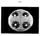
図3は、クレブシエラのペニシリン耐性株によるセフォキシチンの酵素的不活性化を実証する3次元試験を示す。
図4は、図2について上述したとおりの本発明の実施形態を示すが、1つのセフォキシチン円盤だけを示す。図4の“ポジティブ”と記される部分は、本発明の透過剤の歪んだ阻害ゾーンを示す。図4の“ネガティブ”と記される部分は、透過剤を持たない対照試薬円盤が抗生物質円盤の近位に置かれる場合に歪みがないことを示す。
FIG. 1 shows a close-up of the strain zone around the cefoxitin disc.
FIG. 2 illustrates one embodiment of the present invention. Here, an indirect form of the method of the invention is shown, in which five test microorganisms and a negative control organism are tested. In this assay, the fully sensitive strain E. coli ATTC 25922 was used as a lawn culture and assayed for the inactivator produced by the test and control microorganisms. Three commercially produced cefoxitin discs (labeled FOX 30) are handled together in a reagent test disc. A test or control organism was applied to each reagent disk and then placed proximal to the cefoxitin disk. A strong dent at the edge of the inhibition zone indicates inactivation of cefoxitin by an inactivator released from the test microorganism that occurs upon encountering a permeant in the disc. The negative control is undistorted (the bottom disc of the top set of 3 discs is facing the 12 o'clock direction). A weak positive test occurred in the disc closest to 9 o'clock (shown as slight flattening or blunting of the zone of inhibition). This result was obtained with a strain of Klebsiella pneumoniae that was previously determined to be a low level producing strain of cefoxitin inactivated β-lactamase. (This strain is a promising candidate for a quality control strain associated with the acceptable limit of detection sensitivity of this particular antibiotic inactivator sample.)
FIG. 3 shows a three-dimensional test demonstrating enzymatic inactivation of cefoxitin by a penicillin resistant strain of Klebsiella.
FIG. 4 shows an embodiment of the invention as described above for FIG. 2, but shows only one cefoxitin disc. The portion marked “positive” in FIG. 4 represents a distorted inhibition zone of the permeant of the present invention. The portion marked “negative” in FIG. 4 indicates that there is no distortion when the control reagent disk without permeabilizer is placed proximal to the antibiotic disk.
(発明の詳細な説明)
本発明は、透過剤の存在下で微生物をインキュベートすることによって、微生物の細胞壁及び/又は細胞膜の透過性を高めることに基づく微生物の抗生物質感受性を決定する新規な方法を提供する。この透過剤は、好ましくは該微生物を殺しもせず、その複製能力を有意には阻害しないが、それにもかかわらず該微生物を透過する。理論に束縛されたくないが、適切な濃度の透過剤の存在は、該微生物からの抗生物質不活性化因子(例えば、酵素)の遊離又は放出を高めることができると考えられる。このような抗生物質不活性化因子の遊離又は放出は、微生物がその放出された因子で不活性化される抗菌剤の存在下で成長することを可能にする。
(Detailed description of the invention)
The present invention provides a novel method for determining the antibiotic susceptibility of a microorganism based on increasing the permeability of the cell wall and / or cell membrane of the microorganism by incubating the microorganism in the presence of a permeabilizing agent. This permeant preferably does not kill the microorganism and does not significantly inhibit its replication ability, but nevertheless penetrates the microorganism. Without wishing to be bound by theory, it is believed that the presence of an appropriate concentration of permeabilizer can enhance the release or release of antibiotic inactivators (eg, enzymes) from the microorganism. Such release or release of the antibiotic inactivator allows the microorganism to grow in the presence of an antibacterial agent that is inactivated by the released factor.
別の局面では、本発明は、抗生物質不活性化酵素のような抗生物質不活性化因子の存在を決定する新規な方法であって、以下の工程を含む方法を提供する。まず、抗生物質不活性化因子を産生し又は遊離させると推測される微生物の培養を、該抗生物質不活性化因子が不活性化できる抗生物質と、微生物の成長を阻害せずに微生物透過に有効な量で存在する該微生物の透過剤と混合して検定培養を調製する。次に、この検定培養を適切な培養条件下、かつ該抗生物質を不活性化できる微生物によって産生され又は遊離させられた抗生物質不活性化因子の存在を決定するために十分な時間インキュベートする。 In another aspect, the present invention provides a novel method for determining the presence of an antibiotic inactivating factor, such as an antibiotic inactivating enzyme, comprising the following steps. First, culture of microorganisms presumed to produce or liberate antibiotic inactivator is made into antibiotics that can be inactivated by the antibiotic inactivator and microbial permeation without inhibiting microbial growth. An assay culture is prepared by mixing with a permeabilizing agent of the microorganism present in an effective amount. The assay culture is then incubated under appropriate culture conditions and for a time sufficient to determine the presence of an antibiotic inactivator produced or released by a microorganism capable of inactivating the antibiotic.
透過剤を使用し、抗生物質不活性化因子の遊離又は放出を促すことによって、そうでなければ誤って試験抗生物質に対して感受性であると記録したであろう抗生物質耐性微生物を検出できることが分かった。驚くべきことに、該微生物は、微生物にとって普通は致死的であるか、或いは微生物の成長を阻害する透過剤の存在下で成長することができる。 By using penetrants and encouraging the release or release of antibiotic inactivators, it is possible to detect antibiotic-resistant microorganisms that would otherwise be mistakenly recorded as sensitive to the test antibiotic. I understood. Surprisingly, the microorganism is normally lethal to the microorganism or can grow in the presence of a permeabilizing agent that inhibits the growth of the microorganism.
一局面では、本発明は、微生物の成長を阻害せずに微生物透過に有効な量の透過剤の使用を含む。本明細書の他の箇所でさらに詳述するように、透過剤の成長を阻害しない量を決定することは、その有意に微生物の成長を阻害する能力についての該透過剤の滴定を含む日常的実験事項である。同様に、透過剤の微生物透過に有効な量の決定は、試験微生物の既知の抗生物質耐性株からの抗生物質不活性化因子の遊離によってのような、微生物を透過するその能力についての該透過剤の滴定を含む日常的実験事項である。 In one aspect, the invention includes the use of an amount of permeabilizing agent that is effective for microbial permeation without inhibiting microbial growth. As described in further detail elsewhere herein, determining an amount that does not inhibit the growth of permeabilizers involves routine titration of the permeabilizer for its ability to significantly inhibit microbial growth. It is an experimental matter. Similarly, the determination of the amount of permeabilizer effective for microbial permeation is determined by determining the permeation for its ability to permeate the microorganism, such as by release of an antibiotic inactivator from a known antibiotic resistant strain of the test microorganism. It is a routine experiment that involves titration of the agent.
本明細書で述べる量の透過剤を、抗生物質に対するその感受性を決定すべき微生物の培養と混合する。代わりに、混合を行って1種以上の抗生物質不活性化因子を生成又は遊離させる該微生物の能力を決定する。培養は、技術的に周知の方法、例えば、該微生物の接種材料(臨床試料由来のような)を適切な増殖培地中に導入することによって調製する。増殖培地は、Luria又はMeuller-Hintonブロスのような液体増殖培地でよく、或いは必要な栄養物で補充した寒天プレート、例えばMuller-Hinton寒天プレートのような固体増殖培地でよい。 The amount of permeabilizer described herein is mixed with the culture of the microorganism whose sensitivity to antibiotics is to be determined. Instead, mixing is performed to determine the ability of the microorganism to produce or release one or more antibiotic inactivators. The culture is prepared by methods well known in the art, for example by introducing the inoculum of the microorganism (such as from a clinical sample) into a suitable growth medium. The growth medium may be a liquid growth medium such as Luria or Meuller-Hinton broth or a solid growth medium such as an agar plate supplemented with the necessary nutrients, eg, a Muller-Hinton agar plate.
培養を調製したら、検定すべき1種以上の抗生物質(抗菌剤)と微生物を混合する。抗生物質不活性化因子を作り又は排出するその能力について微生物を検定する場合、検定用の対照を与えるためと、不活性化因子自体の産生又は遊離を促すための両方で抗生物質の存在が必要である。抗生物質は、微生物による抗生物質不活性化因子の産生を惹起又は促進するために必要とされうる。抗生物質の非存在下では、微生物は必ずしも抗生物質不活性化因子を作り、維持し、隔離し、又は遊離させる必要がない。 Once the culture is prepared, the microorganism is mixed with one or more antibiotics (antibacterial agents) to be assayed. When assaying a microorganism for its ability to create or excrete an antibiotic inactivator, the presence of the antibiotic is required both to provide a control for the assay and to promote production or release of the inactivator itself It is. Antibiotics may be required to initiate or promote the production of antibiotic inactivators by microorganisms. In the absence of antibiotics, microorganisms do not necessarily have to create, maintain, sequester or release antibiotic inactivators.
抗生物質感受性検定法については本明細書の別の箇所で述べるが、本発明用に適合できるような検定法について限定することを意図しない。培養は、透過剤とも混ぜる。
必要な場合、抗生物質又は透過剤のどちらかがないか、或いは両方ない対照培養を同様の条件下で調製することができる。本明細書の他の箇所で述べるように、液体増殖培地及び液体キャリヤー中の透過剤と混合する場合、検定すべき抗生物質の段階希釈は液体増殖培地内で調製される。対応する試料を液体キャリヤー中で透過剤と共に或いは透過剤なしで混合する。透過剤の非存在下、特定の抗生物質希釈で抗菌活性を示すが、透過剤の存在下、当該希釈で効力がない抗生物質は、該微生物が当該抗生物質を不活性化する因子を遊離させることに基づき、その検定微生物に対して無効とみなされる。
Antibiotic susceptibility assays are described elsewhere herein, but are not intended to limit the assays so that they can be adapted for the present invention. The culture is also mixed with a permeant.
If necessary, control cultures with or without either antibiotics or permeabilizers can be prepared under similar conditions. As described elsewhere herein, serial dilutions of antibiotics to be assayed are prepared in liquid growth medium when mixed with liquid growth medium and permeabilizer in a liquid carrier. Corresponding samples are mixed in a liquid carrier with or without a permeant. Antibiotics that exhibit antibacterial activity at a specific antibiotic dilution in the absence of a penetrant, but that are not effective at that dilution in the presence of a penetrant release the factor that the microorganism inactivates the antibiotic. On the basis of that, it is considered invalid for the assay microorganism.
このようにして調製した培養を適切な培養条件下かつ問題の抗生物質(1つ又は複数)に対する該微生物の感受性を決定するのに十分な時間インキュベートする。例えば、寒天プレートを環境的に制御したインキュベーター内で12〜18時間、摂氏35度でインキュベートすることができる。液体培地内の培養は、作業台上、振とう水浴内で12〜18時間、摂氏35度でインキュベートすることができる。適切な培養条件と十分な時間は、当業者には周知である。 The culture thus prepared is incubated under appropriate culture conditions and for a time sufficient to determine the susceptibility of the microorganism to the antibiotic (s) in question. For example, agar plates can be incubated at 35 degrees Celsius for 12-18 hours in an environmentally controlled incubator. Cultures in liquid media can be incubated at 35 degrees Celsius for 12-18 hours in a shaking water bath on a workbench. Appropriate culture conditions and sufficient time are well known to those skilled in the art.
述べたように、透過剤は、液体又は固体キャリヤー内で供給され、その中に分散又は溶解しうる。例えば、緩衝溶液のような液体透過剤の場合、緩衝溶液はそれ自体液体キャリヤー内にある。代わりに、本発明の方法で使用するため、洗浄剤を水性媒体、又は増殖ブロス内に分散させることができる。 As stated, the permeation agent can be supplied in a liquid or solid carrier and dispersed or dissolved therein. For example, in the case of a liquid penetrant such as a buffer solution, the buffer solution is itself in the liquid carrier. Alternatively, the detergent can be dispersed in an aqueous medium or growth broth for use in the method of the present invention.
別の局面では、透過剤が固体支持体上で供給される。例えば、緩衝溶液を1枚のろ紙のような固体支持体上に含浸させることができる。ろ紙を放置して乾燥させ、透過剤を固体キャリヤー上に分散させる。
一実施形態では、含浸円盤を抗生物質感受性検定で用い、その抗生物質感受性を測定する微生物を、該微生物を殺さず、或いは有意にはその成長を阻害せずに透過させる。これら含浸円盤の調製で使用する透過剤の適切な量(濃度)を決定して、微生物を殺さず或いはその成長を阻害せずに、微生物からの抗生物質不活性化因子の遊離を達成することができる。
In another aspect, a permeant is provided on the solid support. For example, the buffer solution can be impregnated on a solid support such as a piece of filter paper. The filter paper is left to dry and the permeating agent is dispersed on the solid carrier.
In one embodiment, an impregnated disc is used in an antibiotic susceptibility assay to allow a microorganism to measure its antibiotic susceptibility to permeate without killing the microorganism or significantly inhibiting its growth. Determine the appropriate amount (concentration) of permeabilizer used in the preparation of these impregnated discs to achieve the release of the antibiotic inactivator from the microorganism without killing the microorganism or inhibiting its growth. Can do.
本発明は、円盤拡散法又は他の抗生物質感受性決定法によって抗菌剤(本明細書では抗生物質と呼ぶこともある)に対する微生物の感受性を評価する検査室試験で提供される情報量を増やすように設計される。この発明は、微生物の抗生物質を不活性化する能力を日常的に検出するための手段を提供する。この情報は、感染症の治療用の適切な抗感染療法の選択、微生物についての疫学的情報の提供、抗生物質の研究、及び遺伝学及び酵素学のような他の分野の生物学的研究で役に立ちうる。 The present invention increases the amount of information provided in laboratory tests that assess the susceptibility of microorganisms to antimicrobial agents (sometimes referred to herein as antibiotics) by disc diffusion or other antibiotic susceptibility determination methods. Designed to. This invention provides a means for routinely detecting the ability of microorganisms to inactivate antibiotics. This information includes selection of appropriate anti-infective therapies for the treatment of infectious diseases, providing epidemiological information about microorganisms, research on antibiotics, and other areas of biological research such as genetics and enzymology. Can be useful.
好ましい実施形態では、本発明は、細菌又は他の微生物からの抗生物質不活性化酵素の放出を促す化学薬品のような薬剤を含浸させたろ紙円盤を供給することを含む。この化学薬品を含浸させたろ紙円盤は、本明細書では試薬円盤と呼ぶことがある。典型的に、試薬円盤中の薬剤は、細菌のような微生物の外膜、又は外膜と細胞質膜の両方を破壊して該微生物内から抗生物質不活性化酵素を放出する透過剤である。本発明の好ましい方法では、透過剤を含浸させた円盤を供給する。 In a preferred embodiment, the present invention includes providing a filter paper disk impregnated with an agent such as a chemical that promotes the release of antibiotic inactivating enzymes from bacteria or other microorganisms. The filter paper disk impregnated with this chemical may be referred to as a reagent disk in this specification. Typically, the drug in the reagent disk is a permeant that breaks the outer membrane of a microorganism, such as a bacterium, or both the outer membrane and the cytoplasmic membrane, releasing antibiotic inactivating enzymes from within the microorganism. In a preferred method of the invention, a disc impregnated with a permeating agent is provided.
好適な増殖培地含有寒天プレートに試験微生物(例えば、臨床試料、又は抗生物質感受性を決定すべき細菌株)の芝生を接種する。抗生物質含浸円盤を微生物の芝生上に置く。供給された試薬円盤上にさらに試験微生物を接種し、問題の抗生物質円盤に隣接するが接触しない寒天上に当該試薬円盤を置く。寒天プレートを技術的に周知の方法に従ってインキュベートする。インキュベーション後、結果を以下のように解釈する。抗生物質の酵素的不活性化は、試薬円盤近傍の阻害ゾーンの縁を調べることによって検出できる。抗生物質不活性化の結果、通常は円形の阻害ゾーンに歪みが生じる。この歪みの存在は、試験微生物が抗生物質不活性化因子を産生できることを示す。臨床的な見方から、ゾーンの歪みを示す抗生物質の使用は、試験微生物による感染の治療には禁忌を示されるだろう。 Agar plates containing suitable growth media are inoculated with lawn of test microorganisms (eg, clinical samples or bacterial strains whose antibiotic susceptibility is to be determined). Place antibiotic-impregnated disk on microbial lawn. The test microorganism is further inoculated on the supplied reagent disk, and the reagent disk is placed on the agar adjacent to but not in contact with the antibiotic disk in question. The agar plate is incubated according to methods well known in the art. After incubation, the results are interpreted as follows: Enzymatic inactivation of antibiotics can be detected by examining the edge of the inhibition zone near the reagent disk. Antibiotic inactivation results in distortion in the normally circular inhibition zone. The presence of this strain indicates that the test microorganism can produce an antibiotic inactivator. From a clinical point of view, the use of antibiotics that exhibit zone distortion would be contraindicated in the treatment of infection by test microorganisms.
本発明の好ましい実施形態は上述したとおりであるが、本発明は当該実施形態に限定されないことを理解すべきである。ろ紙ストリップ、ニトロセルロース片、又は他のキャリヤーのようなろ紙円盤以外の他の固体キャリヤーを媒体として用いて微生物と試薬との間の接触を達成することができる。E試験[Brown, D. F. J.及びL. Brown, J. Antimicrob. Chemother. 27:185-190(1991)]、又は希釈法のような、円盤拡散試験以外の試験方法論も、本発明の精神と範囲から逸脱することなく、抗生物質不活性化酵素の検出用に変更することができる。 Although preferred embodiments of the present invention are as described above, it should be understood that the present invention is not limited to these embodiments. Other solid carriers other than filter paper discs such as filter paper strips, nitrocellulose strips, or other carriers can be used as media to achieve contact between the microorganism and the reagent. Test methodology other than the disk diffusion test, such as the E test [Brown, DFJ and L. Brown, J. Antimicrob. Chemother. 27: 185-190 (1991)] or the dilution method, is also within the spirit and scope of the present invention. Modifications can be made for the detection of antibiotic inactivating enzymes without deviating.
別の局面では、本発明は、本明細書の他の箇所で論じた抗生物質感受性試験法によって例示されるように、培養中微生物の抗生物質試験の改良方法を提供する。改良点は、培養の成長を有意には阻害しないが、該微生物の透過には有効な量の本発明の透過剤と培養を混合する工程を含む。 In another aspect, the present invention provides an improved method for antibiotic testing of microorganisms in culture, as exemplified by the antibiotic susceptibility testing methods discussed elsewhere herein. The improvement includes mixing the culture with an amount of a permeant of the present invention that does not significantly inhibit the growth of the culture but is effective for permeation of the microorganism.
さらなる実施形態では、本発明は、阻害ゾーンが小さいか又は無い場合、或いは研究又は診断法として微生物を試験するための間接形態を含む。間接形態の試験は、寒天の表面に、大腸菌ATCC 25922のような完全に抗生物質感受性の検定株を接種して行う。この後、この供給された試薬円盤に試験微生物(例えば、臨床用分離培養)の懸濁液を接種し、原因生物の代わりに寒天の表面上で検体株が成長することを除き、上述したように試験を行う。本発明の方法の間接形態は、抗生物質の感受性の同時決定は除外するが、該試験を直接形態の試験で行った場合、阻害ゾーンが小さすぎて結果が得られない微生物の抗生物質不活性化酵素の調査を可能にしうる。 In further embodiments, the invention includes indirect forms for testing microorganisms when the zone of inhibition is small or absent, or as a research or diagnostic method. The indirect test is performed by inoculating the surface of the agar with a completely antibiotic-sensitive assay strain such as E. coli ATCC 25922. Thereafter, the supplied reagent disk is inoculated with a suspension of a test microorganism (eg, clinical isolate), and as described above, except that the specimen strain grows on the surface of the agar instead of the causative organism. Perform the test. The indirect form of the method of the invention excludes the simultaneous determination of antibiotic susceptibility, but when the test is carried out in a direct form test, the inhibitory zone of the microorganism is too small to produce a result and the antibiotic inactivity of the microorganism May enable investigation of oxidative enzymes.
本発明で有用な典型的な透過剤としては、微生物の細胞壁及び/又は細胞膜の透過性を高めるが、検定微生物の成長又は再生を有意には阻害しない量又は濃度で存在する当該薬剤が挙げられる。当業者には周知なように、すべての微生物が細胞壁と細胞膜を両方とも持っているわけではなく、さらに、細胞壁と細胞膜を両方とも有する当該微生物は構造的に同一の壁又は膜を持っていない。例えば、細菌はその細胞壁/細胞膜構造に基づいてグラム陽性又はグラム陰性と広く分類されている。本発明で有用な透過剤は、好ましくは抗生物質不活性化因子が細胞から遊離されるように細胞表面構造(細胞壁又は細胞膜、或いは両方)を透過する。しかし、透過剤は微生物を殺さず、或いはその成長を有意には阻害しないことも好ましい。 Exemplary permeabilizing agents useful in the present invention include those agents that are present in an amount or concentration that increases the permeability of the cell wall and / or cell membrane of the microorganism, but does not significantly inhibit the growth or regeneration of the assay microorganism. . As is well known to those skilled in the art, not all microorganisms have both cell walls and cell membranes, and in addition, those microorganisms that have both cell walls and cell membranes do not have structurally identical walls or membranes. . For example, bacteria are broadly classified as gram positive or gram negative based on their cell wall / cell membrane structure. The permeabilizing agents useful in the present invention preferably permeate the cell surface structure (cell wall or cell membrane, or both) so that the antibiotic inactivator is released from the cell. However, it is also preferred that the permeating agent does not kill or significantly inhibit the growth of microorganisms.
特有の典型的な透過剤としては、以下のものが挙げられる。
塩化ナトリウム(NaCl)、塩化マグネシウム(Mg2Cl)、塩化カリウム(KCl)等のような無機塩;
塩化ベンザルコニウム(BAC)、塩化セチルピリジウム、塩化3-(トリメトキシシリル)プロピルジメチルオクタデシルアンモニウム等のような四級アンモニウム化合物;
トリス/EDTA(TE)緩衝液、NaCl/トリス/EDTA(STE)緩衝液、グルコース/トリス/EDTA(GTE)緩衝液等のような緩衝溶液;
グルコース、デキストロース、スクロース、フルクトース、ラクトース等を含む糖溶液のような低浸透圧性又は高浸透圧性薬剤;
ピペラシリン、アズトレオナム、アムジノシリン、セフタジジム、ポリミキシンB、ポリミキシンBノナペプチド、又はゲンタマイシン等のような抗生物質;
Blind BriteTM、MaxaquinTM、HyVee Glass CleanerTM、Life TreeTM、RaveTM、DawnTM等のような商業的に入手可能な洗浄剤又は界面活性剤含有薬剤;
IsocleanTM濃縮物、Sight SaversTMレンズクリーナー、WashTM(グリーンハンドウォッシュ)、Micro Lab Cleaning SolutionTM、Jet CleanTM試験管クリーナー、LimoneneTM等のような他の商業的に入手可能な物質;
TritonX-100TM、ドデシル硫酸ナトリウム(SDS)、アセトンと混合したSDS(5%)、SarcosylTM、アセトンと混合したSarcosylTM、TweenTM(ポリソルベート)80、アセトンと混合したTweenTM80、TweenTM85、BrijTM56、ポリエチレングリコール、ポリプロピレングリコール、ダンシルポリミキシン等のような他の界面活性剤及び/又は洗浄剤;
他の典型的な透過剤としては、CHAPS(3-[(3-コラミドプロピル)ジメチルアンモニオ]-1-プロパンスルホネート)電気泳動試薬、≦10mM(Sigma cat # C-9426)、乳酸、及びヘキサメタリン酸ナトリウム(ポリリン酸ナトリウム)(キレート剤)が挙げられる。
セクロピン(cecropins)(カチオン性ペプチド)、メリチン(melittin)(カチオン性ペプチド)、バクテネシン(bactenecin)、マガイニン(magainins)(カエル宿主防衛ペプチド)、タクプレシン(tachyplesins)(カチオン性ペプチド)、ポリフェムシン(polyphemusins)(カチオン性ペプチド)のような天然ペプチド、及び合成ペプチド等も好ましい透過剤として使用できる。
Specific typical penetrants include the following.
Inorganic salts such as sodium chloride (NaCl), magnesium chloride (Mg 2 Cl), potassium chloride (KCl) and the like;
Quaternary ammonium compounds such as benzalkonium chloride (BAC), cetylpyridium chloride, 3- (trimethoxysilyl) propyldimethyloctadecylammonium chloride;
Buffer solutions such as Tris / EDTA (TE) buffer, NaCl / Tris / EDTA (STE) buffer, glucose / Tris / EDTA (GTE) buffer, etc .;
Hypotonic or hypertonic agents such as sugar solutions containing glucose, dextrose, sucrose, fructose, lactose, etc .;
Antibiotics such as piperacillin, aztreonam, amidinocillin, ceftazidime, polymyxin B, polymyxin B nonapeptide, or gentamicin;
Commercially available detergents or surfactant-containing agents such as Blind Brite ™ , Maxaquin ™ , HyVee Glass Cleaner ™ , Life Tree ™ , Rave ™ , Dawn ™, etc .;
Other commercially available materials such as Isoclean ™ concentrate, Sight Savers ™ lens cleaner, Wash ™ (green hand wash), Micro Lab Cleaning Solution ™ , Jet Clean ™ test tube cleaner, Limonene ™, etc .;
TritonX-100 TM, sodium dodecyl sulfate (SDS), SDS mixed with acetone (5%), Sarcosyl TM, Sarcosyl TM, Tween TM ( polysorbate) 80 was mixed with acetone, Tween TM 80 were mixed with acetone, Tween TM 85 Other surfactants and / or detergents such as Brij ™ 56, polyethylene glycol, polypropylene glycol, dansyl polymyxin, etc .;
Other typical permeants include CHAPS (3-[(3-Colamidopropyl) dimethylammonio] -1-propanesulfonate) electrophoresis reagent, ≦ 10 mM (Sigma cat # C-9426), lactic acid, and Examples include sodium hexametaphosphate (sodium polyphosphate) (chelating agent).
Cecropins (cationic peptide), melittin (cationic peptide), bactenecin, magainins (frog host defense peptide), tachyplesins (cationic peptide), polyphemusins Natural peptides such as (cationic peptides), synthetic peptides, and the like can also be used as preferred permeating agents.
好ましい透過剤は、Sigma-Aldrichから商業的に入手可能なトリス/EDTA(TE)緩衝液(カタログ番号T-9285)である。供給時、TE緩衝液は100倍濃度で、1.0モルのトリス-HCl(pH約8.0)と0.1モルのEDTAから構成される。好ましくはTE緩衝液を100ミリモル、200ミリモル、0.5モル、1モル又は10モル濃度で円盤上に置く。さらに好ましくは、TE緩衝液を100倍、50倍、25倍及び10倍濃度で円盤上に置く。特定透過剤の最適濃度の決定方法は、本明細書の他の箇所で与えられる。 A preferred permeant is Tris / EDTA (TE) buffer (Catalog No. T-9285) commercially available from Sigma-Aldrich. When supplied, the TE buffer is 100-fold concentrated and consists of 1.0 mol Tris-HCl (pH about 8.0) and 0.1 mol EDTA. Preferably, TE buffer is placed on the disc at a concentration of 100, 200, 0.5, 1 or 10 molar. More preferably, the TE buffer solution is placed on the disc at a concentration of 100 times, 50 times, 25 times and 10 times. Methods for determining the optimal concentration of a particular permeabilizer are given elsewhere herein.
本明細書の他の箇所で記述されるように、透過剤は、好ましくは微生物内から当該微生物の成長を有意に阻害せずに抗生物質不活性化因子を放出させ、或いは放出を促進する。透過剤の微生物から抗生物質不活性化因子を遊離又は放出させる能力は、例えば、酵素のような放出される抗生物質不活性化因子の量の定量化によって決定できる。このような定量化は、例えば、基質としてニトロセフィン又はセファクロルを用いる分光光度的加水分解検定法で決定することができる。微生物から抗生物質不活性化因子を遊離又は放出させる透過剤の能力は、例えば、本発明の方法を用いて、或いは本明細書の他の箇所で述べた3次元試験のような他の抗生物質感受性試験を用いて決定することもできる。微生物から抗生物質不活性化因子を遊離又は放出させる透過剤の能力の典型的な決定法は、本明細書の他の箇所で述べる実施例で記載される。 As described elsewhere herein, the permeabilizing agent preferably releases or facilitates the release of the antibiotic inactivator from within the microorganism without significantly inhibiting the growth of the microorganism. The ability of the penetrant to release or release antibiotic inactivators from microorganisms can be determined, for example, by quantifying the amount of antibiotic inactivator released, such as an enzyme. Such quantification can be determined, for example, by a spectrophotometric hydrolysis assay using nitrocefin or cefaclor as a substrate. The ability of a permeabilizing agent to release or release an antibiotic inactivator from a microorganism can be compared with other antibiotics using, for example, the methods of the present invention or the three-dimensional test described elsewhere herein. It can also be determined using a sensitivity test. Exemplary methods for determining the permeabilizing agent's ability to release or release an antibiotic inactivator from a microorganism are described in the examples set forth elsewhere herein.
また、好ましい透過剤は、微生物の成長を有意には阻害しない。好ましい透過剤のこの特性は、例えば、特定透過剤の濃度を変えて種々の微生物の芝生培養に適用して、該芝生培養の成長の阻害が生じたかを測定することによって決定できる。代わりに、可変濃度の透過剤の存在下における種々の微生物の成長速度を技術的に周知の方法で決定することもできる。透過剤の微生物の成長を許容する能力の典型的な決定法は、本明細書の他の箇所で述べる実施例で記載される。 Also, preferred permeabilizers do not significantly inhibit microbial growth. This property of a preferred permeant can be determined, for example, by varying the concentration of a specific permeant and applying it to a lawn culture of various microorganisms to determine if inhibition of the growth of the lawn culture has occurred. Alternatively, the growth rate of various microorganisms in the presence of variable concentrations of permeabilizer can be determined by methods well known in the art. A typical method for determining the ability of a permeant to allow microbial growth is described in the examples described elsewhere herein.
別の実施形態では、本発明は、抗生物質不活性化因子を遊離させる微生物を検出する抗生物質感受性試験を行うための透過剤含浸円盤と使用説明書を含むキットを含む。好ましい実施形態では、透過剤は、トリス/EDTA緩衝液である。 In another embodiment, the present invention includes a kit comprising a permeant impregnated disk and instructions for performing an antibiotic susceptibility test for detecting a microorganism that liberates an antibiotic inactivator. In a preferred embodiment, the permeating agent is Tris / EDTA buffer.
容易に分かるように、本発明は、他の抗生物質感受性試験法を超える新規な利益と利点を提供する。本発明は、例えば、寒天内のスリット中に細菌接種材料を挿入するする必要がある他の方法の技術的に挑戦する局面ではなく、寒天の表面上で抗生物質感受性試験を実施することを可能にする。さらに、本発明は、当該微生物の成長を有意には阻害せずに、微生物からの抗生物質不活性化因子の放出を促進する透過剤の使用を開示する。現在の抗生物質感受性試験法は、このような因子の放出を促すことができず、誤った正の結果をもたらす可能性がある。従って、現在の抗生物質感受性試験法は、臨床医に特定の微生物がこれらの生体外結果に基づいて抗生物質に感受性であると信じさせ、経験的に該抗生物質が生体内で無効であることが分かるだけである。本発明は、生体内で特定の抗生物質に耐性であると思われる微生物を検出できる、より頑強な生体外情報を提供する。 As will be readily appreciated, the present invention provides new benefits and advantages over other antibiotic susceptibility testing methods. The present invention is capable of performing antibiotic susceptibility testing on the surface of the agar, for example, not a technically challenging aspect of other methods that require the insertion of a bacterial inoculum into a slit in the agar To. Furthermore, the present invention discloses the use of permeabilizers that promote the release of antibiotic inactivators from the microorganism without significantly inhibiting the growth of the microorganism. Current antibiotic susceptibility testing methods do not facilitate the release of such factors and can lead to false positive results. Therefore, current antibiotic susceptibility testing methods allow clinicians to believe that certain microorganisms are sensitive to antibiotics based on these in vitro results, and empirically that the antibiotic is ineffective I just understand. The present invention provides more robust in vitro information that can detect microorganisms that appear to be resistant to specific antibiotics in vivo.
実施例1.抗生物質不活性化因子の放出の決定
この実施例では、大腸菌MISC 208(拡張スペクトルβ-ラクタマーゼSHV-2を産生する)を試験生物として用い、ニトロセフィンが分光光度的加水分解検定法の基質だった。大腸菌株を直接100μlのMuller Hintonブロス中に収集し、100μlの透過剤と混合した。
この加水分解検定法では、活性の単位は以下のように計算する。
単位=光学密度(OD)の変化/分×10
吸光係数
Example 1. Determination of antibiotic inactivator release In this example, Escherichia coli MISC 208 (producing extended spectrum β-lactamase SHV-2) was used as the test organism and nitrocefin was the substrate for the spectrophotometric hydrolysis assay. . E. coli strains were collected directly in 100 μl Muller Hinton broth and mixed with 100 μl permeabilizer.
In this hydrolysis assay, the unit of activity is calculated as follows:
Unit = change in optical density (OD) / min x 10
Extinction coefficient
加水分解検定法は、基本的にO'Callaghan, C. H.ら, A.A.C. 1:283-288(1972)に記載されているとおりに行った。簡単には、ニトロセフィンの100μM溶液を基質として用いた。ニトロセフィンを37℃に予備加熱した。加水分解検定法は、37℃で、-0.024の吸光係数の389.5の波長で行った。適切なキュベットを0.9mlの予備加熱したニトロセフィン基質で満たし、100ulの透過細菌緩衝液を添加した。即座にキュベットを逆さにして中身を混ぜ、キュベットを分光光度計内に置いた。キュベットブロックを温める循環水浴で37℃に温度を維持しながら、5秒間隔で5分まで吸光度を検定した。検定完了時、活性を上式により計算した。 The hydrolysis assay was performed essentially as described in O'Callaghan, C.H. et al., A.A.C. 1: 283-288 (1972). In brief, a 100 μM solution of nitrocefin was used as the substrate. Nitrocefin was preheated to 37 ° C. The hydrolysis assay was performed at 37 ° C. with a wavelength of 389.5 with an extinction coefficient of −0.024. Appropriate cuvettes were filled with 0.9 ml of preheated nitrocefin substrate and 100 ul of permeabilizing bacterial buffer was added. The cuvette was immediately turned upside down to mix the contents, and the cuvette was placed in the spectrophotometer. Absorbance was assayed at 5 second intervals for up to 5 minutes while maintaining the temperature at 37 ° C in a circulating water bath warming the cuvette block. At the completion of the assay, the activity was calculated according to the above formula.
透過剤 β-ラクタマーゼ活性の単位
(基質としてニトロセフィン)
TE/室温 47
TE/0℃ 102
トリス/0℃ 41
水(対照) 0
Permeabilizer unit of β-lactamase activity
(Nitrocefin as substrate)
TE / room temperature 47
TE / 0 ℃ 102
Tris / 0 ℃ 41
Water (control) 0
実施例2.抗生物質不活性化因子の放出の決定
実施例1の実験を大腸菌MISC 208及び加水分解検定法の基質としてセファクロルを用いて繰り返した。
透過剤調製試料 β-ラクタマーゼ活性の単位
(基質としてセファクロル)
塩化ベンザルコニウム 200μg/ml 16
Polymixin B 10μg/ml 3.8
Polymixin B 30μg/ml 15
TE 79
Example 2 Determination of antibiotic inactivator release The experiment of Example 1 was repeated using E. coli MISC 208 and cefaclor as the substrate for the hydrolysis assay.
Permeabilized sample β-lactamase activity unit
(Cefaclor as substrate)
Benzalkonium chloride 200μg / ml 16
Polymixin B 10μg / ml 3.8
Polymixin B 30μg / ml 15
TE 79
実施例3.抗生物質不活性化因子の放出の決定
大腸菌V1104を用いて実施例1の実験を繰り返して種々の透過剤を評価した。ニトロセファンが加水分解基質だった。
透過剤調製試料 β-ラクタマーゼ活性の単位
(基質としてニトロセフィン)
アズトレオナム 210
試薬なし/0℃ 214
試薬なし/45℃ 0
セフタジジム 196
ピペラシリン 202
Example 3 Determination of antibiotic inactivator release The experiment of Example 1 was repeated using E. coli V1104 to evaluate various permeabilizers. Nitrocephan was the hydrolysis substrate.
Permeabilized sample β-lactamase activity unit
(Nitrocefin as substrate)
Aztreonam 210
No reagent / 0 ° C 214
No reagent / 45 ° C 0
Ceftazidime 196
Piperacillin 202
抗生物質不活性化因子の放出の決定で使用可能な他の典型的な微生物としては、下表の微生物が挙げられる。
株 酵素 単位(対ビトロセフィン)
大腸菌V1104 202
大腸菌165* pI 5.95 TEM MSBL 43
MG32 TEM-1+TEM-12 349
K. pneumoniae V1102 227
Other typical microorganisms that can be used in determining the release of antibiotic inactivators include the microorganisms in the table below.
Strain enzyme unit (vs. vitrocefin)
E. coli V1104 202
E. coli 165 * pI 5.95 TEM MSBL 43
MG32 TEM-1 + TEM-12 349
K. pneumoniae V1102 227
実施例4.有意な成長阻害の欠如の決定
種々の試験株を直接100μlのMueller Hintonブロス中に収集し、問題の各透過剤100μlと混合した。次に、これら各混合物20μlをブランクろ紙円盤に適用し、円盤を種々の試験微生物の新しく接種した寒天芝生培養に移し、プレートを37℃で一晩中インキュベートした。終夜のインキュベーション後、培養を円盤周囲の阻害ゾーンの存在又は非存在について調べた。阻害ゾーンから試験で用いた濃度の該透過剤が不適切であると推論した。
Example 4 Determination of lack of significant growth inhibition The various test strains were collected directly in 100 μl of Mueller Hinton broth and mixed with 100 μl of each permeabilizer in question. Next, 20 μl of each of these mixtures was applied to a blank filter paper disc, the disc was transferred to a freshly inoculated agar lawn culture of various test microorganisms, and the plates were incubated at 37 ° C. overnight. After overnight incubation, the cultures were examined for the presence or absence of a peridiscal zone of inhibition. It was inferred from the inhibition zone that the concentration of the permeant used in the test was inappropriate.
この方法の変形は、試薬を前もって微生物と混合しないで直接試薬を円盤に適用して上述したように評価することである。
(試薬円盤の調製)
1.同割合の生理食塩水とトリスEDTA(Sigma Chemicals)の溶液を調製する。
2.20μl分量の透過剤溶液をろ紙円盤上に置き、乾燥させる。
(試験の実施と解釈)
1.透過剤で含浸させたろ紙円盤を20μlの無菌生理食塩水で再水和させた。
2.綿棒又はループを用いて試験生物を再水和円盤上にこすりつけた。
3.Muller Hinton寒天プレートに0.5 McFarlandの大腸菌ATCC 25922を接種した。
4.寒天プレートの表面上にセフォキシチン円盤を置いた。次に試験生物を接種した再水和円盤をセフォキシチン円盤の隣に約0.5〜1.0mm離して置いた。
5.寒天プレートを37℃で約16時間、又は一晩中インキュベートした。
6.セフォキシチンゾーンの歪みは、正の結果として解釈し(すなわち、抗生物質不活性化因子の存在が検出された)、セフォキシチンゾーンの歪みの欠如は、負の結果として解釈した。図1及び2を参照せよ。
A variation of this method is to apply the reagent directly to the disc and evaluate as described above without mixing the reagent in advance with the microorganism.
(Preparation of reagent disk)
1. Prepare a solution of the same proportion of saline and Tris EDTA (Sigma Chemicals).
2. Place 20 μl aliquots of permeabilizer solution on a filter paper disc and allow to dry.
(Examination and interpretation of the test)
1. The filter paper disk impregnated with the permeating agent was rehydrated with 20 μl of sterile physiological saline.
2. The test organism was rubbed onto the rehydration disk using a cotton swab or loop.
3. Muller Hinton agar plates were inoculated with 0.5 McFarland E. coli ATCC 25922.
4). A cefoxitin disc was placed on the surface of the agar plate. Next, the rehydration disc inoculated with the test organism was placed next to the cefoxitin disc about 0.5-1.0 mm apart.
5. Agar plates were incubated at 37 ° C. for approximately 16 hours or overnight.
6). The cefoxitin zone distortion was interpreted as a positive result (ie, the presence of an antibiotic inactivator was detected) and the lack of cefoxitin zone distortion was interpreted as a negative result. See FIGS. 1 and 2.
実施例5.抗生物質耐性微生物の検出
この研究は、pAmpCsを内部にもつと推測されるクレブシエラ(Klebsiella)分離培養の調査のためのその有用性について報告する。
方法:セフォキシチンMICs≧16μg/mlを有するKlebsiella spp.の30個の分離培養を本発明の方法で調査した。本明細書の他の箇所で述べた3次元試験ですべての負の結果を確認し、かつ等電点電気泳動(IEF)とインヒビター研究で正の結果を確認した。等量の生理食塩水で希釈した100倍TEをろ紙円盤に含浸させた。この円盤を20μlの食塩水で再水和させ、試験分離培養の数コロニーを円盤上に接種した。接種円盤を、大腸菌 ATCC 25922の芝生を接種したMuller-Hinton寒天プレート上のセフォキシチン円盤の近傍に(ほとんど触れる)置いた。終夜のインキュベーション後、正の試験は、試験円盤近傍のセフォキシチン阻害ゾーンの扁平化又はへこみとして現れた。負の試験は、歪んでいないゾーンを有した。
結果:試験した30個の分離培養のうち、10個は正で、20個は負だった(3次元試験でも負)。正の分離培養のIEF及び阻害研究は、10個の分離培養のうち9個が、7.2、7.7又は≧9.0のpIsを有する、クロキサシリンによって阻害されるβ-ラクタマーゼを産生することを示し、AmpC産生と一致することが分かった。クレブシエラは染色体AmpCを欠いているので、これら酵素はプラスミド媒介されると解釈した。他の正の分離培養は、高いイミペネムMICを有し、セフォキシチンも加水分解するカルバペネム加水分解酵素を産生した。
Embodiment 5 FIG. Detection of antibiotic-resistant microorganisms This study reports its usefulness for the investigation of Klebsiella isolate cultures suspected of harboring pAmpCs.
Method: Thirty separate cultures of Klebsiella spp. With cefoxitin MICs ≧ 16 μg / ml were investigated with the method of the present invention. All negative results were confirmed by the three-dimensional test described elsewhere in this specification, and positive results were confirmed by isoelectric focusing (IEF) and inhibitor studies. A filter paper disk was impregnated with 100-fold TE diluted with an equal amount of physiological saline. This disc was rehydrated with 20 μl of saline and several colonies of the test isolation culture were inoculated on the disc. The inoculum disc was placed (mostly touched) near the cefoxitin disc on a Muller-Hinton agar plate inoculated with lawn of E. coli ATCC 25922. After overnight incubation, positive tests appeared as flattening or dents in the cefoxitin inhibition zone near the test disc. The negative test had an undistorted zone.
Results: Of the 30 separate cultures tested, 10 were positive and 20 were negative (negative in 3D tests). IEF and inhibition studies of positive isolation cultures show that 9 out of 10 isolation cultures produce cloxacillin-inhibited β-lactamase with a pIs of 7.2, 7.7 or ≧ 9.0, producing AmpC Was found to match. Since Klebsiella lacks chromosomal AmpC, these enzymes were interpreted as plasmid-mediated. Other positive isolation cultures produced carbapenem hydrolase with high imipenem MIC and also hydrolyzing cefoxitin.
(材料と方法)
分離培養
Klebsiella spp.の30個の新しい分離培養を米国の病院内の患者から収集した。
感受性
抗菌感受性は、調査用 Microscan脱水パネルを用いるNCCLS微量希釈方法論で決定した。
3次元試験
セフォキシチンの酵素不活性化は、3次元法で調査した。Muller-Hinton寒天プレートの表面に大腸菌 ATCC 25922の標準化接種材料の芝生を接種した。無菌メス刃でプレート内にスリットを刻んだ。このスリット中に、試験生物の重接種材料(109CFU/ml)を含有する懸濁液をマクロピペット先端からの毛管作用によって分配した。セフォキシチン円盤をスリットから3mmの寒天上に置いた。35℃で一晩中インキュベーション後、プレートを阻害ゾーン縁の特徴的な歪みで示されるようなセフォキシチン不活性化の証拠について検査した。この特徴の非存在がセフォキシチンの有意な不活性化がないことを示した(図3)。
(Materials and methods)
Separation culture
Thirty new isolates of Klebsiella spp. Were collected from patients in a US hospital.
Susceptibility Antimicrobial susceptibility was determined with the NCCLS microdilution methodology using a microscan dehydration panel for investigation.
Three-dimensional test Enzyme inactivation of cefoxitin was investigated by a three-dimensional method. The surface of the Muller-Hinton agar plate was inoculated with lawn of standardized inoculum of E. coli ATCC 25922. A slit was cut into the plate with a sterile scalpel blade. In this slit, a suspension containing the heavy inoculum of the test organism (10 9 CFU / ml) was dispensed by capillary action from the macropipette tip. A cefoxitin disc was placed on
AmpC円盤試験
セフォキシチンの酵素不活性化は、本明細書の他の箇所で述べたように、本発明の透過剤、TEを含浸させたろ紙円盤を使用する円盤試験によっても調査した。円盤を20μlの無菌食塩水で再水和させてから試験生物の数コロニーを接種した。Muller-Hinton寒天プレートの表面に大腸菌 ATCC 25922の標準化接種材料の芝生を接種した。接種円盤を接種プレート上のセフォキシチン円盤の近傍に(ほとんど触れる)置いた。摂氏35度で終夜インキュベーション後、正の試験は試験円盤の近傍のセフォキシチン阻害ゾーンの扁平化又はへこみとして現れた。負の試験は歪みのないゾーンを有した(図4)。
等電点電気泳動
粗製超音波処理物を平台装置(Multiphor LKB)上の両性ポリアクリルアミドゲル(pH範囲3.5〜9.5)上での分析的等電点電気泳動(IEF)に供した。IEFゲルのインヒビターオーバレイを1000μMのクラブラン酸とクロキサシリンで行った。
AmpC Disc Test Enzyme inactivation of cefoxitin was also investigated by a disc test using a filter paper disc impregnated with the permeant of the present invention, TE, as described elsewhere in this specification. The disc was rehydrated with 20 μl of sterile saline before inoculating several colonies of the test organism. The surface of the Muller-Hinton agar plate was inoculated with lawn of standardized inoculum of E. coli ATCC 25922. The inoculum disk was placed (mostly touched) near the cefoxitin disk on the inoculum plate. After overnight incubation at 35 degrees Celsius, the positive test appeared as a flattened or indented cefoxitin-inhibiting zone near the test disc. The negative test had an undistorted zone (Figure 4).
Isoelectric focusing electrophoresis The crude sonicated product was subjected to analytical isoelectric focusing (IEF) on an amphoteric polyacrylamide gel (pH range 3.5 to 9.5) on a flat table (Multiphor LKB). Inhibitor overlay of IEF gel was performed with 1000 μM clavulanic acid and cloxacillin.
結果
セフォキシチンMICs≧16μg/mlを有する30個のKlebsiella spp.分離培養をAmpC円盤試験で検定した。これらのうち20個は負で10個が正だった。負の分離培養は3次元試験で負として確認した。IEF及びインヒビター研究は、10個の正の分離培養のうち9個が7.2、7.7又は≧9.0のpIsを有するβ-ラクタマーゼを産生し、クロキサシリンによって阻害されることを明らかにした。この知見はAmpC産生と一致した。これら酵素は、Klebsiella spp.は染色体AmpCを欠いているので、プラスミド媒介されると解釈した。他の正の分離培養は、高いイミペネムMIC(64μg/ml)を有し、セフォキシチンも加水分解できるカルバペネム加水分解酵素を産生した。
Results Thirty Klebsiella spp. Isolation cultures with cefoxitin MICs ≧ 16 μg / ml were assayed by AmpC disc test. Of these, 20 were negative and 10 were positive. Negative isolation cultures were confirmed as negative in a 3D test. IEF and inhibitor studies revealed that 9 out of 10 positive isolation cultures produced β-lactamase with a pIs of 7.2, 7.7 or ≧ 9.0 and were inhibited by cloxacillin. This finding was consistent with AmpC production. These enzymes were interpreted as plasmid-mediated because Klebsiella spp. Lacks chromosomal AmpC. The other positive isolation culture produced carbapenem hydrolase that had a high imipenem MIC (64 μg / ml) and could also hydrolyze cefoxitin.
上記説明及び実施例は、例証として意図したものであり、限定と解釈すべきでない。この発明の精神及び範囲内でさらに他の変形が可能であり、本技術の当業者には容易に思い浮かぶだろう。 The above description and examples are intended to be illustrative and should not be construed as limiting. Still other variations are possible within the spirit and scope of the invention and will readily occur to those skilled in the art.
Claims (18)
a)その感受性を決定すべき微生物の培養を、感受性を検定すべき抗生物質と、微生物の成長を阻害せずに微生物透過に有効な量で存在する前記微生物の透過剤と混合して検定培養を調製する工程;及び
b)前記検定培養を、適切な培養条件下、かつ前記抗生物質に対する前記微生物の感受性を決定するのに十分な時間インキュベートする工程;
を含む方法。 A method for determining the antibiotic susceptibility of a microorganism, comprising the following steps:
a) The culture of the microorganism whose sensitivity is to be determined is mixed with an antibiotic whose sensitivity is to be assayed and a permeabilizing agent of said microorganism which is present in an amount effective for microbial permeation without inhibiting the growth of the microorganism. B) incubating the assay culture under appropriate culture conditions and for a time sufficient to determine the susceptibility of the microorganism to the antibiotic;
Including methods.
Applications Claiming Priority (2)
| Application Number | Priority Date | Filing Date | Title |
|---|---|---|---|
| US36423202P | 2002-03-13 | 2002-03-13 | |
| PCT/US2003/007890 WO2003078654A1 (en) | 2002-03-13 | 2003-03-13 | Device and method for detecting antibiotic inactivating enzymes |
Publications (2)
| Publication Number | Publication Date |
|---|---|
| JP2005519623A true JP2005519623A (en) | 2005-07-07 |
| JP2005519623A5 JP2005519623A5 (en) | 2007-04-12 |
Family
ID=28041892
Family Applications (1)
| Application Number | Title | Priority Date | Filing Date |
|---|---|---|---|
| JP2003576646A Pending JP2005519623A (en) | 2002-03-13 | 2003-03-13 | Apparatus and method for detecting antibiotic inactivating enzyme |
Country Status (7)
| Country | Link |
|---|---|
| US (5) | US20040115756A1 (en) |
| EP (1) | EP1499741A4 (en) |
| JP (1) | JP2005519623A (en) |
| AU (1) | AU2003254012B2 (en) |
| CA (1) | CA2493870C (en) |
| NZ (1) | NZ536231A (en) |
| WO (1) | WO2003078654A1 (en) |
Cited By (1)
| Publication number | Priority date | Publication date | Assignee | Title |
|---|---|---|---|---|
| JP2013535960A (en) * | 2010-07-08 | 2013-09-19 | ルドルフ プァエンドラー,ハンス | Fluorescent carbapenem |
Families Citing this family (19)
| Publication number | Priority date | Publication date | Assignee | Title |
|---|---|---|---|---|
| JP2005519623A (en) * | 2002-03-13 | 2005-07-07 | クレイトン ユニヴァーシティー | Apparatus and method for detecting antibiotic inactivating enzyme |
| US7291480B2 (en) * | 2002-03-13 | 2007-11-06 | Black Jennifer A | Device and method for detecting antibiotic-inactivating enzymes |
| CA2549697A1 (en) * | 2003-12-15 | 2005-06-23 | Japan Health Sciences Foundation | Method for readily detecting class c beta-lactamase-producing bacteria |
| US7452691B2 (en) * | 2005-07-07 | 2008-11-18 | Creighton University | Device and method for detecting antibiotic inactivating enzymes |
| US8097434B2 (en) | 2007-10-19 | 2012-01-17 | Becton, Dickinson And Company | Methods for the detection of beta-lactamases |
| US8268579B2 (en) | 2008-01-24 | 2012-09-18 | Pilots Point Llc | Method and medium for detecting the presence or absence of pathogenic staphylococci in a first generation test sample |
| US9834807B2 (en) | 2008-10-20 | 2017-12-05 | Becton, Dickinson And Company | Compositions for the detection of intracellular bacterial targets and other intracellular micororganism targets |
| WO2010129532A2 (en) * | 2009-05-05 | 2010-11-11 | Trustees Of Boston University | Method and device for rapid detection of bacterial antibiotic resistance/susceptibility |
| US8846336B2 (en) | 2009-06-27 | 2014-09-30 | Pilots Point Llc | Test mixtures for detecting the presence or absence of target microbes |
| US8546103B2 (en) * | 2009-06-27 | 2013-10-01 | Pilots Point, LLC | Method for detecting the presence or absence of pathogenic Staphylococci in a test sample, including test mixture with micro particles |
| US8524468B2 (en) * | 2009-12-22 | 2013-09-03 | Pilots Point Llc | Method for detecting the presence or absence of methicillin resistant staphylococcus aureus (MRSA) in a test sample |
| US9267164B2 (en) * | 2010-09-26 | 2016-02-23 | Da Yu Enterprises, L.L.C. | Method of recombinant macromolecular production |
| EP2820147B1 (en) * | 2012-02-29 | 2018-08-08 | President and Fellows of Harvard College | Rapid antibiotic susceptibility testing |
| WO2015013177A2 (en) * | 2013-07-25 | 2015-01-29 | Siemens Healthcare Diagnostics Inc. | Rapid detection of carbapenemase and beta-lactamase producing bacteria |
| WO2015114094A1 (en) | 2014-01-30 | 2015-08-06 | Siemens Aktiengesellschaft | Genetic resistance testing |
| US20200131558A1 (en) * | 2017-04-21 | 2020-04-30 | Colorado State University Research Foundation | Paper-based assay for antimicrobial resistance |
| EP3820605A4 (en) * | 2018-07-11 | 2022-02-23 | Selux Diagnostics Inc. | Assays and reagents for antimicrobial susceptibility testing |
| US11291997B2 (en) | 2018-08-02 | 2022-04-05 | Colorado State University Research Foundation | Rotary manifold for paper-based immunoassays |
| US11905286B2 (en) | 2018-08-09 | 2024-02-20 | Antabio Sas | Diazabicyclooctanones as inhibitors of serine beta-lactamases |
Citations (2)
| Publication number | Priority date | Publication date | Assignee | Title |
|---|---|---|---|---|
| JPH06253892A (en) * | 1993-03-08 | 1994-09-13 | Kazuyuki Sugawara | Agar medium and detection of antibacterial agent resistant strain and selection test of effective antibacterial agent using the agar medium |
| JPH09313197A (en) * | 1996-05-28 | 1997-12-09 | Eiken Chem Co Ltd | Method for testing drug sensitivity |
Family Cites Families (17)
| Publication number | Priority date | Publication date | Assignee | Title |
|---|---|---|---|---|
| US3641063A (en) * | 1968-01-22 | 1972-02-08 | Merck & Co Inc | Antibiotic purification process |
| US4467033A (en) * | 1982-06-30 | 1984-08-21 | Nabisco Brands, Inc. | Process for oxidizing L-sorbitol to L-fructose |
| US4492755A (en) | 1982-06-30 | 1985-01-08 | Nabisco Brands, Inc. | Process for isomerizing L-mannose to L-fructose |
| US5501959A (en) * | 1989-01-17 | 1996-03-26 | Alamar Biosciences Laboratory, Inc. | Antibiotic and cytotoxic drug susceptibility assays using resazurin and poising agents |
| US5466583A (en) * | 1991-05-01 | 1995-11-14 | Thomson; Kenneth S. | Method and apparatus for performing 3-dimensional antibiotic susceptibility tests |
| US5338664A (en) * | 1992-12-04 | 1994-08-16 | American Cyanamid Company | Assay for detection of bacterial iron transport inhibitors |
| US5468583A (en) | 1994-12-28 | 1995-11-21 | Eastman Kodak Company | Cyclic bis-dicarboximide electron transport compounds for electrophotography |
| US5856567A (en) * | 1995-06-07 | 1999-01-05 | Novus International, Inc. | Continuous hydrolysis process for preparing 2-hydroxy-4-methylthiobutanioc acid or salts thereof |
| US5747276A (en) * | 1995-09-15 | 1998-05-05 | The Scripps Research Institute | Screening methods for the identification of novel antibiotics |
| WO1998035054A1 (en) * | 1997-02-10 | 1998-08-13 | Ribogene, Inc. | Methods for screening for antibiotics |
| US6406880B1 (en) * | 1997-05-02 | 2002-06-18 | Integrated Research Technology, Llc | Betaines as adjuvants to susceptibility testing and antimicrobial therapy |
| JP4689781B2 (en) * | 1998-09-03 | 2011-05-25 | 独立行政法人科学技術振興機構 | Amino acid transport protein and its gene |
| US6242223B1 (en) | 1998-09-28 | 2001-06-05 | Creighton University | Primers for use in detecting beta-lactamases |
| CA2473079A1 (en) * | 2002-01-10 | 2003-12-24 | Idexx Laboratories, Inc. | Methods and devices for the detection of pathogenic microorganisms and their antimicrobial susceptilbility |
| US7291480B2 (en) | 2002-03-13 | 2007-11-06 | Black Jennifer A | Device and method for detecting antibiotic-inactivating enzymes |
| JP2005519623A (en) | 2002-03-13 | 2005-07-07 | クレイトン ユニヴァーシティー | Apparatus and method for detecting antibiotic inactivating enzyme |
| US7452691B2 (en) | 2005-07-07 | 2008-11-18 | Creighton University | Device and method for detecting antibiotic inactivating enzymes |
-
2003
- 2003-03-13 JP JP2003576646A patent/JP2005519623A/en active Pending
- 2003-03-13 WO PCT/US2003/007890 patent/WO2003078654A1/en active Application Filing
- 2003-03-13 US US10/387,788 patent/US20040115756A1/en not_active Abandoned
- 2003-03-13 AU AU2003254012A patent/AU2003254012B2/en not_active Ceased
- 2003-03-13 CA CA2493870A patent/CA2493870C/en not_active Expired - Lifetime
- 2003-03-13 NZ NZ536231A patent/NZ536231A/en not_active IP Right Cessation
- 2003-03-13 EP EP03744680A patent/EP1499741A4/en not_active Withdrawn
-
2005
- 2005-07-07 US US11/176,420 patent/US7267962B2/en not_active Expired - Lifetime
- 2005-07-08 US US11/177,714 patent/US7166442B2/en not_active Expired - Lifetime
-
2006
- 2006-12-08 US US11/636,103 patent/US7335485B2/en not_active Expired - Lifetime
-
2007
- 2007-07-24 US US11/880,768 patent/US7718392B2/en not_active Expired - Lifetime
Patent Citations (2)
| Publication number | Priority date | Publication date | Assignee | Title |
|---|---|---|---|---|
| JPH06253892A (en) * | 1993-03-08 | 1994-09-13 | Kazuyuki Sugawara | Agar medium and detection of antibacterial agent resistant strain and selection test of effective antibacterial agent using the agar medium |
| JPH09313197A (en) * | 1996-05-28 | 1997-12-09 | Eiken Chem Co Ltd | Method for testing drug sensitivity |
Cited By (1)
| Publication number | Priority date | Publication date | Assignee | Title |
|---|---|---|---|---|
| JP2013535960A (en) * | 2010-07-08 | 2013-09-19 | ルドルフ プァエンドラー,ハンス | Fluorescent carbapenem |
Also Published As
| Publication number | Publication date |
|---|---|
| EP1499741A1 (en) | 2005-01-26 |
| AU2003254012B2 (en) | 2008-03-13 |
| CA2493870C (en) | 2010-07-20 |
| NZ536231A (en) | 2007-07-27 |
| WO2003078654A1 (en) | 2003-09-25 |
| US20070082376A1 (en) | 2007-04-12 |
| US20080026418A1 (en) | 2008-01-31 |
| AU2003254012A1 (en) | 2003-09-29 |
| US7718392B2 (en) | 2010-05-18 |
| EP1499741A4 (en) | 2006-07-05 |
| US7267962B2 (en) | 2007-09-11 |
| CA2493870A1 (en) | 2003-09-25 |
| US7166442B2 (en) | 2007-01-23 |
| US20050277170A1 (en) | 2005-12-15 |
| US20050244917A1 (en) | 2005-11-03 |
| US7335485B2 (en) | 2008-02-26 |
| US20040115756A1 (en) | 2004-06-17 |
Similar Documents
| Publication | Publication Date | Title |
|---|---|---|
| US7166442B2 (en) | Device and method for detecting antibiotic inactivating enzymes | |
| US7807403B2 (en) | Device and direct method for detection of antibiotic-inactivating enzymes | |
| JP5808269B2 (en) | Medium for specific detection of resistant microorganisms | |
| US20110245105A1 (en) | Methods and Kits for Direct Detection and Susceptibility Profiling of Beta-Lactam Resistant Bacteria | |
| JP7376625B2 (en) | Compositions, methods, systems and/or kits for detecting antimicrobial resistance in bacteria | |
| JPH06503969A (en) | Rapidly inducible β-lactamase screen test | |
| US7452691B2 (en) | Device and method for detecting antibiotic inactivating enzymes | |
| Zubair et al. | Resistance pattern and detection of metallo‑beta‑lactamase genes in clinical isolates of Pseudomonas aeruginosa in a central Nigeria tertiary hospital | |
| US20120122148A1 (en) | Media For The Specific Detection Of Gram-Negative Bacteria Resistant To Beta-Lactam Antibiotics | |
| Kanlidere et al. | Rapid detection of beta-lactamase production including carbapenemase by thin layer chromatography | |
| EP3387143B1 (en) | Extraction disc |
Legal Events
| Date | Code | Title | Description |
|---|---|---|---|
| A521 | Request for written amendment filed |
Free format text: JAPANESE INTERMEDIATE CODE: A523 Effective date: 20060313 |
|
| A621 | Written request for application examination |
Free format text: JAPANESE INTERMEDIATE CODE: A621 Effective date: 20060313 |
|
| A521 | Request for written amendment filed |
Free format text: JAPANESE INTERMEDIATE CODE: A523 Effective date: 20070220 |
|
| A131 | Notification of reasons for refusal |
Free format text: JAPANESE INTERMEDIATE CODE: A131 Effective date: 20090511 |
|
| A131 | Notification of reasons for refusal |
Free format text: JAPANESE INTERMEDIATE CODE: A131 Effective date: 20090914 |
|
| A601 | Written request for extension of time |
Free format text: JAPANESE INTERMEDIATE CODE: A601 Effective date: 20091214 |
|
| A602 | Written permission of extension of time |
Free format text: JAPANESE INTERMEDIATE CODE: A602 Effective date: 20091221 |
|
| A521 | Request for written amendment filed |
Free format text: JAPANESE INTERMEDIATE CODE: A523 Effective date: 20100312 |
|
| A131 | Notification of reasons for refusal |
Free format text: JAPANESE INTERMEDIATE CODE: A131 Effective date: 20110221 |
|
| A02 | Decision of refusal |
Free format text: JAPANESE INTERMEDIATE CODE: A02 Effective date: 20110920 |