JP2005508158A - Compositions and methods for cancer treatment - Google Patents
Compositions and methods for cancer treatment Download PDFInfo
- Publication number
- JP2005508158A JP2005508158A JP2003526332A JP2003526332A JP2005508158A JP 2005508158 A JP2005508158 A JP 2005508158A JP 2003526332 A JP2003526332 A JP 2003526332A JP 2003526332 A JP2003526332 A JP 2003526332A JP 2005508158 A JP2005508158 A JP 2005508158A
- Authority
- JP
- Japan
- Prior art keywords
- ndv
- glycoprotein
- strain
- viral
- pharmaceutical composition
- Prior art date
- Legal status (The legal status is an assumption and is not a legal conclusion. Google has not performed a legal analysis and makes no representation as to the accuracy of the status listed.)
- Pending
Links
- 206010028980 Neoplasm Diseases 0.000 title claims abstract description 63
- 201000011510 cancer Diseases 0.000 title claims abstract description 45
- 238000011282 treatment Methods 0.000 title claims abstract description 39
- 239000000203 mixture Substances 0.000 title claims description 53
- 238000000034 method Methods 0.000 title claims description 43
- 241000711404 Avian avulavirus 1 Species 0.000 claims abstract description 168
- 208000015181 infectious disease Diseases 0.000 claims abstract description 56
- 230000002458 infectious effect Effects 0.000 claims abstract description 53
- 230000003612 virological effect Effects 0.000 claims abstract description 51
- 108091033319 polynucleotide Proteins 0.000 claims abstract description 44
- 102000040430 polynucleotide Human genes 0.000 claims abstract description 44
- 239000002157 polynucleotide Substances 0.000 claims abstract description 44
- 230000007774 longterm Effects 0.000 claims abstract description 37
- 108090000623 proteins and genes Proteins 0.000 claims abstract description 33
- 108090000765 processed proteins & peptides Proteins 0.000 claims abstract description 27
- 102000004196 processed proteins & peptides Human genes 0.000 claims abstract description 24
- 102000004169 proteins and genes Human genes 0.000 claims abstract description 24
- 229920001184 polypeptide Polymers 0.000 claims abstract description 23
- 241000700605 Viruses Species 0.000 claims description 104
- 102000003886 Glycoproteins Human genes 0.000 claims description 85
- 108090000288 Glycoproteins Proteins 0.000 claims description 85
- 230000000174 oncolytic effect Effects 0.000 claims description 41
- 239000008194 pharmaceutical composition Substances 0.000 claims description 21
- 239000002773 nucleotide Substances 0.000 claims description 19
- 125000003729 nucleotide group Chemical group 0.000 claims description 19
- 238000002360 preparation method Methods 0.000 claims description 18
- 101710133291 Hemagglutinin-neuraminidase Proteins 0.000 claims description 15
- 238000002347 injection Methods 0.000 claims description 12
- 239000007924 injection Substances 0.000 claims description 12
- 230000004927 fusion Effects 0.000 claims description 11
- 239000013598 vector Substances 0.000 claims description 8
- 230000001394 metastastic effect Effects 0.000 claims description 4
- 206010061289 metastatic neoplasm Diseases 0.000 claims description 4
- 239000013603 viral vector Substances 0.000 claims description 4
- 239000004480 active ingredient Substances 0.000 claims description 3
- 238000007913 intrathecal administration Methods 0.000 claims description 3
- 238000001990 intravenous administration Methods 0.000 claims description 3
- 230000031864 metaphase Effects 0.000 claims description 3
- 210000004877 mucosa Anatomy 0.000 claims description 3
- 238000011200 topical administration Methods 0.000 claims description 3
- FWMNVWWHGCHHJJ-SKKKGAJSSA-N 4-amino-1-[(2r)-6-amino-2-[[(2r)-2-[[(2r)-2-[[(2r)-2-amino-3-phenylpropanoyl]amino]-3-phenylpropanoyl]amino]-4-methylpentanoyl]amino]hexanoyl]piperidine-4-carboxylic acid Chemical compound C([C@H](C(=O)N[C@H](CC(C)C)C(=O)N[C@H](CCCCN)C(=O)N1CCC(N)(CC1)C(O)=O)NC(=O)[C@H](N)CC=1C=CC=CC=1)C1=CC=CC=C1 FWMNVWWHGCHHJJ-SKKKGAJSSA-N 0.000 claims description 2
- 238000011275 oncology therapy Methods 0.000 abstract description 6
- 230000004071 biological effect Effects 0.000 abstract description 3
- 210000004027 cell Anatomy 0.000 description 73
- 235000013601 eggs Nutrition 0.000 description 41
- 230000000694 effects Effects 0.000 description 38
- 108010090054 Membrane Glycoproteins Proteins 0.000 description 23
- 102000012750 Membrane Glycoproteins Human genes 0.000 description 23
- 108010067390 Viral Proteins Proteins 0.000 description 19
- 230000035931 haemagglutination Effects 0.000 description 18
- 239000012530 fluid Substances 0.000 description 17
- 108010006232 Neuraminidase Proteins 0.000 description 14
- 102000005348 Neuraminidase Human genes 0.000 description 14
- 238000010790 dilution Methods 0.000 description 14
- 239000012895 dilution Substances 0.000 description 14
- 108091032973 (ribonucleotides)n+m Proteins 0.000 description 13
- 108020004414 DNA Proteins 0.000 description 13
- 101710154606 Hemagglutinin Proteins 0.000 description 12
- 101710093908 Outer capsid protein VP4 Proteins 0.000 description 12
- 101710135467 Outer capsid protein sigma-1 Proteins 0.000 description 12
- 101710176177 Protein A56 Proteins 0.000 description 12
- 239000000185 hemagglutinin Substances 0.000 description 12
- 102000004142 Trypsin Human genes 0.000 description 11
- 108090000631 Trypsin Proteins 0.000 description 11
- 210000001161 mammalian embryo Anatomy 0.000 description 11
- 238000004519 manufacturing process Methods 0.000 description 11
- 239000012588 trypsin Substances 0.000 description 11
- 206010018910 Haemolysis Diseases 0.000 description 10
- 230000001472 cytotoxic effect Effects 0.000 description 10
- 230000008588 hemolysis Effects 0.000 description 10
- 230000001717 pathogenic effect Effects 0.000 description 10
- 239000013615 primer Substances 0.000 description 10
- 108010068327 4-hydroxyphenylpyruvate dioxygenase Proteins 0.000 description 9
- 239000012634 fragment Substances 0.000 description 9
- 239000000499 gel Substances 0.000 description 9
- 238000000746 purification Methods 0.000 description 9
- 210000002966 serum Anatomy 0.000 description 9
- 210000004881 tumor cell Anatomy 0.000 description 9
- 241000271566 Aves Species 0.000 description 8
- 229930006000 Sucrose Natural products 0.000 description 8
- CZMRCDWAGMRECN-UGDNZRGBSA-N Sucrose Chemical compound O[C@H]1[C@H](O)[C@@H](CO)O[C@@]1(CO)O[C@@H]1[C@H](O)[C@@H](O)[C@H](O)[C@@H](CO)O1 CZMRCDWAGMRECN-UGDNZRGBSA-N 0.000 description 8
- 230000003993 interaction Effects 0.000 description 8
- 210000004379 membrane Anatomy 0.000 description 8
- 239000012528 membrane Substances 0.000 description 8
- 239000005720 sucrose Substances 0.000 description 8
- 239000000725 suspension Substances 0.000 description 8
- 241000287828 Gallus gallus Species 0.000 description 7
- 108091034117 Oligonucleotide Proteins 0.000 description 7
- 230000014509 gene expression Effects 0.000 description 7
- 230000035699 permeability Effects 0.000 description 7
- 239000011541 reaction mixture Substances 0.000 description 7
- 230000006907 apoptotic process Effects 0.000 description 6
- 238000003556 assay Methods 0.000 description 6
- 231100000433 cytotoxic Toxicity 0.000 description 6
- 230000034994 death Effects 0.000 description 6
- 238000002474 experimental method Methods 0.000 description 6
- 239000013604 expression vector Substances 0.000 description 6
- 230000005764 inhibitory process Effects 0.000 description 6
- 230000006820 DNA synthesis Effects 0.000 description 5
- 230000030833 cell death Effects 0.000 description 5
- 210000002257 embryonic structure Anatomy 0.000 description 5
- 238000001727 in vivo Methods 0.000 description 5
- 239000003112 inhibitor Substances 0.000 description 5
- 230000007918 pathogenicity Effects 0.000 description 5
- 238000011160 research Methods 0.000 description 5
- 238000003757 reverse transcription PCR Methods 0.000 description 5
- 239000000243 solution Substances 0.000 description 5
- 210000001519 tissue Anatomy 0.000 description 5
- 102000053602 DNA Human genes 0.000 description 4
- RTZKZFJDLAIYFH-UHFFFAOYSA-N Diethyl ether Chemical compound CCOCC RTZKZFJDLAIYFH-UHFFFAOYSA-N 0.000 description 4
- LFQSCWFLJHTTHZ-UHFFFAOYSA-N Ethanol Chemical compound CCO LFQSCWFLJHTTHZ-UHFFFAOYSA-N 0.000 description 4
- 101150034814 F gene Proteins 0.000 description 4
- 102000014150 Interferons Human genes 0.000 description 4
- 108010050904 Interferons Proteins 0.000 description 4
- 108020000999 Viral RNA Proteins 0.000 description 4
- JLCPHMBAVCMARE-UHFFFAOYSA-N [3-[[3-[[3-[[3-[[3-[[3-[[3-[[3-[[3-[[3-[[3-[[5-(2-amino-6-oxo-1H-purin-9-yl)-3-[[3-[[3-[[3-[[3-[[3-[[5-(2-amino-6-oxo-1H-purin-9-yl)-3-[[5-(2-amino-6-oxo-1H-purin-9-yl)-3-hydroxyoxolan-2-yl]methoxy-hydroxyphosphoryl]oxyoxolan-2-yl]methoxy-hydroxyphosphoryl]oxy-5-(5-methyl-2,4-dioxopyrimidin-1-yl)oxolan-2-yl]methoxy-hydroxyphosphoryl]oxy-5-(6-aminopurin-9-yl)oxolan-2-yl]methoxy-hydroxyphosphoryl]oxy-5-(6-aminopurin-9-yl)oxolan-2-yl]methoxy-hydroxyphosphoryl]oxy-5-(6-aminopurin-9-yl)oxolan-2-yl]methoxy-hydroxyphosphoryl]oxy-5-(6-aminopurin-9-yl)oxolan-2-yl]methoxy-hydroxyphosphoryl]oxyoxolan-2-yl]methoxy-hydroxyphosphoryl]oxy-5-(5-methyl-2,4-dioxopyrimidin-1-yl)oxolan-2-yl]methoxy-hydroxyphosphoryl]oxy-5-(4-amino-2-oxopyrimidin-1-yl)oxolan-2-yl]methoxy-hydroxyphosphoryl]oxy-5-(5-methyl-2,4-dioxopyrimidin-1-yl)oxolan-2-yl]methoxy-hydroxyphosphoryl]oxy-5-(5-methyl-2,4-dioxopyrimidin-1-yl)oxolan-2-yl]methoxy-hydroxyphosphoryl]oxy-5-(6-aminopurin-9-yl)oxolan-2-yl]methoxy-hydroxyphosphoryl]oxy-5-(6-aminopurin-9-yl)oxolan-2-yl]methoxy-hydroxyphosphoryl]oxy-5-(4-amino-2-oxopyrimidin-1-yl)oxolan-2-yl]methoxy-hydroxyphosphoryl]oxy-5-(4-amino-2-oxopyrimidin-1-yl)oxolan-2-yl]methoxy-hydroxyphosphoryl]oxy-5-(4-amino-2-oxopyrimidin-1-yl)oxolan-2-yl]methoxy-hydroxyphosphoryl]oxy-5-(6-aminopurin-9-yl)oxolan-2-yl]methoxy-hydroxyphosphoryl]oxy-5-(4-amino-2-oxopyrimidin-1-yl)oxolan-2-yl]methyl [5-(6-aminopurin-9-yl)-2-(hydroxymethyl)oxolan-3-yl] hydrogen phosphate Polymers Cc1cn(C2CC(OP(O)(=O)OCC3OC(CC3OP(O)(=O)OCC3OC(CC3O)n3cnc4c3nc(N)[nH]c4=O)n3cnc4c3nc(N)[nH]c4=O)C(COP(O)(=O)OC3CC(OC3COP(O)(=O)OC3CC(OC3COP(O)(=O)OC3CC(OC3COP(O)(=O)OC3CC(OC3COP(O)(=O)OC3CC(OC3COP(O)(=O)OC3CC(OC3COP(O)(=O)OC3CC(OC3COP(O)(=O)OC3CC(OC3COP(O)(=O)OC3CC(OC3COP(O)(=O)OC3CC(OC3COP(O)(=O)OC3CC(OC3COP(O)(=O)OC3CC(OC3COP(O)(=O)OC3CC(OC3COP(O)(=O)OC3CC(OC3COP(O)(=O)OC3CC(OC3COP(O)(=O)OC3CC(OC3COP(O)(=O)OC3CC(OC3CO)n3cnc4c(N)ncnc34)n3ccc(N)nc3=O)n3cnc4c(N)ncnc34)n3ccc(N)nc3=O)n3ccc(N)nc3=O)n3ccc(N)nc3=O)n3cnc4c(N)ncnc34)n3cnc4c(N)ncnc34)n3cc(C)c(=O)[nH]c3=O)n3cc(C)c(=O)[nH]c3=O)n3ccc(N)nc3=O)n3cc(C)c(=O)[nH]c3=O)n3cnc4c3nc(N)[nH]c4=O)n3cnc4c(N)ncnc34)n3cnc4c(N)ncnc34)n3cnc4c(N)ncnc34)n3cnc4c(N)ncnc34)O2)c(=O)[nH]c1=O JLCPHMBAVCMARE-UHFFFAOYSA-N 0.000 description 4
- 125000003275 alpha amino acid group Chemical group 0.000 description 4
- 238000005119 centrifugation Methods 0.000 description 4
- 238000003776 cleavage reaction Methods 0.000 description 4
- 239000002299 complementary DNA Substances 0.000 description 4
- 230000003013 cytotoxicity Effects 0.000 description 4
- 231100000135 cytotoxicity Toxicity 0.000 description 4
- 239000003599 detergent Substances 0.000 description 4
- 210000003743 erythrocyte Anatomy 0.000 description 4
- 210000002950 fibroblast Anatomy 0.000 description 4
- 230000012010 growth Effects 0.000 description 4
- 238000000338 in vitro Methods 0.000 description 4
- 229940079322 interferon Drugs 0.000 description 4
- 239000007788 liquid Substances 0.000 description 4
- 210000004779 membrane envelope Anatomy 0.000 description 4
- 244000309459 oncolytic virus Species 0.000 description 4
- 229920002401 polyacrylamide Polymers 0.000 description 4
- 230000010076 replication Effects 0.000 description 4
- 230000007017 scission Effects 0.000 description 4
- 230000035945 sensitivity Effects 0.000 description 4
- 238000012163 sequencing technique Methods 0.000 description 4
- 238000013207 serial dilution Methods 0.000 description 4
- 238000012360 testing method Methods 0.000 description 4
- 241000894006 Bacteria Species 0.000 description 3
- 102000008100 Human Serum Albumin Human genes 0.000 description 3
- 108091006905 Human Serum Albumin Proteins 0.000 description 3
- 108090001074 Nucleocapsid Proteins Proteins 0.000 description 3
- 108010003533 Viral Envelope Proteins Proteins 0.000 description 3
- 238000004458 analytical method Methods 0.000 description 3
- 210000000170 cell membrane Anatomy 0.000 description 3
- 239000003795 chemical substances by application Substances 0.000 description 3
- 230000000120 cytopathologic effect Effects 0.000 description 3
- 238000001962 electrophoresis Methods 0.000 description 3
- 239000000839 emulsion Substances 0.000 description 3
- 238000000605 extraction Methods 0.000 description 3
- 238000011081 inoculation Methods 0.000 description 3
- 230000003472 neutralizing effect Effects 0.000 description 3
- 239000008188 pellet Substances 0.000 description 3
- 230000000644 propagated effect Effects 0.000 description 3
- 238000001179 sorption measurement Methods 0.000 description 3
- 235000000346 sugar Nutrition 0.000 description 3
- 230000001988 toxicity Effects 0.000 description 3
- 231100000419 toxicity Toxicity 0.000 description 3
- 230000029812 viral genome replication Effects 0.000 description 3
- XLYOFNOQVPJJNP-UHFFFAOYSA-N water Chemical compound O XLYOFNOQVPJJNP-UHFFFAOYSA-N 0.000 description 3
- IJGRMHOSHXDMSA-UHFFFAOYSA-N Atomic nitrogen Chemical compound N#N IJGRMHOSHXDMSA-UHFFFAOYSA-N 0.000 description 2
- 101710094648 Coat protein Proteins 0.000 description 2
- 102000004127 Cytokines Human genes 0.000 description 2
- 108090000695 Cytokines Proteins 0.000 description 2
- BWGNESOTFCXPMA-UHFFFAOYSA-N Dihydrogen disulfide Chemical compound SS BWGNESOTFCXPMA-UHFFFAOYSA-N 0.000 description 2
- 102100034343 Integrase Human genes 0.000 description 2
- 206010025323 Lymphomas Diseases 0.000 description 2
- 241000699670 Mus sp. Species 0.000 description 2
- 241000283973 Oryctolagus cuniculus Species 0.000 description 2
- 102000035195 Peptidases Human genes 0.000 description 2
- 108091005804 Peptidases Proteins 0.000 description 2
- 235000014676 Phragmites communis Nutrition 0.000 description 2
- 239000004365 Protease Substances 0.000 description 2
- 108010092799 RNA-directed DNA polymerase Proteins 0.000 description 2
- 108020004511 Recombinant DNA Proteins 0.000 description 2
- 108020004682 Single-Stranded DNA Proteins 0.000 description 2
- 108060008682 Tumor Necrosis Factor Proteins 0.000 description 2
- 102000000852 Tumor Necrosis Factor-alpha Human genes 0.000 description 2
- 230000009471 action Effects 0.000 description 2
- 239000002671 adjuvant Substances 0.000 description 2
- 239000011543 agarose gel Substances 0.000 description 2
- 150000001413 amino acids Chemical class 0.000 description 2
- 230000001093 anti-cancer Effects 0.000 description 2
- 230000002238 attenuated effect Effects 0.000 description 2
- 230000008901 benefit Effects 0.000 description 2
- 239000000872 buffer Substances 0.000 description 2
- 230000010261 cell growth Effects 0.000 description 2
- 238000006243 chemical reaction Methods 0.000 description 2
- 230000001351 cycling effect Effects 0.000 description 2
- 239000003814 drug Substances 0.000 description 2
- 238000005516 engineering process Methods 0.000 description 2
- 230000001747 exhibiting effect Effects 0.000 description 2
- 238000009472 formulation Methods 0.000 description 2
- 102000037865 fusion proteins Human genes 0.000 description 2
- 108020001507 fusion proteins Proteins 0.000 description 2
- 238000000703 high-speed centrifugation Methods 0.000 description 2
- 230000036512 infertility Effects 0.000 description 2
- 230000002401 inhibitory effect Effects 0.000 description 2
- 230000002147 killing effect Effects 0.000 description 2
- 230000000670 limiting effect Effects 0.000 description 2
- 238000000464 low-speed centrifugation Methods 0.000 description 2
- 230000001404 mediated effect Effects 0.000 description 2
- 201000001441 melanoma Diseases 0.000 description 2
- 231100000252 nontoxic Toxicity 0.000 description 2
- 230000003000 nontoxic effect Effects 0.000 description 2
- 230000035515 penetration Effects 0.000 description 2
- 239000002243 precursor Substances 0.000 description 2
- 239000000047 product Substances 0.000 description 2
- 230000004044 response Effects 0.000 description 2
- 230000002441 reversible effect Effects 0.000 description 2
- 239000000523 sample Substances 0.000 description 2
- 238000010186 staining Methods 0.000 description 2
- 239000006228 supernatant Substances 0.000 description 2
- 230000009885 systemic effect Effects 0.000 description 2
- 229940124597 therapeutic agent Drugs 0.000 description 2
- 231100000331 toxic Toxicity 0.000 description 2
- 230000002588 toxic effect Effects 0.000 description 2
- 229960005486 vaccine Drugs 0.000 description 2
- 210000002845 virion Anatomy 0.000 description 2
- 230000001018 virulence Effects 0.000 description 2
- OPCHFPHZPIURNA-MFERNQICSA-N (2s)-2,5-bis(3-aminopropylamino)-n-[2-(dioctadecylamino)acetyl]pentanamide Chemical compound CCCCCCCCCCCCCCCCCCN(CC(=O)NC(=O)[C@H](CCCNCCCN)NCCCN)CCCCCCCCCCCCCCCCCC OPCHFPHZPIURNA-MFERNQICSA-N 0.000 description 1
- 102000040650 (ribonucleotides)n+m Human genes 0.000 description 1
- KSXTUUUQYQYKCR-LQDDAWAPSA-M 2,3-bis[[(z)-octadec-9-enoyl]oxy]propyl-trimethylazanium;chloride Chemical compound [Cl-].CCCCCCCC\C=C/CCCCCCCC(=O)OCC(C[N+](C)(C)C)OC(=O)CCCCCCC\C=C/CCCCCCCC KSXTUUUQYQYKCR-LQDDAWAPSA-M 0.000 description 1
- OALHHIHQOFIMEF-UHFFFAOYSA-N 3',6'-dihydroxy-2',4',5',7'-tetraiodo-3h-spiro[2-benzofuran-1,9'-xanthene]-3-one Chemical compound O1C(=O)C2=CC=CC=C2C21C1=CC(I)=C(O)C(I)=C1OC1=C(I)C(O)=C(I)C=C21 OALHHIHQOFIMEF-UHFFFAOYSA-N 0.000 description 1
- FTZIQBGFCYJWKA-UHFFFAOYSA-N 3-(4,5-dimethylthiazol-2-yl)-2,5-diphenyltetrazolium Chemical compound S1C(C)=C(C)N=C1[N+]1=NC(C=2C=CC=CC=2)=NN1C1=CC=CC=C1 FTZIQBGFCYJWKA-UHFFFAOYSA-N 0.000 description 1
- 239000004475 Arginine Substances 0.000 description 1
- 108010002913 Asialoglycoproteins Proteins 0.000 description 1
- 206010006187 Breast cancer Diseases 0.000 description 1
- 208000026310 Breast neoplasm Diseases 0.000 description 1
- 208000011691 Burkitt lymphomas Diseases 0.000 description 1
- 241000283707 Capra Species 0.000 description 1
- 108010001857 Cell Surface Receptors Proteins 0.000 description 1
- 102000000844 Cell Surface Receptors Human genes 0.000 description 1
- 108010012236 Chemokines Proteins 0.000 description 1
- 102000019034 Chemokines Human genes 0.000 description 1
- 108020004705 Codon Proteins 0.000 description 1
- 108091035707 Consensus sequence Proteins 0.000 description 1
- 239000003155 DNA primer Substances 0.000 description 1
- 238000001712 DNA sequencing Methods 0.000 description 1
- 241000450599 DNA viruses Species 0.000 description 1
- 208000009701 Embryo Loss Diseases 0.000 description 1
- 102000004190 Enzymes Human genes 0.000 description 1
- 108090000790 Enzymes Proteins 0.000 description 1
- 208000032612 Glial tumor Diseases 0.000 description 1
- 206010018338 Glioma Diseases 0.000 description 1
- 229920002527 Glycogen Polymers 0.000 description 1
- 108060003393 Granulin Proteins 0.000 description 1
- 101150008820 HN gene Proteins 0.000 description 1
- 241000238631 Hexapoda Species 0.000 description 1
- 241000282412 Homo Species 0.000 description 1
- 101000632056 Homo sapiens Septin-9 Proteins 0.000 description 1
- 101500021084 Locusta migratoria 5 kDa peptide Proteins 0.000 description 1
- 206010058467 Lung neoplasm malignant Diseases 0.000 description 1
- KDXKERNSBIXSRK-UHFFFAOYSA-N Lysine Natural products NCCCCC(N)C(O)=O KDXKERNSBIXSRK-UHFFFAOYSA-N 0.000 description 1
- 239000004472 Lysine Substances 0.000 description 1
- 101710125418 Major capsid protein Proteins 0.000 description 1
- 108010052285 Membrane Proteins Proteins 0.000 description 1
- 241001465754 Metazoa Species 0.000 description 1
- 241000204031 Mycoplasma Species 0.000 description 1
- 208000010359 Newcastle Disease Diseases 0.000 description 1
- 206010060862 Prostate cancer Diseases 0.000 description 1
- 208000000236 Prostatic Neoplasms Diseases 0.000 description 1
- 101000933967 Pseudomonas phage KPP25 Major capsid protein Proteins 0.000 description 1
- 238000012181 QIAquick gel extraction kit Methods 0.000 description 1
- 240000004808 Saccharomyces cerevisiae Species 0.000 description 1
- 206010039491 Sarcoma Diseases 0.000 description 1
- 102100028024 Septin-9 Human genes 0.000 description 1
- VMHLLURERBWHNL-UHFFFAOYSA-M Sodium acetate Chemical compound [Na+].CC([O-])=O VMHLLURERBWHNL-UHFFFAOYSA-M 0.000 description 1
- FAPWRFPIFSIZLT-UHFFFAOYSA-M Sodium chloride Chemical compound [Na+].[Cl-] FAPWRFPIFSIZLT-UHFFFAOYSA-M 0.000 description 1
- 230000006044 T cell activation Effects 0.000 description 1
- 230000006043 T cell recruitment Effects 0.000 description 1
- 108090000901 Transferrin Proteins 0.000 description 1
- 102000004338 Transferrin Human genes 0.000 description 1
- 108010059722 Viral Fusion Proteins Proteins 0.000 description 1
- 108010065667 Viral Matrix Proteins Proteins 0.000 description 1
- 230000010530 Virus Neutralization Effects 0.000 description 1
- HMNZFMSWFCAGGW-XPWSMXQVSA-N [3-[hydroxy(2-hydroxyethoxy)phosphoryl]oxy-2-[(e)-octadec-9-enoyl]oxypropyl] (e)-octadec-9-enoate Chemical compound CCCCCCCC\C=C\CCCCCCCC(=O)OCC(COP(O)(=O)OCCO)OC(=O)CCCCCCC\C=C\CCCCCCCC HMNZFMSWFCAGGW-XPWSMXQVSA-N 0.000 description 1
- 101150063325 ab gene Proteins 0.000 description 1
- 239000003522 acrylic cement Substances 0.000 description 1
- 239000000654 additive Substances 0.000 description 1
- 125000003158 alcohol group Chemical group 0.000 description 1
- 125000001931 aliphatic group Chemical group 0.000 description 1
- 210000001643 allantois Anatomy 0.000 description 1
- WNROFYMDJYEPJX-UHFFFAOYSA-K aluminium hydroxide Chemical compound [OH-].[OH-].[OH-].[Al+3] WNROFYMDJYEPJX-UHFFFAOYSA-K 0.000 description 1
- 125000000539 amino acid group Chemical group 0.000 description 1
- 230000000844 anti-bacterial effect Effects 0.000 description 1
- 230000005809 anti-tumor immunity Effects 0.000 description 1
- 238000013459 approach Methods 0.000 description 1
- ODKSFYDXXFIFQN-UHFFFAOYSA-N arginine Natural products OC(=O)C(N)CCCNC(N)=N ODKSFYDXXFIFQN-UHFFFAOYSA-N 0.000 description 1
- 229940030547 autologous tumor cell vaccine Drugs 0.000 description 1
- 230000001580 bacterial effect Effects 0.000 description 1
- SQVRNKJHWKZAKO-UHFFFAOYSA-N beta-N-Acetyl-D-neuraminic acid Natural products CC(=O)NC1C(O)CC(O)(C(O)=O)OC1C(O)C(O)CO SQVRNKJHWKZAKO-UHFFFAOYSA-N 0.000 description 1
- 238000004166 bioassay Methods 0.000 description 1
- 230000015572 biosynthetic process Effects 0.000 description 1
- 239000007853 buffer solution Substances 0.000 description 1
- 244000309464 bull Species 0.000 description 1
- 125000002843 carboxylic acid group Chemical group 0.000 description 1
- 230000005779 cell damage Effects 0.000 description 1
- 230000007910 cell fusion Effects 0.000 description 1
- 208000037887 cell injury Diseases 0.000 description 1
- 230000006037 cell lysis Effects 0.000 description 1
- 230000004663 cell proliferation Effects 0.000 description 1
- 108091092356 cellular DNA Proteins 0.000 description 1
- 230000001413 cellular effect Effects 0.000 description 1
- 230000004700 cellular uptake Effects 0.000 description 1
- 230000002490 cerebral effect Effects 0.000 description 1
- 230000008859 change Effects 0.000 description 1
- 238000012512 characterization method Methods 0.000 description 1
- 239000013599 cloning vector Substances 0.000 description 1
- 230000000295 complement effect Effects 0.000 description 1
- 150000001875 compounds Chemical class 0.000 description 1
- 230000001143 conditioned effect Effects 0.000 description 1
- 238000012258 culturing Methods 0.000 description 1
- 231100000263 cytotoxicity test Toxicity 0.000 description 1
- 230000005860 defense response to virus Effects 0.000 description 1
- 230000007812 deficiency Effects 0.000 description 1
- 239000008367 deionised water Substances 0.000 description 1
- 229910021641 deionized water Inorganic materials 0.000 description 1
- 238000011161 development Methods 0.000 description 1
- 238000003745 diagnosis Methods 0.000 description 1
- 238000002405 diagnostic procedure Methods 0.000 description 1
- BNIILDVGGAEEIG-UHFFFAOYSA-L disodium hydrogen phosphate Chemical compound [Na+].[Na+].OP([O-])([O-])=O BNIILDVGGAEEIG-UHFFFAOYSA-L 0.000 description 1
- 229910000397 disodium phosphate Inorganic materials 0.000 description 1
- 235000019800 disodium phosphate Nutrition 0.000 description 1
- 239000002552 dosage form Substances 0.000 description 1
- 239000003937 drug carrier Substances 0.000 description 1
- 241001493065 dsRNA viruses Species 0.000 description 1
- 230000000044 effect on lymphoma Effects 0.000 description 1
- 125000004185 ester group Chemical group 0.000 description 1
- 239000007850 fluorescent dye Substances 0.000 description 1
- -1 for example Substances 0.000 description 1
- 238000010353 genetic engineering Methods 0.000 description 1
- 125000000404 glutamine group Chemical group N[C@@H](CCC(N)=O)C(=O)* 0.000 description 1
- 229940096919 glycogen Drugs 0.000 description 1
- 238000003306 harvesting Methods 0.000 description 1
- 230000002949 hemolytic effect Effects 0.000 description 1
- 231100000171 higher toxicity Toxicity 0.000 description 1
- 239000005556 hormone Substances 0.000 description 1
- 229940088597 hormone Drugs 0.000 description 1
- 210000005260 human cell Anatomy 0.000 description 1
- 230000002209 hydrophobic effect Effects 0.000 description 1
- 230000000521 hyperimmunizing effect Effects 0.000 description 1
- 238000010166 immunofluorescence Methods 0.000 description 1
- 230000003308 immunostimulating effect Effects 0.000 description 1
- 238000007689 inspection Methods 0.000 description 1
- 238000002955 isolation Methods 0.000 description 1
- 230000003902 lesion Effects 0.000 description 1
- 208000032839 leukemia Diseases 0.000 description 1
- 150000002632 lipids Chemical class 0.000 description 1
- 201000005202 lung cancer Diseases 0.000 description 1
- 208000020816 lung neoplasm Diseases 0.000 description 1
- 230000003211 malignant effect Effects 0.000 description 1
- 239000003550 marker Substances 0.000 description 1
- 239000000463 material Substances 0.000 description 1
- 238000005259 measurement Methods 0.000 description 1
- 230000034217 membrane fusion Effects 0.000 description 1
- 108020004999 messenger RNA Proteins 0.000 description 1
- 238000004264 monolayer culture Methods 0.000 description 1
- 210000005170 neoplastic cell Anatomy 0.000 description 1
- 238000006386 neutralization reaction Methods 0.000 description 1
- 229910052757 nitrogen Inorganic materials 0.000 description 1
- 239000002504 physiological saline solution Substances 0.000 description 1
- 210000004180 plasmocyte Anatomy 0.000 description 1
- 229920000642 polymer Polymers 0.000 description 1
- 125000002924 primary amino group Chemical group [H]N([H])* 0.000 description 1
- 238000012545 processing Methods 0.000 description 1
- 230000009465 prokaryotic expression Effects 0.000 description 1
- 230000007026 protein scission Effects 0.000 description 1
- 230000006337 proteolytic cleavage Effects 0.000 description 1
- 102000005962 receptors Human genes 0.000 description 1
- 230000002829 reductive effect Effects 0.000 description 1
- 230000003362 replicative effect Effects 0.000 description 1
- 238000012552 review Methods 0.000 description 1
- 239000003161 ribonuclease inhibitor Substances 0.000 description 1
- 239000013605 shuttle vector Substances 0.000 description 1
- SQVRNKJHWKZAKO-OQPLDHBCSA-N sialic acid Chemical compound CC(=O)N[C@@H]1[C@@H](O)C[C@@](O)(C(O)=O)OC1[C@H](O)[C@H](O)CO SQVRNKJHWKZAKO-OQPLDHBCSA-N 0.000 description 1
- 230000019491 signal transduction Effects 0.000 description 1
- 239000001632 sodium acetate Substances 0.000 description 1
- 235000017281 sodium acetate Nutrition 0.000 description 1
- 239000011780 sodium chloride Substances 0.000 description 1
- 239000001488 sodium phosphate Substances 0.000 description 1
- 239000007787 solid Substances 0.000 description 1
- 238000005063 solubilization Methods 0.000 description 1
- 230000007928 solubilization Effects 0.000 description 1
- 241000894007 species Species 0.000 description 1
- 230000002269 spontaneous effect Effects 0.000 description 1
- 239000007921 spray Substances 0.000 description 1
- 239000007858 starting material Substances 0.000 description 1
- 230000001954 sterilising effect Effects 0.000 description 1
- 238000004659 sterilization and disinfection Methods 0.000 description 1
- 150000008163 sugars Chemical class 0.000 description 1
- 238000011287 therapeutic dose Methods 0.000 description 1
- 230000001225 therapeutic effect Effects 0.000 description 1
- 238000002560 therapeutic procedure Methods 0.000 description 1
- 238000005382 thermal cycling Methods 0.000 description 1
- 239000012096 transfection reagent Substances 0.000 description 1
- 239000012581 transferrin Substances 0.000 description 1
- 238000013519 translation Methods 0.000 description 1
- 239000001226 triphosphate Substances 0.000 description 1
- 235000011178 triphosphate Nutrition 0.000 description 1
- UNXRWKVEANCORM-UHFFFAOYSA-N triphosphoric acid Chemical compound OP(O)(=O)OP(O)(=O)OP(O)(O)=O UNXRWKVEANCORM-UHFFFAOYSA-N 0.000 description 1
- 230000002476 tumorcidal effect Effects 0.000 description 1
- 238000005199 ultracentrifugation Methods 0.000 description 1
- 241000712461 unidentified influenza virus Species 0.000 description 1
- 230000009385 viral infection Effects 0.000 description 1
- 210000000605 viral structure Anatomy 0.000 description 1
- 239000011782 vitamin Substances 0.000 description 1
- 229940088594 vitamin Drugs 0.000 description 1
- 229930003231 vitamin Natural products 0.000 description 1
- 235000013343 vitamin Nutrition 0.000 description 1
- 238000003260 vortexing Methods 0.000 description 1
- 239000008215 water for injection Substances 0.000 description 1
Images


Classifications
-
- C—CHEMISTRY; METALLURGY
- C07—ORGANIC CHEMISTRY
- C07K—PEPTIDES
- C07K14/00—Peptides having more than 20 amino acids; Gastrins; Somatostatins; Melanotropins; Derivatives thereof
- C07K14/005—Peptides having more than 20 amino acids; Gastrins; Somatostatins; Melanotropins; Derivatives thereof from viruses
-
- A—HUMAN NECESSITIES
- A61—MEDICAL OR VETERINARY SCIENCE; HYGIENE
- A61K—PREPARATIONS FOR MEDICAL, DENTAL OR TOILETRY PURPOSES
- A61K35/00—Medicinal preparations containing materials or reaction products thereof with undetermined constitution
- A61K35/66—Microorganisms or materials therefrom
- A61K35/76—Viruses; Subviral particles; Bacteriophages
- A61K35/768—Oncolytic viruses not provided for in groups A61K35/761 - A61K35/766
-
- A—HUMAN NECESSITIES
- A61—MEDICAL OR VETERINARY SCIENCE; HYGIENE
- A61P—SPECIFIC THERAPEUTIC ACTIVITY OF CHEMICAL COMPOUNDS OR MEDICINAL PREPARATIONS
- A61P35/00—Antineoplastic agents
-
- A—HUMAN NECESSITIES
- A61—MEDICAL OR VETERINARY SCIENCE; HYGIENE
- A61P—SPECIFIC THERAPEUTIC ACTIVITY OF CHEMICAL COMPOUNDS OR MEDICINAL PREPARATIONS
- A61P35/00—Antineoplastic agents
- A61P35/04—Antineoplastic agents specific for metastasis
-
- C—CHEMISTRY; METALLURGY
- C12—BIOCHEMISTRY; BEER; SPIRITS; WINE; VINEGAR; MICROBIOLOGY; ENZYMOLOGY; MUTATION OR GENETIC ENGINEERING
- C12N—MICROORGANISMS OR ENZYMES; COMPOSITIONS THEREOF; PROPAGATING, PRESERVING, OR MAINTAINING MICROORGANISMS; MUTATION OR GENETIC ENGINEERING; CULTURE MEDIA
- C12N7/00—Viruses; Bacteriophages; Compositions thereof; Preparation or purification thereof
-
- A—HUMAN NECESSITIES
- A61—MEDICAL OR VETERINARY SCIENCE; HYGIENE
- A61K—PREPARATIONS FOR MEDICAL, DENTAL OR TOILETRY PURPOSES
- A61K48/00—Medicinal preparations containing genetic material which is inserted into cells of the living body to treat genetic diseases; Gene therapy
-
- C—CHEMISTRY; METALLURGY
- C12—BIOCHEMISTRY; BEER; SPIRITS; WINE; VINEGAR; MICROBIOLOGY; ENZYMOLOGY; MUTATION OR GENETIC ENGINEERING
- C12N—MICROORGANISMS OR ENZYMES; COMPOSITIONS THEREOF; PROPAGATING, PRESERVING, OR MAINTAINING MICROORGANISMS; MUTATION OR GENETIC ENGINEERING; CULTURE MEDIA
- C12N2760/00—MICROORGANISMS OR ENZYMES; COMPOSITIONS THEREOF; PROPAGATING, PRESERVING, OR MAINTAINING MICROORGANISMS; MUTATION OR GENETIC ENGINEERING; CULTURE MEDIA ssRNA viruses negative-sense
- C12N2760/00011—Details
- C12N2760/18011—Paramyxoviridae
- C12N2760/18111—Avulavirus, e.g. Newcastle disease virus
- C12N2760/18122—New viral proteins or individual genes, new structural or functional aspects of known viral proteins or genes
-
- C—CHEMISTRY; METALLURGY
- C12—BIOCHEMISTRY; BEER; SPIRITS; WINE; VINEGAR; MICROBIOLOGY; ENZYMOLOGY; MUTATION OR GENETIC ENGINEERING
- C12N—MICROORGANISMS OR ENZYMES; COMPOSITIONS THEREOF; PROPAGATING, PRESERVING, OR MAINTAINING MICROORGANISMS; MUTATION OR GENETIC ENGINEERING; CULTURE MEDIA
- C12N2760/00—MICROORGANISMS OR ENZYMES; COMPOSITIONS THEREOF; PROPAGATING, PRESERVING, OR MAINTAINING MICROORGANISMS; MUTATION OR GENETIC ENGINEERING; CULTURE MEDIA ssRNA viruses negative-sense
- C12N2760/00011—Details
- C12N2760/18011—Paramyxoviridae
- C12N2760/18111—Avulavirus, e.g. Newcastle disease virus
- C12N2760/18132—Use of virus as therapeutic agent, other than vaccine, e.g. as cytolytic agent
-
- C—CHEMISTRY; METALLURGY
- C12—BIOCHEMISTRY; BEER; SPIRITS; WINE; VINEGAR; MICROBIOLOGY; ENZYMOLOGY; MUTATION OR GENETIC ENGINEERING
- C12N—MICROORGANISMS OR ENZYMES; COMPOSITIONS THEREOF; PROPAGATING, PRESERVING, OR MAINTAINING MICROORGANISMS; MUTATION OR GENETIC ENGINEERING; CULTURE MEDIA
- C12N2760/00—MICROORGANISMS OR ENZYMES; COMPOSITIONS THEREOF; PROPAGATING, PRESERVING, OR MAINTAINING MICROORGANISMS; MUTATION OR GENETIC ENGINEERING; CULTURE MEDIA ssRNA viruses negative-sense
- C12N2760/00011—Details
- C12N2760/18011—Paramyxoviridae
- C12N2760/18111—Avulavirus, e.g. Newcastle disease virus
- C12N2760/18161—Methods of inactivation or attenuation
- C12N2760/18164—Methods of inactivation or attenuation by serial passage
Landscapes
- Health & Medical Sciences (AREA)
- Life Sciences & Earth Sciences (AREA)
- Chemical & Material Sciences (AREA)
- Organic Chemistry (AREA)
- Medicinal Chemistry (AREA)
- Virology (AREA)
- General Health & Medical Sciences (AREA)
- Genetics & Genomics (AREA)
- Wood Science & Technology (AREA)
- Engineering & Computer Science (AREA)
- Pharmacology & Pharmacy (AREA)
- Public Health (AREA)
- Veterinary Medicine (AREA)
- Biochemistry (AREA)
- Microbiology (AREA)
- Zoology (AREA)
- Animal Behavior & Ethology (AREA)
- Bioinformatics & Cheminformatics (AREA)
- Oncology (AREA)
- Biomedical Technology (AREA)
- Nuclear Medicine, Radiotherapy & Molecular Imaging (AREA)
- Epidemiology (AREA)
- Biotechnology (AREA)
- General Chemical & Material Sciences (AREA)
- Chemical Kinetics & Catalysis (AREA)
- Mycology (AREA)
- Immunology (AREA)
- General Engineering & Computer Science (AREA)
- Gastroenterology & Hepatology (AREA)
- Biophysics (AREA)
- Molecular Biology (AREA)
- Proteomics, Peptides & Aminoacids (AREA)
- Medicines Containing Material From Animals Or Micro-Organisms (AREA)
- Micro-Organisms Or Cultivation Processes Thereof (AREA)
- Medicines That Contain Protein Lipid Enzymes And Other Medicines (AREA)
Abstract
本発明は癌治療のために有用な長期潜伏感染性菌株を開示する。ニューカッスル病ウイルス(NDV)の好ましい菌株が生物活性に関して特に特徴づけられる。本発明はさらに腫瘍に純系化したNDVを使用することによる癌治療を開示する。別の好ましい実施形態では、少なくとも1つの単離されたウイルスのポリペプチドまたはサブユニットまたはその類似体、または同じ蛋白をコード化している単離されたポリヌクレオチドが癌治療に使用される。The present invention discloses long-term latent infectious strains useful for cancer treatment. Preferred strains of Newcastle disease virus (NDV) are particularly characterized for biological activity. The present invention further discloses cancer treatment by using NDV purified to tumor. In another preferred embodiment, at least one isolated viral polypeptide or subunit or analog thereof, or an isolated polynucleotide encoding the same protein is used for cancer therapy.
Description
【技術分野】
【0001】
本発明は、腫瘍崩壊活性を有するニューカッスル病ウイルスの長期潜伏感染性菌とそのようなウイルスおよび/またはNDVのすべての菌株由来の単離された蛋白の癌治療における使用に関する。
【背景技術】
【0002】
ウイルスが悪性細胞に腫瘍崩壊性作用を発現することは知られており、治療剤として腫瘍崩壊性ウイルスの使用が報告されている(Csatary et al. Cancer Getect Prev(1993)17(6):619−27;Csatary et al. Anticancer Research (1999)19(1B):635−8および総説についてはSinkovics J. of Clinical Virology(2000)16:1−15参照)
【0003】
例えばトリのウイルスであるニューカッスル病ウイルス(NDV)のような腫瘍崩壊性ウイルスがin vitroおよびin vivoで腫瘍細胞に腫瘍崩壊性であることが知られている(Reichard et al. J Surg Res (1992)52(5):448−53; Bar Eli et al. J Cancer Res Clin Oncol(1996)122:1−7およびTsadok−David et al.(1995)J Cancer Reseach Clinical Oncol 121:169−174)
【0004】
ニューカッスル病ウイルスは異なったトリの種類でニューカッスル病を起こすトリRNAパラミクソウイルスである(ウイルス菌株の毒性および個々のトリの年齢による)が、ヒトには病原性は少ないと考えられている。NDVは直鎖の、分葉されていない、一本鎖ネガティブセンスRNAゲノムを含むエンベロープウイルスである。そのビリオンは一本鎖のRNAと6つの構造ポリペプチド(M.W.20,000−80,000)を含むコイル状ヌクレオカプシドからなる。ヌクレオカプシドは蛋白と脂質エンベロープでコーティングされている。ウイルスのエンベロープの内表面にあるマトリックス蛋白(M)はウイルスのアセンブリーに関与し、ウイルスの膜とヌクレオカプシドの蛋白の両方と相互作用している。ウイルスエンベロープの外表面上には2つのウイルス糖蛋白、即ち赤血球凝集素ノイラミダーゼ(HN)と融合糖蛋白(F)がある。HN糖蛋白はウイルスの細胞受容体との結合に関与している。この蛋白に対するモノクロナール抗体はNDVの感染性を中和することが示された。F蛋白は、先ず不活性の前駆体(F0)として発現し、ついで翻訳後切断されて2つのジサルファイド結合のポリペプチド(F1とF2)ができるが、このF蛋白はウイルスのエンベロープの宿主の血漿細胞膜との融合を容易にすることによって、NDVの宿主細胞への浸透に関与している。F蛋白に対する抗血清は溶血およびウイルス誘発細胞融合を阻害した。F糖蛋白とHN糖蛋白はNDVの感染性に重要な役割を果たすので、NDVの遺伝子を純系化するための多大な努力がされている。Bingham等の欧州特許第227414号はNVD Beaudette C鎖のFポリペプチドおよびHNポリペプチドをコード化しているcDNA配列を開示し、トリのNDVの診断に利用できる標識プローブおよびFポリペプチドおよびHNポリペプチドの調製に対してこのヌクレオチド配列の使用を示している。
【0005】
表面の糖鎖FおよびHNの蛋白分解切断の状態は異なったNDV菌株の毒性に関係している。毒性菌株のF0は広範な宿主細胞中でF1とF2に切断されるが、無毒性菌株のF0は僅かな宿主中だけで切断される。従ってこれらの相違がNDVの異なる菌株の分類において、短期潜伏性(高い病原性)、中期潜伏性(中等度の病原性)、長期潜伏性(非病原性)として表される。
【0006】
NDVの表面糖鎖HNおよびFは、その感染性における役割に加えて、NDVの腫瘍崩壊性能に関係すると考えられている(Alissa Waldmann−Kegnovitch(1999)Dept.of Virology, Haddasa Medical School of the Hebrew University of JerusalemのMSc論文)。
【0007】
腫瘍細胞に対する腫瘍崩壊性ウイルスの作用の一部は腫瘍壊死因子の細胞毒性に対する腫瘍細胞の感受性の増加とこれらのウイルスの免疫刺激性に起因している。動物のNDVはT細胞の漸増と活性化に影響する腫瘍壊死因子アルファのようなケモカインおよびサイトカインを局所的に誘導する(Schirrmacher et al.(1998)Semin Oncol 25(6):677−96およびSchirrmacher et al.(1999)Int J Oncol 14(2):205−15)。NDV(73−T)の弱毒化菌株の殺腫瘍細胞作用は感染性ウイルスの複製後の直接の細胞溶解に起因するとする他の報告がある(Lorence et al. J. Nat. Cancer Inst.(1994)86(16)1228−1233)。NDV(RO)の中期潜伏性菌株のDaudiリンパ腫細胞殺菌作用およびNDV Ulster菌株の転移性Esbリンパ腫およびB16−F10メラノーマへの作用は、UVで不活性化したウイルスがこれらの腫瘍細胞を殺す点で、感染性ウイルスと同様有効であることが認められたために、ウイルスの複製に関係しないことが判った(Tsadok−David et al.(1995)J.Cancer Reserch Clinical Oncology 121:169−174およびSchirrmacher et al.(1997)Clin Cancer Res 3(7):1135−48)。
【0008】
ウイルスを使用する癌治療における現在の努力は腫瘍崩壊剤として生きた病原性ウイルスを使用することを含んでいる(上記のCsatary et alおよびCsataryの米国特許第5,602,023号参照)。プロウイルスのWO00/62735はインターフェロンへの反応が乏しい腫瘍細胞を殺すために、ウイルスの任意のインターフェロン感受性菌株を使用することを開示している。プロウイルスの開示は無胸腺症のマウスで異種移植したヒト腫瘍の治療に有用であることが示されたNDVの3つの中期潜伏性菌株(MK107,NJRoakinおよびConnecticut−70726)を含むウイルス菌株のカタログを供給している。これらのマウスへのNDVの投与によって腫瘍の退行が生じたが、これは正常細胞に対して腫瘍細胞中でNDVの複製がより効率的かつ選択的であることによる。NDVによる腫瘍細胞の死に対する差別的な感受性は細胞がインターフェロンを介する抗ウイルス反応を示す能力が無いことと関連することが開示された。上記の特許出願は新生物または腫瘍を感染する方法および新生物または腫瘍をインターフェロン感受性で、複製能のあるRNAまたはDNAウイルスで治療する方法の特許権を主張している。
【0009】
別法としては専ら抗腫瘍免疫のためのワクチンの開発に向けられている。例えばNDVがヒト用の自己腫瘍細胞ワクチンの調製に使用されている(Schirrmacher et al.(1998)Semin Oncol 25(6):677−96に総説されている)。
【0010】
NDVの長期潜伏感染性菌株が癌の治療に使用されていること、またはNDVの異なる菌株、即ち短期、中期または長期潜伏感染性菌株由来の表面糖蛋白が腫瘍崩壊性の性質を有し、癌の治療に有用である可能性があることは背景技術のどこにも教示または示唆されていない。
【発明の開示】
【課題を解決するための手段】
【0011】
本発明の組成物および方法は新生物細胞を殺すためにウイルスおよび/またはウイルス蛋白の腫瘍崩壊性の性質を利用する。本発明は患者をウイルスの病原性菌株に接触させることを避ける癌治療のための組成物と方法を提供する。
【0012】
本発明は、ここでHUJと記される癌治療に有用なNDVの純系の長期潜伏感染性菌株を提供する。
【0013】
本発明は、癌治療のための少なくとも1つのNDVの長期潜伏感染性腫瘍崩壊性の菌株を含む医薬品組成物を提供する。更に本発明は、更に適した担体を含む少なくとも1つのNDVの長期潜伏感染性菌株を含む医薬品組成物を提供する。
【0014】
癌治療において、NDVのHUJ菌株(以下に詳細に記載する)を利用するのが好ましい。更に、組成物がHUJ NDV菌株の各治療用量あたり106−1012の卵感染性用量50%(EID50)を含むことがより好ましい。HUJ NDVによる治療は20 EID50/細胞から2000 EID50/細胞までの範囲内であることが代替的且つ選択的に好ましい。
【0015】
別の実施形態では、本発明の組成物は腫瘍崩壊活性を有する少なくとも1つの単離されたウイルスの糖蛋白またはサブユニットまたはその類似体を含む。更なる実施形態においては、ウイルスの糖蛋白はNDVから得られる。本発明の他の実施形態では、組成物は少なくともNDVのF糖蛋白を含む。F蛋白と云う用語はここで使用されている様に、FとF0の両方を含む。更なる実施形態では、組成物はF糖蛋白と赤血球凝集素活性を有するNDVのHN糖蛋白のサブユニットを含む。更なる実施形態では、組成物はNDVのF糖蛋白とHN糖蛋白を含む。HN糖蛋白と云う用語はここで使用されている様に、HNと、そのC末端で切断して活性HNができるHNOの前駆体の両方を含む。この実施形態で利用されているウイルスの糖蛋白は非感染性であり、従ってNDVの任意の適切な菌株であってよい。選択的および代替的にNDVの短期潜伏感染性菌株、中期潜伏感染性菌株および長期潜伏感染性菌株が使用される。更に組成物は腫瘍崩壊活性を有するウイルス蛋白、またはサブユニットまたはその類似体の任意の組み合わせまたは長期潜伏感染性菌株腫瘍崩壊NDVの全体と腫瘍崩壊活性を有するウイルス蛋白、またはサブユニットまたはその類似体との組み合わせを含んでもよい。
【0016】
本発明は、更に上に記載した医薬品組成物を利用する癌治療法を提供する。
【0017】
本発明の更なる実施形態では、癌の治療は腫瘍崩壊活性を有する少なくとも1つのウイルスポリペプチドまたは類似体またはそのサブユニットをコードしている少なくとも1つの単離されたポリヌクレオチドを利用する。本発明の更なる実施形態では、癌の治療はNDVのF蛋白をコードしている単離されたポリヌクレオチドを利用する。別の実施形態では、NDVのHN蛋白をコードしている単離されたポリヌクレオチドを利用する。更なる実施形態では、F糖蛋白とHN糖蛋白をコードしている単離されたポリヌクレオチドの組み合わせが使用される。
【0018】
蛋白FとHNは糖蛋白であることが知られている。本発明のポリヌクレオチドはそのポリペプチド部分、即ち、ついでin vivoでグリコシル化される部分をコードしている。
【0019】
Fポリペプチドはまたin vivoでより短い2つのポリペプチドF1とF2に切断される。従って本発明は別の分子として、またはジサルファイド結合した一本鎖分子として、またはそれらのバイオ前駆体F0ポリペプチドとして、F1とF2ポリペプチドをコードしているポリヌクレオチドを包含している。
【0020】
完全な蛋白の腫瘍崩壊活性を保持しているポリペプチドの任意の断片は本発明の範囲内であることを明白に理解すべきである。従ってそのような任意の断片をコードしているポリヌクレオチドは本発明の範囲内である。
【0021】
本発明の重要な態様では、NDV RNAのFおよび/またはHNポリペプチドをコードしている単離されたポリヌクレオチド、該ポリペプチドまたは該ポリペプチドの任意の活性断片またはNDV RNAのFおよび/またはHNポリペプチドをコードしているポリヌクレオチドに対して相補性の人工ポリヌクレオチドが提供される。
【0022】
本発明は更に上に定義した組み換えポリヌクレオチドでトランスフェクトまたは感染された宿主細胞を含む。
【0023】
本発明のポリヌクレオチドは組み換えDNA技術によってポリペプチドを製造する場合の中間体と使用してもよい。従って、例えば酵母またはバクテリアで発現された、適切なプロモーターを含む本発明の発現ベクターと本発明のポリヌクレオチドは適切なコードされたポリペプチドを生み出す。代替的且つ選択的にベクターはウイルスのベクターであってもよい。
【0024】
本発明の更なる実施形態では、NDVの長期潜伏感染性菌株、好ましくはHUJ菌株が癌治療用の組成物の調製に使用される。本発明の他の実施形態では、ウイルスの糖蛋白またはサブユニットまたはその類似体が癌治療の組成物の調製に使用される。NDVコーティング糖蛋白が使用されるのが好ましく、F糖蛋白および/またはHN糖蛋白が使用されるのがより好ましい。
【0025】
本発明の実施形態での癌治療のための本発明の方法は活性成分としてNDVの長期潜伏感染性菌株、好ましくはHUJ菌株および/または上に記載した少なくとも1つの単離されたウイルス蛋白を含む組成物の治療有効量を患者に投与する段階を含む。組成物は適した任意の経路で患者に投与してもよい。1つの特に好ましい実施形態では、組成物の腫瘍への直接注入または腫瘍の近くへの注入を利用する。
【0026】
従って、本発明の組成物と方法は生きたウイルスの長期潜伏感染性菌株(病原性が高い)または中期潜伏感染性菌株(病原性が中等度)の使用に含まれるリスクがない癌の治療法を提供する。
本発明は図面と関連づけて書かれた以下の詳細な記載からより完全に理解され評価されるであろう。
【0027】
ウイルスは腫瘍細胞に腫瘍崩壊性の作用を発現すことが知られており、腫瘍崩壊性のウイルスを治療剤として使用することが報告されている。上に記載したように、中等度から高度の病原性を示す非ヒトウイルスを癌治療用いるためにいくつかの努力がなされている。しかし、腫瘍崩壊活性を有る癌治療の非病原性の(長期潜伏感染性)非ヒトウイルスまたは単離されたウイルスの蛋白の使用は従来の技術では報告されていない。従って本発明はあるウイルスと単離されたウイルスの成分の腫瘍崩壊性の性質を利用する癌治療の組成物と方法を開示する。開示された組成物と方法は、初めてそれが必要な固体での癌の治療の安全、有効且つ信頼できる手段を提供する。これらの方法はヒトの治療にウイルスの病原性菌株を使用する短所を克服する。
【0028】
従って、本発明は非ヒトウイルスの長期潜伏感染性菌株、ニューカッスル病ウイルス(NDV)を使用する癌治療の組成物と方法を提供する。さらにそれは腫瘍崩壊活性を有る単離されたウイルス蛋白またはサブユニトまたはその類似体およびウイルス蛋白をコードしている単離されたポリヌクレオチドまたは同じポリヌクレオチドを含んだ構成体を含む癌治療の方法を提供する。ポリヌクレオチドまたは同じポリヌクレオチドを含む構成体はウイルスベクターのポリヌクレオチドを含む任意のベクターのポリヌクレオチドを含んでもよい。本発明は該ポリヌクレオチド、同じポリヌクレオチドを含む構成体および上に記載したベクターのポリヌクレオチドを含む宿主細胞を提供し、これもまた癌治療に用いられる。本発明は更に上に記載した任意の組み合わせを用いた癌治療を提供する。
【0029】
ここにHUJと記したNDVの修飾長期潜伏感染性菌株を以下に開示する。その均一性を保証し、高めるために純系のウイルスを得ることが好ましい。純系のウイルスは、例えば希釈の限定またはプラークの精製のような熟練した技工が利用できる任意の方法で製造できる。本発明の実施形態では、限定希釈で調製された純系のHUL菌株がヒト血清アルブミン(HSA)または他の適したアジュバントのような適切な担体を用いて、または用いずに癌治療用の組成物の調製に用いられる。あらゆる型の癌が本発明の範囲に含まれる。非制限的な例として、次の癌、即ちグリア細胞腫、肺癌、乳癌、前立腺癌、メラノノーマ、白血病および肉腫が治療できる。
【0030】
本発明は少なくとも1つの腫瘍崩壊活性を有する単離されたウイルス蛋白、好ましくはNDVのF糖蛋白とHN糖蛋白を利用する癌治療用の組成物と方法を提供する。F糖蛋白とHN糖蛋白はウイルスの感染性に重要な役割を果たすことが示された。しかし、単離されたF糖蛋白とHN蛋白が腫瘍崩壊性の活性を有することは背景技術のどこにも示唆されていない。本発明は初めて単離されたウイルス蛋白の腫瘍崩壊活性の直接的な証拠を提供する。本発明によると、ウイルス蛋白、好ましくはNDVのF糖蛋白および/またはHN糖蛋白または類似体またはこれらの糖蛋白サブユニットまたはそれらの混合物が癌治療用の組成物の調製に用いられる。
【0031】
ここで用いられているように、「腫瘍崩壊活性」と云う用語は正常細胞に影響することなく、腫瘍細胞に対するin vitroおよびin vivoでの細胞毒性作用を含む。in vitroでの細胞毒性作用は、例えば死んだ細胞に対する選択的染色、DNA合成の阻害またはアポトーシスのような従来の技術で知られている種々の手段で検出される。
【0032】
当業者は「蛋白類似体」と言う用語がウイルスの相対物(即ち融合,赤血球凝集素およびノイラミニダーゼ蛋白等)の機能性を有し、かならずしもウイルスの相対物として同じ配列、二次,三次構造を有さないことを評価するであろう。従って天然のウイルス蛋白として腫瘍崩壊活性を示す切断されたまたは変性された蛋白は本発明の組成物と方法に用いてもよい。
【0033】
NDVのF糖蛋白およびHN糖蛋白の融合、赤血球凝集素およびノイラミニダーゼ活性は単離蛋白の腫瘍崩壊作用に関与している可能性がある。しかし、本発明は単離された蛋白の腫瘍崩壊作用に関与している可能性がある他の活性を示している他のウイルス蛋白を包含する。
【0034】
蛋白は組成物中で、水酸化アルミニウム、エマルジョンまたはサブミクロンエマルジョン(例えば米国特許第5576016,5662932,5716637,5961970のようなアジュバントまたはヒト血清アルブミンのような周知の医薬品の担体と共に使用できる。また腫瘍崩壊活性を有する遺伝子工学でつくられたウイルス蛋白、好ましくは、ウイルス融合、赤血球凝集およびノイラミニダーゼ蛋白が本発明の範囲に含まれる。
【0035】
本発明はNDVのHUJ菌株のF蛋白とHN蛋白をコードしている単離ポリヌクレオチドと同じポリヌクレオチドを含む構成を含む癌治療用の組成物と方法を提供する。HUJのF蛋白をコードしているヌクレオチド配列はLaSota菌株と殆ど同じ(3つのヌクレオチドの相違がある)であることが見出された。従って、本発明は他の長期潜伏感染性菌株のF蛋白をコードしている単離されたポリヌクレオチド配列の使用を包含する。
【0036】
表面の糖蛋白はNDVの任意の天然の菌株から得てもよい。糖蛋白は例えばアメリカ型収集からのRoakin/46VR109(RO)のような長期または中期潜伏感染性のNDV菌株から得るのが好ましい。代替的かつ選択的にHUJまたは他の長期潜伏感染性株かの糖蛋白が使用される。また、糖蛋白は遺伝的に、また遺伝子工学的に作られたウイルス菌株から得てもよい。更に糖蛋白は哺乳動物の発現系,昆虫の発現系またはバクテリアの発現系で例示される発現系から得てもよいが、これらに限定されない。代替的には、本発明においてHNまたはFのような合成蛋白または組替えウイルス蛋白を用いてもよい。
【0037】
組成物は、懸濁液、エマルジョン、スプレー、溶液または当技術分野で周知の原理による任意の他の製剤のような患者への投与に適した任意の剤形であってもよい。本発明の組成物は静脈内、経口、バッカル、鼻腔内、吸入、粘膜への局所投与または皮内、髄腔内、槽内および病変内注入または他の型の注入を含む注入を含む、適した投与経路に適応してもよいが、これらに限定されない。
【0038】
本発明の癌治療法は、実施形態によると、HUJ NDVの治療有効量を患者に投与する段階を含む。HUJ NDVは上に記載した任意の経路で任意の適した経路で患者に投与してもよい。特に好ましい1つの実施形態では、HUJ菌株の注入またはHUJ菌株および/または少なくとも上記1つのウイルス糖蛋白を含む組成物の腫瘍またはその近くへの注入を利用する。
【0039】
本発明の他の実施形態では、癌治療の本発明の方法は少なくとも1つのNDVのウイルス糖蛋白またはサブユニットまたはその類似体の治療有効量を患者に投与する段階(上記のような任意の適した経路で)を含む。ウイルスの蛋白は少なくとも1つのNDVのF糖蛋白、NDVのHN糖蛋白またはNVのF糖蛋白およびHN糖蛋白を含んでもよい。
【0040】
本発明の実施形態による癌患者の治療は全身的であってもよく、その場合は上述の組成物又は単離されたウイルスの全体および/または単離された蛋白が患者に投与される。投与の形は静脈内、経口、バッカル、鼻腔内、吸入、粘膜への局所投与または皮内、髄腔内、槽内および病変内注入または他の型の注入であってもよい。長期潜伏感染性NDVウイルス(例えばHUJ菌株)又は上記のウイルス蛋白又は本発明の組成物を局所的に、または直接に腫瘍またはその近くに投与するのが好ましい。通常、局所投与の形態は例えば病変内への注入のような注入である。
【0041】
本発明の単離されたポリヌクレオチドは、これらのポリヌクレオチドをトランスフェクトされた細胞内での組替えDNA技術によって、腫瘍崩壊活性を有する少なくとも1つのウイルスペプチドの製造に使用される。NDV HUJのFポリペプチドおよび/またはHNポリペプチドの製造にポリヌクレオチドを使用するのが好ましい。ポリヌクレオチドはまた、ポリペプチドの発現を達成する為に、例えばウイルスベクターのような発現ベクターから構成されてもよい。ウイルスのNDV蛋白の発現法はBinghamのEP227414に開示されており、ここに完全に組み入れてある。
【0042】
用語「ポリヌクレオチド」は一本鎖および二本鎖のDNA,RNAおよび16ヌクレオチド以上の長さの異なる化合物または生物的に合成されたヌクレオチドポリマーを含む。
【0043】
用語「人工的」はここで使用されているように、ポリヌクレオチドの製造において、任意の手段によるヒトの介入を意味している。人口的なポリヌクレオチドそれ自身に加えて、本発明は組替え分子を含む。これらは、ベクターポリヌクレオチドおよび上に定義した本発明のポリヌクレオチドからなる又はポリヌクレオチドを含む、それに対して異物であるポリヌクレオチドからの構成と広義に定義できる。通常、ポリヌクレオチドはDNAであり、本発明は特にベクターがクローニングベクター又は発現ベクターであるDNAを含む。発現ベクターは例えば原核細胞発現ベクターまたは真核細胞発現ベクターであることができる。ここの「ベクター」と言う用語はまたシャトルベクターも含む。発現が要求される場合には、ポリヌクレオチドは更に翻訳と所望のウイルス蛋白中へのmRNAの他のプロセシングに有効な種類の一本の配列を含む。
【0044】
本発明は腫瘍崩壊活性を有するウイルス蛋白をコードする単離されたポリヌクレオチド、好ましくはF糖蛋白およびHN糖蛋白を提供し、これらは細胞および/またはそれらの子孫によって発現されるように宿主細胞中に導入され、ついで組替え細胞が治療効果の為にin vivoで投与される。
【0045】
これらの遺伝子を細胞に導入するために、オリゴヌクレオチドに対する膜の透過性を改良することが望ましい。膜の透過性を改良するために、当技術分野で様々な手段が知られている。例えば、オリゴヌクレオチド分子は部分的に不飽和脂肪族炭化水素鎖およびカルボン酸基,エステル基およびアルコール基のような1つまたはそれ以上の極性または荷電基を含む基と結合してもよい。代替的には、オリゴヌクレオチドは、好ましくは膜親和性ペプチドであるペプチド構造と結合してもよい。そのような改良されたオリゴヌクレオチドは膜をより容易に透過し、これはそれらの機能に重要であり、従ってそれらの活性を顕著に増強する。
【0046】
細胞膜を通してのオリゴヌクレオチドの取り込みを増強するために、添加剤を選択してもよい。そのような薬剤は二本鎖DNA分子の細胞の取り込みを増強する薬剤である。例えば、ある脂質分子がこの目的のために開発されており、それには商業的に利用できるトランスフェクション試薬DOTAP(Boehringer Mannhei)、Lipofectin,LipofectamおよびTransfectamが含まれる。
【0047】
膜の透過性を増強する他の方法は、細胞表面受容体に結合することが知られている分子にオリゴヌクレオチドを結合させることによる。結合の形成に適した群の例は砂糖、ビタミン、ホルモン、サイトカイン、トランスフェリン、アシアログリコプロテインおよび類似分子である。例えば、Low等の米国特許第5,108,921はペプチド、蛋白およびオリゴヌクレオチドの膜透過性を増強する目的でこれらの分子の使用および該結合の調製を記載している。
【発明を実施するための最良の形態】
【0048】
本発明を一般的に記載したので、次の実施例を参照して本発明をより容易に理解できよう。これらの実施例は説明のために提供するもので、本発明を限定することを意図していない。
【実施例】
【0049】
NDVのHUJ菌株
NDV HUJ(Master Virus Bank)の標本がInternational Laboratory for Newcastle Disease Virus Veterinary Laboratory Agency New Haw Addlestone Surrey KT153NB UKに保管され、AV997/02の番号が付けられた。ウイルスの標本をニワトリの卵で継代培養し、生存していることが示された。
【0050】
ウイルスは天然のATCCV188として得られたNDVの長期潜伏感染性B1菌株由来であった。ウイルスを4回ニワトリの卵で継代培養し、研究用ストックを調製した。4回目の継代培養(E4ストック)からの感染尿膜液を−70℃で貯蔵した。E4ストックからの感染尿膜液は10−11日齢の胚含有卵中で50回の規則的継代培養を受けた。尿膜液は“NDV lento”と表示されバイアルに分け−80℃で貯蔵した。“NDV lento”は限定希釈によって10−11日齢の胚含有卵中で純系化した。最高の希釈で感染させた卵からの尿膜液は“NDV lento(純系化された)”と表示され−70℃で貯蔵した。“NDV lento(純系化された)”菌株の細胞毒性の試験をDaudi細胞と正常ヒト細胞で行った。菌株は腫瘍崩壊性であることが示され、“NDV HUJ”と再命名された。“NDV HUJ”菌株の大脳内病原性指標(ICPI)を1日齢のヒナトリで検査し、0.0であったが、これはウイルスが長期潜伏感染性、非毒性、非病原性と分類できることを示している。
【0051】
HUJ菌株をハンガリーのPhylaxia−Sanofi(Csatary et al. Anticancer Research (1999)19(1B):635−8))で製造された、卵の連続継代培養(尿膜液)によって得られた弱毒化菌株であるNDVのMTH−68/H菌株と比較した。ウイルスを含む尿膜液とショ糖勾配上で精製したウイルスを比較した。
【0052】
in vitro試験用のHUJ菌株の調整:10−11日齢の胚含有卵中での制限希釈で連続継代培養を行った。最高希釈(その内、僅か1/6がウイルス陽性)からの尿膜液を集め、連続希釈により更に継代培養した。日常の方法を用いて培養、濃縮および精製を行った(Tzadok−David et al, およびSlosaris M, Levy B., Katz R., Zakay−Rones Z.(1989)Avian Dis.33:248−253)。
【0053】
750の培養卵から約640の胚含有卵(10−11日齢)が105−106の胚感染性用量50%(EID50/卵)で尿膜腔に接種された。最初の24時間以内に乾燥した胚は廃棄した。72時間後、生きた胚の卵だけを4℃で16−18時間冷やした。尿膜液(3Lまで)を集め、残骸を除去するために2,000rpmで20分間遠心分離し、赤血球凝集力値(HA)が640−1280/mlの上清を保存した。
【0054】
SS−34ローターを用いて4℃で60分間Sorvall(RC−5遠心分離器)中で18,000rpmで感染尿膜液の遠心分離によってウイルスを濃縮した。濃縮ウイルス(100mlは32,000HA単位を含む)はついでSW−27ローター中の超遠心を用いてショ糖勾配(10−60%)を通して24,000rpmで90分間の遠心分離によって精製した。ウイルスを含むバンドを集め24,000rpmで60分間SW27ローター中でペレット化し、再懸濁し、精製ウイルス懸濁物をMilliporeフィルターを通し、0.5mlにアリコートし、使用するまで−70℃で貯蔵した。
【0055】
当業者は上の結果を得るために他のウイルスの濃縮および精製法が使用できることを評価するであろう。
【0056】
臨床試験用のウイルスの調製:“NDV lento(clone)と引用されるHUJ菌株は10−11日齢の胚含有SPF(特別な病原性のない)卵(SPAFAS Charles River Lab.の子会社であるMexoco,PuebloのAves Libres de Patogenos Especificos S.A.から入手)中で限定希釈によって2回純系化し、220チューブからなるVirus Master Seed Bankを製造した。チューブを−80℃で貯蔵し、さらに精製せずに凍結した収穫された尿膜液を含んだ。Master Seed Bankからの1つのチューブをMaster Bankの製造に使用したのと同じ手順後、300チューブからなるVirus Working Seed Bankに拡大した。Working Bankチューブは−80℃で貯蔵した。チューブはさらに精製せずに凍結した収穫された尿膜液を含む。
【0057】
ウイルス製造の出発物質はNDV HUJ Working Bankのバイアルと10−11日齢の胚含有SPF卵である。
【0058】
約3000の卵を使用する1つの製造ラインによって臨床試験に必要な量の物質が製造される。製造は複数の採取(500卵まで)に分割された。各々の採取にWorking Bankのバイアルが解凍され、ウイルスの懸濁物をGibco PBSに希釈した(105EID50)。小さな穴を卵の上部に手動で開け、ウイルスの懸濁物のアリコートを卵のアミノ尿膜腔に注入した。各々の卵の穴は滅菌したアクリルセメントでシールし、卵を72時間培養した。卵の成育能力をチェックした。キャンドリングで最後の12−24時間以内に死んだと思われる卵は採取から外し、より早く死んだと思われる卵は破棄した。接種の時以降に死んだ全部の卵の割合が25%を超すならば、生きている全部の卵を新たに死んだ卵とともに採取した。死んだ卵の割合が25%未満ならば、生きた卵を更に24時間接種し、その後、新たに死んだ卵と生きている卵を採取した。採取は卵の上部の除去、胚と尿膜液の観察および尿膜液を50mlのボトルにピペットすることであった。ピペットに採った尿膜液が透明でないときは不合格とした。採取した尿膜液は低速度遠心分離で透明にし、4−7℃で貯蔵した。集めた感染した尿膜のアリコートを滅菌性と力価(EID50と赤血球凝集)について検査した。
【0059】
特別な採取から得られたウイルスの合計数を採取した卵の数、採取した尿膜液の量および力価から求めた。約450個の卵から150−300の卵が採取された。採取された液の収量は卵あたり約8−10mlで個々の採取から集められた液体の容量は1000から3280mlの間であった。力価は109.3/mlから1010.2/ml EID50の範囲であり、5つの未精製バルク採取中のウイルスの合計量は採取時点で5.8x10¹³EID50であった。このウイルスをついで高速遠心分離で濃縮し、ショ糖勾配中で以下のように精製した。
【0060】
1−6週間、4−7℃で貯蔵した透明にした未精製バルクウイルスを低速遠心分離(3000rpmで30分)で再度透明にした。各々の採取からの再透明化されたアリコートを取り、更に検査するために−80℃で貯蔵し、追加のバルクのアリコートを工程内滅菌試験のために採った。再透明化されたバルクを4℃で1.5時間12,500の高速で遠心分離し、ペレット化したウイルスをGibco Dulbeco PBSに再懸濁した。全体を回収するために力価を測定した。5つの採取からの再透明化されたバルク液の合計12,260mlを、個々の採取に対する収率が29%から82%の範囲である、EID50力価に基づく平均収率50%の再懸濁されたペレット化ウイルスの合計100mlに濃縮した。再懸濁された濃縮ウイルスの各々のチューブからのアリコートについて滅菌性を検査した。
【0061】
精製のために、濃縮ウイルスを4℃で2.5時間、22,000rpmで20/40/60%のショ糖勾配中で超高速の遠心分離をした。通常の遠心分離管中で、約8mlの濃縮ウイルスを24mlのショ糖勾配の上部に塗った。精製ウイルスは約4.7mlのバンドとして回収した。濃縮ウイルスのバンドを滅菌生理食塩水約10ml中に懸濁し、そのpHを注射用水中で調製した燐酸2ナトリウムのオートクレーブで滅菌した溶液を加えて7.6−7.8に調整した。エンドトキシンを含まないショ糖をGibco Dulbeco PBSに溶解してオートクレーブで滅菌した。臨床用量はウイルスの懸濁物を滅菌生理食塩水で希釈して、約1x10¹⁰EID50/mlの濃度を達成するように精製ウイルスの採取の組み合わせから、調製した。
【0062】
F遺伝子とHN遺伝子のPCRと配列決定:NDV HUJ−master bankと精製ウイルスのF遺伝子とHN遺伝子について決定したヌクレオチド配列は線形RNAの15186の塩基対を含むニューカッスル病ウイルスのLaSota菌株の関連したヌクレオチド配列と比較できる。ウイルスのRNAはニューカッスル病ウイルスHUJmaster seedからQIAGEN QIAampウイルスRNAキットを用いて、製造業者の指示に従って抽出した。ウイルスRNA鋳型の一本鎖DNAコピーを逆転写酵素を用いてRNA鋳型からのcDNAの製造の標準プロトコールを用いて調製した。要するに、2つの反応混合物を調製した。1つの混合物はdH2O6.5μlとプライマーMSF1(下記参照)1μlを含み、他の混合物は5xRT緩衝剤(逆転写酵素緩衝剤)4.0μl、dH2O 4.0μl,40mM NTPs(ヌクレオチド3燐酸塩)1.0μl、MMLV−RT(酵素)0.5μlおよびRNAsin(RNAse阻害剤)0.5μlを含んだ。2.5μlのRNAを7.5μlの混合物1に加え、短い遠心分離を行い、2分間95℃に加熱し、氷上に置いた。混合物2の10μlをRNA/プライマー溶液に加え、37℃で1時間培養した。生成したcDNAを各々の遺伝子に対して3つの重複PCR DNA断片を調製するために用いた。ウイルスのゲノム中のこれらのプライマーの配列を下に示す。各々のPCR反応混合物は25μlのPCR調製混合物 x2(AB gene Corp)、18μlのDH2Oおよび1μlの前方向プライマーおよび1μlの逆プライマーを含んだ。成分は混合され、短時間回転され、熱サイクリングの前に5μlの適切なcDNAを加えた。サイクリングのパラメーターは94℃10分間(1サイクル)、29サイクルに対して94℃(1分)、50℃(1分)および72℃(3分)および72℃5分であった。PCR後、反応混合物をアガロースゲル上で電気泳動し、UVトランスイルミネーターを用いて可視化した。DNA断片サイズをマーカーDNAと比較して推定し、断片を精製した。DNA断片をゲルから切り出し、Qiaquick gel extraction kit(Qiagen cat no.28706)を用いて精製した。
【0063】
DNA配列決定のために、ターミネーターの調製反応混合物(4μl;Applied Biosystems Corp)、PCR産物(濃度によって2−4μl)、シーケーシング用プライマー(1.6μl)および脱イオン水を含む反応混合物を全体の容積が10μlになるように調製した。ついで混合物をプログラム‘BIGD’を用いてPCR機器の中で培養した。配列決定した製品を1μlの25mMグリコーゲンおよび52μlの2M酢酸ナトリウムpH4を加えて沈殿させた。混合物を渦動し、10分間放置し、ついで13,000で30分間遠心分離した。ペレットを残して液体を吸引し、ペレットを80%エタノール150μlを加えて洗浄した。13,000で10分間遠心分離後、アルコールを除き、再度標本を遠心分離した後、残りのアルコールを10回ピペットで除いた。ペレットをブロック上で95℃で2時間加熱して乾燥し、再度15μlTSRに懸濁し、渦動し、ついで遠心分離(パルス)後、再度95℃で2時間加熱し、氷冷した。再度渦動と回転後、標本をABIチューブに移し、ついで遺伝子分析装置(ABI PRISM(商標)310遺伝子分析器)に入れた。自動配列決定器からのデータをDNASTAR/SEqManを用いて編集し、コンセンサス配列を得た。配列を、例えばB1またはLaSotaのような類似のウイルスの公表された配列と並べた。
PCRとシーケーシング用プライマーは次の通りであった。

生物学的アッセイ:HUJウイルスの赤血球凝集と感染性力価は日常使用される方法で測定した(赤血球凝集はSever JL.1962 J. Immunol.80:320−329)。感染性は連続希釈を胚含有卵の尿膜嚢に接種し、接種72時間後に液の赤血球凝集をチェックして測定した。卵の感染用量の50%エンドポイント(EID50)として定義されるウイルスの力価はReedおよびMuench(Reed LJ Muench HA 1938,Amer.JHyg 27:493−497)の方法で計算した。ストックを調製し−70℃で貯蔵した。
【0065】
滅菌性検査:HUJウイルス懸濁物をバクテリアとミコプラズマの存在について検査し、滅菌であることを認めた。
【0066】
HUJのF遺伝子およびHN遺伝子のヌクレオチドとアミノ酸配列:LaSotaの完全なゲノムの4498から7855に対応し、F遺伝子の全部、遺伝子間およびHUJのHN遺伝子の大部分をカバーする3358ヌクレオチド配列,下記に示す(配列番号1)。
上記のヌクレオチド配列から導かれたアミノ酸配列を図10(配列番号2)に示す。この配列中のアステリクスは停止コドンを表している。従ってF蛋白は残基番号553で終わる。
【0068】
アミノ酸配列は長期潜伏性の特徴であるヌクレオチド370において開始するヌクレオチド配列から推測されるSGGGRQGRLIGのアミノ酸#109から#119の融合蛋白切断部位のモチーフを有する。
【0069】
Master Virus Bankからのウイルスの3358のヌクレオチドはF遺伝子の111、1006および1648のヌクレオチドを除いて、NDV(gi:3386504)のLa Sota菌株のそれらと対応した。
【0070】
ショ糖勾配上でウイルス精製後のウイルスのF遺伝子の配列:
上記のように、ショ糖勾配上でウイルス精製後に得られた生産バッチからのウイルスのヌクレオチド配列を次のように測定した。製造業者の指示書に従ってSV Total Isolation kit(Promega)を使用してニューカッスル病ウイルスHUJからウイルスRNAを抽出した。RNAを4つの異なったオリゴヌクレオチドプライマー用いてRT−PCR増幅器に架けた(Access Quick RT−PCR System, Promegaを使用)。これらのプライマーの配列およびウイルスのゲノム中の位置を下に示す。
【0071】
各々の反応混合物は25μlのRT−PCR調整混合物 x2,8μlのRNA,5μlの各前方向および逆方向塩基配列決定プライマーおよび7μlのDDH20を含んでいた。成分はよく混合し、RT−PCR反応(48℃で45分間RT反応)に先立って短時間回転した。PCRのサイクリングパラメーターは94℃で2分間(1サイクル)、40サイクルに対して94℃(30秒)、60℃(1分)および68℃(2分)であり、68℃で7分であった。PCR反応混合物を1%アガロースゲル上に積み、UVトランスイルミネーターを用いて可視化した。バンドの大きさをDNAマーカーと比較して推定した。ゲルからDNA断片を切り出し、Mini−elute Gel抽出キット(Qiagen)を用いて精製した。各断片をddH20に再懸濁した。DNAを塩基配列決定分析に架けた。
RT−PCRおよび塩基配列決定プライマーは
【0072】
生物学的特性化(HUJと比較したMTH)
1) 赤血球凝集とノイラミニダーゼ活性
ノイラミニダーゼはAymard−Henry M et al 1973 BUll Wld Org 48:199−202 およびWarren LJ J Biol Chem 1959 234:1971−1975に従って測定した。このアッセイは基質(fetuine)から、ウイルスの酵素ノイラミニダーゼによって放出した遊離シアル酸を測定する。
これらの結果(表1および2)はノイラミニダーゼ活性はMTH菌株と比べてHUJ菌株でより高いこことを示している。
【0073】
2) 融合活性
融合活性は文献(Nishikawa K et al 1986 J.Viral 60:987−993)に記載されているようにニワトリの赤血球 溶血によって測定した。結果はウイルスの1:32−1:64の希釈が溶血を起こし、これは融合活性が2つのNDV菌株に対して類似していることを示唆している。
【0074】
3) 細胞毒性(腫瘍崩壊)作用
培養中のDaudi細胞へのNDVの細胞毒性作用
ウイルス(20−200 EID50/細胞)を異なった時間間隔でDaudi細胞と培養した。培養の終わりに、死んだ細胞に選択的な染色剤であるエリスロシン Bで染色して成育能力をチェックした(Hanks JHおよびWallace J 1985 Proc Expel Biol Med 98188)。
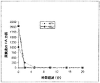
結果のグラフ表示を図1に示す。
結果はNDVのHUJ菌株はMTH菌株と同様Daudi細胞の殺菌に有効であることを示している。
【0075】
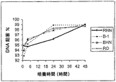
NDV処理後のDaudi細胞のアポトーシス
Daudi細胞を所定時間の間、MTHまたはHUJ菌株(100EID50/細胞)のいずれかの存在下で培養した。アポトーシスはMTTテトラゾリウム(Mosmann T 1983,J of immumol. Methods 65:55−63)を用いて比色定量で測定した。MTTは細胞のアポトーシスを示すODによって表される色相反応である。570nmで測定したODの強度は細胞の成育能力と直接相関している。ODが高い程、成育能力は高く、細胞の死亡率%が低いことを示す。
グラフ表示を図2に示す。
【0076】
MTH菌株の細胞毒性(図1)およびアポトーシス(図2)への作用はHUJ菌株で観察されるよりも速い。しかし、96時間の培養後、両菌株は同じ効果を示した。両ウイルスは細胞の複写を阻止することが見られた。以前、BarEli等はNDVが非癌細胞と比べてリンパ腫細胞に選択的な作用のあることを示した。またNDVは正常ヒト胚線維芽細胞に細胞毒性がないことが見出された。
【0077】
培養中の細胞についてのHUJ菌株の殺細胞の有効性が20 EID50/細胞から2000 EID50/細胞の範囲で試験され、この範囲で有効であることが見出された。従って、HUJ NDVを局所的に腫瘍(単独でまたは組成物中の活性成分として)に投与することを含む本発明の治療は、腫瘍中の細胞数の推定、または腫瘍の大きさの推定、およびHUJ NDV菌株の20 EID50/細胞から2000 EID50/細胞の範囲の投与、または表面糖蛋白の等価な量の投与からなることが好ましい。本発明による患者の全身治療はHUJ NDV菌株の106−1012のEID50の少なくとも1つの用量の投与、または表面糖蛋白の等価な量の投与からなることが好ましい。
【0078】
4) 56℃における赤血球凝集素活性の熱安定性
MTH菌株およびHUJ菌株の赤血球凝集素の熱安定性を“The affinity of Newcastle disease virus to the influenza virus group.Aust.J.Exp.Biol.Med.1942,20,320−328”に記載のF.M.Burnetの方法に従ってニワトリの赤血球を用いて56℃で測定した。
結果を図3Aおよび3Bおよび表3Aおよび3Bに示す。
結果は、HUJ菌株の赤血球凝集素活性は尿膜液中および精製液中の両方で2分後にすでに活性が見られないのでより不安定であるのに対して、MTH菌株の赤血球凝集素活性は56℃で約10分間維持されるのでより熱安定性がある。
【0080】
5) 血清中の熱に不安定なβ阻害剤に対する感受性
NDV菌株は正常血清中のβ阻害剤非特異的阻害剤に感受性であることが知られている。β阻害剤を含むウマ血清を用いたアッセイはHUJ菌株はMTH菌株よりも阻害剤に感受性がより低いことを示した。
NDV菌株の病原性
6) 平均死亡時間
胚の平均死亡時間(MDT)はウイルスの毒性を示す。MDTはManual of Standards for Diagnostic Tests and Vaccines,4thedition,2000に記載されている方法を用いて純系化したウイルスの連続希釈でSPFニワトリ卵を接種して測定した。胚の死を異なった希釈において測定し、MDT(100%の死を起こす最高の希釈で感染した胚の平均死亡時間)を測定した。オリジナルのHungarian MTH菌株のMDTは65時間未満であることが示された。HUJのMDTは96時間を超し、これは長期潜伏性ウイルスに典型的である。ウイルスMaster Seed Bank由来のウイルスworking seed bankからのウイルスで感染したひなトリ胚のMDTは100時間を超した。これらの結果はオリジナルのMTH菌株は中期潜伏性であるのに対して、本発明のHUJは長期潜伏性であることを示す。
【0082】
7) トリプシンの存在下および非存在下でのニワトリ胚線維芽細胞培養(CEF)中のウイルスの複製(Try)
同じHA力価(1:200)の純系化したウイルスを連続希釈でCEFの単層培養中に接種した。培地中のCPE(細胞変性効果)の観察と赤血球凝集(HA)アッセイによって複製を追跡し、ReedおよびMuench法によって組織培養感染力価(TCID50)を測定した。
【0084】
トリプシンの非存在下のウイルスの複製は毒性の増加を示す。それは表面糖蛋白のトリプシン切断部位のアミノ酸残基が多塩基(アルギニンまたはリジン)であることを示している可能性がある。
【0085】
HUJ菌株がトリプシンの存在下(103.0→108.5 TCID50)でより高いレベルで複製するのに対し、MTH菌株はトリプシンの存在の有無にかかわらず類似した力値で複製する。これはMTH菌株が、恐らく切断部位に1つの塩基性アミノ酸を有するHUJウイルスよりもより病原性であることを明らかに示している。
【0086】
8) 血清学
殆どのNDVは血清学的に類似している。発明者等が以前に単離したイスラエル中期潜伏性菌株に対するポリクロナールウサギ抗NDV超免疫血清を使用した時、両菌株は同様に阻害された(HI力価1:1280)。(表6参照)しかし、表6の列I,II,IIIで示すように溶血、赤血球凝集およびノイラミニダーゼの阻害において、MTHで処理した癌患者から得たヒト血清はHUJ菌株に対するよりも、同種のMTH菌株に対してより高い抗体力価を示した。また両NDV菌株に対して類似したHI抗体力価を有する免疫ラット血清は他のウイルス活性(溶血およびノイラミニダーゼ)に対して異なった抗体力価を示した。
【0087】
9) 卵中での中和
MTHで治療された癌患者からの血清を2つのNDV菌株の各々の100 EID50と相互作用させた。ついでその混合物を10−11日齢の胚に接種した。48時間後、ウイルスの中和を赤血球凝集アッセイで測定した。中和抗体濃度は同種の菌株(この場合はMTH)よりも高かった。中和血清力価はMTHに対しては1:320で、HUJではわずか1:20であった(血清1)。中和血清力価はMTHに対しては1:320で、HUJに対してはわずか1:80であった(血清2)。
【0088】
NDV HUJ菌株蛋白の分析
2つのNDV菌株MTHおよびHUJの蛋白を比較するために、精製したビリオン標本(上記のNDV標本と精製参照)をSDSで処理し、変性蛋白を10%SDSポリアクリルアミドゲル中の電気泳動で分析した。NDVビリオン蛋白のSDSポリアクリルアミドゲル分析の写真を図4に示す。MTHおよびHUJの蛋白の10%ポリアクリルアミドゲル中の電気泳動を2μgおよび5μgのウイルス蛋白を用いて実施し、ついでゲルをCoommmassie blueで染色した(Millar NS et al.,(1988).J.Gen.Virol.69(3)、613−20)。
【0089】
図4に見られるように、6つの主要な蛋白をゲル中に溶解した。これらの6つの蛋白はNDVの周知の主要構造蛋白、P−69kD;HN−74kD;FO−62kD;F−56kD;NP−60kDおよびM−38kDに対応した(Hightower,L.E.,Morrison,T.B.およびBratt M.A.(1975)J.Virol.16,1599−1607)。この方法では、菌株MTHとHUJの主要ビリオン蛋白の見かけの分子量に差はなかった。
【0090】
NDV表面糖蛋白
NDV表面糖蛋白の細胞毒性
ニューカッスル病ウイルスの表面糖蛋白のDaudi細胞吸着によってその後の浸透なしに細胞DNA合成の急速な阻害、細胞の増殖の停止、および細胞の事実上の死を生じた。中期潜伏感染性菌株(Roakin)から得られた糖蛋白は長期潜伏感染性菌株から得られたものよりもより有効であった(B−1)。
【0091】
従って、吸着された糖蛋白が細胞膜の完全性を歪め、51Cr放出の増加で示されたように、その透過性を増すと考えられた。細胞の死は外因性ウイルス糖蛋白が介在するシグナル伝達による特別な細胞変性作用と関連すると推定される。
【0092】
この実験に用いられた菌株はアメリカ型収集1971から得られた長期潜伏感染性B−1菌株および中期潜伏感染性Roakin/46VNJ菌株(RO)である。
【0093】
ウイルス表面糖蛋白の製造
疎水性膜蛋白の可溶化のために、精製したウイルスの標本を非イオン性洗剤NP−40(Sigma)、0.2%で30分間4℃で処理した。洗剤を1:1の容量の分析用エーテル(May and Baker Ltd., England)で5回抽出した。ついでエーテルを窒素で蒸発した。ウイルスのコアを4℃で20,000rpm45分間の高速遠心分離(L−2ローターTi50)で除去した。上清中の表面糖蛋白を−70℃に保存した。緩衝溶液を同じ処理に架け、残存洗剤の影饗が全くないことを確実にするために対照の目的に使った。
【0094】
当業者は表面糖蛋白が、他の複数の周知の方法と他の洗剤を用いて得られることを評価するであろう。
【0095】
NDV表面糖蛋白の生物活性
NP−40で処理して得られた分画は表面糖蛋白赤血球凝集素ノイラミニダーゼ(HN)および融合(F)を含んでいた。下の表7に中期潜伏感染性(RHN)および長期潜伏感染性(BHN)菌株由来の糖蛋白分画の生物学的性質を示す。表面糖蛋白が抽出された2つの精製したウイルス標本の感染性は109.3EID50/0.2 mlであった。RoakinまたはB−1菌株、即ち、それぞれRHNおよびBHNから得られた表面糖蛋白を含む可溶成分には感染性は記録されなかった。抽出前に2つのウイルス懸濁物の蛋白濃度(μg/ml)は類似していた。抽出後、予想より低かったが、2つの菌株の、表面糖蛋白分画中の蛋白濃度は類似していた。
【0096】
表面糖蛋白分画の赤血球凝集素活性はオリジナルの全体のウイルス標本と類似していた。しかし、ノイラミニダーゼ活性はRoakinまたはB−1菌株の糖蛋白分画中で完全なウイルス懸濁物の最高値のそれぞれ33%および50%に低下した。溶血活性は完全なウイルス製剤中で高かったが、単離された表面糖蛋白分画中では、この活性の僅かな部分(6%)だけが残った。
NDV表面糖蛋白のDaudi細胞への毒性作用
RHNとBHNウイルスのDaudi細胞への吸着を、希釈したウイルス特異的抗血清(トリとウサギ)と蛍光結合ヤギ抗トリおよび抗ウサギIgGを用いて間接免疫蛍光法でモニターした。90%を超す細胞がウイルス感染60分後、顕著な染色を示した。異なったウイルス製剤を接種後Daudi細胞の生存数および死亡数を異なる時期で測定した(図5AおよびB)。
【0098】
生存細胞の全体の数で測定した細胞の増加は完全に阻害され、72時間後、表面糖蛋白の破壊能の対照および参照として用いた全部のウイルス製剤(RO,B−1)との相互作用後、すべての細胞は死んだ。他の実験では、RHN分画はより低い割合で細胞の増加を阻害し、70%を超す細胞が障害され、破壊された。
【0099】
他方BHN分画は標的癌細胞の個々の単離体に異なったレベルの活性を示した。従って、BHN分画は比較的、Daudi細胞単離体に効果がなく、死亡の割合は対照の細胞に類似していた。しかし、追加のDaudi細胞の単離体を用いた時、それは非常に高い感受性を示し、RHN分画では100%の細胞が、BHN分画では74%の細胞が72時間以内に死んだ(図5Aおよび5B)。次の実験はD−2系を用いて実施した。
【0100】
DNA合成
細胞とNDV菌株および分画RO,RHN、B−1およびBHNとの相互作用の1時間後に、DNA合成の急速な阻害(90−95%)が観察された。この阻害は実験を通して維持され、48時間で99%の阻害に達した(結果を図6および下の表7に示す)。
ウイルス製剤(完全なウイルスまたは単離された表面糖蛋白)を特異的抗血清で前処理によって細胞毒性がなくなるので、阻害作用はNDVウイルスに特異的である。
【0102】
細胞膜透過性の増加
細胞を51Crで標識し、異なったNDV製剤と相互作用させた。異なった時間間隔の後、放射活性の漏洩を非感染対照Daudi細胞からの自然な放出と比較して測定した。図8に示すように、顕著な51Crの放出がNDV RO(59%)とB−1(79%)の相互作用90分後にすでに観察されたのに対し、RHN(12%)とBHN(6%)によっては放出の割合は僅かで低かった。
【0103】
放出はRO、B−1,RHNおよびBHNとの相互作用4時間に、更に60,85,23および18%に増加した。24時間では、完全なウイルスとの相互作用からは100%、RHNとの相互作用からは65%の放出が記録され、BHNと相互作用された細胞ではわずか36%の放出が見られた。対照液と相互作用した細胞又は非感染細胞では膜の透過性の増加および細胞障害は観察されなかった。
【0104】
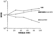
組織培養
培養初期トリ繊維芽細胞中で培養されたウイルスの影響
NDV菌株のBurkittリンパ腫Daudi細胞への細胞毒性作用を検討した。尿膜嚢胚含有卵(CF)中で培養した中期潜伏性(Roakin)および活性弱毒化長期潜伏性菌株と細胞の相互作用の結果、90%の細胞が死に至った。しかしトリ繊維芽細胞中で培養された長期潜伏性菌株は非常に低い活性を示し、細胞死は僅か10%であった(図8A−C)。活性はウイルス表面糖蛋白(赤血球凝集素ノイラミニダーゼ(HN)および融合(F))の切断に依存することが認められた。
【0105】
中期潜伏性および長期潜伏性菌株の糖蛋白はいずれも胚含有卵の中でプロテアーゼによる切断を受けるが、F0とHN0の切断部位に1つのグルタミン残基を有する長期潜伏性菌株はCFのプロテアーゼに感受性がない。トリプシン(CFT)存在下でのCF中のウイルスの培養又は精製ウイルス製剤のトリプシン(NDVT)処理は細胞死(それぞれ66%および93%の細胞死)で検出されたようにウイルス活性を回復した。ノイラミニダーゼおよび赤血球凝集素活性は、赤血球凝集試験、蛍光染色およびノイラミニダーゼアッセイを用いた細胞上へのウイルスの吸着によって示されるように、処理の有無にかかわらず類似している。
【0106】
赤血球の溶血の欠損によって示されるように(卵に成長したウイルスの1:32希釈での71%の溶血に対して、1:2の希釈では僅か31%の溶血が記録された)、CFに成長したウイルスの融合蛋白は殆ど完全に不活性である。トリプシンはCFTとNDVTの1:16希釈でそれぞれ58%と64%の溶血に活性を高めた(表9および4、図9)。
【0107】
従って細胞ウイルス膜の融合に関係のある融合蛋白がウイルスの細胞毒性作用に重要な役割を果たしているようである。
当業者は本発明が上に特別に示し且つ記載したことに限定されないことを評価するであろう。むしろ、本発明の範囲は次に示す請求項によって定義される。
【図面の簡単な説明】
【0109】
【図1】2つのNDV菌株(HUJおよびMTH)の培養中のDaudi細胞への毒性作用の代表的な実験結果を示すグラフである。
【図2】2つのNDV菌株(HUJおよびMTH)と相互作用後の培養中のDaudi細胞のアポトーシスを示すグラフである。
【図3A】2つの実験での2つのNDV菌株(HUJおよびMTH)についての56℃での赤血球凝集素の熱安定性を示すグラフである。
【図3B】2つの実験での2つのNDV菌株(HUJおよびMTH)についての56℃での赤血球凝集素の熱安定性を示すグラフである。
【図4】NDVビリオン蛋白(菌株HUJとMTH)の電気泳動後のSDSポリアクリルアミドゲルの写真である。
【図5】NDV菌株(RoakinおよびB−1)と培養後のDaudi細胞の成育能力および死亡率のグラフを示す。
【図6】NDV菌株(RoakinおよびB−1)の存在下またはこれらの菌株から抽出された表面糖蛋白(RHNおよびBHN)の存在下での培養に応答したDaudi細胞中のDNA合成の阻害を示すグラフである。
【図7】NDV感染細胞からのCr51の放出を示す。
【図8A】組織培養の中にまたはDaudi細胞上の胚含有卵中に伝播したNDVの作用を示したグラフである。細胞の合計数(図8A)、感染後の死んだ細胞の割合(%)(図8B)およびトリプシン処理のNDVの細胞毒性活性への作用(図8C)。
【図8B】組織培養の中にまたはDaudi細胞上の胚含有卵中に伝播したNDVの作用を示したグラフである。細胞の合計数(図8A)、感染後の死んだ細胞の割合(%)(図8B)およびトリプシン処理のNDVの細胞毒性活性への作用(図8C)。
【図8C】組織培養の中にまたはDaudi細胞上の胚含有卵中に伝播したNDVの作用を示したグラフである。細胞の合計数(図8A)、感染後の死んだ細胞の割合(%)(図8B)およびトリプシン処理のNDVの細胞毒性活性への作用(図8C)。
【図9】赤血球の溶血によって指示されたF糖蛋白活性のヒストグラムを示す。
【図10】FおよびHNポリペプタイドの予想アミノ酸配列を示す。【Technical field】
[0001]
The present invention relates to the use of Newcastle disease virus with oncolytic activity as a long-term latent infectious bacterium and isolated proteins from all such strains of virus and / or NDV in cancer therapy.
[Background]
[0002]
Viruses are known to exert oncolytic effects on malignant cells, and the use of oncolytic viruses as therapeutic agents has been reported (Csataly et al. Cancer Gett Prev (1993) 17 (6): 619). -27; Csataly et al. Anticancer Research (1999) 19 (1B): 635-8 and for reviews see Sinkovics J. of Clinical Virology (2000) 16: 1-15).
[0003]
For example, oncolytic viruses such as the avian virus Newcastle disease virus (NDV) are known to be oncolytic to tumor cells in vitro and in vivo (Reichard et al. J Surg Res (1992). ) 52 (5): 448-53; Bar Eli et al. J Cancer Res Clin Oncol (1996) 122: 1-7 and Tsadok-David et al. (1995) J Cancer Research Clinical Oncol 121: 169-174).
[0004]
Newcastle disease virus is an avian RNA paramyxovirus that causes Newcastle disease in different bird species (depending on the toxicity of the virus strain and the age of the individual bird), but is considered less pathogenic to humans. NDV is a linear, unleaved, enveloped virus that contains a single-stranded negative-sense RNA genome. The virion consists of a coiled nucleocapsid containing single-stranded RNA and six structural polypeptides (MW 20,000-80,000). Nucleocapsid is coated with a protein and lipid envelope. Matrix protein (M) on the inner surface of the viral envelope is involved in viral assembly and interacts with both viral membrane and nucleocapsid proteins. There are two viral glycoproteins on the outer surface of the viral envelope: hemagglutinin neuramidase (HN) and fusion glycoprotein (F). HN glycoproteins are involved in binding to viral cellular receptors. Monoclonal antibodies against this protein have been shown to neutralize NDV infectivity. F protein is first an inactive precursor (F0) And then post-translationally cleaved to produce two disulfide-linked polypeptides (F1And F2This F protein is involved in the penetration of NDV into host cells by facilitating the fusion of the viral envelope with the plasma cell membrane of the host. Antisera against F protein inhibited hemolysis and virus-induced cell fusion. Since F glycoprotein and HN glycoprotein play an important role in NDV infectivity, great efforts are being made to purify the NDV gene. EP 227414 to Bingham et al. Discloses a cDNA sequence encoding the NVD Beaudette C chain F and HN polypeptides, and labeled probes and F and HN polypeptides that can be used for the diagnosis of avian NDV The use of this nucleotide sequence for the preparation of
[0005]
The state of proteolytic cleavage of surface sugar chains F and HN is related to the toxicity of different NDV strains. F of virulence strain0Is F in a wide range of host cells1And F2But is non-toxic strain F0Is cleaved in only a few hosts. Therefore, these differences are expressed as short-term latency (high pathogenicity), medium-term latency (moderate pathogenicity), and long-term latency (non-pathogenicity) in the classification of different strains of NDV.
[0006]
The surface sugar chains HN and F of NDV are thought to be related to the oncolytic performance of NDV, in addition to its role in infectivity (Alissa Waldmann-Kegnovich (1999) Dept. of Virology, Haddasa Medical School of the University of Jerusalem MSc paper).
[0007]
Part of the effects of oncolytic viruses on tumor cells is due to the increased sensitivity of tumor cells to the cytotoxicity of tumor necrosis factor and the immunostimulatory properties of these viruses. Animal NDV locally induces chemokines and cytokines such as tumor necrosis factor alpha that affect T cell recruitment and activation (Schirmacher et al. (1998) Semin Oncol 25 (6): 677-96 and Schirmacher). et al. (1999) Int J Oncol 14 (2): 205-15). There are other reports that the tumoricidal cell action of attenuated strains of NDV (73-T) is due to direct cell lysis after replication of infectious virus (Lorence et al. J. Nat. Cancer Inst. (1994). ) 86 (16) 1228-1233). The bactericidal effect of NDV (RO) metastatic latent strains of Daudi lymphoma cells and of NDV Ulster strains on metastatic Esb lymphoma and B16-F10 melanoma is that UV-inactivated virus kills these tumor cells. Was found to be as effective as infectious viruses and was therefore found to be unrelated to viral replication (Tsadok-David et al. (1995) J. Cancer Research Clinical Oncology 121: 169-174 and Schirmacher et al. al. (1997) Clin Cancer Res 3 (7): 1135-48).
[0008]
Current efforts in cancer therapy using viruses include the use of live pathogenic viruses as oncolytic agents (see Csataly et al and Csataly US Pat. No. 5,602,023, supra). Provirus WO 00/62735 discloses the use of any interferon-sensitive strain of virus to kill tumor cells that are poorly responsive to interferon. Provirus disclosure is a catalog of viral strains including three metastatic strains of NDV (MK107, NJRokin and Connecticut-70726) that have been shown to be useful for the treatment of human tumors xenografted in athymic mice Supply. Administration of NDV to these mice resulted in tumor regression due to the more efficient and selective replication of NDV in tumor cells relative to normal cells. It has been disclosed that the differential sensitivity to tumor cell death by NDV is associated with the inability of the cells to exhibit an interferon-mediated antiviral response. The above patent application claims patents for methods of infecting neoplasms or tumors and methods of treating neoplasms or tumors with interferon-sensitive, replicative RNA or DNA viruses.
[0009]
Another approach is dedicated to the development of vaccines for anti-tumor immunity. For example, NDV has been used in the preparation of human autologous tumor cell vaccines (reviewed in Schirmacher et al. (1998) Semin Oncol 25 (6): 677-96).
[0010]
Long-term latent infectious strains of NDV are used in the treatment of cancer, or surface glycoproteins from different strains of NDV, ie short-, medium- or long-term latent infectious strains, have oncolytic properties, None of the background art teaches or suggests that it may be useful in the treatment of.
DISCLOSURE OF THE INVENTION
[Means for Solving the Problems]
[0011]
The compositions and methods of the present invention take advantage of the oncolytic nature of viruses and / or viral proteins to kill neoplastic cells. The present invention provides compositions and methods for treating cancer that avoid contacting a patient with a pathogenic strain of a virus.
[0012]
The present invention provides a pure long-term latent infectious strain of NDV useful for cancer treatment, referred to herein as HUJ.
[0013]
The present invention provides a pharmaceutical composition comprising at least one NDV long-latency infectious oncolytic strain for the treatment of cancer. The present invention further provides a pharmaceutical composition comprising at least one NDV long-term latent infectious strain comprising a further suitable carrier.
[0014]
In cancer treatment, it is preferable to use NDV HUJ strain (described in detail below). Furthermore, the composition is 10 for each therapeutic dose of HUJ NDV strain.6-1012Egg infectious dose of 50% (EID50) Is more preferable. Treatment with HUJ NDV is 20 EID50/ 2000 EID from cells50It is alternatively and selectively preferred to be within the range of up to / cell.
[0015]
In another embodiment, the composition of the invention comprises at least one isolated viral glycoprotein or subunit or analog thereof having oncolytic activity. In a further embodiment, the viral glycoprotein is obtained from NDV. In another embodiment of the invention, the composition comprises at least NDV F glycoprotein. The term F protein is used herein as F and F0Including both. In a further embodiment, the composition comprises F glycoprotein and NDV HN glycoprotein subunits having hemagglutinin activity. In a further embodiment, the composition comprises NDV F glycoprotein and HN glycoprotein. The term HN glycoprotein, as used herein, includes both HN and a precursor of HNO that is cleaved at its C-terminus to form active HN. The viral glycoprotein utilized in this embodiment is non-infectious and thus may be any suitable strain of NDV. Alternatively and alternatively, NDV short-term latent infectious strains, intermediate latent infectious strains and long-term latent infectious strains are used. Further, the composition may be any viral protein having oncolytic activity, or any combination of subunits or analogs thereof, or a long-term latent infectious strain oncolytic NDV and viral protein having oncolytic activity, or subunits or analogs thereof. And a combination thereof.
[0016]
The present invention further provides a cancer therapy utilizing the pharmaceutical composition described above.
[0017]
In a further embodiment of the invention, the treatment of cancer utilizes at least one isolated polynucleotide encoding at least one viral polypeptide or analog or subunit thereof having oncolytic activity. In a further embodiment of the invention, the treatment of cancer utilizes an isolated polynucleotide encoding the NDV F protein. In another embodiment, an isolated polynucleotide encoding the NDV HN protein is utilized. In a further embodiment, a combination of isolated polynucleotides encoding F and HN glycoproteins is used.
[0018]
Proteins F and HN are known to be glycoproteins. A polynucleotide of the invention encodes its polypeptide portion, ie, the portion that is then glycosylated in vivo.
[0019]
The F polypeptide also has two polypeptides F that are shorter in vivo.1And F2Disconnected. Thus, the present invention may be used as another molecule or as a disulfide-linked single-stranded molecule or their bioprecursor F0As a polypeptide, F1And F2A polynucleotide encoding a polypeptide is included.
[0020]
It should be clearly understood that any fragment of a polypeptide that retains the intact protein oncolytic activity is within the scope of the present invention. Accordingly, a polynucleotide encoding any such fragment is within the scope of the invention.
[0021]
In an important aspect of the invention, an isolated polynucleotide encoding the F and / or HN polypeptide of NDV RNA, the polypeptide or any active fragment of the polypeptide or F and / or NDV RNA. Artificial polynucleotides complementary to polynucleotides encoding HN polypeptides are provided.
[0022]
The invention further includes host cells transfected or infected with a recombinant polynucleotide as defined above.
[0023]
The polynucleotide of the present invention may be used as an intermediate in the production of a polypeptide by recombinant DNA technology. Thus, an expression vector of the invention containing a suitable promoter and a polynucleotide of the invention, for example expressed in yeast or bacteria, produces a suitable encoded polypeptide. Alternatively and alternatively, the vector may be a viral vector.
[0024]
In a further embodiment of the invention, a long-latent infectious strain of NDV, preferably the HUJ strain, is used in the preparation of a composition for treating cancer. In other embodiments of the present invention, viral glycoproteins or subunits or analogs thereof are used in the preparation of cancer treatment compositions. NDV coated glycoproteins are preferably used, more preferably F glycoproteins and / or HN glycoproteins.
[0025]
The method of the invention for cancer treatment in an embodiment of the invention comprises as an active ingredient a long-latent infectious strain of NDV, preferably a HUJ strain and / or at least one isolated viral protein as described above. Administering to the patient a therapeutically effective amount of the composition. The composition may be administered to the patient by any suitable route. In one particularly preferred embodiment, direct injection of the composition into the tumor or in the vicinity of the tumor is utilized.
[0026]
Accordingly, the compositions and methods of the present invention are intended to treat cancer without the risk of being involved in the use of long-lived infectious strains (highly pathogenic) or medium-latent infectious strains (moderately pathogenic) of live viruses. I will provide a.
The present invention will be understood and appreciated more fully from the following detailed description, taken in conjunction with the drawings in which:
[0027]
Viruses are known to exert oncolytic effects on tumor cells, and it has been reported that oncolytic viruses are used as therapeutic agents. As described above, several efforts have been made to use non-human viruses exhibiting moderate to high pathogenicity in cancer therapy. However, the use of non-pathogenic (long-term latent infectious) non-human viruses or isolated viral proteins for cancer treatment with oncolytic activity has not been reported in the prior art. Accordingly, the present invention discloses compositions and methods for cancer treatment that take advantage of the oncolytic nature of certain viruses and isolated viral components. The disclosed compositions and methods provide for the first time a safe, effective and reliable means of treatment of cancer in solids where it is needed. These methods overcome the disadvantages of using pathogenic strains of viruses for human therapy.
[0028]
Accordingly, the present invention provides compositions and methods for cancer treatment using the Newcastle Disease Virus (NDV), a long-lived infectious strain of non-human virus. Furthermore, it provides a method of cancer treatment comprising an isolated viral protein or subunit having oncolytic activity or an analogue thereof and an isolated polynucleotide encoding the viral protein or a construct comprising the same polynucleotide. To do. A polynucleotide or construct comprising the same polynucleotide may comprise a polynucleotide of any vector, including a polynucleotide of a viral vector. The present invention provides a host cell comprising said polynucleotide, a construct comprising the same polynucleotide, and a polynucleotide of the vector described above, which is also used for cancer therapy. The present invention further provides cancer treatment using any combination described above.
[0029]
A modified long-term latent infectious strain of NDV labeled HUJ is disclosed below. In order to guarantee and enhance its uniformity, it is preferable to obtain a pure virus. Pure viruses can be produced by any method available to skilled artisans, such as limited dilution or plaque purification. In an embodiment of the present invention, a pure HUL strain prepared at limited dilutions is a composition for cancer treatment with or without a suitable carrier such as human serum albumin (HSA) or other suitable adjuvant. Used in the preparation of Any type of cancer is within the scope of the present invention. As non-limiting examples, the following cancers can be treated: glioma, lung cancer, breast cancer, prostate cancer, melanoma, leukemia and sarcoma.
[0030]
The present invention provides compositions and methods for cancer treatment utilizing at least one isolated viral protein having oncolytic activity, preferably F and HN glycoproteins of NDV. F and HN glycoproteins have been shown to play an important role in viral infectivity. However, none of the background art suggests that the isolated F glycoprotein and HN protein have oncolytic activity. The present invention provides direct evidence of the oncolytic activity of the first isolated viral protein. According to the present invention, viral proteins, preferably NDV F glycoprotein and / or HN glycoprotein or analogs or glycoprotein subunits thereof or mixtures thereof are used in the preparation of a composition for the treatment of cancer.
[0031]
As used herein, the term “oncolytic activity” includes in vitro and in vivo cytotoxic effects on tumor cells without affecting normal cells. In vitro cytotoxic effects are detected by various means known in the art, such as selective staining for dead cells, inhibition of DNA synthesis or apoptosis.
[0032]
Those skilled in the art will understand that the term “protein analogue” has the functionality of a virus counterpart (ie, fusion, hemagglutinin and neuraminidase protein) and has the same sequence, secondary and tertiary structure as the virus counterpart. You will appreciate not having it. Thus, cleaved or denatured proteins that exhibit oncolytic activity as natural viral proteins may be used in the compositions and methods of the present invention.
[0033]
The fusion of NDV F and HN glycoproteins, hemagglutinin and neuraminidase activity may be involved in the oncolytic action of the isolated protein. However, the present invention includes other viral proteins exhibiting other activities that may be involved in the oncolytic activity of the isolated protein.
[0034]
The protein can be used in the composition with aluminum hydroxide, emulsions or submicron emulsions (eg, adjuvants such as US Pat. No. 5,576,016,5629932,5716637,5961970 or well known pharmaceutical carriers such as human serum albumin) and tumors. Viral proteins produced by genetic engineering having disruptive activity, preferably viral fusion, hemagglutination and neuraminidase proteins are within the scope of the present invention.
[0035]
The present invention provides compositions and methods for treating cancer comprising a composition comprising the same polynucleotide as the isolated polynucleotide encoding the F protein and HN protein of the HUJ strain of NDV. The nucleotide sequence encoding the HUJ F protein was found to be almost the same (with 3 nucleotide differences) as the LaSota strain. Accordingly, the present invention encompasses the use of isolated polynucleotide sequences encoding the F protein of other long-term latent infectious strains.
[0036]
The surface glycoprotein may be obtained from any natural strain of NDV. The glycoprotein is preferably obtained from a long-term or medium-latency infectious NDV strain such as, for example, Rokin / 46VR109 (RO) from an American collection. Alternatively and alternatively, glycoproteins of HUJ or other long-lived infectious strains are used. The glycoprotein may also be obtained from a genetically or genetically engineered virus strain. Furthermore, the glycoprotein may be obtained from an expression system exemplified by a mammalian expression system, an insect expression system or a bacterial expression system, but is not limited thereto. Alternatively, synthetic proteins such as HN or F or recombinant viral proteins may be used in the present invention.
[0037]
The composition may be in any dosage form suitable for administration to a patient, such as a suspension, emulsion, spray, solution or any other formulation according to principles well known in the art. Compositions of the invention are suitable, including intravenous, oral, buccal, intranasal, inhalation, topical administration to the mucosa, or injection including intradermal, intrathecal, intracisternal and intralesional injection or other types of injection However, the present invention is not limited to these.
[0038]
The cancer treatment methods of the present invention, according to embodiments, comprise administering to a patient a therapeutically effective amount of HUJ NDV. HUJ NDV may be administered to the patient by any suitable route by any route described above. One particularly preferred embodiment utilizes injection of a HUJ strain or injection of a composition comprising the HUJ strain and / or at least one said viral glycoprotein into or near a tumor.
[0039]
In another embodiment of the invention, the method of the invention for treating cancer comprises administering to a patient a therapeutically effective amount of at least one NDV viral glycoprotein or subunit or analog thereof (any suitable as described above). Included). The viral protein may comprise at least one NDV F glycoprotein, NDV HN glycoprotein or NV F glycoprotein and HN glycoprotein.
[0040]
Treatment of cancer patients according to embodiments of the present invention may be systemic, in which case the above-described composition or isolated whole virus and / or isolated protein is administered to the patient. The form of administration may be intravenous, oral, buccal, intranasal, inhalation, topical administration to the mucosa or intradermal, intrathecal, intracisternal and intralesional injection or other types of injection. It is preferred to administer the long-term latent infectious NDV virus (eg HUJ strain) or the above viral protein or the composition of the invention locally or directly to or near the tumor. Usually, the form of local administration is injection, for example, injection into a lesion.
[0041]
The isolated polynucleotides of the present invention are used in the production of at least one viral peptide having oncolytic activity by recombinant DNA technology in cells transfected with these polynucleotides. It is preferred to use the polynucleotides for the production of NDV HUJ F and / or HN polypeptides. A polynucleotide may also be comprised of an expression vector, such as a viral vector, to effect polypeptide expression. Viral NDV protein expression methods are disclosed in Bingham EP 227414, which is fully incorporated herein.
[0042]
The term “polynucleotide” includes single- and double-stranded DNA, RNA and compounds of 16 or more nucleotide lengths or biologically synthesized nucleotide polymers.
[0043]
The term “artificial”, as used herein, means human intervention by any means in the production of a polynucleotide. In addition to the artificial polynucleotide itself, the present invention includes recombinant molecules. These can be broadly defined as a composition comprising a vector polynucleotide and a polynucleotide of the present invention as defined above or comprising a polynucleotide which is foreign to it. Usually, the polynucleotide is DNA, and the present invention specifically includes DNA in which the vector is a cloning vector or expression vector. The expression vector can be, for example, a prokaryotic expression vector or a eukaryotic expression vector. The term “vector” herein also includes shuttle vectors. Where expression is required, the polynucleotide further comprises a single sequence of a type effective for translation and other processing of the mRNA into the desired viral protein.
[0044]
The present invention provides isolated polynucleotides encoding viral proteins with oncolytic activity, preferably F glycoproteins and HN glycoproteins, which are host cells so that they are expressed by the cells and / or their progeny. Into which the recombinant cells are then administered in vivo for therapeutic effect.
[0045]
In order to introduce these genes into cells, it is desirable to improve membrane permeability to oligonucleotides. Various means are known in the art to improve membrane permeability. For example, the oligonucleotide molecule may be partially linked to groups containing one or more polar or charged groups such as unsaturated aliphatic hydrocarbon chains and carboxylic acid groups, ester groups and alcohol groups. Alternatively, the oligonucleotide may be linked to a peptide structure, preferably a membrane affinity peptide. Such improved oligonucleotides penetrate more easily through the membrane, which is important for their function and thus significantly enhance their activity.
[0046]
Additives may be selected to enhance oligonucleotide uptake across the cell membrane. Such agents are agents that enhance cellular uptake of double-stranded DNA molecules. For example, certain lipid molecules have been developed for this purpose, including the commercially available transfection reagents DOTAP (Boehringer Mannhei), Lipofectin, Lipofectam and Transfectam.
[0047]
Another way to enhance membrane permeability is by attaching oligonucleotides to molecules known to bind to cell surface receptors. Examples of groups suitable for the formation of bonds are sugars, vitamins, hormones, cytokines, transferrin, asialoglycoproteins and similar molecules. For example, US Pat. No. 5,108,921 to Low et al. Describes the use of these molecules and the preparation of the linkages to enhance the membrane permeability of peptides, proteins and oligonucleotides.
BEST MODE FOR CARRYING OUT THE INVENTION
[0048]
Having generally described the invention, it will be more readily understood with reference to the following examples. These examples are provided for purposes of illustration and are not intended to limit the invention.
【Example】
[0049]
NDV HUJ strain
A sample of NDV HUJ (Master Virus Bank) was assigned to the International Laboratory for Newcastle Disaster Veterinary Laboratories Agenda NewNow Hour7. Viral specimens were subcultured with chicken eggs and shown to be alive.
[0050]
The virus was derived from the long-term latent infectious B1 strain of NDV obtained as native ATCCV188. Virus was subcultured 4 times in chicken eggs to prepare a research stock. Infected allantoic fluid from the fourth passage (E4 stock) was stored at -70 ° C. Infected allantoic fluid from the E4 stock received 50 regular passages in 10-11 day old embryonated eggs. The allantoic fluid was labeled “NDV lenti” and was divided into vials and stored at −80 ° C. “NDV lenti” was purified in 10-11 day old embryo-containing eggs by limiting dilution. Allantoic fluid from eggs infected at the highest dilution was labeled “NDV lenti” and stored at −70 ° C. The cytotoxicity test of the “NDV lenti” strain was performed on Daudi cells and normal human cells. The strain was shown to be oncolytic and was renamed “NDV HUJ”. The cerebral virulence index (ICPI) of the “NDV HUJ” strain was tested in 1-day-old chicks and found to be 0.0, indicating that the virus can be classified as long-term latent infectious, non-toxic, and non-pathogenic Is shown.
[0051]
Attenuation obtained by continuous subculture (eg, allantoic fluid) of eggs produced by the HUJ strain in Hungary's Phylaxia-Sanofi (Csataly et al. Anticancer Research (1999) 19 (1B): 635-8)) The strain was compared with the strain NDV MTH-68 / H. Allantoic fluid containing virus and virus purified on sucrose gradient were compared.
[0052]
Preparation of HUJ strains for in vitro testing:Serial subculture was performed at limited dilution in 10-11 day old embryo containing eggs. Allantoic fluid from the highest dilution (of which only 1/6 was virus positive) was collected and further subcultured by serial dilution. Culture, concentration and purification were performed using routine methods (Tzadok-David et al, and Slosaris M, Levy B., Katz R., Zakay-Rones Z. (1989) Avian Dis. 33: 248-253). .
[0053]
About 640 embryo-containing eggs (10-11 days old) out of 750 cultured eggsFive-106Of embryo infectious dose of 50% (EID50/ Egg) was inoculated into the allantoic cavity. Embryos that were dried within the first 24 hours were discarded. After 72 hours, only live embryo eggs were chilled at 4 ° C. for 16-18 hours. Allantoic fluid (up to 3 L) was collected and centrifuged at 2,000 rpm for 20 minutes to remove debris, and the supernatant with a hemagglutination power value (HA) of 640-1280 / ml was stored.
[0054]
Virus was concentrated by centrifugation of infected allantoic fluid at 18,000 rpm in Sorvall (RC-5 centrifuge) for 60 minutes at 4 ° C. using an SS-34 rotor. Concentrated virus (100 ml contains 32,000 HA units) was then purified by centrifugation for 90 minutes at 24,000 rpm through a sucrose gradient (10-60%) using ultracentrifugation in a SW-27 rotor. Bands containing virus were collected, pelleted in SW27 rotor for 60 minutes at 24,000 rpm, resuspended, purified virus suspension passed through Millipore filter, aliquoted to 0.5 ml and stored at -70 ° C until use. .
[0055]
One skilled in the art will appreciate that other viral enrichment and purification methods can be used to obtain the above results.
[0056]
Preparation of virus for clinical trials:"The HUJ strain referred to as NDV lenti (clone) is a 10-11 day old embryo-containing SPF (non-specially pathogenic) egg (Aves Libres de Patogenos Especiosos of Mexoco, Pueblo, a subsidiary of SPAFAS Charles River Lab.) (Purchased from SA) and purified twice by limited dilution to produce a Virus Master Seed Bank consisting of 220 tubes, harvested allantoic membranes stored at −80 ° C. and frozen without further purification One tube from the Master Seed Bank was expanded to a 300-tube Virus Working Seed Bank after the same procedure used to make the Master Bank. king Bank tube comprises allantoic fluid harvested and frozen without stored. tube further purification -80 ° C..
[0057]
The starting materials for virus production are NDV HUJ Working Bank vials and 10-11 day old embryo containing SPF eggs.
[0058]
A single production line that uses about 3000 eggs produces the amount of material needed for clinical trials. The production was divided into multiple collections (up to 500 eggs). For each harvest, a working bank vial was thawed and the virus suspension was diluted in Gibco PBS (10FiveEID50). A small hole was manually drilled in the top of the egg and an aliquot of the virus suspension was injected into the amino allantoic cavity of the egg. Each egg hole was sealed with sterile acrylic cement and the eggs were incubated for 72 hours. Egg growth ability was checked. Eggs that appeared to have died within the last 12-24 hours upon candling were removed from the collection and eggs that appeared to have died earlier were discarded. If the proportion of all eggs that died after the time of inoculation exceeds 25%, all living eggs were collected along with newly dead eggs. If the percentage of dead eggs was less than 25%, live eggs were inoculated for another 24 hours, after which fresh dead and live eggs were collected. Collection was removal of the upper part of the egg, observation of the embryo and allantoic fluid, and pipetting allantoic fluid into a 50 ml bottle. If the allantoic fluid collected in the pipette was not transparent, it was rejected. The collected allantoic fluid was clarified by low speed centrifugation and stored at 4-7 ° C. Collected infected allantoic aliquots were sterilized and titered (EID50And hemagglutination).
[0059]
The total number of viruses obtained from a particular collection was determined from the number of eggs collected, the amount of allantoic fluid collected and the titer. From about 450 eggs, 150-300 eggs were collected. The yield of the collected liquid was about 8-10 ml per egg and the volume of liquid collected from each collection was between 1000 and 3280 ml. The titer is 109.3/ Ml to 1010.2/ Ml EID50The total amount of virus in 5 unpurified bulk collections was 5.8 × 10 at the time of collection13EID50Met. The virus was then concentrated by high speed centrifugation and purified in a sucrose gradient as follows.
[0060]
Clarified unpurified bulk virus stored at 4-7 ° C. for 1-6 weeks was clarified again by low speed centrifugation (3000 rpm for 30 minutes). A re-cleared aliquot from each collection was taken and stored at -80 ° C for further inspection, and an additional bulk aliquot was taken for in-process sterilization testing. The recleared bulk was centrifuged at 12,500 high speed for 1.5 hours at 4 ° C. and the pelleted virus was resuspended in Gibco Dulbco PBS. The titer was measured to recover the whole. A total of 12,260 ml of re-cleared bulk liquid from 5 collections, with EID yields ranging from 29% to 82% for each collection50Concentrated to a total of 100 ml of resuspended pelleted virus with an average yield of 50% based on titer. An aliquot from each tube of resuspended concentrated virus was tested for sterility.
[0061]
For purification, the concentrated virus was ultrafast centrifuged in a 20/40/60% sucrose gradient at 22,000 rpm for 2.5 hours at 4 ° C. In a normal centrifuge tube, approximately 8 ml of concentrated virus was spread on top of a 24 ml sucrose gradient. The purified virus was recovered as a band of about 4.7 ml. The concentrated virus band was suspended in about 10 ml of sterile physiological saline, and its pH was adjusted to 7.6-7.8 by adding a solution sterilized with an autoclave of disodium phosphate prepared in water for injection. Endotoxin-free sucrose was dissolved in Gibco Dulbeco PBS and sterilized by autoclaving. The clinical dose is about 1 × 10 5 diluted with a suspension of virus in sterile saline.TenEID50Prepared from a combination of harvested purified viruses to achieve a concentration of / ml.
[0062]
PCR and sequencing of F and HN genesThe nucleotide sequence determined for the F and HN genes of the NDV HUJ-master bank and purified virus can be compared with the relevant nucleotide sequence of the LaSota strain of Newcastle disease virus containing 15186 base pairs of linear RNA. Viral RNA was extracted from Newcastle disease virus HUJmaster seed using the QIAGEN QIAamp viral RNA kit according to the manufacturer's instructions. A single stranded DNA copy of the viral RNA template was prepared using reverse transcriptase using a standard protocol for the production of cDNA from the RNA template. In short, two reaction mixtures were prepared. One mixture is dH2Contains 6.5 μl O and 1 μl primer MSF1 (see below), the other mixture is 4.0 μl 5 × RT buffer (reverse transcriptase buffer), dH2O 4.0 μl, 40 mM NTPs (nucleotide triphosphate) 1.0 μl, MMLV-RT (enzyme) 0.5 μl and RNAsin (RNAse inhibitor) 0.5 μl were included. 2.5 μl of RNA was added to 7.5 μl of
[0063]
For DNA sequencing, prepare a reaction mixture containing the terminator preparation reaction mixture (4 μl; Applied Biosystems Corp), PCR product (2-4 μl depending on concentration), sea casing primer (1.6 μl) and deionized water. The volume was adjusted to 10 μl. The mixture was then incubated in a PCR machine using the program 'BIGD'. The sequenced product was precipitated by adding 1 μl of 25 mM glycogen and 52 μl of 2M
PCR and sea casing primers were as follows.
Biological assays:HUJ virus hemagglutination and infectious titer were measured by routine methods (hemagglutination was Sever JL. 1962 J. Immunol. 80: 320-329). Infectivity was measured by inoculating serial dilutions into the allantoic sac of embryonated eggs and checking the hemagglutination of the fluid 72 hours after inoculation. 50% endpoint of egg infectious dose (EID50The titer of the virus defined as) was calculated by the method of Reed and Muench (Reed LJ Muench HA 1938, Amer. JHyg 27: 493-497). Stocks were prepared and stored at -70 ° C.
[0065]
Sterility test:The HUJ virus suspension was examined for the presence of bacteria and mycoplasma and found to be sterile.
[0066]
Nucleotide and amino acid sequences of HUJ F and HN genes: The 3358 nucleotide sequence corresponding to 4498-7855 of the complete LaSota genome and covering the entire F gene, the intergenic and most of the HUJ HN gene, shown below (SEQ ID NO: 1).
The amino acid sequence derived from the above nucleotide sequence is shown in FIG. 10 (SEQ ID NO: 2). The asterix in this sequence represents a stop codon. The F protein thus ends with residue number 553.
[0068]
The amino acid sequence has a fusion protein cleavage site motif of amino acids # 109 to # 119 of SGGGRGRGRIG deduced from the nucleotide sequence starting at nucleotide 370, a characteristic of long-term latency.
[0069]
The 3358 nucleotides of the virus from Master Virus Bank corresponded to those of the La Sota strain of NDV (gi: 3386504) except for 111, 1006 and 1648 nucleotides of the F gene.
[0070]
Sequence of viral F gene after virus purification on sucrose gradient:
As described above, the nucleotide sequence of the virus from the production batch obtained after virus purification on a sucrose gradient was determined as follows. Viral RNA was extracted from Newcastle disease virus HUJ using the SV Total Isolation kit (Promega) according to the manufacturer's instructions. RNA was loaded onto an RT-PCR amplifier using 4 different oligonucleotide primers (using Access Quick RT-PCR System, Promega). The sequences of these primers and their position in the viral genome are shown below.
[0071]
Each reaction mixture contained 25 μl RT-PCR conditioned mixture x2, 8 μl RNA, 5 μl of each forward and reverse sequencing primer and 7 μl of DDH20. The components were mixed well and rotated briefly before the RT-PCR reaction (RT reaction at 48 ° C. for 45 minutes). PCR cycling parameters were 94 ° C for 2 minutes (1 cycle), 94 ° C (30 seconds), 60 ° C (1 minute) and 68 ° C (2 minutes) for 40 cycles, 7 minutes at 68 ° C. It was. The PCR reaction mixture was loaded onto a 1% agarose gel and visualized using a UV transilluminator. Band size was estimated by comparison with DNA markers. A DNA fragment was excised from the gel and purified using the Mini-elute Gel extraction kit (Qiagen). Each fragment was resuspended in ddH20. DNA was subjected to sequencing analysis.
RT-PCR and sequencing primers are
[0072]
Biological characterization (MTH compared to HUJ)
1) Hemagglutination and neuraminidase activity
Neuraminidase was measured according to Aymard-Henry M et al 1973 BUll World Org 48: 199-202 and Warren LJ J Biol Chem 1959 234: 1971-1975. This assay measures the free sialic acid released from the fetuine by the viral enzyme neuraminidase.
These results (Tables 1 and 2) show that neuraminidase activity is higher in the HUJ strain compared to the MTH strain.
[0073]
2) Fusion activity
Fusion activity was measured by chicken erythrocyte hemolysis as described in the literature (Nishikawa K et al 1986 J. Viral 60: 987-993). The results indicate that a 1: 32-1: 64 dilution of the virus causes hemolysis, suggesting that the fusion activity is similar for the two NDV strains.
[0074]
3)Cytotoxic (oncolytic) action
Cytotoxic effects of NDV on Daudi cells in culture
Virus (20-200 EID50/ Cell) were cultured with Daudi cells at different time intervals. At the end of the culture, dead cells were stained with erythrosin B, a selective stain, to check for growth potential (Hanks JH and Wallace J 1985 Proc Expel Biol Med 98188).
A graphical representation of the results is shown in FIG.
The results show that the NDV HUJ strain is as effective in killing Daudi cells as the MTH strain.
[0075]
Apoptosis of Daudi cells after NDV treatment
The Daudi cells are allowed to remain in the MTH or HUJ strain (100 EID50/ Cell). Apoptosis was measured colorimetrically using MTT tetrazolium (Mosmann T 1983, J of immunomol. Methods 65: 55-63). MTT is a hue response represented by OD indicating cell apoptosis. The intensity of OD measured at 570 nm directly correlates with cell growth ability. Higher OD indicates higher growth ability and lower% cell mortality.
A graphical representation is shown in FIG.
[0076]
The effect of MTH strain on cytotoxicity (FIG. 1) and apoptosis (FIG. 2) is faster than that observed with the HUJ strain. However, after 96 hours of culture, both strains showed the same effect. Both viruses were found to block cell replication. Previously, BarEli et al. Have shown that NDV has a selective effect on lymphoma cells compared to non-cancerous cells. It was also found that NDV is not cytotoxic to normal human embryonic fibroblasts.
[0077]
Efficacy of HUJ strain killing of cells in culture is 20 EID50/ 2000 EID from cells50Tested in the cell range and found to be effective in this range. Accordingly, the treatment of the present invention comprising administering HUJ NDV locally to a tumor (alone or as an active ingredient in a composition) comprises an estimation of the number of cells in the tumor, or an estimation of the size of the tumor, and 20 EID of HUJ NDV strain50/ 2000 EID from cells50Preferably consisting of administration in the range of cells / cell or equivalent amount of surface glycoprotein. The systemic treatment of patients according to the present invention is the HUJ NDV6-1012EID50Preferably, it comprises administration of at least one dose of or an equivalent amount of surface glycoprotein.
[0078]
4)Thermal stability of hemagglutinin activity at 56 ° C.
The thermal stability of hemagglutinin of MTH and HUJ strains is described in “The affinity of Newcastle virus to the influenza virus group.Aust.J.Exp.Biol.Med.1942,20,320-328”. M.M. Measurements were made at 56 ° C. using chicken erythrocytes according to the method of Burnet.
The results are shown in FIGS. 3A and 3B and Tables 3A and 3B.
The results show that the hemagglutinin activity of the HUJ strain is more unstable since no activity is already seen after 2 minutes in both allantoic fluid and purified solution, whereas the hemagglutinin activity of the MTH strain is It is more thermally stable because it is maintained at 56 ° C. for about 10 minutes.
[0080]
5)Sensitivity to serum-labile β-inhibitors in serum
NDV strains are known to be sensitive to β-inhibitor non-specific inhibitors in normal serum. Assays with horse serum containing β inhibitors showed that the HUJ strain was less sensitive to inhibitors than the MTH strain.
Pathogenicity of NDV strain
6)Average death time
Embryo mean death time (MDT) indicates viral toxicity. MDT is Manual of Standards for Diagnostic Tests and Vaccines, 4thIt was measured by inoculating SPF chicken eggs in a serial dilution of virus purified using the method described in edition, 2000. Embryo death was measured at different dilutions, and MDT (mean death time of embryos infected at the highest dilution causing 100% death) was measured. The original Hungarian MTH strain was shown to have an MDT of less than 65 hours. HUJ's MDT exceeds 96 hours, which is typical for long-lived viruses. MDT of chicken embryos infected with virus from virus working seed bank from virus Master Seed Bank exceeded 100 hours. These results indicate that the original MTH strain is metaphase latent, whereas the HUJ of the present invention is long term latency.
[0082]
7)Viral replication (Try) in chicken embryo fibroblast culture (CEF) in the presence and absence of trypsin
Purified virus with the same HA titer (1: 200) was inoculated in serial dilutions into CEF monolayer cultures. Replication is followed by observation of CPE (cytopathic effect) in medium and hemagglutination (HA) assay, and tissue culture infectious titer (TCID) by Reed and Muench method.50) Was measured.
[0084]
Viral replication in the absence of trypsin indicates increased toxicity. It may indicate that the amino acid residue at the trypsin cleavage site of the surface glycoprotein is polybasic (arginine or lysine).
[0085]
HUJ strain in the presence of trypsin (103.0→ 108.5 TCID50) Replicate at a higher level, whereas MTH strains replicate with similar force values in the presence or absence of trypsin. This clearly shows that the MTH strain is probably more pathogenic than the HUJ virus, which probably has one basic amino acid at the cleavage site.
[0086]
8)Serology
Most NDVs are serologically similar. When the polyclonal rabbit anti-NDV hyperimmune serum against the Israeli mid-latency strain previously isolated by the inventors was used, both strains were similarly inhibited (HI titer 1: 1280). However, human sera obtained from cancer patients treated with MTH were more homologous to the HUJ strain in inhibiting hemolysis, hemagglutination and neuraminidase as shown in Tables I, II, and III. It showed higher antibody titer against MTH strain. Also immunized rat sera with similar HI antibody titers against both NDV strains showed different antibody titers against other viral activities (hemolysis and neuraminidase).
[0087]
9)Neutralization in eggs
Serum from cancer patients treated with MTH was 100 EID of each of the two NDV strains.50Interacted with. The mixture was then inoculated into 10-11 day old embryos. After 48 hours, virus neutralization was measured by hemagglutination assay. The neutralizing antibody concentration was higher than that of the same strain (in this case MTH). Neutralizing serum titers were 1: 320 for MTH and only 1:20 for HUJ (serum 1). Neutralizing serum titers were 1: 320 for MTH and only 1:80 for HUJ (serum 2).
[0088]
Analysis of NDV HUJ strain protein
To compare the proteins of two NDV strains MTH and HUJ, purified virion specimens (see NDV specimen and purification above) were treated with SDS and denatured proteins were analyzed by electrophoresis in a 10% SDS polyacrylamide gel. . A photograph of SDS polyacrylamide gel analysis of NDV virion protein is shown in FIG. Electrophoresis of MTH and HUJ proteins in a 10% polyacrylamide gel was performed with 2 μg and 5 μg of viral protein, and the gel was then stained with Coommmasse blue (Millar NS et al., (1988) J. Gen. Virol.69 (3), 613-20).
[0089]
As seen in FIG. 4, six major proteins were dissolved in the gel. These six proteins correspond to the well-known major structural proteins of NDV, P-69 kD; HN-74 kD; FO-62 kD; F-56 kD; NP-60 kD and M-38 kD (Highwater, LE, Morrison, TB and Bratt MA (1975) J. Virol. 16, 1599-1607). In this method, there was no difference in the apparent molecular weight of the major virion proteins of strains MTH and HUJ.
[0090]
NDV surface glycoprotein
Cytotoxicity of NDV surface glycoprotein
Daudi cell adsorption of Newcastle disease virus surface glycoproteins resulted in rapid inhibition of cellular DNA synthesis, arrest of cell proliferation, and virtual death of cells without subsequent penetration. Glycoproteins obtained from medium-term latent infectious strains (Roakin) were more effective than those obtained from long-term latent infectious strains (B-1).
[0091]
Therefore, the adsorbed glycoprotein distorts the integrity of the cell membrane,51It was thought to increase its permeability, as shown by the increased Cr release. Cell death is presumed to be associated with special cytopathic effects by signal transduction mediated by exogenous viral glycoproteins.
[0092]
The strains used in this experiment are the long-term latent infectious B-1 strain and the mid-term latent infectious Rokin / 46VNJ strain (RO) obtained from the American collection 1971.
[0093]
Production of viral surface glycoproteins
For solubilization of hydrophobic membrane proteins, purified virus specimens were treated with nonionic detergent NP-40 (Sigma), 0.2% for 30 minutes at 4 ° C. The detergent was extracted 5 times with a 1: 1 volume of analytical ether (May and Baker Ltd., England). The ether was then evaporated with nitrogen. The viral core was removed by high speed centrifugation (L-2 rotor Ti50) at 20,000 rpm for 45 minutes at 4 ° C. The surface glycoprotein in the supernatant was stored at -70 ° C. The buffer solution was subjected to the same treatment and used for control purposes to ensure that there was no effect of residual detergent.
[0094]
One skilled in the art will appreciate that surface glycoproteins can be obtained using a number of other well-known methods and other detergents.
[0095]
Biological activity of NDV surface glycoprotein
The fraction obtained by treatment with NP-40 contained surface glycoprotein hemagglutinin neuraminidase (HN) and fusion (F). Table 7 below shows the biological properties of glycoprotein fractions derived from medium-term latent infectious (RHN) and long-term latent infectious (BHN) strains. The infectivity of two purified virus specimens extracted from surface glycoproteins is 109.3EID50/0.2 ml. Rokin or B-1 strains, ie soluble components containing surface glycoproteins obtained from RHN and BHN, respectively, were not recorded infectivity. Prior to extraction, the protein concentrations (μg / ml) of the two virus suspensions were similar. The protein concentrations in the surface glycoprotein fractions of the two strains were similar, although lower than expected after extraction.
[0096]
The hemagglutinin activity of the surface glycoprotein fraction was similar to the original whole virus specimen. However, neuraminidase activity was reduced to 33% and 50% of the highest value of complete virus suspension, respectively, in the glycoprotein fraction of Roakin or B-1 strains. Hemolytic activity was high in the complete virus preparation, but only a small portion (6%) of this activity remained in the isolated surface glycoprotein fraction.
Toxic effect of NDV surface glycoprotein on Daudi cells
Adsorption of RHN and BHN viruses to Daudi cells was monitored by indirect immunofluorescence using diluted virus-specific antisera (birds and rabbits) and fluorescent-conjugated goat anti-bird and anti-rabbit IgG. Over 90% of the cells showed
[0098]
The increase in cells, measured by the total number of viable cells, was completely inhibited, and after 72 hours interaction with all viral preparations (RO, B-1) used as control and reference for the ability to destroy surface glycoproteins Later, all cells died. In other experiments, the RHN fraction inhibited cell growth at a lower rate, and over 70% of the cells were damaged and destroyed.
[0099]
On the other hand, the BHN fraction showed different levels of activity on individual isolates of target cancer cells. Thus, the BHN fraction was relatively ineffective on Daudi cell isolates and the rate of death was similar to control cells. However, when an additional Daudi cell isolate was used, it was very sensitive, with 100% of the RHN fraction dying within 72 hours and 74% of the BHN fraction (Fig. 5A and 5B). The next experiment was carried out using the D-2 system.
[0100]
DNA synthesis
Rapid inhibition of DNA synthesis (90-95%) was observed after 1 hour of interaction of cells with NDV strains and fractions RO, RHN, B-1 and BHN. This inhibition was maintained throughout the experiment, reaching 99% inhibition at 48 hours (results are shown in FIG. 6 and Table 7 below).
The inhibitory action is specific for NDV virus, since pretreatment of a viral preparation (complete virus or isolated surface glycoprotein) with a specific antiserum eliminates cytotoxicity.
[0102]
Increased cell membrane permeability
Cells51Labeled with Cr and interacted with different NDV formulations. After different time intervals, radioactivity leakage was measured relative to spontaneous release from uninfected control Daudi cells. As shown in FIG.51Cr release was already observed after 90 minutes of interaction between NDV RO (59%) and B-1 (79%), whereas the rate of release was small for RHN (12%) and BHN (6%). It was low.
[0103]
Release increased to 60, 85, 23 and 18% at 4 hours of interaction with RO, B-1, RHN and BHN. At 24 hours, 100% release was recorded from full virus interaction, 65% release from RHN interaction, and only 36% release was seen in cells interacting with BHN. No increase in membrane permeability and cell damage was observed in cells interacting with the control solution or in uninfected cells.
[0104]
Tissue culture
Effects of viruses cultured in early avian fibroblasts in culture
The cytotoxic effect of NDV strain on Burkitt lymphoma Daudi cells was examined. 90% of the cells died as a result of the interaction of cells with medium-latency (Roakin) and active attenuated long-latency strains cultured in allantoic embryos (CF). However, long-term latent strains cultured in avian fibroblasts showed very low activity with only 10% cell death (FIGS. 8A-C). Activity was found to depend on cleavage of the viral surface glycoproteins (hemagglutinin neuraminidase (HN) and fusion (F)).
[0105]
Glycoproteins of medium- and long-term latent strains are both cleaved by proteases in embryo-containing eggs, but long-term latent strains with one glutamine residue at the F0 and HN0 cleavage sites are CF proteases. There is no sensitivity. Culture of virus in CF in the presence of trypsin (CFT) or trypsin (NDVT) treatment of purified virus preparation restored virus activity as detected by cell death (66% and 93% cell death, respectively). Neuraminidase and hemagglutinin activity is similar with or without treatment, as shown by hemagglutination test, fluorescent staining and adsorption of virus onto cells using neuraminidase assay.
[0106]
CF showed only 31% hemolysis at 1: 2 dilution versus 71% hemolysis at 1:32 dilution of virus grown in eggs, as indicated by erythrocyte hemolysis deficiency The mature virus fusion protein is almost completely inactive. Trypsin increased activity to 58% and 64% hemolysis at a 1:16 dilution of CFT and NDVT, respectively (Tables 9 and 4, FIG. 9).
[0107]
Therefore, fusion proteins related to cell viral membrane fusion appear to play an important role in the cytotoxic action of the virus.
Those skilled in the art will appreciate that the invention is not limited to what has been particularly shown and described hereinabove. Rather, the scope of the present invention is defined by the claims that follow.
[Brief description of the drawings]
[0109]
FIG. 1 is a graph showing representative experimental results of toxic effects on Daudi cells during culture of two NDV strains (HUJ and MTH).
FIG. 2 is a graph showing apoptosis of Daudi cells in culture after interaction with two NDV strains (HUJ and MTH).
FIG. 3A is a graph showing the thermal stability of hemagglutinin at 56 ° C. for two NDV strains (HUJ and MTH) in two experiments.
FIG. 3B is a graph showing the thermal stability of hemagglutinin at 56 ° C. for two NDV strains (HUJ and MTH) in two experiments.
FIG. 4 is a photograph of an SDS polyacrylamide gel after electrophoresis of NDV virion proteins (strains HUJ and MTH).
FIG. 5 shows a graph of growth potential and mortality of Daudi cells after culturing with NDV strains (Roakin and B-1).
FIG. 6. Inhibition of DNA synthesis in Daudi cells in response to culture in the presence of NDV strains (Roakin and B-1) or in the presence of surface glycoproteins (RHN and BHN) extracted from these strains. It is a graph to show.
FIG. 7: Cr from NDV infected cells51Indicates the release of.
FIG. 8A is a graph showing the effect of NDV propagated in tissue culture or in embryonated eggs on Daudi cells. Total number of cells (FIG. 8A), percentage of dead cells after infection (%) (FIG. 8B) and the effect of trypsin treatment on the cytotoxic activity of NDV (FIG. 8C).
FIG. 8B is a graph showing the effect of NDV propagated in tissue culture or in embryonated eggs on Daudi cells. Total number of cells (FIG. 8A), percentage of dead cells after infection (%) (FIG. 8B) and the effect of trypsin treatment on the cytotoxic activity of NDV (FIG. 8C).
FIG. 8C is a graph showing the effect of NDV propagated in tissue culture or in embryonated eggs on Daudi cells. Total number of cells (FIG. 8A), percentage of dead cells after infection (%) (FIG. 8B) and the effect of trypsin treatment on the cytotoxic activity of NDV (FIG. 8C).
FIG. 9 shows a histogram of F glycoprotein activity as directed by erythrocyte hemolysis.
FIG. 10 shows the predicted amino acid sequences of F and HN polypeptides.
Claims (48)
Applications Claiming Priority (2)
| Application Number | Priority Date | Filing Date | Title |
|---|---|---|---|
| IL14539701A IL145397A0 (en) | 2001-09-12 | 2001-09-12 | Compositions and methods for treatment of cancer |
| PCT/IL2002/000765 WO2003022202A2 (en) | 2001-09-12 | 2002-09-12 | Compositions and methods for treatment of cancer |
Publications (2)
| Publication Number | Publication Date |
|---|---|
| JP2005508158A true JP2005508158A (en) | 2005-03-31 |
| JP2005508158A5 JP2005508158A5 (en) | 2006-01-05 |
Family
ID=11075779
Family Applications (1)
| Application Number | Title | Priority Date | Filing Date |
|---|---|---|---|
| JP2003526332A Pending JP2005508158A (en) | 2001-09-12 | 2002-09-12 | Compositions and methods for cancer treatment |
Country Status (7)
| Country | Link |
|---|---|
| US (2) | US7223389B2 (en) |
| EP (1) | EP1424897A4 (en) |
| JP (1) | JP2005508158A (en) |
| AU (1) | AU2002341356B2 (en) |
| CA (1) | CA2458543A1 (en) |
| IL (1) | IL145397A0 (en) |
| WO (1) | WO2003022202A2 (en) |
Families Citing this family (14)
| Publication number | Priority date | Publication date | Assignee | Title |
|---|---|---|---|---|
| FR2819185A1 (en) * | 2001-01-08 | 2002-07-12 | Oreal | COSMETIC COMPOSITION OF PULP TO PULVERULENT TEXTURE AND ITS USE IN COSMETICS |
| WO2005113018A2 (en) * | 2004-04-27 | 2005-12-01 | Wellstat Biologics Corporation | Cancer treatment using viruses and camptothecins |
| EP1907015B1 (en) * | 2005-07-14 | 2012-01-11 | Wellstat Biologics Corporation | Cancer treatment using viruses, fluoropyrimidines and camptothecins |
| US20090175826A1 (en) * | 2006-06-05 | 2009-07-09 | Elankumaran Subbiah | Genetically-engineered newcastle disease virus as an oncolytic agent, and methods of using same |
| US20100297072A1 (en) * | 2009-05-19 | 2010-11-25 | Depinho Ronald A | Combinations of Immunostimulatory Agents, Oncolytic Virus, and Additional Anticancer Therapy |
| EP2327764B1 (en) | 2009-11-30 | 2011-12-28 | United Cancer Research Institute | New clone of Newcastle disease virus, its manufacture and its application in the medical treatment of cancer |
| SG181680A1 (en) * | 2009-12-14 | 2012-07-30 | Agency Science Tech & Res | A detector module, a method for controlling the detector module and a detection system |
| RU2482129C2 (en) * | 2010-11-11 | 2013-05-20 | Никита Юрьевич Силко | Newcastle disease virus strain for creating based anticancer candidate preparation and studying of oncolysis mechanisms |
| WO2014011072A1 (en) * | 2012-07-11 | 2014-01-16 | Zabelin Vladimir Arkad Evich | Producer of newcastle disease virus strain ndv/anas crecca/omsk/9/2010 usable for producing a candidate anti-cancer preparation on the basis thereof |
| US10155991B2 (en) * | 2013-08-15 | 2018-12-18 | Xueliang James Xia | Biomarkers for use in colorectal cancer |
| RU2562115C1 (en) * | 2014-11-13 | 2015-09-10 | Федеральное государственное автономное образовательное учреждение высшего образования "Новосибирский национальный исследовательский государственный университет"(Новосибирский государственный университет, НГУ) | Newcastle disease virus strain for studying oncolytic properties and mechanisms of oncolysis to create prototype anticancer preparation |
| CN104531866B (en) * | 2014-12-24 | 2017-02-22 | 北京丽阳吉诺科技有限公司 | Biomarker used for colorectal cancer |
| JOP20190256A1 (en) | 2017-05-12 | 2019-10-28 | Icahn School Med Mount Sinai | Newcastle disease viruses and uses thereof |
| CN114774373B (en) * | 2022-04-27 | 2024-07-05 | 北京市农林科学院 | Carrier pigeon newcastle disease virus genetic engineering attenuated strain and preparation method and application thereof |
Family Cites Families (12)
| Publication number | Priority date | Publication date | Assignee | Title |
|---|---|---|---|---|
| US5124148A (en) * | 1988-04-27 | 1992-06-23 | United Cancer Research Institute | Method for treating viral diseases with attenuated virus |
| US5215745A (en) * | 1988-04-27 | 1993-06-01 | United Cancer Research Institute | Method for treating viral diseases with attenuated virus |
| US5118502A (en) * | 1989-06-20 | 1992-06-02 | The University Of Georgia Research Foundation, Inc. | Naturally attenuated newcastle disease vaccine and method of using the same |
| AU672359B2 (en) * | 1991-03-07 | 1996-10-03 | Virogenetics Corporation | Genetically engineered vaccine strain |
| US5602023A (en) * | 1992-03-24 | 1997-02-11 | Csatary; Laszlo K. | Pharmaceutical product containing live, stabilized virus for the therapy of viral and malignant diseases and process for preparing the same |
| WO1994025627A1 (en) * | 1993-04-30 | 1994-11-10 | Lorence Robert M | Methods of treating and detecting cancer using viruses |
| DE69632235T2 (en) * | 1995-10-18 | 2004-08-26 | Akzo Nobel N.V. | Newcastle disease viral vaccines combined |
| FR2749323B1 (en) * | 1996-06-04 | 1998-07-10 | Pasteur Merieux Serums Vacc | PSEUDO-PARTICLES VIRALES USEFUL AS A VECTOR OF NUCLEIC ACID DELIVERY |
| FR2751225B1 (en) * | 1996-07-19 | 1998-11-27 | Rhone Merieux | AVIAN POLYNUCLEOTIDE VACCINE FORMULA |
| US20030044384A1 (en) * | 1997-10-09 | 2003-03-06 | Pro-Virus, Inc. | Treatment of neoplasms with viruses |
| EP0974660A1 (en) * | 1998-06-19 | 2000-01-26 | Stichting Instituut voor Dierhouderij en Diergezondheid (ID-DLO) | Newcastle disease virus infectious clones, vaccines and diagnostic assays |
| CN1332712C (en) * | 2001-05-11 | 2007-08-22 | 沃尔斯泰特生物制剂公司 | Oncolytic virus therapy |
-
2001
- 2001-09-12 IL IL14539701A patent/IL145397A0/en unknown
-
2002
- 2002-09-12 CA CA002458543A patent/CA2458543A1/en not_active Abandoned
- 2002-09-12 AU AU2002341356A patent/AU2002341356B2/en not_active Ceased
- 2002-09-12 WO PCT/IL2002/000765 patent/WO2003022202A2/en active Application Filing
- 2002-09-12 JP JP2003526332A patent/JP2005508158A/en active Pending
- 2002-09-12 EP EP02775172A patent/EP1424897A4/en not_active Withdrawn
-
2004
- 2004-03-11 US US10/800,256 patent/US7223389B2/en not_active Expired - Lifetime
-
2009
- 2009-10-16 US US12/580,722 patent/US20100113335A1/en not_active Abandoned
Non-Patent Citations (1)
| Title |
|---|
| JPN5003026148, TZADOK−DAVID, J. CANCER RES. CLIN. ONCOL., 1995, V121, P169−174 * |
Also Published As
| Publication number | Publication date |
|---|---|
| WO2003022202A2 (en) | 2003-03-20 |
| AU2002341356B2 (en) | 2007-11-15 |
| US20100113335A1 (en) | 2010-05-06 |
| US7223389B2 (en) | 2007-05-29 |
| US20050031642A1 (en) | 2005-02-10 |
| EP1424897A2 (en) | 2004-06-09 |
| CA2458543A1 (en) | 2003-03-20 |
| EP1424897A4 (en) | 2009-06-17 |
| WO2003022202A9 (en) | 2004-04-22 |
| WO2003022202A3 (en) | 2004-03-18 |
| IL145397A0 (en) | 2002-06-30 |
Similar Documents
| Publication | Publication Date | Title |
|---|---|---|
| US20100113335A1 (en) | Compositions and methods for treatement of cancer | |
| JP5508252B2 (en) | Recombinant turkey herpesvirus containing avian influenza gene | |
| KR101548436B1 (en) | Materials and methods for respiratory disease control in canines | |
| JPH09503995A (en) | Method of treating and detecting cancer using virus | |
| JP2022527235A (en) | Mutations that impart genetic stability to additional genes in influenza virus | |
| Yaacov et al. | Selective oncolytic effect of an attenuated Newcastle disease virus (NDV-HUJ) in lung tumors | |
| BR112014015390B1 (en) | MODIFIED MATRIX PROTEIN (M) OF A VESICULAR STOMATITIS VIRUS, RECOMBINANT VESICULAR STOMATITIS VIRUS, VACCINE COMPRISING THE SAME, KIT AND USE OF A VACCINE | |
| AU2002341356A1 (en) | Compositions and methods for treatment of cancer | |
| CN109276580A (en) | It is a kind of for treating the virus of tumour | |
| Lorence et al. | Replication-competent, oncolytic Newcastle disease virus for cancer therapy | |
| JP2023532735A (en) | Recombinant enteroviruses and their uses | |
| KR101390554B1 (en) | Novel Canine Influenza Virus(H3N2), Composition Comprising thereof and Diagnostic Kits of The Novel Canine H3N2 Influenza Virus | |
| Kilbourne et al. | Hemagglutinin mutants of swine influenza virus differing in replication characteristics in their natural host | |
| RU2701953C1 (en) | Method of producing a polyvalent influenza vaccine | |
| JP5603486B2 (en) | Anti-tumor composition | |
| JP2003533182A (en) | Improved method and high titer vaccine for isolating, identifying, quantifying and growing chicken infectious anemia virus | |
| RU2706191C1 (en) | Multivalent influenza vaccine | |
| RU2366710C1 (en) | Method for preparing genetically modified influenza virus and virus proteins and application thereof | |
| RU2706544C1 (en) | Concentrate of influenza vaccine and method for its preparation | |
| RU2681482C1 (en) | Flu virus proteins producer mdck cell (options) | |
| JP2009268471A (en) | Vaccine and vaccine protein using sendai virus vector | |
| RU2681439C1 (en) | Influenza virus the virus-like particle and its obtaining method | |
| RU2680537C1 (en) | Lentivirus plasmida (options), method for its obtaining (options), set of primers for obtaining lentivirus plasmid vector (options) | |
| PT1780216E (en) | Pantropic canine coronavirus | |
| Jäkel | In vivo characterization of a pseudotyped vesicular stomatitis virus for the treatment of Hepatocellular carcinoma |
Legal Events
| Date | Code | Title | Description |
|---|---|---|---|
| A711 | Notification of change in applicant |
Free format text: JAPANESE INTERMEDIATE CODE: A711 Effective date: 20050620 |
|
| A521 | Request for written amendment filed |
Free format text: JAPANESE INTERMEDIATE CODE: A523 Effective date: 20050812 |
|
| A621 | Written request for application examination |
Free format text: JAPANESE INTERMEDIATE CODE: A621 Effective date: 20050812 |
|
| A131 | Notification of reasons for refusal |
Free format text: JAPANESE INTERMEDIATE CODE: A131 Effective date: 20080701 |
|
| A601 | Written request for extension of time |
Free format text: JAPANESE INTERMEDIATE CODE: A601 Effective date: 20080930 |
|
| A602 | Written permission of extension of time |
Free format text: JAPANESE INTERMEDIATE CODE: A602 Effective date: 20081009 |
|
| A601 | Written request for extension of time |
Free format text: JAPANESE INTERMEDIATE CODE: A601 Effective date: 20081031 |
|
| A602 | Written permission of extension of time |
Free format text: JAPANESE INTERMEDIATE CODE: A602 Effective date: 20081111 |
|
| A601 | Written request for extension of time |
Free format text: JAPANESE INTERMEDIATE CODE: A601 Effective date: 20081127 |
|
| A602 | Written permission of extension of time |
Free format text: JAPANESE INTERMEDIATE CODE: A602 Effective date: 20081205 |
|
| A521 | Request for written amendment filed |
Free format text: JAPANESE INTERMEDIATE CODE: A523 Effective date: 20081226 |
|
| A131 | Notification of reasons for refusal |
Free format text: JAPANESE INTERMEDIATE CODE: A131 Effective date: 20091215 |
|
| A601 | Written request for extension of time |
Free format text: JAPANESE INTERMEDIATE CODE: A601 Effective date: 20100315 |
|
| A602 | Written permission of extension of time |
Free format text: JAPANESE INTERMEDIATE CODE: A602 Effective date: 20100324 |
|
| A02 | Decision of refusal |
Free format text: JAPANESE INTERMEDIATE CODE: A02 Effective date: 20100827 |