JP2005292538A - Scan type optical microscope - Google Patents
Scan type optical microscope Download PDFInfo
- Publication number
- JP2005292538A JP2005292538A JP2004108803A JP2004108803A JP2005292538A JP 2005292538 A JP2005292538 A JP 2005292538A JP 2004108803 A JP2004108803 A JP 2004108803A JP 2004108803 A JP2004108803 A JP 2004108803A JP 2005292538 A JP2005292538 A JP 2005292538A
- Authority
- JP
- Japan
- Prior art keywords
- conversion element
- wavefront conversion
- light
- scanning
- optical microscope
- Prior art date
- Legal status (The legal status is an assumption and is not a legal conclusion. Google has not performed a legal analysis and makes no representation as to the accuracy of the status listed.)
- Granted
Links
Images
Landscapes
- Microscoopes, Condenser (AREA)
- Mechanical Light Control Or Optical Switches (AREA)
Abstract
Description
本発明は、走査型光学顕微鏡に関し、特に、波面変換素子を用いたレーザー走査型顕微鏡等の走査型光学顕微鏡に関するものである。 The present invention relates to a scanning optical microscope, and more particularly to a scanning optical microscope such as a laser scanning microscope using a wavefront conversion element.
従来、例えばLSM(レーザー走査型顕微鏡)において、観測する物体の三次元像を得るためには、その物体又は対物レンズを機械的に光軸方向に移動させて、物体内部の各面における光学像を順次取り込んでいく必要があった。しかし、この方法は機械的駆動を必要とするために、位置制御を高い精度と再現性で実現することは困難である。また、物体を移動させる方法においては、物体が大きい場合には高速走査ができない等の問題があった。 Conventionally, in order to obtain a three-dimensional image of an object to be observed in, for example, an LSM (laser scanning microscope), the object or objective lens is mechanically moved in the optical axis direction, and optical images on each surface inside the object. It was necessary to take in sequentially. However, since this method requires mechanical drive, it is difficult to achieve position control with high accuracy and reproducibility. Further, the method of moving the object has a problem that high-speed scanning cannot be performed when the object is large.
さらに、生体物体を観察する際に、対物レンズを物体に直接接触させるか、あるいは、物体を培養液に浸した状態で対物レンズを走査すると、その振動による悪影響を観察する物体に与えることになり、好ましくない。 Furthermore, when observing a biological object, if the objective lens is brought into direct contact with the object, or if the objective lens is scanned while the object is immersed in a culture solution, the adverse effects of the vibration will be exerted on the observed object. It is not preferable.
これらの問題点を解決する方法として、特許文献1記載のアダプティブ光学装置がある。特許文献1のアダプティブ光学装置は、パワーを変化させることのできる光学素子(波面変換素子)を備えた顕微鏡であって、図19、図20にその構成図を示す。この構成において、短パルス・レーザーKPLのビームは、プリチャープ・ユニットPCUに到達し、これからビーム・スプリッターST1及びビーム・スプリッターST2、ST3を経て2つのアダプティブミラーAD1、AD2へ到来し、ここで作動する。第1のアダプティブミラーAD1(粗)は、波面の粗調整用に挿入されており、これによって焦点をZ方向へスライドさせる。第2のアダプティブミラーAD2(精)では、波面歪みと伝搬時間差(PTD)の影響が補正される。レーザー光は、ビーム・スプリッターDBS、x/y走査ユニット、光学部品SL、TL、ミラーSP、さらに対物レンズOLを経由して対象物(物体)Oへ到達する。その対象物Oから到来する光は、ビーム・スプリッターDBS、レンズL、ピンホールPH、及びフィルターEFを経由して検出器PMTへ戻り、この検出器PMTはこれ自体としてPCU、AD1、AD2と同様に制御ユニットに接続されている。これにより、例えばアダプティブミラーAD1、AD2と同様プリチャープ・ユニットも調整して、検出器PMTに最大信号が加わるようにする。
As a method for solving these problems, there is an adaptive optical device described in
この先行例では、観察光路及び/又は照明光路内に波面変換素子を有し、その波面変換素子を用いて光学系の焦点距離を変化させると共に、この焦点距離変化に伴って生じる収差も補正するものである。こうすることによって、対物レンズと物体との距離を変えることなく、物体空間での焦点の形成と移動、さらに収差補正を行うことができる。 In this prior example, a wavefront conversion element is provided in the observation optical path and / or the illumination optical path, and the focal length of the optical system is changed using the wavefront conversion element, and aberrations caused by the focal length change are also corrected. Is. By doing this, the focal point can be formed and moved in the object space, and aberration correction can be performed without changing the distance between the objective lens and the object.
なお、このようなアダプティブ光学装置に用いる波面変換素子として、液晶レンズ、液体レンズ、マイクロミラーデバイス、多数の微小領域からなり各領域の位相が独立して制御できる液晶素子や回折レンズ等を用いることが特許文献2に示されている。
試料内部での集光位置の移動を行う場合には、試料の屈折率の影響等によって収差が生じてしまうので、波面変換素子の正確な制御が必要となる。上記の従来技術では、制御が十分にできないので、焦点を移動した場合に良好な画像を獲得することができない。また、高速な焦点移動を行う場合には、予め波面変換素子を制御するデータを確保し、その制御方法を決定しておく必要があるが、それら決定方法も明確にされていないために、高速で精度の高い画像を獲得することができない。 When moving the condensing position inside the sample, aberration is generated due to the influence of the refractive index of the sample and the like, so that it is necessary to accurately control the wavefront conversion element. In the above prior art, since the control cannot be sufficiently performed, a good image cannot be obtained when the focus is moved. In addition, when performing high-speed focus movement, it is necessary to secure data for controlling the wavefront conversion element in advance and determine the control method. However, since the determination method is not clarified, It is not possible to obtain highly accurate images.
本発明は従来技術のこのような問題点を解決するためになされたものであり、その目的は、波面変換素子を用いたレーザー走査型顕微鏡(LSM)等の走査型光学顕微鏡において、焦点移動を行っても精度の高い画像を獲得することができる走査型光学顕微鏡を提供することにある。 The present invention has been made in order to solve such problems of the prior art, and the object of the present invention is to move the focus in a scanning optical microscope such as a laser scanning microscope (LSM) using a wavefront conversion element. An object of the present invention is to provide a scanning optical microscope that can acquire a highly accurate image even if it is performed.
上記目的を達成する本発明の走査型光学顕微鏡は、光源と、前記光源から発する照明光に任意の波面変換を与える波面変換素子と、前記波面変換素子を制御する制御装置と、前記波面変換素子から発する波面変換後の照明光を互いに直交する方向に走査する光束走査手段と、前記光束走査手段によって進行方向を変えた照明光を物体に集光する対物レンズと、前記物体から発する信号光を検出する検出器とを備えた走査型光学顕微鏡において、
前記制御装置は、照明光の集光位置を変更するのに必要な前記波面変換素子を変調するための制御データを備えており、その制御データに基づいて前記波面変換素子を変調して照明光の集光位置を変更することを特徴とするものである。
The scanning optical microscope of the present invention that achieves the above object includes a light source, a wavefront conversion element that applies arbitrary wavefront conversion to illumination light emitted from the light source, a control device that controls the wavefront conversion element, and the wavefront conversion element. A beam scanning means for scanning the illumination light after wavefront conversion emitted from the light beam in a direction orthogonal to each other, an objective lens for condensing the illumination light whose traveling direction has been changed by the light beam scanning means on the object, and a signal light emitted from the object In a scanning optical microscope equipped with a detector for detection,
The control device includes control data for modulating the wavefront conversion element necessary for changing the condensing position of illumination light, and modulates the wavefront conversion element based on the control data to illuminate light. The condensing position is changed.
この場合に、波面変換素子を変調するための制御データは、特定の参照サンプルに対して照明光の集光形状を最適化するものであることが望ましい。 In this case, it is desirable that the control data for modulating the wavefront conversion element is for optimizing the light collection shape of the illumination light for a specific reference sample.
また、波面変換素子を変調するための前記制御データは、測定物体の集光位置に対して予め集光するように設定されたデータに基づいて予備走査することで得られた画像に基づいて、補正可能になっているものであることが望ましい。 Further, the control data for modulating the wavefront conversion element is based on an image obtained by performing a preliminary scan based on data set in advance to be focused on the focusing position of the measurement object, It is desirable that it can be corrected.
この場合に、予備走査することで得られた画像としては、測定物体に励起光を照射して蛍光撮像することにより得られた画像、分光画像等がある。 In this case, examples of the image obtained by the preliminary scanning include an image obtained by irradiating the measurement object with excitation light and performing fluorescence imaging, and a spectral image.
そして、予備走査することで得られた画像から予備走査時の光学系の収差量を求める収差量演算手段と、求められた収差量から前記波面変換素子の制御のための補正データを求める補正データ演算手段とを備えていることが望ましい。 Then, an aberration amount calculating means for obtaining the aberration amount of the optical system at the time of preliminary scanning from the image obtained by the preliminary scanning, and correction data for obtaining correction data for controlling the wavefront conversion element from the obtained aberration amount It is desirable to include a calculation means.
本発明の走査型光学顕微鏡においては、波面変換素子を制御する制御装置が、照明光の集光位置を変更するのに必要な波面変換素子を変調するための最適な制御データを制作する手法を有し、蓄えられているので、その制御データに基づいて波面変換素子を変調して照明光の集光位置を変更するので、種々の試料に対して集光位置の移動を行っても精度の高い画像を獲得することが可能な走査型光学顕微鏡を提供することができる。 In the scanning optical microscope of the present invention, a method for producing optimal control data for modulating the wavefront conversion element necessary for the control device that controls the wavefront conversion element to change the condensing position of the illumination light. Since it has and is stored, the wavefront conversion element is modulated based on the control data to change the condensing position of the illumination light. A scanning optical microscope capable of acquiring a high image can be provided.
以下に、本発明の走査型光学顕微鏡の実施例を示す。なお、以下の説明に用いる図中において、繰り返し用いられる同一の要素には同一の記号を付し、重複する説明は行わない。また、光束が入射してくる方向を前側、出射していく方向を後側とし、光源としてレーザー発振器を用いたレーザ走査型顕微鏡(LSM)を用いて説明する。 Examples of the scanning optical microscope of the present invention are shown below. Note that, in the drawings used for the following description, the same elements that are repeatedly used are denoted by the same symbols, and overlapping description is not performed. In addition, a description will be given using a laser scanning microscope (LSM) using a laser oscillator as a light source, with the incident direction of the light beam as the front side and the outgoing direction as the rear side.
図1は、本発明の第1の実施例のLSMの全体の構成を示す図であり、この図において、光源としてのレーザー光源11は照明光を発し、その照明光はコリメータレンズ12によって平面波に変換される。次に、この照明光はダイクロイックミラー51で反射した後に、波面変換素子2に入射する。この波面変換素子2は、ミラーの反射面が電気的制御によって制御可能な形状可変ミラー22で構成され、この形状可変ミラー22では、後述する所定の波面変換が行われる。波面変換素子2によって波面変換が施された照明光は、その前側焦平面が波面変換素子2と略一致するように配置されている第三のリレー光学系71に入射する。第三のリレー光学系71を透過した照明光は、次に第二のリレー光学系72を透過し、その後側焦平面に配置してある光束走査手段3に入射する。ここで、第三のリレー光学系71の後側焦平面と第二のリレー光学径72の前側焦平面が略一致するように配置されているので、光束走査手段3と波面変換素子2とは共役な面となる。
FIG. 1 is a diagram showing the entire configuration of the LSM of the first embodiment of the present invention. In this figure, a
光束走査手段3は互いに直交する2つの軸で回転が可能なジンバルミラーからなり、ジンバルミラーで適切に照明光の向きを変えることで、物体面で互いに直行するX方向及びY方向に入射する照明光を走査できるようにする。 The light beam scanning means 3 is composed of a gimbal mirror that can be rotated about two axes orthogonal to each other. By changing the direction of the illumination light appropriately with the gimbal mirror, illumination incident in the X and Y directions perpendicular to each other on the object plane. Allow scanning light.
光束走査手段3で特定の角度に反射された照明光は、第一のリレーレンズ73に入射し、次に結像レンズ74に入射し、最後に対物レンズ4を透過することで、物体Oに集光する。ここで、第一のリレーレンズ73、結像レンズ74、対物レンズ4はテレセントリックな光学系で形成され、それぞれの前側焦平面と後側焦平面が略同一となるようになっている。 The illumination light reflected at a specific angle by the light beam scanning means 3 enters the first relay lens 73, then enters the imaging lens 74, and finally passes through the objective lens 4. Condensate. Here, the first relay lens 73, the imaging lens 74, and the objective lens 4 are formed by a telecentric optical system, and the front focal plane and the rear focal plane are substantially the same.
照明光が集光した物体Oからは測定すべき反射光束が発生し、その光束は照明光が通ってきたのと逆向きの光路を進み、対物レンズ4、結像レンズ74、第一のリレーレンズ73、光束走査手段3、第二のリレーレンズ72、第三のリレーレンズ71と通過し、波面変換素子2で反射される。波面変換素子2で反射された光束は、次にダイクロイックミラー51で検出すべき特定の波長のみが透過し、集光レンズ52に入射する。集光レンズ52の後側焦平面には、ピンホール付きの検出器53がそのピンホールの位置が集光位置と一致するように配置され、ピンホールを通過した光量が検出される。 A reflected light beam to be measured is generated from the object O on which the illumination light is collected, and the light beam travels in an optical path opposite to the direction through which the illumination light passes, and the objective lens 4, the imaging lens 74, and the first relay. It passes through the lens 73, the light beam scanning means 3, the second relay lens 72, and the third relay lens 71, and is reflected by the wavefront conversion element 2. The light beam reflected by the wavefront conversion element 2 transmits only a specific wavelength to be detected by the dichroic mirror 51 next, and enters the condenser lens 52. On the rear focal plane of the condensing lens 52, a detector 53 with a pinhole is arranged so that the position of the pinhole coincides with the condensing position, and the amount of light passing through the pinhole is detected.
ここで、波面変換素子2は、コントローラ61を介してメモリ62に記憶されているテーブルに従って波面変換素子2を構成する形状可変ミラー22の分割電極各々に印加する電圧を制御することにより、物体O位置で光軸方向(Z方向)に集光する位置を変化させる(Zスキャン)と共に、このZ方向集光位置変化に伴って生じる収差を補正するためのものである。なお、以後の実施例においては、テーブルを記憶するメモリ62がコントローラ61に接続されているが、図示は省いてある。
Here, the wavefront conversion element 2 controls the voltage applied to each of the divided electrodes of the
このために、本実施例においては、図2(a)のステップST1に示すように、測定サンプルのスキャン画像をとる前に、図1のLSMを用い、物体O位置に参照サンプルを配置し、フィードバックループ63を経て検出器53で検出される強度信号をコントローラ61に戻し、形状可変ミラー(DFM)22の最適形状をZ方向位置に応じて決定するものである。その後、図2(a)のステップST2において、その決定したDFM22の形状をテーブルとしてメモリ62に登録(記憶)し、このテーブルが完成した後、ステップST3において、通常通り物体Oの位置に測定したいサンプル(測定サンプル)を配置し、ステップST4において、そのテーブルに基づいてコントローラ61がDFM22の形状を変えながらZ方向スキャン(Zスキャン)を行い、Z方向各面における測定サンプルの光学像(スキャン画像)を順次取り込んで三次元像を得るものである。
Therefore, in this embodiment, as shown in step ST1 of FIG. 2A, before taking the scan image of the measurement sample, the LSM of FIG. 1 is used to place the reference sample at the object O position, The intensity signal detected by the detector 53 via the
以下、特に、参照サンプルを配置して形状可変ミラー22の最適形状を決定してDFM22の形状を制御するテーブルを登録する点を詳細に説明する。
In the following, a detailed description will be given of the point of registering a table for controlling the shape of the
参照サンプルとしては、図2(b)に示すように、物体O位置に平面ミラー81をZ方向の所定位置に配置して、レーザー光源11からの照明光がこの平面ミラー81上に集光して反対方向に反射される戻り光の強度を検出器53で検出し、その強度信号をフィードバックループ63を経てコントローラ61に戻し、検出器53で検出される光量が最大になるか目標値より大きくなるように、形状可変ミラー22の形状を制御する分割電極各々に印加する電圧を求める。この操作を平面ミラー81のZ方向位置を変えながら行い、各Z方向位置(ΔZ)に応じた分割電極各々に印加する電圧値をテーブルとして登録(記憶)する(図2(a)のステップST2)。この原理は、平面ミラー81上に収差を伴わずに照明光が集光する場合には、反射光(戻り光)も検出器53の検出面上に点像として結像するため、検出器53の図示してないピンホールで遮断されずに通過して最大光量で検出されることになることにある。
As a reference sample, as shown in FIG. 2B, a
また、参照サンプルとしては、図2(c)に示すように、Z方向に所定間隔で蛍光ビーズや量子ドットからなる蛍光層83を周期的に積層してなる3次元構造の蛍光体82を物体O位置に配置して、レーザー光源11からの照明光をこの蛍光層83のΔZが決まっている何れかの蛍光層83に入射させてそこで生じて反対方向に戻る蛍光(戻り光)の強度を検出器53で検出し、その強度信号をフィードバックループ63を経てコントローラ61に戻し、検出器53で検出される光量が最大になるか目標値より大きくなるように、形状可変ミラー22の分割電極各々に印加する電圧を求める。この操作を全ての蛍光層83について行い、各Z方向位置(ΔZ)に応じた分割電極各々に印加する電圧値をテーブルとして登録(記憶)する(図2(a)のステップST2)。この場合は、蛍光体82として実際の測定サンプルの蛍光波長に合わせて印加する電圧値を決めることができるため、色収差を考慮した最適化が可能になるメリットがある。
Further, as a reference sample, as shown in FIG. 2C, a
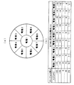
図3は、このようにして決定されるテーブルの例を示す図であり、形状可変ミラー(DFM)22の分割電極配置が図3(a)に示すように9個の電極1〜9からなるとき、照明光を集光するZ方向の位置をサンプルの深さΔZ(μm)とするとき、DFM22の電極1〜9各々に印加すべき電圧値(V)は、図3(b)の表に示すようになり、この図3(b)のテーブルがメモリ62に登録(記憶)される。
FIG. 3 is a diagram showing an example of the table determined in this way, and the divided electrode arrangement of the deformable mirror (DFM) 22 is composed of nine
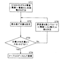
図4に、このようなテーブルを求めるフローの1例を示す。この例は、検出器53で検出される光量が目標値より大きくなるようにDFM22の分割電極1〜9に印加する電圧を求める場合である。まず、ステップST11で、DFM22のそれぞれの電極(電極1〜電極9)に初期電圧を印加する。この初期電圧としては、ΔZの値に対してDFM22に必要とされるパワーが推測されるので、そのパワーに必要な曲率をDFM22に与える電圧とすればよい。次に、ステップST12で、検出器53で光量を測定(検出)する。その後、ステップST13で、その測定された光量が目標値より大きいかどうかを判定する。ここで、検出器53で検出された光量が目標値より大きい場合には、そのΔZの値に対してDFM22の形状が十分であるので、ステップST14で、そのときの電極(電極1〜電極9)に印加された電圧値をテーブルのそのΔZでのデータとして登録する。ステップST13で、測定された光量が目標値より小さい場合には、ステップST15で、評価値を基にパラメータX(この場合は、電極1〜9に印加する電圧)の値を決める。この場合の評価値は、検出器53で検出される光量であり、パラメータXの決定方法としては、最適化問題の解法として知られている勾配法(最急降下法、共役勾配法、ニュートン法等)や直接探査法(DHシンプレックス法、パウエル法、遺伝的アルゴリズム等)の何れかを用いる(非特許文献1、2)。このステップST15は、ステップST13で、検出器53で検出される光量が目標値以上になるか、あるいはその値に変化が見られなくなるまで繰り返される。そして、以上のステップST11〜15をΔZの各値に対して行うことにより、図3のようなテーブルが完成する。
FIG. 4 shows an example of a flow for obtaining such a table. In this example, the voltage applied to the divided
以上の第1の実施例によると、予め参照サンプルを用いてZスキャンするときの形状可変ミラー22の最適な形状が求められているので、測定サンプルへ影響を与えずに精度の高いZスキャンが可能になるメリットがある。
According to the first embodiment described above, since the optimum shape of the
次に、本発明の第2の実施例を説明する。この実施例は、図1のようなレーザ走査型顕微鏡を用いて、参照サンプルを用いないで、測定サンプルに対してZスキャンを行う前に、測定サンプルの予備Zスキャンを行い、実際に獲得される画像を用いて形状可変ミラー(DFM)22の最適形状を決定する例である。 Next, a second embodiment of the present invention will be described. In this embodiment, a laser scanning microscope as shown in FIG. 1 is used, and a reference sample is not used and a Z scan is performed on the measurement sample before performing a Z scan on the measurement sample. This is an example in which the optimum shape of the deformable mirror (DFM) 22 is determined using an image.
まず、全体のフローから説明すると、図5に示すように、まず、ステップST21で、測定したいサンプルを物体O位置に配置して、次のステップST22で、Z位置に対応して予め決められたパワーになる形状(例えば、球面形状)にDFM22を変調(変形)させて測定サンプルをZスキャンし、ステップST23で、検出器53のピンホールを透過した光量データを基に対応するZ位置のスキャン画像を生成する。その後、ステップST24で、獲得された画像を用いてDFM22の最適形状を決定し、Z位置に対する図3と同様のテーブルを再作成し、ステップST25で、測定サンプルに対してそのテーブルに基づいてDFM22の形状を変えながらZスキャンを行い、Z位置各面における測定サンプルの光学像(スキャン画像)を順次取り込んで三次元像を得るものである。上記のステップST21〜ST23が予備Zスキャンを構成している。
First, to explain from the overall flow, as shown in FIG. 5, first, in step ST21, the sample to be measured is arranged at the object O position, and in the next step ST22, it is determined in advance corresponding to the Z position. The
ここで、上記のステップST24で行う、獲得された画像を用いてDFM22の最適形状を決定する過程の1例を以下に説明する。
Here, an example of the process of determining the optimum shape of the
図6にその予備Zスキャンで得られた画像に対する処理過程を示す。予備Zスキャンによって、特定のZ位置での画像Zi が獲得される(図6の最も左に示され、ΔZ=−25μm〜25μmまで5μ間隔だと、11枚i=1〜11)。これらの画像に対して、図6の右中段に示すように、エッジ強調(フィルタ1)や、ボカシ(フィルタ2)、コントラスト強調(フィルタ3)等のフィルタイリング処理を施す。その後、図6の中央下段に示すように、それぞれフィルタリング処理が施されて得られた画像とフィルタリングが行われる前の原画像との比較を行う。比較の方法としては、コントラストの変化量と、画像の中心と周辺の強度差の変化量を計算する。こうして、図6の右下段に示すように、画像Zi に対して各フィルタ1〜3での処理によるコントラストの変化量、及び、中心と周辺の強度差の変化量を評価量として求める。
FIG. 6 shows a process for processing an image obtained by the preliminary Z scan. The image Z i at a specific Z position is acquired by the preliminary Z scan (shown on the leftmost side of FIG. 6, 11 sheets i = 1 to 11 when ΔZ = −25 μm to 25 μm at 5 μ intervals). These images are subjected to filtering processing such as edge enhancement (filter 1), blur (filter 2), contrast enhancement (filter 3), etc., as shown in the middle right part of FIG. Thereafter, as shown in the lower part of the center of FIG. 6, an image obtained by performing the filtering process is compared with the original image before the filtering is performed. As a comparison method, the amount of change in contrast and the amount of change in intensity difference between the center and the periphery of the image are calculated. In this way, as shown in the lower right part of FIG. 6, the amount of contrast change and the amount of change in intensity difference between the center and the periphery of the image Z i due to the processing by the
図7の最も左側に、図6のような処理過程を経て得られた画像Zi に対するフィルタ1〜3によるフィルタイリング処理を施された画像の評価量を示す。この評価量は、次の図8を参照にして説明するバック・プロパゲーション(BP)によるニューラルネットワークを経て、球面収差、コマ収差、非点収差、歪曲収差の各収差量に変換され、その各収差量は図9に示す2次の関数近似を経て、DFM22の予備Zスキャンのときの各電極に加えられる電圧値に対して補正する電圧値に変換される。
The leftmost part of FIG. 7 shows the evaluation amount of the image that has been subjected to the filtering process by the
以上の図6〜図9で示す処理を各Z位置での画像Zi それぞれに施してそれぞれのZ位置に対して得られるDFM22の各電極に加える電圧値が求まり、図3と同様のテーブルが再作成される。ただし、この電圧値は、図5のステップST22でDFM22が所定のパワーになるように変形させた電圧値に対して符号を反転した補正値を加えてなる電圧値である。
The above-described processing shown in FIGS. 6 to 9 is performed on each image Z i at each Z position, and the voltage value to be applied to each electrode of the
図8はフィルタ1〜3によるフィルタイリング処理を施された画像の各評価量からその状態の光学系の球面収差、コマ収差、非点収差、歪曲収差の各収差量を求めるためのニューラルネットワークを示す図である。
FIG. 8 is a neural network for determining the respective aberration amounts of spherical aberration, coma aberration, astigmatism, and distortion aberration of the optical system in that state from the respective evaluation amounts of the image subjected to the filtering process by the
球面収差、コマ収差、非点収差、歪曲収差それぞれが既知の光学系を経た標準的な試料(例えば、テストチャート)の画像をシミュレーションで求める。その画像について、上記のエッジ強調(フィルタ1)や、ボカシ(フィルタ2)、コントラスト強調(フィルタ3)等のフィルタイリング処理を行って、コントラストの変化量及び中心と周辺の強度差の変化量の評価量を求める。これをいくつかの代表的な既知の収差値を持つ複数の光学系を経た画像に対して計算を行い、複数の収差量の組み合わせに対する評価量がシミュレーションで求まることになる。 An image of a standard sample (for example, a test chart) that has passed through an optical system with known spherical aberration, coma, astigmatism, and distortion is obtained by simulation. The image is subjected to filtering processing such as edge enhancement (filter 1), blur (filter 2), contrast enhancement (filter 3), and the like, and the amount of change in contrast and the amount of change in intensity difference between the center and the periphery. The evaluation amount of is calculated. This is calculated for an image having passed through a plurality of optical systems having some typical known aberration values, and an evaluation amount for a combination of a plurality of aberration amounts is obtained by simulation.
この予め求めた評価量と収差量を入力データとして、図8の3層からなるニューラルネットワークにバック・プロパゲーション(BP)により、入力層と中間層の重みw1ij、中間層と出力層の重みw2ijを繰り返し学習させて各ニューロン間の重みw1ij、w2ijが決定される。 Using the evaluation value and aberration amount obtained in advance as input data, the weight w1 ij of the input layer and the intermediate layer, the weight of the intermediate layer and the output layer by back propagation (BP) to the three-layer neural network of FIG. By repeatedly learning w2 ij , the weights w1 ij and w2 ij between the neurons are determined.
このように学習により設定された図8のニューラルネットワークの入力層に、図6で求めた評価量(フィルタ1〜3によるコントラストの変化量と中心と周辺の強度差の変化量)を入力することで、各収差の収差量が出力層に出力されて求まる。
8 is input to the input layer of the neural network of FIG. 8 set by learning in this way (the amount of contrast change by the
以上のようにして、各Z位置での画像Zi を与えた光学系の各収差量が求まったが、この各収差を補正するようにDFM22の形状を変形させなければならない。そのための2次の関数近似式を得なければならない。この過程を図9に示す。
As described above, each aberration amount of the optical system that gives the image Z i at each Z position is obtained. The shape of the
まず、ステップST31において、形状可変ミラー(DFM)22の分割電極個々に印加する電圧と表面形状の関係を予め実験によって求めておく。次いで、ステップST32において、求められたDFM22の表面形状から光学系の各収差(球面収差、コマ収差、非点収差、歪曲収差)それぞれの収差量を光線追跡等の光学シミュレーションにより求める。そして、ステップST33で、各電極に印加する電圧と各収差の収差量との関係を複数組(各電極に印加する電圧を20種類以上変えて)作成しておく。一方、図9の中段に示すように、各電極に印加する電圧値を各収差量の2次の関数と仮定しておく。そして、ステップST34において、ステップST33で得られた各電極に印加する電圧と各収差の収差量との関係をその2次の関数に代入して、2次の関数の係数(a1i,b1ij ,c1i,・・・・,ani,bnij ,cni)を非線形最適化を用いて決定する。この係数が決定すれば、図8のニューラルネットワークを介して求まった各収差の収差量をその2次の関数に代入することにより、DFM22の各電極に印加する補正電圧値を決定することができる。
First, in step ST31, the relationship between the voltage applied to each divided electrode of the deformable mirror (DFM) 22 and the surface shape is obtained in advance by experiments. Next, in step ST32, each aberration amount (spherical aberration, coma aberration, astigmatism, distortion aberration) of the optical system is obtained from the obtained surface shape of the
この補正電圧値の符号を反転して図5のステップST22でDFM22が所定のパワーになるように変形させた電圧値に加えることにより、図3と同様のテーブルが作成される。この作成されたテーブルに基づいて図5に示したステップST25が実行され、測定サンプルにより適合した高精度の三次元像を得ることができるようになる。
The sign of this correction voltage value is inverted and added to the voltage value deformed so that the
次に、本発明の第3の実施例を説明する。この実施例は、図10に示すように、Z方向の予備Zスキャンの間に、図1のレーザ走査型顕微鏡の検出器53の位置にCCDカメラ54を配置し、物体Oとして実際の3次元測定サンプルを配置して、これにレーザ光源11からの光束とは別の図示していないインコヒーレント光源からの励起光55を照射してZ位置毎の蛍光画像を撮像して、実際に獲得される蛍光画像を用いて形状可変ミラー(DFM)22の最適形状を決定する例である。
Next, a third embodiment of the present invention will be described. In this embodiment, as shown in FIG. 10, a CCD camera 54 is arranged at the position of the detector 53 of the laser scanning microscope of FIG. A measurement sample is arranged, and excitation light 55 from an incoherent light source (not shown) different from the light beam from the
まず、全体のフローから説明すると、図11に示すように、まず、ステップST41で、測定したいサンプルを物体O位置に配置して、次のステップST42で、その測定サンプルに励起光55としてインコヒーレントな照明光を用いて照明し(レーザー光源11は動作させない。)、次のステップST43で、Z位置に対応して予め決められたパワーになる形状(例えば、球面形状)にDFM22を変調(変形)させて測定サンプルをZスキャンし(光束走査手段3は動作させない。)、ステップST44で、検出器53の代わりに配置したCCDカメラ54で各Z位置の蛍光画像を獲得する。その後、ステップST45で、獲得された蛍光画像を用いてDFM22の最適形状を決定し、各Z位置に対する図3と同様のテーブルを作成し、ステップST46で、測定サンプルに対してそのテーブルに基づいてDFM22の形状を変えながら通常のZスキャンを行い(レーザー光源11、光束走査手段3は動作させ、図示していないピンホール付きの検出器53で光量を検出する。)、Z位置各面における測定サンプルの光学像(スキャン画像)を順次取り込んで三次元像を得るものである。上記のステップST41〜ST44が予備Zスキャンを構成している。
First, the entire flow will be described. As shown in FIG. 11, first, in step ST41, a sample to be measured is arranged at the object O position, and in the next step ST42, the measurement sample is incoherent as excitation light 55. The illumination light is used for illumination (the
ここで、上記のステップST45で行う、獲得された蛍光画像を用いてDFM22の最適形状を決定する過程の1例を以下に説明する。図12に予備Zスキャンで得られた画像に対する処理過程を示す。図11のステップST44で特定のZ位置での画像Zi が獲得される(図12の最も左に示され、ΔZ=−25μm〜25μmまで5μ間隔だと、11枚i=1〜11)。これらの画像に対して、ステップST51で、それぞれをN×Nの領域に分割し、ステップST52で、個々の領域におけるコントラスト(最大濃度と最小濃度の差)を計算する。各画像Zi に対してN×Nのコントラスト値が求まる。次に、ステップST53で、画像Zi とZ方向に近接するZ位置の画像Zi-1 及び画像Zi+1 との比較を行う。比較としては各領域でのコントラスト値、及び、対応する画素の画素値の差分の平均値を用い、各領域毎に、コントラストの違い及び画素値の違いの平均値が計算される。この2つの量が画像の各領域に対して得られるので、ステップST54で、その相対位置をそのままにして2次元の行列を作成する。同じ領域の2つのスカラー量となるが、コントラストの違いを上に、画素値の違いの平均値を下の段に配置する(図13参照)。次の、ステップST55で、この2次元の行列と予めシミュレーションによって求められた複数の2次元の行列Sl との距離を計算し、この距離の値を評価値とする。ステップST56で、図8と同様のニューラルネットワークを用いて、その評価値をニューラルネットワークの入力層に入力することで、各収差の収差量が出力層に出力されて求まる。そして、ステップST57において、ステップST56で求められた各収差の収差量を同様のニューラルネットワークの入力層に入力することで、DFM22の各電極に印加する電圧が一意的に決定され、図3と同様のテーブルが作成される。
Here, an example of the process of determining the optimum shape of the
図13は、ステップST54での2次元の行列の作成過程と、予めシミュレーションによって複数の2次元の行列Sl を求める過程と、その2つの行列の距離を求める過程と、その距離に基づいて各収差の収差量を出力するニューラルネットワークでの演算過程を示す図である。ステップST61で、N×Nの領域各々に対して、コントラストの違いを上に、画素値の違いの平均値を下の段に配置し、ステップST62で、それらの相対位置をそのままにして2次元のマトリックスxを作成する。一方、ステップST64で、球面収差、コマ収差、非点収差、歪曲収差それぞれが既知の光学系を経た標準的な試料(例えば、テストチャート)の画像をシミュレーションで求める。その画像について、上記の領域分割、各領域毎にコントラストの違い及び画素値の違いの平均値の計算を行い、同様のマトリクス作成過程を経て複数の2次元の行列Sl を用意しておく。そして、ステップST63で、この実際の測定サンプルから得られたマトリックスxとシミュレーションで求められた複数のマトリックスSl の距離を計算する。ここで、マトリックスxとマトリックスSl の距離は、各要素間の差の二乗和のルートで定義される。ステップST65で、得られた距離と収差量を入力データとして、図14の3層からなるニューラルネットワークにバック・プロパゲーション(BP)により、入力層と中間層の重みw1ij、中間層と出力層の重みw2ijを繰り返し学習させて各ニューロン間の重みw1ij、w2ijが決定され、ステップST56で用いるニューラルネットワークが構築される。 Figure 13 is a process of producing a two-dimensional matrix in step ST54, the process of obtaining a plurality of two-dimensional matrix S l in advance by simulation, and the process for obtaining the distance of the two matrices, each based on the distance It is a figure which shows the calculation process in the neural network which outputs the aberration amount of an aberration. In step ST61, for each of the N × N areas, the contrast difference is placed on the upper side, and the average value of the pixel value differences is arranged on the lower stage. The matrix x is created. On the other hand, in step ST64, an image of a standard sample (for example, a test chart) that has passed through an optical system with known spherical aberration, coma aberration, astigmatism, and distortion is obtained by simulation. With respect to the image, the above-described area division, the average value of the difference in contrast and the difference in pixel value for each area are calculated, and a plurality of two-dimensional matrices S 1 are prepared through the same matrix creation process. Then, in step ST63, it computes the distances of the plurality of matrices S l obtained by matrix x and simulation obtained from the actual measurement sample. The distance matrix x and the matrix S l is defined by the root of the square sum of the differences between elements. In step ST65, as input data the distance and aberration amount obtained by the back-propagation (BP) to the neural network consisting of three layers of 14, the weight w1 ij of input layer and the intermediate layer, the intermediate layer output layer by repeating the weighting w2 ij learned weighted- w1 ij, w2 ij is determined between the neurons, neural networks used in step ST56 is constructed.
また、形状可変ミラー(DFM)22の分割電極個々に印加する電圧と表面形状の関係を予め実験によって求めて、求められたDFM22の表面形状から光学系の各収差(球面収差、コマ収差、非点収差、歪曲収差)それぞれの収差量を光線追跡等の光学シミュレーションにより求め、その求めた収差量と分割電極個々の印加電圧との関係から、同様にして、ステップST57で用いるニューラルネットワークも構築される。
Further, the relationship between the voltage applied to each of the divided electrodes of the deformable mirror (DFM) 22 and the surface shape is obtained in advance by experiments, and each aberration (spherical aberration, coma aberration, non-uniformity) of the optical system is obtained from the obtained surface shape of the
以上のようにして、構築された2つのニューラルネットワークを用いて、各Z位置に対する図3と同様のテーブルが作成される。この作成されたテーブルに基づいて図11に示したステップST46が実行され、測定サンプルにより適合した高精度の三次元像を得ることができるようになる。 Using the two neural networks constructed as described above, a table similar to FIG. 3 is created for each Z position. Based on the created table, step ST46 shown in FIG. 11 is executed, and a highly accurate three-dimensional image more suitable for the measurement sample can be obtained.
この実施例では、実際の測定サンプルに対して予備Zスキャンの際にXYスキャンの必要がないので、高速に撮影が可能となり、測定サンプルの褪色の影響を少なくできるメリットがある。 In this embodiment, since an XY scan is not required for the actual measurement sample during the preliminary Z scan, it is possible to take a high-speed image and there is an advantage that the influence of the fading of the measurement sample can be reduced.
次に、本発明の第4の実施例を説明する。この実施例は、図15に示すように、図1のレーザ走査型顕微鏡の配置に対して検出器53に入射する測定光束を分光する分光手段(例えば回折格子)56を備えている点で異なっており、この分光手段56の回折角を変えながら走査することで、複数の分光画像が得られる点に特徴があるものである。 Next, a fourth embodiment of the present invention will be described. As shown in FIG. 15, this embodiment is different from the arrangement of the laser scanning microscope in FIG. 1 in that a spectroscopic means (for example, a diffraction grating) 56 that splits a measurement light beam incident on the detector 53 is provided. In addition, a characteristic is that a plurality of spectral images can be obtained by scanning while changing the diffraction angle of the spectroscopic means 56.
まず、全体のフローから説明すると、図16に示すように、まず、ステップST71で、測定サンプルを物体O位置に配置して、次のステップST72で、Z位置に対応して予め決められたパワーになる形状(例えば、球面形状)にDFM22を変調(変形)させて測定サンプルをZスキャンし、ステップST73で、検出器53のピンホールを透過した光量データを基に対応するZ位置のスキャン画像を生成する。その後、ステップST74で、獲得された画像を用いてDFM22の最適形状を決定し、Z位置に対する図3と同様のテーブルを作成し、ステップST75で、測定サンプルに対してそのテーブルに基づいてDFM22の形状を変えながらZスキャンを行い、Z位置各面における測定サンプルの光学像(スキャン画像)を順次取り込んで三次元像を得るものである。上記のステップST71〜ST73が予備Zスキャンを構成している。
First, the overall flow will be described. As shown in FIG. 16, first, in step ST71, the measurement sample is arranged at the object O position, and in the next step ST72, the power determined in advance corresponding to the Z position. The
ここで、上記のステップST74で行う、獲得された画像を用いてDFM22の最適形状を決定する過程の1例を以下に説明する。
Here, an example of the process of determining the optimum shape of the
図17にその予備Zスキャンで得られた画像に対する処理過程を示す。ステップST81のように、上記予備Zスキャンによって、特定のZ位置での画像Zi が分光データ分だけ獲得される。例えば、波長として、λ1=500nm、λ2=600nm、λ3=700nmの3種類について画像が獲得される。この3つの画像のそれぞれについて、ステップST82で、それぞれをN×Nの領域に分割し、各領域での獲得された複数の画像のコントラストの違い、具体的には、例えばコントラスト値の分散を求める。また、各領域での獲得された複数の画像の対応画素間の画素値の違い、具体的には、例えば画素値の分散を求める。これらの値を評価量とする。ステップST83で、図8と同様のニューラルネットワークを用いて、その評価値をニューラルネットワークの入力層に入力することで、各収差の収差量が出力層に出力されて求まる。そして、ステップST84において、ステップST83で求められた各収差の収差量を同様のニューラルネットワークの入力層に入力することで、DFM22の各電極に印加する電圧が一意的に決定され、図3と同様のテーブルが作成される。
FIG. 17 shows a process for processing an image obtained by the preliminary Z scan. As in step ST81, an image Z i at a specific Z position is acquired for the spectral data by the preliminary Z scan. For example, images are acquired for three types of wavelengths, λ1 = 500 nm, λ2 = 600 nm, and λ3 = 700 nm. In step ST82, each of the three images is divided into N × N areas, and the difference in contrast between the plurality of acquired images in each area, specifically, for example, the variance of contrast values is obtained. . Further, a difference in pixel values between corresponding pixels of a plurality of acquired images in each region, specifically, for example, dispersion of pixel values is obtained. These values are used as evaluation amounts. In step ST83, using the same neural network as in FIG. 8, the evaluation value is input to the input layer of the neural network, whereby the aberration amount of each aberration is output to the output layer. Then, in step ST84, by inputting the aberration amount of each aberration obtained in step ST83 to the input layer of the same neural network, the voltage to be applied to each electrode of the
図18は、ステップST83で用いるニューラルネットワークの構成を示す図であり、このニューラルネットワークを構築するために、球面収差、コマ収差、非点収差、歪曲収差、色収差それぞれが既知の光学系を経た標準的な試料(例えば、テストチャート)の画像をシミュレーションで求める。その画像について、上記の領域分割、各領域毎に、コントラストの違い、対応画素間の画素値の違いの計算を行い、同様の評価値作成過程を経て複数の評価値と収差量を入力データとして、3層からなるニューラルネットワークにバック・プロパゲーション(BP)により、入力層と中間層の重みw1ij、中間層と出力層の重みw2ijを繰り返し学習させて各ニューロン間の重みw1ij、w2ijが決定され、ステップST83で用いるニューラルネットワークが構築される。 FIG. 18 is a diagram showing a configuration of a neural network used in step ST83. In order to construct this neural network, a standard in which spherical aberration, coma aberration, astigmatism, distortion aberration, and chromatic aberration are passed through known optical systems. An image of a typical sample (for example, a test chart) is obtained by simulation. With respect to the image, the above-described region division, the difference in contrast for each region, the difference in pixel value between corresponding pixels are calculated, and a plurality of evaluation values and aberration amounts are input as input data through a similar evaluation value creation process. Weights w1 ij , w2 between neurons by repeatedly learning a weight w1 ij of the input layer and the intermediate layer and a weight w2 ij of the intermediate layer and the output layer by back propagation (BP) in a neural network consisting of three layers ij is determined, and a neural network used in step ST83 is constructed.
また、形状可変ミラー(DFM)22の分割電極個々に印加する電圧と表面形状の関係を予め実験によって求めて、求められたDFM22の表面形状から光学系の各収差(球面収差、コマ収差、非点収差、歪曲収差、色収差)それぞれの収差量を光線追跡等の光学シミュレーションにより求め、その求めた収差量と分割電極個々の印加電圧との関係から、同様にして、ステップST84で用いるニューラルネットワークも構築される。
Further, the relationship between the voltage applied to each of the divided electrodes of the deformable mirror (DFM) 22 and the surface shape is obtained in advance by experiments, and each aberration (spherical aberration, coma aberration, non-uniformity) of the optical system is obtained from the obtained surface shape of the
以上のようにして、構築された2つのニューラルネットワークを用いて、各Z位置に対する図3と同様のテーブルが作成される。この作成されたテーブルに基づいて、図16に示したステップST75が実行され、測定サンプルにより適合した高精度の三次元像を得ることができるようになる。 Using the two neural networks constructed as described above, a table similar to FIG. 3 is created for each Z position. Based on the created table, step ST75 shown in FIG. 16 is executed, and a highly accurate three-dimensional image more suitable for the measurement sample can be obtained.
この実施例では、分光データを用いることで色収差を考慮した高精度の三次元像を得ることができるようになる。 In this embodiment, it is possible to obtain a highly accurate three-dimensional image in consideration of chromatic aberration by using spectral data.
なお、以上の第2〜第4の実施例において、予備Zスキャンを行う際に用いるDFM22の形状変形のための制御データ(テーブル)(図5のステップST22、図11のステップST43、図16のステップST72)は、第1の実施例の参照サンプルを用いて決定したテーブルを用いるようにしてももちろんよい。この場合は、予備Zスキャンでも精度の高い測定が可能となり、最適化がより容易にできるようになる。
In the second to fourth embodiments described above, control data (table) for deforming the shape of the
また、例えば分光データを参照にして、そのテーブルを作りなおすようにしてもよい。このようにテーブルを作りなおすことで、DFM22の時系変化にも対応可能となる。
Further, for example, the table may be recreated with reference to the spectral data. By recreating the table in this way, it is possible to cope with a time-series change of the
以上、本発明の走査型光学顕微鏡をいくつかの実施例に基づいて説明してきたが、それらの実施例は種々の設計変更が可能である。 As described above, the scanning optical microscope of the present invention has been described on the basis of several embodiments. However, these embodiments can be variously modified.
2…波面変換素子
3…光束走査手段
4…対物レンズ
11…レーザー光源
12…コリメータレンズ
22…形状可変ミラー(DFM)
51…ダイクロイックミラー
52…集光レンズ
53…検出器
54…CCDカメラ
55…励起光
56…分光手段
61…コントローラ
62…メモリ
63…フィードバックループ
71…第三のリレー光学系
72…第二のリレー光学系
73…第一のリレーレンズ
74…結像レンズ
81…平面ミラー
82…3次元構造の蛍光体
83…蛍光層
O…対象物(物体)
KPL…短パルス・レーザー
PCU…プリチャープ・ユニット
ST1、ST2、ST3…ビーム・スプリッター
AD1、AD2…アダプティブミラー
DBS…ビーム・スプリッター
SL、TL…光学部品
SP…ミラー
OL…対物レンズ
L…レンズ
PH…ピンホール
EF…フィルター
PMT…検出器
2 ... Wavefront conversion element 3 ... Light beam scanning means 4 ...
51 ... Dichroic mirror 52 ... Condensing lens 53 ... Detector 54 ... CCD camera 55 ... Excitation light 56 ... Spectroscopic means 61 ... Controller 62 ...
KPL ... Short pulse laser PCU ... Pre-chirp units ST1, ST2, ST3 ... Beam splitter AD1, AD2 ... Adaptive mirror DBS ... Beam splitter SL, TL ... Optical component SP ... Mirror OL ... Objective lens L ... Lens PH ... Pin Hall EF ... Filter PMT ... Detector
Claims (6)
前記制御装置は、照明光の集光位置を変更するのに必要な前記波面変換素子を変調するための制御データを備えており、その制御データに基づいて前記波面変換素子を変調して照明光の集光位置を変更することを特徴とする走査型光学顕微鏡。 A light source, a wavefront conversion element that applies arbitrary wavefront conversion to illumination light emitted from the light source, a control device that controls the wavefront conversion element, and the illumination light after wavefront conversion emitted from the wavefront conversion element in directions orthogonal to each other In a scanning optical microscope comprising: a light beam scanning unit that scans; an objective lens that condenses illumination light whose traveling direction has been changed by the light beam scanning unit; and a detector that detects signal light emitted from the object.
The control device includes control data for modulating the wavefront conversion element necessary for changing the condensing position of illumination light, and modulates the wavefront conversion element based on the control data to illuminate light. A scanning optical microscope characterized by changing a light condensing position.
Priority Applications (1)
| Application Number | Priority Date | Filing Date | Title |
|---|---|---|---|
| JP2004108803A JP4615886B2 (en) | 2004-04-01 | 2004-04-01 | Scanning optical microscope |
Applications Claiming Priority (1)
| Application Number | Priority Date | Filing Date | Title |
|---|---|---|---|
| JP2004108803A JP4615886B2 (en) | 2004-04-01 | 2004-04-01 | Scanning optical microscope |
Publications (2)
| Publication Number | Publication Date |
|---|---|
| JP2005292538A true JP2005292538A (en) | 2005-10-20 |
| JP4615886B2 JP4615886B2 (en) | 2011-01-19 |
Family
ID=35325511
Family Applications (1)
| Application Number | Title | Priority Date | Filing Date |
|---|---|---|---|
| JP2004108803A Expired - Fee Related JP4615886B2 (en) | 2004-04-01 | 2004-04-01 | Scanning optical microscope |
Country Status (1)
| Country | Link |
|---|---|
| JP (1) | JP4615886B2 (en) |
Cited By (15)
| Publication number | Priority date | Publication date | Assignee | Title |
|---|---|---|---|---|
| JP2008026643A (en) * | 2006-07-21 | 2008-02-07 | Olympus Corp | Laser scanning microscope |
| WO2009153919A1 (en) * | 2008-06-17 | 2009-12-23 | 株式会社ニコン | Microscope device and microscope device control program |
| JP2011099986A (en) * | 2009-11-06 | 2011-05-19 | Olympus Corp | Laser microscope using phase-modulation-type spatial light modulator |
| JP2012517035A (en) * | 2009-02-04 | 2012-07-26 | エコール ポリテクニック | Method and device for signal acquisition in laser scanning microscopy |
| JP2013160893A (en) * | 2012-02-03 | 2013-08-19 | Olympus Corp | Microscope and aberration correction method for the same |
| JP2015031813A (en) * | 2013-08-02 | 2015-02-16 | 株式会社ニコン | Method for setting compensation optical element and microscope |
| JP2015505620A (en) * | 2012-01-24 | 2015-02-23 | カール ツァイス マイクロスコピー ゲーエムベーハーCarl Zeiss Microscopy Gmbh | Microscope and method for high resolution 3D fluorescence microscopy |
| JP2015510150A (en) * | 2012-02-29 | 2015-04-02 | アジレント・テクノロジーズ・インクAgilent Technologies, Inc. | Software-defined microscope |
| JP5733670B2 (en) * | 2010-04-05 | 2015-06-10 | 国立大学法人大阪大学 | Observation apparatus and observation method |
| JP2015129972A (en) * | 2015-03-31 | 2015-07-16 | 株式会社ニコン | Microscope system |
| JP2016181010A (en) * | 2016-07-15 | 2016-10-13 | 株式会社ニコン | Microscope system |
| US9507138B2 (en) | 2010-10-20 | 2016-11-29 | Nikon Corporation | Microscope system |
| US9563048B2 (en) | 2011-04-12 | 2017-02-07 | Nikon Corporation | Microscope system, server, and program providing intensity distribution of illumination light suitable for observation |
| JP2019148655A (en) * | 2018-02-26 | 2019-09-05 | セイコーエプソン株式会社 | Spectroscopic device, temperature characteristic derivation device, spectroscopic system, and temperature characteristic derivation method |
| US11714274B2 (en) | 2017-11-28 | 2023-08-01 | Nikon Corporation | Microscope system |
Citations (8)
| Publication number | Priority date | Publication date | Assignee | Title |
|---|---|---|---|---|
| JPH11101942A (en) * | 1997-08-01 | 1999-04-13 | Carl Zeiss Jena Gmbh | Adaptive optical device for microscope |
| JP2000121945A (en) * | 1998-10-14 | 2000-04-28 | Olympus Optical Co Ltd | Condenser device |
| JP2001290081A (en) * | 2000-03-15 | 2001-10-19 | Leica Microsystems Heidelberg Gmbh | Sample irradiation equipment |
| JP2002196246A (en) * | 2000-12-26 | 2002-07-12 | Olympus Optical Co Ltd | Scanning optical microscope |
| JP2003057555A (en) * | 2001-08-13 | 2003-02-26 | Olympus Optical Co Ltd | Scanning laser microscope |
| JP2003315650A (en) * | 2002-04-26 | 2003-11-06 | Olympus Optical Co Ltd | Optical device |
| JP2004053922A (en) * | 2002-07-19 | 2004-02-19 | Olympus Corp | Method for correcting scanned image and program therefor, and laser scanning type microscope |
| JP2004317265A (en) * | 2003-04-15 | 2004-11-11 | Institute Of Tsukuba Liaison Co Ltd | Wave front evaluation method and device using information entropy |
-
2004
- 2004-04-01 JP JP2004108803A patent/JP4615886B2/en not_active Expired - Fee Related
Patent Citations (8)
| Publication number | Priority date | Publication date | Assignee | Title |
|---|---|---|---|---|
| JPH11101942A (en) * | 1997-08-01 | 1999-04-13 | Carl Zeiss Jena Gmbh | Adaptive optical device for microscope |
| JP2000121945A (en) * | 1998-10-14 | 2000-04-28 | Olympus Optical Co Ltd | Condenser device |
| JP2001290081A (en) * | 2000-03-15 | 2001-10-19 | Leica Microsystems Heidelberg Gmbh | Sample irradiation equipment |
| JP2002196246A (en) * | 2000-12-26 | 2002-07-12 | Olympus Optical Co Ltd | Scanning optical microscope |
| JP2003057555A (en) * | 2001-08-13 | 2003-02-26 | Olympus Optical Co Ltd | Scanning laser microscope |
| JP2003315650A (en) * | 2002-04-26 | 2003-11-06 | Olympus Optical Co Ltd | Optical device |
| JP2004053922A (en) * | 2002-07-19 | 2004-02-19 | Olympus Corp | Method for correcting scanned image and program therefor, and laser scanning type microscope |
| JP2004317265A (en) * | 2003-04-15 | 2004-11-11 | Institute Of Tsukuba Liaison Co Ltd | Wave front evaluation method and device using information entropy |
Cited By (23)
| Publication number | Priority date | Publication date | Assignee | Title |
|---|---|---|---|---|
| JP2008026643A (en) * | 2006-07-21 | 2008-02-07 | Olympus Corp | Laser scanning microscope |
| WO2009153919A1 (en) * | 2008-06-17 | 2009-12-23 | 株式会社ニコン | Microscope device and microscope device control program |
| US20110141260A1 (en) * | 2008-06-17 | 2011-06-16 | Nikon Corporation | Microscope apparatus and storage medium storing microscope apparatus control program |
| US10241314B2 (en) | 2008-06-17 | 2019-03-26 | Nikon Corporation | Microscope apparatus and storage medium storing microscope apparatus control program |
| JP5381984B2 (en) * | 2008-06-17 | 2014-01-08 | 株式会社ニコン | Microscope device and microscope device control program |
| US9261699B2 (en) | 2008-06-17 | 2016-02-16 | Nikon Corporation | Microscope apparatus and storage medium storing microscope apparatus control program |
| JP2012517035A (en) * | 2009-02-04 | 2012-07-26 | エコール ポリテクニック | Method and device for signal acquisition in laser scanning microscopy |
| JP2011099986A (en) * | 2009-11-06 | 2011-05-19 | Olympus Corp | Laser microscope using phase-modulation-type spatial light modulator |
| US9158100B2 (en) | 2009-11-06 | 2015-10-13 | Olympus Corporation | Laser microscope using phase-modulation type spatial light modulator |
| JP5733670B2 (en) * | 2010-04-05 | 2015-06-10 | 国立大学法人大阪大学 | Observation apparatus and observation method |
| US9507138B2 (en) | 2010-10-20 | 2016-11-29 | Nikon Corporation | Microscope system |
| US10451860B2 (en) | 2010-10-20 | 2019-10-22 | Nikon Corporation | Microscope system |
| US9563048B2 (en) | 2011-04-12 | 2017-02-07 | Nikon Corporation | Microscope system, server, and program providing intensity distribution of illumination light suitable for observation |
| JP2015505620A (en) * | 2012-01-24 | 2015-02-23 | カール ツァイス マイクロスコピー ゲーエムベーハーCarl Zeiss Microscopy Gmbh | Microscope and method for high resolution 3D fluorescence microscopy |
| US9885860B2 (en) | 2012-01-24 | 2018-02-06 | Carl Zeiss Microscopy Gmbh | Microscope and method for high-resolution 3D fluorescence microscopy |
| JP2013160893A (en) * | 2012-02-03 | 2013-08-19 | Olympus Corp | Microscope and aberration correction method for the same |
| JP2015510150A (en) * | 2012-02-29 | 2015-04-02 | アジレント・テクノロジーズ・インクAgilent Technologies, Inc. | Software-defined microscope |
| JP2015031813A (en) * | 2013-08-02 | 2015-02-16 | 株式会社ニコン | Method for setting compensation optical element and microscope |
| JP2015129972A (en) * | 2015-03-31 | 2015-07-16 | 株式会社ニコン | Microscope system |
| JP2016181010A (en) * | 2016-07-15 | 2016-10-13 | 株式会社ニコン | Microscope system |
| US11714274B2 (en) | 2017-11-28 | 2023-08-01 | Nikon Corporation | Microscope system |
| JP2019148655A (en) * | 2018-02-26 | 2019-09-05 | セイコーエプソン株式会社 | Spectroscopic device, temperature characteristic derivation device, spectroscopic system, and temperature characteristic derivation method |
| JP7043885B2 (en) | 2018-02-26 | 2022-03-30 | セイコーエプソン株式会社 | Spectroscopic device, temperature characteristic derivation device, spectroscopic system, spectroscopic method, and temperature characteristic derivation method |
Also Published As
| Publication number | Publication date |
|---|---|
| JP4615886B2 (en) | 2011-01-19 |
Similar Documents
| Publication | Publication Date | Title |
|---|---|---|
| JP4615886B2 (en) | Scanning optical microscope | |
| US10247934B2 (en) | Method for examining a specimen by means of light sheet microscopy | |
| US9477091B2 (en) | Multi-dimensional imaging using multi-focus microscopy | |
| CN104620163B (en) | Light modulation control method, control program, control device and laser irradiation device | |
| EP1744194B1 (en) | Laser scanning microscope and image acquiring method of laser scanning microscope | |
| US10802256B2 (en) | Multifocal scanning fluorescence microscope | |
| JP5381984B2 (en) | Microscope device and microscope device control program | |
| CN107850765B (en) | Method and assembly for beam shaping and optical layer microscopy | |
| CA2267431A1 (en) | Microscope with adaptive optics | |
| JP2008170969A (en) | Microscope objective and fluorescent observation apparatus therewith | |
| JP2018517178A (en) | Configuration and method for light sheet microscopy | |
| JPH11326860A (en) | Wave front converting element and laser scanner using it | |
| JP7481351B2 (en) | Wavefront analyzer, fluorescence microscopic imaging system and method for microscopic imaging of an object - Patents.com | |
| CN114341622A (en) | Super-resolution imaging system and method, biological sample identification system and method, nucleic acid sequencing imaging system and method, and nucleic acid identification system and method | |
| US20100264294A1 (en) | Multi-focal spot generator and multi-focal multi-spot scanning microscope | |
| JPH06506538A (en) | Confocal imaging device for visible and ultraviolet light | |
| JP2008026643A (en) | Laser scanning microscope | |
| WO2017169597A1 (en) | Image acquisition device and image acquisition method | |
| JP4874012B2 (en) | Laser scanning microscope and image acquisition method of laser scanning microscope | |
| CN112912782B (en) | Confocal laser scanning microscope configured to generate line focus | |
| WO2017178538A1 (en) | Method and arrangement for identifying optical aberrations | |
| JP2005345761A (en) | Scanning optical microscopic device and method for restoring object image from the scanning optical microscopic image | |
| US20230236408A1 (en) | A method for obtaining an optically-sectioned image of a sample, and a device suitable for use in such a method | |
| JP6770616B2 (en) | Image acquisition device, image acquisition method, and spatial optical modulation unit | |
| EP3945358B1 (en) | Oblique plane microscope and method for operating the same |
Legal Events
| Date | Code | Title | Description |
|---|---|---|---|
| A621 | Written request for application examination |
Free format text: JAPANESE INTERMEDIATE CODE: A621 Effective date: 20070326 |
|
| A977 | Report on retrieval |
Free format text: JAPANESE INTERMEDIATE CODE: A971007 Effective date: 20100706 |
|
| A131 | Notification of reasons for refusal |
Free format text: JAPANESE INTERMEDIATE CODE: A131 Effective date: 20100714 |
|
| A521 | Written amendment |
Free format text: JAPANESE INTERMEDIATE CODE: A523 Effective date: 20100825 |
|
| TRDD | Decision of grant or rejection written | ||
| A01 | Written decision to grant a patent or to grant a registration (utility model) |
Free format text: JAPANESE INTERMEDIATE CODE: A01 Effective date: 20100929 |
|
| A01 | Written decision to grant a patent or to grant a registration (utility model) |
Free format text: JAPANESE INTERMEDIATE CODE: A01 |
|
| A61 | First payment of annual fees (during grant procedure) |
Free format text: JAPANESE INTERMEDIATE CODE: A61 Effective date: 20101021 |
|
| R151 | Written notification of patent or utility model registration |
Ref document number: 4615886 Country of ref document: JP Free format text: JAPANESE INTERMEDIATE CODE: R151 |
|
| FPAY | Renewal fee payment (event date is renewal date of database) |
Free format text: PAYMENT UNTIL: 20131029 Year of fee payment: 3 |
|
| S531 | Written request for registration of change of domicile |
Free format text: JAPANESE INTERMEDIATE CODE: R313531 |
|
| R350 | Written notification of registration of transfer |
Free format text: JAPANESE INTERMEDIATE CODE: R350 |
|
| R250 | Receipt of annual fees |
Free format text: JAPANESE INTERMEDIATE CODE: R250 |
|
| LAPS | Cancellation because of no payment of annual fees |


