JP2005102702A - Recombinant sendai virus - Google Patents
Recombinant sendai virus Download PDFInfo
- Publication number
- JP2005102702A JP2005102702A JP2004343685A JP2004343685A JP2005102702A JP 2005102702 A JP2005102702 A JP 2005102702A JP 2004343685 A JP2004343685 A JP 2004343685A JP 2004343685 A JP2004343685 A JP 2004343685A JP 2005102702 A JP2005102702 A JP 2005102702A
- Authority
- JP
- Japan
- Prior art keywords
- protein
- sendai virus
- rna
- virus
- dna
- Prior art date
- Legal status (The legal status is an assumption and is not a legal conclusion. Google has not performed a legal analysis and makes no representation as to the accuracy of the status listed.)
- Withdrawn
Links
Images


Abstract
Description
本発明は、組換え体センダイウイルスとその製造方法に関する。 The present invention relates to a recombinant Sendai virus and a method for producing the same.
センダイウイルス(Sendai virus)は、HVJ(Hemagglutinating virus of Japan)とも呼ばれ、パラミクソウイルス科(Paramyxoviridae)、パラミクソウイルス属(Paramyxovirus)に属するパラインフルエンザウイルス1型に分類される。
Sendai virus is also called HVJ (Hemagglutinating virus of Japan), and is classified into
センダイウイルス粒子は多形性であり、直径150〜200nmのエンベロープを有し、中に翻訳の鋳型とはならないゲノムRNA(以下「(−)鎖RNA」と称する。)を有する。センダイウイルスは、歴史的に見ても産業上有用なウイルスとして知られており、とくに細胞のヘテロカリオンや雑種細胞の作製、すなわち細胞融合に広く利用されている。また、膜融合性リポソームの材料として、遺伝子治療用のベクターとしても開発が進められている。さらには、各種インターフェロンの誘導剤としてもセンダイウイルスは利用されている。 Sendai virus particles are polymorphic, have an envelope with a diameter of 150 to 200 nm, and have genomic RNA (hereinafter referred to as “(−) strand RNA”) that does not serve as a template for translation. Sendai virus is historically known as an industrially useful virus, and is particularly widely used for the production of cell heterokaryons and hybrid cells, that is, cell fusion. In addition, as a material for membrane-fusible liposomes, development as a vector for gene therapy is also in progress. Furthermore, Sendai virus is also used as an inducer for various interferons.
ゲノム核酸の形態による分類では、センダイウイルスは、RNAウイルスの、(−)鎖RNAウイルスの、(−)1本鎖RNAウイルスグループに属する。RNAウイル スは、dsRNAウイルス(double stranded RNA virus)、(+)鎖RNAウイルスお よび(−)鎖RNAウイルスの3者に分類される。dsRNAウイルスグループには、レオウイルス、ロタウイルス、植物レオウイルス等があり、分節型の複数の線状dsRNAゲノムを有している。(+)鎖RNAウイルスには、ポリオウイルス、シンドビスウイルス、セムリキ森林ウイルス、日本脳炎ウイルス等があり、1本の(+)鎖RNAをゲノムとして有しており、このRNAゲノムは同時にmRNAとしても機能し、複製や粒子形成に必要な蛋白質を宿主細胞の翻訳機能に依存して生産することができる。言い換えれば、(+)鎖RNAウイルスが有するゲノムRNA自体が伝播力を有する。なお、本明細書において「伝播力」とは、「感染や人工的な手法で核酸が細胞内に導入された後、細胞内に存在する該核酸が複製後、感染性粒子またはそれに準ずる複合体を形成し、別の細胞に次々と伝播することのできる能力」を言う。(+)鎖RNAウイルスに分類されるシンドビスウイルスや(−)鎖RNAウイルスに分類されるセンダイウイルスは、感染能と伝播力とを有するが、パルボウイルス科に分類されるアデノ随伴ウイルス(Adeno-associated virus)は、感染能を有するが、伝播力を有しない(ウイルス粒子が形成されるためには、アデノウイルスの同時感染が必要である)。また、試験管内で人工的に転写されたシンドビスウイルス由来の(+)鎖RNAは伝播力を有する(細胞内に導入されるとウ イルス粒子を形成する)が、試験管内で人工的に転写されたセンダイウイルスRNAは(+)鎖、(−)鎖ともに伝播力を有しない(細胞内に導入されてもウイルス粒子を形成しない)。 In classification according to the form of genomic nucleic acid, Sendai virus belongs to the group of (-) single-stranded RNA viruses of (-) strand RNA viruses of RNA viruses. RNA viruses are classified into three groups: dsRNA virus (double stranded RNA virus), (+) strand RNA virus, and (-) strand RNA virus. The dsRNA virus group includes reovirus, rotavirus, plant reovirus, and the like, and has a plurality of segmented linear dsRNA genomes. The (+) strand RNA viruses include poliovirus, Sindbis virus, Semliki Forest virus, Japanese encephalitis virus, etc., and have one (+) strand RNA as a genome, and this RNA genome is simultaneously converted into mRNA. Can function, and can produce proteins necessary for replication and particle formation depending on the translation function of the host cell. In other words, the genomic RNA itself of the (+) strand RNA virus has the ability to propagate. In this specification, “propagating power” means “infectious particles or complex equivalents thereof after the nucleic acid is introduced into the cell by infection or an artificial technique and then replicated in the cell. "The ability to form and propagate one after another to another cell". Sindbis virus classified as (+)-strand RNA virus and Sendai virus classified as (-)-strand RNA virus have infectivity and transmission ability, but adeno-associated virus (Adeno) classified as parvoviridae -associated virus) is infectious, but has no transmission power (adenovirus co-infection is required for virus particles to form). In addition, Sindbis virus-derived (+)-strand RNA artificially transcribed in vitro has a propagating power (forms virus particles when introduced into cells), but is artificially transcribed in vitro. Sendai virus RNA thus produced has no ability to propagate (+) and (−) strands (does not form virus particles when introduced into cells).
近年では遺伝子治療用のベクターとしてウイルス由来のものが用いられている。ウイルスをベクターとして利用するためには、ウイルス粒子の再構成のための手法が確立している必要がある。(「ウイルス粒子の再構成」とは、ウイルスゲノムの核酸を人工的に作製し、試験管内または細胞内において、もとのウイルスまたは組換え体ウイルスを作製することである。)外来性遺伝子をウイルスベクターに導入するためには、遺伝子操作によって外来性遺伝子を組み込んだウイルスゲノムからウイルス粒子が再構成されなくてはならないからである。ウイルスの再構成技術が確立されれば、ウイルスに所望の外来性遺伝子を導入したり、ウイルスの所望の遺伝子を欠失させたり、不活化させたりしたウイルスを作製することが可能となる。 In recent years, vectors derived from viruses have been used as vectors for gene therapy. In order to use a virus as a vector, it is necessary to establish a method for reconstructing virus particles. ("Reconstruction of viral particles" means to artificially create viral genome nucleic acid and to produce the original virus or recombinant virus in vitro or in cells.) This is because the virus particles must be reconstructed from the virus genome into which the foreign gene has been incorporated by genetic manipulation in order to introduce it into the virus vector. If a virus reconstitution technique is established, it is possible to produce a virus in which a desired foreign gene is introduced into the virus, a desired gene of the virus is deleted, or inactivated.
また、ウイルスの再構成系が構築され、ウイルスの遺伝子操作が可能となれば、ウイルスの機能を遺伝学的に解析する大きなツールとなることは明白である。ウイルス機能の遺伝学的解析は、疾病の予防、治療等の医学的見地からきわめて重要である。例えば、ウイルス核酸の複製メカニズムが解明されれば、その宿主細胞内の核酸の複製機構との差を利用して、宿主細胞にダメージの少ない、核酸の複製を作用点とした抗ウイルス剤を開発することが可能である。また、ウイルス遺伝子のコードする蛋白質がどのような機能を有するかを解明することにより、ウイルス粒子感染能や、ウイルス粒子形成能に関わる蛋白質をターゲットとした抗ウイルス剤を開発することもできよう。また、膜融合能に関わる遺伝子を改良することにより、より優れた膜融合性リポソームを作製し、遺伝子治療用のベクターとして使用することが可能となることが期待できる。また、インターフェロンに代表されるように、ウイルスに感染することにより宿主遺伝子のウイルス抵抗性に関わる遺伝子が活性化され、ウイルス抵抗性を示す場合もある。このような宿主遺伝子の活性化に関しても、ウイルス機能の遺伝学的解析により重要な知見が得られるであろう。 In addition, if a virus reconstitution system is constructed and genetic manipulation of the virus becomes possible, it will be a great tool for genetic analysis of the virus function. Genetic analysis of viral functions is extremely important from a medical standpoint such as prevention and treatment of diseases. For example, if the replication mechanism of viral nucleic acid is elucidated, the development of an antiviral agent based on the replication of nucleic acid that causes less damage to the host cell by utilizing the difference from the replication mechanism of the nucleic acid in the host cell. Is possible. In addition, by elucidating what functions the protein encoded by the viral gene has, it would be possible to develop an antiviral agent targeting a protein involved in virus particle infectivity and virus particle formation ability. Moreover, it can be expected that by improving a gene related to membrane fusion ability, a better membrane fusion liposome can be prepared and used as a gene therapy vector. In addition, as represented by interferon, infecting a virus activates a gene related to the virus resistance of a host gene and may exhibit virus resistance. With regard to activation of such host genes, important knowledge will be obtained by genetic analysis of virus functions.
DNAをゲノム核酸とするDNAウイルスの再構成は比較的早くから行なわれており、例えば、SV40(J. Exp. Cell Res.,43,415-425(1983))のように、精製したゲノムDNAそのものをサルの細胞に導入することにより行なうことが可能である。 Reconstitution of DNA viruses using DNA as a genomic nucleic acid has been carried out relatively early. For example, purified genomic DNA itself such as SV40 (J. Exp. Cell Res., 43, 415-425 (1983)) It is possible to carry out by introducing into the cells.
RNAをゲノム核酸とするRNAウイルスの再構成は、(+)鎖RNAウイルスにおいて開発が先行した。この理由は、ゲノムRNAが、同時にmRNAとして機能するから である。例えば、ポリオウイルスでは、精製したRNA自体が伝播力を有すること が、すでに1959年に報告されている(Journal of Experimental Medicine,110,65-89(1959))。また、セムリキ森林ウイルス(Semliki forest virus; SFV)で は、宿主細胞のDNA依存性RNA転写活性を利用することにより、cDNAを細胞内に導入することによってウイルスの再構成が可能であることが報告されている(Journal of Virology,65,4107-4113(1991))。 The reconstruction of RNA viruses using RNA as genomic nucleic acid was preceded by development in (+) strand RNA viruses. This is because genomic RNA simultaneously functions as mRNA. For example, in Poliovirus, it was already reported in 1959 that the purified RNA itself has transmission ability (Journal of Experimental Medicine, 110, 65-89 (1959)). In addition, Semliki forest virus (SFV) reports that it is possible to reconstruct the virus by introducing the cDNA into the cell by using the DNA-dependent RNA transcriptional activity of the host cell. (Journal of Virology, 65, 4107-4113 (1991)).
さらにはこれらの再構成技術を利用して、遺伝子治療用ベクターの開発も進められている[Bio/Technology,11,916-920(1993)、Nucleic Acids Research,23, 1495-1501(1995)、Human Gene Therapy,6,1161-1167(1995)、Methods in Cell Biology,43,43-53(1994)、Methods in Cell Biology,43,55-78(1994)]。 Furthermore, gene therapy vectors have been developed using these reconstitution techniques [Bio / Technology, 11, 916-920 (1993), Nucleic Acids Research, 23, 1495-1501 (1995), Human Gene. Therapy, 6, 1161-1167 (1995), Methods in Cell Biology, 43, 43-53 (1994), Methods in Cell Biology, 43, 55-78 (1994)].
ところが、前述したとおり、センダイウイルスは産業的に有用なウイルスとして利用しうる長所を多数有しているにもかかわらず、(−)鎖RNAウイルスであ るため、再構成系が確立していなかった。そのことは、ウイルスcDNAを経由したウイルス粒子再構成系がきわめて困難だったことに起因する。 However, as described above, although Sendai virus has many advantages that can be used as an industrially useful virus, a reconstitution system has not been established because it is a (−) strand RNA virus. It was. This is because the virus particle reconstitution system via the viral cDNA was extremely difficult.
前述したように(−)鎖RNAウイルスのRNA(vRNA; viral RNA)またはその相補 鎖RNA(cRNA;complementary RNA)を単独で細胞内に導入しても(−)鎖RNAウイルスは生成されないことが明らかにされている。このことは、(+)鎖RNAウイル スの場合と決定的に違う点である。なお、特開平4-211377号公報には、「負鎖RNAウイルスのゲノムに対応するcDNAおよび感染性の負鎖RNAウイルスの製造方法」について記載があるが、該公報の実験内容がそのまま記載されている「EMBO.J.,9,379-384(1990)」は、実験の再現性がないことが明らかとなり、筆者みずから 論文内容を全面的に取り下げている(EMBO.J.,10,3558(1991)参照)ことからし て、特開平4-211377号公報に記載の技術が本発明の先行技術に該当しないのは明らかである。 As described above, (-)-strand RNA virus may not be generated even when (-) strand RNA virus RNA (vRNA; viral RNA) or its complementary RNA (cRNA; complementary RNA) is introduced into the cell alone. It has been revealed. This is a crucial difference from the (+) strand RNA virus. In addition, Japanese Patent Laid-Open No. 4-21377 describes “a cDNA corresponding to the genome of a negative-strand RNA virus and a method for producing an infectious negative-strand RNA virus”, but the experimental contents of the publication are described as they are. "EMBO.J., 9, 379-384 (1990)" has revealed that the experiment is not reproducible, and the author has completely withdrawn the content of the paper (EMBO. J., 10, 3558 (1991)). Therefore, it is clear that the technique described in Japanese Patent Application Laid-Open No. 4-21377 does not correspond to the prior art of the present invention.
(−)鎖RNAウイルスの再構成系について、インフルエンザウイルスに関して は報告がある(Annu.Rev. Microbiol.,47, 765-790(1993)、Curr. Opin. Genet. DEV.,2,77-81(1992))。インフルエンザウイルスは、8分節ゲノムより構成さ れる(−)鎖RNAウイルスである。これらの報告によれば、あらかじめそのうち の1つのcDNAに外来性遺伝子を挿入し、また外来性遺伝子を含む8本すべてのcDNAから転写されたRNAをあらかじめウイルス由来のNP蛋白質と会合させてRNPとした。これらのRNPと、RNA依存性RNAポリメラーゼとを細胞内に供給することによ り、再構成が成立した。また、(−)鎖一本鎖RNAウイルスについては、ラブド ウイルス科に属する狂犬病ウイルスでcDNAからのウイルス再構成についての報告がある(J. Virol.,68, 713-719(1994))。 (-) Strand RNA virus reconstitution system has been reported for influenza viruses (Annu. Rev. Microbiol., 47, 765-790 (1993), Curr. Opin. Genet. DEV., 2, 77-81). (1992)). Influenza virus is a (-) strand RNA virus composed of an 8-segment genome. According to these reports, an exogenous gene was inserted into one of these cDNAs in advance, and RNA transcribed from all eight cDNAs containing the exogenous gene was pre-associated with a virus-derived NP protein to produce RNP and did. Reconstitution was established by supplying these RNPs and RNA-dependent RNA polymerase into the cells. As for (-) single stranded RNA virus, there is a report on virus reconstitution from cDNA in rabies virus belonging to Rhabdoviridae (J. Virol., 68, 713-719 (1994)).
従って、(−)鎖RNAウイルスの再構成系技術は基本的には公知のものとなったが、センダイウイルスの場合は、この手法をそのまま適用しても、ウイルスを再構成することができなかった。また、ラブドウイルスにおいてウイルス粒子が再構成されたという報告については、マーカー遺伝子の発現やRT-PCR等で確認を行なっているだけであり、生産量の面から十分とはいえなかった。さらには、従来は、再構成に必要な因子を細胞内で供給する目的で、天然型のウイルスや組換え型のワクチニアウイルス等のウイルスを、再構成するべきウイルスの核酸と同時に細胞に供給しており、再構成された所望のウイルスとそれらの有害なウイルスの分離が容易でないという問題があった。 Therefore, the (-) strand RNA virus reconstitution technology has basically become known, but in the case of Sendai virus, the virus cannot be reconstituted even if this method is applied as it is. It was. In addition, the report that virus particles were reconstituted in rhabdoviruses was only confirmed by marker gene expression, RT-PCR, etc., and was not sufficient from the viewpoint of production. Furthermore, conventionally, for the purpose of supplying intracellular factors necessary for reconstitution, viruses such as natural virus and recombinant vaccinia virus are supplied to cells simultaneously with the nucleic acid of the virus to be reconstituted. Therefore, there is a problem that it is not easy to separate the desired virus that has been reconstructed from those harmful viruses.
本発明は、製造効率の良いセンダイウイルス再構成系を確立し、センダイウイルスの遺伝子操作を可能とし、遺伝子治療等の分野で十分実用に耐えうるセンダイウイルスベクターを供給することを課題とする。 An object of the present invention is to establish a Sendai virus reconstitution system with high production efficiency, to enable Sendai virus gene manipulation, and to supply a Sendai virus vector that can be sufficiently practically used in the field of gene therapy and the like.
本発明者らはまず、センダイウイルスの再構成試験に適用するため、センダイウイルスDI粒子(defective interfering particle/EMBO.J.,10,3079-3085(1991)参照)由来のcDNAまたはセンダイウイルスミニゲノムのcDNAを用いて、種々の検討を行なった。その結果、細胞内に導入する、cDNA、転写複製に関するcDNA群、およびT7RNAポリメラーゼ発現ユニットである組換え体ワクチニアウイルスの 量比について、効率の良い条件を見いだした。本発明者らは更に、センダイウイルス全長のcDNAを(+)鎖と(−)鎖の両者とも取得し、細胞内で(+)鎖または(−)鎖のセンダイウイルスRNAが生合成されるようなプラスミドを構築し、転写複製に関するcDNA群を発現している細胞内に導入した。その結果センダイウイルスcDNAよりセンダイウイルス粒子を再構成することに初めて成功した。なお、本発明者らによって、効率良い粒子再構成のためには、細胞内に導入するcDNAの形態が線状よりも環状のほうが適当であり、また(−)鎖RNAが細胞内で転写されるよりも、(+)鎖RNAが細胞内で転写されるほうが粒子形成効率が高いことが新たに見い出された。 First, the present inventors applied cDNA or Sendai virus minigenome derived from Sendai virus DI particles (see defective interfering particle / EMBO.J., 10, 3079-3085 (1991)) for application to Sendai virus reconstitution tests. Various studies were carried out using the cDNA. As a result, we found efficient conditions for the ratio of the amount of cDNA introduced into cells, the cDNA group related to transcriptional replication, and the recombinant vaccinia virus, which is a T7 RNA polymerase expression unit. The present inventors further obtained both the (+) strand and the (−) strand of the full-length Sendai virus cDNA so that (+) strand or (−) strand Sendai virus RNA is biosynthesized in the cell. Plasmids were constructed and introduced into cells expressing cDNAs for transcriptional replication. As a result, we succeeded in reconstructing Sendai virus particles from Sendai virus cDNA for the first time. For the purpose of efficient particle reconstitution by the present inventors, it is appropriate that the form of cDNA introduced into the cell is circular rather than linear, and (−) strand RNA is transcribed inside the cell. It was newly found that the particle formation efficiency is higher when (+)-strand RNA is transcribed in the cell than in the cell.
さらに、本発明者らは、T7RNAポリメラーゼ発現ユニットである組換え体ワクチニアウイルスを用いない場合でもセンダイウイルスの再構成を行いうることを見い出した。すなわち、試験管内で転写したセンダイウイルス全長RNAを細胞内に導入し、初期転写複製酵素群のcDNAをT7プロモーター支配下で転写させた場合、ウイルス粒子が再構成された。このことは、初期転写複製酵素群をすべて発現する細胞を構築すれば、ワクチニアウイルスのようなヘルパーウイルスを全く使用せずに組換え体センダイウイルスを作出することが可能であることを示している。なお、初期転写複製酵素群をすべて発現する細胞は、「J.Virology, 68,8413-8417(1994)」に記載されており、該記載を参照して当業者が作出することが可能である。なお、該文献記載の細胞は、センダイウイルス遺伝子のうち、NP、P/C、Lの3者を染色体上に有している293細胞由来の細胞であり、このものは、NP、P/C、Lの3者の蛋白質を発現している。 Furthermore, the present inventors have found that Sendai virus can be reconstituted even when a recombinant vaccinia virus that is a T7 RNA polymerase expression unit is not used. That is, when Sendai virus full-length RNA transcribed in a test tube was introduced into cells, and the cDNA of the initial transcript replication enzyme group was transcribed under the control of the T7 promoter, virus particles were reconstituted. This indicates that it is possible to create recombinant Sendai virus without using any helper virus such as vaccinia virus by constructing cells that express all the initial transcriptase groups. Yes. The cells that express all the initial transcriptase groups are described in “J. Virology, 68, 8413-8417 (1994)”, and can be created by those skilled in the art with reference to the description. . The cell described in this document is a cell derived from 293 cells having NP, P / C, and L on the chromosome among Sendai virus genes, and these cells are NP, P / C, , L expresses three proteins.
多くのウイルスベクターの例から、核酸からウイルス粒子の再構成が効率よくできるならば、所望のウイルス遺伝子を組み換えたり、外来性遺伝子を挿入したり、または所望のウイルス遺伝子を不活化させたり、欠失させることは、当業者にとって容易になしうることであることは明らかである。即ち、本発明において初めてセンダイウイルス粒子の再構成に成功したことは、本発明によってセンダイウイルスの遺伝子操作が可能となったことを意味することは、当業者には自明のことである。 From many examples of viral vectors, if the virus particles can be efficiently reconstituted from nucleic acids, the desired viral genes can be recombined, foreign genes can be inserted, or the desired viral genes can be inactivated. Obviously, this can be easily done by those skilled in the art. That is, it is obvious to those skilled in the art that the first successful reconstitution of Sendai virus particles in the present invention means that the present invention has enabled Sendai virus genetic manipulation.
すなわち本発明は以下のものを含む。
(1) 所望の外来性遺伝子を含むかまたは所望の遺伝子が欠失もしくは不活化したゲノムを保持し、伝播力を有する組換え体センダイウイルス、
(2) 1つ以上の機能蛋白質遺伝子が改変されていることを特徴とする(1)に記載の組換え体センダイウイルス、
(3) 宿主内で発現可能な外来性遺伝子を有することを特徴とする、(1)または(2)に記載の組換え体センダイウイルス、
(4) (1)〜(3)のいずれかに記載の組換え体センダイウイルスに含まれるRNAを含むRNA、
(5) (1)〜(3)のいずれかに記載の組換え体センダイウイルスに含まれるRNAのcRNAを含むRNA、
(6) (a)(4)または(5)に記載のRNAを転写しうる鋳型cDNAを含むDNAと、(b)該DNAを鋳型として試験管内または細胞内で(4)または(5)に記載のRNAを転写しうるユニットとを含むキット、
(7) (a)センダイウイルスのNP蛋白質、P/C蛋白質およびL蛋白質(各蛋白質 は同等の活性を有する蛋白質でもよい)を発現する宿主と、(b)(4)または(5)に記載のRNAとを含むキット、
(8) センダイウイルスのNP蛋白質、P/C蛋白質およびL蛋白質(各蛋白質は同等の活性を有する蛋白質でもよい)を発現する宿主に、(4)または(5)に記載のRNAを導入することを含む、(1)〜(3)のいずれかに記載の組換え体センダイウイルスの製造方法、
(9) (a)センダイウイルスのNP蛋白質、P/C蛋白質およびL蛋白質を発現する宿主、(b)(4)または(5)のいずれかに記載のRNAまたはcRNAを転写しうる鋳型cDNAを含むDNA、(c)該DNAを鋳型として試験管内または細胞内で(4)または(5)に記載のRNAを転写しうるユニットの3者を含むキット、および
(10) センダイウイルスのNP蛋白質、P/C蛋白質およびL蛋白質を発現する宿主に、(4)または(5)に記載のRNAを転写しうる鋳型cDNAを含むDNAと、該DNAを鋳型として試験管内または細胞内で(4)または(5)に記載のRNAを転写しうるユニットとを導入することを含む、(1)〜(3)のいずれかに記載の組換え体センダイウイルスの製造方法、
(11) 宿主に(3)記載の組換え体センダイウイルスを感染させ、発現した外来性タンパク質を回収する工程を含む、外来性タンパク質の製造方法、
(12) (3)記載の組換え体センダイウイルスを宿主に導入し、培養液または漿尿液を回収することによって取得しうる、発現した外来性タンパク質を含む培養液または漿尿液、および
(13) コードするタンパク質のアンチセンスRNAが転写される向きでプロモーター下流に配置された外来性遺伝子と該プロモーターとを含む、センダイウイルスベクター中に組み込まれた該外来性遺伝子がコードするタンパク質を発現させるためのDNA。
That is, the present invention includes the following.
(1) a recombinant Sendai virus having a propagation ability, which retains a genome containing a desired foreign gene or in which a desired gene is deleted or inactivated;
(2) The recombinant Sendai virus according to (1), wherein one or more functional protein genes are modified,
(3) The recombinant Sendai virus according to (1) or (2), which has a foreign gene that can be expressed in a host,
(4) RNA containing RNA contained in the recombinant Sendai virus according to any one of (1) to (3),
(5) RNA containing cRNA of RNA contained in the recombinant Sendai virus according to any one of (1) to (3),
(6) (a) DNA containing a template cDNA capable of transcribing the RNA described in (4) or (5), and (b) using the DNA as a template in a test tube or cell in (4) or (5) A kit comprising a unit capable of transcribing the described RNA;
(7) (a) a host that expresses Sendai virus NP protein, P / C protein and L protein (each protein may be a protein having equivalent activity); and (b) described in (4) or (5) A kit comprising RNA,
(8) Introducing the RNA described in (4) or (5) into a host that expresses Sendai virus NP protein, P / C protein and L protein (each protein may be a protein having equivalent activity). A method for producing the recombinant Sendai virus according to any one of (1) to (3),
(9) (a) a host expressing NP protein, P / C protein and L protein of Sendai virus, (b) a template cDNA capable of transcribing RNA or cRNA according to any of (4) or (5) (C) a kit comprising three members capable of transcribing the RNA according to (4) or (5) in a test tube or in a cell using the DNA as a template, and (10) an NP protein of Sendai virus, In a host expressing P / C protein and L protein, a DNA containing a template cDNA capable of transcribing the RNA according to (4) or (5), and using the DNA as a template in a test tube or in a cell (4) or A method for producing the recombinant Sendai virus according to any one of (1) to (3), comprising introducing a unit capable of transcribing the RNA according to (5),
(11) A method for producing an exogenous protein comprising a step of infecting a host with the recombinant Sendai virus according to (3) and recovering the expressed exogenous protein,
(12) A culture solution or chorioallantoic fluid containing an expressed foreign protein, which can be obtained by introducing the recombinant Sendai virus according to (3) into a host and collecting the culture broth or chorioallantoic fluid, and ( 13) Expression of a protein encoded by the exogenous gene incorporated in a Sendai virus vector, comprising the exogenous gene arranged downstream of the promoter in a direction in which the antisense RNA of the encoded protein is transcribed and the promoter DNA for.
本発明の組換え体センダイウイルスベクターは、例えば、遺伝子工学的に製造した組換え体センダイウイルスベクターゲノムをコードする組換えcDNAを試験管内で転写し、組換え体センダイウイルスゲノムRNAを製造し、該RNAをセンダイウイルスのNP蛋白質、P/C蛋白質およびL蛋白質(各蛋白質は同等の活性を有する蛋白質でもよい)を同時に発現する宿主に導入することによって得ることができる。また、別法として、本発明のセンダイウイルスベクターは、(i)遺伝子工学的に製造した組換え体センダイウイルスベクターゲノムをコードする組換えcDNA、(ii)該DNAを鋳型として細胞内でRNAを転写しうるユニットを、センダイウイルスのNP蛋白質、P/C蛋白質およびL蛋白質(各蛋白質は同等の活性を有する蛋白質でもよい)を同時に発現する宿主に導入することによって得ることができる。この場合、例えば、(i)は特定のプロモーター下流に接続されおり、(ii)は該特定のプロモーターに作用するDNA依存性RNAポリメラーゼを発現するDNAでありうる。 The recombinant Sendai virus vector of the present invention is, for example, transcribed in vitro in a recombinant cDNA encoding a recombinant Sendai virus vector genome produced by genetic engineering to produce a recombinant Sendai virus genomic RNA, The RNA can be obtained by introducing it into a host that simultaneously expresses Sendai virus NP protein, P / C protein and L protein (each protein may be a protein having equivalent activity). As another method, the Sendai virus vector of the present invention comprises (i) a recombinant cDNA encoding a genetically engineered recombinant Sendai virus vector genome, and (ii) RNA in a cell using the DNA as a template. A transcribable unit can be obtained by introducing a Sendai virus NP protein, P / C protein, and L protein (each protein may be a protein having the same activity) into a host that simultaneously expresses. In this case, for example, (i) may be DNA that is connected downstream of a specific promoter, and (ii) is DNA that expresses a DNA-dependent RNA polymerase that acts on the specific promoter.
本発明の組換え体センダイウイルスにおいて、所望の外来性遺伝子を挿入するかまたは所望の遺伝子を欠失もしくは不活化させる前の材料となるセンダイウイルスとしては、パラインフルエンザ1型に分類される株であれば良く、例えばZ株(Sendai virus Z strain)、フシミ株(Sendai virus Fushimi strain)等が挙げられる。また、DI粒子等の不完全ウイルスや、合成したオリゴヌクレオチド等も、材料の一部として使用することができる。
In the recombinant Sendai virus of the present invention, Sendai virus used as a material before inserting a desired foreign gene or deleting or inactivating the desired gene is a strain classified as
また、本発明の組換え体センダイウイルスは、伝播力を保持する限り、該組換え体に含まれるRNAのいかなる部位にいかなる外来性遺伝子が挿入されていても、またいかなるゲノム遺伝子が欠失または改変されていてもよい。挿入される外来性遺伝子としては、宿主内で発現可能な、各種サイトカインをコードする遺伝子や各種ペプチドホルモンをコードする遺伝子が挙げられる。所望のタンパク質を発現させるためには、所望のタンパク質をコードする外来性遺伝子を挿入する。センダイウイルスRNAにおいては、R1配列(5'-AGGGTCAAAGT-3')とR2配列(5'-GTAAGAAAAA-3')との間に、6の倍数の塩基数を有する配列を挿入することが望ましい(Journal of Virology,Vol.67,No.8,(1993)p.4822-4830)。発現効率挿入した外来性遺伝子の発現量は、遺伝子挿入の位置、また遺伝子の前後のRNA塩基配列により調節しうる。例えば、センダイウイルスRNAにおいては、挿入位置がNP遺伝子に近いほど、挿入された遺伝子の発現量が多いことが知られている。なお、所望のタンパク質を発現させるための宿主としては、組換え体センダイウイルスが感染する細胞であればいかなるものでもよいが、例えば、培養された哺乳動物細胞や鶏卵などがあげられる。これらの宿主に、発現可能な外来性遺伝子を組み込んだ組換え体センダイウイルス感染させ、発現された外来性遺伝子産物を回収することによって、外来性遺伝子産物を効率よく製造することができる。発現されたタンパク質は例えば、培養細胞を宿主とする場合には培養液から、鶏卵を宿主とする場合には尿漿液から、常法によって回収しうる。 Moreover, as long as the recombinant Sendai virus of the present invention retains its transmission ability, any exogenous gene inserted into any site of RNA contained in the recombinant, and any genomic gene deleted or It may be modified. Examples of the foreign gene to be inserted include genes encoding various cytokines and genes encoding various peptide hormones that can be expressed in the host. In order to express a desired protein, a foreign gene encoding the desired protein is inserted. In Sendai virus RNA, it is desirable to insert a sequence having a multiple of 6 between the R1 sequence (5′-AGGGTCAAAGT-3 ′) and the R2 sequence (5′-GTAAGAAAAA-3 ′) ( Journal of Virology, Vol. 67, No. 8, (1993) p.4822-4830). Expression efficiency The expression level of the inserted foreign gene can be controlled by the position of gene insertion and the RNA base sequences before and after the gene. For example, in Sendai virus RNA, it is known that the closer the insertion position is to the NP gene, the greater the expression level of the inserted gene. The host for expressing the desired protein may be any cell as long as it is infected with recombinant Sendai virus. Examples thereof include cultured mammalian cells and chicken eggs. A foreign gene product can be efficiently produced by infecting these hosts with a recombinant Sendai virus in which an exogenous gene that can be expressed is incorporated, and collecting the expressed foreign gene product. The expressed protein can be recovered by a conventional method, for example, from a culture solution when a cultured cell is used as a host, or from urine serum when a chicken egg is used as a host.
なお、外来性遺伝子を(−)鎖のセンダイウイルスRNAが生合成されるようなプラスミドに組み込む際は、外来性遺伝子がコードするタンパク質のアンチセンスRNAが転写される向きで、外来性遺伝子をプロモーター下流に挿入する必要がある。このような「コードするタンパク質のアンチセンスRNAが転写される向きでプロモーター下流に配置された外来性遺伝子と該プロモーターとを含む、センダイウイルスベクター中に組み込まれた該外来性遺伝子がコードするタンパク質を発現させるためのDNA」は、本発明によって初めて利用可能になったものであり、本発明の一部である。 When a foreign gene is incorporated into a plasmid where (-) strand Sendai virus RNA is biosynthesized, the foreign gene is transcribed in the direction in which the antisense RNA of the protein encoded by the foreign gene is transcribed. It needs to be inserted downstream. A protein encoded by the exogenous gene incorporated in a Sendai virus vector, which includes the exogenous gene arranged downstream of the promoter in such a direction that the antisense RNA of the encoding protein is transcribed and the promoter. “DNA for expression” was first made available by the present invention and is part of the present invention.
また、例えば、免疫原性に関与する遺伝子を不活性化したり、RNAの転写効率や複製効率を高めるために、一部のセンダイウイルスのRNA複製に関与する遺伝子を改変したものでも良い。具体的には、例えば複製因子であるNP蛋白質、C/P蛋白質、L蛋白質の少なくとも一つを改変し、転写、複製機能を高めたり弱めたりすることもできる。また、構造体蛋白質の1つであるHN蛋白質は、赤血球凝集素であるヘマグルチニン(hemagglutinin)活性とノイラミニダーゼ(neuraminidase)活性との両者の活性を有するが、例えば前者の活性を弱めることができれば、血液中でのウイルスの安定性を向上させることが可能であろうし、例えば後者の活性を改変することにより、感染能を調節することも可能である。また、膜融合に関わるF蛋白質を改変することにより、再構成されたセンダイウイルスと所望の薬剤や遺伝子等を封入した人工的なリポソームとを融合させた膜融合リポソームの改良に用いることも可能である。 In addition, for example, genes that are involved in RNA replication of some Sendai viruses may be modified in order to inactivate genes involved in immunogenicity or increase RNA transcription efficiency or replication efficiency. Specifically, for example, at least one of NP protein, C / P protein, and L protein, which are replication factors, can be modified to enhance or weaken transcription and replication functions. HN protein, which is one of the structural proteins, has both hemagglutinin activity and neuraminidase activity, which are hemagglutinin. If the former activity can be reduced, for example, blood It may be possible to improve the stability of the virus in it, and it is also possible to regulate the infectivity, for example by modifying the activity of the latter. It can also be used to improve membrane fusion liposomes by fusing the reconstituted Sendai virus with artificial liposomes encapsulating the desired drug or gene by modifying the F protein involved in membrane fusion. is there.
本発明によって、ゲノムRNAの任意の位置に点変異や挿入を導入することが可能となったが、このことによりウイルスの機能の遺伝学的知見が加速度的に蓄積されることが大いに期待される。例えば、ウイルスRNAの複製メカニズムが解明 されれば、その宿主細胞由来の核酸の複製機構との差を利用して、宿主細胞にダメージの少ない、核酸の複製を作用点とした抗ウイルス剤を開発することが可能である。また、ウイルス遺伝子のコードする蛋白質がどのような機能を有するかを解明することにより、ウイルス粒子感染能や、ウイルス粒子形成能に関わる蛋白質をターゲットとした抗ウイルス剤を開発することもできよう。具体的には、例えば、細胞表面の抗原分子となりうるF蛋白質やHN蛋白質の抗原提示エピトー プの解析等に利用できる。また、ウイルスに感染することにより宿主遺伝子のウイルス抵抗性に関わる遺伝子が活性化され、ウイルス抵抗性を示す場合、このような宿主遺伝子の活性化に関しても、ウイルス機能の遺伝学的解析により重要な知見が得られるであろう。センダイウイルスは、インターフェロンの誘導効果を持つため、種々の基礎的実験に用いられている。この誘導に必要な領域を解析することにより、非ウイルス性のインターフェロンの誘導剤を作製することも考えられる。また、本発明の技術はワクチンの開発にも利用できる。生ワクチンは、人工的に遺伝子を改変した組換え体センダイウイルスを発育鶏卵に接種して製造することも可能であるし、このようにして得られた知見を他の(−)鎖RNAウイルス例えば、麻疹ウイルス、おたふく風邪ウイルスのようなワクチ ンの必要性の高いウイルスに応用することもできよう。さらに、本発明によって、遺伝子治療用のベクターとして組換え体センダイウイルスを用いることも可能となった。本発明のウイルスベクターはセンダイウイルスに由来しているので安全性が高く、しかも本ウイルスベクターは伝播力を保持しているので、少量の投与でも大きな治療効果を上げられることが期待される。なお、治療が完了しウイルスベクターの増殖を抑止する必要が生じた際または治療中に、RNA依存性RNAポリメラーゼ阻害剤を投与すれば、宿主にダメージを与えずに、ウイルスベクターの増殖だけを特異的に抑止することができる。 The present invention has made it possible to introduce point mutations and insertions at arbitrary positions in genomic RNA, and this is highly expected to accumulate genetic knowledge of viral functions at an accelerated pace. . For example, if the replication mechanism of viral RNA is elucidated, an antiviral agent that acts on the replication of nucleic acids with less damage to the host cell will be developed using the difference from the replication mechanism of the nucleic acid derived from the host cell. Is possible. In addition, by elucidating what functions the protein encoded by the viral gene has, it would be possible to develop an antiviral agent targeting a protein involved in virus particle infectivity and virus particle formation ability. Specifically, for example, it can be used for analysis of antigen-presenting epitopes of F protein and HN protein that can be antigen molecules on the cell surface. In addition, when a virus is activated and a gene related to virus resistance of a host gene is activated and exhibits virus resistance, activation of such a host gene is also important by genetic analysis of virus function. Knowledge will be gained. Since Sendai virus has an interferon-inducing effect, it is used in various basic experiments. It is also conceivable to produce a non-viral interferon inducer by analyzing the region necessary for this induction. The technology of the present invention can also be used for vaccine development. A live vaccine can be produced by inoculating a recombinant chicken Sendai virus whose gene has been artificially modified into a developing chicken egg, and the knowledge obtained in this way can be used for other (−) strand RNA viruses such as It can also be applied to viruses with a high need for vaccines, such as measles virus and mumps virus. Furthermore, the present invention makes it possible to use a recombinant Sendai virus as a vector for gene therapy. Since the viral vector of the present invention is derived from Sendai virus, it is highly safe, and since the viral vector retains the transmission power, it is expected that a large therapeutic effect can be obtained even with a small amount of administration. In addition, when treatment is completed and it is necessary to inhibit viral vector growth, or during treatment, RNA-dependent RNA polymerase inhibitors can be administered to specifically identify viral vector growth without damaging the host. Can be deterred.
以下実施例により本発明を具体的に説明するが、本発明はこれらの実施例に限定されるものではない。 EXAMPLES The present invention will be specifically described below with reference to examples, but the present invention is not limited to these examples.
[実施例1] センダイウイルス転写ユニットpUC18/T7(-)HVJRz.DNAおよびpUC18/T7(+)HVJRz.DNAの作製
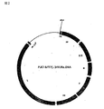
T7 プロモーター、(-)鎖RNAが転写されるように設計されたセンダイウイルスcDNA、リボザイム遺伝子をこの順に保持するDNAを、pUC18プラスミドに挿入した プラスミドpUC18/T7(-)HVJRz.DNAを作製した。また、T7 プロモーター、(+)鎖RNAが転写されるように設計されたセンダイウイルスcDNA、リボザイム遺伝子をこの順に保持するDNAを、pUC18プラスミドに挿入したプラスミドpUC18/T7(+)HVJRz.DNAを作製した。pUC18/T7(-)HVJRz.DNAおよびpUC18/T7(+)HVJRz.DNAの構成を図1および図2に示した。
[Example 1] Preparation of Sendai virus transcription unit pUC18 / T7 (-) HVJRz.DNA and pUC18 / T7 (+) HVJRz.DNA
Plasmid pUC18 / T7 (-) HVJRz.DNA was prepared by inserting the T7 promoter, Sendai virus cDNA designed to transcribe (-) strand RNA, and DNA holding the ribozyme gene in this order into the pUC18 plasmid. In addition, a plasmid pUC18 / T7 (+) HVJRz.DNA was constructed by inserting the T7 promoter, Sendai virus cDNA designed to transcribe (+) strand RNA, and DNA holding the ribozyme gene in this order into the pUC18 plasmid. did. The structures of pUC18 / T7 (-) HVJRz.DNA and pUC18 / T7 (+) HVJRz.DNA are shown in FIG. 1 and FIG.
[実施例2] cDNAからのセンダイウイルス再構成実験
直径6cmのプラスチックシャーレに通常のトリプシン処理を施したLLC-MK2細胞を2,000,000個とMEM培地(MEM +FBS 10%) 2mlとを添加し、CO25%, 37℃の条件下で24時間培養した。培養液を取り除き、1mlのPBSを用いて洗浄した後、多重感染度(moi/multiplicity of infection)が2となるように調製した、T7ポリメラーゼを発現する組換え体ワクチニアウイルスvTF7-3を0.1mlのPBSに懸濁したものを添加した。15分毎にウイルス液が全体にいきわたるようにシャーレを揺らし、1時間の感染を行った。ウイルス溶液を除去し、1mlのPBSを用いて洗浄した。 このシャーレに、cDNA溶液を含む培地を添加した。cDNA溶液を含む培地の作製は、以下のように行なった。
[Example 2] Sendai virus reconstitution experiment from cDNA Add 2,000,000 LLC-MK2 cells treated with normal trypsin to a plastic petri dish with a diameter of 6 cm and 2 ml of MEM medium (MEM + FBS 10%), and CO. 25%, were cultured for 24 hours under the conditions of 37 ° C.. After removing the culture medium and washing with 1 ml of PBS, 0.1% of a recombinant vaccinia virus expressing T7 polymerase, vTF7-3, prepared to have a moi / multiplicity of infection of 2 was obtained. The suspension in ml PBS was added. Every 15 minutes, the petri dish was shaken so that the virus solution spread throughout, and the infection was carried out for 1 hour. The virus solution was removed and washed with 1 ml PBS. A medium containing a cDNA solution was added to the petri dish. The medium containing the cDNA solution was prepared as follows.
表に記した核酸(センダイウイルスの複製に必要な因子を発現するプラスミド、pGEM-L, pGEM-P/C, pGEM-NP を含む)を1.5mlのサンプリングチューブにとり、HBS(Hepes buffered saline; 20mM Hepes pH7.4, 150mM NaCl)を加えて総量を0.1mlにした。表中の (-)または(+)cDNAは、プラスミドpUC18/T7(-)HVJRz.DNAまたはpUC18/T7(+)HVJRz.DNAそのものを示し、/Cは環状のまま、/Lは制限酵素MluIにより直鎖化した後に細胞に導入していることを示す。 Take the nucleic acids listed in the table (including plasmids expressing factors necessary for Sendai virus replication, including pGEM-L, pGEM-P / C, and pGEM-NP) in a 1.5 ml sampling tube and add HBS (Hepes buffered saline; 20 mM) Hepes pH 7.4, 150 mM NaCl) was added to bring the total volume to 0.1 ml. (-) Or (+) cDNA in the table indicates plasmid pUC18 / T7 (-) HVJRz.DNA or pUC18 / T7 (+) HVJRz.DNA itself, / C remains circular, / L indicates restriction enzyme MluI It shows that it introduce | transduced into the cell after linearizing by.
他方、ポリスチレンチューブの中で、HBS 0.07ml, DOTAP(ベーリンガーマンハイム社製)0.03mlを調合し、核酸溶液をこのポリスチレンチューブに移した。この状態で、10分静置した。これに、細胞培養液(2ml MEM +FBS 10%)を添加した。さらにこの中にワクチニアウイルスの阻害剤であるリファンピシン(Rifampicin)とシトシンアラビノシドC(Cytosin arabinoside C/Ara C)を最終濃度がそれぞれ0.1mg/ml, 0.04mg/mlとなるように添加した。これにより、cDNA溶液を含む培地が作製された。 On the other hand, 0.07 ml of HBS and 0.03 ml of DOTAP (Boehringer Mannheim) were prepared in a polystyrene tube, and the nucleic acid solution was transferred to this polystyrene tube. In this state, it was allowed to stand for 10 minutes. To this, cell culture medium (2 ml MEM + FBS 10%) was added. Furthermore, rifampicin (Rifampicin) and cytosine arabinoside C (Cytosin arabinoside C / Ara C), which are inhibitors of vaccinia virus, were added to the final concentrations of 0.1 mg / ml and 0.04 mg / ml, respectively. . As a result, a medium containing the cDNA solution was prepared.
前記のシャーレを40時間 5%CO2 37℃の条件下で培養した。ラバーポリスマンを用いてシャーレ内の細胞をかき取り、エッペンドルフチューブに移し6,000rpm、5分間の遠心を行って細胞成分だけを沈殿し、再度1mlのPBSに懸濁した。この細胞液の一部をそのままの状態、あるいは希釈して10日齢の発育鶏卵に接種した。この細胞液を第1表に示した細胞数となるようにPBSで希釈し、0.5ml 接種 した卵を35℃72時間培養後4℃に移して一晩置いた。この卵の漿尿液をウイルス液として注射器と注射針を用いて回収した。 The petri dish was cultured for 40 hours at 5% CO 2 at 37 ° C. The cells in the petri dish were scraped using a rubber policeman, transferred to an Eppendorf tube, centrifuged at 6,000 rpm for 5 minutes to precipitate only the cellular components, and again suspended in 1 ml of PBS. A part of this cell solution was used as it was or diluted and inoculated into 10-day-old embryonated chicken eggs. This cell solution was diluted with PBS to the number of cells shown in Table 1, and 0.5 ml inoculated eggs were cultured at 35 ° C. for 72 hours, then transferred to 4 ° C. and left overnight. The egg chorioallantoic fluid was collected as a virus solution using a syringe and an injection needle.
回収したウイルス液のHAU (hemmaglutinin unit)と、PFU(plaque forming unit)の測定を以下に示す方法で行った。 The recovered virus solution was measured for HAU (hemmaglutinin unit) and PFU (plaque forming unit) by the method described below.
HAUの測定は以下のように行なった。鶏の血液を、400x g,10分間遠心し、上清を捨てた。残る沈殿を、沈殿の100倍量のPBSで懸濁し、これをさらに400x g, 10分間遠心し、上清を捨てた。この操作をさらに2回、繰り返し、0.1%血球溶液を作製した。ウイルス溶液を段階希釈法により2倍ずつに希釈し、その0.05mlずつ を、96穴のタイタープレートに分注した。このタイタープレートに、さらに0.05mlずつの血球溶液を分注し、軽く振動させてよく混ぜた後、4℃で40分静置した。その後、赤血球の凝集を肉眼で観察し、凝集したもののうち、もっともウイルス溶液の希釈率の高いものの希釈率を、HAUとして示した。 The measurement of HAU was performed as follows. The chicken blood was centrifuged at 400 × g for 10 minutes, and the supernatant was discarded. The remaining precipitate was suspended in PBS 100 times the amount of the precipitate, and this was further centrifuged at 400 × g for 10 minutes, and the supernatant was discarded. This operation was repeated twice more to prepare a 0.1% blood cell solution. The virus solution was diluted 2-fold by a serial dilution method, and 0.05 ml each was dispensed into a 96-well titer plate. To this titer plate, 0.05 ml of blood cell solution was further dispensed, mixed gently by shaking lightly, and allowed to stand at 4 ° C. for 40 minutes. Thereafter, erythrocyte aggregation was observed with the naked eye, and among the aggregates, the dilution rate of the virus solution with the highest dilution rate was indicated as HAU.
PFUの測定は以下のように行なった。CV-1細胞を、6穴のカルチャープレート上に単層になるように生育させた。カルチャープレートの培地を捨て、段階希釈法により10倍づつに希釈したウイルス溶液0.1mlずつをそれぞれのカルチャープレート内ウエルに分注し、37℃、1時間感染させた。感染中に血清の含まれていな い2×MEMと2%寒天を55℃で混ぜ合わせ、さらに最終濃度0.0075mg/mlとなるようにトリプシンを加えた。1時間の感染後、ウイルス溶液を取り除き、寒天と混合 した培地3mlずつをそれぞれのカルチャープレート内ウエルに加え、5%CO2条件下で37℃3日間保温した。0.2mlの0.1%フェノールレッドを加え、37℃ 3時間保温した後、取り除いた。色の付いていないプラークの数を数え、ウイルスの力価をPFU/mlとして評価した。 The measurement of PFU was performed as follows. CV-1 cells were grown in a monolayer on a 6-well culture plate. The culture plate medium was discarded, and 0.1 ml of the virus solution diluted 10-fold by serial dilution was dispensed to each well in each culture plate and infected at 37 ° C. for 1 hour. During infection, 2xMEM without serum and 2% agar were mixed at 55 ° C, and trypsin was added to a final concentration of 0.0075 mg / ml. After 1 hour of infection, the virus solution was removed, 3 ml of medium mixed with agar was added to each well in each culture plate, and incubated at 37 ° C. for 3 days under 5% CO 2 conditions. 0.2 ml of 0.1% phenol red was added and incubated at 37 ° C. for 3 hours, and then removed. The number of uncolored plaques was counted and the virus titer was evaluated as PFU / ml.
表1には、LLC-MK2細胞に導入した鋳型となるセンダイウイルスcDNA、RNA複製に必要な因子のcDNAであるpGEM-L、pGEM-P/CおよびpGEM-NPの量、インキュベーション時間、鶏卵に接種した細胞数、HAU、PFU をそれぞれ示した。

この結果から、cDNAを細胞に導入してセンダイウイルスを再構成できることが示された。また、(+)鎖を転写するcDNAを細胞内に導入したときには、(-)鎖を転写するcDNAを導入したときに比べてウイルス粒子が効率よく再構成されることが示された。さらに、cDNAを環状のままで導入したときには、直鎖状にして導入したときに比べてウイルス粒子が効率よく再構成されることが示された。 From this result, it was shown that Sendai virus can be reconstituted by introducing cDNA into cells. In addition, it was shown that when the cDNA that transcribes the (+) strand was introduced into the cell, the virus particles were reconstituted more efficiently than when the cDNA that transcribes the (−) strand was introduced. Furthermore, it was shown that when the cDNA was introduced in a circular form, the virus particles were reconstituted more efficiently than when the cDNA was introduced in a linear form.
[実施例3] センダイウイルス再構成に必要なRNA複製因子の検討
L, P/C, NPを発現するプラスミドが三者ともに必要かどうかを調べる実験を行った。方法は実施例2と同様であるが、実施例2ではcDNAとともに、pGEM-L, pGEM-P/C, pGEM-NPの3者を細胞内に導入したのに対し、本実験では、pGEM-L, pGEM-P/C, pGEM-NPのうちの任意の2者または一者のみをcDNAとともに細胞内に導入した。
[Example 3] Examination of RNA replication factors required for Sendai virus reconstitution
An experiment was conducted to determine whether all three plasmids expressing L, P / C and NP were necessary. The method is the same as in Example 2, but in Example 2, three genes, pGEM-L, pGEM-P / C, and pGEM-NP, were introduced into the cell together with the cDNA. In this experiment, pGEM- Any two or only one of L, pGEM-P / C, and pGEM-NP were introduced into the cells together with the cDNA.
表2は、LLC-MK2細胞に導入した鋳型となるセンダイウイルスcDNA、RNA複製に必要な因子のcDNAであるpGEM-L、pGEM-P/CおよびpGEM-NPの量、インキュベーション時間、鶏卵に接種した細胞数、HAU、PFU をそれぞれ示した。
[実施例4] in vitro転写RNAからのセンダイウイルス再構成実験
実施例2で、cDNAからセンダイウイルスが再構成されることを示したが、さらにcDNAをin vitroで転写した産物、すなわちvRNA および cRNAでも同様のことができうるかどうかを検討した。
[Example 4] Sendai virus reconstitution experiment from in vitro transcribed RNA In Example 2, it was shown that Sendai virus was reconstituted from cDNA, but products obtained by further transcribed cDNA in vitro, namely vRNA and cRNA. But we examined whether we could do the same.
センダイウイルス転写ユニットpUC18/T7(-)HVJRz.DNAおよびpUC18/T7(+)HVJRz.DNAを制限酵素MluIで直鎖状にした後、これを鋳型として用い、精製T7ポリメラーゼ(EPICENTRE TECHNOLOGIES: Ampliscribe T7 Transcription Kit)によるin vitro RNA合成を行った。in vitro RNA合成の方法はキットのプロトコルに従った。ここで得られたRNA産物を、実施例2のcDNAの代わりに用い、同様の実験を行 い、ウイルス生産の評価はHA試験により行った。
結果を表3に示す。
The results are shown in Table 3.
[実施例5]センダイウイルスベクター内に挿入した外来遺伝子の宿主内での発現の検討
(1) 外来遺伝子(HIV-1 gp120遺伝子)が挿入されたセンダイウイルスベクター「pSeVgp120」の調製
プライマーa(5'-TGCGGCCGCCGTACGGTGGCAATGAGTGAAGGAGAAGT-3')(配列番号:1)及びプライマーd(5'-TTGCGGCCGCGATGAACTTTCACCCTAAGTTTTTVTTACTACGGCGTACGTCATCTTTTTTCTCTCTGC-3')(配列番号:2)を用い、「pNI432」上のHIV-1 gp120遺伝子を標準的なPCR法により増幅した。TAクローニングを行い、NotIで消化し、これをNotIで消化した「pSeV18+」に挿入した。次いで、これをE.Coliに形質転換し、E.Coliの各コロニーのDNAを「Miniprep」法で抽出し、DraIII消化後電気泳動を行い、泳動されたDNAのうち挿入により期待される大きさのDNA断片を含んでいることが確認されたクローンを選抜することで、陽性クローンを得た(以下、この陽性クローンを「クローン9」と称する)。目的の塩基配列であることを確認後、塩化セシウム密度勾配遠心により、DNAを精製した。なお、これにより得られた、gp120の挿入されたpSeV18+を「pSeVgp120」と称する。
[Example 5] Examination of expression in host of foreign gene inserted in Sendai virus vector
(1) Preparation of Sendai virus vector “pSeVgp120” inserted with foreign gene (HIV-1 gp120 gene) 3 ′) (SEQ ID NO: 2) was used to amplify the HIV-1 gp120 gene on “pNI432” by a standard PCR method. TA cloning was performed, digested with NotI, and inserted into “pSeV18 + ” digested with NotI. Next, this is transformed into E. Coli, the DNA of each colony of E. Coli is extracted by the “Miniprep” method, electrophoresed after DraIII digestion, and the expected size by insertion of the migrated DNA A clone that was confirmed to contain the DNA fragment was selected to obtain a positive clone (hereinafter, this positive clone is referred to as “clone 9”). After confirming the target base sequence, the DNA was purified by cesium chloride density gradient centrifugation. The pSeV18 + inserted with gp120 thus obtained is referred to as “pSeVgp120”.
(2) pSeVgp120を保持するセンダイウイルス(SeVgp120)の再構成及びgp120の発現の解析
LLCMK2細胞にpGEM NP, P,Lの他に、さらにpSeVgp120を導入した以外は、実施例2と同様の方法で、発育鶏卵のしょう尿液を回収し、HAUの測定及びgp120発現の検討(ELISA)を行った。HAUの測定は、実施例2と同様の方法で行った。
(2) Reconstitution of Sendai virus (SeVgp120) carrying pSeVgp120 and analysis of gp120 expression
In addition to pGEM NP, P, and L in LLCMK2 cells, pSeVgp120 was further introduced, and the urine fluid of the hen's eggs was collected in the same manner as in Example 2 to measure HAU and study gp120 expression (ELISA) ) The measurement of HAU was performed in the same manner as in Example 2.
また、ELISAは以下のように行った。HIV-1に対するモノクロナール抗体で覆った96ウェルプレートに100μlの試料を添加し、37℃で60分反応させた。PBSで洗浄後、100μlのHRP結合抗HIV-1抗体を添加し、37℃で60分反応させた。これをPBSで洗浄後、テトラメチルベンチジンを添加し、HRP活性で転換される反応生成物の量を酸性条件下、450nmの吸光度で検出することによりgp120の発現量を測定した。この結果を表4左に示す。 Moreover, ELISA was performed as follows. 100 μl of a sample was added to a 96-well plate covered with a monoclonal antibody against HIV-1 and reacted at 37 ° C. for 60 minutes. After washing with PBS, 100 μl of HRP-conjugated anti-HIV-1 antibody was added and reacted at 37 ° C. for 60 minutes. This was washed with PBS, tetramethylbenzidine was added, and the expression level of gp120 was measured by detecting the amount of the reaction product converted by HRP activity under an acidic condition at an absorbance of 450 nm. The results are shown on the left of Table 4.
また、得られたウイルス液は、CV-1細胞に感染させ、同様の検討を行った。CV-1細胞を1プレート当たり5x10⁵細胞となるようにまいて生育させ、培地を捨て、PBS(-)で洗浄し、感染多重度10でウイルス液を添加し、室温で1時間感染させた。ウイルス液を捨てPBS(-)で洗浄し、plainMEM培地(MEM培地に抗生物質AraC、Rif及びトリプシンを添加したもの)を添加して、37℃で48時間反応させた。反応後、培地を回収し、HAUの測定(実施例2と同様の方法)及びgp120発現の検討(ELISA)を行った。この結果を表4中央に示す。なお、CV-1細胞の培養上清を再度発育鶏卵に接種し、これにより得たウイルス液のHAUの測定結果及びgp120発現の検討(ELISA)結果を表4右に示す。
さらに、gp120の発現をウエスタンブロティング法により解析した。SeVgp120を感染させたCV-1細胞の培地を20,000rpmで1時間遠心し、ウイルスを沈殿させ、その上清をTCA(10%(v/v)、氷上で15分)またはで70%エタノール(-20℃)で処理し、15,000rpmで15分遠心し、沈降した蛋白質を「SDS-PAGE Sample buffer」(第一化学)と混合し90℃で3分反応させ、10%アクリルアミドゲル上でSDS-ポリアクリルアミドゲル電気泳動(SDS-PAGE)を行った。泳動後、蛋白質をPVDF膜(第一化学)に転写し、モノクロナール抗体902を室温で1時間反応させた。次いで、T-TBSで洗浄し、抗mIgG(アマシャム社)を室温で1時間反応させ、T-TBSで洗浄した。さらに、HRP結合プロテインA(アマシャム社)を室温で1時間反応させ、T-TBSで洗浄した。これに4-クロロ-1-ナフトール(4CNPlus)(第一化学)を添加し、gp120を検出した。この結果、予想されるgp120の分子量の位置にバンドが検出された。 Furthermore, the expression of gp120 was analyzed by Western blotting. CV-1 cells infected with SeVgp120 were centrifuged at 20,000 rpm for 1 hour to precipitate the virus, and the supernatant was TCA (10% (v / v), 15 minutes on ice) or 70% ethanol (15% on ice). -20 ° C), centrifuged at 15,000 rpm for 15 minutes, the precipitated protein was mixed with "SDS-PAGE Sample buffer" (Daiichi Kagaku), reacted at 90 ° C for 3 minutes, and SDS on a 10% acrylamide gel -Polyacrylamide gel electrophoresis (SDS-PAGE) was performed. After electrophoresis, the protein was transferred to a PVDF membrane (Daiichi Kagaku) and allowed to react with the monoclonal antibody 902 at room temperature for 1 hour. Subsequently, it was washed with T-TBS, anti-mIgG (Amersham) was reacted at room temperature for 1 hour, and washed with T-TBS. Further, HRP-binding protein A (Amersham) was reacted at room temperature for 1 hour and washed with T-TBS. 4-Chloro-1-naphthol (4CNPlus) (Daiichi Kagaku) was added to this to detect gp120. As a result, a band was detected at the expected molecular weight of gp120.
さらに、CV-1細胞へのSeVgp120の感染後の時間とHAUの値及びgp120の発現量との関係を解析した。10cmプレートに5x10⁶細胞となるようにCV-1細胞をまき、感染多重度10でSeVgp120を感染させ、その後30,43,53,70時間目に1mlの培地を回収し、等量の新鮮培地と混合して、HAUの測定、gp120発現の検討(ELISA)およびウエスタンブロティングを行った。この結果を図3に示す。図3から明らかなように、センダイウイルスのHAtiterの増加に伴ってgp120生産量も増加する傾向を示した。 Furthermore, the relationship between the time after infection of SeVgp120 into CV-1 cells, the value of HAU, and the expression level of gp120 was analyzed. CV-1 cells are seeded on a 10cm plate to become 5x10 6 cells, infected with SeVgp120 at a multiplicity of infection of 10, and then 1 ml of the medium is collected at 30, 43, 53, and 70 hours. Were mixed with HAU, and gp120 expression (ELISA) and Western blotting were performed. The result is shown in FIG. As is apparent from FIG. 3, the production of gp120 showed a tendency to increase with the increase of Sendai virus HAtiter.
[実施例6]種々の型の細胞におけるSeVgp120の増殖及びgp120の発現の解析
種々の型の細胞を用いた以外は実施例5と同様の方法で、HAUの測及びgp120発現の検討(ELISA)を行った。この結果を表5に示す。
[実施例7]センダイウイルスベクター内に挿入したルシフェラーゼ遺伝子の宿主内での発現の検討
ベクター挿入用のルシフェラーゼ遺伝子を単離するため、プライマー(5'-AAGCGGCCGCCAAAGTTCACGATGGAAGAC-3'(30mer))(配列番号:3)及びプライマー(5'-TGCGGCCGCGATGAACTTTCACCCTAAGTTTTTCTTACTACGGATTATTACAATTTGGACTTTCCGCCC-3'(69mer))(配列番号:4)を用い、鋳型として「pHvluciRT4」を用いて、標準的なPCR法により両端にNotI部位の付加したルシフェラーゼ遺伝子を単離した。次いで、これをNotIで消化したpSeV18+に挿入し、ルシフェラーゼ遺伝子が挿入されたセンダイウイルスベクターを得た。次いで、LLCMK2細胞に導入し、発育鶏卵に接種した。発育卵のしょう尿膜を切り取り、冷PBS(-)で2回洗浄し、「lysis buffer」(Picagene WACO)25μlを添加し、よく攪拌してから15000rpmで2分間遠心した。その上清を5μlを採取し、基質(IATRON)50μlを添加し、96ウェルプレートに入れ、ルミノメーター(Luminous CT-9000D,DIA-IATRON)で蛍光強度を測定した。活性は、cps(counts per second)で表した。この結果、感染後24時間目のCV-1細胞で、特に高いルシフェラーゼ活性が検出された(表6)。なお、ルシフェラーゼ遺伝子の導入されていないセンダイウイルスを対照として用いた(表中の「SeV」で示してある)。また、表には2クローンの検出結果を示した。
本発明によって、センダイウイルスcDNAより効率よくウイルス粒子を再構成する系が確立され、センダイウイルスにおける遺伝子操作が可能となり、所望の外来性遺伝子を含むかまたは所望の遺伝子が欠失もしくは不活化したゲノムを保持し、伝播力を有する組換え体センダイウイルスを得ることが可能となった。 According to the present invention, a system for reconstructing virus particles more efficiently than Sendai virus cDNA has been established, enabling genetic manipulation in Sendai virus, including a desired foreign gene, or a genome in which a desired gene has been deleted or inactivated. It has become possible to obtain a recombinant Sendai virus that retains the ability to transmit and has a transmission power.
Claims (13)
Priority Applications (1)
| Application Number | Priority Date | Filing Date | Title |
|---|---|---|---|
| JP2004343685A JP2005102702A (en) | 1995-11-01 | 2004-11-29 | Recombinant sendai virus |
Applications Claiming Priority (2)
| Application Number | Priority Date | Filing Date | Title |
|---|---|---|---|
| JP28541795 | 1995-11-01 | ||
| JP2004343685A JP2005102702A (en) | 1995-11-01 | 2004-11-29 | Recombinant sendai virus |
Related Parent Applications (1)
| Application Number | Title | Priority Date | Filing Date |
|---|---|---|---|
| JP2004160423A Division JP3638019B2 (en) | 1995-11-01 | 2004-05-31 | Recombinant Sendai virus |
Related Child Applications (1)
| Application Number | Title | Priority Date | Filing Date |
|---|---|---|---|
| JP2005263310A Division JP3991339B2 (en) | 1995-11-01 | 2005-09-12 | Recombinant Sendai virus |
Publications (1)
| Publication Number | Publication Date |
|---|---|
| JP2005102702A true JP2005102702A (en) | 2005-04-21 |
Family
ID=34553892
Family Applications (1)
| Application Number | Title | Priority Date | Filing Date |
|---|---|---|---|
| JP2004343685A Withdrawn JP2005102702A (en) | 1995-11-01 | 2004-11-29 | Recombinant sendai virus |
Country Status (1)
| Country | Link |
|---|---|
| JP (1) | JP2005102702A (en) |
-
2004
- 2004-11-29 JP JP2004343685A patent/JP2005102702A/en not_active Withdrawn
Similar Documents
| Publication | Publication Date | Title |
|---|---|---|
| KR100525687B1 (en) | Recombinant Sendai Virus | |
| KR100754091B1 (en) | (-)-strand RNA virus vector having autonomously replicating activity | |
| Kato et al. | The paramyxovirus, Sendai virus, V protein encodes a luxury function required for viral pathogenesis | |
| KR100702275B1 (en) | Recombinant influenza viruses for vaccines and gene therapy | |
| ES2523587T3 (en) | Influenza B viruses that have alterations in the hemagglutinin polypeptide | |
| Parks et al. | Enhanced measles virus cDNA rescue and gene expression after heat shock | |
| JP3992200B2 (en) | Membrane fusible liposome using recombinant Sendai virus | |
| JP3638019B2 (en) | Recombinant Sendai virus | |
| JP3991339B2 (en) | Recombinant Sendai virus | |
| JP4791651B2 (en) | Paramyxovirus vector for foreign gene transfer | |
| EP3474892A1 (en) | Compositions and methods comprising measles virus defective interfering particles for the prevention of infectious diseases | |
| JP2005102702A (en) | Recombinant sendai virus | |
| PT1194580E (en) | In vitro reconstitution of segmented negative-strand rna viruses | |
| JP3732204B2 (en) | (−) Strand RNA viral vector having autonomous replication ability | |
| Mühlebach et al. | Development of Entry-Targeted Oncolytic Measles Viruses | |
| CA2236113C (en) | Negative strand rna viral vector having autonomous replication capability | |
| JP2010022338A (en) | Vector using borna disease virus and use thereof | |
| CN114350619A (en) | Recombinant influenza virus strain carrying rabies virus gene and preparation method and application thereof | |
| CN114040979A (en) | Method for producing artificial recombinant RNA virus stably retaining foreign gene | |
| RU2132877C1 (en) | Recombinant rna fragment (variants), ribonucleoproteid complex (variants), plasmid | |
| JP2001275684A (en) | Recombinant canine distemper virus and method for producing the same | |
| JP2004187533A (en) | Nonsegmented (-)chain rna virus expression vector | |
| MXPA05012700A (en) | Recombinant influenza vectors with a polii promoter and ribozymes |
Legal Events
| Date | Code | Title | Description |
|---|---|---|---|
| A131 | Notification of reasons for refusal |
Free format text: JAPANESE INTERMEDIATE CODE: A131 Effective date: 20050325 |
|
| A02 | Decision of refusal |
Free format text: JAPANESE INTERMEDIATE CODE: A02 Effective date: 20050713 |
|
| A761 | Written withdrawal of application |
Free format text: JAPANESE INTERMEDIATE CODE: A761 Effective date: 20051021 |