JP2004528806A - Methods for stable transduction of cells using viral vectors - Google Patents
Methods for stable transduction of cells using viral vectors Download PDFInfo
- Publication number
- JP2004528806A JP2004528806A JP2002522515A JP2002522515A JP2004528806A JP 2004528806 A JP2004528806 A JP 2004528806A JP 2002522515 A JP2002522515 A JP 2002522515A JP 2002522515 A JP2002522515 A JP 2002522515A JP 2004528806 A JP2004528806 A JP 2004528806A
- Authority
- JP
- Japan
- Prior art keywords
- cells
- cell
- transduction
- cell surface
- vector
- Prior art date
- Legal status (The legal status is an assumption and is not a legal conclusion. Google has not performed a legal analysis and makes no representation as to the accuracy of the status listed.)
- Pending
Links
Images


Classifications
-
- C—CHEMISTRY; METALLURGY
- C12—BIOCHEMISTRY; BEER; SPIRITS; WINE; VINEGAR; MICROBIOLOGY; ENZYMOLOGY; MUTATION OR GENETIC ENGINEERING
- C12N—MICROORGANISMS OR ENZYMES; COMPOSITIONS THEREOF; PROPAGATING, PRESERVING, OR MAINTAINING MICROORGANISMS; MUTATION OR GENETIC ENGINEERING; CULTURE MEDIA
- C12N15/00—Mutation or genetic engineering; DNA or RNA concerning genetic engineering, vectors, e.g. plasmids, or their isolation, preparation or purification; Use of hosts therefor
- C12N15/09—Recombinant DNA-technology
- C12N15/87—Introduction of foreign genetic material using processes not otherwise provided for, e.g. co-transformation
-
- C—CHEMISTRY; METALLURGY
- C12—BIOCHEMISTRY; BEER; SPIRITS; WINE; VINEGAR; MICROBIOLOGY; ENZYMOLOGY; MUTATION OR GENETIC ENGINEERING
- C12N—MICROORGANISMS OR ENZYMES; COMPOSITIONS THEREOF; PROPAGATING, PRESERVING, OR MAINTAINING MICROORGANISMS; MUTATION OR GENETIC ENGINEERING; CULTURE MEDIA
- C12N15/00—Mutation or genetic engineering; DNA or RNA concerning genetic engineering, vectors, e.g. plasmids, or their isolation, preparation or purification; Use of hosts therefor
- C12N15/09—Recombinant DNA-technology
- C12N15/63—Introduction of foreign genetic material using vectors; Vectors; Use of hosts therefor; Regulation of expression
- C12N15/79—Vectors or expression systems specially adapted for eukaryotic hosts
- C12N15/85—Vectors or expression systems specially adapted for eukaryotic hosts for animal cells
- C12N15/86—Viral vectors
-
- A—HUMAN NECESSITIES
- A61—MEDICAL OR VETERINARY SCIENCE; HYGIENE
- A61P—SPECIFIC THERAPEUTIC ACTIVITY OF CHEMICAL COMPOUNDS OR MEDICINAL PREPARATIONS
- A61P31/00—Antiinfectives, i.e. antibiotics, antiseptics, chemotherapeutics
- A61P31/12—Antivirals
-
- A—HUMAN NECESSITIES
- A61—MEDICAL OR VETERINARY SCIENCE; HYGIENE
- A61P—SPECIFIC THERAPEUTIC ACTIVITY OF CHEMICAL COMPOUNDS OR MEDICINAL PREPARATIONS
- A61P31/00—Antiinfectives, i.e. antibiotics, antiseptics, chemotherapeutics
- A61P31/12—Antivirals
- A61P31/14—Antivirals for RNA viruses
- A61P31/18—Antivirals for RNA viruses for HIV
-
- A—HUMAN NECESSITIES
- A61—MEDICAL OR VETERINARY SCIENCE; HYGIENE
- A61P—SPECIFIC THERAPEUTIC ACTIVITY OF CHEMICAL COMPOUNDS OR MEDICINAL PREPARATIONS
- A61P35/00—Antineoplastic agents
-
- A—HUMAN NECESSITIES
- A61—MEDICAL OR VETERINARY SCIENCE; HYGIENE
- A61K—PREPARATIONS FOR MEDICAL, DENTAL OR TOILETRY PURPOSES
- A61K48/00—Medicinal preparations containing genetic material which is inserted into cells of the living body to treat genetic diseases; Gene therapy
-
- C—CHEMISTRY; METALLURGY
- C12—BIOCHEMISTRY; BEER; SPIRITS; WINE; VINEGAR; MICROBIOLOGY; ENZYMOLOGY; MUTATION OR GENETIC ENGINEERING
- C12N—MICROORGANISMS OR ENZYMES; COMPOSITIONS THEREOF; PROPAGATING, PRESERVING, OR MAINTAINING MICROORGANISMS; MUTATION OR GENETIC ENGINEERING; CULTURE MEDIA
- C12N2740/00—Reverse transcribing RNA viruses
- C12N2740/00011—Details
- C12N2740/10011—Retroviridae
- C12N2740/16011—Human Immunodeficiency Virus, HIV
- C12N2740/16041—Use of virus, viral particle or viral elements as a vector
- C12N2740/16043—Use of virus, viral particle or viral elements as a vector viral genome or elements thereof as genetic vector
Landscapes
- Health & Medical Sciences (AREA)
- Life Sciences & Earth Sciences (AREA)
- Genetics & Genomics (AREA)
- Chemical & Material Sciences (AREA)
- Engineering & Computer Science (AREA)
- Organic Chemistry (AREA)
- General Health & Medical Sciences (AREA)
- Zoology (AREA)
- Wood Science & Technology (AREA)
- Bioinformatics & Cheminformatics (AREA)
- General Engineering & Computer Science (AREA)
- Biotechnology (AREA)
- Biomedical Technology (AREA)
- Virology (AREA)
- Molecular Biology (AREA)
- Microbiology (AREA)
- Nuclear Medicine, Radiotherapy & Molecular Imaging (AREA)
- Public Health (AREA)
- Veterinary Medicine (AREA)
- Biochemistry (AREA)
- Physics & Mathematics (AREA)
- Chemical Kinetics & Catalysis (AREA)
- Pharmacology & Pharmacy (AREA)
- Biophysics (AREA)
- Animal Behavior & Ethology (AREA)
- Medicinal Chemistry (AREA)
- Plant Pathology (AREA)
- General Chemical & Material Sciences (AREA)
- Oncology (AREA)
- Communicable Diseases (AREA)
- AIDS & HIV (AREA)
- Tropical Medicine & Parasitology (AREA)
- Medicines Containing Material From Animals Or Micro-Organisms (AREA)
- Micro-Organisms Or Cultivation Processes Thereof (AREA)
- Medicines That Contain Protein Lipid Enzymes And Other Medicines (AREA)
Abstract
本発明は、ウイルスベクターを使用する、細胞の効率的な形質導入のための方法およびそれに関連する組成物を提供する。形質導入の効率は、形質導入されるべき細胞を、その細胞表面を結合する1以上の分子と接触させることによって、増大される。細胞表面結合分子との接触は、ウイルスベクターと細胞との間の接触の前か、後か、または同時に生じ得る。形質導入ベクターは、目的の遺伝子を発現するように構築され得、形質導入された細胞が治療剤および予防剤として使用されることを可能にする。The present invention provides methods for efficient transduction of cells using viral vectors and compositions related thereto. The efficiency of transduction is increased by contacting the cell to be transduced with one or more molecules that bind its cell surface. Contact with the cell surface binding molecule can occur before, after, or simultaneously with the contact between the viral vector and the cell. Transduction vectors can be constructed to express a gene of interest, allowing the transduced cells to be used as therapeutic and prophylactic agents.
Description
【0001】
(技術分野)
本発明は、ウイルスベクターを用いた細胞の効率的かつ安定な形質導入のための、方法およびそれに関する組成物に関する。この方法は、形質導入される細胞を、この細胞表面に結合する1つ以上の分子を接触させることによって、形質導入の効率を増大する。この接触工程は、細胞へのウイルスベクターの導入の前、後、または同時に生じ得る。本発明はまた、他の適用(ベクターによって生じる核酸の発現または生きた生物体の治療を含む)における安定に形質導入された細胞の使用に関する。
【0002】
(背景技術)
「トランスフェクション(Transfection)」とは、概して、細胞への遺伝物質の導入のための技術をいい、この技術は、生物学における分子循環(revolution)および組み換え循環に大きく寄与している。高等真核生物細胞での使用のためのトランスフェクション技術の例としては、リン酸カルシウム沈殿、DEAEデキストラン処理、エレクトロポレーション、マイクロインジェクション、リポフェクション、ウイルス感染、ならびに多くの科学的教科書および学術誌に見出される他の技術が挙げられる。
【0003】
トランスフェクション技術のなかでも、ウイルス感染の使用は、ウイルスが細胞へその遺伝物質を導入する天然に存在する手段が、目的の核酸分子を細胞に移入するのに利用されるという点で独特である。このような技術のために改変されそして適用されるウイルスの例としては、アデノウイルス、アデノ随伴ウイルス、単純疱疹ウイルス、およびレトロウイルスが挙げられる。概して、目的の核酸分子は、ウイルスゲノムにクローニングされ得る。ウイルスゲノムの複製およびパッケージングの際、得られたウイルス粒子は、ウイルス進入機構を介して、目的の核酸を細胞に送達し得る。
【0004】
一般に、ウイルスゲノムはまず、目的の核酸の付与(添加)の前に、核酸操作によって複製欠損にされる。得られたウイルスゲノム、またはウイルスベクターは、ウイルス粒子のアセンブリおよび細胞からの放出を完了するために、ヘルパーウイルスまたはパッケージングシステムの使用を必要とする。ウイルスベクターまたはウイルス粒子を用いて、目的の遺伝物質を細胞に移入する場合、この技術は「形質導入(transduction)」と呼ばれる。従って、一般に、細胞を「形質導入する(transduce)」とは、遺伝物質を細胞に移入するためにウイルスベクターまたはウイルス粒子を用いることである。
【0005】
形質導入技術のなかでも、レトロウイルスの使用は、哺乳動物細胞の遺伝子改変のための興味深い題材であった。特に興味深いのは、遺伝的欠損および他の疾患を処置するために細胞に遺伝物質を導入するように改変されたレトロウイルスの使用である。このアプローチの例は、レトロウイルスベクターおよびレンチウイルスベクターが熱心な研究の対象である、造血系の細胞の場合にみられる。
【0006】
例えば、Movassaghらは、活性化T細胞の細胞周期に対する研究からの結果を含むことによって、レトロウイルス媒介形質導入の効率を増大させる企図についての彼らの研究を考察している。その結果、彼らの結果は、形質導入の間の活性な細胞分裂に依存する。この研究はまた、マウスの腫瘍レトロウイルスの使用、および形質導入の前の細胞の有意な前刺激のための要件に限定されている。
【0007】
Juneら(WO96/34970)は、T細胞トランスフェクションを増大するための手段としてT細胞刺激の使用を記載している。活性化された細胞または刺激された細胞でのT細胞形質導入に対する他の研究としては、以下の研究が挙げられる:Douglasら、Hooijbergら、Onoderaら、Klebbaら、Barryら、およびUnutmazら。不幸にも、この研究では、約65%より大きい形質導入効率を実証したものはなかった。
【0008】
Costelloらは、Human Immunodeficiency Virus−1(HIV−1)レンチウイルスベクターを用いた、刺激T細胞および非刺激T細胞の両方の形質導入を記載している。彼らは、刺激された初代T細胞で、ほぼ最大17%の効率、そして非刺激T細胞で19%未満の効率でしかなかったことを、観察した。彼らはまた、HIV−1アクセサリータンパク質の存在を含むことによって、刺激されたT細胞において、効率を36%以下までしか増大できないという限定された能力に気付いた。
【0009】
Chinnasamyらはまた、非刺激T細胞およびマイトジェン刺激T細胞の両方におけるHIV−1アクセサリータンパク質の存在下での形質導入の効率の増大を記載している。Movassaghらと同様に、Chinnasamyらは、レンチウイルスベクターでの形質導入の前に長時間、血液リンパ球を前刺激した。Chinnasamyらは、形質導入の3日後、96%より大きい形質導入効率を最初に観察した。安定に形質導入された細胞のパーセンテージは、形質導入の2週後、71.2%に減少した。Haasらはまた、マーカー遺伝子(緑色蛍光タンパク質)を発現し得るレンチウイルスベクターで形質導入した細胞において、一過性の形質導入、および「偽性形質導入(pseudotransduction)」を観察した。形質導入の3日後でさえ、形質導入された初代CD34+臍帯血細胞におけるマーカー遺伝子の不完全な発現に基づいて、大きい(10%を超える)一過性形質導入が検出された。一過性形質導入によるこのような発現は、形質導入後7日でさえ、約5%で検出可能のままであった。形質導入後約10日後でやっと、一過性形質導入からの発現は、マーカーなしのベクターで形質導入された細胞における発現に酷似した。
【0010】
従って、Chinnasamyらは、2週間後の有効性によって反映されるように、71.2%を超える初代リンパ球の安定な形質導入(ウイルスベクターの組み込み型が、形質導入された細胞の染色体DNAに挿入された)を達成できなかった。これは、前刺激された細胞に対するサイトカインの使用には無関係であった。さらに、Chinnasamyは、彼らが、アクセサリータンパク質(Vif、Vpr、Vpu、およびNef)を発現しないHIVベクターを用いて非刺激リンパ球を(この細胞を、形質導入後に、PHAマイトジェンおよびIL−2サイトカインで後に刺激したにもかかわらず)、有意に形質導入できなかった(形質導入後14日で3.6%のみ)ことを記載している。この結果は、非刺激細胞およびアクセサリータンパク質を含まないベクターの使用によっていくぶん改善されたが、このベクターとともに用いた刺激性プロトコールにかかわらず、刺激細胞または非刺激細胞の安定な形質導入の効率が、形質導入14日後で75%より大きいことは決してなかった。
【0011】
レンチウイルスベクターを用いた安定な形質導入の頻度が低いことがまた、Hassらによって観察された。Hassらは、初代CD34陽性臍帯血細胞を用いて、形質導入7日後、25%未満の最大安定形質導入効率しか達成できなかった。顕著なことに、この形質導入の25%の上限は、過度に高い感染効率(多重度)またはベクター濃度(例えば、9000までの感染効率(MOI)、および108感染単位/mlまでのベクター濃度)の後でさえ、改善され得なかった。
【0012】
Follenziらはまた、インターロイキン3(IL−3)、インターロイキン6(IL−6)、および幹細胞刺激因子(SCF)を含有する3サイトカインカクテルの存在下で細胞を形質導入するために、500という非常に高いMOIを用いた。興味深いことに、このカクテルの使用により、細胞は、ヒトの臨床移植には不適切にさせられる。
【0013】
従って、高い頻度で、ベクターを用いて細胞を安定に形質導入する、より効率的な手段が提供される必要は依然として残っている。さらに、検索ツールとして、そして治療剤としての両方の使用のために、非刺激細胞を形質導入するための、さらに効率的な手段が必要である。
【0014】
上記の文献の引用は、前述のいずれかが関連の先行技術であることを承認するとは意図されない。これらの文献の内容に関する日付または提示に関する全ての記述は、本出願人に利用可能な情報に基づき、そしてこれらの文献の日付または内容の正確性に関していかなる承認も構成しない。
【0015】
(発明の開示)
本発明は、ウイルスベクターおよびウイルス粒子を用いた細胞の安定な形質導入のために高度に有効な方法、およびそれに関する組成物を提供する。「安定な形質導入」とは、ウイルスベクターの組み込み型が、形質導入された細胞の染色体DNAに挿入されたことを意味する。この方法は、形質導入されるべき細胞を、この細胞表面に結合する少なくとも1つの分子と接触するように曝露する工程を包含する。この接触工程は、細胞がウイルスベクター、またはウイルス粒子に曝露される前、間、または後に生じ得る。本明細書において以降は、用語「ウイルスベクター」は、ウイルス由来の核酸の任意の形態を意味するために用いられ、そして形質導入を介して細胞に遺伝物質を移入するために用いられる。この用語は、ウイルスベクター核酸(例えば、DNA、およびRNA)、これらの核酸のカプセル化形態、およびウイルスベクター核酸がパッケージングされたウイルス粒子を包含する。
【0016】
本発明はまた、他の適用(このベクターに存在する核酸の発現による、有用な遺伝子産物およびタンパク質の産生、または疾患に罹患しているかもしくは罹患する危険性のある生存している被験体の治療を含む)における形質導入された細胞の使用を包含する。好ましくは、この被験体はヒトである。
【0017】
形質導入される細胞の表面に結合する少なくとも1つの分子としては、この細胞の表面上のレセプター、マーカー、または他の認識可能部分と物理的に相互作用する任意の分子が挙げられる。基本的に、任意の細胞表面結合分子が、細胞の高い効率の形質導入に用いられ得る。本発明を理論で拘束することはないが、この細胞表面結合分子は、以下を生じ得る:DNA組み込みに対してより受容性である宿主細胞のクロマチン;ベクター遺伝子発現に好ましい部位へのウイルスベクターの優先的な取り込み;キャプシド含有核酸の細胞質へのさらに効率的な進入;細胞膜もしくは内在性膜構造(例えば、エンドソーム)を横切るウイルスの、さらに効率的な進入;またはウイルスベクターの遺伝物質の核移入(トランスポート)のためのさらに許容的な細胞の作成。本発明の方法はまた、上記の可能性のうち複数を含み得る。また、そして上記の可能性の数および多様性から明白なように、本発明は、いずれか1つの理論には限定され得ない。代わりに、そしてヒトの治療におけるこの細胞の可能な使用にマイナスに影響することなく、処置された細胞の100%までの安定な形質導入において、本発明の非凡な発見を考慮すれば、本発明はヒトの細胞治療の分野において新しいアプローチを切り開くことみなされるべきである。
【0018】
全ての細胞表面結合分子ではないが、ウイルスベクターによって効率的かつ安定な形質導入が生じる。例えば、アポトーシスを誘導する細胞表面分子への結合は、細胞の効率的な形質導入を生じず、むしろ細胞死を生じる。細胞死は、細胞(例えば、腫瘍細胞)の殺傷に好ましくあり得るが、運搬(ペイロード)遺伝子または核酸配列を含むベクターでの細胞の安定な形質導入には好ましくない。好ましい細胞表面結合分子は、ウイルスベクターによる形質導入に対してさらに受容性である細胞を生じる。このような分子の例としては、特定の細胞表面レセプターまたはその部分に対する抗体、およびこのようなレセプターについてのリガンドまたは結合ドメインが挙げられる。さらに、抗体の抗原結合フラグメント(例えば、Fab、およびFvフラグメント)は、本発明における使用について意図される。特異的な細胞表面レセプターに対する結合ドメインは、単一のエピトープまたは2つ以上のエピトープを含み得る。
【0019】
本発明における使用のための細胞表面結合分子の好ましい例は、T細胞に結合し、そしてそれらをベクター形質導入に対して、さらに受容性にする、抗CD3抗体および抗CD28抗体である。他の好ましい細胞表面結合分子は、FLT−3リガンド、TPO、およびKitリガンドレセプターに対する抗体またはリガンドであり、これは、レセプターを発現する細胞(例えば、造血幹細胞)をベクター形質導入に対してさらに受容性にさせる。さらなる好ましい細胞表面結合分子は、GM−CSFおよびIL−4レセプターに対する抗体またはリガンドであり、これは、樹状細胞またはそれらの前駆体(例えば、単球、CD34陽性幹細胞、または樹状細胞系統上のその分化した前駆体細胞)を、ベクター形質導入にさらに受容性にする。他の細胞表面結合分子としては、別の細胞の表面に結合する細胞表面上に見出された分子が挙げられる。
【0020】
細胞表面結合分子のさらなる例としては、ポリペプチド、核酸、炭水化物、脂質、およびイオン(全て、他の物質と必要に応じて複合体化した)が挙げられる。好ましくは、この分子は、以下のような血球の表面上に見出された因子と結合する:CD1a、CD1b、CD1c、CD1d、CD2、CD3γ、CD3δ、CD3ε、CD4、CD5、CD6、CD7、CD8α、CD8β、CD9、CD10、CD11a、CD11b、CD11c、CDw12、CD13、CD14、CD15、CD15s、CD16a、CD16b、CD18、CD19、CD20、CD21、CD22、CD23、CD24、CD25、CD26、CD27、CD28、CD29、CD30、CD31、CD32、CD33、CD34、CD35、CD36、CD37、CD38、CD39、CD40、CD41、CD42a、CD42b、CD42c、CD42d、CD43、CD44、CD45、CD45R、CD46、CD47、CD48、CD49a、CD49b、CD49c、CD49d、CD49e、CD49f、CD50、CD51、CD52、CD53、CD54、CD55、CD56、CD57、CD58、CD59、CDw60、CD61、CD62E、CD62L、CD62P、CD63、CD64、CD65、CD66a、CD66b、CD66c、CD66d、CD66e、CD66f、CD67、CD68、CD69、CDw70、CD71、CD72、CD73、CD74、CDw75、CDw76、CD77、CD79α、CD79β、CD80、CD81、CD82、CD83、CD84、CD85、CD86、CD87、CD88、CD89、CD90、CD91、CDw92、CD93、CD94、CD95、CD96、CD97、CD98、CD99、CD100、CD101、CD102、CD103、CD104、CD105、CD106、CD107a、CD107b、CDw108、CDw109、CD114、CD115、CD116、CD117、CD118、CD119、CD120a、CD120b、CD121a、CD121b、CD122、CD123、CDw124、CD125、CD126、CDw127、CDw128a、CDw128b、CDw130、CDw131、CD132、CD133、CD134、CD135、CD136、CDw137、CD138、CD139、CD140a、CD140b、CD141、CD142、CD143、CD144、CDw145、CD146、CD147、CD148、CDw149、CD150、CD151、CD152、CD153、CD154、CD155、CD156、CD157、CD158a、CD158b、CD161、CD162、CD163、CD164、CD165、CD166、およびTCRζ。小文字(例えば、「a」または「b」)は、複数の遺伝子産物からなるか、または構造的に関連するタンパク質のファミリーに属する、複合CD分子を示す。記号「w」は、まだ完全に確認されていない推定CD分子をいう。CD分子のより完全な列挙は、Kishimoto,T(編)に見出される。CD分子に対する現在の情報はまた、以下のWeb:http://www.bsi.vt.edu/immunologyでのAn International WWW Resource/Journalにおいて、Shaw,S.(編)Protein Reviewsに見出される。
【0021】
より好ましいのは、リンパ球、T細胞および白血球の表面上に見出される因子に結合する分子であり、このような因子は、例えば、以下である:CD2、CD3γ、CD3δ、CD3ε、CD5、CD6、CD7、CD8α、CD8β、CD9、CD11a、CD18、CD25、CD26、CD27、CD28、CD29、CD30、CD37、CD38、CD39、CD43、CD44、CD45R、CD46、CD48、CD49a、CD49b、CD49c、CD49d、CD49e、CD49f、CD50、CD53、CD54、CD56、CD57、CD58、CD59、CDw60、CD62L、CD68、CD69、CDw70、CD71、CD73、CDw75、CDw76、CD84、CD85、CD86、CD87、CD89、CD90、CD94、CD96、CD97、CD98、CD99、CD100、CD101、CD103、CD107a、CD107b、CDw108、CDw109、CD118、CD119、CD120b、CD121a、CD122、CDw124、CDw127、CDw128a、CDw130、CD132、CD134、CDw137、CD140a、CD140b、CD143、CD146、CD148、CD152、CD153、CD154、CD155、CD161、CD162、CD165、CD166、およびTCRζ。
【0022】
細胞表面に結合しかつ本発明における使用に適した、さらなる抗体および分子は、Linscott’s Directory of Immunological and Biological Reagents、第11版、2000年1月、発行者:W.D.Linscott,Petaluma,CA(この文献は、この文献が完全に示されるかのように、参考として本明細書によって援用される)に開示される。しかし、本発明のいくつかの実施形態において、この細胞表面結合分子は、サイトカインではない。
【0023】
本発明は、細胞のベクター形質導入を促進する可溶性細胞表面結合分子の使用によって実施形態され得るが、他の好ましい実施形態は、固定化された細胞表面結合分子の使用を含む。好ましくは、この固定化された分子は、抗体である。あるいは、固定化は、細胞表面結合分子を発現する他の細胞の使用を介し得る。造血幹細胞の効率的な形質導入のための好ましい方法は、形質導入の間に分化することなく幹細胞の維持を促進するリガンドをそれらの表面上に発現する、骨髄間質細胞を含むことである。刺激する細胞はネイティブ細胞に限定されないが、任意の細胞が操作されて、形質導入に対する正確な刺激を提供するために適切な細胞表面結合分子を発現し得る。
【0024】
少なくとも1つの分子が細胞表面に結合する能力を増大または強化するさらなる分子もまた、含まれ得る。例えば、特異的な細胞表面レセプターに対する(一次)抗体の可溶性形態が、細胞表面に既に結合した一次抗体を架橋し得る二次抗体と組み合わせて、使用され得る。
【0025】
当然、任意の細胞が、本発明の実施に使用され得る。好ましくは、形質導入されるべきである細胞は、真核生物細胞である。より好ましくは、その細胞は、初代細胞である。しかし、細胞株もまた、本発明の方法を用いて形質導入され得、そして多くの場合、細胞株は、より簡単に形質導入され得る。1つの好ましい実施形態において、形質導入されるべきである細胞は、初代リンパ球(例えば、Tリンパ球)もしくはマクロファージ(例えば、単球マクロファージ)であるか、またはこれらの細胞のいずれかに対する前駆体(例えば、造血幹細胞)である。形質導入に関して好ましい他の細胞は、一般的に造血系の細胞であるか、またはより一般的には、造血によって形成される細胞ならびに造血系の細胞が形成される幹細胞および血球機能と関連する細胞である。このような細胞としては、造血によって形成される顆粒球およびリンパ球ならびに多能性のリンパ性幹細胞および多能性の骨髄性幹細胞が挙げられる。血球機能と関連する細胞としては、免疫幹細胞の機能を補助する細胞(例えば、樹状細胞、内皮細胞、単球およびランゲルハンス細胞のような、抗原提示細胞)が挙げられる。好ましい実施形態において、その細胞は、Tリンパ球(すなわち、T細胞)(例えば、CD4マーカーおよびCD8マーカーを発現するTリンパ球)である。
【0026】
特に好ましい実施形態において、その細胞は、初代CD4+ Tリンパ球または初代CD34+造血幹細胞である。しかし、本発明における使用のためのウイルスベクターが、(以下に議論するように)水疱性口内炎ウイルスエンベロープGタンパク質を用いて偽型にされ得る場合、任意の細胞が、本発明の方法を介して形質導入され得る。このような細胞としては、神経膠星状細胞、皮膚線維芽細胞、上皮細胞、ニューロン、樹状細胞、リンパ球、免疫応答と関連する細胞、血管内皮細胞、腫瘍細胞、腫瘍血管内皮細胞、肝細胞、肺細胞、骨髄細胞、抗原提示細胞、間質細胞、脂肪細胞、筋細胞、膵細胞、腎細胞、卵子または精母細胞(例えば、トランスジェニック動物を作製するため)、生殖細胞系に寄与する細胞、胚性多能性幹細胞もしくはその前駆体、血小板および赤血球のような無核細胞を含む血球などが挙げられるが、これらに限定されない。好ましくは、細胞は、真核生物の多細胞種(例えば、単細胞の酵母細胞とは対照的な細胞)であり、そしてなおより好ましくは、哺乳動物起源(例えば、ヒト細胞)である。
【0027】
形質導入されるべきである細胞は、単一の実体として存在し得るか、または細胞の大きな集団の一部であり得る。このような「細胞の大きな集団」は、例えば、細胞培養物(混合されているかまたは純粋であるかのいずれか)、組織(例えば、上皮組織、間質組織または他の組織)、器官(例えば、心臓、肺、肝臓、胆嚢、膀胱、眼、および他の器官)、器官系(例えば、循環器系、呼吸器系、胃腸系、泌尿器系、神経系、外皮系または他の器官系)、胚盤胞、胚性幹細胞、胎児由来の細胞(例えば、遺伝的障害/遺伝的疾患の処置のため、またはトランスジェニック動物を作製するため)、疾患組織(例えば、腫瘍または感染部位)、または生物(例えば、鳥類、哺乳動物、海洋生物、魚類、植物など)を含み得る。好ましくは、標的化される器官/組織/細胞は、循環器系の器官/組織/細胞(例えば、心臓、血管、および血液を含むが挙げられるが、これらに限定されない)、呼吸器系の器官/組織/細胞(例えば、鼻、咽頭、喉頭、気管、気管支、細気管支、肺など)、胃腸系の器官/組織/細胞(例えば、口組織および口腔組織、咽頭、食道、胃、腸、唾液腺、膵臓、肝臓、胆嚢などを含む)、乳房系の器官/組織/細胞(例えば、胸部上皮細胞および組織中の支持細胞)、泌尿器系の器官/組織/細胞(例えば、腎臓、尿管、膀胱、尿道など)、神経系の器官/組織/細胞(脳および脊髄、ならびに眼のような特別な感覚器官が挙げられるが、これらに限定されない)および外皮系の器官/組織/細胞(例えば、皮膚)である。
【0028】
さらにより好ましくは、形質導入されるべきである細胞は、心臓の細胞、血管(腫瘍血管および感染組織または疾患組織と関連する血管を含む)の細胞、骨髄の細胞、血液の細胞、脳の細胞、リンパ組織の細胞、リンパ節の細胞、脾臓の細胞、肺の細胞、肝臓の細胞、胆嚢の細胞、膀胱の細胞および眼の細胞からなる群より選択される。本発明の1つの特定の実施形態において、細胞は、使用が意図された宿主に対して自系であるが、宿主に対して部分的に適合しないかもしくは完全に適合しない同種異系の細胞、または宿主に対して異種性でさえある細胞もまた、使用され得る。さらに、任意の所定の宿主生物(関連する生物群または種(例えば、ヒト))における使用に適した万能のドナー細胞が、形質導入され得る。本発明のこの後者の実施形態は、細胞、組織、または器官の移植において特に重要であり、その形質導入される細胞の供給源が、移植の結果に重要であり得る。
【0029】
本発明の方法による形質導入のための別の好ましい細胞は、腫瘍細胞であるか、疾患細胞であるか、またはその遺伝子的構成もしくは同じ生物中に存在する他の細胞の遺伝子的構成に起因して時間が経てば異常になる危険性のある細胞である。後者の実施形態は、本発明の形質導入細胞が予防において使用されることを可能にする。乳癌は、予後指標が、疾患が後で起こる前の早期の遺伝的介入として本発明の形質導入細胞を用いて処置することを可能にする、疾患プロセスの1つの例である。しかし、本発明の方法はまた、疾患が検出された後の乳癌の治療的処置に使用され得る。癌治療における本発明のさらなる適用は、多く、そして当業者は、本明細書中に示される本発明を、過度の実験なしに多くの型の癌の治療に対して使用し得る。
【0030】
例としてであり、本発明を限定するわけではないが、1つの適用は、エストロゲン依存性である乳癌における適用である。エストロゲン依存性乳癌中の癌細胞は、エストロゲンレセプターに結合する抗体またはリガンドを治療的ウイルスベクターと組合わせて使用することによって、優先的に形質導入される。例えば、このベクターは、腫瘍阻害遺伝子(例えば、ヘルペスウイルスチミジンキナーゼ遺伝子)を含み得る。従って、形質導入された細胞は、ガンシクロビル(ヘルペスチミジンキナーゼによって活性化され得るプロドラッグ)の添加によって、選択的に殺傷され得る。腫瘍阻害遺伝子および対応するプロドラッグのさらなる例は多数であり、そして当該分野で周知であり、そして過度の実験なしに当業者によって選択され得る。本発明の形質導入方法の適用と組合わせた活性可能なプロドラッグの使用は、他の腫瘍型に広範に適用され得、そして上記の例は、本発明を、成長または増殖についてホルモン依存性であるかまたはいくつかの可溶性因子依存性である腫瘍に限定しない。
【0031】
例えば、Her−2/neu陽性腫瘍細胞は、エストロゲン依存性ではなく、予後指標としては不十分である。なぜなら、このような細胞を含む非エストロゲン依存性腫瘍は、エストロゲンアンタゴニストであるタキソールのような薬剤を用いた処理に対して非常に耐性であるからである。本発明に対して好ましい実施形態は、例えば、骨髄移植プロトコールにおいて、腫瘍で汚染した細胞の形質導入の間にウイルスベクター調製物を用いてHer−2/neuまたはヒレグリンに結合する抗体または他の分子を含むことである。あるいは、形質導入は、腫瘍部位に直接なされ得るか、または腫瘍形成を調節するベクターを用いてインビボで血管内でなされ得る。
【0032】
本発明のなお別の実施形態は、単独または腫瘍細胞を標的化することと組合わせて、腫瘍脈管構造を標的化することである。St Croixら(その文献は、この文献が完全に示されるかのように、参考として本明細書によって援用される)は、SAGE分析によって、正常内皮細胞と比較して腫瘍内皮細胞中に特異的に過剰発現される遺伝子を同定した。これらの遺伝子のうちの多くは、細胞表面分子(例えば、Thy−1細胞表面抗原またはEndo180レクチン)をコードする。アップレギュレートされた細胞表面因子の全てが、本発明の細胞表面結合分子によって結合されて、効率的で安定な遺伝子導入に対する刺激を提供し得る。従って、腫瘍治療に対するアプローチは、腫瘍脈管構造に選択的に結合するが正常な内皮細胞に結合しない細胞表面結合分子の存在下で、治療的ウイルスベクターを用いて腫瘍内皮細胞を形質導入した後に、この腫瘍内皮細胞を殺傷することによってこの腫瘍脈管構造を破壊することである。
【0033】
本発明のなお別の実施形態は、腫瘍中で抗腫瘍遺伝子の発現を選択的に促進するが正常脈管内皮細胞中ではこの抗腫瘍遺伝子の発現を促進しないウイルスベクターの中に、エレメント(例えば、プロモーターまたはmRNA上のシス作用性安定化エレメント/シス作用性分解エレメント)を組み込むことによる、腫瘍脈管構造における抗腫瘍遺伝子の選択的発現である。このような方法は、エキソビボ、インビトロ、またはインビボで生じ得る。腫瘍脈管内皮細胞が標的化される場合、治療のための好ましい方法はインビボである。あるいは、例えば、骨髄移植のために汚染している腫瘍細胞を骨髄から一掃することが目的である場合、治療のための好ましい方法は、エキソビボまたはインビトロで生じる。
【0034】
さらに、インビボ使用は、疾患状態に限定されず、正常細胞を形質導入するために使用され得る。例えば、本発明は、骨髄において造血幹細胞をインビボで形質導入するために使用され得る。抗体または他の細胞表面結合分子(例えば、FLT−3リガンド、TPOおよびKitリガンド、またはこれらの機能的アナログ、あるいは細胞表面結合分子を発現する間質細胞)の任意の組合わせが、高効率の骨髄形質導入のために、骨髄中への直接的な注射の際に、ベクターと共に添加され得る。用語「機能的アナログ」は、本発明の細胞表面結合分子の細胞表面結合活性を保持する任意の分子をいう。このような機能的アナログとしては、FLT−3リガンド、TPOおよびKitリガンドのフラグメント;1つ以上のアミノ酸置換、アミノ酸付加、またはアミノ酸欠失を含む、FLT−3リガンド分子、TPO分子およびKitリガンド分子;ならびに細胞表面結合分子の細胞表面結合活性を模倣する抗体が挙げられる。
【0035】
上記に対する代替のアプローチは、ウイルスベクターの産生細胞として骨髄間質細胞を使用し、そのようにして、ベクター調製物としてではなく細胞治療を介して、ベクターおよび細胞表面結合分子を提供することである。別の例は、皮下注射の間にベクターと共に、CD3抗体およびCD28抗体の機能的アナログ、またはGM−CSFおよびIL−4の機能的アナログをそれぞれ添加することによる、T細胞または樹状細胞の形質導入である。皮下組織中のリンパ液は、標的化組織の効率的な形質導入のためにリンパ節にベクターおよび刺激剤を排出する。
【0036】
本発明は、形質導入される細胞の精製が、必要に応じて、必須とはならないという利点を含む。目的の細胞型を主とした形質導入は、結合される細胞表面部分の選択によって達成され得る。従って、例えば、血球の混合集団において、CD3を発現する細胞(例えば、特定のT細胞)の形質導入は、CD3特異的抗体がその細胞と相互作用するために使用される場合、増強される。このことは、集団中の他の細胞型(例えば、CD3を発現しない顆粒球および単球)よりも優先して生じる。
【0037】
本発明はまた、所望される場合、精製された細胞型または単離された細胞型の形質導入を含む。精製された細胞型または単離された細胞型の使用は、より高い形質導入効率(これは、形質導入される細胞と比較してより高いベクター濃度に起因する)のようなさらなる利点を提供する。
【0038】
精製されたT細胞が形質導入される場合、少なくとも1つの分子が、好ましくは、T細胞上に見出される細胞表面分子に結合する。このような細胞表面分子の例としては、CD3、CD28、CD25、CD71、およびCD69が挙げられる。これらの細胞表面分子に結合する分子の例としては、これらの細胞表面分子を認識する抗体およびモノクローナル抗体が挙げられ、これらの抗体およびモノクローナル抗体のうちの多くは、市販されるか、または過度の実験なしに標準的な技術を用いて容易かつ慣用的に調製される。CD4+細胞またはCD8+細胞の形質導入のための好ましい実施形態において、CD3および/またはCD28を認識するモノクローナル抗体が使用され得る。市販されるこのような抗体の例としては、CD3についてOKT3、そしてCD28についてCD28.2が挙げられる。これらの抗体分子は、可溶性形態で使用され得るか(必要に応じて他の分子によって後で架橋される)、またはビーズもしくは他の固体表面などの上に固定化形態で使用され得る。本発明の特に好ましい実施形態において、抗体は、容器の表面上に固定化され、例えば、ウイルスベクター媒介形質導入のために使用される組織培養用のウェル、プレート、またはバッグの壁に固定化される。理論に束縛されないが、細胞が付着または接触する表面上での固定化抗体の使用は、その細胞表面上での細胞表面相互作用の局所濃度を増大させ得る。
【0039】
造血幹細胞が形質導入される場合、FLT−3リガンド、TPO(トロンボポイエチンまたは巨核球増殖および発生因子(Megakaryocyte Growth and Development Factor))、またはKitリガンドの造血幹細胞レセプターに特異的な抗体が、細胞表面結合分子として使用され得る。あるいは、幹細胞陽性細胞マーカー(CD34またはAC133が挙げられるが、これらに限定されない)に対する抗体が、使用され得る。化合物または組成物を含むリガンドが細胞表面結合分子として使用される場合、全ての天然のリガンド(タンパク質、リガンドまたは異種タンパク質に結合するリガンドを含む)が、可溶性形態または固定化形態のいずれかで使用され得る。固定化形態としては、直接的または間接的(例えば、アビジン/ビオチンを用いて)な、マイクロビーズへの付着が挙げられる。
【0040】
あるいは、リガンドは、ウイルスベクターのウイルスエンベロープ中で発現され得、必要に応じて、キメラタンパク質もしくは融合タンパク質の形態で発現され得、そして/または1つ以上の他のタンパク質と(共有結合的または非共有結合的に)複合体化され得る。このような実施形態において、細胞表面結合分子は、細胞を形質導入するための単一組成物として、ウイルスベクターと組合わせて呈される。ウイルスエンベロープ中で発現され得る細胞表面結合分子のさらなる例としては、上記に列挙した多数の表面因子が挙げられる。
【0041】
他の好ましい細胞表面結合分子(例えば、抗体またはそのフラグメント)は、NotchもしくはDeltaの造血幹細胞レセプターに結合する分子、またはNotchタンパク質もしくはDeltaタンパク質自体に結合する分子、または異種タンパク質に結合するNotchもしくはDeltaのリガンドに結合する分子である。DeltaおよびNotchは、Drosophila発生において広範な種々の細胞運命の決定に影響を与える、細胞表面タンパク質をコードする。DeltaおよびNotchの脊椎動物ホモログは、正常な胚発生に必須である。Deltaホモログは、造血の調節に重要に関与している。可溶性形態のホモログであるDelta−Serrate−lag2(DSL)は、原始造血前駆体の拡大を増強する。造血増殖因子(インターロイキン−3(IL−3)、顆粒球コロニー刺激因子(すなわち、G−CSF)または顆粒球マクロファージコロニー刺激因子(GM−SCF)を含む)と組合わせた場合、DSLは、原始造血前駆体の拡大を促進し、そして同時に、この原始前駆体が、IL−3単独に対して応答性であるより成熟した前駆体細胞へと分化するのを阻害した(Hanらを参照のこと)。DSLは、おそらく、造血細胞において発現されるNotchレセプターを活性化することによって作用し、通常の造血増殖因子に対して応答する細胞の能力を、細胞分化を選択的にブロックするが増殖シグナルをブロックしないことによって調節する(HanおよびMoore,Blood 1999を参照のこと)。従って、DeltaホモログおよびNotchホモログ、これらのホモログに対して機能的アナログである抗体は、細胞(特に、造血幹細胞)の75%より高い効率的なベクター形質導入を達成する際の使用のための、さらに好ましい細胞表面結合分子である。
【0042】
本発明は、開示される方法における使用のための、ウイルスベクターおよびこのウイルスベクターを含む組成物を包含する。このベクターは、好ましくはレトロウイルス(レトロウイルス科)ベクターであり、より好ましくはレンチウイルスベクターである。他のレトロウイルスベクター(例えば、オンコウイルスベクターおよびマウスレトロウイルスベクター)もまた、使用され得る。さらなるベクターが、他のDNAウイルス、またはその生活環のある時点の間にそのゲノムをDNAに変換し得るウイルスから誘導され得る。好ましくは、このウイルスは、アデノウイルス科、パルボウイルス科、ヘパドナウイルス科(Hepandaviridae)(通常は、ヘパドナウイルス科に分類されない、δ型肝炎ウイルスおよびE型肝炎ウイルスを含む)、パポバウイルス科(ポリオーマウイルス科およびパピローマウイルス科を含む)、ヘルペスウイルス科、およびポックスウイルス科由来のウイルスである。
【0043】
レトロウイルス科(すなわち、レトロウイルス)のさらなるウイルスは、オンコウイルス亜科、スプマウイルス亜科、スプマウイルス属、レンチウイルス亜科、およびレンチウイルスの属または亜科のウイルスである。オンコウイルス亜科のRNAウイルスは、望ましくは、ヒトTリンパ球向性ウイルス1型もしくはヒトTリンパ球向性ウイルス2型(すなわち、HTLV−1またはHTLV−2)、またはウシ白血球ウイルス(BLV)、鳥類白血症−肉腫ウイルス(例えば、ラウス肉腫ウイルス(RSV)、鳥類骨髄芽球症ウイルス(AMV)、鳥類赤芽球症ウイルス(AEV)、およびラウス随伴ウイルス(RAV;RAV−0〜RAV−50))、哺乳動物C型ウイルス(例えば、モロニーマウス白血病ウイルス(MuLV)、ハーベイ(Harvey)マウス肉腫ウイルス(HaMSV)、エーベルソンマウス白血病ウイルス(A−MuLV)、AKR−MuLV、ネコ白血病ウイルス(FeLV)、サル肉腫ウイルス、細網内皮症ウイルス(REV)、脾臓壊死ウイルス(SNV))、B型ウイルス(例えば、マウス乳腺癌ウイルス(MMTV))、およびD型ウイルス(例えば、マソン−ファイザーサルウイルス(MPMV)および「SAIDS」ウイルス)である。
【0044】
レンチウイルス亜科のRNAウイルスは、望ましくは、ヒト免疫不全症ウイルス1型またはヒト免疫不全症ウイルス2型(すなわち、HIV−1またはHIV−2(ここで、HIV−1は、以前は、リンパ節症随伴ウイルス3(HTLV−III)および後天性免疫不全症候群(AIDS)関連ウイルス(ARV)と称されていた))、またはAIDSもしくはAIDS様疾患で同定され、かつこれらの疾患に関連する、HIV−1もしくはHIV−2に関連する別のウイルスである。頭文字「HIV」または用語「AIDSウイルス」または「ヒト免疫不全ウイルス」は、総称して、これらのHIVウイルス、ならびにHIV関連ウイルスおよびHIV随伴ウイルスをいうために本明細書に使用される。さらに、レンチウイルス亜科のRNAウイルスは、好ましくは、ビスナウイルス/マエディウイルス(例えば、ヒツジに感染する)、ネコ免疫不全ウイルス(FIV)、ウシレンチウイルス、サル免疫不全ウイルス(SIV)、ウマ伝染性貧血ウイルス(EIAV)、およびヤギ関節炎−脳炎ウイルス(CAEV)である。
【0045】
特に好ましいレンチウイルスベクターは、HIV由来のベクター、より好ましくはHIV−1、HIV−2またはこれらのキメラの組み合わせ由来のベクターである。当然、異なる血清型のレトロウイルス(特に、HIV)が、本発明における使用のためのベクターを調製するために、単独または組合わせて使用され得る。本発明の好ましいベクターは、野生型ウイルスに存在するが「基本」のレンチウイルスベクター中に存在しない、シス作用性エレメントを含む。「基本」のレンチウイルスベクターは、LTRならびに5’リーダー配列およびgagコード配列中のパッケージング配列を最小限で含むが、「基本」のレンチウイルスベクターはまた、必要に応じて、Rev依存性様式でベクターRNAの核輸送を促進するRREエレメントを含み得る。好ましいベクターは、細胞への形質導入効率を増強するヌクレオチド配列をさらに含む。
【0046】
このようなベクターの例は、gagおよびpolの完全配列を含むベクターであるpN2cGFPである。別の例は、pol中の配列約4551〜5096位(この位置は、C.E.Buckler、NIAID、NIH、Bethesda、MDによって厚意によって提供された、登録番号M19921、HIVNL43 9709bpのpNL4−3配列からの参照位置である)を含むベクターである。しかし、ベクター形質導入効率を向上させ得る、wt−HIV由来の任意のシス作用性配列が、使用され得る。本発明を介して効率的な形質導入を可能にするベクターの他の例は、米国特許第5,885,806号に記載されるcr2HIV構築物である。
【0047】
形質導入効率を有意に増大させるには不十分である以前に同定された配列が、形質導入効率を増大させ得る中央DNAフラップ(flap)(pNL4−3の4757位〜4935位に対応する、pLAI3の4793位〜4971位の178塩基対のフラグメント)としてZennouら(2000)によって記載された。本発明は、この小さなフラグメントが形質導入効率を増大させるには不十分であるが、より大きな545塩基対のフラグメント(pNL4−3の4551位〜5096位)またはこれを含むさらにより大きなフラグメントが、本発明の一部として形質導入を増大させ得たという発見を含む。
【0048】
本発明において使用され得るウイルスベクター構築物のさらなる例は、米国特許第5,885,806号(この文献は、この文献が完全に示されるかのように、参考として本明細書によって援用される)に見出される。この特許第5,885,806号中の構築物は、細胞を効率的に形質導入するベクターの範囲を限定しない、単なる例示である。その代わり、この構築物は、本発明を用いた使用のためのウイルスベクターが、野生型ウイルス由来の最小配列を含み得るか、または野生型ウイルスのほぼ全ゲノムまでの配列を含み得るが、複製および/または疾患の生成に必要とされる必須の核酸配列を排除し得るということのさらなる指標を、当業者に提供する。細胞の効率的な形質導入のために必要とされる配列を正確に決定するための方法は、慣用的であり、そして当該分野で周知である。例えば、ウイルス配列を「基本」ベクターに戻す系統的な組込み、またはHIV(例えば、cr2HIV)ゲノムの実質的に全体を含むベクターから配列を欠失させることが、慣用的であり、そして当該分野で周知である。
【0049】
さらに、他のウイルス骨格由来の配列を目的のウイルスベクター(例えば、サイトメガロウイルス(CMV))に配置することもまた、当該分野で周知である。使用される実際のウイルスベクター、それにコードされる種々の補助タンパク質、およびそれに存在する配列に拘わらず、ウイルス遺伝物質は、これらのタンパク質または配列が特定の細胞型において形質導入効率を増加する場合、そのベクターまたはヘルパーゲノムに残され得る。特定の遺伝物質がベクターまたはヘルパーゲノムのいずれかにその配列を組み込むことによって形質導入効率を増加するか否かを決定するために、多くの慣用的なスクリーニングが利用可能である。本発明の好ましい実施形態は、ベクターまたはヘルパーゲノムのいずれにおいても補助タンパク質を含まない。しかし、この好ましさは、補助タンパク質および他の配列がベクターまたはヘルパーゲノムのいずれかに残されて形質導入効率を増加する本発明の実施形態を、排除しない。
【0050】
本発明において使用されるウイルスベクターはまた、「偽型」形成を生じ得、ここでは、異なるウイルスによる細胞の同時感染が、1つのウイルスのゲノムを含む子孫ビリオンを産生し、その1つのウイルスのゲノムが、別のウイルスの1以上のエンベロープタンパク質を含む外層内にカプセル化されている。この現象は、「偽型」ビリオン中に目的のウイルスベクターをパッケージングするために使用されており、これは、その目的のウイルスベクターおよび別のウイルスの少なくとも1つのエンベロープタンパク質または細胞表面分子をコードする遺伝物質の両方を、パッケージング細胞に同時トランスフェクトまたは同時感染させることによる。米国特許第5,512,421号を参照のこと。このような混合ウイルスは、使用される1以上の異種エンベロープタンパク質に対する抗血清によって中和され得る。偽型形成において一般に使用される1つのウイルスは、水疱性口内炎ウイルス(VSV)であり、これは、ラブドウイルスである。偽型化の使用は、使用される異種ウイルスのウイルス侵入機構のエレメントを含むことによって、ウイルスの宿主細胞範囲を拡大する。
【0051】
本発明の使用のためのウイルスベクターおよびVSVの偽型化は、ヌクレオカプシドにカプセル化されたウイルスベクター核酸を含むウイルス粒子を生じ、このヌクレオカプシドは、VSV Gタンパク質を含む膜によって囲まれている。このヌクレオカプシドは、好ましくは、そのウイルスベクターと通常に関連するタンパク質を含む。周辺VSV Gタンパク質を含む膜は、そのウイルスベクターをパッケージングするために使用された細胞から出て行く際に、ウイルス粒子の部分を形成する。パッケージング細胞の例は、米国特許第5,739,018号に記載される。本発明の好ましい実施形態において、ウイルス粒子は、HIVから誘導され、そしてVSV Gタンパク質によって偽型化される。VSV Gタンパク質を含む偽型化されたウイルス粒子は、両栄養性ウイルスウイルスベクターよりも高い効率で、多くの異なる細胞型を感染し得る。宿主細胞の範囲は、哺乳動物種および非哺乳動物種の両方(例えば、ヒト、げっ歯類、魚類、両生類および昆虫類)を含む。
【0052】
本発明の形質導入方法における使用のためのウイルスベクターはまた、そのウイルスに存在するプロモーターの制御下またはそのベクターに導入された異種プロモーターの制御下に、1以上の核酸配列を含みそしてこれを発現する。プロモーターはさらに、厳密に制御された遺伝子発現のためにそのオペロンに隣接するように、隔離エレメント(例えば、赤血球DNAse高感受性部位)を含み得る。好ましいプロモーターとしては、HIV−LTR、CMVプロモーター、PGK、U1、エプスタインバーウイルス由来のEBER転写単位、tRNA、U6およびU7が挙げられる。Pol IIプロモーターが好ましいが、Pol IIIプロモーターもまた使用され得る。組織特異的プロモーターもまた、好ましい実施形態である。例えば、βグロビン遺伝子座制御領域エンハンサーならびにαおよびβグロビンプロモーターは、赤血球および赤血球系細胞における組織特異的発現を提供し得る。別のさらに好ましい実施形態は、そのプロモーターに関連するシス作用性配列を使用することである。例えば、米国特許第5,814,500(参考として本明細書中に援用される)に示されるように、U1遺伝子を使用して、アンチセンス遺伝子発現を増強し得、ここで、非プロモーター配列を使用して、標的のスプライシングされたRNAに、そのアンチセンス分子またはリボザイム分子を標的化する。
【0053】
もちろん、ウイルス由来の任意のシス作用性ヌクレオチド配列が、本発明のウイルスベクターに組み込まれ得る。特に、レトロウイルスゲノム中に見出されるシス作用性配列が好ましい。例えば、gag、pol、env、vif、vpr、vpu、tatまたはrev遺伝子由来のシス作用性ヌクレオチド配列が、本発明のウイルスベクターに組み込まれ、形質導入効率をさらに増加し得る。好ましくは、シス作用性配列は、発現されるポリペプチドをコードしないか;遺伝子変更(例えば、翻訳開始部位の欠失)に起因してポリペプチドまたはその一部として発現されないか;大きいポリペプチドの一部またはフラグメントのみをコードするか;またはネイティブ配列からの1以上の置換、付加または欠失を含む変異体配列である。シス作用性配列の例は、HIV pol遺伝子内に同定されたcPPT(中央ポリプリン領域)配列である。
【0054】
本発明のウイルスベクター中のその1以上の核酸配列は、そのベクターが由来するウイルス中に見出され得るか、または異種配列であり得る。配列は、好ましくは、目的の遺伝子産物をコードする全長または部分配列である。このような配列または遺伝子産物は、好ましくは、細胞中で生物学的効果を生じ得る、生物学的に活性な因子である。このような因子の例として、タンパク質、リボ核酸、酵素、輸送体または他の生物学的に活性な分子が挙げられる。
【0055】
1つの好ましい実施形態において、この因子は、タンパク質(例えば、毒素、転写因子、増殖因子またはサイトカイン、構造タンパク質、または細胞表面分子)である。このタンパク質は、機能が全く同定されていない1以上のドメインを含み得、そして形質導入された細胞に対して同種であり得る。さらに、このタンパク質は、形質導入される細胞において存在しないか、欠損しているか、または変更されているものであり得る。あるいは、このタンパク質は、トランスドミナントネガティブ変異体またはデコイであり、形質導入された細胞において天然のタンパク質がその通常の活性を生じるのを妨げ得る。
【0056】
例えば、核酸配列は、形質導入された細胞中で発現されたRNAまたは発現されるべきRNAを結合、切断および破壊するリボザイムをコードし得る。あるいは、核酸配列は、特定の核酸配列を標的とし、そしてその分解を生じるように設計された、アンチセンス分子をコードし得る。ベクターに含まれる配列は、形質導入された細胞において、過剰発現され得るか、誘導発現され得るか、あるいは細胞またはウイルスの調節性転写制御下にあり得る。意図される用途に依存して、異種配列は、形質導入された細胞についてのマーカーを含む任意の所望のタンパク質をコードし得る。このようなマーカーとしては、特定の耐性表現型のような選択マーカー(例えば、ネオマイシン、MDR−1(P−糖タンパク質)、O6−メチルグアニン−DNA−メチルトランスフェラーゼ(MGMT)、ジヒドロ葉酸レダクターゼ(DHFR)、アルデヒドデヒドロゲナーゼ(ALDH)、グルタチオン−S−トランスフェラーゼ(GST)、スーパーオキシドジスムターゼ(SOD)およびシトシンデアミナーゼ)が挙げられる。総説については、Kocら(参考として本明細書に援用される)を参照のこと。
【0057】
本発明の方法において、形質導入される細胞は、ウイルスベクターの適用の前、後または同時に、細胞表面に結合する少なくとも1つの分子との接触に曝される。例えば、細胞は、形質導入されるウイルスベクターが存在する前、後、またはその存在下で、CD3抗体およびCD28抗体(培養ディッシュの表面上にコーティングされているか、または培養物中に存在するビーズ上に固定化されている)を有する培地で培養され得る。好ましくは、細胞は、ウイルスベクターの最初の接触後のみかまたは最初の接触時のみに、固定化されたCD3抗体および/またはCD28抗体に曝される。これらの条件下では、細胞は、ウイルスベクターでの実際の形質導入前に、細胞表面結合分子に曝されない。細胞表面結合分子との接触がウイルスベクターへの細胞の曝露(形質導入)後に生じる実施形態において、その接触は、好ましくは、形質導入の3日以内、より好ましくは、形質導入後1〜2日以内に行う。
【0058】
ウイルスベクターとの細胞のインキュベーションは、使用される条件および材料に依存して、異なる長さの時間で行い得る。インキュベーション時間に影響を及ぼす因子としては、使用される細胞、ベクターおよびMOI(感染多重度)、細胞表面に結合させるために使用される分子および量、その分子が固定化されているかまたは溶解されているか否か、およびその分子がどのように固定化または溶解されているか、ならびに所望の形質導入効率のレベル、が挙げられる。好ましくは、インキュベーションは、約8時間〜約72時間であり、より好ましくは、約12時間〜約48時間である。特に好ましい実施形態において、インキュベーションは、約24時間であり、必要に応じて一度繰り返される。
【0059】
形質導入される細胞とウイルスベクターとの間の接触は、細胞型に依存して、少なくとも1回行うが、その接触は、1回より多く行い得る。例えば、CD34陽性幹細胞の高効率の形質導入は、ベクターでの複数回の形質導入によって達成されている。本発明の好ましい方法は、細胞表面結合分子(例えば、CD3抗体および/またはCD28抗体またはFLT−3リガンド、TPOまたはKitリガンド)と組み合わせてウイルスベクターを同時に導入し、そして形質導入後約1日間〜約8日間の間に培地を交換することを回避することである。より好ましくは、培地は、形質導入後3日間交換されない。形質導入は、その条件が、細胞または細胞を含む生物に有意に有害なプロセスを伴わずに可能である限り、進行され得る。このような使用のための細胞表面結合タンパク質のさらなる例としては、本明細書上記のタンパク質が挙げられる。
【0060】
同様に、使用されるMOIは、約1〜約400、好ましくは、500未満である。一般に、好ましいMOIは、約2〜約50である。より好ましくは、MOIは、約10〜約30であるが、約1〜約10、約20、約30または約40の範囲も意図される。より好ましくは、約20のMOIである。さらに、細胞当たりのウイルスベクターのコピー数は、少なくとも1つであるべきである。しかし、細胞当たり多くのベクターのコピーもまた、上記の方法と共に使用され得る。細胞当たりのコピーの好ましい範囲は、約1〜約100である。より好ましいコピー数は、ベクターの形質導入から生じる治療効果、予防効果または生物学的効果、あるいは最も効率的な形質導入を提供する、最小のコピー数である。
【0061】
治療適用または予防適用のために、より好ましいコピー数は、細胞または細胞を含む生物に対して有意に有害でなく、細胞によって許容される最大のコピー数である。細胞当たりの最小コピー数および最大コピー数の両方は、形質導入される細胞および存在し得る他の細胞に依存して変化する。最適なコピー数は、慣用的な方法を使用して、当業者によって容易に決定される。例えば、細胞を、漸増濃度または感染多重度で形質導入する。次いで、これらの細胞を、コピー数、治療効果または生物学的効果について、および形質導入された細胞またはそれらを含む宿主に対する有害な効果(例えば、安全性および毒性)について分析する。
【0062】
インビトロでのウイルスベクターとのインキュベーション後、細胞は、種々の時間、細胞表面結合分子の存在下で培養され得、その後、それらの細胞は、形質導入効率について分析されるか、さもなければ、使用される。あるいは、細胞は、細胞増殖を生じる任意の条件下で培養され得る(例えば、インターロイキン−2(IL−2)とのインキュベーション、または細胞表面結合分子、およびその後IL−2とのインキュベーション)。形質導入後のインキュベーションは、任意の期間で行われ得るが、好ましくは、約1日から約7日〜10日である。より長い期間(例えば、約14日)もまた使用され得るが、細胞増殖に対して有害な期間は、好ましくない。細胞をウイルスベクターとのインキュベーションの前に細胞表面結合分子と共に培養する、本発明の実施形態において、培養時間は、約24時間〜約72時間の範囲であり得、最も好ましくは、24時間である。
【0063】
このような形質導入前の培養は、当該分野で教示されるように、例えば、形質導入の前のサイトカインおよび/またはマイトジェンでの細胞の刺激と比較され得る。本発明は、このような刺激の回避から生じる利点を含む。例えば、刺激は、増殖を介して細胞数を拡大し、刺激前よりも刺激後により多くの細胞を生じる。この拡大された細胞のセットの形質導入は、さらにより多くのウイルスベクターおよび関連する形質導入材料(例えば、容器、培地、サイトカインなど)を必要とし、関連するコストを増加する。さらに、細胞の刺激は、さらなる適用に対して細胞の質に影響する。Movassaghらは、3日間の形質導入前刺激の使用が、形質導入およびさらなる培養後のT細胞レパートリーの多様性の悪化を生じたことを記載する。さらに、形質導入前刺激は、活発に分裂していない細胞の形質導入から獲得され得る利点を排除する。
【0064】
本発明を用いて観察された形質導入の効率は、約75%〜100%である。好ましくは、その効率は、少なくとも約75%〜90%である。本発明のより好ましい実施形態は、形質導入効率が、少なくとも約90%〜100%の場合である。最も好ましい実施形態は、少なくとも91%、92%、93%、94%、95%、96%、97%、98%、99%および100%の形質導入効率を有する。
【0065】
上記に加え、形質導入された細胞は、研究において、あるいは、生存中の被験体の疾患状態の処置または予防のために使用され得る。研究使用の例は、Unutmazらによって記載される構造−機能研究である。形質導入された細胞の治療的使用としては、生存中の生物へのそれらの細胞の導入が挙げられる。例えば、HIVに感染したかまたは感染する危険性のある個体から単離した未刺激初代T細胞を、本発明の方法を使用してベクター(米国特許第5,885,806号に記載のような)によって最初に形質導入し得、その後、その形質導入された細胞をその個体に注入し戻す。あるいは、細胞は、ウイルスベクターに存在する異種配列の発現に直接使用され得る。
【0066】
HIVの治療または予防の一部として使用される場合、ベクターは、抗HIV適用に適応された毒素または他の抗ウイルス剤をコードし得る。あるいは、ベクターは、HIVを標的化するように設計された薬剤(例えば、tat、rev、nef、vpuまたはvpr遺伝子のトランスドミナントネガティブ変異体)をコードし得る。他の適用において、形質導入された細胞は、適切なグロビン遺伝子を発現するよう修正され、鎌状赤血球貧血またはサラセミアを処置し得る。免疫細胞もまた形質導入され、それらの免疫機能、抗原に対するそれらの応答、または他の細胞とのそれらの相互作用を調節し得る。当業者は、本発明の形質導入法についての上記の使用、ならびに当該分野で公知の他の使用および適用を認識する。
【0067】
(発明の実施の形態)
本発明は、約75%よりも高い効率でのウイルスベクターによる細胞の安定な形質導入のための方法、およびそれに関連する組成物に関する。安定に形質導入された細胞は、形質導入後約7日〜10日後、必要に応じて、約14日後で、一過的に形質導入された細胞または偽形質導入された細胞と区別され得る。これらの方法は、細胞表面に結合する少なくとも1つの分子を、その形質導入される細胞に接触させることが、安定な形質導入の効率を増加するという事実に関する。驚くことに、この接触工程は、形質導入工程の後に行ってもよい。より驚くことに、最も高いレベルの安定な形質導入は、形質導入を最初に行い、その後、固定化された細胞表面結合分子と接触させた場合に、観察された。
【0068】
本発明の方法は、細胞表面結合分子との接触と組み合わせた、ウイルスベクターでの形質導入工程を包含する。上記のように、接触は、ベクターでの形質導入前、形質導入後または形質導入と同時に行ってもよい。本発明は、任意の細胞、および任意の細胞表面結合分子の使用に広く適用可能である。本発明の方法と共に使用するための細胞としては、未刺激の初代細胞が挙げられ、これは、インビボ供給源および細胞株から新鮮に単離されているものであり、それらの細胞株は、増殖中の状態でそれらの細胞を維持する因子の存在下で種々の時間、事前に培養されたものであってもよい。細胞株を使用する場合、これらは、本発明の方法による形質導入の前に、刺激因子の非存在下で最初に培養され得る。
【0069】
初代細胞の場合、これらを、インビボ供給源から最初に得、その後、必要に応じて、特定の細胞型について選択する。例えば、初代CD4+T細胞および/またはCD8+T細胞を使用する場合、末梢血(PB)サンプルまたは臍帯血(臍帯供給源由来の「CB」)サンプルを最初に得て、その後、CD4+細胞型および/またはCD8+細胞型について富化する。標準的な磁気ビーズ陽性選択、プラスチック接着陰性選択、および/または他の当該分野で認識されている標準的な技術を使用して、混入しているPB細胞からCD4+細胞および/またはCD8+細胞を単離し得る。単離された細胞型の純度は、標準的な技術を使用する免疫表現型決定およびフローサイトメトリーによって決定され得る。
【0070】
単離後、これらの初代細胞は、本発明の方法で使用され、約75%より高い効率でウイルスベクターを用いて形質導入され得る。本発明は、目的の異種遺伝物質を発現し得るHIV−1ベースのベクターで形質導入された初代リンパ球(例えば、T細胞)を用いて、最も有利に使用される。別の好ましい使用は、初代造血幹細胞(例えば、CD34陽性細胞)を用いる。異種遺伝物質が、疾患を処置または予防するためにインビボで使用される治療産物または予防産物であるかあるいはそれらをコードする場合、その形質導入された初代細胞は、インビボ環境(例えば、患者)に導入し戻される。従って、本発明は、遺伝子欠損と闘うことによってかまたはウイルス感染を標的化することによって疾患を処置または予防するための遺伝子治療における、形質導入された細胞の使用を意図する。
【0071】
本発明はまた、以下のための細胞の効率的な形質導入における使用に意図される:遺伝子の機能の決定、哺乳動物細胞における遺伝子の効率的な発現、目的の遺伝子について機能的にスクリーニングするための遺伝子ライブラリー(cDNAライブラリー、および遺伝アンチセンスライブラリーまたはリボザイムライブラリー)の発現、タンパク質−タンパク質またはタンパク質−核酸のツーハイブリッド様検出ストラテジーにおける使用、遺伝子マッピングアプローチ、マイクロアレイまたはタンパク質アレイを用いる高スループット遺伝子スクリーニング分析、あるいはSAGE、プロテオミクスおよび他の機能的分析方法を使用する研究。
【0072】
混合集団における初代細胞の形質導入のために、上記の単離/精製工程は、使用されない。代わりに、形質導入される細胞を、その細胞型に見出される少なくとも1つの適切な細胞表面結合分子または部分を選択し、およびその部分に結合し得る1以上の分子を調製することによって、標的化する。細胞表面部分は、レセプター、マーカー、または標的化される細胞の表面上の他の認識可能なエピトープであり得る。一旦選択されると、その部分と相互作用する分子(例えば、特異的抗体)が、本発明の方法における使用のために調製され得る。
【0073】
例えば、CD4+細胞および/またはCD8+細胞は、最初に精製され、次いで、固定化されたCD3抗体およびCD28抗体の使用と共に本発明の方法によって形質導入されるか、あるいは、同じ抗体を使用することによって、末梢血細胞(PBC)または末梢血単核細胞(PBMNC)のような混合集団の一部として形質導入される。単離または精製するのが困難であり得る、全白血球集団における造血幹細胞は、固定化されたCD34抗体の使用によって混合集団で形質導入され得る。
【0074】
本発明の細胞表面結合分子は、形質導入される細胞の表面に見出される任意の部分を標的化しそして結合し得る。好ましくは、これらの部分は、細胞表面上のレセプター、マーカーあるいは他のタンパク質因子または非タンパク質因子の一部として見出される。これらの部分としては、細胞表面結合分子によって認識されるエピトープが挙げられる。これらのエピトープとしては、ポリペプチド配列、炭水化物、脂質、核酸、イオンおよびそれらの組み合わせを含むエピトープが挙げられる。
【0075】
細胞表面結合分子の例としては、抗体またはその抗原結合フラグメント、および細胞表面レセプターに対するリガンドまたは結合ドメインが挙げられる。細胞表面結合分子は、それ自体で、ポリペプチド、核酸、炭水化物、脂質またはイオンであり得る。好ましくは、この分子は、抗体またはそのフラグメント(例えば、FabフラグメントまたはFvフラグメント)である。より好ましくは、この分子は、可溶性形態で使用されず、むしろ固体媒体(例えば、ビーズ(それらと共に、形質導入される細胞が培養され得る)、組織培養ディッシュ、バッグまたはプレートの表面(その上で、形質導入される細胞が培養され得る)上に固定化される。CD4+細胞またはCD8+細胞の形質導入のための好ましい実施形態において、CD3および/またはCD28を認識するモノクローナル抗体が、ウイルスベクターの存在下で、細胞培養バッグ中で使用され得る。
【0076】
本発明は、その開示された方法の一部として使用するための細胞表面結合分子を含む組成物を包含する。例示的な組成物は、その分子および形質導入されるウイルスベクターを(必要に応じて、形質導入される細胞の存在下で)含む。ウイルスベクターは、任意の供給源に由来し得るが、好ましくは、レトロウイルスベクターに由来し得る。より好ましくは、これらは、レンチウイルスベクターである。特に好ましいレンチウイルスベクターは、ヒト免疫不全症ウイルス(HIV)に由来するウイルス(最も好ましくは、HIV−1、HIV−2、またはそれらのキメラの組み合わせ)のベクターである。もちろん、異なるウイルスベクターが、本発明の方法の使用によって同じ細胞内に同時に形質導入され得る。例えば、1つのベクターは、複製欠損性または条件的に複製するレトロウイルスベクターであり得、一方、第2のベクターは、その第1のベクターが複製/パッケージングされそして増幅されるのを可能にする、パッケージング構築物であり得る。種々のウイルス補助タンパク質がウイルスベクターによってコードされなければならない場合、それらは、細胞に形質導入されるベクターの任意の1つの中に存在し得る。あるいは、ウイルス補助タンパク質が、形質導入に使用されるウイルス粒子中のそれらの存在によって、形質導入プロセス中に存在し得る。このようなウイルス粒子は、有効量の補助タンパク質を同時にパッケージングし、形質導入効率の増加を生じ得る。好ましい実施形態において、ウイルスベクターは、1以上の補助タンパク質をコードしない。
【0077】
本発明の形質導入方法における使用のためのウイルスベクターはまた、プロモーターの制御下に1つ以上の核酸配列を含み得、かつその核酸配列を発現し得る。本発明の1つの実施形態において、核酸配列は、発現の際に、形質導入される細胞における遺伝的欠損を緩和または矯正する、遺伝子産物をコードする。別の実施形態において、その核酸配列は、ウイルス感染を予防または処置し得る遺伝的抗ウイルス薬剤をコードするかまたは構成する。「遺伝的抗ウイルス薬剤」により、遺伝物質によりコードまたは構成される、任意の物質が意味される。そのような薬剤の例が、米国特許第5,885,806号に提供される。それらの薬剤としては、ウイルスタンパク質を阻害することにより機能する薬剤(例えば、逆転写酵素またはプロテアーゼ);標的部位にウイルス因子が結合することを競合することにより機能する薬剤;または例えばリボザイムおよびアンチセンス構築物の場合、分解のためにウイルス薬剤を直接標的化することにより機能する薬剤が、挙げられる。遺伝的抗ウイルス薬剤の他の例としては、アンチセンス、RNAデコイ(decoy)、トランスドミナント変異体、インターフェロン、毒素、RNAスプライシングを調節もしくは改変する核酸、免疫原、およびリボザイム(例えば、「ハンマーヘッド」型およびその外部ガイド配列(EGS)媒介形態)が、挙げられる。
【0078】
あるいは、ウイルスベクターは、形質導入された細胞についてのマーカーをコードし得る。図および以下に提示される例において、グリーン蛍光タンパク質(GFP)が、CD4+細胞に形質導入されたウイルスベクターによりコードされるマーカーである。他のマーカーとしては、上記に列挙されるマーカーが挙げられる。GFPの検出は、(そのベクターで形質導入されただけでなく、FACS分析により検出され得るレベルまでGFPを機能的に発現し得た)機能的に形質導入された細胞の数を同定するために作用し得る。その検出が、形質導入された細胞の実際の数を示さないかもしれないことに留意すべきである。なぜなら、いくつかの細胞は、そのベクターで形質導入されたが、FACS検出において使用される限界未満のレベルでGFPを発現し得るからである。
【0079】
トランスフェクション効率を検出するための別のアプローチは、ポリメラーゼ連鎖反応(PCR)を用いることである。例えば、TaqMan PCRが、形質導入された細胞において安定に組み込まれたウイルスベクターの実際のコピー数を決定するために使用され得る。
【0080】
形質導入されるべき細胞は、細胞表面結合分子と接触する前、接触した後、または接触と同時のいずれかに、そのウイルスベクターと接触するように曝露され得る。従って、その細胞は、まずそのベクターに一定時間曝露され得、その後、その細胞表面結合分子が導入され得る。そのような細胞は、細胞周期に入るようには意図的には刺激されていない、新たに単離されたかまたは新たに調製された、初代細胞であり得る。あるいは、その細胞は、まずその細胞表面結合分子に一定時間曝露され得、その後、そのウイルスベクターと接触され得る。そのベクターと接触された後、過剰のベクターは好ましくは除去されず、そしてその細胞は、細胞成長および/または細胞増殖を導く条件下で培養され得る。そのような条件は、その細胞表面結合分子または他の刺激因子/活性化因子(例えば、T細胞の場合は、サイトカインおよびリンホカイン)の存在下であり得る。あるいは、過剰のベクターが、接触の後でありかつさらなる培養の前に、除去され得る。
【0081】
本発明の別の実施形態は、ウイルスベクターおよび細胞表面結合分子の両方が同時に存在するもとで、細胞を培養することである。そのような細胞は、好ましくは、以前に刺激されていない。一定時間の後、その細胞は、成長誘導条件または増殖誘導条件(例えば、細胞表面結合分子または他の刺激因子/活性化因子が継続して存在すること)のもとで、培養される。あるいは、過剰のベクターが、さらなる培養の前に除去され得る。
【0082】
ウイルスベクターと細胞表面結合分子投与との上記の任意の組み合わせにおいて、そのベクターとのインキュベーションが、必要に応じて少なくとも1回反復され得る。そのベクターとの接触はまた、1回よりも多く(例えば、2回、3回、4回、またはそれ以上)反復され得る。
【0083】
そのウイルスベクターで形質導入され得る細胞のインキュベーションは、使用される条件および材料に依存して、異なる時間の間であり得る。そのインキュベーション時間に影響を与える因子としては、使用される細胞、ベクターおよびMOI(感染多重度)、細胞表面に結合するために使用される分子および量、その分子が固定されているか否かおよびその固定方法、ならびに所望される形質導入効率のレベルが、挙げられる。本発明の好ましい実施形態において、その細胞は、Tリンパ球であり、そのベクターは、HIVベースのベクターであり、そのMOIは、約20であり、その細胞表面結合分子は、ビーズに固定されたCD8抗体よびCD28抗体であり、そして得られる効率は、少なくとも93%である。当業者に明らかであるように、上記の因子のうちのいくつかは直接相関するが、他のものは、逆に相関する。例えば、MOIの減少は効率のレベルをおそらく減少させるが、増加した量の細胞表面結合分子を使用する場合、効率はおそらく維持され得る。
【0084】
ウイルスベクターと形質転換される細胞とのインキュベーションの長さは、好ましくは24時間であり、必要に応じて、リンパ球については1回反復され、造血幹細胞については4回まで反復される。同様に、細胞がウイルスベクターのインキュベーションの前に細胞表面結合分子とともにインキュベートされる実施形態においては、そのインキュベーションは、約12時間〜約96時間にわたってであり得る。好ましくは、細胞表面結合分子とのインキュベーションは、ウイルスベクターと細胞を接触させることと同時に行われる。このような環境下で、その細胞表面結合分子は、ベクターが導入されるときにその細胞と接触されたままであり得る。あるいは、過剰な細胞表面結合分子が、細胞にベクターを導入する前に、その培養物からまず除去され得る。
【0085】
そのベクターと接触させた後、その細胞は、その細胞の成長または増殖をもたらす条件下で培養される。好ましくは、その条件は、細胞表面結合分子の存在下で培養し続けられる。あるいは、その細胞は、細胞表面結合分子とともにまず培養され、その後、細胞増殖をもたらす別の因子(例えば、インターロイキン−2)を含む培地で置換される。なお別の実施形態は、過剰な細胞表面結合分子および過剰なベクターの両方を除去し、その後、成長または増殖をもたらす因子の存在下で培養し、ならびにさらなるベクター形質導入を増強することである。そのような因子としては、マイトジェン(例えば、フィトヘマグルチニン(PHA))およびサイトカイン、増殖因子、活性化剤、細胞表面レセプター、細胞表面分子、可溶性因子、またはこれらの組み合わせ、ならびにこのような分子の活性フラグメント(単独、または別のタンパク質もしくは因子との組み合わせ)、あるいはこれらの組み合わせが、挙げられる。
【0086】
さらなる因子の例としては、上皮増殖因子(EGF)、トランスフォーミング増殖因子α(TNF−α)、アンジオテンシン、トランスフォーミング増殖因子β(TGF−β)、GDF、骨形成タンパク質(BMP)、線維芽細胞増殖因子(FGF)(酸性および塩基性)、血管内皮増殖因子(VEGF)、PIGF、ヒト成長ホルモン(HGH)、ウシ成長ホルモン(BGH)、ヒレグリン、アンフィレグリン(amphiregulin)、Achレセプター誘導活性(ARIA)、RANTES(regulated on activation,normal T expressed and secreted)、アンジオゲニン、肝細胞増殖因子、腫瘍壊死因子β(TNF−β)、腫瘍壊死因子α(TNF−α)、アンジオポイエチン1またはアンジオポイエチン2、インスリン、インスリン増殖因子Iまたはインスリン増殖因子II(IGF−1またはIGF−2)、エフリン(ephrin)、レプチン、インターロイキン1(IL−1)、インターロイキン2(IL−2)、インターロイキン3(IL−3)、インターロイキン4(IL−4)、インターロイキン5(IL−5)、インターロイキン6(IL−6)、インターロイキン7(IL−7)、インターロイキン8(IL−8)、インターロイキン9(IL−9)、インターロイキン10(IL−10)、インターロイキン11(IL−11)、インターロイキン12(IL−12)、インターロイキン13(IL−13)、インターロイキン14(IL−14)、またはインターロイキン15(IL−15)、G−CSF(顆粒球コロニー刺激因子)、GM−CSF(顆粒球−マクロファージコロニー刺激因子)、M−CSF(マクロファージコロニー刺激因子)、LIF(白血病阻害因子)、アンジオスタチン、オンコスタチン、エリスロポエチン(EPO)、インターフェロンα(サブタイプを含む)、インターフェロンβ、インターフェロンγ、およびインターフェロンω、ケモカイン、マクロファージ炎症性タンパク質−1α(MIP−1α)またはマクロファージ炎症性タンパク質−1β(MIP−β)、単球化学誘導蛋白−1(MCP−1)または単球化学誘導蛋白−2(MCP−2)、GROβ、MIF(マクロファージ遊走阻止因子)、MGSA(黒色腫増殖刺激活性)、αインヒビン、HGF、PD−ECGF、bFGF、リンホトキシン、ミューラー阻害物質、FASリガンド、骨原性タンパク質、プレイオトロフィン(pleiotrophin)/ミッドカイン、毛様体神経栄養因子(ciliary neutorophic factor)、アンドロゲン誘導性増殖因子、オートクライン運動性因子、ヘッジホッグ(hedgehog)タンパク質、エストロゲン、プロゲステロン、アンドロゲン、グルココルチコイドレセプター、RAR/RXR、甲状腺レセプター、TRAP/CD40、EDF(赤血球分化因子)、Fic(増殖因子誘導性ケモカイン)、IL−1RA、SDF、NGRリガンドまたはRGDリガンド、NGF、サイモシン−α1、OSM、ケモカインレセプター、幹細胞因子(SCF)、またはこれらの組み合わせが、挙げられる。当業者に明らかであるように、培養条件の選択は、形質導入された細胞およびその後にその細胞使用する意図に関する、当該分野における知識に依存する。例えば、IL−3と、IL−6と、幹細胞因子との組み合わせは、ヒト移植において使用されるべき形質導入細胞のための選択ではない。同様に、培養条件の選択は、好ましくは、細胞生存度または形質導入効率の損害のためではない。
【0087】
好ましくは、形質導入後のインキュベーションは、約4時間にわたるか、または約1日〜約7日もしくは10日にわたる。より好ましくは、約16時間〜約20時間または約4日間、約5日間もしくは約6日間にわたる。約14日間の形質導入後インキュベーションもまた、意図される。
【0088】
本発明を用いて観察される形質導入の効率は、約75〜100%である。好ましくは、この効率は、少なくとも約75〜90%である。本発明のより好ましい実施形態は、形質導入効率が少なくとも90〜95%である。最も好ましい実施形態は、少なくとも91%、92%、93%、94%、95%、96%、97%、98%、99%および100%の形質導入効率を有する。
【0089】
上記に加えて、形質導入された細胞は、生存被験体における疾患状態の研究においてかまたはその疾患状態の処置のために、使用され得る。本発明の一部として特に好ましいのは、目的の遺伝子産物を生成するため、または遺伝子治療の一部として生存生物中に直接導入するための、形質導入細胞の治療用途である。例えば、下記に例示されるように、初代T細胞が、単離され得、そしてウイルスベクターで形質導入され得る。成功する形質導入は、そのベクターによりコードされる遺伝子産物の生成もしくは過剰生成によってか、またはそのベクターにより付与される表現型の生成によって、示される。このように、初代T細胞は、望ましい核酸配列または有用な核酸配列を含みかつ発現し得るベクターでまず形質転換され得、その後、インビボ環境(例えば、生存被験体)に戻され得る。好ましくは、その生存被験体は、HIV−1に罹患している個体、またはHIV−1に罹患する危険がある個体である。
【0090】
別の実施形態において、そのT細胞は、宿主生物中に導入された際にそのT細胞を条件付きで殺傷できる遺伝子または核酸で、形質導入される。これは、プロドラッグアプローチを用いてT細胞を殺傷することによって移植片対宿主病を予防するために、同種異系骨髄における適用を有する。
【0091】
あるいは、その初代細胞は、遺伝子産物が欠損しており得、そしてその欠損は、形質導入されるウイルスベクターにより矯正可能である。そのような細胞は、そのベクターによる形質導入の後、生存被験体中に再導入される。
【0092】
従って、本発明のインビトロ適用およびエキソビボ適用の両方が、意図される。生存被験体中への移入のために、その形質導入細胞は、好ましくは、生物学的に受容可能な溶液または薬学的に受容可能な処方物中にある。このような移入は、静脈内にか、腹腔内にか、または当該分野で公知の他の注入法および非注入法によって、なされ得る。投与されるべき投与量は、種々の因子に依存して変化するが、熟練した実施者によって容易に決定され得る。公知の負荷または十分に設計された負荷がウイルスベクター中にある、本発明の多数の適用が存在し、その適用において、形質導入される遺伝物質により付与される利益は、負の効果のいかなる危険にも勝る。
【0093】
最初に、移入される形質導入細胞の全数は、約104個〜約1010個の範囲にある。このように、105個、106個、107個、108個または109個の細胞が、使用され得る。この実際の数は、形質導入される細胞に依存して変化する。必要な場合は、形質導入された細胞の複数移入が、好ましい実施形態である。さらに、必要な場合は、形質導入された細胞の移入の前に宿主を馴化することが、好ましい実施形態である。馴化レジメンは、当該分野で公知であり;例は、骨髄移植のためのレジメンである。
【0094】
ここに本発明を一般的に記載してきたが、以下の実施例に対する言及を通して、本発明がより容易に理解される。これらの実施例は、特に指示しない限り、本発明の例示のために提供され、本発明の限定であるとは意図されない。
【0095】
(実施例I)
(初代CD4+T細胞の調製)
CD4+T細胞を、標準的プロトコルをわずかに改変して用いて、末梢血から単離した。より詳細には、混入している単球を、付着によって除去した。非付着細胞を、CD4+細胞の陽性選択のために抗CD4抗体でコートした磁気ビーズの存在下に配置した。この磁気ビーズを取り出し、そしてCD4+細胞を単離した。
【0096】
高度に精製されたCD4+細胞が90%を超えていることを、フローサイトメトリーによって確認した。
【0097】
(実施例II)
(細胞表面結合分子と接触した時間を変動させた、初代CD4+T細胞の形質導入)
(細胞表面結合前の形質導入)
初代CD4+細胞(約500,000個)を、MOIが20のpN2cGFPとともに、24時間培養し、その後、αCD3でコートしたビーズおよびαCD28でコートしたビーズを、その培養にさらに7日間添加した。図1は、pN2cGFPのマップを含む。
【0098】
(細胞表面結合後の形質導入)
初代CD4+細胞(約500,000個)を、αCD3でコートしたビーズおよびαCD28でコートしたビーズとともに24時間培養し、その後、MOIが20のpN2cGFPを、その培養に導入してさらに24時間培養した。この細胞を洗浄して過剰なベクターを除去し、その後、上記ビーズを含みベクターを含まない培地中でさらに7日間インキュベートした。
【0099】
(同時の形質導入と細胞表面結合)
初代CD4+細胞(約500,000個)を、αCD3でコートしたビーズおよびαCD28でコートしたビーズの存在下で、MOIが20のpN2cGFPとともに24時間培養した。この細胞を洗浄して過剰なベクターを除去し、その後、上記ビーズを含みベクターを含まない培地中でさらに7日間インキュベートした。
【0100】
(必要に応じたプロトコル置換)
他のウイルスベクターで、pN2cGFPに代えて置換し得る。さらに、過剰なベクターの除去の前に、形質導入を合計2回反復し得る。さらに、形質導入および過剰なベクターの除去の後で、αCD3でコートしたビーズおよびαCD28でコートしたビーズを、インターロイキン−2(10ng/ml)およびPHA−P(3mg/ml)により置換し得る。7日後、その培地を、インターロイキン−2(10ng/ml)を含みPHA−Pを含まない培地で置換し、そしてインキュベーションをさらに7日間継続する。
【0101】
あるいは、αCD3でコートしたビーズおよびαCD28でコートしたビーズとともに形質導入後インキュベーションを7日間行った後、細胞を洗浄し、そしてインターロイキン−2(10ng/ml)の存在下でインキュベーションを継続する。
【0102】
(実施例III)
(形質転換後の分析)
形質転換後でありかつインキュベーションの7日または14日後、細胞を、CD4+についてのフローサイトメトリーおよび/またはグリーン蛍光タンパク質(GFP)により分析した。
【0103】
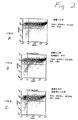
上記3つの形質導入プロトコルの比較を図2に示す。MOIが20のpN2cGFPによる形質導入後に、ビーズに固定したCD3抗体およびCD28抗体と接触させると、約91%の効率を生じた。形質導入前にビーズと接触させると、約89%の効率を生じ、そしてビーズ接触と形質導入とを同時に行うと、約80%の効率を生じた。この実験において、CD4+T細胞を、付着単球除去、CD14 MACS除去およびCD4 MACS濃縮によって、選択した。これらの抗体は、下記のように固定した。ベクターとの接触は、37℃および5% CO2にて行った。培養条件は、2%ヒト血清アルブミンを補充したYssel培地1mlあたりCD4+T細胞500,000個であった。FACS分析を、選択後7日目に行った。MFは、平均蛍光を示す。
【0104】
異なる刺激条件を比較する、7日後実験の結果を、図3に示す。CD4+細胞を、IL−2およびPHA−P、またはビーズに固定したCD3抗体およびCD28抗体のいずれかで24時間処理し、その後、MOIが20のpN2cGFPで1回形質導入した。並行比較において、固定した抗体の使用により、(CD4およびGFPの両方について陽性である細胞により示される)95%を超える形質導入効率を各回生じた。比較により、IL−2およびPHAによる刺激の結果は、わずか70.2〜84.5%の効率を生じた。FACS分析を、選択後7日目に行った。
【0105】
図4は、選択15日後の同様の実験の結果を示す。再度、細胞を、IL−2およびPHA−P、またはビーズに固定したCD3抗体およびCD28抗体のいずれかで24時間処理し、その後、MOIが20のpN2cGFPで1回形質導入した。このPHA−Pおよびビーズを、形質導入後7日目に除去し、そしてその細胞を、選択後15日目まで、1mlあたり500,000個でIL−2だけとともに培養した。固定した抗体を使用した後、その細胞のうち約93%が、CD4およびGFPの両方について陽性であった。IL−2およびPHAで処理した細胞のうちの約75%のみが、CD4およびGFPの両方について陽性のままであった。これらの結果は、7日後に陽性として検出された細胞(図3)のうちの少数が、「偽トランスフェクション(pseudotransfection)」に起因し得たことを示す。
【0106】
(実施例IV)
(異なるベクターは、高効率で細胞を安定に形質導入する)
本実施例は、形質導入のために使用されるベクターの比較である。pN2cGFPは、gagおよびpolコード配列全体を含み、一方pN1(cpt)GFPは、4551〜5096部分(非コード)pol配列を含む。図5に示されるその結果から理解され得るように、両方のベクターは、ビーズに固定したCD3抗体およびCD28抗体による刺激とMOIが20のベクターによる刺激を同時に行った後、非常に効率がよい初代CD4細胞の形質導入を示す。FACS分析を、選択後10日目に実施した。
【0107】
(実施例V)
(形質導入効率に対するMOIの効果)
異なるMOIの効果を図6に示す。ここで、2〜20のMOIを使用すると、形質導入効率72.7〜83.8%を生じた。細胞を、ビーズに固定したCD3抗体およびCD28抗体に24時間接触させ、その後、異なるMOIのpN1(cpt)cGFPで形質導入した。
【0108】
(実施例VI)
(CD34陽性細胞の形質導入)
CD34陽性細胞を、臍帯血から調製し、そしてFLT−3リガンド、TPOおよびKitリガンド(各100ng/ml)の存在下で、pN1cptGFPで4回同時に形質導入した。これらの細胞を、長期培養(LTC−IC)にて5週間培養し、その後、その細胞を、分析前の10日間メチルセルロース中で培養した(結果は、培養中6週間を超える経過時間に由来する)。図7における結果は、CD34未成熟細胞から生じる成熟CD45陽性細胞を分析する。コントロール細胞は、有意な形質導入を示さないが、一方、ベクターで形質導入した細胞は、88%を超える細胞がCD45およびGFP陽性を示す。
【0109】
(実施例VII)
(CD34陽性細胞の長期形質導入)
CD34陽性細胞を、上記のようにpN1(cpt)GFPで形質導入し、そして部分照射したSCIDマウスの骨髄に移植した。8週間後、細胞を単離し、そしてCD45保有成熟ヒト細胞およびGFP発現について、FACSによって分析した。その結果を、図7パネルA〜Dに示す。
【0110】
パネルAは、ベクターで形質導入していないヒト細胞を移植したコントロールマウスによる結果を示す。
【0111】
パネルBは、100ng/mlのFLT−3リガンド、TPOおよびKitリガンドの存在下で、4日連続してMOIが50であるpN1(cpt)GFPベクターで形質導入した細胞を移植したマウスによる結果を示す。このマウスは、形質導入の8週間後の、96.3%の著しい形質導入効率の形質導入されたヒト細胞(CD45陽性細胞)を示す。このマウスにおけるヒト細胞移植のレベルは、以前に報告された結果と一致して、11.1%であった。
【0112】
パネルCおよびDは、パネルBにおいてと同様に処理した他の2匹のマウスによる結果を示す。この結果は、高効率形質導入の再現性を確認し、87.8%および89.6%のCD45陽性細胞がGFP陽性でもあった。
【0113】
平均効率は91.2%であり、これは、長期に安定な形質導入を反映する。
【0114】
(実施例VIII)
(細胞表面結合分子の固定)
本実施例は、以下の実施例における使用のためにエポキシダイナルビーズにCD3(B−B11)抗体およびCD28(B−T3)抗体を直接連結することを記載する。
【0115】
1.0.618gのホウ酸を95mlの組織培養グレード水中に溶解することによって、0.1Mホウ酸溶液を調製する。十分に混合し、そして最高品質のNaOHを使用して、pHを9.5に調整する。最終容積を100mlにし、そして0.2μmフィルターを介して滅菌する。容器を密封し、そして4℃で保存する。
【0116】
2.上記ホウ酸溶液に濃度150μg/mlで抗体を添加する。B−B11抗体およびB−T3抗体の両方について、ホウ酸溶液1mlあたり各75μg添加する。体積を合計1mlにする。ホウ酸濃度は、抗体の添加後に0.05M未満であってはならない。各1mlのホウ酸/抗体溶液について、4×108個のEpoxyビーズを添加する。
【0117】
3.回転輪上で37℃にて24時間インキュベートする。
【0118】
4.ビーズを10分間にわたり3回、各々22℃でビーズ洗浄培地を用いて洗浄する。このビーズ洗浄培地は、カルシウムおよびマグネシウムを含まないリン酸緩衝化生理食塩水、3%ヒト血清アルブミン、5mM EDTA、および0.1アジ化ナトリウムである。
【0119】
5.22℃にてビーズを1回30分間洗浄する。
【0120】
6.4℃で一晩洗浄する。
【0121】
7.新鮮なビーズ洗浄培地で置換し、ビーズを2×10⁸ビーズ/mlになるように再懸濁する。IgGでコートしたビーズは、4℃にて少なくとも6ヶ月安定である。
【0122】
(実施例IX)
(樹状細胞の形質導入)
末梢血から単球を単離し、その後、MOIが50にてpN2cGFPを用い、2つの同時サイトカイン条件を使用して3日間連続して形質導入した。この同時サイトカイン条件は、GM−CSF(800単位/ml)、IL−4(500単位/ml)およびTNF−α(100単位/ml)、またはGM−CSF(5000単位/ml)およびインターフェロン−α(800単位/ml)である。図9のパネルAは、第1のサイトカイン条件が90.2%の効率を生じた、形質導入7日後の結果を示す。第2のサイトカイン条件下で、ベクターで形質導入した細胞は、7日後に92.9%の効率を示す(パネルB)。CD86は、樹状細胞について可能な唯一のマーカーであり、そしてCD86陰性細胞もまた樹状細胞であり得ることに留意すべきである。
【0123】
(参考文献)
【0124】
【表1】

【0125】
ここまで、本発明を完全に記載してきたが、本発明の精神および範囲から逸脱せずに、かつ過度な実験を要さずに、広範な等価のパラメーター、濃度、および条件内で本発明が実行され得ることが、当業者に明らかである。
【0126】
本発明を、その特定の実施形態と組み合わせて記載してきたが、これをさらに改変し得ることが理解される。本出願は、一般に、本発明の原理に従う本発明のいずれのバリエーション、用途または適合をもカバーすることが意図され、そしてこれらには、本発明が属する分野内で公知になるかまたは慣用的に実行され、そして本発明が上記のように添付の特許請求の範囲において示された後に本明細書中の重要な特徴に適用され得るので、本発明の開示からのこのような逸脱が含まれる。
【0127】
本明細書中上記および上記の特許請求の範囲において、用語「1つの」(「a」または「an」)の使用は、単数形を規定することに限定されない。その代わり、この用語の使用は、複数形を包含する。例えば、用語「1つの抗体(an antibody)」は、1つの単一抗体分子の単数形に限定されないが、むしろ、これらの抗体が、言及される抗体の同一コピーである限り、複数の抗体分子の存在を包含する。同様に、「1つのウイルスベクター(a viral vector)」は、1つの単一ウイルスベクター分子または1つのウイルス粒子に限定されない。
【図面の簡単な説明】
【図1】
図1Aおよび1Bは、それぞれ、pN2cGFPおよびpN1GFP(cPT)のマップを示す。種々の制限酵素部位およびHIV由来の成分が示される。pN2cGFP構築物は、CMV(サイトメガロウイルス)プロモーターに作動可能に連結されたGFPコード配列を含み、従って、GFP発現を制御する。pN1GFP(cPT)構築物は、上記でpN1(cpt)CGFPとも呼ばれ、これは、HIV pol遺伝子由来のcPPTを含む。これらの構築物は、上記実施例において使用される。
【図2】
図2は、固定されたCD3抗体およびCD28抗体をコーティングしたビーズを使用する、初代T細胞の形質導入の結果を示す。これらの細胞に、ビーズとの接触前にベクターを接触させたか(パネルA)、ベクターとの接触前にビーズを接触させたか(パネルB)、またはベクターおよびビーズの両方を同時に接触させた(パネルC)。形質導入されたベクターによってコードされるGFPからの蛍光に基づくフローサイトメトリーの結果は、パネルA〜Cの細胞が、それぞれ、90.70%、87.19%および79.14%形質導入されたことを示す。
【図3】
図3は、ウイルスベクターとの接触前にCD4+細胞を刺激するための、IL−2およびPHA−Pまたはビーズ固定化CD3抗体およびCD28抗体のいずれかを使用する形質導入の比較を示す。固定化抗体の使用は、各回で95%を超える形質導入効率を生じた。IL−2およびPHAの使用は、70.2%〜84.5%の効率しか生じなかった。
【図4】
図4は、本発明の方法を使用する、ヒトCD4+T細胞形質導入の頻度を示す。形質導入後15日での、コントロール細胞 対 MOI20で緑色蛍光タンパク質(GFP)を発現し得るベクターによって形質導入された細胞のフローサイトメトリー分析の比較は、形質導入された細胞の約93%がまた緑色の蛍光を生じることを示した。図4は、IL−2およびPHA−Pまたはビーズ固定化CD3抗体およびCD28抗体のいずれかを使用する形質導入後14日での、CD4+細胞およびGFP+細胞についてのFACS分析の結果を示す。抗体で処理した細胞の約93%が、14日後に安定に形質導入されたままであった。IL−2およびPHAで処理した細胞の約75%のみが、同じ時間後で安定に形質導入されたままであった。
【図5】
図5は、異なるウイルスベクターで形質導入された細胞の結果を示す。
【図6】
図6は、トランスフェクション効率に対する異なるMOIの使用に由来する影響を示す。
【図7】
図7は、細胞表面結合分子の存在下でウイルスベクターでの複数回の形質導入後の、臍帯血から調製されたCD34+細胞の安定な形質導入を示す。88%を超える細胞が、形質導入後6週間を超えた後で陽性のままであった。
【図8】
図8、パネルA〜Dは、SCID(重症複合型免疫不全)マウスに対する移植後の長期の形質導入の効率を示す。約8週間後、成熟なまま存続した、平均91%を超える形質導入された細胞が、形質導入されたGFPマーカーの発現について陽性のままであった。
【図9】
図9、パネルAおよびBは、7日後の樹状細胞形質導入の効率を示す。[0001]
(Technical field)
The present invention relates to methods and compositions related thereto for efficient and stable transduction of cells using viral vectors. This method increases the efficiency of transduction by contacting the cell to be transduced with one or more molecules that bind to the cell surface. This contacting step can occur before, after, or simultaneously with the introduction of the viral vector into the cell. The invention also relates to the use of stably transduced cells in other applications, including expression of nucleic acids produced by vectors or treatment of living organisms.
[0002]
(Background technology)
"Transfection" generally refers to a technique for the introduction of genetic material into cells, which technology has made significant contributions to the molecular and recombinant circulations of biology. Examples of transfection techniques for use in higher eukaryotic cells include calcium phosphate precipitation, DEAE dextran treatment, electroporation, microinjection, lipofection, viral infection, and are found in many scientific textbooks and journals Other techniques may be mentioned.
[0003]
Among the transfection techniques, the use of viral infection is unique in that the naturally occurring means by which the virus introduces its genetic material into the cell is used to transfer the nucleic acid molecule of interest into the cell. . Examples of viruses modified and applied for such techniques include adenovirus, adeno-associated virus, herpes simplex virus, and retrovirus. Generally, the nucleic acid molecule of interest can be cloned into the viral genome. Upon replication and packaging of the viral genome, the resulting viral particles can deliver the nucleic acid of interest to cells via a viral entry mechanism.
[0004]
Generally, the viral genome is first made replication-defective by nucleic acid manipulation before the addition (addition) of the nucleic acid of interest. The resulting viral genome, or viral vector, requires the use of a helper virus or packaging system to complete the assembly and release of the viral particles from the cell. When a viral vector or particle is used to transfer the genetic material of interest into cells, this technique is called "transduction". Thus, generally, “transducing” a cell refers to using a viral vector or virus particle to transfer genetic material to the cell.
[0005]
Among the transduction techniques, the use of retroviruses has been an interesting topic for genetic modification of mammalian cells. Of particular interest is the use of retroviruses that have been modified to introduce genetic material into cells to treat genetic deficiencies and other diseases. An example of this approach is in the case of hematopoietic cells, where retroviral and lentiviral vectors are the subject of intense research.
[0006]
For example, Movassag et al. Discuss their work on attempts to increase the efficiency of retrovirus-mediated transduction by including the results from studies on the cell cycle of activated T cells. Consequently, their outcome depends on active cell division during transduction. This study is also limited to the use of murine tumor retroviruses and the requirement for significant pre-stimulation of cells prior to transduction.
[0007]
June et al. (WO 96/34970) describe the use of T cell stimulation as a means to increase T cell transfection. Other studies on T cell transduction with activated or stimulated cells include the following: Douglas et al., Hooiberg et al., Onodera et al., Klebba et al., Barry et al., And Unutmaz et al. Unfortunately, none of the studies demonstrated transduction efficiencies greater than about 65%.
[0008]
Costello et al. Describe the transduction of both stimulated and unstimulated T cells using a Human Immunodeficiency Virus-1 (HIV-1) lentiviral vector. They observed that the stimulated primary T cells had an efficiency of almost up to 17% and the unstimulated T cells had an efficiency of less than 19%. They also noted a limited ability to increase efficiency by less than 36% in stimulated T cells by including the presence of the HIV-1 accessory protein.
[0009]
Also describe increased transduction efficiency in the presence of HIV-1 accessory proteins in both unstimulated and mitogen-stimulated T cells. Like Movassag et al., Chinnasami et al. Pro-stimulated blood lymphocytes for a long time before transduction with a lentiviral vector. First observed transduction efficiencies greater than 96% three days after transduction. The percentage of stably transduced cells decreased to 71.2% two weeks after transduction. Haas et al. Also observed transient transduction and "pseudotransduction" in cells transduced with a lentiviral vector capable of expressing a marker gene (green fluorescent protein). Even three days after transduction, large (> 10%) transient transduction was detected based on incomplete expression of the marker gene in transduced primary CD34 + cord blood cells. Such expression by transient transduction remained detectable at about 5%, even 7 days after transduction. Only about 10 days after transduction, expression from transient transduction closely resembled expression in cells transduced with the markerless vector.
[0010]
Thus, Chinnasami et al. Demonstrated that stable transduction of more than 71.2% of primary lymphocytes (integrated form of the viral vector resulted in the chromosomal DNA of the transduced cells, as reflected by the efficacy after 2 weeks). Inserted) could not be achieved. This was independent of the use of cytokines on prestimulated cells. In addition, Chinnasamy has shown that non-stimulated lymphocytes can be treated with HIV vectors that do not express accessory proteins (Vif, Vpr, Vpu, and Nef) (the cells can be transduced with PHA mitogens and IL-2 cytokines). No significant transduction was achieved (only 3.6% at 14 days post-transduction), despite subsequent stimulation. This result was somewhat improved by the use of unstimulated cells and a vector that did not contain accessory proteins, but regardless of the stimulatory protocol used with this vector, the efficiency of stable transduction of stimulated or unstimulated cells was It was never greater than 75% 14 days after transduction.
[0011]
The low frequency of stable transduction with lentiviral vectors was also observed by Hass et al. Hass et al. Achieved a maximum stable transduction efficiency of less than 25% with primary CD34-positive cord blood cells 7 days after transduction. Remarkably, the upper limit of 25% of this transduction is an excessively high multiplicity of infection (multiplicity) or vector concentration (eg, multiplicity of infection (MOI) up to 9000, and 10%). 8 Even after (infection unit / ml of vector concentration), no improvement could be achieved.
[0012]
Follenzi et al. Also reported that 500 transduced cells in the presence of a three cytokine cocktail containing interleukin 3 (IL-3), interleukin 6 (IL-6), and stem cell stimulating factor (SCF). Very high MOI was used. Interestingly, the use of this cocktail renders cells unsuitable for human clinical transplantation.
[0013]
Thus, there remains a need to provide more efficient means of stably transducing cells with vectors at high frequencies. In addition, there is a need for more efficient means of transducing unstimulated cells, both for use as search tools and as therapeutic agents.
[0014]
Citation of the above document is not intended to be an admission that any of the foregoing is related prior art. All statements as to the date or presentation of the content of these documents are based on the information available to the applicant and do not constitute any admission as to the accuracy of the dates or the content of these documents.
[0015]
(Disclosure of the Invention)
The present invention provides highly effective methods for stable transduction of cells using viral vectors and viral particles, and compositions related thereto. "Stable transduction" means that the integrated form of the viral vector has been inserted into the chromosomal DNA of the transduced cells. The method includes exposing a cell to be transduced into contact with at least one molecule that binds to the cell surface. This contacting step can occur before, during, or after the cells are exposed to the viral vector, or viral particles. Hereinafter, the term “viral vector” is used to mean any form of nucleic acid from a virus, and is used to transfer genetic material into a cell via transduction. The term encompasses viral vector nucleic acids (eg, DNA and RNA), encapsulated forms of these nucleic acids, and viral particles in which the viral vector nucleic acids are packaged.
[0016]
The invention also relates to other applications, such as the production of useful gene products and proteins by the expression of nucleic acids present in this vector, or the treatment of living subjects who are or are at risk of having a disease. Including the use of transduced cells. Preferably, the subject is a human.
[0017]
The at least one molecule that binds to the surface of the cell to be transduced includes any molecule that physically interacts with a receptor, marker, or other recognizable moiety on the surface of the cell. Basically, any cell surface binding molecule can be used for efficient transduction of cells. Without wishing to be bound by theory, this cell surface binding molecule can result in: the host cell chromatin being more receptive to DNA integration; the transfer of the viral vector into a preferred site for vector gene expression. Preferential uptake; more efficient entry of capsid-containing nucleic acids into the cytoplasm; more efficient entry of virus across cell membranes or integral membrane structures (eg, endosomes); or nuclear import of viral vector genetic material ( Creation of more permissive cells for transport). The method of the invention may also include more than one of the above possibilities. Also, and as evident from the number and variety of possibilities described above, the invention may not be limited to any one theory. Considering the extraordinary findings of the present invention, instead and in the stable transduction of up to 100% of the treated cells without negatively affecting the possible use of this cell in human therapy, the present invention Should be viewed as opening up new approaches in the field of human cell therapy.
[0018]
Although not all cell surface binding molecules, viral vectors result in efficient and stable transduction. For example, binding to cell surface molecules that induce apoptosis does not result in efficient transduction of cells, but rather cell death. Cell death may be preferred for killing cells (eg, tumor cells), but not for stable transduction of cells with vectors containing a carrier (payload) gene or nucleic acid sequence. Preferred cell surface binding molecules result in cells that are more receptive to transduction with a viral vector. Examples of such molecules include antibodies to specific cell surface receptors or portions thereof, and ligands or binding domains for such receptors. In addition, antigen binding fragments of antibodies (eg, F ab , And F v Fragment) is intended for use in the present invention. The binding domain for a specific cell surface receptor can include a single epitope or more than one epitope.
[0019]
Preferred examples of cell surface binding molecules for use in the present invention are anti-CD3 and anti-CD28 antibodies that bind to T cells and render them more receptive to vector transduction. Other preferred cell surface binding molecules are antibodies or ligands to FLT-3 ligand, TPO, and Kit ligand receptors, which further render the cells expressing the receptor (eg, hematopoietic stem cells) more amenable to vector transduction. Make it sex. Further preferred cell surface binding molecules are antibodies or ligands for the GM-CSF and IL-4 receptors, which can be used on dendritic cells or their precursors (eg, on monocytes, CD34-positive stem cells, or dendritic cell lineages). Of its differentiated progenitor cells) are more receptive to vector transduction. Other cell surface binding molecules include those found on the cell surface that bind to the surface of another cell.
[0020]
Further examples of cell surface binding molecules include polypeptides, nucleic acids, carbohydrates, lipids, and ions, all optionally complexed with other substances. Preferably, the molecule binds factors found on the surface of blood cells such as: CD1a, CD1b, CD1c, CD1d, CD2, CD3γ, CD3δ, CD3ε, CD4, CD5, CD6, CD7, CD8α. , CD8β, CD9, CD10, CD11a, CD11b, CD11c, CDw12, CD13, CD14, CD15, CD15s, CD16a, CD16b, CD18, CD19, CD20, CD21, CD22, CD23, CD24, CD25, CD26, CD27, CD28, CD29 , CD30, CD31, CD32, CD33, CD34, CD35, CD36, CD37, CD38, CD39, CD40, CD41, CD42a, CD42b, CD42c, CD42d, CD43, CD44, CD45, CD45R, C D46, CD47, CD48, CD49a, CD49b, CD49c, CD49d, CD49e, CD49f, CD50, CD51, CD52, CD53, CD54, CD55, CD56, CD57, CD58, CD59, CDw60, CD61, CD62E, CD62L, CD62P, CD63, CD64, CD65, CD66a, CD66b, CD66c, CD66d, CD66e, CD66f, CD67, CD68, CD69, CDw70, CD71, CD72, CD73, CD74, CDw75, CDw76, CD77, CD79α, CD79β, CD80, CD81, CD82, CD83, CD84, CD85, CD86, CD87, CD88, CD89, CD90, CD91, CDw92, CD93, CD94, CD95, CD9 6, CD97, CD98, CD99, CD100, CD101, CD102, CD103, CD104, CD105, CD106, CD107a, CD107b, CDw108, CDw109, CD114, CD115, CD116, CD117, CD118, CD119, CD120a, CD120b, CD121a, CD121b, CD122, CD123, CDw124, CD125, CD126, CDw127, CDw128a, CDw128b, CDw130, CDw131, CD132, CD133, CD134, CD135, CD136, CDw137, CD138, CD139, CD140a, CD140b, CD141, CD142, CD143, CD144, CDw14. CD146, CD147, CD148, CD 149, CD150, CD151, CD152, CD153, CD154, CD155, CD156, CD157, CD158a, CD158b, CD161, CD162, CD163, CD164, CD165, CD166, and TCR zeta. Lower case letters (eg, “a” or “b”) indicate a complex CD molecule that consists of multiple gene products or belongs to a family of structurally related proteins. The symbol "w" refers to a putative CD molecule that has not yet been fully identified. A more complete listing of CD molecules is found in Kishimoto, T (eds). Current information on CD molecules is also available at the following Web: http: // www. bsi. vt. Shaw, S.D. at An International WWW Resource / Journal at edu / immunology. (Ed.) Found in Protein Reviews.
[0021]
More preferred are molecules that bind to factors found on the surface of lymphocytes, T cells and leukocytes, such factors are for example: CD2, CD3γ, CD3δ, CD3ε, CD5, CD6, CD7, CD8α, CD8β, CD9, CD11a, CD18, CD25, CD26, CD27, CD28, CD29, CD30, CD37, CD38, CD39, CD43, CD44, CD45R, CD46, CD48, CD49a, CD49b, CD49c, CD49d, CD49e, CD49f, CD50, CD53, CD54, CD56, CD57, CD58, CD59, CDw60, CD62L, CD68, CD69, CDw70, CD71, CD73, CDw75, CDw76, CD84, CD85, CD86, CD87, CD 89, CD90, CD94, CD96, CD97, CD98, CD99, CD100, CD101, CD103, CD107a, CD107b, CDw108, CDw109, CD118, CD119, CD120b, CD121a, CD122, CDw124, CDw127, CDw128a, CDw130, CD132, CD134, CDw137, CD140a, CD140b, CD143, CD146, CD148, CD152, CD153, CD154, CD155, CD161, CD162, CD165, CD166, and TCRII.
[0022]
Additional antibodies and molecules that bind to cell surfaces and are suitable for use in the present invention are described in Linscott's Directory of Immunological and Biological Reagents, Eleventh Edition, January 2000, Publisher: W.S. D. Linscott, Petaluma, CA, which is hereby incorporated by reference as if fully set forth. However, in some embodiments of the present invention, the cell surface binding molecule is not a cytokine.
[0023]
While the present invention may be embodied by the use of a soluble cell surface binding molecule that facilitates vector transduction of cells, other preferred embodiments include the use of immobilized cell surface binding molecules. Preferably, the immobilized molecule is an antibody. Alternatively, immobilization may be through the use of other cells that express the cell surface binding molecule. A preferred method for efficient transduction of hematopoietic stem cells is to include bone marrow stromal cells that express a ligand on their surface that promotes maintenance of the stem cells without differentiation during transduction. The stimulating cells are not limited to native cells, but any cell can be engineered to express the appropriate cell surface binding molecule to provide the correct stimulus for transduction.
[0024]
Additional molecules that increase or enhance the ability of at least one molecule to bind to the cell surface can also be included. For example, a soluble form of the (primary) antibody to a specific cell surface receptor can be used in combination with a secondary antibody that can crosslink the primary antibody already bound to the cell surface.
[0025]
Of course, any cell can be used in the practice of the present invention. Preferably, the cells to be transduced are eukaryotic cells. More preferably, the cells are primary cells. However, cell lines can also be transduced using the methods of the invention, and in many cases, cell lines can be more easily transduced. In one preferred embodiment, the cells to be transduced are primary lymphocytes (eg, T lymphocytes) or macrophages (eg, monocyte macrophages), or precursors to any of these cells (Eg, hematopoietic stem cells). Other cells that are preferred for transduction are generally hematopoietic cells or, more generally, cells formed by hematopoiesis and cells associated with hematopoietic stem cells and blood cell function. It is. Such cells include granulocytes and lymphocytes formed by hematopoiesis and pluripotent lymphoid and pluripotent myeloid stem cells. Cells associated with blood cell function include cells that support the function of immune stem cells (eg, antigen presenting cells, such as dendritic cells, endothelial cells, monocytes and Langerhans cells). In a preferred embodiment, the cells are T lymphocytes (ie, T cells) (eg, T lymphocytes that express CD4 and CD8 markers).
[0026]
In a particularly preferred embodiment, the cells are primary CD4 + T lymphocytes or primary CD34 + hematopoietic stem cells. However, if a viral vector for use in the present invention can be pseudotyped with the vesicular stomatitis virus envelope G protein (as discussed below), any cell will be treated via the method of the present invention. It can be transduced. Such cells include astrocytes, skin fibroblasts, epithelial cells, neurons, dendritic cells, lymphocytes, cells associated with the immune response, vascular endothelial cells, tumor cells, tumor vascular endothelial cells, liver Contributes to cells, lung cells, bone marrow cells, antigen presenting cells, stromal cells, adipocytes, muscle cells, pancreatic cells, kidney cells, ova or spermatocytes (eg, to create transgenic animals), germline Cells, progenitor pluripotent stem cells or precursors thereof, blood cells including non-nucleated cells such as platelets and red blood cells, and the like. Preferably, the cell is a eukaryotic multicellular species (eg, a cell as opposed to a unicellular yeast cell), and even more preferably, is of mammalian origin (eg, a human cell).
[0027]
The cells to be transduced can exist as a single entity or can be part of a large population of cells. Such “large populations of cells” include, for example, cell cultures (either mixed or pure), tissues (eg, epithelial, stromal or other tissues), organs (eg, , Heart, lung, liver, gallbladder, bladder, eye, and other organs), organ systems (eg, circulatory, respiratory, gastrointestinal, urinary, nervous, tegumental or other organ systems), Blastocysts, embryonic stem cells, cells from a fetus (eg, for treatment of a genetic disorder / genetic disease or for producing a transgenic animal), diseased tissue (eg, a tumor or site of infection), or organism (Eg, birds, mammals, marine life, fish, plants, etc.). Preferably, the targeted organs / tissues / cells are circulatory organs / tissues / cells (including, but not limited to, heart, blood vessels, and blood), respiratory organs / Tissue / cells (eg, nose, pharynx, larynx, trachea, bronchi, bronchiole, lung, etc.), organs / tissues / cells of the gastrointestinal system (eg, oral and oral tissues, pharynx, esophagus, stomach, intestine, salivary glands , Pancreas, liver, gall bladder, etc.), organs / tissues / cells of the breast system (eg, breast epithelial cells and supporting cells in tissues), organs / tissues / cells of the urinary system (eg, kidney, ureter, bladder) , Urethra, etc.), organs / tissues / cells of the nervous system (including but not limited to brain and spinal cord, and special sensory organs such as the eye), and organs / tissues / cells of the integumentary system (eg, skin) ).
[0028]
Even more preferably, the cells to be transduced are cells of the heart, cells of blood vessels (including tumor vessels and blood vessels associated with infected or diseased tissue), cells of bone marrow, cells of blood, cells of brain , Lymphoid cells, lymph node cells, spleen cells, lung cells, liver cells, gallbladder cells, bladder cells and eye cells. In one particular embodiment of the invention, the cell is an allogeneic cell that is autologous to the host for which it is intended to be used, but is partially or completely incompatible with the host. Or cells that are even heterologous to the host can also be used. In addition, universal donor cells suitable for use in any given host organism (related organisms or species (eg, humans)) can be transduced. This latter embodiment of the invention is particularly important in the transplantation of cells, tissues, or organs, and the source of the transduced cells may be important to the outcome of the transplant.
[0029]
Another preferred cell for transduction according to the method of the invention is a tumor cell, a diseased cell, or due to its genetic makeup or the genetic makeup of other cells present in the same organism. Cells that may become abnormal over time. The latter embodiment allows the transduced cells of the invention to be used in prevention. Breast cancer is one example of a disease process in which prognostic indicators allow treatment with the transduced cells of the invention as an early genetic intervention before the disease later occurs. However, the methods of the invention can also be used for therapeutic treatment of breast cancer after the disease has been detected. There are many additional applications of the present invention in the treatment of cancer, and those skilled in the art may use the invention presented herein for the treatment of many types of cancer without undue experimentation.
[0030]
By way of example, and not limitation, one application is in breast cancer that is estrogen dependent. Cancer cells in estrogen-dependent breast cancer are preferentially transduced by using an antibody or ligand that binds to an estrogen receptor in combination with a therapeutic viral vector. For example, the vector can include a tumor inhibitory gene, such as a herpesvirus thymidine kinase gene. Thus, transduced cells can be selectively killed by the addition of ganciclovir, a prodrug that can be activated by herpes thymidine kinase. Further examples of tumor inhibiting genes and corresponding prodrugs are numerous and well known in the art, and can be selected by one of skill in the art without undue experimentation. The use of activatable prodrugs in combination with the application of the transduction method of the invention can be widely applied to other tumor types, and the above examples show that the invention makes the invention hormone-dependent for growth or proliferation. Not limited to tumors that are or are dependent on some soluble factors.
[0031]
For example, Her-2 / neu-positive tumor cells are not estrogen-dependent and are insufficient as prognostic indicators. This is because non-estrogen dependent tumors containing such cells are very resistant to treatment with agents such as the estrogen antagonist taxol. Preferred embodiments for the present invention include antibodies or other molecules that bind to Her-2 / neu or heregulin using a viral vector preparation during transduction of tumor-contaminated cells, eg, in a bone marrow transplantation protocol. It is to include. Alternatively, transduction can be done directly at the tumor site, or in vivo in vivo using a vector that modulates tumor formation.
[0032]
Yet another embodiment of the present invention is to target tumor vasculature, alone or in combination with targeting tumor cells. St Croix et al., Which is hereby incorporated by reference as if fully set forth, showed by SAGE analysis that specificity in tumor endothelial cells as compared to normal endothelial cells was high. A gene that was overexpressed was identified. Many of these genes encode cell surface molecules (eg, Thy-1 cell surface antigen or Endo180 lectin). All of the up-regulated cell surface factors may be bound by the cell surface binding molecules of the invention to provide a stimulus for efficient and stable gene transfer. Thus, approaches to tumor therapy are based on transducing tumor endothelial cells with therapeutic viral vectors in the presence of cell surface binding molecules that selectively bind to tumor vasculature but do not bind to normal endothelial cells. Destroying the tumor vasculature by killing the tumor endothelial cells.
[0033]
Yet another embodiment of the present invention provides a method for constructing an element (e.g., such as a viral vector) that selectively promotes expression of an anti-tumor gene in a tumor but does not promote expression of the anti-tumor gene in normal vascular endothelial cells. Selective expression of anti-tumor genes in tumor vasculature by incorporating a cis-acting stabilizing element / cis-acting degradation element on the promoter or mRNA. Such methods can occur ex vivo, in vitro, or in vivo. If tumor vascular endothelial cells are targeted, the preferred method for treatment is in vivo. Alternatively, for example, if the aim is to clear contaminating tumor cells from the bone marrow for a bone marrow transplant, the preferred method for treatment occurs ex vivo or in vitro.
[0034]
Furthermore, in vivo use is not limited to disease states and can be used to transduce normal cells. For example, the invention can be used to transduce hematopoietic stem cells in bone marrow in vivo. Any combination of antibodies or other cell surface binding molecules (eg, FLT-3 ligand, TPO and Kit ligands, or functional analogs thereof, or stromal cells expressing cell surface binding molecules) can be highly efficient. For bone marrow transduction, it can be added with the vector upon direct injection into the bone marrow. The term “functional analog” refers to any molecule that retains the cell surface binding activity of a cell surface binding molecule of the invention. Such functional analogs include FLT-3 ligand, TPO and Kit ligand fragments; FLT-3 ligand molecules, TPO molecules and Kit ligand molecules, including one or more amino acid substitutions, amino acid additions, or amino acid deletions. And antibodies that mimic the cell surface binding activity of cell surface binding molecules.
[0035]
An alternative approach to the above is to use bone marrow stromal cells as producer cells of the viral vector, and thus provide the vector and cell surface binding molecule via cell therapy rather than as a vector preparation. . Another example is the characterization of T cells or dendritic cells by adding functional analogs of the CD3 and CD28 antibodies, or GM-CSF and IL-4, respectively, along with the vector during subcutaneous injection. It is an introduction. Lymph in the subcutaneous tissue drains vectors and stimulants to the lymph nodes for efficient transduction of the targeted tissue.
[0036]
The present invention has the advantage that purification of the cells to be transduced is not required, if necessary. Transduction predominantly of the cell type of interest can be achieved by selection of the cell surface portion to be bound. Thus, for example, in a mixed population of blood cells, transduction of cells expressing CD3 (eg, a particular T cell) is enhanced if a CD3-specific antibody is used to interact with the cells. This occurs in preference to other cell types in the population (eg, granulocytes and monocytes that do not express CD3).
[0037]
The present invention also includes transduction of purified or isolated cell types, if desired. The use of purified or isolated cell types offers additional advantages, such as higher transduction efficiencies (due to higher vector concentrations compared to the cells being transduced). .
[0038]
When purified T cells are transduced, at least one molecule preferably binds to a cell surface molecule found on the T cell. Examples of such cell surface molecules include CD3, CD28, CD25, CD71, and CD69. Examples of molecules that bind to these cell surface molecules include antibodies and monoclonal antibodies that recognize these cell surface molecules, many of which are either commercially available or It is easily and routinely prepared using standard techniques without experimentation. In a preferred embodiment for transducing CD4 + or CD8 + cells, a monoclonal antibody that recognizes CD3 and / or CD28 may be used. Examples of such antibodies that are commercially available include OKT3 for CD3 and CD28.2 for CD28. These antibody molecules can be used in a soluble form (subsequently cross-linked by other molecules as needed), or can be used in an immobilized form, such as on beads or other solid surfaces. In a particularly preferred embodiment of the invention, the antibody is immobilized on the surface of a container, for example, on the wall of a well, plate or bag for tissue culture used for viral vector-mediated transduction. You. Without being bound by theory, the use of an immobilized antibody on the surface to which a cell attaches or contacts can increase the local concentration of cell surface interactions on that cell surface.
[0039]
When hematopoietic stem cells are transduced, antibodies specific for FLT-3 ligand, TPO (thrombopoietin or Megakaryocyte Growth and Development Factor), or a hematopoietic stem cell receptor for the Kit ligand, can Can be used as a surface binding molecule. Alternatively, antibodies to stem cell positive cell markers, including but not limited to CD34 or AC133, can be used. When a ligand comprising a compound or composition is used as a cell surface binding molecule, all natural ligands (including proteins, ligands or ligands that bind to heterologous proteins) are used, either in soluble or immobilized form Can be done. Immobilized forms include direct or indirect (eg, using avidin / biotin) attachment to microbeads.
[0040]
Alternatively, the ligand may be expressed in the viral envelope of a viral vector, optionally in the form of a chimeric or fusion protein, and / or (covalently or non-covalently) with one or more other proteins. It can be (covalently) complexed. In such embodiments, the cell surface binding molecule is presented in combination with a viral vector as a single composition for transducing cells. Further examples of cell surface binding molecules that can be expressed in the viral envelope include many of the surface factors listed above.
[0041]
Other preferred cell surface binding molecules (eg, antibodies or fragments thereof) are those that bind to Notch or Delta hematopoietic stem cell receptors, or that bind to Notch or Delta proteins themselves, or that bind to heterologous proteins. Molecule that binds to a ligand. Delta and Notch encode cell surface proteins that influence a wide variety of cell fate decisions in Drosophila development. Vertebrate homologs of Delta and Notch are essential for normal embryonic development. Delta homologs are critically involved in the regulation of hematopoiesis. The soluble form of the homolog, Delta-Serrate-lag2 (DSL), enhances the expansion of primitive hematopoietic precursors. When combined with hematopoietic growth factors (including interleukin-3 (IL-3), granulocyte colony stimulating factor (ie, G-CSF) or granulocyte macrophage colony stimulating factor (GM-SCF)), DSL: It promoted the expansion of primordial hematopoietic progenitors and, at the same time, inhibited the primordial progenitors from differentiating into more mature precursor cells responsive to IL-3 alone (see Han et al. thing). DSL probably acts by activating Notch receptors expressed on hematopoietic cells, selectively blocking cell differentiation's ability to respond to normal hematopoietic growth factors, but blocking growth signals. Adjust by not doing (see Han and Moore, Blood 1999). Accordingly, Delta and Notch homologs, antibodies that are functional analogs to these homologs, are useful for achieving efficient vector transduction of cells (especially hematopoietic stem cells) of greater than 75%, More preferred cell surface binding molecules.
[0042]
The invention includes a viral vector and a composition comprising the viral vector for use in the disclosed methods. This vector is preferably a retrovirus (Retroviridae) vector, more preferably a lentivirus vector. Other retroviral vectors, such as oncoviral and murine retroviral vectors, can also be used. Additional vectors can be derived from other DNA viruses, or viruses that can convert their genome to DNA during some point in their life cycle. Preferably, the virus is an adenoviridae, a parvoviridae, a hepadnaviridae (usually not classified as a hepadnaviridae, including hepatitis delta virus and hepatitis E virus), a papovaviridae ( Viruses, including the polyomaviridae and papillomaviridae), herpesviridae, and poxviridae.
[0043]
Additional viruses of the Retroviridae (ie, retrovirus) are Oncovirinae, Spumavirinae, Spumavirus genus, Lentivirinae, and Lentivirus genus or subfamily viruses. The RNA virus of the Oncovirinae is preferably human T
[0044]
The lentivirus subfamily RNA virus is desirably human
[0045]
Particularly preferred lentiviral vectors are vectors derived from HIV, more preferably vectors derived from HIV-1, HIV-2 or a combination of chimeras thereof. Of course, different serotypes of retroviruses, particularly HIV, can be used alone or in combination to prepare vectors for use in the present invention. Preferred vectors of the present invention contain cis-acting elements that are present in the wild-type virus but not in the "base" lentiviral vector. "Basic" lentiviral vectors contain minimally the LTR and packaging sequences in the 5 'leader sequence and gag coding sequence, while "basic" lentiviral vectors also optionally contain a Rev-dependent format. May include an RRE element that promotes nuclear transport of vector RNA. Preferred vectors further include a nucleotide sequence that enhances the efficiency of transduction of the cell.
[0046]
An example of such a vector is pN2cGFP, a vector containing the complete gag and pol sequences. Another example is the sequence about 4555-5096 in the pol (this position was courtesy provided by CE Buckler, NIAID, NIH, Bethesda, MD, pNL4-3 sequence with accession numbers M19921, HIVNL43 9709 bp. Is a reference position from (). However, any cis-acting sequence from wt-HIV that can improve vector transduction efficiency can be used. Another example of a vector that allows for efficient transduction via the present invention is the cr2HIV construct described in US Pat. No. 5,885,806.
[0047]
A previously identified sequence that was insufficient to significantly increase transduction efficiency was identified as a central DNA flap (corresponding to positions 4775-4935 of pNL4-3, pLAI3 178-bp fragment from positions 4793 to 4971) of Zennou et al. (2000). The present invention concludes that, although this small fragment is not sufficient to increase transduction efficiency, a larger 545 bp fragment (positions 4551-5096 of pNL4-3) or an even larger fragment containing it is Includes the finding that transduction could be increased as part of the present invention.
[0048]
A further example of a viral vector construct that can be used in the present invention is US Pat. No. 5,885,806, which is hereby incorporated by reference as if fully set forth. Is found in The constructs in this patent 5,885,806 are merely illustrative, not limiting the range of vectors that efficiently transduce cells. Instead, the construct may be such that the viral vector for use with the present invention may comprise minimal sequences from the wild-type virus, or may comprise sequences up to almost the entire genome of the wild-type virus, but may not replicate and replicate. Additional indication is provided to those skilled in the art that the essential nucleic acid sequences required for the production of the disease can be eliminated. Methods for accurately determining the sequence required for efficient transduction of cells are conventional and well known in the art. For example, it is conventional to systematically integrate viral sequences back into a "basic" vector, or delete sequences from a vector that contains substantially the entirety of the HIV (eg, cr2HIV) genome, and in the art. It is well known.
[0049]
In addition, placing sequences from other viral backbones into the viral vector of interest (eg, cytomegalovirus (CMV)) is also well known in the art. Regardless of the actual viral vector used, the various auxiliary proteins encoded therein, and the sequences present therein, the viral genetic material will increase the transduction efficiency in certain cell types if these proteins or sequences increase It can be left in the vector or helper genome. Many conventional screens are available to determine whether particular genetic material increases transduction efficiency by integrating its sequence into either the vector or the helper genome. Preferred embodiments of the present invention do not include accessory proteins in either the vector or the helper genome. However, this preference does not preclude embodiments of the invention in which accessory proteins and other sequences are left in either the vector or the helper genome to increase transduction efficiency.
[0050]
The viral vectors used in the present invention can also result in “pseudotyped” formation, wherein co-infection of cells with different viruses produces progeny virions that contain the genome of one virus and that of one virus The genome is encapsulated in an outer layer that contains one or more envelope proteins of another virus. This phenomenon has been used to package a viral vector of interest into "pseudotyped" virions, which encodes the viral vector of interest and at least one envelope protein or cell surface molecule of another virus. By co-transfecting or co-infection of packaging cells with both genetic materials. See U.S. Patent No. 5,512,421. Such mixed viruses can be neutralized by antisera to one or more heterologous envelope proteins used. One virus commonly used in pseudotyping is vesicular stomatitis virus (VSV), which is a rhabdovirus. The use of pseudotyping extends the host cell range of the virus by including elements of the viral entry machinery of the heterologous virus used.
[0051]
Pseudotyping of viral vectors and VSV for use in the present invention results in viral particles containing the viral vector nucleic acid encapsulated in a nucleocapsid, which is surrounded by a membrane containing the VSV G protein. The nucleocapsid preferably contains proteins normally associated with the viral vector. The membrane containing the peripheral VSV G protein forms part of the virus particle as it leaves the cell used to package the viral vector. Examples of packaging cells are described in US Pat. No. 5,739,018. In a preferred embodiment of the invention, the virus particles are derived from HIV and are pseudotyped by the VSV G protein. Pseudotyped viral particles containing the VSV G protein can infect many different cell types with greater efficiency than amphotropic viral viral vectors. The range of host cells includes both mammalian and non-mammalian species (eg, humans, rodents, fish, amphibians, and insects).
[0052]
Viral vectors for use in the transduction methods of the present invention also contain one or more nucleic acid sequences under the control of a promoter present in the virus or under the control of a heterologous promoter introduced into the vector, and express the same. I do. The promoter can further include an isolation element (eg, an erythroid DNAse hypersensitive site) to flank the operon for tightly controlled gene expression. Preferred promoters include HIV-LTR, CMV promoter, PGK, U1, EBER transcription unit from Epstein Barr virus, tRNA, U6 and U7. Although a Pol II promoter is preferred, a Pol III promoter can also be used. Tissue-specific promoters are also a preferred embodiment. For example, the beta globin locus control region enhancer and the alpha and beta globin promoters can provide tissue-specific expression in erythrocytes and erythroid cells. Another more preferred embodiment is to use a cis-acting sequence associated with the promoter. For example, as shown in U.S. Patent No. 5,814,500, which is incorporated herein by reference, the U1 gene can be used to enhance antisense gene expression, where non-promoter sequences Is used to target the antisense or ribozyme molecule to a target spliced RNA.
[0053]
Of course, any cis-acting nucleotide sequence from a virus can be incorporated into the viral vectors of the invention. In particular, cis-acting sequences found in the retroviral genome are preferred. For example, a cis-acting nucleotide sequence from the gag, pol, env, vif, vpr, vpu, tat or rev gene can be incorporated into a viral vector of the invention to further increase transduction efficiency. Preferably, the cis-acting sequence does not encode the expressed polypeptide; is not expressed as a polypeptide or part thereof due to genetic alterations (eg, deletion of the translation initiation site); A variant sequence that encodes only part or a fragment; or contains one or more substitutions, additions or deletions from the native sequence. An example of a cis-acting sequence is the cPPT (central polypurine region) sequence identified within the HIV pol gene.
[0054]
The one or more nucleic acid sequences in the viral vector of the invention may be found in the virus from which the vector is derived, or may be a heterologous sequence. The sequence is preferably a full-length or partial sequence encoding the gene product of interest. Such a sequence or gene product is preferably a biologically active agent that can produce a biological effect in the cell. Examples of such factors include proteins, ribonucleic acids, enzymes, transporters or other biologically active molecules.
[0055]
In one preferred embodiment, the factor is a protein (eg, a toxin, transcription factor, growth factor or cytokine, structural protein, or cell surface molecule). The protein may contain one or more domains for which no function has been identified, and may be homologous to the transduced cells. Further, the protein may be absent, missing or altered in the cell to be transduced. Alternatively, the protein is a transdominant negative mutant or decoy, which can prevent the native protein from producing its normal activity in transduced cells.
[0056]
For example, the nucleic acid sequence can encode a ribozyme that binds, cleaves, and destroys the RNA expressed or the RNA to be expressed in the transduced cells. Alternatively, the nucleic acid sequence may encode an antisense molecule designed to target and cause degradation of a particular nucleic acid sequence. The sequence contained in the vector may be overexpressed, inducibly expressed, or under the cellular or viral regulatory transcriptional control in the transduced cell. The heterologous sequence may encode any desired protein, including a marker for the transduced cells, depending on the intended use. Such markers include selectable markers such as a particular resistance phenotype (eg, neomycin, MDR-1 (P-glycoprotein), O 6 -Methylguanine-DNA-methyltransferase (MGMT), dihydrofolate reductase (DHFR), aldehyde dehydrogenase (ALDH), glutathione-S-transferase (GST), superoxide dismutase (SOD) and cytosine deaminase. For a review, see Koc et al., Which is incorporated herein by reference.
[0057]
In the method of the invention, the cells to be transduced are exposed to contact with at least one molecule that binds to the cell surface before, after or simultaneously with the application of the viral vector. For example, cells can be expressed before, after, or in the presence of the transduced viral vector, with CD3 and CD28 antibodies (on beads that are coated on the surface of a culture dish or present in culture). (Immobilized on a medium). Preferably, the cells are exposed to the immobilized CD3 and / or CD28 antibodies only after or only at the first contact with the viral vector. Under these conditions, the cells are not exposed to cell surface binding molecules before the actual transduction with the viral vector. In embodiments where contact with the cell surface binding molecule occurs after exposure of the cell to the viral vector (transduction), the contacting is preferably within 3 days of transduction, more preferably 1-2 days after transduction. Do within.
[0058]
Incubation of the cells with the viral vector may be for different lengths of time, depending on the conditions and materials used. Factors affecting the incubation time include the cells used, the vector and MOI (multiplicity of infection), the molecules and amounts used to bind to the cell surface, whether the molecules are immobilized or lysed, And whether the molecule is immobilized or dissolved, and the level of transduction efficiency desired. Preferably, the incubation is for about 8 hours to about 72 hours, more preferably, for about 12 hours to about 48 hours. In a particularly preferred embodiment, the incubation is for about 24 hours and is repeated once as needed.
[0059]
The contact between the cells to be transduced and the viral vector is made at least once, depending on the cell type, but the contact can be made more than once. For example, highly efficient transduction of CD34 positive stem cells has been achieved by multiple transductions with the vector. A preferred method of the present invention comprises simultaneously introducing a viral vector in combination with a cell surface binding molecule (eg, a CD3 and / or CD28 antibody or FLT-3 ligand, TPO or Kit ligand), and from about 1 day after transduction. Avoid changing the medium during about 8 days. More preferably, the medium is not changed for 3 days after transduction. Transduction can proceed as long as the conditions are possible without processes that are significantly detrimental to the cell or the organism containing the cell. Further examples of cell surface binding proteins for such uses include the proteins described herein above.
[0060]
Similarly, the MOI used is from about 1 to about 400, preferably less than 500. Generally, a preferred MOI is from about 2 to about 50. More preferably, the MOI is from about 10 to about 30, but ranges from about 1 to about 10, about 20, about 30, or about 40 are also contemplated. More preferably, the MOI is about 20. Further, the number of copies of the viral vector per cell should be at least one. However, many copies of the vector per cell can also be used with the methods described above. The preferred range of copies per cell is from about 1 to about 100. A more preferred copy number is the lowest copy number that provides a therapeutic, prophylactic or biological effect resulting from vector transduction, or the most efficient transduction.
[0061]
For therapeutic or prophylactic applications, a more preferred copy number is the maximum copy number allowed by the cell, without significantly harming the cell or the organism containing the cell. Both the minimum and maximum copy number per cell will vary depending on the cells transduced and other cells that may be present. Optimal copy numbers are readily determined by one skilled in the art using conventional methods. For example, cells are transduced at increasing concentrations or multiplicity of infection. The cells are then analyzed for copy number, therapeutic or biological effects, and for deleterious effects on the transduced cells or the host containing them (eg, safety and toxicity).
[0062]
After incubation with the viral vector in vitro, the cells can be cultured for various times in the presence of cell surface binding molecules, after which the cells are analyzed for transduction efficiency or otherwise used. Is done. Alternatively, cells can be cultured under any conditions that result in cell growth (eg, incubation with interleukin-2 (IL-2), or with a cell surface binding molecule, and then with IL-2). Incubation after transduction can be performed for any period, but is preferably from about 1 day to about 7 to 10 days. Longer periods (eg, about 14 days) can also be used, but periods that are detrimental to cell growth are not preferred. In embodiments of the invention in which the cells are cultured with the cell surface binding molecule prior to incubation with the viral vector, the culture time can range from about 24 hours to about 72 hours, and is most preferably 24 hours. .
[0063]
Such pre-transduction cultures can be compared to, for example, stimulation of cells with cytokines and / or mitogens prior to transduction, as taught in the art. The invention includes the advantages resulting from avoiding such stimuli. For example, stimulation expands the cell number through proliferation, resulting in more cells after stimulation than before stimulation. Transduction of this expanded set of cells requires even more viral vector and associated transducing material (eg, containers, media, cytokines, etc.), and increases the associated costs. In addition, stimulation of cells affects cell quality for further applications. Describe that the use of 3 days of pre-transduction stimulation resulted in a worsening of the T cell repertoire diversity after transduction and further culture. In addition, pre-transduction stimulation eliminates the benefits that can be obtained from transduction of cells that are not actively dividing.
[0064]
The transduction efficiency observed using the present invention is about 75% to 100%. Preferably, the efficiency is at least about 75% to 90%. A more preferred embodiment of the present invention is when the transduction efficiency is at least about 90% to 100%. Most preferred embodiments have transduction efficiencies of at least 91%, 92%, 93%, 94%, 95%, 96%, 97%, 98%, 99% and 100%.
[0065]
In addition to the above, the transduced cells can be used in research or for the treatment or prevention of a disease state in a living subject. An example of a research use is a structure-function study described by Unutmaz et al. Therapeutic uses of the transduced cells include the introduction of those cells into living organisms. For example, unstimulated primary T cells isolated from an individual infected or at risk of being infected with HIV can be transformed into a vector (such as described in US Pat. No. 5,885,806) using the methods of the invention. ) Can be transduced first, after which the transduced cells are injected back into the individual. Alternatively, the cells can be used directly for expression of a heterologous sequence present in a viral vector.
[0066]
When used as part of a treatment or prevention of HIV, the vector may encode a toxin or other anti-viral agent adapted for anti-HIV application. Alternatively, the vector may encode an agent designed to target HIV (eg, a transdominant negative mutant of the tat, rev, nef, vpu or vpr gene). In other applications, the transduced cells may be modified to express the appropriate globin gene to treat sickle cell anemia or thalassemia. Immune cells can also be transduced and regulate their immune function, their response to antigens, or their interaction with other cells. One skilled in the art will recognize the above uses for the transduction methods of the present invention, as well as other uses and applications known in the art.
[0067]
(Embodiment of the invention)
The present invention relates to methods for stable transduction of cells with viral vectors with efficiencies greater than about 75%, and compositions related thereto. Stably transduced cells can be distinguished from transiently or mock-transduced cells about 7-10 days after transduction, and optionally about 14 days after transduction. These methods relate to the fact that contacting at least one molecule that binds to the cell surface with the transduced cell increases the efficiency of stable transduction. Surprisingly, this contacting step may be performed after the transducing step. More surprisingly, the highest levels of stable transduction were observed when transduction was performed first and then contacted with immobilized cell surface binding molecules.
[0068]
The method of the invention involves the step of transducing with a viral vector in combination with contacting with a cell surface binding molecule. As mentioned above, the contacting may be performed before, after or simultaneously with transduction with the vector. The invention is broadly applicable to any cell and use of any cell surface binding molecule. Cells for use with the methods of the invention include unstimulated primary cells, which have been freshly isolated from in vivo sources and cell lines, and which are It may have been pre-cultured for various times in the presence of factors that maintain those cells in a medium state. If cell lines are used, they can first be cultured in the absence of a stimulator prior to transduction by the method of the invention.
[0069]
In the case of primary cells, they are first obtained from an in vivo source and then, if necessary, selected for a particular cell type. For example, if primary CD4 + and / or CD8 + T cells are used, a peripheral blood (PB) or cord blood ("CB" from umbilical cord source) sample is first obtained, and then a CD4 + cell type and / or CD8 + Enrich for cell type. CD4 + and / or CD8 + cells can be isolated from contaminating PB cells using standard magnetic bead positive selection, plastic adhesion negative selection, and / or other standard techniques recognized in the art. Can be released. The purity of the isolated cell type can be determined by immunophenotyping and flow cytometry using standard techniques.
[0070]
After isolation, these primary cells can be used in the methods of the invention and transduced with viral vectors with greater than about 75% efficiency. The present invention is most advantageously used with primary lymphocytes (eg, T cells) transduced with an HIV-1-based vector capable of expressing the heterologous genetic material of interest. Another preferred use uses primary hematopoietic stem cells (eg, CD34 positive cells). When the heterologous genetic material is or encodes a therapeutic or prophylactic product used in vivo to treat or prevent a disease, the transduced primary cells are exposed to an in vivo environment (eg, a patient). Introduced and returned. Thus, the present invention contemplates the use of the transduced cells in gene therapy to treat or prevent disease by combating gene deficiency or by targeting a viral infection.
[0071]
The present invention is also intended for use in the efficient transduction of cells for: determining the function of a gene, efficiently expressing the gene in mammalian cells, and functionally screening for the gene of interest. Expression of gene libraries (cDNA libraries and genetic antisense or ribozyme libraries), use in protein-protein or protein-nucleic acid two-hybrid-like detection strategies, gene mapping approaches, microarray or protein array Studies using throughput gene screening assays, or SAGE, proteomics and other functional analysis methods.
[0072]
For transduction of primary cells in a mixed population, the above isolation / purification steps are not used. Alternatively, the cells to be transduced are targeted by selecting at least one suitable cell surface binding molecule or moiety found in the cell type and preparing one or more molecules capable of binding to the moiety. I do. The cell surface portion can be a receptor, marker, or other recognizable epitope on the surface of the cell to be targeted. Once selected, molecules that interact with the moiety (eg, specific antibodies) can be prepared for use in the methods of the invention.
[0073]
For example, CD4 + and / or CD8 + cells are first purified and then transduced by the methods of the invention with the use of immobilized CD3 and CD28 antibodies, or by using the same antibodies. Transduced as part of a mixed population, such as peripheral blood cells (PBC) or peripheral blood mononuclear cells (PBMNC). Hematopoietic stem cells in a whole leukocyte population, which can be difficult to isolate or purify, can be transduced in a mixed population by use of immobilized CD34 antibodies.
[0074]
The cell surface binding molecules of the invention can target and bind any moiety found on the surface of the cell to be transduced. Preferably, these moieties are found as part of receptors, markers or other protein or non-protein factors on the cell surface. These moieties include epitopes recognized by cell surface binding molecules. These epitopes include epitopes that include polypeptide sequences, carbohydrates, lipids, nucleic acids, ions, and combinations thereof.
[0075]
Examples of cell surface binding molecules include an antibody or antigen binding fragment thereof, and a ligand or binding domain for a cell surface receptor. The cell surface binding molecule may itself be a polypeptide, nucleic acid, carbohydrate, lipid or ion. Preferably, the molecule is an antibody or a fragment thereof (eg, F ab Fragment or F v Fragment). More preferably, the molecule is not used in a soluble form, but rather on a solid medium (eg, beads (with which the cells to be transduced can be cultured), a tissue culture dish, bag or plate surface (on which In a preferred embodiment for transduction of CD4 + or CD8 + cells, a monoclonal antibody that recognizes CD3 and / or CD28 is present in the presence of a viral vector. Below, it can be used in a cell culture bag.
[0076]
The invention includes a composition comprising a cell surface binding molecule for use as part of the disclosed method. An exemplary composition comprises the molecule and the transduced viral vector (optionally in the presence of the transduced cells). The viral vector can be from any source, but preferably is from a retroviral vector. More preferably, these are lentiviral vectors. Particularly preferred lentiviral vectors are those derived from human immunodeficiency virus (HIV), most preferably HIV-1, HIV-2, or chimeric combinations thereof. Of course, different viral vectors can be simultaneously transduced into the same cell by using the methods of the present invention. For example, one vector may be a replication-defective or conditionally replicating retroviral vector, while a second vector allows the first vector to be replicated / packaged and amplified. A packaging construct. If various viral accessory proteins have to be encoded by the viral vector, they may be present in any one of the vectors transduced into the cells. Alternatively, viral accessory proteins may be present during the transduction process due to their presence in the viral particles used for transduction. Such virus particles can simultaneously package an effective amount of an accessory protein, resulting in increased transduction efficiency. In a preferred embodiment, the viral vector does not encode one or more accessory proteins.
[0077]
Viral vectors for use in the transduction methods of the present invention may also include one or more nucleic acid sequences under the control of a promoter and may express the nucleic acid sequences. In one embodiment of the invention, the nucleic acid sequence encodes a gene product that upon expression mitigates or corrects a genetic defect in the transduced cell. In another embodiment, the nucleic acid sequence encodes or constitutes a genetic antiviral agent capable of preventing or treating a viral infection. By "genetic antiviral agent" is meant any substance encoded or constituted by genetic material. Examples of such agents are provided in US Pat. No. 5,885,806. These agents include agents that function by inhibiting viral proteins (eg, reverse transcriptase or protease); agents that function by competing for binding of viral factors to target sites; or, for example, ribozymes and antisense In the case of constructs, agents that function by directly targeting the viral agent for degradation are included. Other examples of genetic antiviral agents include antisense, RNA decoy, transdominant mutants, interferons, toxins, nucleic acids that modulate or modify RNA splicing, immunogens, and ribozymes (eg, “Hammerhead”). "Type and its external guide sequence (EGS) -mediated form).
[0078]
Alternatively, the viral vector may encode a marker for the transduced cells. In the figures and the examples presented below, green fluorescent protein (GFP) is the marker encoded by the viral vector transduced into CD4 + cells. Other markers include the markers listed above. Detection of GFP was performed to identify the number of functionally transduced cells (not only transduced with the vector, but also functionally expressing GFP to a level detectable by FACS analysis). Can work. It should be noted that the detection may not indicate the actual number of transduced cells. Because some cells have been transduced with the vector, but may express GFP at levels below the limits used in FACS detection.
[0079]
Another approach to detecting transfection efficiency is to use the polymerase chain reaction (PCR). For example, TaqMan PCR can be used to determine the actual copy number of a viral vector stably integrated in transduced cells.
[0080]
The cells to be transduced can be exposed to contact with the viral vector either before, after, or simultaneously with contacting the cell surface binding molecule. Thus, the cells can first be exposed to the vector for a period of time, after which the cell surface binding molecules can be introduced. Such cells can be freshly isolated or freshly prepared, primary cells that have not been intentionally stimulated to enter the cell cycle. Alternatively, the cells can be first exposed to the cell surface binding molecule for a period of time and then contacted with the viral vector. After being contacted with the vector, the excess vector is preferably not removed, and the cells can be cultured under conditions that lead to cell growth and / or proliferation. Such conditions can be in the presence of the cell surface binding molecule or other stimulator / activator (eg, for T cells, cytokines and lymphokines). Alternatively, the excess vector can be removed after the contact and before further culturing.
[0081]
Another embodiment of the invention is to culture cells in the presence of both the viral vector and the cell surface binding molecule simultaneously. Such cells are preferably not previously stimulated. After a period of time, the cells are cultured under growth inducing or growth inducing conditions (eg, continued presence of cell surface binding molecules or other stimulators / activators). Alternatively, excess vector can be removed before further culturing.
[0082]
In any of the above combinations of viral vector and cell surface binding molecule administration, incubation with the vector may be repeated at least once, if necessary. Contact with the vector can also be repeated more than once (eg, 2, 3, 4, or more times).
[0083]
Incubation of cells that can be transduced with the viral vector can be for different times, depending on the conditions and materials used. Factors affecting the incubation time include the cell used, the vector and MOI (multiplicity of infection), the molecule and amount used to bind to the cell surface, whether the molecule is immobilized and The immobilization method, as well as the level of transduction efficiency desired, may be mentioned. In a preferred embodiment of the invention, the cell is a T lymphocyte, the vector is an HIV-based vector, the MOI is about 20, and the cell surface binding molecule is immobilized on beads. The CD8 and CD28 antibodies, and the efficiency obtained is at least 93%. As will be apparent to those skilled in the art, some of the above factors are directly correlated, while others are inversely correlated. For example, decreasing the MOI will likely reduce the level of efficiency, but efficiency may be maintained if increased amounts of cell surface binding molecules are used.
[0084]
The length of incubation of the viral vector with the cells to be transformed is preferably 24 hours, repeated once for lymphocytes and up to four for hematopoietic stem cells as needed. Similarly, in embodiments where the cells are incubated with the cell surface binding molecule prior to the incubation of the viral vector, the incubation can range from about 12 hours to about 96 hours. Preferably, the incubation with the cell surface binding molecule is performed simultaneously with contacting the cell with the viral vector. Under such circumstances, the cell surface binding molecule may remain in contact with the cell when the vector is introduced. Alternatively, excess cell surface binding molecules can be first removed from the culture before introducing the vector into the cells.
[0085]
After contacting the vector, the cells are cultured under conditions that result in the growth or proliferation of the cells. Preferably, the conditions are to continue culturing in the presence of the cell surface binding molecule. Alternatively, the cells are first cultured with a cell surface binding molecule and then replaced with a medium containing another factor that results in cell growth (eg, interleukin-2). Yet another embodiment is to remove both the excess cell surface binding molecule and the excess vector, and then culture in the presence of growth or proliferation factors, as well as enhance further vector transduction. Such factors include mitogens (eg, phytohemagglutinin (PHA)) and cytokines, growth factors, activators, cell surface receptors, cell surface molecules, soluble factors, or combinations thereof, and the activity of such molecules. Fragments (alone or in combination with another protein or factor), or combinations thereof.
[0086]
Examples of further factors include epidermal growth factor (EGF), transforming growth factor α (TNF-α), angiotensin, transforming growth factor β (TGF-β), GDF, bone morphogenetic protein (BMP), fibroblasts Growth factor (FGF) (acidic and basic), vascular endothelial growth factor (VEGF), PIGF, human growth hormone (HGH), bovine growth hormone (BGH), heregulin, amphiregulin, Ach receptor inducing activity ( ARIA), RANTES (regulated on activation, normal T expression and secreted), angiogenin, hepatocyte growth factor, tumor necrosis factor β (TNF-β), tumor necrosis factor α (TNF-α), angiopoietin 1 Or angiopoietin 2, insulin, insulin growth factor I or insulin growth factor II (IGF-1 or IGF-2), ephrin, leptin, interleukin 1 (IL-1), interleukin 2 (IL- 2), interleukin 3 (IL-3), interleukin 4 (IL-4), interleukin 5 (IL-5), interleukin 6 (IL-6), interleukin 7 (IL-7), interleukin 8 (IL-8), interleukin 9 (IL-9), interleukin 10 (IL-10), interleukin 11 (IL-11), interleukin 12 (IL-12), interleukin 13 (IL-13) ), Interleukin 14 (IL-14), or interleukin 15 (IL-15), G CSF (granulocyte colony stimulating factor), GM-CSF (granulocyte-macrophage colony stimulating factor), M-CSF (macrophage colony stimulating factor), LIF (leukemia inhibitory factor), angiostatin, oncostatin, erythropoietin (EPO), Interferon α (including subtypes), interferon β, interferon γ, and interferon ω, chemokines, macrophage inflammatory protein-1α (MIP-1α) or macrophage inflammatory protein-1β (MIP-β), monocyte chemoprotein -1 (MCP-1) or monocyte chemo-inducible protein-2 (MCP-2), GROβ, MIF (macrophage migration inhibitory factor), MGSA (melanoma growth stimulating activity), α-inhibin, HGF, PD-ECGF, bFGF , Lymphotoxin, Mueller inhibitor, FAS ligand, osteogenic protein, pleiotrophin / midkine, ciliary neurotrophic factor, androgen-inducible growth factor, autocrine motility factor, hedgehog ) Proteins, estrogen, progesterone, androgen, glucocorticoid receptor, RAR / RXR, thyroid receptor, TRAP / CD40, EDF (erythroid differentiation factor), Fic (growth factor inducible chemokine), IL-1RA, SDF, NGR ligand or RGD Ligand, NGF, thymosin-α1, OSM, chemokine receptor, stem cell factor (SCF), or a combination thereof. As will be apparent to those skilled in the art, the choice of culture conditions will depend on knowledge in the art of the transduced cells and their subsequent intent to use the cells. For example, the combination of IL-3, IL-6 and stem cell factor is not a selection for transduced cells to be used in human transplantation. Similarly, the choice of culture conditions is preferably not for impairment of cell viability or transduction efficiency.
[0087]
Preferably, the post-transduction incubation is for about 4 hours, or for about 1 day to about 7 or 10 days. More preferably, it lasts from about 16 hours to about 20 hours or about 4 days, about 5 days or about 6 days. A post-transduction incubation of about 14 days is also contemplated.
[0088]
The transduction efficiency observed with the present invention is about 75-100%. Preferably, this efficiency is at least about 75-90%. More preferred embodiments of the invention have a transduction efficiency of at least 90-95%. Most preferred embodiments have transduction efficiencies of at least 91%, 92%, 93%, 94%, 95%, 96%, 97%, 98%, 99% and 100%.
[0089]
In addition to the above, the transduced cells can be used in studying or treating a disease state in a living subject. Particularly preferred as part of the present invention is the therapeutic use of transduced cells to produce the gene product of interest or to introduce it directly into living organisms as part of gene therapy. For example, as exemplified below, primary T cells can be isolated and transduced with a viral vector. Successful transduction is indicated by the production or overproduction of the gene product encoded by the vector, or by the generation of the phenotype conferred by the vector. Thus, primary T cells can be first transformed with a vector capable of containing and expressing the desired or useful nucleic acid sequence and then returned to the in vivo environment (eg, a living subject). Preferably, the surviving subject is an individual suffering from, or at risk of having, HIV-1.
[0090]
In another embodiment, the T cells are transduced with a gene or nucleic acid that can conditionally kill the T cells when introduced into a host organism. This has application in allogeneic bone marrow to prevent graft-versus-host disease by killing T cells using a prodrug approach.
[0091]
Alternatively, the primary cells may be deficient in the gene product, and the deficiency can be corrected by the transduced viral vector. Such cells are reintroduced into a surviving subject after transduction with the vector.
[0092]
Thus, both in vitro and ex vivo applications of the invention are contemplated. For transfer into a living subject, the transduced cells are preferably in a biologically acceptable solution or pharmaceutically acceptable formulation. Such transfer can be made intravenously, intraperitoneally, or by other infusion and non-infusion methods known in the art. The dosage to be administered will vary depending on various factors, but can be readily determined by a skilled practitioner. There are numerous applications of the present invention where there is a known or well-designed load in the viral vector, in which application the benefit conferred by the transduced genetic material is any risk of negative effects. Even better.
[0093]
Initially, the total number of transduced cells to be transferred is about 10 4 Individual to about 10 10 Range. Thus, 10 5 Individual, 10 6 Individual, 10 7 Individual, 10 8 Or 10 9 Individual cells can be used. This actual number will vary depending on the cell to be transduced. If necessary, multiple transfections of transduced cells are a preferred embodiment. Further, if necessary, acclimating the host prior to transfection of the transduced cells is a preferred embodiment. Conditioning regimens are known in the art; examples are those for bone marrow transplantation.
[0094]
Although the invention has been described generally herein, the invention can be more readily understood through reference to the following examples. These examples are provided by way of illustration of the invention unless otherwise indicated, and are not intended to be a limitation of the invention.
[0095]
(Example I)
(Preparation of primary CD4 + T cells)
CD4 + T cells were isolated from peripheral blood using standard protocols with minor modifications. More specifically, contaminating monocytes were removed by adhesion. Non-adherent cells were placed in the presence of magnetic beads coated with anti-CD4 antibody for positive selection of CD4 + cells. The magnetic beads were removed and CD4 + cells were isolated.
[0096]
Greater than 90% of highly purified CD4 + cells were confirmed by flow cytometry.
[0097]
(Example II)
(Transduction of primary CD4 + T cells with varying times of contact with cell surface binding molecules)
(Transduction before cell surface binding)
Primary CD4 + cells (approximately 500,000) were cultured with pN2cGFP at an MOI of 20 for 24 hours, after which αCD3-coated and αCD28-coated beads were added to the culture for an additional 7 days. FIG. 1 contains a map of pN2cGFP.
[0098]
(Transduction after cell surface binding)
Primary CD4 + cells (approximately 500,000) were cultured for 24 hours with αCD3-coated and αCD28-coated beads, after which pN2cGFP with a MOI of 20 was introduced into the culture and cultured for an additional 24 hours. The cells were washed to remove excess vector, and then incubated for an additional 7 days in vector-free media containing the beads.
[0099]
(Simultaneous transduction and cell surface binding)
Primary CD4 + cells (approximately 500,000) were cultured for 24 hours with pN2cGFP at an MOI of 20 in the presence of αCD3 and αCD28 coated beads. The cells were washed to remove excess vector, and then incubated for an additional 7 days in medium containing the beads and no vector.
[0100]
(Protocol replacement as needed)
Other viral vectors may substitute for pN2cGFP. In addition, transduction may be repeated a total of two times before removal of excess vector. In addition, after transduction and removal of excess vector, beads coated with αCD3 and beads coated with αCD28 can be replaced by interleukin-2 (10 ng / ml) and PHA-P (3 mg / ml). After 7 days, the medium is replaced with medium containing interleukin-2 (10 ng / ml) without PHA-P and incubation is continued for a further 7 days.
[0101]
Alternatively, post-transduction incubation with αCD3-coated and αCD28-coated beads is performed for 7 days before washing the cells and continuing the incubation in the presence of interleukin-2 (10 ng / ml).
[0102]
(Example III)
(Analysis after transformation)
After transformation and 7 or 14 days after incubation, cells were analyzed by flow cytometry for CD4 + and / or green fluorescent protein (GFP).
[0103]
A comparison of the three transduction protocols is shown in FIG. After transduction with pN2cGFP at an MOI of 20, contact with the CD3 and CD28 antibodies immobilized on the beads resulted in an efficiency of about 91%. Contact with the beads prior to transduction resulted in an efficiency of about 89%, and simultaneous bead contact and transduction resulted in an efficiency of about 80%. In this experiment, CD4 + T cells were selected by adherent monocyte removal, CD14 MACS removal and CD4 MACS enrichment. These antibodies were immobilized as described below. Contact with the vector was performed at 37 ° C and 5% CO 2 I went in. The culture conditions were 500,000 CD4 + T cells per ml of Yssel medium supplemented with 2% human serum albumin. FACS analysis was performed 7 days after selection. MF indicates average fluorescence.
[0104]
The results of the experiment after 7 days comparing different stimulation conditions are shown in FIG. CD4 + cells were treated with either IL-2 and PHA-P, or CD3 and CD28 antibodies immobilized on beads for 24 hours, and then transduced once with pN2cGFP at an MOI of 20. In a side-by-side comparison, the use of immobilized antibody resulted in transduction efficiencies of over 95% each time (indicated by cells positive for both CD4 and GFP). By comparison, the results of stimulation with IL-2 and PHA resulted in efficiencies of only 70.2-84.5%. FACS analysis was performed 7 days after selection.
[0105]
FIG. 4 shows the results of a similar experiment 15 days after selection. Again, cells were treated with either IL-2 and PHA-P, or CD3 and CD28 antibodies immobilized on beads for 24 hours, and then transduced once with pN2cGFP at an MOI of 20. The PHA-P and beads were removed 7 days after transduction and the cells were cultured with 500,000 cells / ml with IL-2 alone until 15 days after selection. After using the immobilized antibody, about 93% of the cells were positive for both CD4 and GFP. Only about 75% of the cells treated with IL-2 and PHA remained positive for both CD4 and GFP. These results indicate that a small number of the cells detected as positive after 7 days (FIG. 3) could be due to "pseudotransfection".
[0106]
(Example IV)
(Different vectors stably transduce cells with high efficiency)
This example is a comparison of vectors used for transduction. pN2cGFP contains the entire gag and pol coding sequence, while pN1 (cpt) GFP contains the 4551-5096 partial (non-coding) pol sequence. As can be seen from the results shown in FIG. 5, both vectors were highly efficient primary after simultaneous stimulation with beads immobilized CD3 and CD28 antibodies and a vector with MOI of 20. 4 shows transduction of CD4 cells. FACS analysis was performed 10 days after selection.
[0107]
(Example V)
(Effect of MOI on transduction efficiency)
The effect of the different MOIs is shown in FIG. Here, using an MOI of 2-20 resulted in transduction efficiencies of 72.7-83.8%. Cells were exposed to the CD3 and CD28 antibodies immobilized on beads for 24 hours and then transduced with different MOIs of pN1 (cpt) cGFP.
[0108]
(Example VI)
(Transduction of CD34-positive cells)
CD34 positive cells were prepared from cord blood and transduced four times with pN1cptGFP in the presence of FLT-3 ligand, TPO and Kit ligand (100 ng / ml each). The cells were cultured in long-term culture (LTC-IC) for 5 weeks, after which the cells were cultured in methylcellulose for 10 days prior to analysis (results from more than 6 weeks elapsed in culture. ). The results in FIG. 7 analyze mature CD45 positive cells arising from CD34 immature cells. Control cells show no significant transduction, whereas cells transduced with the vector show that more than 88% of cells are CD45 and GFP positive.
[0109]
(Example VII)
(Long-term transduction of CD34-positive cells)
CD34 positive cells were transduced with pN1 (cpt) GFP as described above and transplanted into the bone marrow of partially irradiated SCID mice. After eight weeks, cells were isolated and analyzed by FACS for CD45-bearing mature human cells and GFP expression. The results are shown in FIG.
[0110]
Panel A shows the results from control mice transplanted with human cells not transduced with the vector.
[0111]
Panel B shows the results from mice transplanted with cells transduced with pN1 (cpt) GFP vector having an MOI of 50 for 4 consecutive days in the presence of 100 ng / ml of FLT-3 ligand, TPO and Kit ligand. Show. The mouse shows transduced human cells (CD45 positive cells) with a marked transduction efficiency of 96.3% 8 weeks after transduction. The level of human cell transplantation in this mouse was 11.1%, consistent with previously reported results.
[0112]
Panels C and D show the results with the other two mice treated as in panel B. This result confirmed the reproducibility of high-efficiency transduction, with 87.8% and 89.6% of CD45-positive cells also being GFP-positive.
[0113]
The average efficiency is 91.2%, which reflects long-term stable transduction.
[0114]
(Example VIII)
(Immobilization of cell surface binding molecules)
This example describes directly linking the CD3 (B-B11) and CD28 (B-T3) antibodies to epoxy dynal beads for use in the following examples.
[0115]
1. Prepare a 0.1 M boric acid solution by dissolving 0.618 g boric acid in 95 ml tissue culture grade water. Mix well and adjust the pH to 9.5 using the highest quality NaOH. Bring to a final volume of 100 ml and sterilize through a 0.2 μm filter. The container is sealed and stored at 4 ° C.
[0116]
2. An antibody is added to the above boric acid solution at a concentration of 150 μg / ml. For both B-B11 and B-T3 antibodies, add 75 μg each per ml of boric acid solution. Bring the volume to a total of 1 ml. Boric acid concentration should not be less than 0.05M after addition of antibody. For each 1 ml of boric acid / antibody solution, 4 × 10 8 Add Epoxy beads.
[0117]
3. Incubate for 24 hours at 37 ° C. on a rotating wheel.
[0118]
4. The beads are washed three times for 10 minutes each at 22 ° C. using bead wash medium. The bead wash medium is phosphate buffered saline without calcium and magnesium, 3% human serum albumin, 5 mM EDTA, and 0.1 sodium azide.
[0119]
5. Wash beads once at 22 ° C. for 30 minutes.
[0120]
6. Wash at 4 ° C overnight.
[0121]
7. Replace with fresh bead wash medium and remove beads 2x10 8 Resuspend to beads / ml. The beads coated with IgG are stable at 4 ° C. for at least 6 months.
[0122]
(Example IX)
(Transduction of dendritic cells)
Monocytes were isolated from peripheral blood and subsequently transduced using pN2cGFP at an MOI of 50 using two simultaneous cytokine conditions for three consecutive days. The co-cytokine conditions were GM-CSF (800 units / ml), IL-4 (500 units / ml) and TNF-α (100 units / ml), or GM-CSF (5000 units / ml) and interferon-α (800 units / ml). Panel A of FIG. 9 shows the results 7 days after transduction, where the first cytokine condition resulted in an efficiency of 90.2%. Under the second cytokine condition, cells transduced with the vector show 92.9% efficiency after 7 days (Panel B). It should be noted that CD86 is the only marker possible for dendritic cells, and that CD86 negative cells can also be dendritic cells.
[0123]
(References)
[0124]
[Table 1]
[0125]
Having described the invention completely, it has been found that without departing from the spirit and scope of the invention and without undue experimentation the invention may be practiced within a wide range of equivalent parameters, concentrations and conditions. It will be clear to a person skilled in the art that it can be performed.
[0126]
Although the invention has been described in conjunction with its specific embodiments, it will be understood that it can be further modified. This application is generally intended to cover any variations, uses, or adaptations of the invention in accordance with the principles of the invention and will include those which become known or Such departures from the present disclosure are included as they may be made and applied to the important features herein after having been set forth in the appended claims as set forth above.
[0127]
The use of the term “a” (“a” or “an”) in the specification and in the claims above is not limited to the singular. Instead, use of the term encompasses plural forms. For example, the term "an antibody" is not limited to the singular form of a single antibody molecule, but rather, a plurality of antibody molecules as long as these antibodies are the same copy of the referenced antibody. The presence of Similarly, "a viral vector" is not limited to one single viral vector molecule or one viral particle.
[Brief description of the drawings]
FIG.
1A and 1B show maps of pN2cGFP and pN1GFP (cPT), respectively. Various restriction enzyme sites and components from HIV are indicated. The pN2cGFP construct contains a GFP coding sequence operably linked to a CMV (cytomegalovirus) promoter, and thus controls GFP expression. The pN1GFP (cPT) construct is also referred to above as pN1 (cpt) CGFP, which contains cPPT from the HIV pol gene. These constructs are used in the above examples.
FIG. 2
FIG. 2 shows the results of transduction of primary T cells using beads coated with immobilized CD3 and CD28 antibodies. These cells were contacted with the vector before contact with the beads (panel A), with the beads before contact with the vector (panel B), or with both the vector and the beads simultaneously (panel). C). Flow cytometry results based on fluorescence from GFP encoded by the transduced vector show that cells in panels AC were transduced 90.70%, 87.19% and 79.14%, respectively. It indicates that.
FIG. 3
FIG. 3 shows a comparison of transduction using either IL-2 and PHA-P or bead-immobilized CD3 and CD28 antibodies to stimulate CD4 + cells prior to contact with the viral vector. The use of immobilized antibodies resulted in transduction efficiencies of over 95% each time. The use of IL-2 and PHA resulted in efficiencies of only 70.2% to 84.5%.
FIG. 4
FIG. 4 shows the frequency of human CD4 + T cell transduction using the method of the present invention. Comparison of flow cytometric analysis of control cells versus cells transduced with a vector capable of expressing green fluorescent protein (GFP) at MOI 20 at 15 days post-transduction shows that about 93% of the transduced cells also Green fluorescence. FIG. 4 shows the results of FACS analysis on CD4 + and GFP + cells 14 days after transduction using either IL-2 and PHA-P or bead-immobilized CD3 and CD28 antibodies. About 93% of the cells treated with the antibody remained stably transduced after 14 days. Only about 75% of the cells treated with IL-2 and PHA remained stably transduced after the same time.
FIG. 5
FIG. 5 shows the results of cells transduced with different viral vectors.
FIG. 6
FIG. 6 shows the effect from using different MOIs on transfection efficiency.
FIG. 7
FIG. 7 shows stable transduction of CD34 + cells prepared from cord blood after multiple transductions with a viral vector in the presence of cell surface binding molecules. More than 88% of the cells remained positive more than 6 weeks after transduction.
FIG. 8
FIG. 8, Panels A-D show the efficiency of long-term transduction after transplantation into SCID (severe combined immunodeficiency) mice. After about 8 weeks, an average of over 91% of the transduced cells, which remained mature, remained positive for the expression of the transduced GFP marker.
FIG. 9
FIG. 9, panels A and B, show the efficiency of dendritic cell transduction after 7 days.
Claims (32)
Applications Claiming Priority (2)
| Application Number | Priority Date | Filing Date | Title |
|---|---|---|---|
| US09/653,088 US6627442B1 (en) | 2000-08-31 | 2000-08-31 | Methods for stable transduction of cells with hiv-derived viral vectors |
| PCT/US2001/027091 WO2002018609A2 (en) | 2000-08-31 | 2001-08-29 | Methods for stable transduction of cells with viral vectors |
Publications (2)
| Publication Number | Publication Date |
|---|---|
| JP2004528806A true JP2004528806A (en) | 2004-09-24 |
| JP2004528806A5 JP2004528806A5 (en) | 2006-01-05 |
Family
ID=24619453
Family Applications (1)
| Application Number | Title | Priority Date | Filing Date |
|---|---|---|---|
| JP2002522515A Pending JP2004528806A (en) | 2000-08-31 | 2001-08-29 | Methods for stable transduction of cells using viral vectors |
Country Status (15)
| Country | Link |
|---|---|
| US (2) | US6627442B1 (en) |
| EP (2) | EP1315823A2 (en) |
| JP (1) | JP2004528806A (en) |
| KR (1) | KR20030074592A (en) |
| CN (1) | CN100443593C (en) |
| AU (2) | AU8694701A (en) |
| BR (1) | BR0113619A (en) |
| CA (1) | CA2421063C (en) |
| CZ (1) | CZ2003569A3 (en) |
| IL (1) | IL154599A0 (en) |
| MX (1) | MXPA03001758A (en) |
| NO (1) | NO20030901L (en) |
| NZ (1) | NZ524642A (en) |
| RU (1) | RU2280074C2 (en) |
| WO (1) | WO2002018609A2 (en) |
Cited By (1)
| Publication number | Priority date | Publication date | Assignee | Title |
|---|---|---|---|---|
| JP2020518275A (en) * | 2017-05-03 | 2020-06-25 | バイオマリン ファーマシューティカル インコーポレイテッド | Improved lentivirus for transduction of hematopoietic stem cells |
Families Citing this family (97)
| Publication number | Priority date | Publication date | Assignee | Title |
|---|---|---|---|---|
| US20020155511A1 (en) * | 2000-09-08 | 2002-10-24 | Carolyn Horrocks | Novel antibody compositions for the negative selection of specific rat leukocyte subsets |
| DE60235161D1 (en) * | 2001-01-25 | 2010-03-11 | Virxsys Corp | METHODS AND COMPOSITIONS FOR IDENTIFICATION OF GEN-FUNCTION |
| US20030121062A1 (en) * | 2001-12-21 | 2003-06-26 | Oxford Biomedica (Uk) Limited | Transgenic organism |
| US20040002158A1 (en) * | 2002-06-05 | 2004-01-01 | Lung-Ji Chang | Long term expression of lentiviral vectors |
| CA2505403A1 (en) * | 2002-11-07 | 2004-05-27 | Lung-Ji Chang | Modified dendritic cells |
| CA2515123A1 (en) * | 2003-02-04 | 2004-08-19 | University Of Connecticut Health Center | Immunogenic cd91 ligand-antigenic molecule complexes and fusion proteins |
| US20040191215A1 (en) * | 2003-03-25 | 2004-09-30 | Michael Froix | Compositions for induction of a therapeutic response |
| US9044381B2 (en) | 2003-06-24 | 2015-06-02 | Baxter International Inc. | Method for delivering drugs to the brain |
| US8986736B2 (en) | 2003-06-24 | 2015-03-24 | Baxter International Inc. | Method for delivering particulate drugs to tissues |
| JP2007505130A (en) * | 2003-09-09 | 2007-03-08 | バイレクシス コーポレイション | Lentiviral vector-based approach for generating an immune response against HIV in humans |
| EP1715052A4 (en) * | 2004-01-29 | 2007-10-03 | Nat Inst Of Advanced Ind Scien | Composition and method for elevating gene transfer efficiency |
| JP2008502706A (en) | 2004-06-15 | 2008-01-31 | バクスター・インターナショナル・インコーポレイテッド | Ex vivo application of solid particulate therapeutic agents |
| CA2572467A1 (en) * | 2004-07-01 | 2006-01-19 | Virxsys Corporation | Vector packaging cell line |
| KR101450497B1 (en) | 2004-09-14 | 2014-10-13 | 아고스 쎄라퓨틱스, 인코포레이티드 | Strain independent amplification of pathogens and vaccines thereto |
| EP1858332A4 (en) | 2005-02-16 | 2011-06-22 | Lentigen Corp | Lentiviral vectors and their use |
| US20090130144A1 (en) * | 2005-04-15 | 2009-05-21 | University Of Maryland, Baltimore | Direct vaccination of the bone marrow |
| AU2006251621A1 (en) * | 2005-05-20 | 2006-11-30 | Virxsys Corporation | Transduction of primary cells |
| EP2963118B1 (en) * | 2005-06-01 | 2017-08-09 | California Institute of Technology | Method of targeted gene delivery using viral vectors |
| JP2006345852A (en) * | 2005-06-16 | 2006-12-28 | Virxsys Corp | Antibody conjugate |
| WO2008011636A2 (en) * | 2006-07-21 | 2008-01-24 | California Institute Of Technology | Targeted gene delivery for dendritic cell vaccination |
| RU2496482C2 (en) * | 2008-03-05 | 2013-10-27 | Бакстер Интернэшнл Инк. | Compositions and methods for drug delivery |
| US9050269B2 (en) * | 2009-03-10 | 2015-06-09 | The Trustees Of The University Of Pennsylvania | Protection of virus particles from phagocytosis by expression of CD47 |
| US10952965B2 (en) | 2009-05-15 | 2021-03-23 | Baxter International Inc. | Compositions and methods for drug delivery |
| EP2770061B1 (en) | 2009-07-24 | 2018-10-24 | Immune Design Corp. | Non-integrating lentiviral vectors |
| FR2955122B1 (en) * | 2010-01-11 | 2012-09-28 | Ct Nat De Rech Scientique | ORGANOSPECIFIC FELINE ENDOTHELIAL CELLS AND USES THEREOF. |
| ES2625406T3 (en) | 2010-03-25 | 2017-07-19 | Oregon Health & Science University | CMV glycoproteins and recombinant vectors |
| SI2691530T1 (en) | 2011-06-10 | 2018-08-31 | Oregon Health & Science University | Cmv glycoproteins and recombinant vectors |
| US20130189754A1 (en) | 2011-09-12 | 2013-07-25 | International Aids Vaccine Initiative | Immunoselection of recombinant vesicular stomatitis virus expressing hiv-1 proteins by broadly neutralizing antibodies |
| RU2478711C1 (en) * | 2011-12-19 | 2013-04-10 | Открытое акционерное общество "Институт стволовых клеток человека" | Method for providing higher effectiveness of viral transduction |
| MX2014007262A (en) * | 2011-12-22 | 2014-08-01 | Hoffmann La Roche | Full length antibody display system for eukaryotic cells and its use. |
| US8323662B1 (en) | 2012-03-30 | 2012-12-04 | Immune Design Corp. | Methods useful for generating highly mannosylated pseudotyped lentiviral vector particles comprising a Vpx protein |
| US9713635B2 (en) | 2012-03-30 | 2017-07-25 | Immune Design Corp. | Materials and methods for producing improved lentiviral vector particles |
| CN104583231B (en) | 2012-03-30 | 2021-02-26 | 免疫设计公司 | Lentiviral vector particles with improved transduction efficiency for cells expressing DC-SIGN |
| EP2679596B1 (en) | 2012-06-27 | 2017-04-12 | International Aids Vaccine Initiative | HIV-1 env glycoprotein variant |
| US20140037599A1 (en) * | 2012-08-03 | 2014-02-06 | The Trustees Of The University Of Pennsylvania | Compositions and Methods of Treating T Cell Deficiency |
| EP2848937A1 (en) | 2013-09-05 | 2015-03-18 | International Aids Vaccine Initiative | Methods of identifying novel HIV-1 immunogens |
| US10058604B2 (en) | 2013-10-07 | 2018-08-28 | International Aids Vaccine Initiative | Soluble HIV-1 envelope glycoprotein trimers |
| US20170218397A1 (en) * | 2014-07-30 | 2017-08-03 | Texas Tech University System | Conditional cytotoxic gene therapy vector for selectable stem cell modification for anti hiv gene therapy |
| CN104610442B (en) * | 2015-01-30 | 2017-11-24 | 张永辉 | A kind of cell factor particle and its application |
| US10174292B2 (en) | 2015-03-20 | 2019-01-08 | International Aids Vaccine Initiative | Soluble HIV-1 envelope glycoprotein trimers |
| US9931394B2 (en) | 2015-03-23 | 2018-04-03 | International Aids Vaccine Initiative | Soluble HIV-1 envelope glycoprotein trimers |
| MA44909A (en) | 2015-09-15 | 2018-07-25 | Acerta Pharma Bv | THERAPEUTIC ASSOCIATION OF A CD19 INHIBITOR AND A BTK INHIBITOR |
| WO2017139561A1 (en) | 2016-02-12 | 2017-08-17 | Bluebird Bio, Inc. | Vcn enhancer compositions and methods of using the same |
| EP4151720B1 (en) * | 2016-02-12 | 2024-08-07 | bluebird bio, Inc. | Vcn enhancer compositions and methods of using the same |
| US11111505B2 (en) | 2016-03-19 | 2021-09-07 | Exuma Biotech, Corp. | Methods and compositions for transducing lymphocytes and regulating the activity thereof |
| US11325948B2 (en) | 2016-03-19 | 2022-05-10 | Exuma Biotech Corp. | Methods and compositions for genetically modifying lymphocytes to express polypeptides comprising the intracellular domain of MPL |
| ES2885833T3 (en) | 2016-04-20 | 2021-12-15 | Centro De Investig Energeticas Medioambientales Y Tecnologicas O A M P | Composition and methods to enhance PKLR gene expression |
| TWI788307B (en) | 2016-10-31 | 2023-01-01 | 美商艾歐凡斯生物治療公司 | Engineered artificial antigen presenting cells for tumor infiltrating lymphocyte expansion |
| CA3045323A1 (en) | 2016-12-02 | 2018-06-07 | Juno Therapeutics, Inc. | Engineered b cells and related compositions and methods |
| US11597917B2 (en) | 2017-07-06 | 2023-03-07 | The Medical College Of Wisconsin, Inc. | In vitro and in vivo enrichment strategy targeting lymphocytes derived from vector transduced HSCs for therapy of disorders |
| EP3652296A1 (en) | 2017-09-20 | 2020-05-20 | The Charles Stark Draper Laboratory Inc. | Apparatus for efficient genetic modification of cells |
| JP7403461B2 (en) | 2017-10-16 | 2023-12-22 | セントロ デ インベスティガシオンス エネルジェチカス メディオアンビエンタゥス イェ テクノロジカス オー.エイ. エム.ピー. | Lentiviral vectors for PKLR delivery to treat pyruvate kinase deficiency |
| SG11202004457XA (en) | 2017-11-17 | 2020-06-29 | Iovance Biotherapeutics Inc | Til expansion from fine needle aspirates and small biopsies |
| AU2018372631A1 (en) | 2017-11-22 | 2020-05-21 | Mesoblast International Sarl | Cellular compositions and methods of treatment I |
| CA3083118A1 (en) | 2017-11-22 | 2019-05-31 | Iovance Biotherapeutics, Inc. | Expansion of peripheral blood lymphocytes (pbls) from peripheral blood |
| WO2019136459A1 (en) | 2018-01-08 | 2019-07-11 | Iovance Biotherapeutics, Inc. | Processes for generating til products enriched for tumor antigen-specific t-cells |
| US11713446B2 (en) | 2018-01-08 | 2023-08-01 | Iovance Biotherapeutics, Inc. | Processes for generating TIL products enriched for tumor antigen-specific T-cells |
| EP3737743A1 (en) | 2018-01-08 | 2020-11-18 | Iovance Biotherapeutics, Inc. | Processes for generating til products enriched for tumor antigen-specific t-cells |
| MA52533A (en) | 2018-04-27 | 2021-03-03 | Iovance Biotherapeutics Inc | CLOSED CIRCUIT PROCESS FOR THE AMPLIFICATION AND EDITING OF TUMOR INFILTRATION LYMPHOCYTE GENES AND THEIR USES IN IMMUNOTHERAPY |
| AU2019370723A1 (en) | 2018-10-31 | 2021-05-13 | Mesoblast International Sarl | Expansion of hematopoietic stem cells |
| CN112969469A (en) | 2018-11-05 | 2021-06-15 | 艾欧凡斯生物治疗公司 | Treatment of anti-PD-1 antibody refractory NSCLC patients |
| BR112021008266A2 (en) | 2018-11-05 | 2022-01-04 | Iovance Biotherapeutics Inc | Methods for expanding tumor-infiltrating lymphocytes in a therapeutic population of tumor-infiltrating lymphocytes and for treating a subject with cancer, and, therapeutic population of tumor-infiltrating lymphocytes |
| JP2022506508A (en) | 2018-11-05 | 2022-01-17 | アイオバンス バイオセラピューティクス,インコーポレイテッド | Expanded culture of TIL using AKT pathway inhibitors |
| PE20211292A1 (en) | 2018-11-05 | 2021-07-20 | Iovance Biotherapeutics Inc | PROCESSES FOR THE PRODUCTION OF INFILTRATING TUMOR LYMPHOCYTES AND USES OF THESE IN IMMUNOTHERAPY |
| JP2022514023A (en) | 2018-12-19 | 2022-02-09 | アイオバンス バイオセラピューティクス,インコーポレイテッド | Methods and Uses for Expanding Tumor-Infiltrating Lymphocytes Using Manipulated Cytokine Receptor Pairs |
| JP2022522473A (en) | 2019-03-01 | 2022-04-19 | アイオバンス バイオセラピューティクス,インコーポレイテッド | Expansion culture of tumor-infiltrating lymphocytes from humoral tumors and their therapeutic use |
| WO2020232029A1 (en) | 2019-05-13 | 2020-11-19 | Iovance Biotherapeutics, Inc. | Methods and compositions for selecting tumor infiltrating lymphocytes and uses of the same in immunotherapy |
| EP4048295A1 (en) | 2019-10-25 | 2022-08-31 | Iovance Biotherapeutics, Inc. | Gene editing of tumor infiltrating lymphocytes and uses of same in immunotherapy |
| CA3161104A1 (en) | 2019-12-11 | 2021-06-17 | Cecile Chartier-Courtaud | Processes for the production of tumor infiltrating lymphocytes (tils) and methods of using the same |
| WO2021148019A1 (en) * | 2020-01-22 | 2021-07-29 | 科济生物医药(上海)有限公司 | Method for transducing cells with viral vector |
| TW202208616A (en) | 2020-05-04 | 2022-03-01 | 美商艾歐凡斯生物治療公司 | Selection of improved tumor reactive t-cells |
| TW202208617A (en) | 2020-05-04 | 2022-03-01 | 美商艾歐凡斯生物治療公司 | Processes for production of tumor infiltrating lymphocytes and uses of the same in immunotherapy |
| MX2023001615A (en) | 2020-08-07 | 2023-03-08 | Spacecraft Seven Llc | Plakophilin-2 (pkp2) gene therapy using aav vector. |
| JP2023546359A (en) | 2020-10-06 | 2023-11-02 | アイオバンス バイオセラピューティクス,インコーポレイテッド | Treatment of NSCLC patients with tumor-infiltrating lymphocyte therapy |
| WO2022076606A1 (en) | 2020-10-06 | 2022-04-14 | Iovance Biotherapeutics, Inc. | Treatment of nsclc patients with tumor infiltrating lymphocyte therapies |
| WO2022125941A1 (en) | 2020-12-11 | 2022-06-16 | Iovance Biotherapeutics, Inc. | Treatment of cancer patients with tumor infiltrating lymphocyte therapies in combination with braf inhibitors and/or mek inhibitors |
| EP4262827A1 (en) | 2020-12-17 | 2023-10-25 | Iovance Biotherapeutics, Inc. | Treatment of cancers with tumor infiltrating lymphocytes |
| AU2021401302A1 (en) | 2020-12-17 | 2023-07-06 | Iovance Biotherapeutics, Inc. | Treatment with tumor infiltrating lymphocyte therapies in combination with ctla-4 and pd-1 inhibitors |
| CA3206549A1 (en) | 2021-01-29 | 2022-08-04 | Frederick G. Vogt | Methods of making modified tumor infiltrating lymphocytes and their use in adoptive cell therapy |
| TW202304480A (en) | 2021-03-19 | 2023-02-01 | 美商艾歐凡斯生物治療公司 | Methods for tumor infiltrating lymphocyte (til) expansion related to cd39/cd69 selection and gene knockout in tils |
| CA3215830A1 (en) | 2021-04-19 | 2022-10-27 | Rafael CUBAS | Chimeric costimulatory receptors, chemokine receptors, and the use of same in cellular immunotherapies |
| WO2022245754A1 (en) | 2021-05-17 | 2022-11-24 | Iovance Biotherapeutics, Inc. | Pd-1 gene-edited tumor infiltrating lymphocytes and uses of same in immunotherapy |
| WO2023004074A2 (en) | 2021-07-22 | 2023-01-26 | Iovance Biotherapeutics, Inc. | Method for cryopreservation of solid tumor fragments |
| WO2023009716A1 (en) | 2021-07-28 | 2023-02-02 | Iovance Biotherapeutics, Inc. | Treatment of cancer patients with tumor infiltrating lymphocyte therapies in combination with kras inhibitors |
| KR20240109615A (en) | 2021-09-09 | 2024-07-11 | 이오반스 바이오테라퓨틱스, 인크. | Method for generating TIL products using PD-1 TALEN knockdown |
| AR127482A1 (en) | 2021-10-27 | 2024-01-31 | Iovance Biotherapeutics Inc | SYSTEMS AND METHODS TO COORDINATE THE MANUFACTURE OF CELLS FOR PATIENT-SPECIFIC IMMUNOTHERAPY |
| WO2023086803A1 (en) | 2021-11-10 | 2023-05-19 | Iovance Biotherapeutics, Inc. | Methods of expansion treatment utilizing cd8 tumor infiltrating lymphocytes |
| WO2023147488A1 (en) | 2022-01-28 | 2023-08-03 | Iovance Biotherapeutics, Inc. | Cytokine associated tumor infiltrating lymphocytes compositions and methods |
| WO2023196877A1 (en) | 2022-04-06 | 2023-10-12 | Iovance Biotherapeutics, Inc. | Treatment of nsclc patients with tumor infiltrating lymphocyte therapies |
| WO2023201369A1 (en) | 2022-04-15 | 2023-10-19 | Iovance Biotherapeutics, Inc. | Til expansion processes using specific cytokine combinations and/or akti treatment |
| WO2023220608A1 (en) | 2022-05-10 | 2023-11-16 | Iovance Biotherapeutics, Inc. | Treatment of cancer patients with tumor infiltrating lymphocyte therapies in combination with an il-15r agonist |
| WO2024030758A1 (en) | 2022-08-01 | 2024-02-08 | Iovance Biotherapeutics, Inc. | Chimeric costimulatory receptors, chemokine receptors, and the use of same in cellular immunotherapies |
| WO2024098024A1 (en) | 2022-11-04 | 2024-05-10 | Iovance Biotherapeutics, Inc. | Expansion of tumor infiltrating lymphocytes from liquid tumors and therapeutic uses thereof |
| WO2024098027A1 (en) | 2022-11-04 | 2024-05-10 | Iovance Biotherapeutics, Inc. | Methods for tumor infiltrating lymphocyte (til) expansion related to cd39/cd103 selection |
| WO2024112571A2 (en) | 2022-11-21 | 2024-05-30 | Iovance Biotherapeutics, Inc. | Two-dimensional processes for the expansion of tumor infiltrating lymphocytes and therapies therefrom |
| WO2024112711A2 (en) | 2022-11-21 | 2024-05-30 | Iovance Biotherapeutics, Inc. | Methods for assessing proliferation potency of gene-edited t cells |
| WO2024118836A1 (en) | 2022-11-30 | 2024-06-06 | Iovance Biotherapeutics, Inc. | Processes for production of tumor infiltrating lymphocytes with shortened rep step |
Family Cites Families (19)
| Publication number | Priority date | Publication date | Assignee | Title |
|---|---|---|---|---|
| US5591624A (en) * | 1988-03-21 | 1997-01-07 | Chiron Viagene, Inc. | Retroviral packaging cell lines |
| US6905680B2 (en) * | 1988-11-23 | 2005-06-14 | Genetics Institute, Inc. | Methods of treating HIV infected subjects |
| US6352694B1 (en) * | 1994-06-03 | 2002-03-05 | Genetics Institute, Inc. | Methods for inducing a population of T cells to proliferate using agents which recognize TCR/CD3 and ligands which stimulate an accessory molecule on the surface of the T cells |
| US6534055B1 (en) * | 1988-11-23 | 2003-03-18 | Genetics Institute, Inc. | Methods for selectively stimulating proliferation of T cells |
| US5817491A (en) * | 1990-09-21 | 1998-10-06 | The Regents Of The University Of California | VSV G pseusdotyped retroviral vectors |
| US5512421A (en) | 1991-02-19 | 1996-04-30 | The Regents Of The University Of California | Generation, concentration and efficient transfer of VSV-G pseudotyped retroviral vectors |
| US5686278A (en) * | 1994-03-25 | 1997-11-11 | Indiana University Foundation | Methods for enhanced retrovirus-mediated gene transfer |
| US5824547A (en) * | 1994-11-29 | 1998-10-20 | Takara Shuzo Co., Ltd. | Method for production of transfected cells |
| US6692964B1 (en) * | 1995-05-04 | 2004-02-17 | The United States Of America As Represented By The Secretary Of The Navy | Methods for transfecting T cells |
| EP0824594B1 (en) | 1995-05-04 | 2005-04-06 | The United States of America as Representend by The Secretary of the Navy | Improved methods for transfecting t cells |
| US7067318B2 (en) * | 1995-06-07 | 2006-06-27 | The Regents Of The University Of Michigan | Methods for transfecting T cells |
| US6013516A (en) * | 1995-10-06 | 2000-01-11 | The Salk Institute For Biological Studies | Vector and method of use for nucleic acid delivery to non-dividing cells |
| US5885806A (en) | 1995-11-28 | 1999-03-23 | The Johns Hopkins University School Of Medicine | Methods to prepare conditionally replicating viral vectors |
| US5739018A (en) | 1996-08-07 | 1998-04-14 | The Regents Of The University Of California | Packaging cell lines for pseudotyped retroviral vectors |
| DE69703974T2 (en) * | 1996-10-17 | 2001-07-19 | Oxford Biomedica (Uk) Ltd., Oxford | RETROVIRAL VECTORS |
| US5814500A (en) | 1996-10-31 | 1998-09-29 | The Johns Hopkins University School Of Medicine | Delivery construct for antisense nucleic acids and methods of use |
| TWI239352B (en) * | 1997-07-23 | 2005-09-11 | Takara Bio Inc | Gene transfer method with the use of serum-free medium |
| US6060317A (en) * | 1998-08-11 | 2000-05-09 | The United States Of America As Represented By The Department Of Health And Human Services | Method of transducing mammalian cells, and products related thereto |
| DE60238486D1 (en) * | 2001-07-10 | 2011-01-13 | Johnson & Johnson Res Pty Ltd | METHOD FOR THE GENETIC MODIFICATION OF HEMATOPOETIC TEMPORARY CELLS AND USES OF MODIFIED CELLS |
-
2000
- 2000-08-31 US US09/653,088 patent/US6627442B1/en not_active Expired - Fee Related
-
2001
- 2001-08-29 BR BR0113619-4A patent/BR0113619A/en not_active Application Discontinuation
- 2001-08-29 EP EP01966434A patent/EP1315823A2/en not_active Ceased
- 2001-08-29 AU AU8694701A patent/AU8694701A/en active Pending
- 2001-08-29 MX MXPA03001758A patent/MXPA03001758A/en unknown
- 2001-08-29 IL IL15459901A patent/IL154599A0/en unknown
- 2001-08-29 CA CA2421063A patent/CA2421063C/en not_active Expired - Fee Related
- 2001-08-29 EP EP08017638A patent/EP2031069A1/en not_active Withdrawn
- 2001-08-29 KR KR10-2003-7002964A patent/KR20030074592A/en not_active Application Discontinuation
- 2001-08-29 AU AU2001286947A patent/AU2001286947B2/en not_active Ceased
- 2001-08-29 WO PCT/US2001/027091 patent/WO2002018609A2/en active Application Filing
- 2001-08-29 CN CNB018177255A patent/CN100443593C/en not_active Expired - Fee Related
- 2001-08-29 RU RU2003108736/13A patent/RU2280074C2/en not_active IP Right Cessation
- 2001-08-29 JP JP2002522515A patent/JP2004528806A/en active Pending
- 2001-08-29 NZ NZ524642A patent/NZ524642A/en not_active IP Right Cessation
- 2001-08-29 CZ CZ2003569A patent/CZ2003569A3/en unknown
-
2003
- 2003-02-26 NO NO20030901A patent/NO20030901L/en not_active Application Discontinuation
- 2003-09-16 US US10/664,331 patent/US20040062756A1/en not_active Abandoned
Cited By (1)
| Publication number | Priority date | Publication date | Assignee | Title |
|---|---|---|---|---|
| JP2020518275A (en) * | 2017-05-03 | 2020-06-25 | バイオマリン ファーマシューティカル インコーポレイテッド | Improved lentivirus for transduction of hematopoietic stem cells |
Also Published As
| Publication number | Publication date |
|---|---|
| CA2421063C (en) | 2014-05-27 |
| NO20030901L (en) | 2003-04-28 |
| CN100443593C (en) | 2008-12-17 |
| NO20030901D0 (en) | 2003-02-26 |
| EP1315823A2 (en) | 2003-06-04 |
| RU2280074C2 (en) | 2006-07-20 |
| AU2001286947B2 (en) | 2006-09-07 |
| US6627442B1 (en) | 2003-09-30 |
| AU8694701A (en) | 2002-03-13 |
| NZ524642A (en) | 2007-01-26 |
| CA2421063A1 (en) | 2002-03-07 |
| WO2002018609A2 (en) | 2002-03-07 |
| WO2002018609A9 (en) | 2003-08-14 |
| KR20030074592A (en) | 2003-09-19 |
| US20040062756A1 (en) | 2004-04-01 |
| CN1531596A (en) | 2004-09-22 |
| WO2002018609A3 (en) | 2003-01-16 |
| MXPA03001758A (en) | 2004-11-01 |
| BR0113619A (en) | 2004-04-06 |
| CZ2003569A3 (en) | 2003-11-12 |
| EP2031069A1 (en) | 2009-03-04 |
| IL154599A0 (en) | 2003-09-17 |
| RU2003108736A (en) | 2005-02-10 |
Similar Documents
| Publication | Publication Date | Title |
|---|---|---|
| US6627442B1 (en) | Methods for stable transduction of cells with hiv-derived viral vectors | |
| AU2001286947A1 (en) | Methods for stable transduction of cells with viral vectors | |
| US20080274091A1 (en) | Autologous T Cell Manufacturing Processes | |
| Wahlers et al. | Influence of multiplicity of infection and protein stability on retroviral vector-mediated gene expression in hematopoietic cells | |
| US20060003452A1 (en) | Vector packaging cell line | |
| JP2004528806A5 (en) | ||
| IL261002B1 (en) | Vcn enhancer compositions and methods of using the same | |
| Scherr et al. | Gene transfer into hematopoietic stem cells using lentiviral vectors | |
| KR20160075676A (en) | Method | |
| JP2003514565A (en) | Vector capable of immortalizing non-dividing cells and cells immortalized with said vector | |
| EP1778295A1 (en) | Gene therapy of solid tumours by means of retroviral vectors pseudotyped with arenavirus glycoproteins | |
| JP2001517453A (en) | Method | |
| KR20210141479A (en) | Chimeric cytokine receptors | |
| Larochelle et al. | Genetic manipulation of hematopoietic stem cells | |
| US20190284533A1 (en) | Vcn enhancer compositions and methods of using the same | |
| JP2003517312A (en) | Bovine immunodeficiency virus-derived vector | |
| WO2000017376A1 (en) | Replication deficient retroviral vector system and methods of using | |
| Serafini et al. | Elongation factor 1 (EF1α) promoter in a lentiviral backbone improves expression of the CD20 suicide gene in primary T lymphocytes allowing efficient rituximab-mediated lysis | |
| WO2001066150A9 (en) | Highly efficient gene transfer into human repopulating stem cel ls by rd114 pseudotyped retroviral vector particles | |
| Moore et al. | Optimizing conditions for gene transfer into human hematopoietic cells | |
| CA2545938A1 (en) | High-titer retroviral packaging cells | |
| Derdouch et al. | Reconstitution of the myeloid and lymphoid compartments after the transplantation of autologous and genetically modified CD34+ bone marrow cells, following gamma irradiation in cynomolgus macaques | |
| Dando | The Biology of the Stem Cell and Its Environment in the Role of Effective Gene Therapy | |
| Lutzko et al. | 463. Efficient Lentiviral Gene Transfer and Expression in Human Embryonic Stem Cells | |
| Pawliuk | Growth properties and genetic manipulation of murine hemopoietic stem cells |
Legal Events
| Date | Code | Title | Description |
|---|---|---|---|
| A521 | Request for written amendment filed |
Free format text: JAPANESE INTERMEDIATE CODE: A523 Effective date: 20050819 |
|
| A621 | Written request for application examination |
Free format text: JAPANESE INTERMEDIATE CODE: A621 Effective date: 20050819 |
|
| RD04 | Notification of resignation of power of attorney |
Free format text: JAPANESE INTERMEDIATE CODE: A7424 Effective date: 20060111 |
|
| A131 | Notification of reasons for refusal |
Free format text: JAPANESE INTERMEDIATE CODE: A131 Effective date: 20080930 |
|
| A601 | Written request for extension of time |
Free format text: JAPANESE INTERMEDIATE CODE: A601 Effective date: 20081225 |
|
| A602 | Written permission of extension of time |
Free format text: JAPANESE INTERMEDIATE CODE: A602 Effective date: 20090108 |
|
| A02 | Decision of refusal |
Free format text: JAPANESE INTERMEDIATE CODE: A02 Effective date: 20090714 |