JP2004511773A - A method for characterizing molecular interactions using affinity capture tandem mass spectrometry - Google Patents
A method for characterizing molecular interactions using affinity capture tandem mass spectrometry Download PDFInfo
- Publication number
- JP2004511773A JP2004511773A JP2002534819A JP2002534819A JP2004511773A JP 2004511773 A JP2004511773 A JP 2004511773A JP 2002534819 A JP2002534819 A JP 2002534819A JP 2002534819 A JP2002534819 A JP 2002534819A JP 2004511773 A JP2004511773 A JP 2004511773A
- Authority
- JP
- Japan
- Prior art keywords
- binding partner
- probe
- protein
- binding
- affinity capture
- Prior art date
- Legal status (The legal status is an assumption and is not a legal conclusion. Google has not performed a legal analysis and makes no representation as to the accuracy of the status listed.)
- Pending
Links
Images


Classifications
-
- G—PHYSICS
- G01—MEASURING; TESTING
- G01N—INVESTIGATING OR ANALYSING MATERIALS BY DETERMINING THEIR CHEMICAL OR PHYSICAL PROPERTIES
- G01N33/00—Investigating or analysing materials by specific methods not covered by groups G01N1/00 - G01N31/00
- G01N33/48—Biological material, e.g. blood, urine; Haemocytometers
- G01N33/50—Chemical analysis of biological material, e.g. blood, urine; Testing involving biospecific ligand binding methods; Immunological testing
- G01N33/68—Chemical analysis of biological material, e.g. blood, urine; Testing involving biospecific ligand binding methods; Immunological testing involving proteins, peptides or amino acids
- G01N33/6803—General methods of protein analysis not limited to specific proteins or families of proteins
- G01N33/6848—Methods of protein analysis involving mass spectrometry
-
- G—PHYSICS
- G01—MEASURING; TESTING
- G01N—INVESTIGATING OR ANALYSING MATERIALS BY DETERMINING THEIR CHEMICAL OR PHYSICAL PROPERTIES
- G01N33/00—Investigating or analysing materials by specific methods not covered by groups G01N1/00 - G01N31/00
- G01N33/48—Biological material, e.g. blood, urine; Haemocytometers
- G01N33/50—Chemical analysis of biological material, e.g. blood, urine; Testing involving biospecific ligand binding methods; Immunological testing
- G01N33/68—Chemical analysis of biological material, e.g. blood, urine; Testing involving biospecific ligand binding methods; Immunological testing involving proteins, peptides or amino acids
- G01N33/6803—General methods of protein analysis not limited to specific proteins or families of proteins
- G01N33/6818—Sequencing of polypeptides
-
- G—PHYSICS
- G01—MEASURING; TESTING
- G01N—INVESTIGATING OR ANALYSING MATERIALS BY DETERMINING THEIR CHEMICAL OR PHYSICAL PROPERTIES
- G01N33/00—Investigating or analysing materials by specific methods not covered by groups G01N1/00 - G01N31/00
- G01N33/48—Biological material, e.g. blood, urine; Haemocytometers
- G01N33/50—Chemical analysis of biological material, e.g. blood, urine; Testing involving biospecific ligand binding methods; Immunological testing
- G01N33/68—Chemical analysis of biological material, e.g. blood, urine; Testing involving biospecific ligand binding methods; Immunological testing involving proteins, peptides or amino acids
- G01N33/6803—General methods of protein analysis not limited to specific proteins or families of proteins
- G01N33/6848—Methods of protein analysis involving mass spectrometry
- G01N33/6851—Methods of protein analysis involving laser desorption ionisation mass spectrometry
-
- H—ELECTRICITY
- H01—ELECTRIC ELEMENTS
- H01J—ELECTRIC DISCHARGE TUBES OR DISCHARGE LAMPS
- H01J49/00—Particle spectrometers or separator tubes
- H01J49/004—Combinations of spectrometers, tandem spectrometers, e.g. MS/MS, MSn
-
- H—ELECTRICITY
- H01—ELECTRIC ELEMENTS
- H01J—ELECTRIC DISCHARGE TUBES OR DISCHARGE LAMPS
- H01J49/00—Particle spectrometers or separator tubes
- H01J49/02—Details
- H01J49/10—Ion sources; Ion guns
- H01J49/16—Ion sources; Ion guns using surface ionisation, e.g. field-, thermionic- or photo-emission
- H01J49/161—Ion sources; Ion guns using surface ionisation, e.g. field-, thermionic- or photo-emission using photoionisation, e.g. by laser
- H01J49/164—Laser desorption/ionisation, e.g. matrix-assisted laser desorption/ionisation [MALDI]
Landscapes
- Life Sciences & Earth Sciences (AREA)
- Health & Medical Sciences (AREA)
- Engineering & Computer Science (AREA)
- Molecular Biology (AREA)
- Physics & Mathematics (AREA)
- Chemical & Material Sciences (AREA)
- Biomedical Technology (AREA)
- Immunology (AREA)
- Urology & Nephrology (AREA)
- Hematology (AREA)
- Bioinformatics & Computational Biology (AREA)
- Bioinformatics & Cheminformatics (AREA)
- Analytical Chemistry (AREA)
- Microbiology (AREA)
- General Physics & Mathematics (AREA)
- Biotechnology (AREA)
- Proteomics, Peptides & Aminoacids (AREA)
- Food Science & Technology (AREA)
- Medicinal Chemistry (AREA)
- Biophysics (AREA)
- Biochemistry (AREA)
- General Health & Medical Sciences (AREA)
- Cell Biology (AREA)
- Pathology (AREA)
- Optics & Photonics (AREA)
- Spectroscopy & Molecular Physics (AREA)
- Plasma & Fusion (AREA)
- Other Investigation Or Analysis Of Materials By Electrical Means (AREA)
- Investigating Or Analysing Biological Materials (AREA)
- Measuring Or Testing Involving Enzymes Or Micro-Organisms (AREA)
Abstract
本発明は、アフィニティ捕捉プローブインターフェース、レーザ脱離イオン化源、およびタンデム質量分析計を含む分析機器を提供する。また、本発明の機器を利用する、タンパク質発見および同定の新規方法、ならびに分子相互作用の特性付の新規方法も提供する。The present invention provides an analytical instrument that includes an affinity capture probe interface, a laser desorption ionization source, and a tandem mass spectrometer. Also provided are novel methods for discovering and identifying proteins, and for characterizing molecular interactions, utilizing the devices of the present invention.
Description
【0001】
発明の分野
本発明は、化学および生化学分析の分野に関し、特に、タンデム質量分析法による、分析物および分析物間の親和性相互作用の改良された同定および特性決定のための装置および方法に関する。
【0002】
発明の背景
質量分析計の改良された性能および低コストに伴った、エレクトロスプレーイオン化(ESI)およびマトリクス支援レーザ脱離/イオン化(MALDI)技術の到来により、過去10年にわたって、質量分析法(MS)が、複合生化学システムから精製されるタンパク質を含む生物学的に関連する巨大分子の研究において、標準的な分析ツールの間で特定の位置を占めていた。
【0003】
例えば、ペプチド質量フィンガープリント法として既知の技術では、生物学的試料から精製されるタンパク質を同定するために質量分析法が用いられる。同定は、精製されたタンパク質のタンパク質分解フラグメントの質量スペクトルを、予めデータベースに含ませた主要配列から予想される質量と照合することによって行われる。Roepstorff, The Analyst 117: 299−303 (1992); Pappin et al., Curr. Biol. 3 (6): 327−332 (1993); Mann et al., Biol. Mass Spectrom. 22: 338−345 (1993); Yates et al., Anal. Biochem. 213:397−408 (1993); Henzel et al., Proc. Natl. Acad. Sci. USA 90: 5011−5015 (1993); James et al., Biochem. Biophys. Res. Commun. 195:58−64 (1993)。
同様のデータベース調査によるアプローチが開発されている。このアプローチでは、精製されたタンパク質を同定するために、衝突誘導解離(CID)またはMALDIポストソース分解(PSD)から得られるフラグメント質量スペクトルを用いる。 Eng et al., J. Am. Soc. Mass. Spectrom. 5:976−989 (1994); Griffin et al., Rapid Commun. Mass Spectrom.9: 1546−1551 (1995); Yates et al.,米国特許出願第5,538,897号および第6,017,693号;Mann et al., Anal. Chem. 66: 4390−4399 (1994)。
【0004】
単離されたタンパク質の少なくとも部分的なde novo配列決定を可能にする質量分析技術もまた開発されている。Chait et al., Science 262:89−92 (1993); Keough et al., Proc. Natl. Acad. Sci. USA. 96: 7131−6 (1999); Bergman, EXS 88: 133−44 (2000)において検討されている。
【0005】
タンパク質の質量スペクトルの解釈および公開ドメイン配列データベース調査を容易するソフトウェアリソースは、現在、インターネット上で容易にアクセスでき、タンパク質の同定が容易になっている。これらの中には、Protein Prospector(http://prospector.ucsf/edu)、PROWL(http://prowl.rockefeller.edu)およびMascot Search Engine(Matrix Science Ltd., London, UK, www.matrixscience.com)がある。
【0006】
高精度の質量割当てによって有用な情報が得られる(例えば、上記の技術によって精製タンパク質の同定が容易になる)が、このような情報は限られている。有意なさらなる分析力は、MS分析を標的タンパク質の酵素および/または化学的改変と組み合わせること、構造上の構成要素の解明を可能にすること、翻訳後の改変およびタンパク質同定を促進することによって発揮されるであろう。
【0007】
さらに、血液、血清、血漿、リンパ液、間質液、尿、滲出物、全細胞、細胞溶解物、および細胞分泌物などの生物学的複合物質は、通常、数百の生物学的分子、ならびに有機および無機塩を含み、これらは、直接的な質量分析法には用いられない。従って、有意な試料の調製および精製工程は、通常、MS調査の前に必要である。
【0008】
液体クロマトグラフィー(イオン交換、サイズ排除、アフィニティおよび逆相クロマトグラフィー)、膜透析、円心分離、免疫沈降および電気泳動などの従来の試料精製方法は、通常、大量の開始試料を必要とする。このような大量の試料が得られたとしても、これらの精製プロセスでは、少量の成分が失われる傾向があり、非特異的結合および希釈の影響により、分析物を損失することになる。これらの方法はまた、大抵の場合、労働集約的である。
【0009】
このように、従来のような冗長的な高価な液相精製を必要とせずに、異質の試料に存在する主要なタンパク質および主要でないタンパク質の質量分析検出を容易にする方法および装置が求められていることは明白である。さらに、容易な試料精製を可能にするだけでなく、質量分析の前のシリアルおよびパラレル試料改変アプローチを可能にするMSプラットフォームも求められている。
【0010】
上記の需要は、一部には、アフィニティ捕捉レーザ脱離イオン化アプローチの開発によって満たされている。Hutchens et al., Rapid Commun. Mass Spectrom. 7:576−580 (1993);米国特許第5,719,060号、第5,894,063号、第6,020、208号および第6,027,942号。巨大分子のMS分析用のこの新しい手順では、少なくとも1つの表面にアフィニティ試薬を含む新規のレーザ脱離イオン化プローブが用いられる。アフィニティ試薬は、異質の試料から所望の分析物を吸着し、それらを次のレーザ脱離イオン化に適切な形態でプローブの表面に集める。分析物の吸着および脱離の連結によって、オフライン精製アプローチを不要にし、より小さな初期試料の分析が可能になり、質量分析の前のプローブ表面での直接的な試料改変アプローチがさらに容易になる。
【0011】
アフィニティ捕捉レーザ脱離イオン化アプローチによると、質量分析法を、イムノアッセイ(Nelson et al., Anal. Chem. 67:1153〜1158 (1995))およびアフィニティクロマトグラフィー(Brockman et al., Anal. Chem. 67: 4581−4585 (1995))を含む従来の多数の生物学的分析アッセイ形態に適応させることができる。アフィニティ捕捉レーザ脱離イオン化アプローチは、ペプチドおよびタンパク質(Hutchens et al., Rapid Commun. Mass Spectrom. 7: 576−580 (1993); Mouradian et al., J. Amer. Chem. Soc. 118: 8639−8645 (1996);Nelson et al., Rapid Commun. Mass. Spectrom. 9:1380−1385 (1995); Nelson et al., J. Molec. Recognition 12: 77−93 (1999); Brockman et al., J. Mass Spectrom. 33: 1141−1147 (1998); Yip et al., J. Biol. Chem. 271: 32825−33 (1996))だけでなく、オリゴヌクレオチド(Jurinke et al., Anal. Chem. 69: 904−910 (1997); Tang et al., Nucl. Acids Res. 23 :3126−3131 (1995); Liu et al. Anal. Chem. 67: 3482−90 (1995))、細菌(Bundy et al., Anal. Chem. 71: 1460−1463 (1999))、および小分子(Wei et al., Nature 399: 243−246 (1999))の研究にも適用されている。商業上のレベルでは、アフィニティ捕捉レーザ脱離イオン化は、CiphergenのProteinChip(登録商標)Systems(Ciphergen Biosystems, Inc. Fremont, California, USA)において具現化される。
【0012】
アフィニティ捕捉レーザ脱離イオン化技術は、当該技術分野における深刻な問題を解決したが、難点は未だに残っている。
【0013】
このアプローチが生物学的試料からのタンパク質の捕捉に適用されると、捕捉され、次の分析に得られる全タンパク質の約1ピコモルを見ることは一般的である。通常、クロマトグライー表面バイオチップ上のアフィニティ捕捉では、完全な精製は得られない。さらに、自由溶液または2−Dゲルの変性環境において行われる消化と比較して、固相抽出試料に関して見られる消化効率は良好ではない。従って、約50%が目的のタンパク質であり、このタンパク質の約10%を消化することに成功したとすると、最も多くて約50フェムトメートルのペプチドだけが検出に利用できる。
【0014】
データベース調査実験におけるウシフェツインの実質的なトリプシン消化を用いて、例えば、1.0ppmの過剰な精度(大抵のMS技術では現在のところ達成できない)であっても、この複合真核生物ゲノムに対するサーチを行うと、単一のペプチド質量では、タンパク質IDの照合は、あまり信頼性がよくないことが示されている。2つのペプチドについても、同様に信頼性の低い結果が得られる。3つのペプチドが提出されて初めて、300ppm未満のエラーの質量割当てに対して信頼性のある結果が得られる。この場合、大抵のデバイスでは、内部標準較正が必要である。しかし、5またはそれ以上のペプチドにおいては、1000ppmを上回るエラーである質量精度では、さらなる信頼性は得られない。
【0015】
さらに、多数のタンパク質が同時に消化されると、異質のペプチドプールが作成され、良好なデータベース調査は、過剰な精度を必要とするだけでなく、多くの場合、主要な配列情報を必要とする。タンデムMS/MSアプローチは、主要な配列情報を提供することにおける有意な実用性を示している。Biemann et al., Acc. Chem. Res. 27:370−378 (1994); Spengler et al., Rapid Commun. Mass Spectrom. 1991, 5: 198−202 (1991); Spengler et al., Rapid Commun. Mass Spectrom. 6: 105−108 (1992); Yates et al., Anal. Chem. 67: 1426−1436 (1995); Kaufman et al., Rapid Commun. Mass. Spectrom. 7: 902−910 (1993); Kaufman et al., Intern. J. Mass Spectrom. Ion Processes 131: 355−385 (1994)。
【0016】
しかし、最近まで、レーザ脱離に基づいた分析に得られる唯一のMS/MSアプローチは、ポストソース分解分析(PSD)であった。PSDは、ピコモルレベルのペプチドに関する妥当な配列情報を提供することができるが、このフラグメント化プロセスの効率は全体としては低い。このアプローチにおいて頻繁に示される良好でない質量精度および感度と組み合わせられると、アフィニティ捕捉レーザ脱離イオン化プローブにおいて頻繁に見出される少量のタンパク質の分析への適用は、非常に限定されている。近年、衝突誘導解離(CID)MS/MS分析を行うことが可能なレーザ脱離イオン化四重極飛行時間型質量分析計(LDI Qq−TOF)が開発されている。Krutchinksy et al., Rapid Commun. Mass Spectrom. 12: 508−518 (1998)。
【0017】
従って、アフィニティ捕捉レーザ脱離質量分析の感度および質量精度を増加させる装置および方法が求められている。プローブ上の消化効率を増加させ、容易に溶解されるタンパク質の均質でない混合物の消化によって生成されるペプチドを可能にする方法および装置が求められている。アフィニティ捕捉レーザ脱離タンデム質量分析法の効率を増加させる装置および方法が求められている。
【0018】
発明の概要
本発明の目的は、現存するアフィニティ捕捉レーザ脱離イオン化質量分析の感度、質量精度、質量解像度を増加させる装置および方法を提供し、ms/ms能力を加えることである。さらに、本発明の他の目的は、これらの改良された分析能力を利用する生体分子分析の方法を提供することである。
【0019】
本発明は、第1の態様において、分析機器を提供することによって、当該技術分野における上記およびその他の目的および需要を満足する。
【0020】
本発明の分析機器は、レーザ脱離イオン化源と、アフィニティ捕捉プローブインターフェースと、タンデム質量分析計とを有し、アフィニティ捕捉プローブインターフェースは、アフィニティ捕捉プローブと係合し、プローブがタンデム質量分析計と通信している間レーザ脱離源によって照会(interrogate)され、プローブから脱離されたイオンが質量分析計に入るようにプローブを配置することが可能である。
【0021】
通常、レーザ脱離イオン化源は、レーザ励起源およびレーザ光学列を有し、レーザ光学列は、励起された光子をレーザ励起源からプローブインターフェースに送信するように機能する。このような実施形態では、レーザ光学列は、通常、照会されたプローブ表面1平方ミリメートル当たり約20〜1000マイクロジュールのエネルギーを送達する。
【0022】
レーザ励起源は、連続レーザおよびパルスレーザからなる群から選択され、様々な実施形態では、窒素レーザ、Nd:YAGレーザ、エルビウム:YAGレーザおよびCO2レーザからなる群から選択される。現在好ましい実施形態では、レーザ励起源は、パルス窒素レーザである。
【0023】
実施形態の1つのセットでは、レーザ光学列は、レンズ、ミラー、プリズム、減衰器、およびビームスプリッタからなる群から選択される光学構成要素を有する。
【0024】
実施形態の他のセットでは、レーザ光学列は、入力端および出力端を有する光ファイバを有し、レーザ励起源は、上記光ファイバの入力端に結合される。
【0025】
光ファイバレーザ光学列のいくつかの実施形態では、レーザ光学列は、光学減衰器をさらに有する。減衰器は、レーザ励起源と、光ファイバの入力端との間に配置され、レーザ励起源を光ファイバの入力端に結合させるように作用するか、または光ファイバの出力端とプローブとの間に配置され得る。
【0026】
光ファイバ光学列のある実施形態では、光ファイバの出力端は、約200〜400μmの最大直径を有し、入力端は、約400〜1200μmの直径を有する。
【0027】
分析機器はまた、プローブがプローブインターフェースと係合した後視覚化されるように、プローブ観察光学部品を有し得る。
【0028】
ある実施形態では、レーザ光学列は、レーザ励起源を光ファイバの入力端に結合するレーザカプラを有し得る。上記のように、カプラは、光学減衰器として作用し得る。他の実施形態では、カプラは、プローブがプローブインターフェースと係合した後プローブの視覚化を促進するように作用し得る。
【0029】
上記の後者のある実施形態では、カプラまたはファイバのいずれかが二又分岐され、上記レーザ励起源からの少量のエネルギーを分離する。あるいは、このような二又分岐によって、可視光を導入して脱離部位を照会することができる。
【0030】
視覚化光学部品が光学列に含まれるか、またはファイバを含むレーザ光学列が、二又分岐または三又分岐を含む場合、分析機器はさらに、上記プローブから反射される光を検出するように配置されたCCDカメラを有し得る。
【0031】
通常の実施形態では、アフィニティ捕捉プローブインターフェースは、アフィニティ捕捉プローブに可逆的に係合することが可能なプローブホルダーを有する。インターフェースはまた、通常、それ自体プローブホルダーと可逆的に係合することが可能なプローブ導入ポートを有する。
【0032】
通常の実施形態では、プローブインターフェースはさらに、プローブ位置アクチュエータアセンブリおよびインターフェースイオン収集システムを有する。プローブホルダーが導入ポートと係合すると、プローブホルダーは、プローブ位置アクチュエータと接触するように配置され、プローブ位置アクチュエータ自体は、レーザイオン化源(通常、レーザ光学列に対して)およびイオン収集システムの両方に対して、プローブホルダー(通常、その係合したプローブを有する)を移動可能に配置することができる。通常の実施形態では、アクチュエータは、上記プローブホルダーを並進運動可能かつ回転可能に配置することができる。
【0033】
プローブインターフェースは、通常、プローブ導入ポートに接続された真空排出システムも有する。真空排出システムによって、プローブは、準大気圧において、レーザ脱離イオン化源によって照会される。
【0034】
本発明の分析機器は、タンデム質量分析計を有する。タンデム質量分析計は、様々な実施形態において、QqTOF MS、イオントラップMS、イオントラップTOF MS、TOF−TOF MS、およびフーリエ変換イオンサイクロトロン共鳴MSからからなる群から選択される。本発明の分析機器において用いられるのに現在好ましいのは、QqTOF MSである。
【0035】
好ましい実施形態では、タンデム質量分析計は、QqTOF MSであり、レーザ励起源は、パルス窒素レーザであり、プローブにおけるレーザフルエンスは、最小脱離閾値の約2〜4倍であり、タンデム質量分析計は、約20〜50ppmの外部標準質量精度を有する。
【0036】
本発明の分析機器は、アフィニティ捕捉レーザ脱離イオン化プローブと係合するように設計されている。従って、上記の実施形態はすべて、アフィニティ捕捉プローブインターフェースと係合するアフィニティ捕捉プローブを有し得る。
【0037】
これらの実施形態におけるアフィニティ捕捉プローブは、通常、レーザ源と照会可能な関係になるように配置される少なくとも1つの試料吸着面を有する。試料吸着面は、クロマトグラフ吸着面および生体分子アフィニティ面からなる群から選択される。通常、このようなクロマトグラフ吸着面は、逆相、陰イオン交換、陽イオン交換、固定化金属アフィニティ捕捉および混合モード表面からなる群から選択され、生体分子アフィニティ面の生体分子は、抗体、受容体、核酸、レクチン、酵素、ビオチン、アビジン、ストレプトアビジン、ブドウ球菌プロテインA、およびブドウ球菌プロテインGからなる群から選択される。
【0038】
アフィニティ捕捉レーザ脱離イオン化プローブは、レーザ源と照会可能な関係になるように配置され得る複数の別個にアドレス型試料吸着面を有し、少なくとも2つの異なるこのような吸着面を有し得る。
【0039】
他の実施形態では、本発明の分析機器は、タンデム質量分析計の検出器とインターフェースされるデジタルコンピュータを有する。さらに、いくつかの実施形態では、機器はまた、コンピュータに対してローカルであるかまたはコンピュータと通信アクセス可能なデジタルコンピュータによって実行できるソフトウェアプログラムを有し得る。このような実施形態におけるソフトウェアプログラムは、レーザ脱離イオン化源を制御することが可能であるか、タンデム質量分析計によるデータ取得の少なくとも1つの態様を制御することが可能であるか、上記タンデム質量分析計によって得られるデータ上で少なくとも1つの分析ルーチンを実施することが可能であるか、またはこれらの機能の任意のサブセットを可能にする。
【0040】
他の態様では、本発明は、少なくとも1つの試験タンパク質を分析するための方法を提供する。
【0041】
この方法は、(a)アフィニティ捕捉タンパク質バイオチップ上で試験タンパク質または複数のタンパク質を捕捉すること、(b)タンパク質分解剤を用いてタンパク質バイオチップ上で試験タンパク質のタンパク質切断産物を生成すること、および(c)タンデム質量分析計を用いて少なくとも1つのタンパク質切断産物を分析することを含む。この態様のこれらの実施形態では、分析工程は、(i)タンパク質バイオチップからタンパク質切断産物を気相に脱離し、対応する親イオンペプチドを生成すること、(ii)第1の質量分析計を用いて、次のフラグメント化のための親イオンペプチドを選択すること、(iii)気相における選択されたフラグメント化条件下で選択された親イオンペプチドをフラグメント化し、産物イオンフラグメントを製造すること、および(iv)産物イオンフラグメントの質量スペクトルを生成することを含む。このように、質量スペクトルは、試験タンパク質の分析を提供する。
【0042】
本発明のこの態様の特定の実施形態では、方法はさらに、質量スペクトルとデータベース内のタンパク質の理論的質量スペクトルとの一致近似性(closeness−of−fit analysis)の測定値に基づいて、データベースにおける試験タンパク質に対する少なくとも1つのタンパク質同定候補を同定するタンパク質データベース調査プロトコルに、質量スペクトルを提示することによって、試験タンパク質に対する少なくとも1つのタンパク質同定候補を決定するさらなる工程(d)を含む。
【0043】
特に、これらの実施形態の中で、工程(d)はさらに、試験タンパク質の質量および試験タンパク質の元の種をプロトコルに提示することを含む。
【0044】
他の実施形態では、方法はさらに、(i)(b)のタンパク質切断産物の質量スペクトルを生成すること、および(ii)タンパク質分解剤を用いて生成されると予想される同定候補の切断産物の理論的な質量スペクトルと、タンパク質切断産物の質量スペクトルとの間の一致近似性の測定値を決定し、それによりその測定値が試験タンパク質に対応するタンパク質バイオチップ上でタンパク質切断産物を示すコンピュータプロトコルに、タンパク質切断産物の質量スペクトルを提示することによって、同定候補と試験タンパク質とを比較すること(e)をさらに含む。
【0045】
方法のさらに他の実施形態は、選択された親イオンペプチドが同定候補から予想されるタンパク質切断産物に対応しない工程(c)を繰り返す工程(f)、および(f)の選択された親イオンペプチドに対して(d)を繰り返す工程(g)をさらに含む。
【0046】
本発明のこの態様では、試験タンパク質は、第1の生物学的試料と第2の生物学的試料との間で差次的に発現されるタンパク質であり得る。これらのいくつかの実施形態では、第1および第2の生物学的試料は、正常な供給源および病理学上の供給源から得られる。
【0047】
第3の態様では、本発明は、第1の分子結合パートナーと第2の分子結合パートナーとの間の結合相互作用を特性決定する方法を提供する。
【0048】
この態様では、方法は、第2の結合パートナーを第1の結合パートナーに結合することであって、上記第1の結合パートナーは、レーザ脱離イオン化プローブの表面に固定化される、結合することと、第2の結合パートナーをフラグメント化することと、タンデム質量分析計測定によって少なくとも1つのフラグメントを検出することであって、それによって、検出されたフラグメントの質量スペクトルは、結合相互作用の特性を決定し、検出することとを含む。
【0049】
本発明のこの態様の特定の実施形態では、第1の結合パートナーは、まず、第2の結合パートナーが第1の結合パートナーに結合する前に、アフィニティ捕捉プローブの面に固定化される。
【0050】
このような固定は、共有結合などの、第1のパートナーのアフィニティ捕捉プローブへの直接的な結合によって行われ得る。通常の共有結合の実施形態には、第1の結合パートナーのアミンと上記プローブ面のカルボニルジイミダゾール部分との間の共有結合、および上記第1の結合パートナーのアミノもしくはチオール基とプローブ面のエポキシ基との間の共有結合が含まれる。
【0051】
固定はまた、第1の結合パートナーと、プローブ面の金または白金などの金属との間の配位結合または供与結合などの直接的な非共有結合によっても行われ得る。固定はまた、第1の結合パートナーを、逆相、陰イオン交換、陽イオン交換、固定化金属アフィニティ捕捉および混合モード面からなる群から選択される、クロマトグラフ吸着面と相互作用させることによっても行われ得る。
【0052】
あるいは、固定は、間接的であってもよく、これは、間接的であるが共有結合であり得る。これらの後者の特定の実施形態では、第1の結合パートナーは、切断可能なリンカーなどのリンカーを通して共有結合によって固定化され得る。間接的な固定はまた、ビオチン/アビジン、ビオチン/ストレプトアビジン相互作用を介したプローブへの固定などの、非共有結合でも行われ得る。
【0053】
本発明のこの態様では、第1の分子結合パートナーは、タンパク質、核酸、炭化水物および脂質からなる群から選択され得る。通常、第1の結合パートナーは、タンパク質であり、これは、多細胞真核生物、単細胞真核生物、原核生物、およびウイルスからなる群から選択される生物体からの天然に存在するタンパク質、または組換え融合タンパク質などの人工タンパク質であり得る。
【0054】
第1の結合パートナーがタンパク質である実施形態では、タンパク質は、とりわけ、抗体、受容体、転写因子、細胞骨格タンパク質、細胞周期タンパク質、およびリボソームタンパク質からなる群から選択され得る。
【0055】
固定化された第1の結合パートナーへの第2の結合パートナーの結合は、通常の実施形態では、第1の結合パートナーを生物学試料と接触させることによって行われる。試料は、血液、リンパ液、尿、脳脊髄液、滑液、ミルク、唾液、硝子体液、水様液、粘液および精液、もしくは細胞溶解物、または他の形態の試料から選択される流体であり得る。
【0056】
第1の結合パートナーがタンパク質である実施形態を含む様々な実施形態では、第2の結合パートナーはタンパク質であり得る。あるいは、第2の結合パートナーは、組み合わせライブラリー内に存在する化合物であり得る。ここで、第1の結合パートナーへの第2の結合パートナーの結合は、第1の結合パートナーを化学的に合成されたコンビナトリアルライブラリーのアリコートと接触させることによって行われる。さらに他の代替では、第2の結合パートナーは、ファージディスプレイライブラリーなどの生物学的に表示された組み合わせライブラリーの成分であり得る。
【0057】
通常の特定の実施形態では、フラグメント化することは、第2の結合パートナーを酵素と接触させることによって行われる。ここで、第2の結合パートナーは、タンパク質であり、酵素は、通常、トリプシン、Glu−C(V8)プロテアーゼ、エンドプロテイナーゼArg−C(セリンプロテアーゼ)、エンドプロテイナーゼArg−C(システインプロテアーゼ)、Asn−Nプロテアーゼ、およびLys−Cプロテアーゼなどの特異的なエンドプロテアーゼである。あるいは、フラグメント化することは、上記第2の結合パートナーをCNBrなどの液相化学物質と接触させることによって行われ得る。
【0058】
いくつかの実施形態では、方法は、第2の結合パートナーを上記第1の結合パートナーと結合させた後、および第2の結合パートナーをフラグメント化する前に、第2の結合パートナーを変性することをさらに含む。
【0059】
様々な実施形態では、方法は、第2の結合パートナーをフラグメント化した後、プローブを第1の溶離剤、および場合によっては、第2の溶離剤で洗浄する工程をさらに含む。第2の溶離剤は、pH、イオン強度、洗剤強度、および疎水性などの少なくとも1つの溶出特性において第1の溶離剤とは異なる。
【0060】
通常の実施形態では、方法は、フラグメント化の後であって、第2の結合パートナーのフラグメントの検出前に、分子をプローブに吸収するエネルギーを適用する工程をさらに含む。好ましい実施形態では、次に、プローブは、本発明の分析機器のアフィニティ捕捉プローブインターフェース、および機器のレーザ源を用いてプローブからイオン化および脱離された第2の結合パートナーのフラグメントと係合する。
【0061】
機器は、イオンの全質量の測定、フラグメントのサブセットの質量の測定、および単一イオンモニタリング測定を含む、この方法においていくつかのタイプの有用な測定を行うために用いられ得る。
【0062】
有用なことに、方法の実施形態は、第2の結合パートナーのフラグメントの質量分析測定の後、そのフラグメント測定を、フラグメント化酵素の切断規則を第2の結合パートナーの第1のアミノ酸配列に適用することによって予想されるフラグメント測定と比較し、このような比較によって分子間の相互作用の特性を決定する工程を含む。
【0063】
第2の結合パートナーの同定が既知ではない場合、方法は、このような比較の前に、ms/ms分析を通して第2の結合パートナーを同定することをさらに含み得る。このようなMS/MS分析には、第2の結合パートナーの第1のフラグメントを質量分析によって選択する工程と、第2の結合パートナーの第1のフラグメントを気相において解離する工程と、第2の結合パートナーの第1のフラグメントのフラグメントスペクトルを測定する工程と、フラグメントスペクトルを、データベースに予め含ませたアミノ酸配列データから予想されるフラグメントスペクトルと比較する工程とを含み得る。アミノ酸配列データは、実験データおよび予想データからなる群から選択され得る。通常の実施形態では、解離は、衝突誘導解離である。
【0064】
方法のいくつかの実施形態では、第1の結合パートナーは、抗体、T細胞受容体、およびMHC分子からなる群から選択される。他の実施形態では、第1の結合パートナーは、受容体であり、第2の結合パートナーは、受容体のアゴニスト、受容体の部分アゴニスト、上記受容体のアンタゴニスト、および上記受容体の部分アンタゴニストからなる群から選択される。他の実施形態では、第1の結合パートナーは、糖タンパク質受容体であり、第2の結合パートナーは、レクチンである。
【0065】
第4の態様では、本発明は、分析物を検出する方法を提供する。方法は、アフィニティ捕捉プローブを、本発明の分析機器のアフィニティ捕捉プローブインターフェースに係合させることであって、アフィニティ捕捉プローブには分析物が結合している、係合させることと、機器のレーザ源を用いて、プローブから分析物またはそれらのフラグメントを脱離およびイオン化することと、次に、脱離されたイオン上で、タンデム質量分析測定によって分析物を検出することとを含む。
【0066】
この態様では、方法は、脱離およびイオン化する工程の後および検出する工程の前に、上記脱離されたイオンの衝突誘導解離を行うことをさらに含み得る。このような解離の前に、いくつかの実施形態では、イオンのサブセットは、衝突解離用に選択され得る。
【0067】
他の実施形態では、分析物をプローブに吸着する前の工程が行われ得る。さらに他の実施形態では、分析物を吸着させた後および上記プローブを上記プローブインターフェースに係合させる前に、上記プローブおよび上記分析物をエネルギー吸収分子に付着するように接触させる工程が行われ得る。
【0068】
本発明の上記および他の目的および利点は、添付の図面を参照しながら以下の詳細な説明を考慮することによって明白となる。添付の図面では、全体と通して、同様の参照符号は同様の部分を指す。
【0069】
発明の詳細な説明
I.定義
本明細書で用いられているように、特に以下で示されている用語は、以下の定義を有する。特に記載のない限り、本明細書で用いられる用語はすべて、本発明に関係する当業者によって一般に理解される意味をもつ。
【0070】
「分析物」は、検出が望まれる試料の任意の成分を指す。この用語は、試料中の単一の成分または複数の成分を指し得る。
【0071】
「プローブ」は、レーザ脱離イオン化源と照会可能な関係であり、かつ大気圧または準大気圧において同時に通信するように気相イオン分析計と係合して配置されると、分析物から得られるイオンを分光計に導入するために用いられ得るデバイスを指す。本明細書で用いられる「プローブ」は、通常、プローブインターフェースによって可逆的に係合可能である。
【0072】
「アフィニティ補足プローブ」は、プローブが均質でない混合物から分析物を抽出および濃縮するのに十分な相互作用を通して分析物を結合するプローブを指す。高純度濃縮は必要ない。結合相互作用は、通常、プローブの吸着面に分析物を吸着させることによって仲介される。ProteinChip(登録商標)Arrayという用語は、本発明において用いられる、Ciphergen Biosystems, Inc., Fremont, Californiaから入手可能なアフィニティ補足プローブを指す。
【0073】
「吸着」は、分析物の吸着体への検出可能な非共有結合を指す。
【0074】
「吸着体」は、分析物を吸着することができる任意の物質を指す。用語「吸着体」は、本明細書では、単一の物質(「モノプレックス吸着体」)(例えば、化合物または官能基)および複数の異なる物質(「マルチプレックス吸着体」)を指すために用いられる。マルチプレックス吸着体中の吸着体物質は、「吸着体種」と呼ばれる。例えば、プローブ基板上のレーザアドレス型吸着面は、異なる結合特性を有する多くの異なる吸着体種(例えば、陰イオン交換物質、金属キレート化剤、または抗体)によって特徴づけられるマルチプレックス吸着体を含み得る。
【0075】
「吸着面」は、吸着体を含む表面を指す。
【0076】
「クロマトグラフ吸着面」は、分析物間のクロマトグラフ識別または分析物の分離を可能な吸着体を含む表面を指す。このように、この用語は、クロマトグラフ技術において理解されるように、陰イオン交換部分、陽イオン交換部分、逆相部分、金属アフィニティ補足部分および混合モード吸着体を含む表面を指す。
【0077】
「生体分子アフィニティ面」は、特異的結合が可能な生体分子を有する吸着体を含む表面を指す。
【0078】
「特異的結合」は、異質(均質でない)試料に同時に存在する2つの分子種が試料中の他の分子種と結合するよりも優先的に互いに結合する能力を指す。通常、特異的な結合相互作用は、少なくとも2倍、より通常は、10〜100倍を越える反応における偶発の結合相互作用とは区別される。分析物を検出するために用いられると、特異的結合は、異質(均質でない)試料における分析物の存在を決定する際に十分に識別力がある。通常、特異的結合反応の親和性またはアビディティは、少なくとも約10− 7Mであり、より大きな特異性を有する特異的結合反応は、通常、少なくとも10− 8M〜少なくとも約10− 9Mの親和性またはアビディティを有する。
【0079】
「エネルギー吸収分子」および同等の頭字語「EAM」は、プローブに付着すると、レーザ脱離イオン化源からエネルギーを吸着し、その後、接触した分析物の脱離およびイオン化に貢献することが可能な分子を指す。この用語は、米国特許第5,719,060号、第5,894,063号、第6,020,208号および第6,027,942号においてそのように呼ばれるすべての分子を含む。これらの特許の開示は、その全体を参考により本明細書中に援用する。この用語は、明らかに、桂皮酸誘導体、シナピン酸誘導体(「SPA」)、シアノヒドロキシ桂皮酸(「CHCA」)およびジヒドロキシ安息香酸を含む。
【0080】
「タンデム質量分析計」は、イオン混合物中のイオンの2つの連続した段階m/zに基づいた識別を行うことが可能な任意の気相イオン分析計を指す。この用語は、2つの質量分析計、および質量分析の前にイオンを選択的に獲得または保持することが可能な単一の質量分析計を有する分析計を含む。このように、この用語は、明らかに、QqTOF質量分析計、イオントラップ質量分析計、イオントラップ−TOF質量分析計、TOF−TOF質量分析計、およびフーリエ変換イオンサイクロトロン共鳴質量分析計を含む。
【0081】
「溶離剤」は、吸着面の吸着体への分析物の吸着に影響を与えるかまたは変更するために用いられる薬剤(通常は、液体)を指す。溶離剤はまた、本明細書では、「選択性閾値変更剤」とも呼ばれる。
【0082】
「溶離特性」は、吸着面の吸着体への分析物の吸着に影響を与えるかまたは変更する能力に貢献する溶離剤の物理的または化学的特性を指す。2つの溶離剤は、分析物および吸着体と接触して配置されると、吸着体に対する分析物の親和性の程度が異なる場合、異なる溶離特性を有する。溶離特性には、例えば、pH、イオン強度、カオトロピズム(chaotropism)の程度、洗剤強度および温度が含まれる。
【0083】
「生物学試料」および「生物学的試料」は、複製が可能な生物体の少なくとも一部から得られる試料を同等に指す。本明細書で用いられるように、生物学試料は、ウイルス、原核生物、単細胞真核生物、および多細胞真核生物を含む既知の分類界の任意のものから得ることができる。生物学試料は、その培養部分由来のものを含む、生物体全体またはその一部から得ることができる。生物学試料は、ホモジェネート、細胞成分分画、溶解物および液体を含む、本コンテクストに適切な任意の物理的形態であり得る。
【0084】
「生体分子」は、生物学試料において見出すことができるが、必ずしも生物学試料由来である必要ではない分子を指す。
【0085】
「有機生体分子」は、ステロイド、アミノ酸、ヌクレオチド、糖質、ポリペプチド、ポリヌクレオチド、複合炭水化物、および脂質などの生物学試料において見出すことができるが、必ずしも生物学試料由来である必要はない有機分子を指す。
【0086】
「小さな有機分子」は、医薬品において一般に用いられる有機分子に匹敵するサイズの有機分子を指す。この用語には、有機生体高分子(例えば、タンパク質、核酸等)は含まれない。本明細書で用いられる小さな有機分子は、通常、最大約5000Da、最大約2500Da、最大約2000Da、または最大約1000Daのサイズ範囲である。
【0087】
「生体高分子」は、ポリペプチド、ポリヌクレオチド、多糖類、およびポリグリセリド(例えば、ジグリセリドまたはトリグリセリド)などの生物学試料において見出すことができるが、必ずしも生物学試料由来である必要はないポリマーを指す。
【0088】
「フラグメント」は、分析物の化学的、酵素的または物理的分解による産物を指す。フラグメントは、中性またはイオン性状態であり得る。
【0089】
用語「ポリペプチド」、「ペプチド」および「タンパク質」は、本明細書では、アミノ酸モノマー(残基)を含む天然に存在するかまたは合成のポリマーを指すために同義で用いられる。ここで、アミノ酸モノマーは、天然に存在するアミノ酸、天然に存在するアミノ酸構造変異体、およびペプチド結合に関与することが可能な合成の人工類縁体を含む。ポリペプチドは、例えば、炭水化物残基を添加して糖タンパク質を形成することによって改変され得る。用語「ポリペプチド」、「ペプチド」および「タンパク質」は、糖タンパク質および非糖タンパク質を含む。
【0090】
「ポリヌクレオチド」および「核酸」は、ヌクレオチドモノマー(塩基)を含む天然に存在するかまたは合成のポリマーを同等に指す。ポリヌクレオチドは、デオキシリボ核酸(「DNA」)およびリボ核酸(「RNA」)などの天然に存在する核酸、および核酸類縁体を含む。核酸類縁体には、人工の塩基を含む類縁体、およびヌクレオチドモノマーが天然に存在するホスホジエステル結合以外の方法で連結されている類縁体が含まれる。ヌクレオチド類縁体には、例えば、限定はされないが、ホスホロチオエート、ホスホロジチオエート、ホスホロトリエステル、ホスホルアミダート、ボラノホスフェート、メチルホスホネート、キラル−メチルホスホネート、2−O−メチルリボヌクレオチド、ペプチド核酸(PNA)等を含む。
【0091】
本明細書で用いられる「分子結合パートナー」(同等に、「特異的結合パートナー」)は、特異的結合を示す分子対、通常は生体分子の対を指す。限定はされないが、例として、受容体およびリガンド、抗体および抗原、ビオチンおよびアビジン、ならびにビオチンおよびストレプトアビジンが挙げられる。
【0092】
「受容体」は、通常、生物学試料において見出すことができるが、必ずしも生物学試料由来である必要ではない、リガンドとの特異的な結合に関与し得る分子、通常は巨大分子を指す。この用語はさらに、特異的リガンド結合が依然として可能なフラグメントおよび誘導体を含む。
【0093】
「リガンド」は、指定された受容体または抗体との特異的な結合に関与することができる任意の化合物を指す。
【0094】
「抗体」は、リガンドとの特異的な結合に関与することが可能な、少なくとも1つの免疫グロブリン遺伝子または少なくとも1つの免疫グロブリン遺伝子のフラグメントによって実質的にコードされたポリペプチドを指す。この用語は、天然に存在する形態、ならびにフラグメントおよび誘導体を含む。本明細書で用いられる用語の範囲内のフラグメントには、標的分子に対する特異的な結合が依然として可能なフラグメントであれば、Fab、Fab’およびF(ab)’2フラグメントなどの様々なペプチダーゼによる消化によって生成されるフラグメント、化学的解離、化学的切断、および組換えによって生成されるフラグメントを含む。例えば、ファージディスプレイによって生成されるような、通常の組換えフラグメントは、一本鎖FabおよびscFv(「一本鎖可変領域」)フラグメントを含む。この用語の範囲内の誘導体は、種間キメラ抗体およびヒト化抗体を含む、連続して改変されているが、標的分子に特異的に結合することが依然として可能な抗体(またはそのフラグメント)を含む。本明細書で用いられるように、抗体は、天然Bリンパ球、ハイブリドーマ、組換え発現系の細胞培養からの採取、ファージディスプレイによる等を含む任意の既知の技術によって生成され得る。
【0095】
「抗原」は、抗体によって結合され得るリガンドを指す。抗原は、免疫原性である必要はない。抗体と接触する抗原の部分は、「エピトープ」と名付けられる。
【0096】
「フルエンス」は、照会された画像の単位領域当たりに送達されるエネルギーを指す。
【0097】
II.アフィニティ捕捉プローブタンデム質量分析計
第1の態様において、本発明は、アフィニティ捕捉レーザ脱離イオン化試料導入の利点と、高精度、高質量解像度のタンデム質量分析計の利点とを組み合わせる分析機器を提供する。この組み合わせにより、既知の技術を行うための現存するデバイスに対して優れた利点が提供される。さらに、新規の機器によって、タンパク質発見の新規の方法が可能になり、現存するアプローチよりもさらに迅速、さらに効率的、かつさらに感度のよい、特異的結合パートナー同士および特異的結合パートナー間での分子相互作用を同定および特性決定する新規の方法が可能になる。まず、機器について全体的に簡単に説明する。その後、アフィニティ捕捉プローブインターフェースの特性についてさらに詳細に説明する。
【0098】
簡単に、図1を参照する。機器100は、レーザ脱離/イオン化源13、アフィニティ捕捉プローブインターフェース10、およびタンデム質量分析計14を有する。図1は、レーザ源12がパルス窒素レーザであり、タンデム質量分析計14が直交四重極飛行時間質量分析計(QqTOF)タンデムMSである好ましい実施形態を示す。
【0099】
レーザ脱離/イオン化源
レーザ脱離/イオン化源13は、アフィニティ捕捉プローブ16に付着したタンパク質および他の分析物を脱離およびイオン化する、適切に条件付けされ方向付けられた、エネルギー光子を生成する。レーザ脱離/イオン化源13は、レーザ源12、レーザ光学列11、およびオプションとして、プローブ観察光学部品18を含む。
【0100】
レーザ脱離/イオン化源13は、パルスレーザ12を用いるか、または連続レーザ12からのビームを機械的もしくは電子的に切断することによって、パルスレーザエネルギーを生成する。通常、パルスレーザが好ましい。好ましいパルスレーザ源には、窒素レーザ、Nd:YAGレーザ、エルビウム:YAGレーザおよびCO2レーザが含まれる。簡単なフットプリントおよび比較的低いコストのため、パルス窒素レーザが現在好ましい。
【0101】
レーザ12から発せられる光子は、レーザ光学列11によって、プローブ16の面に入射するように方向づけられる。光学列11は、脱離エネルギーの集束スポットの形態の適切な脱離フルエンスが、プローブ16に送達されるように、各レーザパルスの強度を収集、方向づけ、集束、細分、および制御するように機能するレンズ、ミラー、プリズム、減衰器、および/またはビームスプリッタで構成され得る。
【0102】
あるいは、光学列11は、各レーザパルスのエネルギーを収集、方向づけ、および細分するように機能する光ファイバアレイで構成され得る。
【0103】
本実施形態では、レーザ12の出力は、光カプラを用いて光ファイバの入力側に結合される。カプラは、通常、焦点距離および直径がファイバの入力開口数に対して適切であるレンズを含む。
【0104】
ファイバに入射するエネルギーの量は、ファイバに対するレンズ位置の慎重な調整によって制御され得る。この場合、光ファイバカプラは、光学減衰器として二倍になり得る。他の好ましい実施形態では、レーザの全出力エネルギーは、ファイバに結合され、減衰器は、光ファイバの出力側と、光学列の脱離スポット集束素子との間に配置される。さらに他の好ましい実施形態では、光学減衰器は、レーザと光ファイバカプラとの間に配置される。すべての場合において、光学減衰は、レーザ12の出力エネルギーとは独立して、プローブ16の面に適切なレーザフルエンスを送達することを確実にするために用いられる。通常のレーザフルエンスは、1平方ミリメートル当たり約20〜1000μジュールである。
【0105】
光ファイバ成分はレーザから集束されたエネルギーを受け取るときに損傷され得ることが多いことが確認されているため、ファイバの入力側の受け入れ面積を最大にし、入射レーザエネルギーのフルエンスが、ファイバの損傷閾値未満となるようにすることが有利である。後者によって、レーザおよび光ファイバに対して光カプラの相対的な位置を調整すると、光ファイバに対するレーザビームの位置合わせも単純化する。しかし、プローブ16において妥当な脱離フルエンスレベルを得るためには、約200μJ/レーザパルスの最大エネルギーを送達する通常の窒素レーザと共に用いられる場合、最大の出力側ファイバの直径は400μm(ミクロン)を越えてはならない。この問題の解決法は、入力側の直径が約400〜1200ミクロンであり、出力側の直径が200〜400ミクロンであるテーパー状の光ファイバを組み込むことである。
【0106】
通常、脱離スポットは、脱離およびイオン化を誘導するのに十分なフルエンスを維持すると共に、プローブ16の最大の面積に照会することによって、各パルスに対するイオンの生成を最大するサイズに集束されるべきである。直交四重極飛行時間タンデム質量分析計に結合したレーザ脱離/イオン化源における約200μJ/パルスの最大エネルギーを送達する通常の窒素レーザを用いながら、最適なレーザスポットエリアは、0.4平方ミリメートルと0.2平方ミリメートルとの間の範囲に決定された。
【0107】
レーザ脱離/イオン化源13は、通常は光学列11の主要な部分として、プローブ観察光学部品18を含み得る。観察光学部品18は、脱離部位(すなわち、レーザによって照会されるプローブ16の領域)の照射および観察を可能にする照射源、レンズ、ミラー、プリズム、二色性ミラー、バンドパスフィルタ、およびCCDカメラを含み得る。
【0108】
レーザ光学列11が光ファイバを含む場合、観察光学部品18は、光ファイバ自体からの光を利用することができる。
【0109】
例えば、光ファイバカプラは、二又分岐され、少量のレーザ励起エネルギーを分離し、適用されたレーザエネルギーをモニタリングするための手段として用いるようにするか、または二又分岐され、可視光を導入し、脱離部位を照射し得る。
【0110】
上記の2つの実施形態の第1の実施形態では、励起エネルギーのほんの一部は、プローブ16に送達されるレーザエネルギーの実際の量を反射するように較正されたレーザエネルギー回路の主要構成要素である光検出器に衝突するように方向づけられる。第2の実施形態では、可視光は、CCDカメラに結合された別個のセットの光光学部品を通して、または光ファイバの主要な分岐点で反射された光をCCDカメラに方向づける、光ファイバとレーザ励起源との間の、プリズムまたは二色性ミラーを用いることによって、脱離部位を照射し、この領域の観察を可能にするように方向づけられる。あるいは、プリズムまたは二色性ミラーは、光ファイバの照射ファイバ分岐点と、照射源との間に直線上に配置され、この分岐点に結合されるすべての反射像が、CCDカメラに衝突するように方向付けられる。さらに他の実施形態では、ファイバは、1つの分岐が脱離/イオン化レーザパルスを送達し、第2の分岐が脱離部位を照射するための可視光を送達し、第3の分岐が脱離部位からの反射光をCCDカメラに送信するように三又分岐され得る。これらの観察方式のそれぞれに対して、適切なバンドパスフィルタは、プローブ表面上の入射レーザパルスの直接的な反射として起こる、または入射レーザパルスによる電子励起の直接的な結果としてプローブ表面から発せられる二次的な光子である、高エネルギー光子を損傷する可能性のある送信を防止するために、CCDカメラと観察光学列との間で展開されるべきである。
【0111】
プローブインターフェース
アフィニティ捕捉プローブインターフェース10は、アフィニティ捕捉プローブ16と可逆的に係合し、プローブ16を、レーザ源12と照会可能な関係にすると同時に、タンデム質量分析計14と通信するように配置することが可能である。この通信は、大気圧または準大気圧を支持する。
【0112】
プローブインターフェース10は、プローブホルダー、プローブ導入ポート、プローブ位置アクチュエータアセンブリ、真空および空気アセンブリ、およびインターフェースイオン収集システムを含む。
【0113】
プローブホルダーは、プローブ16の形状因子と一致するように形成されたプローブインターフェース10の構成要素である。プローブ16はProteinChip(登録商標)Array(Ciphergen Biosystems,Inc.,Fremont,CA USA)である場合、プローブホルダーは、ProteinChip(登録商標)Arrayの形状因子と一致する。
【0114】
プローブホルダーは、単一のプローブ16または複数のプローブ16を保持し得る。ホルダーは、レーザ脱離/イオン化源13によって照会されるような適切な配向で、かつインターフェースイオン収集システムに対して、各プローブ16を配置する。
【0115】
プローブホルダーは、位置アクチュエータアセンブリと密接に接触する。
【0116】
アクチュエータアセンブリは、プローブの異なる領域が照会され、このような照射から生じるイオンが、収集されてタンデム質量分析計14に導入されるように、レーザ脱離/イオン化源13およびインターフェースイオン収集システムに対して、プローブ16の相対的な位置を移動させる。
【0117】
アクチュエータは、レーザ脱離/イオン化源およびイオン収集システムに対するプローブの位置を一定に維持しながら、プローブ16の並進運動および/または回転運動を支持する電子機械デバイスからなる。このような電子機械デバイスは、限定されないが、機械的または光学位置センサ、ソレノイド、ステッパーモーター、線形モーションアクチュエータと直接もしくは間接的に通信するDCもしくはAC同期モーター、線形もしくは円形モーションガイドレール、ジンバル、ベアリング、またはアクスルを含む。
【0118】
プローブ導入ポートによって、装填されたプローブ16を有するプローブホルダーは、過度のレベルの大気ガスをプローブインターフェース10およびタンデム質量分析計14に導入することなく、プローブ位置アクチュエータアセンブリに配置される。
【0119】
後者を成し遂げるために、プローブ導入ポートは、真空排出システム(プローブ導入ポート排出システム)を用いて、大気ガスをポンプで排出し、チップを作業位置に移動させる前に、目的のポート圧力を成し遂げる。プローブを交換している間、プローブアクチュエータアセンブリは、プローブを作業位置(レーザ脱離源13およびイオン収集システムと配列された位置)から交換位置に移動させる。その間に、アクチュエータは、間もなく大気圧まで引き上げられる交換ポートと、質量分析計の入口との間にシールを提供し得る。質量分析計の入口にシールをした後、大気ガスは、プローブ導入ポート加圧システムによってプローブ導入ポートに導入される。これにより、プローブホルダーの大気面と導入ポートとの間の圧力差が除去され、プローブホルダーは、プローブ位置アクチュエータアセンブリから除去される。
【0120】
前に分析されたプローブ16を除去し、新しいプローブ16を設置した後、プローブホルダーは、その位置アクチュエータに置き換えられ、試料装填プロセスが開始する。上記のように、プローブ導入ポートは、排出システムによって準大気圧までポンプで引き下げられ得る。目的の試料導入圧力を成し遂げると、プローブアクチュエータシステムは、プローブ16を交換位置から作業位置に移動させ、その間に、質量分析計入口へのシールを開く。
【0121】
あるいは、イオンが大気圧に保持された脱離チャンバにおいて生成され、最終的には、質量分析計入口にイオンを導入するイオン光学アセンブリに向けられる場合、プローブ導入ポートは大気圧に維持されるため、これを排出し、加圧する必要はない。
【0122】
プローブ導入ポート排出システムは、真空ポンプ、圧力センサ、真空適合性管および接続取付け具、ならびに真空適合性バルブからなる。真空適合性弁は、同時に作用すると、プローブ16が作業位置に移動され得るように、試料交換に続いて導入ポート内に含まれる大気ガスの制御された排出を可能にする。真空ポンプは、限定はされないが、一段もしくは多段オイル機械ポンプ、スクロールポンプ、または無潤滑式薄膜ポンプであり得る。好ましい実施形態では、真空適合性弁は、電気的に制御されるソレノイドバルブである。同じ実施形態では、圧力センサは、大気圧〜1ミリトルの範囲の圧力領域で動作することが可能な電子センサである。このような圧力センサとしては、限定はされないが、熱電対真空計およびピラニ真空計が挙げられる。同じ実施形態では、このシステムの一斉動作は、アナログ論理回路またはデジタルマイクロプロセッサによって提供される論理制御下で成し遂げられる。アナログ論理回路またはデジタルマイクロプロセッサは、圧力真空計および位置センサからの入力を一致させ、機器全体の動作の一部としての試料ポートの自動的な排出を可能にする。
【0123】
プローブ導入ポート加圧システムは、ガス供給源、圧力センサ、ガス導管および取付け具、およびガス適合性バルブからなる。ガス適合性バルブは、同時に作用すると、交換ポートを加圧し、それによって、プローブホルダーをアクチュエータアセンブリから除去するガスの制御された導入を可能にする。
【0124】
1つの実施形態では、ガス供給源は、未処理の大気ガスである。他の実施形態では、ガス供給源は、加圧システムへの導入の前に、第1に、水分吸収剤トラップを通して、オプションとして、第2に、微粒子フィルターを通して方向づけられる大気ガスである。他の実施形態では、加圧ガスは、大気ガスを用いる代わりに、窒素などの乾燥不活性ガスまたは任意の費用効果的な希ガスの精製された供給源によって供給される。
【0125】
好ましい実施形態では、加圧システムのガス導管、取付け具、いくつかのバルブおよび圧力センサは、排出システムで用いられるものである。同じ実施形態では、このシステムの一斉動作は、アナログ論理回路またはデジタルマイクロプロセッサによって提供される論理制御下で成し遂げられる。アナログ論理回路またはデジタルマイクロプロセッサは、圧力真空計および位置センサからの入力を用い、機器全体の動作の一部としての試料ポートの自動的な加圧を可能にする。
【0126】
プローブインターフェース圧力調節システムは、プローブ16の試料提示(吸着)面とイオン収集システムとの間に存在する脱離チャンバにおいて、選択的なバックグラウンドガス圧力を提供するように機能する。受容可能な脱離チャンバ圧力は、大気圧〜0.1マイクロトルの範囲に及ぶ。好ましい圧力範囲は、1トル〜1ミリトルの範囲に及ぶ。プローブインターフェース圧力調節システムは、ガス供給源、ガス導管および取付け具、ガスフロー調節器、および圧力センサからなる。ガス供給源は、未処理大気ガスである。他の実施形態では、ガス供給源は、調節システムに導入される前に、第1に、水分吸収剤トラップを通して、オプションとして、第2に、微粒子フィルターを通して方向付けられる大気ガスである。他の実施形態では、調節ガスは、窒素などの乾燥不活性ガスまたは任意の費用効果的な希ガスの精製された供給源によって供給される。ガスフロー調節器は、手動で制御されるフローリストリクターであり得る。あるいは、ガスフロー調節は、電子的に制御されるフローリストリクターを用いて成し遂げられ得る。好ましい実施形態では、好ましい脱離チャンバ圧力の閉ループ制御は、アナログ論理回路またはデジタルマイクロプロセッサによって提供される論理制御下で自動的に成し遂げられる。アナログ論理回路またはデジタルマイクロプロセッサは、自動化されたガスフロー調節器と積極的に相互作用し、圧力真空計からの予め確立された読取りを成し遂げる。
【0127】
インターフェースイオン収集システムは、静電イオン収集アセンブリ、オプションの空気イオン収集アセンブリ、および静電またはRFイオンガイドからなる。静電イオン収集アセンブリは、脱離チャンバ内に脱離されたイオンを収集し、それらを質量分析計の入口に方向づけるように機能するDC静電レンズ素子の配置からなる。
【0128】
1つの実施形態では、このアセンブリは、2つの静電素子からなる。第1の素子は、プローブホルダーおよびプローブ表面を含み、第2の素子は、エキストラクターレンズである。エキストラクターレンズは、アレイの表面から0.2〜4mmの間離して配置されている。エキストラクターレンズは、直径2mm〜20mmの範囲のアパーチャを有し、このアパーチャは、脱離部位の中心から質量分析計入口の中心まで延在する垂直軸の周囲に同心円上に設けられている。独立したDC電位は、このアセンブリの各素子に与えられる。
【0129】
好ましい実施形態では、エキストラクターレンズは、直径10mmのアパーチャを有し、アレイ表面から1mm離して設けられている。同じ好ましい実施形態では、エキストラクターとアレイとの間には、10ボルトの電位差が確立される。
【0130】
空気イオン収集アセンブリは、ガスの所定のフローが形成され、脱離チャンバ内に脱離されたイオンの質量分析計入口への大量の転送を助けるように、ガス供給源、導管、管コネクタ、ガスフロー調節器、ガス圧力センサ、およびガス放出ポートからなる。
【0131】
ガス供給源は、未処理の大気ガスである。他の実施形態では、ガス供給源は、システムに導入される前に、第1に、水分吸収剤トラップを通して、オプションとして、第2に、微粒子フィルターを通して方向づけられる大気ガスである。他の実施形態では、イオン収集ガスは、窒素などの乾燥不活性ガスまたは任意の費用効果的な希ガスの精製された供給源によって供給される。
【0132】
ガスフロー調節器は、手動で制御されるフローリストリクターであり得る。あるいは、ガスフロー調節は、電子的に制御されるフローリストリクターを用いて成し遂げられ得る。圧力センサは、限定はされないが、熱電対真空計およびピラニ真空計であり得る。ガス放出ポートは、プローブ16の背後に設けられ、大量のガスフローを、プローブの周囲に、および脱離部位と質量分析計の入口との間に中央に設けられた垂直軸の下方に誘導する。
【0133】
好ましい実施形態では、ガスのフローは、イオンを一掃する十分なフローが脱離チャンバを過剰に加圧せずに生成されるように、類縁体またはデジタル制御回路を用いて自動閉ループ制御下にある。
【0134】
インターフェースイオン収集システムの最終構成要素はイオンガイドである。イオンガイドは、収集されたイオンを質量分析計14に転送するように機能する。これは、静電またはRFの種類であり得る。好ましい実施形態は、多極RFイオンガイドである。後者の例としては、四重極または六重極イオンガイドである。以下にさらに詳細に記載する好ましいQq−TOF機器では、イオンガイドは、四重極RFイオンガイドである。イオンは、静電および空気イオン収集システムによってそれぞれ形成される静電および空気加速力によってイオンガイドに方向づけられる。好ましい実施形態では、イオンガイドのDC静電電位は、エキストラクターレンズのDC静電電位よりも通常10〜20ボルト低い。
【0135】
タンデム質量分析計
本発明の分析機器は、タンデム質量分析計14をさらに有する。タンデム質量分析計14は、直交四重極飛行時間(Qq−TOF)、イオントラップ(IT)、イオントラップ飛行時間(IT−TOF)、飛行時間飛行時間(TOF−TOF)、およびイオンサイクロトロン共鳴(ICR)種を含む群から有効に選択され得る。
【0136】
現在好ましいのは、以下に詳細に記載する直交Qq−TOF MSである。
【0137】
QqTOF MSの主な長所は、優れた質量精度および解像力、ペプチドにおける向上した感度および低いmw範囲、ならびに低エネルギー衝突誘導解離(CID)を用いることによる優れたms/ms性能である。エレクトロスプレイ−イオン化源を有する直交QqTOFは、AB/MDS Sciex(QSTAR(登録商標);AB/MDS− Sciex, Foster City, California, USA)から入手可能である。
【0138】
図2を参照しながら、QqTOFの原理および特徴を簡単に概説する。
【0139】
イオンは、第1の四重極レンズ「q0」の前に脱離チャンバ内で形成される。q0内の圧力は、通常、約0.01〜1トルに維持されるが、大気圧にも維持され得る。このように、脱離されたイオンは、形成された直後にバックグラウンドガスとの衝突によって迅速に冷却される。
【0140】
このイオン集団の冷却または減衰によって、3つの主な利点が提供される。
【0141】
第1に、冷却によって、脱離されたイオンの初期のエネルギー分布は除去され、その総エネルギーは熱エネルギーに近似する点まで減少する。これによって、直交抽出要件は単純化し、イオン位置およびエネルギーの変化が補償され、最終的な解像力は向上する。この向上した解像度の直接的な結果は、低いppmレベルまで引き下げられ高められた質量精度である。
【0142】
衝突冷却の第2の主な利点は、長期のイオン分解率を減少させる能力である。ガス衝突は、内部励起を緩和し、ペプチドおよびタンパク質イオンの安定性を向上させる。この安定化効果は、イオンが約1トルの圧力のバックグラウンドガスの存在下で形成されるとき、最大になると考えられる。他によって公開されている測定では、小グループの損失およびバックグラウンドフラグメント化は、実質的に除去され、高いmwタンパク質および他の不安定な生体高分子(すなわち、複合糖質、DNA等)の送信が向上することを示している。より速い分解メカニズム(迅速かつインソース型分解)が依然として起こる。
【0143】
q0衝突冷却の最終的な利点は、質量分析計へのイオンの疑似連続フローの形成である。q0におけるイオン衝突によって、脱離雲は、q0の軸に沿って広がる。この広がりによって、様々な脱離事象からのイオンが重なり始め、分析計へのイオンのエレクトロスプレイ状の連続導入が形成される状況になる。
【0144】
q0を通過した後、イオンは、第2の四重極22(「Q1」)に入る。この四重極は、イオンガイドまたは質量フィルタとして機能する。ここで、ms/msまたは単一イオンモニタリング(SIM)実験に対してイオン選択が行われる。
【0145】
Q1を出た後、イオンは、衝突セル26内に配置された第3の四重極24(「q2」)に入る。簡単な実験の間、q2は、簡単なrfイオンガイドとして動作する。ms/ms実験に対して、q2は、約10―2トルの圧力において衝突ガスで満たされ、低エネルギーCIDを促進する。
【0146】
q2を出た後、イオンは、q2の出口と、集束グリッド28との間に適用されるDC電位差によってわずかに加速される。この加速は、イオンの速度をY軸方向に「偏向」し、その速度は、m/zの平方根に逆比例する。これは、異なるm/zのすべてのイオンが、直交抽出および自由飛行の後、検出器に衝突する場合に成し遂げられなければならない。このような偏向が成し遂げられない場合、異なるm/zのイオンは、同じY軸速度で直交抽出領域に入る。
【0147】
飛行時間におけるように、より低いm/zのイオンは、より大きなm/zのイオンの前に検出器に衝突する。Y軸における絶対置換度は、Z軸におけるイオンのイオンの飛行時間とイオンのY軸速度との積である。検出器が中間体のmwイオンに対して最適化されたロケーションに配置される場合、より軽いイオンは、図2における検出器の右側に到達する検出器を「アンダーシュート」する。逆に、より大きなm/zのイオンは、検出器を「オーバーシュート」し、図2における検出器の左側に到達する。この結果、すべてのイオンが共通の検出点に衝突する場合には、すべてのイオンは、Z軸速度とY軸速度との一定の比を維持する必要がある。上記のグリッド偏向方法はこれを成し遂げる。
【0148】
集束グリッド28を通過した後、イオンは、直交抽出素子の変調器領域30に到達する。変調器30は、1秒当たり10,000パルス(10kHz)に近いレートでパルスされる。イオンは、イオン光学部品の加速器コラム32に押され、直交飛行時間(0−TOF)の自由飛行領域34に出る。エネルギー補正は、イオンがイオンミラー36に入ると成し遂げられる。ミラーでは、イオンは、方向転換し、迅速に応答するシェブロンアレイマイクロチャネルプレート検出器38に衝突する。
【0149】
この原型の配置に対する代替を用いることもできる。
【0150】
例えば、上記で提示した構造では、高い加速エネルギーでは、O−TOFを行うことは困難である。ペプチドおよびタンパク質に対するイオン検出感度は、イオンエネルギー全体が増加するにつれて向上することは十分に確立されている。人間のインシュリン(MW=5807.65Da)では、検出効率は、通常のマイクロチャネルプレート検出器を用いる場合、35keVのイオンエネルギーで100%に近づく。イオンが20または30keVのエネルギーに加速される場合、自由飛行管ライナー40および他の対応する構成要素は、それぞれ、−20kVまたは−30kVに変動されなければならない。このような電位で簡単なイオン光学素子上に安定した絶縁を提供することが困難であることは既知である。このような電位で複数の素子を安全かつ信頼性をもって変動させることは困難である。1つの解決法は、後段加速技術である。
【0151】
上記のデバイスとは異なって、このような代替のデバイスは、検出器後段加速器(図示せず)を用いる。イオンは、直交抽出素子を離れた後、約4keVのエネルギーに加速され、自由飛行領域は、−4kVで変動される。イオンが後段加速器検出器アセンブリに入ると、さらなる加速が成し遂げられる。このアセンブリでは、イオンは、ライナー電位に保持されるフィールド保持グリッドを通過する。次に、イオンは、フィールド保持グリッドと、検出器の一次イオン変換面との間に確立されるフィールド内でさらなる加速を受ける。このような加速フィールドは、4〜10mmの距離にわたって、約10〜20kVである。
【0152】
直交設計は、イオン形成から飛行時間測定を切り離すため、多数の利点が実現される。
【0153】
イオンシールディングおよびイオン加速フィールド崩壊によるピークの広がりなどの、レーザフルエンスに関連する問題は解決される。なぜなら、脱離プルームのイオンは、TOF質量分析計に直交抽出および加速される前に、拡張および冷却される期間が長い(通常、数ミリ秒)。さらに、直交抽出は、EAMの過剰な中性負荷によって形成される化学的ノイズによる従来の抽出スペクトルである、高レーザエネルギーの開始時に見られる大きな隆起およびベースライン異常の大半が除去される。中性子は、変調器領域においては抽出されないため、イオンのみが検出器に送信され、化学的ノイズはかなり低減される。
【0154】
これらの要因によって、平行して連続なまたは遅延したイオン抽出アプローチにおいて通常用いられるものよりも2〜3倍大きなレーザフルエンスの使用が可能になる。最終的な結果は、向上した外部標準質量精度の決定(通常のエラーは20〜50ppmの間である)、向上した量的な再生、および向上したシグナル対ノイズだけでなく、良好でない試料−EAM均質性の存在下でも「スイートスポット(sweet spots)」を追跡かつ探求する必要性がほぼ完全に除去されることである。さらなる利益は、広いm/z範囲のイオンを分析するために低いおよび高いレーザエネルギー走査を行う必要性がないことである。単一のレーザフルエンスは、現在、低いおよび高いmwイオンを共に見るために用いられ、未知の混合物の分析を著しく簡略化している。
【0155】
恐らく、従来の平行抽出アプローチと比較した場合のこのデバイスの最も印象的な利点の1つは、迅速に試料を配置する必要がないことである。TOF測定はイオン形成プロセスから実質的に除去されるため、イオンの元の位置はもはや重要ではない。さらに、イオン形成は、高電圧抽出フィールドを同時に適用することなく高圧環境で成し遂げられるため、固体試料入口システムの設計要件は非常に緩和される。優れた外部標準質量精度の性能を維持しながら、二次元試料マニピュレータを用いて、簡単なアプローチがとられる。さらに、試料の提示面は、金属または他の導電媒体で形成される必要はない。
【0156】
要するに、レーザ脱離イオン化(LDI)Qq−TOF MSは、現存するLDI−TOF MS技術に対して以下の利点を有する:(1)外部標準質量精度(通常、20〜50ppm)の増加;(2)解像度の向上;(3)ms/ms効率の改良;(4)高および低エネルギー走査に対する必要性を除去する単一の高レーザエネルギーレベルを用いたシグナル生成が容易になったこと;(5)TDC技術および最小脱離閾値よりも2〜4倍のレーザフルエンスを用いた量的能力の向上;(6)二次元試料アクチュエータの要件の低減;(7)プローブ表面を提示する試料様のプラスチック構成要素(例えば、射出成形された二次元プローブアレイ)を用いるための電位;(8)単一なイオンモニタリングを用いた化学的ノイズの低下、およびEAM化学ノイズドメインにおけるイオンに対する測定能力の向上。
【0157】
レーザ脱離イオン化(LDI)Qq−TOF MSは、タンパク質特性決定および同定において、現存するMALDI−PSDアプローチに対して以下の利点を有する。
【0158】
LDI−QqTOFは、より高い質量解像力および質量精度を提供する。データベース調査アプローチでは、この向上した能力により、偽陽性データベースヒットの数が低減され、同定が単純化する。さらに、QqTOFはまた、PSD MS/MSで得ることができる感度よりも一桁より大きなものを提供する。
【0159】
本発明の分析機器は、シングルMS分析に対して、印象的なMS/MS能力および20ppm未満の質量割当てエラーを示す。後者は、単一のアフィニティ捕捉プローブの表面に同時に保持される多数のタンパク質の同定を可能にする。
【0160】
他の構成要素
アフィニティ捕捉プローブタンデムMS機器100は、通常、タンデム質量分析計検出器とインターフェースされるデジタルコンピュータをさらに含む。デジタルコンピュータは、通常、レーザ脱離源12とさらにインターフェースされ、これにより、上記コンピュータは、イオン生成を制御し、データの取得および分析に関与することができる。
【0161】
分析ソフトウェアは、コンピュータに対してローカルかまたはリモートであり得るが、コンピュータに通信アクセス可能なものが可能である。例えば、コンピュータは、ワールドワイドウェブ上で使用可能な、Protein Prospector、PROWL、またはMascot Search Engineのような分析パッケージの使用を可能とするインターネットとの接続を有する。分析ソフトウェアは、LANまたはWANサーバ上に、リモートに存在していてもよい。
【0162】
アフィニティ捕捉プローブ
本明細書中以下の項で詳細に記載されるように、分析を行うため、吸着された分析物を有する少なくとも1つのアフィニティ捕捉プローブ16は、レーザ脱離/イオン化源13が照会し、かつ脱離イオンをタンデム質量分析計14に送達する位置で、プローブインタフェース10に係合される。
【0163】
プローブ16は、典型的に、1つまたは複数の吸着面18を有し、この面は、互いに異なっていてもよい(18a、18b、18c、18d)。通常、複数の吸着面18が存在する場合には、すべては、プローブ16の共通面上に露出される。
【0164】
吸着面18は、通常、クロマトグラフ吸着面かまたは生体分子アフィニティ面のいずれかである。
【0165】
クロマトグラフアフィニティ面は、吸着体のクロマトグラフ識別または分析物の分離が可能な吸着体を有する。したがって、かかる表面は、陰イオン交換部分、陽イオン交換部分、逆相部分、金属アフィニティ捕捉部分、および混合モードの吸収体を含むことができ、かかる用語はクロマトグラフの技術分野で理解される。生体分子アフィニティ表面は、特異的結合が可能な生体分子を含む吸着体を有する。したがって、かかる表面は、抗体、受容体、核酸、レクチン、酵素、ビオチン、アビジン、ストレプトアビジン、ブドウ球菌プロテインAおよびブドウ球菌プロテインGを含むことができる。吸着面は、以下の項にさらに記載されている。
【0166】
インタフェース10は、プローブ16を、レーザ脱離/イオン化源13に対して照会可能な関係に置いている。通常、レーザがプローブの吸着面18に照会することが望ましい。したがって、インタフェース10は、プローブ16の吸着面18を、レーザ脱離/イオン化源13に対して照会可能な関係に置く。吸着面18がプローブ16の一面上のみに位置されている場合には、プローブ16および/またはインタフェース10のプローブホルダーは、非対称のサイズに寸法化されていてもよく、それゆえ、吸着面18がレーザ脱離源13に向かって存在する方向に、プローブ16が挿入されることとなる。
【0167】
プローブ16が複数の吸着面18を有する場合には、レーザ源12が、各吸着面18に個別にアドレスされ得ることが望ましい。これは、レーザ源12とインタフェース10の間に介在されたレンズにより、レーザ源12および/またはインタフェース10を可動とすることにより、あるいはそれらの組合せにより、達成可能である。
【0168】
プローブ16は、シングルMS分析に現在用いられているような、アフィニティ捕捉プローブであり得る(例えば、Ciphergen Biosynthesis, Inc., Fremont, CA USAから市販されているもの)。
【0169】
III.アフィニティプローブタンデムMS機器の適用
上述した本発明の分析機器は、以下に順に記載する、(1)タンパク質の発見および同定、および(2)特異的な結合対の間の相互作用の特性化に意義深い利点を提供し、かつこれらのための新規な方法をもたらす。
【0170】
一般に、上述した分析機器の利点は、アフィニティ捕捉プローブ技術、特に、特異的受容体の結合システムと組み合わされるシングル質量MSおよびタンデムMSモードにおける、高い質量精度の測定が可能なことを含む。
【0171】
A.タンパク質の発見および同定
1.本発明の方法の利点
タンパク質生物学者が解決しようと試みる、1組の関連した問題は、タンパク質の発見、同定、およびアッセイの開発である。タンパク質の発見は、例えば、診断マーカとして機能し、または重要な細胞機能を行うことから生物学的に興味深い系のタンパク質を見出すプロセスである。タンパク質の同定は、発見されたタンパク質の同一性を決定するプロセスである。アッセイの開発は、タンパク質を検出するための信頼可能なアッセイの開発のプロセスである。本発明の方法は、従来の技術と比較して、これらのプロセスを行う専門家に利点を提供する。
【0172】
本発明の第1の利点は、これが、タンパク質の発見からタンパク質の同定、アッセイの開発へのプロセス工程を行うためのシングルプラットフォームを提供することである。SELDI技術に基づくシングルプラットフォームの提供は、発見およびアッセイの認証の間の時間を有意に短縮する。すなわち、従来の技術を用いて数ヶ月要したものが、今や、数週間または数日で可能である。本発明の方法は、実験を行うのに必要な試料の量を有意に低減させる。従来の方法がマイクロモルの分析物を必要としたのに対し、本発明の方法は、ピコモルの分析物で同じ実験を行うことができる。これにより、試料が乏しいか、またはスケールアップが困難な場合の、有意なハードルを越えることができる。
【0173】
以前は、タンパク質の発見および単離は、2Dゲルまたはウェスタンブロットを用いて達成されていた。しかし、差次的に発現されたタンパク質を検出するための、ゲルの互いの比較は、困難な手順である。
【0174】
発見されたタンパク質は、今や、質量分析方法を用いて同定され得る。重要なタンパク質は、単離され、かつプロテアーゼでゲル中で完全にフラグメント化されることが可能であり、そのペプチドフラグメントは、質量分析計および適当なバイオインフォマティクス方法により分析可能である。しかし、ゲルは現在の質量分析方法に適合するものではなく、ペプチドフラグメントをゲルから除去しなければならない。後者のプロセスは、不可避的に試料の減損をもたらし、このアプローチは、多量の開始タンパク質および物質を必要とする。タンパク質が希少であった場合には、重要なタンパク質にその可能性があるように、このことはプロセスの困難さを増大させる。
【0175】
いったん同定されると、専門家は、そのタンパク質を検出するための信頼可能なアッセイを開発する必要がある。通常、これはELISAアッセイの開発を含む。この技術は、同様に、抗体の産生を必要とする。これは、特に、所定のタンパク質が免疫化のための量を産生するのが困難である場合に、時間の無駄となる作業となり得る。
【0176】
したがって、従来の技術は、タンパク質の発見、タンパク質の同定およびタンパク質のアッセイを達成するための3つの異なる技術を必要としていた。本発明の方法は、これを1つの技術で達成可能である。
【0177】
2.タンパク質の発見、同定およびアッセイの開発
タンパク質の発見、同定およびアッセイの開発のための本発明の方法は、1つまたは複数の所定のタンパク質を発見するためのディファレンスマップを準備することと、上記タンパク質をアフィニティ捕捉プローブタンデムMSにより同定することと、アフィニティ捕捉プローブレーザ脱離イオン化クロマトグラフ表面アッセイまたはアフィニティ捕捉プローブレーザ脱離イオン化生体特異的表面アッセイを用いて認証することとを含む。
【0178】
上記方法は、以下のように進行可能である。所定のタンパク質を用意し、または、例えば、濃縮水研究(retentate study)のディファレンスマッピングを用いることにより発見する。これらの方法は、例えば、国際公開第98/59362号(Hutchens and Yip)に記載されている。簡潔には、いくつかの重要な点で異なる2つの生物学的試料(例えば、正常と羅患、機能的と非機能的)を、濃縮水クロマトグラフ方法により検査する。この方法は、試料を、複数の異なるクロマトグラフアフィニティおよび洗浄条件に曝し、続いて、アフィニティ捕捉プローブレーザ脱離イオン化により「保持されたタンパク質」を検査することを含む。2つの試料の間の差次的に発現されたタンパク質は、さらなる検査のための候補である。これらは質量分析計で検査されているので、これらの候補タンパク質の分子量は既知である。
【0179】
通常、上記所定のタンパク質の他に多数のタンパク質がチップ上に保持される。このため、次の任意的な工程は、さらなる分析用に試料を単純化するため、1つまたは複数の所定のタンパク質が保持されるアフィニティおよび洗浄条件を改良することである(これらは、Hatchens and Yipの出願中に同様に記載されている)。単一の所定のタンパク質の捕捉が理想的であるが、約10個以下のタンパク質の捕捉が望ましい。上記改良された方法は、上記所定のタンパク質の改良されたクロマトグラフアッセイを提供する。
【0180】
保持されたタンパク質は、次いで、選択したタンパク質分解作用物質を用いるプローブ上でのフラグメント化に供され、これにより、その後の研究対象のためのペプチドのプールが作成される。切断パターンが既知であり、かつデータベースに格納された、タンパク質のコンピュータ内での(in silico)切断を含むバイオインフォマティクス方法に適合していることから、トリプシンのような特異的エンドプロテアーゼによる消化が有利である。得られるペプチドは、次いで、高解像度、高精度のMS−MS(例えば、100万当り20部未満の質量割り当てエラー、およびおよそ10,000の解像力)により分析される。この時点では、特定のペプチドフラグメントが所定のタンパク質の切断産物か、あるいは他の保持されたタンパク質の1つの切断産物かどうかは、明らかでない場合がある。それにもかかわらず、この分析は、ペプチドフラグメントの1つを選択し(可能であればランダムに、可能であれば所定のタンパク質に対応する情報に基づいて)、かつそのペプチドを気相フラグメント化に供することにより、進行する。かかる方法の1つは、衝突誘導解離(CID)である。MS−MS装置は、質量分析計中で所定のペプチドと他のペプチドを単離するので、ペプチドはチップから単離される必要はない。これにより、選択されたペプチドフラグメントの他のフラグメント化パターンが生成されることとなる。
【0181】
データベース調査プロトコルのような、当該技術分野で既に確立された方法を用いて、フラグメント化パターンからの情報は、ペプチドフラグメントが由来するタンパク質の1つまたは複数の推定同定候補を生成するために、タンパク質のデータベースを照会させるのに用いられる。そのプロトコルは、一般に、予想されたタンパク質の質量スペクトルが、いかに十分に選択されたフラグメントの実際の質量スペクトルに合致しているかを測定する、一致近似性分析を行う。データベース中のタンパク質は、次いで、タンパク質フラグメントがデータベースのタンパク質に対応する、信頼性測定に基づいてランク付けされ得る。ともに既知である、親タンパク質の質量および元の種に関する知識は、生成された同定候補の数を限定する助けとなるであろう。
【0182】
次いで、ペプチドフラグメントが生成されるタンパク質の推定同一性が認証される。推定同定候補の一次配列のデータベース、および用いられたタンパク質分解作用物質の切断パターンからの知識を用い、タンパク質分解作用物質による同定候補の切断から発生するべきペプチドフラグメント、特に、その分子量を予想することができる。この予想されたフラグメントの組は、次いで、チップ上に保持されたタンパク質のタンパク質分解切断後に生成した、実際のフラグメントの組と、その質量に基づいて比較される。予想されたフラグメントが説明される場合には、推定同定候補が、チップ上に保持されたタンパク質の1つの同定に実際に対応することを確信できる。そうでない場合には、その後、そのフラグメントが生成されたタンパク質が同定されるまで、消去法により他の推定同定候補を調べなければならない。この時点で、同定されたタンパク質に対応する生成フラグメントは、すでに説明されたものとして、生成フラグメントのすべての組から除かれる。
【0183】
アフィニティおよび洗浄条件の改良後に、1つのタンパク質のみが保持された場合には、その後、すべてのペプチドフラグメントが説明されたこととなり、プロセスは完了する。しかし、2個以上のタンパク質が保持されている場合には、状況はより複雑化し得る。例えば、分析に用いられたフラグメントは、所定のタンパク質から生成され、またはチップ上に保持されたが、所定のタンパク質ではないタンパク質により生成され得る。この場合、所定のタンパク質が同定され、または保持されたすべてのタンパク質が同定されるまで、記載したMS−MS方法により、説明されないペプチドフラグメントを分析する工程を繰り返すことが有効である。
【0184】
最後に、タンパク質を保持するよう既に決定されたクロマトグラフ表面か、またはアフィニティ捕捉プローブレーザ脱離イオン化アッセイにおいての使用のために展開され得る生体特異的表面のいずれかを用いる、アフィニティ捕捉プローブレーザ脱離イオン化方法により、所定のタンパク質をアッセイすることができる。生体特異的表面の作製は、抗体のような同定されたタンパク質、または受容体が既知の場合には受容体のための結合パートナーを用意することと、これをチップ表面に付着することとを含む。その後、所定のタンパク質は、既述したように、SELDIによりアッセイ可能である。
【0185】
B.分子相互作用の特性付
本発明の分析機器は、まず、特異的結合パートナー間の相互作用の研究対象に対する、高感度の、効果的な、シングルプラットフォームのアプローチを可能とする。
【0186】
特異的結合パートナーの相互作用は、生物学的プロセスの広範なスペクトルの根幹にある。従って、かかる相互作用を測定および特性付する能力は、かかるプロセスの充分な理解に前提的に必要である。すなわち、臨床レベルでは、かかる相互作用を測定および特性付する能力は、これらのプロセスにおける病原性異常の理解に、そして、かかる相互作用を調節、ひいては阻害するために用いられ得る作用物質の合理的設計に、重要である。
【0187】
組織化された真核組織のレベルでは、例えば、哺乳類神経系の細胞間シグナル伝達は、神経伝達物質とその同系受容体との相互作用により媒介される。かかる結合相互作用の分子的性質の理解は、かかるシグナル伝達の充分な理解に必要である。臨床レベルでは、かかる結合相互作用の分子的性質の理解は、シグナル伝達病理学のメカニズムの充分な理解に、およびかかるシグナル伝達病理学を和らげる作用物質であって、パーキンソン病から精神分裂病までを範囲とする疾患や、脅迫性障害からてんかんを範囲とする疾患の治療に有効な作用物質の合理的な設計に必要である。
【0188】
循環レベルでは、例えば、B細胞受容体の循環抗原との相互作用は、B細胞のクローン増殖、分化、および抗原特異的体液免疫応答を誘発するのに必要とされる。抗原の認識に寄与する抗原エピトープの理解は、免疫応答性の充分な理解に不可欠である。臨床レベルでは、かかる理解は、より強い体液免疫を付与するワクチンの設計に重要である。これと類似して、T細胞受容体の、抗原が存在する細胞上のMHCに付随して表れるペプチドとの相互作用は、細胞免疫を誘発するのに不可欠である。抗原の認識に寄与するT細胞エピトープの理解は、より強い細胞免疫を付与するワクチンの設計に重要である。
【0189】
人の細胞レベルでは、細胞外シグナルへの表現型応答は、細胞表面の受容体のリガンドとの最初の相互作用から、シグナルを核に伝達する細胞質内相互作用まで、タンパク質転写因子のDNAとの相互作用まで、少なくとも1つの、ほとんどの場合は分子間相互作用のカスケードにより媒介され、その後、遺伝子発現の変化したパターンが観察される表現型応答をもたらす。
【0190】
例えば、卵巣細胞によりエストロゲンおよびプロゲストロンの特徴的な結合は、排卵に必要とされる。一方では、ステロイドホルモン受容体とそのホルモンのリガンドの間、他方では、リガンド受容体とゲノム中のステロイドホルモン応答要素の間、の結合相互作用の分子的性質の理解は、ホルモン応答の理解のために重要である。かかる理解は、一方で、不妊症の理解に、および排卵、着床、および/または胎児の生育可能性の阻害を意図する、RU486のような作用物質の合理的な設計に重要である。
【0191】
かかる相互作用は、原核系だけでなく、真核系、および真核生物と原核生物との相互作用においても見出される。例えば、あるグラム陰性菌は、原核生物の尿道の侵入に必要な繊毛を作り出す。そして、かかる相互作用の理解は、病理学的プロセスの充分な理解に、およびかかる侵入を阻止可能な作用物質の合理的設計に重要である。
【0192】
多くの技術が、特異的結合パートナー間の、かかる分子間相互作用を研究およびマッピングするため、当該技術分野で用いられている。それぞれが、重大な不利点を有する。
【0193】
第1のかかる方法では、特異的結合対の一員は、クロマトグラフカラムに充填された吸着体上に固定化される。第1の(結合された)結合パートナーと接触する第2の(遊離)結合パートナーの構造内の位置をマッピングするため、第2の(遊離)パートナーは切断される。通常、かかる切断は、特異的タンパク質分解酵素によるものであるが、特異的化学的切断(例えば、CNBrによる)または非特異的化学的加水分解で行われることも可能である。その後、消化物はカラムを通され、第1の(固定化された)パートナーが結合したままの第2の(遊離)パートナーのこれらの部分を結合する。
【0194】
第2のパートナーのペプチドは、通常、塩またはpHの勾配を用いて、その後溶出され、通常、MALDIまたはエレクトロスプレーイオン化を用いる質量分析計内にペプチドを導入することにより、同定される。
【0195】
このアプローチは、いくつかのよく知られ、かつ、有意な問題を有する。第1に、大量の精製された第1の結合パートナーが、特異的吸着体を作製するために必要とされる。第2に、通常精製された大量の第2の結合パートナーが、消化、吸着、および溶出に必要とされ、これは、これらの各ステージは希釈効果と分析物の損失を伴うことによる。さらに、その後の質量分析は非常に高感度であり得るが、液相分析をMSに接続することは、同様に分析物を損失させることがある。
【0196】
おそらく、より基本的な不利点は、第2の結合パートナーを第1の結合パートナーに結合する前に切断することにより、ペプチドフラグメント中に適当に保持される第2の結合パートナー上のこれらの分子構造のみが結合し、その後検出されることになることである。例えば、抗体が線状にエピトープと結合するのではなく不連続に抗原と結合する場合、かかる不連続なエピトープは、フラグメント化により破壊され得る。すなわち、固定化抗体に対する結合を支持することができず、かかる抗原エピトープは検出され得ない。
【0197】
当該技術分野における第2の通常のアプローチは、タンパク質結合パートナー内において、分子間結合に寄与するこれらの残基をマッピングするため、点突然変異を用いることである。
【0198】
この後者のアプローチは、タンパク質結合パートナーがクローニングされ、所望の点突然変異の作製、変化タンパク質の組換え発現、およびそれらの精製を必要とする。その後、変化タンパク質の他のパートナーに対する結合動力学が測定され、分子間相互作用における突然変異残基の効果が決定される。
【0199】
それほど用いられることはないが、結合パートナー間の接触の性質は、結合したパートナーのX線結晶解析により明らかにすることができる。この技術は非常に効果的であり、原子レベルでの解析を提供するが、各結合パートナーが高度に精製されていることを必要とし、さらに、適切な共結晶が形成されることを必要とする。
【0200】
本発明のアフィニティ捕捉タンデム質量分析計は、はるかに少量の開始物質を必要とし、点突然変異分析、結晶化を不要とし、かつ結果的に精製度の制約を低減させる改良されたアプローチを提供する。
【0201】
第1の工程は、結合パートナーをアフィニティ捕捉プローブ上に固定化することである。
【0202】
いずれのパートナーも固定化することができる。しかし、結合接触についての構造的情報が得られるのは、遊離パートナーである。このアプローチの例として受容体/リガンド相互作用を用いると、プローブ上へのリガンドの固定化は、リガンドの結合に関与する受容体領域の同定を可能とするであろう。プローブ上への受容体の固定化は、受容体へのその結合に関与するリガンド領域の同定を可能とするであろう。リガンドがタンパク質、例えば、タンパク質ホルモン、サイトカイン、またはケモカインである場合、各パートナーを順に用いる個別の実験により、分子間接触を二面的に理解することができる。
【0203】
プローブに結合したパートナーは、共有結合的または強い非共有結合的相互作用を用いて固定化される。その選択は、固定化されるべきパートナー上の適切な反応基の利用性、およびプローブ表面の化学的性質に依存する。適した化学が分析技術分野で十分に知られている。
【0204】
例えば、固定化されるべき結合パートナーが遊離アミノ基を有する場合、共有結合が、結合パートナーの遊離アミノ基と、プローブ表面のカルボニルジイミダゾール部分との間に形成され得る。これに類似して、結合パートナーの遊離アミノまたはチオール基は、エポキシ基を有するプローブ表面へのパートナーと、共有的に結合するのに用いられ得る。強い配位または供与結合が、結合パートナーの遊離スルフヒドリル基と、プローブ表面の金または白金との間に形成されてもよい。
【0205】
任意選択的に、プローブ表面上の残った反応性部位は、その後、活性化したプローブ表面への非特異的結合を低減するため、ブロックされてもよい。
【0206】
第2の(遊離)結合パートナーは、次いで、アフィニティ捕捉チップに接触され、第1の(固定化された)結合パートナーとの結合が可能とされる。
【0207】
既知で入手可能である場合、第2の(遊離)結合パートナーは溶液中に純粋な状態で存在可能であるか、または、より通常は、第2の結合パートナーを含有すると思われる生物学的試料のような、異種混合物から捕捉されるであろう。始めの方に記載されている生体マーカ発見アプローチにおけるような、生物学的試料は、血液、血清、血漿、リンパ、間質液、尿、滲出物のような生物学的液体が可能であり、細胞溶解産物、細胞分泌物、またはそれらの一部分留されおよび精製された部分であり得る。
【0208】
プローブは、次いで、定められた溶出特性を有する1つまたは複数の溶離剤で洗浄される。これらの洗浄は、プローブに非特異的に結合する種の数を低減するのに役立つ。
【0209】
次いで、エネルギー吸収分子が、通常は液相中で適用され、乾燥させる。エネルギー吸収分子の適用は、アフィニティ捕捉プローブの既存の使用と同一の様式で達成される。ProteinChip(登録商標)Arrays(Ciphergen Biosystems, Inc., Fremont, CA, USA)が用いられる場合、エネルギー吸収分子は、製造業者の指示書に従って適用される。
【0210】
アフィニティ捕捉プローブに非共有的に結合した種、例えば、第1の(固定化された)結合パートナーに特異的に結合した第2の結合パートナー、プローブ表面に非特異的に結合した分子、第1の結合パートナーに非特異的に結合した分子は、その後、第1の相のレーザ脱離イオン化質量分析において検出される。
【0211】
質量分析計は、PBS II(Ciphergen Biosystems, Inc.(Fremont, CA, USA))のようなシングルステージアフィニティ捕捉LDI−MS装置であってもよい。しかし、本発明のアフィニティ捕捉タンデムMSは、より高い質量精度およびより高い解像度を提供し、好ましい。
【0212】
通常、第2の(遊離)結合パートナーは、以前の研究より既知であるときには、その存否は質量分析により容易に確認可能である。第2の(遊離)結合パートナーが未知のときには、プローブに結合した種それぞれは、順に精査することができる。検出可能な種の数が大きすぎるときには、アフィニティ捕捉プローブは、異なる溶出特性(通常、高いストリンジェンシー)を有する溶離剤で洗浄し、分析について存在する種の数を低減させることができる。
【0213】
第2の(遊離)結合パートナーの第1の(固定化された)結合パートナーに対する結合が確認されると、第2の結合パートナーはフラグメント化される。これは、通常、第2の結合パートナー(同様に、プローブ表面上に固定化された第1の結合パートナーに、この時点では非共有的であるが特異的に結合する)を、トリプシン、Glu−C(V8)プロテアーゼ、エンドプロテイアーゼArg−C(セリンプロテアーゼまたはシステインプロテアーゼArg−C酵素のいずれか)、Asn−NプロテアーゼまたはLys−Cプロテアーゼのような特異的エンドプロテアーゼと接触させることにより達成される。
【0214】
消化の後、ペプチドは質量分析により検出される。
【0215】
例えば、ペプチド質量フィンガープリント分析により第2の結合パートナーの同定を確認するため、第2の結合パートナーのすべてのフラグメントが同定されるべき場合、エネルギー吸収分子が適用可能であり、プローブは、レーザ脱離イオン化による質量分析にペプチドを導入するために用いられる。この目的のため、Ciphergen PBS II 一段加速線形(single acceleration stage linear)TOF MSが使用可能である。本発明のタンデムMSは、より優れた質量精度と質量解像度を提供し、好ましいが、高い解像度および精度は、任意の所与のデータベース問合せ中の、任意の所与の信頼度でリターンされた、推定「ヒット」数を減少させる。
【0216】
しかし、より通常は、固定化された第1の結合パートナーに最も緊密に結合する、第2の結合パートナーのこれらのフラグメントを分析することが望ましい。かかる場合、プローブは、エネルギー吸収分子の添加に先立って、1つまたは複数の溶離剤で洗浄される。
【0217】
この時点で、プローブは、本発明のタンデムMSのインタフェースに挿入され、第2の結合パートナーのフラグメント(通常ペプチド)が検出される。
【0218】
第2の(遊離)結合パートナーの同定が既知のとき、検出されたフラグメントの質量は、フラグメント化酵素の既知の切断則を、第2の結合パートナーの一次アミノ酸配列に適用することにより予測されるものと比較することが可能である。このやり方では、各フラグメントをそれぞれ同定することができ、これにより、第1の結合パートナーとの結合を担う、第2の結合パートナーのこれらの部分の構造内に位置付けることができる。
【0219】
理論的には、一段MS装置を使用可能であるが、実際、第2の結合パートナーから生ずるもの以外のフラグメントが存在し、かかる分析を混乱させる。このため、通常の使用における確実な同定は、本発明の装置の高い質量解像度および高い質量精度から恩恵を受け、ms/ms分析からより頻繁に恩恵を受ける。
【0220】
第2の(遊離)結合パートナーが未知のとき、このパートナーは、ms/ms分析により同定可能である。
【0221】
通常、かかる分析は、MSの第1の工程で第1の親ペプチドを選択し、選択したペプチドをフラグメント化し、次いでMS分析の第2の工程でフラグメント化質量スペクトルを生成する形態をとる。フラグメント化は気相中で、好ましくは、衝突誘起解離により行われる。本発明のアフィニティ捕捉タンデム質量分析計の好ましい実施形態では、CIDが、約10−2トルでの窒素ガスとの衝突によるq2に効果的である。
【0222】
次いで、フラグメントスペクトルは、Yatesらの米国特許第5,538,897号および第6,017,693号中に開示されるもの、Protein Prospector MS−TAG(http://prospector.ucsf/edu)モジュールに用いられるものような、既知のアルゴリズムを用いる配列データベースを問合せるのに用いられる。
【0223】
推定同定は、同定可能な親由来のすべてのペプチドを確認することが必要とされるときには、第2の親ペプチドを選択し、かつこのアプローチを繰り返すことによりさらに認証され得る。
【0224】
その後、第2の結合パートナーが同定されると、分子間相互作用の性質は上述のように研究可能である。フラグメント化酵素(またはCNBrのような化学物質)の既知の切断則が、現在同定された第2の結合パートナーの一次アミノ酸配列に適用され、実験により測定されたペプチドを理論的消化物上にマッピングし、これにより、固定化された第1の結合パートナーに結合し、したがって天然の分子中で結合に寄与する、ペプチドを同定する。なお上記のように、最も緊密に結合するこれらのペプチドを同定するため、実験は、洗浄のストリンジェンシーを高めつつ繰り返してもよい。
【0225】
他の摂動を、分子間結合の性質をさらに明らかとするために行うことができる。
【0226】
第2の結合パートナーのフラグメント化後の、プローブを洗浄するための溶離剤の溶出特性は、相互作用に最も強く寄与するフラグメントを同定するため、あるいは、結合に寄与するpH依存または塩依存接触を同定するため、変更してもよい。
【0227】
原則は、もちろん、クロマトグラフおよび分子生物学の技術分野において十分知られている。洗浄のストリンジェンシーの上昇(例えば、高められた塩濃度、より高い温度)とともに、固定化された第1の結合パートナーに、より低い緊密性なストリンジェンシーで結合したこれらのフラグメントは、第1の結合パートナーから溶出されるであろう。本発明の幾何学では、かかる弱く結合するフラグメントは、プローブから溶出され、その後の質量分析から失われる。このため、プローブ、または同一な片方のプローブがストリンジェンシーを上昇させつつ洗浄され、このため第2の結合パートナーのフラグメントの、工程付けられた一連のサブセットが作製され、ここで、連続したサブセットがそれぞれ、より緊密に結合するフラグメントの、より小さいサブセットを有する、一連の実験を行うことができる。
【0228】
上記のように、第1の(固定化された)および第2の(遊離)結合パートナーは、交換可能であり、これにより、他方のパートナーの結合接触を明らかとすることができる。
【0229】
さらに有用な摂動は、一方または両方の結合パートナーの、翻訳後修飾の除去または変更である。例えば、第1の結合パートナーが糖タンパク質であるとき、第2の結合パートナーの結合の前および/または後の、1つまたは複数の特異的または非特異的グリコシダーゼを用いた処理は、結合に対する糖残基の寄与を明らかとする助けとなるであろう。
【0230】
それと同様に、結合パートナーが核酸であるときには、他の結合パートナーの結合後の、核酸結合パートナーのヌクレアーゼを用いた処理は、不可欠な結合残基の同定の助けとなり得る。
【0231】
分子間相互作用を特性化するための上記アプローチは、当該技術分野のマルチプラットフォームの、労働集約的な、低感度な技術を、シングルプラットフォームの、簡素化された、高感度のアプローチと置き換える。このアプローチは、広い範囲の、異なった生物学的システムおよび問題に適用可能である。
【0232】
上記に提示したように、本発明の方法は、エピトープマッピング、すなわち、抗体、T細胞受容体またはMHCへの結合に寄与する抗原内での接触を同定することに用いられ得る。この方法は、生物学的リガンドのその受容体との結合、核酸に対する転写因子、多タンパク質複合体中の他の転写因子に対する転写因子、の性質を明らかとすることに用いられ得る。
【0233】
タンパク質/タンパク質相互作用に関しては上記で詳細に考察したが、本発明の方法は、レクチンと糖タンパク質、タンパク質と核酸、および低分子と受容体、との間の結合相互作用を明らかとすることに実践可能である。
【0234】
特に低分子リガンドについて、本方法は、既知の受容体のアゴニストおよびアンタゴニストの設計にも適用可能である。
【0235】
過去十年にわたり、低分子を多量にコンビナトリアル的に生成するための、および、1つまたは複数の生物学的プロセスに影響する能力についての、種々の均一的生細胞アッセイ中でかかる分子をスクリーニングするための技術が開発されてきた。例えば、均一的シンチレーション近接アッセイ(homogeneous scintillation proximity assays)が、既知の受容体に結合するためのコンビナトリアルライブラリのスクリーニングに用いられ得る。デジタル画像ベース細胞アッセイ(digital image−based cellular assays)が、受容体の細胞質/核輸送、細胞間カルシウム輸送における変化、細胞運動性における変化のような下流の効果に関するコンビナトリアルライブラリからの化合物のスクリーニングに用いられ得る。
【0236】
しかし、かかるリード化合物が同定されると、低分子のその受容体との相互作用についての詳細な理解は、改良された薬学動態学および治療係数を伴う分子の理にかなった設計を容易とするであろう。本発明の技術は、かかる使用に十分に適している。
【0237】
低分子が、エネルギー吸収分子により提供されるものに近いシグナルを提供するときには、コンビナトリアルライブラリ構成物に関する既知の質量についてのみ、シングルイオンモニタリング観察(single ion monitoring looking)を用いて、MSが行われる。
【0238】
実施例1
前立腺癌生体マーカの同定
伝統的に、前立腺癌腫は、前立腺特異的抗原(PSA)の血中レベルの上昇の発見後の、バイオプシーにより診断されている。健常な男性では、PSAは1ng/ml未満のレベルで存在する。BPHおよび前立腺癌腫の両方に関して、PSAレベルは4〜10ng/mlまで上昇する(Chen et al., J. Urology 157: 2166−2170 (1997)、Qian et al., Clin. Chem. 43: 352−359 (1997))。PSAは、キモトリプシン活性を有し、チロシンおよびロイシンのC末端を切断することが知られている(Qian et al., Clin. Chem. 43: 352−359 (1997))。
【0239】
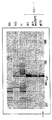
BPHと診断された患者ならびに前立腺癌腫と診断された患者由来の精液プラズマを、ProteinChip(登録商標)ディファレンシャルディスプレイの技術を用いて分析した。図3は、一人のBPHおよび前立腺癌腫患者の精液タンパク質プロファイルを示す。実際のゲル表示(virtual gel display)を用いて、試料間の比較を見やすいものとしている。前立腺癌−BPHのタンパク質プロファイルについての差異プロットを、ゲル観察プロットの下に表示する。差異プロットの実際の表示シグナルは、タンパク質が前立腺癌においてアップレギュレートされたことを示し、一方、負のピークは、前立腺癌における下方へのタンパク質制御を示す。可能な前立腺癌生体マーカを示す、いくつかのユニークなアップレギュレートシグナルが検出された。
【0240】
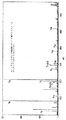
これらのアップレギュレートされたタンパク質の1つのチップ上での単離を、混合モードの表面および中性pH緩衝液での洗浄を用いて達成した(図4参照)。本例では、タンパク質はほぼ同質な程度まで強化した。強化した生体マーカ候補は、その後、トリプシンを用いたin situ消化に供した。インキュベーション後、CHCA(マトリックス)の飽和溶液を添加し、得られた消化産物をSELDI−TOFにより分析した。
【0241】
いくつかのペプチドが検出された(図5参照)。得られたペプチドシグナルをタンパク質データベース分析に提出し、ヒトセメノゲリンI(semenogellin I)の予備的同定を行った。生体マーカが、約5751Daの、セメノゲリンIの分子量(MW52,131Da)よりもはるかに小さい分子量を有していたことから、この同定はいくぶん当惑するものであった。
【0242】
同一の精製タンパク質を、ProteinChip LDI Qq−TOF MS検出に提出した(図6参照)。5751Daの親イオンは、LDI Qq−TOF MS/MS分析(3000M/z)についての現在の質量限界を超えていたので、二重に荷電したイオンをCID MS/MSシーケンシングに用いた(図7参照)。CID MS/MSの結果を用い、タンパク質のデータベース調査を行った。26のms/msイオンのうち15が、セメノゲリンIのフラグメントにタンパク質分解的に由来する、ヒト精塩基性タンパク質(SBP)にバックマッピングされ、この候補生体マーカの確実な同定が可能となった。
【0243】
上記のような最初の研究により潜在的な生体マーカが即時に明らかとなる一方、任意の生体マーカの完全な認証には、発現および有病率に関する統計的に有意な情報を得るための、数ダースひいては数百の関係ある試料の分析が必要である。
【0244】
本明細書中で言及したすべての特許、特許刊行物、および他の刊行された参照文献は、それぞれが個別にかつ特定的に本明細書中に参照により援用されたのと同様に、その全体が参照により援用される。本書類中の種々の参照文献の引用により、出願人らは、いかなる特定の参照文献も、発明に対する「当該技術分野」として認めるものではない。
【0245】
特定の実施例を提示したけれども、上記記載は例示的なものであり、制限的なものではない。これまでに記載した実施形態の1つまたは複数のいかなる特徴も、本発明の他の実施形態の1つまたは複数の他のいかなる特徴と、いかなる方法によっても、組み合わせることが可能である。さらに、本発明の多くの変形が、明細書の検討に基づいて、当業者に明らかとなるであろう。それゆえ、本発明の範囲は、上記記載を参照して決定されるべきではなく、代わりに、等価物のその十分な範囲に加えて、併記の特許請求の範囲を参照して決定されるべきである。
【図面の簡単な説明】
【図1】図1は、本発明の分析機器の実施形態を図式化している。
【図2】図2は、本発明の分析機器における使用に好ましい、直交QqTOFタンデム質量分析計の要素をより詳細に示す。
【図3】図3は、一人のBPHおよび前立腺癌患者の、精液タンパク質プロファイルを示す。
【図4】図4は、図3中で検出可能な、アップレギュレートされたタンパク質の1つのプローブ上単離の結果を示す。
【図5】図5は、強化生体マーカ候補をトリプシンを用いるin situ消化に供した後の、単相のMS分析により検出されたペプチドを示す。
【図6】図6は、本発明の分析装置での、同一精製タンパク質ペプチドの、LDI Qq−TOF MS分析を示す。
【図7】図7は、強化生体マーカ候補の、選択した二重荷電イオンについての、本発明の分析装置からのMS/MS結果を示す。[0001]
Field of the invention
The present invention relates to the field of chemical and biochemical analysis, and in particular, to an apparatus and method for improved identification and characterization of analytes and affinity interactions between analytes by tandem mass spectrometry.
[0002]
Background of the Invention
With the advent of electrospray ionization (ESI) and matrix-assisted laser desorption / ionization (MALDI) technology, along with the improved performance and low cost of mass spectrometers, mass spectrometry (MS) has In studying biologically relevant macromolecules, including proteins purified from complex biochemical systems, they have occupied a particular place among standard analytical tools.
[0003]
For example, a technique known as peptide mass fingerprinting uses mass spectrometry to identify proteins that are purified from biological samples. Identification is performed by checking the mass spectrum of the proteolytic fragment of the purified protein with the mass expected from the main sequence previously included in the database. Roepstoff, The Analyst 117: 299-303 (1992); Pappin et al. , Curr. Biol. 3 (6): 327-332 (1993); Mann et al. , Biol. Mass Spectrom. 22: 338-345 (1993); Yates et al. , Anal. Biochem. 213: 397-408 (1993); Henzel et al. , Proc. Natl. Acad. Sci. ScL USA 90: 5011-5015 (1993); James et al. , Biochem. Biophys. Res. Commun. 195: 58-64 (1993).
Similar database survey approaches have been developed. This approach uses fragment mass spectra obtained from collision-induced dissociation (CID) or MALDI post-source degradation (PSD) to identify purified proteins. Eng et al. , J. et al. Am. Soc. Mass. Spectrom. 5: 976-989 (1994); Griffin et al. , Rapid Commun. Mass Spectrom. 9: 1546-1551 (1995); Yates et al. U.S. Patent Application Nos. 5,538,897 and 6,017,693; Mann et al. , Anal. Chem. 66: 4390-4399 (1994).
[0004]
Mass spectrometric techniques have also been developed that allow at least partial de @ novo sequencing of the isolated protein. Chait et al. , Science 262: 89-92 (1993); Keough et al. , Proc. Natl. Acad. Sci. USA. 96: 7131-6 (1999); Bergman, EXS 88: 133-44 (2000).
[0005]
Software resources that facilitate the interpretation of protein mass spectra and search public domain sequence databases are now readily accessible on the Internet, facilitating protein identification. Some of these include Protein @ Prospector (http: //projector.ucsf/edu), PROWL (http://prowl.rockefeller.edu) and Mascot @ Search \ Engine (MatriceK.Sw., Dictionary. com).
[0006]
Although accurate mass assignment can provide useful information (eg, the techniques described above facilitate identification of purified proteins), such information is limited. Significant additional analytical power is achieved by combining MS analysis with enzymatic and / or chemical modifications of the target protein, allowing elucidation of structural components, promoting post-translational modifications and protein identification. Will be done.
[0007]
In addition, biological complexes such as blood, serum, plasma, lymph, interstitial fluid, urine, exudates, whole cells, cell lysates, and cell secretions typically contain hundreds of biological molecules, and Includes organic and inorganic salts, which are not used for direct mass spectrometry. Therefore, significant sample preparation and purification steps are usually required prior to MS investigation.
[0008]
Conventional sample purification methods such as liquid chromatography (ion exchange, size exclusion, affinity and reverse phase chromatography), membrane dialysis, centrifugation, immunoprecipitation and electrophoresis usually require large starting samples. Even with such a large sample volume, these purification processes tend to lose small amounts of components and will lose analyte due to the effects of non-specific binding and dilution. These methods are also often labor intensive.
[0009]
Thus, there is a need for a method and apparatus that facilitates mass spectrometric detection of major and minor proteins present in heterogeneous samples without the need for the tedious and expensive liquid phase purifications of the prior art. It is clear that there is. Further, there is a need for an MS platform that not only allows for easy sample purification, but also allows for serial and parallel sample modification approaches prior to mass spectrometry.
[0010]
The above needs have been met, in part, by the development of affinity capture laser desorption ionization approaches. Hutchens et al. , Rapid Commun. Mass Spectrom. 7: 576-580 (1993); U.S. Patent Nos. 5,719,060, 5,894,063, 6,020,208 and 6,027,942. This new procedure for MS analysis of macromolecules uses a novel laser desorption ionization probe that contains an affinity reagent on at least one surface. The affinity reagent adsorbs the desired analytes from the foreign sample and collects them on the surface of the probe in a form suitable for subsequent laser desorption ionization. The coupling of analyte adsorption and desorption obviates the need for an off-line purification approach, allows for the analysis of smaller initial samples, and further facilitates a direct sample modification approach at the probe surface prior to mass spectrometry.
[0011]
According to the affinity capture laser desorption ionization approach, mass spectrometry was performed using immunoassays (Nelson et al., Anal. Chem. 67: 1153-1158 (1995)) and affinity chromatography (Brockman et al., Anal. Chem. 67). : 4581-4585 (1995)). Affinity capture laser desorption ionization approaches are described for peptides and proteins (Hutchens et al., Rapid Commun. Mass Spectrom. 7: 576-580 (1993); Mouradian et al., J. Amer. Chem. Soc.-118: Soc. 8645 (1996); Nelson et al., Rapid Commun. Mass. Spectrom. 9: 1380-1385 (1995); Nelson et al., J. Molec. Recognition 12: 77-93 (1999); J. Mass Spectrom. 33: 1141-1147 (1998); Yip et al., J. Biol. Chem. 1: 32825-33 (1996)), as well as oligonucleotides (Jurinke et al., Anal. Chem. 69: 904-910 (1997); Tang et al., Nucl. Acids Res. 23: 3126-3131 (1996)). Liu et al. Anal. Chem. 67: 3482-90 (1995)), bacteria (Bundy et al., Anal. Chem. 71: 1460-1463 (1999)), and small molecules (Wei et al. , Nature 399: 243-246 (1999)). At a commercial level, affinity capture laser desorption ionization is embodied in Ciphergen's ProteinChip® Systems (Ciphergen Biosystems, Inc. Fremont, Calif., USA).
[0012]
Although affinity capture laser desorption ionization technology has solved a serious problem in the art, the difficulties still remain.
[0013]
When this approach is applied to capture proteins from biological samples, it is common to see about 1 picomole of total protein captured and obtained for subsequent analysis. Typically, affinity capture on a chromatographic surface biochip does not provide complete purification. In addition, the digestion efficiency seen for solid phase extracted samples is not as good as compared to digestions performed in free solution or in the denaturing environment of 2-D gels. Thus, assuming that about 50% is the protein of interest and that about 10% of this protein has been successfully digested, only a peptide of at most about 50 femtometers is available for detection.
[0014]
Using substantial tryptic digestion of bovine fetuin in database search experiments, a search for this complex eukaryotic genome, even with an excess of 1.0 ppm, for example (although most MS techniques cannot currently achieve), When done, protein ID matching has been shown to be less reliable with a single peptide mass. Similarly, unreliable results are obtained for the two peptides. Only when three peptides are submitted are reliable results obtained for mass assignments with errors less than 300 ppm. In this case, most devices require an internal standard calibration. However, for 5 or more peptides, mass accuracy, which is an error of more than 1000 ppm, does not provide further reliability.
[0015]
Furthermore, when multiple proteins are digested simultaneously, a heterogeneous peptide pool is created, and good database searches not only require excessive precision, but often also require key sequence information. The tandem MS / MS approach has shown significant utility in providing key sequence information. See Biemann et al. , Acc. Chem. Res. 27: 370-378 (1994); Spengler et al. , Rapid Commun. Mass Spectrom. 1991, 5: 198-202 (1991); Spengler et al. , Rapid Commun. Mass Spectrom. 6: 105-108 (1992); Yates et al. , Anal. Chem. 67: 1426-1436 (1995); Kaufman et al. , Rapid Commun. Mass. Spectrom. 7: 902-910 (1993); Kaufman et al. , Intern. J. Mass Spectrom. Ion Processes 131: 355-385 (1994).
[0016]
However, until recently, the only MS / MS approach available for laser desorption-based analysis was post-source decomposition analysis (PSD). Although PSD can provide reasonable sequence information for picomolar levels of peptide, the efficiency of this fragmentation process is generally low. Combined with the poor mass accuracy and sensitivity often shown in this approach, its application to the analysis of small amounts of proteins frequently found in affinity capture laser desorption ionization probes is very limited. In recent years, a laser desorption ionization quadrupole time-of-flight mass spectrometer (LDI @ Qq-TOF) capable of performing collision induced dissociation (CID) MS / MS analysis has been developed. Krutchinksy et al. , Rapid Commun. Mass Spectrom. 12: 508-518 (1998).
[0017]
Therefore, there is a need for an apparatus and method that increases the sensitivity and mass accuracy of affinity capture laser desorption mass spectrometry. There is a need for a method and apparatus that increases the efficiency of digestion on a probe and allows peptides produced by digestion of a heterogeneous mixture of easily dissolved proteins. There is a need for devices and methods that increase the efficiency of affinity capture laser desorption tandem mass spectrometry.
[0018]
Summary of the Invention
It is an object of the present invention to provide an apparatus and method that increases the sensitivity, mass accuracy, mass resolution of existing affinity capture laser desorption ionization mass spectrometry, and adds ms / ms capability. Yet another object of the present invention is to provide a method for biomolecule analysis that utilizes these improved analytical capabilities.
[0019]
The present invention, in a first aspect, satisfies these and other objects and needs in the art by providing an analytical instrument.
[0020]
The analytical instrument of the present invention has a laser desorption ionization source, an affinity capture probe interface, and a tandem mass spectrometer, wherein the affinity capture probe interface is engaged with the affinity capture probe, and the probe is connected to the tandem mass spectrometer. The probe can be positioned so that ions interrogated by the laser desorption source during communication and desorbed from the probe enter the mass spectrometer.
[0021]
Typically, a laser desorption ionization source has a laser excitation source and a laser optical train, which functions to transmit excited photons from the laser excitation source to the probe interface. In such an embodiment, the laser optic train typically delivers about 20-1000 microjoules of energy per square millimeter of the interrogated probe surface.
[0022]
The laser excitation source is selected from the group consisting of a continuous laser and a pulsed laser, and in various embodiments, nitrogen laser, Nd: YAG laser, erbium: YAG laser and CO2It is selected from the group consisting of lasers. In a currently preferred embodiment, the laser excitation source is a pulsed nitrogen laser.
[0023]
In one set of embodiments, the laser optical train has an optical component selected from the group consisting of a lens, a mirror, a prism, an attenuator, and a beam splitter.
[0024]
In another set of embodiments, the laser optical train has an optical fiber having an input end and an output end, and a laser excitation source is coupled to the input end of the optical fiber.
[0025]
In some embodiments of the fiber optic laser optic train, the laser optic train further comprises an optical attenuator. The attenuator is located between the laser pump source and the input end of the optical fiber and acts to couple the laser pump source to the input end of the optical fiber or between the output end of the optical fiber and the probe. Can be arranged.
[0026]
In some embodiments of the fiber optic array, the output end of the optical fiber has a maximum diameter of about 200-400 μm and the input end has a diameter of about 400-1200 μm.
[0027]
The analytical instrument may also have probe viewing optics so that the probe is visualized after engaging the probe interface.
[0028]
In some embodiments, the laser optic train may include a laser coupler coupling the laser excitation source to the input end of the optical fiber. As mentioned above, the coupler may act as an optical attenuator. In other embodiments, the coupler may act to facilitate visualization of the probe after the probe has engaged the probe interface.
[0029]
In certain of the latter embodiments, either the coupler or the fiber is bifurcated to separate a small amount of energy from the laser excitation source. Alternatively, by such a bifurcation, visible light can be introduced to refer to the desorption site.
[0030]
If the visualization optics are included in the optical train, or the laser optical train including the fibers includes a bifurcated or trifurcated branch, the analytical instrument is further arranged to detect light reflected from the probe. May have a customized CCD camera.
[0031]
In a typical embodiment, the affinity capture probe interface has a probe holder capable of reversibly engaging the affinity capture probe. The interface also typically has a probe introduction port that can itself reversibly engage the probe holder.
[0032]
In a typical embodiment, the probe interface further includes a probe position actuator assembly and an interface ion collection system. When the probe holder is engaged with the introduction port, the probe holder is placed in contact with the probe position actuator, which itself includes both a laser ionization source (typically for a laser optical train) and an ion collection system. , A probe holder (usually having its engaged probe) can be movably disposed. In a typical embodiment, an actuator can position the probe holder in a translatable and rotatable manner.
[0033]
The probe interface also typically has a vacuum exhaust system connected to the probe introduction port. With a vacuum evacuation system, the probe is interrogated at sub-atmospheric pressure by a laser desorption ionization source.
[0034]
The analytical instrument of the present invention has a tandem mass spectrometer. The tandem mass spectrometer, in various embodiments, is selected from the group consisting of QqTOF @ MS, ion trap MS, ion trap TOF @ MS, TOF-TOF @ MS, and Fourier transform ion cyclotron resonance MS. Currently preferred for use in the analytical instrument of the present invention is QqTOF @ MS.
[0035]
In a preferred embodiment, the tandem mass spectrometer is a QqTOF MS, the laser excitation source is a pulsed nitrogen laser, the laser fluence at the probe is about 2-4 times the minimum desorption threshold, and the tandem mass spectrometer Has an external standard mass accuracy of about 20-50 ppm.
[0036]
The analytical instrument of the present invention is designed to engage with an affinity capture laser desorption ionization probe. Thus, all of the above embodiments may have an affinity capture probe that engages the affinity capture probe interface.
[0037]
The affinity capture probe in these embodiments typically has at least one sample adsorption surface arranged in a queryable relationship with the laser source. The sample adsorption surface is selected from the group consisting of a chromatographic adsorption surface and a biomolecule affinity surface. Typically, such chromatographic adsorption surfaces are selected from the group consisting of reversed phase, anion exchange, cation exchange, immobilized metal affinity capture and mixed mode surfaces, wherein the biomolecules on the biomolecule affinity surface are antibodies, receptor It is selected from the group consisting of body, nucleic acid, lectin, enzyme, biotin, avidin, streptavidin, staphylococcal protein A, and staphylococcal protein G.
[0038]
The affinity capture laser desorption ionization probe has a plurality of separately addressed sample adsorption surfaces that can be placed in an interrogable relationship with the laser source and can have at least two different such adsorption surfaces.
[0039]
In another embodiment, the analytical instrument of the present invention has a digital computer interfaced with a tandem mass spectrometer detector. Further, in some embodiments, the device may also have a software program that can be executed by a digital computer that is local to or accessible for communication with the computer. The software program in such an embodiment is capable of controlling a laser desorption ionization source, controlling at least one aspect of data acquisition by a tandem mass spectrometer, It is possible to perform at least one analysis routine on the data obtained by the analyzer or to enable any subset of these functions.
[0040]
In another aspect, the invention provides a method for analyzing at least one test protein.
[0041]
The method comprises: (a) capturing the test protein or proteins on an affinity capture protein biochip; (b) generating a protein cleavage product of the test protein on the protein biochip using a proteolytic agent; And (c) analyzing at least one protein cleavage product using a tandem mass spectrometer. In these embodiments of this aspect, the analyzing step comprises: (i) desorbing the protein cleavage product from the protein biochip into the gas phase to produce a corresponding parent ionic peptide; (ii) using the first mass spectrometer. Using to select a parent ionic peptide for subsequent fragmentation; (iii) fragmenting the selected parent ionic peptide under selected fragmentation conditions in the gas phase to produce a product ion fragment; And (iv) generating a mass spectrum of the product ion fragment. Thus, the mass spectrum provides an analysis of the test protein.
[0042]
In certain embodiments of this aspect of the invention, the method further comprises determining, in the database, a closeness-of-fit analysis of the mass spectrum and the theoretical mass spectrum of the protein in the database. Determining the at least one candidate protein identification for the test protein by presenting the mass spectrum to a protein database search protocol identifying at least one candidate protein identification for the test protein (d).
[0043]
In particular, among these embodiments, step (d) further comprises presenting the mass of the test protein and the original species of the test protein to the protocol.
[0044]
In other embodiments, the method further comprises: (i) generating a mass spectrum of the proteolytic cleavage product of (b); and (ii) a cleavage product of an identified candidate predicted to be produced using the proteolytic agent. Computer that determines a measure of the closeness of fit between the theoretical mass spectrum of the protein and the mass spectrum of the protein cleavage product, whereby the measurement indicates the protein cleavage product on the protein biochip corresponding to the test protein The method further comprises comparing the identified candidate to the test protein by presenting the mass spectrum of the protein cleavage product in the protocol (e).
[0045]
Still other embodiments of the method comprise the steps of (f) repeating step (c) wherein the selected parent ionic peptide does not correspond to a proteolytic cleavage product expected from the identified candidate, and the selected parent ionic peptide of (f) And step (g) of repeating (d).
[0046]
In this aspect of the invention, the test protein may be a protein that is differentially expressed between a first biological sample and a second biological sample. In some of these embodiments, the first and second biological samples are obtained from normal and pathological sources.
[0047]
In a third aspect, the invention provides a method for characterizing a binding interaction between a first molecular binding partner and a second molecular binding partner.
[0048]
In this aspect, the method is binding a second binding partner to a first binding partner, wherein the first binding partner is immobilized on a surface of a laser desorption ionization probe. Fragmenting the second binding partner, and detecting at least one fragment by tandem mass spectrometry, whereby the mass spectrum of the detected fragment indicates the nature of the binding interaction. Determining and detecting.
[0049]
In certain embodiments of this aspect of the invention, the first binding partner is first immobilized on the surface of the affinity capture probe before the second binding partner binds to the first binding partner.
[0050]
Such immobilization may be effected by direct coupling of the first partner to the affinity capture probe, such as by a covalent bond. Typical covalent bonding embodiments include a covalent bond between the amine of the first binding partner and the carbonyldiimidazole moiety of the probe face, and an amino or thiol group of the first binding partner and the epoxy of the probe face. Includes covalent bonds between groups.
[0051]
Immobilization can also be accomplished by a direct non-covalent bond, such as a coordination or dative bond, between the first binding partner and a metal, such as gold or platinum, on the probe surface. Immobilization may also be by interacting the first binding partner with a chromatographic adsorption surface selected from the group consisting of reverse phase, anion exchange, cation exchange, immobilized metal affinity capture and mixed mode surfaces. Can be done.
[0052]
Alternatively, the fixation may be indirect, which may be indirect but covalent. In certain of these latter embodiments, the first binding partner may be covalently immobilized through a linker, such as a cleavable linker. Indirect immobilization can also be performed by non-covalent attachments, such as immobilization on the probe via biotin / avidin, biotin / streptavidin interactions.
[0053]
In this aspect of the invention, the first molecular binding partner may be selected from the group consisting of proteins, nucleic acids, carbohydrates and lipids. Typically, the first binding partner is a protein, which is a naturally occurring protein from an organism selected from the group consisting of multicellular eukaryotes, unicellular eukaryotes, prokaryotes, and viruses, or It can be an artificial protein such as a recombinant fusion protein.
[0054]
In embodiments where the first binding partner is a protein, the protein may be selected from the group consisting of, inter alia, antibodies, receptors, transcription factors, cytoskeletal proteins, cell cycle proteins, and ribosomal proteins.
[0055]
Binding of the second binding partner to the immobilized first binding partner is, in a typical embodiment, performed by contacting the first binding partner with a biological sample. The sample may be a fluid selected from blood, lymph, urine, cerebrospinal fluid, synovial fluid, milk, saliva, vitreous humor, aqueous humor, mucus and semen, or cell lysate, or other form of sample. .
[0056]
In various embodiments, including embodiments where the first binding partner is a protein, the second binding partner can be a protein. Alternatively, the second binding partner can be a compound present in a combinatorial library. Here, the binding of the second binding partner to the first binding partner is performed by contacting the first binding partner with an aliquot of a chemically synthesized combinatorial library. In yet another alternative, the second binding partner can be a component of a biologically displayed combinatorial library, such as a phage display library.
[0057]
In certain general embodiments, fragmenting is performed by contacting the second binding partner with an enzyme. Here, the second binding partner is a protein, and the enzyme is usually trypsin, Glu-C (V8) protease, endoproteinase Arg-C (serine protease), endoproteinase Arg-C (cysteine protease), Asn -N protease, and specific endoproteases such as Lys-C protease. Alternatively, fragmenting may be performed by contacting the second binding partner with a liquid phase chemical such as CNBr.
[0058]
In some embodiments, the method comprises denaturing the second binding partner after binding the second binding partner to the first binding partner and before fragmenting the second binding partner. Further included.
[0059]
In various embodiments, the method further comprises, after fragmenting the second binding partner, washing the probe with a first eluent and, optionally, a second eluent. The second eluent differs from the first eluent in at least one elution property, such as pH, ionic strength, detergent strength, and hydrophobicity.
[0060]
In a typical embodiment, the method further comprises applying energy to absorb the molecule to the probe after fragmentation and before detection of the second binding partner fragment. In a preferred embodiment, the probe then engages with the affinity capture probe interface of the analytical instrument of the present invention and the second binding partner fragment ionized and desorbed from the probe using the laser source of the instrument.
[0061]
The instrument can be used to make several types of useful measurements in this method, including measuring the total mass of ions, measuring the mass of a subset of fragments, and single ion monitoring measurements.
[0062]
Advantageously, an embodiment of the method is that after the mass spectrometry measurement of the fragment of the second binding partner, the fragment measurement is applied to the fragmentation enzyme cleavage rules on the first amino acid sequence of the second binding partner. And determining the nature of the interactions between the molecules by such comparisons with expected fragment measurements.
[0063]
If the identity of the second binding partner is not known, the method may further comprise, prior to such a comparison, identifying the second binding partner through an ms / ms analysis. Such MS / MS analysis includes selecting a first fragment of a second binding partner by mass spectrometry, dissociating the first fragment of the second binding partner in the gas phase, Measuring the fragment spectrum of the first fragment of the binding partner of the above, and comparing the fragment spectrum with the fragment spectrum expected from the amino acid sequence data previously included in the database. Amino acid sequence data can be selected from the group consisting of experimental data and predicted data. In a typical embodiment, the dissociation is a collision-induced dissociation.
[0064]
In some embodiments of the method, the first binding partner is selected from the group consisting of an antibody, a T cell receptor, and an MHC molecule. In other embodiments, the first binding partner is a receptor and the second binding partner is from a receptor agonist, a partial agonist of the receptor, an antagonist of the receptor, and a partial antagonist of the receptor. Selected from the group consisting of: In another embodiment, the first binding partner is a glycoprotein receptor and the second binding partner is a lectin.
[0065]
In a fourth aspect, the present invention provides a method for detecting an analyte. The method comprises engaging an affinity capture probe with an affinity capture probe interface of the analytical instrument of the present invention, wherein the affinity capture probe has an analyte associated therewith, and the instrument laser source. Desorbing and ionizing analytes or fragments thereof from the probe, and then detecting the analyte by tandem mass spectrometry on the desorbed ions.
[0066]
In this aspect, the method may further comprise performing a collision-induced dissociation of the desorbed ions after the steps of desorption and ionization and before the detecting step. Prior to such dissociation, in some embodiments, a subset of ions may be selected for impact dissociation.
[0067]
In other embodiments, a step before adsorbing the analyte to the probe may be performed. In still other embodiments, after adsorbing the analyte and before engaging the probe with the probe interface, a step of contacting the probe and the analyte to adhere to the energy absorbing molecule may be performed. .
[0068]
The above and other objects and advantages of the present invention will become apparent from the following detailed description when taken in conjunction with the accompanying drawings. In the accompanying drawings, like reference characters refer to like parts throughout.
[0069]
DETAILED DESCRIPTION OF THE INVENTION
I. Definition
As used herein, the terms specifically set forth below have the following definitions. Unless defined otherwise, all terms used herein have the meaning commonly understood by one of ordinary skill in the art to which this invention pertains.
[0070]
"Analyte" refers to any component of a sample for which detection is desired. The term may refer to a single component or multiple components in a sample.
[0071]
A "probe" is in interrogable relationship with a laser desorption ionization source and, when placed in engagement with a gas phase ion analyzer to communicate simultaneously at atmospheric or sub-atmospheric pressure, obtains from an analyte. Refers to a device that can be used to introduce the ions into the spectrometer. A "probe" as used herein is typically reversibly engagable by a probe interface.
[0072]
An "affinity capture probe" refers to a probe that binds an analyte through interactions that are sufficient to extract and concentrate the analyte from a heterogeneous mixture. No high purity concentration is required. Binding interactions are usually mediated by adsorbing the analyte on the adsorption surface of the probe. The term ProteinChip® Array, as used in the present invention, is used by Ciphergen Biosystems, Inc. , Fremont, California.
[0073]
"Adsorption" refers to a detectable non-covalent bond of an analyte to an adsorbent.
[0074]
"Adsorbent" refers to any substance that can adsorb an analyte. The term “adsorbent” is used herein to refer to a single substance (“monoplex adsorbent”) (eg, a compound or a functional group) and a plurality of different substances (“multiplex adsorbent”). Can be The adsorbent material in the multiplex adsorber is called "adsorbent species". For example, a laser-addressed adsorption surface on a probe substrate includes a multiplex adsorber characterized by many different adsorbent species (eg, anion exchange materials, metal chelators, or antibodies) with different binding properties. obtain.
[0075]
“Adsorption surface” refers to the surface that contains the adsorbent.
[0076]
"Chromatographic adsorption surface" refers to a surface that includes an adsorbent that allows for chromatographic discrimination between analytes or separation of analytes. Thus, the term refers to a surface that includes an anion exchange portion, a cation exchange portion, a reversed phase portion, a metal affinity capture portion, and a mixed mode adsorbent, as understood in chromatographic technology.
[0077]
"Biomolecule affinity surface" refers to a surface that includes an adsorbent having a biomolecule capable of specific binding.
[0078]
"Specific binding" refers to the ability of two species simultaneously present in a heterogeneous (non-homogeneous) sample to bind to each other preferentially to other species in the sample. Usually, specific binding interactions are distinguished from accidental binding interactions in reactions that are at least 2-fold, more usually more than 10- to 100-fold. When used to detect an analyte, specific binding is sufficiently discriminating in determining the presence of the analyte in a foreign (non-homogeneous) sample. Usually, the affinity or avidity of a specific binding reaction is at least about 10− 7M and a specific binding reaction with greater specificity is usually at least 10− 8M to at least about 10− 9It has M affinity or avidity.
[0079]
"Energy absorbing molecule" and the equivalent acronym "EAM" are molecules that, when attached to a probe, can adsorb energy from a laser desorption ionization source and subsequently contribute to the desorption and ionization of the analyte in contact Point to. This term includes all molecules so-called in US Patent Nos. 5,719,060, 5,894,063, 6,020,208 and 6,027,942. The disclosures of these patents are incorporated herein by reference in their entirety. The term clearly includes cinnamic acid derivatives, sinapinic acid derivatives ("SPA"), cyanohydroxycinnamic acid ("CHCA") and dihydroxybenzoic acid.
[0080]
"Tandem mass spectrometer" refers to any gas phase ion spectrometer that is capable of discriminating ions in an ion mixture based on two consecutive stages m / z. The term includes an analyzer having two mass spectrometers and a single mass spectrometer capable of selectively acquiring or retaining ions prior to mass analysis. Thus, the term clearly includes a QqTOF mass spectrometer, an ion trap mass spectrometer, an ion trap-TOF mass spectrometer, a TOF-TOF mass spectrometer, and a Fourier transform ion cyclotron resonance mass spectrometer.
[0081]
"Eluent" refers to an agent (usually a liquid) used to affect or alter the adsorption of an analyte to an adsorbent on an adsorption surface. Eluents are also referred to herein as "selectivity threshold modifiers."
[0082]
"Eluting properties" refers to the physical or chemical properties of an eluent that contribute to its ability to affect or alter the adsorption of an analyte to an adsorbent on an adsorption surface. The two eluants, when placed in contact with the analyte and the adsorbent, have different elution characteristics if the analytes have different degrees of affinity for the adsorbent. Elution characteristics include, for example, pH, ionic strength, degree of chaotropism, detergent strength and temperature.
[0083]
“Biological sample” and “biological sample” equally refer to a sample obtained from at least a portion of an organism capable of replication. As used herein, a biological sample can be obtained from any of the known taxonomic circles, including viruses, prokaryotes, unicellular eukaryotes, and multicellular eukaryotes. Biological samples can be obtained from whole organisms or parts thereof, including those from culture parts thereof. Biological samples can be in any physical form appropriate for the context, including homogenates, cellular component fractions, lysates and liquids.
[0084]
"Biomolecule" refers to a molecule that can be found in a biological sample, but is not necessarily derived from the biological sample.
[0085]
“Organic biomolecules” can be found in biological samples such as steroids, amino acids, nucleotides, carbohydrates, polypeptides, polynucleotides, complex carbohydrates, and lipids, but are not necessarily derived from biological samples. Refers to a molecule.
[0086]
"Small organic molecule" refers to an organic molecule of a size comparable to organic molecules commonly used in pharmaceuticals. The term does not include organic biopolymers (eg, proteins, nucleic acids, etc.). As used herein, small organic molecules typically range in size up to about 5000 Da, up to about 2500 Da, up to about 2000 Da, or up to about 1000 Da.
[0087]
"Biopolymer" refers to a polymer that can be found in a biological sample such as polypeptides, polynucleotides, polysaccharides, and polyglycerides (eg, diglycerides or triglycerides), but is not necessarily derived from the biological sample. Point.
[0088]
"Fragment" refers to the product of chemical, enzymatic or physical degradation of an analyte. Fragments can be in a neutral or ionic state.
[0089]
The terms "polypeptide", "peptide" and "protein" are used interchangeably herein to refer to a naturally occurring or synthetic polymer comprising amino acid monomers (residues). Here, amino acid monomers include naturally occurring amino acids, naturally occurring amino acid structural variants, and synthetic artificial analogs capable of participating in peptide bonds. Polypeptides can be modified, for example, by adding carbohydrate residues to form glycoproteins. The terms "polypeptide", "peptide" and "protein" include glycoproteins and non-glycoproteins.
[0090]
"Polynucleotide" and "nucleic acid" refer equally to naturally occurring or synthetic polymers containing nucleotide monomers (bases). Polynucleotides include naturally occurring nucleic acids, such as deoxyribonucleic acid ("DNA") and ribonucleic acid ("RNA"), and nucleic acid analogs. Nucleic acid analogs include analogs containing artificial bases, and analogs in which nucleotide monomers are linked in ways other than naturally occurring phosphodiester bonds. Nucleotide analogs include, but are not limited to, phosphorothioates, phosphorodithioates, phosphorotriesters, phosphoramidates, boranophosphates, methylphosphonates, chiral-methylphosphonates, 2-O-methylribonucleotides, peptides Nucleic acid (PNA) and the like.
[0091]
As used herein, "molecular binding partner" (equivalently, "specific binding partner") refers to a pair of molecules that exhibit specific binding, usually a pair of biomolecules. Examples include, but are not limited to, receptors and ligands, antibodies and antigens, biotin and avidin, and biotin and streptavidin.
[0092]
"Receptor" refers to a molecule, usually a macromolecule, that can be found in a biological sample, but need not necessarily be from a biological sample, and that can participate in specific binding to a ligand. The term further includes fragments and derivatives that are still capable of specific ligand binding.
[0093]
"Ligand" refers to any compound that can participate in specific binding to a specified receptor or antibody.
[0094]
"Antibody" refers to a polypeptide substantially encoded by at least one immunoglobulin gene or a fragment of at least one immunoglobulin gene, capable of participating in specific binding to a ligand. The term includes naturally occurring forms, as well as fragments and derivatives. Fragments within the terminology as used herein include those digested by various peptidases, such as Fab, Fab 'and F (ab)' 2 fragments, as long as they are still capable of specific binding to a target molecule. And fragments produced by chemical dissociation, chemical cleavage, and recombination. Common recombinant fragments, such as those produced by phage display, include single-chain Fab and scFv ("single-chain variable region") fragments. Derivatives within this term include antibodies (or fragments thereof) that are continuously modified but still capable of specifically binding to the target molecule, including interspecies chimeric and humanized antibodies. . As used herein, antibodies can be produced by any known technique, including harvesting of natural B lymphocytes, hybridomas, recombinant expression systems from cell culture, phage display, and the like.
[0095]
"Antigen" refers to a ligand that can be bound by an antibody. The antigen need not be immunogenic. The portion of the antigen that makes contact with the antibody is termed the "epitope."
[0096]
"Fluence" refers to the energy delivered per unit area of the queried image.
[0097]
II. Affinity capture probe tandem mass spectrometer
In a first aspect, the present invention provides an analytical instrument that combines the advantages of affinity capture laser desorption ionization sample introduction with the advantages of a high precision, high mass resolution tandem mass spectrometer. This combination offers significant advantages over existing devices for performing known techniques. In addition, new instruments will enable new methods of protein discovery, making molecules between specific binding partners and between specific binding partners faster, more efficient, and more sensitive than existing approaches. New methods of identifying and characterizing interactions are possible. First, the device will be briefly described overall. Thereafter, the characteristics of the affinity capture probe interface will be described in more detail.
[0098]
Briefly, FIG. The
[0099]
Laser desorption / ionization source
Laser desorption /
[0100]
Laser desorption /
[0101]
Photons emitted from
[0102]
Alternatively, optical train 11 may be comprised of an array of fiber optics that function to collect, direct, and subdivide the energy of each laser pulse.
[0103]
In this embodiment, the output of
[0104]
The amount of energy incident on the fiber can be controlled by careful adjustment of the lens position relative to the fiber. In this case, the optical fiber coupler can double as an optical attenuator. In another preferred embodiment, the total output energy of the laser is coupled into the fiber and the attenuator is located between the output side of the optical fiber and the desorption spot focusing element of the optical train. In yet another preferred embodiment, the optical attenuator is located between the laser and the fiber optic coupler. In all cases, optical attenuation is used to ensure that proper laser fluence is delivered to the surface of probe 16 independent of the output energy of
[0105]
It has been found that fiber optic components can often be damaged when receiving focused energy from the laser, thus maximizing the acceptance area on the input side of the fiber and increasing the fluence of the incident laser energy to the fiber damage threshold. Advantageously, it is less than. Adjusting the relative position of the optical coupler with respect to the laser and the optical fiber by the latter also simplifies the alignment of the laser beam with respect to the optical fiber. However, in order to obtain a reasonable desorption fluence level at the probe 16, when used with a conventional nitrogen laser delivering a maximum energy of about 200 μJ / laser pulse, the maximum output fiber diameter is 400 μm (microns). Do not exceed. The solution to this problem is to incorporate a tapered optical fiber with an input diameter of about 400-1200 microns and an output diameter of 200-400 microns.
[0106]
Typically, the desorption spot is focused to a size that maximizes the production of ions for each pulse, while maintaining sufficient fluence to induce desorption and ionization and interrogating the largest area of the probe 16. Should. While using a conventional nitrogen laser delivering a maximum energy of about 200 μJ / pulse in a laser desorption / ionization source coupled to a quadrupole time-of-flight tandem mass spectrometer, the optimal laser spot area is 0.4 square millimeters. And 0.2 square millimeters.
[0107]
Laser desorption /
[0108]
If the laser optical train 11 includes optical fibers, the
[0109]
For example, fiber optic couplers may be bifurcated to separate a small amount of laser excitation energy and used as a means to monitor the applied laser energy, or bifurcated to introduce visible light. Irradiate the detachment site.
[0110]
In the first of the above two embodiments, only a small portion of the excitation energy is a major component of the laser energy circuit that is calibrated to reflect the actual amount of laser energy delivered to the probe 16. It is directed to strike a photodetector. In a second embodiment, the visible light is transmitted through a separate set of opto-optics coupled to the CCD camera, or through an optical fiber and laser pump, which directs the light reflected at the major branch of the optical fiber to the CCD camera. By using a prism or a dichroic mirror between the source, the detachment site is illuminated and oriented to allow observation of this area. Alternatively, a prism or dichroic mirror is arranged in a straight line between the illumination fiber branch of the optical fiber and the illumination source, such that all reflected images coupled to this branch strike the CCD camera. Oriented to In still other embodiments, the fiber includes one branch delivering a desorption / ionization laser pulse, a second branch delivering visible light to illuminate the desorption site, and a third branch desorption. It can be trifurcated to transmit the reflected light from the site to the CCD camera. For each of these viewing modes, a suitable bandpass filter is generated from the probe surface as a direct reflection of the incident laser pulse on the probe surface or as a direct result of electronic excitation by the incident laser pulse. It should be deployed between the CCD camera and the viewing optics to prevent transmission that could damage high energy photons, which are secondary photons.
[0111]
Probe interface
The affinity
[0112]
The
[0113]
The probe holder is a component of the
[0114]
The probe holder may hold a single probe 16 or multiple probes 16. The holder places each probe 16 in the proper orientation as interrogated by the laser desorption /
[0115]
The probe holder is in intimate contact with the position actuator assembly.
[0116]
The actuator assembly provides for a laser desorption /
[0117]
The actuator comprises an electromechanical device that supports the translational and / or rotational movement of the probe 16 while maintaining the position of the probe relative to the laser desorption / ionization source and ion collection system. Such electromechanical devices include, but are not limited to, mechanical or optical position sensors, solenoids, stepper motors, DC or AC synchronous motors that communicate directly or indirectly with linear motion actuators, linear or circular motion guide rails, gimbals, Including bearings or axles.
[0118]
With the probe introduction port, the probe holder with the loaded probe 16 is positioned on the probe position actuator assembly without introducing excessive levels of atmospheric gas into the
[0119]
To accomplish the latter, the probe inlet port uses a vacuum exhaust system (probe inlet port exhaust system) to pump atmospheric gases and achieve the desired port pressure before moving the tip to the working position. While exchanging the probe, the probe actuator assembly moves the probe from the working position (the position aligned with the
[0120]
After removing the previously analyzed probe 16 and installing a new probe 16, the probe holder is replaced by its position actuator and the sample loading process begins. As described above, the probe introduction port can be pumped down to sub-atmospheric pressure by the exhaust system. Upon achieving the desired sample introduction pressure, the probe actuator system moves the probe 16 from the exchange position to the working position while opening the seal to the mass spectrometer inlet.
[0121]
Alternatively, the probe introduction port may be maintained at atmospheric pressure if the ions are generated in a desorption chamber maintained at atmospheric pressure and ultimately directed to an ion optic assembly that introduces ions into the mass spectrometer inlet. It does not need to be discharged and pressurized.
[0122]
The probe introduction port exhaust system consists of a vacuum pump, pressure sensor, vacuum compatible tubing and connection fittings, and a vacuum compatible valve. The vacuum compatible valve, when operated simultaneously, allows for controlled evacuation of atmospheric gases contained in the inlet port following sample exchange so that the probe 16 can be moved to a working position. The vacuum pump can be, but is not limited to, a single or multi-stage oil mechanical pump, a scroll pump, or a non-lubricated membrane pump. In a preferred embodiment, the vacuum compatible valve is an electrically controlled solenoid valve. In the same embodiment, the pressure sensor is an electronic sensor capable of operating in a pressure range from atmospheric pressure to 1 mTorr. Such pressure sensors include, but are not limited to, thermocouple gauges and Pirani gauges. In the same embodiment, the overall operation of the system is accomplished under logic control provided by analog logic or a digital microprocessor. Analog logic or a digital microprocessor matches the inputs from the pressure gauge and position sensor and allows for automatic ejection of the sample port as part of the operation of the entire instrument.
[0123]
The probe introduction port pressurization system consists of a gas source, a pressure sensor, gas conduits and fittings, and a gas compatible valve. The gas compatible valves, when activated simultaneously, pressurize the exchange port, thereby allowing a controlled introduction of gas that removes the probe holder from the actuator assembly.
[0124]
In one embodiment, the gas supply is untreated atmospheric gas. In other embodiments, the gas supply is atmospheric gas that is first directed through a moisture sorbent trap, and optionally secondly through a particulate filter, prior to introduction into the pressurized system. In other embodiments, the pressurized gas is provided by a purified source of a dry inert gas, such as nitrogen, or any cost-effective noble gas, instead of using atmospheric gas.
[0125]
In a preferred embodiment, the gas conduits, fittings, some valves and pressure sensors of the pressurized system are those used in the exhaust system. In the same embodiment, the overall operation of the system is accomplished under logic control provided by analog logic or a digital microprocessor. Analog logic or digital microprocessors use inputs from pressure gauges and position sensors to enable automatic pressurization of the sample port as part of the operation of the entire instrument.
[0126]
The probe interface pressure regulation system functions to provide a selective background gas pressure in a desorption chamber located between the sample presentation (adsorption) surface of the probe 16 and the ion collection system. Acceptable desorption chamber pressures range from atmospheric pressure to 0.1 microTorr. The preferred pressure range extends from 1 torr to 1 mTorr. The probe interface pressure regulation system consists of a gas supply, gas conduits and fittings, a gas flow regulator, and a pressure sensor. The gas supply is untreated atmospheric gas. In other embodiments, the gas source is atmospheric gas that is first directed through a moisture sorbent trap and optionally secondly through a particulate filter before being introduced into the conditioning system. In other embodiments, the conditioning gas is provided by a purified source of a dry inert gas, such as nitrogen, or any cost-effective noble gas. The gas flow regulator can be a manually controlled flow restrictor. Alternatively, gas flow regulation can be accomplished using an electronically controlled flow restrictor. In a preferred embodiment, closed loop control of the preferred desorption chamber pressure is achieved automatically under analog logic or logic control provided by a digital microprocessor. Analog logic or a digital microprocessor actively interacts with the automated gas flow regulator to achieve a pre-established reading from the pressure gauge.
[0127]
The interface ion collection system consists of an electrostatic ion collection assembly, an optional air ion collection assembly, and an electrostatic or RF ion guide. An electrostatic ion collection assembly consists of an arrangement of DC electrostatic lens elements that function to collect desorbed ions in a desorption chamber and direct them to the mass spectrometer inlet.
[0128]
In one embodiment, the assembly consists of two electrostatic elements. The first element includes a probe holder and a probe surface, and the second element is an extractor lens. The extractor lens is located between 0.2 and 4 mm from the surface of the array. The extractor lens has an aperture ranging from 2 mm to 20 mm in diameter, which is provided concentrically about a vertical axis extending from the center of the desorption site to the center of the mass spectrometer inlet. An independent DC potential is provided to each element of the assembly.
[0129]
In a preferred embodiment, the extractor lens has a 10 mm diameter aperture and is located 1 mm away from the array surface. In the same preferred embodiment, a 10 volt potential difference is established between the extractor and the array.
[0130]
The air ion collection assembly includes a gas source, conduit, tubing connector, gas, and gas source to allow a predetermined flow of gas to be formed and assist in transferring large quantities of desorbed ions into the desorption chamber to the mass spectrometer inlet. It consists of a flow regulator, a gas pressure sensor, and a gas discharge port.
[0131]
The gas supply is untreated atmospheric gas. In other embodiments, the gas supply is atmospheric gas that is first directed through a moisture sorbent trap and, optionally, secondly through a particulate filter before being introduced into the system. In other embodiments, the ion collection gas is provided by a purified source of a dry inert gas, such as nitrogen, or any cost-effective noble gas.
[0132]
The gas flow regulator can be a manually controlled flow restrictor. Alternatively, gas flow regulation can be accomplished using an electronically controlled flow restrictor. The pressure sensor can be, but is not limited to, a thermocouple gauge and a Pirani gauge. An outgassing port is provided behind the probe 16 to direct a large amount of gas flow around the probe and below a vertical axis centrally located between the desorption site and the inlet of the mass spectrometer. .
[0133]
In a preferred embodiment, the gas flow is under automatic closed loop control using analogs or digital control circuits such that sufficient flow to purge ions is generated without overpressurizing the desorption chamber. .
[0134]
The final component of the interface ion collection system is the ion guide. The ion guide functions to transfer the collected ions to the
[0135]
Tandem mass spectrometer
The analyzer of the present invention further includes a
[0136]
Presently preferred is the orthogonal Qq-TOF @ MS, described in more detail below.
[0137]
The main advantages of QqTOF @ MS are excellent mass accuracy and resolution, improved sensitivity and low mw range on peptides, and excellent ms / ms performance by using low energy collision induced dissociation (CID). Quadrature QqTOF with an electrospray-ionization source is available from AB / MDS @ Sciex (QSTAR®; AB / MDS-Sciex, Foster City, California, USA).
[0138]
The principles and features of QqTOF will be briefly outlined with reference to FIG.
[0139]
Ions are formed in the desorption chamber before the first quadrupole lens "q0". The pressure in q0 is usually maintained at about 0.01-1 Torr, but can also be maintained at atmospheric pressure. In this way, the desorbed ions are quickly cooled by collision with the background gas immediately after being formed.
[0140]
This cooling or attenuation of the ion population provides three main advantages.
[0141]
First, cooling removes the initial energy distribution of the desorbed ions and reduces their total energy to a point that approximates thermal energy. This simplifies the quadrature extraction requirements, compensates for changes in ion position and energy, and improves final resolution. The direct consequence of this improved resolution is increased mass accuracy, reduced to low ppm levels.
[0142]
A second major advantage of impingement cooling is the ability to reduce long-term ion decomposition rates. Gas collisions mitigate internal excitations and improve the stability of peptide and protein ions. It is believed that this stabilizing effect is maximized when the ions are formed in the presence of a background gas at a pressure of about 1 Torr. In measurements published by others, small group loss and background fragmentation are substantially eliminated and transmission of high mw proteins and other unstable biomacromolecules (ie, glycoconjugates, DNA, etc.). Is improved. A faster degradation mechanism (rapid and in-source decomposition) still occurs.
[0143]
The net benefit of q0 impingement cooling is the formation of a quasi-continuous flow of ions to the mass spectrometer. Due to ion collisions at q0, the detached cloud spreads along the axis of q0. This spreading leads to a situation where ions from various desorption events begin to overlap, forming a continuous electrospray-like introduction of ions into the analyzer.
[0144]
After passing through q0, the ions enter a second quadrupole 22 ("Q1"). This quadrupole functions as an ion guide or mass filter. Here, ion selection is performed for ms / ms or single ion monitoring (SIM) experiments.
[0145]
After exiting Q1, the ions enter a third quadrupole 24 ("q2") located in the
[0146]
After leaving q2, the ions are slightly accelerated by the DC potential difference applied between the exit of q2 and the focusing grid 28. This acceleration “deflects” the velocity of the ions in the Y-axis direction, which is inversely proportional to the square root of m / z. This must be achieved if all ions of different m / z strike the detector after quadrature extraction and free flight. If such a deflection is not achieved, ions of different m / z enter the quadrature extraction region at the same Y-axis velocity.
[0147]
As at the time of flight, the lower m / z ions strike the detector before the higher m / z ions. The absolute degree of substitution on the Y axis is the product of the ion flight time of the ions on the Z axis and the Y axis velocity of the ions. If the detector is placed in a location optimized for the intermediate mw ions, the lighter ions will "undershoot" the detector reaching to the right of the detector in FIG. Conversely, larger m / z ions "overshoot" the detector and reach the left side of the detector in FIG. As a result, if all ions collide at a common detection point, all ions must maintain a constant ratio between Z-axis velocity and Y-axis velocity. The grid deflection method described above accomplishes this.
[0148]
After passing through the focusing grid 28, the ions reach the
[0149]
Alternatives to this prototype arrangement can also be used.
[0150]
For example, with the structure presented above, it is difficult to perform O-TOF with high acceleration energy. It is well established that ion detection sensitivity for peptides and proteins improves as the overall ion energy increases. For human insulin (MW = 5807.65 Da), the detection efficiency approaches 100% at 35 keV ion energy using a normal microchannel plate detector. If the ions are accelerated to an energy of 20 or 30 keV, the free
[0151]
Unlike the devices described above, such alternative devices use a detector post-accelerator (not shown). After leaving the quadrature extraction element, the ions are accelerated to an energy of about 4 keV and the free flight area is varied at -4 kV. As the ions enter the post-accelerator detector assembly, further acceleration is achieved. In this assembly, ions pass through a field holding grid that is held at the liner potential. Next, the ions undergo further acceleration in the field established between the field holding grid and the primary ion conversion surface of the detector. Such an acceleration field is about 10-20 kV over a distance of 4-10 mm.
[0152]
The orthogonal design decouples time-of-flight measurement from ion formation, and thus offers a number of advantages.
[0153]
Problems associated with laser fluence, such as peak broadening due to ion shielding and ion accelerated field collapse, are resolved. Because the ions in the desorption plume are extended and cooled for a long time (typically a few milliseconds) before being orthogonally extracted and accelerated by the TOF mass spectrometer. In addition, orthogonal extraction removes most of the large bumps and baseline anomalies seen at the onset of high laser energies, conventional extraction spectra due to chemical noise formed by excessive neutral loading of the EAM. Since neutrons are not extracted in the modulator region, only ions are transmitted to the detector and chemical noise is significantly reduced.
[0154]
These factors allow the use of laser fluences that are 2-3 times larger than those typically used in parallel, continuous or delayed ion extraction approaches. The end result is the determination of improved external standard mass accuracy (typical errors are between 20-50 ppm), improved quantitative regeneration, and improved signal-to-noise, as well as poor sample-EAM. The need to track and explore "sweet spots" even in the presence of homogeneity is almost completely eliminated. A further benefit is that there is no need to perform low and high laser energy scans to analyze a wide m / z range of ions. A single laser fluence is currently used to view both low and high mw ions, greatly simplifying the analysis of unknown mixtures.
[0155]
Perhaps one of the most impressive advantages of this device when compared to the conventional parallel extraction approach is that there is no need to place the sample quickly. Since the TOF measurement is substantially eliminated from the ion formation process, the original position of the ions is no longer important. In addition, the design requirements of the solid sample inlet system are greatly relaxed because ion formation is accomplished in a high pressure environment without the simultaneous application of a high voltage extraction field. A simple approach is taken with a two-dimensional sample manipulator while maintaining excellent external standard mass accuracy performance. Further, the presentation surface of the sample need not be formed of metal or other conductive media.
[0156]
In short, laser desorption ionization (LDI) Qq-TOF MS has the following advantages over existing LDI-TOF MS technologies: (1) increased external standard mass accuracy (typically 20-50 ppm); ) Improved resolution; (3) improved ms / ms efficiency; (4) ease of signal generation using a single high laser energy level that eliminates the need for high and low energy scanning; (5) ) Improved quantitative capacity using TDC technology and laser fluence 2-4 times above the minimum desorption threshold; (6) Reduced requirement for two-dimensional sample actuator; (7) Sample-like plastic presenting probe surface Potential for using components (eg, injection molded two-dimensional probe arrays); (8) chemical noise reduction using single ion monitoring, and Improve the measurement capability for ions in EAM chemical noise domain.
[0157]
Laser desorption ionization (LDI) Qq-TOF MS has the following advantages over existing MALDI-PSD approaches in protein characterization and identification:
[0158]
LDI-QqTOF offers higher mass resolution and mass accuracy. In a database search approach, this enhanced capability reduces the number of false positive database hits and simplifies identification. In addition, QqTOF also offers orders of magnitude more than the sensitivity that can be obtained with PSD @ MS / MS.
[0159]
The analytical instrument of the present invention shows impressive MS / MS capability and mass assignment error of less than 20 ppm for single MS analysis. The latter allows the identification of multiple proteins that are simultaneously retained on the surface of a single affinity capture probe.
[0160]
Other components
The affinity capture probe
[0161]
The analysis software can be local or remote to the computer, but can be communicatively accessible to the computer. For example, the computer has a connection to the Internet that allows the use of analytics packages available on the World Wide Web, such as Protein @ Projector, PROWL, or Mascot \ Search \ Engine. The analysis software may reside remotely on a LAN or WAN server.
[0162]
Affinity capture probe
To perform the analysis, at least one affinity capture probe 16 having the analyte adsorbed thereto is interrogated by the laser desorption /
[0163]
Probe 16 typically has one or more adsorption surfaces 18, which may be different from each other (18a, 18b, 18c, 18d). Normally, when there are a plurality of suction surfaces 18, all are exposed on a common surface of the probe 16.
[0164]
The
[0165]
The chromatographic affinity surface has an adsorbent that allows for chromatographic identification of the adsorbent or separation of analytes. Thus, such surfaces can include anion exchange moieties, cation exchange moieties, reversed phase moieties, metal affinity capture moieties, and mixed mode absorbers, and such terms are understood in the chromatographic art. The biomolecule affinity surface has an adsorbent containing a biomolecule capable of specific binding. Thus, such surfaces can include antibodies, receptors, nucleic acids, lectins, enzymes, biotin, avidin, streptavidin, staphylococcal protein A and staphylococcal protein G. The adsorption surface is further described in the following section.
[0166]
[0167]
If the probe 16 has a plurality of suction surfaces 18, it is desirable that the
[0168]
Probe 16 can be an affinity capture probe, such as those currently used for single MS analysis (eg, those commercially available from Ciphergen Biosynthesis, Inc., Fremont, CA USA).
[0169]
III. Application of affinity probe tandem MS equipment
The analytical instrument of the present invention described above provides significant advantages for (1) protein discovery and identification, and (2) characterization of interactions between specific binding pairs, as described below, and This leads to new methods for these.
[0170]
In general, the advantages of the analytical instruments described above include the ability to measure with high mass accuracy in affinity capture probe technology, especially in single mass MS and tandem MS modes combined with a specific receptor binding system.
[0171]
A. Protein discovery and identification
1. Advantages of the method of the invention
One set of related problems that protein biologists attempt to solve is protein discovery, identification, and assay development. Protein discovery is the process of finding proteins in biologically interesting systems, for example, by acting as diagnostic markers or performing important cellular functions. Protein identification is the process of determining the identity of a discovered protein. Assay development is the process of developing a reliable assay for detecting proteins. The method of the present invention offers advantages to professionals performing these processes as compared to the prior art.
[0172]
A first advantage of the present invention is that it provides a single platform for performing the process steps from protein discovery to protein identification to assay development. Providing a single platform based on SELDI technology significantly reduces the time between discovery and assay validation. That is, what would take months using conventional techniques is now possible in weeks or days. The method of the present invention significantly reduces the amount of sample required to perform an experiment. Whereas conventional methods required micromolar analytes, the method of the present invention allows the same experiment to be performed with picomolar analytes. This can overcome significant hurdles when the sample is scarce or difficult to scale up.
[0173]
Previously, protein discovery and isolation was achieved using 2D gels or Western blots. However, comparing gels to each other to detect differentially expressed proteins is a difficult procedure.
[0174]
The proteins discovered can now be identified using mass spectrometry. The protein of interest can be isolated and completely fragmented in a gel with a protease, and the peptide fragment can be analyzed by mass spectrometry and appropriate bioinformatics methods. However, gels are not compatible with current mass spectrometry methods and peptide fragments must be removed from the gel. The latter process inevitably results in sample depletion, and this approach requires large amounts of starting protein and material. If the protein was rare, this would increase the difficulty of the process, just as important proteins could be.
[0175]
Once identified, experts need to develop reliable assays to detect the protein. Usually, this involves the development of an ELISA assay. This technique also requires the production of antibodies. This can be a time-consuming task, especially when certain proteins are difficult to produce in quantities for immunization.
[0176]
Thus, the prior art required three different techniques to accomplish protein discovery, protein identification and protein assay. The method of the present invention can accomplish this with one technique.
[0177]
2. Protein discovery, identification and assay development
The method of the present invention for protein discovery, identification and assay development comprises providing a difference map for finding one or more predetermined proteins and identifying the proteins by affinity capture probe tandem MS And authenticating using an affinity capture probe laser desorption ionization chromatographic surface assay or an affinity capture probe laser desorption ionization biospecific surface assay.
[0178]
The above method can proceed as follows. A given protein is prepared or found, for example, by using a difference mapping of a retentate study. These methods are described, for example, in WO 98/59362 (Hutchens and Yip). Briefly, two biological samples that differ in some important respects (eg, normal and diseased, functional and non-functional) are tested by a concentrated water chromatographic method. The method involves exposing the sample to a plurality of different chromatographic affinity and wash conditions, and subsequently examining "retained proteins" by affinity capture probe laser desorption ionization. Differentially expressed proteins between the two samples are candidates for further testing. Since they have been examined by mass spectrometry, the molecular weights of these candidate proteins are known.
[0179]
Usually, a large number of proteins are held on the chip in addition to the predetermined protein. Thus, the next optional step is to improve the affinity and wash conditions under which one or more given proteins are retained to simplify the sample for further analysis (these are Hatchens and Which is also described in the Yip application). While capture of a single, predetermined protein is ideal, capture of about 10 or fewer proteins is desirable. The improved method provides for an improved chromatographic assay of the given protein.
[0180]
The retained protein is then subjected to fragmentation on a probe with the selected proteolytic agent, thereby creating a pool of peptides for subsequent study. Digestion with a specific endoprotease such as trypsin is advantageous because the cleavage pattern is known and compatible with bioinformatics methods involving in silico cleavage of proteins stored in a database. It is. The resulting peptides are then analyzed by high resolution, high precision MS-MS (eg, mass assignment error of less than 20 parts per million, and approximately 10,000 resolution). At this point, it may not be clear whether the particular peptide fragment is a cleavage product of a given protein or one of the other retained proteins. Nevertheless, this analysis selects one of the peptide fragments (preferably randomly, possibly based on the information corresponding to the given protein) and converts the peptide to gas phase fragmentation. It proceeds by offering. One such method is collision-induced dissociation (CID). Since the MS-MS instrument isolates certain and other peptides in the mass spectrometer, the peptides need not be isolated from the chip. This will generate another fragmentation pattern for the selected peptide fragment.
[0181]
Using methods already established in the art, such as database search protocols, the information from the fragmentation pattern can be used to generate one or more putative identification candidates for the protein from which the peptide fragment is derived. Used to query the database of The protocol generally performs a match approximation analysis that measures how well the predicted protein mass spectrum matches the actual mass spectrum of the selected fragment. Proteins in the database can then be ranked based on confidence measures, where protein fragments correspond to proteins in the database. Knowledge of the parent protein mass and the original species, both known, will help limit the number of identified candidates generated.
[0182]
The putative identity of the protein from which the peptide fragment is generated is then verified. Using knowledge of the primary sequence database of putative identification candidates and the knowledge of the proteolytic agent's cleavage pattern used to predict the peptide fragments to be generated from cleavage of the identification candidate by the proteolytic agent, especially their molecular weight Can be. This predicted fragment set is then compared based on its mass with the actual fragment set generated after proteolytic cleavage of the protein retained on the chip. If the expected fragment is accounted for, one can be sure that the putative identification candidate actually corresponds to the identification of one of the proteins retained on the chip. If not, then other putative identification candidates must be examined by elimination until the protein from which the fragment was generated is identified. At this point, the product fragments corresponding to the identified proteins are removed from all sets of product fragments, as described above.
[0183]
If only one protein was retained after improved affinity and wash conditions, then all peptide fragments were accounted for and the process was complete. However, the situation can be more complicated if more than one protein is retained. For example, the fragments used in the analysis may be generated from a given protein or may be produced by a protein that is retained on a chip but is not a given protein. In this case, it is advantageous to repeat the step of analyzing unexplained peptide fragments by the described MS-MS method until a given protein is identified or all retained proteins are identified.
[0184]
Finally, affinity capture probe laser desorption using either a chromatographic surface already determined to retain the protein or a biospecific surface that can be deployed for use in an affinity capture probe laser desorption ionization assay. A given protein can be assayed by the deionization method. Creating a biospecific surface involves providing a binding partner for an identified protein, such as an antibody, or receptor if the receptor is known, and attaching it to the chip surface. . Thereafter, the given protein can be assayed by SELDI, as described above.
[0185]
B. Characterizing molecular interactions
The analytical instrument of the invention first allows a sensitive, effective, single-platform approach to the study of the interaction between specific binding partners.
[0186]
The interaction of specific binding partners is at the heart of a broad spectrum of biological processes. Therefore, the ability to measure and characterize such interactions is a prerequisite for a thorough understanding of such processes. That is, at the clinical level, the ability to measure and characterize such interactions is important for understanding pathogenic abnormalities in these processes and for the rational use of agents that can be used to modulate, and thus inhibit, such interactions. Important to design.
[0187]
At the level of organized eukaryotic tissue, for example, intercellular signaling in the mammalian nervous system is mediated by the interaction of neurotransmitters with their cognate receptors. An understanding of the molecular nature of such binding interactions is necessary for a thorough understanding of such signaling. At the clinical level, an understanding of the molecular nature of such binding interactions will provide a thorough understanding of the mechanisms of signaling pathology, and agents that mitigate such signaling pathologies, from Parkinson's disease to schizophrenia. It is necessary for the rational design of active agents that are effective in treating a range of diseases and diseases ranging from threatening disorders to epilepsy.
[0188]
At the circulating level, for example, the interaction of the B cell receptor with circulating antigens is required to induce B cell clonal expansion, differentiation, and elicit an antigen-specific humoral immune response. Understanding antigen epitopes that contribute to antigen recognition is essential for a thorough understanding of immune responsiveness. At the clinical level, such understanding is important in the design of vaccines that confer stronger humoral immunity. Similarly, the interaction of T cell receptors with peptides that are associated with MHC on cells where antigens are present is essential to elicit cellular immunity. Understanding the T cell epitopes that contribute to antigen recognition is important in designing vaccines that confer stronger cellular immunity.
[0189]
At the human cell level, the phenotypic response to extracellular signals can be attributed to the interaction of protein transcription factors with DNA, from the initial interaction of cell surface receptors with ligands to the cytoplasmic interactions that transmit signals to the nucleus. Until the interaction is mediated by at least one, most often a cascade of intermolecular interactions, which results in a phenotypic response in which altered patterns of gene expression are observed.
[0190]
For example, the characteristic binding of estrogen and progesterone by ovarian cells is required for ovulation. Understanding the molecular nature of the binding interactions, on the one hand, between steroid hormone receptors and their hormone ligands, and on the other hand, between ligand receptors and steroid hormone response elements in the genome, is necessary to understand the hormonal response. Is important. Such understanding, on the other hand, is important for understanding infertility and for the rational design of agents such as RU486, which are intended to inhibit ovulation, implantation, and / or fetal viability.
[0191]
Such interactions are found not only in prokaryotes, but also in eukaryotes, and in interactions between eukaryotes and prokaryotes. For example, some Gram-negative bacteria create the cilia required for prokaryotic entry into the urethra. And understanding such interactions is important for a thorough understanding of pathological processes and for the rational design of agents that can prevent such invasion.
[0192]
Many techniques are used in the art to study and map such intermolecular interactions between specific binding partners. Each has significant disadvantages.
[0193]
In a first such method, members of a specific binding pair are immobilized on an adsorbent packed in a chromatographic column. The second (free) partner is cleaved to map a position in the structure of the second (free) binding partner that contacts the first (bound) binding partner. Usually, such cleavage is by a specific proteolytic enzyme, but can also be performed by specific chemical cleavage (eg, with CNBr) or by non-specific chemical hydrolysis. The digest is then passed through a column to bind these portions of the second (free) partner while the first (immobilized) partner remains bound.
[0194]
The peptide of the second partner is usually eluted with a salt or pH gradient and then identified, usually by introducing the peptide into a mass spectrometer using MALDI or electrospray ionization.
[0195]
This approach has several well-known and significant problems. First, a large amount of a purified first binding partner is required to make a specific adsorbent. Second, large amounts of normally purified second binding partners are required for digestion, adsorption, and elution, because each of these stages is accompanied by a dilution effect and loss of analyte. Further, while subsequent mass spectrometry can be very sensitive, connecting liquid phase analysis to MS can also result in analyte loss.
[0196]
Perhaps a more fundamental disadvantage is that cleavage of the second binding partner prior to binding to the first binding partner results in those molecules on the second binding partner being properly retained in the peptide fragment. Only the structure binds and will be subsequently detected. For example, if the antibody binds the antigen discontinuously rather than linearly, the discontinuous epitope may be destroyed by fragmentation. That is, it cannot support binding to the immobilized antibody and such antigenic epitopes cannot be detected.
[0197]
A second common approach in the art is to use point mutations in protein binding partners to map those residues that contribute to intermolecular binding.
[0198]
This latter approach requires that the protein binding partner be cloned, create the desired point mutations, recombinantly express the altered proteins, and purify them. Thereafter, the binding kinetics of the altered protein to other partners is measured to determine the effect of the mutated residue on intermolecular interactions.
[0199]
Although less commonly used, the nature of the contact between binding partners can be revealed by X-ray crystallography of the bound partners. This technique is very effective and provides analysis at the atomic level, but requires that each binding partner be highly purified and that a proper co-crystal be formed .
[0200]
The affinity capture tandem mass spectrometer of the present invention provides an improved approach that requires much smaller amounts of starting material, eliminates point mutation analysis, crystallization, and consequently reduces purity constraints. .
[0201]
The first step is to immobilize the binding partner on the affinity capture probe.
[0202]
Either partner can be immobilized. However, it is the free partner that provides structural information about the binding contact. Using the receptor / ligand interaction as an example of this approach, immobilization of the ligand on the probe will allow the identification of the receptor region involved in ligand binding. Immobilization of the receptor on the probe will allow the identification of the ligand region responsible for its binding to the receptor. If the ligand is a protein, eg, a protein hormone, cytokine, or chemokine, separate experiments using each partner in turn can provide a two-sided understanding of intermolecular contacts.
[0203]
The partner bound to the probe is immobilized using covalent or strong non-covalent interactions. The choice depends on the availability of appropriate reactive groups on the partner to be immobilized, and the chemistry of the probe surface. Suitable chemistry is well known in the analytical arts.
[0204]
For example, if the binding partner to be immobilized has a free amino group, a covalent bond can be formed between the free amino group of the binding partner and the carbonyl diimidazole moiety on the probe surface. Analogously, the free amino or thiol group of the binding partner can be used to covalently bind the partner to the probe surface with the epoxy group. A strong coordination or dative bond may be formed between the free sulfhydryl group of the binding partner and gold or platinum on the probe surface.
[0205]
Optionally, the remaining reactive sites on the probe surface may then be blocked to reduce non-specific binding to the activated probe surface.
[0206]
The second (free) binding partner is then contacted with an affinity capture chip to allow binding with the first (immobilized) binding partner.
[0207]
If known and available, the second (free) binding partner can be in pure form in solution, or more usually, a biological sample suspected of containing the second binding partner Would be captured from a heterogeneous mixture. The biological sample, as in the biomarker discovery approach described earlier, can be a biological fluid such as blood, serum, plasma, lymph, interstitial fluid, urine, exudate, It may be a cell lysate, a cell secretion, or a partially tethered and purified portion thereof.
[0208]
The probe is then washed with one or more eluants having defined elution characteristics. These washes help to reduce the number of species that bind non-specifically to the probe.
[0209]
The energy absorbing molecule is then applied, usually in the liquid phase, and allowed to dry. The application of energy absorbing molecules is achieved in the same manner as the existing use of affinity capture probes. If ProteinChip® Arrays (Ciphergen Biosystems, Inc., Fremont, Calif., USA) are used, the energy absorbing molecules are applied according to the manufacturer's instructions.
[0210]
A species non-covalently bound to an affinity capture probe, eg, a second binding partner specifically bound to a first (immobilized) binding partner, a molecule non-specifically bound to a probe surface, a first Molecules that non-specifically bind to the binding partner of are then detected in the first phase of laser desorption ionization mass spectrometry.
[0211]
The mass spectrometer may be a single-stage affinity capture LDI-MS device such as PBS II (Ciphergen Biosystems, Inc. (Fremont, CA, USA)). However, the affinity capture tandem MS of the present invention provides higher mass accuracy and higher resolution and is preferred.
[0212]
Usually, when the second (free) binding partner is known from previous studies, its presence can be easily confirmed by mass spectrometry. When the second (free) binding partner is unknown, each species bound to the probe can be probed in turn. If the number of detectable species is too large, the affinity capture probe can be washed with an eluent having different elution characteristics (usually high stringency) to reduce the number of species present for the analysis.
[0213]
Once binding of the second (free) binding partner to the first (immobilized) binding partner is confirmed, the second binding partner is fragmented. This usually involves binding a second binding partner (also non-covalently but specifically at this point to the first binding partner immobilized on the probe surface) to trypsin, Glu- Achieved by contacting with a specific endoprotease such as C (V8) protease, endoproteinase Arg-C (either serine or cysteine protease Arg-C enzyme), Asn-N protease or Lys-C protease. You.
[0214]
After digestion, the peptides are detected by mass spectrometry.
[0215]
For example, to confirm the identity of the second binding partner by peptide mass fingerprint analysis, if all fragments of the second binding partner are to be identified, an energy absorbing molecule can be applied and the probe can be laser degassed. Used to introduce peptides into mass spectrometry by deionization. For this purpose, Ciphergen {PBS {II} single accelerating stage linear TOF MS can be used. The tandem MS of the present invention provides better mass accuracy and mass resolution, and preferably, the higher resolution and accuracy are returned with any given confidence during any given database query. Decrease estimated "hits".
[0216]
However, it is more usually desirable to analyze those fragments of the second binding partner that bind most tightly to the immobilized first binding partner. In such a case, the probe is washed with one or more eluents prior to the addition of the energy absorbing molecule.
[0217]
At this point, the probe is inserted into the interface of the tandem MS of the invention, and a fragment (usually a peptide) of the second binding partner is detected.
[0218]
When the identity of the second (free) binding partner is known, the mass of the detected fragment is predicted by applying the known cleavage rule of the fragmenting enzyme to the primary amino acid sequence of the second binding partner. It is possible to compare with things. In this manner, each fragment can be identified individually, and thereby located within the structure of those portions of the second binding partner that are responsible for binding with the first binding partner.
[0219]
In theory, a one-stage MS instrument could be used, but in fact, fragments other than those originating from the second binding partner are present, confusing such analysis. Thus, reliable identification in normal use benefits from the high mass resolution and high mass accuracy of the device of the invention, and more frequently from ms / ms analysis.
[0220]
When the second (free) binding partner is unknown, this partner can be identified by ms / ms analysis.
[0221]
Typically, such analysis takes the form of selecting a first parent peptide in a first step of MS, fragmenting the selected peptide, and then generating a fragmented mass spectrum in a second step of MS analysis. Fragmentation is performed in the gas phase, preferably by collision induced dissociation. In a preferred embodiment of the affinity capture tandem mass spectrometer of the present invention, the CID is about 10-2Effective for q2 due to collision with nitrogen gas at Torr.
[0222]
The fragment spectra are then disclosed in the Yates et al. U.S. Patent Nos. 5,538,897 and 6,017,693, the Protein \ Prospector \ MS-TAG (http: //prospector.ucsf/edu) module. Used to query a sequence database using known algorithms, such as those used for
[0223]
The putative identification can be further validated by selecting a second parent peptide and repeating this approach when it is necessary to confirm all peptides from the identifiable parent.
[0224]
Thereafter, once the second binding partner is identified, the nature of the intermolecular interaction can be studied as described above. The known cleavage rule of the fragmenting enzyme (or chemical such as CNBr) is applied to the primary amino acid sequence of the currently identified second binding partner and maps the experimentally determined peptide onto the theoretical digest This identifies peptides that bind to the immobilized first binding partner and thus contribute to binding in the native molecule. As noted above, the experiment may be repeated with increasing wash stringency to identify those peptides that bind most tightly.
[0225]
Other perturbations can be made to further characterize the nature of the intermolecular bonds.
[0226]
After fragmentation of the second binding partner, the elution characteristics of the eluent to wash the probe can be used to identify the fragments that most strongly contribute to the interaction or to eliminate pH- or salt-dependent contacts that contribute to binding. It may be changed for identification.
[0227]
The principles are, of course, well known in the art of chromatography and molecular biology. With increased stringency of washing (eg, increased salt concentration, higher temperature), these fragments that bound to the immobilized first binding partner at lower stringency were reduced to the first stringency. Will be eluted from the binding partner. In the geometry of the present invention, such weakly binding fragments are eluted from the probe and are lost from subsequent mass spectrometry. To this end, the probe, or one of the same probe, is washed with increasing stringency, thus creating a stepped series of fragments of the second binding partner fragment, where the contiguous subset is A series of experiments can be performed, each with a smaller subset of the more tightly binding fragments.
[0228]
As described above, the first (immobilized) and second (free) binding partners are interchangeable, so that the binding contacts of the other partner can be revealed.
[0229]
A further useful perturbation is the removal or alteration of post-translational modifications of one or both binding partners. For example, when the first binding partner is a glycoprotein, treatment with one or more specific or non-specific glycosidases before and / or after the binding of the second binding partner will result in the treatment of the sugar for binding. It will help clarify the contribution of the residues.
[0230]
Similarly, when the binding partner is a nucleic acid, treatment of the nucleic acid binding partner with a nuclease after binding of the other binding partner can help identify essential binding residues.
[0231]
The above approaches for characterizing molecular interactions replace the multi-platform, labor intensive, low sensitivity techniques of the art with a single platform, simplified, high sensitivity approach. This approach is applicable to a wide range of different biological systems and problems.
[0232]
As presented above, the methods of the invention can be used for epitope mapping, ie, identifying contacts within an antigen that contribute to binding to an antibody, T cell receptor or MHC. This method can be used to characterize the binding of a biological ligand to its receptor, transcription factors for nucleic acids, and transcription factors for other transcription factors in a multiprotein complex.
[0233]
Although discussed in detail above for protein / protein interactions, the method of the present invention seeks to elucidate the binding interactions between lectins and glycoproteins, proteins and nucleic acids, and small molecules and receptors. It is practicable.
[0234]
Particularly for small molecule ligands, the method is also applicable to the design of agonists and antagonists of known receptors.
[0235]
Over the past decade, screen such molecules in a variety of homogeneous live cell assays for their ability to produce large amounts of small molecules combinatorially and to affect one or more biological processes. The technology for this has been developed. For example, a homogenous scintillation proximity assay can be used to screen combinatorial libraries for binding to known receptors. Digital image-based cellular assays are used to screen compounds from combinatorial libraries for downstream effects such as changes in receptor cytoplasmic / nuclear transport, changes in intracellular calcium transport, changes in cell motility. Can be used.
[0236]
However, once such lead compounds are identified, a detailed understanding of the interaction of small molecules with their receptors facilitates the rational design of molecules with improved pharmacokinetics and therapeutic index Will. The technique of the present invention is well suited for such use.
[0237]
When a small molecule provides a signal close to that provided by an energy absorbing molecule, MS is performed using single ion monitoring looking only for known masses for the combinatorial library components.
[0238]
Example 1
Identification of prostate cancer biomarkers
Traditionally, prostate carcinoma has been diagnosed by biopsy following the discovery of elevated blood levels of prostate specific antigen (PSA). In healthy men, PSA is present at levels below 1 ng / ml. For both BPH and prostate carcinoma, PSA levels increase to 4-10 ng / ml (Chen et al., J. Urology 157: 2166-2170 (1997); Qian et al., Clin. Chem. 43: 352. 359 (1997)). PSA has chymotrypsin activity and is known to cleave the C-terminus of tyrosine and leucine (Qian et al., Clin. Chem. 43: 352-359 (1997)).
[0239]
Semen plasma from patients diagnosed with BPH as well as patients diagnosed with prostate carcinoma were analyzed using the ProteinChip® differential display technique. FIG. 3 shows the semen protein profile of one BPH and prostate carcinoma patient. The actual gel display is used to make the comparison between samples easier to see. The difference plot for the prostate cancer-BPH protein profile is displayed below the gel observation plot. The actual displayed signal of the difference plot indicates that the protein was up-regulated in prostate cancer, while the negative peak indicates downward protein regulation in prostate cancer. Several unique up-regulated signals were detected, indicating possible prostate cancer biomarkers.
[0240]
Isolation of one of these upregulated proteins on one chip was achieved using a mixed mode surface and washing with neutral pH buffer (see FIG. 4). In this example, the protein was fortified to almost the same extent. The enhanced biomarker candidates were then subjected to in situ digestion with trypsin. After incubation, a saturated solution of CHCA (matrix) was added and the resulting digest was analyzed by SELDI-TOF.
[0241]
Several peptides were detected (see FIG. 5). The obtained peptide signal was submitted for protein database analysis, and preliminary identification of human semenogellin I was performed. This identification was somewhat perplexing, as the biomarker had a molecular weight of about 5751 Da, much smaller than the molecular weight of semenogenin I (MW 52,131 Da).
[0242]
The same purified protein was submitted for ProteinChip \ LDI \ Qq-TOF \ MS detection (see Figure 6). Since the 5751 Da parent ion exceeded the current mass limit for LDI Qq-TOF MS / MS analysis (3000 M / z), doubly charged ions were used for CID MS / MS sequencing (Figure 7). reference). Using the results of CID @ MS / MS, a database search of proteins was performed. Of the 26 ms / ms ions, 15 were back-mapped to human semibasic protein (SBP), which is proteolytically derived from a fragment of semenogenin I, allowing reliable identification of this candidate biomarker.
[0243]
While initial studies such as those described above immediately reveal potential biomarkers, complete authentication of any biomarker requires several steps to obtain statistically significant information about expression and prevalence. Dozens and therefore hundreds of related samples need to be analyzed.
[0244]
All patents, patent publications, and other published references mentioned herein are incorporated by reference in their entirety, as if each were individually and specifically incorporated by reference herein. Is incorporated by reference. By citation of various references in this document, Applicants do not admit any particular reference as "art in the art" to the invention.
[0245]
Although specific embodiments have been presented, the above description is illustrative and not restrictive. Any feature of one or more of the above-described embodiments may be combined in any manner with any one or more other features of other embodiments of the present invention. In addition, many modifications of the invention will be apparent to those skilled in the art based on a review of the specification. Therefore, the scope of the invention should not be determined with reference to the above description, but instead should be determined with reference to the appended claims, in addition to their full scope of equivalents. It is.
[Brief description of the drawings]
FIG. 1 schematically illustrates an embodiment of the analytical instrument of the present invention.
FIG. 2 shows elements of a quadrature QqTOF tandem mass spectrometer preferred for use in the analytical instrument of the present invention in more detail.
FIG. 3 shows the semen protein profile of one BPH and prostate cancer patient.
FIG. 4 shows the results of isolation of one of the up-regulated proteins detectable in FIG. 3 on one probe.
FIG. 5 shows peptides detected by monophasic MS analysis after subjecting an enhanced biomarker candidate to in-situ digestion using trypsin.
FIG. 6 shows an LDI Qq-TOF MS analysis of the same purified protein peptide using the analyzer of the present invention.
FIG. 7 shows MS / MS results from the analyzer of the present invention for selected doubly charged ions of enhanced biomarker candidates.
Claims (75)
レーザー脱離イオン化プローブの表面に固定した第1結合パートナーに対して第2結合パートナーを結合させるステップ;
前記第2結合パートナーをフラグメント化するステップ;ならびに次いで
前記フラグメントの少なくとも1つをタンデム質量分析計測定により検出して、
前記検出したフラグメントの質量スペクトルが前記結合相互作用の特性を決定するステップを含んでなる、前記方法。A method for characterizing a binding interaction between a first molecule binding partner and a second molecule binding partner, comprising:
Binding a second binding partner to the first binding partner immobilized on the surface of the laser desorption ionization probe;
Fragmenting the second binding partner; and then detecting at least one of the fragments by tandem mass spectrometry measurement;
The method wherein the mass spectrum of the detected fragment determines the properties of the binding interaction.
さらに前記第2結合パートナーを変性するステップを含んでなる、請求項1に記載の方法。After the step of binding the second binding partner with the first binding partner and before the step of fragmenting said second binding partner:
2. The method of claim 1, further comprising denaturing the second binding partner.
さらに、プローブを第1溶出液を用いて洗浄するステップを含んでなる、請求項1に記載の方法。After the step of fragmenting the second binding partner:
The method of claim 1, further comprising washing the probe with the first eluate.
さらに少なくとも1つの溶出特性が前記第1溶出液と異なる第2溶出液を用いて前記プローブを洗浄するステップを含んでなる、請求項55に記載の方法。After the step of washing the probe with the first eluate and before the step of detecting the fragments by tandem mass spectrometry measurement:
56. The method of claim 55, further comprising washing the probe with a second eluate having at least one elution characteristic different from the first eluate.
さらにプローブにエネルギー吸収分子を適用するステップを含んでなる、請求項1または55に記載の方法。After the step of fragmenting and before the step of detecting said fragment:
56. The method of claim 1 or 55, further comprising applying an energy absorbing molecule to the probe.
さらに、プローブを分析装置のアフィニティ捕捉プローブインターフェースに係合させるステップであり、ここで、前記分析装置はレーザー脱離イオン化源;アフィニティ捕捉プローブインターフェース;及びタンデム質量分析計を含んでなり、かつ前記アフィニティ捕捉プローブインターフェースは、アフィニティ捕捉プローブと係合しかつ前記プローブを前記レーザー源に対して照会可能な関係にかつ同時に前記タンデム質量分析計と連絡して配置することができる、前記ステップ;ならびに次いで
前記レーザー源を用いて第2結合パートナーのフラグメントを前記プローブから脱離しかつイオン化するステップを含んでなる、請求項1に記載の方法。After the fragmenting step and before the fragment detecting step:
Further, the step of engaging a probe with an affinity capture probe interface of the analyzer, wherein the analyzer comprises a laser desorption ionization source; an affinity capture probe interface; and a tandem mass spectrometer; A capture probe interface that engages an affinity capture probe and can place the probe in a queryable relationship to the laser source and simultaneously in communication with the tandem mass spectrometer; 2. The method of claim 1, comprising desorbing and ionizing fragments of a second binding partner from the probe using a laser source.
質量分析によって第2結合パートナーの第1フラグメントを選択するステップ;
第2結合パートナー第1フラグメントを気相で解離するステップ;
第2結合パートナー第1フラグメントのフラグメントスペクトルを測定するステップ、ならびに次いで
前記フラグメントスペクトルを、データベース中の先に登録されたアミノ酸配列データから予想されるフラグメントスペクトルと比較するステップを含んでなる、請求項64に記載の方法。The steps of identifying through ms / ms analysis include:
Selecting the first fragment of the second binding partner by mass spectrometry;
Dissociating the second binding partner first fragment in the gas phase;
Measuring the fragment spectrum of the first fragment of the second binding partner, and then comparing the fragment spectrum with the fragment spectrum expected from previously registered amino acid sequence data in a database. 64. The method according to 64.
Applications Claiming Priority (1)
| Application Number | Priority Date | Filing Date | Title |
|---|---|---|---|
| PCT/US2000/028261 WO2002031484A2 (en) | 2000-10-11 | 2000-10-11 | Methods for characterizing molecular interactions using affinity capture tandem mass spectrometry |
Publications (2)
| Publication Number | Publication Date |
|---|---|
| JP2004511773A true JP2004511773A (en) | 2004-04-15 |
| JP2004511773A5 JP2004511773A5 (en) | 2005-04-14 |
Family
ID=21741885
Family Applications (1)
| Application Number | Title | Priority Date | Filing Date |
|---|---|---|---|
| JP2002534819A Pending JP2004511773A (en) | 2000-10-11 | 2000-10-12 | A method for characterizing molecular interactions using affinity capture tandem mass spectrometry |
Country Status (7)
| Country | Link |
|---|---|
| US (1) | US20030219731A1 (en) |
| EP (1) | EP1327151A2 (en) |
| JP (1) | JP2004511773A (en) |
| CN (1) | CN1474944A (en) |
| AU (1) | AU1199501A (en) |
| CA (1) | CA2425434A1 (en) |
| WO (1) | WO2002031484A2 (en) |
Cited By (2)
| Publication number | Priority date | Publication date | Assignee | Title |
|---|---|---|---|---|
| CN106415260A (en) * | 2014-06-13 | 2017-02-15 | Dh科技发展私人贸易有限公司 | Methods for analysis of lipids using mass spectrometry |
| JP2017534052A (en) * | 2014-10-30 | 2017-11-16 | ディーエイチ テクノロジーズ デベロップメント プライベート リミテッド | Method and system for selecting ions for ion fragmentation |
Families Citing this family (12)
| Publication number | Priority date | Publication date | Assignee | Title |
|---|---|---|---|---|
| SE0101555D0 (en) * | 2001-05-04 | 2001-05-04 | Amersham Pharm Biotech Ab | Fast variable gain detector system and method of controlling the same |
| US6946653B2 (en) * | 2001-11-27 | 2005-09-20 | Ciphergen Biosystems, Inc. | Methods and apparatus for improved laser desorption ionization tandem mass spectrometry |
| US7846748B2 (en) * | 2002-12-02 | 2010-12-07 | The University Of North Carolina At Chapel Hill | Methods of quantitation and identification of peptides and proteins |
| CN1314968C (en) * | 2004-03-11 | 2007-05-09 | 复旦大学 | Trace polypeptide or protein-enriched and its direct analyzing method |
| CN100376895C (en) * | 2004-11-03 | 2008-03-26 | 中国科学院计算技术研究所 | Method for identifying peptide by using tandem mass spectrometry data |
| CN100483394C (en) * | 2006-04-14 | 2009-04-29 | 中国科学院计算技术研究所 | Noise baseline identification method in mass spectrum data processing |
| US7879799B2 (en) * | 2006-08-10 | 2011-02-01 | Institute For Systems Biology | Methods for characterizing glycoproteins and generating antibodies for same |
| US10436803B2 (en) * | 2010-12-28 | 2019-10-08 | Quest Diagnostics Investments Incorporated | Quantitation of insulin by tandem mass spectrometry of insulin B chain |
| GB201110272D0 (en) | 2011-06-17 | 2011-08-03 | Isis Innovation | Metod of detection by mass spectrometry |
| CN103268851B (en) * | 2012-05-21 | 2015-12-30 | 核工业北京地质研究院 | Thermal ionization time-of-flight mass spectrometer and thermal ionization flying time mass spectrum analysis method |
| US10049868B2 (en) * | 2016-12-06 | 2018-08-14 | Rapiscan Systems, Inc. | Apparatus for detecting constituents in a sample and method of using the same |
| CN114383984B (en) * | 2021-12-06 | 2022-09-23 | 浙江大学 | System for capturing particulate matter and measuring phase state, morphology and chemical components of particulate matter |
Family Cites Families (14)
| Publication number | Priority date | Publication date | Assignee | Title |
|---|---|---|---|---|
| US5288644A (en) * | 1990-04-04 | 1994-02-22 | The Rockefeller University | Instrument and method for the sequencing of genome |
| US5202563A (en) * | 1991-05-16 | 1993-04-13 | The Johns Hopkins University | Tandem time-of-flight mass spectrometer |
| US5144127A (en) * | 1991-08-02 | 1992-09-01 | Williams Evan R | Surface induced dissociation with reflectron time-of-flight mass spectrometry |
| US5382793A (en) * | 1992-03-06 | 1995-01-17 | Hewlett-Packard Company | Laser desorption ionization mass monitor (LDIM) |
| US6020208A (en) * | 1994-05-27 | 2000-02-01 | Baylor College Of Medicine | Systems for surface-enhanced affinity capture for desorption and detection of analytes |
| DE69432791T2 (en) * | 1993-05-28 | 2004-06-03 | Baylor College Of Medicine, Houston | METHOD AND MASS SPECTROMETER FOR DESORPTION AND IONIZATION OF ANALYZES |
| US5538897A (en) * | 1994-03-14 | 1996-07-23 | University Of Washington | Use of mass spectrometry fragmentation patterns of peptides to identify amino acid sequences in databases |
| US5498545A (en) * | 1994-07-21 | 1996-03-12 | Vestal; Marvin L. | Mass spectrometer system and method for matrix-assisted laser desorption measurements |
| US5777324A (en) * | 1996-09-19 | 1998-07-07 | Sequenom, Inc. | Method and apparatus for maldi analysis |
| NZ516848A (en) * | 1997-06-20 | 2004-03-26 | Ciphergen Biosystems Inc | Retentate chromatography apparatus with applications in biology and medicine |
| GB9717926D0 (en) * | 1997-08-22 | 1997-10-29 | Micromass Ltd | Methods and apparatus for tandem mass spectrometry |
| US6348688B1 (en) * | 1998-02-06 | 2002-02-19 | Perseptive Biosystems | Tandem time-of-flight mass spectrometer with delayed extraction and method for use |
| US6428956B1 (en) * | 1998-03-02 | 2002-08-06 | Isis Pharmaceuticals, Inc. | Mass spectrometric methods for biomolecular screening |
| US6723564B2 (en) * | 1998-05-07 | 2004-04-20 | Sequenom, Inc. | IR MALDI mass spectrometry of nucleic acids using liquid matrices |
-
2000
- 2000-10-11 CA CA002425434A patent/CA2425434A1/en not_active Abandoned
- 2000-10-11 WO PCT/US2000/028261 patent/WO2002031484A2/en not_active Application Discontinuation
- 2000-10-11 EP EP00973492A patent/EP1327151A2/en not_active Withdrawn
- 2000-10-12 JP JP2002534819A patent/JP2004511773A/en active Pending
- 2000-10-12 CN CNA008199639A patent/CN1474944A/en active Pending
- 2000-10-12 AU AU1199501A patent/AU1199501A/en active Pending
-
2003
- 2003-04-01 US US10/406,023 patent/US20030219731A1/en not_active Abandoned
Cited By (3)
| Publication number | Priority date | Publication date | Assignee | Title |
|---|---|---|---|---|
| CN106415260A (en) * | 2014-06-13 | 2017-02-15 | Dh科技发展私人贸易有限公司 | Methods for analysis of lipids using mass spectrometry |
| CN106415260B (en) * | 2014-06-13 | 2021-04-06 | Dh科技发展私人贸易有限公司 | Method for analyzing lipids using mass spectrometry |
| JP2017534052A (en) * | 2014-10-30 | 2017-11-16 | ディーエイチ テクノロジーズ デベロップメント プライベート リミテッド | Method and system for selecting ions for ion fragmentation |
Also Published As
| Publication number | Publication date |
|---|---|
| WO2002031484A2 (en) | 2002-04-18 |
| WO2002031484A3 (en) | 2002-07-25 |
| WO2002031484A8 (en) | 2003-09-04 |
| AU1199501A (en) | 2002-04-22 |
| CN1474944A (en) | 2004-02-11 |
| EP1327151A2 (en) | 2003-07-16 |
| US20030219731A1 (en) | 2003-11-27 |
| CA2425434A1 (en) | 2002-04-18 |
Similar Documents
| Publication | Publication Date | Title |
|---|---|---|
| US20020182649A1 (en) | Methods for protein identification, characterization and sequencing by tandem mass spectrometry | |
| US8525104B2 (en) | Methods for direct biomolecule identification by matrix-assisted laser desorption ionization (MALDI) mass spectrometry | |
| US20020195555A1 (en) | Apparatus and methods for affinity capture tandem mass spectrometry | |
| US11428696B2 (en) | Mass spectrometry analysis of mutant polypeptides in biological samples | |
| US20070054345A1 (en) | Expression quantification using mass spectrometry | |
| JP2004511773A (en) | A method for characterizing molecular interactions using affinity capture tandem mass spectrometry | |
| WO2008085024A1 (en) | Identification and detection of peptides relating to specific disorders | |
| JP2007538262A (en) | Quantifying expression using mass spectrometry | |
| Gagnon et al. | Targeted mass spectrometry imaging: Specific targeting mass spectrometry imaging technologies from history to perspective | |
| WO2014121031A1 (en) | Characterization of antibody mixtures by mass spectrometry | |
| JP2004524511A (en) | Apparatus and method for affinity capture tandem mass spectrometry | |
| Matthiesen et al. | Introduction to mass spectrometry-based proteomics | |
| US20100216250A1 (en) | Methods for Predicting Trisomy 21 in a Fetus | |
| Zhang | Method development of characterization of N-linked glycoproteins in mass spectrometry | |
| VanSchoiack | Mass Spectrometry: Toward Elucidating the Biosignature of Coccidioidomycosis and Insights into Surface Induced Dissociation of Biologically Relevant Carbohydrates | |
| DeSouza | Cancer Biomarker Discovery Using Mass Spectrometry | |
| Lau | Development of methods for mass spectrometry analysis of proteins in supernatants harvested from in vitro human lung cells (A549) | |
| Allingham | Development of proteomic techniques for biomarker discovery | |
| Arnott | Basics of TSQ/IT Mass Spectrometry: precursor, product, and neutral loss scanning. ESI and nanospray ionisation |