JP2004508012A - Methods for enhancing SPLP-mediated transfection using endosomal membrane destabilizing agents - Google Patents
Methods for enhancing SPLP-mediated transfection using endosomal membrane destabilizing agents Download PDFInfo
- Publication number
- JP2004508012A JP2004508012A JP2001577996A JP2001577996A JP2004508012A JP 2004508012 A JP2004508012 A JP 2004508012A JP 2001577996 A JP2001577996 A JP 2001577996A JP 2001577996 A JP2001577996 A JP 2001577996A JP 2004508012 A JP2004508012 A JP 2004508012A
- Authority
- JP
- Japan
- Prior art keywords
- lipid
- nucleic acid
- splp
- group
- cells
- Prior art date
- Legal status (The legal status is an assumption and is not a legal conclusion. Google has not performed a legal analysis and makes no representation as to the accuracy of the status listed.)
- Pending
Links
Images


Classifications
-
- C—CHEMISTRY; METALLURGY
- C12—BIOCHEMISTRY; BEER; SPIRITS; WINE; VINEGAR; MICROBIOLOGY; ENZYMOLOGY; MUTATION OR GENETIC ENGINEERING
- C12N—MICROORGANISMS OR ENZYMES; COMPOSITIONS THEREOF; PROPAGATING, PRESERVING, OR MAINTAINING MICROORGANISMS; MUTATION OR GENETIC ENGINEERING; CULTURE MEDIA
- C12N15/00—Mutation or genetic engineering; DNA or RNA concerning genetic engineering, vectors, e.g. plasmids, or their isolation, preparation or purification; Use of hosts therefor
- C12N15/09—Recombinant DNA-technology
- C12N15/87—Introduction of foreign genetic material using processes not otherwise provided for, e.g. co-transformation
- C12N15/88—Introduction of foreign genetic material using processes not otherwise provided for, e.g. co-transformation using microencapsulation, e.g. using amphiphile liposome vesicle
-
- A—HUMAN NECESSITIES
- A61—MEDICAL OR VETERINARY SCIENCE; HYGIENE
- A61K—PREPARATIONS FOR MEDICAL, DENTAL OR TOILETRY PURPOSES
- A61K9/00—Medicinal preparations characterised by special physical form
- A61K9/10—Dispersions; Emulsions
- A61K9/127—Liposomes
- A61K9/1271—Non-conventional liposomes, e.g. PEGylated liposomes, liposomes coated with polymers
- A61K9/1272—Non-conventional liposomes, e.g. PEGylated liposomes, liposomes coated with polymers with substantial amounts of non-phosphatidyl, i.e. non-acylglycerophosphate, surfactants as bilayer-forming substances, e.g. cationic lipids
-
- A—HUMAN NECESSITIES
- A61—MEDICAL OR VETERINARY SCIENCE; HYGIENE
- A61P—SPECIFIC THERAPEUTIC ACTIVITY OF CHEMICAL COMPOUNDS OR MEDICINAL PREPARATIONS
- A61P43/00—Drugs for specific purposes, not provided for in groups A61P1/00-A61P41/00
Abstract
本発明は、細胞に核酸を送達するための新規のそして意外にも有効な方法を提供する。これらの方法は、エンドソーム膜不安定剤(例えばカルシウム)の存在が、SPLPまたは「安定化プラスミド−脂質粒子」として処方されるプラスミドのトランスフェクション効率の劇的増大をもたらす、という発見に基づいている。The present invention provides a new and surprisingly effective method for delivering nucleic acids to cells. These methods are based on the discovery that the presence of an endosomal membrane destabilizing agent (eg, calcium) results in a dramatic increase in transfection efficiency of plasmids formulated as SPLPs or “stabilized plasmid-lipid particles”. .
Description
【0001】
発明の背景
臨床的に有用である遺伝子療法には、有効且つ安全な遺伝子送達系が必要とされる。ウイルスベクターは、相対的に効率的な遺伝子送達系であるが、しかし野生型への逆戻りの可能性ならびに免疫応答問題といった種々の制限を蒙る。その結果、非ウイルス性遺伝子送達系が注目増大を受け入れつつある(Worgall, et al., Human Gene Therapy 8: 37−44(1997); Peeters, et al., Human Gene Therapy 7: 1693−1699 (1996); Yei, et al., Gene Therapy 1: 192−200(1994); Hope, et al., Molecular Membrane Biology 15: 1−14(1998))。プラスミドDNA−陽イオン性リポソーム複合体は一般に、最も一般的に用いられる非ウイルス性遺伝子送達ビヒクルである(Felgner, Scientific American 276: 102−106(1997); Chonn, et al., Current Opinion in Biotechnology 6:698−708(1995))。しかしながら複合体は、全身適用に適さない十分に特定されていない大型の系であり、かなりの有毒副作用を引き出す(Harrison, et al., Biotechniques 19: 816−823(1995); Huang, et al., Nature Biotechnology 15: 620−621(1997); Templeton, et al., Nature Biotechnology 15: 647−652(1997); Hofland, et al., Pharmaceutical Research 14:742−749(1997))。
【0002】
近年の研究は、プラスミドDNAが、二重層脂質小胞内に封入された単一プラスミドからなる小(直径約70 nm)「安定化プラスミド−脂質粒子」(SPLP)中に封入され得る(Wheeler, et al., Gene Therapy 6:271−281(1999))。これらのSPLPは、典型的には、「フソゲニック」脂質ジオレオイルホスファチジルエタノールアミン(DOPE)、低レベルの陽イオン性脂質を含有し、ポリ(エチレングリコール)(PEG)コーティングの存在により水性媒質中で安定化される。SPLPは、それらが静脈内(i.v.)注射後に循環寿命延長を示し、遠位腫瘍部位での血管透過性強化のためにこのような領域で選択的に集積し、そしてこれらの腫瘍部位で導入遺伝子発現を媒介し得るので、全身性用途を有する。ルシフェラーゼマーカー遺伝子を含有するSPLPのi.v.注射後に腫瘍で観察される導入遺伝子発現のレベルは、プラスミドDNA−陽イオン性リポソーム複合体(リポプレックス)または裸DNAを用いて達成され得るレベルより優っている。いくつかの用途における最適治療利益のためには、依然として発現レベルの改良が必要とされ得る(例えばMonck, et al., J. Drug Targ. 7:439−452(2000)参照)。
【0003】
陽イオン性ポリ(エチレングリコール)(PEG)脂質またはCPLは、正電荷を付与するために脂質二重層中への挿入のために設計された(Chen, et al., Bioconj. Chem. 11:433−437(2000)参照)。例えば1つまたはそれ以上の遠位正電荷を含有するPEGに結合されたジステアロイル−PE(DSP)含有CPLが合成され、そしてin vitro細胞結合およびリポソームの取込み強化を促進することが示された(Chen, et al., Bioconj. Chem. 11:433−7(2000))。
【0004】
したがって、細胞中に核酸を導入するための新規の且つより効率的な方法に対する当業界における強い必要性が依然として存在する。本発明は、このおよびその他の必要性を取り扱う。
【0005】
発明の要約
本発明は、細胞に核酸を送達するための有効な組成物、方法および使用を提供する。本発明の組成物および方法は、脂質処方物中のエンドソーム膜不安定剤の存在がトランスフェクション効率の劇的増大をもたらすという意外な発見に基づいている。本発明の組成物および方法はin vitroまたはin vivoで用いられ得るし、哺乳類細胞(例えばヒト)を含めた任意の細胞型のトランスフェクション効率を増大するために用いられ得る。
【0006】
このようなものとして、一実施態様において、本発明は、陽イオン性脂質、粒子の凝集を抑制する複合脂質、核酸およびエンドソーム膜不安定剤を含む細胞中に核酸を導入するための核酸−脂質粒子組成物を提供する。好ましい局面では、核酸−脂質粒子は、「安定化プラスミド−脂質粒子」(SPLP)である。典型的にはSPLPは直径150 nm未満であり、二重層脂質小胞内に封入された単一プラスミドを含む。凝集を抑制する複合脂質は、典型的には親水性ポリマーを含む。好ましい実施態様では、親水性ポリマーは、約250〜約7000ダルトンの分子量を有するPEGまたはポリアミド(例えばATTA)である。エンドソーム膜不安定剤は、粒子の内側、粒子の外側、または粒子の内側と外側の両方に存在し得る。好ましくは、エンドソーム膜不安定剤はCa++イオンである。ある種の局面では、Ca++イオンの濃度は約0.1mM〜約100mMである。
【0007】
ある種の実施態様では、凝集を抑制する複合脂質は、「陽イオン性ポリマー脂質」(CPL)である。好ましい局面では、CPLは次式:
A−W−Y I
を有し、式I中、Aは脂質部分であり、Wは親水性ポリマーであり、そしてYはポリ陽イオン性部分である。ある種の好ましい実施態様では、Yはリシン、アルギニン、アスパラギン、グルタミンおよびそれらの組合せから選択される。
【0008】
別の実施態様では、本発明は、細胞中への核酸の導入方法であって、細胞を核酸−脂質粒子組成物と接触させることを包含する方法であり、粒子が陽イオン性脂質、粒子の凝集を抑制する複合脂質、核酸およびエンドソーム膜不安定剤を含む方法を提供する。エンドソーム膜不安定剤は、粒子の内側、粒子の外側、または粒子の内側と外側の両方に存在し得る。ある種の実施態様では、エンドソーム膜不安定剤は、粒子の前に、粒子の後に、同時にまたはそれらの組合せで細胞を接触させる。
【0009】
さらに別の実施態様では、本発明は、脂質二重層中のHII相構造の誘導方法であって、脂質二重層をエンドソーム膜不安定剤と接触させ、それにより脂質二重層中にHII相構造を誘導することを包含する方法を提供する。ある種の局面では、エンドソーム膜不安定剤(例えばCa++イオン)は、低レベルの陽イオン性脂質と相乗的にまたは付加的に作用して、HII相形成を誘導し得る。
【0010】
本発明の組成物、方法および使用は、多数の利益を提供する。例えばエンドソーム膜不安定剤の存在は、核酸のトランスフェクション効率の劇的増大をもたらす。トランスフェクション効率を増大することにより、細胞内の遺伝子産物の量は、大いに増大される。さらに本発明の組成物および方法はin vitroまたはin vivoで用いられ得るし、ヒトを含めた任意の細胞型のトランスフェクション効率を増大するために用いられ得る。
【0011】
本発明のこれらのおよびその他の利点、目的および実施態様を、以下の図面および詳細な説明と一緒になってさらに詳細に説明する。
【0012】
本発明の詳細な説明および好ましい実施態様
I.序論
本発明は、細胞に核酸を送達するための新規の且つ以外にも有効な組成物および方法を提供する。これらの組成物および方法は、エンドソーム膜不安定剤(例えばカルシウム)の存在が、SPLPまたは「安定化核酸(例えばプラスミド)−脂質粒子」として処方される核酸(例えばプラスミド)のトランスフェクション効率の劇的増大をもたらす、という発見に基づいている。典型的には、SPLPは直径約150nm未満(さらに好ましくは直径約70nm)であり、二重層脂質小胞内に封入された単一プラスミドから成る。
【0013】
本明細書中で用いる場合、「エンドソーム膜不安定剤」(EMD)という用語は、エンドソーム膜の崩壊または不安定化を促し、それによりそれらの内容物の放出を強化すると考えられる作用物質(単数または複数)を指す。エンドソームは、典型的には選択的透過性膜により細胞の残りのものから単離される別個の細胞内区画である。適切なEMDとしては、一価金属イオン、例えばK+、Na+、二価金属イオン、例えばMg2+、Ca2+、Mn2+、Co2+、および陽イオン性脂質との金属イオンの組合せが挙げられるが、これらに限定されない。最も好ましいEMDはCa2+イオンであって、この場合、Ca2+イオンを含有するSPLPに関しては、Ca2+イオンの非存在下でのSPLPより約106倍高いトランスフェクション効率が観察される。
【0014】
本発明の方法はin vitroまたはin vivoで用いられ得るし、哺乳類細胞を含めた任意の細胞型のトランスフェクション効率を増大するために用いられ得る。例えばin vitroトランスフェクションに関しては、エンドソーム膜不安定剤(例えばカルシウム)がトランスフェクション培地に付加され得る。例えば広範囲のカルシウム濃度のいずれか、例えば0.1mM〜約100mMの範囲の濃度が用いられ得る。好ましくは約1mM〜約20mM、最も好ましくは約8〜約10mMが用いられる。一実施態様では、エンドソーム膜不安定剤(例えばカルシウム)は、トランスフェクション培地中へのSPLPの希釈後に所望の最終濃度を生じる高濃度で先ずSPLPに付加される。他の実施態様では、エンドソーム膜不安定剤(例えばカルシウム)は、細胞中へのトランスフェクションの時点でSPLPに付加される。エンドソーム膜不安定剤はSPLPと同時投与され得るし、それはSPLPの投与前に投与され得るし、あるいはそれはSPLPの投与後に投与され得る。
【0015】
In vivoでは、トランスフェクションの部位でのエンドソーム膜不安定剤(例えばカルシウム)濃度の局所的増大を生じる任意の方法が用いられ得る。例えば粒子は、エンドソーム膜不安定剤を混入するよう処方され得るし、粒子は投与前に高エンドソーム膜不安定剤(例えばカルシウム)濃度を含有する溶液中に浸漬され得るし、あるいは粒子は高エンドソーム膜不安定剤(例えばカルシウム)濃度を含有する緩衝液または処方物中に投与され得る。このような方法は、粒子の局所的送達のために、例えば腫瘍内注入のために特に用いられ、この場合、例えばカルシウムイオンの同時投与は局所的高カルシウム濃度を生じ、それにより送達の部位またはその付近への粒子のトランスフェクション強化をもたらし得る。さらにエンドソーム膜不安定剤はSPLPと同時投与され得るし、それはSPLPの投与前に投与され得るし、あるいはそれはSPLPの投与後に投与され得る。
【0016】
ある種のin vivoまたはin vitro実施態様では、SPLPは、例えばエンドソーム膜不安定剤をキレート化するためのキレート化分子、例えばエンドソーム膜不安定剤キレート化剤で誘導される脂質をそれらの表面に包含し、それにより粒子の全身的送達後でさえ、局所的高エンドソーム膜不安定剤濃度の生成を可能にするよう処方される。例えばある種のin vivoまたはin vitro実施態様では、粒子は、その表面にカルシウムキレート化分子、例えばカルシウムキレート化剤で誘導される脂質を含み、それにより粒子の全身送達後でさえ局所的高カルシウム濃度の生成を可能にするよう処方される。
【0017】
任意のSPLP粒子を用いて本発明を実行し得る。例えば広範囲の濃度の陽イオン性およびその他の脂質のいずれかを含むSPLPが用いられ得る。同様に、SPLPは、広範な種々の陽イオン性およびその他の脂質のいずれかを含み得る。SPLPは、任意の供給源からの、そして任意のポリヌクレオチド配列を含む任意のプラスミドを用いて調製され得るし、ならびに多数の方法のいずれかを用いて調製され得る。
【0018】
本発明は、SPLP−CPLと呼ばれる陽イオン性PEG脂質を含有するSPLPも提供する。好ましい実施態様では、4つの正電荷を有するPEG脂質を含むSPLP−CPL4が用いられる。SPLPおよびSPLP−CPLは、多数の官能基のいずれかを含むよう誘導され得るし、その例としては、カルシウムキレート化剤、細胞または組織特異的ターゲッティング分子、標識等が挙げられるが、これらに限定されない。
【0019】
本発明で用いるための適切なSPLPおよびSPLP−CPL、ならびにSPLPおよびSPLP−CPLの製造および使用方法は、例えば米国特許出願60/130,151および09/553,639に、ならびにPCT国際出願PCT/CA00/00451に教示されている(これらの教示内容は各々、参照により本明細書中に含まれる)。
【0020】
II.定義
「脂質」という用語は、有機化合物の一群を指し、その例としては脂肪酸のエステルが挙げられ(これらに限定されない)、そして水中で不溶性であるが、しかし多数の有機溶媒中では可溶性であることを特徴とする。それらは通常は少なくとも3つのクラスに分類される:(1)脂肪および油ならびに蝋を含む「単純脂質」;(2)リン脂質および糖脂質を含む「複合脂質」;(3)ステロイドのような「誘導脂質」。
【0021】
「小胞形成脂質」という用語は、疎水性部分および極性頭基を有し、そしてほとんどのリン脂質により例示されるように、単独で、水中で二重層小胞中に自発的に生成され得る任意の両親媒性脂質を含むよう意図される。
「小胞採用脂質」という用語は、他の両親媒性脂質と組合せて脂質二重層中に安定的に組入れられ、二重層膜の内部疎水性領域と接触する疎水性部分および膜の外部極性表面に向けて配向されたその極性頭基部分を有する任意の両親媒性脂質を包含するよう意図される。小胞採用脂質としては、独力で、非ラメラ相を採用する傾向があり、さらに二重層安定化構成成分の存在下で二重層構造を想定し得る脂質が挙げられる。典型的例は、DOPE(ジオレオイルホスファチジルエタノールアミン)である。二重層安定化構成成分としては、SPLPの凝集を抑制する複合脂質、ポリアミドオリゴマー(例えばATTA−脂質誘導体)、ペプチド、タンパク質、洗剤、脂質誘導体、PEG−脂質誘導体、例えばホスファチジルエタノールアミンに結合されたPEG、およびセラミドに接合されたPEGが挙げられるが、これらに限定されない(米国特許第08/485,608号、現在は米国特許第5,885,613号(この記載内容は、参照により本明細書中に含まれる)参照)。
【0022】
「両親媒性脂質」という用語は、一部は、脂質物質の疎水性部分が疎水性相中に配向するが、一方親水性部分は水性層に向けて配向する任意の適切な物質を指す。両親媒性脂質は、通常は脂質小胞の主要構成成分である。親水性特徴は、極性または荷電基、例えば炭水化物、ホスファト、カルボン酸、スルファト、アミノ、スルフヒドリル、ニトロ、ヒドロキシ基等の存在に由来する。疎水性は、非極性基の含入により付与され、その例としては、長鎖飽和および不飽和脂肪族炭化水素基、ならびに1つまたはそれ以上の芳香族、脂環式または複素環式基(単数または複数)により置換されるこのような基が挙げられるが、これらに限定されない。両親媒性化合物の例としては、リン脂質、アミノ脂質およびスフィンゴ脂質が挙げられるが、これらに限定されない。リン脂質の代表例としては、ホスファチジルコリン、ホスファチジルエタノールアミン、ホスファチジルセリン、ホスファチジルイノシトール、ホスファチジン酸、パルミトイルオレオイルホスファチジルコリン、リソホスファチジルコリン、リソホスファチジルエタノールアミン、ジパルミトイルホスファチジルコリン、ジオレオイルホスファチジルコリン、ジステアロイルホスファチジルコリンまたはジリノレオイルホスファチジルコリンが挙げられるが、これらに限定されない。リンを欠くその他の化合物、例えばスフィンゴ脂質、糖スフィンゴ脂質ファミリー、ジアシルグリセロールおよびβ−アシルオキシ酸も、両親媒性脂質と呼ばれる基内である。さらに前記の両親媒性脂質は、他の脂質、例えばトリグリセリドおよびステロールと混合され得る。
【0023】
「中性脂質」という用語は、選定pHで非荷電または中性両イオン性形態で存在する多数の脂質種のいずれかを指す。生理学的pHでは、このような脂質としては、例えばジアシルホスファチジルコリン、ジアシルホスファチジルエタノールアミン、セラミド、スフィンゴミエリン、セファリン、コレステロール、セレブロシドおよびジアシルグリセロールが挙げられる。
【0024】
「疎水性脂質」という用語は、非極性基を有する化合物を指し、それらの基の例としては長鎖飽和および不飽和脂肪族炭化水素基、ならびに1つまたはそれ以上の芳香族、脂環式または複素環式基(単数または複数)により任意に置換されるこのような基が挙げられるが、これらに限定されない。適切な例としては、ジアシルグリセロール、ジアルキルグリセロール、N−N−ジアルキルアミノ、1,2−ジアシルオキシ−3−アミノプロパンおよび1,2−ジアルキル−3−アミノプロパンが上げられるが、これらに限定されない。
【0025】
「ジアシルグリセロリル」という用語は、別々に2〜30個の、エステル結合によりグリセロールの1−および2−位置に結合された炭素原子を有する2−脂肪アシル鎖R1およびR2を意味する。アシル基は飽和され得るか、または種々の程度の不飽和を有し得る。
「ジアルキルグリセロリル」という用語は、エーテル結合によりグリセロールの1−および2−位置に結合された2つのC1〜C30のアルキル鎖を意味する。
【0026】
「N−N−ジアルキルアミノ」という用語は、以下の:
【化3】

【0027】
「1,2−ジアシルオキシ−3−アミノプロパン」という用語は、エステル結合によりプロパンの1−および2−位置に結合された2−脂肪アシル鎖C1〜C30を意味する。アシル基は飽和され得るか、または種々の程度の不飽和を有し得る。プロパン分子の3−位置は、結合された−NH−基を有する。1,2−ジアシルオキシ−3−アミノプロパンは、以下の一般式を有する:
【化4】
「1,2−ジアルキル−3−アミノプロパン」という用語は、エーテル結合によりプロパンの1−および2−位置に結合された2−アルキル鎖(C1〜C30)を意味する。プロパン分子の3−位置は、結合された−NH−基を有する。1,2−ジアシル−3−アミノプロパンは、以下の一般式を有する:
【化5】
【0029】
「陽イオン性脂質」という用語は、選定pHで、例えば生理学的pHで、正味正電荷を保有する多数の脂質種のいずれかを指す。このような脂質としては、N,N−ジオレイル−N,N−ジメチルアンモニウムクロリド(「DODAC」)、N−(2,3−ジオレイルオキシ)プロピル)−N,N,N−トリメチルアンモニウムクロリド(「DOTMA」)、N,N−ジステアリル−N,N−ジメチルアンモニウムブロミド(「DDAB」)、N−(2,3−ジオレオイルオキシ)プロピル)−N,N,N−トリメチルアンモニウムクロリド(「DOTAP」)、3−(N−(N‘,N’−ジメチルアミノエタン)−カルバモイル)コレステロール(「DC−Chol」)およびN−(1,2−ジミリスチルオキシプロプ−3−イル)−N,N−ジメチル−N−ヒドロキシエチルアンモニウムブロミド(「DMRIE」)が挙げられるが、これらに限定されない。さらに、本発明に用いられ得る陽イオン性脂質の多数の市販調製物が利用可能である。これらとしては、例えばLIPOFECTIN(登録商標)(DOTMAおよび1,2−ジオレオイル−sn−3−ホスホエタノールアミン(「DOPE」)を含む市販陽イオン性リポソーム(Gibco/BRL, Grand Island, New York, USA));LIPOFECTAMINE(登録商標)(N−(1−(2,3−ジオレオイルオキシ)プロピル)−N−(2−(スペルミンカルボキサミド)エチル)−N,N−ジメチルアンモニウムトリフルオロアセテート(「DOSPA」)および(「DOPE」)を含む市販陽イオン性リポソーム(Gibco/BRL));ならびにTRANSFECTAM(登録商標)(エタノール中のジオクタデシルアミドグリシルカルボキシスペルミン(「DOGS」)を含む市販陽イオン性脂質(Promega Corp., Madison, Wisconsin, USA))が挙げられる。以下の脂質は陽イオン性であり、生理学的pH以下で正電荷を有する:DODAP、DODMA、DMDMA等。
【0030】
「フソゲニック」という用語は、細胞の膜と融合するリポソーム、SPLPまたはその他の薬剤送達系の能力を指す。膜は、形質膜または細胞小器官周囲の膜、例えばエンドソーム、核等であり得る。フソゲネシスは、このような膜とのリポソームの融合である。
「デンドリマー」という用語は、多世代を有する分枝鎖ポリマーとの関連を包含する。デンドリマーでは、各世代は多分枝点を作り出す。
【0031】
「リガンド」という用語は、反応性官能基を伴う任意の分子、化合物または装置を包含し、例としては、脂質、両イオン性脂質、担体化合物、キレート化部分、生親和性化合物、バイオマテリアル、バイオポリマー、バイオメディカル装置、分析的検出可能化合物、治療的活性化合物、酵素、ペプチド、タンパク質、抗体、免疫刺激剤、放射能標識、フルオロゲン、ビオチン、薬剤、ハプテン、DNA、RNA、多糖、リポソーム、ヴィロソーム、ミセル、免疫グロブリン、官能基、ターゲッティング剤または毒素が挙げられる。前記の一覧は例証であって、網羅的であるよう意図されない。
【0032】
「ATTA」または「ポリアミド」という用語は、米国特許出願第09/218,988号(1998年12月22日提出)に開示された化合物を指すが、これらに限定されない。これらの化合物としては、次式:
【化6】
【0033】
本明細書中で用いる場合、「アルキル」という用語は、分枝または非分枝炭化水素鎖、例えばメチル、エチル、n−プロピル、イソ−プロピル、n−ブチル、sec−ブチル、イソ−ブチル、tertブチル、オクタ−デシルおよび2−メチルペンチルを意味する。これらの基は、このような鎖に一般的に結合される1つまたはそれ以上の官能基、例えばヒドロキシル、ブロモ、フルオロ、クロロ、ヨード、メルカプトまたはチオ、シアノ、アルキルチオ、ヘテロシクリル、アリール、ヘテロアリール、カルボキシル、カルバルコイル、アルキル、アルケニル、ニトロ、アミノ、アルコキシル、アミド等で任意に置換されて、アルキル基、例えばトリフルオロメチル、3−ヒドロキシヘキシル、2−カルボキシプロピル、2−フルオロエチル、カルボキシメチル、シアノブチル等を生成し得る。
【0034】
「アルキレン」という用語は、前記のような二価アルキル、例えばメチレン(−CH2−)、プロピレン(−CH2CH2CH2−)、クロロエチレン(−CHClCH2−)、2−チオブテン(−CH2CH(SH)CH2CH2−)、1−ブロモ−3−ヒドロキシル−4−メチルペンテン(−CHBrCH2CH(OH)CH(CH3)CH2−)等を指す。
【0035】
「アルケニル」という用語は、1つまたはそれ以上の炭素−炭素二重結合を含有する分枝または非分枝炭化水素鎖を意味する。
「アルキニル」という用語は、1つまたはそれ以上の炭素−炭素三重結合を含有する分枝または非分枝単価水素鎖を意味する。
「アリール」という用語は、例えばフェニル、ナフチル、インデニル等のような好ましくは約6〜14個の炭素原子を有する少なくとも1つの芳香族環を形成し、そしてこのような鎖に一般的に結合される1つまたはそれ以上の官能基、例えばヒドロキシル、ブロモ、フルオロ、クロロ、ヨード、メルカプトまたはチオ、シアノ、シアノアミド、アルキルチオ、複素環式、アリール、ヘテロアリール、カルボキシル、カルバルコイル,アルキル、アルケニル、ニトロ、アミノ、アルコキシル、アミド等で置換されてアリール基、例えばビフェニル、ヨードビフェニル、メトキシビフェニル、アントリル、ブロモフェニル、ヨードフェニル、クロロフェニル、ヒドロキシフェニル、メトキシフェニル、ホルミルフェニル、アセチルフェニル、トリフルオロメチルチオフェニル、トリフルオロメトキシフェニル、アルキルチオフェニル、トリアルキルアンモニウムフェニル、アミドフェニル、チアゾリルフェニル、オキサゾリルフェニル、イミダゾリルフェニル、イミダゾリルメチルフェニル等を形成する炭素原子の鎖を意味する。
【0036】
「アシル」という用語は、−C(O)R基(ここでRは前記のようなアルキルまたはアリールである)、例えばホルミル、アセチル、プロピオニルまたはブチリルを意味する。
「アルコキシ」という用語は、−OR−(ここでRはアルキルである)を意味する。
【0037】
「アミド」という用語は、アミド結合:−C(O)NR−(ここでRは水素またはアルキルである)を意味する。
「アミノ」という用語は、アミン結合:−NR−(ここでRは水素またはアルキルまたは末端NH2である)を意味する。
「カルボキシル」という用語は、基−C(O)O−を意味し、そして「カルボニル」という用語は基−C(O)−を意味する。
【0038】
「カルボネート」という用語は、基−OC(O)O−を示す。
「カルバメート」という用語は、基−NHC(O)O−を意味し、そして「尿素」という用語は基−NHC(O)NH−を意味する。
「ホスホロ」という用語は、基−OP(O)(OH)O−を意味する。
「塩基性アミノ酸」という用語は、選定pHで、例えば生理学的pHで正味正電荷を有する天然アミノ酸ならびに合成アミノ酸および/またはアミノ酸模倣物を指す。この群としては、リシン、アルギニン、アスパラギン、グルタミン、ヒスチジン等が挙げられるが、これらに限定されない。
【0039】
「ホスホリルエタノールアミノ」という用語は、基−OP(O)(OH)OCH2CH2NH−を意味する。
「ホスホリルエタノールアミド」という用語は、基−OP(O)(OH)OCH2CH2NHC(O)−を意味する。
「ホスホ」という用語は、5価リン部分−P(O)(OH)O−を意味する。
【0040】
「ホスホエタノールアミノ」という用語は、基−P(O)(OH)OCH2CH2NH−を意味する。
「ホスホエタノールアミド」という用語は、基−P(O)(OH)OCH2CH2NHC(O)−を意味する。
「エチレンオキシド単位」という用語は、基−OCH2CH2−を意味する。
【0041】
「CPL」という用語は、陽イオン性ポリマー脂質、例えば陽イオン性PEG脂質を指す。好ましいCPLは、式IおよびIIの化合物である。このようなCPLは、米国特許出願第09/553,639号(2000年4月20日提出)およびPCT特許出願第CA 00/00451号(2000年4月20日提出。これは2000年10月26日にWO 00/62813として公開された)に開示されている。
【0042】
「d−DSPE−CPL−M」という用語は、1つの正電荷を有するDSPE−CPLを指す「CPL1」という用語に包含される。d−DSPE−CPL−M中の「d−」は、CPLが蛍光ダンシル基を含有することを示す。CPLがダンシル部分なしで合成され得ることは糖業者には明らかであり、したがって「DSPE−CPL−M」という用語は前記の「CPL1」という用語に包含される。
【0043】
「d−DSPE−CPL−D」という用語は、2つの正電荷を有するDSPE−CPLを指す「CPL2」という用語に包含される。
「d−DSPE−CPL−T1」という用語は、3つの正電荷を有するDSPE−CPLを指す「CPL3」という用語に包含される。
「d−DSPE−CPL−Q1」という用語は、4つの正電荷を有するDSPE−CPLを指す「CPL4a」という用語に包含される。
【0044】
「d−DSPE−CPL−Q5」あるいはDSPE−PEGQuad5あるいはDSPE−CPL−4という用語はすべて、4つの正電荷を有するDSPE−CPLを指す「CPL4(またはCPL4b)」という用語に包含される。頭基領域を修飾することにより、1つ(モノまたはM)、2つ(ジまたはD)、3つ(トリまたはT)および4つ(クアドまたはQ)の正電荷を含有するCPLが合成された。種々のQuadCPLが合成され、それゆえこれらはQ1〜Q5と番号を付けられる。
【0045】
略語「HBS」は、Hepes−緩衝化食塩水を指し、「Rho−PE」はローダミン−ホスファチジルエタノールアミンを指し、そして「LUV」は「大型単一ラメラ小胞」を指す。
【0046】
II.核酸−脂質粒子(SPLP)およびその特性
核酸−脂質粒子あるいはSPLPは、典型的には陽イオン性脂質および核酸を含む。このようなSPLPは、好ましくは非陽イオン性脂質および二重層安定化構成成分も含み、さらに好ましくはSPLPの凝集を抑制する複合脂質も含む。本発明のSPLPは、約150 nm未満の平均直径を有し、そして実質的に非毒性である。さらに核酸は、本発明のSPLP中に存在する場合、ヌクレアーゼによる分解に対して水性溶液に耐性である。このようなSPLPは、米国特許第5,976,567号およびPCT特許公告WO96/40964に非常に詳細に開示されている(これらの教示内容はともに、参照により本明細書中に含まれる)。
【0047】
A.SPLP構成成分
種々の適切な陽イオン性脂質が、単独で、あるいは1つまたはそれ以上のその他の陽イオン性脂質種または中性脂質種と組合せて、本発明に用いられ得る。
本発明において有用である陽イオン性脂質は、生理学的pHで正味正電荷を保有する多数の脂質種のいずれか、例えばDODAC、DOTMA、DDAB、DOTAP、DOSPA、DOGS、DC−CholおよびDMRIEまたはそれらの組合せであり得る。多数のこれらの脂質および関連類似体(これらも本発明において有用である)は、同時係属中USSN08/316,399、米国特許第5,208,036号、第5,264,618号、第5,279,833号および第5,283,185号に記載されている(これらの記載内容は、参照により本明細書中に含まれる)。さらに陽イオン性脂質の多数の市販調製物が利用可能であり、本発明に用いられ得る。これらとしては、例えばLIPOFECTIN(登録商標)(DOTMAおよびDOPEを含む市販陽イオン性リポソーム(Gibco/BRL, Grand Island, New York, USA));LIPOFECTAMINE(登録商標)(DOSPAおよびDOPEを含む市販陽イオン性リポソーム(Gibco/BRL));ならびにTRANSFECTAM(登録商標)(DOGSを含む市販陽イオン性脂質(Promega Corp., Madison, Wisconsin, USA))が挙げられる。
【0048】
本発明に用いられる非陽イオン性脂質は、安定複合体を生成し得る種々の中性非荷電性両イオン性または陰イオン性脂質のいずれかであり得る。それらは好ましくは中性であるが、しかしそれらは代替的に正または負に荷電され得る。本発明に有用な非陽イオン性脂質の例としては、リン脂質関連物質、例えばレシチン、ホスファチジルエタノールアミン、リソレシチン、リソホスファチジルエタノールアミン、ホスファチジルセリン、ホスファチジルイノシトール、スフィンゴミエリン、セファリン、カルジオシピン、ホスファチジン酸、セレブロシド、ジセチルホスフェート、ジオレオイルホスファチジルコリン(DOPC)、ジパルミトイル−ホスファチジルコリン(DPPC)、ジオレオイルホスファチジルグリセロール(DOPG)、ジパルミトイルホスファチジルグリセロール(DPPG)、ジオレオイル−ホスファチジルエタノールアミン(DOPE)、パルミトイルオレオイル−ホスファチジルコリン(POPC)、パルミトイルオレオイル−ホスファチジルエタノールアミン(POPE)およびジオレオイル−ホスファチジルエタノールアミン4−(N−マレイミドメチル)−シクロヘキサン−1−カルボキシレート(DOPE−mal)が挙げられる。付加的非リン含有脂質は、例えばステアリルアミン、ドデシルアミン、ヘキサデシルアミン、アセチルパルミテート、グリセロールリシノオレエート、ヘキサデシルステアレート、イソプロピルミリステート、両性アクリル酸ポリマー、トリエタノールアミン−ラウリルスルフェート、アルキル−アリールスルフェートポリエチルオキシル化脂肪酸アミド、ジオクタデシルジメチルアンモニウムブロミド等、ジアシルホスファチジルコリン、ジアシルホスファチジルエタノールアミン、セラミド、スフィンゴミエリン、セファリンおよびセレブロシドである。その他の脂質、例えばリソホスファチジルコリンおよびリソホスファチジルエタノールアミンが存在し得る。非陽イオン性脂質としては、同時係属中USSN08/316,429(この記載内容は、参照により本明細書中に含まれる)に記載されたようなポリエチレングリコール−ベースポリマー、例えばPEG2000、PEG5000、およびリン脂質とまたはセラミドと接合されたポリエチレングリコール(PEG−Cerと呼ばれる)が挙げられる。
【0049】
好ましい実施態様では、非陽イオン性脂質は、ジアシルホスファチジルコリン(例えばジオレオイルホスファチジルコリン、ジパルミトイルホスファチジルコリンおよびジリノレオイルホスファチジルコリン)、ジアシルホスファチジルエタノールアミン(例えばジオレオイルホスファチジルエタノールアミンおよびパルミトイルオレオイルホスファチジルエタノールアミン)、セラミドまたはスフィンゴミエリンである。これらの脂質中のアシル基は、好ましくはC10〜C24の炭素鎖を有する脂肪酸由来のアシル基である。さらに好ましくは、アシル基はラウロイル、ミリストイル、パルミトイル、ステアロイルまたはオレオイルである。特に好ましい実施態様では、非陽イオン性脂質は1,2−sn−ジオレオイルホスファチジルエタノールアミンまたは卵スフィンゴミエリン(ESM)である。
【0050】
一実施態様では、SPLPはさらに、二重層安定化構成成分(BSC)を含む。適切なBSCとしては、ポリアミドオリゴマー、ペプチド、タンパク質、洗剤、脂質誘導体、PEG−脂質、例えばホスファチジルエタノールアミンに結合されたPEGおよびセラミドに結合されたPEG、が挙げられるが、これらに限定されない(米国特許第5,885,613号(この記載内容は、参照により本明細書中に含まれる)参照)。好ましくは二重奏安定化構成成分は、PEG−脂質またはATTA−脂質である。本発明の好ましい実施態様では、BSCは、SPLPの凝集を抑制する複合脂質である。適切な複合脂質としては、PEG−脂質複合体、ATTA−脂質複合体、陽イオン性ポリマー−脂質複合体(CPL)またはそれらの混合物が挙げられるが、これらに限定されない。本発明の好ましい実施態様では、SPLPはPEG−脂質複合体またはATTA−脂質複合体を、CPLと一緒に含む。
【0051】
本発明に用いられるCPLは、以下の構造的特徴を有する:(1)脂質二重層中にCPLを混入するための脂質アンカー、例えば疎水性脂質;(2)陽イオン性頭基に脂質アンカーを連結するための親水性スペーサー、例えばポリエチレングリコール;ならびに(3)陽子化可能陽イオン性頭基を生成するためのポリ陽イオン性部分、例えば天然アミノ酸。このようなものとして、本発明は、式I:
A−W−Y I
(式中、A、WおよびYは以下の通りである)
の化合物を提供する。
【0052】
式Iに関しては、「A」は脂質部分、例えば、脂質アンカーとして作用する両性脂質、中性脂質または疎水性脂質である。適切な脂質例としては、小胞形成脂質または小胞採用脂質が挙げられ、そしてその例としてはジアシルグリセロリル、ジアルキルグリセロリル、N−N−ジアルキルアミノ、1,2−ジアシルオキシ−3−アミノプロパンおよび1,2−ジアルキル−3−アミノプロパンが挙げられるが、これらに限定されない。
【0053】
「W」は、ポリマーまたはオリゴマー、例えば親水性ポリマーまたはオリゴマーである。好ましくは親水性ポリマーは、非免疫原性であるかまたは低固有免疫原性を保有する生適合性ポリマーである。あるいは親水性ポリマーは、適切なアジュバントとともに用いられる場合には、弱抗原性であり得る。適切な非免疫原性ポリマーとしては、PEG、ポリアミド、ポリ乳酸、ポリグリコール酸、ポリ乳酸/ポリグリコール酸コポリマーおよびそれらの組合せが挙げられるが、これらに限定されない。好ましい実施態様では、ポリマーは約250〜約7000ダルトンの分子量を有する。
【0054】
「Y」は、ポリ陽イオン性部分である。ポリ陽イオン性部分という用語は、選定pHで、好ましくは生理学的pHで、正電荷を、好ましくは少なくとも2つの正電荷を有する化合物、誘導体または官能基を指す。適切なポリ陽イオン性部分としては、塩基性アミノ酸およびそれらの誘導体、例えばアルギニン、アスパラギン、グルタミン、リシンおよびヒスチジン;スペルミン;スペルミジン;陽イオン性デンドリマー;ポリアミン;ポリアミン糖;およびアミノ多糖が挙げられる。ポリ陽イオン性部分は、線状、例えば線状テトラリシン、分枝鎖またはデンドリマー構造であり得る。ポリ陽イオン性部分は、約2〜約15の正電荷を、好ましくは約2〜約12の正電荷を、さらに好ましくは約2〜約8の正電荷を、選定pH値で有する。どのポリ陽イオン性部分を用いるかの選択は、所望されるリポソーム用途の種類によって決定され得る。
【0055】
ポリ陽イオン性部分上の電荷は、全リポソーム部分周囲に分布されるか、あるいはそれらは、リポソーム部分のある特定領域において別個の濃度の電荷密度で、例えば電荷スパイクであり得る。電荷密度がリポソーム上に分布される場合、電荷密度は均等に分布されるかまたは非均等に分布され得る。ポリ陽イオン性部分の電荷分布の変動はすべて、本発明に包含される。
【0056】
脂質「A」および非免疫原性ポリマー「W」は、種々の方法により、好ましくは共有結合により結合され得る。当業者に既知の方法は、「A」および「W」の共有結合のために用いられ得る。適切な結合としては、アミド、アミン、カルボキシル、カルボネート、カルバメート、エステルおよびヒドラゾン結合が挙げられるが、これらに限定されない。「A」および「W」は結合を実施するための相補的官能基を有さなければならない、ということは当業者に明らかである。脂質上の1つおよびポリマー上の1つのこれら2つの基の反応は、所望の結合を提供する。例えば脂質がジアシルグリセロールであり、そして末端ヒドロキシルが例えばNHSおよびDCCで活性化されて活性エステルを生成し、次にアミノ基を含有するポリマーと、例えばポリアミドと反応させられる場合(米国特許出願第09/218,988号(1998年12月22日提出)参照)、アミド結合は2つの基の間に形成される。
【0057】
ある種の実施態様では、「W」は「Y」と結合され、好ましくは共有結合される。「A」および「W」に関する場合と同様に、「W」の「Y」との結合は、一方はポリマー上そして他方はポリ陽イオン性部分上の官能基の相補的反応性により生成され得る。例えば「W」上のアミン官能基は、活性化カルボキシル基、例えばアシルクロリドまたはNHSエステルと反応されて、アミンを生成し得る。反応性基の適切な選択により、所望の結合が得られる。その他の活性化カルボキシル基としては、カルボン酸、カルボキシレートエステル、カルボン酸ハリドおよびその他の活性化形態のカルボン酸、例えば反応性無水物が挙げられるが、これらに限定されない。反応性酸ハリドとしては、例えば酸塩化物、酸臭化物および酸フッ化物が挙げられる。
【0058】
ある場合には、ポリ陽イオン性部分は、結合されたリガンド、例えばカルシウムを錯化するためのターゲッティングリガンドまたはキレート化部分を有する。好ましくは、リガンドが結合された後、陽イオン性部分は正電荷を保持する。ある場合には、結合されるリガンドは正電荷を有する。適切なリガンドとしては、反応性官能基を有する化合物または装置(これらに限定されない)が挙げられ、その例としては、脂質、両性脂質、担体化合物、生親和性化合物、バイオマテリアル、バイオポリマー、バイオメディカル装置、分析的検出可能化合物、治療的活性化合物、酵素、ペプチド、タンパク質、抗体、免疫刺激剤、放射能標識、フルオロゲン、ビオチン、薬剤、ハプテン、DNA、RNA、多糖、リポソーム、ヴィロソーム、ミセル、免疫グロブリン、官能基、その他のターゲッティング部分または毒素が挙げられる。エンドソーム膜不安定剤をキレート化または錯化するための適切なキレート化部分をいかに記載する。
【0059】
ある種の好ましい実施態様では、その他の部分が式Iの化合物中に組入れられて、式IIの化合物を生成する:
【0060】
【化7】
【0061】
式II中で、「X」は単一結合、あるいは脂質を少なくとも1つのエチレンオキシド単位に共有結合する官能基である。適切な官能基としては、ホスファチジルエタノールアミノ、ホスファチジルエタノールアミド、ホスホロ、ホスホ、ホスホエタノールアミノ、ホスホエタノールアミド、カルボニル、カルバメート、カルボキシル、カルボネート、アミド、チオアミド、酸素、NR(ここで、Rは水素またはアルキル基である)および硫黄が挙げられるが、これらに限定されない。ある場合には、脂質「A」は、単一結合によりエチレンオキシドに直接結合される。エチレンオキシド単位の数は、約1〜約160、好ましくは約6〜約50の範囲であり得る。
【0062】
式II中、「Z」は単一結合、あるいはエチレンオキシド単位をポリ陽イオン性部分と共有結合する官能基である。適切な官能基としては、ホスホ、ホスホエタノールアミノ、ホスホエタノールアミド、カルボニル、カルバメート、カルボキシル、アミド、チオアミド、NR(ここで、Rは水素原子またはアルキル基からなる群から選択される一成員である)が挙げられるが、これらに限定されない。ある種の実施態様では、末端エチレンオキシド単位は、ポリ陽イオン性部分に直接結合される。
【0063】
式II中、「Y」は式Iと関連して前記したようなポリ陽イオン性部分である。式II中で、指数「n」は約6〜約160の範囲の整数値である。
例示的実施態様では、式IIの化合物は、図21に略記したように一般化手法を用いて合成され得る。図21は、本発明の特定の一実施態様を説明し、したがって単なる実例であって、本明細書中の特許請求の範囲を限定すべきでない。明らかに、図21に説明された反応模式図になされ得る多数のその他の変更、代替事項および修正を当業者は認識する。図21に関しては、クロロホルム溶液中の脂質、例えばDSPおよび塩基、例えばトリエチルアミンの溶液が(t−Boc−NH−PEG3400−CO2NHS)に付加され、溶液は周囲温度で攪拌される。次に溶液は窒素流下で濃縮、乾燥される。次に、クロマトグラフィーを用いて脂質が消失するまで、ジエチルエーテルを用いたクロロホルム混合溶液の反復沈降により、残渣が精製される。精製CPL複合物は溶媒中に溶解され、その後TFAが付加されて、溶液は室温で攪拌される。溶液は、窒素流下で再び濃縮され得る。次にジエチルエーテルを用いた混合物の反復沈降により残渣が精製されて、脂質−PEG−NH2、例えばDSPE−PEG−NH2あるいは1つの陽子化可能陽イオン性頭基を有するDSPE−CPL−1を生じる。次に、本明細書中で前記したようなホスフェートおよびフルオレサミン検定により、ホスホリル−脂質アンカーおよび遠位第一級アミンの比が測定され得る。
【0064】
この例示的実施態様では、陽子化可能アミノ基の数は、ポリ陽イオン性部分を作製するために増大され得る。化学量論的量の例えばNα,Nε−ジ−t−Boc−L−リシンN−ヒドロキシスクシニドを増分的に付加することにより、ポリ陽イオン性部分は約2〜約16の正電荷から増大し得る。前記のように、正電荷は、任意数の適切なポリ陽イオン性部分、例えばリシン、アルギニン、アスパラギン、グルタミン、ヒスチジン、ポリアミンおよびそれらの誘導体または組合せを用いて組入れられ得る。本発明の合成方法を用いて、陽イオン基、例えばアミノ基の数は、CPL合成中に容易に制御され得る。
【0065】
さらに上で説明したように、エンドソーム膜不安定剤は、核酸−脂質粒子中に組み入れられ得る。このような実施態様では、エンドソーム膜不安定剤は、多数の異なる投入技法のいずれかを用いて核酸−脂質粒子中に投入され得る(実施例IおよびII参照)。例示的投入方法は、例えば米国特許第4,885,172号、米国特許第5,059,421号および米国特許第5,171,578号(これらの記載内容は、参照により本明細書中に含まれる)に開示されている。さらに、特に好ましいイオノフォア媒介性投入方法は、米国特許第5,837,282号(この記載内容は、参照により本明細書中に含まれる)に開示され特許請求されている。
【0066】
さらに前記のように、エンドソーム膜不安定剤をキレート化するのに適したキレート化部分がCPLのようなSPLPの脂質構成成分のいずれかに付着、連結または結合され得る。本発明の好ましい実施態様では、キレート化部分は、金属キレート化剤である。金属キレート化剤、例えばエチレンジアミン四酢酸(EDTA)、ジエチレントリアミン五酢酸(DTPA)、エチレンビス(オキシエチレンニトリロ)−四酢酸(EGTA)、1,4,7,10−テトラアザシクロドデカン−N,N‘,N“,N”’−四酢酸、トランス−1,2−シクロヘキシレンジアミン−N,N,N‘,N’−四酢酸、N6−カルボキシメチル−N3,N9−[2,3−ジヒドロキシ−N−メチルプロピルカルバモイルメチル]−3,6,9−トリアザウンデカンジオン酸、N6−カルボキシメチル−N3,N9−ビス(メチルカルバモイルメチル)−3,6,9−トリアザウンデカンジオン酸、N3,N6−ビス(カルボキシメチル)−N9−3−オキサペンタメチレン−カルバモイルメチル−3,6,9−トリアザウンデカンジオン酸またはN3,N6−ビス(カルボキシメチル)−N9−[3,3−ビス(ジヒドロキシホスホリル)−3−ヒドロキシプロピル−カルバモイルメチル]−3,6,9−トリアザウンデカンジオン酸、金属イオン輸送タンパク質、金属イオン封鎖剤、金属キレートリガンド等を用いて、エンドソーム膜不安定剤をキレート化し得る。さらに、米国特許第5,876,695号(この記載内容は、参照により本明細書中に含まれる)に開示された金属キレート化剤も用いられ得る。本発明の組成物および方法に用いるのに適したその他のキレート化剤は、当業者に既知である。
【0067】
2.核酸構成成分
本発明はプラスミドの使用に関して実施例に記載されているが、しかし本明細書中に記載した方法は他のより大きい核酸またはオリゴヌクレオチドに等しく適用可能である、と当業者は理解する。このようなものとして、適切な核酸としては、プラスミド、アンチセンスオリゴヌクレオチド、リボザイムならびにその他のポリ−およびオリゴ−ヌクレオチドが挙げられるが、これらに限定されない。
【0068】
本発明で有用な核酸(複合体および粒子の両方を含む)は、典型的には10〜100,000ヌクレオチド残基を有するヌクレオチドポリマーである。典型的には、核酸は、細胞タンパク質の発現の修復または強化のために被験者に投与されるものである。さらに核酸は、相補的核酸の存在または非存在に関する臨床的診断を提供するための標識(例えば放射能標識、蛍光標識または比色定量的標識)を保有し得る。したがって核酸またはヌクレオチドポリマーは、Stein, et al., Science 261:1004−1011(1993)による再検討に、ならびに米国特許第5,264,423号および第5,276,019号(これらの記載内容は、参照により本明細書中に含まれる)に記載された核酸類似体、例えばアンチセンス誘導体を含有するゲノムDNA、cDNA、mRNAまたはオリゴヌクレオチドを包含する核酸のポリマーであり得る。さらに核酸は、転写および翻訳調節配列、例えばプロモーター配列およびエンハンサー配列をコードし得る。
【0069】
ヌクレオチドポリマーは、一本鎖DNAまたはRNA、あるいは二本鎖DNAまたはDNA−RNAハイブリッドであり得る。二本鎖DNAの例としては、構造遺伝子、制御および終結領域を含む遺伝子、ならびに自己複製計、例えばプラスミドDNAが挙げられる。
一本鎖核酸としては、アンチセンスオリゴヌクレオチド(DNAおよびRNAと相補的)、リボザイムおよび三重鎖形成オリゴヌクレオチドが挙げられる。安定性を増大するために、いくつかの一本鎖化核酸は、好ましくは、安定非ホスホジエステル結合で置換されるヌクレオチド結合、例えばホスホロチオエート、ホスホロジチオエート、ホスホロセレネートまたはO−アルキルホスホトリエステル結合のいくつかまたはすべてを有する。
【0070】
本発明に用いられる核酸としては、1つまたはそれ以上の糖部分においておよび/または1つまたはそれ以上のピリミジンまたはプリン塩基において修飾が成された核酸も挙げられる。糖修飾の例としては、ハロゲン、アルキル基、アミン、アジド基による1つまたはそれ以上のヒドロキシル基の置換を包含し、あるいはエーテルまたはエステルとして機能化される。さらに全糖は、立体的および電子的に類似の構造で、例えばアザ−糖および炭素環式糖類似体で置換され得る。プリンまたはピリミジン塩基部分の修飾としては、例えばアルキル化プリンおよびピリミジン、アシル化プリンまたはピリミジン、あるいは当業者に既知のその他の複素環式置換基が挙げられる。
【0071】
多遺伝子配列も、本発明の方法に用いられ得る。したがって異なるタンパク質に関する配列は、ある鎖またはプラスミド上に置かれ得る。非コード配列も、適切な発現を達成するのに必要な程度に存在する。
本発明の方法に用いられる核酸は、天然供給源から単離され、ATCCまたはGenBankライブラリーのような供給源から得られ、あるいは合成法により調製され得る。合成核酸は、種々の溶液または固相法により調製され得る。一般に、固相合成法が好ましい。ホスフィット−トリエステル、ホスホトリエステルおよびH−ホスホネート化学による核酸の固相合成のための手法の詳細な説明は、広範に利用可能である(例えば米国特許第4,401,796号(Itakura);米国特許第4,458,066号および第4,500,707号(Caruthers等);Beaucage, et al., Tetrahedron Lett., 22:1859−1862(1981);Matteucci, et al., J. Am. Chem. Soc., 103:3185−3191(1981);Caruthers, et al., Genetic Engineering, 4:1−17(1982);Jones, chapter 2, Atkinson, et al., chapter 3, and Sproat, et al., chapter 4, in Oligonucleotide Synthesis: A Practical Approach, Gait(ed.), IRL Press, Washington D.C.(1984);Froehler, et al., Tetrahedron Lett., 27: 469−472(1986);Froehler, et al., Nucleic Acids Res., 14:5399−5407(1986);Sinha, et al., Tetrahedron Lett., 24:5843−5846(1983);およびSinha, et al., Nucl. Acids Res., 12:4539−4557(1984)参照)(これらの記載内容は、参照により本明細書中に含まれる)。
【0072】
a.細胞中の遺伝子の導入および発現のためのベクター
本発明の重要な局面は、in vitroおよびin vivoで選定遺伝子を導入し、その後宿主細胞中で選定遺伝子を発現するための本明細書中で提供される脂質−核酸粒子の使用である。したがって粒子中の核酸は、宿主細胞中で発現され得るベクターを特定的に包含する。プロモーター、エンハンサー、ストレスまたは化学的調節プロモーター、抗生物質感受性または栄養素感受性領域、ならびに治療的タンパク質コード配列は、必要な場合に含まれ得る。
【0073】
要約すると、天然または合成核酸の発現は、典型的には、プロモーター(構成的または誘導可能的)に当該核酸を操作可能的に連結し、発現ベクター中に構築物を組入れ、そして適切な宿主細胞中にベクターを導入することにより達成される。典型的ベクターは、転写および翻訳ターミネーター、転写および翻訳開始配列、ならびに特定核酸の発現の調節に有用なプロモーターを含有する。ベクターは、任意に、少なくとも1つの個々のターミネーター配列、真核生物または原核生物またはその両方でのカセットの複製を可能にする配列(例えばシャトルベクター)、ならびに原核生物および真核生物系の両方のための選択マーカーを含有する遺伝子発現カセットを含む。ベクターは、原核生物、真核生物または好ましくはその両方における複製および組込みに適している(Giliman and Smith(1979), Gene, 8:81−97; Roberts et al. (1987), Nature, 328:731−734; Berger and Kimmel, Guide to Molecular Cloning Techniques, Methods in Enzymology, volume 152, Academic Press, Inc., San Diego, CA(Berger); Sambrook et al.(1989), Molecular Cloning−A Laboratory Manual, (2nd ed.) Vol.1−3, Cold Spring Harbor Laboratory, Cold Spring Harbor Press, N.Y.,(Sambrook);およびF.M. Ausubel et al., Current protocols in Molecular Biology, eds., Current Protocols, a joint venture between Greene Publishing Associates, Inc. and John Wiley & Sons, Inc.,(1994 Supplement)(Ausubel)参照)。生物学的試薬および実験装置のメーカーからの製品情報は、既知の生物学的方法に有用な情報も提供する。このようなメーカーとしては、SIGMA chemical company(Saint Louis, MO)、R&D systems(Minneapolis, MN)、Pharmacia LKB Biotechnology(Piscataway, NJ)、CLONTECH Laboratories, Inc.(Palo Alto, CA)、Chem Genes Corp., Aldrich Chemical Company(Milwaukee, WI)、Glen Research, Inc., GIBCO BRL Life Technologies, Inc.(Gaithersberg, MD)、Fluka Chemica−Biochemika Analytika(Fluka Chemie AG, Buchs, Switzerland)およびApplied Biosystems(Foster City, CA)、ならびに当業者に既知の多数のその他の市販供給元が挙げられる。
【0074】
外来核酸が操作可能的に連結されるベクターは、これらの核酸を宿主細胞中に導入し、それらの複製および/または発現を媒介するために用いられ得る。「クローニングベクター」は、外来核酸を複製し、増幅するために、そして特定の外来核酸含有ベクターのクローンを得るために有用である。「発現ベクター」は、外来核酸の発現を媒介する。いくつかのベクターは、クローニングおよび発現ベクターの両方である。
【0075】
概して、細胞中に外来遺伝子を輸送するために用いられる特定ベクターは、特に重要であるというわけではない。選定宿主細胞中でのはげのために用いられる慣用的ベクターのいずれかが用いられ得る。
発現ベクターは、典型的には真核生物転写単位または真核生物細胞中での外因性遺伝子の発現に必要とされるすべての素子を含有する「発現カセット」を含む。典型的発現カセットは、所望のタンパク質をコードするDNA配列と操作可能的に連結されるプロモーターおよび転写体の効率的ポリアデニル化に必要とされるシグナルを含有する。
【0076】
真核生物プロモーターは、典型的には2つの型の認識配列、即ちTATAボックスおよび上流プロモーター素子を含有する。転写開始部位の25〜30塩基対上流に位置するTATAボックスは、RNA合成を開始するのにRNAポリメラーゼを向けるのに関与すると考えられる。その他の上流プロモーター素子は、転写が開始される速度を確定する。
【0077】
エンハンサー素子は、連結された同種または異種プロモーターから1,000倍まで転写を刺激し得る。エンハンサーは、転写開始部位から下流または上流に置かれる場合に、活性である。ウイルスに由来する多数のエンハンサー素子は広範な宿主範囲を有し、種々の組織中で活性である。例えばSV40初期遺伝子エンハンサーは、多数の細胞型に適している。本発明に適しているその他のエンハンサー/プロモーターの組合せとしては、ポリオーマウイルス、ヒトまたはネズミサイトメガロウイルス由来のもの、種々のレトロウイルス、例えばネズミ白血病ウイルス,ネズミまたはラウス肉腫ウイルスおよびHIVからの長期反復が挙げられる(Enhancers and Eukaryotic Expression, Cold Spring Harbor Press, Cold Spring Harbor, N.Y. 1983参照)(この記載内容は、参照により本明細書中に含まれる)。
【0078】
プロモーター配列のほかに、発現カセットは、効率的終結を提供するための構造遺伝子の下流の転写終結領域も含有すべきである。終結領域は、プロモーター配列と同一供給源から得られるし、あるいは異なる供給源から得られる。
選定構造遺伝子によりコードされるmRNAが効率的に翻訳されるものである場合、ポリアデニル化配列も一般にベクター構築物に付加される。2つの別個の配列素子は、正確且つ効率的ポリアデニル化のために必要とされる。富GUまたはU配列はポリアデニル化部位から下流に位置し、そして6つのヌクレオチドAAUAAAの高保存配列は、11〜30ヌクレオチド上流に位置する。本発明に適した終結およびポリアデニル化シグナルとしては、SV40由来のもの、または発現ベクター上にすでに存在する遺伝子の部分ゲノムコピーが挙げられる。
【0079】
既述の素子のほかに、本発明の発現ベクターは、典型的には、クローン化核酸の発現のレベルを増大するよう、または形質導入化DNAを保有する細胞の同定を促すよう意図されたその他の特殊素子を含有し得る。例えば多数の動物ウイルスは、許容細胞型におけるウイルスゲノムの染色体外複製を促すDNA配列を含有する。これらのウイルスレプリコンを保有するプラスミドは、適切な因子がプラスミド上にまたは宿主のゲノムとともに保有される遺伝子により提供される限り、エピソーム的に複製される。
【0080】
本発明の発現ベクターは、典型的には、細菌中でのベクターのクローニングを促進する原核生物配列、ならびに真核生物細胞、例えば哺乳類細胞中でのみ発現される1つまたはそれ以上の真核生物転写単位をともに含有する。原核生物配列は、好ましくはそれらが真核生物細胞中でのDNAの複製を妨げないよう選択される。
【0081】
選定遺伝子は、普通は、DNA配列がベクター中に機能的に挿入される場合に発現される。「機能的に挿入される」とは、それが適正リーディングフレームおよび配向で挿入され、そして適正調節素子と操作可能的に連結されることを意味する。典型的には、遺伝子はプロモーターから下流に挿入され、そして停止コドンが後に続くが、しかしハイブリッドタンパク質としての生成とその後の切断が、所望により用いられ得る。
【0082】
レトロウイルスのような真核生物ウイルスからの調節素子を含有する発現ベクターが、典型的には用いられる。SV40ベクターとしては、pSVT7およびpMT2が挙げられる。ウシパピローマウイルス由来のベクターとしてはpBV−1MTHAが挙げられるし、エプスタインバーウイルス由来のベクターとしては、pHEBOおよびp2O5が挙げられる。その他のベクターの例としては、pMSG、pAV009/A+、pMTO10/A+、pMAMneo−5、バキュロウイルスpDSVE、ならびにSV−40初期プロモーター、SV−40後期プロモーター、メタロチオネインプロモーター、ネズミ乳癌ウイルスプロモーター、ラウス肉腫ウイルスプロモーター、ポリヘドリンプロモーターまたは真核生物細胞中での発現に有効であることが示されたその他のプロモーターの指令下でタンパク質の発現を可能にする任意のその他のベクターが挙げられる。
【0083】
種々のベクターが用いられ得るが、しかし、ウイルスベクター、例えばレトロウイルスベクターは、標的細胞をトランスフェクトし、そして標的細胞中にゲノムを組入れる高効率性のために、真核生物細胞を修飾するために有用である、ということに留意すべきである。さらに、レトロウイルスベクターを保有するレトロウイルスは、広範な種々の組織からの細胞を感染し得る。
【0084】
前記のレトロウイルスベクターのほかに、細胞はアデノ随伴ウイルスベクターを用いてリポフェクトされ得る(例えばMethods in Enzymology, Vol. 185, Academic Press, Inc., San Diego, CA(D.V. Goeddel, ed.)(1990)またはM. Krieger(1990), Gene Transfer and Expression−A Laboratory Mannual, Stockton Press, New York, NY参照)(これらの記載内容は、参照により本明細書中に含まれる)。アデノ随伴ウイルス(AAV)は、生産的感染を達成するためにヘルパーウイルス、例えばアデノウイルスまたはヘルペスウイルスを必要とする。ヘルパーウイルス機能の非存在下ではAAVは宿主細胞のゲノムに一体化する(部位特異的に)が、しかし一体化AAVゲノムは病原性作用を有さない。一体化過程は、宿主が適切な環境条件(例えば溶解性ヘルパーウイルス)に曝されるまで、AAVゲノムを遺伝的に無傷に保持させて、その時点でそれは溶解性生活環に再入する。Samulski(1993), Current Opinion in Genetic and Development, 3:74−80ならびにその中に引用された参考文献は、AAV生活環の概要を提供する。AAVベクターの概要に関しては、West et al.(1987), Virology, 160:38−47;米国特許第4,797,368号(Carter等(1989));WO93/24641(Carter等(1993));Kotin(1994), Human Gene Therapy, 5:793−801;Muzyczka(1994), J. Clin. Invest., 94:1351およびSamulski(上記)も参照されたい。
【0085】
組換えワクシニアを産生するために設計されたプラスミド、例えばpGS62(Langford, C.L. et al.(1986), Mol. Cell. Biol., 6: 3191−3199)も用いられ得る。このプラスミドは、外来核酸の挿入のためのクローニング部位、挿入核酸の合成を指図するためのワクシニアのP7.5プロモーター、および外来核酸の両端と側面を接するワクシニアTK遺伝子から成る。
【0086】
どのようなベクターが用いられようと、一般にベクターは、当該遺伝子産物をコードする当該遺伝子を発現可能形態で含有するよう遺伝子工学処理される。適切な種類の遺伝子産物としては、細胞傷害性/自殺遺伝子、免疫モジュレーター、細胞受容体リガンド、腫瘍サプレッサーおよび抗血管新生遺伝子が挙げられるが、これらに限定されない。選択される特定遺伝子は、意図される目的または治療によっている。このような当該遺伝子の例は、以下にならびに明細書全体に記載されている。細胞傷害性/自殺遺伝子は、細胞を殺害し、アポトーシスを引き起こし、または細胞周期中に細胞を休止させ得る遺伝子である。このような遺伝子としては、免疫毒素のための遺伝子、チミジンキナーゼ、シトクロームP450 2B1、デオキシシチジンキナーゼまたはシトシンデアミナーゼが挙げられるが、これらに限定されない。アシクロビルおよびガンシクロビル(チミジンキナーゼのための)、シクロホスファミド(シトクロームP450 2B1のための)、5−フルオロシトシン(シトシンデアミナーゼのための)のような作用物質は、典型的には本発明の脂質−核酸組成物とともに(例えば同時にまたは非同時的に)全身投与されて、所望の細胞傷害性または細胞増殖抑制性作用を達成する。免疫モジュレーター遺伝子は、1つまたはそれ以上の免疫応答を調整する遺伝子である。免疫モジュレーター遺伝子の例としては、サイトカイン、例えば増殖因子(例えばTGF−α、TGF−β、EGF、FGF、IGF、NGF、PDGF、CGF、GM−CSF、SCF等)、インターロイキン(例えばIL−2、IL−12、IL−15、IL−20等)、インターフェロン(例えばIFN−δ、IFN−β、IFN−γ等)およびTNFが挙げられる。細胞受容体リガンドとしては、細胞表面受容体(例えばインスリン受容体、EPO受容体、G−タンパク質結合受容体、チロシンキナーゼ活性を有する受容体、サイトカイン受容体、増殖因子受容体等)と結合し、受容体が関与する生理学的経路(例えばグルコースレベル調整、血球発達、有糸分裂誘発等)を調整し(例えば抑制し、活性化する等)得るリガンドが挙げられる。細胞受容体リガンドの例としては、サイトカイン、増殖因子、インターロイキン、インターフェロン、エリスロポイエチン(EPO)、インスリン、グルカゴン、G−タンパク質結合受容体リガンド等が挙げられる。腫瘍サプレッサー遺伝子は、細胞、特に腫瘍細胞の増殖を抑制し得る遺伝子である。したがって腫瘍細胞へのこれらの遺伝子の送達は、癌の治療に有用である。腫瘍サプレッサー遺伝子としては、p53(Lamb et al., Mol. Cell. Biol. 6:1379−1385(1986), Ewen et al., Science 255:85−87(1992), Ewen et al.(1991) Cell 66:1155−1164およびHu et al., EMBO J. 9:1147−1155(1990))、RB1(Toguchida et al.(1993) Genomics 17:535−543)、WT1(Hastie, N.D., Curr. Opin. Genet. Dev. 3:408−413(1993))、NF1(Trofatter et al., Cell 72:791−800(1993), Cawthon et al., Cell 62:193−201(1990))、VHL(Latif et al., Science 260:1317−1320(1993))およびAPC(Gorden et al., Cell 66:589−600(1991))が挙げられるが、これらに限定されない。抗血管新生遺伝子は、新血管形成を抑制し得る。これらの遺伝子は、新血管形成が疾患の病理学的発達に一役を演じる癌を治療するために特に有用である。抗血管新生遺伝子の例としては、エンドスタチン(例えば米国特許第6,174,861号参照)およびアンギオスタチン(例えば米国特許第5,639,725号参照)が挙げられるが、これらに限定されない。
【0087】
ベクターはさらに通常は、核酸増幅、例えばナトリウム、カリウムATPアーゼ、チミジンキナーゼ、アミノグリコシドホスホトランスフェラーゼ、ヒグロマイシンBホスホトランスフェラーゼ、キサンチン−グアニンホスホリボシルトランスフェラーゼ、CAD(カルバミルホスフェートシンテターゼ、アスパルテートトランスカルバミラーゼおよびジヒドロオロターゼ)、アデノシンデアミナーゼ、ジヒドロフォレートレダクターゼおよびアスパラギンシンテターゼおよびウアバイン選択を生じる選択可能マーカーを含む。あるいは、昆虫細胞におけるバキュロウイルスベクターの使用、ポリヘドリンプロモーターまたはその他の強力バキュロウイルスプロモーターの指図下でのコード配列の使用のような核酸増幅を包含しない高収率発現系も適している。
【0088】
プラスミド以外の核酸が用いられる場合、核酸は、Stein, et al., Science 261:1004−1011(1993)による再検討に、ならびに米国特許第5,264,423号および第5,276,019号(これらの記載内容は、参照により本明細書中に含まれる)に記載された核酸類似体、例えばアンチセンス誘導体を含有し得る。
ビリオン粒子をパッケージングするためのサイズ制限のために、ウイルスゲノム中に相対的に小型の非ウイルス核酸配列を取り入れ得るだけであるウイルスベースの遺伝子療法ベクターと違って、本発明の脂質−核酸複合体は、細胞中に大型(例えば50〜5,000キロ塩基)外因性核酸を移すために用いられ得る。リポフェクションのこの局面は、遺伝子療法のための標的であり得る多数の遺伝子が100キロ塩基以上に及び(例えばアミロイド前駆体タンパク質(APP)遺伝子、ハンチントン舞踏病遺伝子)、そして大型相同ターゲッティング構築物または導入遺伝子が治療に必要であり得るため、特に有益である。
【0089】
細胞は、(1)外因性核酸、(2)ポリ陽イオンと共有結合される受容体−リガンドタンパク質(「rlp」)および(3)陽イオン性または中性脂質を含む受容体認識トランスフェクション複合体と細胞を接触することにより、高効率でそして細胞型特異性により、外因性核酸でリポフェクトされ得る。ポリ陽イオン結合受容体認識タンパク質および適切な陽イオン性(または中性)脂質の組合せが核酸をトランスフェクトするために用いられ得るということ、そしてその組合せが、受容体認識タンパク質により付与される細胞型ターゲッティング特異性を保持し、そして陽イオン性脂質、中性脂質またはリポポリアミンの含入により一部付与される高効率トランスフェクションも示すということが判明した。
【0090】
外因性核酸は、典型的にはdsDNA、ssDNA、ssRNA、dsRNAであり、最も典型的には外因性核酸は、プラスミドまたはウイルスゲノムのようなクローニングベクター中のdsDNA、例えばクローン化DNA配列である。多種の外因性核酸が、例えば非結合核酸配列の同時トランスフェクションのために、またはin vivo相同組換えシャッフリングを成し遂げるために、トランスフェクション複合体中に併合され得る。しばしば外因性核酸(単数または複数)は、トランスフェクション複合体を組入れる細胞中で自律的複製ができず、そして相同組換えまたは非相同組込みにより宿主細胞染色体中で一時的に発現されるか、または安定的に組み込まれる。しばしば少なくとも1つの選択可能マーカー(例えばneo(登録商標)発現カセット)が外因性核酸(単数または複数)中に含入されて、外因性核酸(単数または複数)を組入れた細胞の選択を促す。典型的には、外因性核酸は、外因性核酸を組入れた標的細胞中で発現されるポリペプチドをコードする構造遺伝子を含み、そして構造遺伝子は通常は適切なシス−作用調節素子(例えばプロモーター、エンハンサー、ポリアデニル化部位)と操作可能的に連結される。遺伝子療法は種々の方法で実施され得るが、しかし典型的受容体−認識リポフェクション複合体は、少なくとも1つの転写単位を含む核酸を含む。
【0091】
本発明の脂質核酸粒子は、核酸の種のほかに、受容体認識分子(rlm)、例えばタンパク質を含有するよう設計され得る。rlmは、核酸−脂質粒子を含む脂質と共有結合され得る。粒子上のその存在は効率および特異性を増大し、粒子は標的細胞と接触し、進入する。例えば適切なrlmは、例えばエンドサイトーシスおよび/または膜融合の過程によりrlm−ポリ陽イオン複合体を含むトランスフェクション複合体のインターナリゼーションを媒介する標的細胞の細胞表面受容体と結合する非免疫グロブリンタンパク質である。付加的適切なrlm種は、典型的には、ポリペプチドタンパク質(例えば接着分子、例えばICAM−1、ICAM−2、ELAM−1、VCAM−1)を含む天然生理学的リガンドである。真核生物細胞上のウイルス受容体と結合し、ウイルスインターナリゼーションを媒介するウイルスタンパク質(例えばスパイク糖タンパク質)は、rlm−ポリ陽イオン複合体を形成するためのrlm種としても用いられ得る。例としては、細胞表面受容体と結合し、インターナリゼーションおよび/または膜融合をもたらすウイルス糖タンパク質、例えばHSV−1のgB、gC、gD、gE、gHおよびgIビリオン糖タンパク質、ならびにHIV−1のgp120も挙げられる。
【0092】
天然タンパク質の断片および類似体が、ならびに本発明のトランスフェクション複合体を形成するに際してのrlm種としての全長成熟タンパク質が用いられ得る。例えば標的細胞との付着を媒介する接着分子またはビリオン付着タンパク質の一部分を含む断片、類似体および融合タンパク質は、細胞付着および/または膜融合に不可欠でない天然全長タンパク質の他の部分を伴わずにrlm種として用いられ得る。したがって、例えばビリオン糖タンパク質の細胞質尾ペプチド部分は通常は、省かれ得るし、その結果生じるタンパク質は依然として適切なrlmとして役立ち得る。
【0093】
選択されるrlmは、特定標的細胞型に伴って変わる。肝細胞に対する特異的ターゲッティングのためには、アシアロ糖タンパク質(ガラクトース末端性)は、rlm種として選択される。アシアロ糖タンパク質の例としては、アシアロオロソムコイド、アシアロフェツインおよびデシアリル化水疱性口内炎ウイルスビリオンタンパク質が挙げられる。これらは、末端シアル酸およびペヌルチメートガラクトース残基を保有する糖タンパク質の化学的または酵素的デシアリル化により生成される。あるいは、肝細胞を選択的に標的にするリポフェクション複合体を生成するのに適したrlm種は、還元的ラクトース化によりラクトースまたはその他のガラクトース末端炭水化物(例えばアラビノガラクタン)を非ガラクトース保有タンパク質と結合することにより作製され得る。肝細胞ターゲッティングのためのその他の有用なガラクトース末端炭水化物としては、天然糖タンパク質から得られる炭水化物樹、特に末端ガラクトース残基を含有し、または末端ガラクトース残基を曝露するよう酵素的に処理され得るトリ−およびテトラ−触角状構造が挙げられる。マクロファージ、内皮細胞またはリンパ球をターゲッティングするためには、マンノースまたはマンノース−6−ホスフェートを含むrlm種、あるいはこれらの末端炭水化物構造を含む複合炭水化物が用いられ得る。
【0094】
種々の異なる細胞表面受容体が哺乳類細胞の表面に存在するため、非肝臓細胞への核酸の細胞特異的ターゲッティングは、種々のrlm種を含むリポフェクション複合体を包含し得る。例えばトランスフェリンは、トランスフェリン受容体を発現する細胞に対して受容体認識トランスフェクション複合体を形成するための適切なrlmとして用いられ得る。その他の受容体リガンド、例えばポリペプチドホルモン(例えば成長ホルモン、PDGF、FGF、EGF、インスリン、IL−2、IL−4等)は、コグネイト受容体を発現する細胞に受容体認識トランスフェクション複合体を局在化するために用いられ得る。
【0095】
核酸−脂質粒子は、多rlm種を含み得る。しばしば、膜融合活性を有する作用物質(例えばインフルエンザウイルスヘマグルチニン、HSV−1gBおよびgD)は、単独で、またはその他のrlm種と、典型的には膜融合活性を欠くものと組合せて、rlm−ポリ陽イオン複合体を形成するためのrlmとして用いられる。
【0096】
これらのトランスフェクション法は一般に、(1)本質的に外因性核酸、予定細胞表面受容体と結合する非免疫グロブリン受容体認識分子と連結されるポリ陽イオンで本質的に構成されるポリ陽イオン複合体、および本質的に中性または陽イオン性脂質からなる脂質構成成分(任意に第四級アンモニウム洗剤および/またはリポポリアミンを含む)からなる核酸−脂質粒子を生成し、そして(2)予定細胞表面受容体を発現する細胞を、前記細胞中への外因性核酸の取込みを可能にする生理学的トランスフェクション条件下で、受容体認識トランスフェクション複合体と接触させる過程を包含する。代替的実施態様では、rlmは、共有結合により、しばしば架橋剤による共有結合により、またはペプチド結合により、ポリ陽イオンに結合される。
【0097】
III.SPLPおよびSPLP−CPLの調製ならびにサイジング
一実施態様では、本発明は、疎水性核酸−脂質中間物質複合体により生成される脂質−核酸粒子を提供する。複合体は、好ましくは電荷中和される。洗剤ベースのまたは有機溶媒ベースの系におけるこれらの複合体の操作は、核酸が保護される粒子形成をもたらし得る。
【0098】
本発明は、プラスミドまたはその他の核酸が脂質二重層中に封入され、分解から保護される血清安定性プラスミド−脂質粒子の製造方法を提供する。さらに、本発明において生成される粒子は、好ましくは生理学的pHで中性であるかまたは負に荷電される。In vivo適用のためには、中性粒子が有益であるが、一方、in vitro用途に関しては、粒子は、より好ましくは、負に荷電される。これは、核酸が陽イオン性脂質中に封入され得る正荷電リポソーム処方物全体での凝集低減というさらなる利点を提供する。
【0099】
本発明の方法により作製される粒子は、約50〜約150 nmのサイズを有し、大多数の粒子は約65〜85 nmである。粒子は、洗剤透析法により、または構成成分の混合中に単一相を提供するために有機溶媒を利用する逆相法の変法により生成され得る。生成の任意の特定メカニズムに縛られることなく、プラスミドまたはその他の核酸は、陽イオン性脂質の洗剤溶液と接触されて、被覆プラスミド複合体を生成する。これらの被覆プラスミドは、凝集し、沈降し得る。しかしながら洗剤の存在は、この凝集を低減し、そして被覆プラスミドを余分量の脂質(典型的には非陽イオン性脂質)と反応させて、プラスミドまたはその他の核酸が脂質二重層中に封入される粒子を生成する。有機溶媒を用いてプラスミド−脂質粒子の生成に関して以下に記載される方法は、類似の模式図に従う。
【0100】
いくつかの実施態様では、粒子は洗剤透析を用いて生成される。したがって本発明は、血清安定性プラスミド−脂質粒子の製造方法であって、以下の:
(a)洗剤溶液中でプラスミドを陽イオン性脂質と併合して、被覆プラスミド−脂質複合体を形成し、
(b)非陽イオン性脂質を被覆プラスミド−脂質複合体と接触させて、プラスミド−脂質複合体および非陽イオン性脂質を含む洗剤溶液を生成し、そして
(c)過程(b)の洗剤溶液を透析して、血清安定性プラスミド−脂質粒子の溶液を提供するが、この場合、プラスミドは脂質二重層中に封入され、そして粒子は血清安定性であり、約50〜約150 nmのサイズを有する
ことを包含する方法を提供する。被覆プラスミド−脂質複合体の初期溶液は、洗剤溶液中でプラスミドを陽イオン性脂質と併合することにより生成される。
【0101】
これらの実施態様では、洗剤溶液は、好ましくは15〜300mM、さらに好ましくは20〜50mMの臨界ミセル濃度を有する中性洗剤の水性溶液である。適切な洗剤の例としては、例えばN,N‘−((オクタノイルイミノ)−ビス−(トリメチレン))−ビス−(D−グルコンアミド)(BIGCHAP);BRIJ35;デオキシ−BIGCHAP;ドデシルポリ(エチレングリコール)エーテル;Tween20;Tween40;Tween60;Tween80;Tween85;Mega8;Mega9;Zwittergent(登録商標)3−08;Zwittergent(登録商標)3−10;トリトンX−405;ヘキシル−、ヘプチル−、オクチル−およびノニル−β−D−グルコプラノシド;およびヘプチルチオグルコピラノシドが挙げられ、オクチルβ−D−グルコピラノシドおよびTween−20が最も好ましい。洗剤溶液中の洗剤の濃度は、典型的には約100mM〜約2M、好ましくは約200mM〜約1.5Mである。
【0102】
陽イオン性脂質およびプラスミドは、典型的には併合されて、約1:1〜約20:1の、好ましくは約1:1〜約12:1の、さらに好ましくは約2:1〜約6:1の電荷比(+/−)を生じる。さらに溶液中のプラスミドの全体的濃度は、典型的には約25 μg/mL〜約1 mg/mL、好ましくは約25 μg/mL〜約200 mg/mL、さらに好ましくは約50 μg/mL〜約100 mg/mLである。洗剤溶液中のプラスミドおよび陽イオン性脂質の組合せは、典型的には室温で、被覆複合体が生成するのに十分な時間、保持される。あるいはプラスミドおよび陽イオン性脂質は洗剤溶液中で併合され、約37℃までの温度に温められ得る。温度に対して特に感受性であるプラスミドに関しては、被覆複合体は、低温で、典型的には約4℃以下で生成され得る。
【0103】
好ましい実施態様では、生成されるSPLP中の核酸対脂質比(質量/質量比)は、約0.01〜約0.08の範囲である。精製過程が典型的には非封入核酸ならびに空のリポソームを除去するために、出発物質の比もこの範囲内である。別の好ましい実施態様では、SPLP調製は、核酸約400μg/総脂質10mgまたは約0.01〜約0.08の、さらに好ましくは約0.04の核酸対脂質比を用い、これは総脂質1.25mg/核酸50μgに相当する。
【0104】
被覆プラスミド−脂質複合体の洗剤溶液は次に、非陽イオン性脂質と接触されて、プラスミド−脂質複合体および非陽イオン性脂質の洗剤溶液を提供する。この過程で有用である非陽イオン性脂質としては、ジアシルホスファチジルコリン、ジアシルホスファチジルエタノールアミン、セラミド、スフィンゴミエリン、セファリン、カルジオリピンおよびセレブロシドが挙げられる。好ましい実施態様では、非陽イオン性脂質は、ジアシルホスファチジルコリン、ジアシルホスファチジルエタノールアミン、セラミドまたはスフィンゴミエリンである。これらの脂質中のアシル基は、好ましくはC10〜C24の炭素鎖を有する脂肪酸由来のアシル基である。さらに好ましくはアシル基は、ラウロイル、ミリストイル、パルミトイル、ステアロイルまたはオレオイルである。特に好ましい実施態様では、非陽イオン性脂質は1,2−sn−ジオレオイルホスファチジルエタノールアミン(DOPE)、パルミトイルオレオイルホスファチジルコリン(POPC)または卵ホスファチジルコリン(EPC)である。最も好ましい実施態様では、プラスミド−脂質粒子は、in vivoで特性強化を示すフソゲン性粒子であり、非陽イオン性脂質はDOPEである。その他の好ましい実施態様では、非陽イオン性脂質はさらに、同時係属中USSN08/316,429(この記載内容は、参照により本明細書中に含まれる)に記載されたようなルベースのポリマー、例えばPEG2000、PEG5000およびセラミドに接合されたポリエチレングリコールを含む。
【0105】
本発明の方法に用いられる非陽イオン性脂質の量は、典型的にはプラスミド50μgに対して総脂質約2〜約20mgである。好ましくは、総脂質の量は、約5〜約10mg/プラスミド50μgである。
プラスミド−脂質複合体および非陽イオン性脂質の洗剤溶液の生成語、洗剤は、好ましくは透析により除去される。洗剤の除去は、プラスミドを取り囲む脂質−二重層の形成を生じて、約50nm〜約150nmのサイズを有する血清安定性プラスミド−脂質粒子を提供する。このように生成された粒子は凝集せず、任意にサイズ調整されて均一粒子サイズを達成する。
【0106】
血清安定性プラスミド−脂質粒子は、リポソームのサイジングに利用可能な方法のいずれかによりサイズ調整され得る。サイジングは、所望のサイズ範囲および粒子サイズの相対的に狭い分布を達成するために実行され得る。
粒子を所望のサイズにサイジングするために、いくつかの技法が利用可能である。リポソームのために用いられる、そして本発明の粒子にも等しく適用可能である一サイジング方法は、米国特許第4,737,323号に記載されている(この記載内容は、参照により本明細書中に含まれる)。浴またはプローブ音波処理による粒子懸濁液の音波処理は、約50nm未満のサイズの粒子への漸次サイズ低減を生じる。均質化は、大型粒子から小型粒子に断片かするためにエネルギーを剪断することによる別の方法である。典型的均質化手法では、粒子は、選定粒子サイズ、典型的には約60〜80 nmが観察されるまで、標準エマルションホモジナイザーにより再循環される。両方法において、粒子サイズ分布は、慣用的レーザー光線粒子サイズ識別またはQELSによりモニタリングされ得る。
【0107】
小孔ポリカルボネート膜または不斉セラミック膜による粒子の押出しも、相対的に良好に限定されたサイズ分布に粒子サイズを低減するための有効な方法である。典型的には懸濁液は、所望の粒子サイズ分布が達成されるまで、1つまたはそれ以上の回数、膜を通して循環される。粒子は、連続的小孔膜を通して押出されて、サイズの漸次低減を達成し得る。
【0108】
別の群の実施態様では、本発明は、血清安定性プラスミド−脂質粒子の製造方法であって、以下の:
(a)有機溶媒中に陽イオン性脂質および非陽イオン性脂質を含む混合物を調製し、
(b)核酸の水性溶液を過程(a)の前記混合物と接触させて、透明単一相を提供し、そして
(c)前記有機溶媒を除去してプラスミド−脂質粒子の懸濁液を提供するが、この場合、前記プラスミドは脂質二重層中に封入され、そして粒子は血清中で安定であり、約50〜約150 nmのサイズを有する
ことを包含する方法を提供する。
【0109】
この群の実施態様において有用であるプラスミド(または核酸)、陽イオン性脂質および非陽イオン性脂質は、前記の洗剤透析方法に関して記載されたのと同様である。
有機溶媒の選択は、典型的には溶媒極性、ならびに溶媒が粒子形成の後期段階で除去され得る容易性についての考察を包含する。可溶化剤としても用いられる有機溶媒は、プラスミドおよび脂質の透明単一相混合物を提供するのに十分な量である。適切な溶媒としては、クロロホルム、ジクロロメタン、ジエチルエーテル、シクロヘキサン、シクロペンタン、ベンゼン、トルエン、メタノールまたはその他の脂肪族アルコール、例えばプロパノール、イソプロパノール、ブタノール、tert−ブタノール、イソ−ブタノール、ペンタノールおよびヘキサノールが挙げられる。2つまたはそれ以上の溶媒の組合せも、本発明で用いられ得る。
【0110】
プラスミドと、陽イオン性および非陽イオン性脂質の有機溶液との接触は、典型的には水性溶液であるプラスミドの一次溶液および脂質の二次有機溶液を一緒に混合することにより成し遂げられる。この混合は、任意数の方法により、例えば機械的手段により、例えば渦巻ミキサーを用いることにより起こり得る、と当業者は理解する。
【0111】
プラスミドが脂質の有機溶媒と接触された後、有機溶媒は除去され、したがって血清安定性プラスミド−脂質粒子の水性懸濁液を生成する。有機溶媒を除去するために用いられる方法は、典型的には低圧での蒸発、または混合物全体の不活性ガス(例えば窒素またはアルゴン)流の吹込みを包含する。
このように形成された血清安定性プラスミド−脂質粒子は、典型的には約50nm〜150nmにサイズ調整される。さらなるサイズ低減または粒子中のサイズの均質化を達成するために、サイジングは前記と同様に実行され得る。
【0112】
その他の実施態様では、本方法はさらに、本発明の組成物を用いて細胞の形質転換を実行するのに有用である非脂質ポリ陽イオンを付加することを包含する。適切な非脂質ポリ陽イオンの例としては、ヘキサジメチリンブロミド(商標名ポリブレンPOLYBRENE(登録商標)でAldrich Chemical Co., Milwaukee, Wisconsin, USAから販売されている)またはヘキサジメチリンのその他の塩が挙げられる。その他の適切なポリ陽イオンとしては、例えばポリ−L−オルニチン、ポリ−L−アルギニン、ポリ−L−リシン、ポリ−D−リシン、ポリアリルアミンおよびポリエチレンイミンの塩が挙げられる。
【0113】
その他の実施態様では、本発明のプラスミド−脂質粒子中に用いられるポリオキシエチレン複合体は、接合基(即ちホスファチジン酸またはホスファチジルエタノールアミン)を適切に官能化されたポリオキシエチレン誘導体と組合せることにより調製され得る。例えばホスファチジルエタノールアミンは、ポリオキシエチレンビス(p−トルエンスルホネート)と併合されて、ホスファチジルエタノールアミン−ポリオキシエチレン複合体を提供する(Woodle, et al., Biochim. Biophys. Acta 1105:193−200(1992)(この記載内容は、参照により本明細書中に含まれる)参照)。
【0114】
ある種の実施態様では、脂質−核酸複合体の生成は、単一相系(例えばBlighおよびDyer単一相または水性および有機溶媒の同様の混合物)で、または適切な混合による二相系で実行され得る。
複合体の生成が単一相系で実行される場合、陽イオン性脂質および核酸は各々単一相混合物の容積中に溶解される。2つの溶液の組合せは、複合体が生成する単一混合物を提供する。あるいは複合体は、陽イオン性脂質が核酸(水性相中に存在する)と結合し、それを有機相に「引き寄せる」二相混合物中に生成し得る。
【0115】
別の実施態様では、本発明は脂質−核酸粒子の製造方法であって、以下の:
(a)非陽イオン性脂質および洗剤を含む溶液と核酸を接触させて核酸−脂質混合物を生成し、
(b)核酸−脂質混合物と陽イオン性脂質を接触させて核酸の負電荷の一部分を中和し、そして核酸および脂質の電荷中和混合物を生成し、そして
(c)電荷中和混合物から洗剤を除去して、核酸が分解から保護される脂質−核酸粒子を提供する
ことを包含する方法を提供する。
【0116】
一群の実施態様では、非陽イオン性脂質および洗剤の溶液は水性溶液である。核酸と非陽イオン性脂質および洗剤の溶液との接触は、典型的には核酸の一次溶液ならびに脂質および洗剤の二次溶液を一緒に混合することにより成し遂げられる。この混合は、任意数の方法により、例えば機械的手段により、例えば渦巻ミキサーを用いることにより起こり得る、と当業者は理解する。好ましくは核酸溶液は洗剤溶液でもある。本発明の方法に用いられる非陽イオン性脂質の量は、典型的には用いられる陽イオン性脂質の量に基づいて決定され、典型的には陽イオン性脂質の量の約0.2〜5倍、好ましくは用いられる陽イオン性脂質の量の約0.5〜2倍である。
【0117】
このようにして生成される核酸−脂質混合物は、陽イオン性脂質と接触されて、存在する核酸(またはその他のポリ陰イオン性物質)に関連する負電荷の一部分を中和する。用いられる陽イオン性脂質の量は、典型的には核酸の負電荷の少なくとも50%を中和するのに十分な量である。好ましくは負電荷は、少なくとも70%中和され、さらに好ましくは少なくとも90%中和される。本発明において有用である陽イオン性脂質としては、例えばDODAC、DOTMA、DDAB、DOTAP、DC−CholおよびDMRIEが挙げられる。これらの脂質および関連類似体は、同時係属中USSN08/316,399、米国特許第5,208,036号、第5,264,618号、第5,279,833号および第5,283,185号に記載されている(これらの記載内容は、参照により本明細書中に含まれる)。さらに陽イオン性脂質の多数の市販調製物が利用可能であり、本発明に用いられ得る。これらとしては、例えばLIPOFECTIN(登録商標)(DOTMAおよびDOPEを含む市販陽イオン性リポソーム(Gibco/BRL, Grand Island, New York, USA));LIPOFECTAMINE(登録商標)(DOSPAおよびDOPEを含む市販陽イオン性リポソーム(Gibco/BRL));ならびにTRANSFECTAM(登録商標)(DOGSを含む市販陽イオン性脂質(Promega Corp., Madison, Wisconsin, USA))が挙げられる。
【0118】
陽イオン性脂質と核酸−脂質混合物の接触は、多数の技法のいずれかにより、好ましくは陽イオン性脂質の溶液および核酸−脂質混合物を含有する溶液を一緒に混合することにより成し遂げられ得る。2つの溶液の混合(または任意のその他の方法での接触)時に、核酸に関連した負電荷の一部分が中和される。それにもかかわらず、核酸は依然として非縮合状態であり、親水性特徴を獲得する。
【0119】
陽イオン性脂質が核酸−脂質混合物と接触された後、洗剤(または洗剤および有機溶媒の組合せ)が除去され、このようにして脂質−核酸粒子を形成する。洗剤を除去するために用いられる方法は、典型的には透析を包含する。有機溶媒が存在する場合、除去は典型的には減圧での蒸発により、または混合物全体に不活性ガス(例えば窒素またはアルゴン)流を吹き込むことにより成し遂げられる。
【0120】
このように生成された粒子は、典型的には約100nm〜数ミクロンにサイズ調整される。粒子におけるさらなるサイズ低減または粒子の均質性を達成するために、脂質−核酸粒子は音波処理され、濾過され、あるいはリポソーム処方物で用いられ、当業者に既知であるその他のサイジング技法を施される。
その他の実施態様では、方法はさらに、本発明の組成物を用いて細胞のリポフェクションを実行するのに有用である非脂質ポリ陽イオンを付加することを包含する。適切な非脂質ポリ陽イオンの例としては、ヘキサジメチリンブロミド(商標名ポリブレンPOLYBRENE(登録商標)でAldrich Chemical Co., Milwaukee, Wisconsin, USAから販売されている)またはヘキサジメチリンのその他の塩が挙げられる。その他の適切なポリ陽イオンとしては、例えばポリ−L−オルニチン、ポリ−L−アルギニン、ポリ−L−リシン、ポリ−D−リシン、ポリアリルアミンおよびポリエチレンイミンの塩が挙げられる。これらの塩の付加は、好ましくは粒子が形成された後である。
【0121】
別の局面では、本発明は、脂質−核酸粒子の製造方法であって、以下の:
(a)ある量の陽イオン性脂質を溶液中で核酸と接触させる。溶液は約15〜35%の水および約65〜85%の有機溶媒を含み、そして陽イオン性脂質の量は、約0.85〜2.0の+/マイナス電荷比を生じて、疎水性電荷中和脂質−核酸複合体を提供するのに十分な量であり、
(b)溶液中の疎水性電荷中和脂質−核酸複合体を非陽イオン性脂質と接触させて、脂質−核酸混合物を提供し、そして
(c)脂質−核酸混合物から有機溶媒を除去して、核酸が分解から保護される脂質−核酸粒子を提供する
ことを包含する方法を提供する。
【0122】
本発明のこの局面において有用である核酸、非陽イオン性脂質、陽イオン性脂質および有機溶媒は、洗剤を用いた前記の方法に関して記載されたものと同一である。実施態様の一群では、過程(a)の溶液は単一相である。実施態様の別の群では、過程(a)の溶液は二相である。
好ましい実施態様では、陽イオン性脂質は、DODAC、DDAB、DOTMA、DOSPA、DMRIE、DOGSまたはそれらの組合せである。その他の好ましい実施態様では、非陽イオン性脂質はESM、DOPE、ポリエチレングリコールベースのポリマー(例えばPEG2000、PEG5000、PEG改質リン脂質またはPEG改質セラミド)またはそれらの組合せである。さらにその他の好ましい実施態様では、有機溶媒はメタノール、クロロホルム、メチレンクロリド、エタノール、ジエチルエーテルまたはそれらの組合せである。
【0123】
特に好ましい実施態様では、核酸はプラスミドであり、陽イオン性脂質はDODAC、DDAB、DOTMA、DOSPA、DMRIE、DOGSまたはそれらの組合せであり、非陽イオン性脂質はESM、DOPE、ポリエチレングリコールベースのポリマーまたはそれらの組合せであり、そして有機溶媒はメタノール、クロロホルム、メチレンクロリド、エタノール、ジエチルエーテルまたはそれらの組合せである。
【0124】
前記のように、陽イオン性脂質との核酸の接触は、典型的には核酸の一次溶液および脂質の二次溶液を一緒に混合することにより、好ましくは機械的手段により、例えば渦巻ミキサーを用いることにより成し遂げられ得る。その結果生じる混合物は、前記の本発明の一局面に関して記載されたような複合体を含有する。これらの複合体は次に、非陽イオン性脂質の付加および有機溶媒の除去により、粒子に転化される。非陽イオン性脂質の付加は、典型的には、複合体を含有する混合物に非陽イオン性脂質の溶液を単に付加することにより成し遂げられる。逆付加も用いられ得る。有機溶媒のその後の除去は、当業者に既知のそして前記の方法により成し遂げられ得る。
【0125】
本発明の方法のこの局面に用いられる非陽イオン性脂質の量は、典型的には、電荷中和脂質−核酸複合体を提供するために用いられた陽イオン性脂質の量の約0.2〜5倍の量である。好ましくはその量は、用いられた陽イオン性脂質の量の0.5〜2倍である。
さらに別の局面では、本発明は、前記の方法により調製される脂質−核酸粒子を提供する。これらの実施態様では、脂質−核酸粒子は、正味電荷中性であるか、または粒子により大きい遺伝子リポフェクション活性を提供する全体的伝家を保有する。好ましくは、粒子の核酸構成成分は、所望のタンパク質をコードするか、または望ましくないタンパク質の産生を遮断する核酸である。特に好ましい実施態様では、核酸はプラスミドであり、非陽イオン性脂質は卵スフィンゴミエリンであり、そして陽イオン性脂質はDODACである。
【0126】
SPLP−CPL(CPL含有SPLP)の製造のための種々の一般的方法が、本明細書中で考察されている。2つの一般的技法としては、「後挿入」技法、即ち例えば予備生成SPLP中へのCPLの挿入、ならびにCPLが例えばSPLP生成過程中に脂質混合物中に含入される「標準」技法が挙げられる。後挿入技法は、主としてSPLP二重層膜の外側面にCPLを有するSPLPを生じ、一方標準技法は内および外面の両方にCPLを有するSPLPを提供する。
【0127】
特に「後挿入」は、SPLPを生成し(任意の方法により)、そして適切な条件(好ましくは60℃で2〜3時間)下で、CPLの存在下で予備生成SPLPをインキュベートすることを包含する。60〜80%のCPLがレシピエント小胞の外葉片中に挿入されて、約5〜10mol%まで(総脂質と比較して)の最終濃度を生じる。本方法は特に、リン脂質(コレステロールを含有し得る)から作られる小胞に、そしてPEG−脂質(例えばPEG−セラミド)を含有する小胞に有用である。
【0128】
「標準」技法の一例では、本発明のCPL−SPLPは、押出しにより生成され得る。この実施態様では、CPLを含む脂質のすべてがクロロホルム中に同時溶解され、これは次に窒素中およびその後の高真空下で除去される。脂質混合物は適切な緩衝液中で水和され、100nmの孔サイズを有する2つのポリカーボネートフィルターを通して押出される。その結果生じるSPLPは、内および外面の両方にCPLを含有する。さらに別の標準技法では、CPL−SPLPの生成は、例えば米国特許第5,976,567号および第5,981,501号(これらの記載内容は、参照により本明細書中に含まれる)で考察されたような洗剤透析またはエタノール透析法を用いて成し遂げられ得る。
【0129】
IV.医薬調製物
本発明の核酸−脂質粒子は、単独で、または投与経路および標準製薬業務にしたがって選択される生理学的に許容可能な担体(例えば生理食塩水またはリン酸塩緩衝液)との混合物中で投与され得る。一般に、生理食塩水は、医薬上許容可能な担体として用いられる。その他の適切な担体としては、例えば水、緩衝化水、0.4%食塩水、0.3%グリシン等が挙げられ、安定性強化のための糖タンパク質、例えばアルブミン、リポタンパク質、グロブリン等を含む。
【0130】
医薬担体は一般に、粒子形成後に付加される。したがって粒子が形成された後、粒子は医薬上許容可能な担体、例えば生理食塩水中に希釈され得る。
医薬処方物中の粒子の濃度は、選択される特定投与方式にしたがって、広範に、即ち約0.05重量%未満から、通常は約または少なくとも約2〜5重量%、そして10〜30重量%まで変わり得るし、そして主として流体容積、粘度等により選択される。例えば濃度は、治療に関連した流体負荷を下げるために増大され得る。これは、アテローム硬化症関連うっ血性心不全または重症高血圧患者において特に望ましい。あるいは刺激性脂質から成る粒子は、投与部位での炎症を低減するために低濃度に希釈され得る。
【0131】
前記のように、PEG−脂質複合体、例えばPEG−セラミドまたはPEG−PE、ガングリオシドGM1改質脂質またはATTA−脂質を粒子に含入するのがしばしば望ましい。このような構成成分の付加は、粒子凝集を防止し、循環寿命を増大し、そして標的組織への脂質−核酸粒子の送達を増大するための手段を提供する。典型的には、粒子中の構成成分の濃度は、約1〜20%、さらに好ましくは約3〜10%である。
【0132】
医薬組成物は、慣用的な周知の滅菌技法により滅菌され得る。水性溶液は、使用のために包装されまたは無菌条件下で濾過されて、凍結乾燥され、凍結乾燥調製物は投与前に滅菌水性溶液と併合される。組成物は、生理学的条件に近づけるために必要な場合には医薬上許容可能な補助物質、例えばpH調整剤および緩衝剤、張度調整剤等、例えば酢酸ナトリウム、乳酸ナトリウム、塩化ナトリウム、塩化カリウムおよび塩化カルシウムを含有し得る。さらに粒子懸濁液は、貯蔵時のフリーラジカルおよび脂質−過酸化物損害に対して脂質を防護する脂質保護剤を含み得る。親油性フリーラジカル消滅剤、例えばアルファトコフェロール、および水溶性鉄特異的キレート化剤、例えばフェリオキサミンが適している。
【0133】
それらの使用の別の例では、脂質−核酸粒子は、広範囲の局所投与形態、例えばゲル、油、エマルション(これらに限定されない)中に組入れられる。例えば核酸−脂質粒子を含有する懸濁液が処方され、局所的クリーム、ペースト、軟膏、ゲル、ローション等として投与され得る。
本発明は、キット形態の脂質−核酸粒子も提供し得る。キットは、典型的には、脂質−核酸粒子の種々の素子およびエンドソーム膜不安定剤(例えばカルシウムイオン)を保持するために区画化される容器から成る。キットは、好ましくは脱水形態で、それらの再水和および投与のための使用説明書とともに本発明の組成物を含入する。さらにその他の実施態様では、粒子および/または粒子を含む組成物は、粒子の表面に付着されるターゲッティング部分を有する。脂質(例えば本発明の粒子中に用いられるもの)にターゲッティング部分(例えば抗体、タンパク質)を付着させる方法は、当業者には既知である。
【0134】
V.脂質−核酸粒子処方物の投与
本発明の血清安定性核酸−脂質粒子は、細胞中への核酸の導入に有用である。したがって本発明は、細胞中への核酸(例えばプラスミド)の導入方法も提供する。本方法は、前記のように粒子を先ず生成し、次にトランスフェクションが起こるのに十分な時間の間細胞と粒子を接触することにより、in vitroまたはin vivoで実行される。
【0135】
本発明の核酸−脂質粒子は、それらが混合されるかまたは接触されるほとんどすべての細胞型に吸着され得る。一旦吸着されれば、粒子は細胞の一部分によりエンドサイトースされ、脂質を細胞膜と交換し、または細胞と融合する。粒子の核酸部分の移入または組入れは、これらの経路のいずれかにより起こり得る。特に融合が起こると、粒子膜は細胞膜に組み込まれ、粒子の内容物は細胞内流体と併合する。
【0136】
1.In vitro遺伝子移入
in vitro適用に関しては、核酸の送達は植物または動物起源であれ、脊椎動物または無脊椎動物であれ、いかなる組織または型であれ、培養中で増殖する任意の細胞に対してであり得る。好ましい実施態様では、細胞は動物細胞、さらに好ましくは哺乳類細胞、最も好ましくはヒト細胞である。
【0137】
細胞および脂質−核酸粒子間の接触は、in vitroで実行される場合、生物学的適合培地中で起こる。粒子の濃度は特定用途によって広範に変わるが、しかし一般に、約1μmol〜約10mmolである。核酸−脂質粒子による細胞の治療は一般に、生理学的温度(約37℃)で、約1〜48時間、好ましくは約2〜4時間の間実行される。
【0138】
好ましい実施態様の一群では、脂質−核酸粒子懸濁液は、約103〜約105細胞/mL、さらに好ましくは2 x 104細胞/mLの細胞密度を有する60〜80%集密プレート化細胞に付加される。細胞に付加される懸濁液の濃度は、好ましくは約0.01〜0.2μg/mL、さらに好ましくは約0.1μg/mLである。
2.In vivo遺伝子移入
あるいは本発明の組成物は、当業者に既知の方法を用いたin vivo遺伝子移入のためにも用いられ得る。特にZhu, et al., Science 261: 209−211(1993)(この記載内容は、参照により本明細書中に含まれる)は、DOTMA−DOPE複合体を用いたサイトメガロウイルス(CMV)−クロラムフェニコールアセチルトランスフェラーゼ(CAT)発現プラスミドの静脈内送達を記載する。Hyde, et al., Nature 362:250−256(1993)(この記載内容は、参照により本明細書中に含まれる)は、リポソームを用いた、マウスの気道の上皮への、ならびに肺の肺胞への嚢胞性繊維症膜貫通コンダクタンス調節物質(CFTR)遺伝子の送達を記載する。Brigham, et al., Am. J. Med. Sci. 298: 278−281(1989)(この記載内容は、参照により本明細書中に含まれる)は、細胞ない酵素クロラムフェニコールアセチルトランスフェラーゼ(CAT)をコードする機能性原核生物遺伝子によるマウスの肺のin vivoトランスフェクションを記載する。
【0139】
In vivo投与に関しては、医薬組成物は、好ましくは非経口的に、即ち関節内、静脈内、腹腔内、皮下または筋肉内に投与される。さらに好ましくは、医薬組成物は、ボーラス注射により、静脈内にまたは腹腔内に投与される(例えば米国特許第5,286,634号(Stadler等)参照)(この記載内容は、参照により本明細書中に含まれる)。細胞内核酸送達は、Straubringer, et al., METHODS IN ENZYMOLOGY, Academic Press, New York. 101: 512−527(1983); Mannino, et al., Biotechniques 6:682−690(1988); Nicolau, et al., Crit. Rev. Ther. Drug Carrier Syst. 6:239−271(1989)およびBehr, Acc. Chem. Res. 26:274−278(1993)でも考察された。脂質ベースの治療薬のさらにその他の投与方法は、例えば米国特許第3,993,754号(Rahman等)、米国特許第4,145,410号(Sears)、米国特許第4,235,871号(Papahadjopoulos等)、米国特許第4,224,179号(Schneider)、米国特許第4,522,803号(Lenk等)および米国特許第4,588,578号(Fountain等)に記載されている。
【0140】
ある種の実施態様では、医薬調製物は、組織への調製物の直接適用により標的組織と接触され得る。適用は、局所的「開放」または「閉鎖」手法によりなされ得る。「局所的」とは、環境に曝露された組織、例えば皮膚、口腔咽頭部、外耳道等への医薬調製物の直接適用を意味する。「開放」手法は、患者の皮膚を切開し、医薬調製物が適用される下層組織を可視化することを包含する手法である。これは一般に、外科的手法により、例えば肺にアクセスするための開胸術、腹部内臓にアクセスするための側腹切開術、または標的組織へのその他の直接的外科的アプローチにより成し遂げられる。「閉鎖」手法は、内部標的組織が直接可視化されないが、しかし皮膚における小創傷を通して器機を挿入することによりアクセスされる侵襲性手法である。例えば調製物は、針洗浄により腹膜に投与され得る。同様に、医薬調製物は、脊髄麻酔または脊髄のメトラザミド画像形成のために一般に実行されるような腰椎穿刺中の注入とその後の患者の適切な位置調節により髄膜または脊髄に投与され得る。あるいは調製物は、内視鏡装置を通して投与され得る。
【0141】
脂質−核酸粒子は、肺に吸入されるエーロゾル中(Brigham, et al., Am. J. Med. Sci. 298: 278−281(1989)参照)でも、または疾患部位での直接注入(Culver, HUMAN GENE THERAPY, MaryAnn Liebert, Inc., Publishers, New York. Pp.70−71(1994))によっても投与され得る。
本発明の方法は、種々の宿主において実施され得る。好ましい宿主としては、哺乳類、例えばヒト、非ヒト霊長類、イヌ、ネコ、畜牛、ウマ、ヒツジ等が挙げられる。
【0142】
投与される粒子の量は、核酸対脂質の比、用いられる特定の核酸、診断されている疾患状態、患者の年齢、体重および症状、ならびに臨床医の判断によっているが、しかし一般的には約0.01〜約50mg/体重1kg、好ましくは約0.1〜約5mg/体重1kg、または約108〜1010粒子/注射である。
3.遺伝子の機能性コピーの挿入
遺伝子療法のいくつかの方法は、宿主染色体中に遺伝子の機能性コピーを組込むことにより、内因性遺伝子中の欠陥を補償するのに役立つ.挿入遺伝子は宿主DNAを用いて複製され、欠陥遺伝子を補償するレベルで発現される。このアプローチによる治療に対して改善可能な疾患はしばしば、劣性突然変異により特性化される。即ち内因性遺伝子の両コピーは、症状が現れるのを欠いているに違いない。このような疾患としては、嚢胞性繊維症、鎌状赤血球貧血、β−サラセミア、フェニルケトン尿症、ガラクトース血症、ウィルソン病、ヘモクロマトーシス、重症複合型免疫欠損病、α−1−アンチトリプシン欠損症、白子症、アルカプトン尿症、リソソーム蓄積症、エーレルス−ダンロー症候群、血友病、グルコース−6−ホスフェートデヒドロゲナーゼ欠損、無ガンマグロブリン血症、尿崩症、レッシュ−ナイハン症候群、筋ジストロフィー、ヴィスコット−オールドリッチ症候群、ファブリー病、脆弱X症候群等が挙げられる。その他の劣性突然変異は当業界で既知であり、それらを治療するための本発明の方法の使用が本明細書中で意図される。
【0143】
前記の遺伝子欠陥を補償するために外因性機能性遺伝子を導入するためのいくつかの方法が存在する。一アプローチにおいては、疾患に罹患している患者から細胞が取り出され、in vitroで脂質−ベクター複合体と接触させられる。細胞は、疾患症状が明示される組織型から取り出されるべきである。細胞が複製できない場合、用いられるベクターは選択マーカーであり、マーカーをインターナライズおよび発現した細胞が選択され得る.特に選択が実施されない場合、細胞中への遺伝子の移入の頻度が高い、例えば細胞の少なくとも約1、5、10、25または50%であることが重要である。
【0144】
細胞ゲノム中へのベクターの取込み、そして任意に選択後、細胞は患者に再導入される。この適用、そして以下で考察されるその他のもの(優性突然変異を矯正するための部位特異的組換えを除く)においては、補償されつつある欠陥遺伝子に占められているのと同じ部位に、脂質−核酸粒子により供給される遺伝子が送達される必要はない。
【0145】
あるいは脂質−ベクター複合体は、医薬組成物として患者に直接導入され得る。複合体は、治療的有効用量で、治療中の遺伝性障害に罹患した組織(単数または複数)に送達される。このおよびその他の方法において、治療的有効用量とは、疾患およびその合併症の症状を治癒するかまたは少なくとも部分的に阻止するのに十分な量である。前記の症状の治療のための本発明の組成物の有効用量は、多数の異なる因子、例えば投与手段、標的部位、患者の生理学的状態および投与されるその他の薬剤によって変わる。したがって治療投与量は、安全性および効力を最適にするために滴定される必要がある。約10ng〜1g、100ng〜100mg、1μg〜10mg、または30〜300μgDNA/患者が典型的である。投与経路としては、口腔、鼻、胃、静脈内、皮内および筋内が挙げられる。
【0146】
核酸−脂質複合体は、生殖系列変更を達成するために、胚性幹細胞または接合子を移入するためにも用いられ得る(Jaenisch, Science, 240:1468−1474(1988); Gordon et al., Methods Enzymol. 101, 414(1984); Hogan et al., Manipulation of the Mouse Embryo: A Laboratory Manual, C.S.H.L. N.Y.(1986);およびHammer et al., Nature 315:680(1985); Gandolfi et al., J. Reprod. Fert. 81:23−28(1987); Rexroad et al., J. Anim. Sci. 66: 947−953 (1988)およびEyestone et al., J. Reprod. Fert. 85: 715−720 (1989); Camous et al., J. Reprod. Fert. 72: 779−785 (1984); Heyman et al., Theriogenology 27: 5968(1987)参照)。しかしながらこれらの方法は、目下、ヒト胚を操作する場合の倫理的および調節的束縛のために、ヒト治療よりも獣医学的適用により適している。
【0147】
一例として、嚢胞性繊維症(CF)は、通常は、白色人種集団において高発生率を有する致死的劣性遺伝病である。この疾患に関与する遺伝子は、Riordan et al, Science 245:1059−1065(1989)により単離された。それは上皮細胞膜を通しての塩素イオン(Cl−)の移入に関与する嚢胞性繊維症膜貫通コンダクタンス調節物質(CFTR)と呼ばれるタンパク質をコードする。遺伝子の突然変異は、上皮細胞におけるCl−分泌の欠損を引き起こして、種々の臨床的症状発現をもたらす。CFは、外分泌腺分泌濃化、膵不全、腸閉塞および脂肪の吸収不良を含めた多数の症状を有するが、しかし死亡率に影響を及ぼす最も重篤な因子は慢性肺疾患である。したがってCF患者を治療するために、機能性CFTR遺伝子産物に関するコード配列を含有するベクターは、脂質と、任意に製剤賦形剤と複合されて、ベクター−脂質組成物が肺に達するよう、鼻投与により患者に導入され得る。ベクター−脂質複合体の用量は、好ましくは約108〜1010粒子である。
【0148】
別の例として、αまたはγグロブリン遺伝子の欠陥(McDonagh & Nienhuis in Hematology of Infancy and Childhood(eds. Nathan & Oski, Saunders, PA, 1992) at pp.783−879参照)は、遺伝子の機能性コピーを含有する核酸−脂質複合体による造血性幹細胞のex vivo治療により補償され得る。遺伝子は幹細胞中に融合し、これは次に患者に再導入される。ファンコニー貧血補体C群に関与する遺伝子の欠陥は、類似戦略により治療され得る(Walsh et al., J. Clin. Invest. 94:1440−1448(1994)参照)。
【0149】
その他の用途としては、癌細胞または癌になる危険性のある細胞中への腫瘍抑制遺伝子の機能性コピーの導入が挙げられる。内因性腫瘍抑制遺伝子の一方または両方のコピーにおける欠陥を有する個体は、特に癌を発症する危険性がある。例えばリー−フラウメニ癌症候群は、個体が突然変異対p53対立遺伝子を受容して、種々の癌の初期開始を生じる遺伝性症状である(Harris, Science 262:1980−1981(1993) Frebourg et al., PNAS 89:6413−6417(1992); Malkin et al., Science 250:1233(1990))。癌細胞または癌になる危険性のある細胞中での腫瘍抑制遺伝子の発現は、細胞増殖およびその他の癌状態の症状発現を予防し、阻止しおよび/または逆転するのに有効である。本発明に用いるための適切な腫瘍抑制遺伝子としては、p53(Buchman et al., Gene 70:245−252(1988))、APC、DCC、Rb、WT1およびNF1(Marx, Science 260:751−752(1993); Marshall, Cell 64:313−326(1991))が挙げられる。腫瘍抑制遺伝子の機能性コピーを保有する脂質−核酸複合体は、通常は、意図された作用部位に最も近位の経路によりin vivoで投与される。例えば皮膚癌は局所投与により、そして白血病は静脈内投与により治療され得る。
【0150】
4.遺伝子発現の抑制
本発明の核酸−脂質複合体を用いる遺伝子療法の方法は、病原性微生物、例えばHIVに感染したまたは感染する危険性のある患者または細胞の予防的または治療的処置のためにも用いられ得る。妨害ウイルス複製の標的遺伝子機能を遮断する場合のアンチセンス分子の有効性は、多数の異なる系において実証されている(Friedman et al., Nature 335:452−54(1988), Malim et al., Cell 58:205−14(1989) & Trono et al., Cell 59:113−20(1989))。用いられるベクターとしては、アンチセンス転写体をコードするDNAセグメントが挙げられるが、これは病原性微生物からのゲノムのセグメントと相補的である。セグメントは、好ましくは微生物の生活環において不可欠な役割を演じるべきであり、そして微生物に独特でもある(または少なくとも療法を施されている患者の天然ゲノムのゲノムからは少なくとも存在しない)必要がある。例えばHIVウイルス上の抑制のための適切な部位としては、TAR、REVまたはnefが挙げられる(Chatterjee et al., Science 258:1485−1488(1992))。Revは、核からの非スプライス化HIVpre mRNAの輸送を促す調節RNA結合タンパク質である(Malim et al., Nature 338:254(1989))。Tatは、5‘フランキングmRNA中の認識配列を結合することにより機能する転写アクチベーターであると考えられる(Karn & Graeble, Trends Genet. 8:365(1992))。核酸−脂質複合体は、治療的有効用量で、ex vivoに、または静脈内注射により、白血球または造血性幹細胞中に導入される。治療は、HIV−ヒトに予防的に、またはHIVにすでに感染したヒトに投与され得る。
【0151】
接着タンパク質をコードする内因性レシピエント細胞遺伝子の発現を抑制するために、同様の方法が用いられる。接着タンパク質発現の抑制は、望ましくない炎症応答を未然に防ぐのに有用である。選定ベクター中に存在するアンチセンスセグメントにより抑制され得る接着タンパク質としては、インテグリン、セレクチンおよび免疫グロブリン(Ig)スーパーファミリー成員が挙げられる(Springer, Nature 346: 425−433(1990); Osborn, Cell 62:3(1990); Hynes, Cell 69:11(1992)参照)。インテグリンは、一般に短細胞質ドメインを有するα鎖(120〜180kDa)およびβ鎖(90〜110kDa)から成るヘテロ二量体膜貫通糖タンパク質である。3つの既知のインテグリンLFA−1、Mac−1およびP150,95は、CD11a、CD11bおよびCD11cと呼ばれる異なるアルファサブユニット、ならびにCD18と呼ばれる共通のベータサブユニットを有する。LFA−1(αLβ2)は、リンパ球、顆粒球および単球上で発現され、主にICAM−1と呼ばれるIg−ファミリー成員対向受容体と(そしておそらくは小程度にICAM−2と)結合する。ICAM−1は、多数の細胞、例えば白血球および内皮細胞上で発現され、そしてサイトカイン、例えばTNFおよびIL−1により血管内皮上で上向き調節される。Mac−1(αMβ2)は好中球および単球上に分布され、ICAM−1(そしておそらくはICAM−2)とも結合する。第三β2インテグリンであるP150,95(αXβ2)も、好中球および単球上に見出される。セレクチンは、L−セレクチン、E−セレクチンおよびP−セレクチンから成る。
【0152】
5.形質転換される細胞
本発明の組成物および方法は、広範な種々の細胞型をin vivoおよびin vitroで処理するために用いられる。これらの中で最もしばしば遺伝子療法のために標的化されるものは、造血性前駆体(幹)細胞である。その他の細胞としては、標的化細胞の一部分が非分裂中であるかまたはゆっくり分裂しているものが挙げられる。これらの例としては、繊維芽細胞、ケラチノサイト、内皮細胞、骨格筋および平滑筋細胞、骨芽細胞、ニューロン、静止リンパ球、最終分化細胞、緩徐または非周期変化一次細胞、実質細胞、リンパ系細胞、上皮細胞、骨細胞等が挙げられる。本方法および組成物は、広範な種々の脊椎動物、例えば哺乳類、ヒト細胞集団のほかに、特に獣医学的に重要なもの、例えばイヌ、ネコ、ウマ、ウシ、ヒツジ、ヤギ、ネズミ、ウサギ、ブタ等の細胞とともに用いられ得る。
【0153】
細胞の組織培養が必要とされ得る程度に、それは当業界で周知である。Freshney(1994)(Culture of Animal Cells, a Manual of Basic Technique, third edition Wiley−Liss, New York)、Kuchler et al.(1977)Biochemical Methods in Cell Culture and Virology, Kuchler, R.J., Dowden, Hutchinson and Ross, Inc.,ならびにそこに引用された参考文献は、細胞の培養に対する一般的指針を提供する。培養細胞系はしばしば、細胞の単一層の形態であるが、しかし細胞懸濁液も用いられ得る。
【0154】
遺伝子療法は、標的細胞への治療用遺伝子の効率的送達によっている。遺伝子療法のために標的化されたほとんどの体細胞、例えば造血細胞、皮膚繊維芽細胞およびケラチノサイト、肝細胞、内皮細胞、筋肉細胞およびリンパ球は、普通は非分裂製である。遺伝子療法のために最も広範に用いられるベクターであるレトロウイルスベクターは、残念ながら、有効な形質導入のためには細胞分裂を要する(Miller et al., Mol. Cell. Biol. 10:4239−4242(1990))。これは、他の遺伝子療法ベクター、例えばアデノ随伴ベクターに関しても言えることである(Russell et al., Pro. Natl. Acad. Sci. USA 91:8915−8919(1994); Alexander et al., J. Virol. 68:8282−8287(1994); Srivastrava, Blood Cell 20:531−538(1994))。近年、HIVベースのベクターは、非分裂細胞をトランスフェクトすることが報告された。それにもかかわらず、多数の遺伝子療法処置のための好ましい標的である大多数の幹細胞は、普通は増殖中でない。したがって形質導入の効率はしばしば相対的に低く、遺伝子産物は治療的または予防的有効量で発現され得ない。このために、遺伝子移入前または移入中の幹細胞の刺激(例えば増殖因子を用いた処置による)、5−フルオロウラシルによる前処理、サイトカインの存在下での感染、ならびに幹細胞が感染中に分裂する見込みを増大するためのベクター感染期間の延長といった技術を研究者が開発するに至ったが、しかしこれらによりかなえられた成功は限定されていた。
【0155】
6.外来核酸の検出
当該遺伝子をコードする核酸構築物で既定の細胞が形質導入された後、どの細胞または細胞株が遺伝子産物を発現したかを検出すること、そして工学処理細胞中の遺伝子産物の発現のレベルを査定することは、重要である。これは、遺伝子産物をコードする核酸の検出を必要とする。
【0156】
当業者に周知の多数の手段のいずれかにより、本明細書中で核酸およびタンパク質が検出され、定量される。これらの例としては、分析的生化学的方法、例えば分光測光法、放射線撮影、電気泳動、毛管電気泳動、高速液体クロマトグラフィー(HPLC)、薄層クロマトグラフィー(TLC)、超拡散クロマトグラフィー等、ならびに種々の免疫学的方法、例えば流体またはゲル沈降素反応、免疫拡散(単一または二重)、免疫電気泳動、ラジオイムノアッセイ(RIA)、酵素結合イムノソルベント検定法(ELISA)、免疫蛍光検定等が挙げられる。核酸の検出は、周知の方法、例えばサザン分析、ノーザン分析、ゲル電気泳動、PCR、放射能標識、シンチレーション計数およびアフィニティークロマトグラフィーにより行われる。
【0157】
核酸ハイブリダイゼーションフォーマットの選択は重要ではない。種々の核酸ハイブリダイゼーションフォーマットが当業者に既知である。例えば一般的フォーマットとしては、サンドイッチ検定および競合または置換検定が挙げられる。ハイブリダイゼーション技法は一般に、”Nucleic Acid Hybridization, A Practical Approach,” Ed. Hames, B.D. and Higgins, S.J., IRL Press, 1985に記載されている。
【0158】
ハイブリダイゼーション検定の感度は、検出中の標的核酸を増加させる核酸増幅系の使用により強化され得る。分子プローブとして用いるために、またはその後のサブクローニングのための核酸断片を生成するために配列を増幅するのに適したin vitro増幅技法が既知である。このようなin vitro増幅方法により技術者を指図するのに十分な技法の例、例えばポリメラーゼ連鎖反応(PCR)、リガーゼ連鎖反応(LCR)、Qβ−レプリカーゼ増幅およびその他のRNAポリメラーゼ媒介性技法(例えばNASBA)は、Berger、SambrookおよびAusubelならびにMullis et al.(1987)、米国特許第4,683,202号;PCR Protocols A Guide to Methods and Applications(Innis et al. eds) Academic Press Inc. San Diego, CA(1990)(Innis); Arnheim & Levinson(October 1, 1990), C&EN 36−47; The Journal Of NIH Research, 3:81−94(1991); (Kwoh et al., Pro. Natl. Acad. Sci. USA, 86:1173(1989); Guatelli et al., Pro. Natl. Acad. Sci. USA, 87:1874(1990); Lomell et al., J. Clin. Chem. 35:1826(1989); Landegren et al., Science, 241: 1077−1080(1988); Van Brunt, Biotechnology, 8: 291−294(1990); Wu and Wallace, Gene, 4:560(1989); Barringer et al., Gene, 89:117(1990)およびSooknanan and Malek, Biotechnology, 13:563−564(1995)に見出される。In vitro増幅核酸の改良型クローニング方法は、米国特許第5,426,039号(Wallace等)に記載されている。当業界で近年記載されたその他の方法は、核酸配列ベースの増幅(NASBA(登録商標)、Cangene, Mississauga, Ontario)およびQベータレプリカーゼ系である。これらの系は、選択配列が存在する場合にのみPCRまたはLCRプライマーが延長または結繋されるよう設計される突然変異体を直接同定するために用いられ得る。あるいは選択配列は一般に、例えば非特異的PCRプライマーを用いて増幅され、増幅標的領域は、突然変異を示す特異的配列に関して後にプローブされ得る。
【0159】
例えばin vitro増幅法において、プローブとして用いるための、遺伝子プローブとして用いるための、または阻害剤構成成分としてのオリゴヌクレオチドは、典型的には、例えばNeedham−VanDevanter et al., Nucleic Acids Res., 12:6159−6168(1984)に記載されたような自動合成機を用いて、Beaucage and Caruthers, Tetrahedron Lett., 22(20):1859−1862(1981)により記載された固相ホスホラミダイトトリエステル法にしたがって化学的に合成される。オリゴヌクレオチドの精製は、必要な場合には、典型的にはネイティブアクリルアミドゲル電気泳動により、またはPearson and Regnier, J. Chrom., 255:137−149(1983)に記載されたように陰イオン交換HPLCにより実施される。合成オリゴヌクレオチドの配列は、Maxam and Gilbert(1980) in Grossman and Moldave(eds.) Academic Press, New York, Methods in Enzymology, 65:499−560の化学分解法を用いて立証され得る。
【0160】
遺伝子の発現レベルを確定するための代替的手段は、in situハイブリダイゼーションである。In situハイブリダイゼーション検定は周知であり、Angerer et al., Methods Enzymol., 152:649−660(1987)に一般的に記載されている。In situハイブリダイゼーション検定では、細胞は固体支持体、典型的にはガラススライドに固定される。DNAがプローブされる場合、細胞は熱またはアルカリにより変性される。細胞は次に、中等度温度でハイブリダイゼーション溶液と接触されて、標識される特異的プローブのアニーリングを可能にする。プローブは、好ましくは放射性同位元素または蛍光レポーターにより標識される。
【0161】
7.外来遺伝子産物の検出
生成物を産生するための当該遺伝子の発現は、種々の方法により検出されるかまたは定量され得る。好ましい方法は、特異的抗体の使用を包含する。
ポリクローナルおよびモノクローナル抗体の産生方法は、当業者に既知である(例えばColigan(1991), CURRENT PROTOCOLS IN IMMUNOLOGY, Wiley/Greene, NY;およびHarlow and Lane(1989), ANTIBODIES: A LABORATORY MANUAL, Cold Spring Harbor Press, NY; Stites et al.(eds.) BAISIC AND CLINICAL IMMUNOLOGY (4th ed.) Lange Medical Publications, Los Atlos, CAおよびそこに引用された参考文献;Goding(1986), MONOCLONAL ANTIBODIES: PRINCIPLES AND PRACTICE(2nd ed.) Academic Press, New York, NY;およびKohler and Milstein, Nature, 256:495−497(1975)参照)。このような技法としては、ファージまたは類似のベクターにおける組換え抗体のライブラリーからの抗体の選択による抗体調製を包含する(Huse et al., Science, 246:1275−1281(1989)およびWard et al., Nature, 341:544−546(1989)参照)。特異的モノクローナルおよびポリクローナル抗体および抗血清は、通常は少なくとも約.1mMの、さらに通常は少なくとも約1μM、好ましくは少なくとも約.1μMまたはそれ以上で、最も典型的にはそして好ましくは.01μMまたはそれ以上のKDで結合する。
【0162】
試料中の所望のポリペプチド(例えばペプチド、転写体または酵素消化産物)の存在は、ウエスタンブロット分析を用いて検出され得る。本技法は一般に、分子量に基づいたゲル電気泳動により試料産物を分離し、分離タンパク質を適切な固体支持体(例えばニトロセルロースフィルター、ナイロンフィルターまたは誘導ナイロンフィルター)に移し、そして分析物タンパク質と特異的に結合する標識抗体とともに試料をインキュベートすることを包含する。標識抗体は、固体支持体上の分析物と特異的に結合する。これらの抗体は直接標識されるか、あるいは標識抗体と特異的に結合する標識剤、例えば抗体(例えば標識化ヒツジ抗マウス抗体。この場合、分析物に対する抗体はネズミ抗体である)を用いてその後検出される。
【0163】
V.実施例
実施例I:SPLPのトランスフェクション能力に及ぼすカルシウムの作用
A.材料と方法
1.材料。N,N−ジオレイル−N,N−ジメチルアンモニウムクロリド(DODAC)はS. Ansell博士から入手し、そして1−O−(2−(ω−メトキシエチレングリコール)スクシノイル)−2−N−アラキドイルスフィンゴシン(PEG−CerC20)は、Z. Wang博士(Inex Pharmaceuticals Corporation, Burnaby, BC)が合成した。1,2−ジオレオイル−sn−3−ホスホエタノールアミン(DOPE)および1,2−ジオレオイル−sn−グリセロ−3−ホスホコリン(DOPC)は、Northern Lipids (Vancouvert, BC)から入手した。1,2−ジオレオイル−sn−グリセロ−3−(ホスホ−L−セリン)(DOPS)および1,2−ジオレオイル−sn−グリセロ−3−ホスホエタノールアミン−N−(リスサミンローダミンBスルホニル)(Rh−DOPE)は、Avanti Polar Lipids(Alabaster, AL)から購入した。コレステロール(Chol)、オクチルグルコピラノシド(OGP)、HEPES、MgCl2およびNaClは、Sigma Chemical Co.(St Louis, Mo)から入手した。DEAEセファロースCL−6B陰イオン交換カラムおよびセファロースCL−4Bサイジングカラム物質は、Sigma Chemical Co.(St Louis, Mo)から入手した。ルシフェラーゼ検定キットは、Promega Corp. (Madison, WI)から購入した。ピコグリーンdsDNA検出試薬は、Molecular Probes(Eugene, OR)から入手した。ヒトCMV極初期プロモーター−エンハンサー素子の制御下でルシフェラーゼレポーター遺伝子をコードスルプラスミドDNA(pCMVLuc)は、Inex Pharmaceuticals Corporation(Burnaby, BC)から入手した。ウシハムスター腎臓(BHK)細胞は、アメリカ培養細胞コレクションAmerican Tissue Culture Collection(ATCC CCL−10, Rockville, MD)から入手し、10%ウシ胎仔血清(FBS)、100 U/mlのペニシリンおよび100 μg/mlのストレプトマイシンを補充したダルベッコの変法イーグル培地(DMEM)中で培養した。5.0%CO2を含有スル湿潤化大気中で37℃で単一層としてBHK細胞を保持した。
【0164】
2.SPLPの調製。いくつかの修正を加えて、Wheeler, et al., Gene Therapy 6:271−281(1999)に記載されたようにSPLPを調製した。要するに、総量10 μmolのDODAC、DOPE、PEG−CerC20(7:83:10;mol/mol/mol)をクロロホルム中に溶解し、窒素ガス流下で乾燥した。高真空下で2時間、残留溶媒を除去した。その結果生じた脂質フィルムを、連続攪拌しながら、0.2 MOGPを含有するHBS緩衝液(20mMHEPESおよび150mMNaCl、pH7.5)1ml中で水和させた。プラスミドDNA(400μg/ml)を水和脂質に付加し、混合物をHBS緩衝液に対して36〜48時間透析し、2回、緩衝液を取り換えた。DEAE陰イオン交換クロマトグラフィーにより非封入プラスミドを除去し、前記のように(Mok, et al., Biochimica et Biophysica Acta 1419:137−150(1999))スクロース密度勾配を用いて空脂質小胞を除去した。高DODAC含量処方物(DODAC/DOPE/PEG−CCerC20、14:76:10, mol/mol/mol)のために、前記のように(Zhang, et al., Gene Therapy 6:1438−1447(1999))30mMクエン酸ナトリウムを含有するHBS緩衝液中で、SPLPを先ず調製した。前記のピコグリーン検定(Zhang, et al., Gene Therapy 6:1438−1447(1999))を用いてプラスミド閉じ込めに関してSPLPを特性化し、準弾性光散乱を用いてサイズ調整した。
【0165】
3.Ca 2+ の存在下でのトランスフェクション。トランスフェクション前に、1 x 10⁴細胞/96ウエルプレート1ウエルの密度でBHK細胞を一夜プレート化した。CaCl2ストック溶液をdH2O中に調製し、濾過することにより滅菌した。SPLP中に封入されたプラスミドDNA0.5 μgをトランスフェクションのウエル当たりで用いた。実験に必要な場合、先ず適切な濃度のCaにSPLPを付加し、その後、培地を混合物に付加して、100μl/ウエルの最終トランスフェクション容積を得た。細胞に適用されるトランスフェクション培地の最終容積に関して、Ca2+濃度を算定した。最終容積は、20容積%のCa2+およびSPLP混合物ならびに80容積%の培地を含有した。前記のように(Wheeler, et al., Gene Therapy 6:271−281(1999))遺伝子に関する検定の前に、細胞をトランスフェクション複合体とともに適切な時間インキュベートした。Mibro BCAタンパク質検定試薬キット(Pierce, Illinois)を用いて確定された総細胞タンパク質に対して、相対的ルシフェラーゼ活性を標準化した。
【0166】
4.細胞脂質取込みの確定。実験前日に、1 x 10⁵細胞/12ウエルプレート1ウエルでBHK細胞をプレート化した。脂質処方物中に混入した0.5mol%Rh−DOPEを用いて、SPLPを調製した。漸増濃度のCa2+(0〜14mM)と混合したSPLPを、完全培地中に80 nmolの脂質用量で細胞に付加した(最終容積1 ml)。37℃で4、8および24時間インキュベーション後、細胞をPBSで洗浄し、250mMリン酸塩緩衝液(pH8.0)中に0.1%TX−100を含有する緩衝液の付加により溶解した。それぞれ10および10nmのスリット幅で560nmのλexおよび590nmのλemを用いてパーキン−エルマー発光分光光度計で、溶解物のローダミン蛍光を測定した。溶解物蛍光を脂質標準の蛍光と比較することにより脂質取込みを確定し、それを総細胞タンパク質に対して標準化した。細胞内SPLP局在化を確定するために、蛍光顕微鏡を用いた。細胞を、4mol%Rh−DOPで標識した小胞でトランスフェクトした。蛍光顕微鏡下での分析の前に、トランスフェクション培地を完全培地と取り換えた。以下の明細でOmega Opticals(Brattleboro, VT)からのローダミンフィルターを用いて、Axiovert 100ツァイス蛍光顕微鏡(Carl Zeiss Jena GmbH)で蛍光顕微鏡写真を撮った:λex=560±20 nm、600 nmLPおよびDC590 nm。
【0167】
5. 31 P NMR分光測定。3.8μs60°パルスおよび1.5−s反復時間を用いて、81.02 MHzで広帯域分離に関して、ソリッドステート31P NMRスペクトルを記録した。自由誘導減衰(FID)を2500〜300スキャン上で集積し、50Hz線拡大によりフーリエ変換した。リン脂質混合物(25μmolの総リン脂質)を2mlの緩衝液(20mMHEPES緩衝液、pH7.4)中での回転混合により分散させた。200mMCaCl2ストックのアリコートを付加することにより、漸増濃度のCa2+を小胞中で滴定した。3サイクルの凍結−解凍を実施することにより、Ca2+平衡を保証した。Bruker可変温度ユニットにより温度を25℃に維持した。リン酸/D2Oの混合物を、すべての31P NMRスペクトル中での化学シフトに関する参照として用いた。
【0168】
6.プラスミドDNAの細胞内プロセシング。実験前日に、BHK細胞を3 x 10⁵細胞/6ウエルプレート1ウエルでBHK細胞をプレート化した。SPLP中に封入された2.5μgプラスミドDNAを、Ca2+の非存在下または存在下(8mM)で、2、4および8時間、細胞とともにインキュベートした。適切な時点で、細胞をPBSで洗浄し、外部SPLPをトリプシン処理により除去した。遠心分離によりトリプシン処理細胞をペレット化し、細胞を再懸濁して、等張緩衝液(250mMスクロース、3mMMgCl2、50mMHEPES、pH7.2)で洗浄した。その後、1mg/mlでプロナーゼE(Sigma)を含有する溶解緩衝液(10mMトリス、pH7.5、0.5%SDS、1mMEDTA)250μlとともに37℃で一夜インキュベートすることにより、ペレット化細胞を溶解した。DNA(ゲノムDNAおよび送達プラスミドDNA)を前記のように抽出した(Sambrook et al., In Molecular Cloning: A Laboratory Manual I, 1.21−1.52(1989). Cold Spring Harbor, New York, C. Nolan, editor. Cold Spring Harbor Laboratory)。260nmでの吸光度を測定することにより、DNA回収を確定した。各試料からの6μgの総DNAを、一組のpCMVLuc標準(0〜5pg)を用いてナイロン移送膜(Amersham)上でドットブロットさせるか、または1%アガロースゲルに負荷して、60Vで2時間、サザン分析のためにサイズ分別した。両ブロットを、T7QuickPrimeTMキット(Pharmacia Biotech)を用いてPstI切断−pCMVLucプラスミドで調製したP−標識化プラスミドDNAプローブと、68℃で一夜ハイブリダイズした。0.1%SDSを含有する2XSSCでブロットを3回洗浄し、次にPhosphoImagerスクリーン上で曝露し、これをその後走査した(Molecular Dynamics−PhosphoImagerTMSI)。
【0169】
7.SPLP内へのCa 2+ の閉じ込め。SPLP(DODAC/DOPE/PEG−CerC20/Rd−DOPE、10:79.5:10:0.5, mol/mol/mol)を最初に、クエン酸塩緩衝液(150mMクエン酸ナトリウムおよび150mMクエン酸)中でpH4で調製した。HBS緩衝液(pH7.5)で平衡させたDEAE陰イオン交換クロマトグラフィーにより非封入プラスミドを除去し、前記のように(Mok, et al., Biochimica et Biophysica Acta 1419:137−150(1999))スクロース密度勾配を用いて空脂質小胞を除去した。2.5mMCaCl2およびイオノフォアA23187(0.1μg/μmol脂質)とともに室温で30秒間、DNA負荷小胞をインキュベートすることにより、Ca2+負荷を実施した。緩衝液を2回取り換えて、HBS緩衝液中での透析により、非負荷Ca2+およびイオノフォアを除去した。CaCl標準曲線(0〜50nmol)に対する膜非透過性吸収剤指示薬Asenazo III(10mMHEPES緩衝液、pH7中で0.1mM)を用いることにより、TX−100(0.2%)の非存在下および存在下で内部Ca2+濃度を測定した。650nmでの吸光度を、Ca2+存在の指標として測定した。前記のピコグリーン検定(Zhang, et al., Gene Therapy 6:1438−1447(1999))を用いてプラスミド閉じ込めに関してSPLPを特性化し、準弾性光散乱を用いてサイズ調整した。Ca2+の内部濃度は、〜175mMであることが判明した。
【0170】
8.CPLの挿入。CPL挿入前に、多少の修正を加えて、前節に記載したようにSPLPを調製した。全体で10μmolのDODAC、DOPE、PEG−CerC20およびRd−DOPE(7:82.5:10:0.5; mol/mol/mol/mol)を含有するSPLPを、連続攪拌しながら、0.2 MOGPを含有するHBS緩衝液(20mMHEPESおよび150mMNaCl、pH7.5)1ml中で水和させた。プラスミドDNA(400μg/ml)を水和脂質に付加し、混合物をHBS緩衝液に対して36〜48時間透析し、2回、緩衝液を取り換えた。DEAE陰イオン交換クロマトグラフィーにより非封入プラスミドを除去した。ダンシル蛍光マーカーで標識したメタノール中のCPLストックをSPLPに付加して、所望のモル比を得た(小胞脂質に関しては4mol%CPLまで)。CPLおよびSPLPを60℃で3時間までの間インキュベートし、氷上で室温に冷却した。スクロース密度勾配を用いて、空脂質小胞および非挿入CPLをともに除去した。パーキン−エルマー発光分光光度計を用いて、CPLの挿入レベルを定量した。要するに、スクロース密度勾配前の初期ダンシル/ローダミン(D/Ri)蛍光比および単離CPL−SPLPの最終D/R(D/Rf)比を測定した。ローダミン蛍光はλex=560 nmおよびλem=590nmで検定したが、一方、ダンシル蛍光はλex=340 nmおよびλem=510nmで検定し、10および10 nmのスリット幅を用いた。挿入%は、以下のように算定した:
挿入%=([D/R]f)*100/(D/R)i
前記のピコグリーン検定(Zhang et al., 1999)を用いてプラスミド閉込めに関してCPL−SPLPをさらに特性化し、そして準弾性光散乱を用いてサイズ調整した。
【0171】
B.結果
1.SPLPのトランスフェクション能力はCa 2+ の存在下で劇的に強化される。SPLP、特にPEG−CerC20により安定化されたSPLPはin vitroでより低レベルのトランスフェクションを示し得る、ということを過去の研究は示している(Wheeler, et al., Gene Therapy 6:271−281(1999); Mok, et al., Biochimica et Biophysica Acta 1419:137−150(1999))。ここでは、SPLPのトランスフェクション能力に及ぼすCa2+の作用を調べた。洗剤透析法を用いて、DOPE/DODAC/PEG−CerC20(84:6:10; mol/mol/mol)脂質混合物およびpCMVLucから調製したSPLPを、前記の材料と方法に記載したように空小胞および非封入プラスミドから精製した。次に適量のCaCl2をSPLP調製物に付加して、BHK細胞への適用前に培地中に希釈後、所望のCa2+濃度を得た。BHK細胞およびSPLPを一緒に24時間インキュベートし、その後、トランスフェクト化細胞をルシフェラーゼ発現に関して検定した。
【0172】
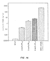
図1に示したように、Ca2+の存在は、ルシフェラーゼ発現レベルの劇的増強を生じ、最適Ca2+濃度ではSPLPトランスフェクション能力の〜600倍の増大を観察した。トランスフェクションにおけるこのCa2+媒介性増大は、プラスミドDNA−陽イオン性脂質複合体に関して前に観察されたものより、SPLP系に関しては有意に大きい。SPLPトランスフェクション能力を刺激するために必要な最適Ca2+濃度は、8〜10mMの範囲で、プラスミドDNA−陽イオン性脂質複合体のトランスフェクション能力の最適刺激に必要な量(5〜25mM)より多少低かった。さらにSPLPのトランスフェクション能力を刺激するCa2+の能力は、非常に特異的であった。図1に示したように、MgCl2またはNaClをCaCl2の代わりに用いた場合、トランスフェクション能力の増強は観察されなかった。
【0173】
2.SPLPはCa 2+ の存在下で安定である。PEG−CerC20を伴うSPLPは、in vivoでの循環時間延長を示し、外部ヌクレアーゼから封入プラスミドを保護し、そして細胞と容易に相互作用する高安定系である(Wheeler, et al., Gene Therapy 6:271−281(1999); Mok, et al., Biochimica et Biophysica Acta 1419:137−150(1999); Monck, et al., J. Drug Targ. 7:439−452(2000))。したがって、Ca2+の存在下でのSPLPのトランスフェクション特性強化が、細胞取込み強化をもたらすSPLPの不安定化または凝集のためでなかったことを実証するのは重要であった。サイズの変化を検出するために準弾性光散乱(QELS)を用いて、そしてDNA漏出を検出するためにピコグリーンフルオロフォア検定を用いて、Ca2+の存在下でのSPLPの安定性を調べた。QELS実験のために、CaCl2をSPLP懸濁液に付加して、50mMという高い濃度を達成した。SPLPサイズまたはサイズ分布の変化は観察されなかった。プラスミド放出実験に関しては、8mMCa2+の存在下または非存在下で10%FBSを含有するHBS緩衝液中で37℃で、SPLPをインキュベートした。ピコグリーン検定を用いて24時間に亘って、プラスミド放出を検定した。プラスミド放出は観察されなかった。
【0174】
3.Ca 2+ はSPLPの細胞取込みに影響を及ぼさない。プラスミドDNA−陽イオン性脂質複合体のトランスフェクション活性を強化するCa2+の能力は、Ca2+の存在下での細胞中への複合体の取込みの増大に寄与した(Lam, et al., Biochim Biophys Acta 1463:279−290(2000))。この点では、複合体と比較した場合のSPLPの低トランスフェクション能力は、少なくとも一部は、SPLPの極低レベルの細胞取込みから生じる(Mok, et al., Biochimica et Biophysica Acta 1419:137−150(1999))。したがって、細胞へのSPLP取込みを増大することによりCa2+がSPLPトランスフェクション能力を刺激するか否かを確定することは、興味深いことであった。0.5mol%Rh−DOPEを含有するSPLPを用いて、前記の材料と方法に記載したように、12mMまでのCa2+の存在下でBHK細胞中へのSPLP取込みを測定した。SPLPを細胞とともに4、8および24時間インキュベートし、細胞内脂質のレベルを測定した。細胞増殖を説明するために、各時点での脂質取込みを総細胞タンパク質に対して標準化した。図2に示したように、試験したCa2+の濃度の範囲全体でSPLPのトランスフェクション能力が数百倍まで変化した場合でも、Ca2+はSPLPの細胞取込みを有意に増大しない、ということを結果は示している。
【0175】
4.蛍光試験はCa 2+ 存在下でのSPLP取込み後のエンドソーム不安定化強化を示す。SPLPの取込みがCa2+の付加により刺激されないという事実は、トランスフェクションのCa2+依存性強化が蓄積されたSPLPのより効率的利用から生じなければならない、ということを示唆する。1つの可能性は、Ca2+がSPLPの取込み後にエンドソームの不安定化を多少促し、したがってプラスミドの細胞内送達を強化する、というものである。蛍光的標識化脂質を含有する小胞の取込み語のエンドソーム不安定化は、細胞内蛍光として蛍光顕微鏡により検出され得るが、一方、安定エンドソーム中への取込みは局在化「小斑点」出現を生じる、ということを過去の研究は示している(Felgner, et al., Pro. Natl. Acad. Sci. USA 84:7413−7417(1987))。SPLPの細胞分布を可視化し得るために、高レベルのRh−DOPE(4mol%)を小胞処方物とともに混入した。このようなRh−標識化SPLPを、10mMCa2+の存在下および非存在下でBHK細胞上でインキュベートし、蛍光顕微鏡により8時間目に細胞形態を検査した。前節で認められたSPLP取り込みの定量的測定値と一致して、Ca2+の非存在下または存在下で、同様レベルのローダミン蛍光を検出した。しかしながら、図3に示したように、蛍光顕微鏡で検出した場合の細胞の外観は、Ca2+の存在下または非存在下においてまったく異なった。多少の小斑点構造は観察されるが、しかし蛍光標識SPLPを含有するBHK細胞は、Ca2+が含まれた場合に、より拡散性のパターンを示した。Ca2+の非存在下では、蛍光パターンは、エンドソーム区画中のSPLP保持と一致して、高小斑点性であった。
【0176】
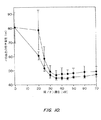
5.プラスミドDNAの細胞内プロセシング。先の蛍光顕微鏡結果は、Ca2+がエンドソーム区画を不安定化することによりトランスフェクションを強化し、したがってSPLP関連プラスミドの細胞質送達を強化する、ということを示唆する。SPLPプラスミドがCa2+の存在下でより容易にエンドソームから逃れ得る場合、それはリソソーム経路での分解を回避し、より無傷な細胞内プラスミドDNAが存在する。ドットブロット検定を用いて、プラスミドDNAの細胞内送達を測定し、サザンブロット分析を用いてプラスミドの組込みを検査した。8mMCa2+の非存在下または存在下で2、4および8時間、細胞をSPLPとともにインキュベートした。異なる系に関する無傷細胞内プラスミドDNAのレベルを、前記の材料と方法に記載したような細胞からのDNAの単離後に比較し、結果を図4に示す。図4Aに示したように、Ca2+の存在下で細胞をSPLPでトランスフェクトすると、BHK中の無傷プラスミドの量は、8時間インキュベーション期間後に約10倍に増大した。これは、Ca2+の存在下で調製されたSPLPでトランスフェクトした細胞中により多くの無傷プラスミドDNAが存在することを示したサザン分析にも反映されている(図4B)。無傷プラスミドDNAのこのようなレベル強化は、Ca2+の代わりにMgを用いた場合には観察されなかったが、このことは、Ca2+の特異性を実証する(図4B)。
【0177】
6.Ca 2+ はエンドソーム膜を不安定化する能力と一致した方法で二重層脂質構造を不安定化する。陰イオン性脂質と併合して、非二重層(HII相)構造を誘導することにより二重層膜を不安定化するイオン対を生成することにより、陽イオン性脂質が高分子、例えばプラスミドDNAの細胞内送達を刺激する、ということを近年の研究は示唆する。この点では、非二重層ヘキサゴナルHII相構造を誘導することにより、不飽和PEと組合せて酸性脂質、例えばホスファチジルセリン(PS)を含有する脂質二重層をCa2+が不安定化し得る、ということが周知である(Hope, et al., FEBS Letters 107:323−326(1976); Tilcock, et al., Biochimica et Biophysica Acta 641:189−201(1981))。ホスファチジルコリン(PC)およびコレステロールを含有する関連系においてHII相構造をCa2+が誘導し得る、ということも示されている。例えばDOPC/DOPE/DOPS/コレステロール(1:1:1:3; モル比)の混合物への付加も、二重層をヘキサゴナルHII相遷移に誘導する(Tilcock, et al., Biochemistry 23: 2696−2703 (1984))。したがって、SPLP中の陽イオン性脂質と協力して作用することにより、Ca2+がSPLPトランスフェクションを刺激して、エンドソーム膜の脂質二重層を不安定化するということが可能である。
【0178】
この可能性を調べるために、DOPC/DOPE/DOPS/Chol(1:1:1:3;モル比)から成るMLVのCa2+依存性多型性を、31P NMRを用いて、少量のDODACの非存在下および存在下で調べた。二重層組織化におけるリン脂質は非対称31P NMR線形状を生じ、低視野肩および高視野ピークを有するが、一方、ヘキサゴナルHII相中のリン脂質は、2つのより狭いものの一因子である逆非対称を有する線形状を生じる(Cullis, et al., Biochimica et Biophysica Acta 559:399−420(1979))。図5Aに示したように、DODACの非存在下で、Ca2+は、0.5:1のCa2+対DOPS比での31P NMRにより報告されているように、二重層からヘキサゴナルHII相への遷移を刺激し得る。あるいは、少量のDODAC(DOPC/DOPE/DOPS/コレステロール/DODAC;1:1:1:3:0.25;モル比)を含有するMLVにおいては、主にHII相構造を誘導するためには、0.25:1のCa2+対DOPS比のみが必要である(図5B)。狭中央ピークは、小ラメラ構造から、または構成成分リン脂質が等方性運動平均化を経る立方体相のような非二重層構造中の脂質から生じ得る。
【0179】
7.外部Ca 2+ はSPLPトランスフェクション能力を強化するために必要とされる。最終組の実験を実行して、SPLP内に封入されたCa2+が導入遺伝子発現を刺激し得るか否かを確定した。他所に詳述されているように(Felgner, et al., Journal of Biological Chemistry 269:2550−2561(1994))、Ca2+は、Ca2+イオノフォアA23187が存在する場合、pH勾配(内側酸性)に応答して、大型単一ラメラ小胞(LUV)中に負荷される。200mMという高い内部Ca2+濃度が達成され得る。前記の材料と方法に記載したように、SPLPはクエン酸塩緩衝液の存在下でpH4で容易に調製され、次に、洗剤透析手法後に外部pHがpH7.5に挙げられる。外部CaCl2およびイオノフォアの付加は次に、SPLP中へのCa2+の負荷を生じて、〜175mMの内部濃度を達成する。図6に示したように、封入Ca2+の存在はSPLPトランスフェクション能力の強化を生じるが、しかしCa2+の外部レベルはトランスフェクション過程を刺激するのに主要な役割を演じる、と思われる。
【0180】
8.改良型SPLP系に及ぼすCa 2+ の作用。SPLPの一制限は、小胞上の限定陽イオン性脂質およびPEGの存在の結果、系が細胞に最適に取込まれない、という点である(Mok, et al., Biochimica et Biophysica Acta 1419:137−150(1999))。正電荷を増大するための簡単な一方法は、陽イオン性含量を増大することによる(Zhang, et al., Gene Therapy 6:1438−1447(1999))。しかしながらSPLPに伴って増大するトランスフェクション効率はDODAC濃度を増大し、したがってこのような系はより低いDNA封入に屈従した。近年、陽イオン性ポリ(エチレングリコール)脂質複合体(CPL)として既知の陽イオン性脂質の新規の種類が合成された(Chen, et al., Bioconj. Chem. 11:433−437(2000))。本試験に用いられる典型的CPLは、疎水性セラミドアンカーを含有し、これは、4つのリシン残基から作られる陽イオン性頭基に連結される親水性PEGスペーサーに結合される。その表面に挿入されるSPLPw/CPLは、リポソームおよび細胞形質膜間の相互作用強化を示す、ということが示されている(Chen, et al., Bioconj. Chem. 11:433−437(2000))。
【0181】
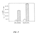
CPL−SPLP系に及ぼすCa2+の影響を調べるために、実験を実行した。より高いDODAC含量(14mol%)を含有するSPLPも含まれた。CPL−SPLP調製のために、前記の洗剤透析法を用いてプラスミドDNAをリポソーム中に負荷し、そして特性化挿入法を用いて予備生成SPLP中にCPLを挿入した。CPLを挿入して、最終4mol%挿入効率を得たが、このレベルは最適細胞結合および取り込みを提供することが示されている。Ca2+を8mMでSPLPおよびCPL−SPLP調製物に付加して、BHK細胞に適用する前に培地中に希釈した。BHK細胞をトランスフェクトリポソームと一緒にインキュベーション後24時間で、ルシフェラーゼに関して検定することにより、遺伝子発現を確定した。図7に示したように、それぞれ高DODAC含量またはCPLを含有するSPLPに関して、トランスフェクションの〜2000倍増および105倍増を検出した。
【0182】
C.考察
本実施例は、Ca2+がin vitroでのSPLPトランスフェクション能力の大強化を生じることを実証する。
Ca2+がSPLPのトランスフェクション能力を刺激するメカニズムは、いくつかの観察を説明しなければならない。第一に、トランスフェクション強化は、Ca2+存在下の無傷プラスミドの高細胞内レベルに起因すると思われる。これらの高レベルのプラスミドは、しかしながら、細胞中へのSPLPの取込み増大から生じると考えられる。第二に、本過程は、蛍光標識SPLPの取込み語の細胞の「小斑点」外観の低減を伴う。最後に、本作用はCa2+特異的である。最初の2つの観察は明らかに、SPLPのエンドサイトーシス後のBHK細胞のエンドソーム不安定化強化と一致する。したがって残存する問題は、Ca2+が特異的方法でこの不安定化をいかにして促し得るか、ということである。この点に関して、エンドソームが不安定化されてそれらの内容物の放出を強化する方法に関するコンセンサスは目下示されていないが、しかしながら多数の先導的観察がなされている。これらの中で主なものは、陽イオン性脂質が高分子、例えばプラスミドおよびアンチセンスオリゴヌクレオチドの細胞内送達を劇的に強化し得るという(Bennett, et al., Mol. Pharmacol. 41:1023−1033(1992); Barron, et al., Gene Ther. 6:1179−1183(1999))、そしてこの過程が、エンドソーム膜を不安定化し、したがってエンドソーム内容物の細胞ない放出を促す陽イオン性脂質の能力によっていると考えられる(Wattiaux, et al., FEBS Letters 417:119−202(1997); Xu, et al., Biochemistry 35:5616−5623(1996))という観察である。陽イオン性脂質は、一般的特性として、非二重層ヘキサゴナルHII相構造を形成するために陰イオン性脂質と併合する能力を示すということを、近年の研究は示して、陽イオン性脂質がエンドソームを不安定化するメカニズムがエンドソーム膜の二重層組織化を崩壊する能力によっているということを提唱した。同様に、Ca2+が二重層組織化を崩壊し、HII相構造を誘導する場合、細胞内送達における同様の強化が予測され得る。
【0183】
Ca2+は陰イオン性脂質を含有する従来の二重層脂質系中にHII相構造を誘導し得るという、そして、他の陽イオン、例えばMg2+はHII相構造を誘導し得ないし、同様の作用を生じるために高濃度を要しないので、この作用はCa2+特異的であるというかなりの証拠が認められる(Tilcock, et al., Biochemistry 23: 2696−2703 (1984))。本発明の実施例に示されているように、Ca2+はDOPC:DOPE:DOPS:Cholから成る二重層中にHII相構造を誘導し得るし、そして低レベルの陽イオン性脂質DODACと相乗的に作用して、HII相形成を誘発し得る。モデル膜行動をエンドソーム内側の行動と直接関連付けることは難しいが、しかし新規の酸性脂質(リソビスホスファチジン酸;LBPA)の生成のためにそれらが「初期」から「後期」に動くので、エンドソームの陰イオン性脂質含量は増大するということ、そしてLBPAと陽イオン性脂質、例えばDODACの混合物がHII相を採用する、ということが既知である。したがってこれらの結果は、Ca2+が陽イオン性脂質と共働してエンドソーム不安定化を促すことによりトランスフェクションを強化する、という理論を支持する。このような提案は、LBPAへのCa2+の付加がHII層の形成を生じるという観察とも一致する。その他の研究者は、リン酸カルシウム(CaPi)媒介性トランスフェクション中のエンドソーム放出の媒介に(Loyter, et al., Pro. Natl. Acad. Sci. USA 79:422−426(1982); Orrantia, et al., Experimental Cell Research 190:170−174(1990))、ならびにポリ陽イオン媒介性遺伝子移入に(Bottger, et al., Biochimica et Biophysica Acta 1395:78−87(1998); Haberland, et al., Biochimica et Biophysica Acta 1445:21−30(1999))、Ca2+が一役を演じる、ということを示唆する。
【0184】
本試験の意外な局面は、前に報告されたプラスミドDNA−陽イオン性脂質複合体のトランスフェクション特性に及ぼすCa2+の影響(Lam, et al., Biochim Biophys Acta 1463:279−290(2000))と、SPLPに関して本明細書中に報告された結果との間の不一致に関する。特に従来の研究は、Ca2+が複合体のトランスフェクション能力を20倍まで強化し得るということ、そしてこれがエンドソーム放出強化というよりむしろ、細胞中への複合体の取込み強化に寄与するということを実証した。意外な局面は、Ca2+存在下でのSPLPのトランスフェクション能力増大は細胞によるSPLPの取込み増大に関連しないが、一方Ca2+は脂質およびプラスミドの両方の取り込みにより立証されるような複合体の取込みの少なくとも2倍増を引き起こしたということである(Lam, et al., Biochim Biophys Acta 1463:279−290(2000))。この不一致は、複合体と比較した場合のSPLPの非常に異なる物理的特性に関連すると思われる。複合体は、高(等モル)レベルの陽イオン性脂質を含有する大型の正荷電系であり、一方、SPLPは小型の安定した、本質的に中性の小胞であり、低レベルの陽イオン性脂質を含有するPEGコーティングを有する。複合体と比較した場合のSPLP中の低レベルの陽イオン性脂質は、SPLP中に存在する陽イオン性脂質がエンドソーム中のすべての利用可能な陰イオン性脂質と併合するのに不十分で、したがって最大不安定化を達成するためにはCa2+の付加的存在を要するので、Ca2+に対する感度増大に直接的に関連し得る。
【0185】
考察の最終トピックは、本明細書に示された結果を、in vivoでのトランスフェクション能力強化を示すSPLPの生成に拡張することに関する。他所で強調されているように(Wheeler, et al., Gene Therapy 6:271−281(1999);Zhang, et al., Gene Therapy 6: 1438−1447 (1999))、SPLP送達の好ましい方法は、全身性適用によるものであり、この場合、疾患部位、例えば腫瘍部位での長期循環寿命および蓄積が必要とされる。本発明の結果は、トランスフェクション強化を生じるために、Ca2+が好ましくはSPLPの外側にあることを示唆する。これを達成するために、SPLPへのCa2+キレート化剤の付着による漸増表面Ca2+濃度を目指す戦略は、in vivoトランスフェクション強化を生じる必要がある。さらに例えば高濃度のカルシウムとともにSPLPの局所的(例えば腫瘍内)送達により、あるいはトランスフェクションの所望部位へのカルシウムの局所送達と組合せたSPLPの全身性送達により、トランスフェクションの部位で、カルシウム濃度の局所的増大が生じ得る。
【0186】
実施例II:陽イオン性PEGを含有する安定化プラスミド−脂質粒子はトランスフェクション能力強化を示す
A.材料と方法
1.材料。DOPEは、Northern Lipids (Vancouvert, BC)から入手した。Rh−DOPEおよびピコグリーンは、Molecular Probes(Eugene, OR)から入手した。DODACは合成し、且つInex Pharmaceuticals(Vancouver, BC)のS. Ansell博士から入手した。PEG−CerC20は、他所に示されたように合成し(Webb, et al., Biochim. Biophys. Acta 1372:272−282(1998))、且つZ. Wang博士(Inex Pharmaceuticals Corporation, Burnaby, BC)により提供された。pCMVLucプラスミドは、ヒトCMV初期プロモーターの制御下でPhotinus pyralisルシフェラーゼ遺伝子をコードし、P. Tam博士(Inex Pharmaceuticals Corporation, Burnaby, BC)により提供された。pCMVGFPプラスミドは、Aequorea victoriaからのグリーン蛍光タンパク質に関する遺伝子を含有し、P. Tam博士(Inex Pharmaceuticals Corporation, Burnaby, BC)により提供された。DEAE−セファロースCL−6B、セファロースCL−4B、オクチル−β−D−ガラクトシドおよびHEPESは、Sigma−Aldrich (Oakville, ON)から入手した。リポフェクチンは、Gibco BRL(Burlington, ON)から入手した。BHK細胞は、R. MacGillivray博士(Department of Biochemistry and Molecular Biology, UBC)から入手した。
【0187】
2.SPLP−CPL 4 の調製。それぞれ非封入プラスミドおよび空小胞を除去するために陰イオン交換(DEAE−セファロースCL−6B)クロマトグラフィーおよびスクロース密度勾配遠心分離による精製を用いて、Wheeler, et al.(Gene Therapy 6:271−281(1999))の方法にしたがって、DOPE:DODAC:PEG−CerC20(84:6:10)からなり、プラスミドpCMVLuc(またはpCMVGFP)を含有するSPLPを調製した。脂質フィルムを生成する前に、83.5:10:6:0.5(DOPE:DODAC:PEG−CerC20:Rh−PE)のモル比でCHCl3中に他の構成成分脂質とともにRh−PEを溶解することにより、Rh−PEを含有するSPLPを調製した。
【0188】
別記しない限り、Hepes緩衝化食塩水(HBS)、pH7.5中で60℃で2〜3時間、CPL4(12.5、19および30nmol)とともにSPLP(500nmol脂質)をインキュベートすることにより、予備生成SPLP中にCPL4を挿入した。HBS中で平衡させたセファロースCL−4Bカラム上でのゲル濾過クロマトグラフィーにより、非組入れCPL4を除去した。分画(1ml)を収集し、CPL4、リン脂質およびDNA含量に関して検定した。3つの構成成分すべてを含有する分画をプールし、濃縮した。それぞれ10および20 nmの励起および発光スリット幅を有するパーキン−エルマーLS52発光分光光度計を用いて、λex=340 nmでの励起後、λem=510nmでのダンシル標識化CPLの蛍光により、CPL4含量を確定した。標準曲線は、HBS中のダンシル化CPLのストック溶液から得た。Rh−PEを含有するSPLPに関しては、それぞれ10および20 nmの励起および発光スリット幅を用いて、λex=560 nmでの励起後、λem=590nmで測定されるRh標識の蛍光から、リン脂質含量を確定した。Rh標識を含有しないSPLPに関しては、Bligh and Dyer(Can. J. Biochem. Physiol. 37:911−917(1959))による脂質抽出後、Fiske−Subbarow(J. Biol. Chem. 66:375−400(1925))の方法を用いて、リン脂質を測定した。前記のようなピコグリーン検定キット(Mok, et al., Biochimica et Biophysica Acta 1419:137−150(1999)を用いて、プラスミドでなを確定した。
【0189】
Rh−PE含有系に関しては、セファロースカラムの前にダンシル体ローダミン比をカラム後の比で割って100%を掛けることにより、CPL4の組入れを確定した。その他の系に関しては、CPL4含量を総脂質含量で割って100を掛けることにより、組入れを確定した。
攪拌しながら25μLのDOPE:DODAC(0.8mM)とともに88μg/mLプラスミドDNA(pCMVLucまたはpCMVGFP)25μLを付加し、その後、細胞に付加する前に、室温で30分間インキュベートすることにより、1.5:1(負対正)の電荷非で、リポプレックスを調製した。リポフェクチンリポプレックスを同様に調製した。
【0190】
小胞方式で操作するNicompモデル270サブミクロン整粒器を用いて、準弾性光散乱(QELS)試験を実施した。Wheeler等(上記)が記載したのと同様に、凍結−割断電子顕微鏡試験を実施した。
50%マウス血清とともにSPLP系をインキュベート後、フェノール:クロロホルム抽出を用いて、サザン分析用のDNAを抽出した。その結果生じたDNAを次に、1%アガロースゲルを通して電気泳動処理し、ナイロン膜(Amersham)に移して、サザン分析を施した。膜を、最新プロトコールによりルシフェラーゼ遺伝子からの無作為プライム化32P−標識PvuII制限断片に曝露した。PhosphorimagerTMSI(Molecular Dynamics)を用いてハイブリダイゼーション強度を定量した。データを、非消化DNAに関して既定量の無傷DNAに変換した。
【0191】
Northern Lipids, Inc., Vancouver, B.C.により実施されたHPLC分析により、PEG−CerC20およびDOPEのレベルを確定した。
3.取込みおよびトランスフェクション試験。すべての取込みおよびトランスフェクション試験に関してトランスフェクション前に、1 x 104細胞/96ウエルプレート1ウエルの密度でBHK細胞を一夜プレート化、形質転換化BHK細胞株(tk−)を用いた。SPLPの細胞取込みを確定するために、12ウエルプレートの各ウエルに1x105BHK細胞を植え付けて、5%CO2中で37℃で、2mlの完全培地(10%FBSを含有するDMEM)中で一夜インキュベートした。40mMCaCl2を含有する培地中のSPLP、SPLP−CPL4、または200μLの容積中のDOPE:DODACリポプレックスを、20Μmの最終脂質用量で800μLの完全培地と混合し、細胞に付加した。プラスミドDNA濃度は、SPLP系およびリポフレックスに関してはそれぞれ1.4μg/mLおよび2.2μg/mLに相当した。細胞を37℃で指定時間インキュベートし、PBSで2回洗浄して、600μLの溶解緩衝液(PBS中の0.1%トリトンX−100)で溶解した。それぞれ10および10nmのスリット幅で、560nmのλexおよび600nmのλemを用いて、ローダミン蛍光を確定した。530nmの発光物フィルターも用いた。溶解物中の蛍光を脂質標準の蛍光と比較することにより脂質取込みを確定し、それをBCAタンパク質検定(Pierce, Rockford, IL)により確定される細胞数に対して標準化した。指示された場合、以下の明細でOmega Opticals(Brattleboro, VT)からのローダミンフィルターを用いて、Axiovert 100ツァイス蛍光顕微鏡(Carl Zeiss Jena GmbH)を用いて蛍光顕微鏡写真を撮った:励起560±20/二色性フィルター590/ロングパス発光600。
【0192】
以下の例外を伴って、前記と同様に、脂質取込みに及ぼすCa2+およびMg2+の作用を調べた。1mLの完全培地中の24ウエルプレート中に、BHK細胞(5x10⁴/ウエル)を植え付けて、37℃で一夜インキュベートした。SPLP−CPL4(40nmol)を総容積100μL中のCaCl2またはMgCl2と混合した。完全培地(400μL)をSPLP−CPL4に付加して、最終陽イオン濃度を4〜14mMとした。次にこの混合物を細胞に付加し、37℃で4時間インキュベートした。次に細胞をPBSで2回洗浄して、600μLの溶解緩衝液(PBS中の0.1%トリトンX−100)で溶解した。
【0193】
別記しない限り、5%CO2中で37℃で一夜のインキュベーション前に、150μL完全培地中の96ウエルプレートの各ウエル中にプレート化した1 x 10⁴BHK細胞を用いて、トランスフェクション試験を実施した。20μLHBS(SPLP)または40mMCaCl2含有するHBS(SPLP−CPL4)中の0.5μgのpCMVLucに対応するSPLPおよびSPLP−CPL4を80μLの完全培地に付加して、プラスミド濃度を5.0μg/mLとした。4時間のトランスフェクション時間ならびに24時間の総インキュベーション時間をルーチンに用いた。トランスフェクション時間は、細胞がプラスミド含有粒子とともにインキュベートされる時間と定義され、一方、総インキュベーション時間は、トランスフェクション時間(この後、トランスフェクション培地が取り換えられる)+その後の、細胞が導入遺伝子発現に関して検定される前にインキュベートされる時間である。24時間後、100μLの溶解緩衝液を用いて細胞を溶解し、40μLの溶解物を96ウエル発光プレートに移した。ルシフェラーゼ反応キット(Promega, Madison, WI)、ルシフェラーゼ標準(Boehringer−Manheim)およびML3200微小滴定プレート発光系(Molecular Dynamics, Chantilly, VA)を用いて、ルシフェラーゼ活性を確定した。BCAタンパク質検定(Pierce, Rockford, IL)により確定される細胞数に対して、活性を標準化した。
【0194】
トランスフェクション時間経過試験は、pCMVLucを含有するSPLP、SPLP−CPL4ならびにリポフェクチン(Gibco BRL, Burlington, ON)およびDOPE/DODACリポプレックスを包含した。前記と同様にリポプレックスを調製した。4、8および24時間のトランスフェクション時間後、トランスフェクション培地を除去し、4および8時間トランスフェクションの場合には、24時間の総インキュベーション時間の間、完全培地に取り換えた。24時間目に、全細胞を溶解し、前記と同様にルシフェラーゼ活性およびタンパク質含量(BCA検定)に関して検定した。
【0195】
pCMVGFPを含有するSPLP−CPL4、DOPE:DODACリポプレックスおよびリポフェクチンリポプレックスをpCMVLucに関して記載したのと同様に調製した。5.0μg/mLのプラスミドDNA用量で前記と同様に、トランスフェクションを実施した。24および48時間試料をインキュベーション後、トランスフェクション培地を除去し、細胞を洗浄して、新鮮な培地を細胞に付加した。次に、ツァイス蛍光顕微鏡下で、細胞を観察した。以下の明細で、フルオレセインフィルター(Omega Opticals)を用いて、GFPを発現する細胞の数を計数した:励起475±20/二色性フィルター500/発光535±22.5。トランスフェクション効率は、GFPを発現する細胞のパーセンテージとして表した。
【0196】
B.結果
1.陽イオン性PEG脂質は予備生成SPLP中に挿入され得る。SPLPは、リポプレックスより低い細胞中への取込みおよびより低いトランスフェクション能力を示す、ということを過去の研究は示している(Mok, et al., Biochimica et Biophysica Acta 1419: 137−150 (1999))。表面関連陽イオン性PEG脂質(CPL)、特にPEG分子の末端に4つの電荷を含有するもの(CPL4;構造に関しては、図8A参照)は、細胞中へのLUVの取込みを劇的に強化し得る。さらにCPLは、簡単なインキュベーションプロトコールを用いて、SPLPと同様に脂質組成物とともに予備生成LUV中に挿入され得る。したがって、SPLP中にCPL4を挿入するために同様の手法が開発され得るか、否かを調べた。6mol%の陽イオン性脂質N,N−ジオレイル−N,N−ジメチルアンモニウムクロリド(DODAC)、84mol%の「フソゲニック」ヘルパー脂質ジレオイルホスファチジルエタノールアミン(DOPE)およびセラミド(Cer)アンカーに結合されたPEG2000から成る10mol%の安定化脂質(PEG−Cer)を含有する脂質混合物から、Wheeler, et al.(Gene Therapy 6:271−281(1999)の洗剤透析手法により、pCMVLucを含有するSPLPを調製した。PEG−Cerのセラミドアンカーは、商法から容易に交換せず、したがって高安定性SPLP系に寄与するCアシル鎖(PEG−CerC20)を含有した(Wheeler, et al., Gene Therapy 6:271−281(1999))。洗剤透析法は、1プラスミド/小胞、遊離プラスミドおよび空小胞を含有するSPLPの混合物を生成した。他所に記載されているように(Wheeler, et al., Gene Therapy 6:271−281(1999))、それぞれDEAEカラムクロマトグラフィーおよび密度遠心分離により、遊離プラスミドおよび空小胞を除去することにより、SPLPを精製した。
【0197】
予備生成SPLP中へのCPL4の後挿入に関する手法は、図8Bに説明されている。精製SPLPを、60℃で3時間まで、CPL4(〜5mol%)とともにインキュベートし、次にカラムクロマトグラフィーにより非組入れCPL4から分離した。図9に示したように、これは、利用可能な80%までのCPL4のSPLPとの会合を生じ、これはSPLP−CPL4系中の総脂質の4mol%に対応する。
【0198】
2.SPLP−CPL 4 はCPL 4 の挿入後に凝集し、二価陽イオンの付加後に脱凝集する。CPLを含有するLUVは凝集する傾向があり、そしてこの凝集は培地のイオン強度を増大することにより抑制され得る、ということを過去の研究は示している。SPLP−CPL4も凝集を受けやすく、そしてこの凝集はNaCl、CaCl2またはMgCl2をSPLP−CPL4処方物に付加することにより逆転され得る、ということが判明した。この作用は、図10に説明されており、これは、準弾性光散乱(QELS)により測定される粒子の平均直径の標準偏差における変化によりモニタリングした場合のSPLP−CPL4の凝集に及ぼすCaCl2およびMgCl2の付加の影響を示している。両陽イオンに関しては、標準偏差は陽イオン濃度の増大に伴って低減し、最適脱凝集は約30mMより上で起こる。この行動は、凍結−割断電子顕微鏡によっても可視化され得る。図11Aに示したように、SPLPの凍結−割断顕微鏡写真は小単一分散粒子を明示するが、一方、CaCl2の非存在下で調製されたSPLP−CPL4は高度に凝集される(図11B)。図11Cに示したように、40mMCaCl2の付加は、この凝集を逆転して、SPLP調製物と同様に単一分散粒子を生成する。
【0199】
QELSおよび凍結割断電子顕微鏡を用いて、CaCl2存在下でのSPLPおよびSPLP−CPL4のサイズを比較した。QELS試験は、SPLPおよびSPLP−CPL4の平均直径がそれぞれ80±19nmおよび76±15nmであることを明示し、一方凍結割断試験は、68±11nmおよび64±14nmの直径を示した。SPLP直径に関するこれらの値は、従来の研究と密接に一致する(Wheeler, et al.(Gene Therapy 6:271−281(1999)))。
【0200】
3.PEG−CerC 20 含量およびSPLP−CPL 4 の安定性。CPL4が挿入されて、4mol%という高いレベルの総SPLP脂質を達成し得るという観察は、SPLP−CPL4の外側単一層中のCPL4のレベルが8mol%であることを示す。PEG−CerC20の初期濃度が10mol%であるとすると、これは、SPLP−CPL4の外側単一層中のPEG−脂質の総レベルが約18mol%になることを示唆する。これらのレベルは、脂質小胞中に通常組み入れられ得るPEG−脂質のレベルより高く(Woodle, et al., Biochim. Biophys. Acta 1113:171−199(1992))、CPL4として取り換えられる外側単一層中のPEG−CerC20のいくつかが挿入されたという可能性を生じる。これは、HPLCを用いてCPL4の挿入の前および後にSPLPに関してPEG−CerC20対−DOPEの比を測定することにより調べた。前記のようにCPL4をSPLP中に挿入した。非組み入れ物質の除去後の分析は、4mol%CPL4(総SPLP脂質に対して標準化)がSPLP中に挿入されたことを確定した。CPL4の挿入前は、PEG−CerC20対−DOPE比は0.091で、7.6mol%のPEG−CerC20含量に対応し、DOPEが脂質含量の84mol%を構成すると想定された。CPL4の挿入後、PEG−CerC20対−DOPE比は0.072であると判明し、6.0mol%のPEG−CerC20含量を示した。CPL4の挿入中にSPLPから失われるPEG−CerC20のすべてが外側単一層から失われると仮定すると、これは、挿入過程中に外側単一層のPEG−CerC20含量画7.6mol%から4.4mol%に低減することを示す。SPLP−CPL4の外側単一膜中の総PEG−脂質含量は、次に外側単一層脂質の12.4mol%であると概算し得る。
【0201】
50%マウス血清中で4時間までのインキュベーション後のSPLPおよびSPLP−CPL4の安定性は、図12で説明されている。すべての場合、封入プラスミドDNAは血清分解から完全に保護された。これに対比して、リポプレックス中のプラスミドの本質的に完全な分解が、血清中でのインキュベーションの30分以内に観察された。
【0202】
4.SPLP−CPL 4 はBHK細胞中への取込み強化およびトランスフェクション能力の劇的強化を示す。次の組の実験は、BHK細胞中へのSPLPの取込みに及ぼす組入れCPLの影響およびその結果生じるSPLP−CPL4系のトランスフェクション能力を確定することを目指した。4mol%までのCPL4を含有するSPLPを40mMCaCl2の存在下で調製し、BHK細胞に付加し(最終CaCl2濃度8mM)、種々の時間の間インキュベートした。次に、前記の材料と方法に示したように、会合SPLP−CPL4に関して細胞を検定した。図13Aに示したように、CPL4を含有しないSPLPの取込みは、8時間のインキュベーション後でさえ最小であるが、しかし3mol%またはそれ以上のレベルのCPL4を含有するSPLPに関しては、取込みは劇的に改善される。例えば4mol%のCPL4を含有するSPLPは、CPLの非存在下でのSPLPに関して達成された値の約50倍より高い蓄積レベルを8時間で示す。この取込み強化は、図13Bに可視的に説明されており、これはローダミン標識化SPLPおよびSPLP−CPL4とともに4時間インキュベーション後のBHK細胞の蛍光顕微鏡写真を示している。前期の材料と方法に記載されたトランスフェクションプロトコールを用いて、SPLP、SPLP−CPL4およびプラスミドDNA−陽イオン性リポソームリポプレックス(DODAC/DOPE;1:1)のトランスフェクション特性を調べた。このプロトコールは、SPLPまたはリポプレックスとともにBHK細胞を4時間(トランスフェクション時間)インキュベートし、その後培地を取り換えて、さらにインキュベートして導入遺伝子発現を最大にすることを包含する。総インキュベーション時間(トランスフェクション時間+培地交換後のインキュベーションの時間)を24時間で一定に保持した。図14に示したように、漸増量のCPL4の存在は、SPLP系に関するトランスフェクション能力の劇的増大を生じた。4mol%CPL4を含有するSPLP−CPL4は、SPLPを用いて達成されたよりも多少高い3x103のルシフェラーゼ発現レベルを示した。
【0203】
5.Ca 2+ はSPLP−CPL 4 のトランスフェクション活性に必要とされる。上記の実施例1は、SPLPのトランスフェクション能力がCa2+の存在に対して高度に感受性であることを実証しており、この場合、〜10mMのCa2+の存在がトランスフェクション能力を数百倍に強化する。したがって、SPLP−CPL4のトランスフェクション活性に及ぼすCa2+の影響を確定することは興味深いことであった。4mol%CPL4を含有するSPLPをBHK細胞とともに48時間、種々の量のMgCl2およびCaCl2の存在下でインキュベートし、ルシフェラーゼ活性を確定した。図15に示したように、トランスフェクション活性は主に、トランスフェクション培地中のCa2+の存在によっていた。10mMの最適CaCl2濃度では、SPLP−CPL4は、対応する量のMgCl2が存在した場合よりも105倍より高いトランスフェクション能力を示した。
【0204】
Ca2+またはMg2+の存在下でのSPLP−CPL4の異なるトランスフェクション特性が細胞中への取込みの差により説明され得るか否かを調べるために、MgCl2またはCaCl2の存在下で4時間インキュベーション後に、BHK細胞中へのSPLP−CPL4の蓄積をモニタリングした。図16に示したように、BHK細胞中へのSPLP−CPL4の取込みは、Ca2+およびMg2+の両方に関して同一である。Ca2+またはMg2+取り込みは、おそらくはBHK細胞の表面の負荷電CPL4結合部位が覆われるために、二価陽イオンの濃度が増大するとわずかに低減する、ということが留意され得る。これらの結果は、Ca2+がSPLPの細胞取込みにほとんど影響を及ぼさないことを示す従来の研究と一致する。
【0205】
SPLP−CPL4は、リポプレックスのトランスフェクション能力に匹敵するかまたはそれ以上であるトランスフェクション能力をin vitroで示す。図14に示したデータは、4時間の間BHK細胞に適用した場合に、SPLP−CPL4より〜100倍高いレベルの遺伝子発現をDOPE/DODACリポプレックスが生じることを示す。SPLP−CPL4が安定系であるとすると、取り込みは長時間にわたっておそらくは継続し得る。したがってSPLP−CPL4またはリポプレックスが4、8および24時間のトランスフェクション時間の間、BHK細胞に適用された場合に達成されるトランスフェクションレベルを調べた。2つの型のリポプレックス、即ちDOPE:DODAC(1:1)リポプレックス(電荷比1.5)、ならびにトランスフェクション試薬リポフェクチンを用いて生成され、電荷比1.5のDOPE/DOTMA(1:1)リポプレックスから成るリポプレックスを用いた。図17に示したように、SPLP−CPL4の能力はトランスフェクション時間の増大に伴って顕著に増大し、これは、SPLP−CPL4系の取込み率がトランスフェクションに関する限定因子であり得ることを示唆する。トランスフェクション期間直後にルシフェラーゼ発現に関して細胞が検定される24時間トランスフェクション時間に関しては、トランスフェクションレベルはリポフェクチンまたはDOPE/DODACリポプレックスにより達成されるものに匹敵する。
【0206】
さらなる実験を実施して、SPLP−CPL4およびリポプレックスを用いた24および48時間のトランスフェクション時間後のトランスフェクションレベルを確定したが、この場合、ルシフェラーゼ活性はトランスフェクション期間直後に検定した。図18Aに示したように、リポフェクチン(DOPE:DOTMA)リポプレックスの活性は24時間後に〜2000ngルシフェラーゼ/細胞タンパク質1gに横ばいになった。同様の結果が、DOPE:DODACリポプレックスに関して得られた。これに対比して、SPLP−CPL4処方物の活性は、インキュベーション時間が増大すると、増大し続けて、48時間で4000ng/細胞タンパク質1mgに対応するルシフェラーゼ発現レベルに達した。この活性は、SPLP(Ca2+非存在下)に関して観察されたよりも約106倍高く、リポフェクチンリポプレックスにより達成されたレベルのほぼ2倍であった。
【0207】
6.SPLP−CPL 4 は非毒性且つ効率的トランスフェクション剤である。リポプレックスが細胞に対して毒性であり得る、ということは周知である。SPLP−CPL4は低レベルの陽イオン性脂質を含有し、おそらくはリポプレックスより低毒性である。それぞれSPLP−CPL4およびリポプレックスに関して約80Μmおよび45μMの総脂質用量に対応する、5.0μg/mLのプラスミドに対応するSPLP−CPL4およびリポプレックスのレベルに24および48時間曝露後の細胞生存可能性を確定することにより、SPLP−CPL4およびリポプレックスの毒性を検定した。図18Bに示したように、SPLP−CPL4は補ドン度毒性を示さなかったが、一方リポプレックスは高毒性であった。細胞生存率は、リポフェクチンリポプレックスとともに48時間インキュベーション語には30%に過ぎなかったが、一方、SPLP−CPL4とともに48時間インキュベーション後には〜95%の細胞が生育可能であった。
【0208】
SPLP−CPL4によりトランスフェクトされた細胞の割合により示されるようなトランスフェクションの効率を確定するための試験も実施した。グリーン蛍光タンパク質(GFP)遺伝子を含有するプラスミドを用いて、トランスフェクト化細胞の割合を確定した。蛍光顕微鏡により、GFP発現を検出した。図19Aおよび19Bに示したように、24時間で約35%の細胞、そして48時間で50%の細胞がSPLP−CPL4によりトランスフェクトされ、見かけの細胞死は認められなかった。これに対比して、リポフェクチンリポプレックスは、35%未満の最大トランスフェクション効率を示し、24時間トランスフェクション時間語の細胞生存率は〜50%に過ぎなかった(図19C)。同様の低トランスフェクション効率および高毒性は、DOPE:DODACリポプレックスを用いても観察された。
【0209】
C.考察
これらの結果は、SPLP中へのCPL4の組入れがBHK細胞中への取込み改善を生じ、そしてCa2+が存在する場合のSPLPのトランスフェクション能力を劇的に強化することを実証する。興味深い3つの点が認められる。第一は、SPLP−CPL4系の化学組成および構造、ならびにSPLPの栄養機能およびトランスフェクション能力を改質するための後挿入手法の一般性に関する。第二は、SPLPの取込み強化、Ca2+の存在および観察されるトランスフェクション活性の間の関連に関する。最後に、SPLP−CPL4系の特性をリポプレックスと比較するのは興味深い。これらの領域の各々を、以下で順次取り扱う。
【0210】
ここに示された結果は、60℃でのインキュベーションを含めた簡単な方法を用いて、陽イオン性PEG脂質CPL4が予備生成SPLP中に挿入され得ることを実証する。例えばSPLP外側単一層中の総脂質の約8mol%に相当するレベルにCPL4を挿入する能力は、同様のインキュベーションプロトコールを用いてPEG−PEが予備生成LUV中に挿入されて、循環寿命延長を示す系を生じ得ることを実証する他の研究者の結果と一致する(Uster, et al., FEBS Lett. 386:243−246(1996))。それは、SPLP系と同様に脂質組成物を含有する予備生成LUV中にCPL4が挿入され得ることを示す従来の結果とも一致する。外側単一層中で達成されるPEG−脂質の総レベル(12.4mol%)は、LUV中へのPEG−脂質の組入れの最大レベルが一般に7〜10mol%である場合より高い(Woodle, et al., Biochim. Biophys. Acta 1113:171−199(1992))。しかしながら、PEG−脂質は、溶解作用が観察される前に15mol%という高いレベルまでLUV中に組入れられうる、と多数の著者が報告している(Edwards, et al., Biophys. J. 73:258−266(1997); Kenworthy, et al., Biophys. J. 68:1903−1920(1995); Hristova, et al., Macromolecules 28:7693−7699(1995))。これらは、構造変化(球から円板への)が、12mol%より高いPEG−PEレベルでジステアロイルホスファチジルコリン(DSPC)リポソームに関してのみ観察され、溶解作用は15mol%より上で観察された冷却電子顕微鏡試験を包含する(Edwards, et al., Biophys. J. 73:258−266(1997))。同様に、X線試験は、非二重層ミセル構造が15mol%より上のPEG−脂質レベルに関して観察されるだけであることを示す(Kenworthy, et al., Biophys. J. 68:1903−1920(1995); Hristova, et al., Macromolecules 28:7693−7699(1995))。
【0211】
SPLP−CPL4系がCPL4の挿入後に凝集する傾向は、CPL4を含有するLUVも凝集する従来の観察と一致する。この凝集の理由は一般に理解されていないが、しかし2つの一般的指摘がなされ得る。第一に、相互作用はおそらくは、高イオン強度での凝集が抑制されるとすると、小胞間の静電的相互作用によるものと思われる。第二に、凝集は後挿入過程それ自体の結果ではなく、そういうものとして、凝集はCPL4を含有するLUVに関しても観察され、この場合、CPL4はLUVが生成された脂質混合物中に存在した。陽イオン性頭基は、リン脂質ホスフェート基のレベルで対向する膜と相互作用する、ということもあり得る。あるいは、凝集現象は、ストレプタビジンのようなタンパク質を結合し得る配座を採用するPEGコーティングの能力に関連し得る(Sheth, et al., Pro. Natl. Acad. Sci. USA 94:8399−8404(1997))。
【0212】
考察の第二点は、CPL4がSPLP系のトランスフェクション能力を増大するメカニズムに関する。リポプレックス系に含入される陽イオン性脂質は細胞中への取込みを刺激する場合に直接的役割を演じる(Miller, et al., Biochemistry 37:12875−12883(1998))ということを、そしてこの取り込みがリポプレックス上の正電荷のために起こる(van der Woude, et al., Biochim. Biophys. Acta 1240:34−40(1995))ということを多数の研究が示している。細胞表面のヘパリンスルホン化プロテオグリカンはこの過程において主要な役割を演じる、ということが示唆された(Mislick, et al., Pro. Natl. Acad. Sci. USA 93:12349−12354(1996); Mounkes, et al., J. Biol. Chem. 273:26164−26170(1998))。CPL4の付加後のSPLPの取込み強化は、Ca2+の類似の付加的存在によるものであり得るが、トランスフェクション能力の増大はCa2+のの付加的存在に大いによっている。実施例Iは、Ca2+の存在が〜600倍のSPLPトランスフェクション能力の増大を生じることを、そしてこの増大が、取込み増大に起因するというよりむしろ、取込み後のエンドソーム膜の不安定化に役立つCa2+の能力に起因することを示す。したがって、SPLPを上回るSPLP−CPL4に関するトランスフェクション能力の観察された改善は、Ca2+の存在によりエンドソーム膜を不安定化する能力増強と結び付けられたCPL4により媒介される取り込みの改善に起因すると言える。この点に関しては、SPLP−CPL4(Ca2+存在下)のトランスフェクション能力は、Ca2+非存在下でのSPLPのトランスフェクション能力と比較して、〜104(図14)の一因子により増大される。これは、4mol%CPLの存在により約50倍に(図13A、4時間インキュベーション)、Ca2+の存在による〜600の一因子を掛けたBHK細胞中へのSPLPの取り込み増大により説明され得る。
【0213】
最後の考察領域は、他の非ウイルスベクターを上回るSPLP−CPLの利点に関するもので、これは、SPLP−CPL4系の十分に限定されたモジュール性、ならびに毒性および能力問題を包含する。第一に、不良限定数のプラスミド/粒子を含有する大型、不均質、不安定系である他の非ウイルス系、例えばリポプレックスに対比して、SPLP−CPL4は、1プラスミド/粒子を含有する小型で、均質な、安定系である(Wheeler, et al., Gene Therapy 6:271−281(1999))。重要な点は、SPLPが、モジュール様式で構築され得る、SPLP−CPL4のようなより洗練された系の基本構成成分である点である。例えばCPLの陽イオン性基の代わりに特異的ターゲッティングリガンドを含有するPEG−脂質の後挿入は、特定の細胞および組織に対して特異的に標的化されるSPLPを生じる必要がある。毒性に関しては、SPLP−CPL4は、リポプレックスよりも、組織培養中のBHK細胞に対して顕著に低毒性である。これはおそらくは、リポプレックスと比較した場合、SPLP中に含入される低比率の陽イオン性脂質に関連する。最後に、CPL4またはCa2+を伴わないSPLPは、プラスミドDNA−陽イオン性脂質複合体または裸プラスミドDNAのトランスフェクション特性よりもすでに優れている全身投与後のin vivoトランスフェクション特性を示す。ここに示した結果は、細胞中へのSPLP取込みを促すリガンドの使用により、そしてCa2+濃度の局所的増大をもたらす方法により、さらに有意の利益が予測され得ることを示唆する。
【0214】
要するに、ここに示した結果は、陽イオン性PEG脂質がSPLP中に後挿入されて、in vitroでBHK細胞中への取り込み改善を示す十分限定されたSPLP−CPL4系を生じ得ることを実証する。Ca2+の存在下では、SPLP−CPL4系は、Ca2+の非存在下でのSPLPと比較した場合、106倍まで増大されるトランスフェクション能力を生じる。これらの結果は、細胞中への取込み後、SPLP系が非毒性、高トランスフェクション効力存在であることを示し、そしてエンドサイトーシスを受ける細胞表面リガンドに対して標的化されるSPLPがin vivoでのトランスフェクション能力の有意の強化をもたらすことを示す。
【0215】
本明細書中に記載された実施例および実施態様は説明のためだけのものであり、そしてそれにかんがみて種々の修正または変更が当業者に示唆され、本出願の精神および範囲内に、ならびに添付の特許請求の範囲内に含まれるものである、と理解されるべきである。本明細書中に引用された出版物、特許および特許出願はすべて、それらの記載内容が参照により本明細書中に含まれる。
【図面の簡単な説明】
【図1】
SPLPトランスフェクションに及ぼすCa2+の作用および特異性。漸増濃度のCaCl2(●)、MgCl2(黒四角)またはNaCl(黒三角)(0〜14mM)を、細胞へのそれらの付加前にSPLP中で滴定した。SPLP(DODAC/DOPE/PEG−CerC20;7:83:10 mol/mol/mol)小胞中に封入された0.5μgのpCMVLucプラスミドを用いて、1 x 104細胞/96ウエルプレートの1ウエルでプレート化された細胞をトランスフェクトした。実施例Iの材料と方法に記載したように細胞をSPLPとともに24時間インキュベートし、Luc活性を測定した。実験はすべて、三重反復試験で実施した。
【図2】
SPLPの細胞取込みに及ぼすCa2+の作用。0.5mol%Rd−標識化DOPE(DODAC/DOPE/PEG−CerC20/Rh−DOPE;7:82.5:10:0.5 mol/mol/mol/mol)を含有するSPLPを用いて、細胞脂質取込みをモニタリングした。Ca2+(0〜14mM)の存在下で調製された80 nmolの脂質小胞を、適切な時間まで、細胞上でインキュベートした。実施例Iの材料と方法に記載したように4時間(●)、8時間(黒四角)または24時間(黒三角)でのRd蛍光を測定することにより、脂質取込みレベルを確定した。実験はすべて、三重反復試験で実施した。
【図3】
SPLPの細胞局在化。4 mol%Rh−DOPE標識化小胞100 nmolを、カルシウム(10mM)の非存在下(A)または存在下(B)で細胞(1 x 10⁵細胞/12ウエルプレートの1ウエルでプレート化)上でインキュベートした。8時間時点で、トランスフェクト培地を完全DMEM培地と置き換えて、蛍光顕微鏡を用いて細胞を検査した。以下の明細でOmega Opticals(Brattleboro, VT)からのローダミンフィルターを用いて、Axiovert 100ツァイス蛍光顕微鏡(Carl Zeiss Jena GmbH)で蛍光顕微鏡写真を撮った:λex=560±20 nm、600 nmLPおよびDC590 nm。
【図4】
プラスミドDNAの細胞内プロセシングは、Caの存在の影響を受けた。実施例Iの材料と方法に記載したように、2.5μgプラスミドDNAを含有するSPLPを用いて、8mMCa2+の非存在下(●)または存在下(黒四角)でBHK細胞をトランスフェクトした。適切な時点(2時間、4時間および8時間)で、細胞からDNAを抽出し、特異的32P−標識化プラスミドDNAプローブとのハイブリダイゼーションにより細胞内DNAを検出した。(A)材料と方法に記載したようなドットブロット分析により確定されるプラスミドDNA取込みのレベル。(B)サザンブロット分析により確定される細胞内プラスミドDNAの完全性。レーン1および11:pCMVLuc対照;レーン2、5、8および12;非トランスフェクト化対照;レーン3、6、9および13:SPLPでトランスフェクトされた細胞;レーン4、7、10および14:SPLPおよび8mMCa2+でトランスフェクトされた細胞;ならびにレーン15:SPLPおよび8mMMg2+でトランスフェクトされた細胞。実験はすべて、三重反復試験で実施した。
【図5】
CaCl2の存在下での種々のモデル膜の31P NMRスペクトル。(A)Ca2+を、0:1〜0.5:1(モル比)の範囲のCa2+/DOPS比で小胞(DOPE/DOPS/DOPC/Chol、1:1:1:3)中で滴定した。(B)Ca2+を、0:1〜0.25:1(モル比)の範囲のCa2+/DOPS比で小胞(DOPE/DOPS/DOPC/Chol/DODAC、1:1:1:3:0.25)中で滴定した。3サイクルの凍結−解凍により、脂質二重層を横断する陽イオンの平衡を保証した。スペクトルを同一ピーク高に釣り合いを取った。実施例Iの材料と方法に記載したように実験を実行した。
【図6】
トランスフェクションに及ぼすCa2+含有SPLPの作用。材料と方法に記載したように、pH勾配の存在下でA23187を用いてCa2+を負荷した。漸増濃度のCa2+(0〜14mM)を、DMEM希釈前にSPLP(黒四角)およびCa2+含有SPLP(●)の両方に付加した。SPLP中に封入された0.5μgのpCMVLucプラスミドを用いて、1 x 10⁴細胞/96ウエルプレートの1ウエルでプレート化された細胞をトランスフェクトした。実施例Iの材料と方法に記載したようにLuc活性を測定した。実験はすべて、三重反復試験で実施した。
【図7】
改良型SPLP系に及ぼすCa2+の作用。より高いDODAC含量(14mol%)またはCPL(4mol%)を含有するSPLPを用いて、8mMCaの存在下(斜線バー)または非存在下(白バー)で細胞をトランスフェクトした。0.5μgのpCMVLucを、各トランスフェクション実験における各処方物中に用いた。実施例Iの材料と方法に略記したように、Luc発現に関して検定する前に、細胞を小胞に24時間曝露した。実験は三重反復試験で実施した。
【図8】
SPLP−CPL4の生成。A.ダンシル化CPL4の構造。CPL4は、脂質アンカー、DSPEに結合されたPEG3400分子の末端に4つの正電荷を保有する。B.予備生成SPLP中へのCPL4の挿入のためのプロトコール。SPLPおよびCPL4を60℃で3時間一緒にインキュベートし、セファロースCL−4Bカラムクロマトグラフィーを用いて非混入CPL4を除去する。さらなる詳細に関しては、実施例IIの材料と方法を参照されたい。
【図9】
60℃でのSPLP中へのCPL4の挿入に関する時間経過。ダンシル化CPL4(0.3μmol)を、1.5mLの総容積中に360μgのpCMVLucを含有する6μmolのDOPE:PEG−CerC20:DODAC:Rh−PE(83.5:10:6:0.5;mol%)から成るSPLPに付加し、60℃でインキュベートした。混合物のアリコート(250μL)を指示された時点で採取して、セファロースCL−4Bカラムクロマトグラフィーを用いて非取込みCPL4を除去した。実施例IIの材料と方法に記載したように、CPL4混入を確定した。
【図10】
CPL4の後挿入のSPLPの脱凝集に及ぼす陽イオン濃度の作用。実施例IIの材料と方法に記載したように、SPLPを調製し、4mol%CPL4を挿入した。漸増濃度のCa2+(●)およびMg2+(黒四角)の存在下でのSPLP−CPL4の平均直径および平均直径の標準偏差を、QELSにより確定した。500mMストック溶液からのCaCl2またはMgCl2をSPLP−CPL4に付加した(400Μl中180nmol)。Ca2+またはMg2+の付加は、約30mMの陽イオン濃度での平均直径の標準偏差の低減により示されるように、より単分散性調製物を生じる。
【図11】
(A)SPLP、(B)SPLP−CPL4および(C)40mMCaCl2の存在下でのSPLP−CPL4の凍結−割断電子顕微鏡写真。SPLP−CPL4は実施例IIの材料と方法に記載したように調製され、そして4mol%CPL4を含有した。プレートAのバーは、200nmに相当する。
【図12】
封入プラスミドのサザン分析により検定した場合のSPLP−CPL4の血清安定性。図9に対する凡例に示したようにSPLPを調製し、後挿入プロトコールを用いて4mol%のCPL4を挿入した。50%マウス血清の存在下で37℃で指示時間の間、5μgpCMVLucを含有するSPLP−CPL4をインキュベートし、1μgのプラスミドDNAに対応する混合物のアリコートを取り出し、プラスミドDNAを抽出し、材料と方法に記載したようにサザン分析に付した。レーン1〜4は、それぞれ0、1、2および4時間インキュベーション時間後の裸プラスミドDNAの行動を示し;レーン5〜8は、0、1、2および4時間インキュベーション時間後のSPLPから抽出されたプラスミドの行動を示し;そしてレーン9〜12は0、1、2および4時間インキュベーション時間後の4mol%CPL4を含有するSPLPから抽出されたプラスミドDNAの行動を示す。
【図13A】
BHK細胞中へのSPLP−CPL4の取込みに及ぼすSPLP中に混入されたCPL4の量の影響。0(●)、2(黒四角)、3(黒三角)または4(◆)mol%CPL4を含有するSPLPの取込みを調べた;比較のために、DOPE:DODACリポプレックス(○)の取込みを示す。SPLP中へのCPL4の挿入およびリポプラックスの調製を、実施例IIの材料と方法に記載したように実施した。SPLP−CPL4培地は、凝集を防止するために40mMのCaCl2を含有し、BHK細胞の付加は、8mMへのCaCl2濃度の希釈を生じた。取込みプロトコールは、10%FBSを含有するDMEM中の105BHK細胞を伴うSPLP−CPL4のインキュベーションを包含した。インキュベーション後、細胞を溶解し、実施例IIの材料と方法に記載したようにローダミン−PEの取込みを測定した。
【図13B】
4時間インキュベーション後のSPLP(パネルI)および4mol%CPL4を含有するSPLP(パネルII)の取込み後のBHKの蛍光顕微鏡写真。左の顕微鏡写真は位相差方式で、そして右の写真は(ローダミン)蛍光方式で撮影した。
【図14】
種々の量のCPL4を含有するSPLPによるトランスフェクション後のBHK細胞中のルシフェラーゼ発現。後挿入法を用いて2、3および4mol%CPL4を含有するSPLPを調製した。実施例IIの材料と方法に記載したように、4時間のトランスフェクション時間および24時間の完全インキュベーション時間を用いて、5.0μg/mLpCMVLを含有するSPLP、SPLP−CPL4およびDOPE:DODAC(1:1)リポプレックスでBHK細胞(104)をトランスフェクトした。培地での希釈およびBHK細胞への付加後のSPLP−CPL含有系中のCaCl2濃度は8mMであった。トランスフェクション後、材料と方法に記載したように細胞を溶解し、ルシフェラーゼおよびBCA検定を実施した。
【図15】
SPLP−CLP4のトランスフェクション能力に及ぼすCa2+(●)およびMg2+(黒四角)の影響。実施例IIの材料と方法に記載したように、後挿入法により、4mol%CPL4を含有するSPLP−CPL4を調製した。漸増濃度のCaCl2またはMgCl2をSPLP−CPL4(5.0μgpCMVLuc/mL)に付加し、BHK細胞に移して、10%FBSを含有するDMEM中で48時間インキュベートした。次に細胞を溶解し、実施例IIの材料と方法に記載したように、ルシフェラーゼ活性およびタンパク質含量を測定した。
【図16】
BHK細胞によるSPLP−CPL4の取込みに及ぼすCa2+(●)およびMg2+(黒四角)の作用。図8に関して示したように陽イオン濃度の増大によりSPLP−CPL4を調製し、10%FBSを含有するDMEM中で4時間、BHK細胞(〜80Μm脂質および〜5.0μgpCMVLuc/mL/ウエル)とともにインキュベートした。次に細胞を溶解し、実施例IIの材料と方法に記載したように、SPLP−CPL4含量(Rh−PE脂質標識により示されるような)および細胞タンパク質を測定した。
【図17】
SPLP、SPLP−CPL4およびリポプレックスに関するトランスフェクション時間の一関数としてのBHK細胞中のルシフェラーゼ発現。後挿入法により、4mol%CPL4を含有するSPLP−CPL4を調製した。DMEMおよび10%FBS中のBHK細胞を、4、8および24時間のトランスフェクション時間および24時間の総インキュベーション時間を用いて、SPLP、SPLP−CPL4およびリポプレックス(5.0μg/mLpCMVL)とともにインキュベートした。次に、ルシフェラーゼ活性およびタンパク質含量に関して細胞を検定した。SPLP−CPL4(●)、SPLP(黒逆三角)、DOPE:DODACリポプレックス(黒四角)およびリポフェクチンリポプレックス(◆)でのトランスフェクション後のルシフェラーゼ活性を、トランスフェクション時間の一関数としてプロットした。リポプレックスを、1.5:1の投入比で調製した。
【図18】
A.BHK細胞によるトランスフェクション時間延長後の4mol%CPL4を含有するSPLP−CPL4(●)およびリポフェクチンリポプレックス(◆)のトランスフェクション能力。SPLP−CPL4およびリポプレックスを、図10に示したように生成した。10%FBSを含有するDMEM中で24および48時間、BHK細胞をトランスフェクトしたが、SPLP−CPL4およびリポフェクチンリポプレックス(投入比1.5:1)は5.0μg/pCMVLuc 1mLを含有した。トランスフェクション後、細胞溶解物中で、ルシフェラーゼ発現レベルおよび細胞タンパク質レベルを測定した。溶解物中のタンパク質含量に関してルシフェラーゼ活性を正規化し、トランスフェクション時間の一関数としてプロットした。
B.細胞溶解物中のタンパク質濃度に基づいた細胞生存により検定した場合のトランスフェクション時間の一関数としての4mol%CPL4を含有するSPLP−CPL4(●)およびリポフェクチンリポプレックス(◆)の毒性。
【図19】
GFPをコードするプラスミドを含有するSPLP−CPL4およびリポプレックスでトランスフェクトされるBHK細胞の蛍光および位相差顕微鏡写真。細胞を、SPLP−CPL4で24時間(A1、A2)および48時間(B1、B2)、ならびにリポフェクチンで24時間(C1、C2)トランスフェクトした。実施例IIの材料と方法に記載したように、pCMVGFPを用いてSPLPおよびリポプレックスを調製した。4mol%CPL4を含有するSPLP−CPL4は後挿入法により調製され、CaCl4を含有して、トランスフェクション培地中に8mMCaCl2濃度を生じた。24および48時間のトランスフェクション時間の間、10%FBSを含有するDMEM中でSPLP−CPL4またはリポフェクチン(5.0μg/mL)とともにBHK細胞(105)をインキュベートし、トランスフェクション期間直後に検査した。
【図20】
陽イオン性リポプレックスにより媒介される細胞膜の崩壊に関するメカニズム。標的細胞との結合(過程1)およびその細胞中へのエンドサイトーシス(過程2)後、陽イオン性リポプレックスを後期エンドソーム区画に移す(過程3)。陽イオン性脂質はエンドソーム膜の脱安定化を誘導して、エンドソーム膜とのリポプレックスの融合(過程4)、あるいは非二重層相へのエンドソーム膜の完全リモデリング(過程5)をもたらす。
【図21】
種々の量の負荷ヘッド基(a.)Et3N/CHCl3;(b.)TFA/CHCl3;c.EtN/CHClNα、Nε−ジ−t−Boc−L−リシンN−ヒドロキシスクシニドエステルを有する陽イオン性PEG−脂質複合体の調製のための合成模式図。[0001]
Background of the Invention
Gene therapy that is clinically useful requires an effective and safe gene delivery system. Viral vectors are a relatively efficient gene delivery system, but suffer from various limitations, such as the potential for reversion to wild type and immune response problems. As a result, non-viral gene delivery systems are accepting increasing attention (Worgall, et al., Human Gene Therapy 8: 37-44 (1997); Peters, et al., Human Gene Therapy 7: 1693-1699 (1997)). 1996); Yei, et al., Gene Therapy 1: 192-200 (1994); Hope, et al., Molecular Membrane Biology 15: 1-14 (1998)). Plasmid DNA-cationic liposome complexes are generally the most commonly used non-viral gene delivery vehicles (Felgner, Scientific American 276: 102-106 (1997); Chonn, et al., Current Opinion in Biotechnology). 6: 698-708 (1995)). However, the conjugate is a large, poorly defined system that is not suitable for systemic application and elicits considerable toxic side effects (Harrison, et al., Biotechniques 19: 816-823 (1995); Huang, et al. , Nature Biotechnology 15: 620-621 (1997); Templeton, et al., Nature Biotechnology 15: 647-652 (1997); Holand, et al., Pharmaceuticals, 1971-49.
[0002]
Recent work has shown that plasmid DNA can be encapsulated in small (about 70 nm diameter) "stabilized plasmid-lipid particles" (SPLP) consisting of a single plasmid encapsulated within bilayer lipid vesicles (Wheeler, et al., Gene Therapy 6: 271-281 (1999)). These SPLPs typically contain "fusogenic" lipid dioleoylphosphatidylethanolamine (DOPE), low levels of cationic lipids, and are in aqueous media due to the presence of a poly (ethylene glycol) (PEG) coating. Is stabilized. SPLPs show that they show prolonged circulatory life after intravenous (iv) injection, selectively accumulate in such areas due to enhanced vascular permeability at distant tumor sites, and Has systemic use because it can mediate transgene expression. SPLP containing the luciferase marker gene i. v. The levels of transgene expression observed in tumors after injection are superior to those achievable with plasmid DNA-cationic liposome complexes (lipoplexes) or naked DNA. Improved expression levels may still be needed for optimal therapeutic benefit in some applications (see, e.g., Monck, et al., J. Drug Targ. 7: 439-452 (2000)).
[0003]
Cationic poly (ethylene glycol) (PEG) lipids or CPL have been designed for insertion into lipid bilayers to confer a positive charge (Chen, et al., Bioconj. Chem. 11: 433). -437 (2000)). For example, distearoyl-PE (DSP) -containing CPLs conjugated to PEG containing one or more distal positive charges have been synthesized and shown to promote enhanced in vitro cell binding and liposome uptake. (Chen, et al., Bioconj. Chem. 11: 433-7 (2000)).
[0004]
Thus, there remains a strong need in the art for new and more efficient methods for introducing nucleic acids into cells. The present invention addresses this and other needs.
[0005]
Summary of the Invention
The present invention provides effective compositions, methods and uses for delivering nucleic acids to cells. The compositions and methods of the present invention are based on the surprising discovery that the presence of an endosomal membrane destabilizing agent in a lipid formulation results in a dramatic increase in transfection efficiency. The compositions and methods of the invention can be used in vitro or in vivo, and can be used to increase the transfection efficiency of any cell type, including mammalian cells (eg, humans).
[0006]
As such, in one embodiment, the invention is directed to nucleic acid-lipids for introducing nucleic acids into cells comprising cationic lipids, complex lipids that inhibit particle aggregation, nucleic acids and endosomal membrane destabilizing agents. A particle composition is provided. In a preferred aspect, the nucleic acid-lipid particles are "stabilized plasmid-lipid particles" (SPLP). Typically, SPLPs are less than 150 nm in diameter and comprise a single plasmid encapsulated within a bilayer lipid vesicle. Complex lipids that inhibit aggregation typically include a hydrophilic polymer. In a preferred embodiment, the hydrophilic polymer is a PEG or polyamide having a molecular weight of about 250 to about 7,000 daltons (eg, ATTA). The endosomal membrane destabilizing agent can be present inside the particle, outside the particle, or both inside and outside the particle. Preferably, the endosomal membrane destabilizer is Ca++It is an ion. In certain aspects, Ca++The concentration of the ions is from about 0.1 mM to about 100 mM.
[0007]
In certain embodiments, the complex lipid that inhibits aggregation is a "cationic polymer lipid" (CPL). In a preferred aspect, CPL is:
AWY−I
Wherein A is a lipid moiety, W is a hydrophilic polymer, and Y is a polycationic moiety. In certain preferred embodiments, Y is selected from lysine, arginine, asparagine, glutamine, and combinations thereof.
[0008]
In another embodiment, the present invention is a method of introducing a nucleic acid into a cell, the method comprising contacting the cell with a nucleic acid-lipid particle composition, wherein the particle comprises a cationic lipid, Provided is a method comprising a complex lipid that inhibits aggregation, a nucleic acid, and an endosomal membrane destabilizing agent. The endosomal membrane destabilizing agent can be present inside the particle, outside the particle, or both inside and outside the particle. In certain embodiments, the endosomal membrane destabilizing agent contacts the cell before, after, simultaneously with, or a combination of the particles.
[0009]
In yet another embodiment, the present invention provides a method for treating
[0010]
The compositions, methods and uses of the present invention provide a number of benefits. For example, the presence of an endosomal membrane destabilizer results in a dramatic increase in transfection efficiency of the nucleic acid. By increasing transfection efficiency, the amount of gene product in the cell is greatly increased. Further, the compositions and methods of the present invention can be used in vitro or in vivo, and can be used to increase the transfection efficiency of any cell type, including humans.
[0011]
These and other advantages, objects, and embodiments of the present invention are described in further detail in conjunction with the following drawings and detailed description.
[0012]
DETAILED DESCRIPTION OF THE INVENTION AND PREFERRED EMBODIMENTS
I. Introduction
The present invention provides novel and otherwise effective compositions and methods for delivering nucleic acids to cells. These compositions and methods demonstrate that the presence of an endosomal membrane destabilizing agent (eg, calcium) plays a role in transfection efficiency of nucleic acids (eg, plasmids) formulated as SPLPs or “stabilized nucleic acids (eg, plasmids) -lipid particles”. It is based on the finding that it leads to a significant increase. Typically, SPLPs are less than about 150 nm in diameter (more preferably, about 70 nm in diameter) and consist of a single plasmid encapsulated within a bilayer lipid vesicle.
[0013]
As used herein, the term “endosomal membrane destabilizing agent” (EMD) refers to an agent (single) that is thought to promote the disruption or destabilization of endosomal membranes, thereby enhancing the release of their contents Or more). Endosomes are discrete intracellular compartments that are typically isolated from the rest of the cell by selectively permeable membranes. Suitable EMDs include monovalent metal ions such as K+, Na+, Divalent metal ions such as Mg2+, Ca2+, Mn2+, Co2+, And combinations of metal ions with cationic lipids. The most preferred EMD is Ca2+Ions, in this case Ca2+For SPLPs containing ions, Ca2+About 10 times less than SPLP in the absence of ions6A twice higher transfection efficiency is observed.
[0014]
The methods of the invention can be used in vitro or in vivo, and can be used to increase the transfection efficiency of any cell type, including mammalian cells. For example, for in vitro transfection, an endosomal membrane destabilizing agent (eg, calcium) can be added to the transfection medium. For example, any of a wide range of calcium concentrations may be used, for example, a concentration in the range of 0.1 mM to about 100 mM. Preferably about 1 mM to about 20 mM, most preferably about 8 to about 10 mM is used. In one embodiment, an endosomal membrane destabilizing agent (eg, calcium) is first added to the SPLP at a high concentration that results in the desired final concentration after dilution of the SPLP into the transfection medium. In another embodiment, an endosomal membrane destabilizing agent (eg, calcium) is added to the SPLP at the time of transfection into the cell. The endosomal membrane destabilizing agent can be co-administered with the SPLP, it can be administered before the administration of the SPLP, or it can be administered after the administration of the SPLP.
[0015]
In vivo, any method that results in a local increase in endosomal membrane destabilizing agent (eg, calcium) concentration at the site of transfection may be used. For example, the particles can be formulated to incorporate an endosomal membrane destabilizing agent, the particles can be immersed in a solution containing a high endosomal membrane destabilizing (eg, calcium) concentration prior to administration, or the particles can be It can be administered in a buffer or formulation containing a membrane destabilizing agent (eg, calcium) concentration. Such methods are particularly used for local delivery of the particles, for example for intratumoral injection, where, for example, co-administration of calcium ions results in a local high calcium concentration, whereby the site of delivery or This may result in enhanced transfection of the particles in the vicinity. Further, the endosomal membrane destabilizing agent may be co-administered with the SPLP, it may be administered before the administration of the SPLP, or it may be administered after the administration of the SPLP.
[0016]
In certain in vivo or in vitro embodiments, SPLPs may include a chelating molecule, such as for chelating an endosomal membrane destabilizing agent, such as a lipid derived with an endosomal membrane destabilizing agent chelator, on their surface. And thereby formulated to allow for the production of high local endosomal membrane destabilizer concentrations, even after systemic delivery of the particles. For example, in certain in vivo or in vitro embodiments, the particles include a calcium-chelating molecule on their surface, such as a lipid derived with a calcium chelator, such that even after systemic delivery of the particles, localized high calcium Formulated to allow for the generation of a concentration.
[0017]
The present invention may be practiced with any SPLP particles. For example, SPLPs containing a wide range of concentrations of cationic and any of the other lipids can be used. Similarly, SPLPs can include any of a wide variety of cationic and other lipids. SPLP can be prepared using any plasmid from any source and containing any polynucleotide sequence, as well as using any of a number of methods.
[0018]
The present invention also provides a SPLP containing a cationic PEG lipid called SPLP-CPL. In a preferred embodiment, a SPLP-CPL comprising a PEG lipid with four positive charges4Is used. SPLP and SPLP-CPL can be derived to contain any of a number of functional groups, including but not limited to calcium chelators, cell or tissue specific targeting molecules, labels and the like. Not done.
[0019]
Suitable SPLPs and SPLP-CPLs and methods of making and using SPLPs and SPLP-CPLs for use in the present invention are described, for example, in
[0020]
II. Definition
The term "lipid" refers to a group of organic compounds, including, but not limited to, esters of fatty acids and being insoluble in water, but soluble in many organic solvents. It is characterized by. They are usually divided into at least three classes: (1) "simple lipids" including fats and oils and waxes; (2) "complex lipids" including phospholipids and glycolipids; "Derived lipids".
[0021]
The term "vesicle-forming lipid" has a hydrophobic moiety and a polar head group, and can be spontaneously formed in bilayer vesicles in water alone, as exemplified by most phospholipids It is intended to include any amphiphilic lipid.
The term "vesicle-embedded lipid" refers to a hydrophobic moiety that is stably incorporated into a lipid bilayer in combination with other amphipathic lipids and contacts the inner hydrophobic region of the bilayer membrane and the outer polar surface of the membrane It is intended to include any amphipathic lipid having its polar head group portion oriented toward Vesicle-adopted lipids include those that tend to adopt a non-lamellar phase on their own, and that can assume a bilayer structure in the presence of a bilayer stabilizing component. A typical example is DOPE (dioleoylphosphatidylethanolamine). Bilayer stabilizing components include complex lipids that inhibit aggregation of SPLP, polyamide oligomers (eg, ATTA-lipid derivatives), peptides, proteins, detergents, lipid derivatives, PEG-lipid derivatives, such as phosphatidylethanolamine. PEG, and PEG conjugated to ceramide, including but not limited to US Patent No. 08 / 485,608, now US Patent No. 5,885,613, the disclosure of which is incorporated herein by reference. Reference included in the book)).
[0022]
The term "amphiphilic lipid" refers to any suitable substance in which, in part, the hydrophobic portion of the lipid material is oriented into the hydrophobic phase, while the hydrophilic portion is oriented toward the aqueous layer. Amphiphilic lipids are usually the major constituent of lipid vesicles. The hydrophilic character comes from the presence of polar or charged groups such as carbohydrates, phosphatos, carboxylic acids, sulfatos, amino, sulfhydryls, nitro, hydroxy groups and the like. Hydrophobicity is imparted by the incorporation of non-polar groups, such as long-chain saturated and unsaturated aliphatic hydrocarbon groups, and one or more aromatic, cycloaliphatic or heterocyclic groups ( (Including, but not limited to, those substituted by one or more). Examples of amphiphilic compounds include, but are not limited to, phospholipids, amino lipids and sphingolipids. Representative examples of phospholipids include phosphatidylcholine, phosphatidylethanolamine, phosphatidylserine, phosphatidylinositol, phosphatidic acid, palmitoyl oleoyl phosphatidylcholine, lysophosphatidylcholine, lysophosphatidylethanolamine, dipalmitoylphosphatidylcholine, dioleoylphosphatidylcholine, and distearoyldithiolylphosphidylcholine. Include, but are not limited to, noreyl phosphatidylcholine. Other compounds that lack phosphorus, such as sphingolipids, the glycosphingolipid family, diacylglycerols and β-acyloxy acids are also within the group called amphiphilic lipids. Further, the above amphiphilic lipids can be mixed with other lipids, such as triglycerides and sterols.
[0023]
The term "neutral lipid" refers to any of a number of lipid species that exist in an uncharged or neutral zwitterionic form at a selected pH. At physiological pH, such lipids include, for example, diacylphosphatidylcholine, diacylphosphatidylethanolamine, ceramide, sphingomyelin, cephalin, cholesterol, cerebroside and diacylglycerol.
[0024]
The term "hydrophobic lipid" refers to compounds having non-polar groups, examples of which are long-chain saturated and unsaturated aliphatic hydrocarbon groups, and one or more aromatic, cycloaliphatic, Or such groups optionally substituted by heterocyclic group (s), but is not limited to such. Suitable examples include, but are not limited to, diacylglycerol, dialkylglycerol, N-N-dialkylamino, 1,2-diacyloxy-3-aminopropane, and 1,2-dialkyl-3-aminopropane. .
[0025]
The term "diacylglycerolyl" refers to a 2-fatty acyl chain R having from 2 to 30 independently bonded carbon atoms at the 1- and 2-positions of glycerol by ester linkages.1And R2Means Acyl groups can be saturated or can have varying degrees of unsaturation.
The term "dialkylglycerolyl" refers to two C-alkyl groups linked to the 1- and 2- positions of glycerol by an ether linkage.1~ C30Means an alkyl chain of
[0026]
The term "NN-dialkylamino" includes the following:
Embedded image
[0027]
The term "1,2-diacyloxy-3-aminopropane" refers to a 2-fatty acyl chain C attached to the 1- and 2-positions of propane by an ester bond.1~ C30Means Acyl groups can be saturated or can have varying degrees of unsaturation. The 3-position of the propane molecule has an attached -NH- group. 1,2-diacyloxy-3-aminopropane has the following general formula:
Embedded image
The term "l, 2-dialkyl-3-aminopropane" refers to a 2-alkyl chain (C1~ C30). The 3-position of the propane molecule has an attached -NH- group. 1,2-Diacyl-3-aminopropane has the following general formula:
Embedded image
[0029]
The term "cationic lipid" refers to any of a number of lipid species that possess a net positive charge at a selected pH, for example, at physiological pH. Such lipids include N, N-dioleyl-N, N-dimethylammonium chloride ("DODAC"), N- (2,3-dioleyloxy) propyl) -N, N, N-trimethylammonium chloride ( "DOTMA"), N, N-distearyl-N, N-dimethylammonium bromide ("DDAB"), N- (2,3-dioleoyloxy) propyl) -N, N, N-trimethylammonium chloride ( “DOTAP”), 3- (N- (N ′, N′-dimethylaminoethane) -carbamoyl) cholesterol (“DC-Chol”) and N- (1,2-dimyristyloxyprop-3-yl)- N, N-dimethyl-N-hydroxyethylammonium bromide ("DMRIE"), but is not limited thereto. In addition, a number of commercial preparations of cationic lipids that can be used in the present invention are available. These include commercially available cationic liposomes containing, for example, LIPOFECTIN® (DOTMA and 1,2-dioleoyl-sn-3-phosphoethanolamine (“DOPE”)) (Gibco / BRL, Grand Island, New York, USA). )); LIPOFECTAMINE (registered trademark) (N- (1- (2,3-dioleoyloxy) propyl) -N- (2- (sperminecarboxamido) ethyl) -N, N-dimethylammonium trifluoroacetate (" Commercially available cationic liposomes ("DOSPA") and ("DOPE"), and commercially available cationic liposomes (Gibco / BRL); and TRANSFECTAM.RTM. Cationic lipid (Promega Corp., Madison, Wisconsin, USA)) and the like. The following lipids are cationic and have a positive charge below physiological pH: DODAP, DODMA, DMDMA, etc.
[0030]
The term "fusogenic" refers to the ability of a liposome, SPLP, or other drug delivery system to fuse with the membrane of a cell. The membrane can be a plasma membrane or a membrane surrounding organelles, such as endosomes, nuclei, and the like. Fusogenesis is the fusion of liposomes with such a membrane.
The term "dendrimer" embraces its association with branched polymers having multiple generations. In dendrimers, each generation creates branch points.
[0031]
The term “ligand” encompasses any molecule, compound or device with a reactive functional group, such as lipids, zwitterionic lipids, carrier compounds, chelating moieties, biocompatible compounds, biomaterials, Biopolymer, biomedical device, analytically detectable compound, therapeutically active compound, enzyme, peptide, protein, antibody, immunostimulant, radioactive label, fluorogen, biotin, drug, hapten, DNA, RNA, polysaccharide, liposome , Virosomes, micelles, immunoglobulins, functional groups, targeting agents or toxins. The above list is illustrative and not intended to be exhaustive.
[0032]
The term "ATTA" or "polyamide" refers to, but is not limited to, the compounds disclosed in US patent application Ser. No. 09 / 218,988, filed on Dec. 22, 1998. These compounds include the following formula:
Embedded image
[0033]
As used herein, the term "alkyl" refers to a branched or unbranched hydrocarbon chain such as methyl, ethyl, n-propyl, iso-propyl, n-butyl, sec-butyl, iso-butyl, Means tertbutyl, octa-decyl and 2-methylpentyl. These groups may have one or more functional groups commonly attached to such chains, such as hydroxyl, bromo, fluoro, chloro, iodo, mercapto or thio, cyano, alkylthio, heterocyclyl, aryl, heteroaryl , Carboxyl, carvalcoyl, alkyl, alkenyl, nitro, amino, alkoxyl, amide and the like, optionally substituted with an alkyl group such as trifluoromethyl, 3-hydroxyhexyl, 2-carboxypropyl, 2-fluoroethyl, carboxymethyl, Can produce cyanobutyl and the like.
[0034]
The term "alkylene" refers to a divalent alkyl as defined above, e.g.2-), Propylene (-CH2CH2CH2-), Chloroethylene (-CHClCH2-), 2-thiobutene (-CH2CH (SH) CH2CH2-), 1-bromo-3-hydroxyl-4-methylpentene (-CHBrCH2CH (OH) CH (CH3) CH2−) Etc.
[0035]
The term "alkenyl" refers to a branched or unbranched hydrocarbon chain containing one or more carbon-carbon double bonds.
The term "alkynyl" means a branched or unbranched monovalent hydrogen chain containing one or more carbon-carbon triple bonds.
The term "aryl" forms at least one aromatic ring, preferably having about 6 to 14 carbon atoms, such as, for example, phenyl, naphthyl, indenyl, and the like, and is generally attached to such a chain. One or more functional groups such as hydroxyl, bromo, fluoro, chloro, iodo, mercapto or thio, cyano, cyanoamide, alkylthio, heterocyclic, aryl, heteroaryl, carboxyl, carbalcoyl, alkyl, alkenyl, nitro, Aryl groups substituted with amino, alkoxyl, amide, etc., for example, biphenyl, iodobiphenyl, methoxybiphenyl, anthryl, bromophenyl, iodophenyl, chlorophenyl, hydroxyphenyl, methoxyphenyl, formylphenyl, acetylphenyl, Trifluoromethylthio phenyl means trifluoromethoxyphenyl, alkylthiophenyl, trialkylammonium phenyl, amido phenyl, thiazolyl-phenyl, benzoxazolyl-phenyl, imidazolylmethyl phenyl, the chain of carbon atoms to form an imidazolylmethyl phenyl and the like.
[0036]
The term "acyl" refers to a -C (O) R group, where R is alkyl or aryl as described above, for example, formyl, acetyl, propionyl or butyryl.
The term "alkoxy" refers to -OR-, where R is alkyl.
[0037]
The term "amide" refers to an amide bond: -C (O) NR-, where R is hydrogen or alkyl.
The term "amino" refers to an amine linkage: -NR-, where R is hydrogen or alkyl or terminal NH.2).
The term “carboxyl” refers to the group —C (O) O— and the term “carbonyl” refers to the group —C (O) —.
[0038]
The term "carbonate" refers to the group -OC (O) O-.
The term "carbamate" refers to the group -NHC (O) O-, and the term "urea" refers to the group -NHC (O) NH-.
The term "phosphoro" refers to the group -OP (O) (OH) O-.
The term "basic amino acid" refers to naturally occurring amino acids and synthetic amino acids and / or amino acid mimetics that have a net positive charge at the selected pH, for example, at physiological pH. This group includes, but is not limited to, lysine, arginine, asparagine, glutamine, histidine and the like.
[0039]
The term "phosphorylethanolamino" refers to the group -OP (O) (OH) OCH2CH2It means NH-.
The term "phosphorylethanolamide" refers to the group -OP (O) (OH) OCH2CH2It means NHC (O)-.
The term "phospho" means a pentavalent phosphorus moiety -P (O) (OH) O-.
[0040]
The term "phosphoethanolamino" refers to the group -P (O) (OH) OCH2CH2It means NH-.
The term "phosphoethanolamide" refers to the group -P (O) (OH) OCH2CH2It means NHC (O)-.
The term "ethylene oxide unit" refers to the group -OCH2CH2Means-.
[0041]
The term "CPL" refers to a cationic polymeric lipid, for example, a cationic PEG lipid. Preferred CPLs are compounds of formulas I and II. Such CPLs are described in U.S. Patent Application Serial No. 09 / 553,639 (filed April 20, 2000) and PCT Patent Application No.
[0042]
The term “d-DSPE-CPL-M” is encompassed by the term “CPL1” which refers to DSPE-CPL having one positive charge. "d-" in d-DSPE-CPL-M indicates that CPL contains a fluorescent dansyl group. It will be apparent to the sugar trader that CPL can be synthesized without a dansyl moiety, thus the term "DSPE-CPL-M" is encompassed by the term "CPL1" above.
[0043]
The term "d-DSPE-CPL-D" is encompassed by the term "CPL2" which refers to DSPE-CPL having two positive charges.
The term "d-DSPE-CPL-T1" is encompassed by the term "CPL3" which refers to DSPE-CPL having three positive charges.
The term "d-DSPE-CPL-Q1" is encompassed by the term "CPL4a" which refers to DSPE-CPL having four positive charges.
[0044]
The terms "d-DSPE-CPL-Q5" or DSPE-PEGQuad5 or DSPE-CPL-4 are all encompassed by the term "CPL4 (or CPL4b)" which refers to DSPE-CPL with four positive charges. By modifying the head group region, CPLs containing one (mono or M), two (di or D), three (tri or T) and four (quad or Q) positive charges are synthesized. Was. Various QuadCPLs are synthesized, and therefore they are numbered Q1-Q5.
[0045]
The abbreviation "HBS" refers to Hepes-buffered saline, "Rho-PE" refers to rhodamine-phosphatidylethanolamine, and "LUV" refers to "large single lamellar vesicle".
[0046]
II. Nucleic acid-lipid particles (SPLP) and their properties
Nucleic acid-lipid particles or SPLPs typically include a cationic lipid and a nucleic acid. Such SPLPs also preferably include a non-cationic lipid and a bilayer stabilizing component, and more preferably also include a complex lipid that inhibits aggregation of the SPLP. The SPLPs of the present invention have an average diameter of less than about 150 nm and are substantially non-toxic. Further, nucleic acids, when present in the SPLPs of the invention, are resistant to aqueous solutions against nuclease degradation. Such SPLPs are disclosed in great detail in US Pat. No. 5,976,567 and PCT Patent Publication WO 96/40964, both of which teachings are incorporated herein by reference.
[0047]
A. SPLP component
A variety of suitable cationic lipids can be used in the present invention, alone or in combination with one or more other cationic or neutral lipid species.
Cationic lipids useful in the present invention include any of a number of lipid species that possess a net positive charge at physiological pH, such as DODAC, DOTMA, DDAB, DOTAP, DOPA, DOGS, DC-Chol, and DMRIE or May be combined. A number of these lipids and related analogs, which are also useful in the present invention, are described in co-pending USSN 08 / 316,399, US Patent Nos. 5,208,036, 5,264,618, 5 Nos., 279,833 and 5,283,185 (the contents of which are incorporated herein by reference). In addition, a number of commercial preparations of cationic lipids are available and can be used in the present invention. These include, for example, LIPOFECTIN® (commercially available cationic liposomes including DOTMA and DOPE (Gibco / BRL, Grand Island, New York, USA)); LIPOFECTAMINE® (commercially available cations including DOSPA and DOPE). Liposomes (Gibco / BRL)); and TRANSFECTAM® (commercially available cationic lipids including DOGS (Promega Corp., Madison, Wis., USA)).
[0048]
The non-cationic lipid used in the present invention can be any of a variety of neutral, uncharged zwitterionic or anionic lipids capable of forming a stable complex. They are preferably neutral, but they can alternatively be positively or negatively charged. Examples of non-cationic lipids useful in the present invention include phospholipid-related substances such as lecithin, phosphatidylethanolamine, lysolecithin, lysophosphatidylethanolamine, phosphatidylserine, phosphatidylinositol, sphingomyelin, cephalin, cardiocipin, phosphatidic acid, Cerebroside, dicetyl phosphate, dioleoylphosphatidylcholine (DOPC), dipalmitoyl-phosphatidylcholine (DPPC), dioleoylphosphatidylglycerol (DOPG), dipalmitoylphosphatidylglycerol (DPPG), dioleoyl-phosphatidylethanolamine (DOPE), palmitoyl oleate Oil-phosphatidylcholine (POPC), palmitoyl oleoyl-phosphatidyl Ethanolamine (POPE) and dioleoyl - phosphatidylethanolamine 4-(N-maleimidomethyl) - cyclohexane-1-carboxylate (DOPE-mal) and the like. Additional non-phosphorus containing lipids include, for example, stearylamine, dodecylamine, hexadecylamine, acetyl palmitate, glycerol ricinoleate, hexadecyl stearate, isopropyl myristate, amphoteric acrylic acid polymers, triethanolamine-lauryl sulfate, Alkyl-aryl sulfate polyethyloxylated fatty acid amides, dioctadecyldimethylammonium bromide, and the like, diacylphosphatidylcholine, diacylphosphatidylethanolamine, ceramide, sphingomyelin, cephalin and cerebroside. Other lipids may be present, such as lysophosphatidylcholine and lysophosphatidylethanolamine. Non-cationic lipids include polyethylene glycol-based polymers such as those described in co-pending USSN 08 / 316,429, the disclosure of which is incorporated herein by reference, such as PEG2000, PEG5000, and the like. Polyethylene glycol conjugated to phospholipids or ceramide (referred to as PEG-Cer).
[0049]
In a preferred embodiment, the non-cationic lipid is diacylphosphatidylcholine (eg, dioleoylphosphatidylcholine, dipalmitoylphosphatidylcholine and dilinoleoylphosphatidylcholine), diacylphosphatidylethanolamine (eg, dioleoylphosphatidylethanolamine and palmitoyloleoylphosphatidylethanolamine). ), Ceramide or sphingomyelin. The acyl groups in these lipids are preferably C10~ C24This is an acyl group derived from a fatty acid having a carbon chain of More preferably, the acyl group is lauroyl, myristoyl, palmitoyl, stearoyl or oleoyl. In a particularly preferred embodiment, the non-cationic lipid is 1,2-sn-dioleoylphosphatidylethanolamine or egg sphingomyelin (ESM).
[0050]
In one embodiment, the SPLP further comprises a bilayer stabilizing component (BSC). Suitable BSCs include, but are not limited to, polyamide oligomers, peptides, proteins, detergents, lipid derivatives, PEG-lipids, such as PEG conjugated to phosphatidylethanolamine and PEG conjugated to ceramide (US See US Patent No. 5,885,613, the contents of which are incorporated herein by reference. Preferably the duet stabilizing component is PEG-lipid or ATTA-lipid. In a preferred embodiment of the present invention, the BSC is a complex lipid that inhibits aggregation of SPLP. Suitable conjugated lipids include, but are not limited to, PEG-lipid conjugate, ATTA-lipid conjugate, cationic polymer-lipid conjugate (CPL) or mixtures thereof. In a preferred embodiment of the invention, the SPLP comprises a PEG-lipid conjugate or an ATTA-lipid conjugate together with a CPL.
[0051]
The CPL used in the present invention has the following structural features: (1) a lipid anchor for incorporating CPL into the lipid bilayer, such as a hydrophobic lipid; (2) a lipid anchor at the cationic head group. A hydrophilic spacer for linking, eg, polyethylene glycol; and (3) a polycationic moiety, eg, a naturally occurring amino acid, for producing a protonizable cationic head group. As such, the present invention provides compounds of formula I:
AWY−I
(Where A, W and Y are as follows)
The compound of is provided.
[0052]
With respect to Formula I, "A" is a lipid moiety, eg, an amphoteric, neutral or hydrophobic lipid that acts as a lipid anchor. Examples of suitable lipids include vesicle-forming lipids or vesicle-equipped lipids, and include diacylglycerolyl, dialkylglycerolyl, N-N-dialkylamino, 1,2-diacyloxy-3-amino Examples include, but are not limited to, propane and 1,2-dialkyl-3-aminopropane.
[0053]
“W” is a polymer or oligomer, eg, a hydrophilic polymer or oligomer. Preferably, the hydrophilic polymer is a biocompatible polymer that is non-immunogenic or possesses low intrinsic immunogenicity. Alternatively, a hydrophilic polymer can be weakly antigenic when used with a suitable adjuvant. Suitable non-immunogenic polymers include, but are not limited to, PEG, polyamide, polylactic acid, polyglycolic acid, polylactic / polyglycolic acid copolymer and combinations thereof. In a preferred embodiment, the polymer has a molecular weight of about 250 to about 7,000 daltons.
[0054]
"Y" is a polycationic moiety. The term polycationic moiety refers to a compound, derivative or functional group having a positive charge, preferably at least two positive charges, at a selected pH, preferably at a physiological pH. Suitable polycationic moieties include basic amino acids and their derivatives, such as arginine, asparagine, glutamine, lysine and histidine; spermine; spermidine; cationic dendrimers; polyamines; polyamine sugars; and aminopolysaccharides. The polycationic moiety can be linear, eg, a linear tetralysine, branched or dendrimeric structure. The polycationic moiety has a positive charge of about 2 to about 15, preferably about 2 to about 12, and more preferably about 2 to about 8 at the selected pH value. The choice of which polycationic moiety to use can be dictated by the type of liposome application desired.
[0055]
The charges on the polycationic moiety may be distributed around the entire liposome moiety, or they may be at discrete concentrations of charge density in certain regions of the liposome moiety, for example, charge spikes. When the charge density is distributed on the liposome, the charge density can be distributed evenly or non-uniformly. All variations in the charge distribution of the polycationic moiety are encompassed by the present invention.
[0056]
Lipid "A" and non-immunogenic polymer "W" can be attached by various methods, preferably covalently. Methods known to those skilled in the art can be used for covalent attachment of “A” and “W”. Suitable linkages include, but are not limited to, amide, amine, carboxyl, carbonate, carbamate, ester and hydrazone linkages. It will be apparent to one skilled in the art that "A" and "W" must have complementary functional groups to effect the conjugation. Reaction of these two groups, one on the lipid and one on the polymer, provides the desired linkage. For example, if the lipid is diacylglycerol and the terminal hydroxyl is activated with, for example, NHS and DCC to produce an active ester, and then reacted with a polymer containing amino groups, for example, with a polyamide (US Patent Application No. / 218,988 (submitted December 22, 1998)), an amide bond is formed between the two groups.
[0057]
In certain embodiments, "W" is linked to "Y", preferably covalently. As with "A" and "W", the linkage of "W" to "Y" can be created by the complementary reactivity of functional groups on the one hand and on the polycationic moiety on the other. . For example, an amine function on “W” can be reacted with an activated carboxyl group, such as an acyl chloride or NHS ester, to produce an amine. With the appropriate choice of reactive groups, the desired linkage is obtained. Other activated carboxyl groups include, but are not limited to, carboxylic acids, carboxylate esters, carboxylic acid halides, and other activated forms of carboxylic acids, such as reactive anhydrides. Reactive acid halides include, for example, acid chlorides, acid bromides, and acid fluorides.
[0058]
In some cases, the polycationic moiety has a bound ligand, eg, a targeting ligand or chelating moiety for complexing calcium. Preferably, after the ligand is bound, the cationic moiety retains a positive charge. In some cases, the bound ligand has a positive charge. Suitable ligands include, but are not limited to, compounds or devices with reactive functional groups, such as lipids, amphoteric lipids, carrier compounds, biocompatible compounds, biomaterials, biopolymers, biopolymers, and the like. Medical devices, analytically detectable compounds, therapeutically active compounds, enzymes, peptides, proteins, antibodies, immunostimulants, radiolabels, fluorogens, biotin, drugs, haptens, DNA, RNA, polysaccharides, liposomes, virosomes, micelles , Immunoglobulins, functional groups, other targeting moieties or toxins. Described are suitable chelating moieties for chelating or complexing endosomal membrane destabilizers.
[0059]
In certain preferred embodiments, other moieties are incorporated into the compound of Formula I to produce a compound of Formula II:
[0060]
Embedded image
[0061]
In Formula II, "X" is a single bond or a functional group that covalently links a lipid to at least one ethylene oxide unit. Suitable functional groups include phosphatidylethanolamino, phosphatidylethanolamide, phosphoro, phospho, phosphoethanolamino, phosphoethanolamide, carbonyl, carbamate, carboxyl, carbonate, amide, thioamide, oxygen, NR (where R is hydrogen or Alkyl)) and sulfur. In some cases, lipid "A" is directly linked to ethylene oxide by a single bond. The number of ethylene oxide units can range from about 1 to about 160, preferably from about 6 to about 50.
[0062]
In Formula II, "Z" is a single bond or a functional group that covalently bonds the ethylene oxide unit to the polycationic moiety. Suitable functional groups include phospho, phosphoethanolamino, phosphoethanolamide, carbonyl, carbamate, carboxyl, amide, thioamide, NR (where R is a member selected from the group consisting of a hydrogen atom or an alkyl group) ), But is not limited thereto. In certain embodiments, the terminal ethylene oxide units are linked directly to the polycationic moiety.
[0063]
In Formula II, “Y” is a polycationic moiety as described above in connection with Formula I. In Formula II, the index "n" is an integer value ranging from about 6 to about 160.
In an exemplary embodiment, compounds of formula II can be synthesized using generalized techniques as outlined in FIG. FIG. 21 illustrates one particular embodiment of the present invention, and is therefore merely illustrative, and should not limit the scope of the claims herein. Obviously, those skilled in the art will recognize many other changes, alternatives, and modifications that can be made to the reaction schematics illustrated in FIG. Referring to FIG. 21, a solution of a lipid, eg, DSP, and a base, eg, triethylamine, in a chloroform solution is (t-Boc-NH-PEG).3400-CO2NHS) and the solution is stirred at ambient temperature. The solution is then concentrated and dried under a stream of nitrogen. Next, the residue is purified by repeated precipitation of a chloroform mixed solution using diethyl ether until the lipid disappears using chromatography. The purified CPL conjugate is dissolved in a solvent, after which TFA is added and the solution is stirred at room temperature. The solution can be concentrated again under a stream of nitrogen. The residue is then purified by repeated precipitation of the mixture with diethyl ether and the lipid-PEG-NH2For example, DSPE-PEG-NH2Alternatively, it yields DSPE-CPL-1 with one protonizable cationic head group. The ratio of phosphoryl-lipid anchor and distal primary amine can then be determined by the phosphate and fluorescamine assays as described herein above.
[0064]
In this exemplary embodiment, the number of protonatable amino groups can be increased to make a polycationic moiety. By incrementally adding a stoichiometric amount of, for example, Nα, Nε-di-t-Boc-L-lysine N-hydroxysuccinide, the polycationic moiety is converted from about 2 to about 16 positive charges. Can increase. As noted above, the positive charge can be incorporated with any number of suitable polycationic moieties, such as lysine, arginine, asparagine, glutamine, histidine, polyamine, and derivatives or combinations thereof. Using the synthesis method of the present invention, the number of cationic groups, eg, amino groups, can be easily controlled during CPL synthesis.
[0065]
As explained further above, endosomal membrane destabilizers can be incorporated into the nucleic acid-lipid particles. In such embodiments, the endosomal membrane destabilizing agent can be loaded into the nucleic acid-lipid particles using any of a number of different loading techniques (see Examples I and II). Exemplary dosing methods are described, for example, in US Pat. Nos. 4,885,172, 5,059,421 and 5,171,578, the contents of which are incorporated herein by reference. Included). Further, a particularly preferred method of ionophore-mediated dosing is disclosed and claimed in US Pat. No. 5,837,282, the disclosure of which is incorporated herein by reference.
[0066]
Further, as described above, a chelating moiety suitable for chelating an endosomal membrane destabilizing agent can be attached, linked or attached to any of the lipid components of SPLP, such as CPL. In a preferred embodiment of the present invention, the chelating moiety is a metal chelator. Metal chelators such as ethylenediaminetetraacetic acid (EDTA), diethylenetriaminepentaacetic acid (DTPA), ethylenebis (oxyethylenenitrile) -tetraacetic acid (EGTA), 1,4,7,10-tetraazacyclododecane-N, N ', N ", N"'-tetraacetic acid, trans-1,2-cyclohexylenediamine-N, N, N ', N'-tetraacetic acid, N6-Carboxymethyl-N3, N9-[2,3-dihydroxy-N-methylpropylcarbamoylmethyl] -3,6,9-triazaundecandioic acid, N6-Carboxymethyl-N3, N9-Bis (methylcarbamoylmethyl) -3,6,9-triazaundecandioic acid, N3, N6-Bis (carboxymethyl) -N9-3-oxapentamethylene-carbamoylmethyl-3,6,9-triazaundecandioic acid or N3, N6-Bis (carboxymethyl) -N9-[3,3-bis (dihydroxyphosphoryl) -3-hydroxypropyl-carbamoylmethyl] -3,6,9-triazaundecandionic acid, metal ion transport protein, metal ion sequestering agent, metal chelating ligand, etc. Can chelate endosomal membrane destabilizers. In addition, the metal chelators disclosed in US Pat. No. 5,876,695, the disclosure of which is incorporated herein by reference, may also be used. Other chelating agents suitable for use in the compositions and methods of the present invention are known to those skilled in the art.
[0067]
2. Nucleic acid component
The invention is described in the examples with reference to the use of plasmids, but those skilled in the art will appreciate that the methods described herein are equally applicable to other larger nucleic acids or oligonucleotides. As such, suitable nucleic acids include, but are not limited to, plasmids, antisense oligonucleotides, ribozymes and other poly- and oligo-nucleotides.
[0068]
Nucleic acids (including both complexes and particles) useful in the invention are typically nucleotide polymers having from 10 to 100,000 nucleotide residues. Typically, the nucleic acid will be administered to a subject to restore or enhance expression of a cellular protein. Further, the nucleic acid can carry a label (eg, a radioactive, fluorescent, or colorimetric label) to provide a clinical diagnosis of the presence or absence of the complementary nucleic acid. Thus, nucleic acids or nucleotide polymers are described in Stein, et al. , Science 261: 1004-1011 (1993), and U.S. Patent Nos. 5,264,423 and 5,276,019, the contents of which are incorporated herein by reference. Can be polymers of nucleic acids, including genomic DNA, cDNA, mRNA or oligonucleotides containing antisense derivatives. In addition, the nucleic acids may encode transcription and translation control sequences, such as promoter and enhancer sequences.
[0069]
The nucleotide polymer can be single-stranded DNA or RNA, or double-stranded DNA or DNA-RNA hybrid. Examples of double-stranded DNA include structural genes, genes containing control and termination regions, and self-replicators, such as plasmid DNA.
Single-stranded nucleic acids include antisense oligonucleotides (complementary to DNA and RNA), ribozymes, and triplex-forming oligonucleotides. To increase stability, some single-stranded nucleic acids are preferably replaced by nucleotide linkages, such as phosphorothioate, phosphorodithioate, phosphoroselenate or O-alkylphosphodies, which are replaced with stable non-phosphodiester bonds. It has some or all of the triester linkages.
[0070]
Nucleic acids used in the present invention also include nucleic acids that have been modified at one or more sugar moieties and / or at one or more pyrimidine or purine bases. Examples of sugar modifications include the replacement of one or more hydroxyl groups with a halogen, alkyl group, amine, azide group, or functionalized as an ether or ester. In addition, all sugars can be substituted with sterically and electronically similar structures, eg, aza-sugars and carbocyclic sugar analogs. Modifications of the purine or pyrimidine base moiety include, for example, alkylated purines and pyrimidines, acylated purines or pyrimidines, or other heterocyclic substituents known to those skilled in the art.
[0071]
Multigene sequences can also be used in the methods of the invention. Thus, sequences for different proteins may be placed on one chain or on a plasmid. Non-coding sequences will also be present to the extent necessary to achieve proper expression.
The nucleic acids used in the methods of the present invention can be isolated from natural sources, obtained from sources such as the ATCC or GenBank libraries, or prepared by synthetic methods. Synthetic nucleic acids can be prepared by various solution or solid phase methods. Generally, solid phase synthesis is preferred. Detailed descriptions of techniques for solid phase synthesis of nucleic acids by phosphite-triester, phosphotriester and H-phosphonate chemistry are widely available (e.g., U.S. Patent No. 4,401,796 (Itakura)). U.S. Patent Nos. 4,458,066 and 4,500,707 (Caruthers et al.); Beaucage, et al., Tetrahedron Lett., 22: 1859-1862 (1981); Matteucci, et al., J .; Am. Chem. Soc., 103: 3185-3191 (1981); Caruthers, et al., Genetic Engineering, 4: 1-17 (1982); Jones,
[0072]
a. Vectors for gene transfer and expression in cells
An important aspect of the invention is the use of the lipid-nucleic acid particles provided herein to introduce a selection gene in vitro and in vivo, and then express the selection gene in a host cell. Thus, the nucleic acid in the particle specifically includes a vector that can be expressed in the host cell. Promoters, enhancers, stress or chemically regulated promoters, antibiotic or nutrient sensitive regions, and therapeutic protein coding sequences can be included as needed.
[0073]
In summary, expression of a natural or synthetic nucleic acid is typically accomplished by operably linking the nucleic acid to a promoter (constitutive or inducible), incorporating the construct into an expression vector, and in a suitable host cell. This is achieved by introducing a vector into Typical vectors contain transcription and translation terminators, transcription and translation initiation sequences, and promoters useful for regulation of the expression of the particular nucleic acid. The vector may optionally include at least one individual terminator sequence, a sequence that enables replication of the cassette in eukaryotes or prokaryotes, or both (eg, a shuttle vector), and both prokaryotic and eukaryotic systems. Gene expression cassette containing a selectable marker for The vectors are suitable for replication and integration in prokaryotes, eukaryotes, or preferably both (Giliman and Smith (1979), Gene, 8: 81-97; Roberts et al. (1987), Nature, 328: 731-734; Berger and Kimmel, Guide to Molecular Cloning Techniques, Methods in Enzymology, volume 152, Academic Press, Inc., San Diego, CA (Berger);. Sambrook et al (1989), Molecular Cloning-A Laboratory Manual, (2nd ed. ) Vol. 1-3, Cold Spring Harbor Laboratory, Cold Spring Harbor Press, NJ. Y. , (Sambrook); M. Ausubel et al. , Current protocols in Molecular Biology, eds. , Current Protocols, a joint venture between Green Publishing Associates, Inc. and John Wiley & Sons, Inc. , (1994 Supplement) (Ausubel)). Product information from manufacturers of biological reagents and laboratory equipment also provides useful information for known biological methods. Examples of such manufacturers include SIGMA chemical company (Saint Louis, MO), R & D systems (Minneapolis, MN), Pharmacia LKB Biotechnology (Piscataway, Int. (Palo Alto, CA), Chem Genes Corp. , Aldrich Chemical Company (Milwaukee, WI), Glen Research, Inc. , GIBCO BRL Life Technologies, Inc. (Gaithersberg, Md.), Fluka Chemica-Biochemika Analytika (Fluka Chemie AG, Buchs, Switzerland) and many commercial sources available from Applied Biosystems, a supplier of Applied Biosystems, Inc. (Foster CAs, Inc.)
[0074]
Vectors into which foreign nucleic acids are operably linked can be used to introduce these nucleic acids into host cells and mediate their replication and / or expression. "Cloning vectors" are useful for replicating and amplifying foreign nucleic acids and for obtaining clones of a particular foreign nucleic acid-containing vector. An “expression vector” mediates the expression of a foreign nucleic acid. Some vectors are both cloning and expression vectors.
[0075]
In general, the particular vector used to deliver the foreign gene into the cell is not particularly important. Any of the conventional vectors used for baldness in a selected host cell can be used.
Expression vectors typically include a "eukaryotic transcription unit" or "expression cassette" containing all elements required for expression of an exogenous gene in a eukaryotic cell. A typical expression cassette contains a promoter operably linked to the DNA sequence encoding the desired protein and the signals required for efficient polyadenylation of the transcript.
[0076]
Eukaryotic promoters typically contain two types of recognition sequences, a TATA box and an upstream promoter element. The TATA box, located 25-30 base pairs upstream of the transcription start site, is thought to be involved in directing RNA polymerase to initiate RNA synthesis. Other upstream promoter elements determine the rate at which transcription is initiated.
[0077]
Enhancer elements can stimulate transcription up to 1,000-fold from linked homologous or heterologous promoters. An enhancer is active when it is located downstream or upstream from the transcription start site. Many enhancer elements from viruses have a broad host range and are active in various tissues. For example, the SV40 early gene enhancer is suitable for many cell types. Other enhancer / promoter combinations suitable for the present invention include those derived from polyomavirus, human or murine cytomegalovirus, various retroviruses such as murine leukemia virus, murine or rous sarcoma virus and long term from HIV. (See Enhancers and Eukaryotic Expression, Cold Spring Harbor Press, Cold Spring Harbor, NY 1983), which is incorporated herein by reference.
[0078]
In addition to the promoter sequence, the expression cassette should also contain a transcription termination region downstream of the structural gene to provide for efficient termination. The termination region may be obtained from the same source as the promoter sequence, or may be obtained from a different source.
If the mRNA encoded by the selected structural gene is to be translated efficiently, a polyadenylation sequence will generally also be added to the vector construct. Two separate alignment elements are required for accurate and efficient polyadenylation. The GU- or U-rich sequence is located downstream from the polyadenylation site, and the highly conserved sequence of six nucleotides AAUAAAA is located 11-30 nucleotides upstream. Termination and polyadenylation signals suitable for the present invention include those derived from SV40 or partial genomic copies of a gene already present on the expression vector.
[0079]
In addition to the elements described above, the expression vectors of the present invention typically contain other nucleic acids designed to increase the level of expression of the cloned nucleic acid or to facilitate the identification of cells harboring the transducing DNA. May be included. For example, many animal viruses contain DNA sequences that promote extrachromosomal replication of the viral genome in permissive cell types. Plasmids carrying these viral replicons replicate episomally as long as the appropriate factors are provided by the gene carried on the plasmid or with the host genome.
[0080]
The expression vector of the present invention typically comprises prokaryotic sequences that facilitate cloning of the vector in bacteria, and one or more eukaryotes that are expressed only in eukaryotic cells, eg, mammalian cells. Contains both transcription units. Prokaryotic sequences are preferably selected such that they do not interfere with DNA replication in eukaryotic cells.
[0081]
The selection gene is usually expressed when the DNA sequence is functionally inserted into the vector. "Operably inserted" means that it is inserted in the proper reading frame and orientation and is operably connected to the proper adjustment element. Typically, the gene is inserted downstream from the promoter and is followed by a stop codon, but production as a hybrid protein and subsequent cleavage can be used if desired.
[0082]
Expression vectors containing regulatory elements from eukaryotic viruses such as retroviruses are typically used. SV40 vectors include pSVT7 and pMT2. Vectors derived from bovine papilloma virus include pBV-1MTHA, and vectors derived from Epstein Bar virus include pHEBO and p2O5. Examples of other vectors include pMSG, pAV009 / A+, PMTO10 / A+, PMAMneo-5, baculovirus pDSVE, and effective for expression in the SV-40 early promoter, SV-40 late promoter, metallothionein promoter, murine breast cancer virus promoter, Rous sarcoma virus promoter, polyhedrin promoter or eukaryotic cells Any other vector that allows expression of the protein under the direction of another promoter that has been shown to be
[0083]
A variety of vectors can be used, however, viral vectors, such as retroviral vectors, are used to modify eukaryotic cells for high efficiency of transfecting the target cell and integrating the genome into the target cell. It should be noted that In addition, retroviruses carrying retroviral vectors can infect cells from a wide variety of tissues.
[0084]
In addition to the retroviral vectors described above, cells can be lipofected using an adeno-associated virus vector (eg, Methods in Enzymology, Vol. 185, Academic Press, Inc., San Diego, CA (DV Goeddel, ed.). (1990) or M. Krieger (1990), Gene Transfer and Expression-A Laboratory Manual, Stockton Press, New York, NY (the contents of which are incorporated herein by reference). Adeno-associated virus (AAV) requires a helper virus, such as an adenovirus or a herpes virus, to achieve a productive infection. In the absence of helper virus function, AAV integrates into the genome of the host cell (site-specifically), but the integrated AAV genome has no pathogenic effects. The integration process causes the AAV genome to remain genetically intact until the host is exposed to the appropriate environmental conditions (eg, lytic helper virus), at which point it reenters the lytic life cycle. Samulski (1993), Current Opinion in Genetic and Development, 3: 74-80 and references cited therein provide an overview of the AAV life cycle. For an overview of AAV vectors, see West et al. (1987), Virology, 160: 38-47; U.S. Pat. No. 4,797,368 (Carter et al. (1989)); WO 93/24641 (Carter et al. (1993)); Kotin (1994), Human Gene Therapy, 5 : 793-801; Muzyczka (1994), J. Am. Clin. Invest. , 94: 1351 and Samulski (supra).
[0085]
Plasmids designed to produce recombinant vaccinia, such as pGS62 (Langford, CL et al. (1986), Mol. Cell. Biol., 6: 3191-3199), may also be used. This plasmid consists of a cloning site for insertion of the foreign nucleic acid, a vaccinia P7.5 promoter to direct the synthesis of the inserted nucleic acid, and the vaccinia TK gene flanking both ends of the foreign nucleic acid.
[0086]
Whatever vector is used, the vector will generally be genetically engineered to contain the gene encoding the gene product in an expressible form. Suitable types of gene products include, but are not limited to, cytotoxic / suicide genes, immune modulators, cell receptor ligands, tumor suppressors and anti-angiogenic genes. The particular gene selected will depend on the intended purpose or treatment. Examples of such genes are described below and throughout the specification. A cytotoxic / suicide gene is a gene that can kill cells, cause apoptosis, or quiesce cells during the cell cycle. Such genes include, but are not limited to, genes for immunotoxins, thymidine kinase, cytochrome P450P2B1, deoxycytidine kinase or cytosine deaminase. Agents such as acyclovir and ganciclovir (for thymidine kinase), cyclophosphamide (for cytochrome P450 @ 2B1), 5-fluorocytosine (for cytosine deaminase) typically comprise the lipids of the invention. -Systemically administered (eg, simultaneously or non-simultaneously) with the nucleic acid composition to achieve the desired cytotoxic or cytostatic effect. An immune modulator gene is a gene that modulates one or more immune responses. Examples of immune modulator genes include cytokines such as growth factors (eg, TGF-α, TGF-β, EGF, FGF, IGF, NGF, PDGF, CGF, GM-CSF, SCF, etc.), interleukins (eg, IL-2 , IL-12, IL-15, IL-20, etc.), interferons (eg, IFN-δ, IFN-β, IFN-γ, etc.) and TNF. Cell receptor ligands bind to cell surface receptors (eg, insulin receptor, EPO receptor, G-protein coupled receptor, receptor having tyrosine kinase activity, cytokine receptor, growth factor receptor, etc.) Ligands that can modulate (eg, suppress, activate, etc.) the physiological pathways involving the receptor (eg, glucose level regulation, blood cell development, mitogenesis, etc.) are included. Examples of cell receptor ligands include cytokines, growth factors, interleukins, interferons, erythropoietin (EPO), insulin, glucagon, G-protein coupled receptor ligands and the like. A tumor suppressor gene is a gene that can suppress the growth of cells, especially tumor cells. Thus, delivery of these genes to tumor cells is useful for treating cancer. Examples of the tumor suppressor gene include p53 (Lamb et al., Mol. Cell. Biol. 6: 1379-1385 (1986), Ewen et al., Science 255: 85-87 (1992), Ewen et al. (1991). Cell 66: 1155-1164 and Hu et al., EMBO J. 9: 1147-1155 (1990), RB1 (Toguchida et al. (1993) Genomics 17: 535-543), WT1 (Hastie, ND. 3: Curr. Opin. Genet. Dev. 3: 408-413 (1993)), NF1 (Trofetter et al., Cell 72: 791-800 (1993), Cawthon et al., Cell 62: 1. 93-201 (1990)), VHL (Latif et al., Science 260: 1317-1320 (1993)) and APC (Gorden et al., Cell 66: 589-600 (1991)). Not limited. Anti-angiogenic genes may suppress neovascularization. These genes are particularly useful for treating cancer where neovascularization plays a role in the pathological development of the disease. Examples of anti-angiogenic genes include, but are not limited to, endostatin (see, eg, US Pat. No. 6,174,861) and angiostatin (see, eg, US Pat. No. 5,639,725).
[0087]
Vectors are further typically used for nucleic acid amplification, such as sodium, potassium ATPase, thymidine kinase, aminoglycoside phosphotransferase, hygromycin B phosphotransferase, xanthine-guanine phosphoribosyltransferase, CAD (carbamyl phosphate synthetase, aspartate transcarbamylase and dihydroololo). Enzymes), adenosine deaminase, dihydrofolate reductase and asparagine synthetase and selectable markers that result in ouabain selection. Alternatively, high yield expression systems that do not involve nucleic acid amplification, such as the use of baculovirus vectors in insect cells, the use of coding sequences under the direction of a polyhedrin promoter or other strong baculovirus promoter, are also suitable.
[0088]
If a nucleic acid other than a plasmid is used, the nucleic acid may be obtained from Stein, et al. , Science 261: 1004-1011 (1993), and U.S. Patent Nos. 5,264,423 and 5,276,019, the contents of which are incorporated herein by reference. Described above, for example, antisense derivatives.
The lipid-nucleic acid complexes of the present invention, unlike virus-based gene therapy vectors, which can only incorporate relatively small non-viral nucleic acid sequences into the viral genome due to size limitations for packaging virion particles The body can be used to transfer large (eg, 50-5,000 kilobase) exogenous nucleic acids into cells. This aspect of lipofection is that multiple genes that may be targets for gene therapy span over 100 kilobases (eg, amyloid precursor protein (APP) gene, Huntington's disease gene), and large homologous targeting constructs or transgenes Is particularly beneficial because it may be necessary for treatment.
[0089]
The cell is a receptor-recognition transfection complex comprising (1) an exogenous nucleic acid, (2) a receptor-ligand protein covalently bound to a polycation ("rlp"), and (3) a cationic or neutral lipid. By contacting cells with the body, it can be lipofected with exogenous nucleic acids with high efficiency and with cell type specificity. That a combination of a polycation-binding receptor-recognition protein and a suitable cationic (or neutral) lipid can be used to transfect a nucleic acid, and that the combination can It has been found that it retains type targeting specificity and also exhibits high efficiency transfection, which is conferred in part by the inclusion of cationic lipids, neutral lipids or lipopolyamines.
[0090]
The exogenous nucleic acid is typically dsDNA, ssDNA, ssRNA, dsRNA, and most typically the exogenous nucleic acid is a dsDNA, eg, a cloned DNA sequence, in a cloning vector such as a plasmid or viral genome. A variety of exogenous nucleic acids can be combined in a transfection complex, for example, for co-transfection of unbound nucleic acid sequences, or to achieve in vivo homologous recombination shuffling. Often, the exogenous nucleic acid (s) will not be able to replicate autonomously in the cell incorporating the transfection complex, and will be transiently expressed in the host cell chromosome by homologous recombination or heterologous integration, or Stably incorporated. Often at least one selectable marker (eg, neo® expression cassette) is included in the exogenous nucleic acid (s) to facilitate selection of cells that have incorporated the exogenous nucleic acid (s). Typically, the exogenous nucleic acid comprises a structural gene encoding a polypeptide expressed in a target cell into which the exogenous nucleic acid has been incorporated, and the structural gene usually comprises a suitable cis-acting regulatory element (eg, a promoter, (Enhancer, polyadenylation site). Gene therapy can be performed in various ways, but a typical receptor-recognition lipofection complex comprises a nucleic acid that contains at least one transcription unit.
[0091]
The lipid nucleic acid particles of the present invention can be designed to contain, in addition to a nucleic acid species, a receptor recognition molecule (rlm), such as a protein. rlm can be covalently linked to lipids, including nucleic acid-lipid particles. Its presence on the particle increases efficiency and specificity, and the particle contacts and enters the target cell. For example, a suitable rlm is a non-immune that binds to a cell surface receptor on a target cell that mediates the internalization of a transfection complex, including the rlm-polycationic complex, for example, through the process of endocytosis and / or membrane fusion It is a globulin protein. Additional suitable rlm species are typically natural physiological ligands, including polypeptide proteins (eg, adhesion molecules such as ICAM-1, ICAM-2, ELAM-1, VCAM-1). Viral proteins (eg, spiked glycoproteins) that bind to viral receptors on eukaryotic cells and mediate viral internalization can also be used as rlm species to form rlm-polycationic complexes. Examples include viral glycoproteins that bind to cell surface receptors and result in internalization and / or membrane fusion, such as gB, gC, gD, gE, gH and gI virion glycoproteins of HSV-1, and HIV-1 Gp120.
[0092]
Fragments and analogs of the native protein can be used, as well as the full-length mature protein as a rlm species in forming the transfection complexes of the invention. For example, fragments, analogs and fusion proteins that include a portion of an adhesion molecule or virion adhesion protein that mediates attachment to a target cell, rlm without other portions of the native full-length protein that are not essential for cell attachment and / or membrane fusion. Can be used as a seed. Thus, for example, the cytoplasmic tail peptide portion of a virion glycoprotein can usually be omitted, and the resulting protein can still serve as a suitable rlm.
[0093]
The rlm chosen will vary with the particular target cell type. For specific targeting to hepatocytes, asialoglycoprotein (galactose-terminal) is selected as the rlm species. Examples of asialoglycoproteins include asialoorosomucoid, asialofetuin, and desialylated vesicular stomatitis virus virion proteins. These are produced by chemical or enzymatic desialylation of glycoproteins bearing terminal sialic acid and penultimate galactose residues. Alternatively, rlm species suitable for generating lipofection complexes that selectively target hepatocytes bind lactose or other galactose-terminated carbohydrates (eg, arabinogalactan) to non-galactose-bearing proteins by reductive lactation. To be produced. Other useful galactose-terminal carbohydrates for hepatocyte targeting include carbohydrate trees derived from natural glycoproteins, especially those containing terminal galactose residues or which can be enzymatically treated to expose terminal galactose residues. -And tetra-antennary structures. For targeting macrophages, endothelial cells or lymphocytes, rlm species containing mannose or mannose-6-phosphate, or complex carbohydrates containing their terminal carbohydrate structures can be used.
[0094]
Because a variety of different cell surface receptors are present on the surface of mammalian cells, cell-specific targeting of nucleic acids to non-liver cells can involve lipofection complexes containing various rlm species. For example, transferrin can be used as a suitable rlm to form a receptor-recognition transfection complex for cells expressing the transferrin receptor. Other receptor ligands, such as polypeptide hormones (e.g., growth hormone, PDGF, FGF, EGF, insulin, IL-2, IL-4, etc.) can bind receptor-recognizing transfection complexes to cells expressing cognate receptors. Can be used for localization.
[0095]
Nucleic acid-lipid particles can include multiple rlm species. Often, agents with fusogenic activity (eg, influenza virus hemagglutinin, HSV-1 gB and gD) are used alone or in combination with other rlm species, typically with those lacking fusogenic activity. Used as rlm to form cation complex.
[0096]
These transfection methods generally involve (1) a polycation consisting essentially of an exogenous nucleic acid, a polycation linked to a non-immunoglobulin receptor-recognition molecule that binds to the intended cell surface receptor. Producing a nucleic acid-lipid particle comprising a complex and a lipid component consisting essentially of a neutral or cationic lipid (optionally comprising a quaternary ammonium detergent and / or lipopolyamine); and (2) Contacting a cell expressing a cell surface receptor with a receptor-recognizing transfection complex under physiological transfection conditions that allow for the uptake of exogenous nucleic acid into said cell. In alternative embodiments, rlm is attached to the polycation by a covalent bond, often by a crosslinker, or by a peptide bond.
[0097]
III. Preparation and sizing of SPLP and SPLP-CPL
In one embodiment, the invention provides a lipid-nucleic acid particle produced by a hydrophobic nucleic acid-lipid intermediate complex. The complex is preferably charge neutralized. Manipulation of these complexes in detergent-based or organic solvent-based systems can result in particle formation where nucleic acids are protected.
[0098]
The present invention provides a method for producing serum-stable plasmid-lipid particles in which a plasmid or other nucleic acid is encapsulated in a lipid bilayer and protected from degradation. Furthermore, the particles produced in the present invention are preferably neutral or negatively charged at physiological pH. For in vivo applications, neutral particles are beneficial, while for in vitro applications, the particles are more preferably negatively charged. This offers the further advantage of reduced aggregation across positively charged liposome formulations where nucleic acids can be encapsulated in cationic lipids.
[0099]
Particles made by the method of the present invention have a size of about 50 to about 150 nm, with the majority of the particles being about 65 to 85 nm. The particles may be produced by detergent dialysis or by a variation of the reverse phase method that utilizes an organic solvent to provide a single phase during mixing of the components. Without being bound by any particular mechanism of production, the plasmid or other nucleic acid is contacted with a detergent solution of a cationic lipid to form a coated plasmid complex. These coated plasmids can aggregate and sediment. However, the presence of the detergent reduces this aggregation and causes the coated plasmid to react with excess lipid, typically a non-cationic lipid, so that the plasmid or other nucleic acid is encapsulated in the lipid bilayer. Generate particles. The method described below for the production of plasmid-lipid particles using organic solvents follows a similar schematic.
[0100]
In some embodiments, the particles are produced using detergent dialysis. Accordingly, the present invention is a method for producing serum-stable plasmid-lipid particles, comprising:
(A) combining the plasmid with the cationic lipid in a detergent solution to form a coated plasmid-lipid complex;
(B) contacting the non-cationic lipid with the coated plasmid-lipid complex to produce a detergent solution comprising the plasmid-lipid complex and the non-cationic lipid;
(C) dialyzing the detergent solution of step (b) to provide a solution of serum-stable plasmid-lipid particles, wherein the plasmid is encapsulated in a lipid bilayer and the particles are serum-stable Has a size of about 50 to about 150 nm
A method comprising: An initial solution of the coated plasmid-lipid complex is generated by combining the plasmid with a cationic lipid in a detergent solution.
[0101]
In these embodiments, the detergent solution is preferably an aqueous solution of a neutral detergent having a critical micelle concentration of 15-300 mM, more preferably 20-50 mM. Examples of suitable detergents include, for example, N, N '-((octanoylimino) -bis- (trimethylene))-bis- (D-gluconamide) (BIGCHAP); BRIJ35; deoxy-BIGCHAP; dodecylpoly (ethylene glycol)
[0102]
The cationic lipid and plasmid are typically combined to provide about 1: 1 to about 20: 1, preferably about 1: 1 to about 12: 1, and more preferably about 2: 1 to about 6: 1. A charge ratio (+/-) of 1: 1 results. Further, the overall concentration of plasmid in solution is typically between about 25 μg / mL and about 1 mg / mL, preferably between about 25 μg / mL and about 200 mg / mL, more preferably between about 50 μg / mL and It is about 100 mg / mL. The combination of plasmid and cationic lipid in the detergent solution is held, typically at room temperature, for a time sufficient for the formation of the coating complex. Alternatively, the plasmid and the cationic lipid can be combined in a detergent solution and warmed to a temperature of up to about 37 ° C. For plasmids that are particularly sensitive to temperature, the coating complex can be produced at low temperatures, typically below about 4 ° C.
[0103]
In a preferred embodiment, the nucleic acid to lipid ratio (mass / mass ratio) in the resulting SPLP ranges from about 0.01 to about 0.08. Starting material ratios are also within this range, as the purification process typically removes unencapsulated nucleic acids as well as empty liposomes. In another preferred embodiment, the SPLP preparation uses a nucleic acid to lipid ratio of about 400 μg nucleic acid / 10 mg total lipid or about 0.01 to about 0.08, more preferably about 0.04, which comprises 1 Equivalent to .25 mg / 50 μg nucleic acid.
[0104]
The detergent solution of the coated plasmid-lipid complex is then contacted with a non-cationic lipid to provide a detergent solution of the plasmid-lipid complex and the non-cationic lipid. Non-cationic lipids useful in this process include diacylphosphatidylcholine, diacylphosphatidylethanolamine, ceramide, sphingomyelin, cephalin, cardiolipin and cerebroside. In a preferred embodiment, the non-cationic lipid is diacylphosphatidylcholine, diacylphosphatidylethanolamine, ceramide or sphingomyelin. The acyl groups in these lipids are preferably C10~ C24This is an acyl group derived from a fatty acid having a carbon chain of More preferably, the acyl group is lauroyl, myristoyl, palmitoyl, stearoyl or oleoyl. In a particularly preferred embodiment, the non-cationic lipid is 1,2-sn-dioleoylphosphatidylethanolamine (DOPE), palmitoyl oleoylphosphatidylcholine (POPC) or egg phosphatidylcholine (EPC). In a most preferred embodiment, the plasmid-lipid particles are fusogenic particles that exhibit enhanced properties in vivo, and the non-cationic lipid is DOPE. In other preferred embodiments, the non-cationic lipid is further comprised of a lubase polymer, such as described in co-pending USSN 08 / 316,429, the disclosure of which is incorporated herein by reference. Including polyethylene glycol conjugated to PEG2000, PEG5000 and ceramide.
[0105]
The amount of non-cationic lipid used in the method of the present invention is typically about 2 to about 20 mg of total lipid per 50 μg of plasmid. Preferably, the amount of total lipid is about 5 to about 10 mg / 50 μg of plasmid.
The production of the detergent solution of the plasmid-lipid complex and the non-cationic lipid, the detergent is preferably removed by dialysis. Removal of the detergent results in the formation of a lipid-bilayer surrounding the plasmid, providing serum-stable plasmid-lipid particles having a size of about 50 nm to about 150 nm. The particles thus produced do not agglomerate and are arbitrarily sized to achieve a uniform particle size.
[0106]
Serum stable plasmid-lipid particles can be sized by any of the methods available for sizing liposomes. Sizing can be performed to achieve the desired size range and relatively narrow distribution of particle sizes.
Several techniques are available for sizing the particles to a desired size. One sizing method used for liposomes and equally applicable to the particles of the present invention is described in US Patent No. 4,737,323, which is incorporated herein by reference. include). Sonication of the particle suspension by bath or probe sonication results in a gradual size reduction to particles less than about 50 nm in size. Homogenization is another method by shearing energy to fragment large particles into small particles. In a typical homogenization procedure, the particles are recirculated by a standard emulsion homogenizer until a selected particle size, typically about 60-80 nm, is observed. In both methods, the particle size distribution can be monitored by conventional laser beam particle size identification or QELS.
[0107]
Extrusion of particles with small pore polycarbonate or asymmetric ceramic membranes is also an effective way to reduce the particle size to a relatively well defined size distribution. Typically, the suspension is circulated through the membrane one or more times until the desired particle size distribution is achieved. The particles can be extruded through a continuous small pore membrane to achieve a gradual reduction in size.
[0108]
In another group of embodiments, the invention is a method of producing a serum-stable plasmid-lipid particle, comprising:
(A) preparing a mixture comprising a cationic lipid and a non-cationic lipid in an organic solvent,
(B) contacting the aqueous solution of nucleic acids with the mixture of step (a) to provide a clear single phase;
(C) removing the organic solvent to provide a suspension of plasmid-lipid particles, wherein the plasmid is encapsulated in a lipid bilayer, and the particles are stable in serum; Has a size of about 150 nm
A method comprising:
[0109]
The plasmids (or nucleic acids), cationic lipids and non-cationic lipids useful in this group of embodiments are as described for the detergent dialysis method above.
The choice of organic solvent typically involves a consideration of solvent polarity, as well as the ease with which the solvent can be removed at a later stage of particle formation. The organic solvent, also used as a solubilizing agent, is in an amount sufficient to provide a clear single phase mixture of plasmid and lipid. Suitable solvents include chloroform, dichloromethane, diethyl ether, cyclohexane, cyclopentane, benzene, toluene, methanol or other aliphatic alcohols such as propanol, isopropanol, butanol, tert-butanol, iso-butanol, pentanol and hexanol. No. Combinations of two or more solvents can also be used in the present invention.
[0110]
Contacting the plasmid with an organic solution of cationic and non-cationic lipids is accomplished by mixing together a primary solution of plasmid and a secondary organic solution of lipid, typically an aqueous solution. The person skilled in the art understands that this mixing can take place by any number of methods, for example by mechanical means, for example by using a vortex mixer.
[0111]
After the plasmid is contacted with the lipid organic solvent, the organic solvent is removed, thus producing an aqueous suspension of serum-stable plasmid-lipid particles. The methods used to remove the organic solvent typically involve evaporation at low pressure or blowing a stream of an inert gas (eg, nitrogen or argon) through the mixture.
The serum-stable plasmid-lipid particles thus formed are typically sized to about 50-150 nm. To achieve further size reduction or homogenization of the size in the particles, sizing may be performed as described above.
[0112]
In another embodiment, the method further comprises adding a non-lipid polycation that is useful for performing cell transformation with the composition of the present invention. Examples of suitable non-lipid polycations include hexadimethylin bromide (sold under the tradename polybrene POLYBRENE® from Aldrich Chemical Co., Milwaukee, Wis., USA) or other salts of hexadimethylin Is mentioned. Other suitable polycations include, for example, salts of poly-L-ornithine, poly-L-arginine, poly-L-lysine, poly-D-lysine, polyallylamine and polyethyleneimine.
[0113]
In another embodiment, the polyoxyethylene conjugate used in the plasmid-lipid particles of the present invention combines a conjugating group (i.e., phosphatidic acid or phosphatidylethanolamine) with an appropriately functionalized polyoxyethylene derivative. Can be prepared by For example, phosphatidylethanolamine is combined with polyoxyethylene bis (p-toluenesulfonate) to provide a phosphatidylethanolamine-polyoxyethylene complex (Woodle, et al., Biochim. Biophys. Acta 1105: 193-200). (1992) (the contents of which are incorporated herein by reference).
[0114]
In certain embodiments, the formation of the lipid-nucleic acid complex is performed in a single-phase system (eg, Bligh and Dyer single-phase or similar mixtures of aqueous and organic solvents) or in a two-phase system with appropriate mixing. Can be done.
If complex formation is performed in a single-phase system, the cationic lipid and nucleic acid are each dissolved in the volume of the single-phase mixture. The combination of the two solutions provides a single mixture from which the complex forms. Alternatively, the complex may be formed in a two-phase mixture where the cationic lipid binds to the nucleic acid (present in the aqueous phase) and "pulls" it to the organic phase.
[0115]
In another embodiment, the present invention is a method for producing a lipid-nucleic acid particle, comprising:
(A) contacting the nucleic acid with a solution comprising a non-cationic lipid and a detergent to form a nucleic acid-lipid mixture;
(B) contacting the nucleic acid-lipid mixture with a cationic lipid to neutralize a portion of the nucleic acid negative charge and produce a charge neutralizing mixture of nucleic acid and lipid;
(C) removing the detergent from the charge neutralization mixture to provide lipid-nucleic acid particles in which the nucleic acid is protected from degradation.
A method comprising:
[0116]
In one group of embodiments, the solution of the non-cationic lipid and the detergent is an aqueous solution. Contacting the nucleic acid with a solution of a non-cationic lipid and a detergent is typically accomplished by mixing together a primary solution of the nucleic acid and a secondary solution of the lipid and detergent. The person skilled in the art understands that this mixing can take place by any number of methods, for example by mechanical means, for example by using a vortex mixer. Preferably, the nucleic acid solution is also a detergent solution. The amount of non-cationic lipid used in the method of the invention is typically determined based on the amount of cationic lipid used, and typically ranges from about 0.2 to about 0.2 to less than the amount of cationic lipid. 5 times, preferably about 0.5 to 2 times the amount of cationic lipid used.
[0117]
The nucleic acid-lipid mixture thus produced is contacted with a cationic lipid to neutralize a portion of the negative charge associated with the nucleic acid (or other polyanionic substance) present. The amount of cationic lipid used is typically sufficient to neutralize at least 50% of the negative charge of the nucleic acid. Preferably, the negative charge is at least 70% neutralized, more preferably at least 90% neutralized. Cationic lipids useful in the present invention include, for example, DODAC, DOTMA, DDAB, DOTAP, DC-Chol, and DMRIE. These lipids and related analogs are described in co-pending USSN 08 / 316,399, US Pat. Nos. 5,208,036, 5,264,618, 5,279,833 and 5,283,185. (These descriptions are incorporated herein by reference). In addition, a number of commercial preparations of cationic lipids are available and can be used in the present invention. These include, for example, LIPOFECTIN® (commercially available cationic liposomes including DOTMA and DOPE (Gibco / BRL, Grand Island, New York, USA)); LIPOFECTAMINE® (commercially available cations including DOSPA and DOPE). Liposomes (Gibco / BRL)); and TRANSFECTAM® (commercially available cationic lipids including DOGS (Promega Corp., Madison, Wis., USA)).
[0118]
Contacting the cationic lipid with the nucleic acid-lipid mixture can be accomplished by any of a number of techniques, preferably by mixing together a solution of the cationic lipid and a solution containing the nucleic acid-lipid mixture. Upon mixing (or contacting in any other manner) of the two solutions, a portion of the negative charge associated with the nucleic acid is neutralized. Nevertheless, the nucleic acid is still in a non-condensed state and acquires a hydrophilic character.
[0119]
After the cationic lipid is contacted with the nucleic acid-lipid mixture, the detergent (or a combination of detergent and organic solvent) is removed, thus forming lipid-nucleic acid particles. The method used to remove the detergent typically involves dialysis. If an organic solvent is present, removal is typically accomplished by evaporation at reduced pressure or by blowing a stream of an inert gas (eg, nitrogen or argon) through the mixture.
[0120]
The particles so generated are typically sized from about 100 nm to several microns. To achieve further size reduction or particle homogeneity in the particles, the lipid-nucleic acid particles are sonicated, filtered, or used in liposome formulations and subjected to other sizing techniques known to those skilled in the art. .
In another embodiment, the method further comprises adding a non-lipid polycation that is useful for performing lipofection of the cells using the composition of the present invention. Examples of suitable non-lipid polycations include hexadimethylin bromide (sold under the tradename polybrene POLYBRENE® from Aldrich Chemical Co., Milwaukee, Wis., USA) or other salts of hexadimethylin Is mentioned. Other suitable polycations include, for example, salts of poly-L-ornithine, poly-L-arginine, poly-L-lysine, poly-D-lysine, polyallylamine and polyethyleneimine. The addition of these salts is preferably after the particles have been formed.
[0121]
In another aspect, the invention is a method for producing a lipid-nucleic acid particle, comprising:
(A) Contacting an amount of a cationic lipid with a nucleic acid in a solution. The solution contains about 15-35% water and about 65-85% organic solvent, and the amount of cationic lipids gives rise to a hydrophobic charge ratio of about 0.85-2.0, An amount sufficient to provide a charge neutralizing lipid-nucleic acid complex,
(B) contacting the hydrophobic charge neutralizing lipid-nucleic acid complex in solution with a non-cationic lipid to provide a lipid-nucleic acid mixture;
(C) removing the organic solvent from the lipid-nucleic acid mixture to provide lipid-nucleic acid particles in which the nucleic acid is protected from degradation.
A method comprising:
[0122]
The nucleic acids, non-cationic lipids, cationic lipids and organic solvents useful in this aspect of the invention are the same as those described for the above method using detergents. In one group of embodiments, the solution of step (a) is a single phase. In another group of embodiments, the solution of step (a) is biphasic.
In a preferred embodiment, the cationic lipid is DODAC, DDAB, DOTMA, DOSPA, DMRIE, DOGS or a combination thereof. In other preferred embodiments, the non-cationic lipid is ESM, DOPE, a polyethylene glycol based polymer (eg, PEG2000, PEG5000, PEG modified phospholipid or PEG modified ceramide) or a combination thereof. In yet another preferred embodiment, the organic solvent is methanol, chloroform, methylene chloride, ethanol, diethyl ether or a combination thereof.
[0123]
In a particularly preferred embodiment, the nucleic acid is a plasmid, the cationic lipid is DODAC, DDAB, DOTMA, DOSPA, DMRIE, DOGS or a combination thereof, and the non-cationic lipid is ESM, DOPE, polyethylene glycol based polymer. Or a combination thereof, and the organic solvent is methanol, chloroform, methylene chloride, ethanol, diethyl ether or a combination thereof.
[0124]
As mentioned above, contacting the nucleic acid with the cationic lipid is typically by mixing together a primary solution of the nucleic acid and a secondary solution of the lipid, preferably by mechanical means, for example using a vortex mixer. Can be achieved by The resulting mixture contains the conjugate as described with respect to one aspect of the invention above. These complexes are then converted to particles by addition of non-cationic lipids and removal of organic solvents. Addition of the non-cationic lipid is typically accomplished by simply adding a solution of the non-cationic lipid to the mixture containing the complex. Reverse addition can also be used. Subsequent removal of the organic solvent can be accomplished by methods known to those skilled in the art and described above.
[0125]
The amount of non-cationic lipid used in this aspect of the method of the invention is typically about 0,3 of the amount of cationic lipid used to provide the charge neutralizing lipid-nucleic acid complex. The amount is 2 to 5 times. Preferably the amount is 0.5 to 2 times the amount of cationic lipid used.
In yet another aspect, the present invention provides a lipid-nucleic acid particle prepared by the above method. In these embodiments, the lipid-nucleic acid particles are net charge neutral or possess an overall ancestor that provides the particles with greater gene lipofection activity. Preferably, the nucleic acid component of the particle is a nucleic acid that encodes a desired protein or blocks the production of an undesired protein. In a particularly preferred embodiment, the nucleic acid is a plasmid, the non-cationic lipid is egg sphingomyelin, and the cationic lipid is DODAC.
[0126]
Various general methods for the production of SPLP-CPL (SPL containing CPL) are discussed herein. Two common techniques include the "post-insertion" technique, i.e., the insertion of CPL, e.g., into a preformed SPLP, and the "standard" technique, where the CPL is included in a lipid mixture, e.g., during the SPLP production process. . Post-insertion techniques yield SPLPs with CPL primarily on the outer surface of the SPLP bilayer membrane, while standard techniques provide SPLPs with CPL on both the inner and outer surfaces.
[0127]
In particular, "post-insertion" involves generating the SPLP (by any method) and incubating the preformed SPLP in the presence of CPL under appropriate conditions (preferably at 60 ° C for 2-3 hours). I do. 60-80% of the CPL is inserted into the outer leaflet of the recipient vesicle, resulting in a final concentration of up to about 5-10 mol% (compared to total lipids). The method is particularly useful for vesicles made from phospholipids (which may contain cholesterol) and for vesicles containing PEG-lipids (eg, PEG-ceramide).
[0128]
In one example of a "standard" technique, the CPL-SPLP of the invention may be produced by extrusion. In this embodiment, all of the lipids, including CPL, are co-dissolved in chloroform, which is then removed in nitrogen and then under high vacuum. The lipid mixture is hydrated in a suitable buffer and extruded through two polycarbonate filters with a pore size of 100 nm. The resulting SPLP contains CPL on both the inside and the outside. In yet another standard technique, the production of CPL-SPLP is described, for example, in U.S. Patent Nos. 5,976,567 and 5,981,501, which are incorporated herein by reference. It can be achieved using detergent dialysis or ethanol dialysis methods as discussed.
[0129]
IV. Pharmaceutical preparation
The nucleic acid-lipid particles of the invention are administered alone or in a mixture with a physiologically acceptable carrier (eg, saline or phosphate buffer) selected according to the route of administration and standard pharmaceutical practice. obtain. Generally, saline is used as a pharmaceutically acceptable carrier. Other suitable carriers include, for example, water, buffered water, 0.4% saline, 0.3% glycine, and the like, and glycoproteins for enhancing stability, such as albumin, lipoprotein, globulin, and the like. Including.
[0130]
The pharmaceutical carrier is generally added after particle formation. Thus, after the particles have been formed, the particles can be diluted in a pharmaceutically acceptable carrier, such as saline.
The concentration of the particles in the pharmaceutical formulation can vary widely, ie, from less than about 0.05% by weight, usually about or at least about 2-5% by weight, and 10-30% by weight, depending on the particular mode of administration chosen. And may be selected primarily by fluid volume, viscosity, etc. For example, the concentration can be increased to reduce the fluid load associated with the treatment. This is particularly desirable in patients with atherosclerosis related congestive heart failure or severe hypertension. Alternatively, particles consisting of irritating lipids can be diluted to low concentrations to reduce inflammation at the site of administration.
[0131]
As described above, PEG-lipid conjugates such as PEG-ceramide or PEG-PE, ganglioside GM1It is often desirable to include modified lipids or ATTA-lipids in the particles. The addition of such components prevents particle aggregation, increases circulatory life, and provides a means for increasing the delivery of lipid-nucleic acid particles to target tissues. Typically, the concentration of the components in the particles is about 1-20%, more preferably about 3-10%.
[0132]
The pharmaceutical compositions can be sterilized by conventional, well-known sterilization techniques. Aqueous solutions are packaged for use or filtered under aseptic conditions and lyophilized, the lyophilized preparation being combined with a sterile aqueous solution prior to administration. The compositions may contain pharmaceutically acceptable auxiliary substances as needed to approximate physiological conditions, such as pH adjusters and buffers, tonicity adjusters, such as sodium acetate, sodium lactate, sodium chloride, potassium chloride. And calcium chloride. In addition, the particle suspension may contain lipid protecting agents that protect lipids against free radicals and lipid-peroxide damage during storage. Suitable are lipophilic free radical scavengers, such as alpha tocopherol, and water-soluble iron-specific chelators, such as ferrioxamine.
[0133]
In another example of their use, the lipid-nucleic acid particles are incorporated into a wide range of topical dosage forms, such as, but not limited to, gels, oils, emulsions. For example, a suspension containing the nucleic acid-lipid particles can be formulated and administered as a topical cream, paste, ointment, gel, lotion, and the like.
The present invention can also provide the lipid-nucleic acid particles in a kit form. Kits typically consist of containers that are compartmentalized to hold various elements of the lipid-nucleic acid particles and an endosomal membrane destabilizing agent (eg, calcium ions). The kits contain the compositions of the invention, preferably in dehydrated form, along with instructions for their rehydration and administration. In yet other embodiments, the particles and / or compositions comprising the particles have targeting moieties attached to the surface of the particles. Methods for attaching targeting moieties (eg, antibodies, proteins) to lipids (eg, those used in the particles of the invention) are known to those of skill in the art.
[0134]
V. Administration of lipid-nucleic acid particle formulations
The serum-stable nucleic acid-lipid particles of the present invention are useful for introducing nucleic acids into cells. Accordingly, the present invention also provides a method for introducing a nucleic acid (eg, a plasmid) into a cell. The method is performed in vitro or in vivo by first producing the particles, as described above, and then contacting the particles with the cells for a time sufficient for transfection to occur.
[0135]
The nucleic acid-lipid particles of the present invention can be adsorbed to almost any cell type with which they are mixed or contacted. Once adsorbed, the particles are endocytosed by a portion of the cell, exchanging lipids with the cell membrane or fusing with the cell. Transfer or incorporation of the nucleic acid portion of the particle can occur by any of these routes. Especially when fusion occurs, the particle membrane is incorporated into the cell membrane and the contents of the particle merge with the intracellular fluid.
[0136]
1. In vitro gene transfer
For in vitro applications, delivery of the nucleic acid can be to any cell, whether of plant or animal origin, vertebrate or invertebrate, and of any tissue or type, growing in culture. In a preferred embodiment, the cells are animal cells, more preferably mammalian cells, and most preferably human cells.
[0137]
Contact between the cells and the lipid-nucleic acid particles takes place in a biologically compatible medium when performed in vitro. The concentration of the particles will vary widely depending on the particular application, but will generally be from about 1 μmol to about 10 mmol. Treatment of the cells with the nucleic acid-lipid particles is generally performed at physiological temperature (about 37 ° C.) for about 1 to 48 hours, preferably for about 2 to 4 hours.
[0138]
In one group of preferred embodiments, the lipid-nucleic acid particle suspension comprises about 103~ About 105Cells / mL, more preferably 2 x 10⁴Added to 60-80% confluent plated cells with a cell density of cells / mL. The concentration of the suspension added to the cells is preferably about 0.01-0.2 μg / mL, more preferably about 0.1 μg / mL.
2. In vivo gene transfer
Alternatively, the compositions of the present invention can be used for in vivo gene transfer using methods known to those skilled in the art. In particular, Zhu, et al. , Science 261: 209-211 (1993), which is incorporated herein by reference, describes a cytomegalovirus (CMV) -chloramphenicol acetyltransferase using a DOTMA-DOPE complex. CAT) describes the intravenous delivery of expression plasmids. Hyde, et al. , Nature 362: 250-256 (1993), the contents of which are incorporated herein by reference, describe cystic use of liposomes in the murine respiratory tract epithelium and in the lung alveoli. The delivery of a fibrosis transmembrane conductance regulator (CFTR) gene is described. Brigham, et al. , Am. J. Med. Sci. 298: 278-281 (1989), which is incorporated herein by reference, describes a mouse progenitor gene encoding a functional prokaryotic gene encoding the cell-free enzyme chloramphenicol acetyltransferase (CAT). In vivo transfection is described.
[0139]
For in vivo administration, the pharmaceutical composition is preferably administered parenterally, ie, intra-articularly, intravenously, intraperitoneally, subcutaneously or intramuscularly. More preferably, the pharmaceutical composition is administered intravenously or intraperitoneally by bolus injection (see, eg, US Pat. No. 5,286,634 (Stadler et al.)), The disclosure of which is incorporated herein by reference. Included in the book). Intracellular nucleic acid delivery is described in Straubringer, et al. , METHODS IN ENZYMOLOGY, Academic Press, New York. 101: 512-527 (1983); Mannino, et al. , Biotechniques 6: 682-690 (1988); Nicolau, et al. , Crit. Rev .. Ther. Drug Carrier Syst. 6: 239-271 (1989) and Behr, Acc. Chem. Res. 26: 274-278 (1993). Still other methods of administering lipid-based therapeutics are described, for example, in US Pat. No. 3,993,754 (Rahman et al.), US Pat. No. 4,145,410 (Sears), US Pat. No. 4,235,871. (Papahadjopoulos et al.), U.S. Pat. No. 4,224,179 (Schneider), U.S. Pat. No. 4,522,803 (Lenk et al.) And U.S. Pat. No. 4,588,578 (Foutain et al.). .
[0140]
In certain embodiments, a pharmaceutical preparation can be contacted with a target tissue by direct application of the preparation to the tissue. The application may be made by a local "open" or "closed" approach. By "topical" is meant the direct application of the pharmaceutical preparation to tissues exposed to the environment, such as the skin, oropharynx, ear canal, and the like. An "open" technique is one that involves incising the patient's skin and visualizing the underlying tissue to which the pharmaceutical preparation is applied. This is generally accomplished by a surgical procedure, for example, thoracotomy to access the lungs, flank incision to access the abdominal viscera, or other direct surgical approach to the target tissue. A "closure" procedure is an invasive procedure in which the internal target tissue is not directly visualized, but is accessed by inserting an instrument through a small wound in the skin. For example, the preparation can be administered to the peritoneum by needle washing. Similarly, the pharmaceutical preparation may be administered to the meninges or spinal cord by injection during lumbar puncture and subsequent proper positioning of the patient as commonly performed for spinal anesthesia or metrazamide imaging of the spinal cord. Alternatively, the preparation can be administered through an endoscopic device.
[0141]
Lipid-nucleic acid particles may be in aerosols inhaled into the lungs (see Brightham, et al., Am. J. Med. Sci. 298: 278-281 (1989)) or by direct injection at the site of disease (Culver, HUMAN GENE THERAPY, Mary Ann Liebert, Inc., Publishers, New York. Pp. 70-71 (1994)).
The method of the present invention can be performed in various hosts. Preferred hosts include mammals, such as humans, non-human primates, dogs, cats, cattle, horses, sheep, and the like.
[0142]
The amount of particles administered will depend on the nucleic acid to lipid ratio, the particular nucleic acid used, the disease state being diagnosed, the age, weight and condition of the patient, and the judgment of the clinician, but will generally be about 0.01 to about 50 mg / kg of body weight, preferably about 0.1 to about 5 mg / kg of body weight, or about 108-1010Particles / injection.
3. Insertion of a functional copy of a gene
Some methods of gene therapy help compensate for defects in endogenous genes by integrating functional copies of the gene into the host chromosome. The inserted gene is replicated using the host DNA and expressed at a level that compensates for the defective gene. Diseases that can be improved by treatment with this approach are often characterized by recessive mutations. That is, both copies of the endogenous gene must lack symptoms. Such diseases include cystic fibrosis, sickle cell anemia, β-thalassemia, phenylketonuria, galactosemia, Wilson's disease, hemochromatosis, severe combined immunodeficiency, α-1-antitrypsin Deficiency, albinism, alkaptonuria, lysosomal storage disease, Ehrles-Danlo syndrome, hemophilia, glucose-6-phosphate dehydrogenase deficiency, agammaglobulinemia, diabetes insipidus, Resch-Nyhan syndrome, muscular dystrophy, Viscot -Aldrich syndrome, Fabry disease, fragile X syndrome and the like. Other recessive mutations are known in the art, and the use of the methods of the invention to treat them is contemplated herein.
[0143]
There are several methods for introducing an exogenous functional gene to compensate for the aforementioned genetic defect. In one approach, cells are removed from a patient suffering from a disease and contacted with a lipid-vector complex in vitro. Cells should be removed from a tissue type that manifests disease symptoms. If the cells cannot replicate, the vector used is a selectable marker, and cells that have internalized and expressed the marker can be selected. It is important that the frequency of gene transfer into the cells is high, for example, at least about 1, 5, 10, 25, or 50% of the cells, particularly if no selection is performed.
[0144]
After incorporation of the vector into the cell genome and, optionally, the cells are reintroduced into the patient. In this application, and others discussed below (except for site-specific recombination to correct for dominant mutations), the same site occupied by the defective gene being compensated for -It is not necessary for the genes supplied by the nucleic acid particles to be delivered.
[0145]
Alternatively, the lipid-vector conjugate can be introduced directly into a patient as a pharmaceutical composition. The conjugate is delivered to the tissue (s) affected by the genetic disorder being treated at a therapeutically effective dose. In this and other methods, a therapeutically effective dose is an amount sufficient to cure or at least partially arrest the symptoms of the disease and its complications. Effective dosages of the compositions of the present invention for the treatment of the aforementioned conditions will depend on a number of different factors, eg, means of administration, target site, physiological condition of the patient, and other drugs being administered. Thus, therapeutic doses need to be titrated to optimize safety and efficacy. About 10 ng to 1 g, 100 ng to 100 mg, 1 μg to 10 mg, or 30 to 300 μg DNA / patient is typical. Routes of administration include buccal, nasal, stomach, intravenous, intradermal and intramuscular.
[0146]
Nucleic acid-lipid complexes can also be used to transfer embryonic stem cells or zygotes to achieve germline alterations (Jaenisch, Science, 240: 1468-1474 (1988); Gordon et al., Methods Enzymol. 101, 414 (1984); Hogan et al., Manipulation of the Mouse Embryo: A Laboratory Manual, C.S.H.L.N.Y. (1986); 680 (1985); Gandolfi et al., J. Reprod. Fert. 81: 23-28 (1987); Rexroad et al., J. Anim. Sci. 66: 947-953. 1988) and Eyestone et al., J. Reprod. Fert. 85: 715-720 (1989); Camous et al., J. Reprod. Fert. 72: 779-785 (1984); Heyman et al., Thol. : 5968 (1987)). However, these methods are currently more suitable for veterinary applications than for human therapy due to ethical and regulatory constraints when manipulating human embryos.
[0147]
As an example, cystic fibrosis (CF) is a lethal recessive genetic disease that usually has a high incidence in Caucasian populations. The gene involved in this disease was isolated by Riordan et al, Science 245: 1059-1065 (1989). It is chloride ion (Cl−) Encodes a protein called the cystic fibrosis transmembrane conductance regulator (CFTR) that is involved in the transfer of). Mutation of the gene is associated with Cl in epithelial cells.−It causes a loss of secretion, leading to various clinical manifestations. CF has a number of symptoms including exocrine gland secretion, pancreatic insufficiency, ileus, and malabsorption of fat, but the most serious factor affecting mortality is chronic lung disease. Thus, to treat CF patients, a vector containing the coding sequence for a functional CFTR gene product is conjugated to lipids and, optionally, formulation excipients to allow intranasal administration such that the vector-lipid composition reaches the lungs. To the patient. The dose of the vector-lipid complex is preferably about 108-1010Particles.
[0148]
As another example, a defect in the α or γ globulin gene (McDonough & Nienhuis in Hematology of Infancy and Childhood (eds. Nathan & Oski, Saunders, PA, 1992) copy at gene, pp. 783-79). Can be compensated for by ex vivo treatment of hematopoietic stem cells with a nucleic acid-lipid complex containing The gene is fused into the stem cells, which are then reintroduced into the patient. Deficiencies in the genes involved in the Fanconi anemia complement group C can be treated by a similar strategy (see Walsh et al., J. Clin. Invest. 94: 1440-1448 (1994)).
[0149]
Other uses include the introduction of a functional copy of a tumor suppressor gene into a cancer cell or a cell at risk of becoming cancerous. Individuals with a defect in one or both copies of the endogenous tumor suppressor gene are particularly at risk of developing cancer. For example, Li-Fraumeni cancer syndrome is a hereditary condition in which an individual accepts a mutation versus the p53 allele, resulting in the early onset of various cancers (Harris, Science 262: 1980-1981 (1993) Freburg et al. , PNAS 89: 6413-6417 (1992); Malkin et al., Science 250: 1233 (1990)). Expression of a tumor suppressor gene in a cancer cell or a cell at risk of becoming cancer is effective in preventing, preventing and / or reversing the manifestation of cell growth and other cancer conditions. Suitable tumor suppressor genes for use in the present invention include p53 (Buchman et al., Gene 70: 245-252 (1988)), APC, DCC, Rb, WT1 and NF1 (Marx, Science 260: 751-752). (1993); Marshall, Cell 64: 313-326 (1991)). Lipid-nucleic acid complexes carrying a functional copy of a tumor suppressor gene are usually administered in vivo by the route most proximal to the intended site of action. For example, skin cancer can be treated by topical administration and leukemia by intravenous administration.
[0150]
4. Suppression of gene expression
The methods of gene therapy using the nucleic acid-lipid complexes of the present invention can also be used for prophylactic or therapeutic treatment of patients or cells infected or at risk of being infected with a pathogenic microorganism, such as HIV. The effectiveness of antisense molecules in blocking target gene function of interfering viral replication has been demonstrated in a number of different systems (Friedman et al., Nature 335: 452-54 (1988), Malim et al.,. Cell 58: 205-14 (1989) & Trono et al., Cell 59: 113-20 (1989)). The vectors used include DNA segments encoding antisense transcripts, which are complementary to segments of the genome from pathogenic microorganisms. The segments should preferably play an essential role in the life cycle of the microorganism and must also be unique to the microorganism (or at least absent from the genome of the natural genome of the patient being treated). For example, suitable sites for suppression on the HIV virus include TAR, REV or nef (Chatterjee et al., Science 258: 1485-1488 (1992)). Rev is a regulatory RNA binding protein that facilitates the transport of unspliced HIV prep mRNA from the nucleus (Malim et al., Nature 338: 254 (1989)). Tat is thought to be a transcriptional activator that functions by binding the recognition sequence in the 5 'flanking mRNA (Karn & Grable, Trends Genet. 8: 365 (1992)). The nucleic acid-lipid complex is introduced into leukocytes or hematopoietic stem cells at a therapeutically effective dose, ex vivo or by intravenous injection. Treatment is HIV−It can be administered prophylactically to humans or to humans already infected with HIV.
[0151]
A similar method is used to suppress the expression of the endogenous recipient cell gene encoding the adhesion protein. Inhibition of adhesion protein expression is useful for obviating unwanted inflammatory responses. Adhesion proteins that can be repressed by the antisense segments present in the selection vector include integrins, selectins and members of the immunoglobulin (Ig) superfamily (Springer, Nature 346: 425-433 (1990); Osborn, Cell 62). : 3 (1990); Hynes, Cell 69:11 (1992)). Integrins are generally heterodimeric transmembrane glycoproteins consisting of an α-chain (120-180 kDa) and a β-chain (90-110 kDa) with a short cytoplasmic domain. The three known integrins LFA-1, Mac-1 and P150,95 have different alpha subunits called CD11a, CD11b and CD11c, and a common beta subunit called CD18. LFA-1 (αLβ2) Is expressed on lymphocytes, granulocytes and monocytes and binds predominantly (and possibly to a lesser extent to ICAM-2) to Ig-family member opposing receptors called ICAM-1. ICAM-1 is expressed on a number of cells, such as leukocytes and endothelial cells, and is upregulated on vascular endothelium by cytokines such as TNF and IL-1. Mac-1 (αMβ2) Are distributed on neutrophils and monocytes and also bind to ICAM-1 (and possibly ICAM-2). A third β2 integrin, P150,95 (αXβ2) Are also found on neutrophils and monocytes. Selectins consist of L-selectin, E-selectin and P-selectin.
[0152]
5. Cells to be transformed
The compositions and methods of the present invention are used to treat a wide variety of cell types in vivo and in vitro. The most often targeted of these for gene therapy are hematopoietic progenitor (stem) cells. Other cells include those in which a portion of the targeted cells are not dividing or are dividing slowly. Examples of these include fibroblasts, keratinocytes, endothelial cells, skeletal and smooth muscle cells, osteoblasts, neurons, quiescent lymphocytes, terminally differentiated cells, slow or acyclically changing primary cells, parenchymal cells, lymphoid cells , Epithelial cells, bone cells and the like. The methods and compositions can be used in a wide variety of vertebrates, e.g., mammals, human cell populations, as well as those of particular veterinary importance, e.g., dogs, cats, horses, cows, sheep, goats, rats, rabbits, It can be used with cells such as pigs.
[0153]
To the extent that tissue culture of cells may be required, it is well known in the art. Freshney (1994) (Culture of Animal Cells, a Manual of Basic Technique, third edition Wiley-Liss, New York), Kuchler et al. (1977) Biochemical Methods in Cell Culture and Virology, Kuchler, R .; J. , Doden, Hutchinson and Ross, Inc. , As well as the references cited therein, provide general guidance for culturing cells. Cultured cell lines are often in the form of a monolayer of cells, but cell suspensions can also be used.
[0154]
Gene therapy relies on the efficient delivery of therapeutic genes to target cells. Most somatic cells targeted for gene therapy, such as hematopoietic cells, dermal fibroblasts and keratinocytes, hepatocytes, endothelial cells, muscle cells and lymphocytes, are usually non-dividing. Unfortunately, retroviral vectors, the most widely used vectors for gene therapy, require cell division for efficient transduction (Miller et al., Mol. Cell. Biol. 10: 4239-4242). (1990)). This is also true for other gene therapy vectors, such as adeno-associated vectors (Russell et al., Pro. Natl. Acad. Sci. USA 91: 8915-8919 (1994); Alexander et al., J. Am. Virol. 68: 8282-8287 (1994); Srivastrava, Blood Cell 20: 531-538 (1994)). Recently, HIV-based vectors have been reported to transfect non-dividing cells. Nevertheless, the majority of stem cells, which are preferred targets for many gene therapy treatments, are not normally proliferating. Thus, transduction efficiencies are often relatively low and gene products cannot be expressed in therapeutically or prophylactically effective amounts. To this end, stimulation of stem cells before or during gene transfer (eg, by treatment with growth factors), pretreatment with 5-fluorouracil, infection in the presence of cytokines, and the likelihood that stem cells will divide during infection Researchers have developed techniques such as extending the duration of vector infections to increase, but these have met with limited success.
[0155]
6. Detection of foreign nucleic acids
Following transduction of a given cell with a nucleic acid construct encoding the gene, detecting which cell or cell line expressed the gene product, and assessing the level of expression of the gene product in engineered cells That is important. This requires detection of the nucleic acid encoding the gene product.
[0156]
Nucleic acids and proteins are detected and quantified herein by any of a number of means well known to those of skill in the art. Examples of these include analytical biochemical methods such as spectrophotometry, radiography, electrophoresis, capillary electrophoresis, high performance liquid chromatography (HPLC), thin layer chromatography (TLC), superdiffusion chromatography, etc. And various immunological methods such as fluid or gel precipitin reaction, immunodiffusion (single or double), immunoelectrophoresis, radioimmunoassay (RIA), enzyme-linked immunosorbent assay (ELISA), immunofluorescence assay, etc. Is mentioned. Detection of nucleic acids is performed by well-known methods, for example, Southern analysis, Northern analysis, gel electrophoresis, PCR, radiolabeling, scintillation counting, and affinity chromatography.
[0157]
The choice of nucleic acid hybridization format is not important. Various nucleic acid hybridization formats are known to those skilled in the art. For example, common formats include sandwich assays and competition or displacement assays. Hybridization techniques are generally described in "Nucleic Acid Hybridization, A Practical Approach," Ed. Hames, B.S. D. and Higgins, S.M. J. , IRL Press, 1985.
[0158]
The sensitivity of a hybridization assay can be enhanced by the use of a nucleic acid amplification system that increases the target nucleic acid being detected. In vitro amplification techniques suitable for amplifying sequences for use as molecular probes or for generating nucleic acid fragments for subsequent subcloning are known. Examples of techniques sufficient to direct the technician through such in vitro amplification methods, such as the polymerase chain reaction (PCR), ligase chain reaction (LCR), Qβ-replicase amplification and other RNA polymerase-mediated techniques (eg, NASBA) are described in Berger, Sambrook and Ausubel and Mullis et al. (1987), US Patent No. 4,683,202; PCR Protocols A Guide to Methods and Applications (Innis et al. Eds) Academic Press Inc. San Diego, CA (1990) (Innis); Arnheim & Levinson (October 1, 1990), C & EN 36-47; The Journal of NIH Research, 3: 81-94 (1991); (Kohh, et al.). Acad.Sci.USA, 86: 1173 (1989); Guatelli et al., Pro.Natl.Acad.Sci.USA, 87: 1874 (1990); Lomell et al., J.Clin.Chem.35: 26: 18. (1989); Landegren et al., Science, 241: 1077-1080 (1988); Van Brunt, Biotechnology, 8: 291-294 ( 990); Wu and Wallace, Gene, 4: 560 (1989); Barringer et al., Gene, 89: 117 (1990) and found in Sooknanan and Malek, Biotechnology, 13: 563-564 (1995). Improved methods for cloning amplified nucleic acids are described in US Patent No. 5,426,039 (Wallace et al.) Other methods recently described in the art include nucleic acid sequence-based amplification (NASBA®). ), Cangene, Mississauga, Ontario) and the Qbeta replicase system, which are designed to extend or ligate PCR or LCR primers only when the selected sequence is present. Alternatively, the selected sequence can generally be amplified using, for example, non-specific PCR primers, and the amplified target region subsequently probed for a specific sequence exhibiting the mutation.
[0159]
Oligonucleotides for use as probes, for use as gene probes, or as inhibitor components, for example in in vitro amplification methods, are typically described in Needham-Van Deventer et al. , Nucleic Acids Res. , 12: 6159-6168 (1984), using an automatic synthesizer as described in Beaucage and Carthers, Tetrahedron Lett. , 22 (20): 1859-1862 (1981), chemically synthesized according to the solid phase phosphoramidite triester method. Purification of the oligonucleotides is performed, if necessary, typically by native acrylamide gel electrophoresis, or by Pearson and Regnier, J. Am. Chrom. , 255: 137-149 (1983). The sequence of the synthetic oligonucleotide is determined using the method of Maxam and Gilbert (1980) in Grossman and Moldave (eds.), Academic Press, New York, Methods in Enzymology, 65: 499-560.
[0160]
An alternative means for determining the expression level of a gene is in situ hybridization. In situ hybridization assays are well known and are described in Angeler et al. , Methods Enzymol. , 152: 649-660 (1987). In an in situ hybridization assay, cells are fixed to a solid support, typically a glass slide. When DNA is probed, cells are denatured by heat or alkali. The cells are then contacted with the hybridization solution at a moderate temperature to allow annealing of the specific probe to be labeled. The probe is preferably labeled with a radioisotope or a fluorescent reporter.
[0161]
7. Detection of foreign gene products
Expression of the gene to produce a product can be detected or quantified by various methods. A preferred method involves the use of specific antibodies.
Methods for producing polyclonal and monoclonal antibodies are known to those of skill in the art (e.g., Colligan (1991), CURRENT PROTOCOLS IN IMMUNOLOGY, Wiley / Greene, NY; and Harlow and Lane (1989), ANTIBOARD, A.R.A.N.A.B.ORB.A.N.A.B.A.R.A.N.A.B.A.) Press, NY; States et al. (Eds.) BAISIC AND CLINICAL IMMUNOLOGY (4th ed. ) Language Medical Publications, Los Atlos, CA and references cited therein; Goding (1986), MONOCLONAL ANTIBODIES: PRINCIPLES AND PRACTICE (2).nd ed. ) Academic Press, New York, NY; and Kohler and Milstein, Nature, 256: 495-497 (1975)). Such techniques include antibody preparation by selection of antibodies from a library of recombinant antibodies in phage or similar vectors (Huse et al., Science, 246: 1275-1281 (1989) and Ward et al. , Nature, 341: 544-546 (1989)). Specific monoclonal and polyclonal antibodies and antisera are usually at least about. 1 mM, more usually at least about 1 μM, preferably at least about. At 1 μM or more, most typically and preferably. K of 01 μM or moreDTo join.
[0162]
The presence of the desired polypeptide (eg, peptide, transcript or enzymatic digestion product) in the sample can be detected using Western blot analysis. The technique generally involves separating the sample products by gel electrophoresis based on molecular weight, transferring the separated proteins to a suitable solid support (eg, a nitrocellulose filter, a nylon filter or a derivatized nylon filter), and binding the analyte proteins with specific Incubating the sample with a labeled antibody that binds to the sample. The labeled antibody specifically binds the analyte on the solid support. These antibodies may be labeled directly or subsequently using a labeling agent that specifically binds to the labeled antibody, such as an antibody (eg, a labeled sheep anti-mouse antibody, in which case the antibody to the analyte is a murine antibody). Is detected.
[0163]
V. Example
Example I: Effect of calcium on transfection capacity of SPLP
A. Materials and methods
1.material.N, N-dioleyl-N, N-dimethylammonium chloride (DODAC) is available from S.A. Obtained from Dr. Ansell and 1-O- (2- (ω-methoxyethylene glycol) succinoyl) -2-N-arachidoyl sphingosine (PEG-CerC)20) Is Z. Synthesized by Dr. Wang (Inex Pharmaceuticals Corporation, Burnaby, BC). 1,2-Dioleoyl-sn-3-phosphoethanolamine (DOPE) and 1,2-dioleoyl-sn-glycero-3-phosphocholine (DOPC) were obtained from Northern Lipids (Vancouvert, BC). 1,2-dioleoyl-sn-glycero-3- (phospho-L-serine) (DOPS) and 1,2-dioleoyl-sn-glycero-3-phosphoethanolamine-N- (rissamine rhodamine B sulfonyl) (Rh -DOPE) was purchased from Avanti Polar Lipids (Alabaster, AL). Cholesterol (Chol), octylglucopyranoside (OGP), HEPES, MgCl2And NaCl are available from Sigma Chemical Co. (St Louis, Mo.). DEAE Sepharose CL-6B anion exchange column and Sepharose CL-4B sizing column materials were purchased from Sigma Chemical Co. (St Louis, Mo.). Luciferase assay kit is available from Promega Corp. (Madison, WI). Picogreen dsDNA detection reagent was obtained from Molecular Probes (Eugene, OR). Through plasmid DNA (pCMVLuc) encoding the luciferase reporter gene under the control of the human CMV immediate early promoter-enhancer element was obtained from Index Pharmaceuticals Corporation (Burnaby, BC). Bovine hamster kidney (BHK) cells were obtained from the American Tissue Culture Collection (ATCC CCL-10, Rockville, Md.), 10% fetal bovine serum (FBS), 100 U / ml penicillin and 100 μg / The cells were cultured in Dulbecco's modified Eagle's medium (DMEM) supplemented with ml of streptomycin. 5.0% CO2BHK cells were maintained as a monolayer at 37 ° C in a sulphated atmosphere containing
[0164]
2.Preparation of SPLP.With some modifications, Wheeler, et al. SPLP was prepared as described in Gene Therapy 6: 271-281 (1999). In short, DODAC, DOPE, PEG-CerC in a total amount of 10 μmol20(7:83:10; mol / mol / mol) was dissolved in chloroform and dried under a stream of nitrogen gas. The residual solvent was removed under high vacuum for 2 hours. The resulting lipid film was hydrated in 1 ml of HBS buffer (20 mM HEPES and 150 mM NaCl, pH 7.5) containing 0.2 MOGP with continuous stirring. Plasmid DNA (400 μg / ml) was added to the hydrated lipid, the mixture was dialyzed for 36-48 hours against HBS buffer, and the buffer was changed twice. Unencapsulated plasmid is removed by DEAE anion exchange chromatography and empty lipid vesicles are removed using a sucrose density gradient as described previously (Mok, et al., Biochimica et Biophysica Acta 1419: 137-150 (1999)). did. High DODAC content formulation (DODAC / DOPE / PEG-CCerC20, 14:76:10, mol / mol / mol) as described above (Zhang, et al., Gene Therapy 6: 1438-1447 (1999)) in HBS buffer containing 30 mM sodium citrate. Thus, SPLP was first prepared. SPLPs were characterized for plasmid confinement using the Pico Green assay described above (Zhang, et al., Gene Therapy 6: 1438-1447 (1999)) and sized using quasi-elastic light scattering.
[0165]
3.Ca 2+ Transfection in the presence of.Before transfection, 1 x 10⁴Cells / 96-well plate BHK cells were plated overnight at a density of 1 well. CaCl2Stock solution in dH2Prepared in O and sterilized by filtration. 0.5 μg of plasmid DNA encapsulated in SPLP was used per well of transfection. If required for the experiment, SPLP was first added to the appropriate concentration of Ca, and then medium was added to the mixture to give a final transfection volume of 100 μl / well. For the final volume of transfection medium applied to the cells,2+The concentration was calculated. The final volume is 20% by volume of Ca2+And SPLP mixture and 80% by volume of medium. Cells were incubated with the transfection complex for the appropriate time prior to the assay for the gene as described above (Wheeler, et al., Gene Therapy 6: 271-281 (1999)). Relative luciferase activity was normalized to total cell protein determined using the Mibro BCA protein assay reagent kit (Pierce, Illinois).
[0166]
4.Confirmation of cellular lipid uptake.The day before the experiment, 1 x 10⁵BHK cells were plated in one well of cells / 12 well plate. SPLP was prepared using 0.5 mol% Rh-DOPE mixed into the lipid formulation. Increasing concentration of Ca2+SPLP mixed with (0-14 mM) was added to the cells at a lipid dose of 80 nmol in complete medium (
[0167]
5. 31 P NMR spectroscopy.Solid state for wideband separation at 81.02 MHz using 3.8 μs 60 ° pulse and 1.5-s repetition time31A P NMR spectrum was recorded. Free induction decay (FID) was integrated over 2500-300 scans and Fourier transformed by 50 Hz line expansion. The phospholipid mixture (25 μmol total phospholipids) was dispersed by spin mixing in 2 ml of buffer (20 mM HEPES buffer, pH 7.4). 200 mM CaCl2By adding aliquots of stock, increasing concentrations of Ca2+Was titrated in the vesicles. By performing three cycles of freeze-thaw, Ca2+Equilibrium was guaranteed. The temperature was maintained at 25 ° C. by a Bruker variable temperature unit. Phosphoric acid / D2The mixture of O31Used as a reference for chemical shifts in P NMR spectra.
[0168]
6.Intracellular processing of plasmid DNA.On the day before the experiment, 3 × 10 BHK cells5BHK cells were plated in one well of the cells / 6 well plate. 2.5 μg plasmid DNA encapsulated in SPLP was2+Were incubated with the cells for 2, 4 and 8 hours in the absence or presence of (8 mM). At appropriate time points, cells were washed with PBS and external SPLP was removed by trypsinization. Pellet trypsinized cells by centrifugation, resuspend cells, and add isotonic buffer (250 mM sucrose, 3
[0169]
7.Ca into SPLP 2+ Confinement.SPLP (DODAC / DOPE / PEG-CerC20 / Rd-DOPE, 10: 79.5: 10: 0.5, mol / mol / mol) was first added to citrate buffer (150 mM sodium citrate and 150 mM citrate). ) Was prepared at
[0170]
8.Insert CPL.Prior to CPL insertion, SPLP was prepared as described in the previous section with some modifications. 10 μmol of DODAC, DOPE, PEG-CerC in total20And SPd containing Rd-DOPE (7: 82.5: 10: 0.5; mol / mol / mol / mol) were added to an HBS buffer solution containing 0.2 MOGP (20 mM HEPES and 150 mM NaCl) with continuous stirring. , PH 7.5) in 1 ml. Plasmid DNA (400 μg / ml) was added to the hydrated lipid, the mixture was dialyzed for 36-48 hours against HBS buffer, and the buffer was changed twice. Non-encapsulated plasmid was removed by DEAE anion exchange chromatography. The CPL stock in methanol, labeled with a dansyl fluorescent marker, was added to SPLP to obtain the desired molar ratio (up to 4 mol% CPL for vesicular lipids). CPL and SPLP were incubated at 60 ° C. for up to 3 hours and cooled on ice to room temperature. Both empty lipid vesicles and uninserted CPL were removed using a sucrose density gradient. The level of CPL insertion was quantified using a Perkin-Elmer emission spectrophotometer. In short, the initial dansyl / rhodamine (D / R) before the sucrose density gradienti) Fluorescence ratio and final D / R of isolated CPL-SPLP (D / Rf) The ratio was measured. Rhodamine fluorescence is λex= 560 nm and λem= 590 nm, while the dansyl fluorescence was λex= 340 nm and λem= 510 nm and slit widths of 10 and 10 nm were used. The% insertion was calculated as follows:
Insertion% = ([D / R]f)*100 / (D / R)i
CPL-SPLP was further characterized for plasmid confinement using the Pico Green assay described above (Zhang et al., 1999) and sized using quasi-elastic light scattering.
[0171]
B. result
1.The transfection ability of SPLP is Ca 2+ Is dramatically enhanced in the presence ofSPLP, especially PEG-CerC20Studies have shown that SPLPs stabilized by can show lower levels of transfection in vitro (Wheeler, et al., Gene Therapy 6: 271-281 (1999); Mok, et al., Biochimica et Biophysica Acta 1419: 137-150 (1999)). Here, the effect of CaLP on the transfection ability of SPLP2+The effect of was investigated. Using detergent dialysis, DOPE / DODAC / PEG-CerC20(84: 6: 10; mol / mol / mol) SPLP prepared from the lipid mixture and pCMVLuc was purified from empty vesicles and unencapsulated plasmid as described in the materials and methods above. Next, an appropriate amount of CaCl2Was added to the SPLP preparation and diluted in medium prior to application to BHK cells, after which the desired Ca was added.2+The concentration was obtained. BHK cells and SPLP were incubated together for 24 hours, after which the transfected cells were assayed for luciferase expression.
[0172]
As shown in FIG.2+Results in a dramatic increase in luciferase expression levels, with optimal Ca2+At concentrations a ~ 600-fold increase in SPLP transfection capacity was observed. This Ca in transfection2+The mediated increase is significantly greater for the SPLP system than previously observed for the plasmid DNA-cationic lipid complex. Optimal Ca required to stimulate SPLP transfection ability2+Concentrations in the range of 8-10 mM were slightly lower than the amount required for optimal stimulation of the transfection capacity of the plasmid DNA-cationic lipid complex (5-25 mM). Further, Ca stimulates the transfection ability of SPLP.2+The ability was very specific. As shown in FIG.2Or NaCl to CaCl2When used in place of, no enhancement of transfection capacity was observed.
[0173]
2.SPLP is Ca 2+ Is stable in the presence ofPEG-CerC20Is a highly stable system that exhibits increased circulation time in vivo, protects the encapsulated plasmid from external nucleases, and readily interacts with cells (Wheeler, et al., Gene Therapy 6: 271-). 281 (1999); Mok, et al., Biochimica et Biophysica Acta 1419: 137-150 (1999); Monck, et al., J. Drug Targ. 7: 439-452 (2000)). Therefore, Ca2+It was important to demonstrate that enhancement of the transfection properties of SPLP in the presence of was not due to destabilization or aggregation of SPLP resulting in enhanced cell uptake. Using quasi-elastic light scattering (QELS) to detect changes in size and the picogreen fluorophore assay to detect DNA leakage, Ca2+The stability of SPLP in the presence of was investigated. For QELS experiments, CaCl2Was added to the SPLP suspension to achieve a concentration as high as 50 mM. No change in SPLP size or size distribution was observed. For plasmid release experiments, 8 mM Ca2+SPLPs were incubated at 37 ° C in HBS buffer containing 10% FBS in the presence or absence of. Plasmid release was assayed for 24 hours using the Picogreen assay. No plasmid release was observed.
[0174]
3.Ca 2+ Does not affect cellular uptake of SPLP.Ca enhances transfection activity of plasmid DNA-cationic lipid complex2+The ability of Ca2+(Lam, et al., Biochim Biophys Acta 1463: 279-290 (2000)). In this regard, the low transfection capacity of SPLP when compared to the complex results, at least in part, from the very low levels of cellular uptake of SPLP (Mok, et al., Biochimica et Biophysica Acta 1419: 137-150). (1999)). Therefore, by increasing SPLP uptake into cells, Ca2+It was interesting to determine if would stimulate SPLP transfection capacity. Using SPLP containing 0.5 mol% Rh-DOPE, as described in the materials and methods above, up to 12 mM Ca2+SPLP uptake into BHK cells was measured in the presence of. SPLP was incubated with the cells for 4, 8 and 24 hours and the levels of intracellular lipids were measured. To account for cell proliferation, lipid uptake at each time point was normalized to total cellular protein. As shown in FIG.2+Even when the transfection ability of SPLP changes up to several hundred times over the entire concentration range of2+The results show that does not significantly increase cellular uptake of SPLP.
[0175]
4.The fluorescence test was performed using Ca 2+ Figure 4 shows enhanced endosomal destabilization after SPLP incorporation in the presence.SPLP uptake is Ca2+The fact that it was not stimulated by the addition of2+This suggests that enhanced dependency must result from more efficient use of accumulated SPLP. One possibility is that Ca2+Slightly promotes endosomal destabilization after SPLP incorporation, thus enhancing intracellular delivery of the plasmid. Endosomal destabilization of the uptake of vesicles containing fluorescently labeled lipids can be detected by fluorescence microscopy as intracellular fluorescence, while incorporation into stable endosomes results in the appearance of localized "vesicles". Previous studies have shown that it does occur (Felgner, et al., Pro. Natl. Acad. Sci. USA 84: 7413-7417 (1987)). To be able to visualize the cellular distribution of SPLP, high levels of Rh-DOPE (4 mol%) were incorporated with the vesicle formulation. Such Rh-labeled SPLP is2+Were incubated on BHK cells in the presence and absence of and cell morphology was examined at 8 hours by fluorescence microscopy. Consistent with the quantitative measurement of SPLP uptake observed in the previous section, Ca2+Similar levels of rhodamine fluorescence were detected in the absence or presence of. However, as shown in FIG. 3, the appearance of the cells when detected with a fluorescence microscope is Ca2+Completely different in the presence or absence of. Some small speckled structures are observed, but BHK cells containing the fluorescently labeled SPLP show Ca2+, A more diffusive pattern was shown. Ca2+In the absence of, the fluorescence pattern was fine speckled, consistent with SPLP retention in the endosomal compartment.
[0176]
5.Intracellular processing of plasmid DNA.The result of the previous fluorescence microscope was Ca2+Enhances transfection by destabilizing the endosomal compartment, and thus enhances cytoplasmic delivery of SPLP-related plasmids. SPLP plasmid is Ca2+If it can escape from the endosome more easily in the presence of, it avoids degradation in the lysosomal pathway and there is more intact intracellular plasmid DNA. Intracellular delivery of plasmid DNA was measured using a dot blot assay, and integration of the plasmid was examined using Southern blot analysis. 8 mM Ca2+Cells were incubated with SPLP for 2, 4 and 8 hours in the absence or presence of. The levels of intact intracellular plasmid DNA for the different systems were compared after isolation of the DNA from the cells as described in the materials and methods above, and the results are shown in FIG. As shown in FIG. 4A, Ca2+When cells were transfected with SPLP in the presence of, the amount of intact plasmid in BHK increased approximately 10-fold after an 8-hour incubation period. This is Ca2+This is also reflected in Southern analysis, which showed that more intact plasmid DNA was present in cells transfected with SPLP prepared in the presence of (FIG. 4B). Such enhanced levels of intact plasmid DNA can2+Was not observed when Mg was used in place of2+(FIG. 4B).
[0177]
6.Ca 2+ Destabilizes the bilayer lipid structure in a manner consistent with its ability to destabilize endosomal membranes.In combination with the anionic lipid, the non-bilayer (HIIRecent studies have shown that cationic lipids stimulate the intracellular delivery of macromolecules, such as plasmid DNA, by generating ion pairs that destabilize the bilayer membrane by inducing phase) structure. Suggest. In this regard, the non-double layer hexagonal HIIBy inducing a phase structure, lipid bilayers containing acidic lipids such as phosphatidylserine (PS) in combination with unsaturated PE2+Is well known to be destabilized (Hope, et al., FEBS Letters 107: 323-326 (1976); Tilcock, et al., Biochimica et Biophysica Acta 641: 189-201 (1981)). H in phosphatidylcholine (PC) and related systems containing cholesterolIIThe phase structure is Ca2+Can also be induced. For example, addition of DOPC / DOPE / DOPS / cholesterol (1: 1: 1: 3; molar ratio) to a mixture can also be achieved by converting the bilayer to hexagonal HIIInduces a phase transition (Tilcock, et al., Biochemistry 23: 2696-2703 (1984)). Thus, by acting in concert with the cationic lipids in SPLP, Ca2+Stimulate SPLP transfection and destabilize the lipid bilayer of endosomal membranes.
[0178]
To investigate this possibility, the MLV Ca consisting of DOPC / DOPE / DOPS / Chol (1: 1: 1: 3; molar ratio) was used.2+Dependency polymorphism,31Examination was performed using P NMR in the absence and presence of small amounts of DODAC. Phospholipids are asymmetric in bilayer organization31P NMR line shape, with low field shoulder and high field peak, while hexagonal HIIPhospholipids in the phase give rise to a linear shape with inverse asymmetry, a factor in two narrower ones (Cullis, et al., Biochimica et Biophysica Acta 559: 399-420 (1979)). As shown in FIG. 5A, in the absence of DODAC, Ca2+Is 0.5: 1 Ca2+In DOPS ratio31Hexagonal H from the bilayer as reported by P @ NMRIIIt can stimulate a transition to a phase. Alternatively, in MLVs containing small amounts of DODAC (DOPC / DOPE / DOPS / cholesterol / DODAC; 1: 1: 1: 3: 0.25; molar ratio), mainly HLVIIIn order to induce a phase structure, 0.25: 1 Ca2+Only the DOPS ratio is needed (FIG. 5B). The narrow central peak may result from a small lamellar structure or from a lipid in a non-bilayer structure such as a cubic phase in which the constituent phospholipids undergo isotropic motion averaging.
[0179]
7.External Ca 2+ Is required to enhance SPLP transfection capacity.A final set of experiments was performed to determine the Ca encapsulated in SPLP.2+Was able to stimulate transgene expression. As detailed elsewhere (Felgner, et al., Journal of Biological Chemistry 269: 2550-2561 (1994)), Ca2+Is Ca2+When the ionophore A23187 is present, it is loaded into large unilamellar vesicles (LUV) in response to a pH gradient (inner acidity). Internal Ca as high as 200 mM2+Concentrations can be achieved. As described in the materials and methods above, SPLP is readily prepared at
[0180]
8.Effect of Ca on improved SPLP system 2+ Action.One limitation of SPLP is that the system is not optimally incorporated into cells as a result of the presence of limited cationic lipids and PEG on vesicles (Mok, et al., Biochimica et Biophysica Acta 1419: 137-150 (1999)). One simple way to increase the positive charge is by increasing the cationic content (Zhang, et al., Gene Therapy 6: 1438-1447 (1999)). However, increasing transfection efficiency with SPLP increased DODAC concentrations, and thus such a system succumbed to lower DNA encapsulation. Recently, a new class of cationic lipids known as cationic poly (ethylene glycol) lipid conjugates (CPL) has been synthesized (Chen, et al., Bioconj. Chem. 11: 433-437 (2000)). ). A typical CPL used in this study contains a hydrophobic ceramide anchor, which is linked to a hydrophilic PEG spacer linked to a cationic head group made up of four lysine residues. SPLPw / CPL inserted on its surface has been shown to exhibit enhanced interaction between liposomes and the plasma membrane (Chen, et al., Bioconj. Chem. 11: 433-437 (2000)). ).
[0181]
Effect of Ca on CPL-SPLP system2+An experiment was performed to determine the effect of An SPLP containing a higher DODAC content (14 mol%) was also included. For CPL-SPLP preparation, plasmid DNA was loaded into liposomes using the detergent dialysis method described above and CPL was inserted into the preformed SPLP using a characterization insert method. CPL was inserted to give a final 4 mol% insertion efficiency, which level has been shown to provide optimal cell binding and uptake. Ca2+Was added to SPLP and CPL-SPLP preparations at 8 mM and diluted in media before applying to BHK cells. Gene expression was determined by assaying BHK cells for luciferase 24 hours after incubation with the transfected liposomes. As shown in FIG. 7, for SPLPs containing high DODAC content or CPL, respectively, ~ 2000-fold increase in transfection and 105A doubling was detected.
[0182]
C. Consideration
In this embodiment, Ca2+Results in a large enhancement of the ability of the SPLP transfection in vitro.
Ca2+The mechanism by which SPLP stimulates the transfection capacity of SPLP must account for some observations. First, transfection enhancement is due to Ca2+It is likely due to high intracellular levels of intact plasmid in the presence. These high levels of plasmid, however, are thought to result from increased uptake of SPLP into cells. Second, the process involves a reduction in the "small" appearance of cells that incorporate the fluorescently labeled SPLP. Finally, the effect is Ca2+Specific. The first two observations are clearly consistent with enhanced endosomal destabilization of BHK cells after endocytosis of SPLP. Therefore, the remaining problem is Ca2+How can this destabilization be promoted in a specific way? In this regard, there is currently no consensus on how endosomes are destabilized to enhance release of their contents, however, a number of leading observations have been made. The principal among these is that cationic lipids can dramatically enhance the intracellular delivery of macromolecules, such as plasmids and antisense oligonucleotides (Bennett, et al., Mol. Pharmacol. 41: 1023). Barron, et al., Gene Ther. 6: 1179-1183 (1999)) and this process destabilizes the endosomal membrane and thus promotes the cell-free release of endosomal contents. Observations are attributed to lipid capacity (Wattiaux, et al., FEBS Letters 417: 119-202 (1997); Xu, et al., Biochemistry 35: 5616-5623 (1996)). Cationic lipids are generally characterized by non-bilayer hexagonal HIIRecent studies show that the mechanism by which cationic lipids destabilize endosomes disrupts the bilayer organization of endosomal membranes, indicating that they exhibit the ability to combine with anionic lipids to form a phase structure. Advocated that they depend on their ability to do so. Similarly, Ca2+Breaks down the double-layer organization, and HIIWhen inducing a phase structure, a similar enhancement in intracellular delivery can be expected.
[0183]
Ca2+Is H in conventional bilayer lipid systems containing anionic lipids.IICan induce a phase structure and other cations such as Mg2+Is HIIThis effect is
[0184]
A surprising aspect of this study was the effect of Ca on the transfection properties of the previously reported plasmid DNA-cationic lipid complex.2+(Lam, et al., Biochim Biophys Acta 1463: 279-290 (2000)) and the results reported herein for SPLP. In particular, previous studies have shown that Ca2+Can enhance the transfection capacity of the complex by up to 20-fold, and demonstrate that this contributes to enhanced uptake of the complex into cells rather than enhanced endosomal release. The surprising aspect is that Ca2+The increased transfection capacity of SPLP in the presence is not associated with increased uptake of SPLP by cells, while2+Caused at least a two-fold increase in complex uptake as evidenced by both lipid and plasmid incorporation (Lam, et al., Biochim Biophys Acta 1463: 279-290 (2000)). This discrepancy appears to be related to the very different physical properties of SPLP when compared to the complex. The complex is a large, positively charged system containing high (equimolar) levels of cationic lipids, while SPLPs are small, stable, essentially neutral vesicles, with low levels of positive Has a PEG coating containing ionic lipids. The low level of cationic lipid in SPLP when compared to the complex is insufficient for the cationic lipid present in SPLP to merge with all available anionic lipids in endosomes, Therefore, in order to achieve maximum instability,2+Requires the additional presence of2+May be directly related to increased sensitivity to
[0185]
The final topic of discussion relates to extending the results presented herein to the generation of SPLPs that show enhanced transfection capacity in vivo. As emphasized elsewhere (Wheeler, et al., Gene Therapy 6: 271-281 (1999); Zhang, et al., Gene Therapy 6: 1434-147 (1999)), the preferred method of SPLP delivery is , By systemic application, where long-term circulatory life and accumulation at the site of the disease, such as a tumor, is required. The result of the present invention is that CaF2+Is preferably outside the SPLP. In order to achieve this, Ca into SPLP2+Increasing surface Ca by attachment of chelating agent2+Strategies for concentration need to result in enhanced in vivo transfection. Further, for example, by local (eg, intratumoral) delivery of SPLP with high concentrations of calcium, or by systemic delivery of SPLP in combination with local delivery of calcium to the desired site of transfection, the concentration of calcium at the site of transfection is reduced. Local enlargement can occur.
[0186]
Example II: Stabilized plasmid-lipid particles containing cationic PEG show enhanced transfection capacity
A. Materials and methods
1.material.DOPE was obtained from Northern Lipids (Vancouvert, BC). Rh-DOPE and Picogreen were obtained from Molecular Probes (Eugene, OR). DODAC was synthesized and manufactured by Inex Pharmaceuticals (Vancouver, BC). Obtained from Dr. Ansell. PEG-CerC20Was synthesized as described elsewhere (Webb, et al., Biochim. Biophys. Acta 1372: 272-282 (1998)) and Z. Courtesy of Dr. Wang (Inex Pharmaceuticals Corporation, Burnaby, BC). The pCMVLuc plasmid encodes the Photinus pyralis luciferase gene under the control of the human CMV early promoter; Provided by Dr. Tam (Inex Pharmaceuticals Corporation, Burnaby, BC). The pCMVGFP plasmid contains the gene for the green fluorescent protein from Aequorea victoria, and the p. Provided by Dr. Tam (Inex Pharmaceuticals Corporation, Burnaby, BC). DEAE-Sepharose CL-6B, Sepharose CL-4B, octyl-β-D-galactoside and HEPES were obtained from Sigma-Aldrich (Oakville, ON). Lipofectin was obtained from Gibco BRL (Burlington, ON). BHK cells are R. Obtained from Dr. MacGillivray (Department of Biochemistry and Molecular Biology, UBC).
[0187]
2.SPLP-CPL 4 Preparation ofUsing anion exchange (DEAE-Sepharose CL-6B) chromatography and purification by sucrose density gradient centrifugation to remove unencapsulated plasmid and empty vesicles respectively, see Wheeler, et al. (Gene Therapy 6: 271-281 (1999)) according to the method of DOPE: DODAC: PEG-CerC.20(84: 6: 10), an SPLP containing the plasmid pCMVLuc (or pCMVGFP) was prepared. Prior to producing the lipid film, 83.5: 10: 6: 0.5 (DOPE: DODAC: PEG-CerC)20: Rh-PE) in a molar ratio of CHCl3An SPLP containing Rh-PE was prepared by dissolving Rh-PE together with other constituent lipids.
[0188]
Unless otherwise stated, CPL in Hepes-buffered saline (HBS), pH 7.5 at 60 ° C. for 2-3 hours.4By incubating SPLP (500 nmol lipid) with (12.5, 19 and 30 nmol), the CPL in preformed SPLP was4Was inserted. By gel filtration chromatography on a Sepharose CL-4B column equilibrated in HBS, unincorporated CPL4Was removed. Collect fractions (1 ml) and4, Phospholipids and DNA content. Fractions containing all three components were pooled and concentrated. Using a Perkin-Elmer LS52 emission spectrophotometer with excitation and emission slit widths of 10 and 20 nm, respectively, λex= 340 nm after excitation, λem= The fluorescence of dansyl-labeled CPL at 510 nm4The content was determined. Standard curves were obtained from stock solutions of dansylated CPL in HBS. For SPLPs containing Rh-PE, using excitation and emission slit widths of 10 and 20 nm respectively,ex= 560 nm, after excitation at λemPhospholipid content was determined from the fluorescence of the Rh label measured at = 590 nm. For SPLP containing no Rh label, after lipid extraction by Bright and Dyer (Can. J. Biochem. Physiol. 37: 911-917 (1959)), Fiske-Subbarow (J. Biol. Chem. 66: 375-400). (1925)), the phospholipid was measured. Plasmids were determined using a Pico Green assay kit as described above (Mok, et al., Biochimica et Biophysica Acta 1419: 137-150 (1999)).
[0189]
For Rh-PE containing systems, the CPL was determined by dividing the ratio of dansyl rhodamine by the ratio after the column and multiplying by 100% before the Sepharose column.4Has been confirmed. For other systems, CPL4Incorporation was determined by dividing the content by the total lipid content and multiplying by 100.
Add 25 μL of 88 μg / mL plasmid DNA (pCMVLuc or pCMVGFP) with 25 μL of DOPE: DODAC (0.8 mM) with stirring, then incubate for 30 minutes at room temperature before adding to the cells. A lipoplex was prepared with a charge of 1: 1 (negative versus positive). Lipofectin lipoplex was similarly prepared.
[0190]
Quasi-elastic light scattering (QELS) tests were performed using a Nicomp model 270 submicron sizer operating in a vesicle mode. Freeze-fracture electron microscopy tests were performed as described by Wheeler et al. (Supra).
After incubating the SPLP system with 50% mouse serum, phenol: chloroform extraction was used to extract DNA for Southern analysis. The resulting DNA was then electrophoresed through a 1% agarose gel, transferred to a nylon membrane (Amersham) and subjected to Southern analysis. Randomly prime membranes from luciferase gene using the latest protocol32Exposure to P-labeled PvuII restriction fragment. PhosphorimagerTMHybridization intensity was quantified using SI (Molecular Dynamics). The data was converted to a defined amount of intact DNA for undigested DNA.
[0191]
Northern Lipids, Inc. , Vancouver, B .; C. HPLC analysis performed by PEG-CerC20And DOPE levels were determined.
3.Uptake and transfection tests.Prior to transfection for all uptake and transfection studies, 1 x 10⁴BHK cells were plated overnight at a density of 1 cell / 96-well plate and transformed BHK cell lines (tk−) Was used. To determine cell uptake of SPLP, 1 × 10 6 was added to each well of a 12 well plate.5BHK cells are implanted and 5% CO2At 37 ° C. in 2 ml of complete medium (DMEM containing 10% FBS) overnight. 40 mM CaCl2, SPLP-CPL in a medium containing4Alternatively, DOPE: DODAC lipoplex in a volume of 200 μL was mixed with 800 μL of complete medium at a final lipid dose of 20 μm and added to the cells. Plasmid DNA concentrations corresponded to 1.4 μg / mL and 2.2 μg / mL for the SPLP system and Lipoflex, respectively. Cells were incubated at 37 ° C. for the indicated times, washed twice with PBS and lysed with 600 μL lysis buffer (0.1% Triton X-100 in PBS). Λ of 560 nm with slit widths of 10 and 10 nm respectivelyexAnd λ at 600 nmemThe rhodamine fluorescence was determined using. A 530 nm emission filter was also used. Lipid uptake was determined by comparing the fluorescence in the lysate with the fluorescence of the lipid standard, which was normalized to the cell number determined by the BCA protein assay (Pierce, Rockford, IL). Where indicated, fluorescence micrographs were taken with an
[0192]
As above, the effect of Ca on lipid uptake is2+And Mg2+The effect of was investigated. BHK cells (5 × 10 5) were placed in a 24-well plate in 1 mL of complete medium.4/ Well) and incubated overnight at 37 ° C. SPLP-CPL4(40 nmol) in a total volume of 100 μL CaCl2Or MgCl2And mixed. Complete medium (400 μL) was added to SPLP-CPL4To a final cation concentration of 4 to 14 mM. This mixture was then added to the cells and incubated at 37 ° C. for 4 hours. The cells were then washed twice with PBS and lysed with 600 μL of lysis buffer (0.1% Triton X-100 in PBS).
[0193]
Unless otherwise specified, 5
[0194]
The transfection time course test was performed for SPLP containing pCMVLuc, SPLP-CPL4And Lipofectin (Gibco BRL, Burlington, ON) and DOPE / DODAC lipoplex. A lipoplex was prepared as described above. After 4, 8 and 24 hours of transfection, the transfection medium was removed and, in the case of 4 and 8 hours transfection, replaced with complete medium for a total incubation time of 24 hours. At 24 hours, all cells were lysed and assayed for luciferase activity and protein content (BCA assay) as described above.
[0195]
SPLP-CPL containing pCMVGFP4, DOPE: DODAC lipoplex and lipofectin lipoplex were prepared as described for pCMVLuc. Transfection was performed as above with a plasmid DNA dose of 5.0 μg / mL. After incubating the samples for 24 and 48 hours, the transfection media was removed, the cells were washed, and fresh media was added to the cells. Next, the cells were observed under a Zeiss fluorescence microscope. In the following specification, the number of cells expressing GFP was counted using a fluorescein filter (Omega Opticals): excitation 475 ± 20 /
[0196]
B. result
1.Cationic PEG lipids can be inserted into the preformed SPLP.Previous studies have shown that SPLPs exhibit lower uptake into cells and lower transfection capacity than lipoplexes (Mok, et al., Biochimica et Biophysica Acta 1419: 137-150 (1999). ). Surface-associated cationic PEG lipids (CPL), especially those containing four charges at the end of the PEG molecule (CPL4For structure, see FIG. 8A) can dramatically enhance LUV uptake into cells. In addition, CPL can be inserted into a pre-generated LUV with a lipid composition similar to SPLP using a simple incubation protocol. Therefore, CPL during SPLP4We investigated whether a similar approach could be developed to insert. Bound to 6 mol% of cationic lipid N, N-dioleyl-N, N-dimethylammonium chloride (DODAC), 84 mol% of "fusogenic" helper lipid gireylphosphatidylethanolamine (DOPE) and ceramide (Cer) anchor. PEG2000From a lipid mixture containing 10 mol% of a stabilized lipid (PEG-Cer) consisting of Wheeler, et al. (SPLP containing pCMVLuc was prepared by the detergent dialysis technique of Gene Therapy 6: 271-281 (1999). Ceramide anchors of PEG-Cer are not easily exchanged from commercial methods and thus contribute to a highly stable SPLP system. C acyl chain (PEG-CerC)20(Wheeler, et al., Gene Therapy 6: 271-281 (1999)). The detergent dialysis method produced a mixture of 1 plasmid / vesicle, free plasmid and SPLP containing empty vesicles. By removing free plasmids and empty vesicles by DEAE column chromatography and density centrifugation, respectively, as described elsewhere (Wheeler, et al., Gene Therapy 6: 271-281 (1999)), respectively. SPLP was purified.
[0197]
CPL into pre-generated SPLP4The technique for post-insertion is described in FIG. 8B. Purified SPLP is treated with CPL at 60 ° C. for up to 3 hours.4(~ 5 mol%) and then unincorporated CPL by column chromatography.4Separated from As shown in FIG. 9, this is the CPL up to 80% available.4Of SPLP-CPL4This corresponds to 4 mol% of the total lipids in the system.
[0198]
2.SPLP-CPL 4 Is CPL 4 Aggregates after insertion and disaggregates after addition of divalent cations.Previous studies have shown that LUVs containing CPL tend to aggregate and this aggregation can be suppressed by increasing the ionic strength of the medium. SPLP-CPL4Are also susceptible to aggregation, and this aggregation is due to NaCl, CaCl2Or MgCl2To SPLP-CPL4It has been found that it can be reversed by adding to the formulation. This effect is illustrated in FIG. 10, which shows the SPLP-CPL as monitored by the change in standard deviation of the mean diameter of the particles as measured by quasi-elastic light scattering (QELS).4Of CaCl on the aggregation of2And MgCl2Shows the effect of adding. For both cations, the standard deviation decreases with increasing cation concentration, and optimal disaggregation occurs above about 30 mM. This behavior can also be visualized by freeze-fracture electron microscopy. As shown in FIG. 11A, a freeze-fracture micrograph of SPLP reveals small monodisperse particles, while CaCl2SPLP-CPL prepared in the absence of4Are highly aggregated (FIG. 11B). As shown in FIG. 11C, 40 mM CaCl2The addition of reverses this aggregation and produces monodisperse particles similar to the SPLP preparation.
[0199]
CaCl using QELS and freeze-fracturing electron microscopy2SPLP and SPLP-CPL in the presence4Were compared in size. The QELS test was performed for SPLP and SPLP-CPL.4Of the samples were 80 ± 19 nm and 76 ± 15 nm, respectively, while freeze-fracture tests showed diameters of 68 ± 11 nm and 64 ± 14 nm. These values for SPLP diameter are in close agreement with previous studies (Wheeler, et al. (Gene Therapy 6: 271-281 (1999))).
[0200]
3.PEG-CerC 20 Content and SPLP-CPL 4 Stability.CPL4Is observed to achieve levels as high as 4 mol% total SPLP lipids, indicating that SPLP-CPL4CPL in the outer monolayer of4
[0201]
SPLP and SPLP-CPL after incubation for up to 4 hours in 50% mouse serum4Is described in FIG. In all cases, the encapsulated plasmid DNA was completely protected from serolysis. In contrast, essentially complete degradation of the plasmid in the lipoplex was observed within 30 minutes of incubation in serum.
[0202]
4.SPLP-CPL 4 Shows enhanced uptake into BHK cells and dramatically enhanced transfection capacity.The next set of experiments demonstrates the effect of incorporated CPL on SPLP uptake into BHK cells and the resulting SPLP-CPL4The aim was to determine the transfection capacity of the system. CPL up to 4mol%4SPLP containing 40 mM CaCl2And added to BHK cells (final CaCl 22(8 mM concentration) and incubated for various times. Next, as shown in the materials and methods above, the associated SPLP-CPL4Cells were assayed for As shown in FIG.4The uptake of SPLP containing no is minimal even after 8 hours of incubation, but at levels of 3 mol% or higher CPL.4For SPLPs containing, the uptake is dramatically improved. For example, 4 mol% CPL4Contains an accumulation level at 8 hours that is about 50 times higher than the value achieved for SPLP in the absence of CPL. This uptake enhancement is visually illustrated in FIG. 13B, which includes rhodamine-labeled SPLP and SPLP-CPL.44 shows fluorescence micrographs of BHK cells after incubation for 4 hours. Using the transfection protocol described in Materials and Methods above, SPLP, SPLP-CPL4And the transfection properties of plasmid DNA-cationic liposome lipoplex (DODAC / DOPE; 1: 1) were examined. This protocol involves incubating BHK cells with SPLP or lipoplex for 4 hours (transfection time), then changing the medium and further incubating to maximize transgene expression. The total incubation time (transfection time + incubation time after medium change) was kept constant at 24 hours. As shown in FIG.4Resulted in a dramatic increase in transfection capacity for the SPLP system. 4mol% CPL4SPLP-CPL containing4Is slightly higher than that achieved with SPLP, 3 × 10³Luciferase expression level.
[0203]
5.Ca 2+ Is SPLP-CPL 4 Required for transfection activity.In Example 1 above, the transfection ability of SPLP was Ca2+Has been demonstrated to be highly sensitive to the presence of2+Presence enhances transfection capacity several hundred times. Therefore, SPLP-CPL4Of transfection activity on Ca2+It was interesting to determine the effect of this. 4mol% CPL4For 48 hours with BHK cells for various amounts of
[0204]
Ca2+Or Mg2+SPLP-CPL in the presence of4To determine if the different transfection properties of the cells could be explained by differences in uptake into cells, MgCl2Or CaCl2SPLP-CPL into BHK cells after 4 hours incubation in the presence of4Was monitored. As shown in FIG. 16, SPLP-CPL into BHK cells4Uptake of Ca2+And Mg2+Is the same for both. Ca2+Or Mg2+Uptake is probably due to the negatively charged CPL on the surface of BHK cells.4It can be noted that the concentration of divalent cations decreases slightly with increasing concentration, because the binding sites are covered. These results indicate that Ca2+Has little effect on cellular uptake of SPLP.
[0205]
SPLP-CPL4Indicates in vitro a transfection capacity comparable to or greater than that of lipoplex. The data shown in FIG. 14 shows that SPLP-CPL when applied to BHK cells for 4 hours.4〜100-fold higher levels of gene expression indicate that DOPE / DODAC lipoplex occurs. SPLP-CPL4Assuming that is a stable system, incorporation can probably continue over a long period of time. Therefore SPLP-CPL4Alternatively, the transfection levels achieved when the lipoplex was applied to BHK cells for 4, 8 and 24 hours of transfection time were examined. Two types of lipoplexes, DOPE: DODAC (1: 1) lipoplex (charge ratio 1.5), and DOPE / DOTMA (1: 1) produced using the transfection reagent lipofectin and having a charge ratio of 1.5. ) A lipoplex consisting of a lipoplex was used. As shown in FIG. 17, the SPLP-CPL4Capacity increased significantly with increasing transfection time, indicating that SPLP-CPL4It suggests that system uptake may be the limiting factor for transfection. For a 24-hour transfection time in which cells are assayed for luciferase expression immediately after the transfection period, transfection levels are comparable to those achieved with lipofectin or DOPE / DODAC lipoplex.
[0206]
Further experiments were performed to determine the SPLP-CPL4And the transfection levels after 24 and 48 hours transfection time with lipoplex were determined, in which case luciferase activity was assayed immediately after the transfection period. As shown in FIG. 18A, the activity of lipofectin (DOPE: DOTMA) lipoplex leveled off to 〜2000 ng luciferase / g of cell protein after 24 hours. Similar results were obtained for DOPE: DODAC lipoplex. In contrast, SPLP-CPL4The activity of the formulation continued to increase with increasing incubation time, reaching luciferase expression levels corresponding to 4000 ng / mg of cell protein at 48 hours. This activity is dependent on SPLP (Ca2+About 10% higher than observed for6Fold higher, almost twice the level achieved with Lipofectin lipoplex.
[0207]
6.SPLP-CPL 4 Is a non-toxic and efficient transfection agent.It is well known that lipoplexes can be toxic to cells. SPLP-CPL4Contains low levels of cationic lipids and is probably less toxic than Lipoplex. Each SPLP-CPL4SPLP-CPL corresponding to 5.0 μg / mL plasmid, corresponding to a total lipid dose of approximately 80 μm and 45 μM for lipoplex4And by determining cell viability after 24 and 48 hours exposure to lipoplex levels, SPLP-CPL4And lipoplex toxicity were assayed. As shown in FIG. 18B, the SPLP-CPL4Showed no codon toxicity, whereas lipoplex was highly toxic. Cell viability was only 30% for the 48 hour incubation with Lipofectin lipoplex, while SPLP-CPL44895% of the cells were viable after 48 hours of incubation.
[0208]
SPLP-CPL4A test was also performed to determine the efficiency of transfection as indicated by the percentage of cells transfected by E. The percentage of transfected cells was determined using a plasmid containing the green fluorescent protein (GFP) gene. GFP expression was detected by fluorescence microscopy. As shown in FIGS. 19A and 19B, about 35% of cells at 24 hours and 50% of cells at 48 hours were SPLP-CPL.4And no apparent cell death was observed. In contrast, lipofectin lipoplex showed a maximal transfection efficiency of less than 35%, with a cell viability of only の 50% at 24 h transfection time (FIG. 19C). Similar low transfection efficiency and high toxicity were also observed with DOPE: DODAC lipoplex.
[0209]
C. Consideration
These results indicate that CPL into SPLP4Incorporation results in improved uptake into BHK cells and Ca2+Demonstrates that it dramatically enhances the transfection capacity of SPLP when present. There are three interesting points. The first is SPLP-CPL4It relates to the chemical composition and structure of the system, and the generality of post-insertion techniques to modify the nutritional function and transfection capacity of SPLP. The second is the enhancement of SPLP uptake, Ca2+And the relationship between the observed transfection activity. Finally, SPLP-CPL4It is interesting to compare the properties of the system with lipoplexes. Each of these areas will be addressed sequentially below.
[0210]
The results presented here demonstrate that the cationic PEG lipid CPL was synthesized using a simple method, including incubation at 60 ° C.4Can be inserted into the pre-generated SPLP. For example, CPL is reduced to a level corresponding to about 8 mol% of total lipids in the SPLP outer monolayer.4The ability to insert PEG-PE is consistent with the results of other investigators demonstrating that PEG-PE can be inserted into preformed LUVs using a similar incubation protocol, resulting in a system exhibiting prolonged circulatory life (Uster , Et al., FEBS Lett. 386: 243-246 (1996)). It contains CPL in preformed LUVs containing the lipid composition as well as the SPLP system.4Is also consistent with the conventional results indicating that a can be inserted. The total level of PEG-lipid achieved in the outer monolayer (12.4 mol%) is higher than when the maximum level of incorporation of PEG-lipid into LUV is generally 7-10 mol% (Woodle, et al.). , Biochim. Biophys. Acta 1113: 171-199 (1992)). However, a number of authors have reported that PEG-lipids can be incorporated into LUVs to levels as high as 15 mol% before lytic effects are observed (Edwards, et al., Biophys. J. 73: 258-266 (1997); Kenworthy, et al., Biophys. J. 68: 1903-1920 (1995); Christova, et al., Macromolecules 28: 7693-7699 (1995)). These were observed in cold electron microscopy where structural changes (sphere to disk) were only observed for distearoyl phosphatidylcholine (DSPC) liposomes at PEG-PE levels higher than 12 mol% and lytic effects were observed above 15 mol%. Testing (Edwards, et al., Biophys. J. 73: 258-266 (1997)). Similarly, X-ray studies show that non-bilayer micellar structures are only observed for PEG-lipid levels above 15 mol% (Kenworthy, et al., Biophys. J. 68: 1903-1920 ( 1995); Christova, et al., Macromolecules 28: 7693-7699 (1995)).
[0211]
SPLP-CPL4System is CPL4The tendency to aggregate after insertion of4Are also consistent with previous observations of aggregation. The reason for this aggregation is not generally understood, but two general remarks can be made. First, the interaction is probably due to electrostatic interactions between vesicles, given that aggregation at high ionic strength is suppressed. Second, aggregation is not a result of the post-insertion process itself, and as such, aggregation is4Are also observed for LUVs containing4Was present in the lipid mixture from which the LUV was generated. It is also possible that the cationic head group interacts with the opposing membrane at the level of the phospholipid phosphate group. Alternatively, the aggregation phenomenon may be related to the ability of the PEG coating to adopt a conformation that can bind proteins such as streptavidin (Sheth, et al., Pro. Natl. Acad. Sci. USA 94: 8399-8404 ( 1997)).
[0212]
The second point of consideration is CPL4Relates to the mechanism by which the transfection capacity of the SPLP system is increased. Cationic lipids included in the lipoplex system play a direct role in stimulating uptake into cells (Miller, et al., Biochemistry 37: 12875-12883 (1998)), and Numerous studies have shown that this incorporation occurs due to a positive charge on the lipoplex (van der Wood, et al., Biochim. Biophys. Acta 1240: 34-40 (1995)). It has been suggested that cell surface heparin sulfonated proteoglycans play a major role in this process (Mislick, et al., Pro. Natl. Acad. Sci. USA 93: 12349-12354 (1996); Munkes, et al., J. Biol. Chem. 273: 26164-26170 (1998)). CPL4The enhancement of SPLP uptake after the addition of2+Although the increased transfection capacity may be due to a similar additional presence of2+It largely depends on the additional presence of. Example I is Ca2+Results in a 600600-fold increase in SPLP transfection capacity, and this increase helps to destabilize the endosomal membrane after uptake, rather than due to increased uptake.2+This is due to the ability of Therefore, SPLP-CPL over SPLP4The observed improvement in transfection capacity with respect to Ca2+Associated with enhanced ability to destabilize endosomal membranes due to the presence of4Mediated uptake. In this regard, SPLP-CPL4(Ca2+Transfection ability in the presence of2+As compared to the transfection ability of SPLP in the absence,4It is increased by one factor (FIG. 14). This was approximately 50-fold (FIG. 13A, 4 hour incubation) due to the presence of 4 mol% CPL,2+Can be explained by increased uptake of SPLP into BHK cells by a factor of 〜600 due to the presence of
[0213]
A final area of consideration relates to the advantages of SPLP-CPL over other non-viral vectors,4Includes the well-defined modularity of the system, as well as toxicity and performance issues. First, SPLP-CPL as compared to other non-viral systems that are large, heterogeneous, unstable containing a limited number of plasmids / particles, such as lipoplexes.4Is a small, homogeneous, stable system containing 1 plasmid / particle (Wheeler, et al., Gene Therapy 6: 271-281 (1999)). Importantly, the SPLP-CPL can be constructed in a modular fashion.4It is a fundamental component of more sophisticated systems such as For example, post-insertion of a PEG-lipid containing a specific targeting ligand in place of the cationic group of CPL needs to result in an SPLP that is specifically targeted to particular cells and tissues. Regarding toxicity, SPLP-CPL4Is significantly less toxic to BHK cells in tissue culture than Lipoplex. This is probably related to the low proportion of cationic lipids included in the SPLP when compared to lipoplexes. Finally, CPL4Or Ca2+The SPLP without the in vivo transfection properties after systemic administration is already superior to the transfection properties of plasmid DNA-cationic lipid complexes or naked plasmid DNA. The results presented here were due to the use of ligands to promote SPLP uptake into cells and to Ca2+It suggests that a method that results in a local increase in concentration could predict additional significant benefits.
[0214]
In summary, the results presented here indicate that the cationic PEG lipids were post-inserted into SPLP to show a well-defined SPLP-CPL showing improved uptake into BHK cells in vitro.4Demonstrates that a system can result. Ca2+In the presence of SPLP-CPL4The system is Ca2+When compared to SPLP in the absence of6This results in up to a two-fold increase in transfection capacity. These results indicate that after uptake into cells, the SPLP system is non-toxic, highly transfected, and that SPLP targeted to cell surface ligands undergoing endocytosis in vivo. Shows a significant enhancement of the transfection capacity of the A.
[0215]
The examples and embodiments described herein are for illustration only, and in view of this, various modifications or alterations will occur to those skilled in the art and are within the spirit and scope of this application, as well as the appended claims. It is to be understood that they fall within the scope of the appended claims. All publications, patents and patent applications cited herein are hereby incorporated by reference.
[Brief description of the drawings]
FIG.
Effect of Ca on SPLP transfection2+Action and specificity. Increasing concentration of CaCl2(●), MgCl2(Solid squares) or NaCl (solid triangles) (0-14 mM) were titrated in SPLP before their addition to the cells. Using 0.5 μg of the pCMVLuc plasmid encapsulated in SPLP (DODAC / DOPE / PEG-CerC20; 7:83:10 mol / mol / mol) vesicles, 1 × 10⁴Cells plated in one well of the cells / 96 well plate were transfected. Cells were incubated with SPLP for 24 hours and Luc activity was measured as described in Materials and Methods of Example I. All experiments were performed in triplicate.
FIG. 2
Effect of Ca on cell uptake of SPLP2+Action. Using SPLP containing 0.5 mol% Rd-labeled DOPE (DODAC / DOPE / PEG-CerC20 / Rh-DOPE; 7: 82.5: 10: 0.5 mol / mol / mol / mol), cells Lipid uptake was monitored.
FIG. 3
Cell localization of SPLP. 100 nmol of 4 mol% Rh-DOPE labeled vesicles were cultured in cells (1 × 10 5) in the absence (A) or presence (B) of calcium (10 mM).5(Plated in one well of a cell / 12 well plate). At 8 hours, the cells were examined using a fluorescence microscope, replacing the transfected medium with complete DMEM medium. Fluorescence micrographs were taken on an
FIG. 4
Intracellular processing of plasmid DNA was affected by the presence of Ca. As described in the Materials and Methods of Example I, 8 mM Ca was used with SPLP containing 2.5 μg plasmid DNA.2+BHK cells were transfected in the absence (●) or presence (closed square). At appropriate time points (2 hours, 4 hours and 8 hours), DNA is extracted from cells and32Intracellular DNA was detected by hybridization with a P-labeled plasmid DNA probe. (A) Level of plasmid DNA uptake determined by dot blot analysis as described in Materials and Methods. (B) Intracellular plasmid DNA integrity as determined by Southern blot analysis.
FIG. 5
CaCl2Of various model membranes in the presence of31P @ NMR spectrum. (A) Ca2+With Ca in the range of 0: 1 to 0.5: 1 (molar ratio)2+/ DOPS ratio in vesicles (DOPE / DOPS / DOPC / Chol, 1: 1: 1: 3). (B) Ca2+In the range of 0: 1 to 0.25: 1 (molar ratio)2+/ DOPS ratio in vesicles (DOPE / DOPS / DOPC / Chol / DODAC, 1: 1: 1: 3: 0.25). Three cycles of freeze-thaw ensured cation equilibrium across the lipid bilayer. The spectra were balanced to the same peak height. The experiments were performed as described in the materials and methods of Example I.
FIG. 6
Effect of Ca on transfection2+Effect of SPLP containing. As described in Materials and Methods, Ca was used with A23187 in the presence of a pH gradient.2+Was loaded. Increasing concentration of Ca2+(0-14 mM) were added to SPLP (solid squares) and Ca before DMEM dilution.2+It was added to both of the contained SPLPs (●). Using 0.5 μg of pCMVLuc plasmid encapsulated in SPLP, 1 × 10⁴Cells plated in one well of the cells / 96 well plate were transfected. Luc activity was measured as described in Materials and Methods of Example I. All experiments were performed in triplicate.
FIG. 7
Effect of Ca on improved SPLP system2+Action. Cells were transfected with SPLPs containing higher DODAC content (14 mol%) or CPL (4 mol%) in the presence (shaded bars) or absence (open bars) of 8 mM Ca. 0.5 μg of pCMVLuc was used in each formulation in each transfection experiment. Cells were exposed to vesicles for 24 hours before assaying for Luc expression, as outlined in the materials and methods of Example I. The experiments were performed in triplicate.
FIG. 8
SPLP-CPL4Generation. A. Dansylated CPL4Structure. CPL4Is a PEG conjugated to a lipid anchor, DSPE3400It carries four positive charges at the end of the molecule. B. CPL into pre-generated SPLP4Protocol for Insertion of. SPLP and CPL4Were incubated together at 60 ° C. for 3 hours and the uncontaminated CPL was purified using Sepharose CL-4B column chromatography.4Is removed. See the materials and methods of Example II for further details.
FIG. 9
CPL into SPLP at 60 ° C4Time lapse for insertion of. Dansylated CPL4(0.3 μmol) was added to 6 μmol DOPE: PEG-CerC containing 360 μg pCMVLuc in a total volume of 1.5 mL.20: DODAC: Rh-PE (83.5: 10: 6: 0.5; mol%) and incubated at 60 ° C. Aliquots (250 μL) of the mixture were taken at the indicated times and unincorporated CPL using Sepharose CL-4B column chromatography.4Was removed. As described in the materials and methods of Example II, the CPL4The contamination was determined.
FIG. 10
CPL4Effect of cation concentration on disaggregation of post-inserted SPLP. SPLP was prepared as described in Materials and Methods of Example II and 4 mol% CPL4Was inserted. Increasing concentration of Ca2+(●) and Mg2+SPLP-CPL in the presence of (solid square)4The mean diameter and standard deviation of the mean diameter were determined by QELS. CaCl from 500 mM stock solution2Or MgCl2To SPLP-CPL4(180 nmol in 400 l). Ca2+Or Mg2+Addition results in a more monodisperse preparation as indicated by a reduction in the standard deviation of the mean diameter at a cation concentration of about 30 mM.
FIG. 11
(A) SPLP, (B) SPLP-CPL4And (C) 40 mM CaCl2SPLP-CPL in the presence of4FIG. 5 is a freeze-cut electron micrograph of FIG. SPLP-CPL4Was prepared as described in the materials and methods of Example II, and 4 mol% CPL4Was contained. Bars on plate A correspond to 200 nm.
FIG.
SPLP-CPL as assayed by Southern analysis of the encapsulated plasmid4Serum stability. SPLPs were prepared as shown in the legend to FIG. 9 and 4 mol% CPL using the post-insertion protocol.4Was inserted. SPLP-CPL containing 5 μg pCMVLuc for the indicated time at 37 ° C. in the presence of 50% mouse serum4Was incubated, an aliquot of the mixture corresponding to 1 μg of plasmid DNA was removed, the plasmid DNA was extracted and subjected to Southern analysis as described in Materials and Methods. Lanes 1-4 show the behavior of naked plasmid DNA after 0, 1, 2 and 4 hours incubation time, respectively; lanes 5-8 were extracted from SPLP after 0, 1, 2 and 4 hours incubation time, respectively. The behavior of the plasmid was demonstrated; and lanes 9-12
FIG. 13A
SPLP-CPL into BHK cells4Effect of CPL in SPLP on the uptake of4The effect of the quantity. 0 (●), 2 (black square), 3 (black triangle) or 4 (◆) mol% CPL4The uptake of SPLP containing was investigated; for comparison, the uptake of DOPE: DODAC lipoplex (ポ) is shown. CPL into SPLP4Was prepared and lipoplux was prepared as described in Materials and Methods of Example II. SPLP-CPL4The medium is 40
FIG. 13B
SPLP (panel I) and 4 mol% CPL after 4 hours incubation4Micrograph of BHK after uptake of SPLP (panel II) containing The left micrograph was taken in phase contrast mode and the right micrograph was taken in (rhodamine) fluorescence mode.
FIG. 14
Various amounts of CPL4Luciferase expression in BHK cells after transfection with SPLP containing. 2, 3 and 4 mol% CPL using post-insertion method4Was prepared. SPLP containing 5.0 μg / mL pCMVL, SPLP-CPL, using a transfection time of 4 hours and a complete incubation time of 24 hours, as described in the materials and methods of Example II.4And DOPE: DODAC (1: 1) lipoplex in BHK cells (104) Was transfected. CaCl in SPLP-CPL containing system after dilution in medium and addition to BHK cells2The concentration was 8 mM. After transfection, cells were lysed and luciferase and BCA assays performed as described in Materials and Methods.
FIG.
SPLP-CLP4Of Ca on transfection ability2+(●) and Mg2+(Black square) effect. As described in the materials and methods of Example II, 4 mol% CPL4SPLP-CPL containing4Was prepared. Increasing concentration of CaCl2Or MgCl2To SPLP-CPL4(5.0 μg pCMVLuc / mL), transferred to BHK cells and incubated in DMEM containing 10% FBS for 48 hours. The cells were then lysed and luciferase activity and protein content were measured as described in Materials and Methods of Example II.
FIG.
SPLP-CPL by BHK cells4Of Ca on the uptake of2+(●) and Mg2+(Black square) action. As shown with respect to FIG. 8, the SPLP-CPL4Was prepared and incubated with BHK cells (〜80 ° m lipid and 55.0 μg pCMVLuc / mL / well) for 4 hours in DMEM containing 10% FBS. The cells are then lysed and SPLP-CPL as described in Materials and Methods of Example II.4Content (as indicated by Rh-PE lipid labeling) and cellular protein were measured.
FIG.
SPLP, SPLP-CPL4And luciferase expression in BHK cells as a function of transfection time for lipoplexes. 4 mol% CPL by post-insertion method4SPLP-CPL containing4Was prepared. BHK cells in DMEM and 10% FBS were converted to SPLP, SPLP-CPL using transfection times of 4, 8, and 24 hours and a total incubation time of 24 hours.4And lipoplex (5.0 μg / mL pCMVL). The cells were then assayed for luciferase activity and protein content. SPLP-CPL4Luciferase activity after transfection with (●), SPLP (black inverted triangle), DOPE: DODAC lipoplex (solid square) and Lipofectin lipoplex (◆) was plotted as a function of transfection time. The lipoplex was prepared at a charge ratio of 1.5: 1.
FIG.
A. 4 mol% CPL after transfection time extension by BHK cells4SPLP-CPL containing4(●) and transfection ability of Lipofectin lipoplex (◆). SPLP-CPL4And lipoplexes were generated as shown in FIG. BHK cells were transfected in DMEM containing 10% FBS for 24 and 48 hours, but SPLP-CPL4And Lipofectin lipoplex (1.5: 1 input ratio) contained 5.0 μg /
B. 4 mol% CPL as a function of transfection time as assayed by cell viability based on protein concentration in cell lysate4SPLP-CPL containing4(●) and lipofectin lipoplex (◆) toxicity.
FIG.
SPLP-CPL containing plasmid encoding GFP4And Fluorescence and phase contrast micrographs of BHK cells transfected with lipoplex. Cells were transformed with SPLP-CPL4For 24 hours (A1, A2) and 48 hours (B1, B2), and lipofectin for 24 hours (C1, C2). SPLP and lipoplex were prepared using pCMVGFP as described in Materials and Methods of Example II. 4mol% CPL4SPLP-CPL containing4Is prepared by the post-insertion method, CaCl4Containing 8
FIG.
Mechanisms involved in cationic lipoplex-mediated cell membrane disruption. After binding to the target cells (step 1) and endocytosis into the cells (step 2), the cationic lipoplex is transferred to the late endosome compartment (step 3). Cationic lipids induce destabilization of the endosomal membrane, resulting in fusion of the lipoplex with the endosomal membrane (step 4) or complete remodeling of the endosomal membrane into a non-bilayer phase (step 5).
FIG. 21
Various amounts of load head base (a.) Et3N / CHCl3(B.) TFA / CHCl3C. Schematic synthesis for the preparation of cationic PEG-lipid conjugates with EtN / CHClNα, Nε-di-t-Boc-L-lysine N-hydroxysuccinide ester.
Claims (68)
A−W−Y I
(式中、Aは脂質部分であり、
Wは親水性ポリマーであり、そして
Yはポリ陽イオン性部分である)
を有する請求項1記載の核酸−脂質粒子組成物。The complex lipid that suppresses particle aggregation is represented by the following formula:
AWY I
(Where A is a lipid moiety;
W is a hydrophilic polymer and Y is a polycationic moiety)
The nucleic acid-lipid particle composition according to claim 1, comprising:
Zは単一結合、または陽イオン性基に前記少なくとも1つのエチレンオキシド単位を共有結合する官能基からなる群から選択される一成員であり、そして
nは約6〜約50の値を有する整数である)
の一般構造を有する請求項17記載の核酸−脂質粒子組成物。Formula II:
Z is a single bond or a member selected from the group consisting of a functional group covalently bonding the at least one ethylene oxide unit to a cationic group, and n is an integer having a value from about 6 to about 50. is there)
The nucleic acid-lipid particle composition according to claim 17, which has the general structure of:
Xがホスホエタノールアミドであり、
ZがNR(ここでRは水素原子である)であり、そして
Yが約1〜約10個の塩基性アミノ酸またはそれらの誘導体からなる群から選択される一成員である
請求項20記載の核酸−脂質粒子組成物。A is a diacylglycerolyl moiety,
X is phosphoethanolamide,
21. The nucleic acid of claim 20, wherein Z is NR (where R is a hydrogen atom) and Y is a member selected from the group consisting of about 1 to about 10 basic amino acids or derivatives thereof. -A lipid particle composition.
Yが4つのリシン残基またはそれらの誘導体を有する陽イオン性基である
請求項23記載の核酸−脂質粒子組成物。A is a diacylglycerolyl moiety having two fatty acyl chains, each acyl chain is a saturated C-18 carbon chain, and Y is a cationic group having four lysine residues or derivatives thereof. Item 24. The nucleic acid-lipid particle composition according to Item 23.
前記細胞を核酸−脂質粒子組成物と接触させ、前記粒子が陽イオン性脂質、粒子の凝集を抑制する複合脂質、および核酸ならびにエンドソーム膜不安定剤を含むことを包含する方法。A method for introducing a nucleic acid into a cell, comprising:
A method comprising contacting said cells with a nucleic acid-lipid particle composition, said particles comprising a cationic lipid, a complex lipid that inhibits particle aggregation, and a nucleic acid and an endosomal membrane destabilizing agent.
A−W−Y I
(式中、Aは脂質部分であり、
Wは親水性ポリマーであり、そして
Yはポリ陽イオン性部分である)
を有する請求項33記載の細胞中への核酸の導入方法。The complex lipid that suppresses particle aggregation is represented by the following formula:
AWY I
(Where A is a lipid moiety;
W is a hydrophilic polymer and Y is a polycationic moiety)
The method for introducing a nucleic acid into a cell according to claim 33, comprising:
Zは単一結合、または陽イオン性基に前記少なくとも1つのエチレンオキシド単位を共有結合する官能基からなる群から選択される一成員であり、そして
nは約6〜約50の値を有する整数である)
の一般構造を有する請求項48記載の細胞中への核酸の導入方法。Formula II:
Z is a single bond or a member selected from the group consisting of a functional group covalently bonding the at least one ethylene oxide unit to a cationic group, and n is an integer having a value from about 6 to about 50. is there)
49. The method for introducing a nucleic acid into a cell according to claim 48, having the general structure of
Xがホスホエタノールアミドであり、
ZがNR(ここでRは水素原子である)であり、そして
Yが約1〜約10個の塩基性アミノ酸またはそれらの誘導体からなる群から選択される一成員である
請求項51記載の細胞中への核酸の導入方法。A is a diacylglycerolyl moiety,
X is phosphoethanolamide,
52. The cell of claim 51, wherein Z is NR (where R is a hydrogen atom) and Y is a member selected from the group consisting of about 1 to about 10 basic amino acids or derivatives thereof. Method for introducing nucleic acid into the inside.
Yが4つのリシン残基またはそれらの誘導体を有する陽イオン性基である
請求項54記載の細胞中への核酸の導入方法。A is a diacylglycerolyl moiety having two fatty acyl chains, each acyl chain is a saturated C-18 carbon chain, and Y is a cationic group having four lysine residues or derivatives thereof. Item 54. The method according to Item 54, wherein the nucleic acid is introduced into cells.
Applications Claiming Priority (4)
| Application Number | Priority Date | Filing Date | Title |
|---|---|---|---|
| PCT/CA2000/000451 WO2000062813A2 (en) | 1999-04-20 | 2000-04-20 | Cationic peg-lipids and methods of use |
| US09/553,639 US6852334B1 (en) | 1999-04-20 | 2000-04-20 | Cationic peg-lipids and methods of use |
| US22794900P | 2000-08-25 | 2000-08-25 | |
| PCT/CA2001/000555 WO2001080900A2 (en) | 2000-04-20 | 2001-04-20 | Enhanced stabilised plasmid-lipid particle-mediated transfection using endosomal membrane |
Publications (1)
| Publication Number | Publication Date |
|---|---|
| JP2004508012A true JP2004508012A (en) | 2004-03-18 |
Family
ID=27168648
Family Applications (1)
| Application Number | Title | Priority Date | Filing Date |
|---|---|---|---|
| JP2001577996A Pending JP2004508012A (en) | 2000-04-20 | 2001-04-20 | Methods for enhancing SPLP-mediated transfection using endosomal membrane destabilizing agents |
Country Status (4)
| Country | Link |
|---|---|
| EP (1) | EP1355670A2 (en) |
| JP (1) | JP2004508012A (en) |
| AU (2) | AU2001254548B2 (en) |
| WO (1) | WO2001080900A2 (en) |
Cited By (7)
| Publication number | Priority date | Publication date | Assignee | Title |
|---|---|---|---|---|
| JP2007063194A (en) * | 2005-08-31 | 2007-03-15 | Tokyo Institute Of Technology | Material-introducing carrier, method for introducing material and kit for introducing material |
| JP2008525514A (en) * | 2004-12-27 | 2008-07-17 | サイレンス・セラピューティクス・アーゲー | Coated lipid complexes and their use |
| JP2009534342A (en) * | 2006-04-20 | 2009-09-24 | サイレンス・セラピューティクス・アーゲー | Lipoplex formulation for specific delivery to vascular endothelium |
| JP2010522206A (en) * | 2007-03-22 | 2010-07-01 | ピーディーエス バイオテクノロジー コーポレイション | Stimulation of immune response by cationic lipids |
| US11612652B2 (en) | 2015-11-13 | 2023-03-28 | Pds Biotechnology Corporation | Lipids as synthetic vectors to enhance antigen processing and presentation ex-vivo in dendritic cell therapy |
| US11801257B2 (en) | 2008-04-17 | 2023-10-31 | Pds Biotechnology Corporation | Stimulation of an immune response by enantiomers of cationic lipids |
| US11904015B2 (en) | 2012-09-21 | 2024-02-20 | Pds Biotechnology Corporation | Vaccine compositions and methods of use |
Families Citing this family (8)
| Publication number | Priority date | Publication date | Assignee | Title |
|---|---|---|---|---|
| US20040048260A1 (en) * | 2002-09-10 | 2004-03-11 | Fu-Hsiung Chang | Transfection of nucleic acid |
| JP4628955B2 (en) | 2003-10-01 | 2011-02-09 | 独立行政法人科学技術振興機構 | Polyarginine-modified liposomes with nuclear translocation ability |
| CN102940891B (en) | 2004-05-05 | 2016-02-24 | 赛伦斯治疗有限公司 | Lipid, lipid complex and application thereof |
| AU2013257401B2 (en) * | 2006-04-20 | 2016-03-17 | Silence Therapeutics Gmbh | Lipoplex formulations for specific delivery to vascular endothelium |
| RU2553561C2 (en) | 2006-07-21 | 2015-06-20 | Сайленс Терапьютикс Аг | Means for inhibiting proteinkinase 3 expression |
| EP2506879A4 (en) * | 2009-12-01 | 2014-03-19 | Protiva Biotherapeutics Inc | Snalp formulations containing antioxidants |
| US20200032293A1 (en) * | 2015-06-10 | 2020-01-30 | Keio University | Nucleic acid delivery carrier, nucleic acid delivery kit, and nucleic acid delivery method |
| CN111979190A (en) * | 2019-05-23 | 2020-11-24 | 南京依莫诺飞生物科技有限公司 | Culture medium for transfecting T cells and application thereof |
Citations (9)
| Publication number | Priority date | Publication date | Assignee | Title |
|---|---|---|---|---|
| WO1996040964A2 (en) * | 1995-06-07 | 1996-12-19 | Inex Pharmaceuticals Corporation | Lipid-nucleic acid particles prepared via a hydrophobic lipid-nucleic acid complex intermediate and use for gene transfer |
| WO1997038010A2 (en) * | 1996-04-11 | 1997-10-16 | The University Of British Columbia | Fusogenic liposomes |
| WO1998016202A2 (en) * | 1996-10-11 | 1998-04-23 | Sequus Pharmaceuticals, Inc. | Fusogenic liposome composition and method |
| WO1998046208A1 (en) * | 1997-04-17 | 1998-10-22 | The Regents Of The University Of Michigan | Hair follicle dna delivery system |
| WO1998051285A2 (en) * | 1997-05-15 | 1998-11-19 | Genzyme Corporation | Cationic amphiphile formulations |
| WO1999005303A1 (en) * | 1997-07-24 | 1999-02-04 | Inex Pharmaceuticals Corporation | Preparation of lipid-nucleic acid particles using a solvent extraction and direct hydration method |
| WO1999005094A1 (en) * | 1997-07-23 | 1999-02-04 | Ribozyme Pharmaceuticals, Incorporated | Novel compositions for the delivery of negatively charged molecules |
| WO1999065461A2 (en) * | 1998-06-19 | 1999-12-23 | Genzyme Corporation | Cationic amphiphile micellar complexes |
| WO2000006120A1 (en) * | 1998-07-31 | 2000-02-10 | Korea Institute Of Science And Technology | Lipid emulsion and solid lipid nanoparticle as a gene or drug carrier |
Family Cites Families (6)
| Publication number | Priority date | Publication date | Assignee | Title |
|---|---|---|---|---|
| US5885613A (en) * | 1994-09-30 | 1999-03-23 | The University Of British Columbia | Bilayer stabilizing components and their use in forming programmable fusogenic liposomes |
| CN100567493C (en) * | 1997-06-27 | 2009-12-09 | 加利福尼亚大学董事会 | Spermatophyte with characteristic of delayed seed distribution |
| CA2271582A1 (en) * | 1998-05-14 | 1999-11-14 | Sean C. Semple | Method for administration of therapeutic agents, including antisense, with repeat dosing |
| JP2002535290A (en) * | 1999-01-21 | 2002-10-22 | ジョージタウン・ユニバーシティ | Ligand-PEG post-coat stabilized lipoplexes and polyplexes for targeted gene transfer |
| CA2370690A1 (en) * | 1999-04-20 | 2000-10-26 | The University Of British Columbia | Cationic peg-lipids and methods of use |
| JP5388395B2 (en) * | 1999-07-15 | 2014-01-15 | ザ・ユニバーシティ・オブ・ブリティッシュ・コロンビア | Method for producing therapeutic agent encapsulated in lipid |
-
2001
- 2001-04-20 JP JP2001577996A patent/JP2004508012A/en active Pending
- 2001-04-20 AU AU2001254548A patent/AU2001254548B2/en not_active Ceased
- 2001-04-20 WO PCT/CA2001/000555 patent/WO2001080900A2/en active Application Filing
- 2001-04-20 AU AU5454801A patent/AU5454801A/en active Pending
- 2001-04-20 EP EP01927519A patent/EP1355670A2/en not_active Ceased
Patent Citations (9)
| Publication number | Priority date | Publication date | Assignee | Title |
|---|---|---|---|---|
| WO1996040964A2 (en) * | 1995-06-07 | 1996-12-19 | Inex Pharmaceuticals Corporation | Lipid-nucleic acid particles prepared via a hydrophobic lipid-nucleic acid complex intermediate and use for gene transfer |
| WO1997038010A2 (en) * | 1996-04-11 | 1997-10-16 | The University Of British Columbia | Fusogenic liposomes |
| WO1998016202A2 (en) * | 1996-10-11 | 1998-04-23 | Sequus Pharmaceuticals, Inc. | Fusogenic liposome composition and method |
| WO1998046208A1 (en) * | 1997-04-17 | 1998-10-22 | The Regents Of The University Of Michigan | Hair follicle dna delivery system |
| WO1998051285A2 (en) * | 1997-05-15 | 1998-11-19 | Genzyme Corporation | Cationic amphiphile formulations |
| WO1999005094A1 (en) * | 1997-07-23 | 1999-02-04 | Ribozyme Pharmaceuticals, Incorporated | Novel compositions for the delivery of negatively charged molecules |
| WO1999005303A1 (en) * | 1997-07-24 | 1999-02-04 | Inex Pharmaceuticals Corporation | Preparation of lipid-nucleic acid particles using a solvent extraction and direct hydration method |
| WO1999065461A2 (en) * | 1998-06-19 | 1999-12-23 | Genzyme Corporation | Cationic amphiphile micellar complexes |
| WO2000006120A1 (en) * | 1998-07-31 | 2000-02-10 | Korea Institute Of Science And Technology | Lipid emulsion and solid lipid nanoparticle as a gene or drug carrier |
Cited By (12)
| Publication number | Priority date | Publication date | Assignee | Title |
|---|---|---|---|---|
| JP2008525514A (en) * | 2004-12-27 | 2008-07-17 | サイレンス・セラピューティクス・アーゲー | Coated lipid complexes and their use |
| JP2007063194A (en) * | 2005-08-31 | 2007-03-15 | Tokyo Institute Of Technology | Material-introducing carrier, method for introducing material and kit for introducing material |
| JP2009534342A (en) * | 2006-04-20 | 2009-09-24 | サイレンス・セラピューティクス・アーゲー | Lipoplex formulation for specific delivery to vascular endothelium |
| JP2014055167A (en) * | 2006-04-20 | 2014-03-27 | Silence Therapeutics Gmbh | Lipoplex formulating agent for delivering specifically to vascular endothelium |
| JP2010522206A (en) * | 2007-03-22 | 2010-07-01 | ピーディーエス バイオテクノロジー コーポレイション | Stimulation of immune response by cationic lipids |
| JP2014058525A (en) * | 2007-03-22 | 2014-04-03 | Pds Biotechnology Corp | Stimulation of immune response by cationic lipid |
| US11911359B2 (en) | 2007-03-22 | 2024-02-27 | Pds Biotechnology Corporation | Stimulation of an immune response by cationic lipids |
| US11801257B2 (en) | 2008-04-17 | 2023-10-31 | Pds Biotechnology Corporation | Stimulation of an immune response by enantiomers of cationic lipids |
| US11904015B2 (en) | 2012-09-21 | 2024-02-20 | Pds Biotechnology Corporation | Vaccine compositions and methods of use |
| US11911465B2 (en) | 2012-09-21 | 2024-02-27 | Pds Biotechnology Corporation | Vaccine compositions and methods of use |
| US11612652B2 (en) | 2015-11-13 | 2023-03-28 | Pds Biotechnology Corporation | Lipids as synthetic vectors to enhance antigen processing and presentation ex-vivo in dendritic cell therapy |
| US11638753B2 (en) | 2015-11-13 | 2023-05-02 | PDS Biotechnology Corporalion | Lipids as synthetic vectors to enhance antigen processing and presentation ex-vivo in dendritic cell therapy |
Also Published As
| Publication number | Publication date |
|---|---|
| AU2001254548B2 (en) | 2006-09-28 |
| AU5454801A (en) | 2001-11-07 |
| WO2001080900A2 (en) | 2001-11-01 |
| WO2001080900A3 (en) | 2003-04-24 |
| EP1355670A2 (en) | 2003-10-29 |
Similar Documents
| Publication | Publication Date | Title |
|---|---|---|
| US7189705B2 (en) | Methods of enhancing SPLP-mediated transfection using endosomal membrane destabilizers | |
| US6586410B1 (en) | Lipid-nucleic acid particles prepared via a hydrophobic lipid-nucleic acid complex intermediate and use for gene transfer | |
| EP0832271B1 (en) | Lipid-nucleic acid particles prepared via a hydrophobic lipid-nucleic acid complex intermediate and use for gene transfer | |
| US5705385A (en) | Lipid-nucleic acid particles prepared via a hydrophobic lipid-nucleic acid complex intermediate and use for gene transfer | |
| US6852334B1 (en) | Cationic peg-lipids and methods of use | |
| JP4764426B2 (en) | Cationic lipids and methods of use | |
| EP2567693B1 (en) | Lipid encapsulated interfering RNA | |
| US8021686B2 (en) | Lipid-encapsulated polyanionic nucleic acid | |
| JP4842821B2 (en) | Polyethylene glycol modified lipid compounds and uses thereof | |
| EP1328254B1 (en) | Lipid formulations for target delivery | |
| US20030077829A1 (en) | Lipid-based formulations | |
| US20060051405A1 (en) | Compositions for the delivery of therapeutic agents and uses thereof | |
| AU2001254548B2 (en) | Enhanced stabilised plasmid-lipid particle-mediated transfection using endosomal membrane | |
| AU2001254548A1 (en) | Enhanced stabilised plasmid-lipid particle-mediated transfection using endosomal membrane | |
| CA2406654A1 (en) | Methods of enhancing splp-mediated transfection using endosomal membrane destabilizers | |
| AU771241B2 (en) | Lipid-nucleic acid particles prepared via a hydrophobic lipid-nucleic acid complex intermediate and use for gene transfer | |
| Bhattacharya et al. | Cationic liposome-DNA complexes in gene therapy | |
| WO2022261101A1 (en) | Apoe and apob modified lipid nanoparticle compositions and uses thereof |
Legal Events
| Date | Code | Title | Description |
|---|---|---|---|
| A621 | Written request for application examination |
Free format text: JAPANESE INTERMEDIATE CODE: A621 Effective date: 20080421 |
|
| A131 | Notification of reasons for refusal |
Free format text: JAPANESE INTERMEDIATE CODE: A131 Effective date: 20101207 |
|
| A02 | Decision of refusal |
Free format text: JAPANESE INTERMEDIATE CODE: A02 Effective date: 20110426 |