【0001】
【発明の属する技術分野】
本発明は、1998年3月18日に出願された合衆国暫定特許出願番号60/078,463と1998年6月19日に出願された合衆国暫定特許出願番号60/089,964の優先権に基づきまたこれを主張する1999年3月12日に出願された合衆国特許出願番号09/267,536の一部継続出願である1999年10月26日に出願された合衆国特許出願番号09/427,333の優先権を主張する。
【0002】
本発明は同種異系抗原に対するT細胞応答を阻害することに関し、また更に前活性化T細胞の再活性化を阻害およびまたは予防することに関する。より詳細には、本発明は免疫エフェクター細胞により引き起こされる外来組織およびまたは細胞およびまたは器官に対する免疫応答を予防し、減少または処置する分野に関する。本発明は更に移植片拒絶およびまたは対宿主性移植片反応を予防し、減少または処置することに関する。
【0003】
【発明の背景技術】
寛容は通常免疫応答の起こる抗原に対する特異的反応の獲得性欠損である。典型的には、寛容を誘導するには寛容化しようとする抗原に対する露出が必要であり、その結果として若干のリンパ球の死または機能的不活性化が起こる。完全な寛容は、二次抗原攻撃に対する検出可能な免疫応答の欠損が特徴である。部分的寛容は免疫応答の量的減少が特徴である。
【0004】
免疫機構の機能は病原体を含む外来物質を除去し、自己抗原に対する非応答性または寛容を維持することである。T細胞寛容は、1)自己ペプチドに反応する胸腺細胞がクローン消失により除去される胸腺において(中心寛容)、および2)免疫寛容を生じる条件下で自己抗原に露出される末梢において(末梢寛容)獲得される。クローン消失は抗原提示細胞に対する細胞死分子の発現からも起こり得る。細胞死分子の代表的な例はFasリガンド(FasL)とTRAILリガンドであり、これらは活性化T細胞上のそれぞれのレセプターであるFasとDR4に結合しT細胞のアポトーシスを誘導する。TNFR上科の構成員であるCD27およびCD27リガンド(CD70)の相互作用もまたT細胞アポトーシスを誘導する。
【0005】
不都合なことに、免疫機能は移植片組織などの有益な侵入物と有害な侵入物とを区別せず、そのため免疫機構は移植された組織または器官を拒絶する。移植された器官に対する拒絶には、宿主内に存在し提供者同種異系抗原または異種抗原を認識する同種異系反応性T細胞が大きく介在する。
【0006】
現在は、移植片に対する免疫応答を予防または減少させるために、患者は強力な免疫抑制剤で処置される。T細胞免疫応答を予防または抑制する薬剤の個々人への注入は移植片拒絶を阻害するが、全面的な免疫抑制、毒性、ましてや日和見感染による死すらも起こし得る。従来の提供者組織拒絶への処置に対する毒性と不完全な応答速度のために、現在の薬剤治療の方式に耐性を持てないまたは良い応答を示さない患者を処置するための代替アプローチが必要である。
【0007】
従って、免疫エフェクター細胞による移植片に対する望ましくない宿主免疫応答の予防およびまたは減少のために、提供者組織に対する宿主拒絶が起こるのを防ぐ方法が必要である。また受容者組織に対する提供者組織による望ましくない免疫応答を除去または減少させる方法も有益であろう。このような免疫応答は対宿主性移植片病として知られている。
【0008】
【発明の概要】
間葉幹細胞が移植において免疫機構による応答を改善するために用いることができ、それにより抗原に対する免疫応答は減少または除去されることは知られている。
【0009】
本発明の一見地に従って、同種異系抗原、特に同種異系組織、器官または細胞に応答するT細胞により引き起こされる免疫応答を減少または抑制するための方法が提供され、免疫応答は間葉幹細胞を用いることで減少または抑制される。間葉幹細胞はT細胞に対して、自己由来である(同一宿主から獲得した)か、または同種異系あるいは異種のものである。T細胞に対して同種異系の間葉幹細胞の場合には、間葉幹細胞はT細胞が応答する細胞または組織に対して自己由来(同一宿主から獲得)であるかまたはT細胞の提供源とT細胞が応答する細胞または組織の提供源の両方に同種異系の宿主から獲得したものである。選択肢として、間葉幹細胞はT細胞の提供源およびT細胞が応答する細胞または組織の提供源のどちらかまたは両方に異種の提供源から獲得することができる。
【0010】
本発明の別の見地に従って、(同種異系抗原、特に同種異系器官、組織または細胞に対して活性化した)活性化T細胞を続いて起こる外来抗原に対するT細胞応答を予防およびまたは減少させるのに有効な量で間葉幹細胞と接触させることにより、活性化T細胞の再刺激を予防するためのプロセスが提供される。用いられる間葉幹細胞はT細胞に対して自己由来およびまたは同種異系である。同種異系間葉幹細胞を用いる場合、間葉幹細胞はT細胞を活性化する組織または細胞と同一の宿主、またはT細胞およびT細胞を活性化する細胞または組織を提供する宿主の両方に同種異系である宿主から獲得することができる。
【0011】
本発明のもう一つの見地に従って、間葉幹細胞は、移植片受容者に対して間葉幹細胞を投与することにより移植片(組織、器官、細胞その他)に対する免疫応答を抑制または改善するために用いられる。間葉幹細胞は移植片に対する免疫応答を抑制または改善するのに有効な量で投与される。間葉幹細胞は移植片受容者に対して自己由来または同種異系あるいは異種である。
【0012】
従って、本発明の一つの方法は提供者組織の受容者の間葉幹細胞への接触を提供する。この見地の一つの実施例において、この方法は間葉幹細胞を提供者移植片の受容者に投与することを含む。間葉幹細胞は移植片の前にまたは同時に、あるいは移植片に続いて受容者に投与することができる。間葉幹細胞は受容者に対して自己由来または同種異系であり、提供者から獲得することができる。本発明の別の見地において、同種異系間葉幹細胞は提供者以外の提供源からも獲得することができ、その提供源は提供者型または受容者型に適合する必要はない。
【0013】
この方法の更なる見地において、移植方法の一部として間葉幹細胞は細胞死を誘導する分子を発現するように修飾される。間葉幹細胞は、分子に対するレセプターを保持する活性化T細胞のアポトーシスを誘導する分子を免疫機構に運搬するために用いることができる。この結果活性化Tリンパ球は削除され、移植片に対する望まれない免疫応答は抑制される。本発明のこの見地に従って、同種異系間葉幹細胞は細胞死分子を発現するように修飾される。分子は間葉幹細胞に対して外因性または内因性であり得る。ここに開示された本方法の望ましい実施例において、間葉幹細胞は細胞死分子FasリガンドまたはTRAILリガンドを発現する。
【0014】
間葉幹細胞は移植片の一部として受容者に投与することもできる。この目的のために、本発明は間葉幹細胞で灌流しまたは間葉幹細胞を含む提供者組織または器官を受容者に提供することにより免疫応答を減少または改善する方法を提供する。間葉幹細胞は器官または組織の提供者または第三者から獲得したものか、あるいはT細胞に対して自己由来のものである。間葉幹細胞は、外来組織が受容者に移植された時に起こる受容者T細胞による外来組織に対する免疫応答を改善する。
【0015】
本発明の更なる実施例において、器官または組織に灌流された間葉幹細胞は活性化T細胞死を誘導する分子を含有することができる。
【0016】
もう一つの実施例において、本発明の方法は、移植片に対する拒絶症状の発病度を減少または除去するために移植片を受容した患者を処置することを提供する。この処置は提供者組織が受容者に移植された後、提供者移植片の受容者に対して間葉幹細胞を投与することからなる。間葉幹細胞は受容者に対して自己由来または同種異系であり得る。同種異系間葉幹細胞は提供者または第三者提供源から獲得することができる。移植片に対する不都合な免疫応答を受ける受容者への間葉幹細胞の提示は、更なる抗原刺激に対するT細胞の無応答を誘導し、それにより活性化T細胞による提供者組織または器官に対する不都合な応答を減少または除去する。
【0017】
本発明の更なる一見地において、提供者組織、器官または細胞による受容者に対する免疫応答すなわち対宿主性移植片応答を減少させる方法が提供され、この方法は提供者組織、器官または細胞を生体外で、受容者体内への組織、器官または細胞の移植に先立って、同種異系(提供者に対して同種異系)間葉幹細胞で処置することから成る。間葉幹細胞は、続いて受容者抗原提示細胞に対して活性化するであろう移植片中のT細胞の反応性を減少させる。そして移植片は、宿主に対する移植片の不都合な応答を起こすことなくあるいは減少されて受容者(宿主)体内に導入される。このようにして、対宿主性移植片病が起こることを防ぐことができる。
【0018】
望ましい実施例において、提供者移植片は、提供者移植片中のT細胞を活性化するために、最初に受容者、または第三者組織、あるいは細胞に対して生体外で露出される。提供者移植片はその後提供者に対して自己由来または同種異系の間葉幹細胞に接触させられる。間葉幹細胞は受容者または第三者の間葉幹細胞でもあり得る。提供者移植片が続いて受容者体内に置かれたときに、間葉幹細胞は、提供者移植片中のT細胞による受容者の抗原刺激に対する不都合な二次免疫応答を減少または阻害するであろう。
【0019】
従って、間葉幹細胞は例えば移植片に先立って受容者から獲得することができる。間葉幹細胞は単離し、必要時まで凍結保存することができる。また間葉幹細胞は必要量まで培養して増殖させ、必要時まで保存される。間葉幹細胞は、提供者移植片によって引き起こされる、受容者(宿主)に対する進行中の不都合な免疫応答を減少させるまたは除去するのに有効な量で受容者に投与される。移植片によって引き起こされる不都合な免疫応答を受ける受容者に対する間葉幹細胞の提示は、進行中の応答を阻害しT細胞の再刺激を予防する。それにより活性化T細胞による受容者組織に対する不都合な応答を減少または除去する。
【0020】
更なる実施例は、受容者間葉幹細胞を活性化T細胞死を誘導する分子で修飾することを含む。
【0021】
このように、本発明の望ましい実施例に従って、ヒト間葉幹細胞は、移植の結果起こる移植片拒絶およびまたは対宿主性移植片病を処置するために、およびまたは移植片拒絶およびまたは対宿主性移植片病を予防または減少させるために用いられる。ヒト間葉幹細胞はまた異種移植片の使用を容易にするために用いられる。前に記載した目的のために非ヒト霊長類細胞などの異種細胞を用いることも本発明に含まれる。
【0022】
更に、MSC培養系およびMSC/混合リンパ球反応培養系から得た上澄みは同種異系抗原に対するT細胞応答に対して抑制効果を持つことが知られている。従って、本発明は更に上澄みを用いる方法を提供する。
【0023】
【発明の説明】
ここで定義したように、同種異系間葉幹細胞は受容者と同種の異なる固体から獲得される。提供者抗原は、受容者に移植された提供者組織が発現する抗原を指す。同種異系抗原は受容者が発現する抗原とは異なる抗原である。移植される提供者組織、器官または細胞が移植片である。移植片の例としては皮膚、骨髄および心臓、膵臓、腎臓、肺および肝臓などの固体組織が含まれる。従って、同種異系抗原は受容者に対して外来の抗原である。
【0024】
発明者は、間葉幹細胞が試験管内で同種異系Tリンパ球と接触した時は同種異系T細胞は増殖しないことを発見した。通常、異なる個体から獲得した細胞の共存培養はT細胞の活性化と増殖により発現するT細胞応答を引き起こし、これは混合リンパ球反応(MLR)として知られている。
【0025】
この予期しない結果は、T細胞が不適合間葉幹細胞に応答しないことを示す。同種異系T細胞によるヒト間葉幹細胞に対する増殖応答の欠如は予期しないものであった。それはヒト間葉幹細胞が免疫原性を与える表面分子を発現する、すなわち同種異系クラスI MHC分子を発現するからである。この発見は間葉幹細胞が免疫機構に対して免疫原性が無いことを示している。
【0026】
発明者は更に間葉幹細胞が同種異系細胞間のMLRを抑制できることを発見した。間葉幹細胞は投与量依存方法において混合リンパ球反応における同種異系T細胞応答を積極的に減少させる。加えて、異なる提供者から獲得した間葉幹細胞はMHC型に関して減少応答の特異性を示さない。従って、間葉幹細胞は、間葉幹細胞に対する同種異系T細胞の増殖応答を減少させるためには、混合リンパ球反応における標的細胞集団に対してMHCが適合する必要はない。間葉幹細胞は応答または刺激細胞あるいはその両方に対して異種でもあり得る。
【0027】
発明者はまた、間葉幹細胞培養系から得た上澄みが同種異系細胞間のMLRを抑制することができることも発見した。ここで用いられたように、間葉幹細胞培養系から得た上澄みは、ここでは「MSC上澄み」とも示されるが、単独培養した間葉幹細胞または免疫応答を受けている細胞、すなわち混合リンパ球反応を受けているT細胞と共存培養した間葉幹細胞から獲得できる。
【0028】
間葉幹細胞上澄みは、投与量依存方法において混合リンパ球反応における同種異系T細胞応答を積極的に減少させる。間葉幹細胞の場合と同様に、異なる提供者から獲得した間葉幹細胞培養系から得た上澄みはMHC型に関して減少応答の特異性を示さなかった。
【0029】
加えて、間葉幹細胞と接触させた混合リンパ球反応から得た上澄みもまた同種異系細胞間のMLRを抑制することができる。このMLR/間葉幹細胞上澄みは投与量依存方法において混合リンパ球反応における同種異系T細胞応答を積極的に減少させ、MHC型に関して減少応答の特異性を示さなかった。
【0030】
可溶性因子または化合物が混合リンパ球反応に対する抑制効果を持つ間葉幹細胞培養培地中に分泌されると考えられる。混合リンパ球反応に露出されたMSCsから獲得した上澄みの使用では強力な抑制効果が見られる。
【0031】
従って、本発明は、提供者組織、器官または細胞の受容者に同種異系間葉幹細胞を投与することにより、免疫応答を減少、阻害または除去する方法を提供する。一つの実施例において、間葉幹細胞は移植片と同時に受容者に投与される。選択肢として、間葉幹細胞を移植片の導入に先だって投与することも可能である。例えば、間葉幹細胞を提供者組織移植の約3日乃至7日前に受容者に投与することも可能である。代替的に、間葉幹細胞を移植片に続いて投与することも可能である。
【0032】
このように間葉幹細胞は、提供者組織の移植に先だって、または同時に受容者に投与することにより、提供者または外来の組織に対する受容者の免疫機構を調整するために用いることが可能である。間葉幹細胞は例えば受容者T細胞による移植片に対する免疫応答を減少または除去するのに有効な量で投与される。間葉幹細胞は受容者T細胞に影響を与え、それにより提供者または外来の組織が提示されたときのT細胞の応答は減少または除去される。このようにして、移植片に対する宿主拒絶は回避されまたはその発症度は減少する。
【0033】
発明者は更に、すでに抗原刺激に露出された、すなわち活性化されたTリンパ球が続いて間葉幹細胞に露出されると、T細胞は続く同種異系細胞による抗原刺激に対して免疫応答を生じさせないまたは減少した免疫応答を生じさせることを発見した。このように間葉幹細胞はT細胞の低反応性状態を誘導する。
【0034】
これらの予期しない結果は、活性化T細胞が前活性化T細胞の間葉幹細胞への露出により、更なる同種異系刺激に対して無応答になることを示す。間葉幹細胞はT細胞に対して自己由来または同種異系であり得る。
【0035】
従って、本発明は、免疫応答を減少または抑制するのに有効な量の間葉幹細胞を移植片に対する不都合な免疫応答を受ける患者に投与することにより、患者を処置する方法を提供する。間葉幹細胞は組織提供者、移植片受容者または第三者から獲得される。更に代わって、MSCsは提供者、受容者またはその両方に対して異種であり得る。
【0036】
間葉幹細胞は更に、活性化T細胞の除去を促進するための細胞死分子を発現するように修飾することができる。例えば細胞死分子は、外因性の細胞死分子を発現するように遺伝子操作された間葉幹細胞によって発現される。
【0037】
もう一つの見地において、本発明は、提供者移植片(対宿主性移植片)による移植片受容者に対する免疫応答を減少、または阻害、あるいは除去する方法を提供する。
【0038】
従って、本発明は、移植に先だって提供者器官または組織を間葉幹細胞と接触させることを提供する。間葉幹細胞は、提供者移植片による受容者に対する不都合な応答を改善、阻害または減少させる。
【0039】
望ましい実施例において、移植に先立って提供者移植片は、提供者移植片中のT細胞を活性化する同種異系(受容者)組織または細胞で処置される。提供者移植片はその後、移植の前に自己由来または同種異系の間葉幹細胞で処置される。間葉幹細胞は、続いて起こる抗原刺激に対するT細胞の再刺激を予防し、または低反応性を誘導する。
【0040】
提供者移植片の前調整のために、間葉幹細胞は更に細胞死分子を発現するように修飾され、それにより間葉幹細胞と接触した活性化T細胞は除去される。
【0041】
従って、例えば骨髄およびまたは末梢血から獲得した造血幹細胞移植の場合においても、移植片による宿主への攻撃は減少または除去できる。骨髄または末梢血幹細胞の受容者への移植に先だって、提供者骨髄を受容者間葉幹細胞で前処置することができる。望ましい実施例において、提供者骨髄は最初に受容者組織/細胞に露出され、その後間葉幹細胞で処置される。これに限定されないが、最初に受容者組織または細胞に接触させることが骨髄中のT細胞を活性化する役割を果たすと考えられている。続く間葉幹細胞での処置が骨髄中でのT細胞の更なる活性化を阻害または除去し、それにより提供者組織による不都合な影響を減少または除去する。すなわちこの治療が対宿主性移植片応答を減少または除去する。
【0042】
更なる実施例において、対宿主性移植片病を患う移植片受容者は、そのような受容者に間葉幹細胞を投与することにより、その発症を減少または除去する処置を受けることができる。間葉幹細胞は提供者に対して自己由来または同種異系であり、同種異系細胞は受容者に対して自己由来の細胞間葉幹細胞または第三者間葉幹細胞であり得る。間葉幹細胞は宿主の移植片拒絶を減少または除去するのに有効な量で投与される。間葉幹細胞は、提供者組織中の活性化T細胞が受容者に対する免疫応答を持つことを阻害または抑制し、それにより対宿主性移植片応答を減少または除去する。
【0043】
受容者の間葉幹細胞は移植に先立って受容者から獲得し保存し、およびまたは進行中の宿主に対する移植片の攻撃を処置するのに十分な量の間葉幹細胞を得るために培養して増殖させる。
【0044】
本発明のもう一つの方法において、提供者組織は間葉幹細胞に露出され、それにより、移植に先立って間葉幹細胞は器官移植片そのものに結合する。この状況では、同種異系受容者細胞により引き起こされる移植片に対する免疫応答は、移植片中に存在する間葉幹細胞によって抑制されるであろう。この免疫応答は移植片拒絶を予防する通常の処置、例えば薬剤を介した免疫抑制を免れたものである。間葉幹細胞は受容者に対して同種異系のものが望ましく、提供者間葉幹細胞、または提供者または受容者以外から獲得した間葉幹細胞である。受容者に対して自己由来の間葉幹細胞を移植片に対する免疫応答を抑制するために用いることができる場合もある。
【0045】
本方法の更なる実施例において、間葉幹細胞は細胞死分子を発現するように遺伝子操作され、それにより同種異系宿主T細胞はこの間葉幹細胞と接触させられて除去されるであろう。
【0046】
更に、初期免疫応答の予防または改善に加えて、局在する間葉幹細胞は起こり得る、続くいずれのT細胞応答をも抑制すると考えられる。
【0047】
ここで用いられるように、「細胞死分子」は、刺激されたT細胞上のこれが認識するレセプターと相互作用しまたは結合して、T細胞死またはアポトーシスを誘導する分子である。Fasは刺激物質に再び露出され活性化されたT細胞のアポトーシスを仲介する(ヴァン パレイス他、Immunity4巻、321−328ページ(1996年))。FasはI型膜レセプターであり、Fasがコグネイトするリガンドにより架橋されると多様な細胞にアポトーシスを誘導する。標的T細胞上のFas分子(CD95)と間葉幹細胞上のFas分子のリガンドFas Lとの相互作用はレセプター凝集を引き起こし、標的細胞のアポトーシスを引き起こす信号を導入する。Fasシステムは、胸腺細胞の陰性選択、体内の免疫細胞侵入阻止部位の維持および細胞障害性Tリンパ球(CTL)介在性細胞障害を含む生体内での一連の細胞機能に含まれると見なされている(グリーンとウエア、Proc Natl Acad Sci、94(12)巻、5986−90ページ、(1997年))。
【0048】
腫瘍壊死因子レセプター(TNFR)科のその他はプログラムされた細胞死に役割を持つ。TRAILリガンドはそのレセプターDR4と相互作用し、種々の形質転換細胞系にアポトーシスを誘導することができる(G.パン他、Science、277巻、815−818ページ(1997年))。また、CD27とそのリガンドCD70(プラサッド他、Proc Natl Acad Sci、94巻、6346−6351ページ(1997年))の発現もアポトーシスを誘導する。Fasの発現は刺激されたT細胞および免疫細胞侵入阻止部位に限られる。TRAILは多くの通常組織に検出される。
【0049】
TrailリガンドおよびCD27の両方は、Fasリガンドとは異なり、操作されていないヒト間葉幹細胞上にも発現する。活性化されしかし休止していないT細胞はTrailレセプターとCD70を発現する。体内に見られるT細胞のほとんどは休止状態にある。T細胞は、MHCおよびB7−1またはB7−2などの適切な補助的刺激分子の両方の場合において細胞と遭遇したとき活性化される。
【0050】
このように、間葉幹細胞上に発現するリガンドが活性化T細胞上の細胞死レセプターへ嵌入することにより、アポトーシスによるT細胞死が起こる。リガンドおよびそのレセプターで特に前に記載したもの以外は、間葉幹細胞中に存在するかまたは間葉幹細胞に導入されるかのどちらかでこの機能を果たすことができる。従って、個人に投与される間葉幹細胞は活性化T細胞を削除し、移植片拒絶病の発症度を減少させる。
【0051】
ここに開示された本発明の方法に従って、本発明の間葉幹細胞は、提供者組織拒絶または対宿主性移植片病を処置する現行の方法と連係して用いることができる、と考えられる。このような使用法の利点は、移植片受容者体内での免疫応答の発病度を改善することにより、治療に用いる薬剤の量およびまたは薬剤治療の適用頻度を減らすことができ、その結果一般免疫の抑制と望まれない副作用が軽減する、ということになる。
【0052】
更に考えられるのは、本発明での間葉幹細胞単独の処置だけが必要であり、長期的な免疫抑制剤治療の必要性は除去される、ということである。代わって、多角的な間葉幹細胞の投与が行われる。
【0053】
従って、ここに開示された本発明は、間葉幹細胞の投与により移植片拒絶を予防または処置することを提供する。間葉幹細胞は、同一種から獲得した器官、組織または細胞、あるいは異種移植器官または組織移植片の移植片拒絶、およびまたは対宿主性移植片病の予防、または処置、あるいは改善のために有効な量で投与される。
【0054】
間葉幹細胞の単一投与量の投与は、T細胞に対して同種異系の組織または「非自己」組織に対するT細胞応答の減少または除去に有効である。Tリンパ球が、間葉幹細胞から分離した後も同種異系細胞に対する非応答性(すなわち、寛容またはアネルギー)を保持する場合には、特に有効である。
【0055】
間葉幹細胞の投与量は広範囲の制限域内で変更され、もちろんそれぞれ特定の場合における個々人の要求量に合わせられる。一般に非経口投与の場合、通常は受容者体重1kg当たり約0.01乃至5百万個の細胞が投与される。用いられる細胞数は受容者の体重と体調、投与量または頻度、そして通常の技術に習熟した人には既知のその他の変数に依存する。間葉幹細胞は移植される組織、器官、または細胞に適した継代を通して、投与することが可能となる。間葉幹細胞は全身に、すなわち非経口的に、静脈内注射によって投与することができ、または、骨髄などの特定組織または器官を標的とすることができる。間葉幹細胞は、細胞の皮下埋め込み、または結合組織、例えば筋肉〔原文通り〕内への幹細胞の注入によって投与することができる。
【0056】
細胞は適切な希釈液で、1ml当たり約0.01乃至約5x10⁶個の細胞濃度で懸濁され得る。注入溶液に適切な希釈液は、細胞および受容者に生物学的、生理学的に適合するもので、緩衝生理食塩水またはその他の適切な希釈液などである。投与する組成物は、適切な無菌状態および安定性を満たす通常の方法に従って処方、産生、および保存しなくてはならない。
【0057】
しかしながら本発明はそれらに限定されることはなく、間葉幹細胞は望ましくは骨髄から単離し、精製して、培養すなわち試験管内で増殖させることができる。間葉幹細胞はここに開示された方法に用いるのに十分な細胞数を獲得するために培養して増殖させる。間葉幹細胞は骨中で発見された形成多能性芽細胞であり、通常骨髄およびその他間葉組織中に非常に低頻度(骨髄では1:100,000)で存在する。キャプランとヘインズワース、合衆国特許番号5,486,359を参照されたい。間葉幹細胞への遺伝子導入は、ガーソン他、合衆国特許番号5,591,625に開示されている。
【0058】
別途言及されていない限り、遺伝子操作は、サムブルックとマニエイティス、MOLECULAR CLONING:A LABORATORY MANUAL、第2版、コールド スプリング ハーバー ラボラトリー プレス、コールド スプリング ハーバー、ニューヨーク(1989年)に開示されたように行われる。
【0059】
考慮すべきは、ここに開示された方法は多方面で、また通常の技術で良く知られる様々な修正、変更と共に実施されるということである。また十分に留意すべきは、細胞型間の作用または相互作用の形態について説明する理論が本発明をある方法に限定すると解釈されるべきでなく、この理論があることにより本発明の方法をより十分に理解できるということである。
【0060】
次に記載する実施例は本発明の見地を更に説明する。しかしながら、ここで説明するように、それらは決して本発明の教義または開示を限定するものではない。
【0061】
【発明の実施の形態】
(実施例1)
間葉幹細胞の同種異系反応性欠損
混合リンパ球反応は提供者の表面抗原の適合性を測定し、提供者組織の拒絶の可能性を示唆するものである。移植片拒絶を引き出す原因となる細胞表面抗原はクラスI、クラスII MHC抗原である。T細胞は外来MHC抗原に対して同種異系反応性である。クラスI、クラスII MHC分子は混合リンパ球反応を刺激する。
【0062】
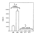
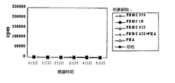
正常ヒト有志者はCOBE SPECTRATMアフェレーシスシステム(コーブ、レークウッド、コロラド)で白血球潟血された。個人Aから獲得したT細胞(TA)1x10⁵個は個人Bから獲得された同種異系PBMCs(mPBMCB)を(T細胞に対するPBMCsの増殖を予防するために)処置するマイトマイシンCと共に平底マイクロタイタウェル内で7日間培養された。mPBMCBsは20Kおよび100Kで接種された。培養基は、T細胞増殖を測定するための培養期間の最終18時間の間3H−チミジンで標識された。結果は図1に示され、TA細胞がPBMCBを外来のものと認識したことを示唆している(「TA+mPBMCB」部参照)。PBMCBsの量が多いほど、T細胞増殖も多かった。
【0063】
PBMCsと同一の提供者から獲得したヒト間葉幹細胞(hMSCs)2x10⁴個は個人Aから獲得したT細胞(TA)1x10⁵個と共培養された。細胞は平底マイクロタイタウェル内で合計7日間培養された。培養基はT細胞増殖を測定するための培養期間の最終18時間の間3H−チミジンで標識された。T細胞との培養前の2日間、ヒト間葉幹細胞は前に記載された細胞数(密集)でマイクロタイタウェル内に接種され、MSCs上の表面抗原発現を刺激するためにIFN−γ(50units/ml)で処置された。非形質導入hMSCs、またはヒトB7−1あるいはヒトB7−2補助的刺激分子を導入されたhMSCsはT細胞と共にインキュベートされた。対照細胞はNeoを導入された。
【0064】
結果は図1に示され(図1「TA+形質導入hMSCs」参照)、Tリンパ球がヒト間葉幹細胞に対して非応答性(増殖しない)であった、すなわち間葉幹細胞が外来のものとして認識されなかったことを示している。
【0065】
この結果は、間葉幹細胞に対する応答の欠如が個体間の遺伝的適合性に依らなかったことを示している。これはT細胞がhMSC提供者から獲得した末梢血単核細胞(PBMCs)を外来のものとして認識したためである。
【0066】
(実施例2)
混合リンパ球反応の抑制
間葉幹細胞が同種異系応答を積極的に抑制するかどうかを確認するために、混合リンパ球反応は、組織培養プレート中に準備された。組織培養プレートは異なる二提供者から獲得した粘着性間葉幹細胞を伴うものと伴わないものである。異なる二提供者は、一方はMLR中の刺激細胞に適合する提供者であり他方は刺激または応答細胞に無関係の提供者である。
【0067】
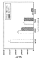
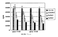
個人Aから獲得したPBMCs(PBMCA)105個は個人Bの標的PBMCs(PBMCB)105個と混合された。PBMCBsはPBMCAsによる活性化に依る増殖を予防するために3000ラドでX線を照射された。従って、PBMCAsのみが増殖する。PBMCAsとPBMCBsが混合されると混合リンパ球反応が起こり、その中でPBMCAs細胞(応答細胞)はPBMCBs(刺激細胞)上の表面抗原により活性化された。培養基は7日間インキュベートされ3H−チミジンで最終18時間標識された。PBMCBsの存在下ではPBMCAsは40,000カウントまで増殖した。図2、第一棒グラフ(「NONE」は間葉幹細胞不在を示す)参照。
【0068】
しかし間葉幹細胞の存在下でPBMCAsとPBMCBsが混合されると、混合リンパ球反応は抑制された。PBMCAs105個は、ヒト間葉幹細胞の粘着性単層で覆われたマイクロタイタプレート内でPBMCBs105個と混合された。間葉幹細胞は、ウェル当たり7500個乃至22,500個の範囲の量でウェル内で平板培養された。二つの間葉幹細胞集団が試験された:ヒト間葉幹細胞が個人Bから獲得したものおよび個人Aまたは個人BのMHC型に適合しない個人(第三者)から獲得したものであった。培養基は7日間培養され3H−チミジンで最終18時間標識された。ヒト間葉幹細胞の存在下ではMLRは抑制された。図2を参照。従って、間葉幹細胞のMHC系統に関係なく間葉幹細胞は混合リンパ球反応を抑制した。
【0069】
結果は図2に示され、ヒト間葉幹細胞が投与依存法において混合リンパ球反応を減少させたことを示唆している。どの提供者からの間葉幹細胞も増殖を同等に十分に抑制し、これはMHC型に関して抑制の特異性が無かったことを示唆している。これらの結果は、細胞が共存培養された場合、間葉幹細胞が積極的に混合リンパ球反応を抑制したことを示す。
【0070】
(実施例3)
二次混合リンパ球反応における無応答性
これらの実験は、MSCsによる前活性化T細胞の抑制により二次刺激時に特異的な無応答性が生じるかどうかを確認するために行われた。
【0071】
提供者248(d248)から獲得したT細胞は提供者273(d273)から獲得した同種異系PBMCsにより7日間特発され、その後単独または同一提供者(d273)から獲得したIFN−γ処置MSCsの存在下で3日間培養された。細胞はその後同一提供者(d273)、自己由来(d248)または「第三者」(d244)PBMCsにより再刺激された。
【0072】
リンパ球調製
末梢血単核細胞(PBMC)はフィコールパック(ファルメイシア)による密度勾配遠心法により調製された。細胞の一部は10%DMSO添加90%FCS中で凍結され、液体窒素内で保存された。解凍後、細胞はMSC培地(低グルコースおよび10%FCS添加DMEM)で2回洗浄され、分析培地(25mMヘペス、1mMピルビン酸ナトリウム、100μM可欠アミノ酸、100U/mlペニシリン、100μg/mlストレプトマイシン、0.25μg/mlアンホテリシンB、5.5x10−5M2−メルカプトエタノール(全試薬はギブコBLR)および5%ヒトAB血清(シグマ、MLR試験済)添加イスコーブ)中に再懸濁された。
【0073】
T細胞富化分画を調製するために、免疫磁気陰性選別法によりPBMCsから単球とBリンパ球を除去した。PBMCsはマウス抗ヒトCD19およびCD14モノクローナル抗体(非アジ化合物/低菌体内毒素(NA/LE)型)と共にインキュベートされ、続いてビオチン結合ヤギ抗マウスIgG(多部位吸着)抗体(全試薬はファルメイシア)とストレプトアビジンマイクロビーズ(ミルテニー バイオテック)と共にインキュベートされた。細胞はその後磁気細胞選別機(MACS、ミルテニー バイオテック)で分離された。T細胞富化分画は約70−約90%のCD3+細胞を含む。
【0074】
MSC培養
ヒトMSCsは、合衆国特許番号5,486,359に記載された通りに骨髄から単離されて、MSC培地を含む培養基中に保持され、3代乃至6代継代したものが使用された。細胞は0.05%トリプシン/EDTA溶液を用いて取り出され、MSC培地で1回洗浄され、70−80%の密集度ですなわち10cm組織培養ディッシュに対してプレート当たり1x106個で平板培養された。平板培養後、500U/mlのIFN−γ(ベーリンガー マンハイム)が添加され、細胞は更に3日間インキュベートされた。T細胞を移す前に、平板培養されたMSCはHBSSで4回、イスコーブで1回洗浄され、分析培地が10cm組織培養ディッシュ中にウェル当たり10mlで添加された。
【0075】
初期(1O)MLR
T細胞(d248)はX線照射PBMCs(d273)により活性化された。刺激に用いたPBMCsはキャビネットX線システム(ファクシトロン・エックスレイ、バッファローグローブ、イリノイ)を用いて3,000ラドでX線を照射された。初期刺激のために応答細胞2x10⁷個は、10cm組織培養ディッシュ中の分析培地20ml中で刺激細胞2x10⁷個と混合された。細胞は37℃、5%二酸化炭素濃度で7日間インキュベートされた。
【0076】
活性化T細胞/MSC培養
1OMLRで活性化されたT細胞は回収されMSC培地で一回洗浄され、分析培地10ml中に106個/mlで再懸濁された。そして自己由来または同種異系MSCsが含まれあるいは培地単独の10cm組織培養ディッシュに添加され、更に3日間培養された。
【0077】
再刺激分析
MSCsまたは培地と共に培養されたT細胞は回収されMSC培地で一回洗浄され、始原提供者、無関係の提供者から獲得したX線照射PBMCsまたは自己由来PBMCsで再刺激された。分析のために、活性化応答細胞5x10⁴個とX線照射刺激細胞5x10⁴個は96ウェルプレート内でインキュベートされた。分析は三回行われた。培養細胞は回収前に〔3H〕チミヂンの1μCi(エイマシャム)で18時間標識された。培養基はハーベスター96(トムテック)を用いて回収され、濾過器はマイクロベータトリラックス液体シンチレーションと発光計数器(E.G.アンドG.ウォーラック)を用いて解析された。データは3回の反復実験の平均cpm±SDとして表示されている。
【0078】
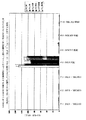
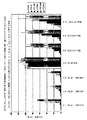
単独培養されたT細胞(正の対照)は2日目に最大となる「同一提供者」再刺激に対する応答の促進を示す。「第三者」応答もまた促進されたが、実際には「同一提供者」と同じ動態であるが最大値は低くまたわずかに開始の遅れが見られた(図3)。同種異系MSCs上で培養されたT細胞は、続く培養6日目までの間は「同一提供者」または「第三者」PBMCsに対して無応答を示した(図4)。
【0079】
(実施例4)
二次混合リンパ球反応における無応答性
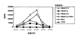
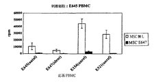
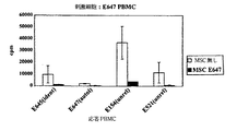
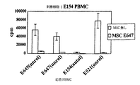
提供者413から獲得されたT細胞は提供者273から獲得されたX線照射PBMCsで7日間再刺激された(容積20ml培養基に1.5x10⁶ml)。異なる提供者413、418および273から獲得したMSCsは10cm組織培養ディッシュ中に1x10⁶/ディッシュで平板培養された。ディッシュはIFN−γで3日間前処置され、前活性化T細胞の混合に先立って洗浄された。
【0080】
MLR中で7日間前活性化されたT細胞は単独でまたはMSCsと共に3日間インキュベートされた(1.0x10⁶/mlT細胞、10ml/ディッシュ)。MSCsとのインキュベートの3日後T細胞は回収され、自己由来(d413)PBMCの存在または不在下で、X線刺激PBMC273(始原提供者)、413(自己由来)、PBMC10(第三者)またはPHA(5μg/ml)で再刺激された。細胞はウェル当たり5x104個で添加され、培養細胞は〔3H〕チミヂンで表示時間で18時間標識された。
【0081】
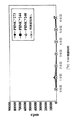
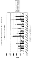
結果は、自己由来(d413)(図5C)、同一提供者(d273)(図5B)および第三者(d418)(図5D)MSCsでの活性化T細胞の処置がT細胞中の抗原刺激に対する無応答性を誘導することを示唆している。MSC処置を伴わない対照培養(図5A)は同種異系PBMCsに対する露出による細胞の再刺激を示した。
【0082】
(実施例5)
イヌMSCsによる初期MLRの抑制
イヌPBMCsはフィコールパック勾配遠心法(1.077)により末梢血から精製された。刺激PBMCsは2200ラド(7分、70kV)でX線照射された。X線照射刺激細胞105個は、前平板培養イヌMSC(E647、2x10⁴個/ウェル)の存在または不在下で応答PBMCs105個と96ウェルプレート中で混合された。培養細胞は6日間インキュベートされ〔3H〕TdR(5Ci/mmol、1μCi/ウェル)で16時間標識された。結果は図6A−6Dに示す。E647とE645は同腹子(DLA同一)である。結果は同種異系と同様自己由来MSCsも初期MLRを抑制することを示す。
【0083】
(実施例6)
非粘着性MSCsによる初期MLRの抑制
d273から獲得したT細胞(2x10⁵個/ウェル)はd244から獲得したX線照射PBMCs(2x105個/ウェル)と混合された。d244またはd273から獲得したMSCsはIFN−γ(900U/ml、3日間)で前処置されまたは無処置のままで、試験日にトリプシン処理されT細胞およびPBMCsと同時に添加された。培養細胞は7日間インキュベートされ、〔3H〕TdR(5Ci/mmol、1μCi/ウェル)が16−18時間添加された。結果は図7に示され、非粘着性MSCsも初期MLRを抑制することを示している。
【0084】
(実施例7)
同種MSCs支持皮膚異系移植片の生存
研究集団
若いヒヒ(パピオ アヌビス)が研究された。雄と受胎していない雌ヒヒの体重は7―20kgで3−16歳の年齢であった。これら動物は結核乳頭腫ウイルスの有無を検査され、サイトメガロウイルス(CMV)で滴定され、糞便浮遊及び塗沫検査を含むシミアンウイルスの検査より成る霊長類ウイルススクリーンで試験された。提供者と受容者の対はPCR類別化を通じて主要組織適合遺伝子複合体(MHC)不等性により決定された。研究期間中、ヒヒは付き添い動物のそばで個別の地域をあてがわれた。
【0085】
MSC単離と培養抗張のための提供者骨髄収穫
MSCsの単離と培養抗張のために骨髄針吸引液が腸骨稜から得られた。骨髄吸引液は週1回連続4週で交互に脇腹から獲得された。吸引液量は動物の血液量の10%の推定値で決められた。血液量は体重の7%と推定された。従って10kgのヒヒは推定血液量0.7リットルであった。血液量の10%の吸引液は従って70ミリリットルになるであろう。
【0086】
手続きに先立ち、セファゾリン500mgが手術時抗菌予防のために筋肉内(IM)投与された。ヒヒは手術のためにケタミン10mg/kg、IM、またキシラジン1mg/kg、IMで手術のために鎮静化され麻酔された。針挿入部位はポビドンヨードでこすり洗いされ次いでアルコールで洗浄された。吸引液は腸骨稜から16ゲージ、2インチ骨髄針を使って獲得された。注射器が針に取付けられ、骨髄液を抜くための吸入装置が利用された。手術後の痛みを除くために鎮痛剤ブプレノルフィンが0.03mg/kg、IM.Q12x2用量で与えられた。
【0087】
提供者骨髄吸引液の送り
骨髄吸引液は注射器からヘパリンナトリウムを含む無菌バキュテーナー(R)に移された。この管はスタイロフォームコンテナーに安置され、室温(RT)で細胞処理設備に送られた。
【0088】
MSCsの単離と培養物の樹立
骨髄の5乃至10mlアリコートがポリプロピレン培養管内でダルベッコ食塩加リン酸緩衝液(DPBS)50mlで希釈された。細胞懸濁液は2200RPMで10分室温(RT)で遠心された。全有核細胞数は酢酸4%液で確認された。細胞は次いで最終濃度で20x106細胞/mlになるようにDPBSで希釈された。10mlまたは200x106細胞が50mlの円錐管でパーコール20ml(比重1.073gm/ml)に負荷され1300RPMで20分遠心された。単核細胞を含む細胞中間期はDPBSで洗浄され、完全培地に再懸濁され、回収物を得るために計数された。パーコール中間期で得られた洗浄単核細胞は完全培地30mlと15−20x106細胞/フラスコ(8.1x104MSC/cm2)を含むT−185フラスコ内で確立され、炭酸ガス濃度5%で37℃のインキュベーターに安置された。
【0089】
MSCの収穫
三つ組フラスコ内の培地は傾瀉されフラスコはDPBS50mlで洗浄された。DPBSを傾瀉した後、0.05%のトリプシン23mlが各三つ組みフラスコに加えられた。フラスコは37℃インキュベーターに3分配置された。細胞剥離後に、完全培地23mlが各フラスコに加えられた。細胞懸濁液は50ml円錐管に移されフラスコは30mlのHBSSで洗浄された。管は2200RPM、5分RTで遠心された。
【0090】
形成/パッケージング
収穫されたMSCsはプラズマ−ライトA(バクスターIVセラピー)85%、DMSO、10%およびMSC提供者血清5%よりなる不凍溶液内で約10x106細胞/mlに形成され、15−20mlを含む袋で冷凍保存された。
【0091】
標識/貯蔵/送り
細胞は1分1−2°で90℃の制御速度フリーザー(クライオメド、フォーマ・サイエンティフィック)を用いて冷凍保存された。サンプルは次いで気相(−120°乃至−150℃)で液体窒素貯蔵フリーザーに移された。
【0092】
用量
20x106細胞/kgのMSC用量を達成するために、最終産物は注入日必要とされる用量の115%で調製された。
【0093】
皮膚の収穫
手術に先立ち、ヒヒは手術時抗菌予防として500mg、IMでセファゾリンを与えられた。ヒヒは10mg/kg、IMでのケタミンで鎮静化され、また静脈内チオペンタール誘導、1−2%イソフルラン吸入麻酔で麻酔された。皮膚が腹前壁から収穫され、前もって標識された食塩水湿潤ガーゼパットに置かれた。創傷欠損部は次いで閉じられた。ヒヒは麻酔から覚めた時にコロニーに戻された。手術後の痛みを除くために、鎮痛剤ブプレノルフィンがQ12x2用量でまたアンセフは2日間毎日投与された。
【0094】
受容者皮膚移植とMSC注入
手術に先立ち、ヒヒは手術時抗菌予防として500mg、IMでセファゾリンを与えられた。ヒヒは鎮静化され10mg/kg、IMと静脈内チオペンタール誘導、1−2%イソフルラン吸入麻酔で麻酔された。皮膚が腹前壁から収穫され前もって標識された食塩水湿潤ガーゼパッドに置かれた。この皮膚は2個の移植片に分割された。1個はも一つの受容者ヒヒの第三者パーティ対照として使用され、も一つはこの同じ動物の自己由来対照として使用された。動物は次いでうつぶせの位置に置かれた。3個の3x2cmの皮膚の切片が肩甲骨の棘に沿った背中から除去された。前にMSC提供者第三者パーティおよび自身に適合するよう手入れされ、適所に縫合された。
【0095】
移植後ヒヒは20x106提供者MSC/kgの用量でMSCの静脈内注入を受けた。末梢血サンプルはMSC注入前、注入1時間後、およびMSC注入1−3日後に確保された。骨髄吸引液はMSC注入0日後、3日、14日および30日後に得られた。
【0096】
手術後の痛みを除くために、鎮痛剤ブプレノルフィンがQ12x2用量で、またアンセフが2日間毎日投与された。動物は毎日観察され、移植片は移植7日後に開始して1日おきに写真撮影された。
【0097】
物理的試験と診断検査
各ヒヒは試験のためにケタミン10mg/kg、IMで鎮静化された。鎮静の間に、2−3ミリリットルの骨髄が腸骨稜から針吸引で確保され、4日、13日および研究終了の30日後にヘパリンナトリウムに収集された。皮膚生検が骨髄吸引の得られた同じ日に収穫された。
【0098】
結果
皮膚同種異系移植片生存に対するMSC注入の作用
未処置対照動物(N=2)は皮膚同種異系移植片平均生存時間が8日±0日であった。未処置MSC提供者のMSC提供者(N=2)への注入は皮膚移植片平均生存時間が11.5±0.71日への延長をもたらした(マン−ホイットニー U テスト、P<0.05)。未処置第三者提供者MSCの提供者同種異系移植片(N=4)への注入は平均生存時間12.3±0.96日の著しい延長をもたらした(マン−ホイットニー U テスト、P<0.003)。
【0099】
受容者6140と6200はMSC提供者6243から、お互いから(第三者移植片)、また彼等自身から(自己移植片)同種移植片を受けた。MSC提供者6243から皮膚移植片収穫の24時間前に6243からのMSCが移植のために輪郭を描かれた腹前部皮膚下部に注射された。移植後に受容者は20x106MSC/kg(6243)の静脈内注入を投与された。第三者同種移植片はいずれも13日に拒絶された。MSC提供者(6243)同種移植片は4日目に出血したことが発見され、この出血は技術的な失敗によるものとされた。病理学試験では、ケラチンは真皮の下でわだち状に徐々に入り込んでいることが認められた。これらのわだちの性質はそれが皮下MSC注射の際に針で形成されたことを示唆している。これら細胞の存在は非常に大きい炎症性応答を引き出した。この炎症性応答は皮膚移植片が適切に粘着/取り込む能力を妨げ、これらの移植片は7日までに完全に壊死した。自己移植片に拒絶されなかった。
【0100】
受容者6654と6659はMSC提供者6593から、お互いから(第三者移植片)、また彼等自身から(自己移植片)同種移植片を受けた。移植後に受容者は20x106MSC/kgの静脈内注入を投与された。MSC提供者同種移植片は11日と12日に拒絶され、また第三者提供者同種移植片は11日と12日に拒絶された。自己移植片は拒絶されなかった。
【0101】
同様に受容者6663と6658はMSC提供者6656から、お互いから(第三者移植片)、また彼等自身から(自己移植片)同種移植片を受けた。移植後受容者は20x106MSC/kgの静脈内注入を投与された。MSC提供者同種移植片は11日に拒絶され、また第三者同種移植片は10日と12日に拒絶された。自己移植片は拒絶されなかった。
【0102】
研究の対照部門にある受容者6532と6720はMSCの投与なしで注入または注射により自己移植片と同種移植片を受けた。同種移植片は8日に拒絶された。自己移植片は拒絶されなかった。
【0103】
同種異系MSC注入と関連する同定可能毒性は存在せず、また続く30日の追跡期間に不都合な臨床的後遺症は見られなかった。血液サンプルはMSCの注入前および移植とMSC注入後1時間、2時間、更に1日、2日、および3日後に獲得された。骨髄吸引液は移植とMSC注入後4日と13日に確保された。
【0104】
これらの結果は、同種異系ヒヒMSCsの単一注入が同種異系皮膚移植片の拒絶を遅延できることを示している。他の免疫抑制治療法は行なわれなかった。同種異系または第三者MSCsの1回の投薬量は拒絶に至る時間を50%増加した(グッドマン他、Am Surg 62巻(6号):435−442ページ(1996年))。
【0105】
(実施例8)
本研究の目的は、イヌにおける適度に高い投与量の提供者イヌ白血球抗原(DLA)−同一同腹子イヌ間葉幹細胞(cMSC)の注入の実現可能性および安定性を証明することである。提供者イヌ白血球抗原(DLA)−同一同腹子イヌ間葉幹細胞(cMSC)は同種異系骨髄移植片定植において細胞10x106個/kgで投与される。第二の目的は、移植後50および100日目での提供者neo−およびGFP−標識cMSCの分布および機能を調べることである。
【0106】
材料と方法
実験動物
ビーグル犬が本研究のために用いられた。0日目で7または9カ月齢のDLA同一同腹子の雄2匹、雌2匹が用いられた。分類のために用いられた方法は、主要組織適合抗原複合体が同じイヌのイヌ白血球抗原(DLA)中クラスII DRB領域の遺伝形質を追跡するための高い多型マイクロサテライト標識の使用を含む。マイクロサテライトは小さい2、3,または4ヌクレオチドの反復であり、対立遺伝子中で十分な長さの違いが見られるため、多世代交雑を経た染色体断片の遺伝形質の追跡に用いることができる。対立遺伝子の分離は、通常各反復を取り囲むDNAの特有な配列から得たプライマーを用いた一段階ポリメラーゼ連鎖反応を用いて測定される。加えて、混合白血球反応はPCRマイクロサテライト標識分析の結果を確定するために、研究用DLA同一同腹子のつがいに対して行われた。
【0107】
研究計画
研究用イヌはDLA同一同腹子の同一提供者から獲得したcMSCおよび骨髄の移植を受けた。骨髄移植片は、全身性X線照射(TBI)に先だって0日目に2匹のDLA同一同腹子提供者から回収され交換された。骨髄離解は、0日目にイヌに920センチグレイ(cGy)(7cGy(9.3R)/分の割合で送られる二つの阻害60CO源からの正中大気露出)の単独TBI照射により誘発された。移植4週前またはそれ以上前の週で提供者骨髄吸引液から単離された培養増殖cMSCは、グリーン蛍光タンパク質(GFP)およびネオマイシンホスホトランスフェラーゼ(neo)遺伝子を含むPapp@OT−24〔原文通り〕が導入された。cMSCは継代1(P1)または継代2(P2)の後に低温保存された。TBIに続いて、cMSCsは解凍され携帯注入ポンプにより15分間静脈内に輸送された。cMSC注入後の1時間乃至2時間の間、骨髄移植片は全有核細胞(TNC)≧1x108個/kgの投与量で静脈内に注入された。
【0108】
シクロスポリンは移植片対宿主性病(GVHD)の予防のために0日目乃至5日目にかけて10mg/kg BID(20mg/kg/day)(サンジミューン(R)インジェクションソリューション、サンド ファーマシューティカル コーポレーション)の投与量で4頭のイヌ全ての静脈内に投与された。グループI.1.aについては6日目乃至50日目(研究の終了)にかけて、またはグループI.1.bについては6日目乃至100日目にかけてシクロスポリンは10mg/kg BID PO(20mg/kg/day)(ネオラル(R)ソフトゼラチンカプセル、サンド ファーマシューティカル コーポレーション)で投与された。受容者への抗生物質の経口投与による通常の補助介護は5日前から、抗生物質の全身性投与は0日目から開始し、移植片が得られるまで続けられた。液体補助は必要に応じて行われた。血小板輸注は、回復中の4頭のイヌ全てに必要とされなかった。標準イヌ処置法では、血小板値が一定して10,000個/mm3より下に下がりまたは処置スタッフが出血の徴候を確認した場合には全血輸血が必要である。必要であれば血小板輸注は、任意の提供者から獲得した全X線照射(2000cGy)血の50ml量で実施される。移植片は最初の完全好中球細胞>500個/mm3、>1,000個/mm3および血小板>10,000個/mm3、50,000個/mm3>、および>100,000個の3連続した測定値の時に定植された。
【0109】
続く造血回復のために、完全血球算定(CBCs)が0日目乃至50日目にかけて、100日の研究グループについてはその後各週で得られた。血清化学分析は0、2日目、それ以降は毎週一回行われた。末梢血サンプルはDNA単離のために全MSC注入の0日目、5分および15分、1時間および2時間そして1日、2日、3日および4日目に採取された。DNAは、産物中に取り込まれるジゴキシゲニンを用いた抗EGFP DNA PCR Elisa法および第2段階として抗ジゴキシゲニン比色定量分析によりGFP標識細胞の存在について評価された。骨髄吸引液は血小板数値が一定して50,000個/mm3に達した時に得られ、同一PCR法を用いてGFP標識細胞の存在について調べられた。培養cMSCはコロニー形成単位(CFU)を調べるために、また更なる抗EGFP PCR分析用にcMSCを増殖させるために樹立された。剖検時に末梢血、骨髄吸引液および骨髄生検材料は抗EGFP PCR分析のために得られた。CFU分析は骨髄吸引液に対して行われ、抗EGFP PCR分析は培養増殖cMSCに対して行われた。組織学的解析は種々の組織におけるGFPの存在について行われた。
【0110】
cMSCの単離、培養増殖、形質導入および低温保存
両側の骨髄吸引液はcMSCの単離、およびイヌCAN−07−01およびCAN−07−02については4週前で、イヌCAN−07−03およびCAN−07−04については9週前での培養樹立のために得られた。骨髄15ml(各上腕骨から7ml)は各イヌから得られた。イヌはブトルファノールの注入、続くジアゼパムおよび塩酸ケタミンの混合物(アベコ カンパニー,インコーポレイテッド、フォートダッジ、アイオワ)の注入により麻酔をかけられた。針挿入部位はポビドンヨードで洗浄されその後アルコールですすぎ落とした。吸引液は、16ゲージ、2インチの骨髄針を用いて各イヌの両上腕骨顆から得られた。注射器は針に取り付けられ、各上腕骨から骨髄8mlを取り出すために吸引した。骨髄吸引液は無菌的手法で15mlのポリプロピレン円錐形チューブに移された。本方法に続いてイヌは回復のために温床に置かれた。
【0111】
骨髄の一部5ml乃至10mlはポリプロピレン培養チューブ中で、ダルベッコリン酸緩衝生理食塩水(DPBS)中で50mlに希釈された。細胞懸濁液は室温(RT)で10分間、2200RPMで遠心された。全有核細胞数は4%酢酸中で測定された。細胞はその後、最終濃度20x106個/mlまでDPBS中で希釈された。10mlまたは200x106個の細胞は50ml円錐形チューブ中で20mlのパーコール(sp.gr. 1,073gm/ml)に乗せられ、20分間1300RPMで遠心された。単核細胞を含む細胞界面はDPBS中で洗浄され、完全培地中に再懸濁され、回復割合を得るために計数された。細胞はその後完全培地中に希釈され、培養細胞は前に記載されたように樹立され5%二酸化炭素濃度で37℃のインキュベーター中に置かれた。
【0112】
2シストロンMuLVレトロウイルスベクターの構成
グリーン蛍光タンパク質(EGFP)レトロウイルスはクラゲ、エクオレアビクトリア(クロンテック、カリフォルニア)からEGFP−1遺伝子を単離することにより構成された。EGFP遺伝子はレトロウイルスベクターpJM573−neo(帰属プラスミドの名称はpOT−24)中にクローン化された。pJM573−neoプラスミドは、修飾されたpN2(ケラー他、1985年、Nature 318巻:149ページ)から得られた。pN2の修飾は次のようであった:マウスレトロウイルスgag開始部位が枠内終止コドンで置換され、5’LTRおよび3’LTRが同一カセット内に構成され、ネオマイシンホスホトランスフェラーゼ遺伝子(neo)および内部リボソームエントリー部位(IRES)がpN2中に挿入された。EGFP pOT24プラスミドの模式図は図8に示す。
【0113】
組換えレトロウイルスの調製
pOT−24は、製造者によって示されたようにDOTAP(ベーリンガー マンハイム)を用いてGP&E86環境栄養性産生細胞中に形質移入された。形質移入細胞は、10%熱失活FBS、ペニシリン−ストレプトマイシン(ライフ テクノロジーズ)および選択標識としての硫酸プロタミン−G418(シグマ)0.5mg/mlの補足されたDMEM−高グルコース(HG)培地中で生育された。培養細胞は密集度70%になるまで維持され、その時点で培地は新鮮レトロウイルス培地(G418無し)に交換され、細胞は32℃で2日間維持された。レトロウイルスを含む培養培地は回収され0.45μmフィルターを通して濾過され−70℃で保存された。両栄養性レトロウイルスは、遠心導入法と続くG418(0.5mg/ml)での選別法を用いてPA317細胞を環境栄養性ウイルスに2回形質導入することにより調製された。レトロウイルス上澄みは回収された。貯留EGFPウイルスの3T3細胞に対する力価は1.2x106CFU/mlであった。GFPレトロウイルス上澄みは−70℃で低温保存された。
【0114】
CAN−07−01およびCAN−07−02
パーコール界面から得られた洗浄された単核細胞は完全培地30mlおよび細胞数10x106個/フラスコを含むT−185フラスコ10個中で樹立された。
【0115】
培養2、6、9日目にフラスコ中の培地は新鮮完全培地と交換された。初期培養の12日目に写真を撮り、細胞は継代0(P0)から継代1(P1)に移された。培地は吸引されフラスコは8mlDPBSで2回洗浄された。トリプシン8mlが添加され、フラスコは37℃インキュベーター中に3分間置かれた。細胞が剥離し、完全培地8mlの添加により反応は止められた。細胞は50ml円錐形チューブに移され貯留された。フラスコはDPBSで洗浄され貯留細胞は室温で5分間、2000RPMで遠心された。上澄みは除去され細胞のペレットは完全培地中に再懸濁された。細胞は貯留され、測定され生存可能性が調べられた。細胞は完全培地18mlおよび細胞数フラスコ当たり0.4x10⁶個を含むT80フラスコ15個中で平板培養された。
【0116】
培養15日目、18フラスコのうち15フラスコに最初の形質導入が行われた。培地は除かれた。レトロウイルス上澄みの一部が解凍され、形質導入混合物を作るためにポリブレンが最終濃度8μg/mlまで添加された。細胞培地は形質導入混合物10mlと交換されフラスコは32℃で1時間、3000RPMで遠心された。遠心後、熱失活胎児ウシ血清(FBS)を用いて調製された完全培地10mlは(形質導入混合物を含む)各フラスコに添加され、フラスコはインキュベーターに戻された。フラスコ3つは形質導入をせず、新鮮培地が交換された。培養16日目、培地は新鮮完全培地に交換された。培養17日目に形質導入法が繰り返された。
【0117】
培養18日目、細胞は前に記載したように回収されP1からP2に移された。細胞3x10⁶個は完全培地100ml中に添加され三つ組みフラスコ(500mm2)中に注入された。三つ組みフラスコ15個は形質導入細胞で調製され3個は非形質導入細胞で調製された。残りの細胞は低温保存された。凍結溶液は10%DMSOおよび90%FBSを含むように調製された。細胞10x106個は凍結溶液1ml中に再懸濁された。バイアルはラベルを貼られ最低4時間はナルジーン クリオ コンテナ中に低温保存された。
【0118】
P2培養22日目、細胞分布と形態を記録するために写真が撮られ、P2細胞は回収され前に記載されたように低温保存された。
【0119】
CAN−07−03およびCAN−07−04
パーコール界面で得られ洗浄された単核細胞は、完全培地20mlおよび細胞12x106個/フラスコを含むT−75フラスコ15個中に樹立された。
【0120】
培養2日目、フラスコおよびディッシュ中の培地は新鮮完全培地と交換された。cMSCの初代培養の6日目、最初の形質導入が前に記載したように行われた。フラスコ3個は形質導入されず新鮮培地は6日目に交換された。培養7日目、培地は新鮮培地に交換された。
【0121】
培養8日目、形質導入法は繰り返された。培養9日目、写真が撮られ、細胞は前に記載されたようにP0からP1に進められた。細胞3x10⁶個は完全培地100mlに添加され三つ組みフラスコ中に注入された。三つ組みフラスコ15個は形質導入細胞で調製され3個は非形質導入細胞で調製された。
【0122】
骨髄吸引液15mlは提供者CAN−07−01、CAN−07−02、CAN−07−03およびCAN−07−04からそれぞれ有核細胞910、1212、856および1948x106個を産生した。パーコール界面から得られた単核細胞数は612、666、588および462x106個であり67.2、55、68.7および23.7%の回復であった。P1時、細胞生存可能性は平均97.1(93.3乃至100の範囲)%であった。提供者CAN−07−01およびCAN−07−02のP2および提供者CAN−07−03およびCAN−07−04のP1細胞の時、形質導入細胞の細胞の生存可能性は平均96.7(96.3乃至97.9の範囲)%であった。非形質導入細胞は95.4(93.3乃至96.9の範囲)%の生存可能性であった。cMSCの低温保存のための回収時には、形質導入細胞の生存可能性は平均99.4(97.4乃至100の範囲)%であり、非形質導入細胞は99.4(97.6乃至100の範囲)%であった。
【0123】
継代2後4日目に回収されフラスコ当たり3x10⁶個で平板培養された、提供者CAN−07−01およびCAN−07−02のフラスコ当たりの産生された形質導入cMSCは5.9および6.7x10⁶個であり、フラスコ当たりの産生された非形質導入cMSCは8.4および7.5x10⁶個であった。継代1(異なる形質導入および継代設定)後4日目に回収されフラスコ当たり3x10⁶個で平板培養された、提供者CAN−07−03およびCAN−07−04のフラスコ当たりの産生された形質導入cMSCは20.0および14.0x10⁶個であり、フラスコ当たりの産生された非形質導入cMSCは25.3および18.0x10⁶個であった。
【0124】
P0培養細胞から獲得したcMSCに対するCFU分析
CFUコロニー分析は完全培地10mlを含む100mlディッシュ3個内で細胞0.5x10⁶個を平板培養することにより初代培養樹立時に調製された。ディッシュは37℃、5%二酸化炭素濃度でインキュベートされた。培地は2日目、4日目に新鮮培地と交換された。培養10日目、CFU分析ディッシュはHBSSで2回すすぎ落とされ、1%グルタルアルデヒドで15分間固定され、HBSSで2回すすぎ落とされ風乾された。ディッシュ中のcMSCはその後0.1%クリスタルバイオレットで染色され、脱イオン水で3回洗浄され風乾された。コロニーは平板培養された細胞106個当たりのコロニー形成数を算定するために測定された。
【0125】
単核細胞単離および培養樹立の日に平板培養され10日目に回収されたCFU分析はイヌCAN−07−01、CAN−07−02、CAN−07−03およびCAN−07−04それぞれから細胞106個当たり56、46.7、114および72コロニーを産生した。
【0126】
P1培養の13日目、細胞分布と形態を記録するために写真が撮られ、P1細胞はトリプシン処理により回収され前に記載されたように低温保存された。
【0127】
三つ組みフラスコ中の培地は傾瀉され、フラスコはDPBS50mlですすぎ落とされた。DPBSの傾瀉後0.25%トリプシン23mlが各三つ組みフラスコに添加された。フラスコは3分間37℃インキュベーター中に置かれた。細胞剥離後23ml完全培地が各フラスコに添加された。細胞懸濁液は50ml円錐形チューブに移され、フラスコはHBSS30mlで洗浄された。チューブは室温で5分間、2200RPMで遠心された。形質導入または非形質導入細胞を含むペレットはそれぞれ貯留され計数された。細胞1x10⁷個の一部は、抗EGFP DNA PCR Elisa分析による形質導入割合の測定のために取っておかれた。
【0128】
回収後、回復され、P1またはP2形質導入され、培養増殖されたcMSCは5分間1300RPMで遠心され、85%プラスマ−ライトA(バクスター IV セラピー)、10%DMSOおよび5%自己由来イヌ血清を含む氷冷不凍溶液中でcMSC1x107個/mlの1ml中に再懸濁された。細胞のアリコートは別々の低温バイアルに各1mlずつ分けられた。チューブはイヌ提供者番号および全生存可能細胞数の書かれたラベルを貼られた。cMSCsは細胞バイアルをナルジーン フリージング コンテナ中に置いて低温保存され、4時間−70℃冷凍庫に置かれその後−70℃で貯蔵庫に移された。
【0129】
産物の低温保存のために細胞を回収した時に、細胞1x10⁷個のアリコートは形質導入効率の測定のために得られた。形質導入効率は、産物中に取り込まれるジゴキシゲニンを用いた抗EGFP DNA PCR Elisa法および第2段階として抗ジゴキシゲニン比色定量分析により分析された。
【0130】
cMSC注入産物
注入の1時間乃至2時間前にcMSCのバイアルは37℃ウォーターバス中で回転させて解凍され、70%エタノールが噴霧されバイオセイフティーキャビネット中で開けられた。cMSC産物はDMEM−LG、細胞提供者に自己由来の30%血清を含む注入培地50ml中に懸濁された。cMSC産物の生存可能性は、実際の生存可能投与量を測定するためにトリパンブルー除去により測定された。各cMSC産物のアリコートは酵母菌単離、好気的および嫌気的生育を受けた。cMSCは、組織培養プラスチックへの接着およびP2(CAN−07−01およびCAN−07−02に対してはP3)培地中での増殖能力について評価された。cMSC1x106および0.16x10⁶個のアリコートは、3個のT−25プラスチック培養フラスコ中で完全イヌ培養培地中に平板培養された。cMSC1x106個を平板培養したフラスコは24時間後、cMSC0.16x106個を平板培養したフラスコは3日目にトリプシン処理により回収され計数された。
【0131】
TBIに続きcMSC懸濁液は、15分乃至20分の間50ml輸送するために手持ちのハーバード バード ミニ インフューザーを用いて頭側静脈に挿入されたカテーテルを通して注入された。
【0132】
生存可能cMSC7.49、7.35、10.0および10.0(平均8.7)x106個/kgの適度に高い投与量は、0日目にイヌCAN−07−01、CAN−07−02、CAN−07−03およびCAN−07−04にそれぞれ注入された。これらの投与量は被験者が受容する通常の投与量の4倍乃至10倍増に相当する。注入された全生存可能cMSCは67.7乃至129(平均93.9)x106個の範囲であった。細胞の生存可能性はトリパンブルー除去による測定で92.1乃至97.6(平均94.9)の範囲であった。cMSC注入はTBI後71乃至146(平均110)分の間に行われた。
【0133】
注入後の血液サンプリング
血液サンプル(2ml)はcMSC注入の前におよび注入の間すなわち注入開始後5分および15分、1時間および2時間そして1、2、3および4日目に得られた。細胞溶解産物はピュアジーンTM(ジェントラ システムズ,インコーポレイテッド)DNAアイソレーションキットを用いて調製された。このキットは、血流中のGFP標識cMSCのレベルを検出するために産物中に取り込まれるジゴキシゲニンを用いた抗EGFP DNA PCR Elisa法および第2段階として抗ジゴキシゲニン比色定量分析を使用する目的で用いられた。
【0134】
骨髄回収および移植片注入
移植片として用いられる骨髄はTBIに先立ってDLA同一同腹子から回収された。吸引液は、11ゲージ、4−6インチのボールトップステンレススチール回収針を用いて両上腕骨から得られた。回収針は100mlの組織培養培地199およびヘパリン4ml(4000U)を含む真空フラスコのポリビニルチューブに取り付けられた。骨髄は300μmおよび200μmサイズの小孔を通され輸送パックコンテナ中に4℃で保存された。骨髄は提供者および受容者のラベルを貼られ後日の注入まで保存された。骨髄全有核細胞数(BM−TNC)は、骨髄回収の間に得られた多量の末梢血中に存在する有核細胞を除外することで修正された。
【0135】
骨髄の全有核細胞数(TNC)は骨髄回収の間に得られた多量の末梢血中に存在するTNCを除去することにより修正された。骨髄の修正投与量は、イヌCAN−07−01、CAN−07−02、CAN−07−03およびCAN−07−04に対してそれぞれTNC4.3、3.5、3.1および2.0(平均3.2)x108個/kgであった。無修正骨髄投与量はTNC5.6、4.2、4.5および2.7(平均4.3)x108個/kgであった。
【0136】
注入の20分前に骨髄は室温に置かれた。cMSC注入の一時間後骨髄は、1分乃至2分の間バックに圧力を加えることにより頭側静脈に挿入された蝶形針を通って静脈内に注入された。
【0137】
補助介護
5日前に、抗生物質の経口投与(硫酸ネオマイシンおよび硫酸ポリミキシン)は1日3回行われた。これらの抗生物質の経口投与は、完全好中球数が500個/mm3に達するまで投与された。0日目、全身性抗生物質ベイトリルは静脈内に1日2回投与され完全好中球数が一定して1,000個/mm3に達するまで続けられた。短時間の放射線毒性の結果としての液体および電解質損失は、食物と水が摂取されるまで1日2回のリンガー液500mlの皮下投与により補われた。
【0138】
種々の血液細胞数
血液サンプル(2ml)は、cMSCの単離のための骨髄吸引の朝、0日目乃至50日目までおよびそれ以降は2週間に1回研究の終了まで、頸静脈または頭側静脈から回収された。血液はEDTAを含むバキュテーナー中に移された。1mm3当たりの全白血球(WBC)および血小板数はシスメックスE2500を用いて測定され、種々の細胞数は固定およびライト染色液での染色後指示どおりに測定された。
【0139】
剖検
血液サンプルはCBC、化学23分析およびPCR評価のために得られた。イヌはブトルファノールと続くジアゼパムおよび塩酸ケタミンの混合物により鎮静された。鎮静後、生検材料および両側の骨髄吸引液は上腕骨、大腿骨および腸骨稜から得られた。その後、安楽死が鎮静剤ペントバルビタールナトリウムの過剰投与で遂行された。50日グループのイヌ(CAN−07−01およびCAN−07−02)は研究43日目に安楽死させられ、100日グループのイヌ(CAN−07−03およびCAN−07−04)は研究100日目に安楽死させられた。組織の全組合わせは動物の剖検時に回収された。
【0140】
組織学的調査のための組織の回収は続いて即座に行われた。組織のサブセットは抗EGFP DNA PCR Elisa分析に用いられた。骨髄吸引液および生検材料は抗EGFP DNA PCR Elisa分析、更なるPCR分析のための培養増殖およびCFU分析に用いられた。
【0141】
組織は約1インチ平方の小片に形を整えられ、10%中性緩衝ホルマリン(pH6.8−7.2)で満たされた、ラベルの貼られた別々の50ml円錐形チューブ中に置かれた。組織はパラフィン中に包埋され、薄切されヘマトキシリンおよびエオシンで染色された。骨髄サンプルは過ヨウ素酸シッフ染色で染色された。
【0142】
剖検に先だって得られた骨髄吸引液は各イヌの左右の上腕骨、大腿骨および超骨稜からラベルの貼られた15mlチューブ中に回収された。組織サンプルのサブセットは剖検時に得られ約1/4インチ平方の小片に形を整えられ、PBSに浸したガーゼに包まれてラベルの貼られたジップロックバック中に別々に置かれた。骨髄吸引液は氷中に浸けられた。
【0143】
CFU分析のための骨髄吸引液の調製
各イヌからPCR分析のために得られた左右上腕骨、大腿骨および腸骨稜の骨髄吸引液の一部は、別々のラベルの貼られた15mlチューブ中に分けられた。骨髄サンプルは氷中に浸けられた。
【0144】
剖検時に得られた骨髄から獲得したcMSCに対するCFU分析
剖検時に得られた骨髄から獲得したcMSCに対して行われたCFUコロニー分析は完全培地10mlを含む100mmディッシュ三つ組みに細胞0.5x10⁶個を平板培養することにより調製された。ディッシュは37℃および5%二酸化炭素濃度でインキュベートされた。培地は2日間乃至4日間で新鮮培地に交換された。培養10日目、CFU分析ディッシュはHBSSで2回すすぎ落とされ1%グルタルアルデヒドで15分間固定されて、HBSSで2回すすぎ落とされ風乾された。ディッシュ中のcMSCはその後0.1%クリスタルバイオレットで染色され脱イオン水で3回洗浄されて風乾された。コロニーは、平板培養された細胞106個当たりのコロニー数を算定するために算出された。
【0145】
DNAの単離および精製
DNAは各組織片から単離された。サンプルの残りは−70℃フリーザー中で低温保存された。DNAは、サンプルをリン酸緩衝生理食塩水(PBS)に置きプロテイナーゼK溶液を添加し、55℃で3時間または組織が溶解するまでインキュベートすることにより単離された。サンプルは続いて37℃で60分間RNアーゼで処置された。サンプルは室温に冷やされタンパク質は沈殿した。サンプルは遠心され、水溶層は100%イソプロパノール中に丁寧に回収された。サンプルは混合され遠心された。ペレットは70%エタノール中で洗浄された。チューブは遠心され、上澄みは抽出され、ペレットはおおよそ1時間乃至6時間乾燥された。DNAは室温で一晩水和され、続いて4℃で保存された。
【0146】
末梢血および骨髄サンプルは最初にRBC溶解溶液(塩化アンモニウム緩衝液)で溶解された。DNAはその後前に記載したように溶解液から単離された。DNAは、脱イオン水998μlおよびサンプルから得られたDNA2μlをキュベット中に添加することにより増量され撹拌された。分光光度計は光学密度(OD)を測定するために用いられた。ODは260および280で読み取られ、DNA濃度はμg/mlで算出された。DNA濃度は脱イオン水を用いて1μg/mlに調整された。
【0147】
抗EGFP DNA PCR Elisa
本研究で用いられた抗EGFP DNA PCR Elisa分析は、GFPに特異的なオリゴヌクレオチドプライマーを利用して注入cMSCsを検出する。遺伝子発現の分析のために、我々はPCR−ELISA(DIG標識/検出)キット(ベーリンガー マンハイム)を利用した。要約すると、PCRは、増幅産物を標識するためのジゴキシゲニン標識ヌクレオチドの存在下で行われた。次にPCR産物25μlは、ストレプトアビジンがコートされたマイクロタイタプレートにおいて、37℃の5’−ビオチン化オリゴヌクレオチドプローブ溶液中で変性されハイブリッド形成された。プローブ−PCR結合産物は、抗ジゴキシゲニンペルオキシダーゼ複合体によりおよび比色定量基質2,2’アジノビス(3−エチルベンゾチアゾリン−スルホン酸)(ABTS)を用いることにより検出された。滴定基準曲線は、分析に用いられたDNAの量当たりのDNA濃度を近似するために形質移入対照cMSCを用いて作成された。DLAクラスIIゲノムDNAのPCRに対する内部標準との最初の相互作用、その後のDNA濃縮液の同量の細胞に対する相互作用および形質導入細胞当たりの一つのレトロウイルスの組込みの推定により概算細胞数を得ることができる。
【0148】
GFPのためのDNAの量的測定は全ての骨髄ヘラ/生検材料において示された。
【0149】
移植後の血球回復
血小板閾値(3連続の数値)が10,000個/mm3となる平均日数は12.8(11−17の範囲)日、50,000個/mm3となるのは19.8(16−25の範囲)日および100,000個/mm3となるのは23.0(20−27の範囲)日であった。完全好中球細胞の閾値(3連続の数値)が500個/mm3となる平均日数は9.3(8−11の範囲)日および1,000個/mm3となるのは10.5(9−13の範囲)日であった。
【0150】
仮骨髄吸引液
血小板が回復して1mm3当たり50,000個以上の値に一定したとき、仮骨髄吸引液は腸骨稜から回収された。この方法は本研究においてCAN−07−01およびCAN−07−02については27日目に、CAN−07−03およびCAN−07−04については29日目に行われた。
【0151】
結果
43日目に安楽死させられたCAN−07−01およびCAN−07−02から獲得した全組織の組織病理学評価については、結果は異所性結合組織および亜急性のGVHDに対して陰性であった。
【0152】
検出可能なDNAシグナルは注入の1時間の間および2日目に再び観察できた。サンプルの一つはGFP DNAについて注入3日後に量的に測定できた。この時間は、自己由来のイヌ移植研究においてシグナルが2および3日目に見られたという前の観察と一致している。
【0153】
CAN−07−03およびCAN−07−04におけるGFP+細胞についての100日目剖検データは、CAN−07−03の大腿骨および上腕骨に、CAN−07−04の上腕骨にGFPシグナル(PCR挿入DNA10μg当たりの同量の1GFP+細胞)を示した。
【0154】
このモデルにおいては、動物の目および耳の充血を観察することにより皮膚移植片対宿主性病(GVHD)を検出することが可能であった。この指標を用いて、間葉肝細胞を受容した動物は間葉肝細胞で処置されなかった対照動物と比較してGVHDの発症度が低かったことが測定された。
【0155】
これらの結果は、同種異系MSCsが骨髄造血細胞の迅速な移植を補助できることを証明している。輸液補助は必要でなかった。GVHDの臨床徴候は無かった。血小板回復はこれまでの対照よりも早かった。これらは同種異系移植後の同質細胞におけるキメラ現象の証拠である。同種異系MSCsを用いることによる移植同種異系組織の選択は、臨床移植の場面で用いることができる移植片材料の範囲を広げる。
【0156】
(実施例9)
MSC上澄み(MLR95)による混合リンパ球反応の抑制
上澄みの産生
提供者155から獲得したT細胞は、抗CD19および抗CD14マイクロビーズ(ミルテニー バイオテック)を用いた陰性免疫磁気選別法によりPBMCから精製された。提供者413から獲得したPBMCは3600ラド(70kVで12分)でX線照射された。24ウェル組織培養プレート中で、T細胞(9x10⁵個/ウェル)は3日間X線照射PBMCsと混合され、その後異なる提供者(219、459、461−全て)から獲得したMSCsが3日間1.2x105個/ウェルで培養細胞中に添加された。対照培養では、同量の培地がMSCsの代わりに添加された。別々のウェルにおいて、同数のMSCsは単独で平板培養され3日間培養された。培養3日後(初期MLRの開始後6日目)細胞はピペッティングにより再懸濁され、細胞懸濁液200μlは96ウェルプレート三つ組みに移され、増殖レベルを測定するために〔3H〕TdR(5Ci/mmol、1μCi/ウェル)で18時間標識された。残りの細胞は1250rpmで10分間遠心され、上澄みは回収され、分別されて−80℃で凍結された。
【0157】
上澄みによる初期MLRの抑制
提供者155から獲得したT細胞は、抗CD19および抗CD14マイクロビーズ(ミルテニー バイオテック)を用いた陰性免疫磁気選別法によりPBMCsから精製された。提供者413または提供者273から獲得したPBMCsは3600ラド(70kVで12分)でX線照射された。96ウェル組織培養プレート中でT細胞(1.5x10⁵個/ウェル)は、種々の上澄みの存在または不在下でPBMCs(1.5x105個/ウェル)と混合された。この上澄みは培養開始時に各希釈濃度(1/8、1/32、1/128、1/512、1/2048および1/8192)で添加されたものである。対照培養においては、同量の培地が上澄みの代わりに添加された。培養細胞は6日間インキュベートされ、その後〔3H〕TdR(5Ci/mmol、1μCi/ウェル)で18時間標識された。
【0158】
図9はT155xPBMC413間のMLRの結果を示す。MSCs+MLR(#1)、MLR+MSC219(#2)、MLR+MSC459(#3)、MLR+MSC461およびMSCs単独(#8)、MSC219単独(#9)、MSC459単独(#10)、MSC461単独の上澄みの存在下では、初期MLRは抑制された。MLR単独(#5)の上澄みの存在下では初期MLRは抑制されなかった。
【0159】
進行中のMLRの抑制
提供者155から獲得したT細胞は、抗CD19および抗CD14マイクロビーズ(ミルテニー バイオテック)を用いた陰性免疫磁気選別法によりPBMCsから精製された。提供者413から獲得したPBMCs(1.5x10⁵個/ウェル)は3600ラド(70kVで12分)でX線照射された。96ウェル組織培養プレート中でT細胞(1.5x105個/ウェル)はPBMCs(1.5x105個/ウェル)と4日間混合され、その後上澄みは種々希釈濃度(1/8、1/32、1/128、1/512、1/2048および1/8192)で培養細胞に添加された。対照培養においては、同量の培地が上澄みの代わりに添加された。培養細胞は2日間(MLR開始後6日目)インキュベートされ、その後〔3H〕TdR(5Ci/mmol、1μCi/ウェル)で18時間標識された。
【0160】
図10はT155xPBMC413間のMLRの結果を示す。MSCs+MLR(#1)、MLR+MSC219(#2)、MLR+MSC459(#3)、MLR+MSC461から獲得した上澄みは進行中のMLRsの強い抑制を示した。MSCs単独(#8)、MSC219単独(#9)、MSC459単独(#10)、MSC461単独から獲得した上澄みは投与依存法において抑制し、1/512希釈まで顕著な効果を示した。MLR単独(#5)の上澄みの存在下では進行中のMLRは抑制されなかった。
【0161】
(実施例10)
異種間葉幹細胞による混合リンパ球反応の抑制
ヒヒMSCsによるヒトMLRsの抑制
ヒト提供者(R4、R6、R7、R11)から獲得した応答PBMCsは、マイクロタイタウェル内でX線照射(3000R)同種異系ヒトPBMCs(S4、S6、S7、S11)と混合された。各集団は細胞1.5x10⁵個/ウェルである。培養は5%ヒトAB血清を含む標準細胞培養培地中で行われた。提供者86243から獲得したヒヒMSCs(bMSCs)はMLRの開始時に2x104個/ウェルで添加された。MSCsはIFNγで処置されなかった。リンパ球増殖は、培養7日目にシンチレーション測定のための細胞回収に先立って細胞を3H−チミジンで18時間標識することにより測定された。図11に示された結果は、ヒヒMSCsが強固なヒトMLRsを50%以上抑制したことを示している。
【0162】
ヒトまたはヒヒMSCsによる異種MLRの抑制
提供者273から獲得した応答ヒトT細胞(hT)は、提供者5957または提供者5909から獲得したX線照射(3000R)ヒヒPBMC(bPBMC)と5%ヒトAB血清を含む標準細胞培養培地中で培養された。提供者244から獲得したヒトMSCs(hMSCs)または提供者6243から獲得したヒヒMSCs(bMSCs)は開始時に培養細胞に添加された。リンパ球増殖は、培養7日目にシンチレーション測定のための細胞回収に先立って細胞を3H−チミジンで18時間標識することにより測定された。図12(bPBMC提供者5957)および図13(bPBMC5909)に示された結果は、ヒトおよびヒヒMSCsの両方が異種ヒトxヒヒMLRを抑制することができることを示している。
【図面の簡単な説明】
【図1】同種異系間葉幹細胞が免疫応答を誘導しないことを示す図。Aから獲得されたT細胞は、投与依存法において異なる量のBから獲得されたPBMCsと混合されたとき増殖した。Aから獲得されたT細胞は、Bから獲得された間葉幹細胞との接触に対する応答として増殖しなかった。間葉幹細胞が十分なT細胞活性化を提供するように操作されていた場合(間葉幹細胞はIFN−γで処置され、また補助的刺激分子B7−1またはB7−2を形質導入された)においても増殖しなかった。
【図2】間葉幹細胞が異なる二個体から獲得したリンパ球間の混合リンパ球反応(MLR)を積極的に抑制することを示す図。受容者に対して同種異系(第三者または提供者)のhMSCsは、MLR中の刺激細胞および応答細胞の両方に不適合であった(白抜き部)か、またはMLR中の刺激細胞に対して適合(提供者)していた(斜線部)。このように、間葉幹細胞はMHC型に関して特異性を持たずにMLRを抑制した。間葉幹細胞は投与依存法においてMLRを抑制した。
【図3】刺激(同種異系)PBMCsにより特発され、MSCsへの露出をせず、その後自己由来PBMCs、同種異系PBMCs(刺激細胞または第三者)に露出されまたは細胞に露出されなかった応答T細胞の二次応答を示す図。
【図4】刺激(同種異系)PBMCsにより活性化され、続いて同種異系MSCs(刺激細胞)と共に培養され、その後自己由来PBMCs、同種異系PBMCs(刺激細胞または第三者)に露出されまたは細胞に露出されなかった応答T細胞の二次応答を示す図。
【図5A】刺激同種異系PBMCsにより前活性化され、活性化後、同種異系(同一提供者(図5B)、または第三者(図5D))あるいは自己由来MSCs(図5C)と共に培養され、その後自己由来または同種異系(同一提供者または第三者)刺激細胞に露出された応答者T細胞の二次応答が抑制されたことを示す図。
【図5B】図5Aと同じ。
【図5C】図5Aと同じ。
【図5D】図5Aと同じ。
【図6A】イヌモデルにおけるMSCsによる初期MLRの抑制を示す図。autol=自己由来、ident=DLA同一同腹子、unrel=無関係
【図6B】図6Aと同じ。
【図6C】図6Aと同じ。
【図6D】図6Aと同じ。
【図7】非粘着MSCsによる初期MLRの抑制を示す図。
【図8】実施例8において用いられるEGFP pOT24プラスミドの模式図を示す図。
【図9】ヒト間葉幹細胞またはヒト間葉幹細胞抑制混合リンパ球反応培養系から産生されたMSC上澄みの初期混合リンパ球反応に対する抑制効果を示す図。
【図10】ヒト間葉幹細胞またはヒト間葉幹細胞抑制混合リンパ球反応培養系から産生されたMSC上澄みの進行中の混合リンパ球反応に対する抑制効果を示す図。
【図11】ヒヒ間葉幹細胞によるヒト応答T細胞とヒト刺激PBMS細胞との間の混合リンパ球反応の抑制を示す図。
【図12】ヒヒまたはヒト間葉幹細胞によるヒト応答T細胞とヒヒ刺激PBMC細胞(提供者5957)との間の混合リンパ球反応の抑制を示す図。
【図13】ヒヒまたはヒト間葉幹細胞によるヒト応答T細胞とヒヒ刺激PBMC細胞(提供者5909)との間の混合リンパ球反応の抑制を示す図。[0001]
TECHNICAL FIELD OF THE INVENTION
The present invention is based on the priorities of US Provisional Patent Application No. 60 / 078,463, filed March 18, 1998 and US Provisional Patent Application No. 60 / 089,964, filed June 19, 1998. US Patent Application No. 09 / 427,333, filed on October 26, 1999, which is a continuation-in-part of US Patent Application No. 09 / 267,536, filed on March 12, 1999, alleging this. Claim priority.
[0002]
The present invention relates to inhibiting T cell responses to allogeneic antigens, and further to inhibiting and / or preventing reactivation of preactivated T cells. More particularly, the present invention relates to the field of preventing, reducing or treating an immune response to foreign tissues and or cells and / or organs caused by immune effector cells. The invention further relates to preventing, reducing or treating graft rejection and / or graft versus host reactions.
[0003]
BACKGROUND OF THE INVENTION
Tolerance is a deficient acquisition of a specific response to an antigen that usually produces an immune response. Typically, induction of tolerance requires exposure to the antigen to be tolerized, resulting in some lymphocyte death or functional inactivation. Complete tolerance is characterized by a lack of detectable immune response to a secondary antigen challenge. Partial tolerance is characterized by a quantitative decrease in the immune response.
[0004]
The function of the immune system is to remove foreign substances, including pathogens, and to maintain non-responsiveness or tolerance to self-antigens. T cell tolerance is 1) in the thymus, where thymocytes responsive to self-peptides are removed by clonal loss (central tolerance), and 2) in the periphery exposed to self-antigens under conditions that result in immune tolerance (peripheral tolerance). Be acquired. Clonal loss can also result from the expression of cell death molecules on antigen presenting cells. Representative examples of cell death molecules are Fas ligand (FasL) and TRAIL ligand, which bind to the respective receptors on activated T cells, Fas and DR4, and induce T cell apoptosis. The interaction of CD27 and CD27 ligand (CD70), members of the TNFR superfamily, also induce T cell apoptosis.
[0005]
Unfortunately, immune function does not distinguish between beneficial and harmful invaders, such as graft tissue, and the immune system rejects the transplanted tissue or organ. Rejection of the transplanted organ is largely mediated by allogeneic reactive T cells that are present in the host and recognize the donor allogeneic or heterologous antigen.
[0006]
Currently, patients are treated with potent immunosuppressants to prevent or reduce the immune response to the graft. Injecting an individual with an agent that prevents or suppresses the T cell immune response inhibits graft rejection, but can also result in total immunosuppression, toxicity, and even death from opportunistic infections. Due to toxicity and incomplete response rates to treatment of conventional donor tissue rejection, an alternative approach is needed to treat patients who are intolerant or do not respond well to current modes of drug therapy .
[0007]
Therefore, there is a need for a method of preventing host rejection of donor tissue from occurring, in order to prevent and / or reduce unwanted host immune responses to the graft by immune effector cells. Also, a method of eliminating or reducing an unwanted immune response by a donor tissue to a recipient tissue would be beneficial. Such an immune response is known as graft versus host disease.
[0008]
Summary of the Invention
It is known that mesenchymal stem cells can be used in transplantation to improve the response by the immune system, thereby reducing or eliminating the immune response to the antigen.
[0009]
According to one aspect of the present invention, there is provided a method for reducing or suppressing an immune response caused by an allogeneic antigen, particularly a T cell responsive to an allogeneic tissue, organ or cell, wherein the immune response comprises a mesenchymal stem cell. It is reduced or suppressed by use. Mesenchymal stem cells are autologous to T cells (obtained from the same host) or are allogeneic or xenogenic. In the case of allogeneic mesenchymal stem cells for T cells, the mesenchymal stem cells are either autologous (acquired from the same host) to the cell or tissue to which the T cell responds, or Obtained from a host allogeneic to both the source of the cell or tissue to which the T cell responds. Alternatively, the mesenchymal stem cells can be obtained from a source heterologous to either or both the source of the T cells and the cells or tissues to which the T cells respond.
[0010]
In accordance with another aspect of the invention, activated T cells (activated against allogeneic antigens, particularly allogeneic organs, tissues or cells) prevent and / or reduce the subsequent T cell response to foreign antigens Contacting mesenchymal stem cells in an effective amount provides a process for preventing restimulation of activated T cells. The mesenchymal stem cells used are autologous and / or allogeneic to T cells. When allogeneic mesenchymal stem cells are used, the mesenchymal stem cells can be allogeneic to the same host as the tissue or cells that activate the T cells, or to both the T cells and the host providing the cells or tissues that activate the T cells. It can be obtained from a host that is a system.
[0011]
According to another aspect of the present invention, mesenchymal stem cells are used to suppress or improve the immune response to a graft (tissue, organ, cell, etc.) by administering the mesenchymal stem cells to a graft recipient. Can be The mesenchymal stem cells are administered in an amount effective to suppress or improve the immune response to the graft. Mesenchymal stem cells are autologous or allogeneic or xenogeneic to the graft recipient.
[0012]
Thus, one method of the invention provides for contacting a recipient mesenchymal stem cell of a donor tissue. In one embodiment of this aspect, the method comprises administering the mesenchymal stem cells to a recipient of a donor graft. The mesenchymal stem cells can be administered to the recipient before or simultaneously with the graft, or following the graft. Mesenchymal stem cells are autologous or allogeneic to the recipient and can be obtained from a donor. In another aspect of the invention, allogeneic mesenchymal stem cells can be obtained from sources other than the donor, and the source need not be compatible with the donor or recipient type.
[0013]
In a further aspect of this method, the mesenchymal stem cells are modified to express a cell death-inducing molecule as part of the transplantation method. Mesenchymal stem cells can be used to deliver molecules to the immune system that induce apoptosis of activated T cells that retain receptors for the molecules. As a result, activated T lymphocytes are eliminated and unwanted immune responses to the graft are suppressed. According to this aspect of the invention, the allogeneic mesenchymal stem cells are modified to express a cell death molecule. The molecule can be exogenous or endogenous to mesenchymal stem cells. In a preferred embodiment of the method disclosed herein, the mesenchymal stem cells express a cell death molecule Fas ligand or TRAIL ligand.
[0014]
Mesenchymal stem cells can also be administered to recipients as part of a graft. To this end, the present invention provides a method of reducing or improving an immune response by perfusing or providing donor tissues or organs containing mesenchymal stem cells to a recipient. Mesenchymal stem cells are obtained from a donor of an organ or tissue or a third party, or are autologous to T cells. Mesenchymal stem cells improve the immune response to foreign tissue by recipient T cells that occurs when the foreign tissue is transplanted into the recipient.
[0015]
In a further embodiment of the present invention, the mesenchymal stem cells perfused into an organ or tissue may contain a molecule that induces activated T cell death.
[0016]
In another embodiment, the methods of the present invention provide for treating a patient receiving a graft to reduce or eliminate the incidence of rejection to the graft. This treatment involves administering mesenchymal stem cells to the recipient of the donor graft after the donor tissue has been transplanted into the recipient. Mesenchymal stem cells can be autologous or allogeneic to the recipient. Allogeneic mesenchymal stem cells can be obtained from a donor or a third party source. Presentation of mesenchymal stem cells to recipients receiving an adverse immune response to the graft induces a T cell non-responsiveness to further antigenic stimulation, thereby causing an adverse response to the donor tissue or organ by activated T cells Is reduced or eliminated.
[0017]
In a further aspect of the present invention, there is provided a method of reducing an immune response to a recipient by a donor tissue, organ or cell, i.e., a graft-versus-host response, comprising exchanging a donor tissue, organ or cell in vitro. And prior to transplanting the tissue, organ or cells into the recipient, treating with allogeneic (allogeneic to the donor) mesenchymal stem cells. Mesenchymal stem cells decrease the reactivity of T cells in the graft that will subsequently activate against recipient antigen presenting cells. The graft is then introduced into the recipient (host) without causing or reducing the adverse response of the graft to the host. In this way, graft-versus-host disease can be prevented from occurring.
[0018]
In a preferred embodiment, the donor graft is first exposed in vitro to a recipient or third party tissue or cells to activate the T cells in the donor graft. The donor graft is then contacted with the donor with autologous or allogeneic mesenchymal stem cells. The mesenchymal stem cells can also be recipient or third party mesenchymal stem cells. When the donor graft is subsequently placed in the recipient, the mesenchymal stem cells will reduce or inhibit the undesired secondary immune response to the recipient's antigen stimulation by the T cells in the donor graft. Would.
[0019]
Thus, mesenchymal stem cells can be obtained from a recipient, for example, prior to a transplant. Mesenchymal stem cells can be isolated and cryopreserved until needed. The mesenchymal stem cells are cultured and expanded to a required amount, and stored until needed. The mesenchymal stem cells are administered to the recipient in an amount effective to reduce or eliminate the ongoing adverse immune response to the recipient (host) caused by the donor graft. Presentation of mesenchymal stem cells to recipients of an adverse immune response caused by the graft inhibits ongoing responses and prevents restimulation of T cells. Thereby reducing or eliminating the adverse response of the activated T cells to the recipient tissue.
[0020]
A further example comprises modifying a recipient mesenchymal stem cell with a molecule that induces activated T cell death.
[0021]
Thus, in accordance with a preferred embodiment of the present invention, human mesenchymal stem cells are used to treat transplant rejection and / or graft versus host disease resulting from transplantation, and / or graft rejection and / or transplantation against host. It is used to prevent or reduce one-sided disease. Human mesenchymal stem cells are also used to facilitate the use of xenografts. The use of heterologous cells, such as non-human primate cells, for the purposes described above is also included in the present invention.
[0022]
Furthermore, supernatants obtained from the MSC culture system and the MSC / mixed lymphocyte reaction culture system are known to have an inhibitory effect on T cell responses to allogeneic antigens. Accordingly, the present invention further provides a method using the supernatant.
[0023]
DESCRIPTION OF THE INVENTION
As defined herein, allogeneic mesenchymal stem cells are obtained from a different individual of the same species as the recipient. Donor antigen refers to an antigen expressed by donor tissue transplanted into the recipient. An alloantigen is an antigen that is different from the antigen expressed by the recipient. The donor tissue, organ or cell to be transplanted is a graft. Examples of grafts include skin, bone marrow and solid tissues such as heart, pancreas, kidney, lung and liver. Thus, an alloantigen is a foreign antigen to the recipient.
[0024]
The inventors have discovered that allogeneic T cells do not proliferate when the mesenchymal stem cells come into contact with allogeneic T lymphocytes in vitro. Normally, co-culture of cells obtained from different individuals causes a T cell response that is expressed by the activation and proliferation of T cells, known as the mixed lymphocyte reaction (MLR).
[0025]
This unexpected result indicates that T cells do not respond to mismatched mesenchymal stem cells. The lack of a proliferative response to human mesenchymal stem cells by allogeneic T cells was unexpected. This is because human mesenchymal stem cells express surface molecules that confer immunogenicity, ie, express allogeneic class I MHC molecules. This finding indicates that mesenchymal stem cells are not immunogenic for the immune system.
[0026]
The inventors have further discovered that mesenchymal stem cells can suppress MLR between allogeneic cells. Mesenchymal stem cells actively reduce the allogeneic T cell response in a mixed lymphocyte reaction in a dose dependent manner. In addition, mesenchymal stem cells obtained from different donors do not show a reduced response specificity for MHC types. Thus, mesenchymal stem cells do not need to be MHC compatible with the target cell population in a mixed lymphocyte reaction to reduce the proliferative response of allogeneic T cells to mesenchymal stem cells. Mesenchymal stem cells can also be xenogenic for responding or stimulating cells or both.
[0027]
The inventors have also discovered that supernatant obtained from a mesenchymal stem cell culture system can suppress MLR between allogeneic cells. As used herein, the supernatant obtained from the mesenchymal stem cell culture system, also referred to herein as the "MSC supernatant", is a monocultured mesenchymal stem cell or a cell that has undergone an immune response, ie, a mixed lymphocyte reaction. Can be obtained from mesenchymal stem cells co-cultured with T cells undergoing the treatment.
[0028]
Mesenchymal stem cell supernatant positively reduces the allogeneic T cell response in a mixed lymphocyte reaction in a dose dependent manner. As with mesenchymal stem cells, supernatants obtained from mesenchymal stem cell cultures obtained from different donors did not show a reduced response specificity for MHC types.
[0029]
In addition, the supernatant obtained from the mixed lymphocyte reaction contacted with mesenchymal stem cells can also suppress MLR between allogeneic cells. This MLR / mesenchymal stem cell supernatant actively reduced the allogeneic T cell response in a mixed lymphocyte reaction in a dose-dependent manner and showed no specificity of the reduced response with respect to the MHC type.
[0030]
It is believed that soluble factors or compounds are secreted into the mesenchymal stem cell culture medium, which has an inhibitory effect on mixed lymphocyte reactions. The use of supernatant obtained from MSCs exposed to mixed lymphocyte reactions has a strong inhibitory effect.
[0031]
Accordingly, the present invention provides a method of reducing, inhibiting or eliminating an immune response by administering an allogeneic mesenchymal stem cell to a recipient of a donor tissue, organ or cell. In one embodiment, the mesenchymal stem cells are administered to the recipient concurrently with the graft. Alternatively, the mesenchymal stem cells can be administered prior to the introduction of the graft. For example, mesenchymal stem cells can be administered to a recipient about 3 to 7 days before donor tissue transplantation. Alternatively, mesenchymal stem cells can be administered following the graft.
[0032]
Thus, the mesenchymal stem cells can be used to modulate the immune system of the recipient against the donor or foreign tissue by administering to the recipient prior to or simultaneously with the transplantation of the donor tissue. The mesenchymal stem cells are administered, for example, in an amount effective to reduce or eliminate the immune response to the graft by the recipient T cells. Mesenchymal stem cells affect recipient T cells, thereby reducing or eliminating the response of T cells when donor or foreign tissue is presented. In this way, host rejection of the graft is avoided or its incidence is reduced.
[0033]
The inventor further states that, if T lymphocytes already exposed to antigen stimulation, ie, activated T lymphocytes, are subsequently exposed to mesenchymal stem cells, the T cells will develop an immune response to subsequent antigenic stimulation by allogeneic cells. It has been found that it does not produce or produces a diminished immune response. Thus, mesenchymal stem cells induce a low-reactivity state of T cells.
[0034]
These unexpected results indicate that exposure of activated T cells to mesenchymal stem cells of pre-activated T cells renders them unresponsive to additional allogeneic stimuli. Mesenchymal stem cells can be autologous or allogeneic to T cells.
[0035]
Accordingly, the present invention provides a method of treating a patient by administering an effective amount of mesenchymal stem cells to reduce or suppress the immune response to the patient receiving an adverse immune response to the graft. Mesenchymal stem cells are obtained from a tissue donor, transplant recipient or a third party. Further alternatively, MSCs may be heterogeneous with respect to the donor, the recipient, or both.
[0036]
Mesenchymal stem cells can be further modified to express a cell death molecule to facilitate removal of activated T cells. For example, a cell death molecule is expressed by a mesenchymal stem cell that has been genetically engineered to express an exogenous cell death molecule.
[0037]
In another aspect, the invention provides a method for reducing, inhibiting or eliminating an immune response to a graft recipient by a donor graft (graft versus host).
[0038]
Accordingly, the present invention provides for contacting a donor organ or tissue with mesenchymal stem cells prior to transplantation. Mesenchymal stem cells ameliorate, inhibit or reduce the adverse response of recipients to donor grafts.
[0039]
In a preferred embodiment, prior to transplantation, the donor graft is treated with allogeneic (recipient) tissue or cells that activate the T cells in the donor graft. Donor grafts are then treated with autologous or allogeneic mesenchymal stem cells prior to transplantation. Mesenchymal stem cells prevent restimulation of T cells against subsequent antigenic stimulation or induce hyporesponsiveness.
[0040]
For donor graft preconditioning, the mesenchymal stem cells are further modified to express a cell death molecule, thereby removing activated T cells in contact with the mesenchymal stem cells.
[0041]
Thus, for example, in the case of hematopoietic stem cell transplants obtained from bone marrow and / or peripheral blood, the attack on the host by the graft can be reduced or eliminated. Donor bone marrow can be pre-treated with recipient mesenchymal stem cells prior to transplantation of the bone marrow or peripheral blood stem cells into the recipient. In a preferred embodiment, donor bone marrow is first exposed to recipient tissue / cells and then treated with mesenchymal stem cells. Without limitation, it is believed that the initial contact with the recipient tissue or cells plays a role in activating T cells in the bone marrow. Subsequent treatment with mesenchymal stem cells inhibits or eliminates further activation of T cells in the bone marrow, thereby reducing or eliminating the adverse effects of donor tissue. That is, this treatment reduces or eliminates the graft-versus-host response.
[0042]
In a further embodiment, a graft recipient suffering from graft-versus-host disease can be treated by administering mesenchymal stem cells to such a recipient to reduce or eliminate its occurrence. The mesenchymal stem cells can be autologous or allogeneic to the donor, and the allogeneic cells can be cell mesenchymal stem cells or autologous mesenchymal stem cells autologous to the recipient. The mesenchymal stem cells are administered in an amount effective to reduce or eliminate graft rejection in the host. Mesenchymal stem cells inhibit or suppress activated T cells in donor tissue from having an immune response to the recipient, thereby reducing or eliminating the graft-versus-host response.
[0043]
Recipient mesenchymal stem cells are obtained and stored from the recipient prior to transplantation, and / or cultured and expanded to obtain sufficient quantities of mesenchymal stem cells to treat the ongoing graft attack on the host Let it.
[0044]
In another method of the invention, the donor tissue is exposed to mesenchymal stem cells, whereby the mesenchymal stem cells bind to the organ graft itself prior to transplantation. In this situation, the immune response to the graft caused by the allogeneic recipient cells will be suppressed by the mesenchymal stem cells present in the graft. This immune response has escaped the usual treatment for preventing graft rejection, for example, drug-mediated immunosuppression. The mesenchymal stem cells are desirably allogeneic to the recipient, and are donor mesenchymal stem cells or mesenchymal stem cells obtained from a source other than the donor or recipient. In some cases, autologous mesenchymal stem cells can be used to suppress the immune response to the graft against the recipient.
[0045]
In a further embodiment of the method, the mesenchymal stem cells are engineered to express a cell death molecule, whereby the allogeneic host T cells will be contacted with the mesenchymal stem cells and removed.
[0046]
Furthermore, in addition to preventing or ameliorating the initial immune response, the localized mesenchymal stem cells will suppress any possible subsequent T cell responses.
[0047]
As used herein, a "cell death molecule" is a molecule that interacts with or binds to its recognized receptor on stimulated T cells to induce T cell death or apoptosis. Fas mediates apoptosis of activated T cells re-exposed to stimuli (Van Paleis et al., Immunity 4, 321-328 (1996)). Fas is a type I membrane receptor that induces apoptosis in a variety of cells when Fas is cross-linked by cognate ligands. Interaction of the Fas molecule on target T cells (CD95) with the ligand of the Fas molecule on mesenchymal stem cells, Fas L, causes receptor aggregation and introduces signals that trigger apoptosis of the target cells. The Fas system has been implicated in a range of cellular functions in vivo, including negative selection of thymocytes, maintenance of immune cell entry arrest sites in the body, and cytotoxic T lymphocyte (CTL) -mediated cytotoxicity. (Green and Wear, Proc Natl Acad Sci, 94 (12), 5986-90, (1997)).
[0048]
Others in the tumor necrosis factor receptor (TNFR) family have a role in programmed cell death. TRAIL ligand can interact with its receptor DR4 and induce apoptosis in various transformed cell lines (G. Pan et al., Science, 277: 815-818 (1997)). Expression of CD27 and its ligand CD70 (Prasad et al., Proc Natl Acad Sci, 94, 6346-6351 (1997)) also induces apoptosis. Fas expression is restricted to stimulated T cells and immune cell entry arrest sites. TRAIL is detected in many normal tissues.
[0049]
Both Trail ligand and CD27, unlike Fas ligand, are also expressed on unengineered human mesenchymal stem cells. Activated but not resting T cells express the Trail receptor and CD70. Most T cells found in the body are dormant. T cells are activated when they encounter cells in both the case of MHC and a suitable co-stimulatory molecule such as B7-1 or B7-2.
[0050]
As described above, the ligand expressed on the mesenchymal stem cell is inserted into the cell death receptor on the activated T cell, whereby T cell death by apoptosis occurs. With the exception of ligands and their receptors, other than those specifically described above, this function can either be present in mesenchymal stem cells or be introduced into mesenchymal stem cells. Thus, mesenchymal stem cells administered to an individual eliminate activated T cells and reduce the incidence of graft rejection.
[0051]
In accordance with the methods of the invention disclosed herein, it is contemplated that the mesenchymal stem cells of the invention can be used in conjunction with current methods of treating donor tissue rejection or graft versus host disease. The advantage of such a use is that by improving the severity of the immune response in the graft recipient, the amount of drug used in treatment and / or the frequency of application of drug treatment can be reduced, thereby reducing general immune response. And the unwanted side effects are reduced.
[0052]
It is further envisioned that only the treatment of mesenchymal stem cells alone in the present invention is required, obviating the need for long-term immunosuppressant therapy. Instead, diversified administration of mesenchymal stem cells is performed.
[0053]
Accordingly, the invention disclosed herein provides for preventing or treating transplant rejection by administering mesenchymal stem cells. Mesenchymal stem cells are effective for preventing, treating, or ameliorating transplant rejection of organs, tissues or cells obtained from the same species, or xenograft organs or tissue grafts, and / or graft disease versus host. Administered in amounts.
[0054]
Administration of a single dose of mesenchymal stem cells is effective in reducing or eliminating T cell responses to allogeneic or "non-self" tissue with T cells. It is particularly effective if the T lymphocytes retain non-responsiveness to allogeneic cells (ie, tolerance or anergy) even after separation from mesenchymal stem cells.
[0055]
The dosage of mesenchymal stem cells can be varied within wide limits and, of course, tailored to the individual requirements in each particular case. Generally, for parenteral administration, usually about 0.01 to 5 million cells are administered per kg recipient weight. The number of cells used will depend on the weight and condition of the recipient, the dosage or frequency, and other variables known to those of ordinary skill in the art. Mesenchymal stem cells can be administered through passages appropriate for the tissue, organ, or cell to be transplanted. Mesenchymal stem cells can be administered systemically, ie, parenterally, by intravenous injection, or they can be targeted to specific tissues or organs, such as bone marrow. Mesenchymal stem cells can be administered by subcutaneous implantation of the cells or by injection of the stem cells into connective tissue, eg, muscle.
[0056]
Cells are diluted at appropriate dilutions from about 0.01 to about 5 × 10⁶Can be suspended at an individual cell concentration. Suitable diluents for infusion solutions are those that are biologically and physiologically compatible with the cell and recipient, such as buffered saline or other suitable diluent. The composition to be administered must be formulated, produced, and stored according to the usual methods of achieving proper sterility and stability.
[0057]
However, the invention is not limited thereto, and the mesenchymal stem cells can desirably be isolated from bone marrow, purified and grown in culture, ie in vitro. Mesenchymal stem cells are cultured and expanded to obtain a sufficient number of cells for use in the methods disclosed herein. Mesenchymal stem cells are formed pluripotent blasts found in bone and are present at a very low frequency (1: 100,000 in bone marrow), usually in bone marrow and other mesenchymal tissues. See Caplan and Hainesworth, U.S. Patent No. 5,486,359. Gene transfer into mesenchymal stem cells is disclosed in Gerson et al., US Pat. No. 5,591,625.
[0058]
Unless otherwise noted, genetic manipulations are performed as disclosed in Sambrook and Maniatis, MOLECULAR CLONING: A LABORATORY MANUAL, Second Edition, Cold Spring Harbor Laboratory Press, Cold Spring Harbor, New York (1989). .
[0059]
It should be noted that the methods disclosed herein may be implemented in a variety of ways and with various modifications and changes that are well known in the ordinary art. It should also be noted that the theory describing the mode of action or interaction between cell types should not be construed as limiting the invention to any particular method, and the existence of this theory makes the method of the invention more effective. It is understandable enough.
[0060]
The following examples further illustrate aspects of the present invention. However, as described herein, they in no way limit the teaching or disclosure of the present invention.
[0061]
BEST MODE FOR CARRYING OUT THE INVENTION
(Example 1)
Allogeneic reactivity deficiency of mesenchymal stem cells
The mixed lymphocyte reaction measures the suitability of the donor's surface antigen and indicates the possibility of rejection of the donor tissue. Cell surface antigens that cause graft rejection are class I, class II MHC antigens. T cells are alloreactive for foreign MHC antigens. Class I, Class II MHC molecules stimulate mixed lymphocyte reactions.
[0062]
Normal human volunteers are COBE @ SPECTRATMLeukocytes were bleeding in the apheresis system (Cove, Lakewood, Colorado). T cells obtained from individual A (TA) 1x10⁵Individuals are allogeneic PBMCs (mPBMCs) obtained from individual BB) Were cultured in flat-bottom microtiter wells for 7 days with mitomycin C treating (to prevent proliferation of PBMCs against T cells). mPBMCBs was inoculated at 20K and 100K. The culture medium was used during the last 18 hours of the culture period to measure T cell proliferation.3Labeled with H-thymidine. The results are shown in FIG.ACells are PBMCB("TA+ MPBMCBSection). PBMCBThe higher the amount of s, the more T cell proliferation.
[0063]
2 × 10 human mesenchymal stem cells (hMSCs) obtained from the same donor as PBMCs4The individual is a T cell (TA) 1x10⁵Co-cultured with individual. Cells were cultured in flat bottom microtiter wells for a total of 7 days. The culture medium is used during the last 18 hours of the culture period to measure T cell proliferation.3Labeled with H-thymidine. For two days prior to culture with T cells, human mesenchymal stem cells were seeded in microtiter wells at the cell number (confluence) described previously, and IFN-γ (50 units) to stimulate surface antigen expression on MSCs. / Ml). Non-transduced hMSCs or hMSCs transfected with human B7-1 or human B7-2 costimulatory molecules were incubated with T cells. Control cells were transduced with Neo.
[0064]
The results are shown in FIG. 1 (FIG. 1 “TA+ Transduced hMSCs "), indicating that T lymphocytes were non-responsive (non-proliferating) to human mesenchymal stem cells, ie, the mesenchymal stem cells were not recognized as foreign.
[0065]
This result indicates that the lack of response to mesenchymal stem cells did not depend on genetic compatibility between individuals. This is because T cells recognized peripheral blood mononuclear cells (PBMCs) obtained from hMSC donors as foreign.
[0066]
(Example 2)
Suppression of mixed lymphocyte reaction
To determine whether mesenchymal stem cells actively suppress allogeneic responses, mixed lymphocyte reactions were prepared in tissue culture plates. Tissue culture plates are with and without adherent mesenchymal stem cells obtained from two different donors. The two different donors are one that is compatible with stimulator cells in the MLR and the other is unrelated to stimulator or responder cells.
[0067]
PBMCs obtained from individual A (PBMCsA) 105The individual is the target PBMCs of the individual B (PBMCB) 105Mixed with pieces. PBMCBs is PBMCAX-radiation at 3000 rads to prevent proliferation due to activation by s. Therefore, PBMCAOnly s proliferates. PBMCAs and PBMCBWhen s is mixed, a mixed lymphocyte reaction occurs in which PBMCAs cells (response cells) are PBMCBActivated by surface antigen on s (stimulator cells). The culture is incubated for 7 days3Labeled with H-thymidine for the last 18 hours. PBMCBPBMC in the presence of sAs grew to 40,000 counts. See Figure 2, first bar graph ("NONE" indicates absence of mesenchymal stem cells).
[0068]
However, in the presence of mesenchymal stem cells, PBMCAs and PBMCBWhen s was mixed, the mixed lymphocyte reaction was suppressed. PBMCAs105Individuals were cultured in PBMCs in microtiter plates covered with an adherent monolayer of human mesenchymal stem cells.Bs105Mixed with pieces. Mesenchymal stem cells were plated in wells in amounts ranging from 7500 to 22,500 cells per well. Two mesenchymal stem cell populations were tested: human mesenchymal stem cells were obtained from individual B and from individuals A or B that did not match the MHC type of individual (third party). The culture medium is cultured for 7 days3Labeled with H-thymidine for the last 18 hours. MLR was suppressed in the presence of human mesenchymal stem cells. See FIG. Therefore, regardless of the MHC lineage of the mesenchymal stem cells, the mesenchymal stem cells suppressed the mixed lymphocyte reaction.
[0069]
The results are shown in FIG. 2 and suggest that human mesenchymal stem cells reduced the mixed lymphocyte response in a dose dependent manner. Mesenchymal stem cells from all donors suppressed proliferation equally well, suggesting that there was no specificity of suppression for MHC types. These results indicate that mesenchymal stem cells actively suppressed the mixed lymphocyte reaction when the cells were co-cultured.
[0070]
(Example 3)
Unresponsiveness in secondary mixed lymphocyte reaction
These experiments were performed to determine whether suppression of pre-activated T cells by MSCs resulted in specific unresponsiveness upon secondary stimulation.
[0071]
T cells obtained from donor 248 (d248) were spontaneous for 7 days by allogeneic PBMCs obtained from donor 273 (d273), followed by the presence of IFN-γ-treated MSCs obtained alone or from the same donor (d273). Cultivated for 3 days. Cells were then restimulated with the same donor (d273), autologous (d248) or "third party" (d244) PBMCs.
[0072]
Lymphocyte preparation
Peripheral blood mononuclear cells (PBMC) were prepared by density gradient centrifugation using Ficoll Pak (Pharmacia). Some of the cells were frozen in 90% FCS with 10% DMSO and stored in liquid nitrogen. After thawing, cells were washed twice with MSC medium (DMEM supplemented with low glucose and 10% FCS) and analyzed medium (25 mM hepes, 1 mM sodium pyruvate, 100 μM non-essential amino acids, 100 U / ml penicillin, 100 μg / ml streptomycin, .25 μg / ml amphotericin B, 5.5 × 10⁻⁵Resuspended in M2-mercaptoethanol (all reagents were Gibco BLR) and 5% human AB serum (Sigma, MLR tested) with Iscove).
[0073]
To prepare a T cell enriched fraction, monocytes and B lymphocytes were removed from PBMCs by immunomagnetic negative sorting. PBMCs were incubated with mouse anti-human CD19 and CD14 monoclonal antibodies (non-azi compound / low bacterial endotoxin (NA / LE) type) followed by a biotin-conjugated goat anti-mouse IgG (multi-site adsorption) antibody (all reagents were Falmecia) ) And streptavidin microbeads (Miltenyi Biotech). Cells were then separated on a magnetic cell sorter (MACS, Miltenyi Biotech). The T cell enriched fraction contains about 70 to about 90% CD3 + cells.
[0074]
MSC culture
Human MSCs were isolated from bone marrow as described in US Pat. No. 5,486,359, maintained in culture medium containing MSC medium, and used for 3 to 6 passages. Cells are harvested using a 0.05% trypsin / EDTA solution, washed once with MSC medium and 1 × 10 6 per plate at 70-80% confluence, ie, 10 cm tissue culture dish.6Individually plated. After plating, 500 U / ml IFN-γ (Boehringer Mannheim) was added and the cells were incubated for a further 3 days. Prior to transferring T cells, plated MSCs were washed 4 times with HBSS and 1 time with Iscove, and assay media was added at 10 ml per well in a 10 cm tissue culture dish.
[0075]
Initial (1O) MLR
T cells (d248) were activated by X-irradiated PBMCs (d273). The PBMCs used for stimulation were irradiated with 3,000 rads using a cabinet X-ray system (Faxitron Xray, Buffalo Grove, Illinois). 2 x 10 responder cells for initial stimulation7Individuals received 2 x 10 stimulated cells in 20 ml of assay medium in a 10 cm tissue culture dish.7Mixed with pieces. Cells were incubated at 37 ° C., 5% carbon dioxide concentration for 7 days.
[0076]
Activated T cell / MSC culture
1OT cells activated with MLR were collected, washed once with MSC medium, and added to 10 ml of assay medium in 10 ml.6Cells / ml. The cells were then added to a 10 cm tissue culture dish containing autologous or allogeneic MSCs or medium alone and cultured for a further 3 days.
[0077]
Restimulation analysis
T cells cultured with MSCs or medium were harvested, washed once with MSC medium, and restimulated with X-irradiated PBMCs or autologous PBMCs obtained from primary and unrelated donors. For analysis, 5x10 activation responder cells4Individual and X-ray irradiation stimulating cells 5 × 10⁴Individuals were incubated in 96 well plates. The analysis was performed three times. Cultured cells before collection3H] Thymidine was labeled for 18 hours with 1 μCi (Amersham). Culture media were harvested using Harvester 96 (Tomtec), and the filters were analyzed using a microbeta relax liquid scintillation and luminescence counter (EG and G. Wallac). Data are expressed as mean cpm ± SD of three replicates.
[0078]
T cells cultured alone (positive control) show an enhanced response to "same donor" restimulation that is maximal on day 2. A "third party" response was also facilitated, but in fact it had the same kinetics as the "same donor" but with a lower maximum and a slightly delayed onset (Figure 3). T cells cultured on allogeneic MSCs showed no response to "same donor" or "third party" PBMCs until the next day of culture (Figure 4).
[0079]
(Example 4)
Unresponsiveness in secondary mixed lymphocyte reaction
T cells obtained from Donor 413 were restimulated for 7 days with X-irradiated PBMCs obtained from Donor 273 (1.5 × 10 5 in 20 ml volume medium).6ml). MSCs obtained from different donors 413, 418 and 273 were 1 × 10 5 in 10 cm tissue culture dishes.6/ Dish. The dishes were pretreated with IFN-γ for 3 days and washed prior to mixing of the preactivated T cells.
[0080]
T cells preactivated in the MLR for 7 days were incubated alone or with MSCs for 3 days (1.0 x 10⁶/ Ml T cells, 10 ml / dish). After 3 days of incubation with MSCs, T cells are harvested and in the presence or absence of autologous (d413) PBMC, X-ray stimulated PBMC 273 (primary donor), 413 (autologous), PBMC10 (third party) or PHA (5 μg / ml). Cells are 5x10 per well4And the cultured cells are [3H] Labeled with thymidine for 18 hours at indicated time.
[0081]
Results indicate that treatment of activated T cells with autologous (d413) (FIG. 5C), same donor (d273) (FIG. 5B) and third party (d418) (FIG. 5D) MSCs resulted in antigen stimulation in T cells. To induce unresponsiveness to Control cultures without MSC treatment (FIG. 5A) showed restimulation of cells by exposure to allogeneic PBMCs.
[0082]
(Example 5)
Suppression of early MLR by canine MSCs
Canine PBMCs were purified from peripheral blood by Ficoll-pack gradient centrifugation (1.077). Stimulated PBMCs were X-irradiated at 2200 rads (7 minutes, 70 kV). X-ray irradiation stimulation cells 105The cells were pre-plated dog MSCs (E647, 2 × 10⁴PBMCs10 in the presence or absence of5And mixed in a 96-well plate. The cultured cells are incubated for 6 days [3H] Labeled with TdR (5 Ci / mmol, 1 μCi / well) for 16 hours. The results are shown in FIGS. 6A-6D. E647 and E645 are littermates (identical to DLA). The results indicate that autologous MSCs as well as allogeneic suppress early MLR.
[0083]
(Example 6)
Suppression of initial MLR by non-sticky MSCs
T cells obtained from d273 (2 × 10⁵X / ray irradiated PBMCs (2 × 10 5) obtained from d2445Per well). MSCs obtained from d244 or d273 were pretreated or left untreated with IFN-γ (900 U / ml, 3 days), trypsinized on the test day and added simultaneously with T cells and PBMCs. The cultured cells were incubated for 7 days, [3[H] TdR (5 Ci / mmol, 1 μCi / well) was added for 16-18 hours. The results are shown in FIG. 7 and show that non-stick MSCs also suppress the initial MLR.
[0084]
(Example 7)
Survival of allogeneic MSCs-supported skin allografts
Research population
A young baboon (Papio anubis) was studied. Males and unfertile baboons weighed 7-20 kg and were 3-16 years of age. The animals were examined for the presence of tuberculosis papillomavirus, titrated with cytomegalovirus (CMV), and tested on a primate virus screen consisting of simian virus tests, including fecal suspension and smear tests. Donor-recipient pairs were determined by major histocompatibility complex (MHC) inequality through PCR categorization. During the study period, baboons were assigned individual areas beside their attendants.
[0085]
Donor bone marrow harvest for MSC isolation and culture tensile
Bone marrow needle aspirates were obtained from the iliac crest for MSCs isolation and culture tension. Bone marrow aspirates were obtained from the flank alternately once a week for four consecutive weeks. Aspirate volume was determined at an estimated value of 10% of the animal's blood volume. Blood volume was estimated at 7% of body weight. Thus, a 10 kg baboon had an estimated blood volume of 0.7 liter. Aspirate at 10% of the blood volume would therefore be 70 milliliters.
[0086]
Prior to the procedure, cefazolin 500 mg was administered intramuscularly (IM) for anti-operative antimicrobial prophylaxis. The baboon was sedated and anesthetized for surgery with ketamine 10 mg / kg IM and xylazine 1 mg / kg IM for surgery. The needle insertion site was scrubbed with povidone-iodine and then with alcohol. Aspirate was obtained from the iliac crest using a 16 gauge, 2 inch bone marrow needle. A syringe was attached to the needle and an inhaler was used to drain the bone marrow fluid. The analgesic buprenorphine 0.03 mg / kg, IM. Given Q2x2 dose.
[0087]
Delivery of donor bone marrow aspirate
The bone marrow aspirate was transferred from the syringe to a sterile vacutainer containing sodium heparin. The tube was placed in a styrofoam container and sent to the cell processing facility at room temperature (RT).
[0088]
Isolation of MSCs and establishment of culture
A 5-10 ml aliquot of bone marrow was diluted with 50 ml of Dulbecco's phosphate buffered saline (DPBS) in polypropylene culture tubes. The cell suspension was centrifuged at 2200 RPM for 10 minutes at room temperature (RT). The total number of nucleated cells was confirmed with a 4% acetic acid solution. Cells are then brought to a final concentration of 20 × 106Diluted with DPBS to cells / ml. 10ml or 200x106Cells were loaded in a 50 ml conical tube to 20 ml Percoll (specific gravity 1.073 gm / ml) and centrifuged at 1300 RPM for 20 minutes. Cell interphase, including mononuclear cells, was washed with DPBS, resuspended in complete medium, and counted to obtain a harvest. The washed mononuclear cells obtained in the middle period of Percoll were mixed with 30 ml of complete medium and 15-20 × 106Cells / flask (8.1 × 10⁴MSC / cm2) Was placed in a T-185 flask and placed in a 37 ° C. incubator at a carbon dioxide concentration of 5%.
[0089]
MSC Harvest
The medium in the triplicate flask was decanted and the flask was washed with 50 ml of DPBS. After decanting the DPBS, 23 ml of 0.05% trypsin was added to each triplicate flask. The flask was placed in a 37 ° C. incubator for 3 minutes. After cell detachment, 23 ml of complete medium was added to each flask. The cell suspension was transferred to a 50 ml conical tube and the flask was washed with 30 ml HBSS. The tubes were spun at 2200 RPM, 5 minutes RT.
[0090]
Forming / Packaging
Harvested MSCs are approximately 10 × 10 5 in an antifreeze solution consisting of 85% Plasma-Lite A (Baxter IV therapy), 10% DMSO, and 5% MSC donor serum.6Formed at cells / ml and stored frozen in bags containing 15-20 ml.
[0091]
Signage / storage / feed
Cells were cryopreserved using a 90 ° C. controlled speed freezer (CryoMed, Former Scientific) at 1-2 ° for 1 minute. The sample was then transferred to the liquid nitrogen storage freezer in the gas phase (-120 ° to -150 ° C).
[0092]
dose
20x106To achieve an MSC dose of cells / kg, the final product was prepared at 115% of the required dose on the day of injection.
[0093]
Skin harvest
Prior to surgery, baboons were given cefazolin IM at 500 mg IM during surgery. Baboons were sedated with ketamine at 10 mg / kg, IM, and anaesthetized with intravenous thiopental-induced, 1-2% isoflurane inhalation anesthesia. Skin was harvested from the ventral anterior wall and placed in a pre-labeled saline moist gauze pad. The wound defect was then closed. The baboon returned to the colony when it woke up from anesthesia. To relieve post-operative pain, the analgesic buprenorphine was administered at a Q12x2 dose and Ansef was administered daily for 2 days.
[0094]
Recipient skin graft and MSC injection
Prior to surgery, baboons were given cefazolin IM at 500 mg IM during surgery. The baboon was sedated and anesthetized with 10 mg / kg, IM and intravenous thiopental induction, 1-2% isoflurane inhalation anesthesia. Skin was harvested from the anterior abdominal wall and placed on a pre-labeled saline moist gauze pad. The skin was split into two grafts. One was used as a third party control for one recipient baboon and one was used as an autologous control for this same animal. The animal was then placed in a prone position. Three 3 x 2 cm skin sections were removed from the back along the spines of the scapula. Previously tailored and sutured in place to suit MSC provider third parties and themselves.
[0095]
20x10 baboons after transplantation6Received intravenous infusion of MSCs at a dose of donor MSC / kg. Peripheral blood samples were obtained before MSC injection, 1 hour after injection, and 1-3 days after MSC injection. Bone marrow aspirates were obtained at 0, 3, 14, and 30 days after MSC injection.
[0096]
To relieve post-operative pain, the analgesic buprenorphine was administered in a Q12x2 dose and Ansef was administered daily for 2 days. Animals were observed daily and grafts were photographed every other day starting 7 days after implantation.
[0097]
Physical tests and diagnostic tests
Each baboon was sedated with ketamine 10 mg / kg, IM for testing. During sedation, 2-3 milliliters of bone marrow were secured by needle aspiration from the iliac crest and collected on sodium heparin at days 4, 13, and 30 after the end of the study. Skin biopsies were harvested the same day bone marrow aspiration was obtained.
[0098]
result
Effect of MSC injection on skin allograft survival
Untreated control animals (N = 2) had a mean skin allograft survival time of 8 ± 0 days. Infusion of untreated MSC donors into MSC donors (N = 2) resulted in an increase in skin graft mean survival time to 11.5 ± 0.71 days (Mann-Whitney U test, P <0. 05). Infusion of untreated third-party donor MSCs into donor allografts (N = 4) resulted in a significant increase in mean survival time 12.3 ± 0.96 days (Mann-Whitney {U} test, P <0.003).
[0099]
Recipients 6140 and 6200 received allografts from MSC donor 6243, from each other (third-party grafts), and from themselves (autografts). MSCs from MSC provider 6243 were injected into the contoured anterior abdominal skin 24 hours prior to skin graft harvest with MSCs from 6243 for transplantation. 20 x 10 recipients after transplant6An intravenous infusion of MSC / kg (6243) was administered. All third-party allografts were rejected on the 13th. The MSC donor (6243) allograft was found to have bleeding on day 4, which was attributed to a technical failure. Pathological examination showed that keratin gradually penetrated beneath the dermis in a rutted manner. These rutting properties suggest that it was formed with a needle upon subcutaneous MSC injection. The presence of these cells elicited a very large inflammatory response. This inflammatory response hindered the ability of skin grafts to properly adhere / take up, and these grafts were completely necrotic by 7 days. No autografts were rejected.
[0100]
Recipients 6654 and 6659 received allografts from MSC donor 6593, from each other (third-party grafts), and from themselves (autografts). 20 x 10 recipients after transplant6An intravenous infusion of MSC / kg was administered. MSC donor allografts were rejected on days 11 and 12, and third party donor allografts were rejected on days 11 and 12. Autografts were not rejected.
[0101]
Similarly, recipients 6663 and 6658 received allografts from MSC donor 6656 from each other (third-party grafts) and from themselves (autografts). 20 x 10 recipients after transplant6An intravenous infusion of MSC / kg was administered. MSC donor allografts were rejected on day 11 and third party allografts were rejected on days 10 and 12. Autografts were not rejected.
[0102]
Recipients 6532 and 6720 in the control arm of the study received autografts and allografts by injection or injection without administration of MSCs. Allografts were rejected on the 8th. Autografts were not rejected.
[0103]
There was no identifiable toxicity associated with the allogeneic MSC injection and no adverse clinical sequelae were seen during the subsequent 30-day follow-up period. Blood samples were obtained before MSC injection and 1 hour, 2 hours, 1 day, 2 days, and 3 days after transplantation and MSC injection. Bone marrow aspirates were obtained 4 and 13 days after transplantation and MSC injection.
[0104]
These results indicate that a single injection of allogeneic baboon MSCs can delay rejection of allogeneic skin grafts. No other immunosuppressive treatment was given. A single dose of allogeneic or third-party MSCs increased the time to rejection by 50% (Goodman et al., Am Surg 62 (6): 435-442 (1996)).
[0105]
(Example 8)
The purpose of this study is to demonstrate the feasibility and stability of injecting moderately high doses of donor canine leukocyte antigen (DLA) -identical litter canine mesenchymal stem cells (cMSC) in dogs. Donor canine leukocyte antigen (DLA) -identical littermate canine mesenchymal stem cells (cMSCs) are 10 × 10 5 cells in allogeneic bone marrow transplant6Administered in individual / kg. The second objective was to examine the distribution and function of donor neo- and GFP-labeled cMSCs at 50 and 100 days after transplantation.
[0106]
Materials and methods
Laboratory animals
Beagle dogs were used for this study. Two male and two female DLA litters were used on day 0 at the age of 7 or 9 months. The method used for the classification involves the use of high polymorphic microsatellite markers to track the genetic traits of the class II ΔDRB region in the canine leukocyte antigen (DLA) of the same canine major histocompatibility complex. Microsatellites are small 2, 3, or 4 nucleotide repeats that show sufficient length differences in alleles and can be used to track the traits of chromosomal fragments through multigenerational crosses. Allele segregation is usually measured using a one-step polymerase chain reaction with primers derived from the unique sequence of DNA surrounding each repeat. In addition, mixed leukocyte reactions were performed on research DLA litter mates to confirm the results of the PCR microsatellite labeling analysis.
[0107]
Research plan
Study dogs received cMSC and bone marrow transplants obtained from the same donor of DLA litters. Bone marrow transplants were collected and replaced from two DLA littermate donors on day 0 prior to systemic x-ray irradiation (TBI). Bone marrow disaggregation shows two inhibitions sent to dogs on day 0 at a rate of 920 cGy (7 cGy (9.3 R) / min)60(Median atmospheric exposure from a CO source). Culture-grown cMSCs isolated from donor bone marrow aspirates 4 weeks prior to transplantation or more were Papp @ OT-24 containing green fluorescent protein (GFP) and neomycin phosphotransferase (neo) genes [literally. ] Was introduced. cMSCs were cryopreserved after passage 1 (P1) or passage 2 (P2). Following TBI, cMSCs were thawed and transported intravenously for 15 minutes by a portable infusion pump. Between 1 and 2 hours after cMSC injection, bone marrow grafts have total nucleated cells (TNC) ≧ 1 × 10⁸It was injected intravenously at a dose of pcs / kg.
[0108]
Cyclosporine was administered from day 0 to day 5 of 10 mg / kg @ BID (20 mg / kg / day) (Sanjimune® Injection Solution, Sand Pharma Co., Ltd.) for the prevention of graft-versus-host disease (GVHD). A dose was administered intravenously in all four dogs. Group I. 1. a from day 6 to day 50 (end of study) or group I.a. 1. For b, from day 6 to day 100, cyclosporin was administered at 10 mg / kg @ BID @ PO (20 mg / kg / day) (Neoral (R) soft gelatin capsule, Sand @ Pharmaceutical Corporation). Routine nursing care with oral antibiotics to the recipient began 5 days ago and systemic antibiotics began on day 0 and continued until a graft was obtained. Liquid assistance was provided as needed. Platelet infusion was not required for all four recovering dogs. In the standard dog treatment method, a constant platelet value of 10,000 / mm3A whole blood transfusion is necessary if it falls below or if the treatment staff sees signs of bleeding. If necessary, platelet infusions are performed with a 50 ml volume of whole X-ray irradiated (2000 cGy) blood obtained from any donor. Grafts are the first complete neutrophil cells> 500 cells / mm3,> 1,000 pieces / mm3And platelets> 10,000 / mm3, 50,000 pieces / mm3Planted at> and> 100,000 three consecutive measurements.
[0109]
Complete blood counts (CBCs) were obtained from day 0 to day 50 for the 100-day study group and weekly thereafter for subsequent hematopoietic recovery. Serum chemistry analysis was performed on days 0, 2 and weekly thereafter. Peripheral blood samples were collected for DNA isolation on days 0, 5 and 15 minutes, 1 and 2 hours and 1, 2, 3, and 4 days of the whole MSC injection. DNA was evaluated for the presence of GFP-labeled cells by the anti-EGFP DNA PCR Elisa method using digoxigenin incorporated into the product and, as a second step, anti-digoxigenin colorimetric analysis. Bone marrow aspirate has a constant platelet count of 50,000 / mm3And were examined for the presence of GFP-labeled cells using the same PCR method. Cultured cMSCs were established to check for colony forming units (CFU) and to expand the cMSCs for further anti-EGFP @ PCR analysis. At necropsy, peripheral blood, bone marrow aspirate and bone marrow biopsy were obtained for anti-EGFP-PCR analysis. CFU analysis was performed on bone marrow aspirates and anti-EGFP @ PCR analysis was performed on cultured expanded cMSCs. Histological analysis was performed on the presence of GFP in various tissues.
[0110]
cMSC isolation, culture growth, transduction and cryopreservation
Bilateral bone marrow aspirates were isolated for cMSCs and 4 weeks prior to canine CAN-07-01 and CAN-07-02 and 9 weeks prior to canine CAN-07-03 and CAN-07-04. Obtained for culture establishment. 15 ml of bone marrow (7 ml from each humerus) was obtained from each dog. Dogs were anesthetized by infusion of butorphanol followed by infusion of a mixture of diazepam and ketamine hydrochloride (Abeco Company, Inc., Fort Dodge, Iowa). The needle insertion site was washed with povidone-iodine and then rinsed off with alcohol. Aspirate was obtained from both humeral condyles of each dog using a 16 gauge, 2 inch bone marrow needle. A syringe was attached to the needle and aspirated to remove 8 ml of bone marrow from each humerus. The bone marrow aspirate was transferred by aseptic technique to a 15 ml polypropylene conical tube. Following this procedure, the dogs were placed on a hotbed for recovery.
[0111]
Aliquots of 5-10 ml of bone marrow were diluted to 50 ml in Dulbecco's phosphate buffered saline (DPBS) in polypropylene culture tubes. The cell suspension was centrifuged at 2200 RPM for 10 minutes at room temperature (RT). Total nucleated cell numbers were determined in 4% acetic acid. Cells are then treated at a final concentration of 20 × 106It was diluted in DPBS to pcs / ml. 10ml or 200x106Individual cells were placed in a 20 ml Percoll (sp.gr. @ 1,073 gm / ml) in a 50 ml conical tube and centrifuged at 1300 RPM for 20 minutes. Cell interfaces containing mononuclear cells were washed in DPBS, resuspended in complete medium, and counted to get a recovery rate. The cells were then diluted in complete medium, and the cultured cells were established and placed in a 37 ° C. incubator at 5% carbon dioxide concentration as previously described.
[0112]
Construction of the bicistronic MuLV retroviral vector
The Green Fluorescent Protein (EGFP) retrovirus was constructed by isolating the EGFP-1 gene from jellyfish, Aequorea Victoria (Clontech, CA). The EGFP gene was cloned into the retroviral vector pJM573-neo (the name of the assigned plasmid was pOT-24). The pJM573-neo plasmid was obtained from a modified pN2 (Keller et al., 1985, Nature 318: 149). The modifications of pN2 were as follows: the mouse retrovirus gag start site was replaced with an in-frame stop codon, the 5 'LTR and the 3' LTR were constructed in the same cassette, the neomycin phosphotransferase gene (neo) and internal A ribosome entry site (IRES) was inserted into pN2. A schematic diagram of the EGFP @ pOT24 plasmid is shown in FIG.
[0113]
Preparation of recombinant retrovirus
pOT-24 was transfected into GP & E86 ecotropic nutritional cells using DOTAP (Boehringer @ Mannheim) as indicated by the manufacturer. Transfected cells were in DMEM-high glucose (HG) medium supplemented with 10% heat-inactivated FBS, penicillin-streptomycin (Life Technologies) and protamine-G418 sulfate (Sigma) 0.5 mg / ml as selection marker. Grew up. Cultured cells were maintained to 70% confluence, at which time the medium was replaced with fresh retrovirus medium (without G418) and the cells were maintained at 32 ° C. for 2 days. The culture medium containing the retrovirus was collected, filtered through a 0.45 μm filter, and stored at -70 ° C. Amphoteric retrovirus was prepared by transducing PA317 cells twice with ecotropic virus using centrifugation followed by selection on G418 (0.5 mg / ml). Retroviral supernatant was recovered. The titer of pooled EGFP virus against 3T3 cells is 1.2 × 10⁶CFU / ml. The GFP retrovirus supernatant was cryopreserved at -70 ° C.
[0114]
CAN-07-01 and CAN-07-02
The washed mononuclear cells obtained from the Percoll interface consist of 30 ml of complete medium and 10 x 10 cells6Established in 10 T-185 flasks, including cells / flask.
[0115]
On days 2, 6, and 9 of the culture, the medium in the flask was replaced with fresh complete medium. Pictures were taken on day 12 of the initial culture and cells were transferred from passage 0 (P0) to passage 1 (P1). The medium was aspirated and the flask was washed twice with 8 ml DPBS. 8 ml of trypsin was added and the flask was placed in a 37 ° C. incubator for 3 minutes. The cells were detached and the reaction was stopped by adding 8 ml of complete medium. Cells were transferred to a 50 ml conical tube and stored. The flask was washed with DPBS and the pooled cells were spun at 2000 RPM for 5 minutes at room temperature. The supernatant was removed and the cell pellet was resuspended in complete medium. Cells were pooled, measured and tested for viability. Cells were 18 ml complete medium and 0.4 x 10 cells / cell count flask.6And plated in 15 T80 flasks containing
[0116]
On day 15 of culture, 15 of 18 flasks were initially transduced. The medium was removed. A portion of the retroviral supernatant was thawed and polybrene was added to a final concentration of 8 μg / ml to make the transduction mixture. The cell medium was replaced with 10 ml of the transduction mixture and the flask was centrifuged at 3000 RPM for 1 hour at 32 ° C. After centrifugation, 10 ml of complete medium prepared with heat-inactivated fetal bovine serum (FBS) was added to each flask (containing the transduction mixture) and the flasks were returned to the incubator. Three flasks were not transduced and the fresh medium was replaced. On day 16 of the culture, the medium was replaced with fresh complete medium. The transduction method was repeated on day 17 of culture.
[0117]
On day 18 of culture, cells were harvested and transferred from P1 to P2 as described previously. 3x10 cells6Individuals were added to 100 ml of complete medium and triplet flasks (500 mm2) Was injected during. Fifteen triplicate flasks were prepared with transduced cells and three were prepared with non-transduced cells. The remaining cells were cryopreserved. The frozen solution was prepared to contain 10% DMSO and 90% FBS. 10x10 cells6Individuals were resuspended in 1 ml of frozen solution. The vials were labeled and cryopreserved in Nalgene Clio containers for a minimum of 4 hours.
[0118]
On day 22 of P2 culture, photographs were taken to record cell distribution and morphology, and P2 cells were harvested and cryopreserved as previously described.
[0119]
CAN-07-03 and CAN-07-04
The washed mononuclear cells obtained at the Percoll interface are 20 ml complete medium and 12 x 10 cells6Established in 15 T-75 flasks containing cells / flask.
[0120]
On the second day of culture, the medium in the flask and dish was replaced with fresh complete medium. On day 6 of primary culture of cMSCs, initial transduction was performed as described previously. Three flasks were not transduced and the fresh medium was changed on day 6. On day 7 of the culture, the medium was replaced with a fresh medium.
[0121]
On day 8 of the culture, the transduction procedure was repeated. On day 9 of culture, pictures were taken and cells were advanced from P0 to P1 as previously described. 3x10 cells6Individuals were added to 100 ml of complete medium and injected into triplicate flasks. Fifteen triplicate flasks were prepared with transduced cells and three were prepared with non-transduced cells.
[0122]
15 ml of bone marrow aspirate was obtained from donors CAN-07-01, CAN-07-02, CAN-07-03 and CAN-07-04, respectively, nucleated cells 910, 1212, 856 and 1948 x 106Individuals were produced. The mononuclear cell numbers obtained from the Percoll interface were 612, 666, 588 and 462 x 106It was 67.2, 55, 68.7 and 23.7% recovery. At P1, cell viability averaged 97.1 (range 93.3-100)%. For P2 cells of donors CAN-07-01 and CAN-07-02 and P1 cells of donors CAN-07-03 and CAN-07-04, the cell viability of the transduced cells averaged 96.7 ( (Ranging from 96.3 to 97.9)%. Non-transduced cells had 95.4% (range 93.3-96.9) viability. When cMSCs were harvested for cryopreservation, the viability of the transduced cells averaged 99.4% (range 97.4-100) and the untransduced cells were 99.4 (97.6-100%). Range)%.
[0123]
Collected 4 days after passage 2 and 3x10 per flask6Transduced cMSCs produced per donor, per flask of CAN-07-01 and CAN-07-02, were 5.9 and 6.7 x 10⁶And the untransduced cMSCs produced per flask were 8.4 and 7.5 x 10⁶Was individual. Collected 4 days after passage 1 (different transduction and passage settings) and 3 × 10 5 per flask6The transduced cMSCs produced per donor, flasks of CAN-07-03 and CAN-07-04, were 20.0 and 14.0 x 10⁶And the non-transduced cMSCs produced per flask were 25.3 and 18.0 x 10⁶Was individual.
[0124]
CFU analysis for cMSCs obtained from P0 cultured cells
CFU colony analysis was performed using 0.5 x 10 cells in three 100 ml dishes containing 10 ml of complete medium.6It was prepared at the time of primary culture establishment by plating the cells. The dishes were incubated at 37 ° C., 5% carbon dioxide concentration. The medium was replaced on day 2 and 4 with fresh medium. On day 10 of culture, the CFU analysis dishes were rinsed twice with HBSS, fixed with 1% glutaraldehyde for 15 minutes, rinsed twice with HBSS and air-dried. The cMSCs in the dish were then stained with 0.1% crystal violet, washed three times with deionized water and air dried. Colonies are plated cells 106It was measured to calculate the number of colonies formed per individual.
[0125]
CFU analysis, plated on the day of mononuclear cell isolation and culture establishment and collected on day 10, was from canine CAN-07-01, CAN-07-02, CAN-07-03 and CAN-07-04, respectively. Cell 10656, 46.7, 114 and 72 colonies were produced per plant.
[0126]
On day 13 of P1 culture, photographs were taken to record cell distribution and morphology, and P1 cells were harvested by trypsinization and cryopreserved as previously described.
[0127]
The medium in the triplicate flask was decanted and the flask was rinsed off with 50 ml of DPBS. After decantation of DPBS, 23 ml of 0.25% trypsin was added to each triplicate flask. The flask was placed in a 37 ° C. incubator for 3 minutes. After cell detachment, 23 ml of complete medium was added to each flask. The cell suspension was transferred to a 50 ml conical tube and the flask was washed with 30 ml HBSS. The tubes were centrifuged at 2200 RPM for 5 minutes at room temperature. Pellets containing transduced or untransduced cells were pooled and counted, respectively. 1x10 cells7Some of the cells were set aside for determination of transduction rate by anti-EGFP \ DNA \ PCR \ Elisa analysis.
[0128]
After recovery, recovered, P1 or P2 transduced, culture grown cMSCs are centrifuged at 1300 RPM for 5 minutes and contain 85% Plasmalite A (Baxter IV therapy), 10% DMSO and 5% autologous dog serum. 1 x 10 cMSC in ice-cold antifreeze solution7Resuspended in 1 ml of cells / ml. Aliquots of cells were divided into separate cryovials, 1 ml each. The tubes were labeled with the dog donor number and total viable cell count. cMSCs were cryopreserved by placing cell vials in Nalgene {freezing} containers, placed in a -70 ° C freezer for 4 hours, and then transferred to a storage at -70 ° C.
[0129]
When cells were harvested for cryopreservation of the product, 1 x 10 cells7Aliquots were obtained for transduction efficiency measurements. Transduction efficiencies were analyzed by the anti-EGFP DNA PCR Elisa method using digoxigenin incorporated into the product and as a second step anti-digoxigenin colorimetric assay.
[0130]
cMSC injection products
One to two hours prior to injection, vials of cMSCs were thawed by rotation in a 37 ° C. water bath, sprayed with 70% ethanol and opened in a biosafety cabinet. The cMSC product was suspended in 50 ml of injection medium containing DMEM-LG, 30% serum autologous to the cell donor. The viability of the cMSC product was measured by trypan blue removal to determine the actual viable dose. Aliquots of each cMSC product underwent yeast isolation, aerobic and anaerobic growth. cMSCs were evaluated for adhesion to tissue culture plastic and their ability to grow in P2 (P3 for CAN-07-01 and CAN-07-02) media. cMSC1x106And 0.16x10⁶Aliquots were plated in complete canine culture medium in three T-25 plastic culture flasks. cMSC1x106After 24 hours, the flask in which the cells were plated was cMSC 0.16 × 10⁶Individually plated flasks were harvested on day 3 by trypsinization and counted.
[0131]
Following TBI, the cMSC suspension was infused through a catheter inserted into the cranial vein using a Harvard Bird mini infuser on hand to deliver 50 ml for 15-20 minutes.
[0132]
Viable cMSCs 7.49, 7.35, 10.0 and 10.0 (average 8.7) x 106A moderately high dose of pcs / kg was injected on day 0 into dog CAN-07-01, CAN-07-02, CAN-07-03 and CAN-07-04, respectively. These dosages correspond to 4- to 10-fold increases over the usual dose received by the subject. Total viable cMSCs injected were 67.7-129 (mean 93.9) x 106Range. Cell viability ranged from 92.1 to 97.6 (mean 94.9) as determined by trypan blue exclusion. cMSC injections were performed between 71 and 146 (110 on average) minutes after TBI.
[0133]
Post-infusion blood sampling
Blood samples (2 ml) were obtained prior to and during the cMSC infusion, 5 and 15 minutes, 1 and 2 hours, and 1, 2, 3, and 4 days after the start of the infusion. Cell lysate is Pure GeneTM(Gentra Systems, Inc.) Prepared using DNA Isolation Kit. This kit is used for the purpose of using the anti-EGFP DNA PCR 血 Elisa method using digoxigenin incorporated into the product to detect the level of GFP-labeled cMSCs in the bloodstream, and the anti-digoxigenin colorimetric assay as a second step. Was done.
[0134]
Bone marrow collection and graft injection
Bone marrow used as a graft was recovered from DLA littermates prior to TBI. Aspirate was obtained from both humerus using an 11 gauge, 4-6 inch ball top stainless steel collection needle. The collection needle was attached to a vacuum flask polyvinyl tube containing 100 ml of tissue culture medium 199 and 4 ml (4000 U) of heparin. Bone marrow was stored at 4 ° C. in transport pack containers through small holes of 300 μm and 200 μm sizes. Bone marrow was labeled with donor and recipient and stored until later injection. Bone marrow total nucleated cell count (BM-TNC) was corrected by excluding nucleated cells present in large amounts of peripheral blood obtained during bone marrow collection.
[0135]
The total nucleated cell count (TNC) of bone marrow was corrected by removing the TNC present in the large amounts of peripheral blood obtained during bone marrow collection. Modified doses of bone marrow were TNC 4.3, 3.5, 3.1 and 2.0 for dogs CAN-07-01, CAN-07-02, CAN-07-03 and CAN-07-04, respectively. (Average 3.2) x 108Pcs / kg. Uncorrected bone marrow doses were TNC 5.6, 4.2, 4.5 and 2.7 (average 4.3) x 108Pcs / kg.
[0136]
Bone marrow was placed at room temperature 20 minutes before injection. One hour after cMSC infusion, bone marrow was infused intravenously through a butterfly needle inserted into the cranial vein by applying pressure to the bag for 1-2 minutes.
[0137]
Assisted care
Five days before, oral antibiotics (neomycin sulfate and polymyxin sulfate) were given three times a day. Oral administration of these antibiotics results in a complete neutrophil count of 500 / mm.3Until the dose was reached. On day 0, the systemic antibiotic baitril was administered intravenously twice daily and the total neutrophil count was constant at 1,000 / mm.3Continued until it reached. Liquid and electrolyte loss as a result of short-term radiotoxicity was compensated for by subcutaneous administration of 500 ml of Ringer's solution twice daily until food and water were consumed.
[0138]
Different blood cell counts
Blood samples (2 ml) were collected from the jugular or cranial vein on the morning of bone marrow aspiration for the isolation of cMSCs, from day 0 to day 50 and thereafter biweekly until study termination. Was. Blood was transferred into a vacutainer containing EDTA. 1mm3Total leukocyte (WBC) and platelet counts per cell were determined using Sysmex E2500, and various cell numbers were determined as indicated after fixation and staining with Wright's stain.
[0139]
autopsy
Blood samples were obtained for CBC, chemical 23 analysis and PCR evaluation. Dogs were sedated with butorphanol followed by a mixture of diazepam and ketamine hydrochloride. After sedation, biopsies and bilateral bone marrow aspirates were obtained from the humerus, femur and iliac crest. Thereafter, euthanasia was performed with an overdose of the sedative sodium pentobarbital. Dogs in the 50-day group (CAN-07-01 and CAN-07-02) were euthanized on study day 43, and dogs in the 100-day group (CAN-07-03 and CAN-07-04) were studied 100 days. He was euthanized on the day. All combinations of tissues were collected at the time of necropsy of the animals.
[0140]
Tissue collection for histological examination was then immediately performed. A subset of the tissues was used for anti-EGFP \ DNA \ PCR \ Elisa analysis. Bone marrow aspirates and biopsies were used for anti-EGFP \ DNA \ PCR \ Elisa analysis, culture growth for further PCR analysis and CFU analysis.
[0141]
The tissue was shaped into approximately 1 inch square pieces and placed in separate, labeled 50 ml conical tubes filled with 10% neutral buffered formalin (pH 6.8-7.2). . Tissues were embedded in paraffin, sectioned and stained with hematoxylin and eosin. Bone marrow samples were stained with periodate Schiff staining.
[0142]
Bone marrow aspirates obtained prior to necropsy were collected from each dog's left and right humerus, femur, and cartilage crest into labeled 15 ml tubes. A subset of the tissue samples were obtained at necropsy, shaped into approximately 1/4 inch square pieces, wrapped in gauze soaked in PBS, and placed separately in labeled zip lockbacks. The bone marrow aspirate was immersed in ice.
[0143]
Preparation of bone marrow aspirate for CFU analysis
Portions of the left and right humerus, femur and iliac crest bone marrow aspirates obtained for PCR analysis from each dog were split into separate labeled 15 ml tubes. Bone marrow samples were immersed in ice.
[0144]
CFU analysis on cMSCs obtained from bone marrow obtained at necropsy
CFU colony analysis performed on cMSCs obtained from bone marrow obtained at necropsy revealed 0.5 × 10 5 cells in a 100 mm dish triplicate containing 10 ml of complete medium.6It was prepared by plating cells. The dishes were incubated at 37 ° C. and 5% carbon dioxide concentration. The medium was replaced with fresh medium in 2-4 days. On day 10 of culture, the CFU analysis dishes were rinsed twice with HBSS, fixed with 1% glutaraldehyde for 15 minutes, rinsed twice with HBSS and air-dried. The cMSCs in the dish were then stained with 0.1% crystal violet, washed three times with deionized water and air dried. The colonies were plated cells 106It was calculated to calculate the number of colonies per individual.
[0145]
DNA isolation and purification
DNA was isolated from each tissue piece. The rest of the sample was cryopreserved in a -70 ° C freezer. DNA was isolated by placing the samples in phosphate buffered saline (PBS), adding proteinase K solution, and incubating at 55 ° C. for 3 hours or until the tissue was lysed. The samples were subsequently treated with RNase at 37 ° C. for 60 minutes. The sample was cooled to room temperature and the protein precipitated. The sample was centrifuged and the aqueous layer was carefully recovered in 100% isopropanol. The sample was mixed and centrifuged. The pellet was washed in 70% ethanol. The tubes were centrifuged, the supernatant was extracted, and the pellet was dried for approximately 1-6 hours. DNA was hydrated overnight at room temperature and subsequently stored at 4 ° C.
[0146]
Peripheral blood and bone marrow samples were first lysed with RBC lysis solution (ammonium chloride buffer). DNA was then isolated from the lysate as previously described. The DNA was increased and agitated by adding 998 μl of deionized water and 2 μl of DNA obtained from the sample into the cuvette. A spectrophotometer was used to measure optical density (OD). The OD was read at 260 and 280, and the DNA concentration was calculated in μg / ml. DNA concentration was adjusted to 1 μg / ml using deionized water.
[0147]
Anti-EGFP DNA DNA PCR Elisa
The anti-EGFP \ DNA \ PCR \ Elisa analysis used in this study utilizes GFP-specific oligonucleotide primers to detect injected cMSCs. For analysis of gene expression we utilized the PCR-ELISA (DIG labeling / detection) kit (Boehringer Mannheim). Briefly, PCR was performed in the presence of digoxigenin-labeled nucleotides to label amplification products. Next, 25 μl of the PCR product was denatured and hybridized in a 5′-biotinylated oligonucleotide probe solution at 37 ° C. in a streptavidin-coated microtiter plate. Probe-PCR binding products were detected by anti-digoxigenin peroxidase conjugate and by using the colorimetric substrate 2,2'azinobis (3-ethylbenzothiazoline-sulfonic acid) (ABTS). A titration reference curve was generated using the transfection control cMSC to approximate the DNA concentration per amount of DNA used in the analysis. Approximate cell numbers are obtained by initial interaction of DLA class II genomic DNA with the internal standard for PCR, followed by interaction of the DNA concentrate with the same amount of cells and estimation of integration of one retrovirus per transduced cell. be able to.
[0148]
Quantitative measurement of DNA for GFP was shown in all bone marrow spatula / biopsy.
[0149]
Blood cell recovery after transplant
Platelet threshold (3 consecutive numbers) 10,000 / mm3The average number of days is 12.8 (range 11-17) days, 50,000 / mm3Is 19.8 (range 16-25) days and 100,000 pieces / mm3Was 23.0 (range 20-27) days. Complete neutrophil cell threshold (3 consecutive numbers) is 500 cells / mm3Are 9.3 (range 8-11) days and 1,000 / mm3Was 10.5 (range 9-13) days.
[0150]
Temporary bone marrow aspirate
Platelets recover 1mm3When constant at a value of 50,000 or more per limb, the aspirate was collected from the iliac crest. The method was performed in this study on day 27 for CAN-07-01 and CAN-07-02 and on day 29 for CAN-07-03 and CAN-07-04.
[0151]
result
For histopathological evaluation of all tissues obtained from CAN-07-01 and CAN-07-02 euthanized on day 43, the results were negative for ectopic connective tissue and subacute GVHD. there were.
[0152]
Detectable DNA signal was again observable during the 1 hour and 2 days of injection. One of the samples could be quantitatively measured 3 days after injection for GFP DNA. This time is consistent with previous observations that a signal was seen at days 2 and 3 in an autologous dog transplant study.
[0153]
Day 100 necropsy data for GFP + cells in CAN-07-03 and CAN-07-04 show GFP signal in the femur and humerus of CAN-07-03, humerus in CAN-07-04 (PCR insertion). The same amount of 1 GFP + cells per 10 μg of DNA) was shown.
[0154]
In this model, it was possible to detect skin graft versus host disease (GVHD) by observing animal eye and ear hyperemia. Using this index, it was determined that animals that received mesenchymal hepatocytes had a lower incidence of GVHD as compared to control animals that were not treated with mesenchymal hepatocytes.
[0155]
These results demonstrate that allogeneic MSCs can support rapid transplantation of bone marrow hematopoietic cells. No infusion assistance was needed. There were no clinical signs of GVHD. Platelet recovery was faster than previous controls. These are evidence of chimerism in allogeneic cells after allogeneic transplantation. Selection of transplanted allogeneic tissue by using allogeneic MSCs extends the range of graft materials that can be used in the context of clinical transplantation.
[0156]
(Example 9)
Inhibition of mixed lymphocyte reaction by MSC supernatant (MLR95)
Production of supernatant
T cells obtained from donor 155 were purified from PBMC by negative immunomagnetic sorting using anti-CD19 and anti-CD14 microbeads (Milteny @ Biotech). PBMC obtained from donor 413 were X-rayed at 3600 rads (70 kV for 12 minutes). In a 24-well tissue culture plate, T cells (9 × 10⁵Cells / well) were mixed with X-irradiated PBMCs for 3 days, after which MSCs obtained from different providers (219, 459, 461-all) were 1.2 × 10 3 days5Cells / well were added into cultured cells. In control cultures, the same amount of medium was added instead of MSCs. In separate wells, the same number of MSCs were plated alone and cultured for 3 days. After 3 days in culture (6 days after the start of the initial MLR), the cells are resuspended by pipetting and 200 μl of the cell suspension are transferred to a 96-well plate triplicate to determine the level of proliferation [3[H] TdR (5 Ci / mmol, 1 μCi / well) for 18 hours. The remaining cells were centrifuged at 1250 rpm for 10 minutes, and the supernatant was collected, sorted and frozen at -80 ° C.
[0157]
Suppression of initial MLR by supernatant
T cells obtained from donor 155 were purified from PBMCs by negative immunomagnetic sorting using anti-CD19 and anti-CD14 microbeads (Milteny @ Biotech). PBMCs obtained from Donor 413 or Donor 273 were X-rayed at 3600 rads (70 kV for 12 minutes). T cells (1.5 × 10 5) in 96-well tissue culture plates5Cells / well) contained PBMCs (1.5 × 10 5) in the presence or absence of various supernatants.5Per well). The supernatant was added at each dilution concentration (1/8, 1/32, 1/128, 1/512, 1/2048 and 1/8192) at the start of the culture. In control cultures, the same amount of medium was added instead of supernatant. The cultured cells were incubated for 6 days and then [3[H] TdR (5 Ci / mmol, 1 μCi / well) for 18 hours.
[0158]
FIG. 9 shows the result of MLR between T155xPBMC413. In the presence of MSCs + MLR (# 1), MLR + MSC219 (# 2), MLR + MSC459 (# 3), MLR + MSC461 and MSCs alone (# 8), MSC219 alone (# 9), MSC459 alone (# 10), and MSC461 alone, The initial MLR was suppressed. The initial MLR was not suppressed in the presence of MLR alone (# 5) supernatant.
[0159]
Ongoing MLR suppression
T cells obtained from donor 155 were purified from PBMCs by negative immunomagnetic sorting using anti-CD19 and anti-CD14 microbeads (Milteny @ Biotech). PBMCs obtained from the provider 413 (1.5 × 10⁵Cells / well) were X-rayed at 3600 rads (70 kV for 12 minutes). T cells (1.5 × 10 5) in 96-well tissue culture plates5PBMCs (1.5 × 10⁵Per well) for 4 days, after which the supernatant was added to the cultured cells at various dilutions (1/8, 1/32, 1/128, 1/512, 1/2048 and 1/8192). In control cultures, the same amount of medium was added instead of supernatant. The cultured cells were incubated for 2 days (6 days after the start of MLR), and then [3[H] TdR (5 Ci / mmol, 1 μCi / well) for 18 hours.
[0160]
FIG. 10 shows the result of MLR between T155 × PBMC413. The supernatant obtained from MSCs + MLR (# 1), MLR + MSC219 (# 2), MLR + MSC459 (# 3), and MLR + MSC461 showed strong suppression of ongoing MLRs. The supernatants obtained from MSCs alone (# 8), MSC219 alone (# 9), MSC459 alone (# 10) and MSC461 alone were suppressed in a dose-dependent manner and showed a remarkable effect up to 1/512 dilution. The ongoing MLR was not suppressed in the presence of MLR alone (# 5) supernatant.
[0161]
(Example 10)
Suppression of mixed lymphocyte reaction by heterogeneous mesenchymal stem cells
Suppression of human MLRs by baboon MSCs
Responsive PBMCs obtained from human donors (R4, R6, R7, R11) were mixed with X-irradiated (3000R) allogeneic human PBMCs (S4, S6, S7, S11) in microtiter wells. Each population contains 1.5 x 10 cells5Pcs / well. Cultures were performed in standard cell culture media containing 5% human AB serum. The baboon MSCs (bMSCs) obtained from the donor 86243 are 2x10 at the start of the MLR4Added per well. MSCs were not treated with IFNγ. Lymphocyte proliferation was determined on day 7 of culture prior to cell harvest for scintillation measurement.3It was determined by labeling with H-thymidine for 18 hours. The results shown in FIG. 11 indicate that baboon MSCs suppressed robust human MLRs by 50% or more.
[0162]
Inhibition of heterologous MLR by human or baboon MSCs
Responding human T cells (hT) obtained from donor 273 were obtained in standard cell culture medium containing X-irradiated (3000R) baboon PBMC (bPBMC) and 5% human AB serum obtained from donor 5957 or 5909. Cultured. Human MSCs (hMSCs) obtained from donor 244 or baboon MSCs (bMSCs) obtained from donor 6243 were added to cultured cells at the start. Lymphocyte proliferation was determined on day 7 of culture prior to cell harvest for scintillation measurement.3It was determined by labeling with H-thymidine for 18 hours. The results shown in FIG. 12 (bPBMC donor 5957) and FIG. 13 (bPBMC5909) indicate that both human and baboon MSCs can suppress heterologous human x baboon MLR.
[Brief description of the drawings]
FIG. 1 shows that allogeneic mesenchymal stem cells do not induce an immune response. T cells obtained from A proliferated when mixed with different amounts of PBMCs obtained from B in a dose dependent manner. T cells obtained from A did not proliferate in response to contact with mesenchymal stem cells obtained from B. If the mesenchymal stem cells have been engineered to provide sufficient T cell activation (the mesenchymal stem cells have been treated with IFN-γ and transduced with the costimulatory molecules B7-1 or B7-2) Did not proliferate.
FIG. 2 shows that mesenchymal stem cells actively suppress mixed lymphocyte reaction (MLR) between lymphocytes obtained from two different individuals. Allogeneic (third party or donor) hMSCs to the recipient were either incompatible with both stimulator and responder cells in the MLR (open), or in contrast to stimulator cells in the MLR And conformed (provider) (shaded area). Thus, mesenchymal stem cells suppressed MLR without specificity for MHC type. Mesenchymal stem cells suppressed MLR in a dose-dependent manner.
FIG. 3. Idiopathic by stimulated (allogeneic) PBMCs, without exposure to MSCs, then exposed to autologous PBMCs, allogeneic PBMCs (stimulated cells or third parties) or not exposed to cells The figure which shows the secondary response of a responding T cell.
FIG. 4. Activated by stimulated (allogeneic) PBMCs, subsequently cultured with allogeneic MSCs (stimulated cells), then exposed to autologous PBMCs, allogeneic PBMCs (stimulated cells or third parties) Or a diagram showing the secondary response of responding T cells that were not exposed to cells.
FIG. 5A. Pre-activated by stimulated allogeneic PBMCs and after activation cultured with allogeneic (same donor (FIG. 5B) or third party (FIG. 5D)) or autologous MSCs (FIG. 5C). FIG. 2 shows that secondary responses of responder T cells exposed to autologous or allogeneic (same donor or third party) stimulator cells were then suppressed.
FIG. 5B is the same as FIG. 5A.
FIG. 5C is the same as FIG. 5A.
FIG. 5D is the same as FIG. 5A.
FIG. 6A shows suppression of initial MLR by MSCs in a dog model. autol = autologous, identity = DLA same litter, unrel = unrelated
FIG. 6B is the same as FIG. 6A.
FIG. 6C is the same as FIG. 6A.
FIG. 6D is the same as FIG. 6A.
FIG. 7 is a diagram showing suppression of initial MLR by non-adhesive MSCs.
FIG. 8 is a diagram showing a schematic diagram of an EGFP @ pOT24 plasmid used in Example 8.
FIG. 9 is a graph showing the inhibitory effect of MSC supernatant produced from human mesenchymal stem cells or human mesenchymal stem cell-suppressed mixed lymphocyte reaction culture system on the initial mixed lymphocyte reaction.
FIG. 10 shows the inhibitory effect on the ongoing mixed lymphocyte reaction of MSC supernatants produced from human mesenchymal stem cells or human mesenchymal stem cell-suppressed mixed lymphocyte reaction culture system.
FIG. 11 shows suppression of mixed lymphocyte reaction between human responsive T cells and human stimulated PBMS cells by baboon mesenchymal stem cells.
FIG. 12 shows suppression of mixed lymphocyte reaction between human responsive T cells and baboon-stimulated PBMC cells (provider 5957) by baboon or human mesenchymal stem cells.
FIG. 13 shows suppression of mixed lymphocyte reaction between human responsive T cells and baboon-stimulated PBMC cells (provider 5909) by baboon or human mesenchymal stem cells.