JP2004505761A - Small particles - Google Patents
Small particles Download PDFInfo
- Publication number
- JP2004505761A JP2004505761A JP2002518933A JP2002518933A JP2004505761A JP 2004505761 A JP2004505761 A JP 2004505761A JP 2002518933 A JP2002518933 A JP 2002518933A JP 2002518933 A JP2002518933 A JP 2002518933A JP 2004505761 A JP2004505761 A JP 2004505761A
- Authority
- JP
- Japan
- Prior art keywords
- particles
- nozzle
- diameter
- stream
- liquid
- Prior art date
- Legal status (The legal status is an assumption and is not a legal conclusion. Google has not performed a legal analysis and makes no representation as to the accuracy of the status listed.)
- Pending
Links
Images


Classifications
-
- A—HUMAN NECESSITIES
- A61—MEDICAL OR VETERINARY SCIENCE; HYGIENE
- A61K—PREPARATIONS FOR MEDICAL, DENTAL OR TOILETRY PURPOSES
- A61K9/00—Medicinal preparations characterised by special physical form
- A61K9/48—Preparations in capsules, e.g. of gelatin, of chocolate
- A61K9/50—Microcapsules having a gas, liquid or semi-solid filling; Solid microparticles or pellets surrounded by a distinct coating layer, e.g. coated microspheres, coated drug crystals
- A61K9/51—Nanocapsules; Nanoparticles
- A61K9/5107—Excipients; Inactive ingredients
- A61K9/513—Organic macromolecular compounds; Dendrimers
- A61K9/5146—Organic macromolecular compounds; Dendrimers obtained otherwise than by reactions only involving carbon-to-carbon unsaturated bonds, e.g. polyethylene glycol, polyamines, polyanhydrides
- A61K9/5153—Polyesters, e.g. poly(lactide-co-glycolide)
-
- A—HUMAN NECESSITIES
- A61—MEDICAL OR VETERINARY SCIENCE; HYGIENE
- A61K—PREPARATIONS FOR MEDICAL, DENTAL OR TOILETRY PURPOSES
- A61K9/00—Medicinal preparations characterised by special physical form
- A61K9/14—Particulate form, e.g. powders, Processes for size reducing of pure drugs or the resulting products, Pure drug nanoparticles
- A61K9/16—Agglomerates; Granulates; Microbeadlets ; Microspheres; Pellets; Solid products obtained by spray drying, spray freeze drying, spray congealing,(multiple) emulsion solvent evaporation or extraction
- A61K9/1682—Processes
- A61K9/1694—Processes resulting in granules or microspheres of the matrix type containing more than 5% of excipient
-
- A—HUMAN NECESSITIES
- A61—MEDICAL OR VETERINARY SCIENCE; HYGIENE
- A61K—PREPARATIONS FOR MEDICAL, DENTAL OR TOILETRY PURPOSES
- A61K9/00—Medicinal preparations characterised by special physical form
- A61K9/48—Preparations in capsules, e.g. of gelatin, of chocolate
- A61K9/50—Microcapsules having a gas, liquid or semi-solid filling; Solid microparticles or pellets surrounded by a distinct coating layer, e.g. coated microspheres, coated drug crystals
- A61K9/5089—Processes
-
- A—HUMAN NECESSITIES
- A61—MEDICAL OR VETERINARY SCIENCE; HYGIENE
- A61K—PREPARATIONS FOR MEDICAL, DENTAL OR TOILETRY PURPOSES
- A61K9/00—Medicinal preparations characterised by special physical form
- A61K9/48—Preparations in capsules, e.g. of gelatin, of chocolate
- A61K9/50—Microcapsules having a gas, liquid or semi-solid filling; Solid microparticles or pellets surrounded by a distinct coating layer, e.g. coated microspheres, coated drug crystals
- A61K9/51—Nanocapsules; Nanoparticles
- A61K9/5192—Processes
-
- B—PERFORMING OPERATIONS; TRANSPORTING
- B01—PHYSICAL OR CHEMICAL PROCESSES OR APPARATUS IN GENERAL
- B01J—CHEMICAL OR PHYSICAL PROCESSES, e.g. CATALYSIS OR COLLOID CHEMISTRY; THEIR RELEVANT APPARATUS
- B01J13/00—Colloid chemistry, e.g. the production of colloidal materials or their solutions, not otherwise provided for; Making microcapsules or microballoons
- B01J13/02—Making microcapsules or microballoons
- B01J13/04—Making microcapsules or microballoons by physical processes, e.g. drying, spraying
-
- B—PERFORMING OPERATIONS; TRANSPORTING
- B82—NANOTECHNOLOGY
- B82B—NANOSTRUCTURES FORMED BY MANIPULATION OF INDIVIDUAL ATOMS, MOLECULES, OR LIMITED COLLECTIONS OF ATOMS OR MOLECULES AS DISCRETE UNITS; MANUFACTURE OR TREATMENT THEREOF
- B82B3/00—Manufacture or treatment of nanostructures by manipulation of individual atoms or molecules, or limited collections of atoms or molecules as discrete units
-
- A—HUMAN NECESSITIES
- A61—MEDICAL OR VETERINARY SCIENCE; HYGIENE
- A61K—PREPARATIONS FOR MEDICAL, DENTAL OR TOILETRY PURPOSES
- A61K9/00—Medicinal preparations characterised by special physical form
- A61K9/14—Particulate form, e.g. powders, Processes for size reducing of pure drugs or the resulting products, Pure drug nanoparticles
- A61K9/16—Agglomerates; Granulates; Microbeadlets ; Microspheres; Pellets; Solid products obtained by spray drying, spray freeze drying, spray congealing,(multiple) emulsion solvent evaporation or extraction
- A61K9/1605—Excipients; Inactive ingredients
- A61K9/1629—Organic macromolecular compounds
- A61K9/1641—Organic macromolecular compounds obtained otherwise than by reactions only involving carbon-to-carbon unsaturated bonds, e.g. polyethylene glycol, poloxamers
- A61K9/1647—Polyesters, e.g. poly(lactide-co-glycolide)
-
- A—HUMAN NECESSITIES
- A61—MEDICAL OR VETERINARY SCIENCE; HYGIENE
- A61K—PREPARATIONS FOR MEDICAL, DENTAL OR TOILETRY PURPOSES
- A61K9/00—Medicinal preparations characterised by special physical form
- A61K9/48—Preparations in capsules, e.g. of gelatin, of chocolate
- A61K9/50—Microcapsules having a gas, liquid or semi-solid filling; Solid microparticles or pellets surrounded by a distinct coating layer, e.g. coated microspheres, coated drug crystals
- A61K9/5005—Wall or coating material
- A61K9/5021—Organic macromolecular compounds
- A61K9/5031—Organic macromolecular compounds obtained otherwise than by reactions only involving carbon-to-carbon unsaturated bonds, e.g. polyethylene glycol, poly(lactide-co-glycolide)
-
- B—PERFORMING OPERATIONS; TRANSPORTING
- B82—NANOTECHNOLOGY
- B82Y—SPECIFIC USES OR APPLICATIONS OF NANOSTRUCTURES; MEASUREMENT OR ANALYSIS OF NANOSTRUCTURES; MANUFACTURE OR TREATMENT OF NANOSTRUCTURES
- B82Y40/00—Manufacture or treatment of nanostructures
-
- Y—GENERAL TAGGING OF NEW TECHNOLOGICAL DEVELOPMENTS; GENERAL TAGGING OF CROSS-SECTIONAL TECHNOLOGIES SPANNING OVER SEVERAL SECTIONS OF THE IPC; TECHNICAL SUBJECTS COVERED BY FORMER USPC CROSS-REFERENCE ART COLLECTIONS [XRACs] AND DIGESTS
- Y10—TECHNICAL SUBJECTS COVERED BY FORMER USPC
- Y10S—TECHNICAL SUBJECTS COVERED BY FORMER USPC CROSS-REFERENCE ART COLLECTIONS [XRACs] AND DIGESTS
- Y10S977/00—Nanotechnology
- Y10S977/902—Specified use of nanostructure
- Y10S977/904—Specified use of nanostructure for medical, immunological, body treatment, or diagnosis
- Y10S977/906—Drug delivery
Abstract
粒子形成方法は、液体を含有する流れを加速し;該流れを振動させて、粒子を形成することを含む。該粒子は、流れを形成するために使用されるノズルの径よりも小なる径を有し、ミクロンサイズ及びナノサイズの粒子の形成が可能になる。Particle formation methods include accelerating a flow containing a liquid; vibrating the flow to form particles. The particles have a smaller diameter than the diameter of the nozzle used to form the flow, allowing the formation of micron- and nano-sized particles.
Description
【0001】
(関連出願)
本出願は2件の仮出願、「生物医学的応用のための厳密に制御されたポリマーの製造」と題されるKyekyoon KimとDaniel W. Packの2000年8月15日出願の仮出願番号第60/225525号;及び「ミクロン及びサブミクロン範囲の均一な中実及び中空球状粒子及び制御されたサイズ、電荷、化学組成及び化学量論のかかる粒子をフィールド注入帯電(FIELD−INJECTION CHARGING)及び電気流体力学的噴霧法を用いて製造するための装置及び方法」と題されるKyekyoon Kimの2000年8月15日出願の仮出願番号第60/225620号の優先権を主張し;その双方を出典明示によりここに取り込む。
【0002】
(背景)
バイオテクノロジーの急速な進歩により数多くのタンパク質及びペプチド治療薬が発見されるに至っており、その多くは最近市場に出たか、もしくは現在、米国食品医薬品庁の規制による審査下にある。しかし、伝統的な小分子医薬とは異なり、タンパク質及びペプチドは一般に経口投与ができない;注射又は注入が必要となることがしばしばである。更に、その脆性と短いインヴィヴォ半減期のために、その医薬が長い時間にわたって局所的又は全身的に送達され得る生体分解性ポリマー手段(device)へタンパク質を封入することが、これらの問題に対して有力で熱心に研究されている解決策である。様々なポリマーを含んでなる生体分解性ミクロスフィアは、製造が比較的簡単であることと注射針を通してインヴィヴォで様々な位置に容易に投与できることから、最も研究されている手段である。
【0003】
沈殿法、噴霧法、相分離法及び乳化法を含む、幾つかのミクロスフィア製造方法が記載されている。乳化及び噴霧法によるアプローチがベンチスケールと工業的規模の双方で一般的に使用されている。スフィアサイズ及びサイズ分布は再現性があるが制御性に乏しいことが多い。平均径の25〜50%の標準偏差となることも珍しくはない。
【0004】
スフィアサイズ及びサイズ分布の制御は徐放薬物送達に対して幾つかの重要な意味を持っている。例えば、典型的には、所望の放出速度と投与経路をもたらす理想的なスフィアサイズが存在する。「小さすぎる」スフィアは封入効率が悪く、注射部位から移動するおそれがあり、その送達物を不当に速く放出してしまうおそれがある。「大きすぎる」スフィアは注射針を容易には通過しないおそれがある。よって、一般的な製造方法によって得られる典型的に多分散性のスフィアは濾過又は篩にかけて所望のサイズ範囲の粒子に分離しなければならず、その範囲を外れるポリマー及び薬剤構成スフィアは廃棄される。
【0005】
およそ1−5μmの径の均一なミクロスフィアは、マクロファージや樹状細胞のような専門の抗原提示細胞(APCs)の受動的なターゲティングに対して理想的である。同様に、10〜20μm径のミクロスフィアは、化学的塞栓形成による腫瘍組織の屈曲毛細血管床のターゲティングに使用されうる。厳密なミクロスフィアの製造が可能になるシステムによりそのような用途に対する至適サイズを特定することができ、商業的な製造と臨床的な実施化に対して効果的な途を提供しうる。
【0006】
徐放性薬物送達技術において長い間達成することが望まれてきた目標は、封入化合物の放出速度を精確に制御することができることであり、ミクロスフィアサイズが放出キネティクスの主たる決定因子である。大きなスフィアは一般に封入化合物をよりゆっくりとより長い時間をかけて放出するが、その他の性質(ポリマー分子量、初期ポロシティ、スフィア内の薬剤分布等々)は変わらない。一定の(すなわちゼロ次の)放出速度が好ましいこともよくあるが、変動する薬剤放出速度も多くの重要な効能に対して有効である。例えば、抗生物質を断続的に高用量にするとバクテリアの耐性の発生を緩和し、またワクチンを不連続的に投与するとしばしば免疫応答を亢進する。
【0007】
薬剤放出速度を制御する方法は、(i)ポリマー化学成分(無水物、エステル類等々)及びコモノマー比率の選択、(ii)ポリマーへ薬剤を結合させること、(iii)ミクロスフィア製剤化のパラメータを変化させ、それによって得られる粒子の物理特性を変化させること、及び(iv)スフィアサイズと分布を操作することである。後者の研究の成功は比較的広いミクロスフィアのサイズ分布によって制約されていた。
【0008】
近年、制御された均一のサイズを持つ生体分解性ポリマーミクロスフィアの製造について幾つかの報告がなされた(P. Sansdrap及びA.J.Moes, Influence of manufacturing parameters on the size characteristics and the release profiles of nifedipine from poly(DL−lactide−co−glycolide) microspheres. Int. J. Pharm. 98(1993) 157−164; B.G. Amsden及びM. Goosen, An examination of the factors affecting the size, distribution, and release characteristics of polymer microbeads made using electrostatics. J. Control. Release 43 (1997) 183−196; K. Shiga, N. Muramatsu及びT. Kondo, Preparation of poly(D,L−lactide) and copoly(lactide−glycolide) microspheres of uniform size. J. Pharm. Pharmacol. 48 (1996) 891−895; B. Amsden, The production of uniformly sized polymer microspheres. Pharm. Res. 16 (1999) 1140−1143; 及びN. Leelarasamee, S. A. Howard, C. J. Malanga及びJ. K. H. Ma, A method for the preparation of polylactic acid microcapsules of controlled particle size and drug loading. J. Microencapsul. 5(1988) 147−157)。しかしながら、これらの方法の何れも狭いサイズ分布を維持しながら薬物送達に適したサイズ範囲(〜1−100μm)の粒子を製造することはできなかった。また、これらの過去の方法は商業的な利用のためにスケールアップすることは難しいと思われる。
【0009】
中空スフィアの製造方法は、N. K. Kim, K. Kim, D.A. Payne及びR.S. Upadhye, ”Fabrication of hollow silica aerogel spheres by a droplet generation method and sol−gel processing”, J. Vac. Sci., Technol. A., vol.7, no.3 pp.1181−1184 (1989)及びK. Kim, K. Y. Jang及びR. S. Upadhye, ”Hollow silica spheres of controlled size and porosity by sol−gel processing”, J. Am. Ceram. Soc., 74:8, pp.1987−1992, (1991)に開示されている。
【0010】
静電噴霧法は、K. Kim及びR. J. Turnbull, ”Generation of charged drops of insulating liquids by electrostatic spraying”, J. Appl. Phys., vol.47, no.5, pp.1964−1969, May 1976, Kim等の米国特許第5344676号及びKim等の米国特許第6060128号に開示されている。
【0011】
中空スフィアを製造するために考案された過去に開発された方法は、液相中に異なった物質を含む2つの同軸に取付けられたノズル(内側ノズルの物質もまた気体であり得る)がスムースな円筒状の噴流をつくりだし、その噴流が音波励起によって均一な液滴に破壊されるデュアルノズル法を用いている。(後記のN.K.Kim等, ”Fabrication of hollow silica aerogel spheres by a droplet generation method and sol−gel processing”及び後記のK. Kim等, ”Hollow silica spheres of controlled size and porosity by sol−gel processingを参照のこと)。この方法で作製可能な最小の液滴は外側ノズルの開口のおよそ二倍である。これは、言い換えると、均一な中実及び中空の小サイズ(約50μm未満の径)のスフィア、特にサブミクロンの範囲のスフィアの製造には現実的な困難が伴うことを示している。その理由は、ノズルの開口が小さくなればなる程、ノズルが詰まってしまう機会が大きくなるためであり、特に封入される医薬化合物がスフィア形成液中に微粒子として懸濁される場合はしかりである。この問題は、使用される物質が粘稠性の場合は更に深刻になる。
【0012】
ノズルタイプの装置から微小液滴を噴霧するための過去の方法では、典型的に得られる最小スフィアサイズはノズル開口のサイズに制約される。通常、ノズル開口より液滴を小さくすることはできない;典型的には液滴径はノズル径の1〜4倍である。このために所望のスフィアサイズが減少すると幾つかの困難を生じる。一つの問題は、サイズが減少するとノズル自体の製造がより困難になることである。これは、ノズルのアレイ(おそらくは1000−2000)を通して液滴を形成する必要がある大規模な製造方法の場合は特にしかりである。第二の制約は、流体を小ノズルを通して送り込むのに必要な圧力から生じる。必要とされる圧力は
Δp=8μLQ/πR4
(ここで、Δpはノズルにおける圧力損失であり、μは流体の粘度であり、Lはノズルの「流路」の長さであり、Qはノズルを通過する流体の体積流量であり、Rはノズル開口の半径である)によって与えられる。よって、必要とされる圧力はR−4のスケールである。〜5μm径の微小液滴を製造することを望む場合は、従来の方法は5μm以下の径のノズルを必要とする。例えば、1mL/分の流量と100センチポアズ(水より100倍粘性がある)の流体粘度で、5μm径のオリフィスは、〜1.1x1010Pa(〜110000atm)の揚程を必要とする。これは明らかに不可能な高圧である。μ〜1cpの水でさえ1mL/分で5μm径のノズルを通して送り込むのに1100atmの圧力を必要とする。よって、5μm径のノズルを通して任意の液体を実際に送り込むには、できるとしても、特別な装置を必要とする。
【0013】
小スフィアを形成する伝統的な方法の他の問題は、プラスミドDNAのような封入されるある種の化合物は剪断力によって損傷を受けることである。損傷は剪断力γと剪断場において費やされる時間θの積に依存する。パイプを流れる流体に対するこの積の平均値は、
(γθ)avg=16/3・(L/D)
(ここで、Lはパイプ長でありDはパイプ直径である)によって与えられる。ノズルのオリフィスはパイプとして近似できる。しかし、入口効果は剪断速度を増加させる傾向があり、これはこの式が低い推定値を与えることを意味する。しかし、γθの値はおよそオリフィス直径に逆比例する。よって、100から5μmとノズル径を減少させると20の係数で任意の封入化合物になされる損傷を増大させる。
【0014】
米国特許第6116516号は、エアゾールをつくり出す安定化された毛細管微小噴流を記述している。微小噴流は液体流の回りに気体を強制的に送り込むことによって形成されている。正しい条件下では、好ましくは90%又はそれ以上が同じ径±3%から30%を持つミクロンサイズのエアゾールが製造される。
【0015】
簡単な概要
小径オリフィスを通して材料を送り込み、ついで音響タイプの波を用いて上記液体を振動させることによって、ミクロン及びナノサイズの球状粒子を製造することがこの発明の目的である。
小径オリフィスを通して材料を送り込み、更なる下方への力を加えることによって、ミクロン及びナノサイズの球状粒子を製造することがまたこの発明の目的であり、該下方への力は、電気流体力学的方法か、第一の液体よりも速い速度で液体に隣接し平行な第二の液体の何れかを含む。上記の方法で音響タイプの波動を用いてミクロン及びナノサイズの球状粒子を製造することがまたこの発明の目的である。
二つの同軸に取り付けられたノズルの一方又は他方を通して通過させられる内部及び外部液体を使用して、内部液体(又は気体)を同軸に含む外側液体の滑らかな円筒噴流をつくり出すことによって、中空のミクロン及びナノサイズの球状粒子を製造することがこの発明の更なる目的である。この噴流は更に音波によって破壊されて均一な液滴にされうる。
【0016】
ミクロ及びナノスフィアを製造するための上記方法の何れかを使用することによってミクロ及びナノスフィアを硬化させるための新規な方法を提供することがこの発明のまた更なる目的であり、使用されるノズル又は透孔は水性浴の表面下に配され、最少の変形でスフィアを硬化させることが可能になる。
ヒトの医療治療のための及び治療用価値のある生物医学的組成物として有用な、上記の方法の何れかによって封入される治療用化合物を製造することがこの発明のまた更なる目的である。
制御されたサイズ、シェル厚及びシェル数で、徐放薬物送達システムを含むヒト及び動物のための生物医学的用途に有用な異なった物質を含有する異なったシェルを持つ多シェル状のミクロ及びナノスフィアを製造することがこの発明のまたその他の目的である。
【0017】
身体の所望の細胞、組織又は領域への受動的な又は能動的なターゲティングを含む生物医学的応用のための、上述のタイプ及び他のタイプのミクロ及びナノスフィアを製造することがこの発明のまた更なる目的である。
生物医学的な応用のため、特に徐放薬物送達システムのために正確に制御されたサイズ及びサイズ分布のマイクロ及びナノスフィア粒子を製造するための装置と方法を提供することがこの発明の更なる目的である。
所望のサイズ、化学組成及び化学量論の安定したマイクロ及びナノスフィアを製造することがこの発明の更なる目的である。
制御された徐放薬物送達システムを含む生物医学的用途のための制御されたサイズのミクロ及びナノスフィアを製造することがこの発明の更なる目的である。
制御された徐放薬物送達システムを含む生物医学的用途のための制御されたサイズの中空のミクロ及びナノスフィアを製造することがまたこの発明の更なる目的である。
これらの目的とその他の目的が以下に更に詳細に記載されるこの発明において提供される。
【0018】
第一の側面では、本発明は、第一の液体を含む第一の流れを加速し;該第一の流れを振動させて、粒子を形成することを含む、粒子の形成方法である。
第二の側面では、本発明は、第一の液体を含む第一の流れを加速することを含む、粒子の形成方法である。加速は第一の流れに電荷を印加することを含む。粒子はコア部とシェル部を有する。
第三の側面では、本発明は、50から100μmの平均径を有する粒子である。粒子の90%は粒子の平均径の2%以内である径を有している。
第四の側面では、本発明は1から50μmの平均径を有する粒子である。粒子の90%は粒子の平均径の1μm以内である径を有している。
第五の側面では、本発明は、上記方法によって調製された粒子である。
第六の側面では、本発明は、(i)第一の液体の第一の流れを形成するための第一のノズルと、(ii)第一の流れと接触する第二の液体の第二の流れを形成するように配向された第二のノズルと、(iii)第一の流れから粒子を形成するためのバイブレータとを具備する、粒子の形成装置である。
【0019】
第七の側面では、本発明は、(i)第一の液体の第一の流れを形成するための第一のノズルと、(ii)第一の流れに電荷を印加するための電荷源と、(iii)第一の流れから粒子を形成するためのバイブレータとを具備する、粒子の形成装置である。
第八の側面では、本発明は、(i)第一の液体の第一の流れを形成するための手段と、(ii)第一の流れを加速するための手段と、(iii)第一の流れを振動させるための手段とを具備する、粒子の形成装置である。
第九の側面では、本発明は、(i)第一の液体の第一の流れを形成するための第一のノズルと、(ii)第一の流れを囲撓する第二の液体の第二の流れを形成するための、第一のノズルを囲撓する第二のノズルと、(iii)第一及び第二の流れの少なくとも一に電荷を印加するための電荷源とを具備する、粒子の形成装置である。
第十の側面では、本発明は上記の装置で粒子を形成することを含む、粒子の形成方法である。
【0020】
(詳細な説明)
本発明は、小径オリフィスを通して材料を送り込み、音波タイプの波で上記液体を振動させることによって、好ましくは球形の、ミクロン及びナノサイズの粒子が製造される方法に関し、流体の速度は液体の後の圧力によってつくり出される速度を越えて増加する。ノズル径は製造される粒子よりも大きくできる。例えば、5μmの液滴は更に大きなノズル、例えば100μm径のノズルから調製することができる。粒子は周囲の液体中に形成され、変形を防止するのに役立つ。
【0021】
非常に小さい粒子を形成するのに必要な圧力は本発明によって大きく低減される。例えば、1mL/分で100μm径のノズルを通して送り込まれる100cpの溶液は〜68000Pa(〜0.67atm)のみのポンプ圧を必要とするか、又はグリセリン(μ〜500cp)のような粘性のある溶液を15atmの揚程で5mL/分で100μm径ノズルを通して送り込むことができる。これらの圧力は高圧液体クロマトグラフィーシステムで一般的に提供されるもののように、市販の高圧ポンプで容易に得られる。更に、剪断力は与えられた粒子径に対して大きく低減され、非常に小さいノズルの場合に遭遇する困難がまた解消される。本発明の側面は、出典明示によりここに取り込まれる「Fabrication of PLG microspheres with precisely controlled and monodisperse size distributions」J. Controlled release 73(1):59−74 (2001年5月18日)に記載されている。
【0022】
本発明はまた小径オリフィスを通して材料を送り込み、更なる下方への力を加えることによってミクロン及びナノサイズの球状粒子が製造される方法に関し、上記下方への力は第一の液体より大なる速度で液体に隣接し平行となる第二の液体流又は電気流体学的技術の何れかを含む。音波タイプの波をまた上記方法で用いることができる。ここで使用される場合、「粒子」という用語には液体粒子(液滴)と固体粒子の双方が含まれる。
本発明は更に内部及び外部液体が二つの同軸に取付けられたノズルの一方又は他方を通して流通させられて、内部の液体(又は気体)を同軸に含む外側液体のスムースな円筒噴霧流をつくり出す、中空のマイクロ及びナノサイズの球形粒子を製造する方法に関する。この噴霧流は更に音波波により均一な液滴に破壊することができる。
【0023】
本発明は更にミクロ又はナノスフィアを硬化させるための新規な方法であって、上記スフィアを製造するために使用されるノズル又はオリフィスが水性浴の表面下に配されて、最小の変形のスフィアの硬化を可能にする方法を提供する。
この発明は更に生物医学的組成物として有用な上記方法の何れかによって封入される治療化合物を提供する。
【0024】
本発明の一実施態様は、非常に小さい均一な中実、中空又は多殻スフィアを製造するために「電気流体力学的噴霧法」及び「デュアルノズル法」を独立して又は組み合わせて用いる。中空及び多殻ミクロ及びナノスフィアの製造を可能にする電気流体力学的噴霧法は新しい。この方法は、任意の他の現存する方法で本質的につくり出すことが困難なミクロンメートル及びナノメートルサイズの中空及び多殻スフィアを製造するのに特に適している点で新規で非常に有用である。更に、非常に均一なスフィア、特に中空及び多殻スフィア中への治療化合物の封入は新規で、他の現存する方法によって製造されるスフィアを使用して得ることができない精確に制御された放出速度を持つ徐放及び標的薬物送達のために非常に有用である。
【0025】
この発明の装置と方法は様々な生物医学的応用に対して特に有用である第二の材料の球形シェル中に第一の材料を封入することを可能にする。
この発明の装置と方法はまた内部ノズルを通しての第一の材料の供給を阻止することによって第二の材料のみの中実スフィアの製造を可能にする。
この発明の他の独特で新規の側面は、この方法を、得られるミクロ及びナノスフィアの電気極性(中性、陽性又は陰性)を選択するために使用することができることである。
【0026】
この概念は二種以上の材料を含有する多殻スフィアを製造するように拡大適用することができる。本発明により50又は100μm径より小さい非常に小さい多殻スフィアを製造することが可能になるという事実は、それを薬物送達システム(DDS)を含む応用に特に有用なものとする。DDS以外の数多くの応用もまた可能である。
本発明はまた制御されたサイズ、殻厚、電荷、化学組成物及び化学量論の中実又は中空の均一なミクロ及びナノスフィアを提供する。そのようなスフィアは生物医学分野で多くの独特の用途を有している。
【0027】
本発明は材料を小径オリフィスを通して送り込み、ついで音波タイプの波で上記液体を振動させて、ミクロン及びナノサイズの球状粒子を製造する方法に関する。
本発明は更に小径オリフィスを通して材料を送り込み、更なる下方への力を上記液体に加えてオリフィスを通して液体を移送することを含むミクロン及びナノサイズの球状粒子を製造する方法に関する。更なる下方への力は、スフィア形成液体より大なる速度で液体に隣接し平行となる第二の液体流又は電気流体学的技術でありうる。これらの方法は更に音波タイプの波の付加によって変形され得る。
【0028】
本発明は更に内部及び外部液体を含有する中空のマイクロ及びナノサイズの球形粒子を製造する方法に関し、内部及び外部液体は二つの同軸に取付けられたノズルの一方又は他方を通して流通させられ、スムースな円筒噴流が上記外部液体の内部に内部液体(又は気体)を同軸に含む外側液体からつくり出される。この噴霧流は音波波により均一な液滴に破壊することができる。中空スフィアの半径と厚みのサイズと比は外部及び内部液体の相対流量、ノズルの相対サイズ、ノズルの相対位置及び音波励起の振幅と周波数を変えることにより、制御される。
【0029】
この方法は、浴液の表面張力が中空スフィアの球状形状と完全性を保持するのに役立つので中空スフィアの半径と厚みの間が大きな比の中空スフィアを製造することを更に容易にする。
本発明はミクロ及びナノスフィアを製造するための上記方法の何れかを利用することによってミクロ及びナノスフィアを硬化させる方法に更に関し、利用されるノズル又はオリフィスは水性浴の表面下に配されて、最小の変形のスフィアの硬化を可能にする。上記のノズルタイプの任意のものが本発明のこの実施態様で作用する。
【0030】
この発明は更に医療的治療のためにヒトに与えられる上記方法の何れかによって封入される治療化合物に更に関する。
本発明はデュアルノズルシステムと電気流体力学的概念を使用してミクロン及びナノサイズの球状粒子を製造することを含むミクロ及びナノスフィアを製造するための方法に関する。
この発明は更にデュアルノズルシステムの内部ノズルにおいて非常に鋭い皮下タイプの針を使用して、半導体及び生物医学用途として有用な中実、中空及び多殻ミクロン及びナノサイズの粒子を製造することに関する。
【0031】
この発明は更に均一な中実、中空及び多殻ミクロン及びナノサイズの粒子であって、中空か満たされたものを含有する新規な生物医学的組成物に関する。
この用途において、スフィア、ビーズ及び粒子という用語は置き換え可能に使用されて、本発明のミクロ及びナノスフィアを記述する。また「中空」という用語はコア部が空か気体を含んでいることを示すために使用される。「多殻」という用語は、コア部が液体(水性、油等々)か又は他のポリマーのような固体である粒子を含む。「中空」及び「多殻」という用語は本出願において別々に使用されるが、これらの用語は他方を含んでいると読まなければならない。
【0032】
ミクロン及びナノサイズのスフィアの製造の必要性が生物医学産業において存在する。精確に制御されたサイズ、サイズ分布及び形態(例えば中空、多殻、中実、多孔性等々)を持つスフィアを形成する能力は幾つかの非常に重要な用途を、特にバイオテクノロジー分野で有している。この方法により多くの薬物送達法を可能にし、及び/又は有意に改善する。
【0033】
この発明の一実施態様は、生物医学的用途、特に徐放性薬物送達システムのために精確に制御されたサイズ、サイズ分布及び形態の中実、中空及び多殻ミクロ及びナノ粒子を製造するための装置と方法に関する。この発明はまた第一の材料を含有する新規なミクロ及びナノスフィアに関する。この発明は更に第2の材料を封入する第一の材料の新規な中空のミクロ及びナノスフィアに関する。
この発明の他の実施態様は、生物医学的応用、特に徐放性薬物送達システムのための精確に制御されたサイズ、サイズ分布及び形態のミクロ及びナノ粒子を製造する装置と方法に関する。
【0034】
この発明の実施態様により小径オリフィスを通して液体材料(例えば有機溶媒に溶解したポリマー、ポリマー溶融物等々)を送り込むことによって粒子又はスフィアの形成を可能にし、小径オリフィスは数ミリメートルから約1ミクロンの径とできる。オリフィスは500nm径と更に小さくすることができる。オリフィスを流出する液体流は制御された周波数と振幅で装置を振動又は揺動させることによって液滴に破壊される。
振動又は揺動は例えば波発生器によって駆動される圧電変換器によって達成することができる。機械的励起は、流れを均一な液滴の列に破壊する周期的不安定性を生じる液体噴流に沿って音波エネルギー波を発すると考えられている。
【0035】
液滴サイズはオリフィス径、溶液流量、振動周波数及び振幅によって決まる。よって、これらの4種のパラメータを変えることによって液滴サイズを制御することができる。更に、固定されたオリフィスを持つ装置が与えられると、液滴サイズはオリフィス開口よりも僅かに大きい最小サイズからオリフィス開口の少なくとも10倍の最大までの範囲内で変化させることができる。
このアプローチ法は音波強さが低いので一般的な超音波ノズルよりも改善され、周波数と溶液流量の間の適合性をしっかりと制御することができる。
【0036】
この発明の更に他の実施態様では、スフィアサイズは、オリフィスを通して液体噴流を「引き込む」更なる下方への力を用い、オリフィスの径より噴流サイズを低減させることによって更に制御することができる。一例は電気的力が作用して、液体噴流、そして得られる液滴の径を低減させる電気流体力学的方法である。電気流体力学的方法は、例えばバッテリーで、あるいは家庭の電流を変換する変圧器と整流器でノズルか液体中に直接に高電圧を印加することによって液体中に所望の極性の電荷を注入することによって作動させられる。外方に向けられた電気的張力がノズル開口の荷電液体メニスカスに生じ、より小さい液滴をノズルから落下させる(「ドリップモード」)。理論によって制約されるものではないが、液滴サイズが低減するこの理由は、協働してノズルから液体を引き出すように作用している重力と電気的な力の2つの力が存在する一方、表面張力がノズルに液体を保持するためであると信じられている。注入される電荷の量が増加すると、それによって電気的張力が増加し、やがて重力及び表面張力に勝って液滴サイズを低減する。ある閾値を越えての電荷注入の更なる増加によって、ノズルから液体を文字通り引き抜く非常に強力な電気的張力が生じて薄い荷電した液体噴流が形成され、これが(「噴流モード」と呼ばれる)かなり均一な液滴に破壊される。噴流モードは、電荷注入が更に増加すると、単一噴流から複数噴流モードまで変化する。
【0037】
使用される更なる下方への力の他の例は、スフィア形成液体よりも大なる速度で、スフィア形成液体と隣接して平行なオリフィスを通過する分離した液体流(典型的には非混和性)である。スフィア形成液体は液液界面における流体抵抗によって引き込まれる。スフィア形成噴流は二つの流れの線形速度の差に比例する係数だけ径が減少する。
【0038】
この発明の方法は更に変形させて、異なった材料の二以上の同心のスフィアからなる「中空」又は多殻粒子を生じさせることができる。例えば薬剤を含有する水相を囲撓するポリマーシェルからなるスフィアを生産することができる。このようなスフィアは2つの同軸に取付けられたノズルを用いることからなるデュアルノズル法を使用して形成することができる。ノズルの一方又は他方に二つの液体を通過させることによって、その内部に他の液体(又は気体)を同軸に含む一液体のスムースな円筒噴流を生じさせることができる。ついで噴流は上述されたように、音波を使用して均一な液滴に破壊することができ、「中空」又は多殻スフィアが得られる。スフィアの半径と厚みの間の比とサイズは、外部及び内部流体の相対流量、ノズルの相対サイズ、ノズルの相対位置及び音波励起の振幅と周波数を変えることによって制御することができる。
【0039】
この発明の他の実施態様は、スフィアの均一性を保持することを可能にしながらミクロ及びナノスフィアの新規な硬化を可能にする。装置から落下する液滴はスフィアを構成する材料のタイプに依存して幾つかの標準的な方法の任意のものによって硬化させてミクロスフィアを形成することができる。重要な考慮事項は、スフィアの捕集、硬化(相転換)及び乾燥の間、均一なサイズ又は所望のサイズを維持することである。ノズルを流出する液滴が空気中を通って落下させられ、ついで、例えば有機溶媒が抽出されることになる液体浴(しばしば水性又は液体窒素)中に入る場合、液体表面とのスフィアの衝突がスフィアの形態を変形させ、あるいは完全に破壊さえしうる。本発明の他の実施態様は従来のこの固有の問題を解消するもので、実施態様は、オリフィスを水性浴の表面下に配し、それによって表面との衝突を避ける。ついでスフィアは撹拌されて、有機溶媒が効果的に抽出される。しかし、撹拌は穏やかでなければならない;通常の撹拌速度では余りに剪断力を生じさせすぎ、粒子を破壊し、サイズ分布を台無しにする。これは、他の噴霧法が液体/空気界面の下に配されたノズルを用いていないので、新しいことであると考えられる。
【0040】
一実施態様では、本発明は中空である非常に小さい均一なスフィアを製造するためにデュアルノズル法と組み合わせて電気流体力学的噴霧法を利用する。所望のサイズ、化学組成及び化学量論のミクロ及びナノスフィアは、流量制限場注入静電噴霧法(flow−limited field injection electrostatic spraying)(FFESS)と命名されたこの新規な方法によって最も安定した形で製造することができる。この実施態様は中空スフィア製造技術と静電噴霧技術の基礎的な考え方を組み合わせている。中空スフィア製造法は、N.K. Kim, K.Kim, D.A.Payne及びR.S.Upadhye, 「Fabrication of hollow silica aerogel spheres by a droplet generation method and sol−gel processing」J. Vac. Sci., Technol. A., vol.7, no.3 pp.1181−1184 (1989)及びK.Kim, K.Y.Jang及びR.S.Upadhye,「Hollow silica spheres of controlled size and porosity by sol−gel processing」J. Am. Ceram. Soc., 74:8, pp.1987−1992, (1991)に開示されている。静電噴霧法は、K.Kim及びR.J.Turnbull,「Generation of charged drops of insulating liquids by electrostatic spraying」J. Appl. Phys., vol.47, no.5, pp.1964−1969, May 1976, Kim等の米国特許第5344676号及びKim等の米国特許第6060128号に開示されている。この実施態様では、本発明は電気流体力学的噴霧法の概念を導入することによって上記の困難性を解消している。スムースな液体噴流を均一な中空液滴に破壊するために機械的な力だけが利用される通常の中空スフィア製造法と異なり、本発明のこの実施態様は作動液体の荷電をつくりだし、得られた電気的張力を利用して液体噴流のサイズをノズル開口のサイズ以下にうまく低減させる。これは、次に、液体噴流の破壊から生じる液滴のサイズを低減する。このようにして、50μm径より小さい、非常に小さい異なった材料を含む均一な多殻スフィアを製造することができる。本発明のこの特定の能力により、制御された薬剤放出又は薬物送達システムを含む多くの求められている生物医学用途の要求に一致する選択材料を含む球状シェル部内にナノメートルサイズの粒子を封入することが可能になる。外部の球状シェル部の厚みを制御する能力とそれを構成する材料がまた薬剤放出速度の制御のための様々なシナリオの製剤化を容易にする。同じ方法が、異なった材料の二以上の層を持つ小さな多殻球状粒子の製造を可能にする二以上のノズルを含む同軸ノズルにも当てはまることは強調されなければならない。
【0041】
本発明の粒子は非常に狭いサイズ分布を有しうる。好ましくは、粒子の少なくとも90%が平均粒子径の2%以内、好ましくは1%以内の径を有している。あるいは、粒子の好ましくは少なくとも95%が平均粒子径の10%以内、より好ましくは5%以内、更により好ましくは2%以内、最も好ましくは1%以内の径を有している。あるいは、好ましくは、粒子の少なくとも95%が平均粒子径の10%以内、より好ましくは5%以内、更により好ましくは2%以内、最も好ましくは1%以内の径を有している。あるいは、好ましくは、粒子の少なくとも98%が平均粒子径の10%以内、より好ましくは5%以内、更により好ましくは2%以内、最も好ましくは1%以内の径を有している。あるいは、好ましくは、粒子の少なくとも99%が平均粒子径の10%以内、より好ましくは5%以内、更により好ましくは2%以内、最も好ましくは1%以内の径を有している。ここに使用するところの、粒子についての「径」及び「平均径」という用語は数平均径を意味する。
【0042】
好ましくは多くとも50μm、より好ましくは1μmから50μm、最も好ましくは1μmから30μmの平均径を持つ粒子の狭いサイズ分布を記述する他の方法は、平均径の特定の長さ内にある径を持つパーセントによる。好ましくは、粒子の90%が粒子の平均径の1μm以内、より好ましくは粒子の平均径の0.5μm以内、最も好ましくは粒子の平均径の0.1μm以内である径を有している。あるいは、好ましくは、粒子の95%が粒子の平均径の1μm以内、より好ましくは粒子の平均径の0.5μm以内、最も好ましくは粒子の平均径の0.1μm以内である径を有している。
【0043】
この発明は、生物医学用途、特に徐放性薬物送達システムのための精確に制御されたサイズ及びサイズ分布のミクロ及びナノ粒子を製造するための装置と方法に関する。この発明はまた第一の材料を含有する、新規なミクロ及びナノスフィアに関する。この発明は更に第二の材料を封入する第一の材料の新規な中空のミクロ及びナノスフィアに関する。
この発明は生物医学用途、特に徐放性薬物送達システムのための精確に制御されたサイズ及びサイズ分布のミクロ及びナノ粒子を製造するための装置と方法に関する。
【0044】
この発明は更に半導体及び生物医学的用途として有用な中実、中空及び多殻ミクロン及びナノサイズの粒子を製造するための、デュアルノズルシステムの内部ノズルでの非常に鋭い皮下タイプの針の使用に関する。
例示的には、粒子又はスフィアの形成は、液体材料(例えば有機溶媒に溶解したポリマー、ポリマー溶融物等々)を小さなオリフィス(数ミリメートルから10マイクロメートルの径)を通して押し出すことによってなされる。オリフィスを流出する液体流は制御された周波数と振幅で装置を振動又は揺動させることによって液滴に破壊される。
振動又は揺動は、例えば波発生器によって駆動される圧電変換器によって達成することができる。機械的励起が液体噴流と共に音波エネルギーの波を生じ、周期的な不安定性を発生させ、それが流れを液滴の列に破壊するものと考えられる。
【0045】
本発明の更に他の実施態様は治療化合物に関する。治療化合物(例えば、ペプチド、タンパク質、核酸、多糖類、脂質、ステロイド類並びに有機及び無機医薬化合物等々)を様々な方法でスフィア中に封入することができる。液相において可溶性である化合物は単に溶解させることができる。非可溶性の材料は小粒子の形態で液体中に懸濁させることができる。あるいは、非可溶性材料は不混和性相に溶解させ、液滴形成前にスフィア形成液体と乳化させることができる。例えば、タンパク質は水性緩衝溶液中に溶解させることができる一方、ポリマー(スフィア形成材料)は塩化メチレン又は酢酸エチルのような有機溶媒に溶解させることができる。水性及び有機溶液は混合し、ホモジナイズし、続いて液滴形成液体になる油中水型エマルジョンを形成することができる。
【0046】
この発明の一実施態様は生体分解性ポリマーである乳酸・グリコール酸共重合体(PLGA)を使用する。PLGAは薬物送達のためのよく研究されたポリマーであり、多くのインヴィヴォでの応用に対してFDAの承認を受けている。しかし、その技術はポリ(オルトエステル)、ポリ(無水物)、ポリ(ホスホエステル)、ポリ(ホスファゼン)等を含む他の材料に一般化することができる。
この発明の実施に有用なオリフィスの非限定的な例は、テーパーノズル、キャピラリチューブ、平板中の簡単な孔部、又はこれらのタイプの何れかの複数のオリフィスのアレイさえからなる。
【0047】
粒子の形成に有用な材料の非限定的な例には、ポリエステル(例えばポリ(乳酸)、ポリ(グリコール酸)及びポリ(乳酸・グリコール酸共重合体)、ポリ(乳酸・リジン共重合体)、ポリ(乳酸・リジングラフト重合体)、ポリ無水物(例えば、ポリ(脂肪酸二量体)、ポリ(フマル酸)、ポリ(セバシン酸)、ポリ(カルボキシフェノキシプロパン)、ポリ(カルボキシフェノキシヘキサン)、これらのモノマーのコポリマー等々)、ポリ(無水物・イミド共重合体)、ポリ(アミド)、ポリ(オルトエステル)、ポリ(イミノカーボネート)、ポリ(ウレタン)、ポリ(オルガノファスファゼン)、ポリ(ホスフェート)、ポリ(エチレンビニルアセテート)及び他のアシル置換セルロースアセテート及びその誘導体、ポリ(カプロラクトン)、ポリ(カーボネート)、ポリ(アミノ酸)、ポリ(アクリレート)、ポリアセタール、ポリ(シアノアクリレート)、ポリ(スチレン)、ポリ(塩化ビニル)、ポリ(フッ化ビニル)、ポリ(ビニルイミダゾール)、クロロスルホン化ポリオレフィン、ポリエチレンオキシド及びそのコポリマー及び混合物が含まれる。
【0048】
薬物送達の他の用途に対しては、出発前駆物質が液相又は溶液中にある限り、スフィアは、ポリマー(例えばポリスチレン)から金属、無機物(シリカ)、低温化学物質(冷凍水素)までの実質的に任意の材料から形成することができる。
本発明によって得ることができるスフィアは約1ナノメートルから約1ミリメートルの範囲である。薬物送達システムのためには、約10nmから約100ミクロンまでのサイズが可能である。本出願を通じて使用されるところのスフィア又はスフィア群という用語は、1のアスペクト比を持つ粒子に限るものではなく、むしろ完全な球体から有意に逸脱した粒子を含む。好ましくは、粒子は1から10、より好ましくは1から2のアスペクト比を有する。
【0049】
ナノという用語は約1から約1000ナノメートルの範囲のサイズを定める。マイクロという用語は約1から約1000ミクロンの範囲のサイズを定める。
二成分ビードの外部シェル厚は用途に応じてビード半径の約99%からビード半径の約1%でありうる。しかし、外部シェルの最小の達成可能な厚みは中空ビードの全サイズとそれを構成する材料の性質に依存するであろう。達成可能な絶対最小厚は数分子層の厚みよりも大きくなければならない。
【0050】
この発明のスフィアは多くの可能な生物医学用途を持つ。
ファゴサイトーシスの受動的ターゲティング。
免疫系細胞、特にマクロファージと樹状細胞(dendrocytes)が免疫の標的である。これらの「専門の(professional)」抗原提示細胞(APCs)がワクチン成分へ所望のT細胞応答を誘発し得る。APCsは典型的には1から10μmの範囲の粒子のファゴサイトーシスを行うことができる;体の細胞の殆どの他のタイプはそのような大きな粒子を内部化できない。ワクチン成分を含むこのサイズの範囲の粒子を作製することによって、ワクチンのAPCsへの送達を受動的にターゲティングできる。現在の技術でこのサイズの粒子の形成は可能であるが、有意に広いサイズ分布を持つもののみである。我々の方法はスフィアの本質的に100%が所望のサイズであるワクチン封入ミクロスフィアを生産することを可能にする。APCターゲティングは、大きすぎるか小さすぎるスフィア中で高価なワクチン成分を浪費しないで最適化することができる。
【0051】
毛細血管塞栓。
血管サイズは体中で変わり、最も小さい血管は毛細血管である。腫瘍組織は典型的には急速に増殖している癌細胞に栄養を送る特に屈曲した毛細血管床を示す。腫瘍組織をターゲティングする一つのアプローチ法は、あるサイズのミクロスフィアに封入された治療化合物を、それらが狭い毛細血管にとどまることができるように送達することである。ミクロスフィアはついで腫瘍に栄養を供給している動脈内に注入される。大きすぎるスフィアはより大きな血管にとどまるようになり、治療化合物は全ての腫瘍に到達することが阻止されうる。小さすぎるスフィアは腫瘍を通過し、他の健全な組織に治療化合物を送達するおそれがある;多くの抗癌剤がもちろん細胞毒性であるため深刻な問題である。精確なサイズのスフィアを製造する我々の能力により、最も小さい毛細血管にとどまるスフィアの数を、従って所望の位置に送達される治療化合物の量を最大にすることが可能である。
【0052】
ナノスフィアによるターゲティング。
1μm径未満の生体分解性ポリマースフィア(ナノ粒子又はナノスフィア)は薬物送達において幾つかの特定の用途を有している。例えば、150nm径未満の粒子は細胞の多くの異なったタイプによるレセプター媒介エンドサイトーシスの標的とされ得る。ナノ粒子はまた経口投与に有用であり、その場合、粒子は腸上皮を横断してM細胞とPeyerパッチに取り上げられうる。大きすぎる粒子は取り上げられることが少なく、小さすぎる粒子は殆ど治療化合物を含んでいない。生体分解性ポリマーのナノ粒子の形成のための最も現代的な方法は、多くのものがあるが、粒子サイズの変動が大きくなる。例えば、位相反転ナノカプセル化法は、ポリマーと薬物が溶媒(例えば塩化メチレン)に溶解され、続いて非溶媒相(例えば石油エーテル)に注がれるが、100から5000nmの粒子を生じる。最大の変動が平均サイズの数パーセントで粒子サイズを制御する我々の能力は最適なナノ粒子ターゲティングを可能にする。
【0053】
中実ミクロスフィアからの薬物放出速度の制御。
多くの徐放性薬物送達システムは生体分解性ポリマーから形成されたミクロスフィアからなる。ミクロスフィアに封入された治療用化合物は、体内の水性環境下で、最も頻繁には加水分解によって、ポリマーが腐食されると、放出される。薬物放出速度はポリマー組成、ポリマー分子量、ミクロスフィアポロシティ等々を含む幾つかのファクターに依存する。しかし、重要なパラメータはスフィアサイズである。一般に、より大きな表面積/体積を持つより小さいスフィアが大きいスフィアよりもより速やかに浸食し治療化合物を放出する。
【0054】
スフィアサイズ分布を精確に制御する能力により、我々は薬物放出速度の類を見ない制御ができる。液体流量、振動周波数及び振幅を変えることにより、スフィアサイズを制御することができる。更に、これえらのパラメータを連続的にかつ円滑に変えることにより、予め定めたサイズ分布をつくり出すことができる。薬物放出速度はスフィアサイズ分布に依存するので、所望の放出速度プロフィール(例えばゼロ次放出、脈動放出等々)を与える適切なサイズ分布をついで決定することができる。
【0055】
複合コア/シェル微小粒子の製造。
複数ノズル法を用いて、二以上の異なった材料の複合粒子を製造することができる。薬物送達のためのコア/シェル微小粒子は過去に製造されているが、不混和性コア及びシェル相を持つもののみである。また、スフィアサイズとシェル厚の制御は劣っていた。シェル厚とスフィア径の優れた制御は可能である。二相は:ポリマーシェル内の気体コア;ポリマーシェル内の薬物含有水性コア(シェルはまた第二の又は同じ化合物を封入可能である);第二の又は同じ化合物を封入する(同じ又は異なった材料の)ポリマーシェル内に薬物化合物を封入するポリマーコアを含みうる。このような粒子は新規で有用な薬物放出速度プロフィールをつくり出しうる。
【0056】
マイクロシェルからの薬物放出速度の制御。
疎水性ポリマーミクロスフィア中への水溶性化合物の封入には典型的には薬物/水性相がポリマー/油相で乳化される複エマルジョン法が必要となる。油中水型エマルジョンを形成するのに必要な物理的力と水/油界面との薬物の接触は治療タンパク質及び核酸を含む薬物化合物へ損傷を及ぼし得る。マイクロシェルへの封入は、物理的力が用いられず水/油界面の面積が大きく低減されるので、この問題を軽減できる。
【0057】
最も重要なことには、マイクロシェルは徐放性薬物放出速度のための前例のない機会を提示する。我々の方法はスフィアの径とポリマーシェルの厚みの双方の精確な制御を可能にする。生体分解性ポリマーのマイクロシェル、特に表面腐食ポリマー(例えばポリ無水物)から構成されたものからの薬物放出は、ひとたび腐食がシェルの完全性をある臨界最小状態まで低減させると、一度の「破裂(burst)」で生じることが予想される。粒子集団中でシェル厚と任意の与えられたシェル厚のマイクロシェルの数を連続して変えることによって、実際に如何なる所望の薬物放出速度プロフィールでも生じさせることができる。
【0058】
ナノシェルの製造。電気流体力学的方法とデュアルノズル法の組み合わせによりポリマーナノシェルの製造が可能になる。上述のように、実質的に任意の化合物を粒子の内部相に封入することができる。DNA(又は他の核酸)を封入すると、遺伝子移送に特に有用な「人工ウイルス」粒子を製造することができる。
本発明によって封入/放出され得る医薬化合物には、限定されるものではないが、核酸、タンパク質及びペプチド類、ホルモン類及びステロイド類、化学療法剤、NSAIDs、ワクチン成分、鎮痛薬、抗生物質、抗鬱薬等々が含まれる。
この発明の実施に有用な核酸には、限定されるものではないが、DNA、RNA、ペプチド−核酸、オリゴヌクレオチドで、安定性を改善するために修飾されたもの(例えばホスホロチオエート、アミノホスホネート又はメチルホスホナート)が含まれる。
【0059】
この発明の実施に有用なタンパク質及びペプチド類には、限定されるものではないが、ヒト成長ホルモン、ウシ成長ホルモン、エリスリポイエチン、トロンボポイエチン、組織プラスミノゲン活性化因子と誘導体、インスリン、モノクローナル抗体(例えば抗ヒト上皮成長因子レセプター2(ハーセプチン)、抗CD20(リツキシマブ)、抗CD18、抗血管内皮成長因子、抗IgE、抗CD11a)及びその誘導体、一本鎖抗体断片、ヒトデオキシリボヌクレアーゼI(ドルナーゼアルファ、プルモザイム)、1型インターフェロン、顆粒球コロニー刺激因子、ロイテナイズホルモン放出ホルモン阻害ペプチド、酢酸ロイプロリド、エンドスタチン、アンギオスタチン、ブタ第VIII因子固化因子、インターフェロンアルファコン−1、パンクレリパーゼ(膵酵素)等々が含まれる。
【0060】
この発明の実施に有用なホルモン類及びステロイド類(コルチコステロイド類)には、限定されるものではないが、酢酸ノルエチンドロン、エチニルエストラジオール、プロゲステロン、エストロゲン、テストステロン、プレドニゾン等々が含まれる。
この発明の実施に有用な化学療法剤には、限定されるものではないが、タクソール(パクリタキセル)、ビンブラスチン、シスプラチン、カルボプラチン、タモキシフェン等々が含まれる。
【0061】
この発明の実施に有用なNSAIDsには、限定されるものではないが、ピロキシカム等々が含まれる。
この発明の実施に有用なワクチン成分には、限定されるものではないが、ヘパタイティスB、ポリオ、麻疹、おたふく風邪、風疹、HIV、ヘパタイティスA(例えばハブリックス)等々が含まれる。
この発明の実施に有用な鎮痛薬には、限定されるものではないが、アスピリン、アセトアミノフェン、イブプロフェン、ナプロキセンナトリウム等々が含まれる。
【0062】
この発明の実施に有用な抗生物質には、限定されるものではないが、アモキシシリン、ペニシリン、サルファ剤、エリスロマイシン、ストレプトマイシン、テトラサイクリン、クラリスロマイシン、シプロフロザシン、テルコナゾール、アジスロマイシン等々が含まれる。
この発明の実施に有用な抗鬱薬には、限定されるものではないが、ゾロフト、フルオキセチン(プロザック)、パロキセチン(パキシル)、シタロプラム、ベンラファキシン、フルボキサミンマレアート、塩酸イミプラミン、リチウム、ネファゾドン等々が含まれる。
【0063】
本発明の実施のために有用な他の生物薬剤学的化合物には、限定されるものではないが、シルデナフィル(バイアグラ)、アシクロビル、ガンシクロビル、フェキソフェニジン、セレコキシブ(セレブレックス)、ロフェコキシブ(ヴィオックス)、アンドロステンジオン、クロロキン、ジフェンヒドラミンHCl、ブスピロン、ドキサゾシンメシレート、ロラタジン、クロルニフィン、グルコン酸亜鉛、酢酸亜鉛、ヒドロコルチゾン、ワルファリン、インディナビルサルフェート、リドカイン、ノバカイン、エストラジオール、酢酸ノルエチンドロン、メドロキシプロゲステロン、デキシフェンフルラミン、デキストロアンフェタミン、ドキシサイクリン、サリドマイド、フルチカゾン、フルダラビンホスフェート、エタネルセプト、メトフォルミンヒドロクロリド、ヒアルロン酸塩、テトラゾシンヒドロクロリド、ロペラミド、イボガイン、クロナゼパム、ケタミン、ラミブジン(3TC)、イソトレチノイン、ニコチン、メフロキン、レボフロキサシン、アトルバスタチン(リピトール)、硝酸ミコナゾール(モニスタート)、リトナビル、ファモチジン、シムバスタチン(ゾコール)、シブトラミンHClモノヒドリド、オフロキサシン、ランゾプロゾール、ラロキシフェン(エビスタ)、サナミビル(レレンザ)、オセルタミビルホスフェート、4−フェニル酪酸ナトリウム塩、クロルプロマジン、ネビラピン、ジドブジン、セチリジンヒドロクロリド(ジルテック)等々が含まれる。
【0064】
この発明の実施のために更に使用することができる材料は、全てその全体がここに出典明示により取り込まれるPhysician’s Desk Reference 2000, 54版, ISBN: 1563633302、AHFS 99 Drug Information, Amer. Soc. of Health System, ISBN: 1879907917及び米国特許第5019400号に見出すことができる。
本発明のスフィアはそこに封入される他の材料を有し得る。本発明のスフィア内に封入可能な材料は、塩、金属、糖、界面活性剤、酸、塩基、安定剤及び放出増強剤(release enhancing agents)からなる群から選択される。
この発明のスフィアは外側シェル内に、コア内に、各シェル中に個々に、又は同軸シェルの様々なレベルに封入されて材料(例えばポリマー、生物医学的化合物及びその他の成分)を含有しうる。
この発明の実施態様は選択されたポリマー(類)及び医薬化合物(群)の均一な混合物を含む。
【0065】
この発明の一側面は本発明の実施を可能にする装置である。該装置は供給材料の流量、注入電荷、及び温度の制御のための、及び得られる中空又は多層液滴の可視的な特徴付けのための適切な機器に接続された同軸の複数ノズルシステムを具備しており、そしてその全てがコンピュータによって制御される。この装置は特に興味のあるある種のスフィアを製造するのに最も適したプロセス条件を最適にするコンピュータによって制御することができ、該機器により望まれる複数シェルスフィアを製造することが可能になる。このようなシステムは制御されたサイズ、シェル厚、化学組成及び化学量論の複数シェルスフィアを製造するための完全な実験装置として開発できる。あるいはそのようなシステムはある種の外部ユーザーの望まれる要求に合致することができるスフィアを製造する中央設備で操業されうる。
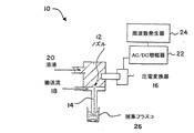
本発明の装置の実施態様を図7に示す。該装置10は溶液20が通過して、噴流14を形成するノズル12を有する。非溶媒液体の搬送流れ18は噴流の回りを流れ、噴流の速度を増加させる。振動が誘導されて、この場合は、増幅器22を介して周波数発生器24によって駆動される圧電変換器16を使用して、噴流を離散させて粒子にする。粒子は捕集フラスコ26に収集されうる。
【0066】
本発明の装置の他の実施態様を図8に示す。該装置28は溶液20が通過するノズル12を有する(噴流は図示せず)。ノズルは捕集フラスコ26内に収容された搬送液体30内にある。電荷源32がノズルに付設されて、流出する噴流に電荷を印加し、その速度を増加させる。別法として、鋭い針を電荷源に付設し、液体中に挿入して、流出する噴流に電荷を印加してもよく;複数ノズル(及び複数液体)の場合には、針を最も応答性のよい液体に配されうる。接地用金属板を捕集部位に含めてもよい。好適には、噴流には振動がまた誘導され、噴流の粒子への離散を制御する。
【0067】
(実施例)
ここに記載する実施例はこの発明の様々な実施態様を例証するものであり、決して限定することを意図するものではない。
実施例1−均一な中実PLGAミクロスフィアの製造
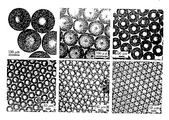
PLGAを酢酸エチル(50mg/ml)に溶解し、溶液を2−3ml/分と変化する流量で60μmの孔部を通して送り込んだ。同時に、音波励起周波数を14から70kHzの範囲で変えた。自由落下スフィアをストロボランプで照らし、画像を10x顕微鏡対物レンズとCCDカメラを使用してビデオテープにとった。図1に示されるように、65から120μmのサイズの均一なスフィアが製造された(最小のスフィアサイズは孔径よりもほんの僅かだけ大であることに留意のこと)。スフィアサイズは、ポリマー溶液の流量が増加し、また超音波周波数を減じると増加した。
同じ方法を使用してであるが孔径、ポリマー溶液流量及び超音波周波数を変えて、15〜500μmの広い範囲にわたって均一なスフィアが形成された。ミクロスフィアは1%ポリ(ビニルアルコール)水溶液中での溶媒抽出/蒸発によって硬化させた。一定量のミクロスフィア懸濁液を顕微鏡カバースリップ上に配して光学顕微鏡によって画像化した。図2に示した代表的な画像は単一調製物内のスフィア径の均一性とこの簡単な装置を使用してミクロスフィアが得られるサイズ範囲を示している。ミクロスフィアは自然に六方最密アレイとして組み込まれた;六角形パターンとサイズの均一性はカバースリップ上の全サンプルにわたって広がっている。
【0068】
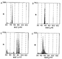
スフィア集団のサイズ分布をCOULTERマルチサイザ(BECKMAN INSTRUMENTS)を使用して測定した。図3A及びBに示されているように、分布は非常に狭い。ピーク幅は機器の較正に使用された商業的サイズ標準のものと同様である。図3Cは、流量を瞬時に増大させて、実験を通して音波周波数を中程度にシフトダウンし、粒径を73から82μmに移行させることにより、サイズ分布を操作することができる能力を示している。図3Dに示された分布を得るには、流量をマニュアルで増加させ、各回の減少倍数の条件を操作して、音波周波数を減少させた。パラメータをマニュアルで操作したので、分布はスムーズではないが、それにもかかわらず、予め決められたミクロスフィアサイズ分布を実際に得ることができることを示している。
【0069】
実施例2−小スフィアサイズのPLGAの調製
この基礎的方法によって得ることができる最小のスフィア径はノズルのサイズによって支配される。オリフィスサイズを〜30μmを越えて減少させることは幾つかの理由から問題がある。第一に、例えば毛細管を引き抜くことによるノズルの再現性のある製造はサイズが減少するにつれてますます難しくなる。第二に、非常に小さいノズルは凝集したポリマー又は異物ダスト粒子によって簡単に詰まり、装置全体の注意深い洗浄と溶液の濾過が必要となる。これらは単にシステムの注意深い工業的技術を用いることで解消しうる技術的問題である。しかし、困った問題は、小オリフィスを通して粘性のあるポリマー溶液を通過させることはオリフィス径が減少するにつれてますます難しくなり、より大なる剪断力を生じることである。よって、電気水力学的噴霧法で約30μm未満の径のミクロスフィアを製造することが探求された。
【0070】
電気水力学的装置はPLGA粒子のサイズを減少させる。これらの予備実験では、PLGA/アセトン溶液を800μmノズルに通した。図4Aに示されるように、加えられた電荷の不存在下では、ノズルから出る液滴は数ミリメートルの径であり、およそノズル径の4倍である。しかし、ノズルへの増加電位が加えられると、液滴サイズが速やかに減少し、最後には〜10μm径のPLGA粒子を生じる(図4B−D)。更に電荷を増大させると、ポリマー溶液を微細ミストとして放出させ、個々の粒子を我々の既存のイメージ化装置を使用して観測するには小さすぎる「マルチ−ジェットモード」の粒子スプレーを生じた。
【0071】
我々の大ざっぱな非最適化装置を用いてでも得ることができる小スフィアサイズを実証するために、PLGA粒子を電気水力学的方法を使用してより小さい(100μm)ガラスノズルから作り出した。粒子を二つの異なった方法によって捕集した。〜3−30μm径のスフィアを、液体窒素浴中にそれらを落下させて、スフィアを凍らせ、続いて冷エタノール中で溶媒を抽出することによって、収集した。およそ10及び5μm径のスフィアを図5A及びBにそれぞれ示す。3μmから100nm径までの小スフィアを、流れの下に配されたシリコンEMスタブ上に捕集し、続いてSEMによってイメージ化した(図5C及びD)。スフィアは僅かな最小の乾燥時間の後でスタブへの落下中に捕捉されたので、スフィアは個別に分離したものではなかった。むしろ、ポリマー/溶媒の液滴はシリコン表面で凝集する傾向があった。しかし、粒子サイズと球形はこのイメージであきらかである。最も重要なことは、スフィアがおよそ1μmの径で、有意な割合がナノメートル範囲であることである。更に、音波励起はこの実験では使用されず、得られたスフィアは完全には均一ではない。しかしながらサイズ分布はかなり均一であるように見える。これらスフィアは比較的大きなノズルと初期の実験設定で製造されたので、この方法のより洗練したバージョンを使用してサブミクロンのスフィアを一貫して生じさせることができることはほぼ疑いがない。
【0072】
実施例3−薬剤の封入
最後に、モデル化合物ローダミンBを使用するポリマーミクロスフィア内に薬剤を封入する能力を証明した。そのフリーの塩基形態のローダミンBを1、3及び5%(質量薬剤/質量ポリマー)の理論負荷でPLGA/酢酸エチル溶液に溶解させた。上記実施例に記載された手順を使用して均一な35、50及び65μm径のミクロスフィアを製造し、硬化させ、捕集した。ローダミンの存在は粒子の均一性に観察できる効果はなかった。ローダミンの封入(〜70%効率)を蛍光顕微鏡によって確認した(図6の右)。ローダミンBはPBS中37℃で7〜10日のインキュベーションの過程にわたってスフィアから放出される。放出速度は、予想されたようにスフィアが小さい場合により速い(図示せず)か、より多くの薬剤を含んでいた(図6の左)。
【0073】
実施例4−油コアを含むPLGポリマーシェルの製造
コア材料を移送する内部ノズル(100μm孔)とシェル材料を移送する「外部」ノズル(250μm孔)を有するデュアルノズルシステムはコア/シェル流の加速のための非溶媒流れを生じさせる第三のノズルの内部に配した。この場合、キャノーラ油を内部ノズルを通して送り込んだ。PLGを塩化メチレン(50mg/mL)に溶解させ、シェル材料を移送する「外部」ノズルを通して送り込んだ。ある場合には粒子のシェルを画像化し、モデル医薬を封入する能力を証明するために少量のローダミンB(〜1%)をPLG相に溶解させた。水中1%(wt/wt)PVAの溶液をコア/シェル流の回りに第三ノズルから流れさせてこの流れを狭くし、得られる粒子が塩化メチレン抽出と粒子硬化のための水性非溶媒浴に入るのを容易にした。所望の粒子サイズに応じて様々な周波数でノズルを音響的に励起させた(0−50kHz)。
【0074】
全体の粒子サイズ、シェル厚及びコア半径は独立に操作した。1%PVA流の速度を変えることによって、個々の粒子の全体直径を調節した。1%PVA流の速度を増大させると、コア及びシェル流の加速が生じ、全体の粒子サイズを減少させる。実施例1に記載されたように音波励起周波数を変えることにより全体の粒子サイズの更なる調節が可能であり、双方のパラメータ(すなわち、1%PVA流の速度と音波励起周波数)を調節して所望の粒子サイズを達成した。また、油(コア)及びPLG(シェル)流の流量の注意深い調節により、様々なコア半径又はシェル厚を有する粒子を得た。PLG流の流量を変化させて、変わらないコア半径と変化する制御シェル厚を有するシェルを製造した。他のパラメータを一定に保ちながら油の流れの流量を変えることにより、変わらないシェル容積と制御されたコア半径を有するシェルを製造した。このようにして、制御可能で明確に定まった全体サイズとコア半径に対するシェル厚の比を示すコア/シェル粒子を製造することができる。この場合、流れの相対流量並びに音波励起周波数を調節することによって、3〜35μmの範囲にわたって制御された均一のシェル厚を有する直径20〜70μmの範囲の均一な粒子を製造した。
【0075】
粒子サイズ分布とアスペクト比を光学顕微鏡と封入ローダミンBの蛍光検出によって実証した。サイズ分布は中実粒子を製造する場合に得られる分布に匹敵することが見出された(図3)。また、粒子は共焦点蛍光顕微鏡を使用して光学断片によって実証したところ、与えられたサンプルにわたって一致した均一のシェル厚とコア半径を有していた。
【0076】
よって、本発明に従って、本明細書において前述した目的と利点を十分に満たす方法が提供されたことは明らかである。本発明をその様々な特定の実施例と実施態様によって説明したが、本発明はそれらに限定されるものではなく、多くの代替例、変更、及び変形が前述の記載に鑑みて当業者には明らかであると理解される。従って、そのような代替例、変更、及び変形の全てを本発明の精神及び広い範囲内に入るものとして包含することが意図されている。
【図面の簡単な説明】
【図1】図1はストロボランプで照らした自由落下スフィアを示しており、画像は10x顕微鏡対物レンズとCCDカメラを用いてビデオテープにとられた。
【図2】図2は15〜500μmの単一の調製物内のスフィア径の均一性を示している。
【図3】図3A及びBはCOULTERマルチサイザーを使用して測定したスフィア集団のサイズ分布を示している。図3Cは流量と音波周波数を瞬時に変えることによるサイズ分布を操作する能力を実証している。図3Dは連続的に変わるサイズ分布を生じさせる能力を実証している。
【図4】図4は、電気流体学的装置がPLGA/酢酸エチル粒子のサイズを減少させることを実証している。図4Dは増加した電圧をノズルへ印加した場合を実証している。
【図5】図5A及びBは、おおざっぱな最適化されていない装置を用いた場合でも得ることができる小スフィアサイズを実証している。図5C及びDは3μm以下の捕集小スフィアを実証している。
【図6】図6はモデル化合物、ローダミンBを用いてポリマーミクロスフィア内に薬物を封入する能力を蛍光顕微鏡測定により実証している。図6はまたPBS中37℃での7〜10日のインキュベーションの過程にわたってのスフィアからのローダミンBの放出を実証している。放出速度はスフィアがより多くの薬物を含む場合、予想以上に速やかであった。
【図7】図7は本発明の装置の一実施態様を示す。
【図8】図8は本発明の装置の他の実施態様を示す。[0001]
(Related application)
This application is directed to two provisional applications, Kyekkyoon Kim and Daniel W., entitled "Preparing Tightly Controlled Polymers for Biomedical Applications." Provisional Application No. 60 / 225,525, filed Aug. 15, 2000; and "Uniform solid and hollow spherical particles in the micron and submicron range and controlled size, charge, chemical composition and stoichiometry. Provisional Application No. 60, filed August 15, 2000, filed August 15, 2000, entitled "Apparatus and Method for Producing Such Particles Using Field-Injection Charging and Electrohydrodynamic Spraying." No./225620; both of which are incorporated herein by reference.
[0002]
(background)
Rapid advances in biotechnology have led to the discovery of numerous protein and peptide therapeutics, many of which have recently been marketed or are currently under review by the US Food and Drug Administration. However, unlike traditional small molecule drugs, proteins and peptides generally cannot be administered orally; injection or infusion is often required. In addition, because of its brittleness and short in vivo half-life, encapsulating proteins in biodegradable polymer devices, where the drug can be delivered locally or systemically over long periods of time, has been a challenge to these problems. A powerful and eagerly researched solution. Biodegradable microspheres comprising a variety of polymers are the most studied means because of their relative simplicity of manufacture and their ease of administration in vivo through injection needles to various locations.
[0003]
Several microsphere production methods have been described, including precipitation, spraying, phase separation and emulsification. Emulsification and spray approaches are commonly used on both bench scale and industrial scale. The sphere size and size distribution are often reproducible but poorly controllable. It is not uncommon for the standard deviation to be 25 to 50% of the average diameter.
[0004]
Control of sphere size and size distribution has several important implications for sustained release drug delivery. For example, there is typically an ideal sphere size that will provide the desired release rate and route of administration. "Too small" spheres have poor encapsulation efficiencies, can migrate from the injection site, and can release the delivery unduly quickly. A "too large" sphere may not pass easily through the needle. Thus, typically polydisperse spheres obtained by common manufacturing methods must be filtered or sieved to separate particles of a desired size range, and polymer and drug constituent spheres outside that range are discarded. .
[0005]
Uniform microspheres, approximately 1-5 μm in diameter, are ideal for passive targeting of specialized antigen presenting cells (APCs) such as macrophages and dendritic cells. Similarly, 10-20 μm diameter microspheres can be used to target bent capillary beds of tumor tissue by chemical embolization. Systems that allow for the production of rigorous microspheres can identify optimal sizes for such applications and provide an effective path to commercial production and clinical implementation.
[0006]
A long-held goal in sustained release drug delivery technology has been the ability to precisely control the release rate of encapsulated compounds, with microsphere size being a major determinant of release kinetics. Large spheres generally release the encapsulated compound more slowly and over a longer period of time, but other properties (polymer molecular weight, initial porosity, drug distribution within the sphere, etc.) do not change. While constant (ie, zero order) release rates are often preferred, variable drug release rates are also valid for many important indications. For example, intermittently high doses of antibiotics reduce the development of bacterial resistance, and discontinuous administration of vaccines often enhances the immune response.
[0007]
Methods of controlling the drug release rate include (i) selecting the polymer chemistry (anhydrides, esters, etc.) and comonomer ratios, (ii) coupling the drug to the polymer, (iii) parameters of the microsphere formulation. Altering, thereby altering the physical properties of the resulting particles, and (iv) manipulating the sphere size and distribution. The success of the latter study was limited by the relatively broad microsphere size distribution.
[0008]
In recent years, several reports have been made on the production of biodegradable polymeric microspheres having a controlled and uniform size (P. Sanstrap and AJ Moes, Influence of manufacturing parameters on the size characteristics and the size of the spheres and the size of the spheres). Nifedine from poly (DL-lactide-co-glycolide) microspheres. Int. J. Pharm. 98 (1993) 157-164; BG Amsedation and M. fotion, Ancestion, Annotation rele J. Control, Release 43 (1997) 183-196; K. Shiga, N.p. J. Pharm. Pharmacol. 48 (1996) 891-895; B. Amsden, The production of uniform sized thermoplastics. 140-1143; and N. Leelarasamee, SA Howard, CJ Malanga and JK H. Ma, A method for the preparation of organic chemicals. 5 (1988) 147-157). However, none of these methods has been able to produce particles in the size range (〜1-100 μm) suitable for drug delivery while maintaining a narrow size distribution. Also, these past methods may be difficult to scale up for commercial use.
[0009]
The method for producing hollow spheres is described in K. Kim, K .; Kim, D.S. A. Payne and R.A. S. Upadhye, "Fabrication of Hollow Silica Aerogel Spheres by a Droplet Generation Method and Sol-Gel Processing", J. Am. Vac. Sci. , Technol. A. , Vol. 7, no. 3 pp. 1181-1184 (1989); Kim, K .; Y. Jang and R.M. S. J. Upadhye, "Hollow silica spheres of controlled size and porosity by sol-gel processing", J. Am. Am. Ceram. Soc. 74: 8 pp. 1987-1992 (1991).
[0010]
Electrostatic spraying is described in K. Kim and R.A. J. Turnbull, "Generation of charged drops of insulating liquids by electrostatic spraying", J. Am. Appl. Phys. , Vol. 47, no. 5, pp. 1964-1969, May 1976, U.S. Patent No. 5,344,676 to Kim et al. And U.S. Patent No. 6,060,128 to Kim et al.
[0011]
A previously developed method devised for producing hollow spheres is that two coaxially mounted nozzles containing different substances in the liquid phase (the substance of the inner nozzle can also be a gas) are smooth. A dual nozzle method is used in which a cylindrical jet is created and the jet is broken into uniform droplets by sound wave excitation. (Nik Kim et al., Below, "Fabrication of hollow silica aerogel spheres by a drop generation generation method and sol-gel processing," K. Kim et al., Ltd. The smallest droplet that can be produced in this way is approximately twice the opening of the outer nozzle, which in turn is a uniform solid and hollow small size (diameter less than about 50 μm). It has been shown that the fabrication of spheres, especially those in the sub-micron range, involves practical difficulties because the smaller the nozzle opening, the greater the noise. This is especially true if the drug compound to be encapsulated is suspended as fine particles in the sphere-forming solution, especially if the substance used is viscous. Becomes even more serious.
[0012]
In past methods for spraying microdroplets from nozzle-type devices, the minimum sphere size typically obtained is limited by the size of the nozzle opening. Usually, the droplet cannot be smaller than the nozzle opening; typically, the droplet size is one to four times the nozzle diameter. This creates some difficulties as the desired sphere size decreases. One problem is that as the size decreases, the manufacture of the nozzle itself becomes more difficult. This is especially true for large-scale manufacturing methods where droplets need to be formed through an array of nozzles (possibly 1000-2000). The second constraint arises from the pressure required to pump fluid through small nozzles. The required pressure is
Δp = 8 μLQ / πR4
(Where Δp is the pressure loss at the nozzle, μ is the viscosity of the fluid, L is the length of the “flow path” of the nozzle, Q is the volumetric flow rate of the fluid passing through the nozzle, and R is Which is the radius of the nozzle opening). Therefore, the required pressure is R-4Scale. If it is desired to produce microdroplets with a diameter of 径 5 μm, the conventional method requires a nozzle with a diameter of 5 μm or less. For example, at a flow rate of 1 mL / min and a fluid viscosity of 100 centipoise (100 times more viscous than water), a 5 μm diameter orifice is ~ 1.1 × 10¹⁰It requires a head of Pa (〜110000 atm). This is clearly impossible high pressure. Even 1 μm to 1 cp of water requires 1100 atm pressure to pump at 1 mL / min through a 5 μm diameter nozzle. Therefore, a special device is required, if at all, to actually feed an arbitrary liquid through a nozzle having a diameter of 5 μm.
[0013]
Another problem with traditional methods of forming small spheres is that certain encapsulated compounds, such as plasmid DNA, are damaged by shear forces. The damage depends on the product of the shear force γ and the time θ spent in the shear field. The average of this product for the fluid flowing through the pipe is
(Γθ) avg = 16/3 · (L / D)
(Where L is the pipe length and D is the pipe diameter). The nozzle orifice can be approximated as a pipe. However, inlet effects tend to increase the shear rate, which means that this equation gives a low estimate. However, the value of γθ is approximately inversely proportional to the orifice diameter. Thus, reducing the nozzle diameter from 100 to 5 μm increases the damage done to any encapsulated compound by a factor of 20.
[0014]
U.S. Patent No. 6,116,516 describes a stabilized capillary microjet that creates an aerosol. Microjets are formed by forcing gas around a liquid stream. Under the right conditions, micron-sized aerosols are produced, preferably with 90% or more having the same diameter ± 3% to 30%.
[0015]
A brief overview
It is an object of the present invention to produce micron and nano size spherical particles by pumping material through a small diameter orifice and then vibrating the liquid using acoustic type waves.
It is also an object of the present invention to produce micron and nano sized spherical particles by feeding material through a small diameter orifice and applying further downward force, wherein the downward force is generated by an electrohydrodynamic method. Or a second liquid adjacent and parallel to the liquid at a faster rate than the first liquid. It is also an object of the present invention to produce micron and nano size spherical particles using acoustic type waves in the above manner.
Using an inner and outer liquid passed through one or the other of two coaxially mounted nozzles, a hollow micron is created by creating a smooth cylindrical jet of an outer liquid containing an inner liquid (or gas) coaxially. It is a further object of the present invention to produce nano-sized spherical particles. This jet can be further broken down by sound waves into uniform droplets.
[0016]
It is yet a further object of the present invention to provide a novel method for curing micro and nanospheres by using any of the above methods for making micro and nanospheres, the nozzle used Alternatively, the pores are located below the surface of the aqueous bath, allowing the spheres to harden with minimal deformation.
It is yet a further object of this invention to produce therapeutic compounds encapsulated by any of the methods described above, which are useful for medical treatment of humans and as therapeutically valuable biomedical compositions.
Multi-shell micro and nano with different shells containing different materials useful for biomedical applications for humans and animals including controlled release drug delivery systems, with controlled size, shell thickness and shell number It is yet another object of the present invention to manufacture a sphere.
[0017]
It is also an object of the present invention to manufacture the above and other types of micro and nanospheres for biomedical applications involving passive or active targeting of desired cells, tissues or regions of the body. It is a further purpose.
It is a further aspect of the present invention to provide an apparatus and method for producing micro- and nanosphere particles of precisely controlled size and size distribution for biomedical applications, especially for sustained release drug delivery systems. Is the purpose.
It is a further object of the invention to produce stable micro and nanospheres of desired size, chemical composition and stoichiometry.
It is a further object of this invention to produce controlled size micro- and nanospheres for biomedical applications involving controlled-release drug delivery systems.
It is also a further object of the present invention to produce controlled size hollow micro and nanospheres for biomedical applications, including controlled controlled release drug delivery systems.
These and other objects are provided in the present invention, which is described in further detail below.
[0018]
In a first aspect, the present invention is a method of forming particles, comprising: accelerating a first stream containing a first liquid; vibrating the first stream to form particles.
In a second aspect, the invention is a method of forming particles, comprising accelerating a first stream containing a first liquid. Acceleration involves applying a charge to the first stream. The particles have a core and a shell.
In a third aspect, the invention is a particle having an average diameter of 50 to 100 μm. 90% of the particles have a diameter that is within 2% of the average diameter of the particles.
In a fourth aspect, the invention is a particle having an average diameter of 1 to 50 μm. 90% of the particles have a diameter that is within 1 μm of the average diameter of the particles.
In a fifth aspect, the invention is a particle prepared by the above method.
In a sixth aspect, the invention provides (i) a first nozzle for forming a first stream of a first liquid; and (ii) a second nozzle of a second liquid in contact with the first stream. And a vibrator for forming particles from the first stream.
[0019]
In a seventh aspect, the invention provides (i) a first nozzle for forming a first stream of a first liquid, and (ii) a charge source for applying charge to the first stream. , (Iii) a vibrator for forming particles from the first stream.
In an eighth aspect, the invention provides (i) means for forming a first stream of a first liquid; (ii) means for accelerating the first stream; and (iii) first means for accelerating the first stream. Means for oscillating the flow of particles.
In a ninth aspect, the invention provides (i) a first nozzle for forming a first flow of a first liquid; and (ii) a second nozzle of a second liquid surrounding the first flow. A second nozzle surrounding the first nozzle for forming a second stream, and (iii) a charge source for applying a charge to at least one of the first and second streams. It is an apparatus for forming particles.
In a tenth aspect, the present invention is a method for forming particles, comprising forming particles with the above apparatus.
[0020]
(Detailed description)
The present invention relates to a method wherein a material, preferably spherical, micron and nano size, is produced by feeding material through a small diameter orifice and oscillating the liquid with a sonic type wave, wherein the velocity of the fluid is It increases beyond the speed created by pressure. The nozzle diameter can be larger than the particles to be produced. For example, a 5 μm droplet can be prepared from a larger nozzle, for example a 100 μm diameter nozzle. The particles form in the surrounding liquid and help prevent deformation.
[0021]
The pressure required to form very small particles is greatly reduced by the present invention. For example, a 100 cp solution pumped through a 100 μm diameter nozzle at 1 mL / min requires a pump pressure of only 〜68000 Pa (〜0.67 atm) or requires a viscous solution such as glycerin (μ〜500 cp). It can be pumped through a 100 μm diameter nozzle at a head of 15 atm at 5 mL / min. These pressures are easily obtained with commercially available high pressure pumps, such as those commonly provided in high pressure liquid chromatography systems. Furthermore, the shear forces are greatly reduced for a given particle size, again eliminating the difficulties encountered with very small nozzles. Aspects of the present invention are described in "Fabrication of PLG microspheres with precision controlled and monodisperse size distributions," J. Appl. Controlled release 73 (1): 59-74 (May 18, 2001).
[0022]
The present invention also relates to a method wherein micron and nano sized spherical particles are produced by feeding material through a small diameter orifice and applying further downward force, said downward force being at a greater rate than the first liquid. Includes either a second liquid stream adjacent to and parallel to the liquid or an electrohydrodynamic technique. Sonic type waves can also be used in the above method. As used herein, the term “particle” includes both liquid particles (droplets) and solid particles.
The present invention further provides a hollow, inner and outer liquid that is passed through one or the other of two coaxially mounted nozzles to create a smooth cylindrical spray stream of an outer liquid coaxially containing the inner liquid (or gas). And micro and nano-sized spherical particles. This spray stream can be further broken down into uniform droplets by the acoustic waves.
[0023]
The present invention is also a novel method for curing micro or nanospheres, wherein the nozzle or orifice used to produce the sphere is located below the surface of the aqueous bath to minimize the deformation of the sphere. A method is provided that allows curing.
The invention further provides a therapeutic compound encapsulated by any of the above methods useful as a biomedical composition.
[0024]
One embodiment of the present invention uses "electrohydrodynamic spraying" and "dual nozzle" independently or in combination to produce very small, uniform, solid, hollow or multi-shelled spheres. Electrohydrodynamic spraying methods that enable the production of hollow and multi-shell micro and nanospheres are new. This method is novel and very useful in that it is particularly suitable for producing micrometer and nanometer sized hollow and multi-shell spheres that are inherently difficult to create by any other existing methods. . Furthermore, encapsulation of therapeutic compounds in very uniform spheres, especially hollow and multi-shell spheres, is novel and precisely controlled release rates that cannot be obtained using spheres made by other existing methods Very useful for sustained release and targeted drug delivery.
[0025]
The devices and methods of the present invention allow for encapsulation of a first material in a spherical shell of a second material that is particularly useful for various biomedical applications.
The apparatus and method of the present invention also allows for the production of a solid sphere of only the second material by blocking the supply of the first material through the internal nozzle.
Another unique and novel aspect of the invention is that the method can be used to select the electrical polarity (neutral, positive or negative) of the resulting micro and nanospheres.
[0026]
This concept can be extended to produce multi-shell spheres containing two or more materials. The fact that the invention makes it possible to produce very small multi-shelled spheres smaller than 50 or 100 μm diameter makes it particularly useful for applications involving drug delivery systems (DDS). Numerous applications other than DDS are also possible.
The present invention also provides solid or hollow uniform micro and nanospheres of controlled size, shell thickness, charge, chemical composition and stoichiometry. Such spheres have many unique uses in the biomedical field.
[0027]
The present invention relates to a method for producing micron and nano size spherical particles by feeding material through a small diameter orifice and then vibrating the liquid with a sonic type wave.
The invention further relates to a method of producing micron and nano sized spherical particles, comprising feeding material through a small diameter orifice and applying a further downward force to the liquid to transfer the liquid through the orifice. The further downward force may be a second liquid flow or electrohydrodynamic technique that is adjacent and parallel to the liquid at a greater rate than the sphere forming liquid. These methods can be further modified by the addition of sonic-type waves.
[0028]
The present invention further relates to a method for producing hollow micro and nano sized spherical particles containing internal and external liquids, wherein the internal and external liquids are flowed through one or the other of two coaxially mounted nozzles to provide a smooth A cylindrical jet is created from the outer liquid that contains the inner liquid (or gas) coaxially within the outer liquid. This spray flow can be broken into uniform droplets by the acoustic waves. The size and ratio of the radius and thickness of the hollow sphere are controlled by changing the relative flow rates of the external and internal liquids, the relative size of the nozzle, the relative position of the nozzle, and the amplitude and frequency of the acoustic excitation.
[0029]
This method further facilitates producing hollow spheres with a large ratio between the radius and thickness of the hollow sphere, as the surface tension of the bath solution helps maintain the spherical shape and integrity of the hollow sphere.
The present invention further relates to a method of curing micro and nanospheres by utilizing any of the above methods for producing micro and nanospheres, wherein the nozzle or orifice utilized is located below the surface of the aqueous bath. Enables hardening of the sphere, with minimal deformation. Any of the above nozzle types will work with this embodiment of the invention.
[0030]
The invention further relates to a therapeutic compound encapsulated by any of the above methods given to a human for medical treatment.
The present invention relates to a method for producing micro and nanospheres, including producing micron and nano sized spherical particles using a dual nozzle system and electrohydrodynamic concepts.
The invention further relates to the use of very sharp subcutaneous needles at the inner nozzle of a dual nozzle system to produce solid, hollow and multi-shell micron and nano sized particles useful for semiconductor and biomedical applications.
[0031]
The invention further relates to novel biomedical compositions containing uniform solid, hollow and multi-shell micron and nano-sized particles, hollow or filled.
In this application, the terms sphere, bead and particle are used interchangeably to describe the micro and nanospheres of the present invention. The term "hollow" is also used to indicate that the core is empty or contains gas. The term "multi-shell" includes particles whose core is liquid (aqueous, oily, etc.) or solid, such as another polymer. Although the terms "hollow" and "multi-shell" are used separately in this application, these terms must be read as including the other.
[0032]
There is a need in the biomedical industry for the production of micron and nano size spheres. The ability to form spheres with precisely controlled size, size distribution and morphology (eg, hollow, multi-shell, solid, porous, etc.) has several very important applications, especially in the biotechnology field. ing. This method allows for and / or significantly improves many drug delivery methods.
[0033]
One embodiment of the present invention is for producing solid, hollow and multi-shelled micro and nanoparticles of precisely controlled size, size distribution and morphology for biomedical applications, particularly sustained release drug delivery systems. Apparatus and method. The invention also relates to novel micro and nano spheres containing the first material. The invention further relates to novel hollow micro and nanospheres of the first material encapsulating the second material.
Another embodiment of the present invention relates to devices and methods for producing precisely controlled size, size distribution and morphology of micro and nanoparticles for biomedical applications, particularly sustained release drug delivery systems.
[0034]
Embodiments of the present invention allow for the formation of particles or spheres by pumping a liquid material (eg, a polymer dissolved in an organic solvent, a polymer melt, etc.) through a small diameter orifice, wherein the small diameter orifice has a diameter of a few millimeters to about 1 micron. it can. The orifice can be as small as 500 nm in diameter. The liquid stream exiting the orifice is broken into droplets by vibrating or rocking the device at a controlled frequency and amplitude.
Vibration or rocking can be achieved, for example, by a piezoelectric transducer driven by a wave generator. Mechanical excitation is believed to emit sonic energy waves along a liquid jet that creates a periodic instability that breaks the flow into a uniform row of droplets.
[0035]
The droplet size is determined by the orifice diameter, solution flow rate, oscillation frequency and amplitude. Therefore, the droplet size can be controlled by changing these four parameters. Further, given a device with a fixed orifice, the droplet size can vary from a minimum size slightly larger than the orifice opening to a maximum of at least ten times the orifice opening.
This approach is improved over typical ultrasonic nozzles due to the lower acoustic intensity and allows tighter control over the compatibility between frequency and solution flow.
[0036]
In yet another embodiment of the present invention, the sphere size can be further controlled by using a further downward force to "pull" the liquid jet through the orifice, reducing the jet size from the diameter of the orifice. One example is an electrohydrodynamic method in which an electric force acts to reduce the size of the liquid jet and the resulting droplet. Electrohydrodynamic methods are by injecting a charge of the desired polarity into the liquid, for example with a battery or by applying a high voltage directly to the nozzle or liquid with a transformer and rectifier that converts the current in the house. Actuated. An outwardly directed electrical tension occurs in the charged liquid meniscus at the nozzle opening, causing smaller droplets to fall from the nozzle ("drip mode"). Without being limited by theory, the reason for the drop in droplet size is that while there are two forces, gravity and electrical forces, that cooperate to draw liquid from the nozzle, It is believed that the surface tension is for retaining the liquid in the nozzle. As the amount of injected charge increases, this increases the electrical tension, which eventually overcomes gravity and surface tension and reduces the droplet size. The further increase in charge injection above a certain threshold creates a very strong electrical tension that literally pulls liquid out of the nozzle, creating a thin charged liquid jet, which is fairly uniform (called the "jet mode"). Is broken down into simple droplets. The jet mode changes from a single jet mode to a multiple jet mode as charge injection further increases.
[0037]
Another example of a further downward force used is a separate liquid stream (typically immiscible) that passes through an orifice parallel to and adjacent to the sphere-forming liquid at a greater velocity than the sphere-forming liquid. ). The sphere forming liquid is drawn by the fluid resistance at the liquid-liquid interface. The sphere-forming jet decreases in diameter by a factor proportional to the difference between the linear velocities of the two streams.
[0038]
The method of the present invention can be further modified to produce "hollow" or multi-shell particles composed of two or more concentric spheres of different materials. For example, a sphere consisting of a polymer shell surrounding a drug-containing aqueous phase can be produced. Such a sphere can be formed using a dual-nozzle method consisting of using two coaxially mounted nozzles. By passing two liquids through one or the other of the nozzles, a smooth cylindrical jet of one liquid containing the other liquid (or gas) coaxially therein can be created. The jet can then be broken into uniform droplets using sound waves, as described above, resulting in a "hollow" or multi-shell sphere. The ratio and size between the sphere radius and thickness can be controlled by varying the relative flow of the external and internal fluids, the relative size of the nozzle, the relative position of the nozzle, and the amplitude and frequency of the acoustic excitation.
[0039]
Other embodiments of the present invention allow for novel hardening of micro and nanospheres while allowing the sphere uniformity to be maintained. Droplets falling from the device can be cured to form microspheres by any of several standard methods, depending on the type of material that makes up the sphere. An important consideration is to maintain a uniform or desired size during sphere collection, curing (phase inversion) and drying. When the droplets exiting the nozzle are dropped through the air and then into, for example, a liquid bath (often aqueous or liquid nitrogen) from which the organic solvent is to be extracted, a collision of the sphere with the liquid surface will occur. The shape of the sphere can be deformed or even completely destroyed. Another embodiment of the present invention overcomes this inherent problem of the prior art, which places the orifice below the surface of the aqueous bath, thereby avoiding collision with the surface. The spheres are then agitated to effectively extract the organic solvent. However, the agitation must be mild; normal agitation speeds create too much shear, destroying the particles and ruining the size distribution. This is believed to be new because other spraying methods do not use nozzles located below the liquid / air interface.
[0040]
In one embodiment, the present invention utilizes electrohydrodynamic spraying in combination with a dual nozzle method to produce hollow, very small, uniform spheres. Micro and nanospheres of the desired size, chemical composition and stoichiometry are the most stable forms by this novel method, named flow-limited field injection electrostatic spraying (FFESS). Can be manufactured. This embodiment combines the basic concepts of hollow sphere manufacturing technology and electrostatic spray technology. Hollow sphere production methods are described in K. Kim, K .; Kim, D.S. A. Payne and R.A. S. Upadhye, "Fabrication of Hollow Silica Aerogel Spheres by a Droplet Generation Method and Sol-Gel Processing" Vac. Sci. , Technol. A. , Vol. 7, no. 3 pp. 1181-1184 (1989); Kim, K .; Y. Jang and R.M. S. Upadhye, "Hollow silica spheres of controlled size and porosity by sol-gel processing," J. Am. Am. Ceram. Soc. 74: 8 pp. 1987-1992 (1991). Electrostatic spraying is described in K. Kim and R.A. J. Turnbull, "Generation of charged drops of insulating liquids by electrostatic spraying", J. Org. Appl. Phys. , Vol. 47, no. 5, pp. 1964-1969, May 1976, U.S. Patent No. 5,344,676 to Kim et al. And U.S. Patent No. 6,060,128 to Kim et al. In this embodiment, the present invention overcomes the above difficulties by introducing the concept of electrohydrodynamic spraying. Unlike conventional hollow sphere manufacturing methods where only mechanical force is used to break a smooth liquid jet into uniform hollow droplets, this embodiment of the invention creates and obtains a charge of the working liquid. Utilizing electric tension, the size of the liquid jet is reduced to the size of the nozzle opening or less. This, in turn, reduces the size of the droplets resulting from the disruption of the liquid jet. In this way, uniform multi-shelled spheres containing very small different materials, smaller than 50 μm diameter, can be produced. This particular ability of the present invention encloses nanometer-sized particles in a spherical shell containing selected materials that meet the needs of many sought-after biomedical applications, including controlled drug release or drug delivery systems. It becomes possible. The ability to control the thickness of the outer spherical shell and the materials that make it up also facilitate the formulation of various scenarios for controlling the rate of drug release. It must be emphasized that the same method applies to coaxial nozzles, including two or more nozzles, which allow the production of small multi-shell spherical particles with two or more layers of different materials.
[0041]
The particles of the present invention can have a very narrow size distribution. Preferably, at least 90% of the particles have a size within 2% of the average particle size, preferably within 1%. Alternatively, preferably at least 95% of the particles have a diameter within 10%, more preferably within 5%, even more preferably within 2%, and most preferably within 1% of the average particle size. Alternatively, preferably, at least 95% of the particles have a size within 10%, more preferably within 5%, even more preferably within 2%, and most preferably within 1% of the average particle size. Alternatively, preferably, at least 98% of the particles have a size within 10%, more preferably within 5%, even more preferably within 2%, and most preferably within 1% of the average particle size. Alternatively, preferably, at least 99% of the particles have a diameter within 10%, more preferably within 5%, even more preferably within 2%, and most preferably within 1% of the average particle size. As used herein, the terms "diameter" and "average diameter" for a particle refer to number average diameter.
[0042]
Another method of describing a narrow size distribution of particles having an average diameter of preferably at most 50 μm, more preferably 1 μm to 50 μm, most preferably 1 μm to 30 μm, has a diameter within a certain length of the average diameter. By percent. Preferably, 90% of the particles have a diameter within 1 μm of the average diameter of the particles, more preferably within 0.5 μm of the average diameter of the particles, and most preferably within 0.1 μm of the average diameter of the particles. Alternatively, preferably, 95% of the particles have a diameter that is within 1 μm of the average diameter of the particles, more preferably within 0.5 μm of the average diameter of the particles, and most preferably within 0.1 μm of the average diameter of the particles. I have.
[0043]
The present invention relates to devices and methods for producing precisely controlled size and size distribution of micro and nanoparticles for biomedical applications, particularly sustained release drug delivery systems. The invention also relates to novel micro and nano spheres containing the first material. The invention further relates to novel hollow micro and nanospheres of the first material encapsulating the second material.
The present invention relates to devices and methods for producing micro- and nanoparticles of precisely controlled size and size distribution for biomedical applications, particularly for sustained release drug delivery systems.
[0044]
The invention further relates to the use of a very sharp subcutaneous needle at the inner nozzle of a dual nozzle system to produce solid, hollow and multi-shell micron and nano size particles useful for semiconductor and biomedical applications. .
Illustratively, the formation of particles or spheres is achieved by extruding a liquid material (eg, a polymer dissolved in an organic solvent, a polymer melt, etc.) through a small orifice (a few millimeters to 10 micrometers in diameter). The liquid stream exiting the orifice is broken into droplets by vibrating or rocking the device at a controlled frequency and amplitude.
Vibration or rocking can be achieved, for example, by a piezoelectric transducer driven by a wave generator. It is believed that the mechanical excitation produces a wave of sonic energy with the liquid jet, creating a periodic instability that breaks the stream into a train of droplets.
[0045]
Yet another embodiment of the present invention relates to a therapeutic compound. Therapeutic compounds (eg, peptides, proteins, nucleic acids, polysaccharides, lipids, steroids, and organic and inorganic pharmaceutical compounds, etc.) can be encapsulated in the spheres in a variety of ways. Compounds that are soluble in the liquid phase can simply be dissolved. Non-soluble materials can be suspended in a liquid in the form of small particles. Alternatively, the non-soluble material can be dissolved in the immiscible phase and emulsified with the sphere-forming liquid before droplet formation. For example, the protein can be dissolved in an aqueous buffer solution, while the polymer (the sphere-forming material) can be dissolved in an organic solvent such as methylene chloride or ethyl acetate. The aqueous and organic solutions can be mixed and homogenized to form a water-in-oil emulsion that subsequently becomes a droplet-forming liquid.
[0046]
One embodiment of the present invention uses a biodegradable polymer, lactic acid-glycolic acid copolymer (PLGA). PLGA is a well-studied polymer for drug delivery and has received FDA approval for many in vivo applications. However, the technique can be generalized to other materials, including poly (orthoesters), poly (anhydrides), poly (phosphoesters), poly (phosphazenes), and the like.
Non-limiting examples of orifices useful in the practice of the present invention include tapered nozzles, capillary tubes, simple holes in a flat plate, or even an array of multiple orifices of any of these types.
[0047]
Non-limiting examples of materials useful for forming particles include polyesters (eg, poly (lactic acid), poly (glycolic acid) and poly (lactic acid / glycolic acid copolymer), poly (lactic acid / lysine copolymer). , Poly (lactic acid / lysine graft polymer), polyanhydride (eg, poly (fatty acid dimer), poly (fumaric acid), poly (sebacic acid), poly (carboxyphenoxypropane), poly (carboxyphenoxyhexane) , Copolymers of these monomers, etc.), poly (anhydride / imide copolymer), poly (amide), poly (orthoester), poly (iminocarbonate), poly (urethane), poly (organophasphazene), Poly (phosphate), poly (ethylene vinyl acetate) and other acyl-substituted cellulose acetates and derivatives thereof, poly (caprola) T), poly (carbonate), poly (amino acid), poly (acrylate), polyacetal, poly (cyanoacrylate), poly (styrene), poly (vinyl chloride), poly (vinyl fluoride), poly (vinyl imidazole), Includes chlorosulfonated polyolefins, polyethylene oxide and copolymers and mixtures thereof.
[0048]
For other uses of drug delivery, the spheres can be substantially from polymers (eg, polystyrene) to metals, minerals (silica), cryogenic chemicals (frozen hydrogen), as long as the starting precursor is in the liquid phase or solution. It can be formed from any material.
The spheres obtainable by the present invention range from about 1 nanometer to about 1 millimeter. For drug delivery systems, sizes from about 10 nm to about 100 microns are possible. The term sphere or group of spheres as used throughout this application is not limited to particles having an aspect ratio of one, but rather includes particles that deviate significantly from a perfect sphere. Preferably, the particles have an aspect ratio of 1 to 10, more preferably 1 to 2.
[0049]
The term nano defines a size ranging from about 1 to about 1000 nanometers. The term micro defines a size ranging from about 1 to about 1000 microns.
The outer shell thickness of a two-component bead can be from about 99% of the bead radius to about 1% of the bead radius, depending on the application. However, the minimum achievable thickness of the outer shell will depend on the overall size of the hollow bead and the nature of the material from which it is made. The achievable absolute minimum thickness must be greater than the thickness of several molecular layers.
[0050]
The spheres of the present invention have many possible biomedical applications.
Passive targeting of phagocytosis.
Immune system cells, particularly macrophages and dendrocytes, are targets of immunity. These "professional" antigen presenting cells (APCs) can elicit the desired T cell response to vaccine components. APCs are capable of phagocytosis of particles, typically in the range of 1 to 10 μm; most other types of cells in the body cannot internalize such large particles. By making particles in this size range containing the vaccine components, the delivery of the vaccine to APCs can be passively targeted. Current technology is capable of forming particles of this size, but only those with a significantly broader size distribution. Our method makes it possible to produce vaccine-loaded microspheres in which essentially 100% of the spheres are of the desired size. APC targeting can be optimized without wasting expensive vaccine components in spheres that are too large or too small.
[0051]
Capillary emboli.
Blood vessel sizes vary throughout the body, with the smallest blood vessels being capillaries. Tumor tissue typically exhibits a particularly bent capillary bed that nourishes rapidly growing cancer cells. One approach to targeting tumor tissue is to deliver therapeutic compounds encapsulated in microspheres of a certain size so that they can remain in narrow capillaries. The microspheres are then injected into the artery feeding the tumor. Spheres that are too large will become lodged in larger blood vessels, and the therapeutic compound may be prevented from reaching all tumors. Spheres that are too small can pass through tumors and deliver therapeutic compounds to other healthy tissues; a serious problem because many anticancer drugs are, of course, cytotoxic. Our ability to produce precisely sized spheres allows us to maximize the number of spheres that stay in the smallest capillaries, and thus the amount of therapeutic compound delivered to a desired location.
[0052]
Targeting with nanospheres.
Biodegradable polymer spheres (nanoparticles or nanospheres) less than 1 μm in diameter have several specific uses in drug delivery. For example, particles smaller than 150 nm in diameter can be targeted for receptor-mediated endocytosis by many different types of cells. Nanoparticles are also useful for oral administration, in which case the particles can cross the intestinal epithelium and be taken up by M cells and Peyer patches. Particles that are too large are rarely picked up, and particles that are too small contain little therapeutic compound. The most modern methods for the formation of biodegradable polymer nanoparticles are many, but with large particle size variability. For example, in phase inversion nanoencapsulation, the polymer and drug are dissolved in a solvent (eg, methylene chloride) and then poured into a non-solvent phase (eg, petroleum ether), resulting in particles between 100 and 5000 nm. Our ability to control the particle size with the largest variation being a few percent of the average size allows for optimal nanoparticle targeting.
[0053]
Control of drug release rate from solid microspheres.
Many sustained release drug delivery systems consist of microspheres formed from biodegradable polymers. Therapeutic compounds encapsulated in the microspheres are released when the polymer is eroded, most often by hydrolysis, in an aqueous environment within the body. The rate of drug release depends on several factors, including polymer composition, polymer molecular weight, microsphere porosity, and the like. However, an important parameter is the sphere size. In general, smaller spheres with a larger surface area / volume erode and release therapeutic compounds more quickly than larger spheres.
[0054]
The ability to precisely control the sphere size distribution gives us unparalleled control over the rate of drug release. By changing the liquid flow rate, vibration frequency and amplitude, the sphere size can be controlled. Further, by changing these parameters continuously and smoothly, a predetermined size distribution can be created. Since the drug release rate depends on the sphere size distribution, an appropriate size distribution that will provide the desired release rate profile (eg, zero order release, pulsatile release, etc.) can then be determined.
[0055]
Production of composite core / shell microparticles.
Using the multiple nozzle method, composite particles of two or more different materials can be produced. Core / shell microparticles for drug delivery have been produced in the past, but only with immiscible core and shell phases. Also, the control of the sphere size and shell thickness was poor. Excellent control of shell thickness and sphere diameter is possible. The two phases are: a gas core in the polymer shell; a drug-containing aqueous core in the polymer shell (the shell can also encapsulate a second or the same compound); encapsulating the second or the same compound (same or different It may include a polymer core encapsulating the drug compound within a polymer shell (of the material). Such particles can create new and useful drug release rate profiles.
[0056]
Control of drug release rate from microshell.
Encapsulation of water-soluble compounds in hydrophobic polymer microspheres typically requires a multiple emulsion method in which the drug / aqueous phase is emulsified with the polymer / oil phase. The physical forces required to form a water-in-oil emulsion and the contact of the drug with the water / oil interface can damage drug compounds, including therapeutic proteins and nucleic acids. Encapsulation in a microshell can alleviate this problem because no physical force is used and the area of the water / oil interface is greatly reduced.
[0057]
Most importantly, microshells offer an unprecedented opportunity for sustained release drug release rates. Our method allows precise control of both sphere diameter and polymer shell thickness. Drug release from microshells of biodegradable polymers, especially those composed of surface eroding polymers (eg, polyanhydrides), can result in a one-time "burst" once erosion reduces the integrity of the shell to some critical minimum. (Burst) ". By continuously varying the shell thickness and the number of microshells of any given shell thickness in the particle population, virtually any desired drug release rate profile can be produced.
[0058]
Manufacture of nanoshells. The combination of the electrohydrodynamic method and the dual nozzle method enables the production of polymer nanoshells. As mentioned above, virtually any compound can be encapsulated in the internal phase of the particles. Encapsulation of DNA (or other nucleic acids) can produce "artificial virus" particles that are particularly useful for gene transfer.
Pharmaceutical compounds that can be encapsulated / released by the present invention include, but are not limited to, nucleic acids, proteins and peptides, hormones and steroids, chemotherapeutic agents, NSAIDs, vaccine components, analgesics, antibiotics, anti- Depressants etc. are included.
Nucleic acids useful in the practice of this invention include, but are not limited to, DNA, RNA, peptide-nucleic acids, and oligonucleotides modified to improve stability (e.g., phosphorothioate, aminophosphonate or methyl). Phosphonates).
[0059]
Proteins and peptides useful in the practice of this invention include, but are not limited to, human growth hormone, bovine growth hormone, erythropoietin, thrombopoietin, tissue plasminogen activator and derivatives, insulin, monoclonal Antibodies (eg, anti-human epidermal growth factor receptor 2 (Herceptin), anti-CD20 (rituximab), anti-CD18, anti-vascular endothelial growth factor, anti-IgE, anti-CD11a) and derivatives thereof, single-chain antibody fragments, human deoxyribonuclease I ( Dornase alfa, pulmozyme),
[0060]
Hormones and steroids (corticosteroids) useful in the practice of this invention include, but are not limited to, norethindrone acetate, ethinyl estradiol, progesterone, estrogen, testosterone, prednisone, and the like.
Chemotherapeutic agents useful in the practice of this invention include, but are not limited to, taxol (paclitaxel), vinblastine, cisplatin, carboplatin, tamoxifen, and the like.
[0061]
NSAIDs useful in the practice of this invention include, but are not limited to, piroxicam and the like.
Vaccine components useful in the practice of this invention include, but are not limited to, hepatitis B, polio, measles, mumps, rubella, HIV, hepatitis A (eg, Habrix), and the like.
Analgesics useful in the practice of this invention include, but are not limited to, aspirin, acetaminophen, ibuprofen, naproxen sodium, and the like.
[0062]
Antibiotics useful in the practice of this invention include, but are not limited to, amoxicillin, penicillin, sulfa drugs, erythromycin, streptomycin, tetracycline, clarithromycin, ciproflosacin, terconazole, azithromycin, and the like.
Antidepressants useful in the practice of this invention include, but are not limited to, zoloft, fluoxetine (Prozac), paroxetine (paxil), citalopram, venlafaxine, fluvoxamine maleate, imipramine hydrochloride, lithium, nefazodone, and the like. included.
[0063]
Other biopharmaceutical compounds useful for the practice of the present invention include, but are not limited to, sildenafil (Viagra), acyclovir, ganciclovir, fexofenidine, celecoxib (Celebrex), rofecoxib (V Ox), androstenedione, chloroquine, diphenhydramine HCl, buspirone, doxazosin mesylate, loratadine, chlornifin, zinc gluconate, zinc acetate, hydrocortisone, warfarin, indinavir sulfate, lidocaine, novacaine, estradiol, norethindrone acetate, medroxyprogesterone , Dexfenfluramine, dextroamphetamine, doxycycline, thalidomide, fluticasone, fludarabine phosphate, etanercept, met Olmin hydrochloride, hyaluronic acid salt, tetrazocine hydrochloride, loperamide, ibogaine, clonazepam, ketamine, lamivudine (3TC), isotretinoin, nicotine, mefloquine, levofloxacin, atorvastatin (lipitor), miconazole nitrate (monistvir), ritonavir Famotidine, simvastatin (zochol), sibutramine HCl monohydride, ofloxacin, lanzoprosol, raloxifene (evista), sanamivir (relenza), oseltamivir phosphate, sodium 4-phenylbutyrate, chlorpromazine, nevirapine, zidovudine, cetirizine hydrochloride, etc. It is.
[0064]
The materials that can be further used for the practice of this invention are all Physician's Desk Reference 2000, 54th Edition, ISBN: 15636333302, AHFS 99 Drug Information, Amer. Soc. of Health System, ISBN: 187907917 and US Patent No. 5,019,400.
The spheres of the present invention may have other materials encapsulated therein. The material that can be encapsulated within the spheres of the present invention is selected from the group consisting of salts, metals, sugars, surfactants, acids, bases, stabilizers, and release enhancing agents.
The spheres of the present invention may contain materials (eg, polymers, biomedical compounds and other components) encapsulated within the outer shell, within the core, individually within each shell, or at various levels of the coaxial shell. .
An embodiment of the present invention includes a homogeneous mixture of the selected polymer (s) and pharmaceutical compound (s).
[0065]
One aspect of the present invention is an apparatus that enables the practice of the present invention. The apparatus comprises a coaxial multi-nozzle system connected to suitable equipment for the control of feed flow, injected charge, and temperature, and for visual characterization of the resulting hollow or multilayer droplets. And all are controlled by a computer. The apparatus can be controlled by a computer that optimizes the process conditions most suitable for producing certain types of spheres of particular interest, and the equipment enables the production of the desired multi-shell sphere. Such a system can be developed as a complete experimental setup for producing multi-shell spheres of controlled size, shell thickness, chemistry and stoichiometry. Alternatively, such a system can be operated in a central facility that manufactures spheres that can meet the desired needs of certain external users.
An embodiment of the device of the present invention is shown in FIG. The
[0066]
Another embodiment of the device of the present invention is shown in FIG. The
[0067]
(Example)
The examples described herein illustrate various embodiments of the present invention and are not intended to be limiting in any way.
Example 1-Production of uniform solid PLGA microspheres
PLGA was dissolved in ethyl acetate (50 mg / ml) and the solution was pumped through a 60 μm hole at a rate varying from 2-3 ml / min. At the same time, the sonic excitation frequency was changed in the range from 14 to 70 kHz. The free-fall sphere was illuminated with a strobe lamp and the images were videotaped using a 10 × microscope objective and a CCD camera. As shown in FIG. 1, uniform spheres of 65 to 120 μm in size were produced (note that the smallest sphere size is only slightly larger than the pore size). The sphere size increased as the flow rate of the polymer solution increased and as the ultrasonic frequency decreased.
Using the same method but varying the pore size, polymer solution flow rate and ultrasonic frequency, uniform spheres were formed over a wide range of 15-500 μm. The microspheres were cured by solvent extraction / evaporation in a 1% aqueous solution of poly (vinyl alcohol). An aliquot of the microsphere suspension was placed on a microscope coverslip and imaged by light microscopy. The representative image shown in FIG. 2 shows the uniformity of the sphere diameter within a single preparation and the size range over which microspheres can be obtained using this simple device. The microspheres were naturally incorporated as a hexagonal close-packed array; the hexagonal pattern and size uniformity spread across all samples on the coverslip.
[0068]
The size distribution of the sphere population was measured using a COULTER multisizer (BECKMAN INSTRUMENTS). As shown in FIGS. 3A and 3B, the distribution is very narrow. The peak width is similar to that of the commercial size standard used to calibrate the instrument. FIG. 3C shows the ability to manipulate the size distribution by instantaneously increasing the flow rate, shifting the acoustic frequency moderately through the experiment, and shifting the particle size from 73 to 82 μm. To obtain the distribution shown in FIG. 3D, the flow rate was manually increased and the acoustic frequency was reduced by manipulating the conditions of the fold reduction each time. Since the parameters were manipulated manually, the distribution is not smooth, but nevertheless shows that a predetermined microsphere size distribution can actually be obtained.
[0069]
Example 2-Preparation of small sphere size PLGA
The minimum sphere diameter obtainable by this basic method is governed by the size of the nozzle. Reducing the orifice size beyond 3030 μm is problematic for several reasons. First, the reproducible production of nozzles, for example by drawing out capillaries, becomes increasingly difficult as the size decreases. Second, very small nozzles are easily plugged with agglomerated polymer or foreign dust particles, requiring careful cleaning of the entire system and filtration of the solution. These are simply technical problems that can be solved by using careful industrial techniques of the system. The problem, however, is that passing the viscous polymer solution through the small orifice becomes increasingly difficult as the orifice diameter decreases, resulting in greater shear forces. Accordingly, it has been sought to produce microspheres with a diameter of less than about 30 μm by electrohydraulic spraying.
[0070]
Electrohydraulic devices reduce the size of PLGA particles. In these preliminary experiments, the PLGA / acetone solution was passed through an 800 μm nozzle. As shown in FIG. 4A, in the absence of an applied charge, the droplet exiting the nozzle is several millimeters in diameter, approximately four times the nozzle diameter. However, when an increasing potential is applied to the nozzle, the droplet size rapidly decreases, eventually producing PLGA particles with a diameter of 径 10 μm (FIGS. 4B-D). Further increasing the charge released the polymer solution as a fine mist, resulting in a "multi-jet mode" particle spray that was too small for individual particles to be observed using our existing imaging equipment.
[0071]
To demonstrate the small sphere size that can be obtained even with our crude non-optimized equipment, PLGA particles were created from smaller (100 μm) glass nozzles using electrohydraulic methods. Particles were collected by two different methods. 3−3-30 μm diameter spheres were collected by dropping them into a liquid nitrogen bath, freezing the spheres, and then extracting the solvent in cold ethanol. Spheres of approximately 10 and 5 μm diameter are shown in FIGS. 5A and B, respectively. Small spheres from 3 μm to 100 nm in diameter were collected on a silicon EM stub placed under the flow and subsequently imaged by SEM (FIGS. 5C and D). The spheres were not individually separated because the spheres were captured during the fall to the stub after a slight minimum drying time. Rather, the polymer / solvent droplets tended to aggregate at the silicon surface. However, particle size and sphere are evident in this image. Most importantly, the spheres are approximately 1 μm in diameter, with a significant fraction in the nanometer range. In addition, sonic excitation was not used in this experiment, and the resulting spheres were not completely uniform. However, the size distribution appears to be fairly uniform. Since these spheres were manufactured with relatively large nozzles and initial experimental settings, there is little doubt that sub-micron spheres can be consistently produced using more sophisticated versions of this method.
[0072]
Example 3-Drug encapsulation
Finally, the ability to encapsulate the drug in polymeric microspheres using the model compound rhodamine B was demonstrated. Rhodamine B in its free base form was dissolved in PLGA / ethyl acetate solution at a theoretical loading of 1, 3 and 5% (mass drug / mass polymer).
[0073]
Example 4-Manufacture of PLG polymer shell containing oil core
A dual nozzle system having an inner nozzle (100 μm hole) to transfer the core material and an “outer” nozzle (250 μm hole) to transfer the shell material provides a third nozzle that produces a non-solvent flow for acceleration of the core / shell flow Arranged inside. In this case, canola oil was fed through an internal nozzle. PLG was dissolved in methylene chloride (50 mg / mL) and pumped through an "external" nozzle that transported the shell material. In some cases, a small amount of rhodamine B (~ 1%) was dissolved in the PLG phase to image the shell of the particles and demonstrate the ability to encapsulate the model drug. A solution of 1% (wt / wt) PVA in water is flowed through a third nozzle around the core / shell flow to narrow this flow and the resulting particles are transferred to an aqueous non-solvent bath for methylene chloride extraction and particle hardening. Easy to enter. The nozzle was excited acoustically at different frequencies (0-50 kHz) depending on the desired particle size.
[0074]
The overall particle size, shell thickness and core radius were operated independently. The overall diameter of the individual particles was adjusted by changing the speed of the 1% PVA flow. Increasing the speed of the 1% PVA stream causes acceleration of the core and shell streams, reducing the overall particle size. Further adjustment of the overall particle size is possible by varying the sonic excitation frequency as described in Example 1, and adjusting both parameters (ie, 1% PVA flow rate and sonic excitation frequency). The desired particle size was achieved. Careful adjustment of the oil (core) and PLG (shell) flow rates also resulted in particles having various core radii or shell thicknesses. The flow rate of the PLG stream was varied to produce a shell with a constant core radius and a varying control shell thickness. By varying the flow rate of the oil stream while keeping the other parameters constant, a shell having an unchanged shell volume and a controlled core radius was produced. In this way, core / shell particles can be produced that exhibit a controllable and well-defined overall size and ratio of shell thickness to core radius. In this case, uniform particles in the range of 20-70 μm in diameter with controlled uniform shell thickness over the range of 3-35 μm were produced by adjusting the relative flow rates of the streams as well as the sonic excitation frequency.
[0075]
Particle size distribution and aspect ratio were demonstrated by light microscopy and fluorescence detection of encapsulated rhodamine B. The size distribution was found to be comparable to that obtained when producing solid particles (FIG. 3). The particles also had consistent shell thickness and core radius consistent over a given sample as demonstrated by optical sections using confocal fluorescence microscopy.
[0076]
Thus, it is apparent that there has been provided, in accordance with the present invention, a method that fully satisfies the objects and advantages set forth herein. Although the present invention has been described in terms of various specific examples and embodiments thereof, the present invention is not limited thereto, and many alternatives, modifications, and variations will occur to those skilled in the art in light of the foregoing description. It is understood to be obvious. Accordingly, all such alternatives, modifications and variations are intended to be included within the spirit and broad scope of the invention.
[Brief description of the drawings]
FIG. 1 shows a free-falling sphere illuminated by a strobe lamp and images were videotaped using a 10 × microscope objective and a CCD camera.
FIG. 2 shows the sphere diameter uniformity within a single preparation of 15-500 μm.
FIGS. 3A and 3B show the sphere population size distribution measured using a COULTER Multisizer. FIG. 3C demonstrates the ability to manipulate the size distribution by instantaneously changing the flow rate and acoustic frequency. FIG. 3D demonstrates the ability to produce a continuously varying size distribution.
FIG. 4 demonstrates that the electrohydrodynamic device reduces the size of PLGA / ethyl acetate particles. FIG. 4D demonstrates the case where an increased voltage is applied to the nozzle.
FIGS. 5A and B demonstrate the small sphere size that can be obtained even with a rough, non-optimized device. FIGS. 5C and D demonstrate collection microspheres of 3 μm or less.
FIG. 6 demonstrates the ability of a model compound, Rhodamine B, to encapsulate a drug in polymeric microspheres by fluorescence microscopy measurements. FIG. 6 also demonstrates the release of rhodamine B from the sphere over the course of a 7-10 day incubation at 37 ° C. in PBS. The release rate was faster than expected when the sphere contained more drug.
FIG. 7 shows one embodiment of the device of the present invention.
FIG. 8 shows another embodiment of the device of the present invention.
Claims (62)
(ii)第一の流れと接触する第二の液体の第二の流れを形成するように配向された第二のノズルと、
(iii)第一の流れから粒子を形成するためのバイブレータとを具備する、粒子の形成装置。(I) a first nozzle for forming a first stream of a first liquid;
(Ii) a second nozzle oriented to form a second stream of a second liquid in contact with the first stream;
And (iii) a vibrator for forming particles from the first stream.
(ii)第一の流れに電荷を印加するための電荷源と、
(iii)第一の流れから粒子を形成するためのバイブレータとを具備する、粒子の形成装置。(I) a first nozzle for forming a first stream of a first liquid;
(Ii) a charge source for applying a charge to the first stream;
And (iii) a vibrator for forming particles from the first stream.
(ii)第一の流れを加速するための手段と、
(iii)第一の流れを振動させるための手段とを具備する、粒子の形成装置。(I) means for forming a first stream of a first liquid;
(Ii) means for accelerating the first flow;
(Iii) means for oscillating the first flow.
(ii)第一の流れを囲撓する第二の液体の第二の流れを形成するための、第一のノズルを囲撓する第二のノズルと、
(iii)第一及び第二の流れの少なくとも一に電荷を印加するための電荷源とを具備する、粒子の形成装置。(I) a first nozzle for forming a first stream of a first liquid;
(Ii) a second nozzle surrounding the first nozzle for forming a second flow of the second liquid surrounding the first flow;
(Iii) a charge source for applying a charge to at least one of the first and second streams.
Applications Claiming Priority (3)
| Application Number | Priority Date | Filing Date | Title |
|---|---|---|---|
| US22562000P | 2000-08-15 | 2000-08-15 | |
| US22552500P | 2000-08-15 | 2000-08-15 | |
| PCT/US2001/025674 WO2002013786A2 (en) | 2000-08-15 | 2001-08-15 | Method of forming microparticles |
Publications (2)
| Publication Number | Publication Date |
|---|---|
| JP2004505761A true JP2004505761A (en) | 2004-02-26 |
| JP2004505761A5 JP2004505761A5 (en) | 2008-11-06 |
Family
ID=26919678
Family Applications (1)
| Application Number | Title | Priority Date | Filing Date |
|---|---|---|---|
| JP2002518933A Pending JP2004505761A (en) | 2000-08-15 | 2001-08-15 | Small particles |
Country Status (9)
| Country | Link |
|---|---|
| US (3) | US6669961B2 (en) |
| EP (1) | EP1355630B1 (en) |
| JP (1) | JP2004505761A (en) |
| KR (2) | KR100867392B1 (en) |
| AT (1) | ATE449596T1 (en) |
| AU (1) | AU2001284982A1 (en) |
| CA (1) | CA2419115C (en) |
| DE (1) | DE60140625D1 (en) |
| WO (1) | WO2002013786A2 (en) |
Cited By (24)
| Publication number | Priority date | Publication date | Assignee | Title |
|---|---|---|---|---|
| JP2006077252A (en) * | 2005-09-09 | 2006-03-23 | Optonix Seimitsu:Kk | Ultrafine particle produced by pressurized vibration and injection granulation |
| JP2006075708A (en) * | 2004-09-09 | 2006-03-23 | Optonix Seimitsu:Kk | Spherical superfine particle and its manufacturing method |
| JP2008502463A (en) * | 2004-06-01 | 2008-01-31 | ボストン サイエンティフィック リミテッド | Embolization |
| WO2008053988A1 (en) * | 2006-11-02 | 2008-05-08 | National University Corporation Nagoya University | Method of producing microcapsules |
| JP2008520407A (en) * | 2004-11-10 | 2008-06-19 | ザ リージェンツ オブ ザ ユニバーシティ オブ ミシガン | Multiphase nanoparticles |
| JP2008528268A (en) * | 2005-01-28 | 2008-07-31 | ウニベルシダ デ セビリャ | Method and apparatus for obtaining micrometer and nanometer size particles |
| JP2011506271A (en) * | 2007-10-23 | 2011-03-03 | コーニンクレッカ フィリップス エレクトロニクス エヌ ヴィ | Method for preparing polymer microparticles |
| US7947772B2 (en) | 2004-11-10 | 2011-05-24 | The Regents Of The University Of Michigan | Multiphasic nano-components comprising colorants |
| US8043480B2 (en) | 2004-11-10 | 2011-10-25 | The Regents Of The University Of Michigan | Methods for forming biodegradable nanocomponents with controlled shapes and sizes via electrified jetting |
| US8187708B2 (en) | 2004-11-10 | 2012-05-29 | The Regents Of The University Of Michigan | Microphasic micro-components and methods for controlling morphology via electrified jetting |
| US8241651B2 (en) | 2004-11-10 | 2012-08-14 | The Regents Of The University Of Michigan | Multiphasic biofunctional nano-components and methods for use thereof |
| US9482861B2 (en) | 2010-10-22 | 2016-11-01 | The Regents Of The University Of Michigan | Optical devices with switchable particles |
| JP2018502167A (en) * | 2015-01-20 | 2018-01-25 | ヒアロ テクノロジーズ,エルエルシー | Microsphere preparation method |
| JP2019522319A (en) * | 2016-06-23 | 2019-08-08 | アマスタン・テクノロジーズ・エル・エル・シー | Lithium-ion battery material |
| US11273491B2 (en) | 2018-06-19 | 2022-03-15 | 6K Inc. | Process for producing spheroidized powder from feedstock materials |
| US11311938B2 (en) | 2019-04-30 | 2022-04-26 | 6K Inc. | Mechanically alloyed powder feedstock |
| US11577314B2 (en) | 2015-12-16 | 2023-02-14 | 6K Inc. | Spheroidal titanium metallic powders with custom microstructures |
| US11590568B2 (en) | 2019-12-19 | 2023-02-28 | 6K Inc. | Process for producing spheroidized powder from feedstock materials |
| US11611130B2 (en) | 2019-04-30 | 2023-03-21 | 6K Inc. | Lithium lanthanum zirconium oxide (LLZO) powder |
| US11717886B2 (en) | 2019-11-18 | 2023-08-08 | 6K Inc. | Unique feedstocks for spherical powders and methods of manufacturing |
| US11839919B2 (en) | 2015-12-16 | 2023-12-12 | 6K Inc. | Spheroidal dehydrogenated metals and metal alloy particles |
| US11855278B2 (en) | 2020-06-25 | 2023-12-26 | 6K, Inc. | Microcomposite alloy structure |
| US11919071B2 (en) | 2020-10-30 | 2024-03-05 | 6K Inc. | Systems and methods for synthesis of spheroidized metal powders |
| US11963287B2 (en) | 2021-09-20 | 2024-04-16 | 6K Inc. | Systems, devices, and methods for starting plasma |
Families Citing this family (209)
| Publication number | Priority date | Publication date | Assignee | Title |
|---|---|---|---|---|
| KR101460240B1 (en) * | 2008-06-13 | 2014-11-21 | 삼성전자주식회사 | Memory-based storage device and block managin technique thereof |
| US20020081732A1 (en) | 2000-10-18 | 2002-06-27 | Bowlin Gary L. | Electroprocessing in drug delivery and cell encapsulation |
| WO2002013786A2 (en) * | 2000-08-15 | 2002-02-21 | Board Of Trustees Of The University Of Illinois | Method of forming microparticles |
| US6884436B2 (en) * | 2000-12-22 | 2005-04-26 | Baxter International Inc. | Method for preparing submicron particle suspensions |
| US8067032B2 (en) | 2000-12-22 | 2011-11-29 | Baxter International Inc. | Method for preparing submicron particles of antineoplastic agents |
| US9700866B2 (en) * | 2000-12-22 | 2017-07-11 | Baxter International Inc. | Surfactant systems for delivery of organic compounds |
| US6951656B2 (en) | 2000-12-22 | 2005-10-04 | Baxter International Inc. | Microprecipitation method for preparing submicron suspensions |
| US20050048126A1 (en) | 2000-12-22 | 2005-03-03 | Barrett Rabinow | Formulation to render an antimicrobial drug potent against organisms normally considered to be resistant to the drug |
| US6518330B2 (en) * | 2001-02-13 | 2003-02-11 | Board Of Trustees Of University Of Illinois | Multifunctional autonomically healing composite material |
| US6610223B2 (en) * | 2001-03-30 | 2003-08-26 | Picoliter Inc. | Focused acoustic energy in the generation of solid particles |
| DE10296976T5 (en) * | 2001-06-22 | 2004-04-29 | Sekisui Chemical Co., Ltd. | Process and apparatus for producing resin particles; and the resin particles so produced |
| US7727221B2 (en) | 2001-06-27 | 2010-06-01 | Cardiac Pacemakers Inc. | Method and device for electrochemical formation of therapeutic species in vivo |
| WO2003022248A1 (en) * | 2001-09-13 | 2003-03-20 | Korea Institute Of Science And Technology | Oily paclitaxel composition and formulation for chemoembolization and preparation method thereof |
| BR0212833A (en) * | 2001-09-26 | 2004-10-13 | Baxter Int | Preparation of submicron sized nanoparticles by dispersion and solvent or liquid phase removal |
| US20060003012A9 (en) | 2001-09-26 | 2006-01-05 | Sean Brynjelsen | Preparation of submicron solid particle suspensions by sonication of multiphase systems |
| US7112340B2 (en) * | 2001-10-19 | 2006-09-26 | Baxter International Inc. | Compositions of and method for preparing stable particles in a frozen aqueous matrix |
| US7462366B2 (en) | 2002-03-29 | 2008-12-09 | Boston Scientific Scimed, Inc. | Drug delivery particle |
| US7053134B2 (en) * | 2002-04-04 | 2006-05-30 | Scimed Life Systems, Inc. | Forming a chemically cross-linked particle of a desired shape and diameter |
| AR040177A1 (en) * | 2002-06-10 | 2005-03-16 | Wyeth Corp | O-DESMETIL-VENLAFAXINA FORMAT SALT |
| US10004729B2 (en) | 2002-07-05 | 2018-06-26 | Collegium Pharmaceutical, Inc. | Tamper-resistant pharmaceutical compositions of opioids and other drugs |
| US7842377B2 (en) * | 2003-08-08 | 2010-11-30 | Boston Scientific Scimed, Inc. | Porous polymeric particle comprising polyvinyl alcohol and having interior to surface porosity-gradient |
| US8012454B2 (en) * | 2002-08-30 | 2011-09-06 | Boston Scientific Scimed, Inc. | Embolization |
| US7883490B2 (en) | 2002-10-23 | 2011-02-08 | Boston Scientific Scimed, Inc. | Mixing and delivery of therapeutic compositions |
| US7623908B2 (en) | 2003-01-24 | 2009-11-24 | The Board Of Trustees Of The University Of Illinois | Nonlinear interferometric vibrational imaging |
| JP2006523593A (en) * | 2003-02-25 | 2006-10-19 | マンフレッド・アール・クーンル | Encapsulated nanoparticles for electromagnetic energy absorption |
| US20080199701A1 (en) * | 2003-02-25 | 2008-08-21 | Kuehnle Manfred R | Encapsulated nanoparticles for the absorption of electromagnetic energy |
| US20050087102A1 (en) * | 2003-02-25 | 2005-04-28 | Manfred R. Kuehnle | Encapsulated nanoparticles for the absorption of electromagnetic energy in ultraviolet range |
| US7511079B2 (en) * | 2003-03-24 | 2009-03-31 | Baxter International Inc. | Methods and apparatuses for the comminution and stabilization of small particles |
| JP4305729B2 (en) | 2003-03-28 | 2009-07-29 | セイコーエプソン株式会社 | Liquid ejecting apparatus and microcapsule manufacturing method |
| EP1610888A2 (en) | 2003-04-10 | 2006-01-04 | President And Fellows Of Harvard College | Formation and control of fluidic species |
| US7285306B1 (en) * | 2003-04-18 | 2007-10-23 | United States Of America As Represented By The Administrator Of The National Aeronautics And Space Administration | Process for self-repair of insulation material |
| DE602004031829D1 (en) | 2003-05-20 | 2011-04-28 | Collins | OPHTHALMIC DRUG DELIVERY SYSTEM |
| US8545463B2 (en) | 2003-05-20 | 2013-10-01 | Optimyst Systems Inc. | Ophthalmic fluid reservoir assembly for use with an ophthalmic fluid delivery device |
| US7217410B2 (en) * | 2003-06-17 | 2007-05-15 | The Board Of Trustees Of The Universtiy Of Illinois | Surface modified protein microparticles |
| US7198777B2 (en) * | 2003-06-17 | 2007-04-03 | The Board Of Trustees Of The University Of Illinois | Optical contrast agents for optically modifying incident radiation |
| US8021593B2 (en) * | 2003-07-31 | 2011-09-20 | Sijtechnology, Inc. | Method of producing a three-dimensional structure and fine three-dimensional structure |
| BRPI0414004A (en) | 2003-08-27 | 2006-10-24 | Harvard College | electronic control of fluidic species |
| US7976823B2 (en) | 2003-08-29 | 2011-07-12 | Boston Scientific Scimed, Inc. | Ferromagnetic particles and methods |
| US20070158450A1 (en) * | 2003-09-09 | 2007-07-12 | John Scattergood | Systems and methods for producing fine particles |
| US7901770B2 (en) | 2003-11-04 | 2011-03-08 | Boston Scientific Scimed, Inc. | Embolic compositions |
| US20050175836A1 (en) * | 2003-11-12 | 2005-08-11 | Xmx Corporation | Physical color new concepts for color pigments |
| US20070141342A1 (en) * | 2003-11-12 | 2007-06-21 | Kuehnle Manfred R | Physical color new concepts for color pigments |
| US7309500B2 (en) * | 2003-12-04 | 2007-12-18 | The Board Of Trustees Of The University Of Illinois | Microparticles |
| CA2847260C (en) * | 2003-12-19 | 2016-06-21 | The University Of North Carolina At Chapel Hill | Methods for fabricating isolated micro- and nano- structures using soft or imprint lithography |
| US9040090B2 (en) | 2003-12-19 | 2015-05-26 | The University Of North Carolina At Chapel Hill | Isolated and fixed micro and nano structures and methods thereof |
| US7610074B2 (en) * | 2004-01-08 | 2009-10-27 | The Board Of Trustees Of The University Of Illinois | Multi-functional plasmon-resonant contrast agents for optical coherence tomography |
| EP1711163A2 (en) * | 2004-02-05 | 2006-10-18 | Baxter International Inc. | Dispersions prepared by use of self-stabilizing agents |
| CA2557107C (en) * | 2004-02-23 | 2009-04-07 | Eyesense Ag | Process for production of ionically crosslinked polysaccharide microspheres |
| US7736671B2 (en) | 2004-03-02 | 2010-06-15 | Boston Scientific Scimed, Inc. | Embolization |
| US8173176B2 (en) | 2004-03-30 | 2012-05-08 | Boston Scientific Scimed, Inc. | Embolization |
| US7566747B2 (en) * | 2004-05-07 | 2009-07-28 | The Board Of Trustees Of The University Of Illinois | Wax particles for protection of activators, and multifunctional autonomically healing composite materials |
| HUE037643T2 (en) | 2004-06-12 | 2018-09-28 | Collegium Pharmaceutical Inc | Abuse-deterrent drug formulations |
| US7748343B2 (en) | 2004-11-22 | 2010-07-06 | The Board Of Trustees Of The University Of Illinois | Electrohydrodynamic spraying system |
| US20060140843A1 (en) * | 2004-12-23 | 2006-06-29 | In-Kyung Sung | Macroporous structures for heterogeneous catalyst support |
| US20060147536A1 (en) * | 2004-12-30 | 2006-07-06 | Fiore Robert A | Method for removing fluids containing dissolved or suspended carbohydrates, lipids, metals, salts, and/or toxins from biological systems |
| US20090104269A1 (en) * | 2005-02-11 | 2009-04-23 | Brian Graham | Nanoformulations |
| WO2006086654A2 (en) * | 2005-02-11 | 2006-08-17 | Battelle Memorial Institute | Nanoformulations |
| US7586618B2 (en) * | 2005-02-28 | 2009-09-08 | The Board Of Trustees Of The University Of Illinois | Distinguishing non-resonant four-wave-mixing noise in coherent stokes and anti-stokes Raman scattering |
| US7858183B2 (en) | 2005-03-02 | 2010-12-28 | Boston Scientific Scimed, Inc. | Particles |
| US7727555B2 (en) * | 2005-03-02 | 2010-06-01 | Boston Scientific Scimed, Inc. | Particles |
| US7725169B2 (en) * | 2005-04-15 | 2010-05-25 | The Board Of Trustees Of The University Of Illinois | Contrast enhanced spectroscopic optical coherence tomography |
| US7963287B2 (en) | 2005-04-28 | 2011-06-21 | Boston Scientific Scimed, Inc. | Tissue-treatment methods |
| US7612152B2 (en) * | 2005-05-06 | 2009-11-03 | The Board Of Trustees Of The University Of Illinois | Self-healing polymers |
| US8961586B2 (en) | 2005-05-24 | 2015-02-24 | Inspiremd Ltd. | Bifurcated stent assemblies |
| US8043323B2 (en) | 2006-10-18 | 2011-10-25 | Inspiremd Ltd. | In vivo filter assembly |
| US10070977B2 (en) | 2005-05-24 | 2018-09-11 | Inspire M.D. Ltd | Stent apparatuses for treatment via body lumens and methods of use |
| AU2006259594A1 (en) * | 2005-06-14 | 2006-12-28 | Baxter Healthcare S.A. | Pharmaceutical formulations for minimizing drug-drug interactions |
| US20060280787A1 (en) * | 2005-06-14 | 2006-12-14 | Baxter International Inc. | Pharmaceutical formulation of the tubulin inhibitor indibulin for oral administration with improved pharmacokinetic properties, and process for the manufacture thereof |
| US9463426B2 (en) * | 2005-06-24 | 2016-10-11 | Boston Scientific Scimed, Inc. | Methods and systems for coating particles |
| US20070059370A1 (en) * | 2005-09-15 | 2007-03-15 | Industrial Technology Research Institute | Method and apparatus for fabricating nanoparticles |
| US7855279B2 (en) | 2005-09-27 | 2010-12-21 | Amunix Operating, Inc. | Unstructured recombinant polymers and uses thereof |
| JP2009509535A (en) * | 2005-09-27 | 2009-03-12 | アムニクス, インコーポレイテッド | Proteinaceous drugs and their use |
| US20090099031A1 (en) * | 2005-09-27 | 2009-04-16 | Stemmer Willem P | Genetic package and uses thereof |
| US7846445B2 (en) * | 2005-09-27 | 2010-12-07 | Amunix Operating, Inc. | Methods for production of unstructured recombinant polymers and uses thereof |
| US7709544B2 (en) * | 2005-10-25 | 2010-05-04 | Massachusetts Institute Of Technology | Microstructure synthesis by flow lithography and polymerization |
| EP1954245A2 (en) * | 2005-11-15 | 2008-08-13 | Baxter International Inc. | Compositions of lipoxygenase inhibitors |
| CN1785153B (en) * | 2005-12-06 | 2010-04-21 | 青岛大学 | Nano-grade material preparation apparatus, and method for preparing nano-anti-oxidation active poly peptide medicine |
| AU2006330627A1 (en) * | 2005-12-16 | 2007-07-05 | University Of Kansas | Nanoclusters for delivery of therapeutics |
| JP4823675B2 (en) * | 2005-12-16 | 2011-11-24 | 花王株式会社 | Method for producing hydrogel particles, hydrogel particles produced thereby, and dispersion using the same |
| US7993749B2 (en) * | 2005-12-16 | 2011-08-09 | University Of Kansas | Nanoparticles comprising a nanoparticle template and a polymer shell having an amide side chain, and method of making thereof |
| US8101197B2 (en) | 2005-12-19 | 2012-01-24 | Stryker Corporation | Forming coils |
| US7947368B2 (en) | 2005-12-21 | 2011-05-24 | Boston Scientific Scimed, Inc. | Block copolymer particles |
| EP1981525B1 (en) | 2005-12-30 | 2015-01-21 | Zensun (Shanghai) Science and Technology Limited | Extended release of neuregulin for improved cardiac function |
| US7723405B2 (en) | 2006-01-05 | 2010-05-25 | The Board Of Trustees Of The University Of Illinois | Self-healing coating system |
| US8840660B2 (en) | 2006-01-05 | 2014-09-23 | Boston Scientific Scimed, Inc. | Bioerodible endoprostheses and methods of making the same |
| US7787129B2 (en) | 2006-01-31 | 2010-08-31 | The Board Of Trustees Of The University Of Illinois | Method and apparatus for measurement of optical properties in tissue |
| US8089029B2 (en) | 2006-02-01 | 2012-01-03 | Boston Scientific Scimed, Inc. | Bioabsorbable metal medical device and method of manufacture |
| US8048150B2 (en) | 2006-04-12 | 2011-11-01 | Boston Scientific Scimed, Inc. | Endoprosthesis having a fiber meshwork disposed thereon |
| US7569625B2 (en) * | 2006-06-02 | 2009-08-04 | The Board Of Trustees Of The University Of Illinois | Self-healing elastomer system |
| WO2008013952A2 (en) | 2006-07-27 | 2008-01-31 | The University Of North Carolina At Chapel Hill | Nanoparticle fabrication methods, systems, and materials for fabricating artificial red blood cells |
| JP2009545407A (en) * | 2006-08-02 | 2009-12-24 | ボストン サイエンティフィック サイムド,インコーポレイテッド | End prosthesis with 3D decomposition control |
| WO2008034066A1 (en) | 2006-09-15 | 2008-03-20 | Boston Scientific Limited | Bioerodible endoprostheses and methods of making the same |
| WO2008034013A2 (en) | 2006-09-15 | 2008-03-20 | Boston Scientific Limited | Medical devices and methods of making the same |
| DE602007011114D1 (en) * | 2006-09-15 | 2011-01-20 | Boston Scient Scimed Inc | BIODEGRADABLE ENDOPROTHESIS WITH BIOSTABILES INORGANIC LAYERS |
| WO2008034047A2 (en) * | 2006-09-15 | 2008-03-20 | Boston Scientific Limited | Endoprosthesis with adjustable surface features |
| JP2010503489A (en) | 2006-09-15 | 2010-02-04 | ボストン サイエンティフィック リミテッド | Biodegradable endoprosthesis and method for producing the same |
| WO2008036548A2 (en) | 2006-09-18 | 2008-03-27 | Boston Scientific Limited | Endoprostheses |
| US20110165236A1 (en) * | 2006-09-22 | 2011-07-07 | Biokey, Inc. | Controlled release hydrogel formulation |
| US20080075785A1 (en) * | 2006-09-22 | 2008-03-27 | San-Laung Chow | Controlled release hydrogel formulation |
| US20100324664A1 (en) * | 2006-10-18 | 2010-12-23 | Asher Holzer | Bifurcated Stent Assemblies |
| US10137015B2 (en) | 2006-10-18 | 2018-11-27 | Inspiremd Ltd. | Knitted stent jackets |
| US20080097577A1 (en) * | 2006-10-20 | 2008-04-24 | Boston Scientific Scimed, Inc. | Medical device hydrogen surface treatment by electrochemical reduction |
| EP1958622A1 (en) * | 2006-11-07 | 2008-08-20 | Royal College of Surgeons in Ireland | Method of producing microcapsules |
| CN101578078B (en) | 2006-11-22 | 2013-01-02 | 印斯拜尔Md有限公司 | Optimized stent jacket |
| US20090152176A1 (en) * | 2006-12-23 | 2009-06-18 | Baxter International Inc. | Magnetic separation of fine particles from compositions |
| ES2506144T3 (en) | 2006-12-28 | 2014-10-13 | Boston Scientific Limited | Bioerodible endoprosthesis and their manufacturing procedure |
| US20080182019A1 (en) * | 2007-01-30 | 2008-07-31 | Robert Retter | Hollow Microsphere Particle Generator |
| US20080182056A1 (en) * | 2007-01-30 | 2008-07-31 | Eric Bakker | Hollow Microsphere Particles |
| US7872563B2 (en) * | 2007-04-09 | 2011-01-18 | The Board Of Trustees Of The University Of Illinois | Variably porous structures |
| US8222988B2 (en) * | 2007-04-09 | 2012-07-17 | The Board Of Trustees Of The University Of Illinois | Porous device for optical and electronic applications and method of fabricating the porous device |
| US8237538B2 (en) * | 2007-04-09 | 2012-08-07 | The Board Of Trustees Of The University Of Illinois | Porous battery electrode for a rechargeable battery and method of making the electrode |
| US20080293814A1 (en) * | 2007-05-22 | 2008-11-27 | Deepak Tiwari | Concentrate esmolol |
| US8426467B2 (en) * | 2007-05-22 | 2013-04-23 | Baxter International Inc. | Colored esmolol concentrate |
| US8722736B2 (en) * | 2007-05-22 | 2014-05-13 | Baxter International Inc. | Multi-dose concentrate esmolol with benzyl alcohol |
| US20080299391A1 (en) * | 2007-05-31 | 2008-12-04 | White Scott R | Capsules, methods for making capsules, and self-healing composites including the same |
| AU2008287340A1 (en) | 2007-08-15 | 2009-02-19 | Amunix, Inc. | Compositions and methods for modifying properties of biologically active polypeptides |
| US8052745B2 (en) | 2007-09-13 | 2011-11-08 | Boston Scientific Scimed, Inc. | Endoprosthesis |
| EP2055299A1 (en) * | 2007-10-23 | 2009-05-06 | Koninklijke Philips Electronics N.V. | Methods for preparing polymer microparticles |
| ES2718612T3 (en) | 2007-12-20 | 2019-07-03 | Evonik Corp | Procedure for preparing microparticles that have a low volume of residual solvent |
| EP2172264A1 (en) * | 2008-01-02 | 2010-04-07 | Ziel Biopharma Ltd | Process and apparatus for the production of microcapsules |
| US20090181254A1 (en) * | 2008-01-15 | 2009-07-16 | The Board Of Trustees Of The University Of Illinois | Multi-capsule system and its use for encapsulating active agents |
| US7751057B2 (en) | 2008-01-18 | 2010-07-06 | The Board Of Trustees Of The University Of Illinois | Magnetomotive optical coherence tomography |
| US8983580B2 (en) | 2008-01-18 | 2015-03-17 | The Board Of Trustees Of The University Of Illinois | Low-coherence interferometry and optical coherence tomography for image-guided surgical treatment of solid tumors |
| US8115934B2 (en) | 2008-01-18 | 2012-02-14 | The Board Of Trustees Of The University Of Illinois | Device and method for imaging the ear using optical coherence tomography |
| EP2085080A1 (en) * | 2008-01-30 | 2009-08-05 | LEK Pharmaceuticals D.D. | Preparation of nanoparticles by using a vibrating nozzle device |
| JP4656167B2 (en) * | 2008-03-03 | 2011-03-23 | 株式会社日立プラントテクノロジー | Fluid equipment |
| US7998192B2 (en) | 2008-05-09 | 2011-08-16 | Boston Scientific Scimed, Inc. | Endoprostheses |
| US20090297621A1 (en) * | 2008-06-03 | 2009-12-03 | Abbott Cardiovascular Systems Inc. | Microparticles For The Treatment Of Disease |
| US8236046B2 (en) | 2008-06-10 | 2012-08-07 | Boston Scientific Scimed, Inc. | Bioerodible endoprosthesis |
| US9216152B2 (en) * | 2008-06-27 | 2015-12-22 | Tepha, Inc. | Injectable delivery of microparticles and compositions therefore |
| MX2011000345A (en) * | 2008-07-21 | 2011-05-02 | Dixie Consumer Products Llc | Paper cup manufacture with microencapsulated adhesive. |
| US7985252B2 (en) | 2008-07-30 | 2011-07-26 | Boston Scientific Scimed, Inc. | Bioerodible endoprosthesis |
| WO2010033452A2 (en) * | 2008-09-19 | 2010-03-25 | Delaware State University Foundation, Inc. | Mono-and multi-element coded libs assays and methods |
| US8382824B2 (en) | 2008-10-03 | 2013-02-26 | Boston Scientific Scimed, Inc. | Medical implant having NANO-crystal grains with barrier layers of metal nitrides or fluorides |
| PT3178835T (en) | 2009-02-03 | 2019-06-19 | Amunix Pharmaceuticals Inc | Extended recombinant polypeptides and compositions comprising same |
| EP2403546A2 (en) | 2009-03-02 | 2012-01-11 | Boston Scientific Scimed, Inc. | Self-buffering medical implants |
| US20100256524A1 (en) | 2009-03-02 | 2010-10-07 | Seventh Sense Biosystems, Inc. | Techniques and devices associated with blood sampling |
| US9033898B2 (en) | 2010-06-23 | 2015-05-19 | Seventh Sense Biosystems, Inc. | Sampling devices and methods involving relatively little pain |
| US9041541B2 (en) | 2010-01-28 | 2015-05-26 | Seventh Sense Biosystems, Inc. | Monitoring or feedback systems and methods |
| WO2010127119A2 (en) * | 2009-04-29 | 2010-11-04 | The Regents Of The University Of Michigan | Multiphasic microfibers for spatially guided cell growth |
| WO2011014722A2 (en) * | 2009-07-30 | 2011-02-03 | Cook Incorporated | Erodible embolization material |
| EP2470559B1 (en) | 2009-08-24 | 2017-03-22 | Amunix Operating Inc. | Coagulation factor ix compositions and methods of making and using same |
| AU2010313456A1 (en) | 2009-10-30 | 2012-05-17 | Ntf Therapeutics, Inc. | Improved neurturin molecules |
| US10668060B2 (en) | 2009-12-10 | 2020-06-02 | Collegium Pharmaceutical, Inc. | Tamper-resistant pharmaceutical compositions of opioids and other drugs |
| EA201290506A1 (en) * | 2009-12-16 | 2013-03-29 | Брихэм Энд Уимен'З Хоспитал, Инк. | PARTICLES FOR DELIVERY OF A SET OF AGENTS |
| JP5432767B2 (en) * | 2010-02-25 | 2014-03-05 | 日本光電工業株式会社 | Remote maintenance system and relay unit |
| EA015780B1 (en) * | 2010-03-11 | 2011-12-30 | Общество С Ограниченной Ответственностью "Цэм" (Ооо "Цэм") | Pharmaceutical composition of prolonged action for appetite reduction |
| US8668732B2 (en) | 2010-03-23 | 2014-03-11 | Boston Scientific Scimed, Inc. | Surface treated bioerodible metal endoprostheses |
| KR101545413B1 (en) | 2010-07-15 | 2015-08-18 | 아이노비아 인코포레이티드 | Drop generating device |
| US8733935B2 (en) | 2010-07-15 | 2014-05-27 | Corinthian Ophthalmic, Inc. | Method and system for performing remote treatment and monitoring |
| US10154923B2 (en) | 2010-07-15 | 2018-12-18 | Eyenovia, Inc. | Drop generating device |
| MX2013000604A (en) | 2010-07-15 | 2013-07-05 | Corinthian Ophthalmic Inc | Ophthalmic drug delivery. |
| ES2561824T3 (en) | 2010-07-16 | 2016-03-01 | Seventh Sense Biosystems, Inc. | Low pressure environment for fluid transfer devices |
| US20130158482A1 (en) | 2010-07-26 | 2013-06-20 | Seventh Sense Biosystems, Inc. | Rapid delivery and/or receiving of fluids |
| WO2012021801A2 (en) | 2010-08-13 | 2012-02-16 | Seventh Sense Biosystems, Inc. | Systems and techniques for monitoring subjects |
| GB201016436D0 (en) | 2010-09-30 | 2010-11-17 | Q Chip Ltd | Method of making solid beads |
| GB201016433D0 (en) | 2010-09-30 | 2010-11-17 | Q Chip Ltd | Apparatus and method for making solid beads |
| WO2012064802A1 (en) | 2010-11-09 | 2012-05-18 | Seventh Sense Biosystems, Inc. | Systems and interfaces for blood sampling |
| GB201102148D0 (en) | 2011-02-08 | 2011-03-23 | Ucl Business Plc | Layered bodies, compositions containing them and processes for producing them |
| EP2701598A1 (en) | 2011-04-29 | 2014-03-05 | Seventh Sense Biosystems, Inc. | Systems and methods for collecting fluid from a subject |
| EP2702406B1 (en) | 2011-04-29 | 2017-06-21 | Seventh Sense Biosystems, Inc. | Plasma or serum production and removal of fluids under reduced pressure |
| KR102237667B1 (en) | 2011-04-29 | 2021-04-12 | 세븐쓰 센스 바이오시스템즈, 인크. | Delivering and/or receiving fluids |
| US20130158468A1 (en) | 2011-12-19 | 2013-06-20 | Seventh Sense Biosystems, Inc. | Delivering and/or receiving material with respect to a subject surface |
| US20130150812A1 (en) | 2011-12-12 | 2013-06-13 | Corinthian Ophthalmic, Inc. | High modulus polymeric ejector mechanism, ejector device, and methods of use |
| WO2013090452A1 (en) | 2011-12-12 | 2013-06-20 | Orbis Biosciences, Inc. | Sustained release particle formulations |
| PT2814840T (en) | 2012-02-15 | 2020-01-28 | Bioverativ Therapeutics Inc | Factor viii compositions and methods of making and using same |
| JP6383666B2 (en) | 2012-02-15 | 2018-08-29 | バイオベラティブ セラピューティクス インコーポレイテッド | Recombinant factor VIII protein |
| US9163169B2 (en) | 2012-03-13 | 2015-10-20 | Georgia-Pacific Chemicals Llc | Adhesive compositions having a reduced cure time and methods for making and using same |
| MX2014013116A (en) | 2012-04-27 | 2015-05-12 | Georgia Pacific Chemicals Llc | Composite products made with lewis acid catalyzed binder compositions that include tannins and multifunctional aldehydes. |
| ES2690824T3 (en) | 2012-07-02 | 2018-11-22 | Boston Scientific Scimed, Inc. | Formation of cardiac valve prosthesis |
| US20140294944A1 (en) | 2013-03-28 | 2014-10-02 | Kimberly-Clark Worldwide, Inc. | Microencapsulation of oxygen liberating reactants |
| MX2015015249A (en) | 2013-05-02 | 2016-02-09 | Glaxosmithkline Ip Dev Ltd | Therapeutic peptides. |
| US10548953B2 (en) | 2013-08-14 | 2020-02-04 | Bioverativ Therapeutics Inc. | Factor VIII-XTEN fusions and uses thereof |
| EP3110403B1 (en) | 2014-02-25 | 2019-11-06 | Orbis Biosciences, Inc. | Taste masking drug formulations |
| WO2015153520A1 (en) | 2014-04-02 | 2015-10-08 | Georgia-Pacific Chemicals Llc | Methods for making lignocellulose composite products with oxidative binders and encapsulated catalyst |
| US20150359804A1 (en) | 2014-06-12 | 2015-12-17 | Orbis Biosciences, Inc. | Extended-release drug delivery compositions |
| US10784498B2 (en) * | 2014-06-26 | 2020-09-22 | Murata Manufacturing Co., Ltd. | Positive electrode, battery, battery pack, electronic device, electric vehicle, electricity storage device, and electric power system |
| CN107850540B (en) | 2015-03-26 | 2021-05-18 | 多佛光电有限责任公司 | Method for preparing fluid sample for laser-induced breakdown spectroscopy and imaging analysis |
| US10299915B2 (en) | 2015-04-09 | 2019-05-28 | Boston Scientific Scimed, Inc. | Synthetic heart valves composed of zwitterionic polymers |
| US10426609B2 (en) | 2015-04-09 | 2019-10-01 | Boston Scientific Scimed, Inc. | Fiber reinforced prosthetic heart valve having undulating fibers |
| US10314696B2 (en) | 2015-04-09 | 2019-06-11 | Boston Scientific Scimed, Inc. | Prosthetic heart valves having fiber reinforced leaflets |
| US10716671B2 (en) | 2015-07-02 | 2020-07-21 | Boston Scientific Scimed, Inc. | Prosthetic heart valve composed of composite fibers |
| US10413403B2 (en) | 2015-07-14 | 2019-09-17 | Boston Scientific Scimed, Inc. | Prosthetic heart valve including self-reinforced composite leaflets |
| MX2018001497A (en) | 2015-08-03 | 2018-05-15 | Bioverativ Therapeutics Inc | Factor ix fusion proteins and methods of making and using same. |
| US10195023B2 (en) | 2015-09-15 | 2019-02-05 | Boston Scientific Scimed, Inc. | Prosthetic heart valves including pre-stressed fibers |
| GB2551944B (en) * | 2015-12-18 | 2021-09-01 | Midatech Pharma Wales Ltd | Microparticle production process and apparatus |
| WO2017168174A1 (en) | 2016-04-02 | 2017-10-05 | N4 Pharma Uk Limited | New pharmaceutical forms of sildenafil |
| CA3022399A1 (en) | 2016-04-26 | 2017-11-02 | Orbis Biosciences, Inc. | Biodegradable polymer microsphere compositions for parenteral administration |
| CN109475409B (en) | 2016-05-19 | 2021-02-19 | 波士顿科学国际有限公司 | Prosthetic valves, valve leaflets and related methods |
| WO2017222575A1 (en) | 2016-06-23 | 2017-12-28 | Collegium Pharmaceutical, Inc. | Process of making more stable abuse-deterrent oral formulations |
| US20180000744A1 (en) * | 2016-06-30 | 2018-01-04 | Georgia Southern Research and Service Foundation, Inc. | Nanoencapsulated compositions |
| US10654040B2 (en) | 2016-08-18 | 2020-05-19 | Northeastern University | Platform for liquid droplet formation and isolation |
| US10707531B1 (en) | 2016-09-27 | 2020-07-07 | New Dominion Enterprises Inc. | All-inorganic solvents for electrolytes |
| US10967102B2 (en) | 2016-09-28 | 2021-04-06 | Boston Scientific Limited | Cell encapsulation membranes, devices and methods |
| US11471419B2 (en) | 2016-09-30 | 2022-10-18 | The Board Of Trustees Of The University Of Illinois | Capsules with intracapsular microspheres for improved survival and function of encapsulated cells |
| TWI631985B (en) * | 2016-10-26 | 2018-08-11 | 財團法人金屬工業研究發展中心 | Method for producing microparticles |
| WO2018200378A1 (en) | 2017-04-25 | 2018-11-01 | Boston Scientific Scimed, Inc. | Biocompatible polyisobutylene-fiber composite materials and methods |
| US11938056B2 (en) | 2017-06-10 | 2024-03-26 | Eyenovia, Inc. | Methods and devices for handling a fluid and delivering the fluid to the eye |
| US11173461B2 (en) | 2018-03-22 | 2021-11-16 | Lawrence Livermore National Security, Llc | Fabricating structured particles through rapid hardening and tailored collection methods |
| US11684498B2 (en) | 2018-10-19 | 2023-06-27 | Inspire M.D Ltd. | Methods of using a self-adjusting stent assembly and kits including same |
| TR201818352A2 (en) * | 2018-12-02 | 2020-06-22 | Bursa Teknik Ueniversitesi | DRIPING DEVICE REDUCING THE PARTICLE SIZE IN MICROENCAPSULATION METHODS |
| FR3089391A1 (en) | 2018-12-05 | 2020-06-12 | V. Mane Fils | METHOD OF IMPROVING THE SENSORY EXPERIENCE OF AN AROMA |
| NL2025932B1 (en) | 2020-06-26 | 2022-02-21 | Iamfluidics Holding B V | Micro-fluidic system and method |
| US11351514B2 (en) | 2020-07-02 | 2022-06-07 | Lawrence Livermore National Security, Llc | Parallelized multiple nozzle system and method to produce layered droplets and fibers for microencapsulation |
| KR20230038183A (en) | 2020-07-10 | 2023-03-17 | 아데어 파마슈티컬스 인코포레이티드 | Swellable oral pharmaceutical composition |
| NL2027229B1 (en) | 2020-12-24 | 2022-07-20 | Medspray B V | Device and method to aerosolize nanoparticle formulations |
| CN117355218A (en) | 2021-05-20 | 2024-01-05 | 威·曼父子有限公司 | Flavor beads and methods of making and using the same |
| WO2022246285A1 (en) * | 2021-05-21 | 2022-11-24 | The Trustees Of Columbia University In The City Of New York | Bio-microbur therapeutic delivery platform |
| GB202205123D0 (en) * | 2022-04-07 | 2022-05-25 | Univ Oxford Innovation Ltd | Encapsulation method and particle |
Family Cites Families (53)
| Publication number | Priority date | Publication date | Assignee | Title |
|---|---|---|---|---|
| US617469A (en) * | 1899-01-10 | Isaac moore | ||
| US2766064A (en) * | 1955-08-22 | 1956-10-09 | Howard V Schweitzer | Paint gun |
| US3579245A (en) * | 1967-12-07 | 1971-05-18 | Teletype Corp | Method of transferring liquid |
| IE45426B1 (en) * | 1976-07-15 | 1982-08-25 | Ici Ltd | Atomisation of liquids |
| DE2725849C3 (en) | 1977-06-08 | 1980-11-13 | Hobeg Hochtemperaturreaktor-Brennelement Gmbh, 6450 Hanau | Apparatus for the production of spherical particles |
| US4313403A (en) * | 1979-09-07 | 1982-02-02 | Bie Jr Norman | Internal combustion engine |
| CA1166413A (en) * | 1980-10-30 | 1984-05-01 | Edward E. Timm | Process and apparatus for preparing uniform size polymer beads |
| US4748043A (en) * | 1986-08-29 | 1988-05-31 | Minnesota Mining And Manufacturing Company | Electrospray coating process |
| EP0265924B2 (en) | 1986-10-29 | 1998-04-22 | Kanegafuchi Kagaku Kogyo Kabushiki Kaisha | Uniform polymer particles |
| US5985354A (en) * | 1995-06-07 | 1999-11-16 | Brown University Research Foundation | Preparation of multiwall polymeric microcapsules from hydrophilic polymers |
| US4861627A (en) * | 1987-05-01 | 1989-08-29 | Massachusetts Institute Of Technology | Preparation of multiwall polymeric microcapsules |
| CH675370A5 (en) * | 1988-06-03 | 1990-09-28 | Ciba Geigy Ag | Filled pill mfr. - by concentric nozzles receiving ingredients from oscillating diaphragms |
| US5019400A (en) | 1989-05-01 | 1991-05-28 | Enzytech, Inc. | Very low temperature casting of controlled release microspheres |
| US5226948A (en) * | 1990-08-30 | 1993-07-13 | University Of Southern California | Method and apparatus for droplet stream manufacturing |
| US5260002A (en) * | 1991-12-23 | 1993-11-09 | Vanderbilt University | Method and apparatus for producing uniform polymeric spheres |
| DE69324280T2 (en) * | 1992-01-13 | 1999-08-12 | Hercules Inc | Heat-bondable fibers for resistant nonwovens |
| US5912015A (en) | 1992-03-12 | 1999-06-15 | Alkermes Controlled Therapeutics, Inc. | Modulated release from biocompatible polymers |
| US5674534A (en) | 1992-06-11 | 1997-10-07 | Alkermes, Inc. | Composition for sustained release of non-aggregated erythropoietin |
| US5716644A (en) | 1992-06-11 | 1998-02-10 | Alkermes, Inc. | Composition for sustained release of non-aggregated erythropoietin |
| US5711968A (en) | 1994-07-25 | 1998-01-27 | Alkermes Controlled Therapeutics, Inc. | Composition and method for the controlled release of metal cation-stabilized interferon |
| US5344676A (en) | 1992-10-23 | 1994-09-06 | The Board Of Trustees Of The University Of Illinois | Method and apparatus for producing nanodrops and nanoparticles and thin film deposits therefrom |
| EP0674506B1 (en) | 1992-12-02 | 2000-08-23 | Alkermes Controlled Therapeutics, Inc. | Controlled release growth hormone containing microspheres |
| DE4242645C2 (en) * | 1992-12-17 | 1997-12-18 | Deutsche Forsch Luft Raumfahrt | Method and device for producing metal balls of approximately the same diameter |
| EP2283821A1 (en) | 1993-11-19 | 2011-02-16 | Alkermes, Inc. | Preparation of biodegradable microparticles containing a biologically active agent |
| US5650173A (en) | 1993-11-19 | 1997-07-22 | Alkermes Controlled Therapeutics Inc. Ii | Preparation of biodegradable microparticles containing a biologically active agent |
| NZ276088A (en) | 1993-11-19 | 1999-07-29 | Janssen Pharmaceutica Nv | [(1,2-benzisoxazol-3-yl)-1-piperidinyl-alkyl-6,7,8,9-tetrahydro-4h- pyrido[1,2-a]-pyrimidin-4-one derivatives (ie risperidone) |
| GB9324250D0 (en) * | 1993-11-25 | 1994-01-12 | Minnesota Mining & Mfg | Inhaler |
| GB9406255D0 (en) * | 1994-03-29 | 1994-05-18 | Electrosols Ltd | Dispensing device |
| US5922253A (en) | 1995-05-18 | 1999-07-13 | Alkermes Controlled Therapeutics, Inc. | Production scale method of forming microparticles |
| EP0850051A2 (en) | 1995-08-31 | 1998-07-01 | Alkermes Controlled Therapeutics, Inc. | Composition for sustained release of an agent |
| EP0885038B1 (en) | 1996-03-01 | 2011-06-15 | The University Of Kansas | Methods and apparatus for particle precipitation and coating using near-critical and supercritical antisolvents |
| US5792477A (en) | 1996-05-07 | 1998-08-11 | Alkermes Controlled Therapeutics, Inc. Ii | Preparation of extended shelf-life biodegradable, biocompatible microparticles containing a biologically active agent |
| US6187214B1 (en) | 1996-05-13 | 2001-02-13 | Universidad De Seville | Method and device for production of components for microfabrication |
| ES2140998B1 (en) | 1996-05-13 | 2000-10-16 | Univ Sevilla | LIQUID ATOMIZATION PROCEDURE. |
| US6196525B1 (en) | 1996-05-13 | 2001-03-06 | Universidad De Sevilla | Device and method for fluid aeration via gas forced through a liquid within an orifice of a pressure chamber |
| US6197835B1 (en) * | 1996-05-13 | 2001-03-06 | Universidad De Sevilla | Device and method for creating spherical particles of uniform size |
| US6189803B1 (en) | 1996-05-13 | 2001-02-20 | University Of Seville | Fuel injection nozzle and method of use |
| US5817343A (en) | 1996-05-14 | 1998-10-06 | Alkermes, Inc. | Method for fabricating polymer-based controlled-release devices |
| US6254854B1 (en) * | 1996-05-24 | 2001-07-03 | The Penn Research Foundation | Porous particles for deep lung delivery |
| US5874111A (en) * | 1997-01-07 | 1999-02-23 | Maitra; Amarnath | Process for the preparation of highly monodispersed polymeric hydrophilic nanoparticles |
| US5948483A (en) * | 1997-03-25 | 1999-09-07 | The Board Of Trustees Of The University Of Illinois | Method and apparatus for producing thin film and nanoparticle deposits |
| US5989463A (en) | 1997-09-24 | 1999-11-23 | Alkermes Controlled Therapeutics, Inc. | Methods for fabricating polymer-based controlled release devices |
| US5954907A (en) * | 1997-10-07 | 1999-09-21 | Avery Dennison Corporation | Process using electrostatic spraying for coating substrates with release coating compositions, pressure sensitive adhesives, and combinations thereof |
| AU2617099A (en) * | 1998-03-07 | 1999-09-20 | Inotech Ag | Method and device for capsulating microbial, plant and animal cells or biological and chemical substances |
| US6224794B1 (en) | 1998-05-06 | 2001-05-01 | Angiotech Pharmaceuticals, Inc. | Methods for microsphere production |
| GB2345010B (en) * | 1998-12-17 | 2002-12-31 | Electrosols Ltd | A delivery device |
| US6194006B1 (en) | 1998-12-30 | 2001-02-27 | Alkermes Controlled Therapeutics Inc. Ii | Preparation of microparticles having a selected release profile |
| EP1194244B1 (en) * | 1999-04-23 | 2005-12-21 | Battelle Memorial Institute | Directionally controlled ehd aerosol sprayer |
| US6458387B1 (en) * | 1999-10-18 | 2002-10-01 | Epic Therapeutics, Inc. | Sustained release microspheres |
| WO2002013786A2 (en) * | 2000-08-15 | 2002-02-21 | Board Of Trustees Of The University Of Illinois | Method of forming microparticles |
| US6599627B2 (en) * | 2000-12-13 | 2003-07-29 | Purdue Research Foundation | Microencapsulation of drugs by solvent exchange |
| US7309500B2 (en) * | 2003-12-04 | 2007-12-18 | The Board Of Trustees Of The University Of Illinois | Microparticles |
| US7748343B2 (en) * | 2004-11-22 | 2010-07-06 | The Board Of Trustees Of The University Of Illinois | Electrohydrodynamic spraying system |
-
2001
- 2001-08-15 WO PCT/US2001/025674 patent/WO2002013786A2/en active Application Filing
- 2001-08-15 KR KR1020037002225A patent/KR100867392B1/en active IP Right Grant
- 2001-08-15 JP JP2002518933A patent/JP2004505761A/en active Pending
- 2001-08-15 AT AT01964090T patent/ATE449596T1/en not_active IP Right Cessation
- 2001-08-15 KR KR1020087012244A patent/KR100902625B1/en not_active IP Right Cessation
- 2001-08-15 CA CA2419115A patent/CA2419115C/en not_active Expired - Lifetime
- 2001-08-15 US US09/931,640 patent/US6669961B2/en not_active Expired - Lifetime
- 2001-08-15 DE DE60140625T patent/DE60140625D1/en not_active Expired - Lifetime
- 2001-08-15 EP EP01964090A patent/EP1355630B1/en not_active Expired - Lifetime
- 2001-08-15 AU AU2001284982A patent/AU2001284982A1/en not_active Abandoned
-
2003
- 2003-07-21 US US10/624,029 patent/US7368130B2/en not_active Expired - Lifetime
-
2008
- 2008-01-18 US US12/016,592 patent/US20080175915A1/en not_active Abandoned
Cited By (33)
| Publication number | Priority date | Publication date | Assignee | Title |
|---|---|---|---|---|
| JP2008502463A (en) * | 2004-06-01 | 2008-01-31 | ボストン サイエンティフィック リミテッド | Embolization |
| US7771788B2 (en) | 2004-09-09 | 2010-08-10 | Optnics Precision Co., Ltd. | Spherical ultrafine particles and process for producing the same |
| JP2006075708A (en) * | 2004-09-09 | 2006-03-23 | Optonix Seimitsu:Kk | Spherical superfine particle and its manufacturing method |
| US7947772B2 (en) | 2004-11-10 | 2011-05-24 | The Regents Of The University Of Michigan | Multiphasic nano-components comprising colorants |
| US8241651B2 (en) | 2004-11-10 | 2012-08-14 | The Regents Of The University Of Michigan | Multiphasic biofunctional nano-components and methods for use thereof |
| JP2008520407A (en) * | 2004-11-10 | 2008-06-19 | ザ リージェンツ オブ ザ ユニバーシティ オブ ミシガン | Multiphase nanoparticles |
| US8187708B2 (en) | 2004-11-10 | 2012-05-29 | The Regents Of The University Of Michigan | Microphasic micro-components and methods for controlling morphology via electrified jetting |
| US8052849B2 (en) | 2004-11-10 | 2011-11-08 | The Regents Of The University Of Michigan | Multi-phasic nanoparticles |
| US8043480B2 (en) | 2004-11-10 | 2011-10-25 | The Regents Of The University Of Michigan | Methods for forming biodegradable nanocomponents with controlled shapes and sizes via electrified jetting |
| JP4690418B2 (en) * | 2004-11-10 | 2011-06-01 | ザ リージェンツ オブ ザ ユニバーシティ オブ ミシガン | Multiphase nanoparticles |
| JP2008528268A (en) * | 2005-01-28 | 2008-07-31 | ウニベルシダ デ セビリャ | Method and apparatus for obtaining micrometer and nanometer size particles |
| JP2006077252A (en) * | 2005-09-09 | 2006-03-23 | Optonix Seimitsu:Kk | Ultrafine particle produced by pressurized vibration and injection granulation |
| WO2008053988A1 (en) * | 2006-11-02 | 2008-05-08 | National University Corporation Nagoya University | Method of producing microcapsules |
| JP2011506271A (en) * | 2007-10-23 | 2011-03-03 | コーニンクレッカ フィリップス エレクトロニクス エヌ ヴィ | Method for preparing polymer microparticles |
| US9482861B2 (en) | 2010-10-22 | 2016-11-01 | The Regents Of The University Of Michigan | Optical devices with switchable particles |
| JP2018502167A (en) * | 2015-01-20 | 2018-01-25 | ヒアロ テクノロジーズ,エルエルシー | Microsphere preparation method |
| US11577314B2 (en) | 2015-12-16 | 2023-02-14 | 6K Inc. | Spheroidal titanium metallic powders with custom microstructures |
| US11839919B2 (en) | 2015-12-16 | 2023-12-12 | 6K Inc. | Spheroidal dehydrogenated metals and metal alloy particles |
| JP2019522319A (en) * | 2016-06-23 | 2019-08-08 | アマスタン・テクノロジーズ・エル・エル・シー | Lithium-ion battery material |
| JP6991165B2 (en) | 2016-06-23 | 2022-01-12 | シックスケー インコーポレイテッド | Lithium-ion battery material |
| JP2022036097A (en) * | 2016-06-23 | 2022-03-04 | シックスケー インコーポレイテッド | Lithium ion battery material |
| JP7303281B2 (en) | 2016-06-23 | 2023-07-04 | シックスケー インコーポレイテッド | lithium ion battery material |
| US11273491B2 (en) | 2018-06-19 | 2022-03-15 | 6K Inc. | Process for producing spheroidized powder from feedstock materials |
| US11465201B2 (en) | 2018-06-19 | 2022-10-11 | 6K Inc. | Process for producing spheroidized powder from feedstock materials |
| US11471941B2 (en) | 2018-06-19 | 2022-10-18 | 6K Inc. | Process for producing spheroidized powder from feedstock materials |
| US11611130B2 (en) | 2019-04-30 | 2023-03-21 | 6K Inc. | Lithium lanthanum zirconium oxide (LLZO) powder |
| US11633785B2 (en) | 2019-04-30 | 2023-04-25 | 6K Inc. | Mechanically alloyed powder feedstock |
| US11311938B2 (en) | 2019-04-30 | 2022-04-26 | 6K Inc. | Mechanically alloyed powder feedstock |
| US11717886B2 (en) | 2019-11-18 | 2023-08-08 | 6K Inc. | Unique feedstocks for spherical powders and methods of manufacturing |
| US11590568B2 (en) | 2019-12-19 | 2023-02-28 | 6K Inc. | Process for producing spheroidized powder from feedstock materials |
| US11855278B2 (en) | 2020-06-25 | 2023-12-26 | 6K, Inc. | Microcomposite alloy structure |
| US11919071B2 (en) | 2020-10-30 | 2024-03-05 | 6K Inc. | Systems and methods for synthesis of spheroidized metal powders |
| US11963287B2 (en) | 2021-09-20 | 2024-04-16 | 6K Inc. | Systems, devices, and methods for starting plasma |
Also Published As
| Publication number | Publication date |
|---|---|
| CA2419115A1 (en) | 2002-02-21 |
| US20020054912A1 (en) | 2002-05-09 |
| DE60140625D1 (en) | 2010-01-07 |
| KR20080050540A (en) | 2008-06-05 |
| EP1355630A2 (en) | 2003-10-29 |
| KR100902625B1 (en) | 2009-06-15 |
| CA2419115C (en) | 2011-03-08 |
| US20040022939A1 (en) | 2004-02-05 |
| KR20030067660A (en) | 2003-08-14 |
| ATE449596T1 (en) | 2009-12-15 |
| US7368130B2 (en) | 2008-05-06 |
| US6669961B2 (en) | 2003-12-30 |
| KR100867392B1 (en) | 2008-11-06 |
| EP1355630B1 (en) | 2009-11-25 |
| WO2002013786A3 (en) | 2003-08-28 |
| WO2002013786A2 (en) | 2002-02-21 |
| AU2001284982A1 (en) | 2002-02-25 |
| US20080175915A1 (en) | 2008-07-24 |
Similar Documents
| Publication | Publication Date | Title |
|---|---|---|
| US6669961B2 (en) | Microparticles | |
| US8409621B2 (en) | Microparticles | |
| Chong et al. | Advances in fabricating double-emulsion droplets and their biomedical applications | |
| Shrimal et al. | A review on novel methodologies for drug nanoparticle preparation: Microfluidic approach | |
| Berkland et al. | Fabrication of PLG microspheres with precisely controlled and monodisperse size distributions | |
| Date et al. | Current strategies for engineering drug nanoparticles | |
| Chattopadhyay et al. | Production of antibiotic nanoparticles using supercritical CO2 as antisolvent with enhanced mass transfer | |
| Tran et al. | Why and how to prepare biodegradable, monodispersed, polymeric microparticles in the field of pharmacy? | |
| AU2001261625B2 (en) | High mass throughput particle generation using multiple nozzle spraying | |
| JP2005514186A (en) | Method for producing fine particles | |
| JP2007515392A (en) | Process for the production of emulsion-based microparticles | |
| Padalkar et al. | Microparticles: an approach for betterment of drug delivery system | |
| Ali et al. | Electrohydrodynamic atomisation driven design and engineering of opportunistic particulate systems for applications in drug delivery, therapeutics and pharmaceutics | |
| TWI643680B (en) | Nozzle | |
| JP2018502167A (en) | Microsphere preparation method | |
| Liu et al. | Tailoring particle microstructures via supercritical CO2 processes for particular drug delivery | |
| JP2004262810A (en) | Method for producing medicine-containing composite fine particle and transpulmonary preparation | |
| JP2004035446A (en) | Method for producing microsphere and apparatus for producing the same | |
| Faramarzi et al. | Polymeric pharmaceutical nanoparticles developed by electrospray | |
| Martin-Banderas et al. | Use of Flow Focusing® technology to produce tobramycin-loaded PLGA microparticles for pulmonary drug delivery | |
| Song et al. | Application of nanofluids to microsphere generation using MEMS technology | |
| TWI660789B (en) | Particle forming apparatus with the nozzle and particle forming method | |
| Cui et al. | Towards monodisperse microbubble populations via microfluidic chip flow-focusing | |
| Berkland | Control of micro-and nano-sphere size distributions: Implications in drug delivery | |
| Berkland | Engineering Pharmaceutical Nanoparticles |
Legal Events
| Date | Code | Title | Description |
|---|---|---|---|
| A621 | Written request for application examination |
Free format text: JAPANESE INTERMEDIATE CODE: A621 Effective date: 20080815 |
|
| A521 | Request for written amendment filed |
Free format text: JAPANESE INTERMEDIATE CODE: A523 Effective date: 20080909 |
|
| A871 | Explanation of circumstances concerning accelerated examination |
Free format text: JAPANESE INTERMEDIATE CODE: A871 Effective date: 20080909 |
|
| A975 | Report on accelerated examination |
Free format text: JAPANESE INTERMEDIATE CODE: A971005 Effective date: 20081107 |
|
| A131 | Notification of reasons for refusal |
Free format text: JAPANESE INTERMEDIATE CODE: A131 Effective date: 20090106 |
|
| A02 | Decision of refusal |
Free format text: JAPANESE INTERMEDIATE CODE: A02 Effective date: 20090623 |