JP2004500370A - Methods and compositions for determining the purity of chemically synthesized nucleic acids - Google Patents
Methods and compositions for determining the purity of chemically synthesized nucleic acids Download PDFInfo
- Publication number
- JP2004500370A JP2004500370A JP2001550285A JP2001550285A JP2004500370A JP 2004500370 A JP2004500370 A JP 2004500370A JP 2001550285 A JP2001550285 A JP 2001550285A JP 2001550285 A JP2001550285 A JP 2001550285A JP 2004500370 A JP2004500370 A JP 2004500370A
- Authority
- JP
- Japan
- Prior art keywords
- oligonucleotide
- antibody
- array
- protecting group
- oligonucleotides
- Prior art date
- Legal status (The legal status is an assumption and is not a legal conclusion. Google has not performed a legal analysis and makes no representation as to the accuracy of the status listed.)
- Withdrawn
Links
- 0 **C(CC1CO*)C(*)C1OP(*)(O)=O Chemical compound **C(CC1CO*)C(*)C1OP(*)(O)=O 0.000 description 1
Images


Classifications
-
- C—CHEMISTRY; METALLURGY
- C07—ORGANIC CHEMISTRY
- C07K—PEPTIDES
- C07K16/00—Immunoglobulins [IGs], e.g. monoclonal or polyclonal antibodies
- C07K16/44—Immunoglobulins [IGs], e.g. monoclonal or polyclonal antibodies against material not provided for elsewhere, e.g. haptens, metals, DNA, RNA, amino acids
-
- C—CHEMISTRY; METALLURGY
- C07—ORGANIC CHEMISTRY
- C07K—PEPTIDES
- C07K16/00—Immunoglobulins [IGs], e.g. monoclonal or polyclonal antibodies
- C07K16/18—Immunoglobulins [IGs], e.g. monoclonal or polyclonal antibodies against material from animals or humans
-
- C—CHEMISTRY; METALLURGY
- C12—BIOCHEMISTRY; BEER; SPIRITS; WINE; VINEGAR; MICROBIOLOGY; ENZYMOLOGY; MUTATION OR GENETIC ENGINEERING
- C12Q—MEASURING OR TESTING PROCESSES INVOLVING ENZYMES, NUCLEIC ACIDS OR MICROORGANISMS; COMPOSITIONS OR TEST PAPERS THEREFOR; PROCESSES OF PREPARING SUCH COMPOSITIONS; CONDITION-RESPONSIVE CONTROL IN MICROBIOLOGICAL OR ENZYMOLOGICAL PROCESSES
- C12Q1/00—Measuring or testing processes involving enzymes, nucleic acids or microorganisms; Compositions therefor; Processes of preparing such compositions
- C12Q1/68—Measuring or testing processes involving enzymes, nucleic acids or microorganisms; Compositions therefor; Processes of preparing such compositions involving nucleic acids
- C12Q1/6804—Nucleic acid analysis using immunogens
-
- B—PERFORMING OPERATIONS; TRANSPORTING
- B01—PHYSICAL OR CHEMICAL PROCESSES OR APPARATUS IN GENERAL
- B01J—CHEMICAL OR PHYSICAL PROCESSES, e.g. CATALYSIS OR COLLOID CHEMISTRY; THEIR RELEVANT APPARATUS
- B01J2219/00—Chemical, physical or physico-chemical processes in general; Their relevant apparatus
- B01J2219/00274—Sequential or parallel reactions; Apparatus and devices for combinatorial chemistry or for making arrays; Chemical library technology
-
- Y—GENERAL TAGGING OF NEW TECHNOLOGICAL DEVELOPMENTS; GENERAL TAGGING OF CROSS-SECTIONAL TECHNOLOGIES SPANNING OVER SEVERAL SECTIONS OF THE IPC; TECHNICAL SUBJECTS COVERED BY FORMER USPC CROSS-REFERENCE ART COLLECTIONS [XRACs] AND DIGESTS
- Y02—TECHNOLOGIES OR APPLICATIONS FOR MITIGATION OR ADAPTATION AGAINST CLIMATE CHANGE
- Y02P—CLIMATE CHANGE MITIGATION TECHNOLOGIES IN THE PRODUCTION OR PROCESSING OF GOODS
- Y02P20/00—Technologies relating to chemical industry
- Y02P20/50—Improvements relating to the production of bulk chemicals
- Y02P20/55—Design of synthesis routes, e.g. reducing the use of auxiliary or protecting groups
Landscapes
- Chemical & Material Sciences (AREA)
- Organic Chemistry (AREA)
- Health & Medical Sciences (AREA)
- Life Sciences & Earth Sciences (AREA)
- Immunology (AREA)
- Proteomics, Peptides & Aminoacids (AREA)
- Biochemistry (AREA)
- Biophysics (AREA)
- General Health & Medical Sciences (AREA)
- Genetics & Genomics (AREA)
- Molecular Biology (AREA)
- Analytical Chemistry (AREA)
- Wood Science & Technology (AREA)
- Zoology (AREA)
- Engineering & Computer Science (AREA)
- Medicinal Chemistry (AREA)
- Physics & Mathematics (AREA)
- Microbiology (AREA)
- Biotechnology (AREA)
- Pathology (AREA)
- Bioinformatics & Cheminformatics (AREA)
- General Engineering & Computer Science (AREA)
- Peptides Or Proteins (AREA)
- Measuring Or Testing Involving Enzymes Or Micro-Organisms (AREA)
- Saccharide Compounds (AREA)
- Apparatus Associated With Microorganisms And Enzymes (AREA)
- Preparation Of Compounds By Using Micro-Organisms (AREA)
- Micro-Organisms Or Cultivation Processes Thereof (AREA)
Abstract
本願は、有機保護基が共有結合している合成オリゴマー(例えば、オリゴヌクレオチドまたはオリゴペプチド)に特異的に結合し、有機保護基が共有結合していない場合にはその合成オリゴマーに結合しない抗体を記載している。このような抗体を製造する方法および使用する方法と、このような抗体を製造する細胞と、このような抗体を用いたアッセイ手法に使用することができる固定オリゴマーを有する物品も開示されている。The present application describes an antibody that specifically binds to a synthetic oligomer having an organic protecting group covalently bonded thereto (eg, an oligonucleotide or an oligopeptide) and does not bind to the synthetic oligomer when the organic protecting group is not covalently bonded. It has been described. Also disclosed are methods of making and using such antibodies, cells having such antibodies, and articles having immobilized oligomers that can be used in assays using such antibodies.
Description
【0001】
[関連出願]
本願は、その開示内容が全体として本明細書に参照として組み入れられている、1999年12月31日に提出された同じ出願人による同時係属出願第09/476,975号の一部継続出願である。
【0002】
[技術分野]
本発明は、オリゴマー、特にオリゴヌクレオチドの化学合成後に残存する保護基の検出と、同定と、定量とに関する。
【0003】
[背景技術]
この10年に、DNAおよびRNAなどの核酸の固体担体での自動化学合成が開発された。これらの化学的方法には、ヌクレオチド塩基アデニン、チミン、シトシンおよびグアニンの環外アミンを保護し、RNAリボースの2’OHをブロックすることによって合成させる薬品の使用が含まれる。合成の核酸生成物内の塩基は、固体担体から核酸を切断すると脱保護される。しかし、塩基の脱保護程度は容易に測定されない。
【0004】
例えば、合成RNAの塩基脱保護後にも、生成物は、リボース部分の2’OHの保護として2’−ジメチルシリルtert−ブチル基を含有する。この保護基は、RNAの化学および構造に影響しないように、化学的手段によって慎重に除去される。しかし、2’OHの脱保護の程度は容易に測定されない。核酸は高速液体クロマトグラフィーまたはゲル電気泳動によって精製される。しかし、合成の望ましくない生成物の一部は、1つ以上の保護基および特に50ヌクレオチドより長いオリゴマーでは、全長の配列からの分離が困難な全長より短い(中断された)配列を含有する完全な核酸配列である。現在、各保護基が、存在する場合には、どの程度生成物に残存するか、生成物のどのくらいの割合が全長であるかを測定する容易な方法はない。一般に、Davis,G.E.,Gehrke,C.W.,Kuo,K.C.,and Agris,P.F.(1979)Major and Modified Nucleosides in tRNA Hydrolysates by High Performance Liquid Chromatography.J.Chromatogr.173:281−298;Agris,P.F.,Tompson,J.G.,Gehrke,C.W.,Kuo,K.C.,and Rice,R.H.(1980)High−Performance Liquid Chromatography and Mass Spectrometry of Transfer RNA Bases for Isotropic Abundance.J.Chromatogr.194:205−212;Gehrke,C.W.,Kuo,K.C.,McCune,R.A.,Gerhardt,K.O., and Agris,P.F.(1981)Quantitative Enczymatic Hydrolysis of tRNAs:RP−HPLC of tRNA Nucleosides.J.Chromatogr.230:297−308;Chromatography and Modification of Nucleosides Volumes A,B and C(Gehrke,C.W.and Kuo,K.C.T.,eds),Elsevier Publishing Co.1990;Agris,P.F. and Sierzputowska−Gracz,H.(1990)Three Dimensional Dynamic Structure of tRNA’s by Nuclear Magnetic Resonance.In Chromatography and Modification of Nucleosides(Gehrke,C.W.and Kuo,K.C.T.,eds.),Elsevier Publishing Co.,pp.225−253;Agris,P.F.,Hayden,J.,Sierzputowska−Gracz,H.,Ditson,S.,Degres,J.A.,Tempesta,M.,Kuo,K.C. and Gehrke and Gehrke,C.W.(1990)Compendium on Biological, Biochemical, Chemical,Physical and Spectroscopic Properties of RNA and DNA Nucleosides.In Chromatography and Modification of Nucleosides,Elsevier Publishing Co.参照。
【0005】
保護基の除去が不完全であることおよび簡単なアッセイがないことは、2つの業界にとって、また全世界の数多くの研究者にとって問題である。(i)現在翌日渡しにより核酸配列合成を提供している多数の会社は、生成物が脱保護された程度を顧客に伝えることが難しい、(ii)製薬会社は、導入または販売しようとしている治療用または診断用オリゴヌクレオチド生成物の純度および/または鎖長を規制機関に対して容易には証明できない。従って、全長のオリゴヌクレオチド生成物の純度および割合を測定する簡単で信頼できる方法の必要性がある。
【0006】
[発明の開示]
本発明の一態様は、有機保護基が共有結合している合成オリゴマー(すなわち、オリゴヌクレオチドまたはオリゴペプチド)に特異的に結合し、有機保護基が共有結合していない場合にはその合成オリゴマーに結合しない抗体(例えば、モノクローナル抗体またはポリクローナル抗体)である。
【0007】
本発明の第2の態様は、上記の抗体を発現する、細胞培養物および単離細胞を含む細胞を含む。このような細胞は、抗体を発現する異種核酸を含有し、発現するハイブリドーマ細胞および組み換え細胞を含む。
【0008】
本発明の第3の態様は、(a)上記の抗体に合成オリゴヌクレオチドを接触させるステップと、(b)前記抗体と前記オリゴマーとの結合の有無を検出するステップであって、結合の存在が前記合成オリゴマーの不完全な脱保護を示すステップとを含む、イムノアッセイによって合成オリゴヌクレオチドの不完全な脱保護を検出する方法である。不均一な(heterogeneous)イムノアッセイおよび均一な(homogeneous)イムノアッセイを含む、任意の好適なアッセイ方式を使用することができる。例えば、イムノアッセイはイムノブロット−ドットアッセイであっても、サンドイッチアッセイであってもよい。
【0009】
本発明の第4の態様は、完全に脱保護された合成オリゴマーから(部分的および完全に保護されている合成オリゴマーを含む)保護されている合成オリゴマーを分離する方法である。本発明の方法は、(a)保護されている合成オリゴマーと完全に脱保護された合成オリゴマーの混合物を上記の抗体に接触させるステップであって、保護されている合成オリゴマーは有機保護基が共有結合されており、その結果、保護されている合成オリゴマーが抗体に結合するステップと、完全に脱保護されたオリゴマーから抗体を分離するステップとを含む。抗体は、分離を容易にするために、固体担体に固定されてもよい。保護されている合成オリゴマーは、部分的に保護されている合成オリゴマー(用途の1つは、全長および中断された(aborted)配列オリゴマーの同定および/または精製である)または脱保護を受けていない完全に保護されている合成オリゴマーであってもよい。アフィニティークロマトグラフィーを含むが、これに限定されない任意の分離方式を使用することができる。
【0010】
本発明の第5の態様は、イムノアッセイにおいて合成オリゴマーの不完全な脱保護を測定するのに有用な物品であって、(a)少なくとも2つの離れた別個の領域が形成された表面部分を有する固体担体(例えば、ニトロセルロースストリップ)と、(b)前記離れた別個の領域の1つに結合し、保護基が結合されている第1のオリゴマーと、(c)前記離れた別個の領域の別の1つに結合し、前記保護基が結合されていない第2のオリゴマーとを含み、前記第1および第2のオリゴマーのヌクレオチド配列が同じである物品である。好ましい実施態様において、物品は、(d)前記離れた別個の領域の別の1つに結合し、前記第1のオリゴマーに結合している前記保護基が結合している第3のオリゴマーをさらに含み、前記第3のオリゴマーが部分的に脱保護されており、前記第1、第2および第3のオリゴマーのヌクレオチド配列が同じである。
【0011】
本発明の第6の態様は、有機保護基が共有結合した合成オリゴマーに特異的に結合し、前記有機保護基が共有結合していない場合には前記合成オリゴマーに結合しない抗体を作製する方法であって、(a)前記有機保護基が共有結合した(好ましくは、スクシニルリンカーで共有結合した)前記合成オリゴマーを固体粒状担体で合成するステップ(または、前記保護基が共有結合した1ヌクレオチドのモノマーを固体担体で合成するステップ)と、前記オリゴマーを前記固体担体からはずすことなく、(b)前記抗体を形成するのに十分な量の、前記固体担体に結合した前記合成オリゴマー(または、前記固体担体に結合したモノマー)で動物を免疫するステップを含む方法である。必要に応じて、固体担体は、タンパク質(例えば、ウシ血清アルブミン)などの担体グループと交換してもよい。
【0012】
要約すると、本発明の抗体および方法は、有機合成方法に使用される保護基の定性および定量的検出などのイムノアッセイに有用であり、研究、治療、診断および生物医科学にオリゴヌクレオチドまたはペプチドの特定の用途がある。本発明の抗体は、副産物夾雑物からの最終生成物の分離などの精製技法に使用することができる。本発明は、遺伝子治療の薬剤、アンチセンス、antigeneの品質管理および遺伝子発現のコントロール、保護基を含有してもよい生物医学ポリマーの品質管理、並びに合成オリゴマー、特にオリゴヌクレオチドまたはペプチドの精製および特徴づけなどの、オリゴヌクレオチドおよびペプチド合成の品質管理過程に使用することができる。
【0013】
本発明は、本明細書の図面および以下に記載する明細書により詳細に説明されている。
【0014】
[好ましい実施態様の詳細な説明]
1.一般的な定義
本明細書において使用される「抗体」は、モノクローナル抗体およびポリクローナル抗体の両方をいい、(IgGおよびIgM抗体を含むが、これらに限定されない)任意の免疫グロブリン型の抗体をいい、高頻度可変領域または結合領域を保持する抗体断片を含む。抗体はいかなる起源のものであってもよいが、典型的には、哺乳類である(例えば、ウマ、ラット、マウス、ウサギ、ヤギ)。抗体は、既知の技法により、ニトロセルロース、アガロース、ガラス、有機ポリマー(「プラスチック」)等に結合または固定されてもよく、既知の技法により、他の検出可能な基で標識されても、または検出可能な基に接続されてもよい。
【0015】
抗体のオリゴマーへの選択的な結合に関して本明細書において使用される「結合」は当技術上通常の意味を有する。一般に、イムノアッセイまたは親和性精製技法において識別するために、抗体は、少なくとも約Kd=10−6、10−7または10−8Mの親和性で保護されているオリゴマーに結合するべきであり、約Kd=10−2、10−3または10−4Mを超えない親和性で保護されていないオリゴマーと結合するべきである。
【0016】
本明細書において使用される「オリゴマー」は、DNAおよびRNAなどの天然型の形態の合成オリゴマー、並びに以下に考察する修飾された骨格の化合物を含む、合成オリゴヌクレオチドおよび合成オリゴペプチドをいう。オリゴヌクレオチドは本発明を実施する際に現在好ましく、本発明は、本明細書のオリゴヌクレオチドに言及して主に説明されている。しかし、本明細書に記載されている方法および技法はまたオリゴペプチド、オリゴサッカライド等(すなわち、合成に保護基を必要とする、合成によって生成される任意のポリマー)に適用することもできる。
【0017】
本明細書において使用される「ヌクレオチド」は、ペントース、含窒素複素環塩基(典型的には、ペントースの1位に結合している)およびリン酸塩またはリン酸基(典型的には、ペントースの5’位に結合している)を含むオリゴヌクレオチドのサブユニットをいうが、オリゴヌクレオチドの5’末端ヌクレオチドの3’位が欠損しているかまたは3’位に結合していると考えられる。これらの構造は周知である。例えば、A.Lehninger,Biochemistry,309−320を参照)。「ヌクレオシド」は、典型的には、リン酸またはリン酸塩を欠損しているヌクレオチドをいう。
【0018】
本明細書において使用される「保護基」は当技術上従来の意味を持ち、(典型的には、有機合成において)その分子が関係する化学反応の前に、分子中の原子に結合、典型的には、共有結合し、その結果、保護基が結合している原子に化学反応が起きない化学的部分、基または置換基をいう。典型的には、最終生成物が生成されるために、保護基は中間体分子から化学的に除去されるが、除去技法により最終生成物の部分的な脱保護(すなわち、その分子に残存する少なくとも1つの保護基が存在する)だけが生じるわけではないことがある。保護基は、上記の抗体を作製または試験する目的のために分子に意図的に残されることがある。
【0019】
本明細書において使用される「脱保護」または「脱保護された」は、分子からの化学的なオリゴヌクレオチド合成に使用される保護基がないことをいう。このような保護基は以下に記載されている。保護基が鎖の末端である場合には、このような保護基の存在はオリゴヌクレオチドの不十分な伸長を示すことができる。化学的に合成されたオリゴヌクレオチドは、理想的には、完全に脱保護されているが、本発明は、このようなオリゴヌクレオチドの部分的または不完全な脱保護(すなわち、オリゴヌクレオチドに以下に記載する少なくとも1つの保護基が存在する)を検出するために使用される。
【0020】
オリゴヌクレオチドに関して本明細書において使用される「塩基」は、プリン(例えば、アデニン、グアニン)またはピリミジン(例えば、ウラシル、チミン、シトシン)の誘導体であるまたは含窒素複素環塩基をいう。ピリミジン塩基は、1環窒素によってペントースに結合し、プリン塩基は9環窒素によってペントースに結合する。好ましい塩基は、グアニン、アデニンおよびシトシンなどの遊離アミノ基を含有するものである(次いで、遊離アミノ基の水素の1つまたは2つの置換によって、保護基が遊離アミノ酸基に共有結合される)。しかし、保護するための遊離アミノ基または合成中に保護を必要とする他の基をオリゴヌクレオチド中に含有する、標準的または修飾/稀(rare)な任意のプリンまたはピリミジン塩基に使用することができる。標準的および修飾/稀な塩基の例は以下の表1に記載されているヌクレオシドに見られるものである。
【0021】
【表1】

出願人らは、本明細書に引用されている全ての米国特許参照文献の開示内容が全体として本明細書に参照として組み入れられていることを意図している。
【0023】
2.保護基
特定の保護基は、合成されるオリゴマーおよびそのオリゴマーが合成される方法に依存する。
【0024】
オリゴヌクレオチドの合成に好適な保護基には、N、OまたはSなどの1つ以上のヘテロ原子を含有してもよく、置換または未置換であってもよい(例えば、カルボニル基)アルキル、アリール(aryl)、アルキルアリール、アリールアルキル基が含まれる。保護基の例には、以下を含むが、これらに限定されない:アセチル、イソブチリル、2−(t−ブチルジフェニル−シリルオキシメチル)ベンゾイル、ナフタロイル、イソ−ブチリルオキシカルボニル、レブリニル(levulinyl)、フルオレニルメトキシカルボニル、2−ニトロチオフェニル、2,2,2−トリクロロ−t−ブトキシカルボニル、エトキシカルボニル、ベンジルオキシカルボニル、p−ニトロフェニル−エチルオキシカルボニル、N’N−ジメチルホルムアミジン、ホルミル、ベンゾイル、トルイル、2,4−6−トリメチルベンゾイル、アニソイル、2,4−ジメチルフェニル、2,4,6−トリメチルフェニル、トリフェニルチオメチル、ピボロイルオキシメチル、t−ブトキシカルボニル、p−ニトロフェニルエチル、メトキシエトキシメチル、ブチルチオカルボニル、2−メチル−ピリジン−5−イル、2−ニトロチオフェニル、2,4−ジニトロチオフェニル、2−ニトロ−4−メチルチオフェニル、p−ニトロフェニルスルホニルエチル、5−クロロ−8−ヒドロキシキノリン、チオフェニル、β−シアノエチル、フェニルエチル、p−ニトロフェニルエチル、ピリジルエチル、2−N−メチルイミダゾリルフェニル、メチル、アリル(allyl)、トリクロロエチル、ジベンゾイル、p−ニトロフェニルエトキシカルボニル、ベンゾイルおよびその置換誘導体、2(アセトキシメチル)ベンゾイル、4,4’、4’’−トリス(ベンジルオキシ)トリチル、5−メチルピリジノ(pyridyno)−2−イル、フェニルチオエチル、ジフェニルカルバモイル、3,4−ジメトキシベンジル、3−クロロフェニル、2−ニトロフェニル、9−フェニルキサンテン−9−イル、9−(p−メトキシフェニル)キサンテン−9−イル、9−(p−オクタデシルオキシフェニル)キサンテン−9−イル、「架橋」ビス−ジメトキシトリチル基、フタノイル、スクシニル、ベンゼンスルホニルエトキシカルボニル、4,4’、4’’−トリス(ベブリニルオキシ(bevulinyloxy))トリストリチル、p−フェニルアゾフェニルオキシカルボニル、o−置換ベンゾイル、4,4’、4’’−トリス(4,5−ジクロロファルイミジン(phalimidin)トリチル、レベリニル(levelinyl)、アルキルオキシおよびアリールオキシアセチル、1,3−ベンゾジチオール−2−イル、テトラヒドロフラニル、[2−(メチルチオ)フェニル]チオメチル、1−(2−クロロエチ(ethy)オキシ)エチル、1−[(2−フルオロ−フェニル]4−メトキシピペリジン−4−イル、4−メトキシテトラヒドロピラン−4−イル、(1−メチル−1−メトキシ)エチル、テトラヒドロピラニル、3−メトキシ−1,5−ジカルボメトキシペンタム(pentam)−3−イル、2−ニトロベンジル、ベンジル、4−ニトロフェニルエチル−スルホニル、t−ブチルジメチルシリル、4−メトキシベンジル、3,4−ジメトキシベンジル、9−p−メトキシフェニルチオキサンテン−9−イル、式R1R2R3C−(ここで、R1、R2およびR3は、各々独立に、フェニル、p−モノメトキシフェニル、o−モノメトキシフェニル、ビフェニル、p−フルオロフェニル、p−クロロフェニル、p−メチルフェニル、p−ニトロフェニル等からなる群から選択される)の化合物。
【0025】
3.オリゴヌクレオチド
保護基を含有し、本発明を実施するために使用することができる合成オリゴヌクレオチドには、DNAおよびRNAなどの天然型、並びにホスホネート、ホスホールアミド、ホスホンアミド、ホスファイト、ホスフィンアミド等、などのポリ(ホスフェート誘導体)スルホン、スルホネート、スルファイト、スルホンアミド、スルフェンアミド(sulfenamides)のようなポリ(硫黄誘導体)等などの修飾された骨格の化合物が含まれる。本発明の抗体は特定の「試薬」または「ベンチマーク」オリゴヌクレオチドとの選択的な結合によって特徴づけることができるが、同じ抗体は、同じ保護基を含有する種々の他のオリゴヌクレオチド(例えば、より長いヌクレオチド)または他の化合物にも結合することができるということが注目される。
【0026】
例えば、抗体が選択的に結合するオリゴヌクレオチドは、3〜20のヌクレオチドからなってもよく、前記ヌクレオチドの1つは以下の式(I)、
【化6】
R1はHまたはβ−シアノエチルなどの保護基であるが、ただし、前記保護されている塩基が前記オリゴヌクレオチドの3’末端ヌクレオチドでない場合には、R1は隣接ヌクレオチドとの共有結合であり、
R2はHまたは−OR3であり、
R3はHまたはtert−ブチルジメチルシリルなどの保護基であり、
塩基はプリンまたはピリミジン塩基であり、
R4は、アセチル(Ac)、ベンゾイル(Bz)、ジメチルホルムアミジン(dmf)、イソブチル(isobutyrl)(Ibu)、フェノキシアセチル(Pac)およびイソプロピル−フェノキシアセチル(ipr−Pac)からなる群から選択される保護基などの前記塩基のアミノ基に結合した保護基であるが、ただし、R、R1、R3およびR4の1つが保護基である場合には、R、R1、R3およびR4の残りは保護基でない)の保護されたヌクレオチドである。
【0027】
上記の1つの特定の実施態様において、抗体は、3〜20ヌクレオチドからなり、式(I)、
【化7】
R1は隣接ヌクレオチドとの共有結合であり、
R2は−Hまたは−OHであり、
塩基はプリンまたはピリミジン塩基である)の保護されたヌクレオチドである5’ヌクレオチドを有するオリゴヌクレオチドに選択的に結合するものであってもよい。
【0028】
上記の別の特定の実施態様において、抗体は、3〜20ヌクレオチドからなり、式(I)、
【化8】
R1はβ−シアノエチルなどの保護基であり、
R2は−Hまたは−OHであり、
塩基はプリンまたはピリミジン塩基である)の保護されたヌクレオチドである3’ヌクレオチドを有するオリゴヌクレオチドに選択的に結合するものであってもよい。
【0029】
上記の別の特定の実施態様において、抗体は、3〜20ヌクレオチドからなり、前記ヌクレオチドの1つは、式(I)、
【化9】
R1は隣接ヌクレオチドとの共有結合であり、
R2は−OR3であり、
R3はtert−ブチルジメチルシリルなどの保護基であり、
塩基はプリンまたはピリジン塩基である)の保護されたヌクレオチドであるオリゴヌクレオチドに選択的に結合するものであってもよい。
【0030】
上記のさらに別の特定の実施態様において、抗体は、3〜20ヌクレオチドからなり、前記ヌクレオチドの1つは、式(I)、
【化10】
R1は隣接ヌクレオチドとの共有結合であり、
R2は−Hまたは−OHであり、
塩基はプリンまたはピリジン塩基であり、
R4は、アセチル、ベンゾイル、ジメチルホルムアミジン、イソブチリル、フェノキシアセチルおよびイソプロピル−フェノキシアセチルなどの、前記塩基のアミノ基に結合した保護基である)の保護されたヌクレオチドであるオリゴヌクレオチドに選択的に結合するものであってもよい。
【0031】
従って、上記に示す構造に使用することができる保護された塩基の例には、以下の、
【化11】
【0032】
本発明の一実施態様において、オリゴヌクレオチドはペプチド核酸であり、保護基は、米国特許第6,133,444号に記載されているものを含むが、これらに限定されないペプチド核酸の合成に使用されるような保護基である。
【0033】
上記のさらに別の特定の実施態様において、抗体は3〜20ヌクレオチドからなり、前記ヌクレオチドの1つは、米国特許第5,744,101号および同第5,489,678号(Affymaxに付与されている)に記載されているものを含むが、これらに限定されない感光性保護基で保護されているオリゴヌクレオチドに選択的に結合するものであってもよい。
【0034】
4.抗体
上記のように、本発明は、有機保護基が共有結合した合成オリゴヌクレオチドに特異的に結合し、前記有機保護基が共有結合していない場合には前記合成オリゴヌクレオチドに結合しない抗体(例えば、モノクローナル抗体またはポリクローナル抗体)を提供する。
【0035】
抗体は、既知の技法により固体担体に固定(または結合)された状態で提供されても、遊離の結合していない形態(例えば、凍結乾燥、凍結、水性担体中等)で提供されてもよい。抗体が固定されるかどうかは、抗体を使用する特定のイムノアッセイまたは親和性精製技法に依存し、このような技法の既知のパラメーターによって決定される。同様に、典型的には、抗体を使用するイムノアッセイ方式に応じて、酵素(例えば、西洋ワサビペルオキシダーゼ)、ビオチンまたはアビジンなどの数多くの結合対、放射性基または緑色蛍光タンパク質などの蛍光基などの好適な検出可能な基を既知の技法により抗体に結合または接合することができる。
【0036】
5.イムノアッセイ方法
本発明は、イムノアッセイによって(保護基を含有する中断された(aborted)配列を含む)合成オリゴヌクレオチドの不完全な脱保護を検出する方法を提供する。一般に、このようなイムノアッセイは、(a)上記の抗体に合成オリゴヌクレオチドを接触させるステップと、(b)前記抗体と前記オリゴヌクレオチドとの結合の有無を検出するステップであって、結合の存在が前記合成オリゴヌクレオチドの不完全な脱保護を示すステップとを含む。不均一なイムノアッセイおよび均一なイムノアッセイを含む、任意の好適なアッセイ方式を使用することができる。例えば、イムノアッセイはイムノドット−ブロットアッセイであっても、またはサンドイッチアッセイであってもよい。脱保護について試験されるオリゴヌクレオチドは、溶液または固体担体に固定された形態などの任意の好適な形態であってもよい。
【0037】
好ましい実施態様において、検出方法は、抗体と試験オリゴヌクレオチドとの結合を、抗体と既知のオリゴヌクレオチドセットとの結合と比較し、全てが共通の固体担体に固定されている「ディップスティック」等を使用する。イムノアッセイにおいて合成オリゴヌクレオチドの不完全な脱保護を測定するのに有用な、図10に例示するような物品は、(a)少なくとも2つの離れた別個の領域26および27が形成されている表面部分を有する固体担体(例えば、ニトロセルロースストリップ)25と、(b)前記離れた別個の領域の1つに結合し、保護基が(例えば、少なくとも1つの保護基)結合している第1のオリゴヌクレオチドと、(c)前記離れた別個の領域の別の1つに結合し、前記保護基が結合していない第2のオリゴヌクレオチドとを含み、前記第1および第2のオリゴヌクレオチドのヌクレオチド配列が同じである。好ましい実施態様において、本発明の物品は、(d)前記離れた別個の領域28の別の1つに結合し、前記第1のオリゴヌクレオチドに結合している前記保護基が結合している第3のオリゴヌクレオチドをさらに含み、前記第3のオリゴヌクレオチドが部分的に脱保護されており(すなわち、共有結合されている保護基の数が、第1のオリゴヌクレオチドと第2のオリゴヌクレオチドに結合しているものの中間であり、例えば、保護基が第1のオリゴヌクレオチドより少なくとも1、2、3または4つ以上多く、第1のオリゴヌクレオチドより少なくとも10、20以上多い)、前記第1、第2および第3のオリゴヌクレオチドのヌクレオチド配列が同じである。当然のことであるが、望ましい場合には、別の離れた別個の位置の基板に、異なる数の保護基を有するさらに多くのオリゴヌクレオチドを含んでもよい。別のオリゴヌクレオチドが結合する別個の領域は、ドットなどの任意の形態であってもよい。
【0038】
6.親和性精製方法
イムノアッセイ以外に、本発明はまた、完全に脱保護されたオリゴヌクレオチドを、部分的に脱保護されたオリゴヌクレオチド(完全に保護されているオリゴヌクレオチドを含む)(例えば、保護基を除去するために脱保護過程を実施したオリゴヌクレオチドと脱保護過程を実施していないオリゴヌクレオチドの両方)から分離する親和性精製技法を提供する。このような手法は、典型的には、(a)保護されている合成オリゴヌクレオチドと完全に脱保護された合成オリゴヌクレオチドの混合物を上記の抗体に接触させるステップであって、保護されている合成オリゴヌクレオチドは、抗体が選択できる有機保護基が共有結合されており、その結果、保護されている合成オリゴヌクレオチドが抗体に結合するステップと、次いで前記抗体を前記完全に脱保護されたオリゴヌクレオチドから分離するステップとを含む。保護されている合成オリゴヌクレオチドは部分的に保護されている合成オリゴヌクレオチドであっても、脱保護を受けていない完全に保護されている合成オリゴヌクレオチドであってもよい。アフィニティークロマトグラフィーを含むが、これに限定されない任意の分離方式を使用することができる。
【0039】
7.抗体の作製
有機保護基が共有結合している合成オリゴヌクレオチドに特異的に結合し、前記有機保護基が共有結合していない場合には、前記合成オリゴヌクレオチドに結合しない抗体を作製する方法は、(a)有機保護基が共有結合した合成オリゴヌクレオチドを(好ましくは、例えば、スクシニルリンカーで共有結合している)固体粒状担体で合成するステップと、前記固体担体からオリゴヌクレオチドまたはヌクレオチドをはずすことなく、(b)抗体を形成するのに十分な量の、固体担体に結合した合成オリゴヌクレオチドで動物を免疫するステップとを含む。また、有機保護基が結合している1つのヌクレオチドを固体粒状担体に結合してもよく、本明細書に上記するように使用することができる。
【0040】
合成ステップは、既知の技法により固体担体上で実施することができる。固体担体は合成前は粒状形態であってもよく、合成後は粒子に断片化されてもよい。一般に、固体担体は、全体が完全に固体であっても、多孔性であっても、変形可能であっても、または硬くてもよいビーズである。ビーズは、一般に、直径が少なくとも10、20または50〜250、500または2000μmであり、最も典型的には、直径50〜250μmである。セルロース、多孔性ガラス、シリカゲル、ジビニルベンゼンを架橋したポリスチレンビーズなどのポリスチレンビーズ、ポリエチレングリコール/ポリスチレンなどのグラフトコポリマービーズ、ポリアクリルアミドビーズ、ラテックスビーズ、ジメチルアクリルアミドビーズ、直鎖状ポリスチレンを接続した架橋ポリスチレンまたはフッ素化エチレンポリマーなどの疎水性ポリマーをコーティングしたガラスビーズなどの複合体等を含む、任意の便利な組成物を固体担体に使用することができる。粒子またはビーズなどの離れた別個の個体担体を使用する場合には、それらは、一般に、総反応混合物の約1〜99重量パーセントを含む。
【0041】
好ましい実施態様において、合成ステップの次で、免疫ステップの前に、固体担体を(例えば、粉砕によって)断片化するステップを実施する。ポリクローナル抗体を既知の技法により動物の血清から回収するか、または脾臓細胞を動物から回収してもよく、複数のハイブリドーマ細胞系統を脾臓細胞から作製し、次いで抗体を作製する特定のハイブリドーマ細胞系統を複数のハイブリドーマ細胞系統から単離することができる。
【0042】
核酸および他の合成に使用される保護基に対する抗血清/ポリクローナル抗体およびモノクローナル抗体を作製する特定のプロトコールは、典型的には、以下のステップを含む。(a)保護基を含有する、または含有しないオリゴヌクレオチドおよび他を作製するステップ、(b)そのような調製物で動物を免疫するステップ、(c)保護基に対する抗体を示すものを同定するために動物をスクリーニングするステップ、(d)典型的な融合方法によってモノクローナル抗体を作製するステップと、(e)必要に応じて、抗体操作によってscFab、Fab断片および抗体分子全体を作製するステップ、および(f)保護基に対するモノクローナル抗体を評価し、特徴づけるステップ。これらのステップの各々は以下にさらに詳細に考察されている。
【0043】
保護基を含有する合成オリゴヌクレオチドは、当業者に既知の種々の方法で合成することができる。例えば、細孔性ガラス(Controlled pore glass(CPG))ビーズに接続している個々のヌクレオチドに保護基を結合することができる。例として以下が挙げられる:
CPGビーズ−dT(DMT基のみ)。
【0044】
別の方法として、CPGビーズに接続しているオリゴヌクレオチド鎖に保護基を結合することができる。例として以下が挙げられる:
Bz−dCおよびIbu−dGによるPac−dA−Pac−dA−CPGビーズ、
Bz−dCおよびIbu−dGによるIpr−Pac−dG−Ipr−Pac−dG−CPGビーズ、
Bz−dCおよびIbu−dGによるAc−dC−Ac−dC−CPGビーズ、
Bz−dCおよびIbu−dGによるdmf−G−dmf−G−CPGビーズ、および
上記の4つのオリゴヌクレオチドの混合物。
【0045】
さらに別の方法では、部分的に脱保護されているオリゴヌクレオチド鎖に保護基を結合してもよい(脱保護手法は以下に詳細に記載されている)。例として以下が挙げられる:
ポリdT20mers(DMT基のみ)、
ポリdT20mers(シアノエチル基のみ)、
ポリIbu−dG20mers(部分的に脱保護されている)、
ポリIpr−Pac−dG20mers(部分的に脱保護されている)、
ポリBz−dC20mers(部分的に脱保護されている)、
ポリPac−dA20mers(部分的に脱保護されている)、および
ポリAc−dC20mers(部分的に脱保護されている)。
【0046】
本明細書に記載されているように生成される合成オリゴヌクレオチドは以下のように部分的に脱保護することができる:(a)合成ポリヌクレオチドに30%水酸化アンモニウム溶液を添加し、次いで室温において異なる時間インキュベーションし(5、10および30分)、(b)処理したオリゴマーのアンモニウム溶液を取り、アンモニウム対酢酸1:4比により1:1希釈し、4℃に事前に冷却した酢酸に添加し、(c)氷浴で30分試料を維持し、(d)スピード−Vacで試料を乾燥し、(e)乾燥したペレットを水に溶解し、(f)Sephadex G−25カラムで試料を脱塩し、(g)スピード−Vacで試料を乾燥し、(h)脱塩した試料を水に溶解する。
【0047】
本明細書に記載されているように生成される合成オリゴヌクレオチドは任意の好適な技法によって完全に脱保護することができる。1つの特定の技法は以下のようである:(a)合成オリゴヌクレオチドに30%水酸化アンモニウム溶液を添加し、次いで65℃において6時間インキュベーションし、(b)スピード−Vacで試料を乾燥し、(c)乾燥したペレットを水に溶解し、(d)Sephadex G−25カラムで試料を脱塩し、(e)スピード−Vacで試料を乾燥し、(f)脱塩した試料を水に溶解する。
【0048】
部分的および完全に脱保護されたオリゴヌクレオチドは、さらに使用するために、または手法を証明するために、ゲル電気泳動、尿素−アクリルアミドゲル電気泳動、T4ポリヌクレオチドキナーゼによる5’末端標識、HPLC分析、質量分析法等を含むが、これらに限定されない任意の好適な手段によって、特徴づけることができる。
【0049】
滅菌生理食塩溶液などの好適な担体にオリゴヌクレオチドを加えたものの非経口注射によって上記のオリゴヌクレオチドで好適な動物を免疫することができる。注射は、皮下、腹腔内、静脈内、動脈内、筋肉内等を含むが、これらに限定されない任意の好適な経路によってもよい。好適な動物は、典型的には、マウス、ウサギ、ラット等を含む哺乳類である。
【0050】
特定の実施態様において、モノクローナル抗体を作製するためには、若い雌のBALB/cマウスを使用し、抗原物質注射の時間経過は以下のようにする:
初日 最初の注射
14日め 最初の追加免疫投与
28日め 2回めの追加免疫投与
融合4日前 最後の追加免疫投与
望ましい場合には、追加の注射を使用してもよい。抗原量は、1回あたり各マウスについて、50μgまたは100μgの未保護(対照抗体用)または保護オリゴヌクレオチドであってもよい。好ましくは、オリゴヌクレオチド合成の担体として使用されるビーズまたは他の個体担体が動物に注射される場合には、ビーズまたは粒子は水に懸濁され、次いでマウスに注射される。ヌクレオチド溶液を使用する場合には、溶液をフロイントの完全または不完全アジュバントと混合し、マウスに注射する。
【0051】
ポリクローナル抗体は、既知の技法により上記のように免疫または接種した動物から回収することができ、または既知の技法により、脾臓細胞を動物から回収し、ハイブリドーマ細胞系統を脾臓細胞から作製し、ハイブリドーマ細胞系統を望ましい抗体の作製についてスクリーニングすることができる。
【0052】
3’または5’末端にビオチン分子を含有するまたは含有しないオリゴヌクレオチド(以下に記載するELISAアッセイ用)は標準的な技法により合成することができる。例として以下が挙げられる:
ポリIbu−dG20mers(ビオチン有または無)
ポリIbu−dA20mers(ビオチン有または無)
ポリIbu−dC20mers(ビオチン有または無)
ポリIpr−Pac−dG20mers(ビオチン有または無)
ポリBz−dC20mers(ビオチン有または無)
ポリBz−dA20mers(ビオチン有または無)
ポリdT20mers(ビオチン有または無)
ポリPac−dA20mers(ビオチン有または無)
ポリAc−dC20mers(ビオチン有または無)、および
ポリdmf−G20mers(ビオチン有または無)。
【0053】
上記のように作製される抗体は、その結合特性を測定するために、ウェスタンブロットおよびイムノドット−ブロットを含むが、これらに限定されない任意の好適な技法によって特徴づけることができる。
【0054】
ポリクローナル抗体およびモノクローナル抗体を使用する以外に、本発明は、組み換えDNAによる抗体の作製、すなわち「抗体操作」技法を含む。例えば、ハイブリドーマ細胞から単離したmRNAをcDNAライブラリーを構築するために使用することができ、抗体全体または抗体断片(例えば、scFabまたはFab断片)をコードする配列を単離し、好適な発現ベクターに挿入し、この発現ベクターを、抗体をコードする単離したcDNAを発現する宿主細胞に挿入することができる。
【0055】
モノクローナルFab断片を、当業者に既知の組み換え技法によって大腸菌(Escherichia coli)中で作製することができる。例えば、W.Huse,Science246,1275−81(1989)参照。
【0056】
8.抗体のスクリーニング
保護基特異的抗体についての血清およびハイブリドーマ細胞培養培地のスクリーニングは以下のように実施することができる。
【0057】
A.血清
1.固体担体に(直接またはオリゴマーを介して)接続した保護基を接種する予定のマウスから免疫前(免疫する前)血清を標準的な手段によって採取する。
2.接種後血清も採取する。
3.特異的な保護基が、マイクロタイタープレートに結合されたビオチン化オリゴヌクレオチドに残存するELISAアッセイを実施する。他のマイクロタイタープレートウェルは、保護基のない対照オリゴマーまたは他の保護基のオリゴヌクレオチドを含有する。二次抗体は、抗体を可視化するためにホスファターゼがコンジュゲートされているヤギ抗マウスIgGである。
4.特異的な保護基に対して陽性の活性を有するマウスに追加抗原投与し、ハイブリドーマを作製するために犠牲にした。
【0058】
B.ハイブリドーマ細胞培養培地
1.約100の培養物を各脾臓ハイブリッド細胞生成物から作製した。
2.培養物をマイクロタイタープレートウェル、96ウェルプレートで増殖する。
3.培養培地を各ウェルから除去し、上記のように、〜1000マイクロタイタープレートウェルの各々がプレートに結合された保護されたオリゴヌクレオチドを含有するELISAアッセイに使用した。
4.陽性の活性を有する抗体を産生する培養物をより大型の培養ウェル、24ウェルマイクロタイタープレートに移した。
5.大型の培養物の培養培地を、保護基に対する活性について再度試験し、特異性についてもアッセイする;すなわち、保護基がないものおよび他の保護基の対照。
6.陽性である培養物を分離(希釈)し、再度試験し、各最終培養が1細胞の結果となる点;すなわち、モノカルチャーまで再度分離した。これらの最終培養物の培地を特異性および親和性について十分に評価する。特異性および親和性はドット−ブロットアッセイを使用して評価する。
【0059】
C.ELISAアッセイの代わりのドット−ブロットアッセイ
1.一部の保護基に対する抗体は、マイクロタイタープレートウェル環境において試験するのに扱い難く、ドット−ブロットアッセイを使用して試験する必要がある。一例は、5’−末端保護基、ジメチル−トリチル(DMT)である。
2.ニトロセルロース膜でのドット−ブロットアッセイは、ほとんどの目的のための用途において別の文献に記載されているように実施される。しかし、利用できる培地が少量しかない〜1000マイクロタイターウェル培養物により抗体産生を評価する際にはこれは不可能である。従って、新規改良法が開発された。
a)保護されているオリゴヌクレオチドを、UV−架橋を使用してニトロセルロースにドット状に結合する。DMTの場合には、膜上の5’−DMTの存在は弱酸でドットを処理することによって確認され、反転は黄色−橙色に変わる。3’−ビオチンの存在は市販のアビジン染色で確認することができる。
b)膜をブロックする(ドット−ブロットアッセイ参照)。
c)乾燥した膜のドットを慎重に印をつけ(鉛筆)、膜から「くりぬく」。
c)個々のドットを、個々のマイクロタイタープレートウェルの細胞培養培地に加えてインキュベーションする。
d)個々のドットを取り、洗浄し、二次抗体、ホスファターゼ(phosphotase)反応を実施し、適当な試薬を用いたマイクロタイタープレートウェルを使用して呈色させる。
e)陽性のドットを、少量の培養培地を得た元のマイクロタイタープレートウェル培養物に戻って関連させる。
f)さらに培養および分離をBに記載するように実施する。
【0060】
9.マイクロアレイの試験
本発明は、マイクロアレイなどの固体支持体に固定したオリゴヌクレオチドを、固体担体で合成したオリゴヌクレオチドの不十分な脱保護または伸長について試験またはスクリーニングするために使用することができる。
【0061】
本発明を実施するために使用される固体担体は、典型的には、別個の固体担体である。別個の固体担体は物理的に互いに離れていても、または単一構造基板の表面部分の別個の領域であってもよい。このような「チップ型」または「ピン型」固体担体は既知である。例えば、Pirrungに付与された米国特許第5,143,854号、Ellmanに付与された米国特許第5,288,514号(ピン型担体)、Fodorらに付与された米国特許第5,510,270号(チップ型担体)を参照。本発明を実施するために使用することができるオリゴヌクレオチドアレイの別の限定するものではない例およびこれらを製造する方法は、米国特許第5,631,734号、同第5,599,695号、同第5,593,839号、同第5,578,832号、同第5,510,270号、同第5,571,639号、同第6,056,926号、同第5,445,934号および同第5,703,223号に記載されているものを含むが、これらに限定されない。このような装置は、本発明を実施するためにそこに記載されているように使用することができる。
【0062】
アレイを形成する固体担体または基板は、ケイ素を含む任意の好適な物質を含んでもよい。オリゴヌクレオチドはマイクロアレイ上のin situでモノマー(または個々のヌクレオチド)からin situ重合または成長(grown)させてもよく(この場合には、固体担体を分析装置に通過させることができないので、保護基を検出するのに現在利用可能な技法のどれも、アレイ上のオリゴヌクレオチドの不完全な脱保護または伸長を検出するのに有用ではない)、またはオリゴヌクレオチドを別に重合し、次いで固体担体の適当な領域に接続してもよい。アレイは、異なる離れた別個の領域に任意の数の異なるオリゴヌクレオチドを含んでもよく、例として、異なる離れた別個の領域に少なくとも1,000、少なくとも2,000、少なくとも10,000または少なくとも20,000の異なるオリゴヌクレオチドのアレイを含む。
【0063】
一般に、オリゴヌクレオチドの不十分な脱保護または不十分な伸長についてオリゴヌクレオチドアレイをスクリーニングする方法は以下のステップを含む。
(a)上記のオリゴヌクレオチドアレイを提供するステップ、
(b)上記の抗体(すなわち、有機保護基が共有結合した合成オリゴヌクレオチドに特異的に結合し、前記有機保護基が共有結合していない場合には、前記合成オリゴヌクレオチドに結合しない抗体)を提供するステップであって、好ましくは、抗体は、そのアレイが保有するオリゴヌクレオチドの有機合成過程中に保護基が使用される場合に、有機保護基を有するオリゴヌクレオチドに特異的に結合するステップ、次いで
(c)前記抗体に前記オリゴヌクレオチドアレイを接触させ、それによってオリゴヌクレオチドの不十分な脱保護または不十分な伸長の存在を検出するステップであって、定性的であっても、定量的であってもよいこのような検出は上記の任意の好適なイムノアッセイ技法によって実施することができるステップ。
【0064】
本発明の方法において、ステップ(b)〜(c)を、各反復時に異なる抗体を用いて少なくとも1回反復してもよく、その結果、アレイ中のオリゴヌクレオチドに存在することがある複数の異なる保護基を検出することができる。
【0065】
好ましくは、離れた別個の領域の1つ以上(例えば、複数)のオリゴヌクレオチドの不十分な脱保護(保護基が存在する)が一旦検出されたら、本発明の方法は、アレイの少なくとも1つの離れた別個の位置(または複数の離れた別個の位置)のオリゴヌクレオチドの不十分な脱保護または不十分な伸長の存在を記録する記録または証拠を作成するステップをさらに含む。証拠は、(不十分な伸長を含む)不十分な脱保護の定性的または定量的証拠であってもよい。
【0066】
上記の方法は、図11に例示する補正可能なオリゴヌクレオチドアレイを提供する。アレイは、
(a)複数の異なるオリゴヌクレオチドが固定された基板30であって、前記異なるオリゴヌクレオチドが前記基板の異なる離れた別個の位置31に固定されている基板30と、
(b)前記アレイに関連する複数の証拠であって、これらの証拠は複数の異なるオリゴヌクレオチドの不十分な脱保護または不十分な伸長の存在を記録し、前記異なるオリゴヌクレオチドは前記アレイの離れた別個の位置に位置づけられている証拠とを組み合わせて含む。これらの証拠は、マイクロリソグラフィーなどの技法によってアレイ32の領域に印刷されてもよく、紙などの従来の媒体に印刷されて、アレイとともに出荷されてもよく、アレイチップ(位置32に組み込まれてもよい)に接続されたまたはアレイチップに形成されたメモリーまたは記憶装置に収納されてもよく、フロッピーディスクまたはCD−ROMなどのコンピュータ読み取り可能な媒体に提供することができる別のデータまたはコンピュータファイルの形態で提供されてもよく、アレイのエンドユーザーによるダウンロードのためにワールドワイドウェブのウェブサイトに格納されてもよい。証拠が別のデータファイルの形態で提供される場合には、アレイは、好ましくは、アレイに形成される、またはアレイに接続されるまたはアレイに関連するコード番号などの識別子(identifier)をさらに含む(例えば、アレイを含む包装に印刷される、またはアレイと共に包装されるインフォメーションシートに印刷される、および/またはアレイに直接印刷される)。不十分な脱保護および/または伸長の記録を含む正しい証拠が、アレイの最終的なエンドユーザーによってアレイに最終的に関連づけられることを保証するために、識別子を別の証拠に関連づけることができる(例えば、データシートに印刷する、コンピュータファイルのパスワード、ファイル識別子および/またはアクセスコード等として使用する)。
【0067】
米国特許第5,925,562号、同第6,017,496号、同第5,751,629号および同第5,741,462号に記載されているように、アレイに接続したデータ装置または記憶装置を既知の技法により実施することができ、本発明を実施するためにこのような装置をそこに記載されているように使用することができる。
【0068】
アレイのエンドユーザーは、
(a)上記の基板を提供するステップ、
(b)上記の前記アレイに関連する少なくとも1つまたは複数の証拠を提供するステップ、
(c)試験化合物を提供するステップであって、試験化合物は、試験化合物ライブラリーのメンバーであっても、タンパク質、ペプチドまたはオリゴヌクレオチド(例えば、DNAまたはmRNAなどのRNA)などの任意の好適な化合物であってもよいステップ、
(d)(例えば、アレイに試験化合物を接触させることによって)前記複数の異なるオリゴヌクレオチドの少なくとも1つへの前記試験化合物の結合を検出するステップ、次いで、
(d)(i)前記検出された結合、および(ii)不十分な脱保護または不十分な伸長の存在を記録する前記証拠から、アレイの1つ以上のオリゴヌクレオチドへの試験化合物の結合の程度(単に、結合の有無を含む)を検出し、測定するステップを含む方法において、前記アレイのオリゴヌクレオチドの不十分な脱保護または不十分な伸長を補正するために上記の証拠を使用することができる。従って、アレイの1つ以上の位置のオリゴヌクレオチドの不十分な脱保護または不十分な伸長は、測定ステップ中に補正することができる。このような補正は、アレイの特定の離れた別個の領域を無視することを含む(例えば、同じオリゴヌクレオチドを含有するアレイの他の離れた別個の領域を支持する)、任意の手段によって実施することができる。別の例では、1つ以上の位置が不十分な脱保護または伸長を含有し、それによってそのような位置への結合が減少する場合には、記録されている証拠によって対照を可能にしない場合に示されるものより大きい結合を示すために、そのアレイを用いた実験から誘導される結合データをそれらの位置について上向きに調整することができる。検出または測定ステップは、結合の程度の呈色表示を形成する、結合の程度の数値表示を形成する、結合の程度のグラフまたは他の記号表示を形成する等などの任意の好適な手段によって実施することができる。結合の程度は、結合親和性、結合量、または結合親和性と結合量の両方であるが、典型的には、アレイの特定の離れた別個の領域に結合する試験化合物の量の表示である結合の表示であってもよい。
【0069】
本発明は、限定することを意図するものではない、以下の実施例においてさらに詳細に説明されている。
【0070】
[実施例1]
オリゴヌクレオチドの合成
合成は、製造業者のプロトコールに従い、ABI DNA/RNA Synthesizer、モデル394(PE Biosystems,850 Lincoln Centre Drive,Foster City,CA 94404)で実施した。合成中、わずかに改良した1マイクロモルスケールサイクルを使用した(製造業者の取り扱い説明書参照)。主な出発物質(および供給業者/製造業者は括弧内に記載されている)は以下のようであった。
アクティベーター(0.45M テトラゾールのアセトニトリル溶液)、CAP A(無水酢酸/テトラヒドロフラン/2,6ルチジン),CAP B(N−メチルイミダゾール/テトラヒドロフラン)および酸化剤(0.02Mヨウ素/ピリジン/THF/H2O)(Prime Synthesis)
Pac−dA(5’−ジメトキシトリチル−N−フェノキシアセチル−2’−デオキシアデノシン、3’−[(2−シアノエチル)−(N,N−ジイソプロピル)]−ホスホールアミダイト(Glen Research)
Ipr−Pac−dG(5’−ジメトキシトリチル−N−p−イソプロピル−フェノキシアセチル−2’−グアノシン、3’ −[(2−シアノエチル)−(N,N−ジイソプロピル)]−ホスホールアミダイト(Glen Research)
Ac−dC(5’−ジメトキシトリチル−アセチル−2’−デオキシシチジン、3’−[(2−シアノエチル)−(N,N−ジイソプロピル)]−ホスホールアミダイト(Glen Research)
dmf−G(5’−ジメトキシトリチル−ジメチルホルムアミジン−グアノシン、2’−O−TBDMS−3’−[(2−シアノエチル)−(N,N−ジイソプロピル)]−ホスホールアミダイト(Glen Research)
Bz−dC−CPGビーズ(5’−ジメトキシトリチル−N−ベンゾイル−2’−デオキシシチジン、3’−[(2−シアノエチル)−(N,N−ジイソプロピル)]−ホスホールアミダイト−スクシニルリンカー−ビーズ(3000Ang)(CPG Inc.)
Ibu−dG−CPGビーズ(5’−ジメトキシトリチル−N−イソブチル−2’−デオキシシチジン、3’−[(2−シアノエチル)−(N,N−ジイソプロピル)]−ホスホールアミダイト−スクシニルリンカー−ビーズ(3000Ang)(CPG Inc.)
【0071】
以下の化合物を合成した。化合物は以下に示すビーズに結合した:
Pac−dA−Pac−dA−Bz−dC−スクシニルリンカー−ビーズ
Pac−dA−Pac−dA−Ibu−dG−スクシニルリンカー−ビーズ
Ipr−Pac−dG−Ipr−Pac−dG−Bz−dC−スクシニルリンカー−ビーズ
Ipr−Pac−dG−Ipr−Pac−dG−Ibu−dG−スクシニルリンカー−ビーズ
Ac−dC−Ac−dC−Bz−dC−スクシニルリンカー−ビーズ
Ac−dC−Ac−dC−Ibu−dG−スクシニルリンカー−ビーズ
dmf−G−dmf−G−Bz−dC−スクシニルリンカー−ビーズ
dmf−G−dmf−G−Ibu−dG−スクシニルリンカー−ビーズ。
【0072】
上記の化合物は、以下の実施例2にさらに記載されているように、抗体を作製するために、オリゴヌクレオチドを固体担体から分離することなく、免疫原として動物に直接投与した。
【0073】
[実施例2]
動物の接種
8〜12週齢の雌BALB/cマウスをCharles River,Raleigh,North Carolina,USAから購入した。マウスは、フィルターキャップ付きのケージで飼育した。
【0074】
オリゴヌクレオチド鎖の合成を実施例1に記載されているように実施したら、ガラス板の間にビーズを置いて、ガラス板を手で押してヌクレオチド付きビーズをやさしく粉砕した。
【0075】
上記の8つのオリゴヌクレオチドの各々5μMを4mlのPBS(150mM塩化ナトリウムを100mMリン酸緩衝液に加えたもの、pH7.2)に混合した。
【0076】
混合物を十分に撹拌して、粉砕したビーズを懸濁させた。撹拌した混合物150μLを取り、注射筒のPBS300μLに添加した。注射直前に、破壊したビーズを懸濁させるために、注射筒を振とうすることによって、ビーズを含有する溶液を再度混合した。次いで、十分に混合した溶液の150μLまたは300μLをマウスの腹腔内に注射した。この手法を最初の注射と以下の追加抗原投与に使用した。
注射時間スケジュール
注射 日数(日)
1回め 0
2回め 14日め
3回め 28日め
4回め 42日め
5回め 56日め
6回め 70日め
7回め 84日め
8回め 98日め
9回め 112日め
10回め 138日め
11回め(最後、融合の4日前) 142日め
【0077】
最後の注射の4日後、脾臓細胞を動物から採取し、ハイブリドーマ細胞系統を作製するために既知の技法により骨髄腫細胞(P3x.63.Ag8.653)と融合し、次いで特定の細胞系統を単離し、本発明の望ましい抗体を作製するために、以下に記載する特徴であるかどうかを判定するためにスクリーニングした。
【0078】
[実施例3]
抗体の特徴づけのためのイムノドット−ブロットアッセイ
イムノドット−ブロットアッセイは膜の紙へのオリゴヌクレオチドのUV架橋に関係し、生成物オリゴマーの保護基を検出、同定および定量するための試験キットに直接適用することができる。この手法は以下のように実施することができる:(a)TBS(10mM Tris,pH7.2;150mM NaCl)で膜の紙を濡らす、(b)減圧下で膜の紙に試験対象のオリゴヌクレオチドをドットする、(c)膜の紙にヌクレオチドをUV架橋する、(d)室温において2時間または4℃において終夜1%カゼイン−TBST(TBSプラスTween20,0.1容量%)で膜の紙をブロックする、(e)TBSTで各々15分ずつ3回膜を洗浄する、(f)室温においてプレートを試験対象の試料(1%カゼイン−TBSTで希釈)と共に1時間インキュベーションすることによって抗原−抗体複合体を形成する、(g)上記のように洗浄する、(h)室温において1時間第2抗体接合体(1%カゼイン−TBSTで希釈)と反応させる、(i)上記のように洗浄する、(j)膜を基板溶液と共にインキュベーションすることによって呈色反応を形成する。
【0079】
[実施例4]
モノクローナル抗体1H11のドット−ブロットアッセイ
上記の実施例2に記載するように作製したモノクローナル抗体1H11をドット−ブロットアッセイによって特徴づけた。結果は、図1の棒グラフとして示す。図1において、レーン(またはカラム)1および2は、それぞれ、65℃において6時間および4℃において15分間NH4OHで処理したオリゴPac−dA20mersを示す。カラム3および4は、それぞれ、65℃において6時間および15分間NH4OHで処理したオリゴBz−dC20mersを示す。カラム5および6は、それぞれ、65℃において6時間および15分間NH4OHで処理したオリゴAc−dC20mersを示す。カラム7および8は、それぞれ、65℃において6時間および15分間NH4OHで処理したオリゴIpr−Pac−dG20mersを示す。カラム9および10は、それぞれ、65℃において6時間および15分間NH4OHで処理したオリゴIbu−dG20mersを示す。カラム11、12および13は、それぞれ、DMT基だけおよびシアノエチル基だけを有する完全に脱保護されているオリゴdT20mersを示す。抗体活性はELISAの光学密度(479nm)として示し(以下の実施例7)、ドット−ブロットアッセイの陽性または陰性の結果は、棒グラフの各カラムの上方の白丸または黒丸で示してある。モノクローナル抗体1H11の活性はカラム10ではオリゴIbu−dG20merに選択的に結合していることに注目すべきである。
【0080】
[実施例5]
モノクローナル抗体7H3のドット−ブロットアッセイ
上記の実施例2に記載するように作製したモノクローナル抗体7H3を、上記の実施例3に記載するドット−ブロットアッセイによって特徴づけた。結果は、図1の棒グラフとして示す。図1において、レーン(またはカラム)1および2は、それぞれ、65℃において6時間および4℃において15分間NH4OHで処理したオリゴPac−dA20mersを示す。カラム3および4は、それぞれ、65℃において6時間および15分間NH4OHで処理したオリゴBz−dC20mersを示す。カラム5および6は、それぞれ、65℃において6時間および15分間NH4OHで処理したオリゴAc−dC20mersを示す。カラム7および8は、それぞれ、65℃において6時間および15分間NH4OHで処理したオリゴIpr−Pac−dG20mersを示す。カラム9および10は、それぞれ、65℃において6時間および15分間NH4OHで処理したオリゴIbu−dG20mersを示す。カラム11、12および13は、それぞれ、DMT基だけおよびシアノエチル基だけを有する完全に脱保護されているオリゴdT20mersを示す。抗体活性は上記の光学密度として示し、ドット−ブロットアッセイの陽性または陰性の結果は、棒グラフの各カラムの上方の白丸または黒丸で示してある。モノクローナル抗体1H11の活性はカラム4ではオリゴBz−dC20merに選択的に結合していることに注目すべきである。
【0081】
[実施例6]
抗体を特徴づけるためのウェスタンブロットアッセイ
ウェスタンブロットアッセイは、オリゴヌクレオチドのゲルから膜の紙への低電圧移動およびオリゴヌクレオチドの膜へのUV架橋に関係する。このアッセイは以下のように実施することができる:(a)10mMMgCl2を含有する15%未変性ゲルを注ぐ、(b)ゲルのウェルにオリゴヌクレオチド(オリゴマー)をロードする、(c)氷浴中で200ボルトでゲルを泳動する、(d)氷浴中で25ボルトで25分間ゲルから膜の紙にオリゴヌクレオチドを移動させる、(e)膜にヌクレオチドをUV架橋する、(f)室温において2時間または4℃において終夜1%カゼイン−TBSTで膜の紙をブロックする、(g)TBSTで各々15分ずつ3回膜を洗浄する、(h)室温において試験対象の試料(1%カゼイン−TBSTで希釈)を1時間インキュベーションする、(i)上記のように洗浄する、(j)室温において1時間膜を第2抗体接合体(1%カゼイン−TBSTで希釈)と共にインキュベーションする、(k)上記のように洗浄する、(l)膜を基板溶液と共にインキュベーションすることによって呈色を形成する。
【0082】
[実施例7]
抗原としてビオチン化ポリヌクレオチドを使用した抗体の検出およびストレプトアビジン−ビオチン系に関係するELISA
抗体を検出するための酵素結合免疫吸着アッセイ(ELISA)は以下のように実施する:(a)ストレプトアビジンを事前にコーティングしたマイクロタイタープレートを事前スクリーニングする、(b)試験対象のビオチン化オリゴヌクレオチドまたは他の物質(濃度5μg/mlのPBS溶液)(PBS:15mM NaCl,10mM リン酸緩衝液、pH7.4)の調製物でプレートをコーティングし、次いで室温において2時間インキュベーションする、(c)0.1%TweenのPBS溶液(PBST)で各々15分ずつ3回を洗浄する、(d)室温において時間または4℃において終夜1%カゼインのPBST溶液でブロックする、(e)上記のように洗浄する、(f)プレートを抗体と共に室温において1時間インキュベーションすることによって抗原−抗体複合体を形成する、(g)上記のように洗浄する、(f)室温において1時間第2抗体−ペルオキシダーゼ接合体(1%カゼイン−PBST溶液)と反応させる、(i)上記のように洗浄する、(j)テトラメチルベンジジン(TMB)溶液(TMB溶液:42mM TMB、0.004%H2O2、0.1M酢酸緩衝液、pH5.6)を添加し、室温において15分間インキュベーションし、次いで2MH2SO4で反応を停止することによって呈色反応を形成する、(k)469nmの吸光度値を読む。
【0083】
[実施例8]
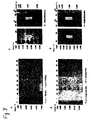
ベンゾイル、イソブチリルおよびイソプロピルフェノキシアセチルに対するモノクローナル抗体のELISAおよびドット−ブロットアッセイ
上記の実施例2に記載するように作製した、保護基ベンゾイル(Bz)、イソブチリル(ibu)およびイソプロピルフェノキシアセチル(ipr−Pac)に対するモノクローナル抗体(mAb)を、上記の実施例3に記載する、標準的なELISAアッセイおよびドット−ブロットアッセイによって特徴づけた。96−ウェルマイクロタイタープレートに各々結合した20残基のビオチン化核酸で展開したELISAアッセイは、それぞれの抗原に対する抗体の特異性を証明した。図3A、図4Aおよび図5Aは、それぞれ、Bz、ibuおよびipr−Pacに対するモノクローナル抗体の結果を示す。図は、オリゴdC(Bz)と名づけられる、すなわち、最初はBzで保護されていた、完全に脱保護されている(<1%Bz残存)dC残基のホモポリマー(レーン1、白ぬきの棒)、保護されている(>97%Bz残存)オリゴBz−dC(レーン2、影つきの棒)、完全に(<1%ipr−Pac残存)脱保護されているオリゴdG(ipr−Pac)(レーン3)、保護されている(>76%ipr−Pac)オリゴipr−PacdG(レーン4)、完全に(<1%ibu残存)脱保護されているオリゴdG(ibu)(レーン5)、保護されている(>91%ibu残存)オリゴibu−dG(レーン6)および完全に脱保護されているオリゴdT(レーン7)を示す。dTポリマーは、1つの保護基以外に、弱酸で5’−末端残基の5’OHから除去されるジメチルトリチル(DMT)を有した。最後にレーン8は、DMTが残存するオリゴdTを示す。
【0084】
20merのDNAsをニトロセルロース膜にUVで結合した、抗BzmAb、抗ibumAbおよび抗ipr−PacmAbのドット−ブロットアッセイを実施した。膜に適用した20merDNAの量を図3B、図4Bおよび図5Bの右に示し、アッセイ感度のレベルを証明する。抗BzmAbを試験するために使用したDNAsは、ELISAについて記載したものプラス脱保護されているオリゴdA(Bz)、保護されているオリゴBz−dA、オリゴdC(ibu)、オリゴibu−dC、オリゴdA(ibu)およびオリゴibu−dAであった。図3Bは、抗BzmAbはdAおよびdCの保護基を認識したことを示している。抗ibumAbを試験するために使用したDNAsは、ELISAについて記載したものプラス保護されているオリゴibu−dA、脱保護されているオリゴdA(ibu)、オリゴibu−dC、オリゴdC(ibu)であり、全てドット−ブロットの上部に記載されている。図4Bは、抗ibumAbは、保護基のうち最も一般的な用途の、dGのibuを認識したが、dAでも認識したことを示している。抗ipr−PacmAbを試験するために使用したDNAsは、ELISAについて記載したものプラス保護されているオリゴibu−dA、脱保護されているオリゴdA(ibu)、オリゴibu−dC(ibu)、オリゴBz dA(Bz)であり、全てドット−ブロットの上部に記載されている。図5Bは、抗ipr−PacmAbは、保護基のうち最も一般的な用途の、dGのipr−Pacを認識したが、dAおよびdCでも認識したことを示している。mAbはまたibu保護基を認識した(ibu−dG、ibu−dAおよびibu−dC)。この交差反応は、抗体は、ipr−Pacおよびibuに共通の化学、おそらくCH(CH3)2の識別に選択性が高いことを示している。従って、抗ibuおよび抗ipr−PacmAbsは、オリゴに残存する保護基を同定するために組み合わせて使用することができると思われる。
【0085】
抗ibumAbのドット−ブロットアッセイには大量のDNAを試験した(図4C)。この実験の結果は、ibu保護基は、どの核塩基が保護されているかにかかわらず、mAbによって認識されることを証明した。
【0086】
図3C、図4Cおよび図5Cは、部分的に脱保護されたオリゴマーを再処理して、残存する保護基を除去し、mAbで再試験することができることを証明している。図3Cは、抗BzmAbは再度脱保護されたオリゴマーオリゴBz−dCを認識したことを示している(中央のカラム)。同様に、図4Dは、抗ibumAbは再度脱保護されたオリゴマーオリゴibu−dG(中央のカラム)を認識することを示し、図5Cは、抗ipr−PacmAbは再度脱保護されたオリゴマーオリゴipr−Pac−dGおよびオリゴibu−dG(それぞれ、左から2番めおよび4番めのカラム)を認識することを示している。従って、この方法は、高価な核酸試料を破棄する必要なく、品質管理に適用可能である。
【0087】
保護基Bz、ibuおよびipr−Pacを有するRNA標品を合成し、Bz(図3D)、ibu(図4E)およびipr−Pac(図5D)に対するmAbを用いた保護基の同定についてアッセイした。ドット−ブロットアッセイは、モノクローナル抗体はRNAとDNAを識別しないことを明らかに示している。RNAにはDNAより高いバックグラウンドシグナルが存在したが、特に少量のRNAでは保護基のあるRNAと保護基のないRNAには有意な差があった。膜のRNAの量は試料の吸光度から推定した。
【0088】
[実施例9]
保護基のmAbドット−ブロットアッセイとHPLC
標準化した20merのオリゴdC分子に残存するBz基のドット−ブロット検出を実施例3に記載するように実施した。完全に脱保護されたdC20merと未処理のdC20merを、全く独立した異なる定量方法を使用して分析した。2つのオリゴマーを構成ヌクレオシドに加水分解し、濃縮した試料を用いた認識されている高速液体クロマトグラフィー(HPLC)を使用して同定し、定量した。感度が悪いので、HPLC検出は、mAbアッセイに使用されるBz−dCの量の50〜100倍が必要であった(図7参照)。図6Aは、保護されているオリゴBz−dCのnmole量のBz基に対して試験した抗BzmAbの結果(右のカラム)およびBz−dCの同じnmole量のBz−の結果(左のカラム)示す。各量のBz−dCオリゴを同じ鎖長(20mer)の完全に脱保護されているdCオリゴで希釈して、2500倍のdCが存在する場合でも(すなわち、0.04%)mAbの検出感度があることを証明した。mAbアッセイは、mAbは、2500倍過剰量のdCがDNA中に存在する場合でも、DNAのBz基を検出することができることを証明した。
【0089】
mAb応答を定量するために、図6Aに示すドット−ブロットに濃度測定を実施した。バックグラウンドを引いた後、HPLCで測定したオリゴBz−dCのBz基の関数として残存する密度をプロットした(図6B)。データは、抗BzmAb検出の高感度は0.1〜1.0nmolの範囲において直線状であることを示した。
【0090】
次に、mAb応答が、ドット−ブロット膜のDNA量の増加に伴って増加するかどうかを測定した。Bzの量は標準的なHPLC方法で測定した。保護されている試料と脱保護されている試料の1/2500の比の混合物中のBz保護基の検出は、膜のDNAの量を増加することによって増加するが、比は維持されることをこの実験は示した(図6C)。
【0091】
最後に、BzのmAb検出とHPLC検出の直接比較を示すために、実験を実施した。オリゴBz−dC(20mer)のdCのBzを検出するために抗BzmAbをドット−ブロットアッセイに使用した。mAbアッセイにより検出され、濃度測定によって定量されたBz基の密度応答を、各ドットのDNAのBzの量に対してプロットした(図7A)。DNA中のBzの量は、大量のDNAの消化並びにBz−dCモノヌクレオシドのHPLC同定および定量による分析によって較正した。HPLC実験では、3つのBz−dCオリゴ試料を加水分解し、HPLCによって組成を分析した。UV−ダイオードアレイディテクターの応答を試料中のBzの量に対してプロットした(図7B)。試料の量は、既知量のBz−dCで「ピークを生じた(spiked)」試料と比較することによって求めた。スパイク(spike)として試料に添加したBz−dCの量は、秤量したBz−dCストックによるものであった。従って、HPLC応答は、既知量のBz−dCを用いて較正した。これらの実験の結果は、抗BzmAbによるBzの検出はpmole範囲内であるが、BzのHPLC検出はnmole範囲に制限されることを示している。
【0092】
[実施例10]
市販の試料中の残存保護基の検出
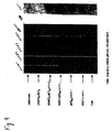
市販の試料中の残存保護基のmAbによる検出を証明するために盲検検討を実施した。この実験の目的は、オリゴ合成業界において通常実施されているように処理されたおそらく完全に脱保護されている試料においてmAb技術で保護基を検出および同定できるかどうかを判定することであった。選択した8社によって使用されている保護基の性質は未知であったので、実験は盲検試験であった。8社の各々製の2つの20merオリゴ(オリゴdA−dCおよびオリゴdG−dT)を合成して、脱保護するように注文し、できるだけ理想的な条件下で塩を除去した。オリゴは通常どおり速達で出荷され、ドット−ブロットによるmAb分析を実施した。1社(#6)とおそらくもう1社(#2)製のdA−dCオリゴは、抗BzmAb試験によって測定したとき、Bz保護基が残存していた(図8A)。2社(#2および#6)製のdG−dTオリゴは、抗ipr−PacmAbで測定したとき、ipr−Pac保護基が残存していた(図8B)。市販の試料中の残存保護基は、試料の量を増加し、さらに脱保護して再分析することによって確認した。#2社および#6社製のオリゴdA−dC試料は、Bz保護基の存在を確認するためにより大量で試験した。また、標準的なプロトコールを使用して、残存する保護基を除去するために試料を処理した。さらに脱保護した後の再分析は、基は今度は除去されたことを示した(図8C)。これは、高価な核酸試料を再処理して保護基を除去すると、それらを破棄する必要がないことを証明している。オリゴdG−dT試料を再処理して、残存する保護基を除去し、抗ipr−PacmAbで再分析すると、ipr−Pac基はDNAを犠牲にすることなく除去されうるという結果が得られた(図8D)。
【0093】
ジメトリチルに対するポリクローナル抗体
5’末端保護基、ジメトリチル(DMT)に対するポリクローナル抗体の作製および分析は実施例2に記載されているとおりであった。4匹のマウスにDMTを接種し、数週間抗原で追加免疫投与してから、血清をマウスから採取した。DMT[DMT−OH]、デオキシヌクレオチドトリマーd(T)3[(DMT)3−d(T)3]の5’−末端の3つのDMT、3’−ビオチンを有するデオキシヌクレオチド20mer[(DMT)3−d(T)20−ビオチン]の5’−末端の3つのDMT、3’−ビオチンを有するデオキシヌクレオチド20mer[DMT−d(T)20−ビオチン]の5’−末端の1つのDMT、3’−ビオチンを有するdT20mer[d(T)20−ビオチン],ビオチンを有するDMT[DMT−ビオチン]の1つのDMTおよびトリス−ボレート生理食塩液対照をニトロセルロース膜に適用し、次いで抗DMT抗体を評価するためのマウス血清(接種マウス#1〜4および対照血清、正常)、DMTの存在をあきらかにするための弱酸(TBS)およびビオチンの存在を明らかにするためのアビジンを用いてアッセイした(図9)。マウス#2および#4の血清はDMT[(DMT)3−d(T)3として]を認識したが、#1、#3および正常なマウスは認識しなかった。弱酸は黄色としてDMTの存在を明らかにし(図には示していない)、アビジンはビオチンの存在を明らかにした。
【0094】
上記は本発明を例示するものであり、本発明を限定するものと解釈されるべきではない。本発明は、以下の請求の範囲およびそこに含まれる請求の範囲の等価物によって規定される。
【図面の簡単な説明】
【図1】
オリゴIbu−dG20mersに選択的に結合する、モノクローナル抗体1H11のドット−ブロットイムノアッセイである。
【図2】
オリゴBz−dC20mersに選択的に結合する、モノクローナル抗体7H3のドット−ブロットイムノアッセイである。
【図3】
核酸の化学合成に通常使用される保護基、ベンゾイル(Bz)のモノクローナル抗体(mAb)の特異性および検出感度を証明するELISA(A)およびドット−ブロット(B)を示す。部分的に脱保護されたオリゴマーオリゴBz−dC(中央のカラム)を再処理して、残存する保護基を除去し、mAbで再試験することができる(C)。保護基Bz、ibuおよびipr−Pacを有するRNA標品を合成し、Bzに対するmAbによる保護基の同定をアッセイした(D)。
【図4】
核酸の化学合成に通常使用される保護基、イソブトリル(ibu)のモノクローナル抗体(mAb)の特異性および感度並びにその検出を証明するELISA(A)およびドット−ブロット(B)結果を示す。大量のDNAを用いたドット−ブロットアッセイは、ibu保護基は、どの核酸塩基が保護されているかにかかわらず、mAbによって認識されたことを証明している(C)。部分的に脱保護されたオリゴマーオリゴBz−dC(中央のカラム)を再処理して、残存する保護基を除去し、mAbで再試験することができる(D)。保護基Bz、ibuおよびipr−Pacを有するRNA標品を合成し、ibuに対するmAbによる保護基の同定をアッセイした(E)。
【図5】
核酸の化学合成に通常使用される保護基、イソプロピルフェノキシアセチル(ipr−Pac)のモノクローナル抗体(mAb)の特異性および感度並びにその検出を証明するELISA(A)およびドット−ブロット(B)結果を示す。部分的に脱保護されたオリゴマーオリゴipr−Pac−dGおよびオリゴibu−dG(それぞれ、左から2番目および4番目のカラム)を再処理して、残存する保護基を除去し、mAbで再試験することができる(C)。保護基Bz、ibuおよびipr−Pacを有するRNA標品を合成し、ipr−Pacに対するmAbによる保護基の同定をアッセイした(D)。
【図6】
HPLCに関連する技術の感度および定量応答を証明する、保護基のmAbドット−ブロットアッセイを示す。標準の20merオリゴdC分子に残存するBz基のドット−ブロット検出を実施し(A)、mAb応答の定量(B)を行った。mAb応答は、ドット−ブロット膜のDNA量を増加させて分析した(C)。左のカラムは保護されているBz−dC20merである。右のカラムは、保護されているBz−dCと2500倍過剰量の完全に脱保護されたオリゴdC(Bz)である。
【図7】
それぞれ、pmole(A)およびnmol範囲(B)のBzのmAbおよびHPLC検出の直接比較を示す。
【図8】
市販の試料中に残存する保護基の検出を証明する盲検試験を示す。dA−dCオリゴは抗BzmAb(A)で分析し、dG−dTオリゴは抗ipr−PacmAb(B)で分析した。#2社および#6社製のオリゴdA−dC試料は、Bz保護基の存在を確認するために大量で試験した(C)。また、標準的なプロトコールを使用して残存する保護基を除去するために試料を処理した。オリゴdG−dT試料はipr−Pac保護基についてアッセイした(D)。試料を再処理して、残存する保護基を除去し、(C)のように再分析した。
【図9】
5’末端保護基、ジメトリチル(DMT)に対するポリクローナル抗体の作製および分析を示す。
【図10】
配列は同じで類似したオリゴヌクレオチドを、保護または脱保護の程度の差についてスクリーニングする試験標準として使用することができる、配列が同じで、脱保護の程度が異なる種々のオリゴヌクレオチドを含有する基板を示す。
【図11】
本発明の抗体を用いて、保護基の存在または不十分な伸長をスクリーニングすることができるオリゴヌクレオチドアレイを例示する。[0001]
[Related application]
This application is a continuation-in-part of co-pending application Ser. No. 09 / 476,975, filed Dec. 31, 1999, filed on Dec. 31, 1999, the disclosure of which is incorporated herein by reference in its entirety. is there.
[0002]
[Technical field]
The present invention relates to the detection, identification and quantification of protecting groups remaining after chemical synthesis of oligomers, especially oligonucleotides.
[0003]
[Background Art]
In the last decade, automated chemical synthesis of nucleic acids such as DNA and RNA on solid supports has been developed. These chemical methods include the use of chemicals that protect the exocyclic amines of the nucleotide bases adenine, thymine, cytosine and guanine and are synthesized by blocking the 2'OH of RNA ribose. Bases in the synthetic nucleic acid product are deprotected upon cleavage of the nucleic acid from the solid support. However, the degree of base deprotection is not easily measured.
[0004]
For example, even after base deprotection of synthetic RNA, the product contains a 2'-dimethylsilyl tert-butyl group as a protection for the 2 'OH of the ribose moiety. This protecting group is carefully removed by chemical means so as not to affect the chemistry and structure of the RNA. However, the degree of deprotection of 2'OH is not easily measured. Nucleic acids are purified by high performance liquid chromatography or gel electrophoresis. However, some of the undesired products of the synthesis contain one or more protecting groups and, especially for oligomers longer than 50 nucleotides, complete sequences containing shorter (interrupted) sequences that are difficult to separate from full-length sequences. Nucleic acid sequence. Currently, there is no easy way to determine how much of each protecting group, if any, remains in the product and what percentage of the product is full length. See generally, Davis, G .; E. FIG. Gehrke, C .; W. Kuo, K .; C. , And @Agris, P .; F. (1979) Major and Modified, Nucleosides, intRNA, Hydrolysates, by, High, Performance, Liquid, Chromatography. J. Chromatogr. 173: 281-298; Agris, P .; F. , Thompson, J.M. G. FIG. Gehrke, C .; W. Kuo, K .; C. , And @ Rice, R.A. H. (1980) High-Performance Liquid, Chromatography and Mass, Spectrometry, of Transfer, RNA, Bases, Isotropic, Abundance. J. Chromatogr. 194: 205-212; Gehrke, C.E. W. Kuo, K .; C. McCune, R .; A. Gerhardt, K .; O. , {And} Agris, P .; F. (1981) Quantitative Enzymatic Hydrolysis of tRNAs: RP-HPLC of tRNA Nucleosides. J. Chromatogr. 230: 297-308; Chromatography and Modification of Nucleosides Volumes A, B and C (Gehrke, CW and Kuo, KCT, eds), Elsevier Publishing. 1990; Agris, P .; F. {And} Sierzputowska-Gracz, H .; (1990) Three Dimensional Dynamic Structure of tRNA's by Nuclear Magnetic Resonance. In Chromatography and Modification of Nucleosides (Gehrke, CW and Kuo, KCT, eds.), Elsevier Publishing Co. Pp. Agris, P. 225-253; F. Hayden, J. et al. , Sierzputowska-Gracz, H .; , Ditson, S .; , Degres, J .; A. , Tempesta, M .; Kuo, K .; C. And Gehrke and Gehrke, C.I. W. (1990) Compendium on Biological, Biochemical, Chemical, Physical and Spectroscopic Properties, of RNA, and DNA and Nucleosides. In Chromatography and Modification of Nucleosides, Elsevier Publishing Co. reference.
[0005]
Incomplete removal of protecting groups and the lack of a simple assay is a problem for the two industries and for many researchers worldwide. (I) Many companies that currently offer next-day nucleic acid sequence synthesis have difficulty communicating to customers the extent to which products have been deprotected; (ii) Pharmaceutical companies are trying to introduce or sell the treatment The purity and / or chain length of oligonucleotide or diagnostic oligonucleotide products cannot be easily demonstrated to regulatory agencies. Therefore, there is a need for a simple and reliable method for measuring the purity and proportion of full-length oligonucleotide products.
[0006]
[Disclosure of the Invention]
One embodiment of the present invention provides for specific binding to a synthetic oligomer having an organic protecting group covalently attached (i.e., an oligonucleotide or oligopeptide), and when the organic protecting group is not covalently attached, to the synthetic oligomer. An antibody that does not bind (eg, a monoclonal or polyclonal antibody).
[0007]
A second aspect of the invention includes cells, including cell cultures and isolated cells, that express the above-described antibodies. Such cells include hybridoma cells and recombinant cells that contain and express a heterologous nucleic acid that expresses the antibody.
[0008]
A third aspect of the present invention is a step of (a) contacting the above-mentioned antibody with a synthetic oligonucleotide, and (b) a step of detecting the presence or absence of a bond between the antibody and the oligomer, wherein the presence or absence of the bond is determined. Indicating the incomplete deprotection of the synthetic oligomer, wherein the incomplete deprotection of the synthetic oligonucleotide is detected by an immunoassay. Any suitable assay format can be used, including heterogeneous immunoassays and homogeneous immunoassays. For example, the immunoassay may be an immunoblot-dot assay or a sandwich assay.
[0009]
A fourth aspect of the invention is a method of separating a protected synthetic oligomer (including a partially and fully protected synthetic oligomer) from a fully deprotected synthetic oligomer. The method of the present invention comprises the step of (a) contacting a mixture of a protected synthetic oligomer and a completely deprotected synthetic oligomer with the above-mentioned antibody, wherein the protected synthetic oligomer shares an organic protecting group. Comprising binding the synthetic oligomer, which is bound, and thus protected, to the antibody, and separating the antibody from the completely deprotected oligomer. Antibodies may be immobilized on a solid support to facilitate separation. Protected synthetic oligomers are not partially protected synthetic oligomers (one application is the identification and / or purification of full-length and aborted sequence oligomers) or have not been deprotected. It may be a completely protected synthetic oligomer. Any separation mode can be used, including but not limited to affinity chromatography.
[0010]
A fifth aspect of the invention is an article useful for measuring incomplete deprotection of a synthetic oligomer in an immunoassay, comprising: (a) a surface portion having at least two separate discrete regions formed thereon. A solid support (eg, a nitrocellulose strip); (b) a first oligomer bound to one of said discrete regions and having a protecting group attached thereto; and (c) a first oligomer of said discrete regions. A second oligomer attached to another and not having the protecting group attached thereto, wherein the nucleotide sequences of the first and second oligomers are the same. In a preferred embodiment, the article further comprises (d) a third oligomer attached to another one of said separate discrete regions and said protecting group attached to said first oligomer. Wherein the third oligomer is partially deprotected and the first, second and third oligomers have the same nucleotide sequence.
[0011]
A sixth aspect of the present invention is a method for producing an antibody that specifically binds to a synthetic oligomer having an organic protective group covalently bonded thereto and does not bind to the synthetic oligomer when the organic protective group is not covalently bonded. (A) a step of synthesizing the synthetic oligomer to which the organic protective group is covalently bonded (preferably covalently bonded by a succinyl linker) on a solid particulate carrier (or a monomer of one nucleotide to which the protective group is covalently bonded) (B) synthesizing the solid oligomer (or the solid) in an amount sufficient to form the antibody without removing the oligomer from the solid support. Immunizing an animal with a carrier-coupled monomer). If desired, the solid carrier may be exchanged for a carrier group such as a protein (eg, bovine serum albumin).
[0012]
In summary, the antibodies and methods of the present invention are useful for immunoassays such as the qualitative and quantitative detection of protecting groups used in organic synthesis methods, and for the identification of oligonucleotides or peptides in research, therapy, diagnosis and biomedical sciences. There are applications. The antibodies of the present invention can be used for purification techniques such as separation of the end product from by-product contaminants. The present invention relates to quality control of gene therapy drugs, antisense, antigene and control of gene expression, quality control of biomedical polymers that may contain protecting groups, and purification and characterization of synthetic oligomers, especially oligonucleotides or peptides. It can be used in quality control processes for oligonucleotide and peptide synthesis, such as labeling.
[0013]
The present invention is described in more detail in the drawings herein and the specification set forth below.
[0014]
[Detailed description of preferred embodiments]
1. General definition
As used herein, “antibody” refers to both monoclonal and polyclonal antibodies, refers to any immunoglobulin-type antibody (including, but not limited to, IgG and IgM antibodies), and includes a hypervariable region. Alternatively, it contains an antibody fragment that retains the binding region. Antibodies can be of any origin, but are typically mammalian (eg, horse, rat, mouse, rabbit, goat). The antibody may be bound or immobilized to nitrocellulose, agarose, glass, organic polymers ("plastics"), etc. by known techniques, labeled with other detectable groups by known techniques, or It may be connected to a detectable group.
[0015]
"Binding" as used herein with respect to selective binding of an antibody to an oligomer has its ordinary meaning in the art. Generally, an antibody will have at least about K for identification in an immunoassay or affinity purification technique.d= 10-6, 10-7Or 10-8M should bind to the protected oligomer with an affinity of about Md= 10-2, 10-3Or 10-4It should bind to unprotected oligomers with an affinity not exceeding M.
[0016]
As used herein, "oligomer" refers to synthetic oligomers in their natural form, such as DNA and RNA, as well as synthetic oligonucleotides and oligopeptides, including the modified backbone compounds discussed below. Oligonucleotides are presently preferred in practicing the present invention, and the present invention has been described primarily with reference to the oligonucleotides herein. However, the methods and techniques described herein can also be applied to oligopeptides, oligosaccharides, and the like (ie, any synthetically produced polymer that requires a protecting group for its synthesis).
[0017]
As used herein, "nucleotide" refers to a pentose, a nitrogen-containing heterocyclic base (typically attached to
[0018]
As used herein, “protecting group” has its conventional meaning in the art and typically binds to an atom in a molecule, typically in an organic synthesis, prior to a chemical reaction involving the molecule. Specifically, it refers to a chemical moiety, group, or substituent that is covalently bonded so that no chemical reaction occurs at the atom to which the protecting group is bonded. Typically, the protecting groups are chemically removed from the intermediate molecule in order to produce the final product, but the removal technique results in a partial deprotection of the final product (ie, a residue on the molecule). At least one protecting group is present). Protecting groups may be intentionally left on the molecule for the purpose of making or testing the antibodies described above.
[0019]
As used herein, "deprotected" or "deprotected" refers to the absence of a protecting group used for chemical oligonucleotide synthesis from a molecule. Such protecting groups are described below. If the protecting group is at the end of the chain, the presence of such a protecting group can indicate insufficient extension of the oligonucleotide. Although chemically synthesized oligonucleotides are ideally fully deprotected, the present invention provides for partial or incomplete deprotection of such oligonucleotides (i.e., (The presence of at least one of the described protecting groups is present).
[0020]
"Base" as used herein with respect to oligonucleotides refers to a derivative of a purine (eg, adenine, guanine) or a pyrimidine (eg, uracil, thymine, cytosine) or a nitrogen-containing heterocyclic base. Pyrimidine bases bind to pentose by one ring nitrogen and purine bases bind to pentose by nine ring nitrogen. Preferred bases are those containing a free amino group, such as guanine, adenine and cytosine (the protecting group is then covalently attached to the free amino acid group by one or two substitutions of hydrogens of the free amino group). However, it can be used for any standard or modified / rare purine or pyrimidine base that contains free amino groups for protection or other groups that require protection during synthesis in the oligonucleotide. it can. Examples of standard and modified / rare bases are those found in the nucleosides listed in Table 1 below.
[0021]
[Table 1]
Applicants intend that the disclosures of all U.S. patent references cited herein are hereby incorporated by reference in their entirety.
[0023]
2. Protecting group
The particular protecting group will depend on the oligomer being synthesized and the manner in which the oligomer is synthesized.
[0024]
Suitable protecting groups for the synthesis of oligonucleotides may contain one or more heteroatoms such as N, O or S, and may be substituted or unsubstituted (eg, carbonyl) alkyl, aryl (Aryl), alkylaryl and arylalkyl groups. Examples of protecting groups include, but are not limited to, acetyl, isobutyryl, 2- (t-butyldiphenyl-silyloxymethyl) benzoyl, naphthaloyl, iso-butyryloxycarbonyl, levulinyl, fullinyl. Oleenylmethoxycarbonyl, 2-nitrothiophenyl, 2,2,2-trichloro-t-butoxycarbonyl, ethoxycarbonyl, benzyloxycarbonyl, p-nitrophenyl-ethyloxycarbonyl, N'N-dimethylformamidine, formyl, Benzoyl, toluyl, 2,4-6-trimethylbenzoyl, anisoyl, 2,4-dimethylphenyl, 2,4,6-trimethylphenyl, triphenylthiomethyl, pivoboroyloxymethyl, t-butoxycarbonyl, p-nitrophenyl Ethyl, methoxyethoxymethyl, butylthiocarbonyl, 2-methyl-pyridin-5-yl, 2-nitrothiophenyl, 2,4-dinitrothiophenyl, 2-nitro-4-methylthiophenyl, p-nitrophenylsulfonylethyl, 5-chloro-8-hydroxyquinoline, thiophenyl, β-cyanoethyl, phenylethyl, p-nitrophenylethyl, pyridylethyl, 2-N-methylimidazolylphenyl, methyl, allyl, trichloroethyl, dibenzoyl, p-nitro Phenylethoxycarbonyl, benzoyl and substituted derivatives thereof, 2 (acetoxymethyl) benzoyl, 4,4 ′, 4 ″ -tris (benzyloxy) trityl, 5-methylpyridino-2-yl, phenylthioethyl, diphenyl Phenylcarbamoyl, 3,4-dimethoxybenzyl, 3-chlorophenyl, 2-nitrophenyl, 9-phenylxanthen-9-yl, 9- (p-methoxyphenyl) xanthen-9-yl, 9- (p-octadecyloxyphenyl ) Xanthen-9-yl, "bridged" bis-dimethoxytrityl group, phthalanoyl, succinyl, benzenesulfonylethoxycarbonyl, 4,4 ', 4 "-tris (bevulinyloxy) tristrityl, p-phenylazophenyloxy Carbonyl, o-substituted benzoyl, 4,4 ', 4 "-tris (4,5-dichlorophalimidin trityl, levelinyl, alkyloxy and aryloxyacetyl, 1,3-benzodithi 2-yl, tetrahydrofuranyl, [2- (methylthio) phenyl] thiomethyl, 1- (2-chloroethyl (oxy) oxy) ethyl, 1-[(2-fluoro-phenyl) 4-methoxypiperidin-4- Yl, 4-methoxytetrahydropyran-4-yl, (1-methyl-1-methoxy) ethyl, tetrahydropyranyl, 3-methoxy-1,5-dicarbomethoxypentam-3-yl, 2- Nitrobenzyl, benzyl, 4-nitrophenylethyl-sulfonyl, t-butyldimethylsilyl, 4-methoxybenzyl, 3,4-dimethoxybenzyl, 9-p-methoxyphenylthioxanthen-9-yl, formula R1R2R3C- (where R1, R2And R3Are each independently selected from the group consisting of phenyl, p-monomethoxyphenyl, o-monomethoxyphenyl, biphenyl, p-fluorophenyl, p-chlorophenyl, p-methylphenyl, p-nitrophenyl, etc.) Compound.
[0025]
3. Oligonucleotide
Synthetic oligonucleotides containing protecting groups and which can be used to practice the invention include natural forms such as DNA and RNA, and phosphonates, phosphoramides, phosphonamides, phosphites, phosphinamides, and the like. And modified poly (phosphate derivatives) such as sulfones, sulfonates, sulfites, sulfonamides, poly (sulfur derivatives) such as sulfenamides, and the like. Although the antibodies of the invention can be characterized by selective binding to a particular "reagent" or "benchmark" oligonucleotide, the same antibody can be used for a variety of other oligonucleotides containing the same protecting group (e.g., more It is noted that they can also bind to long nucleotides) or other compounds.
[0026]
For example, the oligonucleotide to which the antibody selectively binds may consist of 3-20 nucleotides, one of said nucleotides having the formula (I):
Embedded image
R1Is a protecting group such as H or β-cyanoethyl, provided that when the protected base is not the 3 ′ terminal nucleotide of the oligonucleotide, R1Is a covalent bond to an adjacent nucleotide,
R2Is H or -OR3And
R3Is a protecting group such as H or tert-butyldimethylsilyl;
The base is a purine or pyrimidine base,
R4Is a protection selected from the group consisting of acetyl (Ac), benzoyl (Bz), dimethylformamidine (dmf), isobutyl (Ibu), phenoxyacetyl (Pac) and isopropyl-phenoxyacetyl (ipr-Pac). A protecting group bonded to the amino group of the base such as a group, provided that R, R1, R3And R4Is one of the protecting groups, R, R1, R3And R4Is a protected nucleotide).
[0027]
In one particular embodiment of the above, the antibody consists of 3-20 nucleotides and has the formula (I):
Embedded image
R1Is a covalent bond to an adjacent nucleotide,
R2Is -H or -OH;
The base may be a purine or pyrimidine base), which selectively binds to an oligonucleotide having a 5 'nucleotide that is a protected nucleotide.
[0028]
In another particular embodiment of the above, the antibody consists of 3-20 nucleotides and has formula (I):
Embedded image
R1Is a protecting group such as β-cyanoethyl,
R2Is -H or -OH;
The base may be a purine or pyrimidine base), which selectively binds to an oligonucleotide having a 3 'nucleotide that is a protected nucleotide.
[0029]
In another particular embodiment of the above, the antibody consists of 3-20 nucleotides, wherein one of said nucleotides has the formula (I):
Embedded image
R1Is a covalent bond to an adjacent nucleotide,
R2Is -OR3And
R3Is a protecting group such as tert-butyldimethylsilyl,
The base may be a purine or pyridine base), which selectively binds to an oligonucleotide that is a protected nucleotide.
[0030]
In yet another specific embodiment of the above, the antibody consists of 3-20 nucleotides, wherein one of said nucleotides has the formula (I):
Embedded image
R1Is a covalent bond to an adjacent nucleotide,
R2Is -H or -OH;
The base is a purine or pyridine base,
R4Is a protecting group attached to the amino group of the base, such as acetyl, benzoyl, dimethylformamidine, isobutyryl, phenoxyacetyl and isopropyl-phenoxyacetyl). It may be something.
[0031]
Accordingly, examples of protected bases that can be used in the structures shown above include:
Embedded image
[0032]
In one embodiment of the invention, the oligonucleotide is a peptide nucleic acid and the protecting groups are used for the synthesis of peptide nucleic acids, including but not limited to those described in U.S. Patent No. 6,133,444. Such a protecting group.
[0033]
In yet another particular embodiment of the above, the antibody consists of 3-20 nucleotides, one of which is assigned to U.S. Patent Nos. 5,744,101 and 5,489,678 (Affymax. ), But are not limited thereto, and may selectively bind to an oligonucleotide protected with a photosensitive protecting group.
[0034]
4. antibody
As described above, the present invention provides an antibody that specifically binds to a synthetic oligonucleotide to which an organic protective group is covalently bonded, and does not bind to the synthetic oligonucleotide when the organic protective group is not covalently bonded (for example, Monoclonal or polyclonal antibodies).
[0035]
Antibodies may be provided immobilized (or bound) to a solid support by known techniques, or may be provided in free, unbound form (eg, lyophilized, frozen, in an aqueous carrier, etc.). Whether an antibody is immobilized will depend on the particular immunoassay or affinity purification technique using the antibody, and will be determined by the known parameters of such technique. Similarly, typically depending on the immunoassay format using the antibody, a number of binding pairs such as enzymes (eg, horseradish peroxidase), biotin or avidin, radioactive groups or fluorescent groups such as green fluorescent proteins, etc. Any detectable group can be attached or conjugated to the antibody by known techniques.
[0036]
5. Immunoassay method
The present invention provides a method for detecting incomplete deprotection of synthetic oligonucleotides (including aborted sequences containing protecting groups) by immunoassay. Generally, such an immunoassay comprises the steps of (a) contacting a synthetic oligonucleotide with the antibody described above, and (b) detecting the presence or absence of binding between the antibody and the oligonucleotide, wherein the presence or absence of the binding is determined. Indicating incomplete deprotection of said synthetic oligonucleotide. Any suitable assay format can be used, including heterogeneous and homogeneous immunoassays. For example, the immunoassay may be an immunodot-blot assay or a sandwich assay. The oligonucleotides to be tested for deprotection may be in any suitable form, such as in a solution or immobilized on a solid support.
[0037]
In a preferred embodiment, the detection method compares the binding of the antibody to the test oligonucleotide with the binding of the antibody to a known set of oligonucleotides, such as a `` dipstick '', all immobilized on a common solid support. use. Articles useful in measuring incomplete deprotection of synthetic oligonucleotides in immunoassays, such as those illustrated in FIG. 10, include (a) a surface portion on which at least two separate discrete regions 26 and 27 are formed A solid support (eg, a nitrocellulose strip) 25 having: and (b) a first oligo that binds to one of said discrete regions and has a protecting group (eg, at least one protecting group) attached thereto. A nucleotide sequence of said first and second oligonucleotides comprising: a nucleotide and (c) a second oligonucleotide attached to another one of said separate discrete regions and not having said protecting group attached thereto. Are the same. In a preferred embodiment, the article of the present invention comprises: (d) a compound having the protective group attached to another one of the separate discrete regions 28 and attached to the first oligonucleotide. 3 oligonucleotides, wherein the third oligonucleotide is partially deprotected (ie, the number of protecting groups covalently linked to the first oligonucleotide and the second oligonucleotide The protective group is at least 1, 2, 3 or 4 or more more than the first oligonucleotide, and at least 10, 20 or more more than the first oligonucleotide). The nucleotide sequences of the second and third oligonucleotides are the same. Of course, if desired, the substrate at a separate, discrete location may contain more oligonucleotides with different numbers of protecting groups. The distinct region to which another oligonucleotide binds may be in any form, such as a dot.
[0038]
6. Affinity purification method
In addition to immunoassays, the present invention also provides for fully deprotected oligonucleotides, including partially deprotected oligonucleotides (including fully protected oligonucleotides) (eg, to remove protecting groups). An affinity purification technique that separates both oligonucleotides that have undergone the deprotection step and those that have not. Such an approach typically involves (a) contacting a mixture of a protected synthetic oligonucleotide and a fully deprotected synthetic oligonucleotide with the antibody described above, wherein the protected synthetic oligonucleotide is The oligonucleotide is covalently linked to an organic protecting group from which the antibody can be selected so that the protected synthetic oligonucleotide binds to the antibody, and then removes the antibody from the fully deprotected oligonucleotide. Separating. The protected synthetic oligonucleotide may be a partially protected synthetic oligonucleotide or a fully protected synthetic oligonucleotide that has not been deprotected. Any separation mode can be used, including but not limited to affinity chromatography.
[0039]
7. Production of antibodies
A method for preparing an antibody that specifically binds to a synthetic oligonucleotide having an organic protective group covalently bonded thereto and does not bind to the synthetic oligonucleotide when the organic protective group is not covalently bonded, comprises the steps of (a) Synthesizing a synthetic oligonucleotide having a covalently bound organic protecting group on a solid particulate support (preferably covalently linked, for example, with a succinyl linker), without removing the oligonucleotide or nucleotide from said solid support, (b ) Immunizing the animal with a sufficient amount of the synthetic oligonucleotide attached to a solid support to form an antibody. Also, one nucleotide to which an organic protecting group is attached may be attached to a solid particulate carrier and can be used as described herein above.
[0040]
The synthesis steps can be performed on solid supports by known techniques. The solid support may be in particulate form before synthesis, or may be fragmented into particles after synthesis. Generally, a solid support is a bead that can be entirely solid, porous, deformable, or rigid. Beads generally have a diameter of at least 10, 20 or 50-250, 500 or 2000 μm, and most typically 50-250 μm in diameter. Cellulose, porous glass, silica gel, polystyrene beads such as polystyrene beads cross-linked with divinylbenzene, graft copolymer beads such as polyethylene glycol / polystyrene, polyacrylamide beads, latex beads, dimethylacrylamide beads, cross-linked polystyrene connected to linear polystyrene Alternatively, any convenient composition can be used for the solid support, including composites such as glass beads coated with a hydrophobic polymer such as a fluorinated ethylene polymer. If separate discrete solid carriers, such as particles or beads, are used, they generally contain about 1-99 weight percent of the total reaction mixture.
[0041]
In a preferred embodiment, following the synthesis step and prior to the immunization step, a step of fragmenting (eg, by milling) the solid support is performed. Polyclonal antibodies may be recovered from the serum of the animal by known techniques, or spleen cells may be recovered from the animal, and multiple hybridoma cell lines are generated from the spleen cells, and the specific hybridoma cell line from which the antibody is generated is then generated. It can be isolated from multiple hybridoma cell lines.
[0042]
Particular protocols for generating antisera / polyclonal and monoclonal antibodies to protecting groups used for nucleic acids and other syntheses typically include the following steps. (A) making oligonucleotides and others with or without protecting groups, (b) immunizing animals with such preparations, (c) identifying those that exhibit antibodies to the protecting groups. (D) producing monoclonal antibodies by typical fusion methods; (e) producing scFabs, Fab fragments and whole antibody molecules by antibody manipulation, if necessary; and ( f) evaluating and characterizing the monoclonal antibody against the protecting group. Each of these steps is discussed in further detail below.
[0043]
Synthetic oligonucleotides containing protecting groups can be synthesized by various methods known to those skilled in the art. For example, protecting groups can be attached to individual nucleotides that are connected to a controlled glass (CPG) bead. Examples include:
CPG beads-dT (DMT group only).
[0044]
Alternatively, protecting groups can be attached to the oligonucleotide chains connected to the CPG beads. Examples include:
Pac-dA-Pac-dA-CPG beads with Bz-dC and Ibu-dG,
Ipr-Pac-dG-Ipr-Pac-dG-CPG beads by Bz-dC and Ibu-dG,
Ac-dC-Ac-dC-CPG beads with Bz-dC and Ibu-dG,
Dmf-G-dmf-G-CPG beads with Bz-dC and Ibu-dG, and
A mixture of the above four oligonucleotides.
[0045]
In yet another method, a protecting group may be attached to the partially deprotected oligonucleotide chain (deprotection techniques are described in detail below). Examples include:
Poly dT20mers (DMT group only),
Poly dT20mers (cyanoethyl groups only),
Poly Ibu-dG20mers (partially deprotected),
Poly Ipr-Pac-dG20mers (partially deprotected),
Poly Bz-dC20mers (partially deprotected),
PolyPac-dA20mers (partially deprotected), and
PolyAc-dC20mers (partially deprotected).
[0046]
Synthetic oligonucleotides produced as described herein can be partially deprotected as follows: (a) adding a 30% ammonium hydroxide solution to the synthetic polynucleotide followed by room temperature (5, 10 and 30 minutes), take (b) the ammonium solution of the treated oligomer, dilute 1: 1 with a 1: 4 ratio of ammonium to acetic acid and add to acetic acid pre-cooled to 4 ° C (C) maintaining the sample in an ice bath for 30 minutes, (d) drying the sample with Speed-Vac, (e) dissolving the dried pellet in water, and (f) separating the sample with a Sephadex G-25 column. Desalting, (g) drying the sample with Speed-Vac, (h) dissolving the desalted sample in water.
[0047]
Synthetic oligonucleotides produced as described herein can be completely deprotected by any suitable technique. One particular technique is as follows: (a) adding a 30% ammonium hydroxide solution to the synthetic oligonucleotide, then incubating at 65 ° C. for 6 hours, (b) drying the sample on a Speed-Vac, (C) Dissolve the dried pellets in water, (d) desalinate the sample with a Sephadex G-25 column, (e) dry the sample with Speed-Vac, (f) dissolve the desalted sample in water I do.
[0048]
The partially and fully deprotected oligonucleotides were used for further use or to demonstrate the procedure, gel electrophoresis, urea-acrylamide gel electrophoresis, 5 'end labeling with T4 polynucleotide kinase, HPLC analysis , Mass spectrometry, etc., but can be characterized by any suitable means.
[0049]
Parenteral injections of the oligonucleotides in a suitable carrier, such as a sterile saline solution, can be used to immunize a suitable animal with the above oligonucleotides. Injection may be by any suitable route, including, but not limited to, subcutaneous, intraperitoneal, intravenous, intraarterial, intramuscular, and the like. Suitable animals are typically mammals, including mice, rabbits, rats, and the like.
[0050]
In certain embodiments, to generate monoclonal antibodies, young female BALB / c mice are used, and the time course of antigenic substance injection is as follows:
First day First injection
Day 28 Second booster dose
4 days before fusion Last booster dose
If desired, additional injections may be used. The amount of antigen may be 50 μg or 100 μg of unprotected (for control antibody) or protected oligonucleotide per mouse per dose. Preferably, when beads or other solid carriers used as carriers for oligonucleotide synthesis are injected into animals, the beads or particles are suspended in water and then injected into mice. If a nucleotide solution is used, the solution is mixed with Freund's complete or incomplete adjuvant and injected into mice.
[0051]
Polyclonal antibodies can be recovered from animals immunized or inoculated as described above by known techniques, or by known techniques, spleen cells can be recovered from animals, a hybridoma cell line can be generated from spleen cells, hybridoma cells Strains can be screened for production of the desired antibody.
[0052]
Oligonucleotides with or without a biotin molecule at the 3 'or 5' end (for the ELISA assays described below) can be synthesized by standard techniques. Examples include:
Poly Ibu-dG20mers (with or without biotin)
Poly Ibu-dA20mers (with or without biotin)
Poly Ibu-dC20mers (with or without biotin)
Poly Ipr-Pac-dG20mers (with or without biotin)
Poly Bz-dC20mers (with or without biotin)
Poly Bz-dA20mers (with or without biotin)
Poly dT20mers (with or without biotin)
PolyPac-dA20mers (with or without biotin)
PolyAc-dC20mers (with or without biotin), and
Poly dmf-G20mers (with or without biotin).
[0053]
Antibodies produced as described above can be characterized by any suitable technique, including but not limited to Western blots and immunodot-blots, to determine their binding properties.
[0054]
In addition to using polyclonal and monoclonal antibodies, the present invention involves the production of antibodies with recombinant DNA, or "antibody engineering" techniques. For example, mRNA isolated from hybridoma cells can be used to construct a cDNA library, and sequences encoding whole antibodies or antibody fragments (eg, scFab or Fab fragments) can be isolated and placed in a suitable expression vector. Upon insertion, the expression vector can be inserted into a host cell that expresses the isolated cDNA encoding the antibody.
[0055]
Monoclonal Fab fragments can be produced in Escherichia coli by recombinant techniques known to those skilled in the art. For example, W.S. Huse, Science 246, 1275-81 (1989).
[0056]
8. Antibody screening
Screening of serum and hybridoma cell culture media for protecting group-specific antibodies can be performed as follows.
[0057]
A. serum
1. Pre-immune (pre-immune) serum is collected by standard means from mice that are to be inoculated with protecting groups (directly or via oligomers) attached to a solid support.
2. Serum is also collected after inoculation.
3. An ELISA assay is performed in which specific protecting groups remain on the biotinylated oligonucleotide bound to the microtiter plate. Other microtiter plate wells contain control oligomers without protecting groups or oligonucleotides with other protecting groups. The secondary antibody is a goat anti-mouse IgG to which phosphatase has been conjugated to visualize the antibody.
4. Mice with positive activity for specific protecting groups were boosted and sacrificed to generate hybridomas.
[0058]
B. Hybridoma cell culture medium
1. Approximately 100 cultures were made from each spleen hybrid cell product.
2. Cultures are grown in microtiter plate wells, 96 well plates.
3. Culture medium was removed from each well and used in an ELISA assay, as described above, where each of the -1000 microtiter plate wells contained protected oligonucleotide bound to the plate.
4. Cultures producing antibodies with positive activity were transferred to larger culture wells, 24-well microtiter plates.
5. The culture medium of the large culture is retested for activity against protecting groups and also assayed for specificity; ie, no protecting group and other protecting group controls.
6. Positive cultures were separated (diluted) and tested again, and the point where each final culture resulted in one cell; that is, again separated to monoculture. The medium of these final cultures is fully evaluated for specificity and affinity. Specificity and affinity are assessed using a dot-blot assay.
[0059]
C. Dot-blot assay instead of ELISA assay
1. Antibodies to some protecting groups are cumbersome to test in a microtiter plate well environment and need to be tested using a dot-blot assay. One example is the 5'-terminal protecting group, dimethyl-trityl (DMT).
2. Dot-blot assays on nitrocellulose membranes are performed as described elsewhere in applications for most purposes. However, this is not possible when assessing antibody production with only a small amount of medium available to 1000 microtiter well cultures. Therefore, a new and improved method was developed.
a) Dot binding of protected oligonucleotides to nitrocellulose using UV-crosslinking. In the case of DMT, the presence of 5'-DMT on the membrane is confirmed by treating the dots with a weak acid and the reversal turns yellow-orange. The presence of 3'-biotin can be confirmed by commercially available avidin staining.
b) Block the membrane (see dot-blot assay).
c) Carefully mark the dots of the dried membrane (pencils) and “cut out” from the membrane.
c) Add individual dots to cell culture media in individual microtiter plate wells and incubate.
d) Take individual dots, wash, perform secondary antibody, phosphatase reaction, and color using microtiter plate wells with appropriate reagents.
e) Positive dots are associated back to the original microtiter plate well culture from which a small amount of culture medium was obtained.
f) Further cultivation and separation are performed as described in B.
[0060]
9. Microarray testing
The invention can be used to test or screen oligonucleotides immobilized on a solid support such as a microarray for insufficient deprotection or extension of oligonucleotides synthesized on a solid support.
[0061]
The solid support used to practice the invention is typically a separate solid support. The separate solid supports may be physically separated from each other or may be separate areas of a surface portion of a single structure substrate. Such "chip" or "pin" solid carriers are known. For example, U.S. Pat. No. 5,143,854 to Pirrun, U.S. Pat. No. 5,288,514 to Ellman (pin-shaped carrier), and U.S. Pat. No. 5,510, to Fodor et al. See No. 270 (tip-type carrier). Other non-limiting examples of oligonucleotide arrays that can be used to practice the present invention, and methods of making them, are described in US Pat. Nos. 5,631,734 and 5,599,695. No. 5,593,839, No. 5,578,832, No. 5,510,270, No. 5,571,639, No. 6,056,926, No. 5, Nos. 445,934 and 5,703,223, but are not limited thereto. Such a device can be used as described herein to practice the invention.
[0062]
The solid support or substrate forming the array may include any suitable material, including silicon. Oligonucleotides may be polymerized or grown in situ from monomers (or individual nucleotides) in situ on the microarray (in which case the solid support cannot be passed through the analyzer, so the protecting group None of the currently available techniques for detecting oligonucleotides are useful for detecting incomplete deprotection or extension of oligonucleotides on an array), or separately polymerizing the oligonucleotides and then May be connected to an appropriate area. The array may include any number of different oligonucleotides in different discrete areas, for example, at least 1,000, at least 2,000, at least 10,000 or at least 20,000 in different discrete areas. Contains an array of 000 different oligonucleotides.
[0063]
Generally, a method of screening an oligonucleotide array for insufficient deprotection or insufficient extension of the oligonucleotide comprises the following steps.
(A) providing the above oligonucleotide array,
(B) the above antibody (that is, an antibody that specifically binds to a synthetic oligonucleotide having an organic protective group covalently bonded thereto and does not bind to the synthetic oligonucleotide when the organic protective group is not covalently bonded) Providing, preferably, the antibody specifically binds to an oligonucleotide having an organic protecting group, if a protecting group is used during the process of organic synthesis of the oligonucleotide carried by the array; Then
(C) contacting the oligonucleotide array with the antibody, thereby detecting the presence of insufficient deprotection or insufficient extension of the oligonucleotide, which may be qualitative or quantitative. Such detection may be performed by any suitable immunoassay technique described above.
[0064]
In the method of the present invention, steps (b)-(c) may be repeated at least once with a different antibody at each iteration, such that a plurality of different oligonucleotides in the array may be present. Protecting groups can be detected.
[0065]
Preferably, once insufficient deprotection (presence of a protecting group) of one or more (eg, a plurality) of oligonucleotides in separate discrete regions has been detected, the method of the invention comprises the steps of: The method further includes creating a record or evidence that records the presence of insufficient deprotection or insufficient extension of the oligonucleotide at the discrete discrete location (or multiple discrete discrete locations). The evidence may be qualitative or quantitative evidence of insufficient deprotection (including insufficient extension).
[0066]
The above method provides a correctable oligonucleotide array illustrated in FIG. The array is
(A) a
(B) a plurality of evidence associated with the array, wherein the evidence records the presence of insufficient deprotection or insufficient extension of a plurality of different oligonucleotides, wherein the different oligonucleotides are separated from the array; Including evidence that is located in a separate location. These evidences may be printed on the area of the
[0067]
Data devices connected to an array as described in U.S. Patent Nos. 5,925,562, 6,017,496, 5,751,629 and 5,741,462. Or the storage device can be implemented by known techniques, and such an apparatus can be used to implement the invention, as described therein.
[0068]
Array end-users
(A) providing the above substrate;
(B) providing at least one or more evidences associated with the array as described above;
(C) providing a test compound, wherein the test compound is a member of a test compound library or any suitable protein, peptide or oligonucleotide (eg, RNA such as DNA or mRNA). A step which may be a compound,
(D) detecting the binding of the test compound to at least one of the plurality of different oligonucleotides (eg, by contacting the test compound with an array);
(D) from (i) the detected binding, and (ii) the evidence recording the presence of insufficient deprotection or insufficient extension, the binding of the test compound to one or more oligonucleotides of the array. Using the above evidence to correct for insufficient deprotection or insufficient extension of the oligonucleotides of the array in a method comprising detecting and measuring the extent (including simply the presence or absence of binding). Can be. Thus, inadequate deprotection or inadequate extension of oligonucleotides at one or more positions in the array can be corrected during the measurement step. Such corrections are performed by any means, including ignoring certain discrete discrete regions of the array (eg, supporting other discrete discrete regions of the array containing the same oligonucleotide). be able to. In another example, if the recorded evidence does not allow control, if one or more positions contain insufficient deprotection or extension, thereby reducing binding to such positions The binding data derived from experiments with the array can be adjusted upwards for their location to show binding greater than that shown in Table 1. The detecting or measuring step is performed by any suitable means, such as forming a colored indication of the degree of coupling, forming a numerical indication of the degree of coupling, forming a graph or other symbolic indication of the degree of coupling, etc. can do. The degree of binding is the binding affinity, the amount of binding, or both the binding affinity and the amount of binding, but is typically an indication of the amount of test compound that binds to a specific, discrete area of the array. It may be a display of the combination.
[0069]
The present invention is described in further detail in the following examples, which are not intended to be limiting.
[0070]
[Example 1]
Oligonucleotide synthesis
Synthesis was performed on an ABI DNA / RNA Synthesizer, Model 394 (PE Biosystems, 850 Lincoln Center Drive, Foster City, CA 94404) according to the manufacturer's protocol. During the synthesis, a slightly modified 1 micromolar scale cycle was used (see manufacturer's instructions). The main starting materials (and suppliers / manufacturers are listed in parentheses) were as follows:
Activator (0.45 M tetrazole in acetonitrile), CAP A (acetic anhydride / tetrahydrofuran / 2,6 lutidine), CAP B (N-methylimidazole / tetrahydrofuran) and oxidizing agent (0.02 M iodine / pyridine / THF / H2O) (Prime @ Synthesis)
Pac-dA (5'-dimethoxytrityl-N-phenoxyacetyl-2'-deoxyadenosine, 3 '-[(2-cyanoethyl)-(N, N-diisopropyl)]-phosphoramidite (Glen @ Research)
Ipr-Pac-dG (5′-dimethoxytrityl-Np-isopropyl-phenoxyacetyl-2′-guanosine, 3 ′-[(2-cyanoethyl)-(N, N-diisopropyl)]-phosphoramidite (Glen Research)
Ac-dC (5'-dimethoxytrityl-acetyl-2'-deoxycytidine, 3 '-[(2-cyanoethyl)-(N, N-diisopropyl)]-phosphoramidite (Glen @ Research)
dmf-G (5'-dimethoxytrityl-dimethylformamidine-guanosine, 2'-O-TBDMS-3 '-[(2-cyanoethyl)-(N, N-diisopropyl)]-phosphoramidite (Glen @ Research)
Bz-dC-CPG beads (5'-dimethoxytrityl-N-benzoyl-2'-deoxycytidine, 3 '-[(2-cyanoethyl)-(N, N-diisopropyl)]-phosphoramidite-succinyl linker-beads (3000 Ang) (CPG Inc.)
Ibu-dG-CPG beads (5'-dimethoxytrityl-N-isobutyl-2'-deoxycytidine, 3 '-[(2-cyanoethyl)-(N, N-diisopropyl)]-phosphoramidite-succinyl linker-beads (3000 Ang) (CPG Inc.)
[0071]
The following compounds were synthesized. Compound bound to the beads shown below:
Pac-dA-Pac-dA-Bz-dC-succinyl linker-beads
Pac-dA-Pac-dA-Ibu-dG-succinyl linker-beads
Ipr-Pac-dG-Ipr-Pac-dG-Bz-dC-succinyl linker-beads
Ipr-Pac-dG-Ipr-Pac-dG-Ibu-dG-succinyl linker-beads
Ac-dC-Ac-dC-Bz-dC-succinyl linker-beads
Ac-dC-Ac-dC-Ibu-dG-succinyl linker-beads
dmf-G-dmf-G-Bz-dC-succinyl linker-beads
dmf-G-dmf-G-Ibu-dG-succinyl linker-beads.
[0072]
The above compounds were administered directly to the animals as immunogens without separating the oligonucleotide from the solid support to generate antibodies, as further described in Example 2 below.
[0073]
[Example 2]
Animal inoculation
Eight to 12 week old female BALB / c mice were purchased from Charles \ River, Raleigh, North \ Carolina, USA. Mice were housed in cages with filter caps.
[0074]
Once the synthesis of the oligonucleotide chain was carried out as described in Example 1, the beads were placed between the glass plates and the glass plates were pressed by hand to gently grind the beads with nucleotides.
[0075]
5 μM of each of the above eight oligonucleotides was mixed with 4 ml of PBS (150 mM sodium chloride added to 100 mM phosphate buffer, pH 7.2).
[0076]
The mixture was stirred well to suspend the ground beads. 150 μL of the stirred mixture was taken and added to 300 μL of PBS in a syringe. Immediately prior to injection, the solution containing the beads was mixed again by shaking the syringe to suspend the broken beads. The mice were then injected intraperitoneally with 150 μL or 300 μL of the well mixed solution. This procedure was used for the first injection and the following booster challenge.
Injection time schedule
Injection days (days)
First time $ 0
2nd 14th day
3rd @ 28th day
4th @ 42nd day
5th @ 56th day
6th @ 70th day
7th @ 84th day
8th @ 98th day
9th @ 112th day
10th @ 138th
11th time (4 days before fusion) @ 142nd day
[0077]
Four days after the last injection, spleen cells are harvested from the animal and fused with myeloma cells (P3x.63.Ag8.653) by known techniques to create a hybridoma cell line, and then the specific cell line is isolated. In order to generate the desired antibodies of the invention, they were screened to determine if they had the characteristics described below.
[0078]
[Example 3]
Immunodot-blot assay for antibody characterization
The immunodot-blot assay involves the UV crosslinking of oligonucleotides to membrane paper and can be applied directly to test kits for detecting, identifying and quantifying protecting groups on product oligomers. This procedure can be performed as follows: (a) wetting the membrane paper with TBS (10 mM @ Tris, pH 7.2; 150 mM @ NaCl); (b) oligonucleotides to be tested on the membrane paper under reduced pressure (C) UV crosslink nucleotides to membrane paper, (d) Membrane paper with 1% casein-TBST (TBS plus
[0079]
[Example 4]
Dot-blot assay of monoclonal antibody 1H11
Monoclonal antibody 1H11 made as described in Example 2 above was characterized by a dot-blot assay. The results are shown as a bar graph in FIG. In FIG. 1, lanes (or columns) 1 and 2 show NH at 65 ° C. for 6 hours and 4 ° C. for 15 minutes, respectively.4Shows oligo-Pac-dA20mers treated with OH.
[0080]
[Example 5]
Dot-blot assay of monoclonal antibody 7H3
Monoclonal antibody 7H3 made as described in Example 2 above was characterized by the dot-blot assay described in Example 3 above. The results are shown as a bar graph in FIG. In FIG. 1, lanes (or columns) 1 and 2 show NH at 65 ° C. for 6 hours and 4 ° C. for 15 minutes, respectively.4Shows oligo-Pac-dA20mers treated with OH.
[0081]
[Example 6]
Western blot assays to characterize antibodies
Western blot assays involve low voltage transfer of oligonucleotides from gel to membrane paper and UV crosslinking of oligonucleotides to membranes. This assay can be performed as follows: (a) 10 mM MgCl2Pour a 15% native gel containing (b) load the oligonucleotides (oligomers) into the wells of the gel, (c) run the gel at 200 volts in an ice bath, (d) run the gel in an ice bath. Transfer oligonucleotide from gel to membrane paper with volts for 25 minutes, (e) UV cross-link nucleotides to membrane, (f) Block membrane paper with 1% casein-TBST for 2 hours at room temperature or overnight at 4 ° C (G) wash the membrane three times for 15 minutes each with TBST, (h) incubate the sample to be tested (diluted with 1% casein-TBST) for 1 hour at room temperature, (i) wash as above (J) incubating the membrane with a second antibody conjugate (diluted with 1% casein-TBST) for 1 hour at room temperature, (k) washing as above, (l) To form a color by incubation with the substrate solution.
[0082]
[Example 7]
Detection of Antibodies Using Biotinylated Polynucleotides as Antigen and ELI Related to Streptavidin-Biotin SystemSA
An enzyme-linked immunosorbent assay (ELISA) for detecting antibodies is performed as follows: (a) pre-screening microtiter plates pre-coated with streptavidin, (b) biotinylated oligonucleotides to be tested Alternatively, coat the plate with a preparation of another substance (5 μg / ml in PBS solution) (PBS: 15 mM NaCl, 10 mM phosphate buffer, pH 7.4), then incubate for 2 hours at room temperature, (c) 0 Wash three times with 1% Tween in PBS (PBST) for 15 minutes each, (d) Block with 1% casein in PBST for 1 hour at room temperature or overnight at 4 ° C., (e) Wash as above (F) Incubate the plate with the antibody for 1 hour at room temperature. (G) washing as above, (f) reacting with a second antibody-peroxidase conjugate (1% casein-PBST solution) for 1 hour at room temperature, i) Wash as above, (j) Tetramethylbenzidine (TMB) solution (TMB solution: 42 mM @ TMB, 0.004% H)2O2, 0.1 M acetate buffer, pH 5.6), incubate for 15 minutes at room temperature, and then add 2 MH2SO4The color reaction is formed by stopping the reaction with (k) Read absorbance value at 469 nm.
[0083]
Example 8
ELISA and dot-blot assays of monoclonal antibodies to benzoyl, isobutyryl and isopropylphenoxyacetyl
Monoclonal antibodies (mAbs) against the protecting groups benzoyl (Bz), isobutyryl (ibu) and isopropylphenoxyacetyl (ipr-Pac), prepared as described in Example 2 above, are described in Example 3 above. Characterized by standard ELISA and dot-blot assays. An ELISA assay developed with 20 residues of biotinylated nucleic acid each bound to a 96-well microtiter plate demonstrated the specificity of the antibody for each antigen. FIGS. 3A, 4A and 5A show the results of monoclonal antibodies to Bz, ibu and ipr-Pac, respectively. The figure shows a homopolymer of fully deprotected (<1% Bz remaining) dC residues, named oligo dC (Bz), ie, originally protected with Bz (
[0084]
Anti-BzmAb, anti-ibumAb and anti-ipr-PacmAb dot-blot assays were performed with 20-mer DNAs bound to nitrocellulose membranes by UV. The amount of 20mer DNA applied to the membrane is shown on the right of FIGS. 3B, 4B and 5B, demonstrating the level of assay sensitivity. The DNAs used to test anti-BzmAbs were those described for ELISA plus deprotected oligo dA (Bz), protected oligo Bz-dA, oligo dC (ibu), oligo ibu-dC, oligo dA (ibu) and oligo ibu-dA. FIG. 3B shows that the anti-BzmAb recognized dA and dC protecting groups. DNAs used to test anti-ibuAbs were those described for ELISA plus protected oligo ibu-dA, deprotected oligo dA (ibu), oligo ibu-dC, oligo dC (ibu). , All at the top of the dot-blot. FIG. 4B shows that anti-ibumAb recognized the most common use of protecting groups, dG ibu, but also dA. DNAs used to test anti-ipr-PacmAbs were those described for ELISA plus protected oligo ibu-dA, deprotected oligo dA (ibu), oligo ibu-dC (ibu), oligo Bz dA (Bz), all listed at the top of the dot-blot. FIG. 5B shows that the anti-ipr-PacmAb recognized the most common use of protecting groups, ipR-Pac of dG, but also dA and dC. The mAb also recognized the ibu protecting group (ibu-dG, ibu-dA and ibu-dC). This cross-reactivity indicates that the antibody has a common chemistry to ipr-Pac and ibu, probably CH (CH3)2It shows that there is high selectivity for discrimination. Thus, anti-ibu and anti-ipr-PacmAbs could be used in combination to identify residual protecting groups on the oligo.
[0085]
Large amounts of DNA were tested for dot-blot assays for anti-ibumAbs (FIG. 4C). The results of this experiment demonstrated that the ibu protecting group was recognized by the mAb, regardless of which nucleobase was protected.
[0086]
FIGS. 3C, 4C and 5C demonstrate that partially deprotected oligomers can be reprocessed to remove residual protecting groups and retested with mAbs. FIG. 3C shows that the anti-BzmAb recognized the oligomer oligo Bz-dC that was again deprotected (middle column). Similarly, FIG. 4D shows that the anti-ibumAb recognizes the re-deprotected oligomeric oligo ibu-dG (center column), and FIG. 5C shows that the anti-ipr-PacmAb re-deprotected the oligomeric oligo ipr-dG. It shows that Pac-dG and oligo ibu-dG (the second and fourth columns from the left, respectively) are recognized. Therefore, this method is applicable to quality control without having to discard expensive nucleic acid samples.
[0087]
RNA preparations with protecting groups Bz, ibu and ipr-Pac were synthesized and assayed for the identification of protecting groups using mAbs for Bz (FIG. 3D), ibu (FIG. 4E) and ipr-Pac (FIG. 5D). The dot-blot assay clearly shows that the monoclonal antibody does not distinguish between RNA and DNA. RNA had a higher background signal than DNA, but with small amounts of RNA there was a significant difference between protected and unprotected RNA. The amount of RNA on the membrane was estimated from the absorbance of the sample.
[0088]
[Example 9]
MAb dot-blot assay for protecting groups and HPLC
Dot-blot detection of Bz groups remaining on the standardized 20-mer oligo dC molecule was performed as described in Example 3. Completely deprotected and untreated dC20mer were analyzed using completely independent and different quantification methods. The two oligomers were hydrolyzed to constituent nucleosides and identified and quantified using recognized high performance liquid chromatography (HPLC) using concentrated samples. Due to poor sensitivity, HPLC detection required 50-100 times the amount of Bz-dC used in the mAb assay (see FIG. 7). FIG. 6A shows the results of the anti-BzmAb tested against the nmole amount of Bz group of protected oligo Bz-dC (right column) and the same nmole amount of Bz-dC for Bz-dC (left column). Show. Each amount of Bz-dC oligo was diluted with a fully deprotected dC oligo of the same chain length (20 mer) to detect the mAb in the presence of 2500-fold dC (ie 0.04%). Prove that there is. The mAb assay demonstrated that the mAb was able to detect the Bz group of DNA even when a 2500-fold excess of dC was present in the DNA.
[0089]
Densitometry was performed on the dot-blot shown in FIG. 6A to quantify the mAb response. After subtracting the background, the density remaining as a function of the Bz groups of the oligo Bz-dC as determined by HPLC was plotted (FIG. 6B). The data showed that the high sensitivity of anti-BzmAb detection was linear in the range of 0.1-1.0 nmol.
[0090]
Next, it was determined whether the mAb response increased with increasing DNA content of the dot-blot membrane. The amount of Bz was measured by a standard HPLC method. Detection of the Bz protecting group in a 1/2500 ratio mixture of protected and deprotected samples increases with increasing amounts of DNA in the membrane, but the ratio is maintained. This experiment was shown (FIG. 6C).
[0091]
Finally, experiments were performed to show a direct comparison of Bz mAb detection with HPLC detection. Anti-BzmAb was used in dot-blot assays to detect dC Bz of oligo Bz-dC (20mer). The density response of the Bz groups detected by the mAb assay and quantified by densitometry was plotted against the amount of Bz in the DNA of each dot (FIG. 7A). The amount of Bz in the DNA was calibrated by digestion of large amounts of DNA and analysis by HPLC identification and quantification of Bz-dC mononucleoside. In HPLC experiments, three Bz-dC oligo samples were hydrolyzed and analyzed for composition by HPLC. The response of the UV-diode array detector was plotted against the amount of Bz in the sample (FIG. 7B). The amount of the sample was determined by comparing to a "spiked" sample with a known amount of Bz-dC. The amount of Bz-dC added to the sample as a spike was due to the weighed Bz-dC stock. Therefore, the HPLC response was calibrated using known amounts of Bz-dC. The results of these experiments indicate that the detection of Bz with the anti-BzmAb is in the pmole range, but the HPLC detection of Bz is restricted to the nmole range.
[0092]
[Example 10]
Detection of residual protecting groups in commercially available samples
A blind study was performed to demonstrate the mAb detection of residual protecting groups in commercial samples. The purpose of this experiment was to determine if mAb technology could detect and identify protecting groups in a possibly completely deprotected sample that had been processed as commonly practiced in the oligo synthesis industry. The experiment was a blinded test because the nature of the protecting groups used by the eight companies selected was unknown. Two 20-mer oligos (oligo dA-dC and oligo dG-dT) from each of the eight companies were synthesized, ordered to be deprotected, and salt removed under ideal conditions as much as possible. Oligos were shipped by express as usual and mAb analysis by dot-blot was performed. The dA-dC oligos from one (# 6) and possibly another (# 2) had Bz protecting groups remaining as measured by anti-BzmAb test (FIG. 8A). The dG-dT oligos from two companies (# 2 and # 6) had the ipr-Pac protecting group remaining as measured by anti-ipr-PacmAb (FIG. 8B). Residual protecting groups in commercial samples were confirmed by increasing the amount of sample, further deprotecting and reanalyzing. Oligo dA-dC samples from companies # 2 and # 6 were tested in larger quantities to confirm the presence of the Bz protecting group. Samples were also processed to remove residual protecting groups using standard protocols. Reanalysis after further deprotection showed that the group was now removed (FIG. 8C). This demonstrates that once expensive nucleic acid samples are reprocessed to remove protecting groups, they do not need to be discarded. Reprocessing of the oligo dG-dT sample to remove residual protecting groups and re-analysis with anti-ipr-PacmAb gave the result that the ipr-Pac group could be removed without sacrificing DNA ( (FIG. 8D).
[0093]
Polyclonal antibodies against dimetrityl
Preparation and analysis of polyclonal antibodies to the 5 'terminal protecting group, dimetrityl (DMT), was as described in Example 2. Four mice were inoculated with DMT and boosted with antigen for several weeks before serum was collected from the mice. DMT [DMT-OH], deoxynucleotide trimer d (T)3[(DMT)3−d (T)3], A deoxynucleotide 20mer with three DMTs at the 5'-end and 3'-biotin [(DMT)3−d (T)20-Biotin] 20mer [DMT-d (T)] deoxynucleotide with three DMTs at the 5'-end of 3'-biotin20-DOT20mer [d (T) with one DMT at the 5'-end of 3'-biotin20-Biotin], DMT with biotin [DMT-biotin], one DMT and a tris-borate saline control were applied to the nitrocellulose membrane, and then mouse serum (inoculated mice # 1- # 1) to evaluate
[0094]
The above is illustrative of the present invention and should not be construed as limiting the invention. The invention is defined by the following claims, with equivalents of the claims to be included therein.
[Brief description of the drawings]
FIG.
Figure 4 is a dot-blot immunoassay for monoclonal antibody 1H11 that selectively binds to oligo Ibu-dG20mers.
FIG. 2
FIG. 4 is a dot-blot immunoassay for monoclonal antibody 7H3, which selectively binds to oligo Bz-dC20mers.
FIG. 3
Shown are ELISAs (A) and dot-blot (B) demonstrating the specificity and sensitivity of the monoclonal antibody (mAb) of benzoyl (Bz), a protecting group commonly used for chemical synthesis of nucleic acids. The partially deprotected oligomeric oligo Bz-dC (middle column) can be reprocessed to remove residual protecting groups and retested with the mAb (C). RNA preparations with protecting groups Bz, ibu and ipr-Pac were synthesized and assayed for mAb protecting group identification for Bz (D).
FIG. 4
Figure 3 shows the ELISA and dot-blot (B) results demonstrating the specificity and sensitivity of isobutryl (ibu) monoclonal antibody (mAb) and its detection, a protecting group commonly used in the chemical synthesis of nucleic acids. Dot-blot assays using large amounts of DNA demonstrate that the ibu protecting group was recognized by the mAb, regardless of which nucleobase was protected (C). The partially deprotected oligomeric oligo Bz-dC (middle column) can be reprocessed to remove residual protecting groups and retested with the mAb (D). RNA preparations containing the protecting groups Bz, ibu and ipr-Pac were synthesized and assayed for the identification of the protecting group by mAb on ibu (E).
FIG. 5
ELISA (A) and dot-blot (B) results demonstrating the specificity and sensitivity of a monoclonal antibody (mAb) of isopropylphenoxyacetyl (ipr-Pac), a protecting group commonly used in the chemical synthesis of nucleic acids, and its detection. Show. The partially deprotected oligomeric oligos ipr-Pac-dG and oligo ibu-dG (second and fourth columns from the left, respectively) were re-treated to remove residual protecting groups and retested with mAbs (C). RNA preparations with protecting groups Bz, ibu and ipr-Pac were synthesized and assayed for mAb protecting group identification for ipr-Pac (D).
FIG. 6
FIG. 4 shows a mAb dot-blot assay for protecting groups, demonstrating the sensitivity and quantitative response of the technique associated with HPLC. Dot-blot detection of the Bz group remaining in the standard 20mer oligo dC molecule was performed (A) and the mAb response was quantified (B). mAb responses were analyzed by increasing the amount of DNA in the dot-blot membrane (C). The left column is the protected Bz-dC20mer. The right column is the protected Bz-dC and a 2500-fold excess of fully deprotected oligo dC (Bz).
FIG. 7
Shown is a direct comparison of Bz mAb and HPLC detection in the pmole (A) and nmol range (B), respectively.
FIG. 8
Figure 3 shows a blind test demonstrating the detection of residual protecting groups in a commercial sample. dA-dC oligos were analyzed with anti-BzmAb (A) and dG-dT oligos were analyzed with anti-ipr-PacmAb (B). Oligo dA-dC samples from companies # 2 and # 6 were tested in large quantities to confirm the presence of the Bz protecting group (C). Samples were also processed to remove residual protecting groups using standard protocols. Oligo dG-dT samples were assayed for ipr-Pac protecting groups (D). The sample was reprocessed to remove any remaining protecting groups and re-analyzed as in (C).
FIG. 9
2 shows the generation and analysis of a polyclonal antibody against the 5 'terminal protecting group, dimetrityl (DMT).
FIG. 10
Oligonucleotides having the same sequence and similar can be used as test standards to screen for differences in the degree of protection or deprotection. Show.
FIG. 11
An oligonucleotide array that can be screened for the presence or insufficient extension of a protecting group using the antibody of the present invention is illustrated.
Claims (55)
R1はHまたは保護基であるが、ただし、前記保護されている塩基が前記ヌクレオチドの3’末端ヌクレオチドでない場合には、R1は隣接ヌクレオチドとの共有結合であり、
R2はHまたは−OR3であり、
R3はHまたは保護基であり、
塩基はプリンまたはピリミジン塩基であり、
R4は前記塩基のアミノ基に結合した保護基であるが、
ただし、R、R1、R3およびR4の1つが保護基である場合には、R、R1、R3およびR4の残りは保護基でない)の保護されているヌクレオチドである、請求項1に記載の抗体。The oligonucleotide consists of 3-20 nucleotides, wherein one of the nucleotides is of the formula (I):
R 1 is H or a protecting group, provided that when the protected base is not the 3 ′ terminal nucleotide of the nucleotide, R 1 is a covalent bond with an adjacent nucleotide;
R 2 is H or -OR 3,
R 3 is H or a protecting group;
The base is a purine or pyrimidine base,
R 4 is a protecting group bonded to the amino group of the base,
Provided that one of R, R 1 , R 3 and R 4 is a protecting group, and the remainder of R, R 1 , R 3 and R 4 is not a protecting group). Item 7. The antibody according to Item 1.
R1は隣接ヌクレオチドとの共有結合であり、
R2は−Hまたは−OHであり、
塩基はプリンまたはピリミジン塩基である)の保護されているヌクレオチドである、請求項1に記載の抗体。The oligonucleotide comprises 3-20 nucleotides and has a 5 ′ nucleotide, wherein the 5 ′ nucleotide is of the formula (I):
R 1 is a covalent bond with an adjacent nucleotide;
R 2 is -H or -OH;
The antibody of claim 1, wherein the base is a purine or pyrimidine base).
R1は保護基であり、
R2は−Hまたは−OHであり、
塩基はプリンまたはピリミジン塩基である)の保護されているヌクレオチドである、請求項1に記載の抗体The oligonucleotide comprises 3-20 nucleotides and has a 3 ′ nucleotide, wherein the 3 ′ nucleotide has the formula (I):
R 1 is a protecting group,
R 2 is -H or -OH;
The antibody of claim 1, wherein the base is a purine or pyrimidine base).
R1は隣接ヌクレオチドとの共有結合であり、
R2は−OR3であり、
R3は保護基であり、
塩基はプリンまたはピリミジン塩基である)の保護されているヌクレオチドである、請求項1に記載の抗体The oligonucleotide consists of 3-20 nucleotides, wherein one of the nucleotides is of the formula (I):
R 1 is a covalent bond with an adjacent nucleotide;
R 2 is —OR 3 ,
R 3 is a protecting group;
The antibody of claim 1, wherein the base is a purine or pyrimidine base).
R1は隣接ヌクレオチドとの共有結合であり、
R2は−Hまたは−OHであり、
塩基はプリンまたはピリミジン塩基であり、
R4は前記塩基のアミノ基に結合した保護基である)の保護されているヌクレオチドである、請求項1に記載の抗体。The oligonucleotide consists of 3-20 nucleotides, wherein one of the nucleotides is of the formula (I):
R 1 is a covalent bond with an adjacent nucleotide;
R 2 is -H or -OH;
The base is a purine or pyrimidine base,
R 4 is a protecting group bonded to the amino group of the base).
前記抗体と前記オリゴヌクレオチドとの結合の有無を検出するステップであって、結合の存在が前記合成オリゴヌクレオチドの不完全な脱保護を示すステップと
を含む、イムノアッセイによって合成オリゴヌクレオチドの不完全な脱保護を検出する方法。Contacting the antibody of claim 1 with a synthetic oligonucleotide;
Detecting the presence or absence of a bond between the antibody and the oligonucleotide, wherein the presence of the bond indicates incomplete deprotection of the synthetic oligonucleotide. How to detect protection.
保護されている合成オリゴヌクレオチドと完全に脱保護された合成オリゴヌクレオチドの混合物を請求項1に記載の抗体に接触させるステップであって、前記保護されている合成オリゴヌクレオチドに有機保護基が共有結合されており、その結果前記保護されている合成オリゴヌクレオチドは前記抗体に結合するステップと、
前記完全に脱保護された合成オリゴヌクレオチドから前記抗体を分離するステップと
を含む方法。A method of separating a protected synthetic oligonucleotide from a completely deprotected synthetic oligonucleotide,
Contacting a mixture of the protected synthetic oligonucleotide and the completely deprotected synthetic oligonucleotide with the antibody of claim 1, wherein an organic protecting group is covalently attached to the protected synthetic oligonucleotide. Wherein the protected synthetic oligonucleotide binds to the antibody; and
Separating the antibody from the fully deprotected synthetic oligonucleotide.
少なくとも2つの離れた別個の領域が形成されている表面部分を有する固体担体と、
前記離れた別個の領域の1つに結合した、保護基が結合している第1のオリゴヌクレオチドと、
前記離れた別個の領域の残りの1つに結合した、前記保護基が結合していない第2のオリゴヌクレオチドと
を含み、前記第1および第2のオリゴヌクレオチドのヌクレオチド配列が同じである物品。Articles useful for measuring incomplete deprotection of synthetic oligonucleotides in immunoassays,
A solid support having a surface portion on which at least two separate discrete regions are formed;
A first oligonucleotide having a protecting group attached thereto, wherein the first oligonucleotide is attached to one of said separate discrete regions;
An article, wherein said second oligonucleotide is free of said protecting group, attached to the remaining one of said separate discrete regions, wherein said first and second oligonucleotides have the same nucleotide sequence.
前記第3のオリゴヌクレオチドが部分的に脱保護されており、
前記第1、第2および第3のオリゴヌクレオチドのヌクレオチド配列が同じである、請求項23に記載の物品。A third oligonucleotide attached to the remaining one of the separated distinct regions, wherein the protecting group is attached to the first oligonucleotide;
The third oligonucleotide is partially deprotected;
24. The article of claim 23, wherein said first, second and third oligonucleotides have the same nucleotide sequence.
前記有機保護基が共有結合した前記合成オリゴヌクレオチドを固体粒状担体で合成するステップ、または前記有機保護基が結合したヌクレオチドを前記固体担体で合成するステップと、
前記固体担体から前記オリゴヌクレオチドまたはヌクレオチドをはずすことなく、前記抗体を形成するのに十分な量の、前記固体担体に結合した前記合成オリゴマーまたはヌクレオチドにより動物を免疫するステップと
を含む方法。A method for producing an antibody in which an organic protective group specifically binds to a synthetic oligonucleotide having a covalent bond, and wherein the organic protective group is not covalently bonded, the antibody does not bind to the synthetic oligonucleotide. ,
Synthesizing the synthetic oligonucleotide having the organic protective group covalently bonded thereto on a solid particulate carrier, or synthesizing the nucleotide having the organic protective group bonded thereto on the solid support,
Immunizing an animal with an amount of said synthetic oligomer or nucleotide attached to said solid support sufficient to form said antibody without removing said oligonucleotide or nucleotide from said solid support.
前記脾臓細胞から複数のハイブリドーマ細胞系統を作製するステップと、次いで、
前記複数のハイブリドーマ細胞系統から、前記抗体を作製する特定のハイブリドーマ細胞系統を単離するステップと
をさらに含む請求項26に記載の方法。Recovering spleen cells from the animal; and
Generating a plurality of hybridoma cell lines from the spleen cells; and
Isolating, from said plurality of hybridoma cell lines, the particular hybridoma cell line that makes said antibody.
(a)複数の異なるオリゴヌクレオチドが固定された基板を含むオリゴヌクレオチドアレイを提供するステップであって、前記異なるオリゴヌクレオチドは、前記基板の異なる離れた別個の位置に固定されているステップと、
(b)有機保護基が共有結合した合成オリゴヌクレオチドに特異的に結合し、前記有機保護基が共有結合していない場合には、前記合成オリゴヌクレオチドに結合しない抗体を提供するステップと、
(c)前記オリゴヌクレオチドアレイに前記抗体を接触させ、それによって前記アレイの選択された別個の位置への前記抗体の結合の有無を検出するステップであって、前記アレイの離れた別個の位置への結合の存在がオリゴヌクレオチドの不十分な脱保護または不十分な伸長を示すステップと
を含む方法。A method of screening an oligonucleotide array for insufficient deprotection or insufficient extension of the oligonucleotide,
(A) providing an oligonucleotide array comprising a substrate having a plurality of different oligonucleotides immobilized thereon, wherein the different oligonucleotides are immobilized at different and discrete locations on the substrate;
(B) providing an antibody that specifically binds to the synthetic oligonucleotide to which the organic protecting group is covalently bonded and does not bind to the synthetic oligonucleotide when the organic protecting group is not covalently bonded;
(C) contacting the antibody with the oligonucleotide array, thereby detecting the presence or absence of binding of the antibody to a selected discrete location on the array, wherein the antibody is located at a discrete discrete location on the array. Indicating the insufficient deprotection or insufficient extension of the oligonucleotide.
(b)前記アレイに関連する複数の証拠であって、前記証拠は少なくとも1つのオリゴヌクレオチドの不十分な脱保護または不十分な伸長の存在を記録し、前記少なくとも1つのオリゴヌクレオチドの各々は、前記アレイの異なる離れた別個の位置に位置していることを特徴とする証拠と
を組み合わせて含む修正可能なオリゴヌクレオチドアレイ。(A) a substrate having a plurality of different oligonucleotides immobilized thereon, wherein the different oligonucleotides are immobilized at different and separate positions on the substrate;
(B) a plurality of evidence associated with the array, wherein the evidence records the presence of insufficient deprotection or insufficient extension of at least one oligonucleotide, wherein each of the at least one oligonucleotide is A modifiable oligonucleotide array comprising a combination of evidence characterized by being located at different and distinct locations on the array.
(b)前記アレイに関連する証拠を提供するステップであって、前記証拠が少なくとも1つのオリゴヌクレオチドの不十分な脱保護または不十分な伸長の存在を記録し、前記少なくとも1つのオリゴヌクレオチドが前記アレイの離れた別個の位置に位置しているステップと、
(c)試験化合物を提供するステップと、
(d)前記複数の異なるオリゴヌクレオチドの少なくとも1つへの前記試験化合物の結合を検出するステップと、次いで、
(e)(i)前記検出された結合と(ii)不十分な脱保護または不十分な伸長の存在を記録する前記証拠から、前記オリゴヌクレオチドへの前記試験化合物の結合の程度を測定するステップであって、それによって前記不十分な脱保護または不十分な伸長が前記測定ステップ中に補正されるステップと
を含む方法。A method for correcting insufficient deprotection or insufficient extension of oligonucleotides in the array using an oligonucleotide array, the method comprising: (a) providing a substrate having a plurality of different oligonucleotides immobilized thereon; Wherein said different oligonucleotides are immobilized at different, separate and distinct locations on said substrate;
(B) providing evidence associated with the array, wherein the evidence records the presence of insufficient deprotection or insufficient extension of at least one oligonucleotide, wherein the at least one oligonucleotide is Steps located at separate, discrete locations on the array;
(C) providing a test compound;
(D) detecting binding of the test compound to at least one of the plurality of different oligonucleotides;
(E) determining the extent of binding of the test compound to the oligonucleotide from the evidence recording the presence of (i) the detected binding and (ii) insufficient deprotection or insufficient extension. Whereby said insufficient deprotection or insufficient extension is corrected during said measuring step.
(a)複数の異なるオリゴヌクレオチドが固定されている基板を提供するステップであって、前記異なるオリゴヌクレオチドが前記基板の異なる離れた別個の位置に固定されているステップと、
(b)前記アレイに関連する証拠を提供するステップであって、前記証拠が少なくとも1つのオリゴヌクレオチドの不十分な脱保護または不十分な伸長の存在を記録し、前記少なくとも1つのオリゴヌクレオチドが前記アレイの離れた別個の位置に位置づけられているステップと、
(c)試験化合物を提供するステップと、
(d)前記試験化合物と前記アレイを接触させるステップと、
(e)不十分な脱保護を有する離れた別個の位置における前記少なくとも1つのオリゴヌクレオチドを分析から削除するステップであって、前記アレイへの前記試験化合物の結合が、分析から削除されていない離れた別個の位置上で残存するオリゴヌクレオチドにより検出されるステップと、
(f)前記アレイにおいて離れた別個の位置上で前記残存するオリゴヌクレオチドへの前記試験化合物の結合を検出するステップと
を含む方法。A method of using an oligonucleotide array while compensating for insufficient deprotection or insufficient extension of the oligonucleotides of said array,
(A) providing a substrate having a plurality of different oligonucleotides immobilized thereon, wherein the different oligonucleotides are immobilized at different, discrete and distinct locations on the substrate;
(B) providing evidence associated with the array, wherein the evidence records the presence of insufficient deprotection or insufficient extension of at least one oligonucleotide, wherein the at least one oligonucleotide is Steps located at discrete, discrete locations on the array;
(C) providing a test compound;
(D) contacting the test compound with the array;
(E) removing from the analysis the at least one oligonucleotide at a discrete location having insufficient deprotection, wherein the binding of the test compound to the array is not removed from the analysis. Detected by the oligonucleotide remaining on the separate location.
(F) detecting the binding of the test compound to the remaining oligonucleotide on a discrete location in the array.
Applications Claiming Priority (2)
| Application Number | Priority Date | Filing Date | Title |
|---|---|---|---|
| US47697599A | 1999-12-31 | 1999-12-31 | |
| PCT/US2000/035600 WO2001049745A1 (en) | 1999-12-31 | 2000-12-29 | Methods and compositions for determining the purity of chemically synthesized nucleic acids |
Related Child Applications (1)
| Application Number | Title | Priority Date | Filing Date |
|---|---|---|---|
| JP2011286484A Division JP5753775B2 (en) | 1999-12-31 | 2011-12-27 | Methods and compositions for measuring the purity of chemically synthesized nucleic acids |
Publications (1)
| Publication Number | Publication Date |
|---|---|
| JP2004500370A true JP2004500370A (en) | 2004-01-08 |
Family
ID=23893992
Family Applications (2)
| Application Number | Title | Priority Date | Filing Date |
|---|---|---|---|
| JP2001550285A Withdrawn JP2004500370A (en) | 1999-12-31 | 2000-12-29 | Methods and compositions for determining the purity of chemically synthesized nucleic acids |
| JP2011286484A Expired - Fee Related JP5753775B2 (en) | 1999-12-31 | 2011-12-27 | Methods and compositions for measuring the purity of chemically synthesized nucleic acids |
Family Applications After (1)
| Application Number | Title | Priority Date | Filing Date |
|---|---|---|---|
| JP2011286484A Expired - Fee Related JP5753775B2 (en) | 1999-12-31 | 2011-12-27 | Methods and compositions for measuring the purity of chemically synthesized nucleic acids |
Country Status (7)
| Country | Link |
|---|---|
| US (6) | US6929907B2 (en) |
| EP (2) | EP1944321A3 (en) |
| JP (2) | JP2004500370A (en) |
| AU (1) | AU781801B2 (en) |
| CA (1) | CA2402131C (en) |
| NZ (1) | NZ519909A (en) |
| WO (1) | WO2001049745A1 (en) |
Cited By (1)
| Publication number | Priority date | Publication date | Assignee | Title |
|---|---|---|---|---|
| JP2009162769A (en) * | 2001-06-29 | 2009-07-23 | Veri-Q Inc | Methods and compositions for determining purity of chemically synthesized nucleic acid and purifying chemically synthesized nucleic acid |
Families Citing this family (10)
| Publication number | Priority date | Publication date | Assignee | Title |
|---|---|---|---|---|
| US6929907B2 (en) | 1999-12-31 | 2005-08-16 | North Carolina State University | Methods and compositions for determining the purity of chemically synthesized nucleic acids |
| US20040235022A1 (en) * | 2003-03-19 | 2004-11-25 | Ralf Mauritz | Quality control method for manufacturing biopolymer arrays |
| EP2942401A1 (en) | 2014-05-09 | 2015-11-11 | Lifecodexx AG | Detection of DNA that originates from a specific cell-type |
| EP2942400A1 (en) | 2014-05-09 | 2015-11-11 | Lifecodexx AG | Multiplex detection of DNA that originates from a specific cell-type |
| AU2015257654A1 (en) | 2014-05-09 | 2016-11-10 | Lifecodexx Ag | Detection of DNA that originates from a specific cell-type and related methods |
| WO2015181367A1 (en) * | 2014-05-29 | 2015-12-03 | Ge Healthcare Bio-Sciences Corp | Improvements in and relating to cell culture microscopy slides |
| DK3168309T3 (en) | 2015-11-10 | 2020-06-22 | Eurofins Lifecodexx Gmbh | DETECTION OF Fetal Chromosomal Aneuploidies Using DNA Regions With Different Methylation Between Foster And The Pregnant Female |
| EP3377116A4 (en) | 2015-11-19 | 2019-07-10 | The Trustees of The University of Pennsylvania | Compositions and methods for correction of heritable ocular disease |
| EP3380462B1 (en) | 2015-11-25 | 2021-02-17 | Del Mar Pharmaceuticals (BC) Ltd. | Methods for analysis and resolution of preparations of dianhydrogalactitol and derivatives or analogs thereof |
| CN107167533A (en) * | 2017-06-09 | 2017-09-15 | 江苏泰洁检测技术有限公司 | Phenoxy acetic acid method for measurement of concentration in carboxylic acids in a kind of water |
Citations (3)
| Publication number | Priority date | Publication date | Assignee | Title |
|---|---|---|---|---|
| US5221736A (en) * | 1988-12-21 | 1993-06-22 | Bionebraska, Inc. | Sequential peptide and oligonucleotide syntheses using immunoaffinity techniques |
| WO1998003532A1 (en) * | 1996-07-24 | 1998-01-29 | Hanna Michelle M | Base-protected nucleotide analogs with protected thiol groups |
| JPH1180185A (en) * | 1997-09-05 | 1999-03-26 | Res Dev Corp Of Japan | Chemical synthesis of oligonucleotide |
Family Cites Families (28)
| Publication number | Priority date | Publication date | Assignee | Title |
|---|---|---|---|---|
| US4853219A (en) * | 1987-08-06 | 1989-08-01 | President And Fellows Of Harvard College | Antibodies to angiogenin: immunotherapeutic agents |
| US5744101A (en) | 1989-06-07 | 1998-04-28 | Affymax Technologies N.V. | Photolabile nucleoside protecting groups |
| US5143854A (en) | 1989-06-07 | 1992-09-01 | Affymax Technologies N.V. | Large scale photolithographic solid phase synthesis of polypeptides and receptor binding screening thereof |
| US5451463A (en) | 1989-08-28 | 1995-09-19 | Clontech Laboratories, Inc. | Non-nucleoside 1,3-diol reagents for labeling synthetic oligonucleotides |
| FR2695563B1 (en) | 1992-09-11 | 1994-12-02 | Pasteur Institut | Microparticles carrying antigens and their use for the induction of humoral or cellular responses. |
| US5288514A (en) | 1992-09-14 | 1994-02-22 | The Regents Of The University Of California | Solid phase and combinatorial synthesis of benzodiazepine compounds on a solid support |
| US5503805A (en) | 1993-11-02 | 1996-04-02 | Affymax Technologies N.V. | Apparatus and method for parallel coupling reactions |
| US6133444A (en) | 1993-12-22 | 2000-10-17 | Perseptive Biosystems, Inc. | Synthons for the synthesis and deprotection of peptide nucleic acids under mild conditions |
| US5578832A (en) | 1994-09-02 | 1996-11-26 | Affymetrix, Inc. | Method and apparatus for imaging a sample on a device |
| US5631734A (en) | 1994-02-10 | 1997-05-20 | Affymetrix, Inc. | Method and apparatus for detection of fluorescently labeled materials |
| US5843644A (en) | 1994-03-01 | 1998-12-01 | The United States Of America As Represented By The Secretary Of The Department Of Health And Human Services | Isolation of cellular material under microscopic visualization using an adhesive/extraction reagent tipped probe |
| US5571639A (en) | 1994-05-24 | 1996-11-05 | Affymax Technologies N.V. | Computer-aided engineering system for design of sequence arrays and lithographic masks |
| US5703223A (en) | 1994-09-02 | 1997-12-30 | Thomas Jefferson University | Solid phase synthesis of oligonucleotides with stereospecific substituted phosphonate linkages by pentavalent grignard coupling |
| US5670320A (en) * | 1994-11-14 | 1997-09-23 | Emory University | Detection of mitochondrial DNA mutation 14459 associated with dystonia and/or Leber's hereditary optic neuropathy |
| US5599695A (en) | 1995-02-27 | 1997-02-04 | Affymetrix, Inc. | Printing molecular library arrays using deprotection agents solely in the vapor phase |
| US5925562A (en) | 1995-04-25 | 1999-07-20 | Irori | Remotely programmable matrices with memories |
| US5741462A (en) | 1995-04-25 | 1998-04-21 | Irori | Remotely programmable matrices with memories |
| US5751629A (en) | 1995-04-25 | 1998-05-12 | Irori | Remotely programmable matrices with memories |
| US6017496A (en) | 1995-06-07 | 2000-01-25 | Irori | Matrices with memories and uses thereof |
| US5843655A (en) * | 1995-09-18 | 1998-12-01 | Affymetrix, Inc. | Methods for testing oligonucleotide arrays |
| US20020150921A1 (en) * | 1996-02-09 | 2002-10-17 | Francis Barany | Detection of nucleic acid sequence differences using the ligase detection reaction with addressable arrays |
| US6852487B1 (en) * | 1996-02-09 | 2005-02-08 | Cornell Research Foundation, Inc. | Detection of nucleic acid sequence differences using the ligase detection reaction with addressable arrays |
| EP1001020A4 (en) * | 1997-07-29 | 2005-03-02 | Sumitomo Electric Industries | Antihuman pre-b cell receptor antibodies |
| CA2330277A1 (en) * | 1998-04-27 | 1999-11-04 | Sidney Kimmel Cancer Center | Reduced complexity nucleic acid targets and methods of using same |
| US7027629B2 (en) * | 1999-11-05 | 2006-04-11 | Agilent Technologies, Inc. | Method of extracting locations of nucleic acid array features |
| US6929907B2 (en) * | 1999-12-31 | 2005-08-16 | North Carolina State University | Methods and compositions for determining the purity of chemically synthesized nucleic acids |
| EP1182602B1 (en) * | 2000-08-25 | 2007-04-25 | STMicroelectronics S.r.l. | A system for the automatic analysis of DNA microarray images |
| WO2002101353A2 (en) * | 2001-06-08 | 2002-12-19 | U.S. Genomics, Inc. | Methods and products for analyzing nucleic acids based on methylation status |
-
2000
- 2000-12-22 US US09/747,467 patent/US6929907B2/en not_active Expired - Lifetime
- 2000-12-29 EP EP08006268A patent/EP1944321A3/en not_active Withdrawn
- 2000-12-29 WO PCT/US2000/035600 patent/WO2001049745A1/en active IP Right Grant
- 2000-12-29 EP EP00990402A patent/EP1242459A4/en not_active Withdrawn
- 2000-12-29 AU AU27432/01A patent/AU781801B2/en not_active Ceased
- 2000-12-29 CA CA2402131A patent/CA2402131C/en not_active Expired - Fee Related
- 2000-12-29 JP JP2001550285A patent/JP2004500370A/en not_active Withdrawn
- 2000-12-29 NZ NZ519909A patent/NZ519909A/en not_active IP Right Cessation
-
2002
- 2002-07-08 US US10/190,795 patent/US7901892B2/en not_active Expired - Fee Related
-
2011
- 2011-01-28 US US13/016,409 patent/US8173377B2/en not_active Expired - Fee Related
- 2011-12-27 JP JP2011286484A patent/JP5753775B2/en not_active Expired - Fee Related
-
2012
- 2012-04-05 US US13/440,613 patent/US8323910B2/en not_active Expired - Fee Related
- 2012-11-06 US US13/670,102 patent/US8697355B2/en not_active Expired - Fee Related
-
2014
- 2014-02-27 US US14/192,387 patent/US9273149B2/en not_active Expired - Fee Related
Patent Citations (3)
| Publication number | Priority date | Publication date | Assignee | Title |
|---|---|---|---|---|
| US5221736A (en) * | 1988-12-21 | 1993-06-22 | Bionebraska, Inc. | Sequential peptide and oligonucleotide syntheses using immunoaffinity techniques |
| WO1998003532A1 (en) * | 1996-07-24 | 1998-01-29 | Hanna Michelle M | Base-protected nucleotide analogs with protected thiol groups |
| JPH1180185A (en) * | 1997-09-05 | 1999-03-26 | Res Dev Corp Of Japan | Chemical synthesis of oligonucleotide |
Cited By (1)
| Publication number | Priority date | Publication date | Assignee | Title |
|---|---|---|---|---|
| JP2009162769A (en) * | 2001-06-29 | 2009-07-23 | Veri-Q Inc | Methods and compositions for determining purity of chemically synthesized nucleic acid and purifying chemically synthesized nucleic acid |
Also Published As
| Publication number | Publication date |
|---|---|
| AU781801B2 (en) | 2005-06-16 |
| JP5753775B2 (en) | 2015-07-22 |
| NZ519909A (en) | 2004-08-27 |
| WO2001049745A1 (en) | 2001-07-12 |
| US20030044831A1 (en) | 2003-03-06 |
| US20120198575A1 (en) | 2012-08-02 |
| US8323910B2 (en) | 2012-12-04 |
| JP2012107019A (en) | 2012-06-07 |
| AU2743201A (en) | 2001-07-16 |
| EP1242459A1 (en) | 2002-09-25 |
| WO2001049745A9 (en) | 2001-09-20 |
| US20140193855A1 (en) | 2014-07-10 |
| US20110190154A1 (en) | 2011-08-04 |
| US7901892B2 (en) | 2011-03-08 |
| EP1944321A3 (en) | 2008-10-22 |
| US8697355B2 (en) | 2014-04-15 |
| EP1944321A2 (en) | 2008-07-16 |
| US20130071951A1 (en) | 2013-03-21 |
| US8173377B2 (en) | 2012-05-08 |
| US20020045167A1 (en) | 2002-04-18 |
| CA2402131A1 (en) | 2001-07-12 |
| US9273149B2 (en) | 2016-03-01 |
| EP1242459A4 (en) | 2004-09-08 |
| CA2402131C (en) | 2013-04-02 |
| US6929907B2 (en) | 2005-08-16 |
Similar Documents
| Publication | Publication Date | Title |
|---|---|---|
| JP5753775B2 (en) | Methods and compositions for measuring the purity of chemically synthesized nucleic acids | |
| EP0739356B1 (en) | Polyclonal antibody to pna/nucleic acid complexes | |
| JP5013693B2 (en) | Generation of motif-specific and context-independent antibodies using peptide libraries as antigens | |
| JP2020508037A (en) | Stepwise sequencing with unlabeled reversible terminators or natural nucleotides | |
| WO2011143583A1 (en) | Binding assays for markers | |
| WO1991000288A1 (en) | Hydrophobic nucleic acid probe | |
| US6541203B2 (en) | Detecting structural or synthetic information about chemical compounds | |
| JP5208813B2 (en) | Methods and compositions for determining the purity of chemically synthesized nucleic acids and purifying chemically synthesized nucleic acids | |
| AU2002318434A1 (en) | Methods and compositions for determining the purity of and purifying chemically synthesized nucleic acids | |
| Fu et al. | Identification and quantification of protecting groups remaining in commercial oligonucleotide products using monoclonal antibodies |
Legal Events
| Date | Code | Title | Description |
|---|---|---|---|
| A621 | Written request for application examination |
Free format text: JAPANESE INTERMEDIATE CODE: A621 Effective date: 20070730 |
|
| A131 | Notification of reasons for refusal |
Free format text: JAPANESE INTERMEDIATE CODE: A131 Effective date: 20100604 |
|
| A601 | Written request for extension of time |
Free format text: JAPANESE INTERMEDIATE CODE: A601 Effective date: 20100903 |
|
| A602 | Written permission of extension of time |
Free format text: JAPANESE INTERMEDIATE CODE: A602 Effective date: 20100910 |
|
| A521 | Request for written amendment filed |
Free format text: JAPANESE INTERMEDIATE CODE: A523 Effective date: 20101206 |
|
| A02 | Decision of refusal |
Free format text: JAPANESE INTERMEDIATE CODE: A02 Effective date: 20110830 |
|
| A521 | Request for written amendment filed |
Free format text: JAPANESE INTERMEDIATE CODE: A523 Effective date: 20111227 |
|
| A761 | Written withdrawal of application |
Free format text: JAPANESE INTERMEDIATE CODE: A761 Effective date: 20120322 |