JP2004321807A - Apparatus and method for diagnosing sleep apnea - Google Patents
Apparatus and method for diagnosing sleep apnea Download PDFInfo
- Publication number
- JP2004321807A JP2004321807A JP2004130107A JP2004130107A JP2004321807A JP 2004321807 A JP2004321807 A JP 2004321807A JP 2004130107 A JP2004130107 A JP 2004130107A JP 2004130107 A JP2004130107 A JP 2004130107A JP 2004321807 A JP2004321807 A JP 2004321807A
- Authority
- JP
- Japan
- Prior art keywords
- light
- sleep apnea
- output
- unit
- ratio
- Prior art date
- Legal status (The legal status is an assumption and is not a legal conclusion. Google has not performed a legal analysis and makes no representation as to the accuracy of the status listed.)
- Granted
Links
Images


Classifications
-
- A—HUMAN NECESSITIES
- A61—MEDICAL OR VETERINARY SCIENCE; HYGIENE
- A61B—DIAGNOSIS; SURGERY; IDENTIFICATION
- A61B5/00—Measuring for diagnostic purposes; Identification of persons
- A61B5/02—Detecting, measuring or recording pulse, heart rate, blood pressure or blood flow; Combined pulse/heart-rate/blood pressure determination; Evaluating a cardiovascular condition not otherwise provided for, e.g. using combinations of techniques provided for in this group with electrocardiography or electroauscultation; Heart catheters for measuring blood pressure
- A61B5/0205—Simultaneously evaluating both cardiovascular conditions and different types of body conditions, e.g. heart and respiratory condition
-
- A—HUMAN NECESSITIES
- A61—MEDICAL OR VETERINARY SCIENCE; HYGIENE
- A61B—DIAGNOSIS; SURGERY; IDENTIFICATION
- A61B5/00—Measuring for diagnostic purposes; Identification of persons
- A61B5/145—Measuring characteristics of blood in vivo, e.g. gas concentration, pH value; Measuring characteristics of body fluids or tissues, e.g. interstitial fluid, cerebral tissue
- A61B5/1455—Measuring characteristics of blood in vivo, e.g. gas concentration, pH value; Measuring characteristics of body fluids or tissues, e.g. interstitial fluid, cerebral tissue using optical sensors, e.g. spectral photometrical oximeters
- A61B5/14551—Measuring characteristics of blood in vivo, e.g. gas concentration, pH value; Measuring characteristics of body fluids or tissues, e.g. interstitial fluid, cerebral tissue using optical sensors, e.g. spectral photometrical oximeters for measuring blood gases
-
- A—HUMAN NECESSITIES
- A61—MEDICAL OR VETERINARY SCIENCE; HYGIENE
- A61B—DIAGNOSIS; SURGERY; IDENTIFICATION
- A61B5/00—Measuring for diagnostic purposes; Identification of persons
- A61B5/48—Other medical applications
- A61B5/4806—Sleep evaluation
- A61B5/4818—Sleep apnoea
-
- A—HUMAN NECESSITIES
- A61—MEDICAL OR VETERINARY SCIENCE; HYGIENE
- A61B—DIAGNOSIS; SURGERY; IDENTIFICATION
- A61B2503/00—Evaluating a particular growth phase or type of persons or animals
- A61B2503/06—Children, e.g. for attention deficit diagnosis
Abstract
Description
本発明は睡眠無呼吸の診断装置及び方法に係り、身体一部分に相異なる2波長帯域の光を照射して現れた結果を利用し、睡眠中に呼吸停止が起きるか否かを診断する装置及び方法に関する。 The present invention relates to a sleep apnea diagnosing apparatus and method, and irradiates light of two different wavelength bands to a part of a body and uses the result of the irradiation to diagnose whether or not respiratory arrest occurs during sleep. About the method.
睡眠無呼吸というのは、睡眠中に一時的に呼吸が停止する現象を言い、酸素飽和度というのは、血液中に酸素がどの程度含まれていているかを示す。一般的に、睡眠無呼吸により体内に酸素が供給されなければ、体内血液中の酸素飽和度が非正常的に低くなる。
睡眠無呼吸による夜間の睡眠分裂は、昼間の過度な眠気(EDS:Excessive Daytime Sleepness)を誘発し、動脈血内の酸素飽和度を低下させる。酸素飽和度の低下は、高血圧、不整脈などを起こすだけではなく、はなはだしくは睡眠中に心臓マヒ、突然死などの深刻な結果を招きうる。米国の場合、成人人口の20%がいびきかき現象を示し、このうち50%が睡眠無呼吸症であるとすでに調査されている。
Sleep apnea is a phenomenon in which breathing is temporarily stopped during sleep, and oxygen saturation indicates how much oxygen is contained in blood. Generally, if oxygen is not supplied to the body due to sleep apnea, the oxygen saturation in the body blood abnormally decreases.
Night sleep disruption due to sleep apnea induces excessive daytime sleepiness (EDS), which reduces oxygen saturation in arterial blood. Decreased oxygen saturation not only causes hypertension, arrhythmia, etc., but can also have serious consequences such as cardiac paralysis and sudden death during sleep. In the United States, 20% of the adult population has a snoring phenomenon, of which 50% has already been investigated as having sleep apnea.
一方、睡眠無呼吸により子供らには、集中力欠如による散漫な行動、昼間の過度な眠気、不規則な睡眠姿勢、胸廓収縮、肋骨発作の症状が現れ、学業成績の低下、心理的または精神的障害を誘発もする。また乳児や新生児の睡眠中の急死の原因になることもある。
睡眠無呼吸は、一般的に閉鎖性、混合性、中心性の3つに分けられる。このうち、閉鎖性睡眠無呼吸は、反復的な上気道の閉鎖と特徴づけられ、これにより動脈血内の酸素飽和度が低下する。臨床的には、夜間睡眠中10秒以上呼吸をしない症状が1時間あたり5回以上現れたり、7時間の睡眠の間30回以上現れれば、睡眠無呼吸症候群と分類できる。いびきかきは、上気道の軟口蓋が震えて出る音であり、睡眠無呼吸症候群を疑いうる有力な予測因子となりうる。
On the other hand, sleep apnea causes children to experience distracted behavior due to lack of concentration, excessive daytime sleepiness, irregular sleep posture, chest contraction, rib attacks, reduced academic performance, psychological or mental It also induces dysfunction. It can also cause infants and newborns to die suddenly during sleep.
Sleep apnea is generally divided into three types: closed, mixed, and central. Of these, closed sleep apnea is characterized by repeated upper airway closure, which reduces oxygen saturation in arterial blood. Clinically, if a symptom that does not breathe for 10 seconds or more during nighttime sleep appears 5 times or more per hour or 30 times or more during 7 hours of sleep, it can be classified as sleep apnea syndrome. Snoring is the sound of the soft palate of the upper airway shaking, and can be a powerful predictor of sleep apnea.
睡眠無呼吸検査は、一般的に睡眠多元検査を通じてなされる。睡眠多元検査は、睡眠の構造と機能、睡眠中に発生した事件などを客観的に評価するものであり、睡眠8時間の間に脳波、眼球運動、下顎筋電図、足の筋電図、心電図、いびきかき、血圧、呼吸運動、動脈血内の酸素飽和度などを総合的に測定し、同時にビデオで患者の睡眠中の行動異常を記録するものである。この記録を睡眠検査専門技師と睡眠医学専門医とが判読し、いびきかきがどのくらい激しいか、不整脈発生があるか否か、血圧上昇があるか否か、睡眠中に他の問題が発生しているか否か、正常人の睡眠とどんな点で差があるかなどに関する包括的な結果を得る。 The sleep apnea test is generally performed through a multiple sleep test. The multiple sleep test objectively evaluates the structure and function of sleep, incidents that occurred during sleep, and the like. During eight hours of sleep, EEG, eye movement, mandibular electromyogram, foot electromyogram, It measures the electrocardiogram, snoring, blood pressure, respiratory movement, oxygen saturation in arterial blood, etc. at the same time, and simultaneously records the behavioral abnormalities of the patient during sleep by video. This record is interpreted by a sleep technologist and a sleep medicine physician to determine how severe the snoring is, whether arrhythmias occur, whether blood pressure rises, and whether other problems occur during sleep. Comprehensive results are obtained, such as whether or not there is a difference from a normal person's sleep.
睡眠無呼吸の診断と関連した従来技術としては、特許文献1がある。この技術は、心電図を測定するため胸に付着した電極に、呼吸による胸部体積の変化による影響が反映されることを利用した。キュービックスプラインインタポレーションを利用した信号処理を通じて心電図信号に反映された呼吸の影響を抽出する方法を使用した。この方法は、付加的な装置を利用して呼吸を測定し難い状況で呼吸と心電図との相関性を見るために利用されている。しかし、この方法は心電図を測定するために電極を身体の特定の位置に付着せねばならないが、このような作業が一般人には容易ではない上に複雑な測定準備過程が必要であるという問題点がある。 Patent Literature 1 discloses a conventional technique related to the diagnosis of sleep apnea. This technique utilizes the fact that the electrodes attached to the chest to measure the electrocardiogram reflect the effects of changes in chest volume due to breathing. A method of extracting the influence of respiration reflected on the electrocardiogram signal through signal processing using cubic spline interpolation was used. This method is used to check the correlation between respiration and electrocardiogram in a situation where it is difficult to measure respiration using an additional device. However, this method requires that an electrode be attached to a specific position on the body in order to measure an electrocardiogram, but such a task is not easy for ordinary people and requires a complicated measurement preparation process. There is.
特許文献2は、臨床医や専門家の助けなしに家庭で一般人が容易に呼吸を測定できるようにする装置であり、鼻孔部位に付着された温度センサを利用して呼吸の過程で呼吸による温度変化を利用して睡眠無呼吸を診断するシステムを開示している。この技術は、測定部位にセンサが正確に位置しているか否かを発光ダイオード(LED)で表示する。しかし、鼻の周囲に付着した温度センサを利用して呼吸を測定することは、中心性無呼吸診断には使用できないために、実際の臨床では鼻を通過する息による温度変化だけでは呼吸を測定できない問題点がある。 Patent Literature 2 discloses a device that allows a general person to easily measure respiration at home without the help of a clinician or an expert. The device uses a temperature sensor attached to a nostril site to measure the temperature due to respiration in the process of respiration. A system for diagnosing sleep apnea utilizing changes is disclosed. In this technique, a light emitting diode (LED) indicates whether or not the sensor is accurately positioned at a measurement site. However, measuring respiration using a temperature sensor attached to the periphery of the nose cannot be used for central apnea diagnosis, so in actual clinical practice respiration is measured only by temperature changes caused by breath passing through the nose. There is a problem that cannot be done.
特許文献3は、無呼吸により酸素飽和度が低くなっていて、正常に呼吸をするようになれば酸素飽和度が正常に戻る現象を利用して睡眠無呼吸を診断するシステムを開示している。この技術は、連続的に酸素飽和度を測定してグラフ化して睡眠無呼吸が発生する時に変化する酸素飽和度の傾きを測定して無呼吸発生を診断する。この技術では、酸素飽和度を測定するため2波長の光によるピークとバレーとを検出し、この値間の比率を利用して回帰式を作成する比較的複雑な過程を通じて睡眠無呼吸を診断する。しかし、人体を対象に酸素飽和度を測定するための光血量計の波形は、体の動きによる同雑音誤差のような要因に比較的影響されやすく、高周波数帯域の雑音によってピーク及びバレーを測定する時に誤差が発生する問題点がある。
特許文献4は、シャツ形態の衣服に呼吸を測定するためのセンサを一体型で構成し、人体での呼吸、心電図などの生理学的信号を比較的便利に測定するシステムを開示している。この技術は、長時間の検査中に非正常的な生理信号が検出されれば、被検者または検査者に警報を送って異常を知らせる。しかし、開示された衣服形態のシステムは、被検者が着用するのに便利であるが、呼吸を測定するために体に密着させることとなり、場合によって被検者に窮屈さや不便さを誘発させる。 Patent Document 4 discloses a system in which a sensor for measuring respiration is integrated with a shirt-shaped garment, and a physiological signal such as respiration in a human body and an electrocardiogram is relatively conveniently measured. According to this technique, if an abnormal physiological signal is detected during a long-term examination, an alarm is sent to the subject or the examiner to notify the abnormality. However, the disclosed garment-type system, while convenient for the subject to wear, comes into intimate contact with the body to measure breathing, possibly inducing the subject to be cramped or inconvenient. .
特許文献5は、光血量計を周波数帯域によって分析し、呼吸に該当する周波数成分を抽出して無呼吸を診断する方法であり、正常人の呼吸を測定すれば非常に正確な予測が可能である。しかし、呼吸成分を抽出し出す過程でフィルタのリンギング現象によって呼吸が発生していない場合にも呼吸が発生したと診断する問題点を有している。 Patent Literature 5 is a method for analyzing an optical blood volume meter according to a frequency band, extracting a frequency component corresponding to respiration, and diagnosing apnea. If the respiration of a normal person is measured, very accurate prediction is possible. It is. However, there is a problem that it is diagnosed that respiration has occurred even when respiration has not occurred due to the ringing phenomenon of the filter in the process of extracting the respiration component.
本発明が解決しようとする技術的課題は、相異なる2波長帯域の光を利用して光血量を測定し、測定された2値の比を利用して睡眠無呼吸を診断する装置及び方法を提供するところにある。 A technical problem to be solved by the present invention is to provide a device and method for measuring light blood volume using light in two different wavelength bands and diagnosing sleep apnea using the ratio of the measured binary values. To provide
前記技術的課題を解決するための本発明は、被検者の身体一部分に光を照射した後、出力される光を所定処理して被検者が睡眠中に一時的に呼吸が停止した状態にあるか否かを診断する睡眠無呼吸の診断装置において、所定制御信号によって少なくとも相異なる2波長帯域の光を順次に生成する光源部と、前記光源部で生成されて身体の一部分に照射されて出力される光を感知して電気信号に変換する光感知部と、前記光感知部から出力される電気信号の時間差を実質的になくした後で前記電気信号の比を求め、求められた比を所定基準値と比較して睡眠無呼吸状態を診断する診断部と、前記少なくとも2波長帯域の光を生成するように前記制御信号を前記光源部に出力し、前記診断部に前記基準値を提供する制御部とを含むことを特徴とする。 The present invention for solving the above-mentioned technical problem is that, after irradiating a part of the body of the subject with light, the output light is subjected to a predetermined process, and the subject temporarily stops breathing during sleep. In a sleep apnea diagnosing device that diagnoses whether or not there is, a light source unit that sequentially generates light of at least two different wavelength bands according to a predetermined control signal, and a part of the body generated by the light source unit and irradiated to a part of the body A light sensing unit that senses the light output from the light sensing unit and converts the light signal into an electric signal, and a ratio of the electric signal is obtained after substantially eliminating a time difference between the electric signals output from the light sensing unit. A diagnostic unit for diagnosing a sleep apnea condition by comparing the ratio with a predetermined reference value, and outputting the control signal to the light source unit so as to generate light in the at least two wavelength bands; And a control unit for providing .
前記技術的課題を解決するための本発明は、被検者の身体一部分に光を照射した後、出力される光を処理して被検者が睡眠中に一時的に呼吸が停止した状態にあるか否かを診断する睡眠無呼吸の診断装置において、(a)少なくとも相異なる2波長帯域の光を順次に生成する段階と、(b)前記(a)段階で生成されて身体一部分に照射されて出力される光を感知して電気信号に変換する段階と、(c)前記(b)段階で出力される電気信号をサンプリングし、サンプリングされた電気信号の時間差が実質的にないように各電気信号を遅延する段階と、(d)前記電気信号の比を求め、求めた比を所定基準値と比較して被検者が睡眠無呼吸状態にあるか否かを判別する段階とを含むことを特徴とする。 The present invention for solving the technical problem is that, after irradiating light to a body part of the subject, the output light is processed and the subject temporarily stops breathing during sleep. A diagnostic apparatus for sleep apnea for diagnosing whether or not there is: a step of (a) sequentially generating light of at least two different wavelength bands; and (b) irradiating a part of the body generated in the step (a). And (c) sampling the electric signal output in the step (b) so that there is substantially no time difference between the sampled electric signals. Delaying each electrical signal; and (d) determining the ratio of the electrical signals and comparing the determined ratio with a predetermined reference value to determine whether the subject is in a sleep apnea condition. It is characterized by including.
本発明によれば、睡眠無呼吸の原因に関係なく家庭でも診断できる。また、睡眠中に発生する自発的または非自発的な動きによる雑音のために従来技術では測定が不可であった場合にも、本発明ではRIを測定するので診断が可能である。
反射型または透過型PPG(光血量計)は比較的測定が便利であり、指、足指、手首、乳児の脳天など身体のいなかる部位でも測定可能である。
According to the present invention, diagnosis can be performed at home regardless of the cause of sleep apnea. Also, in the case where measurement cannot be performed by the conventional technique due to noise caused by spontaneous or involuntary movement occurring during sleep, RI can be measured in the present invention, so that diagnosis is possible.
Reflective or transmissive PPG (optical blood flow meter) is relatively convenient to measure, and can be measured at any part of the body, such as the finger, toe, wrist, or baby's brain.
従来の酸素飽和度の測定方式の無呼吸検出器やインピーダンス変化を測定する方法はアナログ/デジタル(A/D)変換器を使用するために高性能のマイクロコントローラが必要である。しかし、本願発明ではA/D変換器が必要ではなくしてアナログハードウェアだけを使用するので、低性能のマイクロコントローラだけでも具現可能である。
また、従来の酸素飽和度測定のための光血量計では、AC成分のピーク値とバレー値とをそれぞれ検出せねばならない。この過程で、測定者の動き呼吸のような内外部的な要因により検出値に誤差が発生しうる。しかし、本願発明の場合に光血量計でのDC値を利用するので、雑音による影響を相対的に少なく受けることとなり、低周波数のDC変動成分もやはり2信号間の比率測定により消去される。
The conventional apnea detector of the oxygen saturation measurement method and the method of measuring the impedance change require a high-performance microcontroller because an analog / digital (A / D) converter is used. However, in the present invention, an A / D converter is not required, and only analog hardware is used. Therefore, only a low-performance microcontroller can be realized.
Further, in the conventional optical blood volume meter for measuring the oxygen saturation, the peak value and the valley value of the AC component must be detected. In this process, an error may occur in the detection value due to an internal or external factor such as the movement and breathing of the measurer. However, in the case of the present invention, since the DC value of the optical blood volume meter is used, the influence of noise is relatively small, and the low-frequency DC fluctuation component is also eliminated by measuring the ratio between the two signals. .
以下、添付された図面を参照して本発明をさらに詳細に説明する。
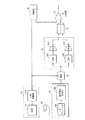
図1は、本発明による睡眠無呼吸の診断装置に関わるブロック図である。図示されたような睡眠無呼吸の診断装置は、光源部11、制御部12、光感知部13、マルチプレクサ(MUX)14、遅延部15、ディバイダ16及び比較器17を備える。
光源部11は、相異なる波長帯域の光を生成する光源111と光源111に駆動電流を提供する光源駆動部112とを備える。
Hereinafter, the present invention will be described in more detail with reference to the accompanying drawings.
FIG. 1 is a block diagram relating to a diagnostic apparatus for sleep apnea according to the present invention. The illustrated diagnostic apparatus for sleep apnea includes a
The
光感知部13は、光源111で生成される光信号を電流に変換するフォトディテクタ131及び前記電流を電圧に変換する電流/電圧(I/V)変換器132を備える。
遅延部15は、MUX14からそれぞれ出力される2信号をサンプリングし、所定時間遅延する2つのサンプルアンドホルダ(S/H)151,153と2つの増幅器152,154とを備える。
前記MUX14及び比較器17は、光感知部13から出力される電圧を処理し、被検者が睡眠無呼吸状態にあるか否かを診断する。
The
The
The
前記構成を有する睡眠無呼吸の診断装置の動作は、次の通りなされる。光源駆動部112は、制御部12の制御信号によって光源111に駆動電流を出力する。前記制御信号は、光源111が赤色波長帯域の光を生成するか、または赤外線波長帯域の光を生成するかを制御する。ここで、光源111は、赤色波長帯域と赤外線波長帯域とから発光する少なくとも2つの発光ダイオードを含む発光ダイオードアレイであることが適切である。
The operation of the sleep apnea diagnostic apparatus having the above-described configuration is performed as follows. The light
制御部12は、前記発光ダイオードアレイを波長によって順次に点灯及び消灯するように制御信号を出力する。
光源111で生成された光は、血液の中のヘモグロビンに含まれた酸素の量を測定するように指を含んだ身体の一部分18に照射される。ここで、身体の一部分18は、動脈による脈動成分が測定されるいかなる部位でも可能である。
フォトディテクタ131は、光源10で生成されて身体の一部分18を透過したり、あるいは身体の一部分18で反射された光を感知して光度に該当する電流を出力し、I/V変換器132は前記電流を電圧に変換する。
The
The light generated by the
The
MUX14は、制御部12から出力される制御信号を選択信号にし、光感知部13から出力された光を波長によって赤色光と赤外線とに分離して出力する。
遅延部15は、MUX14から分離されて出力される電圧値をそれぞれサンプリングして所定時間ホールドして増幅する。これは、MUX14で時間差をおいて出力される2電圧に対して時間差がないように適切に遅延し、実質的に同時に2電圧を出力するためのものである。
The
The
ディバイダ16は、2つの増幅器152,154から出力される2電圧を分ける。比較器17は、制御部12から提供される基準値とディバイダ16で分けられる値とを比較して、睡眠無呼吸のいかんを診断する。被検者が睡眠無呼吸状態であると判別されれば、制御部12は、別途の警報装置(図示せず)を通じて被検者または検査者に警告を送りもする。比較器17と制御部12とは、必要によって無線に連結し、無拘束型システムでも具現が可能である。
The
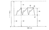
前記睡眠無呼吸の診断装置についての理論的な背景は次の通りである。睡眠無呼吸のように、呼吸が一時的に停止して体内に酸素が供給されなければ、体内血液中にある酸素の飽和度が非正常的に低くなる。このような酸素飽和度は、一般的に光学的に測定される。図2は、波長による酸化ヘモグロビンと還元ヘモグロビンの光吸収係数を図示したものである。図示されたところによれば、血液中のヘモグロビンが酸素を包含しているか否かによって、光の波長に関わり全く異なる傾向の吸収係数を有していることが分かる。従って、赤色帯域の光が人体で反応された量と赤外線帯域の光が反応された量とを比較して、酸素飽和度を予測できる。このような酸素飽和度を予測するために身体の一部分に光を照射すれば、照射された光の透過または反射によって、図3に図示されたようなPPG波形を得る。図示された波形において、参照番号30は、身体に照射された光量、31は吸収された光量、32は身体に透過された光量、33は心臓拍動周期、34は動脈脈動成分による透過光の強度の変化量(AC成分)、35は動脈の非脈動成分による透過光の強度の変化量(DC成分)、36は心臓拍動のピーク、そして37は心臓拍動のバレーをそれぞれ示す。
The theoretical background of the sleep apnea diagnostic device is as follows. If the breathing is temporarily stopped and oxygen is not supplied to the body, as in sleep apnea, the saturation of oxygen in the blood in the body becomes abnormally low. Such oxygen saturation is generally measured optically. FIG. 2 illustrates the light absorption coefficients of oxyhemoglobin and reduced hemoglobin according to wavelength. As shown in the figure, it can be seen that the hemoglobin in the blood has an absorption coefficient that has a completely different tendency depending on the wavelength of light, depending on whether or not the hemoglobin contains oxygen. Therefore, it is possible to predict the oxygen saturation by comparing the amount of the red band light reacted by the human body with the amount of the infrared band light reacted. If a part of the body is irradiated with light in order to predict the oxygen saturation, the PPG waveform shown in FIG. 3 is obtained by transmitting or reflecting the irradiated light. In the illustrated waveform,
PPGのAC成分は、心臓拍動による血流の変化が血流量の変化に反映されたものを測定した結果である。このAC成分を利用して心臓拍動数を測定する方法が、広く利用されている。AC成分は、呼吸や人間の自発的/非自発的な動きによって現れることもあるが、心臓拍動により起きるものよりも非周期的であってパワーも小さく現れる。PPGのDC成分は骨、皮膚または皮下組織のように経時的に変化しない体内成分によって光が吸収または散乱されて発生する。 The AC component of the PPG is a result of measurement of a change in blood flow due to a heart beat reflected in a change in blood flow. A method of measuring the number of heart beats using this AC component is widely used. The AC component may appear due to breathing or spontaneous / involuntary movement of the human, but it is aperiodic and has less power than that caused by a heart beat. The DC component of PPG is generated when light is absorbed or scattered by a body component that does not change over time, such as bone, skin, or subcutaneous tissue.
無呼吸により体内に酸素が供給されなければ還元ヘモグロビンが増加し、それにより図2に図示されたように、還元ヘモグロビンの吸収係数がさらに大きい赤色帯域の光が、一層多く減衰される。図4及び図5は、それぞれ正常呼吸時と無呼吸時のPPG波形を図示したものである。図示されたところによれば、正常呼吸時には経時的に同じ傾きパターンを有することが分かる。しかし、無呼吸時には還元ヘモグロビンの増加により660nmの赤色波長帯域の光量が減少する傾向を見せることが分かる。 If no oxygen is supplied to the body due to apnea, the amount of reduced hemoglobin increases, and as shown in FIG. 2, light in the red band where the absorption coefficient of reduced hemoglobin is larger is further attenuated. 4 and 5 show PPG waveforms during normal breathing and during apnea, respectively. As shown in the figure, it can be seen that the normal inclination has the same inclination pattern over time. However, it can be seen that during apnea, the amount of light in the red wavelength band of 660 nm tends to decrease due to an increase in reduced hemoglobin.
一般的に、図2のPPGでDC成分は、人の指の太さあるいは光が透過する該当する身体の一部分の特徴によって大きな差を有する。従って、相異なる波長帯域の光に関わる透過比を求めるならば、該当身体部分の特徴による差を補償できる。従って、本発明ではMUX14と遅延部15とを介してそれぞれ出力される、赤色光と赤外線のDC成分を利用する。ディバイダ16は、赤色光のDC成分で赤外線のDC成分を割り算する。この時、割った値をレシオメトリックインデックス(RI:Ratiometric Index)という。比較器17は、割った値を制御部12から供給される基準値と比較し、ディバイダ16の出力値が基準値より大きいならば、睡眠中に一時的に呼吸が停止したと判断する。
In general, in the PPG of FIG. 2, the DC component has a large difference depending on the thickness of a human finger or a characteristic of a part of a corresponding body through which light passes. Therefore, if the transmission ratios relating to light in different wavelength bands are obtained, differences due to the characteristics of the corresponding body part can be compensated. Therefore, in the present invention, red light and infrared DC components output via the
図6及び図7は、それぞれ正常呼吸状態と無呼吸時の赤色波長帯域の光と赤外線波長帯域光とのDC成分比を図示したものである。図示されたところによれば、正常呼吸状態では原信号の傾向と大きく異ならないが、無呼吸時には2信号間のDC成分比、すなわちRIがだんだんと大きくなる。従って、RIが基準値より大きくなれば、睡眠無呼吸が発生したと判断する。 FIGS. 6 and 7 show the DC component ratios of the light in the red wavelength band and the light in the infrared wavelength band in the normal respiration state and the apnea, respectively. As shown in the figure, the tendency of the original signal does not greatly differ in the normal breathing state, but the DC component ratio between the two signals, that is, the RI gradually increases in the apnea. Therefore, when RI becomes larger than the reference value, it is determined that sleep apnea has occurred.
次の表は、無呼吸と正常呼吸時の平均RIを測定した結果である。測定は6人の被検者に対して1分単位で6回にわたって反復測定を実施した。1分単位の測定で3回は人為的に呼吸を10秒以上止めるようにし、残りの3回は正常に呼吸をして測定した。使われた光は赤色帯域の660nm波長と赤外線帯域の940nm波長とである。 The following table shows the results of measuring the average RI during apnea and normal breathing. The measurement was repeated six times in one minute units for six subjects. The breathing was artificially stopped for 10 seconds or more three times in one-minute measurements, and the remaining three times were measured after breathing normally. The light used was a 660 nm wavelength in the red band and a 940 nm wavelength in the infrared band.

この時、呼吸を10秒以上止めた場合に測定されたデータでRIを計算すれば、0.096〜0.56と示され、正常呼吸状態で測定されたデータで計算されたRIは、0.02〜0.07範囲の値を表した。この結果より0.07〜0.096間の値を比較器17の基準値と使用することが適切でありうる。
従来技術の場合、呼吸や動きによる雑音によって影響を受けたPPG波形でピークを探せないデータが発生すれば、この結果は、酸素飽和度予測に反映されるので、これを利用して睡眠無呼吸を診断するようになれば、エラーが発生する。
次の表は6人の被検者において3回反復して人為的に動きによる雑音を発生させ、この時の酸素飽和度予測値と実際値との差を求めたものである。
At this time, if the RI is calculated based on the data measured when the breathing is stopped for 10 seconds or more, it is shown as 0.096 to 0.56, and the RI calculated based on the data measured in the normal breathing state is 0. 0.02 to 0.07. From this result, it may be appropriate to use a value between 0.07 and 0.096 as the reference value for
In the case of the prior art, if data in which a peak cannot be found is generated in a PPG waveform affected by noise due to breathing or movement, the result is reflected in the prediction of oxygen saturation. An error will occur if it is diagnosed.
The following table shows the difference between the predicted value and the actual value of oxygen saturation at this time, in which the noise due to motion was artificially generated by repeating three times in six subjects.
表によれば、本発明の場合にAC成分のピークとバレーとを使用せずにDC成分の比を使用するために、無呼吸診断時に動きによる雑音には特別な影響を受けないことが分かる。 According to the table, since the ratio of the DC component is used without using the peak and the valley of the AC component in the case of the present invention, it is found that the noise due to the motion is not particularly affected during the apnea diagnosis. .
本発明の睡眠無呼吸の診断装置及び方法は、例えば被検者の睡眠無呼吸のいかんを診断する診断システムに使用でき、サイズが比較的小さい診断システムにも効果的に適用可能である。 INDUSTRIAL APPLICABILITY The diagnostic device and method for sleep apnea of the present invention can be used, for example, in a diagnostic system for diagnosing sleep apnea in a subject, and can be effectively applied to a diagnostic system having a relatively small size.
11…光源部
111…光源
112…光源駆動部
12…制御部
13…光感知部
131…フォトディテクタ
132…電流/電圧変換器
14…MUX
15…遅延部
151,153…サンプルアンドホルダ
152,154…増幅器
16…ディバイダ
17…比較器
DESCRIPTION OF
15
Claims (12)
所定制御信号によって少なくとも相異なる2波長帯域の光を順次に生成する光源部と、
前記光源部で生成され、身体の一部分に照射されて出力される光を感知し、電気信号に変換する光感知部と、
前記光感知部から出力される電気信号の時間差を実質的になくした後で前記電気信号の比を求め、求めた比を所定基準値と比較して睡眠無呼吸状態を診断する診断部と、
前記少なくとも2波長帯域の光を生成するように前記制御信号を前記光源部に出力し、前記診断部に前記基準値を提供する制御部と
を含むことを特徴とする睡眠無呼吸の診断装置。 Diagnosis of sleep apnea in which a part of the body of the subject is irradiated with light, and then the output light is subjected to a predetermined process to determine whether the subject is in a state of temporarily stopping breathing during sleep. In the device,
A light source unit for sequentially generating light of at least two different wavelength bands by a predetermined control signal,
A light sensing unit that is generated by the light source unit, senses light emitted to a part of the body and output, and converts the light into an electric signal,
Diagnosing a sleep apnea condition by determining the ratio of the electrical signals after substantially eliminating the time difference between the electrical signals output from the light sensing unit, and comparing the determined ratio with a predetermined reference value;
A control unit that outputs the control signal to the light source unit so as to generate the light of the at least two wavelength bands, and provides the reference value to the diagnostic unit.
少なくとも赤色及び赤外線波長帯域の光を生成する発光ダイオードアレイであることを特徴とする請求項1に記載の睡眠無呼吸の診断装置。 The light source unit,
The diagnostic apparatus for sleep apnea according to claim 1, wherein the diagnostic apparatus is a light emitting diode array that generates light in at least red and infrared wavelength bands.
前記生成された光を被検者の動脈による脈動成分が測定されるいかなる部分にも照射することを特徴とする請求項2に記載の睡眠無呼吸の診断装置。 The light source unit,
The apparatus for diagnosing sleep apnea according to claim 2, wherein the generated light is applied to any part where a pulsation component due to an artery of the subject is measured.
前記発光ダイオードアレイを波長によって順次に点灯した後で消灯する制御信号を前記光源部に出力する請求項2に記載の睡眠無呼吸の診断装置。 The control unit includes:
The diagnostic apparatus for sleep apnea according to claim 2, wherein a control signal for sequentially turning on and off the light emitting diode array according to wavelength is output to the light source unit.
前記光源部で生成され、身体の一部分に照射されて出力される光を感知し、電流信号を出力するフォトディテクタと、
前記電流信号を電圧信号に変換する電流/電圧変換器と
を備えることを特徴とする請求項1に記載の睡眠無呼吸の診断装置。 The light sensing unit,
A photodetector that is generated by the light source unit, senses light emitted to a part of the body and output, and outputs a current signal,
The diagnostic apparatus for sleep apnea according to claim 1, further comprising: a current / voltage converter that converts the current signal into a voltage signal.
前記光感知部から出力される電気信号を前記制御信号によって分離するマルチプレクサと、
分離された電気信号をそれぞれサンプリングし、サンプリングされた電気信号が実質的に同時に出力されるように該当時間前記サンプリングされた電気信号を遅延する遅延部と、
前記遅延部から出力されるサンプリングされた電気信号の比を求めるディバイダと、
求めた比を前記基準値と比較して睡眠無呼吸状態のいかんを判別する比較器と
を備えることを特徴とする請求項5に記載の睡眠無呼吸の診断装置。 The diagnostic unit includes:
A multiplexer that separates an electric signal output from the light sensing unit by the control signal,
A delay unit that samples each of the separated electric signals and delays the sampled electric signals for a corresponding time so that the sampled electric signals are output substantially simultaneously;
A divider for determining a ratio of the sampled electric signal output from the delay unit;
The diagnostic device for sleep apnea according to claim 5, further comprising: a comparator that compares the obtained ratio with the reference value to determine whether the patient is in a sleep apnea state.
前記光感知部から出力される電気信号を前記制御信号によって分離するマルチプレクサと、
分離された電気信号をそれぞれサンプリングし、サンプリングされた電気信号が実質的に同時に出力されるように該当時間前記サンプリングされた電気信号を遅延する遅延部と、
前記遅延部から出力されるサンプリングされた電気信号の比を求めるディバイダと、
求めた比を前記基準値と比較して睡眠無呼吸状態のいかんを判別する比較器と
を備えることを特徴とする請求項1に記載の睡眠無呼吸の診断装置。 The diagnostic unit includes:
A multiplexer that separates an electric signal output from the light sensing unit by the control signal,
A delay unit that samples each of the separated electric signals and delays the sampled electric signals for a corresponding time so that the sampled electric signals are output substantially simultaneously;
A divider for determining a ratio of the sampled electric signal output from the delay unit;
The diagnostic device for sleep apnea according to claim 1, further comprising: a comparator for comparing the obtained ratio with the reference value to determine whether the patient is in a sleep apnea state.
(a)少なくとも相異なる2波長帯域の光を順次に生成する段階と、
(b)前記(a)段階で生成されて身体一部分に照射されて出力される光を感知して電気信号に変換する段階と、
(c)前記(b)段階から出力される電気信号をサンプリングし、サンプリングされた電気信号の時間差が実質的にないように各電気信号を遅延する段階と、
(d)前記電気信号の比を求め、求めた比を所定基準値と比較し、被検対象が睡眠無呼吸状態であるか否かを判別する段階と
を含むことを特徴とする睡眠無呼吸の診断方法。 A device for diagnosing sleep apnea, which irradiates light to a body part of a subject and processes the output light to diagnose whether or not the subject temporarily stops breathing during sleep. At
(A) sequentially generating light of at least two different wavelength bands;
(B) detecting the light generated in the step (a) and emitted to a part of the body and output and converting the light into an electric signal;
(C) sampling the electric signal output from the step (b), and delaying each electric signal so that there is substantially no time difference between the sampled electric signals;
(D) determining the ratio of the electrical signals, comparing the determined ratio with a predetermined reference value, and determining whether the subject is in a sleep apnea state. Diagnostic method.
赤色波長帯域及び赤外線波長帯域であることを特徴とする請求項8に記載の睡眠無呼吸の診断方法。 The two wavelength bands in the step (a) are as follows:
9. The method for diagnosing sleep apnea according to claim 8, wherein the sleep apnea is a red wavelength band and an infrared wavelength band.
正常呼吸状態と無呼吸状態とでそれぞれ前記サンプリングされた電気信号の比を複数回求め、求めた電気信号の比の間値に該当する値を前記基準値として提供することを特徴とする請求項8に記載の睡眠無呼吸の診断方法。
The reference value of the step (d) is:
The ratio of the sampled electric signal in each of a normal breathing state and an apnea state is obtained a plurality of times, and a value corresponding to a value between the obtained electric signal ratios is provided as the reference value. 9. The method for diagnosing sleep apnea according to 8.
Applications Claiming Priority (1)
| Application Number | Priority Date | Filing Date | Title |
|---|---|---|---|
| KR1020030026396A KR100552681B1 (en) | 2003-04-25 | 2003-04-25 | Apparatus and method for diagnosing sleep apnea |
Publications (2)
| Publication Number | Publication Date |
|---|---|
| JP2004321807A true JP2004321807A (en) | 2004-11-18 |
| JP3824615B2 JP3824615B2 (en) | 2006-09-20 |
Family
ID=32960261
Family Applications (1)
| Application Number | Title | Priority Date | Filing Date |
|---|---|---|---|
| JP2004130107A Expired - Lifetime JP3824615B2 (en) | 2003-04-25 | 2004-04-26 | Sleep apnea diagnostic apparatus and method |
Country Status (5)
| Country | Link |
|---|---|
| US (1) | US7169110B2 (en) |
| EP (1) | EP1470780B1 (en) |
| JP (1) | JP3824615B2 (en) |
| KR (1) | KR100552681B1 (en) |
| DE (1) | DE602004005770T2 (en) |
Cited By (1)
| Publication number | Priority date | Publication date | Assignee | Title |
|---|---|---|---|---|
| JP2010193949A (en) * | 2009-02-23 | 2010-09-09 | Nippon Koden Corp | Blood oxygen saturation measuring apparatus |
Families Citing this family (41)
| Publication number | Priority date | Publication date | Assignee | Title |
|---|---|---|---|---|
| US20070000494A1 (en) * | 1999-06-30 | 2007-01-04 | Banner Michael J | Ventilator monitor system and method of using same |
| US7024235B2 (en) | 2002-06-20 | 2006-04-04 | University Of Florida Research Foundation, Inc. | Specially configured nasal pulse oximeter/photoplethysmography probes, and combined nasal probe/cannula, selectively with sampler for capnography, and covering sleeves for same |
| DE102004025200A1 (en) * | 2004-05-22 | 2005-12-22 | Weinmann Geräte für Medizin GmbH & Co. KG | Device for detecting the severity of a disease and method for controlling a detection device |
| WO2006005169A1 (en) * | 2004-07-09 | 2006-01-19 | Telemedic Inc | Vital sign monitoring system and method |
| DE102005013429A1 (en) * | 2005-03-21 | 2006-09-28 | Flore, Ingo, Dr. | Mobile diagnostic device |
| US7785262B2 (en) * | 2005-04-25 | 2010-08-31 | University Of Florida Research Foundation, Inc. | Method and apparatus for diagnosing respiratory disorders and determining the degree of exacerbations |
| JP2007130182A (en) * | 2005-11-09 | 2007-05-31 | Toshiba Corp | Illumination controller, illumination control system, illumination control method, and illumination control program |
| AU2007217783A1 (en) | 2006-02-16 | 2007-08-30 | Imthera Medical, Inc. | An RFID based apparatus, system, and method for therapeutic treatment of a patient |
| FR2913588B1 (en) * | 2007-03-12 | 2010-05-07 | Groupe Ecoles Telecomm | AMBULATORY TELEVIGILANCE SYSTEM COMPRISING A DEVICE FOR PULSE DEBRISING, ACTIMETRY AND FALL DETECTION |
| DE102007020038A1 (en) | 2007-04-27 | 2008-10-30 | Fraunhofer-Gesellschaft zur Förderung der angewandten Forschung e.V. | Evidence of apnea with blood pressure dependent detected signals |
| US20100323379A1 (en) * | 2007-09-27 | 2010-12-23 | Somers Virend K | Sleep apnea |
| WO2009048581A1 (en) | 2007-10-09 | 2009-04-16 | Imthera Medical, Inc. | System and method for neural stimulation |
| NL1036012C (en) * | 2008-10-03 | 2010-04-06 | Stephan Arend Hulsbergen | MONITORING SYSTEM, RING FITTED WITH SUCH A SYSTEM, AND A SENSOR AND A PROCESSING UNIT AS PART OF THIS SYSTEM. |
| WO2010042404A1 (en) | 2008-10-09 | 2010-04-15 | Imthera Medical, Inc. | Method of stimulating a hypoglossal nerve for controlling the position of a patient's tongue |
| US8378832B2 (en) * | 2009-07-09 | 2013-02-19 | Harry J. Cassidy | Breathing disorder treatment system and method |
| AU2010318651A1 (en) | 2009-11-10 | 2012-05-03 | Imthera Medical, Inc. | System for stimulating a hypoglossal nerve for controlling the position of a patient's tongue |
| KR101142126B1 (en) * | 2010-01-08 | 2012-05-09 | (주)아이엠 | A pulse oximeter improving signal quality |
| WO2012024401A2 (en) | 2010-08-17 | 2012-02-23 | University Of Florida Research Foundation, Inc. | Intelligent drug and/or fluid delivery system to optimizing medical treatment or therapy using pharmacodynamic and/or pharmacokinetic data |
| EP2484279A1 (en) * | 2011-02-03 | 2012-08-08 | BIOTRONIK SE & Co. KG | Blood flow sensor |
| US9265458B2 (en) | 2012-12-04 | 2016-02-23 | Sync-Think, Inc. | Application of smooth pursuit cognitive testing paradigms to clinical drug development |
| US9380976B2 (en) | 2013-03-11 | 2016-07-05 | Sync-Think, Inc. | Optical neuroinformatics |
| WO2014210588A1 (en) * | 2013-06-28 | 2014-12-31 | North Carolina State University | Systems and methods for determining sleep patterns and circadian rhythms |
| US20150335293A1 (en) * | 2013-08-30 | 2015-11-26 | Maxim Integrated Products, Inc. | Systems and techniques to determine whether a signal is associated with a periodic biologic function |
| JP2015125544A (en) * | 2013-12-26 | 2015-07-06 | 株式会社東芝 | Electronic apparatus, method and program |
| US20160081616A1 (en) * | 2014-09-23 | 2016-03-24 | Boe Technology Group Co., Ltd. | Apparatus and method for processing electroencephalogram, and sleep monitoring wearable device |
| US11160459B2 (en) * | 2015-06-12 | 2021-11-02 | ChroniSense Medical Ltd. | Monitoring health status of people suffering from chronic diseases |
| US10687742B2 (en) | 2015-06-12 | 2020-06-23 | ChroniSense Medical Ltd. | Using invariant factors for pulse oximetry |
| US10470692B2 (en) * | 2015-06-12 | 2019-11-12 | ChroniSense Medical Ltd. | System for performing pulse oximetry |
| US11160461B2 (en) | 2015-06-12 | 2021-11-02 | ChroniSense Medical Ltd. | Blood pressure measurement using a wearable device |
| US11712190B2 (en) | 2015-06-12 | 2023-08-01 | ChroniSense Medical Ltd. | Wearable device electrocardiogram |
| US11464457B2 (en) | 2015-06-12 | 2022-10-11 | ChroniSense Medical Ltd. | Determining an early warning score based on wearable device measurements |
| US10952638B2 (en) | 2015-06-12 | 2021-03-23 | ChroniSense Medical Ltd. | System and method for monitoring respiratory rate and oxygen saturation |
| US10541652B2 (en) | 2015-06-27 | 2020-01-21 | Intel Corporation | Apparatus and method for filter settling calibration to improve speed of tracking and cancelling of DC offset |
| US10568530B2 (en) | 2015-06-27 | 2020-02-25 | Intel Corporation | Apparatus and method for tracking and cancelling DC offset to acquire small AC signal |
| US9717424B2 (en) * | 2015-10-19 | 2017-08-01 | Garmin Switzerland Gmbh | System and method for generating a PPG signal |
| US10398381B1 (en) * | 2015-11-19 | 2019-09-03 | Fitbit, Inc. | System and method for characterizing cardiac arrhythmia |
| US10231632B2 (en) * | 2016-02-22 | 2019-03-19 | Intel Corporation | Apparatus and method for tracking and cancelling DC offset to acquire small AC signal using dual feedback loops |
| US11000235B2 (en) | 2016-03-14 | 2021-05-11 | ChroniSense Medical Ltd. | Monitoring procedure for early warning of cardiac episodes |
| WO2017188540A1 (en) * | 2016-04-28 | 2017-11-02 | 엘지전자 주식회사 | Watch-type terminal and method for controlling same |
| US20190053754A1 (en) | 2017-08-18 | 2019-02-21 | Fitbit, Inc. | Automated detection of breathing disturbances |
| CN114376538A (en) * | 2020-10-21 | 2022-04-22 | 华为技术有限公司 | Method for periodically measuring blood oxygen and electronic equipment |
Family Cites Families (21)
| Publication number | Priority date | Publication date | Assignee | Title |
|---|---|---|---|---|
| US3799672A (en) * | 1972-09-15 | 1974-03-26 | Us Health Education & Welfare | Oximeter for monitoring oxygen saturation in blood |
| JPS61228831A (en) | 1985-04-02 | 1986-10-13 | ミノルタ株式会社 | Apparatus for detecting non-respiration fit |
| US4651741A (en) * | 1985-05-30 | 1987-03-24 | Baxter Travenol Laboratories, Inc. | Method and apparatus for determining oxygen saturation in vivo |
| JPS635729A (en) | 1986-06-25 | 1988-01-11 | ミノルタ株式会社 | Apparatus for detecting attack of apnea |
| US4800495A (en) * | 1986-08-18 | 1989-01-24 | Physio-Control Corporation | Method and apparatus for processing signals used in oximetry |
| US4819752A (en) * | 1987-10-02 | 1989-04-11 | Datascope Corp. | Blood constituent measuring device and method |
| US4869254A (en) * | 1988-03-30 | 1989-09-26 | Nellcor Incorporated | Method and apparatus for calculating arterial oxygen saturation |
| SE465551B (en) | 1990-02-16 | 1991-09-30 | Aake Oeberg | DEVICE FOR DETERMINING A HEART AND RESPIRATORY FREQUENCY THROUGH PHOTOPLETISMOGRAPHICAL SEATING |
| JPH0422339A (en) | 1990-05-16 | 1992-01-27 | Otax Kk | Apnea syndrome inspecting device |
| JPH0638965A (en) * | 1992-07-23 | 1994-02-15 | Minolta Camera Co Ltd | Respiration diagnostic apparatus |
| WO1994004071A1 (en) | 1992-08-19 | 1994-03-03 | Lynn Lawrence A | Apparatus for the diagnosis of sleep apnea |
| US6342039B1 (en) | 1992-08-19 | 2002-01-29 | Lawrence A. Lynn | Microprocessor system for the simplified diagnosis of sleep apnea |
| US6172743B1 (en) * | 1992-10-07 | 2001-01-09 | Chemtrix, Inc. | Technique for measuring a blood analyte by non-invasive spectrometry in living tissue |
| WO1994027492A1 (en) * | 1993-05-21 | 1994-12-08 | Nims, Inc. | Discriminating between valid and artifactual pulse waveforms |
| JP4555919B2 (en) | 1997-03-17 | 2010-10-06 | ノンインベイシブ モニタリング システムズ インコーポレイテッド | Physiological signature feedback system |
| IL122875A0 (en) | 1998-01-08 | 1998-08-16 | S L P Ltd | An integrated sleep apnea screening system |
| CA2334964C (en) * | 1998-06-11 | 2009-03-24 | Spo Medical Equipment Ltd. | Physiological stress detector device and method |
| US6415174B1 (en) | 1998-11-09 | 2002-07-02 | Board Of Regents The University Of Texas System | ECG derived respiratory rhythms for improved diagnosis of sleep apnea |
| KR100340240B1 (en) | 1999-05-28 | 2002-06-12 | 임현수 | A potodetector equipment used in measuring oxygen saturation and amount of blood flow |
| AU2002246880B2 (en) * | 2000-12-29 | 2006-12-07 | Watermark Medical, Inc. | Sleep apnea risk evaluation |
| US6997879B1 (en) * | 2002-07-09 | 2006-02-14 | Pacesetter, Inc. | Methods and devices for reduction of motion-induced noise in optical vascular plethysmography |
-
2003
- 2003-04-25 KR KR1020030026396A patent/KR100552681B1/en active IP Right Grant
-
2004
- 2004-04-21 DE DE602004005770T patent/DE602004005770T2/en not_active Expired - Lifetime
- 2004-04-21 EP EP04252327A patent/EP1470780B1/en not_active Expired - Lifetime
- 2004-04-23 US US10/830,113 patent/US7169110B2/en active Active
- 2004-04-26 JP JP2004130107A patent/JP3824615B2/en not_active Expired - Lifetime
Cited By (2)
| Publication number | Priority date | Publication date | Assignee | Title |
|---|---|---|---|---|
| JP2010193949A (en) * | 2009-02-23 | 2010-09-09 | Nippon Koden Corp | Blood oxygen saturation measuring apparatus |
| US8565846B2 (en) | 2009-02-23 | 2013-10-22 | Nihon Kohden Corporation | Blood oxygen saturation measuring apparatus |
Also Published As
| Publication number | Publication date |
|---|---|
| DE602004005770D1 (en) | 2007-05-24 |
| KR20040092170A (en) | 2004-11-03 |
| DE602004005770T2 (en) | 2008-01-10 |
| KR100552681B1 (en) | 2006-02-20 |
| EP1470780A1 (en) | 2004-10-27 |
| US20040215095A1 (en) | 2004-10-28 |
| US7169110B2 (en) | 2007-01-30 |
| EP1470780B1 (en) | 2007-04-11 |
| JP3824615B2 (en) | 2006-09-20 |
Similar Documents
| Publication | Publication Date | Title |
|---|---|---|
| JP3824615B2 (en) | Sleep apnea diagnostic apparatus and method | |
| US20210068768A1 (en) | System for determining confidence in respiratory rate measurements | |
| US6529752B2 (en) | Sleep disorder breathing event counter | |
| JP7019611B2 (en) | Methods and devices for determining the subject's respiratory information | |
| US7570979B2 (en) | Methods and apparatus for patient monitoring | |
| US6709402B2 (en) | Apparatus and method for monitoring respiration with a pulse oximeter | |
| US7828739B2 (en) | Apnea detection system | |
| KR100455289B1 (en) | Method of diagnosing using a ray and apparatus thereof | |
| JP4424781B2 (en) | Matching degree recognition unit | |
| AU2003275133B2 (en) | Autonomic nervous system monitoring | |
| CN112040846A (en) | Method for estimating blood pressure and degree of arteriosclerosis based on photoplethysmography (PPG) signal | |
| US20040044276A1 (en) | Method and appratus for measuring pulsus paradoxus | |
| JP2002516689A (en) | Stereo pulse oximeter | |
| Chacon et al. | A wearable pulse oximeter with wireless communication and motion artifact tailoring for continuous use | |
| JP2009089883A (en) | Atrial fibrillation detector, system and method | |
| Ungureanu et al. | Real-time extraction of the respiratory rate from photoplethysmographic signal using wearable devices | |
| Yoon et al. | Development of a compact home health monitor for telemedicine | |
| TWI620550B (en) | Braces system with blood oxygen concentration sensing | |
| Garibay et al. | Analysis and Synthesis of Respiratory Rate for Male Patients | |
| Wang et al. | A Wearable Solution for Obstructive Sleep Apnea Risk Evaluation Based on Optical Sensor | |
| Tawiah | A reusable, low-cost and self-sufficient sensorbased neonatal pulse oximeter | |
| Rao et al. | A comparative analysis of reflective and transmissive PPG sensor in pulse acquisition system | |
| Priyadharshini et al. | Pulse Sensor for Diagnosis and Analysis of Heart Rate Using Peak Detection Technique | |
| Penzel et al. | Neue Methoden zur nicht-invasiven Erfassung des Sympathikotonus im Schlaf | |
| Mamilov et al. | Venous saturation and blood flow behavior during laser-induced photodissociation of oxyhemoglobin |
Legal Events
| Date | Code | Title | Description |
|---|---|---|---|
| A131 | Notification of reasons for refusal |
Free format text: JAPANESE INTERMEDIATE CODE: A131 Effective date: 20060221 |
|
| A521 | Request for written amendment filed |
Free format text: JAPANESE INTERMEDIATE CODE: A523 Effective date: 20060509 |
|
| TRDD | Decision of grant or rejection written | ||
| A01 | Written decision to grant a patent or to grant a registration (utility model) |
Free format text: JAPANESE INTERMEDIATE CODE: A01 Effective date: 20060530 |
|
| A61 | First payment of annual fees (during grant procedure) |
Free format text: JAPANESE INTERMEDIATE CODE: A61 Effective date: 20060627 |
|
| R150 | Certificate of patent or registration of utility model |
Ref document number: 3824615 Country of ref document: JP Free format text: JAPANESE INTERMEDIATE CODE: R150 Free format text: JAPANESE INTERMEDIATE CODE: R150 |
|
| FPAY | Renewal fee payment (event date is renewal date of database) |
Free format text: PAYMENT UNTIL: 20090707 Year of fee payment: 3 |
|
| FPAY | Renewal fee payment (event date is renewal date of database) |
Free format text: PAYMENT UNTIL: 20100707 Year of fee payment: 4 |
|
| R250 | Receipt of annual fees |
Free format text: JAPANESE INTERMEDIATE CODE: R250 |
|
| FPAY | Renewal fee payment (event date is renewal date of database) |
Free format text: PAYMENT UNTIL: 20110707 Year of fee payment: 5 |
|
| R250 | Receipt of annual fees |
Free format text: JAPANESE INTERMEDIATE CODE: R250 |
|
| FPAY | Renewal fee payment (event date is renewal date of database) |
Free format text: PAYMENT UNTIL: 20110707 Year of fee payment: 5 |
|
| FPAY | Renewal fee payment (event date is renewal date of database) |
Free format text: PAYMENT UNTIL: 20120707 Year of fee payment: 6 |
|
| R250 | Receipt of annual fees |
Free format text: JAPANESE INTERMEDIATE CODE: R250 |
|
| FPAY | Renewal fee payment (event date is renewal date of database) |
Free format text: PAYMENT UNTIL: 20120707 Year of fee payment: 6 |
|
| FPAY | Renewal fee payment (event date is renewal date of database) |
Free format text: PAYMENT UNTIL: 20130707 Year of fee payment: 7 |
|
| R250 | Receipt of annual fees |
Free format text: JAPANESE INTERMEDIATE CODE: R250 |
|
| R250 | Receipt of annual fees |
Free format text: JAPANESE INTERMEDIATE CODE: R250 |
|
| R250 | Receipt of annual fees |
Free format text: JAPANESE INTERMEDIATE CODE: R250 |
|
| R250 | Receipt of annual fees |
Free format text: JAPANESE INTERMEDIATE CODE: R250 |
|
| R250 | Receipt of annual fees |
Free format text: JAPANESE INTERMEDIATE CODE: R250 |
|
| R250 | Receipt of annual fees |
Free format text: JAPANESE INTERMEDIATE CODE: R250 |
|
| R250 | Receipt of annual fees |
Free format text: JAPANESE INTERMEDIATE CODE: R250 |
|
| R250 | Receipt of annual fees |
Free format text: JAPANESE INTERMEDIATE CODE: R250 |
|
| R250 | Receipt of annual fees |
Free format text: JAPANESE INTERMEDIATE CODE: R250 |
|
| R250 | Receipt of annual fees |
Free format text: JAPANESE INTERMEDIATE CODE: R250 |
|
| R250 | Receipt of annual fees |
Free format text: JAPANESE INTERMEDIATE CODE: R250 |
|
| R250 | Receipt of annual fees |
Free format text: JAPANESE INTERMEDIATE CODE: R250 |