JP2004236505A - Vector and medicinal composition containing cell death inducing gene - Google Patents
Vector and medicinal composition containing cell death inducing gene Download PDFInfo
- Publication number
- JP2004236505A JP2004236505A JP2003025909A JP2003025909A JP2004236505A JP 2004236505 A JP2004236505 A JP 2004236505A JP 2003025909 A JP2003025909 A JP 2003025909A JP 2003025909 A JP2003025909 A JP 2003025909A JP 2004236505 A JP2004236505 A JP 2004236505A
- Authority
- JP
- Japan
- Prior art keywords
- gene
- vector
- cell death
- cells
- pa28α
- Prior art date
- Legal status (The legal status is an assumption and is not a legal conclusion. Google has not performed a legal analysis and makes no representation as to the accuracy of the status listed.)
- Granted
Links
Images


Classifications
-
- A—HUMAN NECESSITIES
- A61—MEDICAL OR VETERINARY SCIENCE; HYGIENE
- A61K—PREPARATIONS FOR MEDICAL, DENTAL OR TOILETRY PURPOSES
- A61K38/00—Medicinal preparations containing peptides
- A61K38/16—Peptides having more than 20 amino acids; Gastrins; Somatostatins; Melanotropins; Derivatives thereof
- A61K38/17—Peptides having more than 20 amino acids; Gastrins; Somatostatins; Melanotropins; Derivatives thereof from animals; from humans
- A61K38/1703—Peptides having more than 20 amino acids; Gastrins; Somatostatins; Melanotropins; Derivatives thereof from animals; from humans from vertebrates
- A61K38/1709—Peptides having more than 20 amino acids; Gastrins; Somatostatins; Melanotropins; Derivatives thereof from animals; from humans from vertebrates from mammals
-
- A—HUMAN NECESSITIES
- A61—MEDICAL OR VETERINARY SCIENCE; HYGIENE
- A61P—SPECIFIC THERAPEUTIC ACTIVITY OF CHEMICAL COMPOUNDS OR MEDICINAL PREPARATIONS
- A61P35/00—Antineoplastic agents
-
- A—HUMAN NECESSITIES
- A61—MEDICAL OR VETERINARY SCIENCE; HYGIENE
- A61P—SPECIFIC THERAPEUTIC ACTIVITY OF CHEMICAL COMPOUNDS OR MEDICINAL PREPARATIONS
- A61P43/00—Drugs for specific purposes, not provided for in groups A61P1/00-A61P41/00
-
- C—CHEMISTRY; METALLURGY
- C07—ORGANIC CHEMISTRY
- C07K—PEPTIDES
- C07K14/00—Peptides having more than 20 amino acids; Gastrins; Somatostatins; Melanotropins; Derivatives thereof
- C07K14/435—Peptides having more than 20 amino acids; Gastrins; Somatostatins; Melanotropins; Derivatives thereof from animals; from humans
- C07K14/46—Peptides having more than 20 amino acids; Gastrins; Somatostatins; Melanotropins; Derivatives thereof from animals; from humans from vertebrates
- C07K14/47—Peptides having more than 20 amino acids; Gastrins; Somatostatins; Melanotropins; Derivatives thereof from animals; from humans from vertebrates from mammals
- C07K14/4701—Peptides having more than 20 amino acids; Gastrins; Somatostatins; Melanotropins; Derivatives thereof from animals; from humans from vertebrates from mammals not used
- C07K14/4702—Regulators; Modulating activity
-
- C—CHEMISTRY; METALLURGY
- C12—BIOCHEMISTRY; BEER; SPIRITS; WINE; VINEGAR; MICROBIOLOGY; ENZYMOLOGY; MUTATION OR GENETIC ENGINEERING
- C12N—MICROORGANISMS OR ENZYMES; COMPOSITIONS THEREOF; PROPAGATING, PRESERVING, OR MAINTAINING MICROORGANISMS; MUTATION OR GENETIC ENGINEERING; CULTURE MEDIA
- C12N2799/00—Uses of viruses
- C12N2799/02—Uses of viruses as vector
- C12N2799/021—Uses of viruses as vector for the expression of a heterologous nucleic acid
- C12N2799/027—Uses of viruses as vector for the expression of a heterologous nucleic acid where the vector is derived from a retrovirus
Landscapes
- Health & Medical Sciences (AREA)
- Life Sciences & Earth Sciences (AREA)
- Chemical & Material Sciences (AREA)
- Medicinal Chemistry (AREA)
- Organic Chemistry (AREA)
- General Health & Medical Sciences (AREA)
- Proteomics, Peptides & Aminoacids (AREA)
- Gastroenterology & Hepatology (AREA)
- Zoology (AREA)
- Pharmacology & Pharmacy (AREA)
- Animal Behavior & Ethology (AREA)
- Public Health (AREA)
- Veterinary Medicine (AREA)
- Engineering & Computer Science (AREA)
- Bioinformatics & Cheminformatics (AREA)
- Nuclear Medicine, Radiotherapy & Molecular Imaging (AREA)
- Marine Sciences & Fisheries (AREA)
- General Chemical & Material Sciences (AREA)
- Immunology (AREA)
- Chemical Kinetics & Catalysis (AREA)
- Epidemiology (AREA)
- Toxicology (AREA)
- Biochemistry (AREA)
- Biophysics (AREA)
- Genetics & Genomics (AREA)
- Molecular Biology (AREA)
- Medicines That Contain Protein Lipid Enzymes And Other Medicines (AREA)
- Medicines Containing Material From Animals Or Micro-Organisms (AREA)
Abstract
Description
【0001】
【発明の属する技術分野】
本発明は、PA28をコードする遺伝子を含有するベクターに関する。
【0002】
特に、本発明は、細胞死誘発遺伝子PA28αを含有する医薬組成物に関する。
【0003】
【従来の技術】
PA28は、20Sプロテアソーム活性化分子である。このPA28は、PA28αとPA28βの2種のサブユニットが交互に会合したヘテロオリゴマーとして細胞質に存在し、20Sプロテアソームの両端に会合してフットボール型プロテアソームを形成することが知られている。このフットボール型プロテアソームは、タンパク質分解に関しては不活性のままであるが、ペプチド分解活性が大幅に上昇しており、内在性抗原の迅速・適応的な処理に関与することが明らかとなっている。とりわけ、MHCクラスI分子に提示されるペプチドの産生に重要な役割を担うことが提唱されている。すなわち、PA28分子は、タンパク質ではなくペプチドの分解活性を促進する作用を有する調節因子であると考えられてきた。
【0004】
しかし、最近、20sプロテアソームの両端にPA700(ユビキチン化タンパク質を認識して結合する分子である)とPA28を併せ持ったプロテアソーム複合体(いわゆるハイブリッドプロテアソーム)が細胞内に存在することが明らかにされ、この発見以来、PA28分子もタンパク質分解そのものに関与する可能性が示唆されている。
【0005】
一方、従来より腫瘍細胞の遺伝子治療として、腫瘍細胞にインターフェロンγ、IL2、IL12、B7などの遺伝子を導入する試みがなされており、種々のサイトカイン遺伝子導入による癌治療が試みられている。たとえば、インターフェロンγなどは、癌細胞に対してin vitroで直接的な増殖抑制または障害活性を示す。これらは癌ワクチンとして使用されてきた(非特許文献1、2)。
【0006】
その他に、リンパ球(たとえば、リンホカイン活性化キラー細胞(LAK)、細胞障害性T細胞(CTL)、腫瘍浸潤リンパ球(TIL)などのリンパ球)などにサイトカイン遺伝子を導入し、導入細胞から分泌されるサイトカインによりオートクラインあるいはパラクラインでリンパ球を活性化して受動免疫能を増強することによる腫瘍細胞の遺伝子治療が試みられている。
【0007】
また、直接抗腫瘍活性のあるIFN遺伝子またはTNF遺伝子を導入することにより腫瘍局所での抗腫瘍性を高めようとする試みもなされている(非特許文献3)。
【0008】
しかし、免疫エフェクター機構を活性化する遺伝子の導入は、いずれの遺伝子導入においても癌細胞を認識するT細胞の活性化を目的としており、癌細胞自体の性状には何らの変化を与えるものではない。逆に、癌細胞に対して直接的に増殖抑制効果を示す遺伝子の導入は、免疫エフェクター機構を介した抗腫瘍効果に変化を与えるものではない。
【0009】
【非特許文献1】
Fearon et al.、Interleukin−2 production by tumor cells bypasses T helper function in the generation of an antitumor response.、“Cell”、(USA)、1990年、60巻、p.397−403.
【0010】
【非特許文献2】
Meijer et al.、Adoptive cellular therapy with tumor vaccine draining lymph node lymphocytes after vaccination with HLA−B7/beta2−microglobulin gene−modified autologous tumor cells.、“J Immunother.”、2002年、25巻、p.359−72。
【0011】
【非特許文献3】
Nishihara et al.、Augmentation of tumor targeting in a line ofglioma−specific mouse cytotoxic T−lymphocytes by retroviral expression of mouse gamma−interferon complementary DNA.、“Cancer Res.”、(USA)、1988年、48巻、p.4730
【0012】
【発明が解決しようとする課題】
したがって、本発明は、上記のような問題を解決すべく、抗腫瘍活性を発揮し、かつT細胞免疫系を活性化することが可能な癌の遺伝子治療のための医薬組成物を提供することである。
【0013】
【課題を解決するための手段】
本発明者は、上記問題を解決すべく鋭意研究を行った結果、以下の知見を得るに至った。
【0014】
すなわち、PA28遺伝子導入細胞において、細胞周期関連分子であるcdc2、Kip1、cdk4、survivin等の発現が著しく低下していること(mRNAレベルは不変)、逆にPA28のドミナント−ネガティブ体の遺伝子導入細胞では、これらの発現が亢進していることを見出した。ドミナント−ネガティブ体の遺伝子導入細胞では、上記タンパク質のユビキチン化の促進も観察された。これらの事実は、PA28がこれらのタンパク質分解を促進することを示している。また、PA28遺伝子の導入により、細胞周期異常に伴うアポトーシスの誘発が引き起こされることを明らかにした。さらに驚いたことに、PA28過剰発現腫瘍は、in vitroでアポトーシスを誘発するだけでなく、in vivoにおいても退縮拒絶されることがマウスを用いた腫瘍の移植実験で明らかになった。
【0015】
以上のように、PA28は、多くのタンパク質分子の分解に関与しており、その結果の総和としてアポトーシスという現象を癌細胞に引き起こすという、全く新しいアポトーシス誘発メカニズムを見出した。
【0016】
このように、PA28遺伝子導入癌細胞は、即座に細胞周期異常(M期における分裂阻害)、およびそれに伴うアポトーシスの亢進をもたらす一方、PA28遺伝子導入癌細胞では、MHCクラスI分子の高発現に伴う癌抗原ペプチドの発現増加により、細胞障害性T細胞によるより強力な抗原認識が行われる。したがって、PA28遺伝子導入により癌細胞自体の細胞死を誘発すると同時にT細胞免疫系をも活性化することができる。
【0017】
上記知見に基づいて、PA28遺伝子を組み込んだウイルスベクターを用いて癌細胞にPA28を過剰発現させることにより、癌細胞の細胞周期遅延、アポトーシス誘発を引き起こすことを利用し、PA28遺伝子を導入した癌細胞は、in vivoにおいても早期に退縮拒絶されることに成功し、本発明を完成するに至った。
【0018】
すなわち、本発明は、PA28遺伝子をコードする遺伝子を含む細胞死誘発ベクターを提供する。
【0019】
また、本発明は、上記ベクターを含有する細胞死誘発剤を提供する。
【0020】
さらに、本発明は、上記ベクターを含有する抗腫瘍医薬組成物を提供する。
【0021】
さらに、本発明は、上記ベクターであって、PA28遺伝子がPA28α遺伝子であるベクターを提供する。
【0022】
さらに、本発明は、上記細胞死誘発剤であって、PA28遺伝子がPA28α遺伝子である医薬組成物を提供する。
【0023】
さらに、本発明は、上記抗腫瘍医薬組成物であって、PA28遺伝子がPA28α遺伝子である医薬組成物を提供する。
【0024】
さらに、本発明は、上記ベクターは、ウイルスベクターであるベクターを提供する。
【0025】
さらに、本発明は、上記ベクターは、ウイルスベクターである上記細胞死誘発剤を提供する。
【0026】
さらに、本発明は、上記ベクターは、ウイルスベクターである上記抗腫瘍医薬組成物を提供する。
【0027】
【発明の実施の形態】
以下、本発明を詳細に説明する。
【0028】
本発明の細胞死誘発ベクターは、PA28遺伝子を含むことを特徴とする。本発明の細胞死誘発剤および抗腫瘍医薬組成物は、PA28遺伝子を含むベクター(本発明の細胞死誘発ベクター)を有効成分として含有することを特徴とする。細胞死誘発ベクターとは、細胞死を誘発するためのベクターであり、細胞に該ベクターを導入したときに、導入された細胞に細胞死(アポトーシス)を誘発することが可能なベクターを意味する。したがって、本発明の細胞死誘発ベクター、細胞死誘発剤または抗腫瘍医薬組成物が標的細胞に導入されると、該標的細胞の細胞周期の遅滞または停止を引き起こし、細胞死(アポトーシス)を誘発することができる。
【0029】
アポトーシスは、細胞の染色体DNAのヌクレオソーム単位の断片化などを特徴とする細胞死である。このアポトーシスの検出は、たとえばAnnexin−V染色による解析およびTUNEL法、核染色などによるDNA断片化の検出等の周知の方法によって解析することができる。
【0030】
本発明に使用されるPA28遺伝子は、PA28α遺伝子、PA28β遺伝子、およびその両方であってもよいが、PA28α遺伝子であることが好ましい。また、本発明のPA28(好ましくはPA28α)遺伝子は、その起源を特に限定するものではなく、ヒトの他、マウス、ラット、ウサギなどの哺乳類由来の遺伝子が挙げられる。
【0031】
本発明のPA28遺伝子は、PA28類似体であってもよく、PA28遺伝子と実質的に同等の生物学的活性を有する限り、PA28アミノ酸配列において1または数個のアミノ酸が欠失、置換もしくは付加されたアミノ酸配列をコードする遺伝子が包含される。したがって、ヒトPA28のアミノ酸配列と90%以上、好ましくは95%以上、より好ましくは97%以上の相同性を有するアミノ酸をコードするものが本発明の範囲に含まれる。また、本発明のPA28遺伝子類似体には、上記ヒトPA28をコードする遺伝子とストリンジェントな条件下でハイブリダイズするDNAも含まれる。これらの類似体は、たとえば、部位特異的突然変異/PCR法などの周知の技術を使用することによって、遺伝子工学的にPA28(好ましくはPA28α)をコードするDNA中に変異を導入することも可能である(Sambrook,Fritsch,and Maniatis,”Molecular Cloning,A Laboratory Manual”(2nd ed)(Cold Spring Harbor Press,Cold Spring Harbor,N.Y.,1989)に記載されているような従来の組換え技術を使用する)。上記ストリンジェントな条件としては、上記文献記載のハイブリダイゼーション条件が挙げられ、具体的には、ホルムアミド濃度:45%(v/v)、塩濃度:5 ×SSPE、温度:42 ℃の条件下でハイブリダイズさせ、塩濃度: 2 ×SSPE、温度:42℃の条件下で洗浄するという条件などが挙げられる。
【0032】
本発明に使用されるPA28をコードする遺伝子(たとえばcDNA)は、以下のような方法によって得ることができる。
【0033】
PA28分子は、本来正常細胞および癌細胞に多かれ少なかれ発現していることが知られている。したがって、まずこれらの細胞から、全mRNAを精製する。具体的には、細胞を、グアニジンイソチオシアネートを含んだフェノールまたはフェノール−クロロフォルム溶液でホモジナイズし、高速遠心により水相と有機層に分離した後、水相に含まれる全RNAをイソプロパノールに加え沈殿させて回収するか、あるいはショ糖もしくはセシウムクロライド密度勾配遠心法により回収する。この全RNAをテンプレートにし、オリゴ(dT)プライマーを使用して、mRNA(即ち、poly(A)RNA)から逆転転写酵素による逆転写反応でcDNAを合成する。
【0034】
このcDNAには、たとえば、あらかじめファージまたはプラスミドベクターに連結可能なように適当な制限酵素部位を作製し、これを同様の制限酵素部位をもつファージ若しくはプラスミドベクターに連結する。このようにして得られたベクターで大腸菌を形質転換またはトランスフェクションしてcDNAライブラリーを作製する。また、種々の細胞の全RNAからのcDNAライブラリーが市販されているので、これらを使用してもよい。
【0035】
次に、上記cDNAライブラリーには、目的のDNA以外に種々の異なった情報をもったDNA断片も挿入されているため、PA28(好ましくは、PA28α)をコードするDNAのみを選抜する必要がある。PA28遺伝子の塩基配列は既に明らかとなっているので、この配列情報に基づいて全長PA28を含む配列が増幅されるようにプライマーを設計し、上記cDNAライブラリーをテンプレートとしてPCR反応を行えば、PA28αをコードするDNAのみを増幅することができる。具体的には、たとえばヒトPA28αcDNAを増幅する場合であれば、ヒトPA28αcDNAの1〜20位および735〜750位の塩基配列に基づいてこれらに相補的なDNA配列をプライマーとして作製し、上記ライブラリーをテンプレートとしてPCRを行うことにより、PA28αDNAを特異的に増幅することができる。
【0036】
PCRは、たとえば変性94℃、1分;アニーリング58℃、1分;伸長72℃、1分を1サイクルとして20サイクル以上、好ましくは30サイクル以上の条件を使用できる。目的のPA28DNAをクローン化および増幅した後、このDNAを回収し、電気泳動等によって精製する。これを適切な発現ベクターに組込むことにより、本発明の細胞死誘発ベクターを作製する。
【0037】
本発明の細胞死誘発ベクターに使用するための発現ベクターは、挿入したPA28遺伝子を発現可能であれば、いずれのベクターを使用してもよい。たとえば、ウイルスベクターおよび非ウイルスベクターのいずれを使用することもできるが、好ましくは、ウイルスベクターである。
【0038】
ウイルスベクターとしては、組換えアデノウイルス、レトロウイルス等が挙げられる。より具体的には、たとえば、無毒化したレトロウイルス、アデノウイルス、アデノ随伴ウイルス、ヘルペスウイルス、ワクシニアウイルス、ポックスウイルス、ポリオウイルス、シンビスウイルス、センダイウイルス、SV40、免疫不全症ウイルス(HIV)等のDNAウイルスまたはRNA ウイルスなどが挙げられる。ウイルスベクターのうち、アデノウイルスの感染効率は、他のウイルスベクターを用いた場合よりもはるかに高いことが知られており、この観点からは、アデノウイルスベクター系を用いることが好ましい。
【0039】
一方、非ウイルス性ベクターには、哺乳類細胞用ベクターが含まれる。
【0040】
本発明の細胞死誘発ベクターは、上述したように増幅したPA28(好ましくはPA28α)遺伝子を適切な発現ベクターに組み込むことによって構築することがでるが、PA28遺伝子のベクターへ挿入は、任意の手段を使用することができる。たとえば、Sambrook,Fritsch,and Maniatis,”Molecular Cloning,A Laboratory Manual”(2nd ed)(Cold Spring Harbor Press,Cold Spring Harbor,N.Y.,1989)に記載されているような従来の組換え技術、またはその他の相同組換え技術等を使用して、PA28遺伝子を発現ベクターに挿入し、本発明のベクターを構築することができる。使用する発現ベクターにマルチクローニングサイトが存在するのであれば、該クローニングサイトにPA28遺伝子を挿入すればよい。
【0041】
本発明の細胞死誘発ベクターは、複製開始点、選択マーカー、プロモーターを含み、必要に応じてエンハンサー、転写終結配列(ターミネーター)、リボソーム結合部位、ポリアデニル化シグナル等を含んでいてもよい。
【0042】
選択マーカーは、形質転換宿主細胞を選択するための表現型を宿主に付与するための遺伝子であり、たとえば哺乳類細胞用ベクターには、ネオマイシン耐性遺伝子、チミジンキナーゼ遺伝子、ジヒドロ葉酸還元酵素を使用することができる。
【0043】
ベクターは、商業的に入手可能なものを使用することもできる。このようなベクターには、たとえば、ウイルスベクターであれば、pMSCVレトロウイルスベクター(Clonetech)などがある。非ウイルス性哺乳類細胞用であれば、pXT1、pSG5(Stratagene)、pSVK3、pBPV、pMSG、pSVL SV40(Pharmacia)などがある。
【0044】
本発明の細胞死誘発剤および抗腫瘍医薬組成物は、上記ベクターを単独で含むものであってもよいが、その他の成分を含有していてもよい。他の成分としては、併用するためのその他の薬剤(たとえば、その他の遺伝子を含有するベクター)、担体などが挙げられる。また、その投与経路に応じて種々の薬学的に許容可能な担体を含んでいてもよい。
【0045】
本発明の細胞死誘発剤および抗腫瘍医薬組成物は、PA28遺伝子を含有するベクターを細胞にまたは患者に投与するための適切な担体、賦形剤およびその他の薬剤と混合して製剤にすることにより、PA28遺伝子の発現を向上させることができる。たとえば、製剤形態が注射剤である場合、常法に従って適切な溶剤(PBS等の緩衝液、生理食塩水、滅菌水など)に溶解して調製されてもよい。また、リポソーム等によって投与される場合は、本発明のベクターを適切なリボソーム(たとえば商標名LIPOFECTIN(Life Technologies.Inc.,Bethesda,Md.)で入手できるカチオン性脂質から製造されるリポソームが好ましい)に形成して投与すればよい。特に、抗体をカップリングさせたリポソームを使用するなど、既知のドラッグデリバリー技術を使用することにより、選択した組織および細胞特異的に本発明のベクター(医薬組成物)をデリバリーすることもできる。
【0046】
ウイルスベクターは、in vivoで細胞に摂取されて組み込まれ、挿入された構築物を含むウイルスDNAを発現することは周知であり、細胞に組換えウイルスを感染させることによって、細胞内に遺伝子を導入することも可能である。
【0047】
本発明の細胞死誘発剤および抗腫瘍医薬組成物の患者への導入法としては、細胞死誘発剤を直接体内に導入するin vivo法、並びにヒトからある種の細胞を取り出して体外で細胞死誘発剤を該細胞に導入してその細胞を体内に戻すex vivo法などの既知の方法を使用すればよい。in vivo法により投与する場合は、細胞死誘発対象の細胞、組織、標的臓器等に応じて、上述したような適切な投与経路により投与すればよい。たとえば、静脈、動脈、皮下、皮内、筋肉内などに投与するか、または病変の認められる組織そのものに直接局所投与することができる。製剤形態としては、上記の各投与形態に合った種々の製剤形態で投与することができる。
【0048】
その他、非ウイルス性ベクターの細胞への遺伝子導入法としては、リン酸−カルシウム共沈法、微小ガラス管を用いたDNAの直接注入法などが挙げられる。また、組織への遺伝子導入法としては、リポソームによる遺伝子導入法、受容体介在性遺伝子導入法、パーティクル銃で担体(金属粒子)とともにDNA分子を細胞に移入する方法、正電荷ポリマーによる導入法などの公知となったいずれの組換え発現ベクターを細胞内に取り込ませてもよい。
【0049】
本発明のベクター(またはベクターを含有する細胞死誘発剤もしくは抗腫瘍医薬組成物)を、腫瘍に直接投与するのが好ましい。本発明により、種々の細胞および組織に細胞死を誘発することが可能となり、また腫瘍を抑制または治療することができる。
【0050】
【発明の効果】
本発明の細胞死誘発ベクターまたは細胞死誘発剤を細胞(特に腫瘍細胞)に導入することにより、該細胞に細胞周期異常に伴う細胞死(アポトーシス)を誘発することが可能となる。
【0051】
また、本発明の抗腫瘍医薬組成物を腫瘍細胞(癌患者など)に導入することにより、抗腫瘍作用、すなわち腫瘍の退縮、拒絶を引き起こすことが可能となる。すなわち、導入された細胞に細胞死が誘発されると共に、PA28によるプロテアソームの活性化により、MHCクラスI分子の高発現に伴う癌抗原ペプチドの発現増加がなされ、細胞障害性T細胞によるより強い抗原認識が行われる。したがって、癌細胞自体の細胞死を誘発すると同時にT細胞免疫応答性を活性化することができる。
【0052】
【実施例】
(材料および方法)
1 PA28α遺伝子発現癌細胞の樹立
1.1 PA28α遺伝子の作製
PA28αをコードするcDNAは、以下のように作製した。
【0053】
マウス脾臓細胞をグアニジンイソチオシアネートを含んだフェノール−クロロフォルム溶液でホモジナイズし、高速遠心により水相と有機層に分離した後、水相に含まれる全RNAをイソプロパノールに加え沈殿させて回収した。この全RNAをテンプレートにし、オリゴdTプライマーを使用して、mRNAから逆転写酵素による逆転写反応でcDNAを合成した。
【0054】
次いで、1〜20位および735〜750位の塩基配列に基づいてこれらに相補的なDNA配列をプライマーとして作製し、上記作製したcDNAライブラリーをテンプレートとして、PA28αをコードするcDNAを増幅した。PCRは、変性94℃、1分;アニーリング58℃、1分;伸長70℃、1分を1サイクルとして30サイクルの条件を使用した。増幅したcDNAは、フェノール−クロロフォルム−イソアミルアルコール溶液を使用して精製し、さらにエタノール沈殿を行い回収した。
【0055】
1.2 レトロウイルスベクターへのPA28α遺伝子の組み込み
作製したPA28αをコードするcDNAを、クローンテック社のpMSCVレトロウイルスベクターのマルチクローニング部位に存在する5’HpaI、3’EcoRI部位にクローニングする。得られたベクター10μlをDOTAPリポゾーマルトランスフェクション試薬(ベーリンガーマンハイム社)を使用してpT67レトロウイルスパッケージ細胞にトランスフェクションする。2μg/mlピューロマイシンで培養を2週間継続してウイルス産生細胞を選択する。6ウェルプレートで培養してウェルの約80%の面積を細胞で占拠した状態のとき、いったんピューロマイシンを培地から除去して(4mlの新鮮培養液に置き換える)、さらに48時間培養してその上清を回収する。この上清中にはレトロウイルス(1×107CFU/ml)が存在している。
【0056】
1.3 レトロウイルスベクターによるPA28α遺伝子の癌細胞への導入
上で得られたレトロウイルス上清2mlにポリブレンを5μg/mlになるように添加し、これを癌細胞1×106個のペレットに加えてよく攪拌した後、6ウェルプレートの一つにまく。24時間後に新鮮培養液を2ml添加する。48時間後に培養液2mlを静かに除去して新たにピューロマイシン2.0μg/mlを含む新鮮培養液2mlと入れ替える。この状態で培養を続け過増殖にならないよう適宜新鮮培養液(ピューロマイシン2.0μg/ml)に入れ替えていく。ピューロマイシンの添加をはじめて約2週間後に、PA28αを安定して発現した癌細胞が得られる。
【0057】
このようにして得られた遺伝子導入癌細胞を用いて以下の実験を行った。
【0058】
2 遺伝子導入癌細胞のin vitroにおける解析
2.1細胞周期(実施例1)
1×10⁶個の細胞を遠心し、上清を除去する。
【0059】
ボルテックスにて攪拌しながら−20℃の70%エタノール2mlを注ぎ込む。
【0060】
−20℃で12時間置く。軽くボルテックスで攪拌した後に遠心し、エタノールを十分に除きタッピングして細胞沈さをほぐす。
【0061】
PCB(Na2HPO4 0.2M、クエン酸4mM)100μlを添加し、混和して室温で約30分放置する。
【0062】
遠心して上清を十分に除去し、タッピングで細胞沈さをほぐす。
【0063】
PI−RNAse溶液(propidium Iodide 10μg/ml、RNAse10mg/ml)を1ml添加してボルテックスで軽く攪拌する。
【0064】
室温で20分放置して、FACScan(Beckton Dickinson, San Joes, CA,USA)にて解析し、横軸をlinear scaleのDNA量、縦軸を細胞数のヒストグラムを作製する。
【0065】
2.2 Annexin VとPI染色によるアポトーシス細胞の検出(実施例2)
アポトーシス細胞を含む1〜2×10⁶個の細胞をPBS(−)に懸濁し、遠心して上清を除去し、タッピングで細胞沈さをほぐす。
【0066】
結合バッファー(HEPES 10mM、NaCl 150mM、KCl 5mM、MgCl2・6H2O 1mM、CaCl2・6H2O 1.8M)にて5×10⁵個/mlになるように調製。
【0067】
細胞浮遊液490μlにアネキシンV−FITCを5μl、PI(250μl/ml:結合バッファー)溶液を5μl加えて穏やかに混和し、氷冷にて10分間染色後直ちに測定する。
【0068】
FACScanにて解析し、横軸をlog scale(FL1:FITC−アネキシンV)、縦軸をlogscale(FL2:PI)のドットプロット表示とする。
【0069】
アネキシンV染色性(−)、PI染色性(−):アポトーシス初期細胞であり、
アネキシンV染色性(+)、PI染色性(+):アポトーシス後期細胞またはネクローシス細胞である。
【0070】
2.3 TUNEL法(TdTアッセイ)(実施例3)
1〜2×10⁶個の細胞を遠心し、上清を除去してタッピングで細胞沈さをほぐす。
【0071】
4℃の1%ホルムアルデヒド−PBS 200μlを添加して、ピペッティングで混和し、氷中に15分放置する。
【0072】
遠心後上清を十分に除き、沈さをボルテックスにて攪拌しながら−20℃の70%エタノール2mlを注ぎ込む。−20℃で12時間置く。
【0073】
遠心し、エタノールを除去して冷PBSで2回遠心洗浄する。
【0074】
上清を十分取り除き、タッピングせずに沈さに平行緩衝液(カコジル酸カリウム0.2M、CoCl2 2.5mM、DTT 0.1mM、BSA 0.25mg/ml)100μlを添加し、ピペッティングで混和する。
【0075】
遠心して上清を十分に除き、タッピングはせずにTdT反応液(FITC−dUTP 5μM、TdT 5unit)50mlを添加してピペッティングで混和する。
【0076】
37℃恒温槽で1時間遮光状態で反応させる。(この間15分ごとにピペティングで混和する)冷PBSで2回遠心洗浄する。
【0077】
上清を十分に除いてタッピングし、PI−RNAse溶液を1ml添加してボルテックスで軽く攪拌する。
【0078】
遮光状態で20分間放置し、FACScanにて解析し、横軸をlinear scaleのDNA量(PI)、縦軸をDNA断裂の頻度(FITC)のドットプロット表示とする。
【0079】
3 in vivoにおける腫瘍移植実験(実施例4)
PA28α遺伝子発現癌細胞、ここではMethA PA28αおよびE.G7 PA28α、さらにPA28αドミナント−ネガティブPA28α(ΔC5)を発現したMethA PA28α(ΔC5)およびE.G7 PA28α(ΔC5)、コントロールとしてからベクターのみを導入したMethA mockおよびE.G7 mockを準備した。
【0080】
MethA mock、MethA PA28α、MethA PA28α(ΔC5)は、各1×10⁷個の生細胞を、またE.G7 mock、E.G7 28α、E.G7 PA28α(ΔC5)は、各1×10⁶個の生細胞を同系マウス皮下(MethAはBALB/c、E.G7はC57BL/6)に接種した。
【0081】
腫瘍接種後、週に2〜3回腫瘍径の長径と短径を計測し、その平均値を計算して経時間的に腫瘍増殖パターンを観察した。
【0082】
(結果)
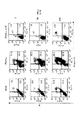
実施例1
マウス腫瘍E.G7、MethA、Bl6にレトロウイルスベクターを用いてPA28α、PA28αΔC5(ドミナント−ネガティブ)を遺伝子導入した。薬剤耐性の安定株を樹立した後、細胞周期をPI染色によって解析した。PA28α発現腫瘍では、G2/M期における遅滞(8倍体の出現によって特徴づけられる)が観察された。PA28αΔC5発現腫瘍では、G1期の亢進がいずれの細胞においても観察された(図1)。
【0083】
実施例2
マウス腫瘍E.G7にレトロウイルスベクターを用いてPA28α、PA28αΔC5(ドミナントネガティブ)を遺伝子導入した。薬剤耐性の安定株を樹立した後、PI染色と、Annexin−V染色によってアポトーシスの解析を行った。PA28α発現腫瘍では、Annexin−V陽性細胞集団の出現が認められた。IFN−γの存在下では、濃度依存的にAnnexin−V陽性が増加し、PI陽性細胞も出現してきた。また、PA28αΔC5発現腫瘍では、IFN−γの存在下でもAnnexin−V陽性集団は認められなかった。
【0084】
実施例3
マウス腫瘍E.G7にレトロウイルスベクターを用いてPA28α、PA28αΔC5(ドミナントネガティブ)を遺伝子導入した。薬剤耐性の安定株を樹立した後、PI染色によりDNA量(細胞周期)を検出した。また、Tunnel法によっても断片化したDNA(DNA fragmentation)を検出した。PA28α発現腫瘍では、M期において分裂できない細胞集団の出現が認められた。さらに、全細胞周期にわたってDNA fragmantationが認められた。また、IFN−γの存在下では、濃度依存的にDNA fragmentationの程度が増加した。一方、PA28αΔC5発現腫瘍では、IFN−γの存在下でもDNA fragmentationは認められなかった。
【0085】
実施例4
マウス腫瘍E.G7、MethAにレトロウイルスベクターを用いてPA28α、PA28α>C5(ドミナントネガティブ)を遺伝子導入し、薬剤耐性の安定株を樹立した。これら安定株を同系マウスに移植した後、腫瘍増殖を観察した(一群3匹より成る)。PA28α遺伝子を導入した腫瘍では、いずれも腫瘍の退縮、拒絶が観察された。一方、PA28αΔC5(ドミナントネガティブ)を導入した細胞では、移植後の腫瘍径は次第に大きくなった。
【0086】
(考察)
PA28α遺伝子を導入することにより、癌細胞が細胞死(アポトーシス)に陥ることが明らかとなった。また、in vivoにおいても、PA28α遺伝子を導入することによって癌細胞に細胞死が誘発され、腫瘍増殖が退縮、拒絶されることが明らかとなった。
【0087】
癌患者では、腫瘍自体が様々な免疫回避機構を駆使して免疫系から逃れている。また、同時にアポトーシスに対する抵抗性も獲得している。このような状況下で、PA28遺伝子を腫瘍に導入すれば有効な遺伝子治療への道が開ける。
【図面の簡単な説明】
【図1】PA28α遺伝子を導入した細胞における細胞周期の解析を示す図。
【図2】PA28α遺伝子を導入した細胞におけるアポトーシスの解析を示す図。
【図3】PA28α遺伝子を導入した細胞におけるDNA断片化(DNA frgmentation)の解析を示す図。
【図4】in vivoにおけるPA28α遺伝子導入の効果を示す図。[0001]
BACKGROUND OF THE INVENTION
The present invention relates to a vector containing a gene encoding PA28.
[0002]
In particular, the present invention relates to a pharmaceutical composition containing the cell death inducing gene PA28α.
[0003]
[Prior art]
PA28 is a 20S proteasome activation molecule. This PA28 is known to exist in the cytoplasm as a hetero-oligomer in which two subunits of PA28α and PA28β are alternately associated, and associates with both ends of the 20S proteasome to form a football-type proteasome. This football-type proteasome remains inactive with respect to proteolysis, but has a greatly increased peptide-degrading activity and has been shown to be involved in rapid and adaptive processing of endogenous antigens. In particular, it has been proposed to play an important role in the production of peptides presented on MHC class I molecules. That is, the PA28 molecule has been considered to be a regulator having an action of promoting the degradation activity of a peptide, not a protein.
[0004]
However, recently, it has been clarified that a proteasome complex (so-called hybrid proteasome) having both PA700 (a molecule that recognizes and binds to ubiquitinated proteins) and PA28 exists at both ends of the 20s proteasome. Since its discovery, it has been suggested that the PA28 molecule may also be involved in proteolysis itself.
[0005]
On the other hand, as gene therapy for tumor cells, attempts have been made to introduce genes such as interferon γ, IL2, IL12, and B7 into tumor cells, and cancer treatment by introducing various cytokine genes has been attempted. For example, interferon γ or the like exhibits direct growth suppression or damage activity in vitro against cancer cells. These have been used as cancer vaccines (Non-Patent Documents 1 and 2).
[0006]
In addition, cytokine genes are introduced into lymphocytes (for example, lymphokine-activated killer cells (LAK), cytotoxic T cells (CTL), tumor infiltrating lymphocytes (TIL), etc.) and secreted from the introduced cells. Gene therapy for tumor cells has been attempted by activating lymphocytes with autocrine or paracrine to enhance passive immunity by the cytokines produced.
[0007]
Attempts have also been made to increase the antitumor properties at the tumor site by directly introducing an IFN gene or TNF gene having antitumor activity (Non-patent Document 3).
[0008]
However, the introduction of a gene that activates the immune effector mechanism is aimed at activating T cells that recognize cancer cells in any of the gene introductions, and does not give any change to the properties of the cancer cells themselves. . Conversely, the introduction of a gene that directly exhibits a growth inhibitory effect on cancer cells does not change the anti-tumor effect via the immune effector mechanism.
[0009]
[Non-Patent Document 1]
Fearon et al. , Interleukin-2 production by tumor cells bypasses the helper function in the generation of anitarian response. "Cell", (USA), 1990, 60, p. 397-403.
[0010]
[Non-Patent Document 2]
Meijer et al. , Adoptive cellular therapeutic with truminal vaccine draining lymph node lymphocytes after vaccination with HLA-B7 / beta2-microglobulin gene "J Immunother.", 2002, 25, p. 359-72.
[0011]
[Non-Patent Document 3]
Nishihara et al. , Augmentation of tutor targeting in a line of glioma-specific mouse cytotoxic T-lymphocytics by retroviral amplification of mouse-finger ligation. "Cancer Res." (USA), 1988, 48, p. 4730
[0012]
[Problems to be solved by the invention]
Accordingly, the present invention provides a pharmaceutical composition for gene therapy for cancer that exhibits antitumor activity and can activate the T cell immune system in order to solve the above-described problems. It is.
[0013]
[Means for Solving the Problems]
As a result of intensive studies to solve the above problems, the present inventors have obtained the following knowledge.
[0014]
That is, expression of cell cycle-related molecules such as cdc2, Kip1, cdk4, survivin and the like is remarkably decreased in the PA28 gene-introduced cell (mRNA level is unchanged), and conversely, a PA28 dominant-negative gene-introduced cell Then, they found that their expression was enhanced. In the dominant-negative gene-transfected cells, promotion of ubiquitination of the protein was also observed. These facts indicate that PA28 promotes these proteolysis. In addition, it was clarified that the introduction of PA28 gene induces apoptosis associated with abnormal cell cycle. Surprisingly, it was revealed by tumor transplantation experiments using mice that PA28-overexpressing tumors not only induce apoptosis in vitro, but are also rejected in vivo.
[0015]
As described above, PA28 is involved in the degradation of many protein molecules, and as a sum of the results, PA28 has found a completely new mechanism for inducing apoptosis that causes a phenomenon called apoptosis in cancer cells.
[0016]
Thus, PA28 gene-introduced cancer cells immediately cause cell cycle abnormalities (inhibition of division in M phase) and the accompanying increase in apoptosis, while PA28 gene-introduced cancer cells are accompanied by high expression of MHC class I molecules. Increased expression of cancer antigen peptides results in stronger antigen recognition by cytotoxic T cells. Therefore, by introducing PA28 gene, cell death of cancer cells themselves can be induced and at the same time the T cell immune system can be activated.
[0017]
Based on the above findings, cancer cells into which PA28 gene is introduced by utilizing the fact that PA28 is overexpressed in cancer cells using a viral vector incorporating PA28 gene, thereby causing cell cycle delay and apoptosis induction of cancer cells. Succeeded in rejecting regression in an early stage even in vivo, and completed the present invention.
[0018]
That is, the present invention provides a cell death inducing vector containing a gene encoding the PA28 gene.
[0019]
The present invention also provides a cell death inducing agent containing the vector.
[0020]
Furthermore, the present invention provides an antitumor pharmaceutical composition containing the above vector.
[0021]
Furthermore, the present invention provides the above vector, wherein the PA28 gene is a PA28α gene.
[0022]
Furthermore, the present invention provides a pharmaceutical composition as described above, wherein the PA28 gene is the PA28α gene.
[0023]
Furthermore, the present invention provides the antitumor pharmaceutical composition described above, wherein the PA28 gene is the PA28α gene.
[0024]
Furthermore, the present invention provides a vector in which the vector is a viral vector.
[0025]
Furthermore, the present invention provides the cell death inducer, wherein the vector is a viral vector.
[0026]
Furthermore, the present invention provides the antitumor pharmaceutical composition, wherein the vector is a viral vector.
[0027]
DETAILED DESCRIPTION OF THE INVENTION
Hereinafter, the present invention will be described in detail.
[0028]
The cell death inducing vector of the present invention is characterized by containing the PA28 gene. The cell death inducing agent and antitumor pharmaceutical composition of the present invention are characterized by containing a vector containing the PA28 gene (cell death inducing vector of the present invention) as an active ingredient. The cell death inducing vector is a vector for inducing cell death, and means a vector capable of inducing cell death (apoptosis) in the introduced cell when the vector is introduced into the cell. Therefore, when the cell death inducing vector, cell death inducing agent or antitumor pharmaceutical composition of the present invention is introduced into a target cell, the cell cycle of the target cell is delayed or stopped, and cell death (apoptosis) is induced. be able to.
[0029]
Apoptosis is cell death characterized by fragmentation of nucleosome units of chromosomal DNA of cells. This detection of apoptosis can be analyzed by a well-known method such as analysis by Annexin-V staining and detection of DNA fragmentation by TUNEL method, nuclear staining, or the like.
[0030]
The PA28 gene used in the present invention may be a PA28α gene, a PA28β gene, or both, but is preferably a PA28α gene. The origin of the PA28 (preferably PA28α) gene of the present invention is not particularly limited, and examples thereof include genes derived from mammals such as mice, rats and rabbits in addition to humans.
[0031]
The PA28 gene of the present invention may be a PA28 analogue, and one or several amino acids are deleted, substituted or added in the PA28 amino acid sequence as long as it has substantially the same biological activity as the PA28 gene. And genes encoding the amino acid sequences. Accordingly, those encoding amino acids having a homology of 90% or more, preferably 95% or more, more preferably 97% or more with the amino acid sequence of human PA28 are included in the scope of the present invention. The PA28 gene analog of the present invention also includes DNA that hybridizes under stringent conditions with the gene encoding human PA28. These analogs can be genetically engineered to introduce mutations into DNA encoding PA28 (preferably PA28α) by using well-known techniques such as site-directed mutagenesis / PCR. (Sambrook, Fritsch, and Maniatis, “Molecular Cloning, A Laboratory Manual” (2nd ed) (represented in Cold Spring Harbor Press, Cold Spring Harbor, NY, 1989). Technology). Examples of the stringent conditions include hybridization conditions described in the above-mentioned literature, and specifically, under the conditions of formamide concentration: 45% (v / v), salt concentration: 5 × SSPE, temperature: 42 ° C. Examples of the conditions include hybridization and washing under conditions of salt concentration: 2 × SSPE and temperature: 42 ° C.
[0032]
The gene (eg, cDNA) encoding PA28 used in the present invention can be obtained by the following method.
[0033]
It is known that PA28 molecules are more or less expressed in normal cells and cancer cells. Therefore, total mRNA is first purified from these cells. Specifically, the cells are homogenized with a phenol or phenol-chloroform solution containing guanidine isothiocyanate, separated into an aqueous phase and an organic layer by high-speed centrifugation, and then total RNA contained in the aqueous phase is added to isopropanol and precipitated. Or by sucrose or cesium chloride density gradient centrifugation. Using this total RNA as a template, an oligo (dT) primer is used to synthesize cDNA from mRNA (ie, poly (A) RNA) by reverse transcription using reverse transcriptase.
[0034]
In this cDNA, for example, an appropriate restriction enzyme site is prepared in advance so that it can be linked to a phage or plasmid vector, and this is linked to a phage or plasmid vector having the same restriction enzyme site. Escherichia coli is transformed or transfected with the vector thus obtained to prepare a cDNA library. In addition, cDNA libraries from total RNA of various cells are commercially available, and these may be used.
[0035]
Next, since DNA fragments having various different information other than the target DNA are also inserted into the cDNA library, it is necessary to select only DNA encoding PA28 (preferably PA28α). . Since the base sequence of the PA28 gene has already been clarified, if a primer is designed so that a sequence containing the full-length PA28 is amplified based on this sequence information, and a PCR reaction is carried out using the cDNA library as a template, PA28α Only DNA encoding can be amplified. Specifically, for example, in the case of amplifying human PA28α cDNA, based on the nucleotide sequences of human PA28α cDNA at positions 1-20 and 735-750, DNA sequences complementary thereto are prepared as primers, and the library By performing PCR using as a template, PA28α DNA can be specifically amplified.
[0036]
For PCR, for example, denaturation 94 ° C., 1 minute; annealing 58 ° C., 1 minute; extension 72 ° C., 1 minute as one cycle, 20 cycles or more, preferably 30 cycles or more can be used. After the desired PA28 DNA is cloned and amplified, this DNA is recovered and purified by electrophoresis or the like. By incorporating this into an appropriate expression vector, the cell death inducing vector of the present invention is prepared.
[0037]
As an expression vector for use in the cell death induction vector of the present invention, any vector may be used as long as the inserted PA28 gene can be expressed. For example, either a viral vector or a non-viral vector can be used, but a viral vector is preferable.
[0038]
Examples of viral vectors include recombinant adenoviruses and retroviruses. More specifically, for example, detoxified retrovirus, adenovirus, adeno-associated virus, herpes virus, vaccinia virus, poxvirus, poliovirus, shinbis virus, Sendai virus, SV40, immunodeficiency virus (HIV), etc. DNA viruses or RNA viruses. Among viral vectors, the infection efficiency of adenovirus is known to be much higher than when other viral vectors are used. From this viewpoint, it is preferable to use an adenoviral vector system.
[0039]
On the other hand, non-viral vectors include mammalian cell vectors.
[0040]
The cell death inducing vector of the present invention can be constructed by incorporating the PA28 (preferably PA28α) gene amplified as described above into an appropriate expression vector. However, any means can be used for inserting the PA28 gene into the vector. Can be used. For example, recombination is described in Sambrook, Fritsch, and Maniatis, “Molecular Cloning, A Laboratory Manual” (2nd ed) (Cold Spring Harbor Press, Cold Spring Harbor, NY, 1989). Alternatively, the PA28 gene can be inserted into an expression vector using other homologous recombination techniques or the like to construct the vector of the present invention. If the expression vector to be used has a multiple cloning site, the PA28 gene may be inserted into the cloning site.
[0041]
The cell death induction vector of the present invention contains a replication origin, a selection marker, and a promoter, and may contain an enhancer, a transcription termination sequence (terminator), a ribosome binding site, a polyadenylation signal, and the like as necessary.
[0042]
The selectable marker is a gene for conferring to the host a phenotype for selecting transformed host cells. For example, neomycin resistance gene, thymidine kinase gene, dihydrofolate reductase should be used for mammalian cell vectors. Can do.
[0043]
Commercially available vectors can also be used. Examples of such a vector include a pMSCV retrovirus vector (Clonetech) if it is a viral vector. For non-viral mammalian cells, there are pXT1, pSG5 (Stratagene), pSVK3, pBPV, pMSG, pSVL SV40 (Pharmacia) and the like.
[0044]
The cell death inducer and antitumor pharmaceutical composition of the present invention may contain the above-mentioned vector alone, but may contain other components. Examples of other components include other drugs for use in combination (for example, vectors containing other genes), carriers and the like. In addition, various pharmaceutically acceptable carriers may be included depending on the administration route.
[0045]
The cell death-inducing agent and antitumor pharmaceutical composition of the present invention are formulated by mixing a vector containing the PA28 gene with cells, or with an appropriate carrier, excipient and other agent for administration to a patient. Thus, the expression of the PA28 gene can be improved. For example, when the preparation form is an injection, it may be prepared by dissolving in an appropriate solvent (buffer solution such as PBS, physiological saline, sterilized water, etc.) according to a conventional method. In addition, when administered by liposome or the like, the vector of the present invention is preferably treated with a suitable ribosome (for example, a liposome produced from a cationic lipid available under the trade name LIPOFECTIN (Life Technologies. Inc., Bethesda, Md.)). It may be formed and administered. In particular, the vector (pharmaceutical composition) of the present invention can also be delivered specifically to a selected tissue and cell by using a known drug delivery technique such as using a liposome to which an antibody is coupled.
[0046]
Viral vectors are well-known to be ingested and incorporated into cells in vivo and to express viral DNA containing the inserted construct, and introduce genes into cells by infecting cells with recombinant viruses. It is also possible.
[0047]
Examples of the method of introducing the cell death inducer and antitumor pharmaceutical composition of the present invention into a patient include an in vivo method in which the cell death inducer is directly introduced into the body, and cell death outside the body by taking out certain cells from humans. A known method such as an ex vivo method may be used in which an inducer is introduced into the cell and the cell is returned to the body. In the case of administration by an in vivo method, administration may be performed by an appropriate administration route as described above according to the cell, tissue, target organ or the like to be induced for cell death. For example, it can be administered intravenously, arterially, subcutaneously, intradermally, intramuscularly, or directly locally to the tissue where the lesion is observed. As a formulation form, it can administer with the various formulation form suitable for each said administration form.
[0048]
In addition, examples of the method for introducing a gene into a non-viral vector cell include a phosphate-calcium coprecipitation method and a direct injection method of DNA using a micro glass tube. In addition, as a gene introduction method to a tissue, a gene introduction method using liposome, a receptor-mediated gene introduction method, a method of transferring a DNA molecule together with a carrier (metal particle) with a particle gun, a introduction method using a positively charged polymer, etc. Any of the known recombinant expression vectors may be incorporated into cells.
[0049]
The vector of the present invention (or cell death-inducing agent or antitumor pharmaceutical composition containing the vector) is preferably administered directly to the tumor. According to the present invention, cell death can be induced in various cells and tissues, and tumors can be suppressed or treated.
[0050]
【The invention's effect】
By introducing the cell death inducing vector or cell death inducing agent of the present invention into a cell (particularly a tumor cell), it becomes possible to induce cell death (apoptosis) associated with abnormal cell cycle in the cell.
[0051]
Moreover, by introducing the antitumor pharmaceutical composition of the present invention into tumor cells (such as cancer patients), it becomes possible to cause an antitumor action, that is, regression or rejection of the tumor. That is, cell death is induced in the introduced cells, and the activation of the proteasome by PA28 increases the expression of cancer antigen peptides accompanying high expression of MHC class I molecules, resulting in stronger antigens from cytotoxic T cells. Recognition is performed. Therefore, T cell immune responsiveness can be activated simultaneously with inducing cell death of the cancer cell itself.
[0052]
【Example】
(Materials and methods)
1 Establishment of PA28α gene expressing cancer cells
1.1 Preparation of PA28α gene
CDNA encoding PA28α was prepared as follows.
[0053]
Mouse spleen cells were homogenized with a phenol-chloroform solution containing guanidine isothiocyanate and separated into an aqueous phase and an organic layer by high-speed centrifugation, and then total RNA contained in the aqueous phase was added to isopropanol and precipitated and collected. Using this total RNA as a template, cDNA was synthesized from mRNA by reverse transcription using reverse transcriptase using oligo dT primer.
[0054]
Next, based on the nucleotide sequences at positions 1-20 and 735-750, DNA sequences complementary to these were prepared as primers, and cDNA encoding PA28α was amplified using the prepared cDNA library as a template. PCR was performed under the conditions of denaturation 94 ° C., 1 minute; annealing 58 ° C., 1 minute; extension 70 ° C., 1 minute as 30 cycles. The amplified cDNA was purified using a phenol-chloroform-isoamyl alcohol solution and further recovered by ethanol precipitation.
[0055]
1.2 Incorporation of PA28α gene into retroviral vector
The prepared cDNA encoding PA28α is cloned into the 5′HpaI and 3′EcoRI sites present in the multiple cloning site of the pMSCV retroviral vector of Clontech. 10 μl of the resulting vector is transfected into pT67 retroviral package cells using DOTAP liposomal transfection reagent (Boehringer Mannheim). The culture is continued for 2 weeks with 2 μg / ml puromycin, and virus-producing cells are selected. When culturing in a 6-well plate and occupying about 80% of the area of the wells with cells, puromycin was once removed from the medium (replaced with 4 ml of fresh medium) and cultured for another 48 hours. Collect Qing. In this supernatant, retrovirus (1 × 10 7 CFU / ml) is present.
[0056]
1.3 Introduction of PA28α gene into cancer cells by retroviral vector
Polybrene was added to 2 ml of the retrovirus supernatant obtained above so as to have a concentration of 5 μg / ml. 6 Add to each pellet and stir well, then roll into one of 6-well plates. After 24 hours, add 2 ml of fresh culture. After 48 hours, 2 ml of the culture medium is gently removed and replaced with 2 ml of fresh culture medium containing 2.0 μg / ml of puromycin. The culture is continued in this state, and the medium is appropriately replaced with a fresh culture medium (puromycin 2.0 μg / ml) so as not to overgrow. Approximately two weeks after the addition of puromycin, cancer cells stably expressing PA28α are obtained.
[0057]
The following experiment was performed using the gene-transferred cancer cells thus obtained.
[0058]
2 In vitro analysis of transgenic cancer cells
2.1 Cell cycle (Example 1)
1 × 10 6 The cells are centrifuged and the supernatant is removed.
[0059]
Pour 2 ml of -20 ° C 70% ethanol with vortexing.
[0060]
Place at −20 ° C. for 12 hours. Lightly vortex and centrifuge, then remove ethanol thoroughly and tap to loosen the cell sediment.
[0061]
PCB (Na 2 HPO 4 Add 100 μl of 0.2 M, 4 mM citric acid), mix and leave at room temperature for about 30 minutes.
[0062]
Centrifuge to remove the supernatant, and loosen the cells by tapping.
[0063]
Add 1 ml of PI-RNAse solution (
[0064]
The sample is allowed to stand at room temperature for 20 minutes, and analyzed by FACScan (Beckton Dickinson, San Joes, Calif., USA), and the horizontal axis represents the linear scale DNA amount and the vertical axis represents the cell number histogram.
[0065]
2.2 Detection of apoptotic cells by Annexin V and PI staining (Example 2)
1-2 × 10 containing apoptotic cells 6 Individual cells are suspended in PBS (−), centrifuged to remove the supernatant, and cell sedimentation is loosened by tapping.
[0066]
Binding buffer (
[0067]
Add 5 μl of annexin V-FITC and 5 μl of PI (250 μl / ml: binding buffer) solution to 490 μl of cell suspension, mix gently, and measure immediately after staining with ice-cooling for 10 minutes.
[0068]
Analysis is performed by FACScan, and the horizontal axis is log scale (FL1: FITC-annexin V), and the vertical axis is log scale (FL2: PI).
[0069]
Annexin V staining (-), PI staining (-): early apoptotic cells,
Annexin V staining (+), PI staining (+): Late apoptotic cells or necrotic cells.
[0070]
2.3 TUNEL method (TdT assay) (Example 3)
1-2 × 10 6 The cells are centrifuged, the supernatant is removed, and the cell sediment is loosened by tapping.
[0071]
Add 200 μl of 1% formaldehyde-PBS at 4 ° C., mix by pipetting, and leave on ice for 15 minutes.
[0072]
After centrifugation, the supernatant is sufficiently removed, and 2 ml of -20 ° C 70% ethanol is poured while the precipitate is vortexed. Place at −20 ° C. for 12 hours.
[0073]
Centrifuge, remove ethanol and centrifuge twice with cold PBS.
[0074]
Thoroughly remove the supernatant, add 100 μl of parallel buffer (potassium cacodylate 0.2 M, CoCl 2 2.5 mM, DTT 0.1 mM, BSA 0.25 mg / ml) to the sediment without tapping, and mix by pipetting. To do.
[0075]
The supernatant is sufficiently removed by centrifugation, and 50 ml of TdT reaction solution (FITC-dUTP 5 μM, TdT 5 unit) is added without tapping and mixed by pipetting.
[0076]
The reaction is carried out in a light-shielded state for 1 hour in a 37 ° C. constant temperature bath. (Mix by pipetting every 15 minutes during this time) Centrifuge twice with cold PBS.
[0077]
The supernatant is sufficiently removed and tapped, and 1 ml of PI-RNAse solution is added and vortexed gently.
[0078]
The sample is allowed to stand for 20 minutes in a light-shielded state, analyzed by FACScan, and the horizontal axis represents the linear scale DNA amount (PI) and the vertical axis represents the frequency of DNA fragmentation (FITC).
[0079]
3 Tumor transplantation experiment in vivo (Example 4)
PA28α gene expressing cancer cells, here MethA PA28α and E. coli. G7 PA28α, MethA PA28α (ΔC5) and E. coli expressing PA28α dominant-negative PA28α (ΔC5) and E. coli. G7 PA28α (ΔC5), MethA mock and E. coli into which only the vector was introduced as a control. G7 mock was prepared.
[0080]
MethA mock, MethA PA28α and MethA PA28α (ΔC5) are each 1 × 10 7 Individual living cells, and E. coli. G7 mock, E.I. G7 28α, E.I. Each G7 PA28α (ΔC5) is 1 × 10 6 Live cells were inoculated subcutaneously into syngeneic mice (MethA for BALB / c, E.G7 for C57BL / 6).
[0081]
After tumor inoculation, the major axis and minor axis of the tumor diameter were measured 2-3 times a week, the average value was calculated, and the tumor growth pattern was observed over time.
[0082]
(result)
Example 1
Mouse tumor PA28α and PA28αΔC5 (dominant-negative) were introduced into G7, MethA and B16 using a retroviral vector. After establishing a drug-resistant stable strain, the cell cycle was analyzed by PI staining. In PA28α expressing tumors, a delay in G2 / M phase (characterized by the appearance of octaploids) was observed. In PA28αΔC5-expressing tumors, enhanced G1 phase was observed in all cells (FIG. 1).
[0083]
Example 2
Mouse tumor PA28α and PA28αΔC5 (dominant negative) were introduced into G7 using a retroviral vector. After establishing a drug-resistant stable strain, apoptosis was analyzed by PI staining and Annexin-V staining. In PA28α-expressing tumors, the appearance of Annexin-V positive cell population was observed. In the presence of IFN-γ, Annexin-V positivity increased in a concentration-dependent manner, and PI-positive cells also appeared. In addition, in the PA28αΔC5-expressing tumor, an Annexin-V positive population was not observed even in the presence of IFN-γ.
[0084]
Example 3
Mouse tumor PA28α and PA28αΔC5 (dominant negative) were introduced into G7 using a retroviral vector. After establishing a drug-resistant stable strain, the amount of DNA (cell cycle) was detected by PI staining. In addition, fragmented DNA (DNA fragmentation) was also detected by the Tunnel method. In PA28α-expressing tumors, the appearance of cell populations that cannot divide in the M phase was observed. In addition, DNA fragmentation was observed throughout the entire cell cycle. In the presence of IFN-γ, the degree of DNA fragmentation increased in a concentration-dependent manner. On the other hand, in the PA28αΔC5-expressing tumor, DNA fragmentation was not observed even in the presence of IFN-γ.
[0085]
Example 4
Mouse tumor A PA28α, PA28α> C5 (dominant negative) gene was introduced into G7 and MethA using a retroviral vector to establish a drug-resistant stable strain. After these stable strains were transplanted into syngeneic mice, tumor growth was observed (consisting of 3 mice per group). Tumor regression and rejection were observed in all tumors into which the PA28α gene was introduced. On the other hand, in the cells into which PA28αΔC5 (dominant negative) was introduced, the tumor diameter after transplantation gradually increased.
[0086]
(Discussion)
It has been clarified that introduction of the PA28α gene causes cancer cells to fall into cell death (apoptosis). Also in vivo, it was revealed that by introducing the PA28α gene, cell death was induced in cancer cells, and tumor growth was regressed and rejected.
[0087]
In cancer patients, the tumor itself escapes the immune system using various immune evasion mechanisms. At the same time, it has acquired resistance to apoptosis. Under such circumstances, introduction of the PA28 gene into a tumor opens the way to effective gene therapy.
[Brief description of the drawings]
FIG. 1 is a diagram showing analysis of the cell cycle in cells into which a PA28α gene has been introduced.
FIG. 2 is a diagram showing an analysis of apoptosis in cells into which a PA28α gene has been introduced.
FIG. 3 is a diagram showing analysis of DNA fragmentation in cells into which a PA28α gene has been introduced.
FIG. 4 is a graph showing the effect of PA28α gene introduction in vivo.
Claims (9)
Priority Applications (2)
| Application Number | Priority Date | Filing Date | Title |
|---|---|---|---|
| JP2003025909A JP3752544B2 (en) | 2003-02-03 | 2003-02-03 | Vector and pharmaceutical composition containing cell death-inducing gene |
| US10/769,856 US20040156830A1 (en) | 2003-02-03 | 2004-02-03 | Vector and pharmaceutical composition containing cell death inducing gene |
Applications Claiming Priority (1)
| Application Number | Priority Date | Filing Date | Title |
|---|---|---|---|
| JP2003025909A JP3752544B2 (en) | 2003-02-03 | 2003-02-03 | Vector and pharmaceutical composition containing cell death-inducing gene |
Publications (2)
| Publication Number | Publication Date |
|---|---|
| JP2004236505A true JP2004236505A (en) | 2004-08-26 |
| JP3752544B2 JP3752544B2 (en) | 2006-03-08 |
Family
ID=32820793
Family Applications (1)
| Application Number | Title | Priority Date | Filing Date |
|---|---|---|---|
| JP2003025909A Expired - Lifetime JP3752544B2 (en) | 2003-02-03 | 2003-02-03 | Vector and pharmaceutical composition containing cell death-inducing gene |
Country Status (2)
| Country | Link |
|---|---|
| US (1) | US20040156830A1 (en) |
| JP (1) | JP3752544B2 (en) |
Cited By (1)
| Publication number | Priority date | Publication date | Assignee | Title |
|---|---|---|---|---|
| WO2006036004A1 (en) * | 2004-09-29 | 2006-04-06 | Oncolys Biopharma, Inc. | Telomelysin-gfp gene-containing recombinant virus |
-
2003
- 2003-02-03 JP JP2003025909A patent/JP3752544B2/en not_active Expired - Lifetime
-
2004
- 2004-02-03 US US10/769,856 patent/US20040156830A1/en not_active Abandoned
Cited By (5)
| Publication number | Priority date | Publication date | Assignee | Title |
|---|---|---|---|---|
| WO2006036004A1 (en) * | 2004-09-29 | 2006-04-06 | Oncolys Biopharma, Inc. | Telomelysin-gfp gene-containing recombinant virus |
| JPWO2006036004A1 (en) * | 2004-09-29 | 2008-05-15 | オンコリスバイオファーマ株式会社 | Telomerisin-GFP gene-containing recombinant virus |
| EA011880B1 (en) * | 2004-09-29 | 2009-06-30 | Онколис Биофарма, Инк. | Telomelysin-gfp gene-containing recombinant virus |
| US7943373B2 (en) | 2004-09-29 | 2011-05-17 | Oncolys Biopharma, Inc. | Telomelysin/GFP-expressing recombinant virus |
| JP5006045B2 (en) * | 2004-09-29 | 2012-08-22 | オンコリスバイオファーマ株式会社 | Telomerisin-GFP gene-containing recombinant virus |
Also Published As
| Publication number | Publication date |
|---|---|
| JP3752544B2 (en) | 2006-03-08 |
| US20040156830A1 (en) | 2004-08-12 |
Similar Documents
| Publication | Publication Date | Title |
|---|---|---|
| Ouyang et al. | IL-10 family cytokines IL-10 and IL-22: from basic science to clinical translation | |
| JP4280708B2 (en) | Stromal cell-derived factor-1 (SDF-1) mediates homing and tissue regeneration of stem cells in ischemic cardiomyopathy | |
| US20100135958A1 (en) | Method for treating cancer in humans | |
| JP2000501394A (en) | Methods and compositions for cancer diagnosis and treatment | |
| KR100911624B1 (en) | Methods for Effectively Coexpressing ????? and ????? | |
| JP2011036251A (en) | Virus vector, and use of the same for gene therapy | |
| JP2014208650A (en) | New therapeutic agent for malignant mesothelioma and immunostimulant | |
| US20020183271A1 (en) | Methods of treatment involving human MDA-7 | |
| KR101949186B1 (en) | Immunity inducing agent | |
| JP3752544B2 (en) | Vector and pharmaceutical composition containing cell death-inducing gene | |
| JP5954175B2 (en) | Immune inducer | |
| WO2007018229A1 (en) | Treatment of adult t-cell leukemia | |
| KR102520880B1 (en) | immune inducer | |
| US20050069960A1 (en) | Novel complementing receptor-ligand pairs and adoptive immunotherapy using the same | |
| US20230310653A1 (en) | THERAPEUTIC AGENT FOR EGFR GENE MUTATION-POSITIVE LUNG CANCER COMPRISING REIC/Dkk-3 GENE | |
| WO2016175309A1 (en) | Immunity-inducing agent | |
| JP2004262797A (en) | Anti-tumor agent using interleukin-23 gene | |
| WO2017170338A1 (en) | Immunity inducing agent | |
| JP5572938B2 (en) | Immune inducer | |
| JP2009035534A (en) | Hla-a24-binding antigen peptide and use thereof | |
| JP2008260689A (en) | Medicinal composition containing hla-a24 molecule-binding peptide originating in parathyroid hormone-related protein | |
| MXPA99012024A (en) | Use of mhc class ii ligands as adjuvant for vaccination and of lag-3 in cancer treatment | |
| JP2006166908A (en) | Therapeutic agent for animal disorder and method for producing the same |
Legal Events
| Date | Code | Title | Description |
|---|---|---|---|
| A711 | Notification of change in applicant |
Free format text: JAPANESE INTERMEDIATE CODE: A712 Effective date: 20040428 |
|
| RD03 | Notification of appointment of power of attorney |
Free format text: JAPANESE INTERMEDIATE CODE: A7423 Effective date: 20040702 |
|
| A131 | Notification of reasons for refusal |
Free format text: JAPANESE INTERMEDIATE CODE: A131 Effective date: 20050830 |
|
| A521 | Request for written amendment filed |
Free format text: JAPANESE INTERMEDIATE CODE: A523 Effective date: 20051019 |
|
| TRDD | Decision of grant or rejection written | ||
| A01 | Written decision to grant a patent or to grant a registration (utility model) |
Free format text: JAPANESE INTERMEDIATE CODE: A01 Effective date: 20051115 |
|
| R150 | Certificate of patent or registration of utility model |
Ref document number: 3752544 Country of ref document: JP Free format text: JAPANESE INTERMEDIATE CODE: R150 Free format text: JAPANESE INTERMEDIATE CODE: R150 |
|
| EXPY | Cancellation because of completion of term |