JP2004147656A - Method for diagnosis and treatment of pancreatic carcinoma and composition useful for the same - Google Patents
Method for diagnosis and treatment of pancreatic carcinoma and composition useful for the same Download PDFInfo
- Publication number
- JP2004147656A JP2004147656A JP2003371406A JP2003371406A JP2004147656A JP 2004147656 A JP2004147656 A JP 2004147656A JP 2003371406 A JP2003371406 A JP 2003371406A JP 2003371406 A JP2003371406 A JP 2003371406A JP 2004147656 A JP2004147656 A JP 2004147656A
- Authority
- JP
- Japan
- Prior art keywords
- nucleic acid
- ukw
- sample
- hybridization
- pancreatic
- Prior art date
- Legal status (The legal status is an assumption and is not a legal conclusion. Google has not performed a legal analysis and makes no representation as to the accuracy of the status listed.)
- Granted
Links
Images


Classifications
-
- G—PHYSICS
- G01—MEASURING; TESTING
- G01N—INVESTIGATING OR ANALYSING MATERIALS BY DETERMINING THEIR CHEMICAL OR PHYSICAL PROPERTIES
- G01N33/00—Investigating or analysing materials by specific methods not covered by groups G01N1/00 - G01N31/00
- G01N33/48—Biological material, e.g. blood, urine; Haemocytometers
- G01N33/50—Chemical analysis of biological material, e.g. blood, urine; Testing involving biospecific ligand binding methods; Immunological testing
- G01N33/53—Immunoassay; Biospecific binding assay; Materials therefor
- G01N33/574—Immunoassay; Biospecific binding assay; Materials therefor for cancer
- G01N33/57407—Specifically defined cancers
- G01N33/57438—Specifically defined cancers of liver, pancreas or kidney
-
- A—HUMAN NECESSITIES
- A61—MEDICAL OR VETERINARY SCIENCE; HYGIENE
- A61P—SPECIFIC THERAPEUTIC ACTIVITY OF CHEMICAL COMPOUNDS OR MEDICINAL PREPARATIONS
- A61P35/00—Antineoplastic agents
Abstract
Description
本発明は、膵癌の特異的な診断、そのための組成物、および膵癌の治療のための治療方法に関する。 The present invention relates to a specific diagnosis of pancreatic cancer, compositions therefor, and therapeutic methods for treating pancreatic cancer.
膵癌は診断および制御が困難な種類の癌であり、このような癌のほとんどは極めて急速に増殖しうる。 Pancreatic cancer is a type of cancer that is difficult to diagnose and control, and most such cancers can grow very quickly.
外分泌膵臓の新生物は、膵管細胞、膵腺房細胞および間質細胞から生じる可能性がある。膵癌の80%は膵管上皮に由来する。これらの腫瘍の60%は膵頭部に位置し、10%は膵尾部、30%は膵体部に位置するかびまん性である(非特許文献1)。組織学的には、これらの腫瘍は高分化型、中分化型および低分化型として等級付けされる。一部の腫瘍は、腺扁平上皮型、粘液型、未分化型、または骨芽細胞様巨細胞を伴う未分化型に分類される(非特許文献2)。本疾患は早期には無症候性であり、早期診断のための検査がなく、局所および内臓への転移に関して攻撃的であるため、本疾患の予後は極めて不良である。切除可能な腫瘍は20%に過ぎず、承認を得た化学療法による延命効果もかなり乏しい(非特許文献3)。このため、膵臓腫瘍の早期診断のための新たな標的を同定することは極めて重要な課題である。 Exocrine pancreatic neoplasms can arise from pancreatic duct cells, pancreatic acinar cells and stromal cells. 80% of pancreatic cancers are derived from pancreatic ductal epithelium. 60% of these tumors are located in the head of the pancreas, 10% are in the tail of the pancreas, and 30% are diffuse in the body of the pancreas (Non-Patent Document 1). Histologically, these tumors are graded as well differentiated, moderately differentiated and poorly differentiated. Some tumors are classified into glandular squamous cell type, mucous type, undifferentiated type, or undifferentiated type with osteoblast-like giant cells (Non-Patent Document 2). The prognosis of the disease is extremely poor because it is asymptomatic early, has no tests for early diagnosis, and is aggressive with respect to local and visceral metastases. Only 20% of tumors can be resected, and the survival benefit of approved chemotherapy is quite poor (Non-Patent Document 3). For this reason, identifying new targets for early diagnosis of pancreatic tumors is a very important task.
特許文献1は、分泌型および膜貫通型のポリペプチド、ならびにそれらをコードする核酸を記載している。図130はUKWのポリペプチド配列を示している。
本発明の目的は、膵癌の診断および治療のための方法、ならびにそれらに有用な組成物を提供することである。 It is an object of the present invention to provide methods for the diagnosis and treatment of pancreatic cancer, and compositions useful therefor.
驚くべきことに、ポリペプチドUKWをコードする核酸が膵臓腫瘍細胞で特異的に過剰発現されるのに対して、正常膵細胞または他の由来の腫瘍細胞、例えば、乳癌細胞または結腸癌細胞における発現はかなり低いことが明らかになった。したがって、UKWは膵癌の特異的な診断および治療法に役立つ新たな標的である。 Surprisingly, the nucleic acid encoding the polypeptide UKW is specifically overexpressed in pancreatic tumor cells, whereas expression in normal pancreatic cells or other derived tumor cells, such as breast or colon cancer cells Turned out to be quite low. Thus, UKW is a new target for specific diagnosis and treatment of pancreatic cancer.
したがって本発明は、患者における膵癌の有無を判定するための方法であって、
(i)患者から生体試料を入手する段階;
(ii)試料中の、UKWをコードする核酸の量またはポリペプチドUKWの量を検出する段階;および
(iii)核酸またはポリペプチドの量を、腫瘍誘発性または腫瘍誘発性でない、細胞内のUKWの発現または存在を判別する境界線を示す所定の標準値と比較し、それによって患者における膵癌の有無を判定する段階、
を含む方法を提供する。
Accordingly, the present invention is a method for determining the presence or absence of pancreatic cancer in a patient,
(I) obtaining a biological sample from a patient;
(Ii) detecting the amount of UKW-encoding nucleic acid or polypeptide UKW in the sample; and (iii) determining the amount of nucleic acid or polypeptide in the tumor-inducing or non-tumorigenic UKW Comparing with a predetermined standard value indicating a boundary line for determining the expression or presence of, thereby determining the presence or absence of pancreatic cancer in the patient,
A method comprising:
検出は、UKW核酸またはポリペプチドと結合する結合物質を用いることによって行うことが好ましい。より好ましくは、結合物質はストリンジェントな条件下でUKW核酸とハイブリダイズするプローブまたは抗体であり、これは好ましくは、UKWポリペプチド、好ましくはその細胞外ドメインに結合するモノクローナル抗体である。 Preferably, the detection is performed by using a binding substance that binds to the UKW nucleic acid or polypeptide. More preferably, the binding agent is a probe or antibody that hybridizes under stringent conditions to the UKW nucleic acid, which is preferably a monoclonal antibody that binds to the UKW polypeptide, preferably to its extracellular domain.
さらに本発明は、被験試料および同じ個体または同じ種の異なる個体の非膵臓腫瘍細胞に由来する第2の試料を用いる、患者の組織または体液の被験試料が膵臓腫瘍細胞を含むか否か、または膵臓腫瘍細胞に由来するか否かを判定するためのプロセスであって、
(a)それぞれの試料を、ストリンジェントなハイブリダイゼーション条件下で、
(i)配列番号:1の核酸配列またはその断片;
(ii)(i)の任意の核酸配列に対して相補的な核酸配列;
(iii)(i)の配列とストリンジェントな条件下でハイブリダイズする核酸配列;および
(iv)(ii)の配列とストリンジェントな条件下でハイブリダイズする核酸配列、
からなる群より選択される核酸プローブとともにインキュベートする段階;
(b)それぞれの試料の、前記プローブとのハイブリダイゼーションの概量を決定する段階、ならびに
(c)被験試料のハイブリダイゼーションの概量を、前記第2の試料のハイブリダイゼーションの概量と比較し、被験試料が特定の核酸または核酸の混合物を第2の試料よりも多量に含むか否かを明らかにする段階、
を含むプロセスを提供する。
Furthermore, the present invention relates to the use of a test sample and a second sample derived from a non-pancreatic tumor cell of the same individual or a different individual of the same species, whether the test sample of the patient's tissue or body fluid contains pancreatic tumor cells, or A process for determining whether or not derived from pancreatic tumor cells,
(A) running each sample under stringent hybridization conditions,
(I) the nucleic acid sequence of SEQ ID NO: 1 or a fragment thereof;
(Ii) a nucleic acid sequence complementary to any nucleic acid sequence of (i);
(Iii) a nucleic acid sequence that hybridizes under stringent conditions with the sequence of (i); and (iv) a nucleic acid sequence that hybridizes under stringent conditions with the sequence of (ii),
Incubating with a nucleic acid probe selected from the group consisting of:
(B) determining the approximate amount of hybridization of each sample with the probe; and (c) comparing the approximate amount of hybridization of the test sample with the approximate amount of hybridization of the second sample. Determining whether the test sample contains a particular nucleic acid or a mixture of nucleic acids in a greater amount than the second sample;
Provide a process that includes
本発明はまた、膵臓腫瘍の検出のための方法であって、
a)体液、細胞または前記細胞の細胞抽出物もしくは細胞培養上清の群から選択される、膵癌に罹患している疑いのある患者の試料であって、核酸を含む試料を、
(i)配列番号:1に示された核酸、または前記配列に対して相補的な核酸、および
(ii)(i)に由来する核酸のいずれか1つとハイブリダイズする核酸、
からなる群より選択される核酸プローブとともにインキュベートする段階、ならびに
b)好ましくは試料の核酸および/もしくは核酸プローブの別の結合パートナーにより、またはX線撮影法により、ハイブリダイゼーションを検出する段階、
を含む方法も含む。
The present invention also provides a method for the detection of pancreatic tumor,
a) a sample of a patient suspected of having pancreatic cancer, selected from the group of body fluids, cells or cell extracts or cell culture supernatants of said cells, said sample comprising nucleic acids,
(I) a nucleic acid set forth in SEQ ID NO: 1, or a nucleic acid complementary to the sequence, and (ii) a nucleic acid that hybridizes with any one of the nucleic acids derived from (i),
Incubating with a nucleic acid probe selected from the group consisting of:
b) detecting hybridization, preferably by another binding partner of the sample nucleic acid and / or nucleic acid probe, or by radiography;
And methods including:
いずれの方法も抗体を用いることによって実施することができ、この際、UKWポリペプチドは抗体とポリペプチドとの相互作用によって検出される。 Either method can be performed by using an antibody, wherein the UKW polypeptide is detected by the interaction between the antibody and the polypeptide.
本発明はさらに、患者における膵癌の進行をモニタリングするための方法も提供する。このような方法では、膵癌に罹患している患者の生体試料(例えば、血液などの体液、細胞可溶化物、またはRNA試料の逆転写物)中のUKW核酸またはポリペプチドの量を、少なくとも2つの異なる時点で決定して比較する。この量の変化から、膵癌の進行に関する情報を導き出すことができる。 The invention further provides a method for monitoring the progression of pancreatic cancer in a patient. In such methods, the amount of UKW nucleic acid or polypeptide in a biological sample (eg, a body fluid such as blood, a cell lysate, or a reverse transcript of an RNA sample) of a patient suffering from pancreatic cancer is determined by at least 2 Determine and compare at two different times. From this change in amount, information regarding the progression of pancreatic cancer can be derived.
本発明はさらに、膵癌に罹患しているか、またはそれが疑われる患者から得た試料を用いる診断アッセイ法における、UKW核酸とのハイブリダイゼーション用の1つまたは複数のオリゴヌクレオチドプローブまたはプライマーを含む、診断キットも含む。 The invention further comprises one or more oligonucleotide probes or primers for hybridization with UKW nucleic acids in a diagnostic assay using a sample obtained from a patient suffering from or suspected of having pancreatic cancer. Also includes diagnostic kits.
本発明はさらに、UKWポリペプチドに対する抗体を、膵癌の治療に治療的有効量で用いることも含む。抗体を膵臓に対して局所的に投与することが好ましく、膵頭部に局所的に投与することが特に好ましい。 The invention further includes using an antibody against the UKW polypeptide in a therapeutically effective amount to treat pancreatic cancer. It is preferable to administer the antibody locally to the pancreas, and it is particularly preferable to administer the antibody locally to the head of the pancreas.
本発明のさらにもう1つの態様では、UKWポリペプチドに対する治療的有効量の抗体を、膵癌性疾患の治療のために、ならびに/または膵癌性疾患に起因する転移の予防および/もしくは阻害のために患者に投与する。 In yet another embodiment of the present invention, a therapeutically effective amount of an antibody against a UKW polypeptide is used to treat a pancreatic cancer disease and / or to prevent and / or inhibit metastasis caused by a pancreatic cancer disease. Administer to patients.
本発明に係る、患者における膵癌の有無を判定するための方法においては、
(1)(i)患者から生体試料を入手する段階;
(ii)試料中の、UKWをコードする核酸の量またはポリペプチドUKWの量を検出する段階;および
(iii)核酸またはポリペプチドの量を、腫瘍誘発性または腫瘍誘発性でない、細胞内のUKWの発現または存在を判別する境界線を示す所定の標準値と比較し、それによって患者における膵癌の有無を判定する段階、
を含むことを特徴とする。
According to the present invention, in a method for determining the presence or absence of pancreatic cancer in a patient,
(1) (i) obtaining a biological sample from a patient;
(Ii) detecting the amount of UKW-encoding nucleic acid or polypeptide UKW in the sample; and (iii) determining the amount of nucleic acid or polypeptide in the tumor-inducing or non-tumorigenic UKW Comparing with a predetermined standard value indicating a boundary line for determining the expression or presence of, thereby determining the presence or absence of pancreatic cancer in the patient,
It is characterized by including.
本発明に係る、被験試料および同じ個体または同じ種の異なる個体の非膵臓腫瘍細胞に由来する第2の試料を用いる、患者の組織もしくは体液の被験試料が膵臓腫瘍細胞を含むか否か、または膵臓腫瘍細胞に由来するか否かを判定するためのプロセスにおいては、
(2)(a)それぞれの試料を、ストリンジェントなハイブリダイゼーション条件下で、
(i)配列番号:1の核酸配列またはその断片;
(ii)(i)の任意の核酸配列に対して相補的な核酸配列;
(iii)(i)の配列とストリンジェントな条件下でハイブリダイズする核酸配列;および
(iv)(ii)の配列とストリンジェントな条件下でハイブリダイズする核酸配列、
からなる群より選択される核酸プローブとともにインキュベートする段階;
(b)それぞれの試料の該プローブとのハイブリダイゼーションの概量を決定する段階、ならびに
(c)被験試料のハイブリダイゼーションの概量を第2の試料のハイブリダイゼーションの概量と比較し、被験試料が特定の核酸または核酸の混合物を該第2の試料よりも多量に含むか否かを明らかにする段階、
を含むことを特徴とする。
Using a test sample and a second sample derived from a non-pancreatic tumor cell of the same individual or a different individual of the same species according to the present invention, whether the test sample of the tissue or body fluid of the patient contains pancreatic tumor cells, or In the process for determining whether or not derived from pancreatic tumor cells,
(2) (a) each sample was subjected to stringent hybridization conditions,
(I) the nucleic acid sequence of SEQ ID NO: 1 or a fragment thereof;
(Ii) a nucleic acid sequence complementary to any nucleic acid sequence of (i);
(Iii) a nucleic acid sequence that hybridizes under stringent conditions with the sequence of (i); and (iv) a nucleic acid sequence that hybridizes under stringent conditions with the sequence of (ii),
Incubating with a nucleic acid probe selected from the group consisting of:
(B) determining the approximate amount of hybridization of each sample with the probe; and (c) comparing the approximate amount of hybridization of the test sample with the approximate amount of hybridization of the second sample, Determining whether the sample comprises a particular nucleic acid or mixture of nucleic acids in a greater amount than the second sample.
It is characterized by including.
本発明に係る、膵臓腫瘍の検出のための方法においては、
(3)(a)体液、細胞または細胞の細胞抽出物もしくは細胞培養上清の群から選択される、膵癌に罹患していることが疑われる患者の試料であって、核酸を含む試料を、
(i)配列番号:1に示された核酸、または該配列に対して相補的な核酸、および
(ii)(i)に由来する核酸のいずれか1つとハイブリダイズする核酸、
からなる群より選択される核酸プローブとともにインキュベートする段階、ならびに
(b)好ましくは試料の核酸および/もしくは核酸プローブの別の結合パートナーにより、またはX線撮影法により、ハイブリダイゼーションを検出する段階、
を含むことを特徴とする。
According to the present invention, in a method for detecting a pancreatic tumor,
(3) (a) a sample of a patient suspected of suffering from pancreatic cancer, which is selected from the group of a body fluid, a cell, or a cell extract of a cell or a cell culture supernatant;
(I) a nucleic acid set forth in SEQ ID NO: 1, or a nucleic acid complementary to the sequence, and (ii) a nucleic acid that hybridizes with any one of the nucleic acids derived from (i),
Incubating with a nucleic acid probe selected from the group consisting of: and (b) detecting hybridization, preferably with the nucleic acid of the sample and / or another binding partner of the nucleic acid probe, or by radiography.
It is characterized by including.
本発明に係る、配列番号:2の配列のポリペプチドUKWと結合する抗体の使用においては、(4)膵臓腫瘍細胞の増殖能力および/または浸潤能力を阻害するための組成物の製造における使用であることを特徴とする。 The use of an antibody that binds to the polypeptide UKW having the sequence of SEQ ID NO: 2 according to the present invention includes (4) use of the composition in the manufacture of a composition for inhibiting the growth ability and / or invasion ability of pancreatic tumor cells. There is a feature.
本発明に係る抗体の使用においては、(5)組成物をインビトロの細胞培養物に対して投与する、上記(4)記載の抗体の使用であることを特徴とする。 The use of the antibody according to the present invention is characterized in that (5) the antibody according to (4) above, wherein the composition is administered to an in vitro cell culture.
本発明に係る抗体の使用においては、(6)組成物が薬学的組成物であり、薬学的組成物を膵臓腫瘍に罹患している哺乳動物対象に投与する、上記(4)記載の抗体の使用であることを特徴とする。 In the use of the antibody according to the present invention, (6) the antibody according to (4), wherein the composition is a pharmaceutical composition, and the pharmaceutical composition is administered to a mammalian subject suffering from pancreatic tumor. It is characterized by use.
本発明に係る薬学的組成物においては、(7)配列番号:1のポリペプチド配列を有するUKWに対する抗体を、薬学的に許容される担体とともに含むことを特徴とする。 The pharmaceutical composition according to the present invention comprises (7) an antibody against UKW having the polypeptide sequence of SEQ ID NO: 1 together with a pharmaceutically acceptable carrier.
本発明により、膵癌の特異的な診断、そのための組成物、および膵癌の治療の治療方法が提供された。 The present invention provides a specific diagnosis of pancreatic cancer, a composition therefor, and a method of treating pancreatic cancer.
本明細書で用いる「UKW」という用語は、配列番号:2のポリペプチドをコードする核酸、好ましくは配列番号:1のDNA配列および関連するmRNA配列、ならびに配列番号:2のコードされるポリペプチドのことを意味する。UKWは膜貫通型受容体タンパク質であるため、本ポリペプチドは診断目的に非常に興味深く、抗体結合のためのエピトープとしてはUKWポリペプチドの細胞外ドメインが好ましい。このため、核酸試料およびプローブを、この領域、特に他の遺伝子およびポリペプチドとの相同性が低い部分に対して指向させることが好ましい。 As used herein, the term "UKW" refers to a nucleic acid encoding the polypeptide of SEQ ID NO: 2, preferably the DNA sequence of SEQ ID NO: 1 and related mRNA sequences, and the encoded polypeptide of SEQ ID NO: 2 Means that. Since UKW is a transmembrane receptor protein, this polypeptide is very interesting for diagnostic purposes, and the extracellular domain of UKW polypeptide is preferred as an epitope for antibody binding. For this reason, it is preferred that nucleic acid samples and probes be directed to this region, in particular, to regions of low homology to other genes and polypeptides.
UKW受容体は373アミノ酸から構成される膜貫通タンパク質であり、18アミノ酸のシグナル配列を有する。本受容体は、215アミノ酸の細胞外ドメイン、23アミノ酸の膜貫通ドメイン、および117アミノ酸の細胞質ドメインからなる。細胞外ドメインは93アミノ酸および72アミノ酸から構成される2つの免疫グロブリンC2型折りたたみ構造を呈する。 The UKW receptor is a transmembrane protein composed of 373 amino acids and has a signal sequence of 18 amino acids. This receptor consists of an extracellular domain of 215 amino acids, a transmembrane domain of 23 amino acids, and a cytoplasmic domain of 117 amino acids. The extracellular domain exhibits two immunoglobulin C2-type folds composed of 93 and 72 amino acids.
「核酸またはポリペプチド」という語句は、本出願の全体を通じて、UKW活性を有する核酸またはポリペプチドを指し、これは組換えDNA法によって作製された場合には細胞材料もしくは培地を実質的に含まず、化学的に合成された場合には化学前駆物質または他の化学物質を実質的に含まない。このような核酸は、核酸が由来する生物において、核酸と本来は隣接している配列(すなわち、核酸の5'および3'末端に位置する配列)を含まないことが好ましい。 The phrase "nucleic acid or polypeptide" throughout this application refers to a nucleic acid or polypeptide having UKW activity, which is substantially free of cellular material or medium when made by recombinant DNA methods. Substantially free of chemical precursors or other chemicals when chemically synthesized. Such nucleic acids preferably do not contain sequences originally contiguous with the nucleic acid in the organism from which the nucleic acid is derived (ie, sequences located at the 5 'and 3' ends of the nucleic acid).
本明細書で用いる「UKWに対する核酸プローブおよびプライマー」は、ハイブリダイゼーション法によるUKW核酸の検出に有用な核酸断片を意味する。ハイブリダイゼーションの技法および条件は当業者に周知である。このようなハイブリダイゼーション条件は、例えば、5×SSC、0.5%SDS、1.0mmol/l EDTA、pH 8.0の溶液による洗浄に続いて、50℃〜60℃、5×SSCでの一晩のハイブリダイゼーション、 0.1%SDSを含む2×SSCによる室温での40分間の洗浄を行い、その後に、0.1×SSC、0.1%SDSによる50℃での40分間の洗浄を新たな溶液に1回交換しながら行うことを含む、中程度のストリンジェントな条件である。また、高ストリンジェントなハイブリダイゼーション条件として、より高い温度(例えば、65℃〜70℃)をハイブリダイゼーションに用いることも可能である。核酸プローブおよびプライマーは通常、少なくとも約50個の連続した位置、最も好ましくは200〜300ヌクレオチドであるUKW核酸セグメントからなる。プローブおよびプライマーの最適化は、最先端技術に従って実施しうる。このようなプローブおよびプライマーの設計に一般に用いられるインフォマティクス用ソフトウエアがある(http://wwwgenome.wi.mit.edu/genome_software/other/primer3.html)。 As used herein, “nucleic acid probe and primer for UKW” refers to a nucleic acid fragment useful for detecting UKW nucleic acid by a hybridization method. Hybridization techniques and conditions are well known to those skilled in the art. Such hybridization conditions include, for example, washing with a solution of 5 × SSC, 0.5% SDS, 1.0 mmol / l EDTA, pH 8.0, followed by hybridization at 50 ° C. to 60 ° C. and 5 × SSC overnight. Wash with 2xSSC containing 0.1% SDS at room temperature for 40 minutes, then wash with 0.1xSSC, 0.1% SDS at 50 ° C for 40 minutes with one exchange of a new solution Moderately stringent conditions, including: Further, as a high stringent hybridization condition, a higher temperature (for example, 65 ° C. to 70 ° C.) can be used for hybridization. Nucleic acid probes and primers usually consist of a UKW nucleic acid segment that is at least about 50 contiguous positions, most preferably 200-300 nucleotides. Optimization of probes and primers may be performed according to the state of the art. There is informatics software commonly used for designing such probes and primers (http://wwwgenome.wi.mit.edu/genome_software/other/primer3.html).
選択性を高めるには、比較的低い塩濃度および/または高温の条件、例えば、約0.02mol/l〜約0.15mol/lの塩濃度および約50℃〜約70℃の温度を用いることが好ましい。 To enhance selectivity, it is preferred to use relatively low salt concentration and / or high temperature conditions, for example, a salt concentration of about 0.02 mol / l to about 0.15 mol / l and a temperature of about 50 ° C. to about 70 ° C. .
UKWポリペプチドは、特異的プローブおよびプライマーを用いる診断アッセイで同定することができる。通常、このような方法は、試料中の標的配列をPCR法などの増幅法によって増幅することを含む。定量的な検出は、PCR法により、好ましくは、Roche Diagnostics GmbH, DEのLightCycler(登録商標)などを用いる定量RT-PCRの使用により、行うことができる。 UKW polypeptides can be identified in diagnostic assays using specific probes and primers. Usually, such a method involves amplifying a target sequence in a sample by an amplification method such as a PCR method. Quantitative detection can be performed by PCR, preferably by use of quantitative RT-PCR using, for example, LightCycler® from Roche Diagnostics GmbH, DE.
本発明の1つの好ましい態様では、試料のコード核酸を、例えば既知のPCR法により、検査の前に増幅する。通常は、核酸診断の枠組みに含まれる、誘導体化した(標識した)核酸プローブを用いる。このプローブを、担体と結合させた試料由来の変性DNA、RNAまたはRT-DNAと接触させ、このプロセスにおける温度、イオン強度、pHおよび他の緩衝液条件は、核酸プローブの長さおよび組成ならびに予想されるハイブリッド体の融解温度に応じて、標識したDNAまたはRNAが相同なDNAまたはRNAと結合しうるように選択する(ハイブリダイゼーションについては、Wahl, G.M.ら、Proc. Natl. Acad. Sci. USA 76 (1979) 3683〜3687も参照されたい)。適した担体は、ニトロセルロースに基づく膜もしくは担体材料(例えば、Schleicher and Schull、BA 85、Amersham Hybond, C.)、粉末状の強化型もしくは結合型ニトロセルロース、または種々の官能基(例えば、ニトロ基)により誘導体化されたナイロン膜(例えば、Schleicher and Schull、Nytran;NEN, Gene Screen;Amersham Hybond M.;Pall Biodyne)である。 In one preferred embodiment of the invention, the coding nucleic acid of the sample is amplified prior to testing, for example by known PCR methods. Usually, a derivatized (labeled) nucleic acid probe included in the framework of nucleic acid diagnosis is used. The probe is contacted with denatured DNA, RNA or RT-DNA from a sample that has been bound to a carrier, and the temperature, ionic strength, pH and other buffer conditions in this process are dependent on the length and composition of the nucleic acid probe and the expected Depending on the melting temperature of the hybrid to be used, the choice is made such that the labeled DNA or RNA can bind to homologous DNA or RNA (for hybridization, Wahl, GM et al., Proc. Natl. Acad. Sci. USA 76 (1979) 3683-3687). Suitable carriers include nitrocellulose-based membrane or carrier materials (eg, Schleicher and Schull, BA 85, Amersham Hybond, C.), powdered reinforced or bound nitrocellulose, or various functional groups (eg, nitrocellulose). Nylon membranes (eg, Schleicher and Schull, Nytran; NEN, Gene Screen; Amersham Hybond M .; Pall Biodyne).
被験試料が膵臓腫瘍細胞を含むか否かを判定するためには、核酸と1つまたは複数の標的核酸とのハイブリダイゼーションの概量を測定する。ハイブリダイゼーションの概量を定量的に測定する必要はないが、定量的測定は本発明に含まれる。通常は、ハイブリダイゼーションの概量を、例えばハイブリダイゼーションの検出時の視覚的検査により、定性的に測定する。例えば、試料中の標的核酸とハイブリダイズする標識核酸を分離するためにゲルを用いる場合には、その結果得られたバンドを視覚的に検査することができる。同じ種の個体からの膵臓腫瘍細胞を含まない、単離された核酸のハイブリダイゼーションを行う場合も、同じプロトコールに従う。被験試料におけるハイブリダイゼーションの概量を膵臓腫瘍細胞を含まない試料におけるハイブリダイゼーションの概量と比較して、被験試料が1つまたは複数の標的核酸を膵臓腫瘍細胞を含まない試料よりも多量に含むか否かを明らかにすることができる。 To determine whether a test sample contains pancreatic tumor cells, the approximate amount of hybridization between the nucleic acid and one or more target nucleic acids is measured. It is not necessary to quantitatively measure the approximate amount of hybridization, but quantitative measurement is included in the present invention. Usually, the approximate amount of hybridization is qualitatively determined, for example, by visual inspection upon detection of hybridization. For example, if a gel is used to separate labeled nucleic acids that hybridize to target nucleic acids in a sample, the resulting bands can be visually inspected. The same protocol is followed when hybridizing isolated nucleic acids without pancreatic tumor cells from individuals of the same species. Comparing the approximate amount of hybridization in the test sample to the approximate amount of hybridization in the sample without pancreatic tumor cells, the test sample contains one or more target nucleic acids at a higher level than the sample without pancreatic tumor cells Or not.
本発明によるさらにもう1つの方法では、第2の試料は用いない。UKW遺伝子の発現が上方制御されるか否かを検出するためには、細胞のUKWのmRNAレベルを標準遺伝子(ハウスキーピング遺伝子(例えば、Shaper, N.L.ら、J. Mammary Gland Biol. Neoplasia 3 (1998) 315-324;Wu, Y.Y.およびRees, J.L.、Acta Derm. Venereol. 80(2000)2-3)のmRNAレベルと、好ましくはRT-PCRによって比較する。 In yet another method according to the present invention, no second sample is used. In order to detect whether the expression of the UKW gene is up-regulated, the level of UKW mRNA in the cells can be measured using a standard gene (such as a housekeeping gene (eg, Shaper, NL et al., J. Mammary Gland Biol. Neoplasia 3 (1998)). 315-324; mRNA levels from Wu, YY and Rees, JL, Acta Derm. Venereol. 80 (2000) 2-3), preferably by RT-PCR.
本発明に従って示されるように、UKW核酸は、膵臓腫瘍細胞を含まない試料よりも、および/またはハウスキーピング遺伝子よりも、膵臓腫瘍試料でより多量に発現される。膵臓腫瘍細胞を含む被験試料は、膵臓腫瘍細胞を含まない試料よりもUKW核酸を多量に含むと考えられる。被験試料が上方制御されたUKW核酸を含むこと、すなわち細胞が膵臓腫瘍細胞または乳癌の腫瘍細胞であることを同定するためには、被験試料が有するUKW核酸の概量が、膵臓腫瘍細胞を含まない試料における概量よりもかなり多いことが好ましい。例えば、上方制御されたUKW遺伝子を有する被験試料では膵臓腫瘍細胞を含まない試料よりもUKW遺伝子の量が約15倍〜約60倍多くてもよく、またはUKW mRNAの量がグリセロールアルデヒド3-リン酸デヒドロゲナーゼ(GAPDH)もしくはポルフォビリノーゲンデアミナーゼなどのハウスキーピング遺伝子のmRNAよりも、少なくとも3倍多くてもよい。 As shown in accordance with the present invention, UKW nucleic acids are more highly expressed in pancreatic tumor samples than in samples that do not contain pancreatic tumor cells and / or than housekeeping genes. A test sample containing pancreatic tumor cells will contain a greater amount of UKW nucleic acid than a sample without pancreatic tumor cells. To identify that the test sample contains up-regulated UKW nucleic acids, i.e., that the cells are pancreatic tumor cells or breast cancer tumor cells, the approximate amount of UKW nucleic acids that the test sample has contains pancreatic tumor cells. Preferably, it is significantly greater than the approximate amount in the no sample. For example, a test sample having an up-regulated UKW gene may have about 15-fold to about 60-fold more UKW gene than a sample without pancreatic tumor cells, or the amount of UKW mRNA may be glycerol aldehyde 3-phosphate. It may be at least three times more than the mRNA of a housekeeping gene such as acid dehydrogenase (GAPDH) or porphobilinogen deaminase.
プローブおよび核酸のハイブリダイゼーションの方法は当業者に知られており、例えば国際公開公報第89/06698号、EP-A 0 200 362、米国特許第2,915,082号、EP-A 0 063 879、EP-A 0 173 251、EP-A 0 128 018に記載されている。
Methods for hybridization of probes and nucleic acids are known to those skilled in the art, for example, WO 89/06698, EP-
続いて、非特異的結合を防ぐために十分な洗浄および飽和を行った後に、担体を抗体または抗体断片とともにインキュベートすることにより、ハイブリダイズしているDNAまたはRNAを検出する。抗体または抗体断片を、ハイブリダイゼーション中に核酸プローブに取り込まれた物質に対して指向させる。次に抗体を標識する。しかし、直接標識したDNAを用いることも可能である。抗体とのインキュベーションの後に、特異的に結合した抗体複合体のみを検出するために再度洗浄する。続いて、抗体または抗体断片上の標識を利用して、既知の方法に従って測定を行う。 Subsequently, after sufficient washing and saturation to prevent non-specific binding, the hybridizing DNA or RNA is detected by incubating the carrier with the antibody or antibody fragment. The antibody or antibody fragment is directed against the substance incorporated into the nucleic acid probe during hybridization. Next, the antibody is labeled. However, it is also possible to use directly labeled DNA. After incubation with the antibody, it is washed again to detect only the specifically bound antibody complex. Subsequently, the measurement is performed according to a known method using the label on the antibody or the antibody fragment.
発現の検出は、例えば、以下によって行うことができる:
−固定した全細胞との、固定した塗沫組織とのインサイチューハイブリダイゼーション、
−コロニーハイブリダイゼーション(細胞)およびプラークハイブリダイゼーション(ファージおよびウイルス)、
−サザンハイブリダイゼーション(DNAの検出)、
−ノーザンハイブリダイゼーション(RNAの検出)、
−血清分析(例えば、スロットブロット分析による血清中の細胞の細胞型分析)、
−増幅によるもの(例えば、PCR法)。
Detection of expression can be performed, for example, by:
-In situ hybridization with fixed whole cells, with fixed smear tissue
Colony hybridization (cells) and plaque hybridization (phages and viruses),
-Southern hybridization (detection of DNA),
-Northern hybridization (detection of RNA),
Serum analysis (eg cell type analysis of cells in serum by slot blot analysis),
By amplification (for example by PCR).
このように、本発明による核酸は、膵臓腫瘍の診断および特徴決定における有益なマーカーである。 Thus, the nucleic acids according to the invention are valuable markers in the diagnosis and characterization of pancreatic tumors.
本発明によれば、膵臓腫瘍のインビボでの進行を阻害するために、UKW発現の阻害物質(例えば、抗体またはアンチセンスヌクレオチド)を用いることができる。 According to the present invention, inhibitors of UKW expression (eg, antibodies or antisense nucleotides) can be used to inhibit in vivo progression of pancreatic tumors.
本発明はさらに、UKWのアンタゴニストまたはUKWの発現に対する阻害物質(例えば、抗体およびアンチセンスヌクレオチド)を同定および単離するための方法を提供する。このようなアンタゴニストまたは阻害物質は、膵臓腫瘍の進行を阻害し、インビボでの膵臓腫瘍細胞の大規模なアポトーシスを引き起こすために用いることができる。 The present invention further provides methods for identifying and isolating UKW antagonists or inhibitors of UKW expression (eg, antibodies and antisense nucleotides). Such antagonists or inhibitors can be used to inhibit pancreatic tumor progression and cause extensive apoptosis of pancreatic tumor cells in vivo.
本発明により、癌の治療に有用なUKWアンタゴニストの同定および単離のための方法が提供される。これらの方法には、本発明によるポリペプチドの発現を調節するための方法、本発明によるタンパク質に選択的に結合しうるUKWアンタゴニストを同定するための方法、および前記ポリペプチドの活性を調節しうるUKWアンタゴニストを同定するための方法が含まれる。方法にはさらに、UKW遺伝子のmRNAへの転写を調節する、好ましくは阻害するための方法も含まれる。これらの方法はインビトロまたはインビボで実施することができ、本発明の細胞系およびトランスジェニック動物を利用および確立してもよい。 The present invention provides methods for identifying and isolating UKW antagonists useful for treating cancer. These methods include methods for modulating expression of a polypeptide according to the invention, methods for identifying UKW antagonists that can selectively bind to a protein according to the invention, and modulating the activity of said polypeptide. Methods for identifying UKW antagonists are included. The method further includes a method for regulating, preferably inhibiting, transcription of the UKW gene into mRNA. These methods can be performed in vitro or in vivo and may utilize and establish cell lines and transgenic animals of the invention.
UKWアンタゴニストは、UKWポリペプチドの生物活性を低下させるもしくは阻害する、および/またはUKW遺伝子の転写もしくは翻訳を阻害する物質または化合物と定義される。一般に、UKWアンタゴニストに対するスクリーニング手順は、候補物質を、UKWの発現によって浸潤性が媒介される宿主細胞と、UKW活性を測定するのに適した条件下で接触させることを含む。 A UKW antagonist is defined as a substance or compound that reduces or inhibits the biological activity of a UKW polypeptide and / or inhibits the transcription or translation of a UKW gene. Generally, screening procedures for UKW antagonists involve contacting a candidate substance with a host cell whose invasiveness is mediated by expression of UKW under conditions suitable for measuring UKW activity.
UKW活性はいくつかの方法で測定しうる。活性化は通常、インビトロでの運動性および浸潤性の増加などの細胞生理の変化により、または分化状態の変化により、または増殖の亢進をもたらす細胞代謝の変化によって明らかである。 UKW activity can be measured in several ways. Activation is usually evident by changes in cell physiology, such as increased motility and invasiveness in vitro, or by changes in differentiation status, or by changes in cell metabolism that result in enhanced proliferation.
UKWポリペプチドは組換え手段により、または合成的に産生することができる。原核生物で組換え法によって産生する場合には、グリコシル化されていないUKWポリペプチドが得られる。本発明により提供される核酸配列を利用して、任意の所望の細胞(例えば、ヒト細胞のほか、他の哺乳動物の細胞も)のゲノム中のUKW遺伝子またはその変異型を検索し、これらを同定し、かつUKWタンパク質をコードする所望の遺伝子を単離することが可能である。このようなプロセスおよび適したハイブリダイゼーション条件は当業者に知られており、例えば、Sambrookら、「Molecular Cloning: A Laboratory Manual」(1989)Cold Spring Harbor Laboratory Press、New York、USA、およびHames, B.D., Higgins, S.G.、「Nucleic Acid Hybridisation-A Practical Approach」(1985)IRL Press、Oxford、Englandに記載されている。この場合は通常、これらの刊行物に記載された標準的プロトコールを実験に用いる。 UKW polypeptides can be produced by recombinant means or synthetically. When produced recombinantly in prokaryotes, a non-glycosylated UKW polypeptide is obtained. Utilizing the nucleic acid sequence provided by the present invention, the UKW gene or a variant thereof is searched in the genome of any desired cells (eg, human cells as well as other mammalian cells), and these are searched for. It is possible to identify and isolate the desired gene encoding the UKW protein. Such processes and suitable hybridization conditions are known to those skilled in the art and are described, for example, in Sambrook et al., "Molecular Cloning: A Laboratory Manual" (1989) Cold Spring Harbor Laboratory Press, New York, USA, and Hames, BD. , Higgins, SG, "Nucleic Acid Hybridization-A Practical Approach" (1985) IRL Press, Oxford, England. In this case, the standard protocols described in these publications are usually used for the experiments.
UKWポリペプチドをコードするこのような核酸を利用して、本発明によるポリペプチドを再現性のある様式で大量に入手することができる。原核宿主細胞または真核宿主細胞などの原核生物または真核生物における発現のためには、当業者に知られた方法に従って、核酸を適した発現ベクター中に組み込む。このような発現ベクターは、調節性/誘導性プロモーターを含むことが好ましい。続いて、これらの組換えベクターを、適した宿主細胞、例えば、原核宿主細胞としては大腸菌(E. coli)、または真核宿主細胞としては出芽酵母(Saccharomyces cerevieiae)、奇形癌細胞株PA-1、sc 9117(Buttner, R.ら、Mol. Cell. Biol. 11 (1991) 3573-3583)、昆虫細胞、CHO細胞もしくはCOS細胞などに発現のために導入し、形質転換または形質導入がなされた宿主細胞を、異種遺伝子の発現を許容する条件下で培養する。タンパク質の単離は、既知の方法に従って、宿主細胞または宿主細胞の培養上清から行うことができる。このような方法は、例えば、Ausubel I.、Frederick M.、「Current Protocols in Mol. Biol.」(1992)、John Wiley and Sons、New Yorkに記載されている。細胞培養物中に可溶型が認められなければ、タンパク質のインビトロ再活性化が必要なこともある。 Utilizing such nucleic acids encoding UKW polypeptides, polypeptides according to the invention can be obtained in large quantities in a reproducible manner. For expression in prokaryotes or eukaryotes, such as prokaryotic or eukaryotic host cells, the nucleic acid is incorporated into a suitable expression vector according to methods known to those skilled in the art. Such expression vectors preferably include a regulatable / inducible promoter. Subsequently, these recombinant vectors are transformed into a suitable host cell, for example, E. coli as a prokaryotic host cell, or Saccharomyces cerevieiae as a eukaryotic host cell, or the teratocarcinoma cell line PA-1. , Sc 9117 (Buttner, R. et al., Mol. Cell. Biol. 11 (1991) 3573-3583), insect cells, CHO cells or COS cells for expression, transformation or transduction. The host cell is cultured under conditions that permit expression of the heterologous gene. The protein can be isolated from the host cell or the culture supernatant of the host cell according to a known method. Such methods are described, for example, in Ausubel I., Frederick M., "Current Protocols in Mol. Biol." (1992), John Wiley and Sons, New York. If no soluble form is found in the cell culture, in vitro reactivation of the protein may be necessary.
UKWポリペプチドまたはその断片は、組換え産生の後に、免疫沈降、ゲル濾過、イオン交換クロマトグラフィー、クロマトフォーカシング、等電点フォーカシング、選択的沈降、電気泳動などを含む既知のタンパク質精製法を用いるアフィニティークロマトグラフィーによって単離することができ、UKWに対する抗体の作製に用いることができる。 The UKW polypeptide or fragment thereof may be subjected to recombinant production followed by affinity using known protein purification methods, including immunoprecipitation, gel filtration, ion exchange chromatography, chromatofocusing, isoelectric focusing, selective precipitation, electrophoresis, etc. It can be isolated by chromatography and used to generate antibodies against UKW.
本発明はさらに、UKWの発現のために適した組換え発現ベクター、このような発現ベクターをトランスフェクトした組換え宿主細胞、ならびにUKW遺伝子によってコードされるタンパク質の組換え産生のためのプロセスも含む。 The invention further includes recombinant expression vectors suitable for the expression of UKW, recombinant host cells transfected with such expression vectors, and processes for the recombinant production of the protein encoded by the UKW gene. .
UKWに対する抗体は、最先端技術において公知の方法に従って作製しうる。例えば、UKWポリペプチド配列の約10〜100アミノ酸を含むポリペプチドを用いて、モノクローナル抗体またはポリクローナル抗体を作製することができる。UKWに由来する適したポリペプチドには、好ましくは、細胞外ドメインからのアミノ酸、好ましくはアミノ酸70位〜80位、99位〜113位、120位〜140位および167位〜182位が含まれる。 Antibodies to UKW can be made according to methods known in the state of the art. For example, a monoclonal or polyclonal antibody can be made using a polypeptide comprising about 10-100 amino acids of the UKW polypeptide sequence. Suitable polypeptides from UKW preferably include amino acids from the extracellular domain, preferably amino acids 70-80, 99-113, 120-140 and 167-182. .
その結果得られた抗体を、固相酵素免疫アッセイ法などの標準的な技法を用いて、UKWとの結合能に関してスクリーニングすることができる。抗原エピトープの同定のため、および抗体の作製のための方法は、例えば、Mole、「Epitope Mapping」、Methods in Molecular Biology、第10巻、Manson(編)、p. 105-116、Humana Press, Inc.、1992;Price、「Production and Characterization of Synthetic Peptide-Derived Antibodies」、Monoclonal Antibodies: Production, Engineering, and Clinical Application、RitterおよびLadyman(編)、pp. 60-84、Cambridge University Press、1995;Morris(編)、「Epitope Mapping Protocols 25」、Humane Press, Inc.、1996;およびColiganら(編)、「Current Protocols in Immunology」、pp. 9.3.1-9.3.5およびpp. 9.4.1-9.4.11、John Wiley & Sons、1997に記載されている。
The resulting antibodies can be screened for their ability to bind UKW using standard techniques such as enzyme-linked immunosorbent assays. Methods for the identification of antigenic epitopes and for the production of antibodies are described, for example, in Mole, "Epitope Mapping", Methods in Molecular Biology,
本発明による有用な抗体、特に治療目的に有用な抗体は、膵臓腫瘍細胞の増殖能および浸潤能を低下させることによって同定しうる。この目的のためには、膵臓腫瘍細胞または膵臓腫瘍細胞株、好ましくは細胞株SUIT-2 007をUKWに対する抗体で処理し、増殖能および浸潤能を細胞増殖試薬WST-1(テトラゾリウム塩試薬、Roche Diagnostics GmbH、DE)およびマトリゲル浸潤アッセイ法(BDS Biosciences、www.bdbiosciences.com)によって測定する。 Antibodies useful according to the invention, especially for therapeutic purposes, can be identified by reducing the proliferative and invasive potential of pancreatic tumor cells. For this purpose, a pancreatic tumor cell or a pancreatic tumor cell line, preferably the cell line SUIT-007, is treated with an antibody against UKW, and its proliferative and invasive potential is determined by the cell proliferation reagent WST-1 (tetrazolium salt reagent, Roche Diagnostics GmbH, DE) and Matrigel invasion assay (BDS Biosciences, www.bdbiosciences.com).
抗UKW抗体は任意の動物種に由来するものでもよく、またはキメラ抗体もしくはヒト化抗体でもよい。特に好ましいのはヒト抗体である。ヒトモノクローナル抗体は、例えば、抗原誘発に反応して特異的なヒト抗体を産生するように操作したトランスジェニックマウスから入手する。この技法では、ヒト重鎖および軽鎖遺伝子座のエレメントを、内因性重鎖および軽鎖遺伝子座が標的破壊された胚性幹細胞系に由来するマウス系統に導入する。このトランスジェニックマウスはヒト抗原に対して特異的なヒト抗体を合成することができ、このマウスを用いてヒト抗体分泌性ハイブリドーマを作製することができる。トランスジェニックマウスからヒト抗体を入手するための方法は、例えば、Green, L.L.ら、Nat. Genet. 7 (1994) 13-21;Lonberg, N.ら、Nature 368 (1994) 856-859;およびTaylor, L.D.ら、Int. Immun. 6 (1994) 579-591に記載されている。 The anti-UKW antibody may be from any animal species, or may be a chimeric or humanized antibody. Particularly preferred are human antibodies. Human monoclonal antibodies are obtained, for example, from transgenic mice that have been engineered to produce specific human antibodies in response to antigen challenge. In this technique, elements of the human heavy and light chain loci are introduced into a mouse strain derived from an embryonic stem cell line in which the endogenous heavy and light chain loci have been targeted disrupted. The transgenic mouse can synthesize a human antibody specific to a human antigen, and the mouse can be used to prepare a human antibody-secreting hybridoma. Methods for obtaining human antibodies from transgenic mice are described, for example, in Green, LL et al., Nat. Genet. 7 (1994) 13-21; Lonberg, N. et al., Nature 368 (1994) 856-859; and Taylor , LD et al., Int. Immun. 6 (1994) 579-591.
ハイブリドーマ培養物からのモノクローナル抗体の単離および精製は、十分に確立されたさまざまな技法によって行うことができる。このような単離法には、プロテイン-Aセファロースを用いるアフィニティークロマトグラフィー、サイズ排除クロマトグラフィー、およびイオン交換クロマトグラフィーが含まれる(例えば、Coliganら(編)、「Current Protocols in Immunology」、pp. 2.7.1-2.7.12およびpp. 2.9.1-2.9.3、John Wiley & Sons、1997、Bainesら、「Purification of Immunoglobulin G (IgG)」、Methods in Molecular Biology、第10巻、p. 79-104、The Humana Press, Inc.、1992を参照されたい)。
Isolation and purification of monoclonal antibodies from hybridoma cultures can be performed by a variety of well-established techniques. Such isolation methods include affinity chromatography using Protein-A Sepharose, size exclusion chromatography, and ion exchange chromatography (see, eg, Coligan et al. (Eds.), "Current Protocols in Immunology", pp. 2.7.1-2.7.12 and pp. 2.9.1-2.9.3, John Wiley & Sons, 1997, Baines et al., "Purification of Immunoglobulin G (IgG)", Methods in Molecular Biology,
抗体は本発明による免疫測定法に用いることができる。検出は、生体試料を抗体と接触させ、次に生体試料を、抗体と結合する検出可能な標識がなされた分子と接触させることによって実施しうる。抗体をアビジン/ストレプトアビジン(またはビオチン)と結合させ、検出可能な標識がなされた分子にビオチン(またはアビジン/ストレプトアビジン)を含めることができる。この基本的な技法のさまざまな変法が当業者には周知である。または、抗体を検出可能な標識と結合させて免疫複合体を形成させることもできる。適した検出可能な標識には、例えば、放射性同位元素、蛍光性標識、化学発光性標識、酵素標識、生物発光性標識またはコロイド金が含まれる。このような検出可能な標識がなされた免疫複合体の作製および検出の方法は当業者に周知であり、以下にさらに詳細に説明する。 Antibodies can be used in the immunoassay according to the present invention. Detection can be performed by contacting the biological sample with the antibody, and then contacting the biological sample with a detectably labeled molecule that binds to the antibody. The antibody can be conjugated to avidin / streptavidin (or biotin) and the detectably labeled molecule can include biotin (or avidin / streptavidin). Various modifications of this basic technique are well known to those skilled in the art. Alternatively, the antibody can be conjugated to a detectable label to form an immune complex. Suitable detectable labels include, for example, radioisotopes, fluorescent labels, chemiluminescent labels, enzyme labels, bioluminescent labels or colloidal gold. Methods for making and detecting such detectably labeled immune complexes are well known to those of skill in the art and are described in further detail below.
本発明による抗体は、膵臓腫瘍に罹患している患者の治療に用いられることが好ましい。抗UKW抗体を含む薬学的組成物を用いるこのような治療法の利点は、インビボ膵臓腫瘍モデルで示すことができる。このようなモデルは、Alves, F.ら、Pancreas 23(2001)227-235に記載されている。このインビボモデルには、重症複合免疫不全(SCID)マウスにおける膵管腺癌の同所性移植モデルが含まれる。 The antibodies according to the invention are preferably used for treating patients suffering from pancreatic tumors. The benefits of such a treatment using a pharmaceutical composition comprising an anti-UKW antibody can be demonstrated in an in vivo pancreatic tumor model. Such a model is described in Alves, F. et al., Pancreas 23 (2001) 227-235. This in vivo model includes an orthotopic transplant model of pancreatic ductal adenocarcinoma in severe combined immunodeficiency (SCID) mice.
一般に、投与する抗UKW抗体の用量は、対象の年齢、体重、身長、性別、医学的全身状態および過去の病歴などの要因によって異なると考えられる。一例としては、抗UKW抗体組成物を、投与1回当たり20 mg〜100 mgタンパク質などの低タンパク質用量として単回または繰り返し投与することができる。または抗体を、投与1回当たり30 mg〜90 mgタンパク質、または40 mg〜80 mgタンパク質、または50 mg〜70 mgタンパク質の用量で投与することもできる。 In general, the dose of anti-UKW antibody to be administered will depend on factors such as the subject's age, weight, height, sex, general medical condition, and past medical history. As an example, the anti-UKW antibody composition can be administered singly or repeatedly as a low protein dose, such as 20-100 mg protein per dose. Alternatively, the antibody can be administered at a dose of 30-90 mg protein, or 40-80 mg protein, or 50-70 mg protein per dose.
対象への抗体成分の投与は、好ましくは静脈内、筋肉内への、局所カテーテルを用いた灌流により、好ましくは膵臓への直接的または近接的に行うことができる。投与は連続注入でもよく、単回または多回ボーラス投与によるものでもよい。 Administration of the antibody component to the subject can be by intravenous, intramuscular, perfusion using a local catheter, preferably directly or in close proximity to the pancreas. Administration may be by continuous infusion or by single or multiple bolus doses.
薬学的に有用な組成物を調製するために、抗UKW抗体を含む薬学的組成物を既知の方法に従って処方することができ、これにより、治療的タンパク質を薬学的に許容される担体との混合物として配合する。組成物は、その投与がレシピエント患者にとって耐性であれば、「薬学的に許容される担体」であるという。滅菌リン酸緩衝食塩水は薬学的に許容される担体の一例である。他の適した担体は当業者に周知である。例えば、Gennaro(編)、「Remington's Pharmaceutical Sciences」第19版、Mack Publishing Company、1995を参照されたい。 To prepare a pharmaceutically useful composition, a pharmaceutical composition comprising the anti-UKW antibody can be formulated according to known methods, whereby the therapeutic protein is mixed with a pharmaceutically acceptable carrier. Formulated as A composition is said to be a "pharmaceutically acceptable carrier" if its administration is resistant to the recipient patient. Sterile phosphate buffered saline is an example of a pharmaceutically acceptable carrier. Other suitable carriers are well-known to those skilled in the art. See, for example, Gennaro (eds.), "Remington's Pharmaceutical Sciences," 19th Edition, Mack Publishing Company, 1995.
治療を目的として、治療的有効量の抗UKW抗体および薬学的に許容される担体を患者に投与する。抗体および薬学的に許容される担体の配合物は、投与される量が生理的に有意であれば、「治療的有効量」投与されるという。 For therapeutic purposes, a therapeutically effective amount of an anti-UKW antibody and a pharmaceutically acceptable carrier are administered to the patient. A combination of an antibody and a pharmaceutically acceptable carrier is said to be administered a "therapeutically effective amount" if the amount administered is physiologically significant.
抗UKW抗体を含む薬学的組成物は、注射用または注入用の液体の形態として調製することが好ましい。 Pharmaceutical compositions containing anti-UKW antibodies are preferably prepared in the form of liquids for injection or infusion.
以下の実施例、参考文献、配列表および図面は本発明の理解を助ける目的で提供するものであり、その真の範囲は添付する特許請求の範囲に示されている。示した手順に、本発明の趣旨を逸脱することなく修正を加えうることは理解されている。 The following examples, references, sequence listing and figures are provided to aid the understanding of the present invention, the true scope of which is set forth in the appended claims. It is understood that modifications can be made to the procedures shown without departing from the spirit of the invention.
材料および方法:
ノーザンブロット:
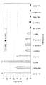
SUIT-2 007およびSUIT-2 028細胞株からの全RNA 10 μgを変性1%アガロースホルムアルデヒドゲル上に並べてローディングし、電気泳動によるサイズ分離を行った。BrightStar-Plus(商標)正荷電ナイロン膜へのブロッティングを毛細管性下方移行によって行った。UV架橋(Stratagene UV Stratalinker 2400)の後にブロットをハイブリダイズさせた。Strip-EZ(商標)DNA Kit(Ambion Inc.、Austin、Texas)を用いて、RT-PCR産物をα-[32P]dATPにより2×10⁹ cpm/μgの比活性で標識した。放射性プローブとのプレハイブリダイゼーション(30分間)およびハイブリダイゼーション(一晩)を、68℃のExpressHyb(商標)ハイブリダイゼーション溶液(Clontech、Palo Alto、CA、USA)中で行った。室温の溶液1(2×SSC、0.05%SDS)中で、連続攪拌および洗浄液1を数回交換しながら膜を30分〜40分間洗浄し、その後、50℃の溶液2(0.1×SSC、0.1%SDS)中で新たな溶液に1回交換しながら40分間洗浄した。続いて膜をCronex医用X線フィルム(Sterling Diagnostic Imaging Inc.、USA)に-70℃で2時間露出した。mRNAのローディングおよび膜への移行が等しくなされたことは、ブロットをα-[32P]dATP標識したGAPDH cDNAプローブと再びハイブリダイズさせることによって評価した。
Materials and methods:
Northern blot:
10 μg of total RNA from the SUIT-007 and SUIT-2028 cell lines was loaded side by side on a
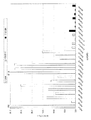
多組織発現アレイ(MTE(商標))
製造者の説明書に従って、ブロットをUKW cDNAに由来するα-[32P]dATP標識プローブとハイブリダイズさせ、X線フィルムに-70℃で62時間露出した。
Multi-tissue expression array (MTE ™)
The blot was hybridized with an α- [ 32 P] dATP-labeled probe from UKW cDNA according to the manufacturer's instructions and exposed to X-ray film at -70 ° C. for 62 hours.
Taqman(登録商標)-PCR
TaqMan(登録商標)法およびABI PRISM 7700装置(Applied Biosystems、Foster City、CA)を用いてリアルタイム定量PCRを行った。膵臓腺癌、慢性膵炎、および正常膵臓の凍結組織から単離した全RNA10 μgを容量20 μl中での逆転写反応に用いた。続いて、200 ngのcDNAを、4 μlの10×SYBR-Green緩衝液、3 mM MgCl2、1 mM dNTD、0.2単位のウラシル-Nグリコシラーゼ、1単位のAmpliTaq Gold、4 μlのプライマー混合物(各300 nMのプライマー:順方向
![]()
![]()
Real-time quantitative PCR was performed using the TaqMan® method and the ABI PRISM 7700 instrument (Applied Biosystems, Foster City, CA). 10 μg of total RNA isolated from frozen tissues of pancreatic adenocarcinoma, chronic pancreatitis, and normal pancreas was used for the reverse transcription reaction in a volume of 20 μl. Subsequently, 200 ng of cDNA was combined with 4 μl of 10 × SYBR-Green buffer, 3 mM MgCl 2 , 1 mM dNTD, 0.2 units of uracil-N glycosylase, 1 unit of AmpliTaq Gold, 4 μl of primer mixture (each 300 nM primer: forward
![]()
![]()
プライマー:

ライトサイクラー(LightCycler)-PCR
Taq DNAポリメラーゼ、SYBR-Green I、デオキシリボヌクレオシド三リン酸(LightCycler DNA master SYBR-Green I、Roche Molecular Biochemicals)を含む市販のマスター混合物を用い、LightCyclerキャピラリー内でLightCycler(Roche Molecular Biochemicals、Mannheim、Germany)を用いて、LightCycler-PCRを行った。プライマー(順方向
![]()
![]()
および伸長(72℃ 8秒間)を37サイクル行った。温度推移速度はすべて20℃/秒に設定した。PCR増幅の完了後に融解曲線分析を行った。この手順のためには、PCR産物を95℃で変性させ、65℃でアニーリングさせた上で、95℃まで徐々に加熱した。SYBR-Green I蛍光を0.1℃毎に段階的にモニターした。融解曲線を肉眼検査によって分析し、増幅されたUKW遺伝子の再配列構造は85℃〜88℃で融解した。プライマー二量体の形成の対照として、鋳型DNAを含まない対照(「水対照」)を各実験に含めた。通常は小さなピークが78℃に認められ、これは85℃〜88℃で特定の増幅産物によって生じたピークとは区別された。膵癌細胞株Suit-2 007由来のcDNAの連続希釈(1:10、1:50および1:80)を用いて、カルネキシンに対する検量線を作成した。UKW cDNAおよびカルネキシンの相対量を検量線に基づいて決定した。UKW遺伝子の相対的発現量は、各試料に関してUKW遺伝子およびカルネキシンの定常レベルを除算することによって得た、正規化値として算出した。カルネキシン順方向プライマー
![]()
![]()
Using a commercially available master mixture containing Taq DNA polymerase, SYBR-Green I, deoxyribonucleoside triphosphate (LightCycler DNA master SYBR-Green I, Roche Molecular Biochemicals), LightCycler (Roche Molecular Biochemicals, Mannheim, Germany) in a LightCycler capillary Was used to perform LightCycler-PCR. Primer (forward
![]()
![]()
And elongation (72 ° C. for 8 seconds) for 37 cycles. All temperature transition rates were set at 20 ° C / sec. After completion of the PCR amplification, a melting curve analysis was performed. For this procedure, the PCR product was denatured at 95 ° C., annealed at 65 ° C., and then gradually heated to 95 ° C. SYBR-Green I fluorescence was monitored in steps every 0.1 ° C. The melting curves were analyzed by visual inspection and the rearranged structure of the amplified UKW gene melted at 85-88 ° C. A control without template DNA ("water control") was included in each experiment as a control for primer dimer formation. Usually a small peak was observed at 78 ° C., which was distinct from peaks produced by specific amplification products at 85 ° C. to 88 ° C. Calibration curves for calnexin were generated using serial dilutions (1:10, 1:50 and 1:80) of cDNA from the pancreatic cancer cell line Suit-007. The relative amounts of UKW cDNA and calnexin were determined based on a calibration curve. The relative expression of the UKW gene was calculated as a normalized value obtained by dividing the steady-state levels of the UKW gene and calnexin for each sample. Calnexin forward primer
![]()
![]()
UKWポリペプチドに対する抗体の作製
免疫化のために、アミノ酸167位〜182位およびアミノ酸306位〜320位のペプチド(配列番号:2)を合成し、担体分子と結合させた。ウサギ2匹をペプチド混合物により免疫化した。
Generation of Antibodies to UKW Polypeptides For immunization, peptides at amino acids 167-182 and amino acids 306-320 (SEQ ID NO: 2) were synthesized and coupled to a carrier molecule. Two rabbits were immunized with the peptide mixture.
標準的な免疫化プロトコール(ウサギに対して):
第0日:免疫前血清の採取後に1回目の免疫化
第14日:2回目の免疫化
第28日:3回目の免疫化
第38日:血液試料の採取
第56日:4回目の免疫化
第66日:血液試料の採取
第80日:完全放血
Standard immunization protocol (for rabbits):
Day 0: First immunization after collection of pre-immune serum Day 14: Second immunization Day 28: Third immunization Day 38: Collection of blood sample Day 56: Fourth immunization Day 66: Collection of blood sample Day 80: Complete exsanguination
回収した抗血清はアフィニティークロマトグラフィーによって精製した。 The collected antiserum was purified by affinity chromatography.
Claims (7)
(i)患者から生体試料を入手する段階;
(ii)試料中の、UKWをコードする核酸の量またはポリペプチドUKWの量を検出する段階;および
(iii)核酸またはポリペプチドの量を、腫瘍誘発性または腫瘍誘発性でない、細胞内のUKWの発現または存在を判別する境界線を示す所定の標準値と比較し、それによって患者における膵癌の有無を判定する段階、
を含む方法。 A method for determining the presence or absence of pancreatic cancer in a patient,
(I) obtaining a biological sample from a patient;
(Ii) detecting the amount of UKW-encoding nucleic acid or polypeptide UKW in the sample; and (iii) determining the amount of nucleic acid or polypeptide in the tumor-inducing or non-tumorigenic UKW Comparing with a predetermined standard value indicating a boundary line for determining the expression or presence of, thereby determining the presence or absence of pancreatic cancer in the patient,
A method that includes
(a)それぞれの試料を、ストリンジェントなハイブリダイゼーション条件下で、
(i)配列番号:1の核酸配列またはその断片;
(ii)(i)の任意の核酸配列に対して相補的な核酸配列;
(iii)(i)の配列とストリンジェントな条件下でハイブリダイズする核酸配列;および
(iv)(ii)の配列とストリンジェントな条件下でハイブリダイズする核酸配列、
からなる群より選択される核酸プローブとともにインキュベートする段階;
(b)それぞれの試料の該プローブとのハイブリダイゼーションの概量を決定する段階、ならびに
(c)被験試料のハイブリダイゼーションの概量を第2の試料のハイブリダイゼーションの概量と比較し、被験試料が特定の核酸または核酸の混合物を該第2の試料よりも多量に含むか否かを明らかにする段階、
を含むプロセス。 Using a test sample and a second sample from a non-pancreatic tumor cell of the same individual or a different individual of the same species, whether the test sample in the tissue or body fluid of the patient contains pancreatic tumor cells, or derived from pancreatic tumor cells A process for determining whether to do
(A) running each sample under stringent hybridization conditions,
(I) the nucleic acid sequence of SEQ ID NO: 1 or a fragment thereof;
(Ii) a nucleic acid sequence complementary to any nucleic acid sequence of (i);
(Iii) a nucleic acid sequence that hybridizes under stringent conditions with the sequence of (i); and (iv) a nucleic acid sequence that hybridizes under stringent conditions with the sequence of (ii),
Incubating with a nucleic acid probe selected from the group consisting of:
(B) determining the approximate amount of hybridization of each sample with the probe; and (c) comparing the approximate amount of hybridization of the test sample with the approximate amount of hybridization of the second sample, Determining whether the sample comprises a particular nucleic acid or mixture of nucleic acids in a greater amount than the second sample.
Including the process.
a)体液、細胞または細胞の細胞抽出物もしくは細胞培養上清の群から選択される、膵癌に罹患していることが疑われる患者の試料であって、核酸を含む試料を、
(i)配列番号:1に示された核酸、または該配列に対して相補的な核酸、および
(ii)(i)に由来する核酸のいずれか1つとハイブリダイズする核酸、
からなる群より選択される核酸プローブとともにインキュベートする段階、ならびに
b)好ましくは試料の核酸および/もしくは核酸プローブの別の結合パートナーにより、またはX線撮影法により、ハイブリダイゼーションを検出する段階、
を含む方法。 A method for detecting pancreatic tumor, comprising:
a) a sample of a patient suspected of having pancreatic cancer, wherein the sample comprises nucleic acids, selected from the group of body fluids, cells or cell extracts of cells or cell culture supernatants;
(I) a nucleic acid set forth in SEQ ID NO: 1, or a nucleic acid complementary to the sequence, and (ii) a nucleic acid that hybridizes with any one of the nucleic acids derived from (i),
Incubating with a nucleic acid probe selected from the group consisting of:
b) detecting hybridization, preferably by another binding partner of the sample nucleic acid and / or nucleic acid probe, or by radiography;
A method that includes
A pharmaceutical composition comprising an antibody against UKW having the polypeptide sequence of SEQ ID NO: 1 together with a pharmaceutically acceptable carrier.
Applications Claiming Priority (1)
| Application Number | Priority Date | Filing Date | Title |
|---|---|---|---|
| EP02024539 | 2002-10-31 |
Related Child Applications (1)
| Application Number | Title | Priority Date | Filing Date |
|---|---|---|---|
| JP2009201213A Division JP2009284907A (en) | 2002-10-31 | 2009-09-01 | Method for diagnosis and therapy for pancreatic cancer and composition useful therein |
Publications (2)
| Publication Number | Publication Date |
|---|---|
| JP2004147656A true JP2004147656A (en) | 2004-05-27 |
| JP4394924B2 JP4394924B2 (en) | 2010-01-06 |
Family
ID=32405681
Family Applications (2)
| Application Number | Title | Priority Date | Filing Date |
|---|---|---|---|
| JP2003371406A Expired - Fee Related JP4394924B2 (en) | 2002-10-31 | 2003-10-31 | Methods for diagnosis and treatment of pancreatic cancer and compositions useful therefor |
| JP2009201213A Withdrawn JP2009284907A (en) | 2002-10-31 | 2009-09-01 | Method for diagnosis and therapy for pancreatic cancer and composition useful therein |
Family Applications After (1)
| Application Number | Title | Priority Date | Filing Date |
|---|---|---|---|
| JP2009201213A Withdrawn JP2009284907A (en) | 2002-10-31 | 2009-09-01 | Method for diagnosis and therapy for pancreatic cancer and composition useful therein |
Country Status (6)
| Country | Link |
|---|---|
| US (2) | US7429452B2 (en) |
| EP (1) | EP1416279B1 (en) |
| JP (2) | JP4394924B2 (en) |
| AT (1) | ATE419533T1 (en) |
| DE (1) | DE60325541D1 (en) |
| ES (1) | ES2318079T3 (en) |
Cited By (1)
| Publication number | Priority date | Publication date | Assignee | Title |
|---|---|---|---|---|
| JP2019516981A (en) * | 2016-05-10 | 2019-06-20 | イミュノヴィア・アーベー | Method, array, and use thereof |
Families Citing this family (13)
| Publication number | Priority date | Publication date | Assignee | Title |
|---|---|---|---|---|
| WO2006022690A1 (en) * | 2004-08-06 | 2006-03-02 | Applera Corporation | Method and compositions for treating diseases targeting cd71 |
| WO2006038904A2 (en) * | 2004-08-06 | 2006-04-13 | Applera Corporation | Method and compositions for treating diseases targeting e-cadherin |
| US11001881B2 (en) | 2006-08-24 | 2021-05-11 | California Institute Of Technology | Methods for detecting analytes |
| US11525156B2 (en) | 2006-07-28 | 2022-12-13 | California Institute Of Technology | Multiplex Q-PCR arrays |
| WO2008014485A2 (en) * | 2006-07-28 | 2008-01-31 | California Institute Of Technology | Multiplex q-pcr arrays |
| US11560588B2 (en) | 2006-08-24 | 2023-01-24 | California Institute Of Technology | Multiplex Q-PCR arrays |
| DK2140269T3 (en) | 2007-03-27 | 2013-12-16 | Immunovia Ab | PROTEIN SIGNATURE / MARKERS TO DETECT ADENOCARCINOM |
| US8538733B2 (en) | 2008-01-25 | 2013-09-17 | Life Technologies Corporation | Methods for the analysis of dissociation melt curve data |
| US9708647B2 (en) | 2015-03-23 | 2017-07-18 | Insilixa, Inc. | Multiplexed analysis of nucleic acid hybridization thermodynamics using integrated arrays |
| US9499861B1 (en) | 2015-09-10 | 2016-11-22 | Insilixa, Inc. | Methods and systems for multiplex quantitative nucleic acid amplification |
| WO2017155858A1 (en) | 2016-03-07 | 2017-09-14 | Insilixa, Inc. | Nucleic acid sequence identification using solid-phase cyclic single base extension |
| EP3937780A4 (en) | 2019-03-14 | 2022-12-07 | InSilixa, Inc. | Methods and systems for time-gated fluorescent-based detection |
| GB202010970D0 (en) | 2020-07-16 | 2020-09-02 | Immunovia Ab | Methods, arrays and uses thereof |
Family Cites Families (5)
| Publication number | Priority date | Publication date | Assignee | Title |
|---|---|---|---|---|
| US81659A (en) * | 1868-09-01 | m ax i mi | ||
| CA1339709C (en) | 1987-02-19 | 1998-03-10 | Scripps Clinic And Research Foundation | Monoclonal antibodies to human pancreatic cancer |
| US5912143A (en) * | 1996-12-27 | 1999-06-15 | Incyte Pharmaceuticals, Inc. | Polynucleotides encoding a human mage protein homolog |
| AU3395900A (en) | 1999-03-12 | 2000-10-04 | Human Genome Sciences, Inc. | Human lung cancer associated gene sequences and polypeptides |
| AU2001273150A1 (en) * | 2000-07-20 | 2002-02-05 | Genentech Inc. | Secreted and transmembrane polypeptides and nucleic acids encoding the same |
-
2003
- 2003-10-29 AT AT03024982T patent/ATE419533T1/en not_active IP Right Cessation
- 2003-10-29 ES ES03024982T patent/ES2318079T3/en not_active Expired - Lifetime
- 2003-10-29 US US10/696,487 patent/US7429452B2/en not_active Expired - Fee Related
- 2003-10-29 DE DE60325541T patent/DE60325541D1/en not_active Expired - Lifetime
- 2003-10-29 EP EP03024982A patent/EP1416279B1/en not_active Expired - Lifetime
- 2003-10-31 JP JP2003371406A patent/JP4394924B2/en not_active Expired - Fee Related
-
2008
- 2008-04-24 US US12/150,089 patent/US20090136939A1/en not_active Abandoned
-
2009
- 2009-09-01 JP JP2009201213A patent/JP2009284907A/en not_active Withdrawn
Cited By (1)
| Publication number | Priority date | Publication date | Assignee | Title |
|---|---|---|---|---|
| JP2019516981A (en) * | 2016-05-10 | 2019-06-20 | イミュノヴィア・アーベー | Method, array, and use thereof |
Also Published As
| Publication number | Publication date |
|---|---|
| ES2318079T3 (en) | 2009-05-01 |
| JP4394924B2 (en) | 2010-01-06 |
| US7429452B2 (en) | 2008-09-30 |
| US20090136939A1 (en) | 2009-05-28 |
| EP1416279A1 (en) | 2004-05-06 |
| ATE419533T1 (en) | 2009-01-15 |
| JP2009284907A (en) | 2009-12-10 |
| US20040110219A1 (en) | 2004-06-10 |
| EP1416279B1 (en) | 2008-12-31 |
| DE60325541D1 (en) | 2009-02-12 |
Similar Documents
| Publication | Publication Date | Title |
|---|---|---|
| JP2009284907A (en) | Method for diagnosis and therapy for pancreatic cancer and composition useful therein | |
| DK2456889T3 (en) | Markers of endometrial cancer | |
| JP6048660B2 (en) | Methods and compositions for diagnosing and treating thyroid cancer | |
| CA2638974A1 (en) | Screening for anti-cancer compounds using netrin-1 activity | |
| KR102047502B1 (en) | Composition for diagnosing cancer using potassium channel protein | |
| WO2008097466A2 (en) | Metastasis specific splice variants of mena and uses thereof in diagnosis, prognosis and treatment of tumors | |
| Pyle-Chenault et al. | VSGP/F-spondin: a new ovarian cancer marker | |
| JP2013213774A (en) | Biomarker for inspecting tuberculosis | |
| US20090061436A1 (en) | Peptide sequence that promotes tumor invasion | |
| EP2148932A1 (en) | Sox11 expression in malignant lymphomas | |
| JP5145549B2 (en) | Tumor marker | |
| CA2604983A1 (en) | Biomarker for sensitivity to mtor inhibitor therapy in kidney cancer | |
| KR102055350B1 (en) | Biomarker for Diagnosis of Anticancer drug Resistance of Colon Cancer and Uses thereof | |
| WO2014185466A1 (en) | Prognosis evaluation method for pancreatic cancer | |
| JP5316749B2 (en) | Cisplatin resistance gene diagnosis method and cisplatin therapeutic effect gene diagnosis kit | |
| JP2009535033A (en) | Compositions and methods for detection of Cripto-3 | |
| JP2010216826A (en) | Method for examinination of mammary cancer using novel tumor marker | |
| WO2022080305A1 (en) | Anti-ptdss2 antibody | |
| CA2852629A1 (en) | Usp2a peptides and antibodies | |
| JP6041297B2 (en) | Diagnostic method and diagnostic kit for canine lymphoma | |
| JP6306124B2 (en) | Tuberculosis testing biomarker | |
| AU2008309349A1 (en) | Method of detecting the risk of cancer using genetic markers | |
| CN115485559A (en) | Biomarkers predictive of response to checkpoint inhibitors | |
| JP4474551B2 (en) | Growth inhibitor and invasive inhibitor of glioma cell line U251 | |
| US7928188B2 (en) | Antigen polypeptide for the diagnosis and/or treatment of ovarian cancer |
Legal Events
| Date | Code | Title | Description |
|---|---|---|---|
| A621 | Written request for application examination |
Free format text: JAPANESE INTERMEDIATE CODE: A621 Effective date: 20060518 |
|
| A131 | Notification of reasons for refusal |
Free format text: JAPANESE INTERMEDIATE CODE: A131 Effective date: 20090610 |
|
| A521 | Request for written amendment filed |
Free format text: JAPANESE INTERMEDIATE CODE: A523 Effective date: 20090901 |
|
| TRDD | Decision of grant or rejection written | ||
| A01 | Written decision to grant a patent or to grant a registration (utility model) |
Free format text: JAPANESE INTERMEDIATE CODE: A01 Effective date: 20090925 |
|
| A01 | Written decision to grant a patent or to grant a registration (utility model) |
Free format text: JAPANESE INTERMEDIATE CODE: A01 |
|
| A61 | First payment of annual fees (during grant procedure) |
Free format text: JAPANESE INTERMEDIATE CODE: A61 Effective date: 20091016 |
|
| R150 | Certificate of patent or registration of utility model |
Free format text: JAPANESE INTERMEDIATE CODE: R150 |
|
| FPAY | Renewal fee payment (event date is renewal date of database) |
Free format text: PAYMENT UNTIL: 20121023 Year of fee payment: 3 |
|
| LAPS | Cancellation because of no payment of annual fees |