ES2914692T3 - Improved umbilical cord-derived adhesive stem cells, preparation method therefor, and use thereof - Google Patents
Improved umbilical cord-derived adhesive stem cells, preparation method therefor, and use thereof Download PDFInfo
- Publication number
- ES2914692T3 ES2914692T3 ES16835478T ES16835478T ES2914692T3 ES 2914692 T3 ES2914692 T3 ES 2914692T3 ES 16835478 T ES16835478 T ES 16835478T ES 16835478 T ES16835478 T ES 16835478T ES 2914692 T3 ES2914692 T3 ES 2914692T3
- Authority
- ES
- Spain
- Prior art keywords
- stem cells
- umbilical cord
- derived adherent
- adherent stem
- potentiated
- Prior art date
- Legal status (The legal status is an assumption and is not a legal conclusion. Google has not performed a legal analysis and makes no representation as to the accuracy of the status listed.)
- Active
Links
Classifications
-
- C—CHEMISTRY; METALLURGY
- C12—BIOCHEMISTRY; BEER; SPIRITS; WINE; VINEGAR; MICROBIOLOGY; ENZYMOLOGY; MUTATION OR GENETIC ENGINEERING
- C12N—MICROORGANISMS OR ENZYMES; COMPOSITIONS THEREOF; PROPAGATING, PRESERVING, OR MAINTAINING MICROORGANISMS; MUTATION OR GENETIC ENGINEERING; CULTURE MEDIA
- C12N5/00—Undifferentiated human, animal or plant cells, e.g. cell lines; Tissues; Cultivation or maintenance thereof; Culture media therefor
- C12N5/06—Animal cells or tissues; Human cells or tissues
- C12N5/0602—Vertebrate cells
- C12N5/0603—Embryonic cells ; Embryoid bodies
- C12N5/0605—Cells from extra-embryonic tissues, e.g. placenta, amnion, yolk sac, Wharton's jelly
-
- A—HUMAN NECESSITIES
- A61—MEDICAL OR VETERINARY SCIENCE; HYGIENE
- A61K—PREPARATIONS FOR MEDICAL, DENTAL OR TOILETRY PURPOSES
- A61K35/00—Medicinal preparations containing materials or reaction products thereof with undetermined constitution
- A61K35/12—Materials from mammals; Compositions comprising non-specified tissues or cells; Compositions comprising non-embryonic stem cells; Genetically modified cells
- A61K35/48—Reproductive organs
- A61K35/51—Umbilical cord; Umbilical cord blood; Umbilical stem cells
-
- A—HUMAN NECESSITIES
- A61—MEDICAL OR VETERINARY SCIENCE; HYGIENE
- A61K—PREPARATIONS FOR MEDICAL, DENTAL OR TOILETRY PURPOSES
- A61K35/00—Medicinal preparations containing materials or reaction products thereof with undetermined constitution
- A61K35/12—Materials from mammals; Compositions comprising non-specified tissues or cells; Compositions comprising non-embryonic stem cells; Genetically modified cells
- A61K35/48—Reproductive organs
- A61K35/50—Placenta; Placental stem cells; Amniotic fluid; Amnion; Amniotic stem cells
-
- A—HUMAN NECESSITIES
- A61—MEDICAL OR VETERINARY SCIENCE; HYGIENE
- A61P—SPECIFIC THERAPEUTIC ACTIVITY OF CHEMICAL COMPOUNDS OR MEDICINAL PREPARATIONS
- A61P25/00—Drugs for disorders of the nervous system
-
- A—HUMAN NECESSITIES
- A61—MEDICAL OR VETERINARY SCIENCE; HYGIENE
- A61P—SPECIFIC THERAPEUTIC ACTIVITY OF CHEMICAL COMPOUNDS OR MEDICINAL PREPARATIONS
- A61P37/00—Drugs for immunological or allergic disorders
-
- A—HUMAN NECESSITIES
- A61—MEDICAL OR VETERINARY SCIENCE; HYGIENE
- A61P—SPECIFIC THERAPEUTIC ACTIVITY OF CHEMICAL COMPOUNDS OR MEDICINAL PREPARATIONS
- A61P9/00—Drugs for disorders of the cardiovascular system
-
- C—CHEMISTRY; METALLURGY
- C12—BIOCHEMISTRY; BEER; SPIRITS; WINE; VINEGAR; MICROBIOLOGY; ENZYMOLOGY; MUTATION OR GENETIC ENGINEERING
- C12N—MICROORGANISMS OR ENZYMES; COMPOSITIONS THEREOF; PROPAGATING, PRESERVING, OR MAINTAINING MICROORGANISMS; MUTATION OR GENETIC ENGINEERING; CULTURE MEDIA
- C12N5/00—Undifferentiated human, animal or plant cells, e.g. cell lines; Tissues; Cultivation or maintenance thereof; Culture media therefor
- C12N5/06—Animal cells or tissues; Human cells or tissues
- C12N5/0602—Vertebrate cells
- C12N5/0652—Cells of skeletal and connective tissues; Mesenchyme
- C12N5/0662—Stem cells
- C12N5/0665—Blood-borne mesenchymal stem cells, e.g. from umbilical cord blood
-
- C—CHEMISTRY; METALLURGY
- C12—BIOCHEMISTRY; BEER; SPIRITS; WINE; VINEGAR; MICROBIOLOGY; ENZYMOLOGY; MUTATION OR GENETIC ENGINEERING
- C12N—MICROORGANISMS OR ENZYMES; COMPOSITIONS THEREOF; PROPAGATING, PRESERVING, OR MAINTAINING MICROORGANISMS; MUTATION OR GENETIC ENGINEERING; CULTURE MEDIA
- C12N2500/00—Specific components of cell culture medium
- C12N2500/02—Atmosphere, e.g. low oxygen conditions
-
- C—CHEMISTRY; METALLURGY
- C12—BIOCHEMISTRY; BEER; SPIRITS; WINE; VINEGAR; MICROBIOLOGY; ENZYMOLOGY; MUTATION OR GENETIC ENGINEERING
- C12N—MICROORGANISMS OR ENZYMES; COMPOSITIONS THEREOF; PROPAGATING, PRESERVING, OR MAINTAINING MICROORGANISMS; MUTATION OR GENETIC ENGINEERING; CULTURE MEDIA
- C12N2501/00—Active agents used in cell culture processes, e.g. differentation
- C12N2501/10—Growth factors
- C12N2501/119—Other fibroblast growth factors, e.g. FGF-4, FGF-8, FGF-10
-
- C—CHEMISTRY; METALLURGY
- C12—BIOCHEMISTRY; BEER; SPIRITS; WINE; VINEGAR; MICROBIOLOGY; ENZYMOLOGY; MUTATION OR GENETIC ENGINEERING
- C12N—MICROORGANISMS OR ENZYMES; COMPOSITIONS THEREOF; PROPAGATING, PRESERVING, OR MAINTAINING MICROORGANISMS; MUTATION OR GENETIC ENGINEERING; CULTURE MEDIA
- C12N2501/00—Active agents used in cell culture processes, e.g. differentation
- C12N2501/90—Polysaccharides
- C12N2501/91—Heparin
-
- C—CHEMISTRY; METALLURGY
- C12—BIOCHEMISTRY; BEER; SPIRITS; WINE; VINEGAR; MICROBIOLOGY; ENZYMOLOGY; MUTATION OR GENETIC ENGINEERING
- C12N—MICROORGANISMS OR ENZYMES; COMPOSITIONS THEREOF; PROPAGATING, PRESERVING, OR MAINTAINING MICROORGANISMS; MUTATION OR GENETIC ENGINEERING; CULTURE MEDIA
- C12N5/00—Undifferentiated human, animal or plant cells, e.g. cell lines; Tissues; Cultivation or maintenance thereof; Culture media therefor
- C12N5/06—Animal cells or tissues; Human cells or tissues
- C12N5/0602—Vertebrate cells
- C12N5/0634—Cells from the blood or the immune system
- C12N5/0647—Haematopoietic stem cells; Uncommitted or multipotent progenitors
-
- C—CHEMISTRY; METALLURGY
- C12—BIOCHEMISTRY; BEER; SPIRITS; WINE; VINEGAR; MICROBIOLOGY; ENZYMOLOGY; MUTATION OR GENETIC ENGINEERING
- C12Y—ENZYMES
- C12Y304/00—Hydrolases acting on peptide bonds, i.e. peptidases (3.4)
- C12Y304/24—Metalloendopeptidases (3.4.24)
- C12Y304/24003—Microbial collagenase (3.4.24.3)
Abstract
Células madre adherentes derivadas de cordón umbilical potenciadas que tienen las siguientes características (a) a (g): a) tener un nivel de expresión más alto de TAGLN, COL1A1, IGFBP4, STC1, LRRC17, y IL33, en comparación con células madre de médula ósea; b) tener un nivel de expresión más bajo de TPMT, NAGK, ANXA4, CCND1, SERPINE1, PRNP, y CYP1B1, en comparación con células madre de médula ósea; c) mantener la morfología de fibroblastos adherentes durante subcultivo; d) tener la capacidad de diferenciarse para dar adipocitos, osteocitos, o condrocitos; e) tener características de antígeno de superficie Oct4-, SSEA4+, Tra-1-60-, CD11c-, CD8a-, CD200+, CD3- , CD1a-, CD16-, CD86-, CD40-, CD141+, CD61+, CD87+ y MIC A/B-; f) tener un nivel de expresión más alto de uno o más seleccionados del grupo que consiste en S100A10, BNIP3, IGFBP5, NDUFA4L2, DPYD, y SCARA3, en comparación con las cultivadas en condición de normoxia; y g) tener un nivel de expresión más bajo de uno o más seleccionados del grupo que consiste en IL8, ALDH1A1, DLC1, CTHRC1, y CPA4, en comparación con las cultivadas en condición de normoxia.Enhanced umbilical cord-derived adherent stem cells having the following characteristics (a) to (g): a) have a higher expression level of TAGLN, COL1A1, IGFBP4, STC1, LRRC17, and IL33, compared to stem cells from bone marrow; b) have a lower expression level of TPMT, NAGK, ANXA4, CCND1, SERPINE1, PRNP, and CYP1B1, compared to bone marrow stem cells; c) maintain adherent fibroblast morphology during subculture; d) have the ability to differentiate into adipocytes, osteocytes, or chondrocytes; e) have surface antigen characteristics Oct4-, SSEA4+, Tra-1-60-, CD11c-, CD8a-, CD200+, CD3-, CD1a-, CD16-, CD86-, CD40-, CD141+, CD61+, CD87+ and MIC A/B-; f) have a higher expression level of one or more selected from the group consisting of S100A10, BNIP3, IGFBP5, NDUFA4L2, DPYD, and SCARA3, compared to those cultured under normoxic condition; and g) have a lower expression level of one or more selected from the group consisting of IL8, ALDH1A1, DLC1, CTHRC1, and CPA4, compared to those cultured under normoxic condition.
Description
DESCRIPCIÓNDESCRIPTION
Células madre adhesivas derivadas de cordón umbilical mejoradas, método de preparación para las mismas, y uso de las mismasImproved umbilical cord-derived adhesive stem cells, preparation method therefor, and use thereof
Campo técnicotechnical field
La presente divulgación se refiere a células madre adherentes derivadas de cordón umbilical potenciadas, un método de preparación de las mismas, y uso de las mismas.The present disclosure relates to potentiated umbilical cord-derived adherent stem cells, a method of preparing them, and use thereof.
Antecedentes de la técnicaBackground art
Un agente terapéutico celular es un fármaco usado con el propósito de prevenir o tratar una enfermedad específica mediante el cambio de características de las células mediante un método de proliferación o selección de células ex vivo para restaurar las funciones de células y tejidos, y recientemente ha recibido mucha atención en los campos de enfermedades intratables y medicamentos regenerativos. Los agentes terapéuticos celulares pueden clasificarse en agentes terapéuticos de células somáticas y agentes terapéuticos de células madre según el grado de diferenciación, y los agentes terapéuticos de células madre pueden clasificarse en agentes terapéuticos de células madre embrionarias y agentes terapéuticos de células madre adultas.A cell therapeutic agent is a drug used for the purpose of preventing or treating a specific disease by changing the characteristics of cells by an ex vivo cell selection or proliferation method to restore cell and tissue functions, and has recently received a lot of attention in the fields of intractable diseases and regenerative medicines. Cellular therapeutics can be classified into somatic cell therapeutics and stem cell therapeutics according to the degree of differentiation, and stem cell therapeutics can be classified into embryonic stem cell therapeutics and adult stem cell therapeutics.
Hasta ahora, la mayoría de los estudios con respecto a células madre adultas se han realizado en la médula ósea. En algunos casos, se cultivan y administran células madre aisladas de tejido adiposo o sangre del cordón umbilical. Sin embargo, células madre se recogen de la médula ósea y el tejido adiposo mediante métodos invasivos, y las células madre aisladas de pacientes en la edad adulta o la senescencia tienen una capacidad de diferenciación y proliferación reducida. Aunque la sangre del cordón umbilical es fácil de recoger, el contenido de células madre en la sangre del cordón umbilical es bajo.Until now, most studies regarding adult stem cells have been done in bone marrow. In some cases, stem cells isolated from adipose tissue or umbilical cord blood are cultured and administered. However, stem cells are harvested from bone marrow and adipose tissue by invasive methods, and stem cells isolated from patients in adulthood or senescence have reduced differentiation and proliferation capacity. Although umbilical cord blood is easy to collect, the stem cell content in cord blood is low.
A diferencia de células derivadas de tejido adiposo o médula ósea, las del cordón umbilical (CU) no es invasivo y son fáciles de extraer porque se extraen de tejidos ya separados del cuerpo. A diferencia de las células madre derivadas de embriones, el Cu está libre de cuestiones éticas. Recientemente, el CU ha recibido mucha atención como material útil para enfermedades intratables o medicamentos regenerativos. Dado que las células derivadas de CU son células primitivas que satisfacen las capacidades de proliferación y diferenciación al mismo tiempo, existen ventajas de que pueden usarse para la regeneración de órganos y también usarse después de la diferenciación según características de los órganos. Sin embargo, el CU es un tejido en el que están presentes muchos tipos diferentes de células, y, por lo tanto, se requieren estudios para descubrir células óptimas como agentes terapéuticos y para demostrar nuevas características de células que pueden separarse o extraerse como poblaciones celulares homogéneas.Unlike cells derived from adipose tissue or bone marrow, those from the umbilical cord (UC) are non-invasive and easy to extract because they are extracted from tissues already separated from the body. Unlike stem cells derived from embryos, Cu is free from ethical questions. Recently, CU has received a lot of attention as a useful material for intractable diseases or regenerative medicine. Since UC-derived cells are primitive cells that satisfy proliferation and differentiation capabilities at the same time, there are advantages that they can be used for organ regeneration and also used after differentiation according to organ characteristics. However, UC is a tissue in which many different cell types are present, and therefore studies are required to discover optimal cells as therapeutic agents and to demonstrate new characteristics of cells that can be separated or extracted as cell populations. homogeneous.
Descripción detallada de la invenciónDetailed description of the invention
Un aspecto proporciona células madre adherentes derivadas de cordón umbilical potenciadas o poblaciones celulares de las mismas.One aspect provides enhanced umbilical cord-derived adherent stem cells or cell populations thereof.
Otro aspecto proporciona un método para preparar las células madre adherentes derivadas de cordón umbilical potenciadas, incluyendo el método someter a cultivo adherente un cordón umbilical aislado en una placa de cultivo; aislar células madre adherentes derivadas de cordón umbilical potenciadas poniendo en contacto el cordón umbilical cultivado con una enzima de disociación; subcultivar las células madre adherentes derivadas de cordón umbilical potenciadas aisladas en un medio que contiene factor de crecimiento de fibroblastos 4 (FGF-4) y heparina.Another aspect provides a method of preparing the enhanced umbilical cord-derived adherent stem cells, the method including subjecting an isolated umbilical cord to adherent culture in a culture dish; isolating enhanced umbilical cord-derived adherent stem cells by contacting the cultured umbilical cord with a dissociating enzyme; Subculture the isolated potentiated umbilical cord-derived adherent stem cells in medium containing fibroblast growth factor 4 (FGF-4) and heparin.
Otro aspecto más proporciona una composición farmacéutica que incluye las células madre adherentes derivadas de cordón umbilical potenciadas, las poblaciones celulares de las mismas, o un cultivo de las mismas como principio activo.Yet another aspect provides a pharmaceutical composition that includes the potentiated umbilical cord-derived adherent stem cells, cell populations thereof, or a culture thereof as an active ingredient.
Problema técnicotechnical problem
Solución técnicatechnical solution
Un aspecto proporciona células madre adherentes derivadas de cordón umbilical potenciadas.One aspect provides enhanced umbilical cord-derived adherent stem cells.
Las células madre adherentes derivadas de cordón umbilical potenciadas tienen las características (a) a (e): Enhanced umbilical cord-derived adherent stem cells have the characteristics (a) to (e):
a) tener un nivel de expresión más alto de COL1A1, IGFBP4, TAGLN, STC1, LRRC17, y IL33, en comparación con células madre de médula ósea;a) have a higher expression level of COL1A1, IGFBP4, TAGLN, STC1, LRRC17, and IL33, compared to bone marrow stem cells;
b) tener un nivel de expresión más bajo de TPMT, NAGK, ANXA4, CCND1, SERPINE1, PRNP, y CYP1B1, en comparación con células madre de médula ósea; b) have a lower expression level of TPMT, NAGK, ANXA4, CCND1, SERPINE1, PRNP, and CYP1B1, compared to bone marrow stem cells;
c) mantener la morfología de fibroblastos adherentes durante subcultivo;c) maintain adherent fibroblast morphology during subculture;
d) tener la capacidad de diferenciarse para dar adipocitos, osteocitos, o condrocitos; yd) have the ability to differentiate into adipocytes, osteocytes, or chondrocytes; Y
e) tener antígeno de superficie Oct4-, CD200+, Tra-1-60-, CD3-, CD1a-, CD11c-, CD16-, CD86-, CD8a-, CD40-, CD141+, CD61+, CD87+, MIC A/B-, y SSEA4+.e) have surface antigen Oct4-, CD200+, Tra-1-60-, CD3-, CD1a-, CD11c-, CD16-, CD86-, CD8a-, CD40-, CD141+, CD61+, CD87+, MIC A/B- , and SSEA4+.
Las células madre adherentes derivadas de cordón umbilical potenciadas tienen además las características (f) a (g): Enhanced umbilical cord-derived adherent stem cells further have characteristics (f) to (g):
f) tener un nivel de expresión más alto de uno o más seleccionados del grupo que consiste en S100A10, BNIP3, IGFBP5, NDUFA4L2, DpYD, y SCARA3, en comparación con las cultivadas en condición de normoxia; y f) having a higher expression level of one or more selected from the group consisting of S100A10, BNIP3, IGFBP5, NDUFA4L2, DpYD, and SCARA3, compared to those cultured under normoxic condition; Y
g) tener un nivel de expresión más bajo de uno o más seleccionados del grupo que consiste en IL8, ALDH1A1, DLC1, CTHRC1, y CPA4, en comparación con las cultivadas en condición de normoxia.g) have a lower expression level of one or more selected from the group consisting of IL8, ALDH1A1, DLC1, CTHRC1, and CPA4, compared to those cultured under normoxic condition.
Las células madre adherentes derivadas de cordón umbilical potenciadas pueden tener además: h) un nivel de expresión más alto de uno o más seleccionados del grupo que consiste en SNCA, DSG2, NRP2, y PLAT, en comparación con células madre de médula ósea.Enhanced umbilical cord-derived adherent stem cells may further have: h) a higher expression level of one or more selected from the group consisting of SNCA, DSG2, NRP2, and PLAT, compared to bone marrow stem cells.
Las características de antígeno de superficie e) de las células madre adherentes derivadas de cordón umbilical potenciadas pueden incluir además Nanog-. Además, CD61+ de las características de antígeno de superficie e) pueden ser una característica de antígeno de superficie de sobreexpresarse en una condición de hipoxia.Surface antigen characteristics e) of the enhanced umbilical cord-derived adherent stem cells may further include Nanog-. Furthermore, CD61+ of the surface antigen characteristics e) may be a surface antigen characteristic of being overexpressed in a hypoxic condition.
El término “cordón umbilical”, como se usa en el presente documento, se refiere a un tubo que conecta la madre y el vientre para permitir que el feto de mamífero crezca en la placenta, y generalmente se refiere a un tejido compuesto por tres vasos, es decir, dos arterias umbilicales y una vena umbilical, que están rodeadas por gelatina de Wharton. Por lo tanto, en la presente divulgación, las “células madre adherentes derivadas de cordón umbilical potenciadas (células madre adherentes de cordón umbilical potenciadas)” o las “células madre adherentes derivadas de cordón umbilical (células madre adherentes de cordón umbilical)” se refieren a células que se derivan del cordón umbilical o del tejido de gelatina de Wharton del cordón umbilical y tienen la capacidad de diferenciarse para dar muchas células diferentes y una característica de crecimiento adherente en la superficie de una placa de cultivo.The term "umbilical cord," as used herein, refers to a tube that connects the mother and the womb to allow the mammalian fetus to grow into the placenta, and generally refers to a tissue composed of three vessels , that is, two umbilical arteries and one umbilical vein, which are surrounded by Wharton's jelly. Therefore, in the present disclosure, "enhanced umbilical cord-derived adherent stem cells (enhanced umbilical cord adherent stem cells)" or "umbilical cord-derived adherent stem cells (umbilical cord adherent stem cells)" refer to a cells that are derived from umbilical cord or Wharton's jelly tissue of the umbilical cord and have the ability to differentiate into many different cells and a characteristic of adherent growth on the surface of a culture dish.
Al menos aproximadamente un 20 %, aproximadamente un 25 %, aproximadamente un 30 %, aproximadamente un 35 %, aproximadamente un 40 %, aproximadamente un 45 %, aproximadamente un 50 %, aproximadamente un 55 %, aproximadamente un 60 %, aproximadamente un 65 %, aproximadamente un 70 %, aproximadamente un 75 %, aproximadamente un 80 %, aproximadamente un 85 %, aproximadamente un 90 %, aproximadamente un 95 %, aproximadamente un 98 %, o aproximadamente un 99 % de las células madre adherentes derivadas de cordón umbilical potenciadas proporcionadas en la presente invención pueden expresar marcador de superficie positivo para CD200, CD141, CD61, CD87, o SSEA4, que es un marcador celular expresado en la superficie celular, y al menos aproximadamente un 70 % o menos, al menos aproximadamente un 60 % o menos, al menos aproximadamente un 50 % o menos, al menos aproximadamente un 40 % o menos, al menos aproximadamente un 30 % o menos, al menos aproximadamente un 20 % o menos, al menos aproximadamente un 10 % o menos, al menos aproximadamente un 5 % o menos, o al menos aproximadamente un 1 % o menos de las mismas pueden expresar marcador de superficie negativo para Oct4, Nanog, Tra-1-60, CD3, CD1a, CD11c, CD16, CD86, CD8a, MIC A/B, o CD40 que es un marcador de células madre. El término “positivo”, como se usa en el presente documento, con respecto a un marcador de células madre, significa que el marcador celular existe en una gran cantidad o una alta concentración, en comparación con la de otras células no madre como referencia. Es decir, cualquier marcador está presente dentro o sobre la superficie de una célula, y, por lo tanto, si una célula puede distinguirse de uno o más tipos de células usando el marcador, la célula puede ser positiva para el marcador. Además, el término “positivo” significa que las células tienen señales de intensidad más alta que una intensidad de fondo, por ejemplo, las células tienen el marcador en una cantidad suficiente para poder detectarse en un dispositivo de medición celular. Por ejemplo, las células pueden etiquetarse de manera detectable con anticuerpos específicos de CD200, y cuando las señales de estos anticuerpos son detectablemente más intensas que las de un control (por ejemplo, intensidad de fondo), las células son “CD200+”. El término “negativo”, como se usa en el presente documento, significa que aunque se usan anticuerpos específicos para un marcador de superficie celular particular, el marcador no puede detectarse, en comparación con la intensidad de fondo. Por ejemplo, si una célula no puede etiquetarse de manera detectable con un anticuerpo específico de CD3, la célula es “CD3-”. At least about 20%, about 25%, about 30%, about 35%, about 40%, about 45%, about 50%, about 55%, about 60%, about 65 %, about 70%, about 75%, about 80%, about 85%, about 90%, about 95%, about 98%, or about 99% of cord-derived adherent stem cells umbilical cords provided in the present invention may express positive surface marker for CD200, CD141, CD61, CD87, or SSEA4, which is a cell marker expressed on the cell surface, and at least about 70% or less, at least about a 60% or less, at least about 50% or less, at least about 40% or less, at least about 30% or less, at least about 20% or less, at least about At least 10% or less, at least about 5% or less, or at least about 1% or less of these may express negative surface marker for Oct4, Nanog, Tra-1-60, CD3, CD1a, CD11c , CD16, CD86, CD8a, MIC A/B, or CD40 which is a marker for stem cells. The term "positive", as used herein, with respect to a stem cell marker, means that the cell marker exists in a high amount or concentration, compared to that of other non-stem cells as a reference. That is, any marker is present within or on the surface of a cell, and therefore, if a cell can be distinguished from one or more cell types using the marker, the cell may be positive for the marker. Furthermore, the term "positive" means that the cells have signals of intensity higher than a background intensity, eg, the cells have the marker in sufficient quantity to be detectable in a cell measurement device. For example, cells can be detectably labeled with CD200-specific antibodies, and when the signals from these antibodies are detectably more intense than a control (eg, background intensity), the cells are "CD200+." The term "negative", as used herein, means that although antibodies specific for a particular cell surface marker are used, the marker cannot be detected, compared to background intensity. For example, if a cell cannot be detectably labeled with a CD3-specific antibody, the cell is "CD3-".
Las características inmunológicas anteriores pueden determinarse mediante métodos comunes conocidos en la técnica a los que pertenece la presente divulgación. Por ejemplo, pueden usarse diversos métodos tales como citometría de flujo, tinción inmunohistoquímica, RT-PCR, etc....The above immunological characteristics can be determined by common methods known in the art to which the present disclosure pertains. For example, various methods such as flow cytometry, immunohistochemical staining, RT-PCR, etc... can be used.
Las células madre adherentes derivadas de cordón umbilical potenciadas tienen un nivel de expresión más alto de COL1A1, IGFBP4, TAGLN, STC1, LRRC17 y IL33, en comparación con células madre derivadas de médula ósea. Los genes altamente expresados en las células madre adherentes derivadas de cordón umbilical potenciadas según una realización específica, en comparación con las células madre derivadas de médula ósea, pueden incluir además S100A10, SQSTM1, DSTN, DCN, PHGDH, FBLN1, MFGE8, HLA-A, VASN, o KIAA1199. No hay informes sobre la asociación entre los genes anteriores y las células madre adherentes derivadas de cordón umbilical potenciadas. Una diferencia en los niveles de expresión de los genes entre las células madre adherentes derivadas de cordón umbilical potenciadas según una realización específica y las células madre derivadas de médula ósea puede ser dos veces o más alta. La diferencia en los niveles de expresión puede determinarse mediante, por ejemplo, comparación de los niveles de expresión génica a un nivel de ARNm. Además, la diferencia en los niveles de expresión puede determinarse mediante, por ejemplo, análisis de micromatrices.Enhanced umbilical cord-derived adherent stem cells have a higher expression level of COL1A1, IGFBP4, TAGLN, STC1, LRRC17, and IL33, compared to bone marrow-derived stem cells. Genes highly expressed in umbilical cord-derived adherent stem cells enhanced according to a specific embodiment, as compared to bone marrow-derived stem cells, may further include S100A10, SQSTM1, DSTN, DCN, PHGDH, FBLN1, MFGE8, HLA-A , VASN, or KIAA1199. There are no reports on the association between the above genes and enhanced umbilical cord-derived adherent stem cells. A difference in expression levels of genes between umbilical cord-derived adherent stem cells enhanced according to a specific embodiment and bone marrow-derived stem cells may be two-fold or higher. The difference in expression levels can be determined by, for example, comparing gene expression levels to an mRNA level. Furthermore, the difference in expression levels can be determined by, for example, microarray analysis.
Además, las células madre adherentes derivadas de cordón umbilical potenciadas tienen un nivel de expresión más bajo de TPMT, NAGK, ANXA4, CCND1, SERPINE1, PRNP, y CYP1B1, en comparación con células madre derivadas de médula ósea. Los genes o proteínas expresados débilmente en las células madre adherentes derivadas de cordón umbilical potenciadas según una realización específica, en comparación con las células madre derivadas de médula ósea, pueden incluir MTA2A, TM4SF1, HIST1H4C, y NME1. No hay informes sobre la asociación entre los genes o proteínas anteriores y las células madre adherentes derivadas de cordón umbilical potenciadas. Una diferencia en los niveles de expresión de los genes o las proteínas entre las células madre adherentes derivadas de cordón umbilical potenciadas según una realización específica y las células madre derivadas de médula ósea puede ser dos veces o más alta. La diferencia en los niveles de expresión puede determinarse mediante, por ejemplo, la comparación de los niveles de expresión génica a un nivel de ARNm. Además, la diferencia en los niveles de expresión puede determinarse mediante, por ejemplo, análisis de micromatrices.In addition, enhanced umbilical cord-derived adherent stem cells have a lower expression level of TPMT, NAGK, ANXA4, CCND1, SERPINE1, PRNP, and CYP1B1, compared to bone marrow-derived stem cells. Genes or proteins weakly expressed in the enhanced umbilical cord-derived adherent stem cells according to a specific embodiment, as compared to bone marrow-derived stem cells, may include MTA2A, TM4SF1, HIST1H4C, and NME1. There are no reports on the association between the above genes or proteins and enhanced umbilical cord-derived adherent stem cells. A difference in expression levels of genes or proteins between umbilical cord-derived adherent stem cells enhanced according to a specific embodiment and bone marrow-derived stem cells may be two-fold or higher. The difference in expression levels can be determined by, for example, comparing gene expression levels to an mRNA level. Furthermore, the difference in expression levels can be determined by, for example, microarray analysis.
Además, las células madre adherentes derivadas de cordón umbilical potenciadas tienen una morfología de fibroblastos en subcultivo. En una realización específica, las células pueden tener la propiedad de que las células requieran adhesión a la superficie para crecer in vitro, y pueden exhibir una morfología específica de fibroblastos en forma de huso.In addition, potentiated umbilical cord-derived adherent stem cells have a subcultured fibroblast morphology. In a specific embodiment, the cells may have the property that the cells require surface adhesion to grow in vitro, and may exhibit a spindle-shaped fibroblast-specific morphology.
En otra realización específica, las células madre adherentes derivadas de cordón umbilical potenciadas pueden tener capacidad de formación de colonias. Las células pueden tener una alta capacidad de formación de colonias, en comparación con las cultivadas en condición de normoxia.In another specific embodiment, the enhanced umbilical cord-derived adherent stem cells may have colony-forming ability. Cells may have a high colony-forming ability, compared to those grown under normoxic condition.
Además, las células madre adherentes derivadas de cordón umbilical potenciadas tienen la capacidad de diferenciarse para dar adipocitos, osteocitos, condrocitos. Pueden inducirse a las células a diferenciarse para dar linajes celulares particulares, por ejemplo, adipocitos, condrocitos, osteoblastos, células hematopoyéticas, miocitos, células vasculares, neuronas, o hepatocitos.Furthermore, potentiated umbilical cord-derived adherent stem cells have the ability to differentiate into adipocytes, osteocytes, chondrocytes. Cells can be induced to differentiate into particular cell lineages, for example, adipocytes, chondrocytes, osteoblasts, hematopoietic cells, myocytes, vascular cells, neurons, or hepatocytes.
El término “diferenciación”, como se usa en el presente documento, se refiere a un proceso por el cual células pasan a estar más especializadas en estructura o función durante el crecimiento celular a través de la división y proliferación, es decir, un proceso mediante el cual células, tejidos, etc. de un cambio de forma o función del organismo vivo para realizar la tarea dada. La determinación de la diferenciación para dar linajes celulares particulares puede lograrse mediante métodos bien conocidos en la técnica, y la diferenciación para dar células particulares puede inducirse a través de los métodos conocidos. Además, la diferenciación puede confirmarse midiendo cambios en marcadores de superficie celular (por ejemplo, tinción de células con anticuerpos específicos de tejido o específicos de marcador celular) y morfología usando técnicas tales como citometría de flujo o inmunocitoquímica, o examinando la morfología de células usando un microscopio óptico o un microscopio confocal, o midiendo cambios en la expresión génica usando técnicas bien conocidas en la técnica, tales como PCR y elaboración de perfiles de expresión génica.The term "differentiation," as used herein, refers to a process by which cells become more specialized in structure or function during cell growth through division and proliferation, that is, a process by which cells, tissues, etc. of a change of form or function of the living organism to perform the given task. Determination of differentiation into particular cell lineages can be achieved by methods well known in the art, and differentiation into particular cells can be induced by known methods. In addition, differentiation can be confirmed by measuring changes in cell surface markers (eg, staining cells with tissue-specific or cell marker-specific antibodies) and morphology using techniques such as flow cytometry or immunocytochemistry, or by examining cell morphology using a light microscope or a confocal microscope, or by measuring changes in gene expression using techniques well known in the art, such as PCR and gene expression profiling.
Además, las células madre adherentes derivadas de cordón umbilical potenciadas pueden secretar IL-6, IL-8, G-CSF, GM-CSF, MCP-3, VEGF, GRO, IFNy, IL-1a, IL-1b, IL-1ra, IL-3, IL-4, IL-7, IL-9, IL-12(p40), IL12(P70), IL-13, IL-14, IFNa2, MDC, sIL-2Ra, Eotaxina, ligando de Flt-3, MCP-1, MIP-1a, MIP1b, RANTE, fractalquina, IP-10, EGF, FGF-2, IGF-1 SR, EpCAM, IGFBP3, o una combinación de estas proteínas. Además, las células madre adherentes derivadas de cordón umbilical potenciadas pueden secretar, por ejemplo, dos o más, tres o más, cuatro o más, cinco o más, seis o más, siete o más, ocho o más, nueve o más, diez o más seleccionados del grupo que consiste en IL-6, IL-8, G-CSF, GM-CSF, MCP-3, VEGF, y GRO, y todas las proteínas.In addition, potentiated umbilical cord-derived adherent stem cells can secrete IL-6, IL-8, G-CSF, GM-CSF, MCP-3, VEGF, GRO, IFNγ, IL-1a, IL-1b, IL-1ra , IL-3, IL-4, IL-7, IL-9, IL-12(p40), IL12(p70), IL-13, IL-14, IFNa2, MDC, sIL-2Ra, Eotaxin, Flt ligand -3, MCP-1, MIP-1a, MIP1b, RANTE, fractalkine, IP-10, EGF, FGF-2, IGF-1 SR, EpCAM, IGFBP3, or a combination of these proteins. In addition, potentiated umbilical cord-derived adherent stem cells may secrete, for example, two or more, three or more, four or more, five or more, six or more, seven or more, eight or more, nine or more, ten or more selected from the group consisting of IL-6, IL-8, G-CSF, GM-CSF, MCP-3, VEGF, and GRO, and all proteins.
Además, las células madre adherentes derivadas de cordón umbilical potenciadas cultivadas en una condición de hipoxia pueden tener un nivel de expresión aumentado de uno o más genes o proteínas seleccionados del grupo que consiste en S100A10, BNIP3, IGFBP5, PGK1, TPI1, DCN, PGM1, PFKFB3, LOC644774, MME, MIR1978, SLC2A3, BHLHB2, BNIP3L, IGFBP5, NDUFA4L2, DPYD, y SCARA3, en comparación con las cultivadas en condición de normoxia. Específicamente, las células madre adherentes derivadas de cordón umbilical potenciadas cultivadas en una condición de hipoxia pueden tener un nivel de expresión aumentado de dos o más, tres o más, cuatro o más, cinco o más, o seis o más genes o proteínas seleccionados del grupo que consiste en S100A10, BNIP3, IGFBP5, PGK1, TPI1, DCN, PGM1, PFKFB3, LOC644774, MME, MIR1978, SLC2A3, BHLHB2, BNIP3L, IGFBP5, NDUFA4L2, DPYD, y SCARA3, o nivel de expresión aumentado de todos los genes o proteínas, en comparación con las cultivadas en condición de normoxia. No hay informes sobre la asociación entre los genes o proteínas anteriores y las células madre adherentes derivadas de cordón umbilical potenciadas. Una diferencia en los niveles de expresión puede ser dos veces o más alta. La diferencia en los niveles de expresión puede determinarse mediante, por ejemplo, la comparación de los niveles de expresión de genes y proteínas a nivel de ARNm o a nivel de proteína. Además, la diferencia en los niveles de expresión puede determinarse mediante, por ejemplo, análisis de micromatrices y análisis proteómico.Furthermore, potentiated umbilical cord-derived adherent stem cells cultured under a hypoxic condition may have an increased expression level of one or more genes or proteins selected from the group consisting of S100A10, BNIP3, IGFBP5, PGK1, TPI1, DCN, PGM1 , PFKFB3, LOC644774, MME, MIR1978, SLC2A3, BHLHB2, BNIP3L, IGFBP5, NDUFA4L2, DPYD, and SCARA3, compared to those cultured under normoxic condition. Specifically, potentiated umbilical cord-derived adherent stem cells cultured under a hypoxic condition may have an increased expression level of two or more, three or more, four or more, five or more, or six or more genes or proteins selected from the group consisting of S100A10, BNIP3, IGFBP5, PGK1, TPI1, DCN, PGM1, PFKFB3, LOC644774, MME, MIR1978, SLC2A3, BHLHB2, BNIP3L, IGFBP5, NDUFA4L2, DPYD, and SCARA3, or increased expression level of all genes or protein, compared to those cultured under normoxic condition. There are no reports on the association between the above genes or proteins and enhanced umbilical cord-derived adherent stem cells. A difference in expression levels can be two-fold or higher. The difference in expression levels can be determined by, for example, comparing the expression levels of genes and proteins at the mRNA level or at the protein level. Furthermore, the difference in expression levels can be determined by, for example, microarray analysis and analysis. proteomic.
Además, las células madre adherentes derivadas de cordón umbilical potenciadas cultivadas en una condición de hipoxia pueden tener un nivel de expresión disminuido de uno o más genes o proteínas seleccionados del grupo que consiste en IL8, ALDH1A1, NQO1, DLC1, CTHRC1, y CPA4, en comparación con las cultivadas en condición de normoxia. Específicamente, las células madre adherentes derivadas de cordón umbilical potenciadas cultivadas en una condición de hipoxia pueden tener un nivel de expresión disminuido de dos o más o tres o más genes o proteínas seleccionados del grupo que consiste en IL8, ALDH1A1, NQO1, DLC1, CTHRC1 y CPA4, o nivel de expresión disminuido de todos los genes o proteínas, en comparación con las cultivadas en condición de normoxia. No hay informes sobre la asociación entre los genes o proteínas anteriores y las células madre adherentes derivadas de cordón umbilical potenciadas. Una diferencia en los niveles de expresión puede ser dos veces o más alta. La diferencia en los niveles de expresión puede determinarse mediante, por ejemplo, la comparación de los niveles de expresión de genes y proteínas a nivel de ARNm o a nivel de proteína. Además, la diferencia en los niveles de expresión puede determinarse mediante, por ejemplo, análisis de micromatrices y análisis proteómico.In addition, potentiated umbilical cord-derived adherent stem cells cultured under a hypoxic condition may have a decreased expression level of one or more genes or proteins selected from the group consisting of IL8, ALDH1A1, NQO1, DLC1, CTHRC1, and CPA4, compared to those cultured under normoxic condition. Specifically, potentiated umbilical cord-derived adherent stem cells cultured under a hypoxic condition may have decreased expression level of two or more or three or more genes or proteins selected from the group consisting of IL8, ALDH1A1, NQO1, DLC1, CTHRC1 and CPA4, or decreased expression level of all genes or proteins, compared to those cultured under normoxic condition. There are no reports on the association between the above genes or proteins and enhanced umbilical cord-derived adherent stem cells. A difference in expression levels can be two-fold or higher. The difference in expression levels can be determined by, for example, comparing the expression levels of genes and proteins at the mRNA level or at the protein level. Furthermore, the difference in expression levels can be determined by, for example, microarray analysis and proteomic analysis.
Otro aspecto proporciona poblaciones celulares de células madre adherentes derivadas de cordón umbilical potenciadas.Another aspect provides cell populations of enhanced umbilical cord-derived adherent stem cells.
Las células madre adherentes derivadas de cordón umbilical son las mismas que las descritas anteriormente.Umbilical cord-derived adherent stem cells are the same as those described above.
Otro aspecto más proporciona un método para preparar las células madre adherentes derivadas de cordón umbilical potenciadas, incluyendo el método someter a cultivo adherente un cordón umbilical aislado en una placa de cultivo; aislar células madre adherentes derivadas de cordón umbilical potenciadas poniendo en contacto el cordón umbilical cultivado con una enzima de disociación; subcultivar las células madre adherentes derivadas de cordón umbilical potenciadas aisladas en un medio que contiene factor de crecimiento de fibroblastos 4 (FGF-4) y heparina.Still another aspect provides a method of preparing the potentiated umbilical cord-derived adherent stem cells, the method including subjecting an isolated umbilical cord to adherent culture in a culture dish; isolating enhanced umbilical cord-derived adherent stem cells by contacting the cultured umbilical cord with a dissociating enzyme; Subculture the isolated potentiated umbilical cord-derived adherent stem cells in medium containing fibroblast growth factor 4 (FGF-4) and heparin.
El cordón umbilical puede ser un cordón umbilical que está separado de una madre sana (por ejemplo, madre negativa para VIH, VHC, VHB) después del parto. Es decir, el “cordón umbilical separado” puede referirse a un cordón umbilical separado del cuerpo de la madre después del parto. El cordón umbilical separado puede almacenarse en un contenedor estéril con hielo inmediatamente después de separarse.The umbilical cord can be an umbilical cord that is severed from a healthy mother (eg, HIV, HCV, HBV negative mother) after delivery. That is, "separated umbilical cord" can refer to an umbilical cord separated from the mother's body after childbirth. The separated umbilical cord can be stored in a sterile container on ice immediately after it is separated.
Un método para separar y obtener el cordón umbilical de la placenta puede incluir, por ejemplo, separar el cordón umbilical de la placenta separada; retirar sangre externa del cordón umbilical separado; retirar arterias y venas del cordón umbilical con la sangre retirada; y/o cortar el cordón umbilical, a partir de lo que se retiran la arteria y la vena, en un tamaño predeterminado (por ejemplo, de 1 mm a 20 mm). La retirada de la sangre puede realizarse usando DPBS sin Ca/Mg, o DPBS sin Ca/Mg que contiene gentamicina.A method of separating and harvesting the umbilical cord from the placenta may include, for example, separating the umbilical cord from the separated placenta; remove external blood from the separated umbilical cord; remove arteries and veins from the umbilical cord with the withdrawn blood; and/or cutting the umbilical cord, from which the artery and vein are removed, to a predetermined size (eg, 1mm to 20mm). Blood withdrawal can be performed using DPBS without Ca/Mg, or DPBS without Ca/Mg containing gentamicin.
A continuación, Pueden aislarse células madre a partir del cordón umbilical que se corta en un tamaño pequeño (por ejemplo, cordón umbilical separado). El aislamiento de las células madre adherentes derivadas de cordón umbilical potenciadas puede incluir el cultivo adherente del cordón umbilical separado en una placa de cultivo durante de 5 días a 20 días, por ejemplo, de 10 días a 20 días, por ejemplo, durante de 10 días a 15 días; confirmación de que las células se extienden a partir del tejido de cordón umbilical cultivado; y/o tratamiento del tejido del cordón umbilical con una enzima de disociación.Stem cells can then be isolated from the umbilical cord that is cut to a small size (eg, umbilical cord severed). Isolation of the enhanced umbilical cord-derived adherent stem cells may include adherent culture of the separated umbilical cord in a culture dish for 5 days to 20 days, eg, 10 days to 20 days, eg, for 10 days. days to 15 days; confirmation that the cells spread from the cultured umbilical cord tissue; and/or treatment of the umbilical cord tissue with a dissociating enzyme.
La enzima de disociación puede incluir colagenasa. La colagenasa puede referirse a una enzima que escinde enlaces peptídicos de colágeno, y puede incluir colagenasa tipo I, tipo II, tipo III, tipo IV, o una combinación de las mismas. Además, la enzima de disociación puede incluir colagenasa de 5 U/ml a 30 U/ml, por ejemplo, de 5 U/ml a 25 U/ml, de 10 U/ml a 25 U/ml, o de 20 U/ml. Además, la enzima de disociación puede incluir tripsina, y/o dispasa. Además, una disolución que incluye la enzima de disociación puede incluir agua, solución salina, por ejemplo, HBSS (solución salina equilibrada de Hank) que contiene colagenasa, tripsina, y/o dispasa. Además, un tiempo de tratamiento de la enzima de disociación puede ser, por ejemplo, de 1 hora a 20 horas, de 2 horas a 10 horas, de 4 horas a 9 horas, o de 5 horas a 6 horas.The dissociating enzyme may include collagenase. Collagenase may refer to an enzyme that cleaves collagen peptide bonds, and may include collagenase type I, type II, type III, type IV, or a combination thereof. In addition, the dissociating enzyme may include collagenase from 5 U/ml to 30 U/ml, for example, from 5 U/ml to 25 U/ml, from 10 U/ml to 25 U/ml, or from 20 U/ml. ml. Furthermore, the dissociation enzyme may include trypsin, and/or dispase. In addition, a solution including the dissociating enzyme may include water, saline, eg, HBSS (Hank's Balanced Salt Solution) containing collagenase, trypsin, and/or dispase. Also, a dissociation enzyme treatment time may be, for example, from 1 hour to 20 hours, from 2 hours to 10 hours, from 4 hours to 9 hours, or from 5 hours to 6 hours.
En una realización específica, la reacción entre el tejido y la enzima de disociación puede permitirse en agitación, y puede realizarse la agitación, por ejemplo, a de aproximadamente 20 °C a aproximadamente 40 °C, de aproximadamente 30 °C a aproximadamente 40 °C, o de aproximadamente 35 °C a aproximadamente 40 °C, por ejemplo, a aproximadamente 37 °C, durante de aproximadamente 5 minutos a aproximadamente 60 minutos o de aproximadamente 10 minutos a aproximadamente 30 minutos, por ejemplo, durante de aproximadamente 10 minutos a aproximadamente 30 minutos dos veces.In a specific embodiment, the reaction between the tissue and the dissociating enzyme may be allowed under agitation, and the agitation may be performed, for example, at from about 20°C to about 40°C, from about 30°C to about 40°C. C, or from about 35°C to about 40°C, for example, to about 37°C, for from about 5 minutes to about 60 minutes or from about 10 minutes to about 30 minutes, for example, for about 10 minutes to about 30 minutes twice.
Adicionalmente, después de la reacción del tejido y la enzima de disociación, puede realizarse adicionalmente un proceso de inactivación de la enzima de disociación, y, por ejemplo, la reacción enzimática puede terminarse añadiendo FBS. Además, un método de aislamiento de células de tejido, por ejemplo, células madre adherentes derivadas de cordón umbilical potenciadas a partir de la disolución de reacción enzimática puede realizarse mediante un método común conocido en la técnica. Por ejemplo, después de la centrifugación, las células pueden aislarse usando un filtro de células. Additionally, after the reaction of the tissue and the dissociating enzyme, a dissociating enzyme inactivation process may be further performed, and, for example, the enzyme reaction may be terminated by adding FBS. In addition, a method of isolating tissue cells, for example, potentiated umbilical cord-derived adherent stem cells from the enzyme reaction solution can be performed by a common method known in the art. For example, after centrifugation, cells can be isolated using a cell strainer.
El término “aislamiento de las células madre adherentes derivadas de cordón umbilical potenciadas”, como se usa en el presente documento, significa retirada de al menos un 20 %, 30 %, 40 %, 50 %, 60 %, 70 %, 80 %, 90 %, 95 % o 99 % de células normalmente asociadas con las células madre en un cordón umbilical de mamífero no tratado. Puede decirse que poblaciones celulares que contienen células madre obtenidas de un órgano “están aisladas”, cuando otras células normalmente asociadas con las células madre en el órgano no tratado son inferiores al 50 % de la totalidad de células.The term "isolation of enhanced umbilical cord-derived adherent stem cells," as used herein, means removal of at least 20%, 30%, 40%, 50%, 60%, 70%, 80% , 90%, 95% or 99% of cells normally associated with stem cells in an untreated mammalian umbilical cord. Cell populations containing stem cells obtained from an organ can be said to be "isolated" when other cells normally associated with stem cells in the untreated organ are less than 50% of all cells.
A continuación, el subcultivo puede realizarse tomando las células madre adherentes derivadas de cordón umbilical potenciadas aisladas como P0.Subculturing can then be performed by taking the isolated potentiated umbilical cord-derived adherent stem cells as P0.
El subcultivo puede incluir además tratar la enzima recombinante sin componentes animales (ACF) antes del trasplante celular para el subcultivo. El término “enzima sin componentes animales”, como se usa en el presente documento, significa que la enzima se origina a partir de un ser vivo no animal, lo que significa que la enzima no se purifica a partir de una fuente de suministro animal. La enzima sin componentes animales puede originarse a partir de recombinación, por ejemplo, originada a partir de bacterias, levaduras, o plantas. La enzima originada a partir de recombinación puede significar cualquier enzima producida por tecnología de ADN recombinante, incluyendo el uso de microorganismos, por ejemplo, bacterias, virus, levaduras, plantas, etc. La enzima puede ser tripsina recombinante sin componentes animales, por ejemplo, tripsina recombinante producida en maíz. La tripsina recombinante sin componentes animales está disponible comercialmente, y, por ejemplo, puede ser TrypLE™ Select (GIBCO Invitrogen), TrypLE™ Express (GIBCO Invitrogen), TrypZean™ (Sigma Aldrich), o Recombinant Trypsin Solution™ (Biological Industries).Subculturing may further include treating the recombinant enzyme without animal components (ACF) prior to cell transplantation for subculturing. The term "animal-free enzyme," as used herein, means that the enzyme originates from a non-animal living source, which means that the enzyme is not purified from an animal supply source. The enzyme without animal components may originate from recombination, for example, originating from bacteria, yeast, or plants. Recombinantly derived enzyme can mean any enzyme produced by recombinant DNA technology, including the use of microorganisms, eg, bacteria, viruses, yeast, plants, etc. The enzyme may be recombinant trypsin without animal components, for example, recombinant trypsin produced in corn. Recombinant trypsin without animal components is commercially available, and may, for example, be TrypLE™ Select (GIBCO Invitrogen), TrypLE™ Express (GIBCO Invitrogen), TrypZean™ (Sigma Aldrich), or Recombinant Trypsin Solution™ (Biological Industries).
El subcultivo puede incluir cultivar las células en un medio de cultivo de células madre, por ejemplo, en un medio enriquecido con factor de crecimiento de fibroblastos (FGF-4) y heparina. Una concentración de f GF-4 en el medio puede ser de aproximadamente 10 ng/ml a aproximadamente 40 ng/ml, o de aproximadamente 20 ng/ml a aproximadamente 30 mg/ml, por ejemplo, 25 ng/ml. Una concentración de heparina en el medio puede ser de aproximadamente 0,5 |ig/ml a aproximadamente 2 |ig/ml, o de aproximadamente 0,5 |ig/ml a aproximadamente 1,5 |ig/ml, por ejemplo, de aproximadamente 1 |ig/ml. El medio puede incluir además, por ejemplo, suero bovino fetal y antibióticos (por ejemplo, penicilina, estreptomicina, gentamicina, etc.). En una realización específica, pueden usarse un medio CS-CM enriquecido con suero bovino fetal al 10 %, 50 |ig/ml de gentamicina, 1 |ig/ml de heparina, y 25 ng/ml de FGF-4. El subcultivo puede realizarse de aproximadamente 20 °C a aproximadamente 40 °C, de aproximadamente 30 °C a aproximadamente 40 °C, o de aproximadamente 35 °C a aproximadamente 40 °C, por ejemplo, a aproximadamente 37 °C, y puede ser un tiempo de cultivo para cada subcultivo, por ejemplo, de 2 días a 7 días, o de 3 días a 5 días.Subculturing may include culturing the cells in stem cell culture medium, for example, in medium supplemented with fibroblast growth factor (FGF-4) and heparin. A concentration of fGF-4 in the medium can be from about 10 ng/ml to about 40 ng/ml, or from about 20 ng/ml to about 30 mg/ml, eg, 25 ng/ml. A concentration of heparin in the medium can be from about 0.5 µg/ml to about 2 µg/ml, or from about 0.5 µg/ml to about 1.5 µg/ml, for example, from about 1 µg/mL. The medium may further include, for example, fetal bovine serum and antibiotics (eg, penicillin, streptomycin, gentamicin, etc.). In a specific embodiment, CS-CM medium supplemented with 10% fetal bovine serum, 50 µg/ml gentamicin, 1 µg/ml heparin, and 25 ng/ml FGF-4 can be used. Subculturing can be performed at about 20°C to about 40°C, about 30°C to about 40°C, or about 35°C to about 40°C, for example, about 37°C, and can be a culture time for each subculture, for example, from 2 days to 7 days, or from 3 days to 5 days.
En el método para preparar las células madre adherentes derivadas de cordón umbilical potenciadas según una realización específica, un número de pases del subcultivo no está particularmente limitado, y el número de pases puede seleccionarse apropiadamente según el número deseado de células proliferantes. Comúnmente, el número de paso puede ser al menos 1 paso o más, o 10 pases o más. Por ejemplo, pueden realizarse de 1 pase a 20 pases o de 3 pases a 15 pases para obtener el número acumulativo clínicamente requerido de células proliferantes.In the method for preparing the enhanced umbilical cord-derived adherent stem cells according to a specific embodiment, a passage number of the subculture is not particularly limited, and the passage number may be appropriately selected according to the desired number of proliferating cells. Commonly, the step number can be at least 1 step or more, or 10 steps or more. For example, 1 to 20 passages or 3 to 15 passages can be performed to obtain the clinically required cumulative number of proliferating cells.
Además, tras el subcultivo, el tratamiento de la enzima recombinante sin componentes animales también puede realizarse adicionalmente como se describió anteriormente. Es decir, en cada fase de subcultivo antes del subcultivo de las células a la siguiente fase, las células se trataron con la enzima recombinante sin componentes animales y se recogieron para aumentar la pureza de las células. Por ejemplo, la enzima recombinante sin componentes animales puede tratarse antes de transferir las células para P2 en la fase desde P1 hasta P2.Furthermore, after subculturing, the treatment of the recombinant enzyme without animal components can also be further performed as described above. That is, at each stage of subculturing before subculturing the cells to the next stage, the cells were treated with the recombinant enzyme without animal components and harvested to increase the purity of the cells. For example, recombinant enzyme without animal components can be treated before transferring cells to P2 at the P1 to P2 stage.
El subcultivo puede ser subcultivo en una condición de hipoxia a un nivel de oxígeno más bajo que la condición de normoxia del 21 %. El término “hipoxia” significa una presión parcial de oxígeno más baja a una presión parcial de oxígeno del 21 %, que es una condición de normoxia general. La condición de hipoxia puede ser una condición que tiene una presión parcial de oxígeno del 1 % al 15 %, del 1 % al 12 %, del 1 % al 10 %, o del 1 % al 5 %, por ejemplo, 1 %, 2 %, 3 %, 4 %, 5 %, 6 %, 7 %, 8 % o 9 %.The subculture may be subculture in a hypoxic condition at a lower oxygen level than the 21% normoxic condition. The term "hypoxia" means a lower oxygen partial pressure at 21% oxygen partial pressure, which is a condition of general normoxia. The hypoxic condition may be a condition having a partial pressure of oxygen of 1% to 15%, 1% to 12%, 1% to 10%, or 1% to 5%, for example, 1%, 2%, 3%, 4%, 5%, 6%, 7%, 8% or 9%.
En una realización específica, cuando las células se subcultivan en condición de hipoxia, un nivel de expresión de uno o más seleccionados del grupo que consiste en S100A10, BNIP3, IGFBP5, p Gk 1, TPI1, DCN, p Gm 1, PFKFB3, LOC644774, MME, MIR1978, SLC2A3, BHLHB2, BNIP3L, IGFBP5, NDUFA4L2, DPYD, y SCARA3 puede aumentarse, o un nivel de expresión de uno o más seleccionados del grupo que consiste en IL8, ALDH1A1, NQO1, DLC1, CTHRC1, y CPA4 puede disminuirse, en comparación con las subcultivadas en condición de normoxia. In a specific embodiment, when cells are subcultured under hypoxic condition, an expression level of one or more selected from the group consisting of S100A10, BNIP3, IGFBP5, pGk 1, TPI1, DCN, pGm 1, PFKFB3, LOC644774 , MME, MIR1978, SLC2A3, BHLHB2, BNIP3L, IGFBP5, NDUFA4L2, DPYD, and SCARA3 may be increased, or an expression level of one or more selected from the group consisting of IL8, ALDH1A1, NQO1, DLC1, CTHRC1, and CPA4 may decreased, compared to those subcultured under normoxic condition.
Las células madre adherentes derivadas de cordón umbilical potenciadas preparadas mediante el método de preparación anterior pueden tener las características descritas anteriormente, y, por ejemplo, las células madre adherentes derivadas de cordón umbilical potenciadas preparadas tienen las características (a) a (e):The potentiated umbilical cord-derived adherent stem cells prepared by the above preparation method may have the characteristics described above, and, for example, the potentiated umbilical cord-derived adherent stem cells prepared have the characteristics (a) to (e):
a) tener un nivel de expresión más alto de COL1A1, IGFBP4, TAGLN, STC1, LRRC17, y IL33, en comparación con células madre de médula ósea;a) have a higher expression level of COL1A1, IGFBP4, TAGLN, STC1, LRRC17, and IL33, compared to bone marrow stem cells;
b) tener un nivel de expresión más bajo de TPMT, NAGK, ANXA4, CCND1, SERPINE1, PRNP, y CYP1B1, en comparación con células madre de médula ósea;b) have a lower expression level of TPMT, NAGK, ANXA4, CCND1, SERPINE1, PRNP, and CYP1B1, in comparison with bone marrow stem cells;
c) mantener la morfología de fibroblastos adherentes durante subcultivo;c) maintain adherent fibroblast morphology during subculture;
d) tener la capacidad de diferenciarse para dar adipocitos, osteocitos, o condrocitos; yd) have the ability to differentiate into adipocytes, osteocytes, or chondrocytes; Y
e) tener características de antígeno de superficie Oct4-, CD200+, Tra-1-60-, CD3-, CD1a-, CD11c-, CD16-, CD86-, CD8a-, CD40-, CD141+, CD61+, CD87+, MIC A/B-, y SSEA4+.e) have surface antigen characteristics Oct4-, CD200+, Tra-1-60-, CD3-, CD1a-, CD11c-, CD16-, CD86-, CD8a-, CD40-, CD141+, CD61+, CD87+, MIC A/ B-, and SSEA4+.
Las células madre adherentes derivadas de cordón umbilical potenciadas tienen además las características (f) a (g): f) tener un nivel de expresión más alto de uno o más seleccionados del grupo que consiste en S100A10, BNIP3, IGFBP5, NDUFA4L2, DpYD, y SCARA3, en comparación con aquellas en condición de normoxia;The enhanced umbilical cord-derived adherent stem cells further have the characteristics (f) to (g): f) have a higher expression level of one or more selected from the group consisting of S100A10, BNIP3, IGFBP5, NDUFA4L2, DpYD, and SCARA3, compared to those in normoxic condition;
g) tener un nivel de expresión más bajo de uno o más seleccionados del grupo que consiste en IL8, ALDH1A1, DLC1, CTHRC1, y CPA4, en comparación con aquellas en condición de normoxia;g) having a lower expression level of one or more selected from the group consisting of IL8, ALDH1A1, DLC1, CTHRC1, and CPA4, compared to those in normoxic condition;
Las células madre adherentes derivadas de cordón umbilical potenciadas pueden tener además la característica: h) tener un nivel de expresión más alto de uno o más seleccionados del grupo que consiste en SNCA, DSG2, NRP2, y PLAT, en comparación con células madre de médula ósea.Enhanced umbilical cord-derived adherent stem cells may further have the characteristic: h) have a higher expression level of one or more selected from the group consisting of SNCA, DSG2, NRP2, and PLAT, compared to marrow stem cells that is.
Las características de antígeno de superficie e) de las células madre adherentes derivadas de cordón umbilical potenciadas pueden incluir además Nanog-. Además, CD61+ de las características de antígeno de superficie e) pueden ser una característica de antígeno de superficie de sobreexpresarse en una condición de hipoxia.Surface antigen characteristics e) of the enhanced umbilical cord-derived adherent stem cells may further include Nanog-. Furthermore, CD61+ of the surface antigen characteristics e) may be a surface antigen characteristic of being overexpressed in a hypoxic condition.
Otro aspecto más proporciona un agente terapéutico celular, o una composición o agente farmacéutico que incluye las células madre adherentes derivadas de cordón umbilical potenciadas, las poblaciones celulares de las mismas, o un cultivo de las mismas como principio activo.Yet another aspect provides a cellular therapeutic agent, or a composition or pharmaceutical agent that includes the potentiated umbilical cord-derived adherent stem cells, cell populations thereof, or a culture thereof as an active ingredient.
Otro aspecto más proporciona las células madre adherentes derivadas de cordón umbilical potenciadas, las poblaciones celulares de las mismas, o el cultivo de las mismas para su uso en la preparación del agente terapéutico celular, o la composición o agente farmacéutico.Yet another aspect provides potentiated umbilical cord-derived adherent stem cells, cell populations thereof, or culture thereof for use in the preparation of the cellular therapeutic agent, or pharmaceutical agent or composition.
Por ejemplo, se proporciona el agente terapéutico celular, o la composición o agente farmacéutico que incluye las células madre adherentes derivadas de cordón umbilical potenciadas que tienen las características (a) a (e), o las poblaciones celulares de las mismas:For example, there is provided the cellular therapeutic agent, or pharmaceutical agent or composition including potentiated umbilical cord-derived adherent stem cells having characteristics (a) through (e), or cell populations thereof:
a) tener un nivel de expresión más alto de COL1A1, IGFBP4, TAGLN, STC1, LRRC17, y IL33, en comparación con células madre de médula ósea;a) have a higher expression level of COL1A1, IGFBP4, TAGLN, STC1, LRRC17, and IL33, compared to bone marrow stem cells;
b) tener un nivel de expresión más bajo de TPMT, NAGK, ANXA4, CCND1, SERPINE1, PRNP, y CYP1B1, en comparación con células madre de médula ósea;b) have a lower expression level of TPMT, NAGK, ANXA4, CCND1, SERPINE1, PRNP, and CYP1B1, compared to bone marrow stem cells;
c) mantener la morfología de fibroblastos adherentes durante subcultivo;c) maintain adherent fibroblast morphology during subculture;
d) tener la capacidad de diferenciarse para dar adipocitos, osteocitos, o condrocitos; yd) have the ability to differentiate into adipocytes, osteocytes, or chondrocytes; Y
e) tener características de antígeno de superficie Oct4-, CD200+, Tra-1-60-, CD3-, CD1a-, CD11c-, CD16-, CD86-, CD8a-, CD40-, CD141+, CD61+, CD87+, MIC A/B-, y SSEA4+.e) have surface antigen characteristics Oct4-, CD200+, Tra-1-60-, CD3-, CD1a-, CD11c-, CD16-, CD86-, CD8a-, CD40-, CD141+, CD61+, CD87+, MIC A/ B-, and SSEA4+.
Las células madre adherentes derivadas de cordón umbilical potenciadas tienen además las características (f) a (g): f) tener un nivel de expresión más alto de uno o más seleccionados del grupo que consiste en S100A10, BNIP3, IGFBP5, NDUFA4L2, DpYD, y SCARA3, en comparación con aquellas en condición de normoxia;The enhanced umbilical cord-derived adherent stem cells further have the characteristics (f) to (g): f) have a higher expression level of one or more selected from the group consisting of S100A10, BNIP3, IGFBP5, NDUFA4L2, DpYD, and SCARA3, compared to those in normoxic condition;
g) tener un nivel de expresión más bajo de uno o más seleccionados del grupo que consiste en IL8, ALDH1A1, DLC1, CTHRC1, y CPA4, en comparación con aquellas en condición de normoxia.g) have a lower expression level of one or more selected from the group consisting of IL8, ALDH1A1, DLC1, CTHRC1, and CPA4, compared to those in normoxic condition.
Las células madre adherentes derivadas de cordón umbilical potenciadas pueden tener además la característica h) tener un nivel de expresión más alto de uno o más seleccionados del grupo que consiste en SNCA, DSG2, NRP2, y PLAT, en comparación con células madre de médula ósea.Enhanced umbilical cord-derived adherent stem cells may further have the characteristic h) have a higher expression level of one or more selected from the group consisting of SNCA, DSG2, NRP2, and PLAT, compared to bone marrow stem cells. .
Además, el aspecto anterior incluye una composición farmacéutica que incluye el cultivo de las células madre adherentes derivadas de cordón umbilical potenciadas. Además, se proporciona, por ejemplo, una composición farmacéutica para su uso en el tratamiento o prevención de enfermedades inflamatorias, enfermedades isquémicas, y/o enfermedades neurodegenerativas, incluyendo la composición farmacéutica las células madre adherentes derivadas de cordón umbilical potenciadas, las poblaciones celulares de las mismas, o el cultivo de las mismas como principio activo.Furthermore, the above aspect includes a pharmaceutical composition that includes the culture of the enhanced umbilical cord-derived adherent stem cells. Furthermore, there is provided, for example, a pharmaceutical composition for use in the treatment or prevention of inflammatory diseases, ischemic diseases, and/or neurodegenerative diseases, the pharmaceutical composition including adherent stem cells. potentiated umbilical cord derivatives, their cell populations, or their culture as active ingredient.
Otro aspecto más proporciona las células madre adherentes derivadas de cordón umbilical potenciadas, las poblaciones celulares de las mismas, o el cultivo de las mismas para su uso en la preparación de un fármaco para tratar o prevenir una enfermedad, por ejemplo, enfermedades inflamatorias, enfermedades isquémicas, y/o enfermedades neurodegenerativas.Still another aspect provides the potentiated umbilical cord-derived adherent stem cells, cell populations thereof, or culture thereof for use in the preparation of a drug for treating or preventing a disease, for example, inflammatory diseases, diseases ischemic, and/or neurodegenerative diseases.
Las células madre adherentes derivadas de cordón umbilical potenciadas son las mismas que las descritas anteriormente.The potentiated umbilical cord-derived adherent stem cells are the same as those described above.
Las células madre adherentes derivadas de cordón umbilical potenciadas según una realización específica pueden liberar proteínas (por ejemplo, IL-6, IL-8, G-CSF, GM-CSF, MCP-3, VEGF, o GRO) que son ventajosas para el tratamiento de enfermedades como se describió anteriormente y pueden tener una capacidad notable para migrar a tejidos dañados, así como efectos antiinflamatorios, de regeneración vascular, y de regeneración nerviosa. Por lo tanto, las células madre adherentes derivadas de cordón umbilical potenciadas pueden aplicarse de manera útil a un agente terapéutico celular o una composición farmacéutica para la prevención o el tratamiento de diversas enfermedades, incluyendo enfermedades inflamatorias, enfermedades isquémicas, y/o enfermedades neurodegenerativas.Umbilical cord-derived adherent stem cells enhanced according to a specific embodiment can release proteins (eg, IL-6, IL-8, G-CSF, GM-CSF, MCP-3, VEGF, or GRO) that are advantageous for the treatment of diseases as described above and may have a remarkable ability to migrate to damaged tissues, as well as anti-inflammatory, vascular regeneration, and nerve regeneration effects. Therefore, the potentiated umbilical cord-derived adherent stem cells can be usefully applied to a cellular therapeutic agent or a pharmaceutical composition for the prevention or treatment of various diseases, including inflammatory diseases, ischemic diseases, and/or neurodegenerative diseases.
Ejemplos de las enfermedades pueden incluir enfermedades inflamatorias, enfermedades isquémicas, y/o enfermedades neurodegenerativas. Ejemplos de las enfermedades inflamatorias pueden incluir bronquitis, gastritis, arteriosclerosis, artritis, enfermedad intestinal inflamatoria (EII), hepatitis, colecistitis, infecciones fúngicas, úlcera gástrica, asma, dermatitis atópica, tendinitis o nefritis. Ejemplos de las enfermedades isquémicas pueden incluir accidente cerebrovascular isquémico, infarto de miocardio, enfermedad cardíaca isquémica, enfermedad cerebral isquémica, insuficiencia cardíaca isquémica, enteritis isquémica, enfermedad vascular isquémica, enfermedad ocular isquémica, retinopatía isquémica, glaucoma isquémico, insuficiencia renal isquémica, o enfermedad de una extremidad isquémica. El “accidente cerebrovascular isquémico” o “accidente cerebrovascular” puede referirse a una enfermedad causada por tejidos o células cerebrales necróticas resultantes de la reducción del flujo sanguíneo cerebral durante un determinado período de tiempo o más, y puede usarse indistintamente con “infarto cerebral”. Examples of the diseases may include inflammatory diseases, ischemic diseases, and/or neurodegenerative diseases. Examples of the inflammatory diseases may include bronchitis, gastritis, arteriosclerosis, arthritis, inflammatory bowel disease (IBD), hepatitis, cholecystitis, fungal infections, gastric ulcer, asthma, atopic dermatitis, tendonitis, or nephritis. Examples of ischemic diseases may include ischemic stroke, myocardial infarction, ischemic heart disease, ischemic brain disease, ischemic heart failure, ischemic enteritis, ischemic vascular disease, ischemic eye disease, ischemic retinopathy, ischemic glaucoma, ischemic renal failure, or ischemic disease. of an ischemic limb. “Ischemic stroke” or “cerebrovascular accident” can refer to a disease caused by necrotic brain cells or tissues resulting from reduced cerebral blood flow for a certain period of time or longer, and can be used interchangeably with “cerebral infarction.”
Ejemplos de enfermedades neurodegenerativas pueden incluir lesión de médula espinal, esclerosis múltiple, enfermedad de Alzheimer, demencia frontotemporal, parálisis supranuclear progresiva, degeneración corticobasal, enfermedad de Pick, o demencia pugilística, (DP).Examples of neurodegenerative diseases may include spinal cord injury, multiple sclerosis, Alzheimer's disease, frontotemporal dementia, progressive supranuclear palsy, corticobasal degeneration, Pick's disease, or dementia pugilistica, (DP).
Una dosificación del agente terapéutico celular o la composición farmacéutica según una realización específica puede ser de 1,0 x 10³ a 1,0 x 10¹⁰ células/kg (peso corporal) o sujeto, o de 1,0 x 10⁷ a 1,0 x 10⁸ células/kg (peso corporal) o sujeto, basándose en las células madre adherentes derivadas de cordón umbilical potenciadas. Sin embargo, la dosificación puede prescribirse de manera diversa dependiendo de diversos factores tales como un método de formulación, un modo de administración, la edad del paciente, peso corporal, sexo, afecciones patológicas, dieta, un tiempo de administración, una vía de administración, una tasa de excreción, y sensibilidad a la reacción, y los expertos en la técnica pueden ajustar adecuadamente la dosificación, considerando estos factores. La frecuencia de administración puede estar una o dos veces o más dentro del intervalo clínicamente permitido de efectos secundarios, y la administración puede darse a un sitio o dos o más sitios. La dosificación por kg o por sujeto para animales no humanos puede ser la misma que para seres humanos, o puede convertirse a partir de la dosificación descrita anteriormente, por ejemplo, basándose en una relación de volumen (por ejemplo, valor medio) entre órganos (corazón, etc.) de los sujetos humanos y animales. Animales a tratar según una realización específica pueden ser ejemplificados por seres humanos y otros mamíferos deseados, y específicamente, pueden incluir seres humanos, monos, ratones, ratas, conejos, ovejas, vacas, perros, caballos, cerdos, etc.A dosage of the cell therapeutic agent or the pharmaceutical composition according to a specific embodiment may be from 1.0 x 10³ to 1.0 x 10¹⁰ cells/kg (body weight) or subject, or from 1.0 x 10⁷ to 1.0 x 10⁸ cells/kg (body weight) or subject, based on potentiated umbilical cord-derived adherent stem cells. However, the dosage may be variously prescribed depending on various factors such as a formulation method, an administration mode, a patient's age, body weight, sex, pathological conditions, diet, an administration time, an administration route , an excretion rate, and reaction sensitivity, and those skilled in the art can appropriately adjust the dosage, considering these factors. The administration frequency may be once or twice or more within the clinically permissible range of side effects, and the administration may be given to one site or two or more sites. The dosage per kg or per subject for non-human animals may be the same as for humans, or may be converted from the dosage described above, for example, based on a volume ratio (eg, mean value) between organs ( heart, etc.) of human and animal subjects. Animals to be treated according to a specific embodiment may be exemplified by humans and other desired mammals, and specifically, may include humans, monkeys, mice, rats, rabbits, sheep, cattle, dogs, horses, pigs, etc.
El agente terapéutico celular o la composición farmacéutica según una realización específica puede incluir las células madre adherentes derivadas de cordón umbilical potenciadas y portadores y/o aditivos farmacéuticamente aceptables como principio activo, y, por ejemplo, pueden incluir agua esterilizada, solución salina fisiológica, un tampón estándar (por ejemplo, ácido fosfórico, ácido cítrico, u otros ácidos orgánicos), un estabilizador, una sal, un antioxidante (por ejemplo, ácido ascórbico, etc.), un tensioactivo, un agente de suspensión, un agente isotónico, un conservante, etc. Para administración local, el agente terapéutico celular o la composición farmacéutica se combina preferiblemente con una sustancia orgánica tal como un biopolímero, una sustancia inorgánica tal como hidroxiapatita, específicamente, matriz de colágeno, un polímero o copolímero de ácido poliláctico, un polímero o copolímero de polietilenglicol, y derivados químicos de los mismos. Cuando el agente terapéutico celular o la composición farmacéutica según una realización específica se prepara en una formulación inyectable, pueden disolverse poblaciones celulares en un portador farmacéuticamente aceptable o pueden congelarse en un estado de disolución en el que están disueltas las poblaciones celulares.The cellular therapeutic agent or the pharmaceutical composition according to a specific embodiment may include the potentiated umbilical cord-derived adherent stem cells and carriers and/or pharmaceutically acceptable additives as the active ingredient, and, for example, may include sterilized water, physiological saline, a standard buffer (for example, phosphoric acid, citric acid, or other organic acids), a stabilizer, a salt, an antioxidant (for example, ascorbic acid, etc.), a surfactant, a suspending agent, an isotonic agent, an preservative etc For local administration, the cellular therapeutic agent or pharmaceutical composition is preferably combined with an organic substance such as a biopolymer, an inorganic substance such as hydroxyapatite, specifically collagen matrix, a polylactic acid polymer or copolymer, a polylactic acid polymer or copolymer. polyethylene glycol, and chemical derivatives thereof. When the cell therapeutic agent or pharmaceutical composition according to a specific embodiment is prepared in an injectable formulation, cell populations may be dissolved in a pharmaceutically acceptable carrier or may be frozen in a state of solution in which the cell populations are dissolved.
Las células madre adherentes derivadas de cordón umbilical potenciadas según una realización específica pueden usarse en diversos tipos de protocolos terapéuticos para potenciar, tratar, o reemplazar un órgano o un tejido del cuerpo por injerto, trasplante, o inyección de poblaciones celulares deseadas, por ejemplo, células madre o poblaciones celulares derivadas de células madre. Las células madre adherentes derivadas de cordón umbilical potenciadas pueden usarse para reemplazar o potenciar tejidos existentes de modo que el tejido pueda convertirse en un tejido recién alterado o pueda unirse con un tejido o estructura biológica. Además, en protocolos terapéuticos de uso de células madre derivadas de tejidos distintos del cordón umbilical, la célula madre puede reemplazarse por las células madre adherentes derivadas de cordón umbilical potenciadas de la presente divulgación.Enhanced umbilical cord-derived adherent stem cells according to a specific embodiment can be used in various types of therapeutic protocols to enhance, treat, or replace an organ or tissue in the body by grafting, transplanting, or injecting desired cell populations, for example, stem cells or cell populations derived from stem cells. Enhanced umbilical cord-derived adherent stem cells can be used to replace or enhance existing tissue so that the tissue can become a newly altered tissue or can join with a biological tissue or structure. Furthermore, in therapeutic protocols using stem cells derived from tissues other than umbilical cord, the stem cell may be replaced by the enhanced umbilical cord-derived adherent stem cells of the present disclosure.
El agente terapéutico celular o la composición farmacéutica según una realización específica puede incluir, si es necesario, un agente de suspensión, un adyuvante de solubilización, un estabilizador, un agente isotónico, un conservante, un inhibidor de adsorción, un tensioactivo, un diluyente, un excipiente, un ajustador de pH, un agente analgésico, un tampón, un agente reductor, un antioxidante, etc., dependiendo del modo de administración y la formulación. Además de los descritos anteriormente, portadores y agentes farmacéuticamente aceptables adecuados en la presente divulgación se describen en detalle en una bibliografía [Remington's Pharmaceutical Sciences, 19a ed., 1995].The cell therapeutic agent or pharmaceutical composition according to a specific embodiment may include, if necessary, a suspending agent, a solubilizing aid, a stabilizer, an isotonic agent, a preservative, an adsorption inhibitor, a surfactant, a diluent, an excipient, a pH adjuster, an analgesic agent, a buffer, a reducing agent, an antioxidant, etc., depending on the mode of administration and formulation. In addition to those described above, suitable pharmaceutically acceptable carriers and agents in the present disclosure are described in detail in a literature [Remington's Pharmaceutical Sciences, 19th ed., 1995].
El agente terapéutico celular o la composición farmacéutica según una realización específica puede formularse en una forma de dosificación unitaria o en un recipiente multidosis usando un portador y/o excipiente farmacéuticamente aceptable según un método que puede llevarse a cabo fácilmente por los expertos en la técnica a los que pertenece la presente divulgación. A este respecto, la formulación puede estar en forma de una disolución, una suspensión, o una emulsión en un medio oleoso o acuoso, un polvo, gránulos, un comprimido, o una cápsula. Además, el agente terapéutico celular puede prepararse como una formulación inyectable. En este caso, pueden usarse ingredientes comunes conocidos para la formulación, y la formulación puede prepararse mediante un método común.The cellular therapeutic agent or pharmaceutical composition according to a specific embodiment may be formulated in a unit dosage form or in a multidose container using a pharmaceutically acceptable carrier and/or excipient according to a method that can be easily carried out by those skilled in the art. to which this disclosure pertains. In this regard, the formulation may be in the form of a solution, a suspension, or an emulsion in an oily or aqueous medium, a powder, granules, a tablet, or a capsule. Furthermore, the cellular therapeutic agent can be prepared as an injectable formulation. In this case, common known ingredients can be used for the formulation, and the formulation can be prepared by a common method.
Efectos ventajosos de la invenciónAdvantageous effects of the invention
Células madre adherentes derivadas de cordón umbilical potenciadas según un aspecto pueden tener efectos antiinflamatorios, de regeneración vascular, y de regeneración nerviosa, por lo tanto, aplicándose de manera útil a una composición farmacéutica o un agente terapéutico celular para tratar o prevenir diversas enfermedades.Enhanced umbilical cord-derived adherent stem cells according to one aspect can have anti-inflammatory, vascular regeneration, and nerve regeneration effects, thus, being usefully applied to a pharmaceutical composition or cell therapeutic agent for treating or preventing various diseases.
Puede usarse un método para preparar las células madre adherentes derivadas de cordón umbilical potenciadas según otro aspecto para aumentar la pureza, rendimiento, y tasa de proliferación de células madre de un tejido de cordón umbilical.A method for preparing the enhanced umbilical cord-derived adherent stem cells according to another aspect can be used to increase the purity, yield, and proliferation rate of stem cells from an umbilical cord tissue.
Descripción de los dibujosDescription of the drawings
La figura 1A muestra la morfología celular antes y después del tratamiento de una enzima de disociación en el aislamiento de células madre adherentes derivadas de cordón umbilical potenciadas según una realización específica; la figura 1B muestra la morfología celular según el tiempo de tratamiento de la enzima de disociación en el aislamiento de las células madre adherentes derivadas de cordón umbilical potenciadas según una realización específica, G1: Grupo tratado con Col I, G2: Grupo tratado con Col I después de la unión al tejido;Figure 1A shows cell morphology before and after dissociation enzyme treatment in the isolation of enhanced umbilical cord-derived adherent stem cells according to a specific embodiment; Figure 1B shows the cell morphology according to the dissociation enzyme treatment time in the isolation of umbilical cord-derived adherent stem cells enhanced according to a specific embodiment, G1: Group treated with Col I, G2: Group treated with Col I after bonding to fabric;
la figura 2 muestra la comparación entre una condición de hipoxia y una condición de normoxia en el cultivo de las células madre adherentes derivadas de cordón umbilical potenciadas según una realización específica: 3 %: presión parcial de oxígeno baja (3 % de O2), 21 %: presión parcial de oxígeno normal (21 % de O2);Figure 2 shows the comparison between a hypoxic condition and a normoxic condition in the culture of umbilical cord-derived adherent stem cells enhanced according to a specific embodiment: 3%: low oxygen partial pressure (3% O 2 ), 21%: normal oxygen partial pressure (21% O 2 );
la figura 3 muestra los resultados del cariotipado para examinar la estabilidad genética de las células madre adherentes derivadas de cordón umbilical potenciadas según una realización específica;Figure 3 shows the results of karyotyping to examine the genetic stability of umbilical cord-derived adherent stem cells enhanced according to a specific embodiment;
la figura 4 muestra los resultados del análisis de proteínas de superficie de las células madre adherentes derivadas de cordón umbilical potenciadas según una realización específica;Figure 4 shows the results of surface protein analysis of umbilical cord-derived adherent stem cells enhanced according to a specific embodiment;
la figura 5 muestra los resultados del análisis de multipotencia de las células madre adherentes derivadas de cordón umbilical potenciadas según una realización específica;Figure 5 shows the results of multipotency analysis of umbilical cord-derived adherent stem cells enhanced according to a specific embodiment;
la figura 6 muestra los resultados de comparar y analizar las expresiones de proteínas de las células madre adherentes derivadas de cordón umbilical potenciadas según una realización específica;Figure 6 shows the results of comparing and analyzing the protein expressions of enhanced umbilical cord-derived adherent stem cells according to a specific embodiment;
la figura 7A muestra los resultados del análisis de un efecto antiinflamatorio de las células madre adherentes derivadas de cordón umbilical potenciadas según una realización específica;Figure 7A shows the results of analysis of an anti-inflammatory effect of umbilical cord-derived adherent stem cells enhanced according to a specific embodiment;
la figura 7B muestra los resultados del análisis de un efecto de regeneración vascular de las células madre adherentes derivadas de cordón umbilical potenciadas según una realización específica; yFig. 7B shows the results of analysis of a vascular regeneration effect of umbilical cord-derived adherent stem cells enhanced according to a specific embodiment; Y
la figura 7C muestra los resultados del análisis de un efecto de regeneración nerviosa de las células madre adherentes derivadas de cordón umbilical potenciadas según una realización específica. Fig. 7C shows the results of analysis of a nerve regeneration effect of umbilical cord-derived adherent stem cells enhanced according to a specific embodiment.
Modo de la invenciónmode of invention
A continuación en el presente documento, la presente invención se describirá con más detalle. Sin embargo, estos ejemplos son solo para fines ilustrativos, y el alcance de la presente invención no pretende que esté limitado por estos ejemplos.Hereinafter, the present invention will be described in more detail. However, these examples are for illustrative purposes only, and the scope of the present invention is not intended to be limited by these examples.
Ejemplo 1: Preparación de células madre adherentes derivadas de cordón umbilical potenciadas, caracterización de las mismas, y análisis de efectos antiinflamatorios, de regeneración nerviosa y de regeneración vascularExample 1: Preparation of potentiated umbilical cord-derived adherent stem cells, their characterization, and analysis of anti-inflammatory, nerve regeneration and vascular regeneration effects
1. Preparación de células madre adherentes derivadas de cordón umbilical potenciadas y comparación según el método de cultivo1. Preparation of Enhanced Umbilical Cord-derived Adherent Stem Cells and Comparison According to Culture Method
(1.1) Aislamiento y cultivo de células madre adherentes derivadas de cordón umbilical potenciadas 1 (1.1) Isolation and culture of potentiated umbilical cord-derived adherent stem cells 1
Después de que se firmase un formulario de consentimiento informado por una mujer sana que tuvo un parto de manera normal y recibió directamente información sobre la investigación, se separó un cordón umbilical de un tejido placentario recogido durante la extracción de placenta normal. El cordón umbilical retirado se lavó con DPBS sin Ca/Mg dos o cinco veces para retirar la sangre, y luego se retiraron dos arterias y una vena sin retirar una capa externa de amnios, y luego el cordón umbilical se cortó en un tamaño de 1 mm a 5 mm. A continuación, el cordón umbilical se sometió a cultivo adherente en una placa de cultivo durante de 10 días a 15 días. Después de confirmar que las células se extendieron a partir de los tejidos cultivados, se trataron 200 U/ml de colagenasa I durante de 5 horas a 6 horas para aislar células madre adherentes derivadas de cordón umbilical potenciadas. Antes y después del tratamiento de colagenasa I, la morfología celular se examinó bajo un microscopio óptico a un aumento de 40x, 100x para confirmar que las células se extendieron a partir del tejido adherente del cordón umbilical, y los resultados se muestran en la figura 1A.After an informed consent form was signed by a healthy woman who had a normal delivery and was directly informed about the research, an umbilical cord was separated from placental tissue collected during normal placental extraction. The removed umbilical cord was washed with DPBS without Ca/Mg two or five times to remove blood, and then two arteries and one vein were removed without removing an outer layer of amnion, and then the umbilical cord was cut into 1 size. mm to 5mm. Next, the umbilical cord was subjected to adherent culture in a culture plate for 10 days to 15 days. After confirming that the cells spread from the cultured tissues, they were treated with 200 U/ml collagenase I for 5 hours to 6 hours to isolate enhanced umbilical cord-derived adherent stem cells. Before and after collagenase I treatment, cell morphology was examined under a light microscope at 40x, 100x magnification to confirm that the cells spread from the adherent tissue of the umbilical cord, and the results are shown in Figure 1A. .
la figura 1A muestra la morfología celular antes y después del tratamiento de una enzima de disociación en el aislamiento de células madre adherentes derivadas de cordón umbilical potenciadas según una realización específica. Figure 1A shows cell morphology before and after dissociation enzyme treatment in the isolation of enhanced umbilical cord-derived adherent stem cells according to a specific embodiment.
Como se muestra en la figura 1A, después del tratamiento de colagenasa I, que es la enzima de disociación, se observó una morfología celular homogénea.As shown in Figure 1A, after collagenase I treatment, which is the dissociating enzyme, a homogeneous cell morphology was observed.
Posteriormente, las células aisladas como P0 se cultivaron en MEM alfa GlutaMAX (medio CS-CM) que contenía 25 ng/ml de FGF4, 1 ug/ml de heparina, y FBS al 10 % a 37 °C en condiciones de cultivo de hipoxia (3 % de O2). Posteriormente, el medio CS-CM se reemplazó cada de 3 días a 4 días para retirar células que no se adhirieron al fondo del matraz. Se subcultivaron células mediante tratamiento con TrypLE (Invitrogen), que es una enzima recombinante sin componentes animales (ACF), en una incubadora a 37 °C durante un corto tiempo (3 minutos) en un primer pase.Subsequently, cells isolated as P0 were cultured in GlutaMAX alpha MEM (CS-CM medium) containing 25 ng/ml FGF4, 1 ug/ml heparin, and 10% FBS at 37 °C under hypoxic culture conditions. (3% O2 ). Subsequently, the CS-CM medium was replaced every 3 to 4 days to remove cells that did not adhere to the bottom of the flask. Cells were subcultured by treatment with TrypLE (Invitrogen), which is a recombinant enzyme free of animal components (ACF), in a 37°C incubator for a short time (3 minutes) in a first passage.
(1.2) Aislamiento y cultivo de células madre adherentes procedentes de cordón umbilical potenciadas 2 (1.2) Isolation and culture of potentiated adherent stem cells from umbilical cord 2
Se aislaron células madre adherentes derivadas de cordón umbilical potenciadas y se cultivaron de la misma manera que en (1.1), excepto porque el tratamiento de colagenasa I se realizó antes de unir el cordón umbilical a la placa de cultivo en (1.1).Enhanced umbilical cord-derived adherent stem cells were isolated and cultured in the same manner as in (1.1), except that collagenase I treatment was performed before attaching the umbilical cord to the culture dish in (1.1).
(1.3) Aislamiento y cultivo de células madre adherentes derivadas de cordón umbilical potenciadas 3 (1.3) Isolation and culture of potentiated umbilical cord-derived adherent stem cells 3
Se aislaron células madre adherentes derivadas de cordón umbilical potenciadas y se cultivaron de la misma manera que en (1.1), excepto porque las células madre aisladas en (1.1) se cultivaron en condición de normoxia (21 % de O2).Enhanced umbilical cord-derived adherent stem cells were isolated and cultured in the same manner as in (1.1), except that the stem cells isolated in (1.1) were cultured under normoxic condition (21% O 2 ).
(1.4) Análisis de comparación según el tiempo de tratamiento de enzima de disociación(1.4) Comparison analysis according to dissociation enzyme treatment time
Para comparar las tasas de recuperación celular de las células madre aisladas en (1.1) y (1.2), las células madre aisladas en (1.2) y (1.1) se denominaron G1 y G2, respectivamente. La morfología celular de las mismas se examinó bajo un microscopio óptico a un aumento de 40x, 100x y los resultados se muestran en la figura 1B.To compare the cell recovery rates of the stem cells isolated in (1.1) and (1.2), the stem cells isolated in (1.2) and (1.1) were designated G1 and G2, respectively. The cell morphology thereof was examined under a light microscope at 40x, 100x magnification, and the results are shown in Fig. 1B.
La figura 1B muestra la morfología celular según el tiempo de tratamiento de enzima de disociación en el aislamiento de las células madre adherentes derivadas de cordón umbilical potenciadas según una realización específica.Figure 1B shows the cell morphology according to dissociation enzyme treatment time in the isolation of umbilical cord-derived adherent stem cells enhanced according to a specific embodiment.
Además, los pesos de tejido de G1 y G2, se compararon el número de células después del tratamiento de enzima de disociación y el número de células (P0). In addition, tissue weights of G1 and G2, cell number after dissociation enzyme treatment and cell number (P0) were compared.
[Tabla 1][Table 1]
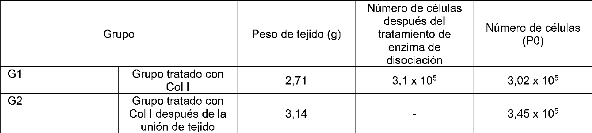
Como se muestra en la figura 1B y la tabla 1, cuando la enzima de disociación se trató antes del cultivo del cordón umbilical, la mayoría de células tenían morfología en forma de adoquín y proliferaron lentamente para mostrar un bajo rendimiento celular. Cuando la enzima de disociación se trató después del cultivo del cordón umbilical, las células tenían una morfología celular homogénea y proliferaron rápidamente para mostrar un alto rendimiento celular.As shown in Fig. 1B and Table 1, when the dissociating enzyme was treated before umbilical cord culture, most cells had cobblestone morphology and slowly proliferated to show low cell yield. When the dissociating enzyme was treated after umbilical cord culture, the cells had a homogeneous cell morphology and rapidly proliferated to show high cell yield.
(1.5) Análisis de comparación según las condiciones de hipoxia y normoxia(1.5) Comparison analysis according to hypoxic and normoxic conditions
Para comparar las curvas de crecimiento y los tiempos de duplicación de células adherentes subcultivadas durante de 1 pase a 20 pases (P1 a P20) en una condición de hipoxia (1.1) y una condición de normoxia (1.2), cada uno se sembró del mismo número de células en una placa de 6 pocillos, y las células se recogieron cuando ocuparon el 70 % ~ 80 % del área de la parte inferior de la placa. Posteriormente, se mezclaron 10 pl de la muestra con 10 pl de azul de tripano, y se inyectaron 10 pl de esto en una sección de medición de un hemocitómetro. El número de células se contó usando un contador de células automatizado. En este momento, también se registró el tiempo para calcular el tiempo de duplicación. El tiempo de duplicación, que es un tiempo que tarda una celda en duplicarse, se calculó usando el número total de células y el tiempo en que se midió el número. Los resultados se muestran en las figuras 2A y 2B. To compare the growth curves and doubling times of adherent cells subcultured for 1 passage to 20 passages (P1 to P20) in a hypoxic condition (1.1) and a normoxic condition (1.2), each was seeded from the same number of cells in a 6-well plate, and cells were harvested when they occupied 70% ~ 80% of the area of the bottom of the plate. Subsequently, 10 μl of the sample was mixed with 10 μl of trypan blue, and 10 μl of this was injected into a measuring section of a hemocytometer. The number of cells was counted using an automated cell counter. At this time, the time was also recorded to calculate the doubling time. The doubling time, which is a time it takes for a cell to duplicate, was calculated using the total number of cells and the time the number was measured. The results are shown in Figures 2A and 2B.
Además, para analizar la capacidad de formación de colonias de las células, 150 células por placa se extendieron en una placa de cultivo de 100 mm, y se cultivaron en 12 ml de medio de cultivo durante de 10 días a 14 días. La formación de colonias celulares se examinó bajo un microscopio. A continuación, las células se lavaron con DPBS, y se añadieron de 2 ml a 3 ml de una disolución mixta de glutaraldehído y cristal violeta a células en la placa y se tiñeron durante 30 minutos. Las células se lavaron cuidadosamente con agua estéril y el número de colonias se contó bajo un microscopio, y se presentó como valores medios para analizar los resultados. Los resultados se muestran en la figura 2C.Further, to test the colony-forming ability of cells, 150 cells per plate were spread in a 100-mm culture dish, and cultured in 12 ml of culture medium for 10 days to 14 days. Cell colony formation was examined under a microscope. Then, the cells were washed with DPBS, and 2 ml to 3 ml of a mixed solution of glutaraldehyde and crystal violet was added to cells in the plate and stained for 30 minutes. The cells were carefully washed with sterile water and the number of colonies was counted under a microscope, and presented as mean values to analyze the results. The results are shown in Figure 2C.
Además, para analizar la capacidad de las células para migrar a tejidos dañados, la superficie superior del filtro Transwell se recubrió con 0,1 % de gelatina a 37 °C durante 1 hora. Se suspendieron 5 x 10⁵ células en 100 pl de medio libre de suero, y se sembraron en la cámara superior del inserto Transwell. Antes de la siembra, las células se privaron de alimento en el medio sin suero durante la noche. Se añadieron 600 pl de un medio (CS-CM) que contenía quimiostatos a la cámara inferior. Se cultivaron células en una incubadora durante la noche. Las células restantes en el lado superior del filtro se retiraron usando un hisopo de algodón empapado con PBS frío. El filtro Transwell se cortó usando un bisturí, seguido de tinción de Giemsa. El lado inferior se levantó y se colocó sobre un vidrio de portaobjetos y se montó. Las células migradas se examinaron contando el número de células teñidas bajo un microscopio óptico. Los resultados se muestran en la figura 2D.Additionally, to test the ability of cells to migrate to damaged tissues, the top surface of the Transwell filter was coated with 0.1% gelatin at 37°C for 1 hour. 5 x 10 5 cells were suspended in 100 μl of serum-free medium, and seeded in the upper chamber of the Transwell insert. Prior to seeding, cells were starved in serum-free medium overnight. 600 pl of a medium (CS-CM) containing chemostats was added to the lower chamber. Cells were grown in an incubator overnight. Cells remaining on the upper side of the filter were removed using a cotton swab soaked in cold PBS. The Transwell filter was cut using a scalpel, followed by Giemsa staining. The lower side was lifted and placed on a slide glass and mounted. The migrated cells were examined by counting the number of stained cells under a light microscope. The results are shown in Figure 2D.
La figura 2 muestra la comparación entre una condición de hipoxia y una condición de normoxia en el cultivo de las células madre adherentes derivadas de cordón umbilical potenciadas según una realización específica. Como se muestra en las figuras 2A y 2B, las células cultivadas en condición de hipoxia mostraron una rápida tasa de proliferación celular de un tiempo de duplicación corto. Además, como se muestra en la figura 2C, el número de colonias aumentó aproximadamente dos veces, y como se muestra en la figura 2D, la capacidad de las células para migrar a tejidos dañados mejoró aproximadamente 1,6 veces.Figure 2 shows the comparison between a hypoxic condition and a normoxic condition in the culture of the umbilical cord-derived adherent stem cells enhanced according to a specific embodiment. As shown in Figures 2A and 2B, cells cultured under hypoxic condition showed a rapid rate of cell proliferation with a short doubling time. Furthermore, as shown in Figure 2C, the number of colonies increased by approximately two-fold, and as shown in Figure 2D, the ability of cells to migrate to damaged tissues was improved by approximately 1.6-fold.
2. Caracterización de células madre adherentes derivadas de cordón umbilical potenciadas2. Characterization of enhanced umbilical cord-derived adherent stem cells
(2.1) Análisis de la estabilidad genética de células madre adherentes derivadas de cordón umbilical potenciadas(2.1) Genetic stability analysis of potentiated umbilical cord-derived adherent stem cells
Con el fin de analizar la estabilidad genética de las células madre adherentes derivadas de cordón umbilical potenciadas preparadas en (1.1) y (1.3), se realizó análisis de tinción cromosómica GTG.In order to analyze the genetic stability of the potentiated umbilical cord-derived adherent stem cells prepared in (1.1) and (1.3), GTG chromosomal staining analysis was performed.
En detalle, se extrajeron ADN de las células de P7 y P14 usando un kit de extracción de ADN Promega, y se usaron como muestras. Se usó un chip Illumina HumanOmni1-Quad y se usó un escáner iSCAN® para la medición. En primer lugar, se amplificaron 400 ng de cada muestra de ADN mediante amplificación del genoma completo, y se fragmentaron aleatoriamente por un método químico, y después se purificaron por precipitación con 2-propanol. El chip se trató previamente con una disolución tampón, y después se aplicó la muestra de ADN al chip. Después de la incubación durante aproximadamente 16 horas, se realizaron tinción, extensión de cebador específico de alelo (ASPE), hibridación, retirada de dianas, y lavado. Posteriormente, se realizó el escaneo por NluminaiScan, y los datos se analizaron usando un software GenomeStudio®. Los resultados se muestran en la figura 3.In detail, DNA was extracted from P7 and P14 cells using a Promega DNA extraction kit, and used as samples. An Illumina HumanOmni1-Quad chip was used and an iSCAN® scanner was used for measurement. First, 400 ng of each DNA sample was amplified by whole genome amplification, and randomly fragmented by chemical method, and then purified by 2-propanol precipitation. The chip was previously treated with a buffer solution, and then the DNA sample was applied to the chip. After the incubation for approximately 16 hours, staining, allele-specific primer extension (ASPE), hybridization, target removal, and washing were performed. Subsequently, scanning was performed by NluminaiScan, and the data was analyzed using GenomeStudio® software. The results are shown in figure 3.
La figura 3 muestra los resultados del cariotipado para examinar la estabilidad genética de las células madre adherentes derivadas de cordón umbilical potenciadas según una realización específica.Figure 3 shows the results of karyotyping to examine the genetic stability of umbilical cord-derived adherent stem cells enhanced according to a specific embodiment.
Como se muestra en la figura 3, las células madre adherentes derivadas de cordón umbilical potenciadas preparadas mediante el método de preparación según una realización específica no mostraron anomalías genéticas hasta que se cultivaron hasta P14.As shown in Fig. 3, the enhanced umbilical cord-derived adherent stem cells prepared by the preparation method according to a specific embodiment did not show genetic abnormalities until they were cultured to P14.
(2.2) Análisis de proteínas de superficie de células madre adherentes derivadas de cordón umbilical potenciadas(2.2) Surface Protein Analysis of Enhanced Umbilical Cord-Derived Adherent Stem Cells
Con el fin de analizar las proteínas de superficie de las células madre adherentes derivadas de cordón umbilical potenciadas preparadas en (1.1), se realizaron citometría de flujo y análisis de tinción de inmunofluorescencia. In order to analyze the surface proteins of the potentiated umbilical cord-derived adherent stem cells prepared in (1.1), flow cytometry and immunofluorescence staining analysis were performed.
En detalle, para citometría de flujo, las células se lavaron con DPBS, y después se hicieron reaccionar con marcadores Tra-1-60, CD3, CD1a, CD11c, CD16, CD14, CD86, CD8a, CD19, CD40, CD80, CD200, CD141, CD61, CD87, MIC A/B, SSEA4 en FBS al 2 % que contiene DPBS en hielo durante 20 minutos. A continuación, se analizaron antígenos de superficie mediante FACS Calibur (Becton Bickinson), y los resultados se muestran en la figura 4A.In detail, for flow cytometry, cells were washed with DPBS, and then reacted with markers Tra-1-60, CD3, CD1a, CD11c, CD16, CD14, CD86, CD8a, CD19, CD40, CD80, CD200, CD141, CD61, CD87, MIC A/B, SSEA4 in 2% FBS containing DPBS on ice for 20 min. Surface antigens were then analyzed by FACS Calibur (Becton Bickinson), and the results are shown in Figure 4A.
Para examinar la expresión de Oct4 y Nanog, que son marcadores de células madre embrionarias, se realizó tinción de inmunofluorescencia. En primer lugar, las células se lavaron con DPBS tres veces, y luego se fijaron con paraformaldehído al 4 % en una placa de cultivo a temperatura ambiente durante 10 minutos. Después de la fijación, las células se lavaron con DPBS tres veces. A continuación, las células se permearon con disolución de Triton X-100 al 0,2 % a temperatura ambiente durante 10 minutos, y se lavaron con DPBS tres veces. El bloqueo se realizó usando suero de cabra normal al 10 % a temperatura ambiente durante 30 minutos. Anticuerpos primarios (Oct4, Nanog) se añadieron, y se hicieron reaccionar durante la noche a 4 °C en la oscuridad. Posteriormente, las células se lavaron con DPBS tres veces, y se añadieron anticuerpos secundarios a las mismas, y se hicieron reaccionar a temperatura ambiente durante 1 hora. Por último, las células se lavaron con DPBS tres veces, y se observaron bajo un microscopio de fluorescencia. Los resultados se muestran en la figura 4B.To examine the expression of Oct4 and Nanog, which are embryonic stem cell markers, immunofluorescence staining was performed. Cells were first washed with DPBS three times, and then fixed with 4% paraformaldehyde in a culture dish at room temperature for 10 minutes. After fixation, cells were washed with DPBS three times. Cells were then permeated with 0.2% Triton X-100 solution at room temperature for 10 minutes, and washed with DPBS three times. Blocking was performed using 10% normal goat serum at room temperature for 30 minutes. Primary antibodies (Oct4, Nanog) were added, and reacted overnight at 4°C in the dark. Subsequently, the cells were washed with DPBS three times, and secondary antibodies were added thereto, and reacted at room temperature for 1 hour. Finally, the cells were washed with DPBS three times and observed under a fluorescence microscope. The results are shown in Figure 4B.
La figura 4 muestra los resultados del análisis de proteínas de superficie de las células madre adherentes derivadas de cordón umbilical potenciadas según una realización específica.Figure 4 shows the results of surface protein analysis of umbilical cord-derived adherent stem cells enhanced according to a specific embodiment.
Como se muestra en la figura 4A, las células madre adherentes derivadas de cordón umbilical potenciadas según una realización específica fueron células selectivamente positivas para CD200, CD141, CD61, CD87, SSEA4, y selectivamente negativas para TRA-1, CD3, CD1a, CD11c, CD16, CD86, CD8a, CD40, MIC A/B, y adicionalmente, selectivamente positivas para CD61 en una condición de hipoxia. Además, como se muestra en la figura 4B, las células madre adherentes derivadas de cordón umbilical potenciadas según una realización específica no expresaron proteínas Oct4 y Nanog que son marcadores específicos de células madre embrionarias.As shown in Figure 4A, the umbilical cord-derived adherent stem cells enhanced according to a specific embodiment were cells selectively positive for CD200, CD141, CD61, CD87, SSEA4, and selectively negative for TRA-1, CD3, CD1a, CD11c, CD16, CD86, CD8a, CD40, MIC A/B, and additionally selectively positive for CD61 in a hypoxic condition. Furthermore, as shown in Figure 4B, umbilical cord-derived adherent stem cells enhanced according to a specific embodiment did not express Oct4 and Nanog proteins which are specific markers of embryonic stem cells.
(2.3) Análisis de capacidad de diferenciación de células madre adherentes derivadas de cordón umbilical potenciadas(2.3) Differentiation capacity analysis of potentiated umbilical cord-derived adherent stem cells
(2.3.1) Análisis de capacidad de diferenciación de adipocitos(2.3.1) Adipocyte differentiation capacity analysis
El análisis de la capacidad de diferenciación de adipocitos de las células madre adherentes derivadas de cordón umbilical potenciadas se realizó de la siguiente manera.Analysis of the adipocyte differentiation capacity of the enhanced umbilical cord-derived adherent stem cells was performed as follows.
Las células madre adherentes derivadas de cordón umbilical potenciadas preparadas en (1.1) y (1.3) se cultivaron en un medio de diferenciación de adipogénesis (kit de diferenciación de adipogénesis StemPro®, Life Technology) durante 2 semanas mientras se reemplazaba el medio cada tres días. A continuación, el medio de cultivo se retiró y las células se lavaron con DPBS sin Ca/Mg, y se hicieron reaccionar con paraformaldehído al 4 % a temperatura ambiente durante 15 minutos. Las células se lavaron con isopropanol al 60 % y después se hicieron reaccionar con rojo aceite O durante 10 minutos. A continuación, las células se lavaron con agua purificada, y se observaron adipocitos bajo un microscopio. Los resultados se muestran en la figura 5.The potentiated umbilical cord-derived adherent stem cells prepared in (1.1) and (1.3) were cultured in adipogenesis differentiation medium (StemPro® Adipogenesis Differentiation Kit, Life Technology) for 2 weeks while the medium was replaced every three days. . Then, the culture medium was removed and the cells were washed with DPBS without Ca/Mg, and reacted with 4% paraformaldehyde at room temperature for 15 minutes. Cells were washed with 60% isopropanol and then reacted with oil red O for 10 minutes. Next, the cells were washed with purified water, and adipocytes were observed under a microscope. The results are shown in figure 5.
(2.3.2) Análisis de capacidad de diferenciación de osteocitos(2.3.2) Osteocyte differentiation capacity analysis
El análisis de capacidad de diferenciación de osteocitos de las células madre adherentes derivadas de cordón umbilical potenciadas se realizó de la siguiente manera.Osteocyte differentiation capacity analysis of the potentiated umbilical cord-derived adherent stem cells was performed as follows.
Las células madre adherentes derivadas de cordón umbilical potenciadas preparadas en (1.1) y (1.3) se cultivaron en un medio de diferenciación de osteogénesis (kit de diferenciación de osteogénesis StemPro®, Life Technology) durante 2 semanas mientras se reemplazaba el medio cada tres días. A continuación, el medio de cultivo se retiró y las células se lavaron con DPBS sin Ca/Mg, y se hicieron reaccionar con paraformaldehído al 4 % a temperatura ambiente durante 15 minutos. Después de la reacción, las células se lavaron con agua purificada, y se hicieron reaccionar con una disolución de nitrato de plata al 1 % a temperatura ambiente durante 15 minutos. Las células se lavaron con agua purificada y después se hicieron reaccionar con una disolución de tiosulfato de sodio al 5 % a temperatura ambiente durante 5 minutos. A continuación, las células se lavaron con agua purificada, y se hicieron reaccionar con una disolución roja rápida nuclear al 0,1 % a temperatura ambiente durante 5 minutos. A continuación, las células se lavaron con agua purificada, y se observó una muestra de deposición de calcio bajo un microscopio. Los resultados se muestran en la figura 5.The potentiated umbilical cord-derived adherent stem cells prepared in (1.1) and (1.3) were cultured in osteogenesis differentiation medium (StemPro® Osteogenesis Differentiation Kit, Life Technology) for 2 weeks while medium was replaced every three days. Then, the culture medium was removed and the cells were washed with DPBS without Ca/Mg, and reacted with 4% paraformaldehyde at room temperature for 15 minutes. After the reaction, the cells were washed with purified water, and reacted with 1% silver nitrate solution at room temperature for 15 minutes. The cells were washed with purified water, and then reacted with 5% sodium thiosulfate solution at room temperature for 5 minutes. Then, the cells were washed with purified water, and reacted with 0.1% nuclear fast red solution at room temperature for 5 minutes. Then, the cells were washed with purified water, and a sample of calcium deposition was observed under a microscope. The results are shown in figure 5.
(2.3.3) Análisis de capacidad de diferenciación de condrocitos(2.3.3) Analysis of ability to differentiate chondrocytes
El análisis de capacidad de diferenciación de condrocitos de las células madre adherentes derivadas de cordón umbilical potenciadas se realizó de la siguiente manera.The chondrocyte differentiation capacity analysis of the enhanced umbilical cord-derived adherent stem cells was performed as follows.
2 x 10⁵ de las células madre adherentes derivadas de cordón umbilical potenciadas preparadas en (1.1) y (1.3) se colocaron en un tubo de 15 ml, y se centrifugaron a 1.500 rpm durante 5 minutos. El sobrenadante se descartó, y solo las células se cultivaron en un medio de diferenciación de condrogénesis (kit de diferenciación de condrogénesis StemPro®, Life Technology) con una tapa cerrada de manera suelta durante 3 semanas mientras se reemplazaba el medio cada tres días. A continuación, la masa celular se convirtió en un bloque de parafina y se cortó, seguido de tinción con azul alcián. Posteriormente, los condrocitos teñidos con color azul se analizaron con un microscopio óptico y los resultados se muestran en la figura 5.2 x 10 5 of the enhanced umbilical cord-derived adherent stem cells prepared in (1.1) and (1.3) were placed in a 15 ml tube, and centrifuged at 1,500 rpm for 5 minutes. The supernatant was discarded, and cells alone were cultured in chondrogenesis differentiation medium (StemPro® Chondrogenesis Differentiation Kit, Life Technology) with a lid loosely closed for 3 weeks while the medium was replaced every three days. The cell mass was then made into a paraffin block and sectioned, followed by Alcian blue staining. Subsequently, the blue-stained chondrocytes were analyzed under a light microscope and the results are shown in Figure 5.
La figura 5 muestra los resultados del análisis de multipotencia de las células madre adherentes derivadas de cordón umbilical potenciadas según una realización específica.Figure 5 shows the results of multipotency analysis of umbilical cord-derived adherent stem cells enhanced according to a specific embodiment.
Como se muestra en la figura 5, el resultado del análisis de la capacidad de diferenciación de adipocitos mostró que las sustancias grandes y pequeñas (grasas), como gotas, se identificaron en rojo. Además, el resultado del análisis de la capacidad de diferenciación de osteocitos mostró la deposición de partículas de calcio marrón-negro sobre el fondo rosa. Además, el resultado del análisis de la capacidad de diferenciación de condrocitos mostró que durante la diferenciación para dar condrocitos, glicoproteínas que representan la rigidez y elasticidad del cartílago se secretaron y se identificaron en azul. Estos resultados mostraron que las células madre adherentes derivadas de cordón umbilical potenciadas según una realización específica preparadas por el método de (1.1) y (1.3) pueden diferenciarse para dar adipocitos, osteocitos, o condrocitos, lo que indica multipotencia.As shown in Fig. 5, the result of adipocyte differentiation ability analysis showed that large and small (fatty) substances, such as droplets, were identified in red. In addition, the result of the analysis of the ability to differentiate osteocytes showed the deposition of brown-black calcium particles on the pink background. In addition, the result of analysis of chondrocyte differentiation ability showed that during differentiation into chondrocytes, glycoproteins representing the stiffness and elasticity of cartilage were secreted and identified in blue. These results showed that umbilical cord-derived adherent stem cells enhanced according to a specific embodiment prepared by the method of (1.1) and (1.3) can differentiate into adipocytes, osteocytes, or chondrocytes, indicating multipotency.
(2.4) Análisis de elaboración de perfiles y cuantificación de proteínas secretoras de células madre adherentes derivadas de cordón umbilical potenciadas(2.4) Profiling analysis and quantification of secretory proteins from potentiated umbilical cord-derived adherent stem cells
Con el fin de analizar las proteínas secretoras de las células madre adherentes derivadas de cordón umbilical potenciadas preparadas en (1.1), análisis de perlas múltiples (panel de citoquinas/quimiocinas humanas 1 MILLIPLEX, Merck Millipore, Billerica, Ma, e E.UU.).In order to analyze the secretory proteins of the potentiated umbilical cord-derived adherent stem cells prepared in (1.1), multi-bead assays (MILLIPLEX Human Cytokine/Chemokine Panel 1, Merck Millipore, Billerica, Ma, USA. ).
En detalle, las células madre adherentes derivadas de cordón umbilical potenciadas se cultivaron durante 24 horas, y el cultivo de las mismas se incubó con perlas de captura recubiertas con anticuerpo a temperatura ambiente durante 2 horas, seguido de lavado. Las perlas se incubaron con anticuerpo quimiocina y citocina anti-humano etiquetado con biotina durante 1 hora y se incubaron con estreptavidina ficoeritrina durante 30 minutos. Por último, las perlas se lavaron, y para la cuantificación, se usó un programa Luminex 200 para analizar los niveles de expresión de proteínas secretoras. Los resultados se muestran en la tabla 2. In detail, the potentiated umbilical cord-derived adherent stem cells were cultured for 24 hours, and the culture thereof was incubated with antibody-coated capture beads at room temperature for 2 hours, followed by washing. The beads were incubated with biotin-labeled anti-human chemokine and cytokine antibody for 1 hour and incubated with streptavidin phycoerythrin for 30 minutes. Finally, the beads were washed, and for quantification, a Luminex 200 program was used to analyze the expression levels of secretory proteins. Results are shown in table 2.
[Tabla 2][Table 2]

Como se muestra en la tabla 2, puede verse que las células madre adherentes derivadas de cordón umbilical potenciadas según una realización específica secretan IL-6, IL-8, G-CSF, GM-CSF, MCP-1, MCP-3, VEGF, GRO, IGF-1 SR, EpCAM, IGFBP3, etc.As shown in Table 2, it can be seen that umbilical cord-derived adherent stem cells enhanced according to a specific embodiment secrete IL-6, IL-8, G-CSF, GM-CSF, MCP-1, MCP-3, VEGF , GRO, IGF-1 SR, EpCAM, IGFBP3, etc.
(2.5) Análisis de la expresión génica de células madre adherentes derivadas de cordón umbilical potenciadas Se analizaron las expresiones génicas de las células madre derivadas de cordón umbilical potenciadas preparadas en (1.1) comparándolas con aquellas de células madre de médula ósea. (2.5) Gene expression analysis of potentiated umbilical cord-derived adherent stem cells Gene expressions of potentiated umbilical cord-derived stem cells prepared in (1.1) were analyzed by comparing them with those of bone marrow stem cells.
En detalle, para analizar genes expresados específicamente en células madre adherentes derivadas de cordón umbilical potenciadas comparándolos con aquellos de células madre de médula ósea, se extrajeron ARN a partir de las células madre adherentes derivadas de cordón umbilical potenciadas y las células madre de médula ósea, seguido de etiquetado y purificación. Los ADNc etiquetados se hibridaron con el kit Expression Beadchip de Illumina para obtener resultados. Los datos se analizaron estadísticamente, y se muestran en la siguiente tabla 3 y la figura 6. In detail, to analyze genes specifically expressed in enhanced umbilical cord-derived adherent stem cells compared to those in bone marrow stem cells, RNA was extracted from the enhanced umbilical cord-derived adherent stem cells and bone marrow stem cells, followed by labeling and purification. Labeled cDNAs were hybridized with the Illumina Expression Beadchip Kit to obtain results. The data was statistically analyzed, and is shown in the following table 3 and figure 6.
[Tabla 3][Table 3]

Como se muestra en la tabla 3 y la figura 6, se observó alta expresión de COL1A1, IGFBP4, TAGLN, S100A10, SQSTM1, DSTN, DCN, PHGDH, FBLN1, MFGE8, HLA-A, VASN, KIAA1199, STC1, LRRC17, IL33, SNCA, DSG2, NRP2, PLAT en las células madre adherentes derivadas de cordón umbilical potenciadas y se observó baja expresión de CCND1, SERPINE1, PRNP, MT2A, TM4SF1, HIST1H4C, NME1, CXCL6, NTSR1, PTGS2, CYP1B1, TPMT, NAGK, ANXA4 en las células madre adherentes derivadas de cordón umbilical potenciadas, en comparación con células madre de médula ósea.As shown in Table 3 and Figure 6, high expression of COL1A1, IGFBP4, TAGLN, S100A10, SQSTM1, DSTN, DCN, PHGDH, FBLN1, MFGE8, HLA-A, VASN, KIAA1199, STC1, LRRC17, IL33 was observed. , SNCA, DSG2, NRP2, PLAT in potentiated umbilical cord-derived adherent stem cells and low expression of CCND1, SERPINE1, PRNP, MT2A, TM4SF1, HIST1H4C, NME1, CXCL6, NTSR1, PTGS2, CYP1B1, TPMT, NAGK, ANXA4 in enhanced umbilical cord-derived adherent stem cells, compared to bone marrow stem cells.
Entre los genes, COL1A1, que es un gen altamente expresado, es colágeno alfa-1 de tipo I, y se sabe que se expresa en colágeno de tejidos conjuntivos, incluido el cartílago. Sin embargo, no hay informes sobre la asociación entre este gen y las células madre adherentes derivadas de cordón umbilical potenciadas.Among the genes, COL1A1, which is a highly expressed gene, is collagen alpha-1 type I, and it is known to be expressed in collagen of connective tissues, including cartilage. However, there are no reports on the association between this gene and enhanced umbilical cord-derived adherent stem cells.
Entre los genes, IGFBP4, que es un gen altamente expresado, es una proteína de unión al factor de crecimiento similar a la insulina, y conocido como proteína que inhibe diversas células cancerosas. Hay un informe de que se detecta IGFBP4 en el suero de la sangre del cordón umbilical, pero no hay informes sobre la asociación entre este gen y las células madre adherentes derivadas de cordón umbilical potenciadas.Among the genes, IGFBP4, which is a highly expressed gene, is an insulin-like growth factor-binding protein, and known as a protein that inhibits various cancer cells. There is a report that IGFBP4 is detected in umbilical cord blood serum, but there are no reports on the association between this gene and enhanced umbilical cord-derived adherent stem cells.
Entre los genes, TAGLN, que es un gen altamente expresado, es un gen expresado en fibroblastos y músculos lisos, y su función aún no se ha revelado. Existe un informe de que TAGLN se expresa en células madre de médula ósea, pero no hay informes sobre la asociación entre este gen y las células madre adherentes derivadas de cordón umbilical potenciadas.Among the genes, TAGLN, which is a highly expressed gene, is a gene expressed in fibroblasts and smooth muscles, and its function has not been revealed yet. There is a report that TAGLN is expressed in bone marrow stem cells, but there are no reports on the association between this gene and potentiated umbilical cord-derived adherent stem cells.
Entre los genes, CCND1, que es un gen débilmente expresado, es la ciclina D1. La sobreexpresión de CCND1 conduce a una transición rápida de la fase G1 a la fase S del ciclo celular para facilitar el crecimiento celular. Hay informes de que CCND1 se expresa altamente en células cancerosas, y las células madre de la sangre del cordón umbilical inhiben el glioma C6 a través de la regulación por disminución de CCND1. Sin embargo, no hay informes sobre la asociación entre este gen y las células madre adherentes derivadas de cordón umbilical potenciadas.Among the genes, CCND1, which is a weakly expressed gene, is cyclin D1. CCND1 overexpression leads to a rapid transition from the G1 phase to the S phase of the cell cycle to facilitate cell growth. There are reports that CCND1 is highly expressed in cancer cells, and umbilical cord blood stem cells inhibit C6 glioma through downregulation of CCND1. However, there are no reports on the association between this gene and enhanced umbilical cord-derived adherent stem cells.
Entre los genes, SERPINE1, que es un gen débilmente expresado, se conoce como inhibidor del activador del plasminógeno endotelial, y se conoce por funcionar como activador del plasminógeno tisular (tPA). Sin embargo, no hay informes sobre la asociación entre este gen y las células madre adherentes derivadas de cordón umbilical potenciadas.Among the genes, SERPINE1, which is a weakly expressed gene, is known as endothelial plasminogen activator inhibitor, and is known to function as tissue plasminogen activator (tPA). However, there are no reports on the association between this gene and enhanced umbilical cord-derived adherent stem cells.
Entre los genes, PRNP, que es un gen débilmente expresado, es una proteína de prión principal (CD230), y se sabe que se expresa en diversos tejidos, así como en el sistema nervioso. Se informa que la anomalía del gen PRNP provoca un trastorno neurológico. Sin embargo, no hay informes sobre la asociación entre este gen y las células madre adherentes derivadas de cordón umbilical potenciadas.Among the genes, PRNP, which is a weakly expressed gene, is a major prion protein (CD230), and it is known to be expressed in various tissues as well as in the nervous system. It is reported that the PRNP gene abnormality causes a neurological disorder. However, there are no reports on the association between this gene and enhanced umbilical cord-derived adherent stem cells.
La expresión génica de las células madre derivadas de cordón umbilical potenciadas preparadas en (1.1) y (1.3) se comparó y analizó de la misma manera que anteriormente, y los resultados se muestran en la siguiente tabla 4 y la figura 6.The gene expression of the enhanced umbilical cord-derived stem cells prepared in (1.1) and (1.3) was compared and analyzed in the same manner as above, and the results are shown in the following Table 4 and Fig. 6.
[Tabla 4][Table 4]

Como se muestra en la tabla 4 y la figura 6, se observó alta expresión de S100A10, BNIP3, IGFBP5, PGK1, TPI1, DCN, PGM1, PFKFB3, LOC644774, MME, MIR1978, PGK1, SLC2A3, BHLHB2, BNIP3L, IGFBP5, NDUFA4L2, DPYD, y SCARA3 en las células madre adherentes derivadas de cordón umbilical potenciadas cultivadas en una condición de hipoxia y se observó baja expresión de IL8, ALDH1A1, NQO1, DLC1, CTHRC1, y CPA4 en las células madre adherentes derivadas de cordón umbilical potenciadas cultivadas en una condición de hipoxia, en comparación con aquellas en condición de normoxia.As shown in Table 4 and Figure 6, high expression of S100A10, BNIP3, IGFBP5, PGK1, TPI1, DCN, PGM1, PFKFB3, LOC644774, MME, MIR1978, PGK1, SLC2A3, BHLHB2, BNIP3L, IGFBP5, NDUFA4L2 was observed. , DPYD, and SCARA3 in potentiated umbilical cord-derived adherent stem cells cultured under a hypoxic condition and low expression of IL8, ALDH1A1, NQO1, DLC1, CTHRC1, and CPA4 was observed in cultured potentiated umbilical cord-derived adherent stem cells. in a hypoxic condition, compared to those in a normoxic condition.
Entre los genes, S100A10, que es un gen altamente expresado, es la proteína A10 de unión a calcio S100, y regula el ciclo celular y la diferenciación. Además, se sabe que S100A10 funciona en exocitosis y endocitosis. Se estudió S100A10 como una de las proteínas altamente expresadas durante la diferenciación de células madre de médula ósea en hueso. Sin embargo, no hay informes sobre la asociación entre este gen y las células madre adherentes derivadas de cordón umbilical potenciadas.Among the genes, S100A10, which is a highly expressed gene, is S100 calcium-binding protein A10, and it regulates cell cycle and differentiation. Furthermore, S100A10 is known to function in exocytosis and endocytosis. S100A10 was studied as one of the highly expressed proteins during differentiation of bone marrow stem cells into bone. However, there are no reports on the association between this gene and enhanced umbilical cord-derived adherent stem cells.
Entre los genes, se sabe que BNIP3, que es un gen altamente expresado, está altamente expresado en UCB-MSC, cuando se comparó la expresión génica a niveles de ARNm de UCB-MSC (células madre derivadas de sangre de cordón umbilical) y UCB-MNC (células sanguíneas derivadas de sangre de cordón umbilical). Además, se recogieron células madre amnióticas en una condición de hipoxia, y luego se sometieron a análisis de micromatrices de ARNm. Como resultado, se sabe que el gen de BNIP3 aumenta significativamente en la condición de hipoxia. Sin embargo, no hay informes sobre la asociación entre este gen y las células madre adherentes derivadas de cordón umbilical potenciadas.Among genes, BNIP3, which is a highly expressed gene, is known to be highly expressed in UCB-MSCs, when gene expression was compared at mRNA levels of UCB-MSCs (umbilical cord blood-derived stem cells) and UCB -MNC (blood cells derived from umbilical cord blood). In addition, amniotic stem cells were collected under a hypoxic condition, and then subjected to mRNA microarray analysis. As a result, the BNIP3 gene is known to be significantly increased in the hypoxic condition. However, there are no reports on the association between this gene and enhanced umbilical cord-derived adherent stem cells.
Entre los genes, IGFBP5, que es un gen altamente expresado, es una proteína 5 de unión al factor de crecimiento similar a la insulina, y funciona en el desarrollo y se localiza en el espacio extracelular. Se informó la expresión del gen de IGFBP5 en células madre de médula ósea, pero no hay informes sobre la asociación entre este gen y las células madre adherentes derivadas de cordón umbilical potenciadas.Among the genes, IGFBP5, which is a highly expressed gene, is an insulin-like growth factor-binding protein 5, and it functions in development and localizes in the extracellular space. Expression of the IGFBP5 gene in bone marrow stem cells has been reported, but there are no reports on the association between this gene and potentiated umbilical cord-derived adherent stem cells.
Entre los genes, se informa que IL8, que es un gen débilmente expresado, se libera desde fagocitos y células mesenquimales para activar la quimiotaxis inductora de neutrófilos cuando se expone a entornos inflamatorios. Sin embargo, no hay informes sobre la asociación entre este gen y las células madre adherentes derivadas de cordón umbilical potenciadas. Among the genes, IL8, which is a weakly expressed gene, is reported to be released from phagocytes and mesenchymal cells to activate neutrophil-inducing chemotaxis when exposed to inflammatory environments. However, there are no reports on the association between this gene and enhanced umbilical cord-derived adherent stem cells.
Entre los genes, ALDH1A1, que es un gen débilmente expresado, es la familia 1 de aldehido deshidrogenasa, miembro A1, y es una enzima implicada en la principal vía de oxidación del metabolismo del alcohol. Sin embargo, no hay informes sobre la asociación entre este gen y las células madre adherentes derivadas de cordón umbilical potenciadas.Among the genes, ALDH1A1, which is a weakly expressed gene, is aldehyde dehydrogenase family 1, member A1, and is an enzyme involved in the major oxidation pathway of alcohol metabolism. However, there are no reports on the association between this gene and enhanced umbilical cord-derived adherent stem cells.
3. Análisis de efectos antiinflamatorios, de regeneración vascular, y de regeneración nerviosa de células madre adherentes derivadas de cordón umbilical potenciadas3. Analysis of anti-inflammatory, vascular regeneration, and nerve regeneration effects of potentiated umbilical cord-derived adherent stem cells
(3.1) Análisis de efecto antiinflamatorio de células madre adherentes derivadas de cordón umbilical potenciadas(3.1) Analysis of anti-inflammatory effect of potentiated umbilical cord-derived adherent stem cells
Para analizar un efecto antiinflamatorio de las células madre adherentes derivadas de cordón umbilical potenciadas preparadas en (1.1), se analizaron la capacidad de inhibición de la proliferación de PBMC y la IL-10 secretada por PBMC activadas.To test an anti-inflammatory effect of the potentiated umbilical cord-derived adherent stem cells prepared in (1.1), inhibition ability of PBMC proliferation and IL-10 secreted by activated PBMC were tested.
En detalle, la capacidad de inhibición de la proliferación de PBMC se analizó de la siguiente manera. En primer lugar, las células madre adherentes derivadas de cordón umbilical potenciadas se inocularon a diferentes concentraciones en una placa de 24 pocillos y se cultivaron durante 24 horas. Posteriormente, las PBMC teñidas con CFSE se estimularon mediante la adición de PHA, y se cocultivaron con las células madre adherentes derivadas de cordón umbilical potenciadas durante 5 días. Posteriormente, se examinó si la inflamación se inhibía por citocinas secretadas por las células madre adherentes derivadas de cordón umbilical potenciadas o se inhibía por contacto directo de célula a célula por presencia o ausencia de Transwell. Los resultados se muestran en la figura 7A.In detail, the proliferation inhibition ability of PBMC was tested as follows. First, potentiated umbilical cord-derived adherent stem cells were inoculated at different concentrations into a 24-well plate and cultured for 24 hours. Subsequently, CFSE-stained PBMCs were stimulated by the addition of PHA, and co-cultured with the potentiated umbilical cord-derived adherent stem cells for 5 days. Subsequently, we examined whether inflammation was inhibited by cytokines secreted by the enhanced umbilical cord-derived adherent stem cells or inhibited by direct cell-to-cell contact by the presence or absence of Transwell. The results are shown in Figure 7A.
Además, para analizar la IL-10 secretada por PBMC activadas, el cocultivo se realizó durante 5 horas y durante 5 días, y después se recogió el medio acondicionado. La cantidad de IL-10 secretada se midió usando el kit ELISA de IL-10 humana (R&D Systems), y los resultados se muestran en la figura 7A.Furthermore, to analyze IL-10 secreted by activated PBMC, co-culture was performed for 5 hours and for 5 days, and then the conditioned medium was collected. The amount of IL-10 secreted was measured using the human IL-10 ELISA kit (R&D Systems), and the results are shown in Figure 7A.
La figura 7A muestra los resultados del análisis del efecto antiinflamatorio de las células madre adherentes derivadas de cordón umbilical potenciadas según una realización específica.Figure 7A shows the results of the analysis of the anti-inflammatory effect of umbilical cord-derived adherent stem cells enhanced according to a specific embodiment.
Como se muestra en la figura 7A, las células madre adherentes derivadas de cordón umbilical potenciadas según una realización específica inhibieron la proliferación de PBMC, en comparación con un grupo de control. Además, cuando una relación de células madre adherentes derivadas de cordón umbilical potenciadas: PBMC es 1:10, hasta aproximadamente 30,51 ± 1,74 % de la inhibición de proliferación de PBMC se observó por cocultivo indirecto. Además, los resultados del análisis de IL-10 secretada por PBMC activadas mostraron que las PBMC activadas secretaban citocina antiinflamatoria (IL-10), y las células madre adherentes derivadas de cordón umbilical potenciadas funcionan para aumentar la secreción de IL-10 de PBMC.As shown in Figure 7A, umbilical cord-derived adherent stem cells enhanced according to a specific embodiment inhibited PBMC proliferation, compared to a control group. Furthermore, when a ratio of potentiated umbilical cord-derived adherent stem cells:PBMC is 1:10, up to about 30.51±1.74% inhibition of PBMC proliferation was observed by indirect co-culture. In addition, the results of analysis of IL-10 secreted by activated PBMCs showed that activated PBMCs secreted anti-inflammatory cytokine (IL-10), and enhanced umbilical cord-derived adherent stem cells function to increase IL-10 secretion from PBMCs.
Los resultados sugieren que las células madre adherentes derivadas de cordón umbilical potenciadas según una realización específica pueden aplicarse de manera útil al tratamiento de enfermedades inflamatorias.The results suggest that umbilical cord-derived adherent stem cells enhanced according to a specific embodiment can be usefully applied to the treatment of inflammatory diseases.
(3.2) Análisis de efecto de regeneración vascular de células madre adherentes derivadas de cordón umbilical potenciadas(3.2) Analysis of vascular regeneration effect of potentiated umbilical cord-derived adherent stem cells
Para analizar un efecto de regeneración vascular de las células madre adherentes derivadas de cordón umbilical potenciadas preparadas en (1.1), se analizó la proliferación de células endoteliales vasculares.To test a vascular regeneration effect of the potentiated umbilical cord-derived adherent stem cells prepared in (1.1), vascular endothelial cell proliferation was tested.
En detalle, se recogieron EBM-2 y medio acondicionado de las células madre adherentes derivadas de cordón umbilical potenciadas y se prepararon como muestras. Posteriormente, se inocularon células endoteliales vasculares (HUVEC) en una placa de 96 pocillos. Cuando se hicieron proliferar las células durante aproximadamente 1 día, EBM-2 y el medio de cultivo de las células madre adherentes derivadas de cordón umbilical potenciadas se añadieron a las mismas, respectivamente y se cultivaron durante 4 días. Se añadió un reactivo del kit de ensayo de viabilidad celular Cyto XTM (WST-1) al medio en una cantidad del 10 % del mismo, y se dejó reaccionar en una incubadora durante de 2 horas a 3 horas. Posteriormente, se analizaron las tasas de proliferación de células endoteliales vasculares a 450 nm usando un microlector, y los resultados se muestran en la figura 7B.In detail, EBM-2 and conditioned medium were collected from the enhanced umbilical cord-derived adherent stem cells and prepared as samples. Subsequently, vascular endothelial cells (HUVEC) were inoculated into a 96-well plate. When the cells were proliferated for about 1 day, EBM-2 and the culture medium of the enhanced umbilical cord-derived adherent stem cells were added thereto, respectively, and cultured for 4 days. A reagent of Cyto XTM cell viability assay kit (WST-1) was added to the medium in an amount of 10% thereof, and reacted in an incubator for 2 hours to 3 hours. Subsequently, vascular endothelial cell proliferation rates at 450 nm were analyzed using a microreader, and the results are shown in Figure 7B.
La figura 7B muestra los resultados del análisis del efecto de regeneración vascular de las células madre adherentes derivadas de cordón umbilical potenciadas según una realización específica.Figure 7B shows the results of analysis of vascular regeneration effect of umbilical cord-derived adherent stem cells enhanced according to a specific embodiment.
Como se muestra en la figura 7B, cuando la tasa de proliferación de las células endoteliales vasculares cultivadas en medio EBM-2 se tomó como del 100 %, la tasa de proliferación de las células endoteliales vasculares cultivadas en el medio acondicionado de las células madre adherentes derivadas de cordón umbilical potenciadas fue de 172 ± 15,22 %.As shown in Fig. 7B, when the proliferation rate of vascular endothelial cells cultured in EBM-2 medium was taken as 100%, the proliferation rate of vascular endothelial cells cultured in the adherent stem cell conditioned medium potentiated umbilical cord derived was 172 ± 15.22%.
El resultado sugiere que las células madre adherentes derivadas de cordón umbilical potenciadas según una realización específica tienen el efecto de regeneración vascular. The result suggests that the umbilical cord-derived adherent stem cells enhanced according to a specific embodiment have the effect of vascular regeneration.
(3.3) Análisis de efecto de regeneración nerviosa de células madre adherentes derivadas de cordón umbilical potenciadas(3.3) Nerve regeneration effect analysis of potentiated umbilical cord-derived adherent stem cells
Para analizar un efecto de regeneración nerviosa de las células madre adherentes derivadas de cordón umbilical potenciadas preparadas en (1.1), se analizó la proliferación de células nerviosas.To test a nerve regeneration effect of the potentiated umbilical cord-derived adherent stem cells prepared in (1.1), nerve cell proliferation was tested.
En detalle, se recogieron MEM y medio acondicionado de las células madre adherentes derivadas de cordón umbilical potenciadas y se prepararon como muestras. Posteriormente, las células nerviosas (SH-SY5Y) se inocularon en una placa de 96 pocillos. Cuando se hicieron proliferar las células durante aproximadamente 1 día, MEM y el medio de cultivo de las células madre adherentes derivadas de cordón umbilical potenciadas se añadieron a las mismas, respectivamente y se cultivaron durante 4 días. Se añadió un reactivo del kit de ensayo de viabilidad celular Cyto XTM (WST-1) al medio en una cantidad del 10 % del mismo, y se dejó reaccionar en una incubadora durante de 2 horas a 3 horas. Posteriormente, las tasas de proliferación de células nerviosas se analizaron a 450 nm usando un microlector, y los resultados se muestran en la figura 7C.In detail, MEM and conditioned medium were collected from the enhanced umbilical cord-derived adherent stem cells and prepared as samples. Nerve cells (SH-SY5Y) were subsequently inoculated into a 96-well plate. When the cells were proliferated for about 1 day, MEM and the culture medium of the enhanced umbilical cord-derived adherent stem cells were added thereto, respectively, and cultured for 4 days. A reagent of Cyto XTM cell viability assay kit (WST-1) was added to the medium in an amount of 10% thereof, and reacted in an incubator for 2 hours to 3 hours. Subsequently, nerve cell proliferation rates were analyzed at 450 nm using a microreader, and the results are shown in Figure 7C.
La figura 7C muestra los resultados del análisis del efecto de regeneración nerviosa de las células madre adherentes derivadas de cordón umbilical potenciadas según una realización específica.Fig. 7C shows the results of analysis of nerve regeneration effect of umbilical cord-derived adherent stem cells enhanced according to a specific embodiment.
Como se muestra en la figura 7C, cuando la tasa de proliferación de las células nerviosas cultivadas en medio MEM se tomó como del 100 %, la tasa de proliferación de las células nerviosas cultivadas en el medio acondicionado de las células madre adherentes derivadas de cordón umbilical potenciadas fue de 302 ± 15,97 %.As shown in Fig. 7C, when the proliferation rate of nerve cells cultured in MEM medium was taken as 100%, the proliferation rate of nerve cells cultured in the conditioned medium of umbilical cord-derived adherent stem cells potentiated was 302 ± 15.97%.
El resultado sugiere que las células madre adherentes derivadas de cordón umbilical potenciadas según una realización específica tienen el efecto de regeneración nerviosa. The result suggests that the umbilical cord-derived adherent stem cells enhanced according to a specific embodiment have the effect of nerve regeneration.
Claims (19)
Applications Claiming Priority (2)
| Application Number | Priority Date | Filing Date | Title |
|---|---|---|---|
| KR20150113858 | 2015-08-12 | ||
| PCT/KR2016/008887 WO2017026838A1 (en) | 2015-08-12 | 2016-08-12 | Improved umbilical cord-derived adhesive stem cells, preparation method therefor, and use thereof |
Publications (1)
| Publication Number | Publication Date |
|---|---|
| ES2914692T3 true ES2914692T3 (en) | 2022-06-15 |
Family
ID=58315308
Family Applications (1)
| Application Number | Title | Priority Date | Filing Date |
|---|---|---|---|
| ES16835478T Active ES2914692T3 (en) | 2015-08-12 | 2016-08-12 | Improved umbilical cord-derived adhesive stem cells, preparation method therefor, and use thereof |
Country Status (6)
| Country | Link |
|---|---|
| US (1) | US11690877B2 (en) |
| EP (1) | EP3336176B1 (en) |
| JP (2) | JP6648259B2 (en) |
| KR (2) | KR20170020273A (en) |
| CN (1) | CN108138137A (en) |
| ES (1) | ES2914692T3 (en) |
Families Citing this family (7)
| Publication number | Priority date | Publication date | Assignee | Title |
|---|---|---|---|---|
| JP6648259B2 (en) * | 2015-08-12 | 2020-02-14 | チャ バイオテック カンパニー リミテッド | Improved umbilical cord-derived adherent stem cell, method for producing the same and use thereof |
| US20210338740A1 (en) * | 2018-11-08 | 2021-11-04 | Pluristem Ltd. | Therapeutic methods and compositions |
| US11951134B2 (en) | 2019-09-30 | 2024-04-09 | North Carolina State University | Cationic platelet lysate compositions and related methods |
| KR102224273B1 (en) * | 2019-10-10 | 2021-03-08 | 고려대학교 산학협력단 | Stem Cell-derived Mature Cardiomyocytes and Cardiovascular Disease Model Using the Same |
| KR20220055921A (en) * | 2020-10-27 | 2022-05-04 | 서울대학교산학협력단 | Pharmaceutical composition for preventing or treating tendor or ligament diseases comprising Umbilical Cord Derived-Stem Cells |
| KR20240029698A (en) * | 2022-08-26 | 2024-03-06 | (주)메디노 | Pharmaceutical composition for preventing or treating neurological diseases including improved stem cells expressing TIMP-1, MCP-1, GROα and IL-6, and Methods for selecting the improved stem cells |
| WO2024080661A1 (en) * | 2022-10-12 | 2024-04-18 | (주) 엘피스셀테라퓨틱스 | Novel angiogenic stem cell |
Family Cites Families (15)
| Publication number | Priority date | Publication date | Assignee | Title |
|---|---|---|---|---|
| US20060223177A1 (en) | 2003-06-27 | 2006-10-05 | Ethicon Inc. | Postpartum cells derived from umbilical cord tissue, and methods of making and using the same |
| KR20160092062A (en) * | 2007-09-26 | 2016-08-03 | 안트로제네시스 코포레이션 | Angiogenic cells from human placental perfusate |
| KR20180108887A (en) | 2008-08-20 | 2018-10-04 | 안트로제네시스 코포레이션 | Treatment of stroke using isolated placental cells |
| DK2375907T3 (en) * | 2008-11-21 | 2019-06-11 | Celularity Inc | TREATMENT OF DISEASES, DISORDERS OR CONDITIONS IN THE LUNGERHOUSES USING PLACENTAL CELLS |
| WO2012131618A1 (en) | 2011-03-30 | 2012-10-04 | Stempeutics Research Private Limited | A composition comprising pooled wharton's jelly derived mesenchymal stem cells and methods thereof |
| WO2013009100A2 (en) | 2011-07-11 | 2013-01-17 | (주)차바이오메드 | Method for manufacturing umbilical cord extract and usage of same |
| JP6106688B2 (en) | 2011-12-02 | 2017-04-05 | フェイト セラピューティクス,インコーポレイテッド | Improved method of treating ischemia |
| CA2862661C (en) * | 2011-12-30 | 2021-05-25 | Amit Patel | Methods and compositions for the clinical derivation of an allogenic cell and therapeutic uses |
| BR112014020119A2 (en) * | 2012-02-13 | 2020-10-27 | Gamida-Cell Ltd | culture of mesenchymal stem cells |
| WO2013172793A1 (en) * | 2012-05-18 | 2013-11-21 | Agency For Science, Technology & Research | Umbilical cord mesenchymal stem cell exosomes |
| KR101384549B1 (en) | 2012-07-11 | 2014-04-11 | 한밭대학교 산학협력단 | Method and apparatus for judging ultimate strength reduction in channel bolted connection with curling |
| US10017733B2 (en) | 2013-02-15 | 2018-07-10 | Sung Kwang Medical Foundation | Production of parthenogenetic stem cells and patient-specific human embryonic stem cells using somatic cell nuclear transfer |
| US20150104470A1 (en) * | 2013-10-11 | 2015-04-16 | Neil H. Riordan | Immune modulation by peri-lymphatic or intra-lymphatic cell therapy |
| EP3140417B1 (en) * | 2014-05-09 | 2021-04-21 | Reelabs Private Limited | Foetal polymix of mesenchymal stem cells under hypoxic conditions for the treatment of clinical disorders |
| JP6648259B2 (en) * | 2015-08-12 | 2020-02-14 | チャ バイオテック カンパニー リミテッド | Improved umbilical cord-derived adherent stem cell, method for producing the same and use thereof |
-
2016
- 2016-08-12 JP JP2018507615A patent/JP6648259B2/en active Active
- 2016-08-12 CN CN201680057936.5A patent/CN108138137A/en active Pending
- 2016-08-12 US US15/752,057 patent/US11690877B2/en active Active
- 2016-08-12 KR KR1020160102721A patent/KR20170020273A/en active Application Filing
- 2016-08-12 EP EP16835478.5A patent/EP3336176B1/en active Active
- 2016-08-12 ES ES16835478T patent/ES2914692T3/en active Active
-
2019
- 2019-02-12 KR KR1020190016260A patent/KR102414662B1/en active IP Right Grant
-
2020
- 2020-01-15 JP JP2020004030A patent/JP6921249B2/en active Active
Also Published As
| Publication number | Publication date |
|---|---|
| KR102414662B1 (en) | 2022-07-01 |
| US20180236007A1 (en) | 2018-08-23 |
| JP2020072725A (en) | 2020-05-14 |
| KR20190017006A (en) | 2019-02-19 |
| EP3336176A4 (en) | 2019-04-10 |
| US11690877B2 (en) | 2023-07-04 |
| JP2018522576A (en) | 2018-08-16 |
| CN108138137A (en) | 2018-06-08 |
| JP6648259B2 (en) | 2020-02-14 |
| JP6921249B2 (en) | 2021-08-18 |
| EP3336176B1 (en) | 2022-04-20 |
| KR20170020273A (en) | 2017-02-22 |
| EP3336176A1 (en) | 2018-06-20 |
Similar Documents
| Publication | Publication Date | Title |
|---|---|---|
| ES2914692T3 (en) | Improved umbilical cord-derived adhesive stem cells, preparation method therefor, and use thereof | |
| US20200306319A1 (en) | Methods for treating radiation or chemical injury | |
| KR101557256B1 (en) | Methods For Cell Expansion And Uses Of Cells And Conditioned Media Produced Thereby For Therapy | |
| JP6478243B2 (en) | Method for culturing mesenchymal stem cells | |
| US10035834B2 (en) | Mesenchymal stem cells, compositions, and methods for treatment of cardiac tissue damage | |
| US10479978B2 (en) | Postnatal adherent cells and preparation method therefor | |
| US20140017209A1 (en) | Methods for treating radiation or chemical injury | |
| CN104487568A (en) | Mesenchymal-like stem cells derived from human embryonic stem cells, methods and uses thereof | |
| KR20080063406A (en) | Immunomodulation using placental stem cells | |
| KR20120140197A (en) | Testicular somatic cell-derived multipotent stem cells, process for the preparation thereof, and pharmaceutical composition for treating erectile dysfunction comprising the same | |
| KR20120006386A (en) | Stem cell derived from first trimester placenta and cellular therapeutic agents comprising the same | |
| TWI642781B (en) | Quadri-positive stromal cell (qpsc) population for superior cell protection and immunomodulation | |
| CA2767825A1 (en) | Cardiac tissue-derived cells | |
| KR102011634B1 (en) | Enhanced postnatal adherent cells and use thereof | |
| US20230302059A1 (en) | Umbilical cord-derived adherent stem cells, preparation method therefor, and use thereof | |
| KR101626872B1 (en) | Non-enzymatic method for isolating multipotent perivascular cells derived from human umbilical cord and cells using thereof |