ES2843777T3 - Antitumor response against modified autoepitopes - Google Patents
Antitumor response against modified autoepitopes Download PDFInfo
- Publication number
- ES2843777T3 ES2843777T3 ES17194252T ES17194252T ES2843777T3 ES 2843777 T3 ES2843777 T3 ES 2843777T3 ES 17194252 T ES17194252 T ES 17194252T ES 17194252 T ES17194252 T ES 17194252T ES 2843777 T3 ES2843777 T3 ES 2843777T3
- Authority
- ES
- Spain
- Prior art keywords
- citrullinated
- cells
- responses
- vimentin
- peptide
- Prior art date
- Legal status (The legal status is an assumption and is not a legal conclusion. Google has not performed a legal analysis and makes no representation as to the accuracy of the status listed.)
- Active
Links
- 230000006023 anti-tumor response Effects 0.000 title description 19
- 108090000765 processed proteins & peptides Proteins 0.000 claims abstract description 281
- 206010028980 Neoplasm Diseases 0.000 claims abstract description 173
- 210000001744 T-lymphocyte Anatomy 0.000 claims abstract description 82
- 238000000034 method Methods 0.000 claims abstract description 40
- 230000005867 T cell response Effects 0.000 claims abstract description 30
- 238000000338 in vitro Methods 0.000 claims abstract description 30
- 238000012216 screening Methods 0.000 claims abstract description 19
- 230000006698 induction Effects 0.000 claims abstract description 14
- 230000005809 anti-tumor immunity Effects 0.000 claims abstract description 7
- 230000004044 response Effects 0.000 description 232
- 108010065472 Vimentin Proteins 0.000 description 141
- 102000013127 Vimentin Human genes 0.000 description 140
- 210000005048 vimentin Anatomy 0.000 description 139
- 101150018417 VIM gene Proteins 0.000 description 137
- 210000004027 cell Anatomy 0.000 description 129
- 102000004196 processed proteins & peptides Human genes 0.000 description 119
- 241000699670 Mus sp. Species 0.000 description 91
- 210000004988 splenocyte Anatomy 0.000 description 91
- 230000014509 gene expression Effects 0.000 description 90
- 108090000623 proteins and genes Proteins 0.000 description 73
- 102100036011 T-cell surface glycoprotein CD4 Human genes 0.000 description 67
- 108020004414 DNA Proteins 0.000 description 63
- 102000013691 Interleukin-17 Human genes 0.000 description 56
- 108050003558 Interleukin-17 Proteins 0.000 description 56
- 102100035731 Protein-arginine deiminase type-4 Human genes 0.000 description 53
- 229960002173 citrulline Drugs 0.000 description 52
- 102000004169 proteins and genes Human genes 0.000 description 52
- 108010046732 HLA-DR4 Antigen Proteins 0.000 description 51
- RHGKLRLOHDJJDR-BYPYZUCNSA-N L-citrulline Chemical compound NC(=O)NCCC[C@H]([NH3+])C([O-])=O RHGKLRLOHDJJDR-BYPYZUCNSA-N 0.000 description 51
- 238000011830 transgenic mouse model Methods 0.000 description 51
- 241000699660 Mus musculus Species 0.000 description 50
- 241000282414 Homo sapiens Species 0.000 description 49
- 101000735566 Homo sapiens Protein-arginine deiminase type-4 Proteins 0.000 description 49
- RHGKLRLOHDJJDR-UHFFFAOYSA-N Ndelta-carbamoyl-DL-ornithine Natural products OC(=O)C(N)CCCNC(N)=O RHGKLRLOHDJJDR-UHFFFAOYSA-N 0.000 description 49
- 235000013477 citrulline Nutrition 0.000 description 49
- 150000007523 nucleic acids Chemical class 0.000 description 49
- 235000018102 proteins Nutrition 0.000 description 48
- 108020004707 nucleic acids Proteins 0.000 description 46
- 102000039446 nucleic acids Human genes 0.000 description 46
- 239000002671 adjuvant Substances 0.000 description 45
- 230000004083 survival effect Effects 0.000 description 45
- 230000003053 immunization Effects 0.000 description 39
- 101000735558 Homo sapiens Protein-arginine deiminase type-2 Proteins 0.000 description 38
- 102100035735 Protein-arginine deiminase type-2 Human genes 0.000 description 38
- 238000002649 immunization Methods 0.000 description 38
- 230000006329 citrullination Effects 0.000 description 35
- 201000011510 cancer Diseases 0.000 description 34
- 238000011510 Elispot assay Methods 0.000 description 32
- 150000001413 amino acids Chemical class 0.000 description 32
- 230000028993 immune response Effects 0.000 description 32
- 101000803403 Homo sapiens Vimentin Proteins 0.000 description 31
- 102000057393 human VIM Human genes 0.000 description 30
- 102000001235 protein arginine deiminase Human genes 0.000 description 30
- 108060006632 protein arginine deiminase Proteins 0.000 description 30
- 210000004881 tumor cell Anatomy 0.000 description 30
- 241001529936 Murinae Species 0.000 description 29
- 206010061535 Ovarian neoplasm Diseases 0.000 description 28
- 102100037907 High mobility group protein B1 Human genes 0.000 description 27
- 101710168537 High mobility group protein B1 Proteins 0.000 description 27
- 102000003814 Interleukin-10 Human genes 0.000 description 26
- 108090000174 Interleukin-10 Proteins 0.000 description 26
- 229940024606 amino acid Drugs 0.000 description 25
- 235000001014 amino acid Nutrition 0.000 description 25
- 235000009697 arginine Nutrition 0.000 description 24
- 208000001333 Colorectal Neoplasms Diseases 0.000 description 23
- 108700018351 Major Histocompatibility Complex Proteins 0.000 description 23
- 230000020382 suppression by virus of host antigen processing and presentation of peptide antigen via MHC class I Effects 0.000 description 23
- 210000001519 tissue Anatomy 0.000 description 23
- 241000699666 Mus <mouse, genus> Species 0.000 description 20
- 108010074032 HLA-A2 Antigen Proteins 0.000 description 18
- 102000025850 HLA-A2 Antigen Human genes 0.000 description 18
- 230000027455 binding Effects 0.000 description 18
- 238000003114 enzyme-linked immunosorbent spot assay Methods 0.000 description 17
- 206010039073 rheumatoid arthritis Diseases 0.000 description 17
- 206010033128 Ovarian cancer Diseases 0.000 description 16
- 239000000427 antigen Substances 0.000 description 16
- 108091007433 antigens Proteins 0.000 description 16
- 102000036639 antigens Human genes 0.000 description 16
- 239000000203 mixture Substances 0.000 description 16
- 238000011282 treatment Methods 0.000 description 16
- 239000004475 Arginine Substances 0.000 description 15
- ODKSFYDXXFIFQN-UHFFFAOYSA-N arginine Natural products OC(=O)C(N)CCCNC(N)=N ODKSFYDXXFIFQN-UHFFFAOYSA-N 0.000 description 15
- 230000004900 autophagic degradation Effects 0.000 description 15
- 108010002350 Interleukin-2 Proteins 0.000 description 14
- 102000000588 Interleukin-2 Human genes 0.000 description 14
- 125000000637 arginyl group Chemical class N[C@@H](CCCNC(N)=N)C(=O)* 0.000 description 14
- 230000007705 epithelial mesenchymal transition Effects 0.000 description 14
- 108010093013 HLA-DR1 Antigen Proteins 0.000 description 13
- 102100023972 Keratin, type II cytoskeletal 8 Human genes 0.000 description 13
- 108010070511 Keratin-8 Proteins 0.000 description 13
- 108091054438 MHC class II family Proteins 0.000 description 13
- 239000003814 drug Substances 0.000 description 13
- 238000001727 in vivo Methods 0.000 description 13
- 230000004048 modification Effects 0.000 description 13
- 238000012986 modification Methods 0.000 description 13
- 102000043131 MHC class II family Human genes 0.000 description 12
- 239000003550 marker Substances 0.000 description 12
- 206010009944 Colon cancer Diseases 0.000 description 11
- 108010041986 DNA Vaccines Proteins 0.000 description 11
- 229940021995 DNA vaccine Drugs 0.000 description 11
- 238000010824 Kaplan-Meier survival analysis Methods 0.000 description 11
- ZDXPYRJPNDTMRX-VKHMYHEASA-N L-glutamine Chemical compound OC(=O)[C@@H](N)CCC(N)=O ZDXPYRJPNDTMRX-VKHMYHEASA-N 0.000 description 11
- 210000000612 antigen-presenting cell Anatomy 0.000 description 11
- 230000000903 blocking effect Effects 0.000 description 11
- 208000037265 diseases, disorders, signs and symptoms Diseases 0.000 description 11
- 102100030417 Matrilysin Human genes 0.000 description 10
- 238000003556 assay Methods 0.000 description 10
- 108091006007 citrullinated proteins Proteins 0.000 description 10
- 230000004069 differentiation Effects 0.000 description 10
- 201000010099 disease Diseases 0.000 description 10
- ZDXPYRJPNDTMRX-UHFFFAOYSA-N glutamine Natural products OC(=O)C(N)CCC(N)=O ZDXPYRJPNDTMRX-UHFFFAOYSA-N 0.000 description 10
- 210000003963 intermediate filament Anatomy 0.000 description 10
- 210000004698 lymphocyte Anatomy 0.000 description 10
- 238000004519 manufacturing process Methods 0.000 description 10
- 210000004940 nucleus Anatomy 0.000 description 10
- 239000012071 phase Substances 0.000 description 10
- 229920001184 polypeptide Polymers 0.000 description 10
- 229940045513 CTLA4 antagonist Drugs 0.000 description 9
- 241000282412 Homo Species 0.000 description 9
- 102000011782 Keratins Human genes 0.000 description 9
- 108010076876 Keratins Proteins 0.000 description 9
- 230000000694 effects Effects 0.000 description 9
- 239000013604 expression vector Substances 0.000 description 9
- 210000003819 peripheral blood mononuclear cell Anatomy 0.000 description 9
- 210000001541 thymus gland Anatomy 0.000 description 9
- 108091032973 (ribonucleotides)n+m Proteins 0.000 description 8
- 102000004190 Enzymes Human genes 0.000 description 8
- 108090000790 Enzymes Proteins 0.000 description 8
- 101000990912 Homo sapiens Matrilysin Proteins 0.000 description 8
- 230000004913 activation Effects 0.000 description 8
- 238000004458 analytical method Methods 0.000 description 8
- -1 cytokeratins Proteins 0.000 description 8
- 210000004443 dendritic cell Anatomy 0.000 description 8
- 229940088598 enzyme Drugs 0.000 description 8
- 210000002540 macrophage Anatomy 0.000 description 8
- 230000001404 mediated effect Effects 0.000 description 8
- 201000001441 melanoma Diseases 0.000 description 8
- 108091005601 modified peptides Proteins 0.000 description 8
- 239000000178 monomer Substances 0.000 description 8
- 239000013612 plasmid Substances 0.000 description 8
- 238000010837 poor prognosis Methods 0.000 description 8
- 102000005962 receptors Human genes 0.000 description 8
- 108020003175 receptors Proteins 0.000 description 8
- 238000012552 review Methods 0.000 description 8
- 210000002966 serum Anatomy 0.000 description 8
- 239000013598 vector Substances 0.000 description 8
- DCXYFEDJOCDNAF-UHFFFAOYSA-N Asparagine Natural products OC(=O)C(N)CC(N)=O DCXYFEDJOCDNAF-UHFFFAOYSA-N 0.000 description 7
- 102100039620 Granulocyte-macrophage colony-stimulating factor Human genes 0.000 description 7
- 101710113864 Heat shock protein 90 Proteins 0.000 description 7
- 102100034051 Heat shock protein HSP 90-alpha Human genes 0.000 description 7
- DCXYFEDJOCDNAF-REOHCLBHSA-N L-asparagine Chemical compound OC(=O)[C@@H](N)CC(N)=O DCXYFEDJOCDNAF-REOHCLBHSA-N 0.000 description 7
- FAPWRFPIFSIZLT-UHFFFAOYSA-M Sodium chloride Chemical compound [Na+].[Cl-] FAPWRFPIFSIZLT-UHFFFAOYSA-M 0.000 description 7
- 235000009582 asparagine Nutrition 0.000 description 7
- 229960001230 asparagine Drugs 0.000 description 7
- 210000003719 b-lymphocyte Anatomy 0.000 description 7
- 210000004970 cd4 cell Anatomy 0.000 description 7
- 238000002512 chemotherapy Methods 0.000 description 7
- 230000009615 deamination Effects 0.000 description 7
- 238000006481 deamination reaction Methods 0.000 description 7
- 229940079593 drug Drugs 0.000 description 7
- 108020004999 messenger RNA Proteins 0.000 description 7
- 238000006396 nitration reaction Methods 0.000 description 7
- BASFCYQUMIYNBI-UHFFFAOYSA-N platinum Chemical compound [Pt] BASFCYQUMIYNBI-UHFFFAOYSA-N 0.000 description 7
- 230000000638 stimulation Effects 0.000 description 7
- 238000012360 testing method Methods 0.000 description 7
- 230000004614 tumor growth Effects 0.000 description 7
- 210000004291 uterus Anatomy 0.000 description 7
- 102100021569 Apoptosis regulator Bcl-2 Human genes 0.000 description 6
- 208000023275 Autoimmune disease Diseases 0.000 description 6
- 102000004127 Cytokines Human genes 0.000 description 6
- 108090000695 Cytokines Proteins 0.000 description 6
- 101000746373 Homo sapiens Granulocyte-macrophage colony-stimulating factor Proteins 0.000 description 6
- 101800000324 Immunoglobulin A1 protease translocator Proteins 0.000 description 6
- OUYCCCASQSFEME-QMMMGPOBSA-N L-tyrosine Chemical compound OC(=O)[C@@H](N)CC1=CC=C(O)C=C1 OUYCCCASQSFEME-QMMMGPOBSA-N 0.000 description 6
- ZMXDDKWLCZADIW-UHFFFAOYSA-N N,N-Dimethylformamide Chemical compound CN(C)C=O ZMXDDKWLCZADIW-UHFFFAOYSA-N 0.000 description 6
- 241000283973 Oryctolagus cuniculus Species 0.000 description 6
- 210000000481 breast Anatomy 0.000 description 6
- 239000002299 complementary DNA Substances 0.000 description 6
- 230000003013 cytotoxicity Effects 0.000 description 6
- 231100000135 cytotoxicity Toxicity 0.000 description 6
- 238000010790 dilution Methods 0.000 description 6
- 239000012895 dilution Substances 0.000 description 6
- 239000012636 effector Substances 0.000 description 6
- 210000002602 induced regulatory T cell Anatomy 0.000 description 6
- 230000003834 intracellular effect Effects 0.000 description 6
- 210000002027 skeletal muscle Anatomy 0.000 description 6
- 210000003491 skin Anatomy 0.000 description 6
- 239000000243 solution Substances 0.000 description 6
- 210000000952 spleen Anatomy 0.000 description 6
- 238000001356 surgical procedure Methods 0.000 description 6
- OUYCCCASQSFEME-UHFFFAOYSA-N tyrosine Natural products OC(=O)C(N)CC1=CC=C(O)C=C1 OUYCCCASQSFEME-UHFFFAOYSA-N 0.000 description 6
- 229960005486 vaccine Drugs 0.000 description 6
- 208000026310 Breast neoplasm Diseases 0.000 description 5
- 102100025064 Cellular tumor antigen p53 Human genes 0.000 description 5
- LFQSCWFLJHTTHZ-UHFFFAOYSA-N Ethanol Chemical compound CCO LFQSCWFLJHTTHZ-UHFFFAOYSA-N 0.000 description 5
- 101000721661 Homo sapiens Cellular tumor antigen p53 Proteins 0.000 description 5
- 101001133056 Homo sapiens Mucin-1 Proteins 0.000 description 5
- QIVBCDIJIAJPQS-VIFPVBQESA-N L-tryptophane Chemical compound C1=CC=C2C(C[C@H](N)C(O)=O)=CNC2=C1 QIVBCDIJIAJPQS-VIFPVBQESA-N 0.000 description 5
- 102100034256 Mucin-1 Human genes 0.000 description 5
- QIVBCDIJIAJPQS-UHFFFAOYSA-N Tryptophan Natural products C1=CC=C2C(CC(N)C(O)=O)=CNC2=C1 QIVBCDIJIAJPQS-UHFFFAOYSA-N 0.000 description 5
- 230000006907 apoptotic process Effects 0.000 description 5
- 238000006243 chemical reaction Methods 0.000 description 5
- RHGKLRLOHDJJDR-UHFFFAOYSA-M citrullinate Chemical compound [O-]C(=O)C(N)CCCNC(N)=O RHGKLRLOHDJJDR-UHFFFAOYSA-M 0.000 description 5
- 210000001072 colon Anatomy 0.000 description 5
- 210000003238 esophagus Anatomy 0.000 description 5
- 230000006870 function Effects 0.000 description 5
- 102000054766 genetic haplotypes Human genes 0.000 description 5
- 210000002443 helper t lymphocyte Anatomy 0.000 description 5
- 230000036039 immunity Effects 0.000 description 5
- 238000007912 intraperitoneal administration Methods 0.000 description 5
- 210000004072 lung Anatomy 0.000 description 5
- 239000004005 microsphere Substances 0.000 description 5
- 210000001672 ovary Anatomy 0.000 description 5
- 230000003647 oxidation Effects 0.000 description 5
- 238000007254 oxidation reaction Methods 0.000 description 5
- 210000002741 palatine tonsil Anatomy 0.000 description 5
- 238000010647 peptide synthesis reaction Methods 0.000 description 5
- 230000003389 potentiating effect Effects 0.000 description 5
- 210000000664 rectum Anatomy 0.000 description 5
- 230000002441 reversible effect Effects 0.000 description 5
- 238000010186 staining Methods 0.000 description 5
- 210000003932 urinary bladder Anatomy 0.000 description 5
- 230000009790 vascular invasion Effects 0.000 description 5
- YBJHBAHKTGYVGT-ZKWXMUAHSA-N (+)-Biotin Chemical compound N1C(=O)N[C@@H]2[C@H](CCCCC(=O)O)SC[C@@H]21 YBJHBAHKTGYVGT-ZKWXMUAHSA-N 0.000 description 4
- 102100034540 Adenomatous polyposis coli protein Human genes 0.000 description 4
- 102000005369 Aldehyde Dehydrogenase Human genes 0.000 description 4
- 108020002663 Aldehyde Dehydrogenase Proteins 0.000 description 4
- 108091012583 BCL2 Proteins 0.000 description 4
- 102100025248 C-X-C motif chemokine 10 Human genes 0.000 description 4
- 102100022002 CD59 glycoprotein Human genes 0.000 description 4
- OYPRJOBELJOOCE-UHFFFAOYSA-N Calcium Chemical compound [Ca] OYPRJOBELJOOCE-UHFFFAOYSA-N 0.000 description 4
- 102100025570 Cancer/testis antigen 1 Human genes 0.000 description 4
- 201000009030 Carcinoma Diseases 0.000 description 4
- 208000015943 Coeliac disease Diseases 0.000 description 4
- 102000001398 Granzyme Human genes 0.000 description 4
- 108060005986 Granzyme Proteins 0.000 description 4
- 108010058597 HLA-DR Antigens Proteins 0.000 description 4
- 102000006354 HLA-DR Antigens Human genes 0.000 description 4
- WZUVPPKBWHMQCE-UHFFFAOYSA-N Haematoxylin Chemical compound C12=CC(O)=C(O)C=C2CC2(O)C1C1=CC=C(O)C(O)=C1OC2 WZUVPPKBWHMQCE-UHFFFAOYSA-N 0.000 description 4
- 101000590281 Homo sapiens 26S proteasome non-ATPase regulatory subunit 14 Proteins 0.000 description 4
- 101000858088 Homo sapiens C-X-C motif chemokine 10 Proteins 0.000 description 4
- 101000897400 Homo sapiens CD59 glycoprotein Proteins 0.000 description 4
- 101000856237 Homo sapiens Cancer/testis antigen 1 Proteins 0.000 description 4
- 101000880770 Homo sapiens Protein SSX2 Proteins 0.000 description 4
- 101001114059 Homo sapiens Protein-arginine deiminase type-1 Proteins 0.000 description 4
- 101000607306 Homo sapiens UL16-binding protein 1 Proteins 0.000 description 4
- 108010025568 Nucleophosmin Proteins 0.000 description 4
- 102100028251 Phosphoglycerate kinase 1 Human genes 0.000 description 4
- 101710139464 Phosphoglycerate kinase 1 Proteins 0.000 description 4
- 102100037686 Protein SSX2 Human genes 0.000 description 4
- 102100023222 Protein-arginine deiminase type-1 Human genes 0.000 description 4
- 239000006146 Roswell Park Memorial Institute medium Substances 0.000 description 4
- 101150011263 Tap2 gene Proteins 0.000 description 4
- 210000000447 Th1 cell Anatomy 0.000 description 4
- 208000031981 Thrombocytopenic Idiopathic Purpura Diseases 0.000 description 4
- 102100040012 UL16-binding protein 1 Human genes 0.000 description 4
- 210000000577 adipose tissue Anatomy 0.000 description 4
- 229940037003 alum Drugs 0.000 description 4
- 230000000259 anti-tumor effect Effects 0.000 description 4
- 230000005975 antitumor immune response Effects 0.000 description 4
- 201000003710 autoimmune thrombocytopenic purpura Diseases 0.000 description 4
- 210000004556 brain Anatomy 0.000 description 4
- 239000011575 calcium Substances 0.000 description 4
- 229910052791 calcium Inorganic materials 0.000 description 4
- 230000001413 cellular effect Effects 0.000 description 4
- 210000001638 cerebellum Anatomy 0.000 description 4
- 230000008859 change Effects 0.000 description 4
- 238000010367 cloning Methods 0.000 description 4
- 230000002596 correlated effect Effects 0.000 description 4
- 238000012217 deletion Methods 0.000 description 4
- 230000037430 deletion Effects 0.000 description 4
- 210000000188 diaphragm Anatomy 0.000 description 4
- 210000001198 duodenum Anatomy 0.000 description 4
- 210000002615 epidermis Anatomy 0.000 description 4
- 210000002919 epithelial cell Anatomy 0.000 description 4
- 210000004408 hybridoma Anatomy 0.000 description 4
- 210000003405 ileum Anatomy 0.000 description 4
- 230000008105 immune reaction Effects 0.000 description 4
- 210000001630 jejunum Anatomy 0.000 description 4
- 210000003734 kidney Anatomy 0.000 description 4
- 239000007788 liquid Substances 0.000 description 4
- 210000004185 liver Anatomy 0.000 description 4
- 108091070501 miRNA Proteins 0.000 description 4
- 238000002493 microarray Methods 0.000 description 4
- 201000006417 multiple sclerosis Diseases 0.000 description 4
- 210000002501 natural regulatory T cell Anatomy 0.000 description 4
- 210000000496 pancreas Anatomy 0.000 description 4
- 239000013641 positive control Substances 0.000 description 4
- 230000004481 post-translational protein modification Effects 0.000 description 4
- 238000004393 prognosis Methods 0.000 description 4
- 230000002829 reductive effect Effects 0.000 description 4
- 239000011780 sodium chloride Substances 0.000 description 4
- 239000007790 solid phase Substances 0.000 description 4
- 241000894007 species Species 0.000 description 4
- 210000002784 stomach Anatomy 0.000 description 4
- 238000007920 subcutaneous administration Methods 0.000 description 4
- 210000001550 testis Anatomy 0.000 description 4
- 238000001890 transfection Methods 0.000 description 4
- 206010006187 Breast cancer Diseases 0.000 description 3
- 102000014914 Carrier Proteins Human genes 0.000 description 3
- VYZAMTAEIAYCRO-UHFFFAOYSA-N Chromium Chemical compound [Cr] VYZAMTAEIAYCRO-UHFFFAOYSA-N 0.000 description 3
- BPWATVWOHQZVRP-NSHDSACASA-N Cl-Amidine Chemical compound ClCC(=N)NCCC[C@@H](C(=O)N)NC(=O)C1=CC=CC=C1 BPWATVWOHQZVRP-NSHDSACASA-N 0.000 description 3
- 206010014759 Endometrial neoplasm Diseases 0.000 description 3
- 241000283073 Equus caballus Species 0.000 description 3
- LYCAIKOWRPUZTN-UHFFFAOYSA-N Ethylene glycol Chemical compound OCCO LYCAIKOWRPUZTN-UHFFFAOYSA-N 0.000 description 3
- 108010061711 Gliadin Proteins 0.000 description 3
- PEDCQBHIVMGVHV-UHFFFAOYSA-N Glycerine Chemical compound OCC(O)CO PEDCQBHIVMGVHV-UHFFFAOYSA-N 0.000 description 3
- 108010033040 Histones Proteins 0.000 description 3
- 101150106931 IFNG gene Proteins 0.000 description 3
- 108060003951 Immunoglobulin Proteins 0.000 description 3
- 108090000978 Interleukin-4 Proteins 0.000 description 3
- 206010058467 Lung neoplasm malignant Diseases 0.000 description 3
- 102000043129 MHC class I family Human genes 0.000 description 3
- 108091054437 MHC class I family Proteins 0.000 description 3
- 108090000855 Matrilysin Proteins 0.000 description 3
- OKKJLVBELUTLKV-UHFFFAOYSA-N Methanol Chemical compound OC OKKJLVBELUTLKV-UHFFFAOYSA-N 0.000 description 3
- 101100263696 Mus musculus Vim gene Proteins 0.000 description 3
- CTQNGGLPUBDAKN-UHFFFAOYSA-N O-Xylene Chemical compound CC1=CC=CC=C1C CTQNGGLPUBDAKN-UHFFFAOYSA-N 0.000 description 3
- DNIAPMSPPWPWGF-UHFFFAOYSA-N Propylene glycol Chemical compound CC(O)CO DNIAPMSPPWPWGF-UHFFFAOYSA-N 0.000 description 3
- 208000000236 Prostatic Neoplasms Diseases 0.000 description 3
- 208000005718 Stomach Neoplasms Diseases 0.000 description 3
- 108010090804 Streptavidin Proteins 0.000 description 3
- 108700012920 TNF Proteins 0.000 description 3
- 241000700605 Viruses Species 0.000 description 3
- 230000009471 action Effects 0.000 description 3
- 208000009956 adenocarcinoma Diseases 0.000 description 3
- 230000008901 benefit Effects 0.000 description 3
- 108091008324 binding proteins Proteins 0.000 description 3
- 229960002685 biotin Drugs 0.000 description 3
- 239000011616 biotin Substances 0.000 description 3
- 239000000872 buffer Substances 0.000 description 3
- 230000015556 catabolic process Effects 0.000 description 3
- 230000024245 cell differentiation Effects 0.000 description 3
- 210000003169 central nervous system Anatomy 0.000 description 3
- 210000003679 cervix uteri Anatomy 0.000 description 3
- 229910052804 chromium Inorganic materials 0.000 description 3
- 239000011651 chromium Substances 0.000 description 3
- KRKNYBCHXYNGOX-UHFFFAOYSA-N citric acid Chemical compound OC(=O)CC(O)(C(O)=O)CC(O)=O KRKNYBCHXYNGOX-UHFFFAOYSA-N 0.000 description 3
- 238000012790 confirmation Methods 0.000 description 3
- 230000009260 cross reactivity Effects 0.000 description 3
- 238000005520 cutting process Methods 0.000 description 3
- 230000001086 cytosolic effect Effects 0.000 description 3
- 230000001419 dependent effect Effects 0.000 description 3
- 201000001981 dermatomyositis Diseases 0.000 description 3
- UQLDLKMNUJERMK-UHFFFAOYSA-L di(octadecanoyloxy)lead Chemical compound [Pb+2].CCCCCCCCCCCCCCCCCC([O-])=O.CCCCCCCCCCCCCCCCCC([O-])=O UQLDLKMNUJERMK-UHFFFAOYSA-L 0.000 description 3
- 238000003745 diagnosis Methods 0.000 description 3
- 210000003527 eukaryotic cell Anatomy 0.000 description 3
- 238000002474 experimental method Methods 0.000 description 3
- 210000000232 gallbladder Anatomy 0.000 description 3
- 206010017758 gastric cancer Diseases 0.000 description 3
- 230000012010 growth Effects 0.000 description 3
- 210000002216 heart Anatomy 0.000 description 3
- 102000018358 immunoglobulin Human genes 0.000 description 3
- 238000010348 incorporation Methods 0.000 description 3
- 238000011534 incubation Methods 0.000 description 3
- 230000002757 inflammatory effect Effects 0.000 description 3
- 230000004054 inflammatory process Effects 0.000 description 3
- 239000003112 inhibitor Substances 0.000 description 3
- 230000005764 inhibitory process Effects 0.000 description 3
- 238000002347 injection Methods 0.000 description 3
- 239000007924 injection Substances 0.000 description 3
- 230000010468 interferon response Effects 0.000 description 3
- 239000007791 liquid phase Substances 0.000 description 3
- 239000000463 material Substances 0.000 description 3
- 239000002679 microRNA Substances 0.000 description 3
- 210000001616 monocyte Anatomy 0.000 description 3
- 230000035772 mutation Effects 0.000 description 3
- 230000003472 neutralizing effect Effects 0.000 description 3
- 125000003729 nucleotide group Chemical group 0.000 description 3
- 210000005259 peripheral blood Anatomy 0.000 description 3
- 239000011886 peripheral blood Substances 0.000 description 3
- 210000002826 placenta Anatomy 0.000 description 3
- 229910052697 platinum Inorganic materials 0.000 description 3
- 238000003752 polymerase chain reaction Methods 0.000 description 3
- 230000035755 proliferation Effects 0.000 description 3
- 230000001681 protective effect Effects 0.000 description 3
- 230000009467 reduction Effects 0.000 description 3
- 210000003289 regulatory T cell Anatomy 0.000 description 3
- 230000001105 regulatory effect Effects 0.000 description 3
- 238000011160 research Methods 0.000 description 3
- 210000002536 stromal cell Anatomy 0.000 description 3
- 239000006228 supernatant Substances 0.000 description 3
- 208000011580 syndromic disease Diseases 0.000 description 3
- 238000003786 synthesis reaction Methods 0.000 description 3
- 210000001685 thyroid gland Anatomy 0.000 description 3
- 239000008096 xylene Substances 0.000 description 3
- JKMHFZQWWAIEOD-UHFFFAOYSA-N 2-[4-(2-hydroxyethyl)piperazin-1-yl]ethanesulfonic acid Chemical compound OCC[NH+]1CCN(CCS([O-])(=O)=O)CC1 JKMHFZQWWAIEOD-UHFFFAOYSA-N 0.000 description 2
- FSASIHFSFGAIJM-UHFFFAOYSA-N 3-methyladenine Chemical compound CN1C=NC(N)=C2N=CN=C12 FSASIHFSFGAIJM-UHFFFAOYSA-N 0.000 description 2
- 102100038222 60 kDa heat shock protein, mitochondrial Human genes 0.000 description 2
- 102000007469 Actins Human genes 0.000 description 2
- 108010085238 Actins Proteins 0.000 description 2
- 102100036601 Aggrecan core protein Human genes 0.000 description 2
- 108010067219 Aggrecans Proteins 0.000 description 2
- 108010088751 Albumins Proteins 0.000 description 2
- 102000009027 Albumins Human genes 0.000 description 2
- QGZKDVFQNNGYKY-UHFFFAOYSA-N Ammonia Chemical compound N QGZKDVFQNNGYKY-UHFFFAOYSA-N 0.000 description 2
- 101100438241 Arabidopsis thaliana CAM5 gene Proteins 0.000 description 2
- 101100272964 Arabidopsis thaliana CYP71B15 gene Proteins 0.000 description 2
- 102000015827 Asporin Human genes 0.000 description 2
- 108050004044 Asporin Proteins 0.000 description 2
- 208000000659 Autoimmune lymphoproliferative syndrome Diseases 0.000 description 2
- 108090001008 Avidin Proteins 0.000 description 2
- 102100030009 Azurocidin Human genes 0.000 description 2
- 101710154607 Azurocidin Proteins 0.000 description 2
- 241000894006 Bacteria Species 0.000 description 2
- 241000283690 Bos taurus Species 0.000 description 2
- 108010021064 CTLA-4 Antigen Proteins 0.000 description 2
- 102100029968 Calreticulin Human genes 0.000 description 2
- 108090000549 Calreticulin Proteins 0.000 description 2
- BQENDLAVTKRQMS-SBBGFIFASA-L Carbenoxolone sodium Chemical compound [Na+].[Na+].C([C@H]1C2=CC(=O)[C@H]34)[C@@](C)(C([O-])=O)CC[C@]1(C)CC[C@@]2(C)[C@]4(C)CC[C@@H]1[C@]3(C)CC[C@H](OC(=O)CCC([O-])=O)C1(C)C BQENDLAVTKRQMS-SBBGFIFASA-L 0.000 description 2
- 102000003908 Cathepsin D Human genes 0.000 description 2
- 108090000258 Cathepsin D Proteins 0.000 description 2
- 206010008342 Cervix carcinoma Diseases 0.000 description 2
- 108010058432 Chaperonin 60 Proteins 0.000 description 2
- 208000030808 Clear cell renal carcinoma Diseases 0.000 description 2
- 102100039498 Cytotoxic T-lymphocyte protein 4 Human genes 0.000 description 2
- AOJJSUZBOXZQNB-TZSSRYMLSA-N Doxorubicin Chemical compound O([C@H]1C[C@@](O)(CC=2C(O)=C3C(=O)C=4C=CC=C(C=4C(=O)C3=C(O)C=21)OC)C(=O)CO)[C@H]1C[C@H](N)[C@H](O)[C@H](C)O1 AOJJSUZBOXZQNB-TZSSRYMLSA-N 0.000 description 2
- 108010000912 Egg Proteins Proteins 0.000 description 2
- 102000002322 Egg Proteins Human genes 0.000 description 2
- 206010014733 Endometrial cancer Diseases 0.000 description 2
- 241000588724 Escherichia coli Species 0.000 description 2
- 208000001640 Fibromyalgia Diseases 0.000 description 2
- 102100028314 Filaggrin Human genes 0.000 description 2
- 101710088660 Filaggrin Proteins 0.000 description 2
- 108010068561 Fructose-Bisphosphate Aldolase Proteins 0.000 description 2
- 102000001390 Fructose-Bisphosphate Aldolase Human genes 0.000 description 2
- 241000287828 Gallus gallus Species 0.000 description 2
- 206010017993 Gastrointestinal neoplasms Diseases 0.000 description 2
- WQZGKKKJIJFFOK-GASJEMHNSA-N Glucose Natural products OC[C@H]1OC(O)[C@H](O)[C@@H](O)[C@@H]1O WQZGKKKJIJFFOK-GASJEMHNSA-N 0.000 description 2
- DHMQDGOQFOQNFH-UHFFFAOYSA-N Glycine Chemical group NCC(O)=O DHMQDGOQFOQNFH-UHFFFAOYSA-N 0.000 description 2
- 208000009329 Graft vs Host Disease Diseases 0.000 description 2
- 239000007995 HEPES buffer Substances 0.000 description 2
- 102100028967 HLA class I histocompatibility antigen, alpha chain G Human genes 0.000 description 2
- 101710197836 HLA class I histocompatibility antigen, alpha chain G Proteins 0.000 description 2
- 208000035186 Hemolytic Autoimmune Anemia Diseases 0.000 description 2
- NTYJJOPFIAHURM-UHFFFAOYSA-N Histamine Chemical compound NCCC1=CN=CN1 NTYJJOPFIAHURM-UHFFFAOYSA-N 0.000 description 2
- 101000971171 Homo sapiens Apoptosis regulator Bcl-2 Proteins 0.000 description 2
- 101000959820 Homo sapiens Interferon alpha-1/13 Proteins 0.000 description 2
- 101001055222 Homo sapiens Interleukin-8 Proteins 0.000 description 2
- 101001100327 Homo sapiens RNA-binding protein 45 Proteins 0.000 description 2
- 101001132524 Homo sapiens Retinoic acid early transcript 1E Proteins 0.000 description 2
- 101000617130 Homo sapiens Stromal cell-derived factor 1 Proteins 0.000 description 2
- 101000914484 Homo sapiens T-lymphocyte activation antigen CD80 Proteins 0.000 description 2
- MHAJPDPJQMAIIY-UHFFFAOYSA-N Hydrogen peroxide Chemical compound OO MHAJPDPJQMAIIY-UHFFFAOYSA-N 0.000 description 2
- 206010021245 Idiopathic thrombocytopenic purpura Diseases 0.000 description 2
- 206010061218 Inflammation Diseases 0.000 description 2
- 102100040019 Interferon alpha-1/13 Human genes 0.000 description 2
- 108010065805 Interleukin-12 Proteins 0.000 description 2
- 102000013462 Interleukin-12 Human genes 0.000 description 2
- 108090001005 Interleukin-6 Proteins 0.000 description 2
- 102100026236 Interleukin-8 Human genes 0.000 description 2
- CKLJMWTZIZZHCS-REOHCLBHSA-N L-aspartic acid Chemical compound OC(=O)[C@@H](N)CC(O)=O CKLJMWTZIZZHCS-REOHCLBHSA-N 0.000 description 2
- 206010025323 Lymphomas Diseases 0.000 description 2
- 206010027476 Metastases Diseases 0.000 description 2
- 241001465754 Metazoa Species 0.000 description 2
- 102000009645 Mitochondrial Aldehyde Dehydrogenase Human genes 0.000 description 2
- 108010009513 Mitochondrial Aldehyde Dehydrogenase Proteins 0.000 description 2
- 102000008299 Nitric Oxide Synthase Human genes 0.000 description 2
- 108010021487 Nitric Oxide Synthase Proteins 0.000 description 2
- MWUXSHHQAYIFBG-UHFFFAOYSA-N Nitric oxide Chemical compound O=[N] MWUXSHHQAYIFBG-UHFFFAOYSA-N 0.000 description 2
- 102100022678 Nucleophosmin Human genes 0.000 description 2
- 101150030164 PADI3 gene Proteins 0.000 description 2
- 206010033701 Papillary thyroid cancer Diseases 0.000 description 2
- 241001494479 Pecora Species 0.000 description 2
- 206010034277 Pemphigoid Diseases 0.000 description 2
- 102000002508 Peptide Elongation Factors Human genes 0.000 description 2
- 108010068204 Peptide Elongation Factors Proteins 0.000 description 2
- 241000009328 Perro Species 0.000 description 2
- 108010022181 Phosphopyruvate Hydratase Proteins 0.000 description 2
- 206010060862 Prostate cancer Diseases 0.000 description 2
- 102000006010 Protein Disulfide-Isomerase Human genes 0.000 description 2
- 102100035734 Protein-arginine deiminase type-3 Human genes 0.000 description 2
- 201000004681 Psoriasis Diseases 0.000 description 2
- 102100038823 RNA-binding protein 45 Human genes 0.000 description 2
- 208000006265 Renal cell carcinoma Diseases 0.000 description 2
- 102100033964 Retinoic acid early transcript 1E Human genes 0.000 description 2
- 229920002684 Sepharose Polymers 0.000 description 2
- UIIMBOGNXHQVGW-UHFFFAOYSA-M Sodium bicarbonate Chemical compound [Na+].OC([O-])=O UIIMBOGNXHQVGW-UHFFFAOYSA-M 0.000 description 2
- 102100021669 Stromal cell-derived factor 1 Human genes 0.000 description 2
- 241000282898 Sus scrofa Species 0.000 description 2
- 102100027222 T-lymphocyte activation antigen CD80 Human genes 0.000 description 2
- 210000000068 Th17 cell Anatomy 0.000 description 2
- 101710120037 Toxin CcdB Proteins 0.000 description 2
- 108700015934 Triose-phosphate isomerases Proteins 0.000 description 2
- 102100033598 Triosephosphate isomerase Human genes 0.000 description 2
- 102000003425 Tyrosinase Human genes 0.000 description 2
- 108060008724 Tyrosinase Proteins 0.000 description 2
- 208000006105 Uterine Cervical Neoplasms Diseases 0.000 description 2
- 238000007792 addition Methods 0.000 description 2
- 108060000200 adenylate cyclase Proteins 0.000 description 2
- 102000030621 adenylate cyclase Human genes 0.000 description 2
- 238000009098 adjuvant therapy Methods 0.000 description 2
- 238000001042 affinity chromatography Methods 0.000 description 2
- 230000003321 amplification Effects 0.000 description 2
- 230000000890 antigenic effect Effects 0.000 description 2
- 239000002246 antineoplastic agent Substances 0.000 description 2
- 239000004019 antithrombin Substances 0.000 description 2
- 230000001640 apoptogenic effect Effects 0.000 description 2
- 229940009098 aspartate Drugs 0.000 description 2
- 230000005784 autoimmunity Effects 0.000 description 2
- 210000004957 autophagosome Anatomy 0.000 description 2
- 230000001580 bacterial effect Effects 0.000 description 2
- WQZGKKKJIJFFOK-VFUOTHLCSA-N beta-D-glucose Chemical compound OC[C@H]1O[C@@H](O)[C@H](O)[C@@H](O)[C@@H]1O WQZGKKKJIJFFOK-VFUOTHLCSA-N 0.000 description 2
- 230000033228 biological regulation Effects 0.000 description 2
- 235000020958 biotin Nutrition 0.000 description 2
- 210000004958 brain cell Anatomy 0.000 description 2
- 210000000845 cartilage Anatomy 0.000 description 2
- 238000004113 cell culture Methods 0.000 description 2
- 201000010881 cervical cancer Diseases 0.000 description 2
- 239000003153 chemical reaction reagent Substances 0.000 description 2
- 239000003795 chemical substances by application Substances 0.000 description 2
- 239000003593 chromogenic compound Substances 0.000 description 2
- 230000003081 coactivator Effects 0.000 description 2
- 238000010276 construction Methods 0.000 description 2
- 239000012228 culture supernatant Substances 0.000 description 2
- 238000012258 culturing Methods 0.000 description 2
- 208000035250 cutaneous malignant susceptibility to 1 melanoma Diseases 0.000 description 2
- 230000009089 cytolysis Effects 0.000 description 2
- 210000000805 cytoplasm Anatomy 0.000 description 2
- 210000004292 cytoskeleton Anatomy 0.000 description 2
- 230000001472 cytotoxic effect Effects 0.000 description 2
- 230000006378 damage Effects 0.000 description 2
- 238000007405 data analysis Methods 0.000 description 2
- 238000006731 degradation reaction Methods 0.000 description 2
- 238000001514 detection method Methods 0.000 description 2
- 239000008121 dextrose Substances 0.000 description 2
- 238000009826 distribution Methods 0.000 description 2
- 239000003937 drug carrier Substances 0.000 description 2
- 238000004520 electroporation Methods 0.000 description 2
- 201000003914 endometrial carcinoma Diseases 0.000 description 2
- 238000005516 engineering process Methods 0.000 description 2
- 230000008029 eradication Effects 0.000 description 2
- 238000011156 evaluation Methods 0.000 description 2
- 238000000684 flow cytometry Methods 0.000 description 2
- 238000009472 formulation Methods 0.000 description 2
- 108010017007 glucose-regulated proteins Proteins 0.000 description 2
- 208000024908 graft versus host disease Diseases 0.000 description 2
- 210000003780 hair follicle Anatomy 0.000 description 2
- 210000003958 hematopoietic stem cell Anatomy 0.000 description 2
- 235000003642 hunger Nutrition 0.000 description 2
- 238000003384 imaging method Methods 0.000 description 2
- 238000003018 immunoassay Methods 0.000 description 2
- 238000002513 implantation Methods 0.000 description 2
- 238000007901 in situ hybridization Methods 0.000 description 2
- 230000004941 influx Effects 0.000 description 2
- NOESYZHRGYRDHS-UHFFFAOYSA-N insulin Chemical compound N1C(=O)C(NC(=O)C(CCC(N)=O)NC(=O)C(CCC(O)=O)NC(=O)C(C(C)C)NC(=O)C(NC(=O)CN)C(C)CC)CSSCC(C(NC(CO)C(=O)NC(CC(C)C)C(=O)NC(CC=2C=CC(O)=CC=2)C(=O)NC(CCC(N)=O)C(=O)NC(CC(C)C)C(=O)NC(CCC(O)=O)C(=O)NC(CC(N)=O)C(=O)NC(CC=2C=CC(O)=CC=2)C(=O)NC(CSSCC(NC(=O)C(C(C)C)NC(=O)C(CC(C)C)NC(=O)C(CC=2C=CC(O)=CC=2)NC(=O)C(CC(C)C)NC(=O)C(C)NC(=O)C(CCC(O)=O)NC(=O)C(C(C)C)NC(=O)C(CC(C)C)NC(=O)C(CC=2NC=NC=2)NC(=O)C(CO)NC(=O)CNC2=O)C(=O)NCC(=O)NC(CCC(O)=O)C(=O)NC(CCCNC(N)=N)C(=O)NCC(=O)NC(CC=3C=CC=CC=3)C(=O)NC(CC=3C=CC=CC=3)C(=O)NC(CC=3C=CC(O)=CC=3)C(=O)NC(C(C)O)C(=O)N3C(CCC3)C(=O)NC(CCCCN)C(=O)NC(C)C(O)=O)C(=O)NC(CC(N)=O)C(O)=O)=O)NC(=O)C(C(C)CC)NC(=O)C(CO)NC(=O)C(C(C)O)NC(=O)C1CSSCC2NC(=O)C(CC(C)C)NC(=O)C(NC(=O)C(CCC(N)=O)NC(=O)C(CC(N)=O)NC(=O)C(NC(=O)C(N)CC=1C=CC=CC=1)C(C)C)CC1=CN=CN1 NOESYZHRGYRDHS-UHFFFAOYSA-N 0.000 description 2
- 230000003993 interaction Effects 0.000 description 2
- 239000011229 interlayer Substances 0.000 description 2
- 230000031261 interleukin-10 production Effects 0.000 description 2
- 239000002555 ionophore Substances 0.000 description 2
- 230000000236 ionophoric effect Effects 0.000 description 2
- 238000002955 isolation Methods 0.000 description 2
- 210000000265 leukocyte Anatomy 0.000 description 2
- 239000003446 ligand Substances 0.000 description 2
- 230000000670 limiting effect Effects 0.000 description 2
- 239000002502 liposome Substances 0.000 description 2
- 201000005202 lung cancer Diseases 0.000 description 2
- 208000020816 lung neoplasm Diseases 0.000 description 2
- 206010025135 lupus erythematosus Diseases 0.000 description 2
- 229960000274 lysozyme Drugs 0.000 description 2
- 239000004325 lysozyme Substances 0.000 description 2
- 235000010335 lysozyme Nutrition 0.000 description 2
- 230000003211 malignant effect Effects 0.000 description 2
- 210000004962 mammalian cell Anatomy 0.000 description 2
- 239000002609 medium Substances 0.000 description 2
- 230000009401 metastasis Effects 0.000 description 2
- 210000003007 myelin sheath Anatomy 0.000 description 2
- 239000013642 negative control Substances 0.000 description 2
- 238000003199 nucleic acid amplification method Methods 0.000 description 2
- 239000002773 nucleotide Substances 0.000 description 2
- 210000000056 organ Anatomy 0.000 description 2
- 230000008506 pathogenesis Effects 0.000 description 2
- 230000007170 pathology Effects 0.000 description 2
- 239000008194 pharmaceutical composition Substances 0.000 description 2
- 239000002504 physiological saline solution Substances 0.000 description 2
- 239000000843 powder Substances 0.000 description 2
- 239000002243 precursor Substances 0.000 description 2
- 238000002360 preparation method Methods 0.000 description 2
- 230000002265 prevention Effects 0.000 description 2
- 238000012545 processing Methods 0.000 description 2
- 230000002062 proliferating effect Effects 0.000 description 2
- 230000009696 proliferative response Effects 0.000 description 2
- 108020003519 protein disulfide isomerase Proteins 0.000 description 2
- 102000027426 receptor tyrosine kinases Human genes 0.000 description 2
- 108091008598 receptor tyrosine kinases Proteins 0.000 description 2
- 238000002271 resection Methods 0.000 description 2
- 230000035945 sensitivity Effects 0.000 description 2
- 210000002460 smooth muscle Anatomy 0.000 description 2
- 230000037351 starvation Effects 0.000 description 2
- 230000004936 stimulating effect Effects 0.000 description 2
- 201000011549 stomach cancer Diseases 0.000 description 2
- UCSJYZPVAKXKNQ-HZYVHMACSA-N streptomycin Chemical compound CN[C@H]1[C@H](O)[C@@H](O)[C@H](CO)O[C@H]1O[C@@H]1[C@](C=O)(O)[C@H](C)O[C@H]1O[C@@H]1[C@@H](NC(N)=N)[C@H](O)[C@@H](NC(N)=N)[C@H](O)[C@H]1O UCSJYZPVAKXKNQ-HZYVHMACSA-N 0.000 description 2
- 229960005322 streptomycin Drugs 0.000 description 2
- 238000006467 substitution reaction Methods 0.000 description 2
- 239000003826 tablet Substances 0.000 description 2
- 229940124597 therapeutic agent Drugs 0.000 description 2
- 230000001225 therapeutic effect Effects 0.000 description 2
- 208000030045 thyroid gland papillary carcinoma Diseases 0.000 description 2
- 239000012096 transfection reagent Substances 0.000 description 2
- 241000701447 unidentified baculovirus Species 0.000 description 2
- 238000002255 vaccination Methods 0.000 description 2
- 238000003828 vacuum filtration Methods 0.000 description 2
- 238000005406 washing Methods 0.000 description 2
- XLYOFNOQVPJJNP-UHFFFAOYSA-N water Substances O XLYOFNOQVPJJNP-UHFFFAOYSA-N 0.000 description 2
- YXTKHLHCVFUPPT-YYFJYKOTSA-N (2s)-2-[[4-[(2-amino-5-formyl-4-oxo-1,6,7,8-tetrahydropteridin-6-yl)methylamino]benzoyl]amino]pentanedioic acid;(1r,2r)-1,2-dimethanidylcyclohexane;5-fluoro-1h-pyrimidine-2,4-dione;oxalic acid;platinum(2+) Chemical compound [Pt+2].OC(=O)C(O)=O.[CH2-][C@@H]1CCCC[C@H]1[CH2-].FC1=CNC(=O)NC1=O.C1NC=2NC(N)=NC(=O)C=2N(C=O)C1CNC1=CC=C(C(=O)N[C@@H](CCC(O)=O)C(O)=O)C=C1 YXTKHLHCVFUPPT-YYFJYKOTSA-N 0.000 description 1
- GIANIJCPTPUNBA-QMMMGPOBSA-N (2s)-3-(4-hydroxyphenyl)-2-nitramidopropanoic acid Chemical class [O-][N+](=O)N[C@H](C(=O)O)CC1=CC=C(O)C=C1 GIANIJCPTPUNBA-QMMMGPOBSA-N 0.000 description 1
- 125000003088 (fluoren-9-ylmethoxy)carbonyl group Chemical group 0.000 description 1
- 102000040650 (ribonucleotides)n+m Human genes 0.000 description 1
- KISWVXRQTGLFGD-UHFFFAOYSA-N 2-[[2-[[6-amino-2-[[2-[[2-[[5-amino-2-[[2-[[1-[2-[[6-amino-2-[(2,5-diamino-5-oxopentanoyl)amino]hexanoyl]amino]-5-(diaminomethylideneamino)pentanoyl]pyrrolidine-2-carbonyl]amino]-3-hydroxypropanoyl]amino]-5-oxopentanoyl]amino]-5-(diaminomethylideneamino)p Chemical compound C1CCN(C(=O)C(CCCN=C(N)N)NC(=O)C(CCCCN)NC(=O)C(N)CCC(N)=O)C1C(=O)NC(CO)C(=O)NC(CCC(N)=O)C(=O)NC(CCCN=C(N)N)C(=O)NC(CO)C(=O)NC(CCCCN)C(=O)NC(C(=O)NC(CC(C)C)C(O)=O)CC1=CC=C(O)C=C1 KISWVXRQTGLFGD-UHFFFAOYSA-N 0.000 description 1
- 125000003974 3-carbamimidamidopropyl group Chemical group C(N)(=N)NCCC* 0.000 description 1
- ZPBYVFQJHWLTFB-UHFFFAOYSA-N 3-methyl-7H-purin-6-imine Chemical compound CN1C=NC(=N)C2=C1NC=N2 ZPBYVFQJHWLTFB-UHFFFAOYSA-N 0.000 description 1
- FBTSQILOGYXGMD-LURJTMIESA-N 3-nitro-L-tyrosine Chemical compound OC(=O)[C@@H](N)CC1=CC=C(O)C([N+]([O-])=O)=C1 FBTSQILOGYXGMD-LURJTMIESA-N 0.000 description 1
- 238000010600 3H thymidine incorporation assay Methods 0.000 description 1
- HZWWPUTXBJEENE-UHFFFAOYSA-N 5-amino-2-[[1-[5-amino-2-[[1-[2-amino-3-(4-hydroxyphenyl)propanoyl]pyrrolidine-2-carbonyl]amino]-5-oxopentanoyl]pyrrolidine-2-carbonyl]amino]-5-oxopentanoic acid Chemical compound C1CCC(C(=O)NC(CCC(N)=O)C(=O)N2C(CCC2)C(=O)NC(CCC(N)=O)C(O)=O)N1C(=O)C(N)CC1=CC=C(O)C=C1 HZWWPUTXBJEENE-UHFFFAOYSA-N 0.000 description 1
- ORILYTVJVMAKLC-UHFFFAOYSA-N Adamantane Natural products C1C(C2)CC3CC1CC2C3 ORILYTVJVMAKLC-UHFFFAOYSA-N 0.000 description 1
- 208000008190 Agammaglobulinemia Diseases 0.000 description 1
- 102000002260 Alkaline Phosphatase Human genes 0.000 description 1
- 108020004774 Alkaline Phosphatase Proteins 0.000 description 1
- 102100038910 Alpha-enolase Human genes 0.000 description 1
- 206010002556 Ankylosing Spondylitis Diseases 0.000 description 1
- 102100030343 Antigen peptide transporter 2 Human genes 0.000 description 1
- 208000003343 Antiphospholipid Syndrome Diseases 0.000 description 1
- 102000004452 Arginase Human genes 0.000 description 1
- 108700024123 Arginases Proteins 0.000 description 1
- 108010082340 Arginine deiminase Proteins 0.000 description 1
- 206010003827 Autoimmune hepatitis Diseases 0.000 description 1
- 102100036465 Autoimmune regulator Human genes 0.000 description 1
- 208000009299 Benign Mucous Membrane Pemphigoid Diseases 0.000 description 1
- 208000008439 Biliary Liver Cirrhosis Diseases 0.000 description 1
- 208000033222 Biliary cirrhosis primary Diseases 0.000 description 1
- LSNNMFCWUKXFEE-UHFFFAOYSA-M Bisulfite Chemical compound OS([O-])=O LSNNMFCWUKXFEE-UHFFFAOYSA-M 0.000 description 1
- 208000011691 Burkitt lymphomas Diseases 0.000 description 1
- 101150013553 CD40 gene Proteins 0.000 description 1
- 210000001239 CD8-positive, alpha-beta cytotoxic T lymphocyte Anatomy 0.000 description 1
- 201000002829 CREST Syndrome Diseases 0.000 description 1
- UXVMQQNJUSDDNG-UHFFFAOYSA-L Calcium chloride Chemical compound [Cl-].[Cl-].[Ca+2] UXVMQQNJUSDDNG-UHFFFAOYSA-L 0.000 description 1
- 241000283707 Capra Species 0.000 description 1
- 208000005623 Carcinogenesis Diseases 0.000 description 1
- 208000017897 Carcinoma of esophagus Diseases 0.000 description 1
- 208000031229 Cardiomyopathies Diseases 0.000 description 1
- 102000011727 Caspases Human genes 0.000 description 1
- 108010076667 Caspases Proteins 0.000 description 1
- 208000030939 Chronic inflammatory demyelinating polyneuropathy Diseases 0.000 description 1
- 108091026890 Coding region Proteins 0.000 description 1
- 108020004705 Codon Proteins 0.000 description 1
- 208000011038 Cold agglutinin disease Diseases 0.000 description 1
- 206010009868 Cold type haemolytic anaemia Diseases 0.000 description 1
- 206010009900 Colitis ulcerative Diseases 0.000 description 1
- 102000008186 Collagen Human genes 0.000 description 1
- 108010035532 Collagen Proteins 0.000 description 1
- 108010047041 Complementarity Determining Regions Proteins 0.000 description 1
- 241000699800 Cricetinae Species 0.000 description 1
- 208000011231 Crohn disease Diseases 0.000 description 1
- 208000019707 Cryoglobulinemic vasculitis Diseases 0.000 description 1
- 102000010831 Cytoskeletal Proteins Human genes 0.000 description 1
- 108010037414 Cytoskeletal Proteins Proteins 0.000 description 1
- 201000004624 Dermatitis Diseases 0.000 description 1
- 229920002307 Dextran Polymers 0.000 description 1
- 102100021238 Dynamin-2 Human genes 0.000 description 1
- 108010031111 EBV-encoded nuclear antigen 1 Proteins 0.000 description 1
- 238000002965 ELISA Methods 0.000 description 1
- 241000206602 Eukaryota Species 0.000 description 1
- 108050001049 Extracellular proteins Proteins 0.000 description 1
- 102000009123 Fibrin Human genes 0.000 description 1
- 108010073385 Fibrin Proteins 0.000 description 1
- BWGVNKXGVNDBDI-UHFFFAOYSA-N Fibrin monomer Chemical compound CNC(=O)CNC(=O)CN BWGVNKXGVNDBDI-UHFFFAOYSA-N 0.000 description 1
- 102000008946 Fibrinogen Human genes 0.000 description 1
- 108010049003 Fibrinogen Proteins 0.000 description 1
- 238000012413 Fluorescence activated cell sorting analysis Methods 0.000 description 1
- 102000004315 Forkhead Transcription Factors Human genes 0.000 description 1
- 108090000852 Forkhead Transcription Factors Proteins 0.000 description 1
- 108010010803 Gelatin Proteins 0.000 description 1
- 108700028146 Genetic Enhancer Elements Proteins 0.000 description 1
- 108700007698 Genetic Terminator Regions Proteins 0.000 description 1
- 208000007465 Giant cell arteritis Diseases 0.000 description 1
- 108010068370 Glutens Proteins 0.000 description 1
- 239000004471 Glycine Substances 0.000 description 1
- 108010001483 Glycogen Synthase Proteins 0.000 description 1
- 108010017213 Granulocyte-Macrophage Colony-Stimulating Factor Proteins 0.000 description 1
- 206010072579 Granulomatosis with polyangiitis Diseases 0.000 description 1
- 208000003807 Graves Disease Diseases 0.000 description 1
- 208000015023 Graves' disease Diseases 0.000 description 1
- 101150076356 H2-Ea gene Proteins 0.000 description 1
- 102100040505 HLA class II histocompatibility antigen, DR alpha chain Human genes 0.000 description 1
- 108010091980 HLA-B16 antigen Proteins 0.000 description 1
- 108010067802 HLA-DR alpha-Chains Proteins 0.000 description 1
- 102000055207 HMGB1 Human genes 0.000 description 1
- 108700010013 HMGB1 Proteins 0.000 description 1
- 208000030836 Hashimoto thyroiditis Diseases 0.000 description 1
- 108010004889 Heat-Shock Proteins Proteins 0.000 description 1
- 102000002812 Heat-Shock Proteins Human genes 0.000 description 1
- 241000238631 Hexapoda Species 0.000 description 1
- 102000000543 Histamine Receptors Human genes 0.000 description 1
- 108010002059 Histamine Receptors Proteins 0.000 description 1
- 102000018713 Histocompatibility Antigens Class II Human genes 0.000 description 1
- 102000006947 Histones Human genes 0.000 description 1
- 101000928549 Homo sapiens Autoimmune regulator Proteins 0.000 description 1
- 101100005713 Homo sapiens CD4 gene Proteins 0.000 description 1
- 101000817607 Homo sapiens Dynamin-2 Proteins 0.000 description 1
- 101000623901 Homo sapiens Mucin-16 Proteins 0.000 description 1
- 101100301068 Homo sapiens RBM45 gene Proteins 0.000 description 1
- 101000611183 Homo sapiens Tumor necrosis factor Proteins 0.000 description 1
- GRRNUXAQVGOGFE-UHFFFAOYSA-N Hygromycin-B Natural products OC1C(NC)CC(N)C(O)C1OC1C2OC3(C(C(O)C(O)C(C(N)CO)O3)O)OC2C(O)C(CO)O1 GRRNUXAQVGOGFE-UHFFFAOYSA-N 0.000 description 1
- 206010020983 Hypogammaglobulinaemia Diseases 0.000 description 1
- 201000009794 Idiopathic Pulmonary Fibrosis Diseases 0.000 description 1
- 208000010159 IgA glomerulonephritis Diseases 0.000 description 1
- 206010021263 IgA nephropathy Diseases 0.000 description 1
- 108010021625 Immunoglobulin Fragments Proteins 0.000 description 1
- 102000008394 Immunoglobulin Fragments Human genes 0.000 description 1
- 102100023915 Insulin Human genes 0.000 description 1
- 108090001061 Insulin Proteins 0.000 description 1
- 108010002352 Interleukin-1 Proteins 0.000 description 1
- 102000000589 Interleukin-1 Human genes 0.000 description 1
- 108090000176 Interleukin-13 Proteins 0.000 description 1
- 108010065637 Interleukin-23 Proteins 0.000 description 1
- 108010002616 Interleukin-5 Proteins 0.000 description 1
- 102000012411 Intermediate Filament Proteins Human genes 0.000 description 1
- 108010061998 Intermediate Filament Proteins Proteins 0.000 description 1
- 208000003456 Juvenile Arthritis Diseases 0.000 description 1
- 206010059176 Juvenile idiopathic arthritis Diseases 0.000 description 1
- 102100033421 Keratin, type I cytoskeletal 18 Human genes 0.000 description 1
- 102100033420 Keratin, type I cytoskeletal 19 Human genes 0.000 description 1
- 108010066327 Keratin-18 Proteins 0.000 description 1
- 108010066302 Keratin-19 Proteins 0.000 description 1
- WHUUTDBJXJRKMK-VKHMYHEASA-N L-glutamic acid Chemical compound OC(=O)[C@@H](N)CCC(O)=O WHUUTDBJXJRKMK-VKHMYHEASA-N 0.000 description 1
- 229930182816 L-glutamine Natural products 0.000 description 1
- 208000007433 Lymphatic Metastasis Diseases 0.000 description 1
- 206010064281 Malignant atrophic papulosis Diseases 0.000 description 1
- 241000124008 Mammalia Species 0.000 description 1
- 208000027530 Meniere disease Diseases 0.000 description 1
- 206010027459 Metastases to lymph nodes Diseases 0.000 description 1
- 208000003250 Mixed connective tissue disease Diseases 0.000 description 1
- 108010063954 Mucins Proteins 0.000 description 1
- 102000015728 Mucins Human genes 0.000 description 1
- 208000012192 Mucous membrane pemphigoid Diseases 0.000 description 1
- 108010014251 Muramidase Proteins 0.000 description 1
- 102000016943 Muramidase Human genes 0.000 description 1
- 101100383057 Mus musculus Cd59a gene Proteins 0.000 description 1
- 101000803350 Mus musculus Vimentin Proteins 0.000 description 1
- 102000047918 Myelin Basic Human genes 0.000 description 1
- 108010083674 Myelin Proteins Proteins 0.000 description 1
- 102000006386 Myelin Proteins Human genes 0.000 description 1
- 101710107068 Myelin basic protein Proteins 0.000 description 1
- 108010062010 N-Acetylmuramoyl-L-alanine Amidase Proteins 0.000 description 1
- 230000004988 N-glycosylation Effects 0.000 description 1
- 102100027673 NCK-interacting protein with SH3 domain Human genes 0.000 description 1
- 101710203469 NCK-interacting protein with SH3 domain Proteins 0.000 description 1
- 206010029260 Neuroblastoma Diseases 0.000 description 1
- 108010077850 Nuclear Localization Signals Proteins 0.000 description 1
- 108700001237 Nucleic Acid-Based Vaccines Proteins 0.000 description 1
- 108091028043 Nucleic acid sequence Proteins 0.000 description 1
- 102000011931 Nucleoproteins Human genes 0.000 description 1
- 108010061100 Nucleoproteins Proteins 0.000 description 1
- 108091034117 Oligonucleotide Proteins 0.000 description 1
- 101150097440 PADI6 gene Proteins 0.000 description 1
- 229910019142 PO4 Inorganic materials 0.000 description 1
- 201000011152 Pemphigus Diseases 0.000 description 1
- 229930182555 Penicillin Natural products 0.000 description 1
- JGSARLDLIJGVTE-MBNYWOFBSA-N Penicillin G Chemical compound N([C@H]1[C@H]2SC([C@@H](N2C1=O)C(O)=O)(C)C)C(=O)CC1=CC=CC=C1 JGSARLDLIJGVTE-MBNYWOFBSA-N 0.000 description 1
- 208000031845 Pernicious anaemia Diseases 0.000 description 1
- 102000012288 Phosphopyruvate Hydratase Human genes 0.000 description 1
- 206010065159 Polychondritis Diseases 0.000 description 1
- 239000002202 Polyethylene glycol Substances 0.000 description 1
- 208000007048 Polymyalgia Rheumatica Diseases 0.000 description 1
- 208000012654 Primary biliary cholangitis Diseases 0.000 description 1
- 108700039882 Protein Glutamine gamma Glutamyltransferase 2 Proteins 0.000 description 1
- 108010029485 Protein Isoforms Proteins 0.000 description 1
- 102000001708 Protein Isoforms Human genes 0.000 description 1
- 229940096437 Protein S Drugs 0.000 description 1
- 102100035732 Protein-arginine deiminase type-6 Human genes 0.000 description 1
- 102100038095 Protein-glutamine gamma-glutamyltransferase 2 Human genes 0.000 description 1
- 101000933967 Pseudomonas phage KPP25 Major capsid protein Proteins 0.000 description 1
- 238000012228 RNA interference-mediated gene silencing Methods 0.000 description 1
- 239000012980 RPMI-1640 medium Substances 0.000 description 1
- 208000012322 Raynaud phenomenon Diseases 0.000 description 1
- 208000033464 Reiter syndrome Diseases 0.000 description 1
- 208000025747 Rheumatic disease Diseases 0.000 description 1
- 102000013674 S-100 Human genes 0.000 description 1
- 108700021018 S100 Proteins 0.000 description 1
- 240000004808 Saccharomyces cerevisiae Species 0.000 description 1
- 206010039491 Sarcoma Diseases 0.000 description 1
- 101100113084 Schizosaccharomyces pombe (strain 972 / ATCC 24843) mcs2 gene Proteins 0.000 description 1
- 206010039710 Scleroderma Diseases 0.000 description 1
- 238000012300 Sequence Analysis Methods 0.000 description 1
- MTCFGRXMJLQNBG-UHFFFAOYSA-N Serine Chemical group OCC(N)C(O)=O MTCFGRXMJLQNBG-UHFFFAOYSA-N 0.000 description 1
- 208000021386 Sjogren Syndrome Diseases 0.000 description 1
- 108020004459 Small interfering RNA Proteins 0.000 description 1
- 108091081024 Start codon Proteins 0.000 description 1
- 206010072148 Stiff-Person syndrome Diseases 0.000 description 1
- 108010008038 Synthetic Vaccines Proteins 0.000 description 1
- 230000006052 T cell proliferation Effects 0.000 description 1
- 108091008874 T cell receptors Proteins 0.000 description 1
- 102000016266 T-Cell Antigen Receptors Human genes 0.000 description 1
- 101800000849 Tachykinin-associated peptide 2 Proteins 0.000 description 1
- 208000001106 Takayasu Arteritis Diseases 0.000 description 1
- 208000024313 Testicular Neoplasms Diseases 0.000 description 1
- 206010057644 Testis cancer Diseases 0.000 description 1
- 210000004241 Th2 cell Anatomy 0.000 description 1
- IQFYYKKMVGJFEH-XLPZGREQSA-N Thymidine Chemical compound O=C1NC(=O)C(C)=CN1[C@@H]1O[C@H](CO)[C@@H](O)C1 IQFYYKKMVGJFEH-XLPZGREQSA-N 0.000 description 1
- 241000209140 Triticum Species 0.000 description 1
- 235000021307 Triticum Nutrition 0.000 description 1
- 108700025716 Tumor Suppressor Genes Proteins 0.000 description 1
- 102000044209 Tumor Suppressor Genes Human genes 0.000 description 1
- 102100040247 Tumor necrosis factor Human genes 0.000 description 1
- 102100040245 Tumor necrosis factor receptor superfamily member 5 Human genes 0.000 description 1
- 201000006704 Ulcerative Colitis Diseases 0.000 description 1
- 206010046851 Uveitis Diseases 0.000 description 1
- 206010046865 Vaccinia virus infection Diseases 0.000 description 1
- 206010047115 Vasculitis Diseases 0.000 description 1
- 206010047642 Vitiligo Diseases 0.000 description 1
- 108010084455 Zeocin Proteins 0.000 description 1
- 230000001594 aberrant effect Effects 0.000 description 1
- 239000004480 active ingredient Substances 0.000 description 1
- 239000008186 active pharmaceutical agent Substances 0.000 description 1
- 239000000654 additive Substances 0.000 description 1
- 238000011226 adjuvant chemotherapy Methods 0.000 description 1
- 230000032683 aging Effects 0.000 description 1
- 208000004631 alopecia areata Diseases 0.000 description 1
- 125000000539 amino acid group Chemical group 0.000 description 1
- 229910021529 ammonia Inorganic materials 0.000 description 1
- 239000010775 animal oil Substances 0.000 description 1
- 230000003466 anti-cipated effect Effects 0.000 description 1
- 230000000118 anti-neoplastic effect Effects 0.000 description 1
- 229940034982 antineoplastic agent Drugs 0.000 description 1
- 239000003963 antioxidant agent Substances 0.000 description 1
- 230000009118 appropriate response Effects 0.000 description 1
- 239000007864 aqueous solution Substances 0.000 description 1
- 206010003230 arteritis Diseases 0.000 description 1
- 206010003246 arthritis Diseases 0.000 description 1
- 239000012298 atmosphere Substances 0.000 description 1
- QVGXLLKOCUKJST-UHFFFAOYSA-N atomic oxygen Chemical compound [O] QVGXLLKOCUKJST-UHFFFAOYSA-N 0.000 description 1
- 230000001746 atrial effect Effects 0.000 description 1
- 201000000448 autoimmune hemolytic anemia Diseases 0.000 description 1
- 208000027625 autoimmune inner ear disease Diseases 0.000 description 1
- 230000006472 autoimmune response Effects 0.000 description 1
- 239000012822 autophagy inhibitor Substances 0.000 description 1
- 238000002869 basic local alignment search tool Methods 0.000 description 1
- 230000004071 biological effect Effects 0.000 description 1
- 229930189065 blasticidin Natural products 0.000 description 1
- 210000004369 blood Anatomy 0.000 description 1
- 239000008280 blood Substances 0.000 description 1
- 210000000601 blood cell Anatomy 0.000 description 1
- 210000001185 bone marrow Anatomy 0.000 description 1
- GEHJBWKLJVFKPS-UHFFFAOYSA-N bromochloroacetic acid Chemical compound OC(=O)C(Cl)Br GEHJBWKLJVFKPS-UHFFFAOYSA-N 0.000 description 1
- 239000007975 buffered saline Substances 0.000 description 1
- 208000000594 bullous pemphigoid Diseases 0.000 description 1
- 239000006227 byproduct Substances 0.000 description 1
- 239000001110 calcium chloride Substances 0.000 description 1
- 229910001628 calcium chloride Inorganic materials 0.000 description 1
- 239000003710 calcium ionophore Substances 0.000 description 1
- 239000001506 calcium phosphate Substances 0.000 description 1
- 229910000389 calcium phosphate Inorganic materials 0.000 description 1
- 235000011010 calcium phosphates Nutrition 0.000 description 1
- BPKIGYQJPYCAOW-FFJTTWKXSA-I calcium;potassium;disodium;(2s)-2-hydroxypropanoate;dichloride;dihydroxide;hydrate Chemical compound O.[OH-].[OH-].[Na+].[Na+].[Cl-].[Cl-].[K+].[Ca+2].C[C@H](O)C([O-])=O BPKIGYQJPYCAOW-FFJTTWKXSA-I 0.000 description 1
- 230000036952 cancer formation Effects 0.000 description 1
- 238000002619 cancer immunotherapy Methods 0.000 description 1
- 230000009400 cancer invasion Effects 0.000 description 1
- 239000002775 capsule Substances 0.000 description 1
- 150000001720 carbohydrates Chemical class 0.000 description 1
- 229910002091 carbon monoxide Inorganic materials 0.000 description 1
- BVKZGUZCCUSVTD-UHFFFAOYSA-N carbonic acid Chemical compound OC(O)=O BVKZGUZCCUSVTD-UHFFFAOYSA-N 0.000 description 1
- 229960004562 carboplatin Drugs 0.000 description 1
- 190000008236 carboplatin Chemical compound 0.000 description 1
- 231100000504 carcinogenesis Toxicity 0.000 description 1
- 239000000969 carrier Substances 0.000 description 1
- 230000011712 cell development Effects 0.000 description 1
- 230000012292 cell migration Effects 0.000 description 1
- 230000004663 cell proliferation Effects 0.000 description 1
- 238000001516 cell proliferation assay Methods 0.000 description 1
- 230000005859 cell recognition Effects 0.000 description 1
- 230000036755 cellular response Effects 0.000 description 1
- 230000005754 cellular signaling Effects 0.000 description 1
- 201000007455 central nervous system cancer Diseases 0.000 description 1
- 208000025997 central nervous system neoplasm Diseases 0.000 description 1
- 238000005119 centrifugation Methods 0.000 description 1
- 238000012512 characterization method Methods 0.000 description 1
- 210000000991 chicken egg Anatomy 0.000 description 1
- 210000004978 chinese hamster ovary cell Anatomy 0.000 description 1
- 210000000349 chromosome Anatomy 0.000 description 1
- 208000037976 chronic inflammation Diseases 0.000 description 1
- 201000005795 chronic inflammatory demyelinating polyneuritis Diseases 0.000 description 1
- 208000037893 chronic inflammatory disorder Diseases 0.000 description 1
- 201000010002 cicatricial pemphigoid Diseases 0.000 description 1
- 239000007979 citrate buffer Substances 0.000 description 1
- 230000004186 co-expression Effects 0.000 description 1
- 230000008045 co-localization Effects 0.000 description 1
- 229920001436 collagen Polymers 0.000 description 1
- 238000009096 combination chemotherapy Methods 0.000 description 1
- 238000012875 competitive assay Methods 0.000 description 1
- 230000000295 complement effect Effects 0.000 description 1
- 238000002591 computed tomography Methods 0.000 description 1
- 230000001143 conditioned effect Effects 0.000 description 1
- 210000001608 connective tissue cell Anatomy 0.000 description 1
- 238000011254 conventional chemotherapy Methods 0.000 description 1
- 238000007796 conventional method Methods 0.000 description 1
- 230000006003 cornification Effects 0.000 description 1
- 230000000875 corresponding effect Effects 0.000 description 1
- 230000008878 coupling Effects 0.000 description 1
- 238000010168 coupling process Methods 0.000 description 1
- 238000005859 coupling reaction Methods 0.000 description 1
- 230000037029 cross reaction Effects 0.000 description 1
- 210000000172 cytosol Anatomy 0.000 description 1
- 231100000433 cytotoxic Toxicity 0.000 description 1
- 210000001151 cytotoxic T lymphocyte Anatomy 0.000 description 1
- 229940127089 cytotoxic agent Drugs 0.000 description 1
- 230000007423 decrease Effects 0.000 description 1
- 230000003247 decreasing effect Effects 0.000 description 1
- 230000032459 dedifferentiation Effects 0.000 description 1
- 230000007812 deficiency Effects 0.000 description 1
- 230000002950 deficient Effects 0.000 description 1
- 230000003111 delayed effect Effects 0.000 description 1
- 238000013461 design Methods 0.000 description 1
- 206010012601 diabetes mellitus Diseases 0.000 description 1
- 239000000032 diagnostic agent Substances 0.000 description 1
- 229940039227 diagnostic agent Drugs 0.000 description 1
- 235000005911 diet Nutrition 0.000 description 1
- 230000000378 dietary effect Effects 0.000 description 1
- 230000029087 digestion Effects 0.000 description 1
- 239000003085 diluting agent Substances 0.000 description 1
- PXLIDIMHPNPGMH-PJWPDVOUSA-N disodium;dioxido(dioxo)chromium-51 Chemical compound [Na+].[Na+].[O-][51Cr]([O-])(=O)=O PXLIDIMHPNPGMH-PJWPDVOUSA-N 0.000 description 1
- 208000035475 disorder Diseases 0.000 description 1
- 229960004679 doxorubicin Drugs 0.000 description 1
- 210000003162 effector t lymphocyte Anatomy 0.000 description 1
- 230000008030 elimination Effects 0.000 description 1
- 238000003379 elimination reaction Methods 0.000 description 1
- 230000013020 embryo development Effects 0.000 description 1
- 210000002257 embryonic structure Anatomy 0.000 description 1
- 230000002357 endometrial effect Effects 0.000 description 1
- 208000023965 endometrium neoplasm Diseases 0.000 description 1
- 230000002255 enzymatic effect Effects 0.000 description 1
- 230000007247 enzymatic mechanism Effects 0.000 description 1
- YQGOJNYOYNNSMM-UHFFFAOYSA-N eosin Chemical compound [Na+].OC(=O)C1=CC=CC=C1C1=C2C=C(Br)C(=O)C(Br)=C2OC2=C(Br)C(O)=C(Br)C=C21 YQGOJNYOYNNSMM-UHFFFAOYSA-N 0.000 description 1
- 210000000981 epithelium Anatomy 0.000 description 1
- CJAONIOAQZUHPN-KKLWWLSJSA-N ethyl 12-[[2-[(2r,3r)-3-[2-[(12-ethoxy-12-oxododecyl)-methylamino]-2-oxoethoxy]butan-2-yl]oxyacetyl]-methylamino]dodecanoate Chemical compound CCOC(=O)CCCCCCCCCCCN(C)C(=O)CO[C@H](C)[C@@H](C)OCC(=O)N(C)CCCCCCCCCCCC(=O)OCC CJAONIOAQZUHPN-KKLWWLSJSA-N 0.000 description 1
- 239000013613 expression plasmid Substances 0.000 description 1
- 239000004744 fabric Substances 0.000 description 1
- 206010016256 fatigue Diseases 0.000 description 1
- 210000004996 female reproductive system Anatomy 0.000 description 1
- 229950003499 fibrin Drugs 0.000 description 1
- 229940012952 fibrinogen Drugs 0.000 description 1
- ZZUFCTLCJUWOSV-UHFFFAOYSA-N furosemide Chemical compound C1=C(Cl)C(S(=O)(=O)N)=CC(C(O)=O)=C1NCC1=CC=CO1 ZZUFCTLCJUWOSV-UHFFFAOYSA-N 0.000 description 1
- 208000010749 gastric carcinoma Diseases 0.000 description 1
- 230000002496 gastric effect Effects 0.000 description 1
- 229920000159 gelatin Polymers 0.000 description 1
- 239000008273 gelatin Substances 0.000 description 1
- 235000019322 gelatine Nutrition 0.000 description 1
- 235000011852 gelatine desserts Nutrition 0.000 description 1
- 230000004545 gene duplication Effects 0.000 description 1
- 230000009368 gene silencing by RNA Effects 0.000 description 1
- 210000004907 gland Anatomy 0.000 description 1
- 229930195712 glutamate Natural products 0.000 description 1
- 125000000404 glutamine group Chemical group N[C@@H](CCC(N)=O)C(=O)* 0.000 description 1
- 235000021312 gluten Nutrition 0.000 description 1
- 150000002334 glycols Chemical class 0.000 description 1
- PCHJSUWPFVWCPO-UHFFFAOYSA-N gold Chemical compound [Au] PCHJSUWPFVWCPO-UHFFFAOYSA-N 0.000 description 1
- 229910052737 gold Inorganic materials 0.000 description 1
- 239000010931 gold Substances 0.000 description 1
- 210000003714 granulocyte Anatomy 0.000 description 1
- 210000004209 hair Anatomy 0.000 description 1
- 210000002064 heart cell Anatomy 0.000 description 1
- 108010021083 hen egg lysozyme Proteins 0.000 description 1
- 208000002672 hepatitis B Diseases 0.000 description 1
- 229960001340 histamine Drugs 0.000 description 1
- 102000052074 human MMP7 Human genes 0.000 description 1
- 239000001257 hydrogen Substances 0.000 description 1
- 229910052739 hydrogen Inorganic materials 0.000 description 1
- GRRNUXAQVGOGFE-NZSRVPFOSA-N hygromycin B Chemical compound O[C@@H]1[C@@H](NC)C[C@@H](N)[C@H](O)[C@H]1O[C@H]1[C@H]2O[C@@]3([C@@H]([C@@H](O)[C@@H](O)[C@@H](C(N)CO)O3)O)O[C@H]2[C@@H](O)[C@@H](CO)O1 GRRNUXAQVGOGFE-NZSRVPFOSA-N 0.000 description 1
- 229940097277 hygromycin b Drugs 0.000 description 1
- 239000012642 immune effector Substances 0.000 description 1
- 230000008629 immune suppression Effects 0.000 description 1
- 210000000987 immune system Anatomy 0.000 description 1
- 238000003125 immunofluorescent labeling Methods 0.000 description 1
- 230000002163 immunogen Effects 0.000 description 1
- 238000002991 immunohistochemical analysis Methods 0.000 description 1
- 238000003364 immunohistochemistry Methods 0.000 description 1
- 229940121354 immunomodulator Drugs 0.000 description 1
- 230000003308 immunostimulating effect Effects 0.000 description 1
- 238000009169 immunotherapy Methods 0.000 description 1
- 230000001771 impaired effect Effects 0.000 description 1
- 238000000099 in vitro assay Methods 0.000 description 1
- 230000005917 in vivo anti-tumor Effects 0.000 description 1
- 238000011065 in-situ storage Methods 0.000 description 1
- 230000002779 inactivation Effects 0.000 description 1
- 239000000411 inducer Substances 0.000 description 1
- 238000011221 initial treatment Methods 0.000 description 1
- 230000000977 initiatory effect Effects 0.000 description 1
- 210000001208 inner root sheath cell Anatomy 0.000 description 1
- 229940125396 insulin Drugs 0.000 description 1
- 230000010354 integration Effects 0.000 description 1
- 229940117681 interleukin-12 Drugs 0.000 description 1
- 230000024949 interleukin-17 production Effects 0.000 description 1
- 108010074108 interleukin-21 Proteins 0.000 description 1
- 208000036971 interstitial lung disease 2 Diseases 0.000 description 1
- 230000000968 intestinal effect Effects 0.000 description 1
- 238000007918 intramuscular administration Methods 0.000 description 1
- 238000001990 intravenous administration Methods 0.000 description 1
- 238000010253 intravenous injection Methods 0.000 description 1
- 229960005386 ipilimumab Drugs 0.000 description 1
- 210000000281 joint capsule Anatomy 0.000 description 1
- 210000002510 keratinocyte Anatomy 0.000 description 1
- 210000003292 kidney cell Anatomy 0.000 description 1
- 210000001821 langerhans cell Anatomy 0.000 description 1
- 230000021633 leukocyte mediated immunity Effects 0.000 description 1
- 239000012669 liquid formulation Substances 0.000 description 1
- 239000006193 liquid solution Substances 0.000 description 1
- 210000005229 liver cell Anatomy 0.000 description 1
- 230000004807 localization Effects 0.000 description 1
- 201000005296 lung carcinoma Diseases 0.000 description 1
- 210000001165 lymph node Anatomy 0.000 description 1
- 230000000329 lymphopenic effect Effects 0.000 description 1
- 239000008176 lyophilized powder Substances 0.000 description 1
- 230000014759 maintenance of location Effects 0.000 description 1
- 210000001161 mammalian embryo Anatomy 0.000 description 1
- 230000007246 mechanism Effects 0.000 description 1
- 239000012528 membrane Substances 0.000 description 1
- 230000004060 metabolic process Effects 0.000 description 1
- 230000001394 metastastic effect Effects 0.000 description 1
- 206010061289 metastatic neoplasm Diseases 0.000 description 1
- 125000001360 methionine group Chemical group N[C@@H](CCSC)C(=O)* 0.000 description 1
- 239000002480 mineral oil Substances 0.000 description 1
- 235000010446 mineral oil Nutrition 0.000 description 1
- 102000035118 modified proteins Human genes 0.000 description 1
- 108091005573 modified proteins Proteins 0.000 description 1
- 238000001823 molecular biology technique Methods 0.000 description 1
- 238000010369 molecular cloning Methods 0.000 description 1
- 238000012544 monitoring process Methods 0.000 description 1
- 238000010172 mouse model Methods 0.000 description 1
- 201000010879 mucinous adenocarcinoma Diseases 0.000 description 1
- 238000000491 multivariate analysis Methods 0.000 description 1
- 238000002703 mutagenesis Methods 0.000 description 1
- 231100000350 mutagenesis Toxicity 0.000 description 1
- 206010028417 myasthenia gravis Diseases 0.000 description 1
- 210000005012 myelin Anatomy 0.000 description 1
- 210000004897 n-terminal region Anatomy 0.000 description 1
- 210000000822 natural killer cell Anatomy 0.000 description 1
- 239000006225 natural substrate Substances 0.000 description 1
- 230000001613 neoplastic effect Effects 0.000 description 1
- 210000002569 neuron Anatomy 0.000 description 1
- 230000007935 neutral effect Effects 0.000 description 1
- 210000000440 neutrophil Anatomy 0.000 description 1
- 125000004433 nitrogen atom Chemical group N* 0.000 description 1
- 230000009871 nonspecific binding Effects 0.000 description 1
- 229940023146 nucleic acid vaccine Drugs 0.000 description 1
- 239000003921 oil Substances 0.000 description 1
- 235000019198 oils Nutrition 0.000 description 1
- 210000000287 oocyte Anatomy 0.000 description 1
- 210000003463 organelle Anatomy 0.000 description 1
- 230000008520 organization Effects 0.000 description 1
- 230000002611 ovarian Effects 0.000 description 1
- 230000002018 overexpression Effects 0.000 description 1
- 210000004681 ovum Anatomy 0.000 description 1
- 239000001301 oxygen Substances 0.000 description 1
- 229910052760 oxygen Inorganic materials 0.000 description 1
- 239000002245 particle Substances 0.000 description 1
- 244000052769 pathogen Species 0.000 description 1
- 238000010827 pathological analysis Methods 0.000 description 1
- 230000001575 pathological effect Effects 0.000 description 1
- 230000037361 pathway Effects 0.000 description 1
- 201000001976 pemphigus vulgaris Diseases 0.000 description 1
- 229940049954 penicillin Drugs 0.000 description 1
- 230000002093 peripheral effect Effects 0.000 description 1
- 210000003024 peritoneal macrophage Anatomy 0.000 description 1
- 102000013415 peroxidase activity proteins Human genes 0.000 description 1
- 108040007629 peroxidase activity proteins Proteins 0.000 description 1
- 239000003208 petroleum Substances 0.000 description 1
- CWCMIVBLVUHDHK-ZSNHEYEWSA-N phleomycin D1 Chemical compound N([C@H](C(=O)N[C@H](C)[C@@H](O)[C@H](C)C(=O)N[C@@H]([C@H](O)C)C(=O)NCCC=1SC[C@@H](N=1)C=1SC=C(N=1)C(=O)NCCCCNC(N)=N)[C@@H](O[C@H]1[C@H]([C@@H](O)[C@H](O)[C@H](CO)O1)O[C@@H]1[C@H]([C@@H](OC(N)=O)[C@H](O)[C@@H](CO)O1)O)C=1N=CNC=1)C(=O)C1=NC([C@H](CC(N)=O)NC[C@H](N)C(N)=O)=NC(N)=C1C CWCMIVBLVUHDHK-ZSNHEYEWSA-N 0.000 description 1
- 239000010452 phosphate Substances 0.000 description 1
- 125000002467 phosphate group Chemical group [H]OP(=O)(O[H])O[*] 0.000 description 1
- 239000004014 plasticizer Substances 0.000 description 1
- 230000008488 polyadenylation Effects 0.000 description 1
- 201000006292 polyarteritis nodosa Diseases 0.000 description 1
- 229920001223 polyethylene glycol Polymers 0.000 description 1
- 208000005987 polymyositis Diseases 0.000 description 1
- 239000003755 preservative agent Substances 0.000 description 1
- 230000007112 pro inflammatory response Effects 0.000 description 1
- 230000008569 process Effects 0.000 description 1
- 230000000770 proinflammatory effect Effects 0.000 description 1
- 210000001236 prokaryotic cell Anatomy 0.000 description 1
- 235000013930 proline Nutrition 0.000 description 1
- 125000001500 prolyl group Chemical class [H]N1C([H])(C(=O)[*])C([H])([H])C([H])([H])C1([H])[H] 0.000 description 1
- 210000002307 prostate Anatomy 0.000 description 1
- 208000023958 prostate neoplasm Diseases 0.000 description 1
- 125000006239 protecting group Chemical group 0.000 description 1
- 238000000159 protein binding assay Methods 0.000 description 1
- 230000012846 protein folding Effects 0.000 description 1
- 230000002797 proteolythic effect Effects 0.000 description 1
- 230000001185 psoriatic effect Effects 0.000 description 1
- 230000005855 radiation Effects 0.000 description 1
- 230000003439 radiotherapeutic effect Effects 0.000 description 1
- 238000001959 radiotherapy Methods 0.000 description 1
- 208000002574 reactive arthritis Diseases 0.000 description 1
- 239000003642 reactive oxygen metabolite Substances 0.000 description 1
- 230000006798 recombination Effects 0.000 description 1
- 238000005215 recombination Methods 0.000 description 1
- 230000007115 recruitment Effects 0.000 description 1
- 238000004064 recycling Methods 0.000 description 1
- 230000008261 resistance mechanism Effects 0.000 description 1
- 108091008146 restriction endonucleases Proteins 0.000 description 1
- 201000003068 rheumatic fever Diseases 0.000 description 1
- 239000000523 sample Substances 0.000 description 1
- 201000000306 sarcoidosis Diseases 0.000 description 1
- 238000003345 scintillation counting Methods 0.000 description 1
- 230000003248 secreting effect Effects 0.000 description 1
- 230000028327 secretion Effects 0.000 description 1
- 238000012163 sequencing technique Methods 0.000 description 1
- 239000012679 serum free medium Substances 0.000 description 1
- 230000019491 signal transduction Effects 0.000 description 1
- 238000002741 site-directed mutagenesis Methods 0.000 description 1
- 210000002363 skeletal muscle cell Anatomy 0.000 description 1
- 150000003384 small molecules Chemical class 0.000 description 1
- 210000000329 smooth muscle myocyte Anatomy 0.000 description 1
- 235000017557 sodium bicarbonate Nutrition 0.000 description 1
- 229910000030 sodium bicarbonate Inorganic materials 0.000 description 1
- 229950009396 sodium chromate (51cr) Drugs 0.000 description 1
- 239000007787 solid Substances 0.000 description 1
- 230000009870 specific binding Effects 0.000 description 1
- 239000003381 stabilizer Substances 0.000 description 1
- 238000010561 standard procedure Methods 0.000 description 1
- 201000000498 stomach carcinoma Diseases 0.000 description 1
- 238000003860 storage Methods 0.000 description 1
- 239000000126 substance Substances 0.000 description 1
- 239000000758 substrate Substances 0.000 description 1
- 230000001629 suppression Effects 0.000 description 1
- 210000005222 synovial tissue Anatomy 0.000 description 1
- 210000002437 synoviocyte Anatomy 0.000 description 1
- 230000008685 targeting Effects 0.000 description 1
- 206010043207 temporal arteritis Diseases 0.000 description 1
- 230000002123 temporal effect Effects 0.000 description 1
- 230000002381 testicular Effects 0.000 description 1
- 201000003120 testicular cancer Diseases 0.000 description 1
- 230000002992 thymic effect Effects 0.000 description 1
- 229940104230 thymidine Drugs 0.000 description 1
- 238000013518 transcription Methods 0.000 description 1
- 230000035897 transcription Effects 0.000 description 1
- 238000010361 transduction Methods 0.000 description 1
- 230000026683 transduction Effects 0.000 description 1
- 238000012546 transfer Methods 0.000 description 1
- 230000009466 transformation Effects 0.000 description 1
- 230000009261 transgenic effect Effects 0.000 description 1
- 230000007704 transition Effects 0.000 description 1
- 230000032258 transport Effects 0.000 description 1
- QORWJWZARLRLPR-UHFFFAOYSA-H tricalcium bis(phosphate) Chemical compound [Ca+2].[Ca+2].[Ca+2].[O-]P([O-])([O-])=O.[O-]P([O-])([O-])=O QORWJWZARLRLPR-UHFFFAOYSA-H 0.000 description 1
- 125000000430 tryptophan group Chemical group [H]N([H])C(C(=O)O*)C([H])([H])C1=C([H])N([H])C2=C([H])C([H])=C([H])C([H])=C12 0.000 description 1
- 230000005748 tumor development Effects 0.000 description 1
- 230000005909 tumor killing Effects 0.000 description 1
- 230000005751 tumor progression Effects 0.000 description 1
- 210000003171 tumor-infiltrating lymphocyte Anatomy 0.000 description 1
- 125000001493 tyrosinyl group Chemical group [H]OC1=C([H])C([H])=C(C([H])=C1[H])C([H])([H])C([H])(N([H])[H])C(*)=O 0.000 description 1
- 241001515965 unidentified phage Species 0.000 description 1
- 241001430294 unidentified retrovirus Species 0.000 description 1
- 230000003827 upregulation Effects 0.000 description 1
- 208000007089 vaccinia Diseases 0.000 description 1
- 235000015112 vegetable and seed oil Nutrition 0.000 description 1
- 239000008158 vegetable oil Substances 0.000 description 1
- 239000003981 vehicle Substances 0.000 description 1
- 230000003612 virological effect Effects 0.000 description 1
- 238000012800 visualization Methods 0.000 description 1
- 238000001262 western blot Methods 0.000 description 1
- 210000002268 wool Anatomy 0.000 description 1
Classifications
-
- C—CHEMISTRY; METALLURGY
- C07—ORGANIC CHEMISTRY
- C07K—PEPTIDES
- C07K14/00—Peptides having more than 20 amino acids; Gastrins; Somatostatins; Melanotropins; Derivatives thereof
- C07K14/435—Peptides having more than 20 amino acids; Gastrins; Somatostatins; Melanotropins; Derivatives thereof from animals; from humans
- C07K14/46—Peptides having more than 20 amino acids; Gastrins; Somatostatins; Melanotropins; Derivatives thereof from animals; from humans from vertebrates
- C07K14/47—Peptides having more than 20 amino acids; Gastrins; Somatostatins; Melanotropins; Derivatives thereof from animals; from humans from vertebrates from mammals
-
- A—HUMAN NECESSITIES
- A61—MEDICAL OR VETERINARY SCIENCE; HYGIENE
- A61K—PREPARATIONS FOR MEDICAL, DENTAL OR TOILETRY PURPOSES
- A61K39/00—Medicinal preparations containing antigens or antibodies
- A61K39/0005—Vertebrate antigens
- A61K39/0011—Cancer antigens
-
- A—HUMAN NECESSITIES
- A61—MEDICAL OR VETERINARY SCIENCE; HYGIENE
- A61K—PREPARATIONS FOR MEDICAL, DENTAL OR TOILETRY PURPOSES
- A61K39/00—Medicinal preparations containing antigens or antibodies
- A61K39/0005—Vertebrate antigens
- A61K39/0011—Cancer antigens
- A61K39/001154—Enzymes
- A61K39/001162—Kinases, e.g. Raf or Src
-
- A—HUMAN NECESSITIES
- A61—MEDICAL OR VETERINARY SCIENCE; HYGIENE
- A61K—PREPARATIONS FOR MEDICAL, DENTAL OR TOILETRY PURPOSES
- A61K39/00—Medicinal preparations containing antigens or antibodies
- A61K39/0005—Vertebrate antigens
- A61K39/0011—Cancer antigens
- A61K39/001169—Tumor associated carbohydrates
- A61K39/00117—Mucins, e.g. MUC-1
-
- A—HUMAN NECESSITIES
- A61—MEDICAL OR VETERINARY SCIENCE; HYGIENE
- A61K—PREPARATIONS FOR MEDICAL, DENTAL OR TOILETRY PURPOSES
- A61K39/00—Medicinal preparations containing antigens or antibodies
- A61K39/0005—Vertebrate antigens
- A61K39/0011—Cancer antigens
- A61K39/001176—Heat shock proteins
-
- A—HUMAN NECESSITIES
- A61—MEDICAL OR VETERINARY SCIENCE; HYGIENE
- A61K—PREPARATIONS FOR MEDICAL, DENTAL OR TOILETRY PURPOSES
- A61K39/00—Medicinal preparations containing antigens or antibodies
- A61K39/0005—Vertebrate antigens
- A61K39/0011—Cancer antigens
- A61K39/00118—Cancer antigens from embryonic or fetal origin
- A61K39/001182—Carcinoembryonic antigen [CEA]
-
- A—HUMAN NECESSITIES
- A61—MEDICAL OR VETERINARY SCIENCE; HYGIENE
- A61K—PREPARATIONS FOR MEDICAL, DENTAL OR TOILETRY PURPOSES
- A61K39/00—Medicinal preparations containing antigens or antibodies
- A61K39/0005—Vertebrate antigens
- A61K39/0011—Cancer antigens
- A61K39/001184—Cancer testis antigens, e.g. SSX, BAGE, GAGE or SAGE
- A61K39/001188—NY-ESO
-
- A—HUMAN NECESSITIES
- A61—MEDICAL OR VETERINARY SCIENCE; HYGIENE
- A61P—SPECIFIC THERAPEUTIC ACTIVITY OF CHEMICAL COMPOUNDS OR MEDICINAL PREPARATIONS
- A61P35/00—Antineoplastic agents
-
- A—HUMAN NECESSITIES
- A61—MEDICAL OR VETERINARY SCIENCE; HYGIENE
- A61P—SPECIFIC THERAPEUTIC ACTIVITY OF CHEMICAL COMPOUNDS OR MEDICINAL PREPARATIONS
- A61P37/00—Drugs for immunological or allergic disorders
- A61P37/02—Immunomodulators
-
- C—CHEMISTRY; METALLURGY
- C07—ORGANIC CHEMISTRY
- C07K—PEPTIDES
- C07K14/00—Peptides having more than 20 amino acids; Gastrins; Somatostatins; Melanotropins; Derivatives thereof
- C07K14/435—Peptides having more than 20 amino acids; Gastrins; Somatostatins; Melanotropins; Derivatives thereof from animals; from humans
- C07K14/46—Peptides having more than 20 amino acids; Gastrins; Somatostatins; Melanotropins; Derivatives thereof from animals; from humans from vertebrates
- C07K14/47—Peptides having more than 20 amino acids; Gastrins; Somatostatins; Melanotropins; Derivatives thereof from animals; from humans from vertebrates from mammals
- C07K14/4701—Peptides having more than 20 amino acids; Gastrins; Somatostatins; Melanotropins; Derivatives thereof from animals; from humans from vertebrates from mammals not used
- C07K14/4748—Tumour specific antigens; Tumour rejection antigen precursors [TRAP], e.g. MAGE
-
- C—CHEMISTRY; METALLURGY
- C07—ORGANIC CHEMISTRY
- C07K—PEPTIDES
- C07K7/00—Peptides having 5 to 20 amino acids in a fully defined sequence; Derivatives thereof
- C07K7/04—Linear peptides containing only normal peptide links
- C07K7/06—Linear peptides containing only normal peptide links having 5 to 11 amino acids
-
- C—CHEMISTRY; METALLURGY
- C07—ORGANIC CHEMISTRY
- C07K—PEPTIDES
- C07K7/00—Peptides having 5 to 20 amino acids in a fully defined sequence; Derivatives thereof
- C07K7/04—Linear peptides containing only normal peptide links
- C07K7/08—Linear peptides containing only normal peptide links having 12 to 20 amino acids
-
- G—PHYSICS
- G01—MEASURING; TESTING
- G01N—INVESTIGATING OR ANALYSING MATERIALS BY DETERMINING THEIR CHEMICAL OR PHYSICAL PROPERTIES
- G01N33/00—Investigating or analysing materials by specific methods not covered by groups G01N1/00 - G01N31/00
- G01N33/48—Biological material, e.g. blood, urine; Haemocytometers
- G01N33/50—Chemical analysis of biological material, e.g. blood, urine; Testing involving biospecific ligand binding methods; Immunological testing
- G01N33/53—Immunoassay; Biospecific binding assay; Materials therefor
- G01N33/574—Immunoassay; Biospecific binding assay; Materials therefor for cancer
- G01N33/57407—Specifically defined cancers
- G01N33/57419—Specifically defined cancers of colon
-
- G—PHYSICS
- G01—MEASURING; TESTING
- G01N—INVESTIGATING OR ANALYSING MATERIALS BY DETERMINING THEIR CHEMICAL OR PHYSICAL PROPERTIES
- G01N33/00—Investigating or analysing materials by specific methods not covered by groups G01N1/00 - G01N31/00
- G01N33/48—Biological material, e.g. blood, urine; Haemocytometers
- G01N33/50—Chemical analysis of biological material, e.g. blood, urine; Testing involving biospecific ligand binding methods; Immunological testing
- G01N33/53—Immunoassay; Biospecific binding assay; Materials therefor
- G01N33/574—Immunoassay; Biospecific binding assay; Materials therefor for cancer
- G01N33/57407—Specifically defined cancers
- G01N33/57449—Specifically defined cancers of ovaries
-
- G—PHYSICS
- G01—MEASURING; TESTING
- G01N—INVESTIGATING OR ANALYSING MATERIALS BY DETERMINING THEIR CHEMICAL OR PHYSICAL PROPERTIES
- G01N33/00—Investigating or analysing materials by specific methods not covered by groups G01N1/00 - G01N31/00
- G01N33/48—Biological material, e.g. blood, urine; Haemocytometers
- G01N33/50—Chemical analysis of biological material, e.g. blood, urine; Testing involving biospecific ligand binding methods; Immunological testing
- G01N33/53—Immunoassay; Biospecific binding assay; Materials therefor
- G01N33/574—Immunoassay; Biospecific binding assay; Materials therefor for cancer
- G01N33/57484—Immunoassay; Biospecific binding assay; Materials therefor for cancer involving compounds serving as markers for tumor, cancer, neoplasia, e.g. cellular determinants, receptors, heat shock/stress proteins, A-protein, oligosaccharides, metabolites
- G01N33/57492—Immunoassay; Biospecific binding assay; Materials therefor for cancer involving compounds serving as markers for tumor, cancer, neoplasia, e.g. cellular determinants, receptors, heat shock/stress proteins, A-protein, oligosaccharides, metabolites involving compounds localized on the membrane of tumor or cancer cells
-
- G—PHYSICS
- G01—MEASURING; TESTING
- G01N—INVESTIGATING OR ANALYSING MATERIALS BY DETERMINING THEIR CHEMICAL OR PHYSICAL PROPERTIES
- G01N33/00—Investigating or analysing materials by specific methods not covered by groups G01N1/00 - G01N31/00
- G01N33/48—Biological material, e.g. blood, urine; Haemocytometers
- G01N33/50—Chemical analysis of biological material, e.g. blood, urine; Testing involving biospecific ligand binding methods; Immunological testing
- G01N33/53—Immunoassay; Biospecific binding assay; Materials therefor
- G01N33/574—Immunoassay; Biospecific binding assay; Materials therefor for cancer
- G01N33/57484—Immunoassay; Biospecific binding assay; Materials therefor for cancer involving compounds serving as markers for tumor, cancer, neoplasia, e.g. cellular determinants, receptors, heat shock/stress proteins, A-protein, oligosaccharides, metabolites
- G01N33/57496—Immunoassay; Biospecific binding assay; Materials therefor for cancer involving compounds serving as markers for tumor, cancer, neoplasia, e.g. cellular determinants, receptors, heat shock/stress proteins, A-protein, oligosaccharides, metabolites involving intracellular compounds
-
- G—PHYSICS
- G01—MEASURING; TESTING
- G01N—INVESTIGATING OR ANALYSING MATERIALS BY DETERMINING THEIR CHEMICAL OR PHYSICAL PROPERTIES
- G01N2333/00—Assays involving biological materials from specific organisms or of a specific nature
- G01N2333/435—Assays involving biological materials from specific organisms or of a specific nature from animals; from humans
- G01N2333/46—Assays involving biological materials from specific organisms or of a specific nature from animals; from humans from vertebrates
- G01N2333/47—Assays involving proteins of known structure or function as defined in the subgroups
-
- G—PHYSICS
- G01—MEASURING; TESTING
- G01N—INVESTIGATING OR ANALYSING MATERIALS BY DETERMINING THEIR CHEMICAL OR PHYSICAL PROPERTIES
- G01N2333/00—Assays involving biological materials from specific organisms or of a specific nature
- G01N2333/90—Enzymes; Proenzymes
- G01N2333/914—Hydrolases (3)
- G01N2333/978—Hydrolases (3) acting on carbon to nitrogen bonds other than peptide bonds (3.5)
-
- G—PHYSICS
- G01—MEASURING; TESTING
- G01N—INVESTIGATING OR ANALYSING MATERIALS BY DETERMINING THEIR CHEMICAL OR PHYSICAL PROPERTIES
- G01N2440/00—Post-translational modifications [PTMs] in chemical analysis of biological material
- G01N2440/18—Post-translational modifications [PTMs] in chemical analysis of biological material citrullination
-
- G—PHYSICS
- G01—MEASURING; TESTING
- G01N—INVESTIGATING OR ANALYSING MATERIALS BY DETERMINING THEIR CHEMICAL OR PHYSICAL PROPERTIES
- G01N2800/00—Detection or diagnosis of diseases
- G01N2800/52—Predicting or monitoring the response to treatment, e.g. for selection of therapy based on assay results in personalised medicine; Prognosis
Abstract
Un método de cribado in vitro para identificar un epítopo de linfocitos T citrulinados de un péptido diana que estimula la inmunidad antitumoral, que comprende: el cribado del péptido diana para la inducción de una respuesta de linfocitos T específica contra un epítopo citrulinado; y el cribado de linfocitos T específicos para el epítopo citrulinado para el reconocimiento de tumores.An in vitro screening method for identifying a citrullinated T cell epitope of a target peptide that stimulates antitumor immunity, comprising: screening the target peptide for induction of a specific T cell response against a citrullinated epitope; and the screening of T lymphocytes specific for the citrullinated epitope for the recognition of tumors.
Description
DESCRIPCIÓNDESCRIPTION
Respuesta antitumoral contra autoepítopos modificadosAntitumor response against modified autoepitopes
La presente invención se refiere, en líneas generales, al cribado de péptidos modificados que pueden usarse como dianas para inmunoterapia contra el cáncer. Estos péptidos modificados pueden usarse como vacunas o como dianas para tratamiento con anticuerpos monoclonales (mAb). Dichas vacunas o mAb pueden usarse en el tratamiento del cáncer.The present invention relates broadly to the screening of modified peptides that can be used as targets for cancer immunotherapy. These modified peptides can be used as vaccines or as targets for monoclonal antibody (mAb) treatment. Said vaccines or mAbs can be used in the treatment of cancer.
El foco de las respuestas inmunitarias antitumorales ha estado dirigido en gran medida a la generación de respuestas CD8 específicas de tumor debido, en parte, a la ausencia de expresión de MHC de clase II por la mayoría de los tumores sólidos. Estos tumores, por lo tanto, constituyen mejores dianas para linfocitos T CD8 que linfocitos T CD4. Sin embargo, los tratamientos que implican linfocitos T CD8 han provocado únicamente respuestas moderadas y de corta vida en los pacientes. Se ha sabido durante muchos años que las células auxiliares CD4 desempeñan una función central en la inducción de respuestas inmunitarias específicas de epítopo, ya sean mediadas por anticuerpo o por CD8. Se ha informado de que las respuestas CD8 de memoria se alteran si se inducen en ausencia de auxiliar CD4 [1,2]. Inicialmente se creyó que esto se debía a su secreción de IL-2 [3], pero más recientemente se ha creído que también se debe a la modificación/activación de las células dendríticas (DC) que a su vez activan las células CD8 [4-7]. En la mayoría de casos, esta ayuda se proporciona por epítopos CD4 exógenos originados en patógenos o insertados en vacunas. Sin embargo, las respuestas CD4 específicas de tumor también son necesarias en el sitio del tumor para potenciar la inflamación, que provoca el reclutamiento potenciado y la retención de células CD8, linfocitos NK y otros mediadores inflamatorios de la inmunidad antitumoral [8-11]. La implicación de los linfocitos T auxiliares CD4 en la inmunidad contra el cáncer se interpretó además a partir de estudios usando ratones con CD4 o MHC de clase II deficientes donde sucede la regresión del tumor, lo que indica la importancia de los linfocitos T CD4 en la erradicación de los tumores. Los informes más recientes en la bibliografía sugieren la importancia de linfocitos T CD4 reactivos de tumor que desempeñan una función directa en la erradicación del tumor, así como la acción superior de linfocitos T Th17 específicos de tumor en la mediación de la destrucción del tumor in vivo [12-15]. Las respuestas CD4 de alta frecuencia y avidez contra epítopos específicos de tumor pueden generarse si el repertorio no está sujeto a tolerancia [16-18]. Cuando el repertorio está sujeto a autotolerancia, la inducción de las respuestas CD4 específicas de epítopo es mucho más limitada y las respuestas resultantes son de menor frecuencia y avidez.The focus of antitumor immune responses has largely been on the generation of tumor-specific CD8 responses due, in part, to the absence of MHC class II expression by most solid tumors. These tumors therefore constitute better targets for CD8 T cells than CD4 T cells. However, treatments involving CD8 T cells have elicited only moderate, short-lived responses in patients. CD4 helper cells have been known for many years to play a central role in the induction of epitope-specific immune responses, whether mediated by antibody or CD8. Memory CD8 responses have been reported to be impaired if induced in the absence of CD4 helper [1,2]. This was initially believed to be due to their secretion of IL-2 [3], but more recently it has also been believed to be due to the modification / activation of dendritic cells (DC) which in turn activate CD8 cells [4 -7]. In most cases, this help is provided by exogenous CD4 epitopes originated in pathogens or inserted in vaccines. However, tumor-specific CD4 responses are also required at the tumor site to enhance inflammation, leading to enhanced recruitment and retention of CD8 cells, NK cells, and other inflammatory mediators of antitumor immunity [8-11]. The involvement of CD4 helper T lymphocytes in cancer immunity was further interpreted from studies using CD4 or MHC class II deficient mice where tumor regression occurs, indicating the importance of CD4 T lymphocytes in the eradication of tumors. The most recent reports in the literature suggest the importance of tumor-reactive CD4 T cells that play a direct role in tumor eradication, as well as the superior action of tumor-specific Th17 T cells in mediating tumor destruction in vivo. [12-15]. High frequency CD4 avidity responses against tumor specific epitopes can be generated if the repertoire is not subject to tolerance [16-18]. When the repertoire is subject to self-tolerance, the induction of epitope-specific CD4 responses is much more limited and the resulting responses are less frequent and avid.
Hay varios tipos de epítopos tumorales que pueden abordarse dentro de los tumores. Estos incluyen epítopos específicos de tumor y epítopos asociados a tumor. Los primeros surgen debido a mutaciones o a duplicaciones o a eliminaciones de segmentos más grandes de ADN que se adquieren durante la generación y patogénesis del tumor. Las mutaciones puntuales son difíciles de abordar por el sistema inmunitario ya que deben crear nuevos epítopos porque crean nuevos restos de anclaje que pueden unirse a MHC o nuevos aminoácidos que interactúan más fuertemente con receptores de linfocitos T o B (TCR o BCR6). Incluso si las mutaciones generan nuevos epítopos, frecuentemente se reconocen solamente por una parte pequeña de individuos con el haplotipo MHC apropiado. Las duplicaciones y las eliminaciones génicas son difíciles de abordar ya que habitualmente son únicas para cánceres individuales. Por el contrario, los epítopos asociados a tumor son epítopos normales sobreexpresados. Dichos epítopos normales habitualmente se expresan dentro del timo directamente o mediante la enzima AIRE, y los linfocitos T que reconocen estos epítopos con alta afinidad se eliminan o diferencian en linfocitos T reguladores naturales. Aunque se ha informado de respuestas CD8 contra epítopos asociados a tumor, hay muy pocas respuestas CD4 contra epítopos asociados a tumor, lo que sugiere que el repertorio contra estos epítopos está más fuertemente regulado para células CD4 que para células CD8. Las únicas respuestas CD4 presentadas son contra antígenos testiculares de cáncer que se expresan durante la embriogénesis, pero permanecen activos únicamente en los gametos en adultos y no pueden someterse a tolerancia tímica, aunque las evidencias recientes son que AIRE también puede presentar estos epítopos en el timo. El reto es, por lo tanto, encontrar un repertorio de linfocitos T que puedan reconocer autoepítopos y atacar a los tumores.There are several types of tumor epitopes that can be addressed within tumors. These include tumor specific epitopes and tumor associated epitopes. The former arise due to mutations or duplications or deletions of larger segments of DNA that are acquired during tumor generation and pathogenesis. Point mutations are difficult for the immune system to address as they must create new epitopes because they create new anchor residues that can bind to MHC or new amino acids that interact more strongly with T or B lymphocyte receptors (TCR or BCR6). Even if the mutations generate new epitopes, they are often only recognized by a small portion of individuals with the appropriate MHC haplotype. Gene duplications and deletions are difficult to address as they are usually unique to individual cancers. In contrast, tumor associated epitopes are overexpressed normal epitopes. Such normal epitopes are usually expressed within the thymus directly or by the AIRE enzyme, and T lymphocytes that recognize these epitopes with high affinity are deleted or differentiated into natural regulatory T lymphocytes. Although CD8 responses against tumor-associated epitopes have been reported, there are very few CD4 responses against tumor-associated epitopes, suggesting that the repertoire against these epitopes is more strongly regulated for CD4 cells than for CD8 cells. The only CD4 responses presented are against testicular cancer antigens that are expressed during embryogenesis, but remain active only in adult gametes and cannot undergo thymic tolerance, although recent evidence is that AIR can also present these epitopes in the thymus. . The challenge is therefore to find a repertoire of T lymphocytes that can recognize autoepitopes and attack tumors.
Los autores de la invención han descubierto inesperadamente que determinadas modificaciones a los epítopos de linfocitos T o B producen epítopos que generan una respuesta inmunitaria más fuerte en comparación con los epítopos no modificados. Sorprendentemente, los autores de la invención han descubierto que determinados epítopos modificados están asociados con tumores y, por consiguiente, pueden usarse para generar una respuesta inmunitaria contra dichos tumores.We have unexpectedly discovered that certain modifications to T or B lymphocyte epitopes produce epitopes that elicit a stronger immune response compared to unmodified epitopes. Surprisingly, the inventors have discovered that certain modified epitopes are associated with tumors and therefore can be used to generate an immune response against such tumors.
En la técnica anterior, Irelan et al.,The Journal of Experimental Medicine, vol. 208, no. 13, 2011-12-12, páginas 2625 2632, desvelan que el procesamiento de la proteína de lisozima de la clara de huevo de la gallina (HEL, siglas del inglés hen egg-white lysozyme) dio como resultado la citrulación de péptidos presentados en las moléculas del MHC de clase II por las células presentadoras de antígeno. La presentación de los péptidos citrulinados pero no de los péptidos no modificados se asoció con autofagia. Pardoll et al, Current Opinion in Immunol, vol. 10, no. 5, 1998-10 01, páginas 588-594, revisaron el papel de las respuestas de los linfocitos T CD4+ sobre la inmunidad antitumoral. Ossendorp et al, The Journal of Experimental Medicine, vol. 187, no. 5, 1998-03-02, páginas 693-702, desvelan que la inducción de linfocitos T CD4+, específicos de tumores, a través de la vacunación con un epítopo vírico específico de linfocitos T auxiliares, contenido en un péptido sintético, da como resultado una inmunidad protectora contra células tumorales inducidas por virus, negativas al complejo mayor de histocompatibilidad (MHC, major histocompatibility complex) de clase II. En el documento US 2011/070253 se desvelan péptidos derivados de vimentina, que contienen arginina, que tienen la capacidad de unirse al MHC de clase I.In the prior art, Irelan et al., The Journal of Experimental Medicine, vol. 208, no. 13, 2011-12-12, pages 2625-2632, reveal that processing of hen egg-white lysozyme protein lysozyme (HEL) resulted in citrulation of peptides presented in MHC class II molecules by antigen-presenting cells. Presentation of citrullinated peptides but not unmodified peptides was associated with autophagy. Pardoll et al, Current Opinion in Immunol, vol. 10, no. 5, 1998-10 01, pages 588-594, reviewed the role of CD4 + T cell responses on antitumor immunity. Ossendorp et al, The Journal of Experimental Medicine, vol. 187, no. 5, 1998-03-02, pages 693-702, reveal that the induction of tumor-specific CD4 + T lymphocytes through vaccination with a specific viral epitope of helper T lymphocytes, contained in a synthetic peptide, results in a protective immunity against virus-induced tumor cells negative for class II major histocompatibility complex (MHC). In US 2011/070253, vimentin-derived, arginine-containing peptides are disclosed which have the ability to bind to MHC class I.
De acuerdo con la invención, se proporciona un método de cribado in vitro para identificar un epítopo de linfocitos T citrulinados de un péptido diana que estimula la inmunidad antitumoral, que comprende:In accordance with the invention, there is provided an in vitro screening method for identifying a citrullinated T lymphocyte epitope of a target peptide that stimulates antitumor immunity, comprising:
el cribado del péptido diana para la inducción de una respuesta de linfocitos T específica contra un epítopo citrulinado; yscreening the target peptide for the induction of a specific T cell response against a citrullinated epitope; Y
el cribado de linfocitos T específicos para el epítopo citrulinado para el reconocimiento de tumores.the screening of T lymphocytes specific for the citrullinated epitope for the recognition of tumors.
El cribado para la inducción de una respuesta de los linfocitos T contra un epítopo citrulinado puede comprender la clasificación de linfocitos T CD4 y CD8 para identificar si el epítopo citrulinado es un epítopo de CD4 o CD8.Screening for the induction of a T cell response against a citrullinated epitope may comprise sorting CD4 and CD8 T cells to identify whether the citrullinated epitope is a CD4 or CD8 epitope.
En este documento se divulga un epítopo asociado a tumor que estimula una reacción inmunitaria contra el tumor para su uso en medicina, teniendo el epítopo una modificación seleccionada de desiminación de arginina, nitración de tirosina, oxidación de triptófano y desaminación de glutamina o asparagina.Disclosed herein is a tumor associated epitope that stimulates an immune reaction against the tumor for use in medicine, the epitope having a selected modification of arginine deimination, tyrosine nitration, tryptophan oxidation, and glutamine or asparagine deamination.
También se divulga un epítopo asociado a tumor que estimula una reacción inmunitaria contra el tumor, y/o un ácido nucleico que comprende una secuencia que codifica dicho epítopo, para su uso en un método para tratar el cáncer, teniendo el epítopo una modificación seleccionada de desiminación de arginina, nitración de tirosina, oxidación de triptófano y desaminación de glutamina o asparagina, así como el uso de dicho epítopo y/o ácido nucleico en la fabricación de un medicamento para el tratamiento del cáncer. En el presente documento se divulga un método de tratamiento del cáncer, que comprende administrar un epítopo o un ácido nucleico divulgado en el este documento a un sujeto que necesite dicho tratamiento. El epítopo puede ser un epítopo de linfocito T o B. Los epítopos pueden usarse en solitario o en combinación. Además, pueden usarse en combinación con otros agentes terapéuticos, tales como agentes antineoplásicos incluyendo, aunque sin limitación, fármacos de bloqueo del punto de control tales como ipilimumab.Also disclosed is a tumor associated epitope that stimulates an immune reaction against the tumor, and / or a nucleic acid comprising a sequence encoding said epitope, for use in a method of treating cancer, the epitope having a modification selected from deimination of arginine, nitration of tyrosine, oxidation of tryptophan and deamination of glutamine or asparagine, as well as the use of said epitope and / or nucleic acid in the manufacture of a drug for the treatment of cancer. Disclosed herein is a method of treating cancer, which comprises administering an epitope or nucleic acid disclosed herein to a subject in need of such treatment. The epitope can be a T or B lymphocyte epitope. The epitopes can be used alone or in combination. In addition, they can be used in combination with other therapeutic agents, such as antineoplastic agents including, but not limited to, checkpoint blocking drugs such as ipilimumab.
Una modificación preferida es la desiminación de arginina para formar citrulina. La arginina libre puede convertirse en citrulina libre por la óxido nítrico sintetasa en eucariotas y por la arginina desiminasa en bacterias. La citrulina es un aminoácido modificado pero, como no tiene ARNt, no puede incorporarse directamente en las proteínas y se produce por modificación postraduccional de la arginina por la acción de peptidilarginina desiminasa (PAD), una familia de enzimas encontrada en una diversidad de tejidos. El término "citrulinación" o "desiminación" se refiere a la modificación de arginina en citrulina, que puede ser una modificación postraduccional por la enzima PAD dentro de una secuencia peptídica. Esto se muestra en más detalle en la figura 1 de los dibujos adjuntos. En la reacción de arginina en citrulina, uno de los átomos de nitrógeno terminales de la cadena lateral de arginina se remplaza por un oxígeno. La reacción usa una molécula de H2O y produce amoniaco como producto secundario. Esta conversión de arginina en citrulina puede tener consecuencias importantes para la estructura y la función de las proteínas, ya que la arginina está cargada positivamente a pH neutro, mientras que la citrulina está sin cargar. Con la hidrofobicidad amentada de una proteína, puede haber cambios en el plegamiento de proteína. Hay cinco enzimas PAD, concretamente PAD1, 2, 3, 4 y 6. Aunque son muy homólogas con una identidad de secuencia de un 5-60 % a nivel de aminoácidos, tienen diferentes ubicaciones (PAD1 - epidermis y útero; PAD2 - cerebro, sistema reproductor femenino, músculo esquelético y células hematopoyéticas; PAD3 - folículo capilar y epitelio; PAD 4 - células hematopoyéticas, pulmón, esófago, carcinomas de mama y ovario; PAD 6 - ovocitos y embriones previos al implante [19]. Aunque hay algún solapamiento con respecto a las proteínas diana, cada miembro de la familia también parece abordar un conjunto único de proteínas celulares. Esto se determina por la exposición superficial y el agrupamiento de argininas que las hacen buenas dianas y secuencias flanqueantes de aminoácidos preferidas. Para PAD4, se prefieren aminoácidos pequeños no polares en las posiciones -2 y 2 y prolinas que flanquean la arginina previenen la citrulinación [20, 21]. Por tanto, no todas las argininas dentro de una proteína se citrulinan y no todas se presentan en antígenos MHC para el reconocimiento de linfocitos T.A preferred modification is deimination of arginine to form citrulline. Free arginine can be converted to free citrulline by nitric oxide synthase in eukaryotes and by arginine deiminase in bacteria. Citrulline is a modified amino acid but, as it does not have tRNA, it cannot be incorporated directly into proteins and is produced by post-translational modification of arginine by the action of peptidylarginine deiminase (PAD), a family of enzymes found in a variety of tissues. The term "citrullination" or "deimination" refers to the modification of arginine in citrulline, which can be a post-translational modification by the PAD enzyme within a peptide sequence. This is shown in more detail in Figure 1 of the accompanying drawings. In the reaction of arginine in citrulline, one of the terminal nitrogen atoms of the arginine side chain is replaced by an oxygen. The reaction uses a molecule of H 2 O and produces ammonia as a by-product. This conversion of arginine to citrulline can have important consequences for the structure and function of proteins, since arginine is positively charged at neutral pH, whereas citrulline is uncharged. With increased hydrophobicity of a protein, there can be changes in protein folding. There are five PAD enzymes, specifically PAD1, 2, 3, 4 and 6. Although they are very homologous with a sequence identity of 5-60% at the amino acid level, they have different locations (PAD1 - epidermis and uterus; PAD2 - brain, female reproductive system, skeletal muscle, and hematopoietic cells; PAD3 - hair follicle and epithelium; PAD 4 - hematopoietic cells, lung, esophagus, breast and ovarian carcinomas; PAD 6 - preimplantation oocytes and embryos [19]. Although there is some overlap With respect to target proteins, each member of the family also appears to address a unique set of cellular proteins. This is determined by surface exposure and clustering of arginines that make them good targets and preferred amino acid flanking sequences. For PAD4, it is prefer small nonpolar amino acids at positions -2 and 2 and prolines flanking arginine prevent citrullination [20, 21]. Therefore, not all arginines within a protein s and citrulline and not all are present on MHC antigens for T lymphocyte recognition.
Los epítopos citrulinados derivados de BiP, NY-ESO-1, MMP7, citoqueratinas, MUC1, CEA.CAM5, CD59, bcl2, pcatenina, CXCL8, CXCL10, CXCI12, a enolasa, proteína básica de mielina, histona, nucleofosmina, B23, complejo coactivador, antitrombina, agrecano, factor 1a de elongación, proteína asociada a adenilciclasa (CAP1), proteína regulada por glucosa, ALDH2, proteína de la capa intermedia del cartílago (CLIP), aldolasa, fosfoglicerato cinasa 1 (PGK1), calreticulina, HSP60, HSP90, proteínas 1 y 2 de unión a elemento muy anterior (FUSE-BP), asporina, catepsina D, proteína de unión a heparina, p-actina, F-actina, subunidad a-1 de la proteína del capuchón (CapZa-1), albúmina, receptor g de histamina, precursor de proteína disulfuro-isomerasa ER60, aldehído deshidrogenasa mitocondrial (ALDH2) y glucógeno sintasa cinasa-3p (GSK3p) pueden usarse para estimular inmunidad antineoplásica. Las referencias Uniprot para estas proteínas se exponen en la figura 32 de los dibujos adjuntos. Citrullinated epitopes derived from BiP, NY-ESO-1, MMP7, cytokeratins, MUC1, CEA.CAM5, CD59, bcl2, pcatenin, CXCL8, CXCL10, CXCI12, a enolase, myelin basic protein, histone, nucleophosmin, B23, complex coactivator, antithrombin, aggrecan, elongation factor 1a, adenyl cyclase associated protein (CAP1), glucose regulated protein, ALDH2, cartilage interlayer protein (CLIP), aldolase, phosphoglycerate kinase 1 (PGK1), calreticulin, HSP60, HSP90, very old element binding proteins 1 and 2 (FUSE-BP), asporin, cathepsin D, heparin binding protein, p-actin, F-actin, cap protein a-1 subunit (CapZa-1 ), albumin, histamine g receptor, protein disulfide isomerase precursor ER60, mitochondrial aldehyde dehydrogenase (ALDH2), and glycogen synthase kinase-3p (GSK3p) can be used to stimulate antineoplastic immunity. Uniprot references for these proteins are set forth in Figure 32 of the accompanying drawings.
Se encuentran anticuerpos contra una diversidad de proteínas citrulinadas, incluyendo filagrina, colágeno, a-enolasa, fibrinógeno y vimentina en pacientes con artritis reumatoide (AR) y se usan como marcadores específicos para diagnosticar la enfermedad [22-24]. Más recientemente, se han descrito otras varias proteínas citrulinadas incluyendo histona, nucleofosmina, B23, complejo coactivador, antitrombina, agrecano, factor 1a de elongación, proteína asociada a adenilciclasa (CAPI), proteína regulada por glucosa, ALDH2, proteína de la capa intermedia del cartílago (CLIP), aldolasa, fosfoglicerato cinasa 1 (PGK1), calreticulina, HSP60, HSP90, BiP, proteínas 1 y 2 de unión a elemento muy anterior (FUSE-BP), asporina, catepsina D, proteína de unión a heparina, p-actina, F-actina, subunidad a-1 de la proteína del capuchón (CapZa-1), albúmina, receptor de histamina, precursor de proteína disulfuro-isomerasa ER60, aldehído deshidrogenasa mitocondrial (ALDH2) como dianas para anticuerpos en pacientes con AR [49]. Además, se ha postulado que la citrulina por sí misma no es suficiente para la generación de una respuesta inmunitaria; en su lugar, los aminoácidos que rodean la citrulina son esenciales en la determinación de la antigenicidad del epítopo [25-27].Antibodies against a variety of citrullinated proteins, including filaggrin, collagen, α-enolase, fibrinogen, and vimentin are found in patients with rheumatoid arthritis (RA) and are used as specific markers to diagnose the disease [22-24]. More recently, several other citrullinated proteins have been described including histone, nucleophosmin, B23, coactivator complex, antithrombin, aggrecan, elongation factor 1a, Adenyl cyclase-associated protein (CAPI), glucose-regulated protein, ALDH2, cartilage interlayer protein (CLIP), aldolase, phosphoglycerate kinase 1 (PGK1), calreticulin, HSP60, HSP90, BiP, binding proteins 1 and 2 much older element (FUSE-BP), asporin, cathepsin D, heparin binding protein, p-actin, F-actin, cap protein a-1 subunit (CapZa-1), albumin, histamine receptor, precursor protein disulfide isomerase ER60, mitochondrial aldehyde dehydrogenase (ALDH2) as targets for antibodies in RA patients [49]. Furthermore, it has been postulated that citrulline by itself is not sufficient for the generation of an immune response; instead, the amino acids surrounding citrulline are essential in determining the antigenicity of the epitope [25-27].
Además de la citrulinación, se han observado otras varias modificaciones postraduccionales dentro de los péptidos unidos a MHC. Estos incluyen nitración de restos de tirosina [28] y oxidación de restos de triptófano [29]. La activación de macrófagos y células dendríticas incluye la producción de óxido nítrico y otras especies reactivas de oxígeno. La exposición de proteínas puede dar lugar a la nitración de tirosinas y a un menor grado de triptófano. Las células CD4 han demostrado reconocer específicamente estas modificaciones en epítopos derivados de lisozima de huevo de gallina [29]. Sin embargo, la función de los epítopos nitrados en cualquier enfermedad aún tiene que establecerse. La presencia de altos niveles de nitrotirosinas en linfocitos de infiltración en tumor prostático sugiere la producción local de peroxinitritos. La inhibición de la actividad de la arginasa y la óxido nítrico sintetasa, enzimas clave en el metabolismo de la arginina que se expresan mucho en próstata maligna, pero no en próstata normal, redujo la nitración de tirosina y la restauración de los linfocitos de infiltración tumoral [30]. La desaminación de glutamina o de asparagina puede ser el resultado del envejecimiento, pero una proporción significativa de esta modificación se genera por mecanismos enzimáticos. La desaminación crea cadenas laterales predominantemente cargadas negativamente (glutamato o aspartato) y altera la capacidad de la glutamina y la asparagina de formar enlaces de hidrógeno. Las asparaginas pueden convertirse en aspartato por N-glucanasa y, por lo tanto, esta modificación está íntimamente ligada a la N-glucosilación. Los restos de glutamina pueden desaminarse por transglutamato tisular. La desaminación de glutamina en gliadinas del gluten del trigo de la dieta es crucial para la enfermedad celiaca, un trastorno autoinmunitario asociado con intolerancia al gluten en individuos predispuestos genéticamente. Los haplotipos HLA, DQ2 y DQ8 tienen fuerte afinidad por péptidos de gliadina desaminados y estimulan respuestas de linfocitos T contra el revestimiento gastrointestinal [31].In addition to citrullination, several other post-translational modifications have been observed within MHC-linked peptides. These include nitration of tyrosine residues [28] and oxidation of tryptophan residues [29]. Activation of macrophages and dendritic cells includes the production of nitric oxide and other reactive oxygen species. Protein exposure can lead to tyrosine nitration and a lower degree of tryptophan. CD4 cells have been shown to specifically recognize these modifications in epitopes derived from chicken egg lysozyme [29]. However, the role of nitrated epitopes in any disease has yet to be established. The presence of high levels of nitrotyrosines in infiltrating lymphocytes in a prostate tumor suggests the local production of peroxynitrites. Inhibition of arginase and nitric oxide synthase activity, key enzymes in arginine metabolism that are highly expressed in malignant but not normal prostate, reduced tyrosine nitration and restoration of tumor infiltrating lymphocytes [30]. Glutamine or asparagine deamination can be the result of aging, but a significant proportion of this modification is generated by enzymatic mechanisms. Deamination creates predominantly negatively charged side chains (glutamate or aspartate) and impairs the ability of glutamine and asparagine to form hydrogen bonds. Asparagine can be converted to aspartate by N-glucanase and, therefore, this modification is closely linked to N-glycosylation. Glutamine residues can be deaminated by tissue transglutamate. Glutamine deamination to gliadins from dietary wheat gluten is crucial for celiac disease, an autoimmune disorder associated with gluten intolerance in genetically predisposed individuals. The HLA, DQ2, and DQ8 haplotypes have strong affinity for deaminated gliadin peptides and stimulate T-cell responses against the gastrointestinal lining [31].
Un epítopo puede comprender, o consistir esencialmente en, una secuencia seleccionada de:An epitope may comprise, or consist essentially of, a sequence selected from:
IQKLYGKRS, preferiblemente SQDDIKGIQKLYGKRS (MMP7 -247),IQKLYGKRS, preferably SQDDIKGIQKLYGKRS (MMP7 -247),
NILTIRLTAA, preferiblemente PGVLLKEFTVSGNILTIRLTAADHR (NYESO-1-119),NILTIRLTAA, preferably PGVLLKEFTVSGNILTIRLTAADHR (NYESO-1-119),
ILTIRLTAA, preferiblemente PGVLLKEFTVSGNILTIRLTAADHR (NYESO-1-119),ILTIRLTAA, preferably PGVLLKEFTVSGNILTIRLTAADHR (NYESO-1-119),
EIRELQSQ, preferiblemente EEEIRELQSQISDTSVVLS (citoqueratina 8229-247),EIRELQSQ, preferably EEEIRELQSQISDTSVVLS (cytokeratin 8229-247),
AKQDMARQLREYQEL, preferiblemente AKQDMARQLREYQELMNVKL (citoqueratina 8: 363-382),AKQDMARQLREYQEL, preferably AKQDMARQLREYQELMNVKL (cytokeratin 8: 363-382),
AKQDMARQ, preferiblemente LQRAKQDMARQLREYQELM (citoqueratina 8: 360-378),AKQDMARQ, preferably LQRAKQDMARQLREYQELM (cytokeratin 8: 360-378),
ISSSSFSRV, preferiblemente PGSRISSSSFSRVGSS (citoqueratina 8: 29-44),ISSSSFSRV, preferably PGSRISSSSFSRVGSS (cytokeratin 8: 29-44),
PRAFSSRS, preferiblemente STSGPRAFSSRSYTSg Pg (citoqueratina 8: 13-30),PRAFSSRS, preferably STSGPRAFSSRSYTS g P g (cytokeratin 8: 13-30),
EAALQRAKQ, preferiblemente ELEAALQRAKQDMARQL (citoqueratina 8: 355-371),EAALQRAKQ, preferably ELEAALQRAKQDMARQL (cytokeratin 8: 355-371),
LEVDPNIQAVRTQE, preferiblemente LEVDPNIQAVRTQEKEQI (citoqueratina 8: 78-95), yLEVDPNIQAVRTQE, preferably LEVDPNIQAVRTQEKEQI (cytokeratin 8: 78-95), and
QKKLKLVRT, preferiblemente AQKKLKLVRTSPEYGMP (ING4158-174)QKKLKLVRT, preferably AQKKLKLVRTSPEYGMP (ING4158-174)
LKLVRTSPE, preferiblemente AQKKLKLVRTSPEYGMP (ING4158-174)LKLVRTSPE, preferably AQKKLKLVRTSPEYGMP (ING4158-174)
KKLKLVRTS, preferiblemente AQKKLKLVRTSPEYGMP (ING4158-174)KKLKLVRTS, preferably AQKKLKLVRTSPEYGMP (ING4158-174)
YMSSARSLS, MSSARSLSS o TEYMSSARS, preferiblemente KLATEYMSSARSLSSEEK (ING444-58)YMSSARSLS, MSSARSLSS or TEYMSSARS, preferably KLATEYMSSARSLSSEEK (ING444-58)
FDLFENRKK, preferiblemente RAPFDLFENRKKKNN (HSP90-346-360)FDLFENRKK, preferably RAPFDLFENRKKKNN (HSP90-346-360)
YLNFIRGVV o FIRGVVDSE, preferiblemente IPEYLNFIRGVVDSE (HSP90 - 378-392)YLNFIRGVV or FIRGVVDSE, preferably IPEYLNFIRGVVDSE (HSP90 - 378-392)
LRYYTSASG, LLRYYTSAS o LSELLRYYT, preferiblemente RKKLSELLRYYTSASGDEMVSL (HSP90-456-477) RRRLSELLRYHTSQS (HSP90 beta 456-460)LRYYTSASG, LLRYYTSAS or LSELLRYYT, preferably RKKLSELLRYYTSASGDEMVSL (HSP90-456-477) RRRLSELLRYHTSQS (HSP90 beta 456-460)
VGVFKNGRV o FKNGRVEII, preferiblemente YSCVGVFKNGRVEII (BiP39-53)VGVFKNGRV or FKNGRVEII, preferably YSCVGVFKNGRVEII (BiP39-53)
YFNDAQRQA, preferiblemente VPAYFNDAQRQATKDA (BiP 172-186)YFNDAQRQA, preferably VPAYFNDAQRQATKDA (BiP 172-186)
VTFEIDVNG, preferiblemente EVTFEIDVNGILRVT (BiP 497-511)VTFEIDVNG, preferably EVTFEIDVNGILRVT (BiP 497-511)
ITNDQNRLT, preferiblemente KITITNDQNRLTPEE (BiP 522-536)ITNDQNRLT, preferably KITITNDQNRLTPEE (BiP 522-536)
LQIVARLKN o VARLKNNNR, preferiblemente NCALQIVARLKNNNR (CXCL12-54-68)LQIVARLKN or VARLKNNNR, preferably NCALQIVARLKNNNR (CXCL12-54-68)
VEIIATMKK o RVEIIATMK, preferiblemente CPRVEIIATMKKKGE (CXCL1057-71)VEIIATMKK or RVEIIATMK, preferably CPRVEIIATMKKKGE (CXCL1057-71)
en la que uno o más de los restos R está sustituido por citrulina. En NYESO-1-119, se prefiere que el primer resto R (en la posición 136) se sustituya por citrulina. En la citoqueratina 8: 360-378, se prefiere que el segundo resto R (en la posición 369) se sustituya por citrulina. En la citoqueratina 8: 29-44, se prefiere que el segundo resto R (en la posición 40) se sustituya por citrulina. En HSP90, BiP, ING4, CXCL10 y CXCL12, se prefiere que todos los restos R dentro de las secuencias se sustituyan por citrulina. En este documento se divulga un péptido que comprende, que consiste esencialmente en o que consiste en, la secuencia de aminoácidos YVTTSTcitTYSLGSALcit, que opcionalmente comprende, que consiste esencialmente en o que consiste en, la secuencia de aminoácidos citSYVTTSTcitTYSLGSALcitPSTS (vim28-49), en la que cit representa citrulina.wherein one or more of the R moieties is substituted for citrulline. In NYESO-1-119, it is preferred that the first R residue (at position 136) is replaced by citrulline. In cytokeratin 8: 360-378, it is preferred that the second R residue (at position 369) is replaced by citrulline. In cytokeratin 8: 29-44, it is preferred that the second R residue (at position 40) is replaced by citrulline. In HSP90, BiP, ING4, CXCL10, and CXCL12, it is preferred that all R residues within the sequences are replaced by citrulline. This document discloses a peptide comprising, consisting essentially of or consisting of, the amino acid sequence YVTTSTcitTYSLGSALcit, optionally comprising, consisting essentially of or consisting of, the amino acid sequence citSYVTTSTcitTYSLGSALcitPSTS (vim28-49), in the one cit represents citrulline.
Un epítopo preferido es uno derivado de vimentina y que está citrulinado. Los autores de la invención han descubierto inesperadamente que los epítopos citrulinados derivados de vimentina pueden usarse para generar una respuesta inmunitaria contra tumores incluyendo, aunque sin restricción, melanoma, tumor de mama, endometrio, colorrectal y de ovario.A preferred epitope is one derived from vimentin and which is citrullinated. The inventors have It was unexpectedly discovered that vimentin-derived citrullinated epitopes can be used to generate an immune response against tumors including, but not limited to, melanoma, breast, endometrial, colorectal, and ovarian tumors.
Un epítopo preferido de vimentina comprende, consiste esencialmente en o consiste en, YVTTSTRTYSLGSALR (vimentina 30-45), que opcionalmente puede estar citrulinado. Cualquiera o ambas argininas pueden estar citrulinadas. El epítopo puede comprender, consistir esencialmente en o consistir en, RSYVTTSTRTYSLGSALRPSTS (vimentina 28-49), que opcionalmente puede estar citrulinado. Una cualquiera o dos, o las tres argininas presentes pueden estar citrulinadas. Se prefiere que los tres restos R estén citrulinados. El segundo resto R (en la posición 36) puede estar sustituido por citrulina.A preferred epitope of vimentin comprises, consists essentially of or consists of, YVTTSTRTYSLGSALR (vimentin 30-45), which may optionally be citrullinated. Either or both arginines can be citrullinated. The epitope may comprise, consist essentially of, or consist of, RSYVTTSTRTYSLGSALRPSTS (vimentin 28-49), which may optionally be citrullinated. Any one or two or all three arginines present may be citrullinated. It is preferred that all three R moieties are citrullinated. The second R residue (at position 36) can be substituted for citrulline.
Los epítopos alternativos de vimentina comprenden, consisten esencialmente en o consisten en, al menos una de las siguientes secuencias:Alternative epitopes of vimentin comprise, consist essentially of, or consist of, at least one of the following sequences:
VRLRSSVPG o RLRSSVPGV, preferiblemente SAVRLRSSVPGVR (vim65-77), yVRLRSSVPG or RLRSSVPGV, preferably SAVRLRSSVPGVR (vim65-77), and
FSSLNLRET, preferiblemente LPNFSSLNLRETNLDSLPL (vim415-433),FSSLNLRET, preferably LPNFSSLNLRETNLDSLPL (vim415-433),
en la que uno o más de los restos R está sustituido por citrulina. En vim65-77, se prefiere que el segundo resto R (en la posición 70) esté sustituido por citrulina.wherein one or more of the R moieties is substituted for citrulline. In vim65-77, it is preferred that the second R moiety (at position 70) is substituted by citrulline.
Epítopos alternativos adicionales de vimentina comprenden, consisten esencialmente en o consisten en, al menos una de las siguientes secuencias:Additional alternative epitopes of vimentin comprise, consist essentially of, or consist of, at least one of the following sequences:
RSSVPGVRL, SAVRLRSSV, ATRSSAVRL, YATRSSAVRLRSSVPGVRL (vim 61-79),RSSVPGVRL, SAVRLRSSV, ATRSSAVRL, YATRSSAVRLRSSVPGVRL (vim 61-79),
RSSVPGVRL, GVRLLQDSV, RLRSSVPGVRLLQDSVDFS (vim 69-87),RSSVPGVRL, GVRLLQDSV, RLRSSVPGVRLLQDSVDFS (vim 69-87),
QLKGQGKSR, KSRLGDLYE, EQLKGQGKSRLGDLYEEEM (vim125-154),QLKGQGKSR, KSRLGDLYE, EQLKGQGKSRLGDLYEEEM (vim125-154),
ELRRQVDQL, EMRELRRQV, DLYEEEMRELRRQVDQLTN (vim 148-166),ELRRQVDQL, EMRELRRQV, DLYEEEMRELRRQVDQLTN (vim 148-166),
QLTNDKARV, VEVERDNLA, LTNDKARVE, VDQLTNDKARVEVERDNLA (vim 161-179),QLTNDKARV, VEVERDNLA, LTNDKARVE, VDQLTNDKARVEVERDNLA (vim 161-179),
EVERDNLAE, NDKARVEVERDNLAEDIMR (vim 166-184),EVERDNLAE, NDKARVEVERDNLAEDIMR (vim 166-184),
IMRLREKLQ, DNLAEDIMRLREKLQEEML (vim 176-194),IMRLREKLQ, DNLAEDIMRLREKLQEEML (vim 176-194),
QREEAENTL, KLQEEMLQR, EKLQEEMLQREEAENTLQS (vim 187-205), y/oQREEAENTL, KLQEEMLQR, EKLQEEMLQREEAENTLQS (vim 187-205), and / or
FRQDVDNAS, ENTLQSFRQ, EAENTLQSFRQDVDNASLA (vim 198-216),FRQDVDNAS, ENTLQSFRQ, EAENTLQSFRQDVDNASLA (vim 198-216),
en las que uno o más de los restos R pueden estar sustituidos por citrulina. En lo anterior, los restos R preferidos para sustitución a citrulina están subrayados. La secuencia o secuencias centrales se muestran antes del epítopo. En este documento se divulga un epítopo que comprende, consiste esencialmente en o consiste en, la secuencia YVTTSTRTYSLGSALR y que comprende opcionalmente la secuencia RSYVTTSTRTYSLGSALRPSTS (vim28-49), y/o un ácido nucleico que codifica dicho epítopo, para su uso en un método de tratamiento del cáncer.wherein one or more of the R moieties may be substituted for citrulline. In the above, preferred R moieties for substitution to citrulline are underlined. The core sequence (s) are shown before the epitope. An epitope comprising, consisting essentially of or consisting of, the sequence YVTTSTRTYSLGSALR and optionally comprising the sequence RSYVTTSTRTYSLGSALRPSTS (vim28-49), and / or a nucleic acid encoding said epitope, is disclosed herein for use in a method. cancer treatment.
En este documento se divulga un epítopo que comprende, consiste esencialmente en o consiste en la secuencia FSSLNLRET, preferiblemente LPNFSSLNLRETNLDSLPL (vim415-433), y/o un ácido nucleico que codifica dicho epítopo, para su uso en un método de tratamiento de enfermedad autoinmunitaria. Los autores de la invención han descubierto que este epítopo estimula una respuesta IL10 que suprime la respuesta inmunitaria global.This document discloses an epitope comprising, consisting essentially of or consisting of the sequence FSSLNLRET, preferably LPNFSSLNLRETNLDSLPL (vim415-433), and / or a nucleic acid encoding said epitope, for use in a method of treating autoimmune disease . We have discovered that this epitope stimulates an IL10 response that suppresses the overall immune response.
Salvo que se indique de otro modo, los restos subrayados en las secuencias anteriores indican las secuencias centrales. Los aminoácidos fuera de estas secuencias centrales pueden sustituirse de forma conservativa por otros aminoácidos, preferiblemente por aminoácidos que tengan un tamaño y/o carga similar al aminoácido natural que se está remplazando, o pueden eliminarse. De forma adicional o alternativa, 1, 2, 3, 4, 5 o todos los aminoácidos que no son del núcleo pueden sustituirse o eliminarse. Los epítopos útiles en la presente invención pueden tener una longitud, hidrofobicidad y/o carga que sea óptima para el reconocimiento por linfocitos T CD4+ en el contexto de moléculas HLA de clase II. Los epítopos pueden comprender al menos 13, 14, 15, 16 o más aminoácidos. Los expertos en la materia apreciarán que pueden identificarse epítopos adicionales útiles en la invención usando las diversas técnicas expuestas en el ejemplo 10 en este documento.Unless otherwise indicated, underlined residues in the above sequences indicate core sequences. Amino acids outside of these core sequences can be conservatively substituted for other amino acids, preferably amino acids having a similar size and / or charge to the natural amino acid being replaced, or they can be deleted. Additionally or alternatively, 1, 2, 3, 4, 5, or all non-core amino acids can be substituted or removed. Epitopes useful in the present invention may have a length, hydrophobicity, and / or charge that is optimal for recognition by CD4 + T cells in the context of HLA class II molecules. Epitopes can comprise at least 13, 14, 15, 16 or more amino acids. Those skilled in the art will appreciate that additional epitopes useful in the invention can be identified using the various techniques set forth in Example 10 herein.
Los autores de la invención también han descubierto que la administración de ácido nucleico, tal como ADN o ARN, que codifica vimentina de longitud completa (véase la figura 2 para la secuencia de vimentina humana. El resto de metionina en el extremo N puede incluirse o excluirse), da lugar a fuertes respuestas inmunitarias.We have also discovered that delivery of nucleic acid, such as DNA or RNA, encoding full-length vimentin (see Figure 2 for the sequence of human vimentin. The methionine residue at the N-terminus can be included or excluded), leads to strong immune responses.
La vimentina (vim) está altamente conservada entre aquellas especies en las que se ha clonado el gen (pollo, ratón, perro, oveja, vaca, caballo, cerdo y ser humano). Por consiguiente, la vimentina y los epítopos derivados de vimentina, tales como los analizados anteriormente, así como los ácidos nucleicos que codifican estos, pueden usarse para tratar el cáncer en mamíferos no humanos.Vimentin (vim) is highly conserved among those species in which the gene has been cloned (chicken, mouse, dog, sheep, cow, horse, pig, and human). Accordingly, vimentin and vimentin-derived epitopes, such as those discussed above, as well as the nucleic acids that encode these, can be used to treat cancer in non-human mammals.
El solicitante ha demostrado previamente que la incorporación de epítopos de linfocitos T dentro de una construcción de ADN de anticuerpo potencia tanto la frecuencia como la avidez de la respuesta de linfocitos T [32, 33]. Como se analiza en más detalle en los ejemplos siguientes, los autores de la invención clonaron una diversidad de epítopos exógenos y autoepítopos en construcciones de ADN de anticuerpo y cribaron las respuestas de linfocitos T en ratones transgénicos para HLA. Todos los epítopos exógenos estimularon fuertes respuestas de linfocitos T, pero el único autoepítopo que estimuló una respuesta significativa fue vimentina 28-49. Incluso el epítopo gp100 humano, que únicamente difería en un cambio conservativo de aminoácidos del epítopo de ratón natural, estimulaba una fuerte respuesta en ratones mientras que el epítopo de ratón completamente conservado no lograba estimular una respuesta en ratones. Esto sugiere que hay tolerancia completa/eliminación de linfocitos T que reconocen el epítopo de ratón, pero no el epítopo humano muy similar. La implicación es que el epítopo humano no estimulaba respuestas de linfocitos T en seres humanos y, aunque el epítopo de ratón podría, los linfocitos T no podrán reconocer el epítopo humano procesado de forma natural dentro del tumor. Por el contrario, el autoepítopo vimentina 28-49 estimulaba una respuesta IFNy en ratones transgénicos para HLA-DR4. Los estudios previos han demostrado que el epítopo vim 28-49 no lograba estimular una respuesta inmunitaria en donadores normales o pacientes con artritis reumatoide (AR) [34]. Por el contrario, los autores de la invención han demostrado que los pacientes con cáncer generan una respuesta inmunitaria contra vim 28-49. Esto sugiere, inesperadamente, que este epítopo no está sometido a autotolerancia y hay un repertorio de linfocitos T tanto en ratones como en seres humanos que puede reconocer y responder a este epítopo.Applicant has previously shown that incorporation of T lymphocyte epitopes into an antibody DNA construct enhances both the frequency and avidity of the T lymphocyte response [32,33]. How I know Discussed in more detail in the following examples, the inventors cloned a variety of exogenous epitopes and autoepitopes into antibody DNA constructs and screened for T cell responses in HLA transgenic mice. All exogenous epitopes stimulated strong T cell responses, but the only autoepitope that stimulated a significant response was vimentin 28-49. Even the human gp100 epitope, which differed only in a conservative amino acid change from the wild-type mouse epitope, stimulated a strong response in mice whereas the fully conserved mouse epitope failed to stimulate a response in mice. This suggests that there is complete tolerance / clearance of T lymphocytes that recognize the mouse epitope, but not the very similar human epitope. The implication is that the human epitope did not stimulate T cell responses in humans and, although the mouse epitope could, the T cells will not be able to recognize the naturally processed human epitope within the tumor. In contrast, the autoepitope vimentin 28-49 stimulated an IFN response and in HLA-DR4 transgenic mice. Previous studies have shown that the vim 28-49 epitope failed to stimulate an immune response in normal donors or patients with rheumatoid arthritis (RA) [34]. In contrast, the inventors have shown that cancer patients elicit an immune response against vim 28-49. This unexpectedly suggests that this epitope is not self-tolerant and there is a repertoire of T lymphocytes in both mice and humans that can recognize and respond to this epitope.
La vimentina (gi/4507895) es una proteína intracelular a homodimérica encontrada en filamentos intermedios (IF), específicamente IF de clase III, en células mesenquimatosas y en otras células no epiteliales. Los IF son un componente principal del citoesqueleto de células eucariotas superiores y están compuestos de varias proteínas estructuralmente relacionadas diferentes. Diferentes genes de la proteína IF se expresan en diferentes tejidos. Se han caracterizado tanto los genes de vimentina humanos como murinos (véase, por ejemplo, [35, 36]).Vimentin (gi / 4507895) is a homodimeric intracellular protein found in intermediate filaments (IF), specifically class III IF, in mesenchymal cells and in other non-epithelial cells. IFs are a major component of the cytoskeleton of higher eukaryotic cells and are composed of several different structurally related proteins. Different genes of the IF protein are expressed in different tissues. Both human and murine vimentin genes have been characterized (see, for example, [35,36]).
La vimentina, junto con otras proteínas IF, se ha usado en la clasificación histológica de tumores humanos (para las revisiones véase [37, 38]) y como marcador para la desdiferenciación en varios tipos de tumores. Se han descrito agentes de diagnóstico y agentes terapéuticos dirigidos por vimentina para enfermedad neoplásica resistente a múltiples fármacos (solicitud de patente de Estados Unidos número 20100260667), ya que tiene marcadores tumorales que se localizan con vimentina y otros IF, véase, por ejemplo, el documento WO0127269. El último describe un marcador novedoso para neuroblastomas llamado VIP54 y su uso en la detección y formación de imágenes celulares de IF como marcador del desarrollo y la progresión tumoral. Se conoces otros varios ejemplos que muestran cambios en el nivel expresado y en la distribución intracelular de vimentina en diferentes tipos de tumores sólidos humanos y líneas celulares de tumores sólidos [39, 40]. Sin embargo, hasta ahora no hay demostración de respuestas de linfocitos T contra vimentina no modificada.Vimentin, along with other IF proteins, has been used in the histological classification of human tumors (for reviews see [37, 38]) and as a marker for dedifferentiation in various tumor types. Vimentin-targeted diagnostic agents and therapeutic agents for multidrug-resistant neoplastic disease have been described (US Patent Application No. 20100260667), as it has tumor markers that are localized with vimentin and other IFs, see, for example, WO0127269. The latter describes a novel marker for neuroblastomas called VIP54 and its use in the detection and cellular imaging of IF as a marker of tumor development and progression. Several other examples are known showing changes in the expressed level and intracellular distribution of vimentin in different types of human solid tumors and solid tumor cell lines [39, 40]. However, so far there is no demonstration of T cell responses against unmodified vimentin.
La vimentina humana es una proteína de 57 kDa, que comprende 466 aminoácidos (véase la figura 2 de los dibujos adjuntos) y es una de las proteínas más ampliamente expresada y altamente conservada de la familia de proteínas IF de tipo III. Está ausente en el citosol, pero se expresa en el núcleo y como una proteína extracelular. Está implicada en la organización dinámica del citoesqueleto, con una función vital en el transporte de orgánulos, la migración celular y la proliferación.Human vimentin is a 57 kDa protein, comprising 466 amino acids (see Figure 2 of the accompanying drawings) and is one of the most widely expressed and highly conserved proteins of the type III IF protein family. It is absent in the cytosol, but is expressed in the nucleus and as an extracellular protein. It is involved in the dynamic organization of the cytoskeleton, with a vital role in organelle transport, cell migration, and proliferation.
Transición mesenquimatosa epitelial (EMT); La vimentina también se sobreexpresa en diversos cánceres epiteliales, incluyendo cáncer de próstata, cáncer gastrointestinal, cáncer de mama, cáncer pulmonar, melanoma maligno y tumores del sistema nervioso central. También se encuentra en cáncer cervical [41], carcinoma renal de células claras [42], determinados tipos de linfomas [43], carcinoma papilar de tiroides [44] y carcinomas de endometrio [45]. Su sobreexpresión en cáncer se correlaciona con crecimiento tumoral acelerado, invasión y mal pronóstico. En cánceres gástricos, la expresión de vimentina se ha asociado muy frecuentemente con el fenotipo invasivo de cáncer gástrico y se sugiere que desempeña una función importante en la metástasis de carcinomas gástricos, así como servir como marcador de pronóstico [46, 47]. Los sarcomas de tejido blando y algunos cánceres epiteliales que muestran fenotipos de transición epitelial a mesenquimatoso (EMT) expresan vimentina. El cambio de células de carcinoma de un fenotipo de tipo epitelial a mesenquimatoso mediante EMT se reconoce como una etapa pertinente en la metástasis de tumores sólidos (véase la figura 3 de los dibujos adjuntos). Adicionalmente, este cambio fenotípico de las células de carcinoma está asociado con la adquisición de mecanismos de resistencia tumoral que reducen los efectos antitumorales de la radiación, la quimioterapia y algunos tratamientos dirigidos a moléculas pequeñas. Aunque la vimentina está reconocida como un marcador para EMT, su función en la tumorigénesis sigue sin estar clara. Uno de los inductores más estudiados de EMT as TGFp, que ha mostrado en múltiples tipos de tumor cooperar con las rutas de señalización de RTK u otras rutas para dirigir las células tumorales hacia un fenotipo más mesenquimatoso, metastásico. Ivaska [48] ha demostrado que la vimentina contribuye a EMT regulando por aumento la expresión génica de varios genes ligados a EMT, especialmente la expresión del receptor tirosina cinasa AxI promigrador. Varios estudios apoyan la noción de que la vimentina funciona como regulador positivo de EMT y su regulación por aumento es un requisito previo para la inducción de EMT [48, 49]. Todos los cánceres mencionados anteriormente pueden tratarse, independientemente del epítopo, así como el cáncer de ovario y gastrointestinal. Puede tratarse el cáncer de próstata, cáncer de mama, cáncer pulmonar, melanoma maligno, tumores del sistema nervioso central, cáncer cervical, carcinoma renal de células claras, linfomas, carcinoma papilar de tiroides y carcinomas de endometrio.Epithelial mesenchymal transition (EMT); Vimentin is also overexpressed in various epithelial cancers, including prostate cancer, gastrointestinal cancer, breast cancer, lung cancer, malignant melanoma, and tumors of the central nervous system. It is also found in cervical cancer [41], clear cell renal carcinoma [42], certain types of lymphomas [43], papillary thyroid carcinoma [44], and endometrial carcinomas [45]. Its overexpression in cancer is correlated with accelerated tumor growth, invasion, and poor prognosis. In gastric cancers, vimentin expression has been very frequently associated with the invasive phenotype of gastric cancer and it is suggested that it plays an important role in gastric carcinoma metastasis, as well as serving as a prognostic marker [46, 47]. Soft tissue sarcomas and some epithelial cancers showing epithelial to mesenchymal transition phenotypes (EMT) express vimentin. The change of carcinoma cells from an epithelial to mesenchymal type phenotype by EMT is recognized as a relevant stage in solid tumor metastasis (see Figure 3 of the accompanying drawings). Additionally, this phenotypic change in carcinoma cells is associated with the acquisition of tumor resistance mechanisms that reduce the antitumor effects of radiation, chemotherapy and some treatments directed at small molecules. Although vimentin is recognized as a marker for EMT, its role in tumorigenesis remains unclear. One of the most studied inducers of EMT as TGFp, which has been shown in multiple tumor types to cooperate with RTK signaling pathways or other pathways to direct tumor cells towards a more mesenchymal, metastatic phenotype. Ivaska [48] has shown that vimentin contributes to EMT by up-regulating the gene expression of several EMT-linked genes, especially the expression of the promoter receptor tyrosine kinase AxI. Several studies support the notion that vimentin functions as a positive regulator of EMT and its upregulation is a prerequisite for induction of EMT [48, 49]. All cancers mentioned above can be treated, regardless of epitope, as well as ovarian and gastrointestinal cancer. Prostate cancer, breast cancer, lung cancer, malignant melanoma, central nervous system tumors, cervical cancer, clear cell renal carcinoma, lymphomas, papillary thyroid carcinoma, and endometrial carcinoma can be treated.
La vimentina 28-49 y otros epítopos pueden suministrarse in vivo como un péptido, opcionalmente en forma de péptido como se divulga en el documento WO02/058728. Los autores de la invención han descubierto sorprendentemente cuales son los epítopos dan lugar a fuertes respuestas inmunitarias cuando se administran como un péptido. Dichos epítopos pueden administrarse como únicamente la secuencia del epítopo, o como un polipéptido que contiene el epítopo, o incluso como la proteína de longitud completa. Como alternativa, los epítopos pueden administrarse in vivo como un ácido nucleico que codifica el epítopo, que codifica un polipéptido que contiene el epítopo o incluso que codifica la proteína de longitud completa. Dichos ácidos nucleicos pueden estar en forma de un mini gen, es decir, que codifica una secuencia líder y el epítopo o una secuencia líder y la proteína de longitud completa. Como alternativa, pueden estar en forma de ácidos nucleicos como se divulga en el documento WO2008/116937. En este documento se divulga un ácido nucleico que comprende al menos una secuencia que codifica una cadena pesada recombinante de una molécula de inmunoglobulina, teniendo la cadena pesada al menos un epítopo de linfocitos T heterólogo en la misma, en la que el epítopo de linfocitos T es un epítopo divulgado en este documento, preferiblemente vimentina 28-49. Preferiblemente, el ácido nucleico comprende además un promotor no específico, y puede ser ADN. La molécula de inmunoglobulina puede ser un anticuerpo. El epítopo puede insertarse en una CDR de la cadena pesada, preferentemente CDR3 de la cadena pesada. Los ácidos nucleicos que codifican epítopos útiles en la presente invención pueden dirigirse a células presentadoras de antígeno y a otras células que expresan enzimas PAD, preferiblemente enzimas PAD4. Los ácidos nucleicos divulgados en este documento pueden dirigirse, incluyendo un ácido nucleico de acuerdo con un agente de dirección, tal como Fc o un anticuerpo monoclonal dirigido a un antígeno diferente en APC, por ejemplo, mAb anti-DEC205 o mediante una inyección intradérmica ya que la piel tiene una gran cantidad de APC.Vimentin 28-49 and other epitopes can be delivered in vivo as a peptide, optionally in the form of peptide as disclosed in WO02 / 058728. The inventors have surprisingly discovered which epitopes give rise to strong immune responses when administered as a peptide. Such epitopes can be administered as the epitope sequence alone, or as a polypeptide containing the epitope, or even as the full-length protein. Alternatively, the epitopes can be administered in vivo as nucleic acid encoding the epitope, encoding a polypeptide containing the epitope, or even encoding the full-length protein. Said nucleic acids may be in the form of a mini gene, that is, encoding a leader sequence and the epitope or a leader sequence and the full-length protein. Alternatively, they can be in the form of nucleic acids as disclosed in WO2008 / 116937. Disclosed herein is a nucleic acid comprising at least one sequence encoding a recombinant heavy chain of an immunoglobulin molecule, the heavy chain having at least one heterologous T lymphocyte epitope therein, wherein the T lymphocyte epitope is an epitope disclosed herein, preferably vimentin 28-49. Preferably, the nucleic acid further comprises a non-specific promoter, and can be DNA. The immunoglobulin molecule can be an antibody. The epitope can be inserted into a heavy chain CDR, preferably heavy chain CDR3. Nucleic acids encoding epitopes useful in the present invention can be targeted to antigen-presenting cells and to other cells that express PAD enzymes, preferably PAD4 enzymes. The nucleic acids disclosed herein can be targeted, including a nucleic acid according to a targeting agent, such as Fc or a monoclonal antibody directed to a different antigen on APC, for example anti-DEC205 mAb, or by intradermal injection already that the skin has a large amount of APC.
Los estudios previos habían demostrado que vim 26-44 citrulinada en las posiciones 28 y 36, vim 36-54 citrulinada en las posiciones 36 y 45 y vim 415-433 citrulinada en la posición 424 pueden estimular respuestas de linfocitos T en ratones HLA-DR4 y/o pacientes con AR [34]. Como se describe en detalle en los ejemplos, para ensayar si los epítopos vim dentro de una construcción de ADN estaban estimulando respuestas inmunitarias contra vimentina citrulinada, se inmunizaron ratones con ADN que codificaba los epítopos de vimentina y se cribaron para respuestas contra los epítopos no modificados y citrulinados. Los ratones inmunizados con ADN de vim 28-49 o ADN que codifica la secuencia de vimentina completa respondieron más fuertemente al epítopo 28-49 citrulinado que los epítopos de tipo silvestre. Esto sugiere que, cuando se traduce el ADN, los epítopos vim se citrulinan, el epítopo citrulinado se une con mayor afinidad a MHC o que los linfocitos T estimulados con epítopos vim no modificados reconocen los epítopos citrulinados con más avidez.Previous studies had shown that vim 26-44 citrullinated at positions 28 and 36, vim 36-54 citrullinated at positions 36 and 45, and vim 415-433 citrullinated at position 424 can stimulate T cell responses in HLA-DR4 mice. and / or patients with RA [34]. As described in detail in the examples, to test whether vim epitopes within a DNA construct were stimulating immune responses against citrullinated vimentin, mice were immunized with DNA encoding the vimentin epitopes and screened for responses against unmodified epitopes. and citrullinated. Mice immunized with vim 28-49 DNA or DNA encoding the entire vimentin sequence responded more strongly to the citrullinated epitope 28-49 than wild-type epitopes. This suggests that, when DNA is translated, vim epitopes are citrullinated, the citrullinated epitope binds with higher affinity to MHC, or that unmodified vim epitopes stimulated T cells recognize citrullinated epitopes more avidly.
En contraste con vim 28-49, el ADN de vim 415-433 o ADN que codifica la secuencia de vimentina completa únicamente estimulaba respuestas que reconocían el epítopo vim415-433 citrulinado, pero no vim415-433 de tipo silvestre, lo que confirma que el ADN dentro de las células presentadoras de antígeno (APC) se estaba traduciendo en un péptido que se estaba citrulinando. En este contexto, recientemente se ha demostrado que las células dendríticas expresan las enzimas de citrulinación PAD2 y PAD4 [51]. Fue de interés adicional que el ADN que codifica vim 415-433 diera una fuerte respuesta Th17. Las vacunas de ADN que codifican epítopos de MMP7 y NY-ESO-1 también estimularon linfocitos T que podían reconocer epítopos citrulinados y no modificados. La respuesta MMP-7 era predominantemente Th17 mientras que la respuesta nYeSO-1 era predominantemente Th1.Asimismo, los pacientes con cáncer muestran respuestas contra estos péptidos MMP7 y NYESO-1 modificados y no modificados.In contrast to vim 28-49, DNA from vim 415-433 or DNA encoding the entire vimentin sequence only stimulated responses that recognized the citrullinated epitope vim415-433, but not wild-type vim415-433, confirming that the DNA within antigen presenting cells (APC) was being translated into a peptide that was being citrullinated. In this context, dendritic cells have recently been shown to express the citrullination enzymes PAD2 and PAD4 [51]. It was of additional interest that the DNA encoding vim 415-433 gave a strong Th17 response. DNA vaccines encoding MMP7 and NY-ESO-1 epitopes also stimulated T lymphocytes that could recognize citrullinated and unmodified epitopes. The MMP-7 response was predominantly Th17 while the nYeSO-1 response was predominantly Th1. Also, cancer patients show responses against these modified and unmodified MMP7 and NYESO-1 peptides.
Ha habido varios informes que indican que los monocitos expresan enzimas PAD [27, 52, 53]. Más recientemente, se ha demostrado que las células dendríticas derivadas de médula ósea y también los macrófagos peritoneales expresan PAD2 y PAD4 [51]. La citrulinación como parte del proceso inflamatorio está únicamente comenzando a explotarse. La estimulación de células mononucleares de sangre periférica con IFNy y ARN bicatenario causa citrulinación de las quimiocinas CXCL8 y CXCL10, con un efecto fundamental sobre su uso de receptor, el procesamiento proteolítico y las actividades biológicas [54-56]. Esto puede implicar que los mediadores de sobrecarga celular mediante receptores TOLL, receptores DAMP u otras proteínas de choque térmico pueden permitir la activación fisiológica de enzimas PAD tanto dentro de las APC como dentro de las células diana, tales como células infectadas o células tumorales, para permitir la ruptura de la tolerancia a autoepítopos modificados y la inducción de respuestas inmunitarias. La citrulinación dentro de células dendríticas parece estar relacionada con autofagia [57], pero esto se ha demostrado únicamente con lisozima de huevo de gallina. Como se divulga en este documento, el ADN que codifica epítopos no modificados puede estimular respuestas de linfocitos T que son específicas para el epítopo citrulinado. Esto implica que el ADN debe traducirse y citrulinarse de forma postraduccional dentro de las APC para estimular los linfocitos T que son específicos para el epítopo citrulinado. Esta es la primera vez que las vacunas de ácido nucleico han demostrado estimular la citrulinación. No hay informes de tumores que citrulinen ningún epítopo presentado en moléculas MHC.There have been several reports indicating that monocytes express PAD enzymes [27,52,53]. More recently, bone marrow-derived dendritic cells and also peritoneal macrophages have been shown to express PAD2 and PAD4 [51]. Citrullination as part of the inflammatory process is only beginning to explode. Stimulation of peripheral blood mononuclear cells with IFNy and double-stranded RNA causes citrullination of the chemokines CXCL8 and CXCL10, with a fundamental effect on their receptor use, proteolytic processing, and biological activities [54–56]. This may imply that mediators of cell overload via TOLL receptors, DAMP receptors, or other heat shock proteins may allow the physiological activation of PAD enzymes both within APCs and within target cells, such as infected cells or tumor cells, to allow the breakdown of tolerance to modified autoepitopes and the induction of immune responses. Citrullination within dendritic cells appears to be related to autophagy [57], but this has only been demonstrated with hen egg lysozyme. As disclosed herein, DNA encoding unmodified epitopes can stimulate T cell responses that are specific for the citrullinated epitope. This implies that DNA must be translated and citrullinated post-translationally within APCs to stimulate T lymphocytes that are specific for the citrullinated epitope. This is the first time that nucleic acid vaccines have been shown to stimulate citrullination. There are no reports of tumors that citrulline any epitope displayed on MHC molecules.
Además de mostrar que esos epítopos codificantes dentro del ADN de anticuerpo dan respuestas de mayor frecuencia y mayor avidez, los autores de la invención también han demostrado en este documento que los péptidos citrulinados pueden estimular respuestas de linfocitos T. Los péptidos vim 28-49 citrulinados estimulaban respuestas predominantemente Th1 que reconocían epítopos modificados, aunque hubo algo de reacción cruzada contra péptidos de tipo silvestre. El péptido vim 28-49 de tipo silvestre estimuló respuestas Th1 contra epítopos tanto de tipo silvestre como modificados. Cuando los pacientes con cáncer se estimularon con cualquier péptido, 2/11 respondieron a los epítopos no modificados y 5/11 a vim 28-49 citrulinada. Hill et al. [50] ha demostrado previamente que un epítopo vim 65-77 citrulinado modificado que sustituye A por L en la posición 33 se une más fuertemente a HLA-DR4 que el epítopo no modificado. Sin embargo, este es un antígeno sintético y se ha demostrado en ratones transgénicos para DR4 que el epítopo vim 65-77 natural incluso si está citrulinado no induce respuestas inmunitarias [34]. En contraste, se ha demostrado en ratones HLA-DR4, que los péptidos vim 28-49 y vim 65-77 citrulinados estimulaban respuestas Th1 que reconocían predominantemente el epítopo modificado, mientras que el epítopo de tipo silvestre era un mal inmunógeno. El análisis adicional confirmó que estas respuestas estaban mediadas por linfocitos T CD4. Vim65-77 citrulinada también estimulaba respuestas en ratones transgénicos HLA-A2.DR1. Sin embargo, el análisis adicional reveló que esta era una respuesta CD8 y el epítopo específico era RLcitSSVPGV. Este es el primer epítopo CD8 citrulinado descrito.In addition to showing that those coding epitopes within antibody DNA give higher frequency and higher avidity responses, the inventors have also demonstrated herein that citrullinated peptides can stimulate T cell responses. Citrullinated vim 28-49 peptides they stimulated predominantly Th1 responses that recognized modified epitopes, although there was some cross-reaction against wild-type peptides. The wild-type vim 28-49 peptide stimulated Th1 responses against both wild-type and modified epitopes. When cancer patients were challenged with either peptide, 2/11 responded to unmodified epitopes and 5/11 to citrullinated vim 28-49. Hill et al. [50] has previously shown that a modified citrullinated vim 65-77 epitope substituting A for L at position 33 binds more strongly to HLA-DR4 than the unmodified epitope. However, this is a synthetic antigen and it has been shown in DR4 transgenic mice that the natural vim 65-77 epitope even if it is citrullinated does not induce immune responses [34]. In contrast, it has been shown in HLA-DR4 mice that citrullinated peptides vim 28-49 and vim 65-77 stimulated Th1 responses that predominantly recognized the modified epitope, whereas the wild-type epitope was a poor immunogen. Further analysis confirmed that these responses were mediated by CD4 T cells. Citrullinated Vim65-77 also stimulated responses in HLA-A2.DR1 transgenic mice. However, further analysis revealed that this was a CD8 response and the specific epitope was RLcitSSVPGV. This is the first citrullinated CD8 epitope described.
Inesperadamente, se ha demostrado en pacientes con cáncer, que 4/9 pacientes respondían a vim 65-77 y 4/9 a vim 65-77 citrulinada, pero únicamente dos de estos pacientes respondían a ambos péptidos. Como con la inmunización con ADN, los ratones inmunizados con péptido vim 415-433 citrulinado estimulaban respuestas que eran específicas para los epítopos modificados, mientras que el epítopo de tipo silvestre no lograba estimular una respuesta. Además, el péptido vim 415-433 citrulinado estimulaba una respuesta IL-17 muy fuerte. En contraste, en pacientes con cáncer, 8/11 respondieron al epítopo de tipo silvestre mientras que únicamente 5/11 respondieron a vim 415-433 citrulinado. Esto sugiere que tanto los epítopos no modificados como los modificados pueden estimular respuestas de linfocitos T en pacientes y que estas respuestas de linfocitos T no siempre reaccionan de forma cruzada. Esto es también la primera demostración de que los pacientes con cáncer pueden responder a epítopos citrulinados, que previamente se había mostrado restringida a pacientes con AR.Unexpectedly, it has been shown in cancer patients that 4/9 patients responded to vim 65-77 and 4/9 to vim 65-77 citrullinated, but only two of these patients responded to both peptides. As with DNA immunization, mice immunized with citrullinated vim 415-433 peptide stimulated responses that were specific for the modified epitopes, whereas the wild-type epitope failed to stimulate a response. Furthermore, the citrullinated peptide vim 415-433 stimulated a very strong IL-17 response. In contrast, in cancer patients, 8/11 responded to the wild-type epitope while only 5/11 responded to citrullinated vim 415-433. This suggests that both unmodified and modified epitopes can stimulate T cell responses in patients and that these T cell responses do not always cross-react. This is also the first demonstration that cancer patients can respond to citrullinated epitopes, which had previously been shown to be restricted to RA patients.
Los péptidos modificados y el ADN que codifica dichos péptidos pueden estimular respuestas inmunitarias contra epítopos modificados, y estos epítopos modificados se expresan por células tumorales diana. La vimentina citrulinada está presente en macrófagos inflamatorios [58, 59] y se observa durante la apoptosis inducida por ionóforo de calcio de macrófagos humanos y de ratón. Los macrófagos expresan PAD tanto a nivel de ARN como a nivel de proteína [60] y la vimentina citrulinada se expresan in vitro en células de tipo macrófago después del flujo entrante de Ca2+ inducido por ionóforo [27, 58]. Sin embargo, normalmente la concentración citosólica de Ca2+ (aproximadamente 10'7M) es demasiado baja para la actividad de la enzima PAD. La concentración mínima de Ca2+ necesaria para la actividad de PAD es aproximadamente 100 veces superior que la concentración citosólica normal de Ca2+. Como estos ionóforos y el flujo entrante de Ca2+ concomitante causan apoptosis, se sugirió que la citrulinación de vimentina está asociada con la apoptosis. De hecho, la citrulina desempeña una función importante en la preparación de las proteínas intracelulares para su degradación durante la apoptosis. Se ha demostrado que la vimentina se citrulina en macrófagos que experimentan apoptosis, y se demostró que se generaban anticuerpos contra la vimentina citrulinada en el caso de disposición inapropiada de material apoptótico [27]. Dichos estudios sugieren una función importante para las modificaciones postraduccionales en la regulación de la función de la vimentina especialmente dado que las modificaciones parecen específicas de célula y tejido. Sin embargo, si las PAD se activan únicamente en células tumorales apoptóticas, entonces las proteínas citrulinadas serían malas dianas ya que estas células ya están muriendo. Sin embargo, los autores de la invención han demostrado que los linfocitos T que reconocen epítopos modificados generan potentes respuestas antitumorales, lo que sugiere que las células tumorales viables como las APC pueden liberar localmente altos niveles de calcio intracelular que provoca citrulinación de las proteínas diana. Además, los linfocitos T inducidos por vim 415-433 y vim 28-49 citrulinadas inducen linfocitos T CD4 que pueden reconocer y eliminar células tumorales en el contexto de MHC de clase II y expresión de epítopo, pero no células normales.Modified peptides and the DNA encoding said peptides can stimulate immune responses against modified epitopes, and these modified epitopes are expressed by target tumor cells. Citrullinated vimentin is present in inflammatory macrophages [58, 59] and is observed during calcium ionophore-induced apoptosis of human and mouse macrophages. Macrophages express PAD at both the RNA and protein levels [60] and citrullinated vimentin is expressed in vitro in macrophage-like cells after ionophore-induced Ca2 + influx [27, 58]. However, normally the cytosolic Ca2 + concentration (approximately 10.7M) is too low for the activity of the PAD enzyme. The minimum Ca2 + concentration required for PAD activity is approximately 100 times higher than the normal cytosolic Ca2 + concentration. Since these ionophores and the concomitant Ca2 + influx cause apoptosis, it was suggested that vimentin citrullination is associated with apoptosis. In fact, citrulline plays an important role in preparing intracellular proteins for degradation during apoptosis. Vimentin has been shown to be citrulline in macrophages that undergo apoptosis, and antibodies against citrullinated vimentin were shown to be generated in the case of inappropriate disposition of apoptotic material [27]. Such studies suggest an important role for post-translational modifications in the regulation of vimentin function especially since the modifications appear cell and tissue specific. However, if PADs are only activated in apoptotic tumor cells, then citrullinated proteins would be poor targets since these cells are already dying. However, the inventors have shown that T lymphocytes that recognize modified epitopes generate potent antitumor responses, suggesting that viable tumor cells such as APCs can locally release high levels of intracellular calcium causing citrullination of target proteins. Furthermore, citrullinated vim 415-433 and vim 28-49 induced T cells induce CD4 T cells that can recognize and kill tumor cells in the context of MHC class II and epitope expression, but not normal cells.
Aunque se demostró que vim 415-433 citrulinada estimulaba respuesta de linfocitos T mientras que el epítopo no modificado no, esto podría haberse relacionado con la diferencia de dos aminoácidos entre vim 415-433 humana y de ratón. Los ratones se cribaron, por lo tanto, para respuestas contra el epítopo de ratón. Los ratones inmunizados con vimentina humana citrulinada reconocían la vimentina de ratón. Además, como el epítopo humano, este era de una respuesta Th1/Th17. Previamente, la inducción de respuestas Th1 o Th17 ha demostrado estar fuertemente influida por adyuvantes inmunitarios que crean el medio correcto de citocinas para las respuestas apropiadas. IFNy y IL-12 activan la diferenciación de células Th1. Las células Th1 producen IFNy, IL-2, TNF y GM-CSF y estimulan, por ejemplo, la activación de linfocitos T CD8+. En contraste, las células Th2 se diferencian por IL-4. Secretan IL-4, IL-5 e IL-13 y ayudan a los linfocitos B en la producción de anticuerpos y regulan por disminución las respuestas proinflamatorias inducidas por células Th1. TGFp, IL-1, IL-6, IL-21 e IL-23 activan la diferenciación de células Th17. Producen IL-17 e IL-6. Los linfocitos T CD4+ se diferencian en iTreg en ausencia de citocinas proinflamatorias, pero altas concentraciones de TGFp (véase la figura 4 de los dibujos adjuntos). Estas células secretan TGFp e IL-10, y se caracterizan por la secuencia p3 del factor de transcripción Forkhead (Foxp3). Controlan y contrarrestan las respuestas inmunitarias por otras células T efectoras, y están implicados en la prevención de autoinmunidad [61]. Although citrullinated vim 415-433 was shown to stimulate T cell response while the unmodified epitope did not, this could have been related to the two amino acid difference between human and mouse vim 415-433. Mice were therefore screened for responses against the mouse epitope. Mice immunized with citrullinated human vimentin recognized mouse vimentin. Furthermore, like the human epitope, this was of a Th1 / Th17 response. Previously, the induction of Th1 or Th17 responses has been shown to be strongly influenced by immune adjuvants that create the correct cytokine environment for appropriate responses. IFNy and IL-12 activate Th1 cell differentiation. Th1 cells produce IFN and IL-2, TNF and GM-CSF and stimulate, for example, the activation of CD8 + T lymphocytes. In contrast, Th2 cells differentiate by IL-4. They secrete IL-4, IL-5, and IL-13 and assist B lymphocytes in the production of antibodies and down-regulate pro-inflammatory responses induced by Th1 cells. TGFp, IL-1, IL-6, IL-21, and IL-23 activate Th17 cell differentiation. They produce IL-17 and IL-6. CD4 + T cells differentiate in iTreg in the absence of pro-inflammatory cytokines, but high concentrations of TGFp (see Figure 4 of the accompanying drawings). These cells secrete TGFp and IL-10, and are characterized by the sequence p3 of the Forkhead transcription factor (Foxp3). They control and counteract immune responses by other effector T cells, and are involved in the prevention of autoimmunity [61].
El péptido vim 415-433 citrulinado, por lo tanto, se inmunizó con una diversidad de adyuvantes o, de hecho, sin adyuvante. Los adyuvantes inmunoestimuladores tales como CpG/MPLA y GMCSF estimulaban ambos respuestas Th1/Th17 y eran necesarios, ya que no hubo respuesta en ausencia de adyuvante. Los adyuvantes inertes tales como alumbre y adyuvante incompleto de Freund causaban inducción de respuestas predominantemente IL-10 similar a esa inmunización con péptido vim 415-433 de tipo silvestre que también inducía una respuesta IL-10, lo que sugiere la inducción de una respuesta iTreg. En contraste, la combinación de péptido vim 415-433 citrulinado con un adyuvante óptimo indujo una respuesta IFNy/IL-17, lo que sugiere una respuesta Th17. Esta es la primera ilustración de que los epítopos de linfocitos T pueden determinar la diferenciación de linfocitos T auxiliares.The citrullinated peptide vim 415-433 was therefore immunized with a variety of adjuvants or indeed without adjuvant. Immunostimulatory adjuvants such as CpG / MPLA and GMCSF stimulated both Th1 / Th17 responses and were necessary, as there was no response in the absence of adjuvant. Inert adjuvants such as alum and Freund's incomplete adjuvant caused induction of predominantly IL-10 responses similar to that immunization with wild-type vim 415-433 peptide which also induced an IL-10 response, suggesting induction of an iTreg response. . In contrast, the combination of citrullinated peptide vim 415-433 with an optimal adjuvant induced an IFNy / IL-17 response, suggesting a Th17 response. This is the first illustration that T lymphocyte epitopes can determine the differentiation of helper T lymphocytes.
Como los ratones respondieron con una fuerte respuesta Th1/Th17 a una única inmunización con péptido 415-433 citrulinado, esto puede haber reflejado un refuerzo a una respuesta Treg natural. Sin embargo, la reducción de Treg naturales no logró influir en la frecuencia o la avidez de la respuesta inmunitaria a vim 415-433 citrulinada, lo que sugiere que esto era improbable. Puede ser que el epítopo vim 415-433 de tipo silvestre presentado in situ hubiera estimulado una respuesta iTreg, caracterizada por la producción de IL-10. De hecho, los ratones inmunizados con péptido vim 415-433 citrulinado generaron respuestas IFNy, IL-17 e IL-10 mientras que los ratones inmunizados con epítopo no modificado únicamente estimularon una respuesta IL-10. El péptido vim 415-433 no modificado también estimuló respuestas IL-10 en pacientes con cáncer. Los mab anti-CTLA-4 pueden bloquear la interacción de CTLA-4 con su receptor afín CD80/86, evitando de este modo la inhibición de linfocitos T inducidos por este ligando. La inmunización de ratones con péptido vim 415-433 citrulinado en presencia de un mab anti-CTLA-4 aumentó significativamente la avidez de la respuesta de linfocitos T de 10'6M a 10'8M.As the mice responded with a strong Th1 / Th17 response to a single immunization with citrullinated peptide 415-433, this may have reflected a boost to a wild-type Treg response. However, the reduction in natural Tregs failed to influence the frequency or avidity of the immune response to citrullinated vim 415-433, suggesting that this was unlikely. It may be that the wild-type vim 415-433 epitope presented in situ had stimulated an iTreg response, characterized by IL-10 production. In fact, mice immunized with citrullinated vim 415-433 peptide generated IFNy, IL-17 and IL-10 responses while mice immunized with unmodified epitope only stimulated an IL-10 response. The unmodified peptide vim 415-433 also stimulated IL-10 responses in cancer patients. Anti-CTLA-4 mabs can block the interaction of CTLA-4 with its cognate receptor CD80 / 86, thus preventing the inhibition of T lymphocytes induced by this ligand. Immunization of mice with citrullinated peptide vim 415-433 in the presence of an anti-CTLA-4 mab significantly increased the avidity of the T lymphocyte response from 10.6M to 10.8M.
En este documento se divulga un epítopo modificado que estimula una reacción inmunitaria IL-10 contra autoantígenos, y/o un ácido nucleico que codifica dicho epítopo, para su uso en medicina, teniendo el epítopo una modificación seleccionada de desiminación de arginina en citrulina, nitración de tirosina, oxidación de triptófano y desaminación de glutamina o asparagina. En este documento se divulga un método para modular la diferenciación de linfocitos T auxiliares, que comprende poner en contacto un linfocito T auxiliar con un epítopo y/o ácido nucleico como se define en este documento y opcionalmente un adyuvante. En este documento se divulga un epítopo y/o un ácido nucleico como se define en este documento y opcionalmente un adyuvante para su uso en un método de modulación de la diferenciación de linfocitos T auxiliares. En este documento se divulga el uso de un epítopo y/o un ácido nucleico como se define en este documento y opcionalmente un adyuvante en la fabricación de un medicamento para modular la diferenciación de linfocitos T auxiliares. Los epítopos útiles en estos métodos pueden ser un epítopo como se divulga en este documento, y/o puede ser un epítopo modificado que estimula una reacción inmunitaria IL-10 contra autoantígenos, teniendo el epítopo una modificación seleccionada de desiminación de arginina en citrulina, nitración de tirosina, oxidación de triptófano y desaminación de glutamina o asparagina. El adyuvante puede ser adyuvante incompleto de Freund, CpG, MPLA, GMCSF y adyuvante completo de Freund. This document discloses a modified epitope that stimulates an IL-10 immune reaction against autoantigens, and / or a nucleic acid that encodes said epitope, for use in medicine, the epitope having a selected modification of deimination of arginine in citrulline, nitration of tyrosine, oxidation of tryptophan and deamination of glutamine or asparagine. Disclosed herein is a method for modulating T helper lymphocyte differentiation, comprising contacting a helper T lymphocyte with an epitope and / or nucleic acid as defined herein and optionally an adjuvant. Disclosed herein is an epitope and / or nucleic acid as defined herein and optionally an adjuvant for use in a method of modulating T helper lymphocyte differentiation. Disclosed herein is the use of an epitope and / or nucleic acid as defined herein and optionally an adjuvant in the manufacture of a medicament to modulate T helper lymphocyte differentiation. Epitopes useful in these methods may be an epitope as disclosed herein, and / or may be a modified epitope that stimulates an IL-10 immune reaction against autoantigens, the epitope having a selected modification of arginine deimination in citrulline, nitration of tyrosine, oxidation of tryptophan and deamination of glutamine or asparagine. The adjuvant can be incomplete Freund's adjuvant, CpG, MPLA, GMCSF, and complete Freund's adjuvant.
Los epítopos y/o ácidos nucleicos útiles en este documento pueden usarse para dirigir la diferenciación de linfocitos T auxiliares para estimular una respuesta inmunitaria. Por tanto, pueden usarse junto con vacunas para aumentar la eficacia de dichas vacunas. Como se demostró por los autores de la invención, el péptido vim 415-433 citrulinado inducía una respuesta IFNy/IL-17, lo que sugiere una respuesta Th17. Esto está en contraste con la respuesta normal para vim 415-433 y otros autoepítopos, que causan diferenciación de linfocitos T auxiliares en células iTreg. The epitopes and / or nucleic acids useful herein can be used to direct the differentiation of T helper lymphocytes to stimulate an immune response. Therefore, they can be used in conjunction with vaccines to increase the efficacy of such vaccines. As demonstrated by the inventors, citrullinated peptide vim 415-433 induced an IFNy / IL-17 response, suggesting a Th17 response. This is in contrast to the normal response for vim 415-433 and other autoepitopes, which cause differentiation of T helper lymphocytes into iTreg cells.
Como alternativa, los epítopos y/o ácidos nucleicos útiles en la invención pueden usarse para dirigir la diferenciación de linfocitos T auxiliares para suprimir una respuesta inmunitaria. ING4 citrulinado da lugar a una respuesta IL-10, lo que sugiere una respuesta iTreg y supresión de una respuesta inmunitaria. Dichos epítopos pueden usarse en el tratamiento de enfermedades en las que se desea supresión inmunitaria, tales como enfermedades autoinmunitarias y la enfermedad del injerto contra hospedador (EICH). Los ejemplos de enfermedades autoinmunitarias incluyen alopecia areata, espondilitis anquilosante, síndrome antifosfolípido, enfermedad autoinmunitaria de Addison, anemia hemolítica autoinmunitaria, hepatitis autoinmunitaria, enfermedad autoinmunitaria del oído interno, síndrome linfoproliferativo autoinmunitario (ALPS), púrpura trombocitopénica autoinmunitaria (ATP), enfermedad de Behcet, penfigoide ampolloso, cardiomiopatía, celiaquía-dermatitis, síndrome inmunitario de fatiga crónica, síndrome de deficiencia (c FiDS), polineuropatía desmielinizante inflamatoria crónica, penfigoide cicatricial, enfermedad de las aglutininas frías, síndrome CREST, enfermedad de Crohn, enfermedad de Dego, dermatomiositis, dermatomiositis -juvenil, lupus discoide, crioglobulinemia esencial mixta, fibromialgia - fibromiositis, enfermedad de Grave, enfermedad de Guillain-Barre, tiroiditis de Hashimoto, fibrosis pulmonar idiopática, púrpura trombocitopénica idiopática (ITP), nefropatía de IgA, diabetes insulinodependiente (tipo I), artritis juvenil, lupus, enfermedad de Meniere, enfermedad del tejido conectivo mixto, esclerosis múltiple, miastenia grave, pénfigo vulgar, anemia perniciosa, poliarteritis nodosa, policondritis, síndromes poliglandulares, polimialgia reumática, polimiositis y dermatomiositis, agammaglobulinemia primaria, cirrosis biliar primaria, psoriasis, fenómeno de Raynaud, síndrome de Reiter, fiebre reumática, artritis reumatoide, sarcoidosis, esclerodermia, síndrome de Sjogren, síndrome de Stiff-Man, arteritis de Takayasu, arteritis temporal/arteritis de gigantocitos, colitis ulcerosa, uveítis, vasculitis, vitíligo y granulomatosis de Wegener.Alternatively, the epitopes and / or nucleic acids useful in the invention can be used to direct the differentiation of T helper lymphocytes to suppress an immune response. Citrullinated ING4 elicits an IL-10 response, suggesting an iTreg response and suppression of an immune response. Such epitopes can be used in the treatment of diseases in which immune suppression is desired, such as autoimmune diseases and graft versus host disease (GVHD). Examples of autoimmune diseases include alopecia areata, ankylosing spondylitis, antiphospholipid syndrome, Addison's autoimmune disease, autoimmune hemolytic anemia, autoimmune hepatitis, autoimmune inner ear disease, autoimmune lymphoproliferative syndrome (ALPS), autoimmune thrombocytopenic purpura (ATP) , bullous pemphigoid, cardiomyopathy, celiac disease-dermatitis, chronic fatigue immune syndrome, deficiency syndrome ( c F i DS), chronic inflammatory demyelinating polyneuropathy, cicatricial pemphigoid, cold agglutinin disease, CREST syndrome, Crohn's disease, Dego's disease , dermatomyositis, dermatomyositis -juvenile, discoid lupus, mixed essential cryoglobulinemia, fibromyalgia - fibromyositis, Grave's disease, Guillain-Barre disease, Hashimoto's thyroiditis, idiopathic pulmonary fibrosis, idiopathic thrombocytopenic purpura (ITP), IgA nephropathy, diabetes insulin-dependent (type I), juvenile arthritis, lupus, Meniere's disease, mixed connective tissue disease, multiple sclerosis, myasthenia gravis, pemphigus vulgaris, pernicious anemia, polyarteritis nodosa, polychondritis, polyglandular syndromes, polymyalgia rheumatica, polymyositis and dermatomyositis, primary agammaglobulinemia , primary biliary cirrhosis, psoriasis, Raynaud's phenomenon, Reiter's syndrome, rheumatic fever, rheumatoid arthritis, sarcoidosis, scleroderma, Sjogren's syndrome, Stiff-Man syndrome, Takayasu's arteritis, temporal arteritis / gigantocyte arteritis, ulcerative colitis, uveitis , vasculitis, vitiligo and Wegener's granulomatosis.
En este documento se divulga un método de predicción de la supervivencia en cáncer colorrectal, que comprende determinar el nivel de peptidilarginina desiminasa 4 (PAD4) en una célula de cáncer colorrectal. Un 94 % de los tumores expresan PAD4. Las células tumorales que pierden la expresión de PAD4 indican un mal pronóstico y sugieren una supervivencia reducida. Por tanto, si un cáncer colorrectal muestra poca o ninguna expresión de PAD4, entonces el paciente tiene un mal pronóstico. Sin el deseo de limitarse a teoría alguna, esto puede estar relacionado con la ausencia de epítopos citrulinados que pueden reconocerse y eliminarse por linfocitos T. Por tanto, el tumor evade el control inmunitario. A la inversa, si el tumor expresa PAD4, entonces las dianas se desiminan, se controla el crecimiento tumoral por la respuesta inmunitaria y paciente tiene un mejor pronóstico.In this document a method of predicting survival in colorectal cancer is disclosed, which comprises determining the level of peptidylarginine deiminase 4 (PAD4) in a colorectal cancer cell. 94% of tumors express PAD4. Tumor cells that lose PAD4 expression indicate a poor prognosis and suggest reduced survival. Therefore, if a colorectal cancer shows little or no expression of PAD4, then the patient has a poor prognosis. Without wishing to be bound by theory, this may be related to the absence of citrullinated epitopes that can be recognized and removed by T lymphocytes. Therefore, the tumor evades immune control. Conversely, if the tumor expresses PAD4, then the targets are deiminated, tumor growth is controlled by the immune response, and the patient has a better prognosis.
El nivel de PAD4 puede determinarse usando un anticuerpo u otro inmunoensayo para medir la cantidad de la proteína PAD4 en la célula. Como alternativa, puede medirse el nivel de ARNm de PAD4 por hibridación in situ. Los expertos en la materia serán conscientes de técnicas alternativas adecuadas para determinar el nivel de ARNm, así como técnicas alternativas para determinar el nivel de una proteína.The level of PAD4 can be determined using an antibody or other immunoassay to measure the amount of the PAD4 protein in the cell. Alternatively, the level of PAD4 mRNA can be measured by in situ hybridization. Those skilled in the art will be aware of suitable alternative techniques for determining the level of mRNA, as well as alternative techniques for determining the level of a protein.
Un 90 % de los tumores colorrectales expresaban vimentina, que se encuentra principalmente en células estromales e infiltrantes. En un 15 % de las células, algunas células epiteliales también se tiñeron y en un 12 % de los tumores se tiñó más de un 75 % de todas las células. Únicamente un 6 % de los tumores colorrectales no lograron teñirse con un mAb específico de PAD4. EL análisis de supervivencia de Kaplan Meier demostró que hay una correlación con intensidad de PAD4 y supervivencia. En un modelo de múltiples variables, la fase de TNM (p=<0,0001), la invasión vascular (p=<0,0001) y la expresión de PAD4 (p=0,004) fueron indicadores independientes de la supervivencia del paciente, lo que sugiere que PAD4 podría ser un marcador de pronóstico útil en cáncer colorrectal. Hubo una correlación entre la expresión de PAD4 y BCL2 (p=0,01), p-catenina (p=0,001), el número de linfocitos T CD8 (p=0,006), MUC1 (p=0,000), CEA (p=0,000), Cd59 (p=0,038) y vimentina (p=0,000), lo que sugiere que todas estas pueden ser dianas para proteínas modificadas.90% of colorectal tumors expressed vimentin, which is found mainly in stromal and infiltrating cells. In 15% of cells, some epithelial cells also stained, and in 12% of tumors more than 75% of all cells were stained. Only 6% of colorectal tumors failed to stain with a PAD4-specific mAb. Kaplan Meier survival analysis showed that there is a correlation with PAD4 intensity and survival. In a multivariate model, TNM phase (p = <0.0001), vascular invasion (p = <0.0001), and PAD4 expression (p = 0.004) were independent indicators of patient survival, suggesting that PAD4 could be a useful prognostic marker in colorectal cancer. There was a correlation between the expression of PAD4 and BCL2 (p = 0.01), p-catenin (p = 0.001), the number of CD8 T lymphocytes (p = 0.006), MUC1 (p = 0.000), CEA (p = 0.000), Cd59 (p = 0.038) and vimentin (p = 0.000), suggesting that these may all be targets for modified proteins.
En experimentos realizados por los autores de la invención, un 89 % de los tumores de ovario se tiñeron fuertemente para vimentina y únicamente un 4 % no logró teñirse con un anticuerpo anti-PAD4. Además, un 82 % también expresó citrulina, lo que sugiere que la enzima estaba activa. La expresión de PAD4 se correlacionaba con las proteínas relacionadas con sobrecarga RAET1E y ULBP1, lo que sugiere activación de esta enzima cuando las células se sobrecargan. También hubo una fuerte correlación con vimentina, lo que sugiere que la vimentina citrulinada es una buena diana en cáncer de ovario. HMGB1 se expresó por un 87 % de los tumores y también se correlacionó con la expresión de vimentina.In experiments carried out by the inventors, 89% of ovarian tumors stained strongly for vimentin and only 4% failed to stain with an anti-PAD4 antibody. Additionally, 82% also expressed citrulline, suggesting that the enzyme was active. PAD4 expression correlated with the overload-related proteins RAET1E and ULBP1, suggesting activation of this enzyme when cells become overloaded. There was also a strong correlation with vimentin, suggesting that citrullinated vimentin is a good target in ovarian cancer. HMGB1 was expressed by 87% of the tumors and was also correlated with the expression of vimentin.
En este documento se divulga un método de predicción de la supervivencia en cáncer de ovario, que comprende determinar el nivel de peptidilarginina desiminasa 2 (PAD2) y/o proteína B1 del grupo de alta movilidad (HMGB1) en una célula de cáncer de ovario. Un 91 % de los tumores expresan PAD2. Las células tumorales que pierden la expresión de PAD2 indican un mal pronóstico y sugieren una supervivencia reducida. Por tanto, si un cáncer de ovario muestra poca o ninguna expresión de pAD2, entonces el paciente tiene un mal pronóstico. Sin el deseo de limitarse a teoría alguna, esto puede estar relacionado con la ausencia de epítopos citrulinados que pueden reconocerse y eliminarse por linfocitos T. Por tanto, el tumor evade el control inmunitario. A la inversa, si el tumor expresa PAD2, entonces las dianas se desiminan, se controla el crecimiento tumoral por la respuesta inmunitaria y paciente tiene un mejor pronóstico.In this document a method of predicting survival in ovarian cancer is disclosed, which comprises determining the level of peptidylarginine deiminase 2 (PAD2) and / or high mobility group protein B1 (HMGB1) in an ovarian cancer cell. 91% of tumors express PAD2. Tumor cells that lose PAD2 expression indicate a poor prognosis and suggest reduced survival. Therefore, if an ovarian cancer shows little or no expression of p AD2, then the patient has a poor prognosis. Without wishing to be bound by theory, this may be related to the absence of citrullinated epitopes that can be recognized and removed by T lymphocytes. Therefore, the tumor evades immune control. Conversely, if the tumor expresses PAD2, then the targets are deimilated, tumor growth is controlled by the immune response, and the patient has a better prognosis.
En experimentos realizados por los autores de la invención, un 16 % de los tumores de ovario se tiñeron fuertemente para PAD2. Además, un 91 % también expresa citrulina. La expresión de PAD2 se correlaciona con la expresión de HMGB1, lo que sugiere una asociación con autofagia y con la expresión de MHC, que sugiere una vinculación con las respuestas inmunitarias.In experiments performed by the inventors, 16% of ovarian tumors stained strongly for PAD2. In addition, 91% also express citrulline. The expression of PAD2 correlates with the expression of HMGB1, which suggests an association with autophagy and with the expression of MHC, which suggests a link with immune responses.
Un 87 % de los tumores expresan HMGB1. Las células tumorales que pierden la expresión de HMGB1 indican un mejor pronóstico y sugieren una supervivencia aumentada. Los tumores con altos niveles de HMGB1 tuvieron fuerte autofagia, que es un mecanismo potente de supervivencia y está correlacionado con un mal pronóstico. Por tanto, si un cáncer de ovario muestra poca o ninguna expresión de HMGB1, entonces el paciente tiene un mal pronóstico, niveles bajos un mejor pronóstico y alta expresión un mal pronóstico.87% of tumors express HMGB1. Tumor cells that lose HMGB1 expression indicate a better prognosis and suggest increased survival. Tumors with high levels of HMGB1 had strong autophagy, which is a strong survival mechanism and is correlated with a poor prognosis. Therefore, if an ovarian cancer shows little or no expression of HMGB1, then the patient has a poor prognosis, low levels a better prognosis, and high expression a poor prognosis.
El nivel de HMGB1 puede determinarse usando un anticuerpo u otro inmunoensayo para medir la cantidad de la proteína HMGB1 en la célula. Como alternativa, puede medirse el nivel de ARNm de HMGB1 por hibridación in situ. Los expertos en la materia serán conscientes de técnicas alternativas adecuadas para determinar el nivel de ARNm, así como técnicas alternativas para determinar el nivel de una proteína.The level of HMGB1 can be determined using an antibody or other immunoassay to measure the amount of the HMGB1 protein in the cell. Alternatively, the level of HMGB1 mRNA can be measured by in situ hybridization. Those skilled in the art will be aware of suitable alternative techniques for determining the level of mRNA, as well as alternative techniques for determining the level of a protein.
Un 87 % de los tumores de ovario expresaban HMGB1, que se encuentra principalmente en núcleos y citoplasma. El análisis de supervivencia de Kaplan Meier demostró que hay una correlación con los niveles de HMBG1 y la supervivencia (p=0,001). En un modelo de múltiples variables, la fase de TNM (p=<0,0001), el tipo de tumor (p=0,031) la respuesta a quimioterapia (p=<0,001) y la pérdida de HMGB1 (p=0,002) fueron indicadores independientes de la supervivencia del paciente, lo que sugiere que HMGB1 podría ser un marcador de pronóstico útil en cáncer de ovario.87% of ovarian tumors expressed HMGB1, which is found mainly in nuclei and cytoplasm. Kaplan Meier survival analysis showed that there is a correlation with HMBG1 levels and survival (p = 0.001). In a multivariate model, TNM phase (p = <0.0001), tumor type (p = 0.031), response to chemotherapy (p = <0.001), and HMGB1 loss (p = 0.002) were independent indicators of patient survival, suggesting that HMGB1 could be a useful prognostic marker in ovarian cancer.
En pacientes que mostraban expresión elevada de HMGB1 y baja de PAD2, 219 de 310 pacientes (70 %) obtuvieron la peor supervivencia media de 50 meses, y los pacientes con expresión baja de HMGB1 y baja de PAD2 presentaron la mejor supervivencia, con 41 de 310 pacientes (13 %) que tienen un tiempo de supervivencia medio de 101 meses. Sin el deseo de limitarse a teoría alguna, esto puede estar relacionado con la ausencia de epítopos citrulinados y la ausencia de autofagia del tumor evade el control inmunitario y tiene una mala capacidad de supervivencia.In patients with high expression of HMGB1 and low expression of PAD2, 219 of 310 patients (70%) had the worst median survival of 50 months, and patients with low expression of HMGB1 and low expression of PAD2 had the best survival, with 41 of 310 patients (13%) who have a mean survival time of 101 months. Without wishing to be bound by theory, this may be related to the absence of citrullinated epitopes and the absence of autophagy of the tumor evades immune control and has poor survivability.
En mamíferos, se han identificado cinco isotipos de PAD, cada uno codificado por un gen distinto, [60]. Todas estas enzimas dependen fuertemente de la presencia de Ca2+ para su actividad. Todos los isotipos de PAD presentan homologías de secuencia mutuas amplias. La diferencia más notable entre los isotipos es su expresión específica de tejido. Todos los isotipos pueden citrulinar la mayoría de las proteínas con argininas accesibles in vitro [62], aunque determinadas proteínas se citrulinan más rápidamente que otras por PAD individuales [63]. Los estudios de péptidos indican que determinados aminoácidos que flanquean los restos de arginina influyen en su susceptibilidad a la citrulinación [20].In mammals, five isotypes of PAD have been identified, each encoded by a different gene, [60]. All of these enzymes strongly depend on the presence of Ca2 + for their activity. All PAD isotypes exhibit broad mutual sequence homologies. The most notable difference between the isotypes is their specific expression of tissue. All isotypes can citrullinate most proteins with accessible arginines in vitro [62], although certain proteins are citrullinated more rapidly than others by individual PADs [63]. Peptide studies indicate that certain amino acids flanking arginine residues influence their susceptibility to citrullination [20].
Todas estas enzimas dependen fuertemente de la presencia de Ca2+ para su actividad. Todos los isotipos de PAD presentan homologías de secuencia mutuas amplias. La diferencia más notable entre los isotipos es su expresión específica de tejido. Todos los isotipos pueden citrulinar la mayoría de las proteínas con argininas accesibles in vitro [62], aunque determinadas proteínas se citrulinan más rápidamente que otras por PAD individuales [63]. Los estudios de péptidos indican que determinados aminoácidos que flanquean los restos de arginina influyen en su susceptibilidad a la citrulinación [20].All of these enzymes strongly depend on the presence of Ca2 + for their activity. All PAD isotypes exhibit broad mutual sequence homologies. The most notable difference between the isotypes is their tissue-specific expression. All isotypes can citrullinate most proteins with accessible arginines in vitro [62], although certain proteins are citrullinated more rapidly than others by individual PADs [63]. Peptide studies indicate that certain amino acids flanking arginine residues influence their susceptibility to citrullination [20].
EL isotipo de PAD más ampliamente expresado, PAD2, está presente en muchos tejidos diferentes, como el músculo esquelético, el cerebro, el bazo, las glándulas de secreción y los macrófagos. A pesar de este amplio patrón de expresión, únicamente la proteína de unión a mielina (MBP) en el sistema nervioso central, donde la citrulinación fisiológica es importante para asegurar el aislamiento eléctrico de las vainas de mielina, y la vimentina se han identificado como sustratos naturales. En esclerosis múltiple (EM), un trastorno inflamatorio crónico del SNC, la vaina de mielina se destruye y los pacientes generan respuestas autoinmunitarias contra MBP citrulinada. En adultos sanos, un 18 % de MBP está citrulinada mientras que en EM más de un 45 % está citrulinada, lo que da lugar a desplegamiento aumentado y degradación.The most widely expressed isotype of PAD, PAD2, is present in many different tissues, including skeletal muscle, brain, spleen, secretory glands, and macrophages. Despite this broad expression pattern, only myelin-binding protein (MBP) in the central nervous system, where physiological citrullination is important to ensure electrical isolation of myelin sheaths, and vimentin have been identified as substrates. natural. In multiple sclerosis (MS), a chronic inflammatory disorder of the CNS, the myelin sheath is destroyed and patients generate autoimmune responses against citrullinated MBP. In healthy adults, 18% of MBP is citrullinated while in MS more than 45% is citrullinated, resulting in increased unfolding and degradation.
PAD1 se expresa principalmente en la epidermis y el útero y, durante la diferenciación terminal de los queratinocitos, queratinas (K1 y K10) y la proteína asociada a queratina filagrina se citrulinan. En general, la citrulinación disminuye la carga en las proteínas, lo que da lugar a desplegamiento auricular, flexibilidad reducida y cornificación de la epidermis. La psoriasis está causada por citrulinación aberrante en epidermis psoriásica por PAD1. PAD3 está restringida a folículos capilares/lanosos donde su sustrato natural es la tricolialina que es una proteína estructural principal en las células de la vaina de la raíz interior de los folículos capilares. PAD6 se expresa por células del óvulo y el embrión.PAD1 is expressed primarily in the epidermis and uterus and, during terminal differentiation of keratinocytes, keratins (K1 and K10) and the keratin-associated protein filaggrin are citrullinated. In general, citrullination decreases the load on proteins, resulting in atrial unfolding, reduced flexibility, and cornification of the epidermis. Psoriasis is caused by aberrant citrullination in psoriatic epidermis by PAD1. PAD3 is restricted to hair / wool follicles where its natural substrate is tricholyalin which is a major structural protein in the inner root sheath cells of hair follicles. PAD6 is expressed by cells of the ovum and the embryo.
Los leucocitos inflamatorios incluyendo linfocitos T y B sinoviales, los macrófagos, los neutrófilos, así como los sinoviocitos de tipo fibroblasto expresan dos isoformas de PAD, 2 y 4 [27, 64]. A diferencia de otras PAD, que están principalmente localizadas todas en el citoplasma de las células, pAD4 está ubicada en el núcleo. La señal de localización nuclear de PAD4 se encontró en la región del extremo N de la proteína. PAD4 se expresa principalmente en granulocitos de sangre periférica y monocitos. PAD4 también se expresa por muchos tejidos tumorales, incluyendo adenocarcinomas [65]. La inmunohistoquímica también detectó la colocalización de PAD4 con la citoqueratina y la transferencia de Western detectó señales de citrulina en citoqueratina extraída de tumores. Además, la citoqueratina 8, la citoqueratina 18, la citoqueratina 19 después de la citrulinación in vitro resistió la digestión por caspasas. Esto podría conferir una ventaja de crecimiento a los tumores. La proteína que contiene citrulina no era detectable en leucocitos periféricos aunque estas células mostraban fuerte expresión de PAD4. Se ha sugerido que la concentración de calcio es un factor clave en la activación de PAD4 y la consecuente citrulinación. Además, la citrulinación de la cápsula sinovial en artritis reumatoide (AR) se producía principalmente en sus depósitos extracelulares, mientras que la proteína citrulinada se ha ubicado dentro de las células tumorales. La citrulinación de las histonas puede alterar la expresión génica dentro de los tumores y la citrulinación de ING4 inhibe su asociación con p53, lo que da lugar a inactivación de este potente gen supresor tumoral [66].Inflammatory leukocytes including synovial T and B lymphocytes, macrophages, neutrophils, as well as fibroblast-like synoviocytes express two isoforms of PAD, 2 and 4 [27, 64]. Unlike other PADs, which are all primarily located in the cytoplasm of cells, pAD4 is located in the nucleus. The PAD4 nuclear localization signal was found in the N-terminal region of the protein. PAD4 is expressed mainly in peripheral blood granulocytes and monocytes. PAD4 is also expressed by many tumor tissues, including adenocarcinomas [65]. Immunohistochemistry also detected the colocalization of PAD4 with cytokeratin and Western blotting detected citrulline signals in cytokeratin extracted from tumors. Furthermore, cytokeratin 8, cytokeratin 18, cytokeratin 19 after citrullination in vitro resisted digestion by caspases. This could confer a growth advantage on tumors. The citrulline-containing protein was not detectable in peripheral leukocytes although these cells showed strong expression of PAD4. Calcium concentration has been suggested to be a key factor in PAD4 activation and consequent citrullination. Furthermore, citrullination of the synovial capsule in rheumatoid arthritis (RA) occurred mainly in its extracellular deposits, while citrullinated protein has been located within tumor cells. Citrullination of histones can alter gene expression within tumors, and citrullination of ING4 inhibits its association with p53, resulting in inactivation of this potent tumor suppressor gene [66].
En este documento se divulgan polipéptidos que tienen la secuencia de aminoácidos expuesta anteriormente y/o en la tabla 3, 8, 9 o 10 y secuencias que tienen identidad sustancial con las mismas, por ejemplo, un 70 %, un 80 %, un 85 %, un 90 %, un 95 % o un 99 % de identidad con las mismas, así como su uso en medicina y en particular en un método de tratamiento del cáncer. El porcentaje de identidad de dos secuencias de aminoácidos o de dos secuencias de ácido nucleico se determina en general alineando las secuencias con fines de comparación óptima (por ejemplo, pueden introducirse huecos en la primera secuencia para la mejor alineación con la segunda secuencia) y comparando los restos de aminoácido o de nucleótidos en las posiciones correspondientes. La "mejor alineación" es una alineación de dos secuencias que provoca el porcentaje de identidad más elevado. EL porcentaje de identidad se determina comparando la cantidad de restos de aminoácido o nucleótidos idénticos dentro de las secuencias (es decir, % de identidad = número de posiciones idénticas/número total de posiciones x 100).Disclosed herein are polypeptides that have the amino acid sequence set forth above and / or in Table 3, 8, 9, or 10 and sequences that have substantial identity thereto, eg, 70%, 80%, 85%. %, 90%, 95% or 99% identity with them, as well as their use in medicine and in particular in a method of treating cancer. The percent identity of two amino acid sequences or two nucleic acid sequences is generally determined by aligning the sequences for optimal comparison purposes (eg, gaps can be introduced in the first sequence for best alignment with the second sequence) and comparing the amino acid or nucleotide residues at the corresponding positions. The "best alignment" is an alignment of two sequences that causes the highest percent identity. Percent identity is determined by comparing the number of identical nucleotide or amino acid residues within the sequences (ie,% identity = number of identical positions / total number of positions x 100).
La determinación del porcentaje de identidad entre dos secuencias puede conseguirse usando un algoritmo matemático conocido para los expertos en la materia. Un ejemplo de un algoritmo matemático para comparar dos secuencias es el algoritmo de Karlin y Altschul modificado como en [67]. Los programas NBLAST y XBLAST de Altschul, et al. han incorporado dicho algoritmo [68]. Pueden realizarse búsquedas de nucleótidos BLAST con el programa NBLAST, valor = 100, longitud de palabra = 12 para obtener secuencias de nucleótidos homólogas a una molécula de ácido nucleico divulgada en este documento. Las búsquedas de proteínas BLAST pueden realizarse con el programa XBLAST, valor = 50, longitud de palabra = 3 para obtener secuencias de aminoácidos homólogas a una molécula proteínica divulgada en este documento. Para obtener alineaciones con huecos con fines de comparación, puede utilizarse Gapped BLAST como se describe en [69]. Como alternativa, puede usarse PSI-Blast para realizar una búsqueda iterada que detecte relaciones distantes entre moléculas. Cuando se utilizan los programas BLAST, Gapped BLAST y PSI-Blast, pueden usarse los parámetros por defecto de los programas respectivos (por ejemplo, XBLAST y NBLAST). Véase http://www.ncbi.nlm.nih.gov. Otro ejemplo de un algoritmo matemático utilizado para la comparación de secuencias es el algoritmo de Myers y Miller [70]. El programa ALIGN (versión 2.0) que es parte del paquete del programa informático de alineación de secuencias GCG ha incorporado dicho algoritmo. Otros algoritmos para el análisis de secuencia conocidos en la técnica incluyen ADVANCE y ADAM como se describe en [71] y FASTA descrito en [72]. Dentro de FASTA, ktup es una opción de control que establece la sensibilidad y la velocidad de la búsqueda.Determination of the percent identity between two sequences can be accomplished using a mathematical algorithm known to those of skill in the art. An example of a mathematical algorithm to compare two sequences is the Karlin and Altschul algorithm modified as in [67]. The NBLAST and XBLAST programs of Altschul, et al. have incorporated this algorithm [68]. BLAST nucleotide searches can be performed with the NBLAST program, value = 100, wordlength = 12 to obtain nucleotide sequences homologous to a nucleic acid molecule disclosed herein. BLAST protein searches can be performed with the XBLAST program, value = 50, wordlength = 3 to obtain amino acid sequences homologous to a protein molecule disclosed herein. To obtain gapped alignments for comparison purposes, Gapped BLAST can be used as described in [69]. Alternatively, PSI-Blast can be used to perform an iterated search that detects distant relationships between molecules. When using the BLAST, Gapped BLAST and PSI-Blast programs, the default parameters of the respective programs (eg, XBLAST and NBLAST) can be used. See http://www.ncbi.nlm.nih.gov. Another example of a mathematical algorithm used for sequence comparison is the Myers and Miller algorithm [70]. The ALIGN program (version 2.0) which is part of the GCG sequence alignment software package has incorporated such an algorithm. Other algorithms for sequence analysis known in the art include ADVANCE and ADAM as described in [71] and FASTA described in [72]. Within FASTA, ktup is a control option that sets the sensitivity and speed of the search.
Como se usa en este documento, el término "tratamiento" incluye cualquier régimen que pueda beneficiar a un ser humano o animal no humano. El polipéptido o ácido nucleico puede emplearse en combinación con un vehículo o vehículos farmacéuticamente aceptables. Dichos vehículos pueden incluir, aunque sin limitación, solución salina, solución salina tamponada, dextrosa, liposomas, agua, glicerol, etanol y combinaciones de los mismos.As used herein, the term "treatment" includes any regimen that can benefit a human or non-human animal. The polypeptide or nucleic acid can be used in combination with a pharmaceutically acceptable carrier or carriers. Such carriers can include, but are not limited to, saline, buffered saline, dextrose, liposomes, water, glycerol, ethanol, and combinations thereof.
Los péptidos útiles en este documento pueden sintetizarse usando química de Fmoc u otras técnicas convencionales conocidas para los expertos en la materia.The peptides useful herein can be synthesized using Fmoc chemistry or other standard techniques known to those of skill in the art.
Se prevé que las inyecciones serán la vía principal para la administración terapéutica de las composiciones divulgadas en este documento, aunque también puede usarse el suministro a través de un catéter u otros tubos quirúrgicos. Algunas vías adecuadas de administración incluyen administración intravenosa, subcutánea, intradérmica, intraperitoneal e intramuscular. Pueden utilizarse formulaciones líquidas después de su reconstitución a partir de formulaciones en polvo.Injections are anticipated to be the primary route for therapeutic administration of the compositions disclosed herein, although delivery through a catheter or other surgical tubes may also be used. Some suitable routes of administration include intravenous, subcutaneous, intradermal, intraperitoneal, and intramuscular administration. Liquid formulations can be used after reconstitution from powder formulations.
Para inyección intravenosa, o inyección en el sitio afectado, el ingrediente activo estará en forma de una solución acuosa parenteralmente aceptable que es apirógena, tiene pH adecuado, es isotónica y mantiene la estabilidad. Los expertos en la materia serán muy capaces de preparar soluciones adecuadas usando, por ejemplo, vehículos isotónicos tales como solución de cloruro de sodio, solución de Ringer o solución de Ringer lactato. Pueden incluirse conservantes, estabilizantes, tampones, antioxidantes y/u otros aditivos, según lo necesario.For intravenous injection, or injection into the affected site, the active ingredient will be in the form of a parenterally acceptable aqueous solution that is pyrogen-free, has adequate pH, is isotonic, and maintains stability. Those skilled in the art will be well able to prepare suitable solutions using, for example, isotonic vehicles such as sodium chloride solution, Ringer's solution or lactated Ringer's solution. Preservatives, stabilizers, buffers, antioxidants, and / or other additives can be included as necessary.
Las composiciones farmacéuticas para la administración oral pueden estar en forma de comprimido, cápsula, polvo o líquido. Un comprimido puede comprender un vehículo sólido tal como gelatina o un adyuvante. Las composiciones farmacéuticas líquidas generalmente comprenden un vehículo líquido tal como agua, petróleo, aceites animales o vegetales, aceite mineral o aceite sintético. Puede incluirse solución salina fisiológica, dextrosa u otra solución de sacáridos o glicoles tales como etilenglicol, propilenglicol o polietilenglicol. Cuando la formulación es un líquido puede ser, por ejemplo, una solución salina fisiológica que contiene tampón que no es fosfato a pH 6,8-7,6, o un polvo liofilizado.Pharmaceutical compositions for oral administration can be in tablet, capsule, powder or liquid form. A tablet can comprise a solid carrier such as gelatin or an adjuvant. Liquid pharmaceutical compositions generally comprise a liquid carrier such as water, petroleum, animal or vegetable oils, mineral oil, or synthetic oil. Physiological saline, dextrose or other solution of saccharides or glycols such as ethylene glycol, propylene glycol or polyethylene glycol can be included. When the formulation is a liquid it can be, for example, a physiological saline solution containing buffer that is not phosphate at pH 6.8-7.6, or a lyophilized powder.
La composición puede administrarse de una manera localizada a un sitio tumoral u otro sitio deseado o puede suministrarse de una manera que aborde el tumor u otras células.The composition can be delivered in a localized manner to a tumor site or other desired site or it can be delivered in a manner that targets the tumor or other cells.
Las composiciones se administran preferiblemente a un individuo a una "cantidad terapéuticamente eficaz", que es suficiente para mostrar un beneficio al individuo. La cantidad real administrada, y la tasa y la línea temporal de administración, dependerá de la naturaleza y gravedad de lo que se esté tratando. La prescripción del tratamiento, por ejemplo, las decisiones sobre la dosificación, etc., pertenecen a la responsabilidad de los médicos de familia y otros facultativos, y normalmente se tiene en cuenta el trastorno a tratar, el estado del paciente individual, el sitio de suministro, el método de administración y otros factores conocidos por los facultativos. Las composiciones divulgadas en este documento son particularmente pertinentes para el tratamiento del cáncer, y en la prevención de la reaparición de dichas afecciones después del tratamiento inicial o la cirugía. Los ejemplos de las técnicas y protocolos mencionados anteriormente pueden encontrarse en Remington's Pharmaceutical Sciences [73]. Una composición puede administrarse en solitario o en combinación con otros tratamientos, de forma simultánea o secuencial dependiendo de la afección a tratar. Otros tratamientos contra el cáncer incluyen otros anticuerpos monoclonales, otros agentes quimioterapéuticos, otras técnicas radioterapéuticas u otra inmunoterapia conocida en la técnica. Una aplicación particular de las composiciones divulgadas en este documento es como un complemento a la cirugía, es decir, para ayudar a reducir el riesgo de la reaparición del cáncer después de eliminar un tumor. Las composiciones de divulgadas en este documento pueden generarse total o parcialmente por síntesis química. La composición puede prepararse fácilmente de acuerdo con métodos convencionales y bien establecidos de síntesis de péptidos en fase líquida o, preferiblemente, fase sólida, cuyas descripciones generales están ampliamente disponibles (véase, por ejemplo, en Solid Phase Peptide Synthesis, 2a edición [74], en The Practice of Peptide Synthesis [75] y Applied Biosystems 430A Users Manual, ABI Inc., o pueden prepararse en solución, por el método en fase líquida o por cualquier combinación de química en fase sólida, fase líquida y en solución, por ejemplo, completando en primer lugar la parte peptídica respectiva y, después, si se desea y es apropiado, después de la eliminación de cualquier grupo protector que esté presente, por la introducción del resto X por reacción del ácido carbónico o sulfónico respectivo o un derivado reactivo del mismo.The compositions are preferably administered to an individual in a "therapeutically effective amount", which is sufficient to show benefit to the individual. The actual amount administered, and the rate and timeline of administration, will depend on the nature and severity of what is being treated. The prescription of the treatment, for example, the decisions on the dosage, etc., belong to the responsibility of the family doctors and other doctors, and usually the disorder to be treated, the condition of the individual patient, the site of the treatment are taken into account. supply, method of administration and other factors known to practitioners. The compositions disclosed herein are particularly relevant for the treatment of cancer, and in the prevention of the recurrence of such conditions after initial treatment or surgery. Examples of the techniques and protocols mentioned above can be found in Remington's Pharmaceutical Sciences [73]. A composition can be administered alone or in combination with other treatments, simultaneously or sequentially depending on the condition to be treated. Other cancer treatments include other monoclonal antibodies, other chemotherapeutic agents, other radiotherapeutic techniques, or other immunotherapy known in the art. One particular application of the compositions disclosed herein is as an adjunct to surgery, that is, to help reduce the risk of cancer recurrence after removing a tumor. The compositions of disclosed in this document can be generated totally or partially by chemical synthesis. The composition can easily be prepared according to conventional and well-established methods of liquid phase peptide synthesis or, preferably, solid phase, the general descriptions of which are widely available (see, for example, in Solid Phase Peptide Synthesis, 2nd edition [74] , in The Practice of Peptide Synthesis [75] and Applied Biosystems 430A Users Manual, ABI Inc., or they can be prepared in solution, by the liquid phase method or by any combination of solid phase, liquid phase and solution chemistry, by for example, by first completing the respective peptide part and then, if desired and appropriate, after removal of any protecting group that is present, by the introduction of residue X by reaction of the respective carbonic or sulfonic acid or a derivative reagent of the same.
Otra manera conveniente de producir una composición divulgada en este documento, es expresar el ácido nucleico que la codifica, mediante el uso de ácido nucleico en un sistema de expresión. Another convenient way to produce a composition disclosed herein is to express the nucleic acid that encodes it, through the use of nucleic acid in an expression system.
En este documento se divulga un ácido nucleico aislado que codifica una composición como se divulga en este documento. En este documento se divulga un ácido nucleico que codifica una composición como se divulga en este documento. Los expertos en la materia serán capaces de determinar sustituciones, eliminaciones y/o adiciones a dichos ácidos nucleicos que aún proporcionarán una composición de la presente invención. El ácido nucleico puede ser ADN, ADNc o ARN tal como ARNm obtenido por clonación o producido por síntesis química. Para uso terapéutico, el ácido nucleico está preferiblemente en una forma que puede expresarse en el sujeto a tratar. El polipéptido útil en este documento o el ácido nucleico divulgado en este documento, puede proporcionarse como un aislado, en forma aislada y/o purificada, o libre o sustancialmente libre de material con el que está asociado de forma natural. En el caso de un ácido nucleico, puede estar libre o sustancialmente libre de ácido nucleico que flanquea el gen en el genoma humano, excepto posiblemente una o más secuencias reguladoras para la expresión. El ácido nucleico puede ser completa o parcialmente sintético y puede incluir ADN genómico, ADNc o ARN. Cuando el ácido nucleico divulgado en este documento incluye ARN, una referencia a la secuencia mostrada debe interpretarse como una referencia al equivalente de ARN, con U sustituyendo T.An isolated nucleic acid encoding a composition as disclosed herein is disclosed herein. Disclosed herein is a nucleic acid encoding a composition as disclosed herein. Those skilled in the art will be able to determine substitutions, deletions and / or additions to such nucleic acids that will still provide a composition of the present invention. The nucleic acid can be DNA, cDNA or RNA such as mRNA obtained by cloning or produced by chemical synthesis. For therapeutic use, the nucleic acid is preferably in a form that can be expressed in the subject to be treated. The polypeptide useful herein, or the nucleic acid disclosed herein, may be provided as an isolate, in isolated and / or purified form, or free or substantially free of material with which it is naturally associated. In the case of a nucleic acid, it may be free or substantially free of nucleic acid flanking the gene in the human genome, except possibly one or more regulatory sequences for expression. The nucleic acid can be wholly or partially synthetic and can include genomic DNA, cDNA, or RNA. When the nucleic acid disclosed herein includes RNA, a reference to the sequence shown is to be construed as a reference to the RNA equivalent, with U substituting for T.
Un experto en la materia puede preparar fácilmente secuencias de ácido nucleico que codifican un polipéptido útil en este documento, por ejemplo, usando la información y las referencias contenidas en este documento y las técnicas conocidas en el campo técnico (por ejemplo, véase [76, 77]), dadas las secuencias de ácido nucleico y los clones disponibles. Estas técnicas incluyen (i) el uso de la reacción en cadena de la polimerasa (PCR) para amplificar muestras de dicho ácido nucleico, por ejemplo, de fuentes genómicas, (ii) síntesis química o (iii) preparación de secuencias de ADNc. El ADN que codifica el polipéptido puede generarse y usarse de cualquier manera adecuada conocida para los expertos en la materia, incluyendo recoger ADN codificante, identificar sitios adecuados de reconocimiento de enzimas de restricción a cualquier lado de la parte a expresar, y cortar dicha parte del ADN. La parte puede unirse entonces de forma funcional a un promotor adecuado en un sistema convencional de expresión disponible en el mercado. Otra estrategia recombinante es amplificar la parte pertinente del ADN con cebadores adecuados de PCR. Pueden hacerse modificaciones a las secuencias, por ejemplo, usando mutagénesis dirigida al sitio, para dar lugar a la expresión de un péptido modificado o para tener en cuenta las preferencias de codones en las células hospedadoras usadas para expresar el ácido nucleico.One skilled in the art can easily prepare nucleic acid sequences encoding a polypeptide useful herein, for example, using the information and references contained herein and techniques known in the technical field (for example, see [76, 77]), given the nucleic acid sequences and available clones. These techniques include (i) the use of polymerase chain reaction (PCR) to amplify samples of said nucleic acid, for example from genomic sources, (ii) chemical synthesis or (iii) preparation of cDNA sequences. The DNA encoding the polypeptide can be generated and used in any suitable manner known to those of skill in the art, including collecting encoding DNA, identifying suitable restriction enzyme recognition sites on either side of the part to be expressed, and cutting said part of the DNA. The part can then be functionally linked to a suitable promoter in a conventional commercially available expression system. Another recombinant strategy is to amplify the relevant part of the DNA with suitable PCR primers. Modifications to the sequences can be made, for example, using site-directed mutagenesis, to result in the expression of a modified peptide or to take into account codon preferences in the host cells used to express the nucleic acid.
En este documento se divulgan construcciones en forma de plásmidos, vectores, casetes de transcripción o de expresión que comprenden al menos un ácido nucleico como se describe anteriormente. En este documento se divulga una célula hospedadora recombinante que comprende una o más construcciones como anteriormente. En este documento se divulga un método de producción de la composición, cuyo método comprende la expresión a partir de ácido nucleico codificante. La expresión puede conseguirse convenientemente cultivando en condiciones apropiadas células hospedadoras recombinantes que contienen el ácido nucleico. Después de la producción por expresión, puede aislarse la composición y/o purificarse usando cualquier técnica adecuada, después usarse según lo apropiado.Constructs in the form of plasmids, vectors, transcription or expression cassettes comprising at least one nucleic acid as described above are disclosed herein. Disclosed herein is a recombinant host cell comprising one or more constructs as above. In this document a method of production of the composition is disclosed, which method comprises expression from coding nucleic acid. Expression can be conveniently achieved by culturing recombinant host cells containing the nucleic acid under appropriate conditions. After production by expression, the composition can be isolated and / or purified using any suitable technique, then used as appropriate.
Los sistemas para la clonación y la expresión de un polipéptido en una diversidad de diferentes células hospedadoras son bien conocidos. Las células hospedadoras adecuadas incluyen bacterias, células de mamífero, sistemas de levaduras y baculovirus. Las células de mamífero disponibles en la técnica para la expresión de un polipéptido heterólogo incluyen células de ovario de hámster chino, células HeLa, células de riñón de cría de hámster, células de melanoma de ratón NSO y muchas otras. Un hospedador bacteriano preferido habitual es E. coli. La expresión de anticuerpos y fragmentos de anticuerpo en células procariotas, tales como E. coli está bien establecida en la técnica. Para una revisión, véase, por ejemplo, [78]. La expresión en células eucariotas en cultivo también está disponible para los expertos en la materia como una opción en la producción de un miembro de unión específico, véase para una reciente revisión, por ejemplo, [79, 80].Systems for the cloning and expression of a polypeptide in a variety of different host cells are well known. Suitable host cells include bacteria, mammalian cells, yeast systems, and baculovirus. Mammalian cells available in the art for the expression of a heterologous polypeptide include Chinese hamster ovary cells, HeLa cells, baby hamster kidney cells, NSO mouse melanoma cells, and many others. A common preferred bacterial host is E. coli. The expression of antibodies and antibody fragments in prokaryotic cells, such as E. coli is well established in the art. For a review, see, for example, [78]. Expression in eukaryotic cells in culture is also available to those of skill in the art as an option in the production of a specific binding member, see for a recent review, eg [79, 80].
Pueden elegirse o construirse vectores adecuados, que contienen secuencias reguladoras apropiadas, incluyendo secuencias promotoras, secuencias terminadoras, secuencias de poliadenilación, secuencias potenciadoras, genes marcadores y otras secuencias según lo apropiado. Los vectores pueden ser plásmidos, virus, por ejemplo, fagos o fagómidos, según lo apropiado. Para detalles adicionales véase, por ejemplo, Molecular Cloning: A Laboratory Manual [76]. Se describen muchas técnicas y protocolos conocidos para la manipulación de ácido nucleico, por ejemplo, en la preparación de construcciones de ácido nucleico, mutagénesis, secuenciación, introducción de ADN en células y expresión génica, y el análisis de proteínas, en detalle en Short Protocols in Molecular Biology [77]. Suitable vectors can be chosen or constructed, containing appropriate regulatory sequences, including promoter sequences, terminator sequences, polyadenylation sequences, enhancer sequences, marker genes, and other sequences as appropriate. Vectors can be plasmids, viruses, eg, phage or phagemid, as appropriate. For additional details see, for example, Molecular Cloning: A Laboratory Manual [76]. Many known techniques and protocols for nucleic acid manipulation are described, for example, in the preparation of nucleic acid constructs, mutagenesis, sequencing, introduction of DNA into cells and gene expression, and the analysis of proteins, in detail in Short Protocols in Molecular Biology [77].
En este documento se divulga una célula hospedadora, que puede estar aislada, que contiene el ácido nucleico como se divulga en este documento. En este documento se divulga un método que comprende introducir dicho ácido nucleico en una célula hospedadora. La introducción puede emplear cualquier técnica disponible. Para células eucariotas, las técnicas adecuadas pueden incluir transfección con fosfato de calcio, DEAE-Dextrano, electroporación, transfección mediada por liposomas y transducción usando retrovirus y otros virus, por ejemplo, vaccinia o, para células de insecto, baculovirus. Para células bacterianas, las técnicas adecuadas pueden incluir transformación con cloruro de calcio, electroporación y transfección usando bacteriófagos. La introducción puede estar seguida por la provocación o concesión de la expresión del ácido nucleico, por ejemplo, cultivando las células hospedadoras en condiciones para la expresión del gen.Disclosed herein is a host cell, which may be isolated, that contains the nucleic acid as disclosed herein. In this document a method is disclosed which comprises introducing said nucleic acid into a host cell. The introduction can employ any available technique. For eukaryotic cells, suitable techniques may include calcium phosphate transfection, DEAE-Dextran, electroporation, liposome-mediated transfection, and transduction using retroviruses and other viruses, eg, vaccinia or, for insect cells, baculovirus. For bacterial cells, suitable techniques may include calcium chloride transformation, electroporation, and transfection using bacteriophage. Introduction may be followed by elicitation or concession of nucleic acid expression, for example, by culturing host cells under conditions for gene expression.
El ácido nucleico puede integrarse en el genoma (por ejemplo, cromosoma) de la célula hospedadora. La integración puede promoverse por la inclusión de secuencias que promueven la recombinación con el genoma, de acuerdo con técnicas convencionales.The nucleic acid can integrate into the genome (eg, chromosome) of the host cell. The integration it can be promoted by the inclusion of sequences that promote recombination with the genome, according to conventional techniques.
En este documento se divulga un método que comprende usar una construcción como se indica anteriormente en un sistema de expresión para expresar un polipéptido como se describe anteriormente.Disclosed herein is a method comprising using a construct as above in an expression system to express a polypeptide as described above.
Las características preferidas de cada aspecto de la invención son como para cada uno de los otros aspectos mutatis mutandis.The preferred features of each aspect of the invention are as for each of the other aspects mutatis mutandis.
EjemplosExamples
La presente invención se describirá ahora adicionalmente con referencia a los siguientes ejemplos y los dibujos adjuntos.The present invention will now be further described with reference to the following examples and accompanying drawings.
Figura 1: La conversión enzimática de arginina en citrulina dentro de las proteínas está catalizada por PAD. Figura 2: Secuencia de aminoácidos de vimentina humana.Figure 1: The enzymatic conversion of arginine to citrulline within proteins is catalyzed by PAD. Figure 2: Human vimentin amino acid sequence.
Figura 3: Cambios fenotípicos pertinentes que definen la transición epitelial a mesenquimatosa (EMT) y su proceso inverso, la transición mesenquimatosa a epitelial (MET). Figura tomada de [81].Figure 3: Relevant phenotypic changes defining the epithelial to mesenchymal transition (EMT) and its reverse process, the mesenchymal to epithelial transition (MET). Figure taken from [81].
Figura 4: Subpoblaciones de linfocitos T CD4+. Las poblaciones principales de linfocitos T CD4+ que son Th1, Th2 y Th17, así como iTreg. Los Treg controlan las respuestas de las células efectoras inmunitarias. Figura tomada de Kaufmann [61].Figure 4: Subpopulations of CD4 + T lymphocytes. The major populations of CD4 + T lymphocytes which are Th1, Th2 and Th17, as well as iTreg. Tregs control immune effector cell responses. Figure taken from Kaufmann [61].
Figura 5. Respuestas de linfocitos T CD4 contra epítopos auxiliares codificados dentro del ADN de anticuerpo. Se inmunizaron ratones transgénicos para HLA-DR4 o HLA-DR1 con una construcción de ADN de anticuerpo que contenía los epítopos auxiliares en CDRL1 o CDRH3 mediante pistola génica. Todos los ratones se inmunizaron tres veces en los días 0, 7 y 14.Figure 5. Responses of CD4 T cells against helper epitopes encoded within the antibody DNA. HLA-DR4 or HLA-DR1 transgenic mice were immunized with an antibody DNA construct containing the helper epitopes on CDRL1 or CDRH3 by gene gun. All mice were immunized three times on days 0, 7, and 14.
Las respuestas específicas para el epítopo auxiliar se analizaron ex vivo en el día 20 por ensayo Elispot de IFNy contra el péptido auxiliar pertinente y un control irrelevante. Las respuestas se miden como manchas/millón de esplenocitos.Responses specific to the helper epitope were tested ex vivo on day 20 by IFN Elispot assay and against the relevant helper peptide and an irrelevant control. Responses are measured as spots / million splenocytes.
Figura 6. El epítopo auxiliar de vimentina 28-49 insertado en el sitio CDRH3 del vector de expresión doble de ADN de anticuerpo huIgG1 se procesa, citrulina y presenta in vivo.Figure 6. The vimentin helper epitope 28-49 inserted into the CDRH3 site of the huIgG1 antibody double DNA expression vector is processed, citrulline, and displayed in vivo.
Se inmunizaron ratones transgénicos para HLA-DR4 con la construcción de ADN de anticuerpo mediante pistola génica una vez a la semana durante 3 semanas consecutivas. En el día 19, se analizaron los esplenocitos in vitro contra vimentina 28-49 de tipo silvestre y péptidos citrulinados a concentración 5 pM por elispot (i) IFN gamma, (ii) IL-17 y (iii) IL-2.HLA-DR4 transgenic mice were immunized with the gene gun antibody DNA construct once a week for 3 consecutive weeks. On day 19, splenocytes were tested in vitro against wild-type vimentin 28-49 and citrullinated peptides at 5 pM concentration by elispot (i) IFN gamma, (ii) IL-17 and (iii) IL-2.
Figura 7. El epítopo auxiliar de vimentina 415-433 insertado en el sitio CDRH3 del vector de expresión doble de ADN de anticuerpo huIgG1 se procesa, citrulina y presenta in vivo.Figure 7. The vimentin helper epitope 415-433 inserted into the CDRH3 site of the huIgG1 antibody double DNA expression vector is processed, citrulline, and presented in vivo.
Se inmunizaron ratones transgénicos para HLA-DR4 con la construcción de ADN de anticuerpo mediante pistola génica una vez a la semana durante 3 semanas consecutivas. En el día 19, se analizaron los esplenocitos in vitro contra vimentina humana 415-433 de tipo silvestre y péptidos citrulinados a concentración 5 pM por elispot (i) IFN gamma, (ii) IL-17 y (iii) IL-2.HLA-DR4 transgenic mice were immunized with the gene gun antibody DNA construct once a week for 3 consecutive weeks. On day 19, splenocytes were tested in vitro against wild-type human vimentin 415-433 and citrullinated peptides at 5 pM concentration by elispot (i) IFN gamma, (ii) IL-17 and (iii) IL-2.
Figura 8. Respuestas CD4 en ratones DR1 provocadas por inmunización con una construcción de ADN de anticuerpo que codifica el epítopo auxiliar MMP-7.Figure 8. CD4 responses in DR1 mice elicited by immunization with an antibody DNA construct encoding the MMP-7 helper epitope.
Los ratones transgénicos para HLA-DR1 se inmunizaron con una construcción de ADN de anticuerpo huIgG1 que contenía el epítopo auxiliar MMP-7 247 en CDRL1 mediante pistola génica. Todos los ratones se inmunizaron tres veces en los días 0, 7 y 14. Las respuestas específicas para el epítopo auxiliar se analizaron ex vivo en el día 20 contra MMP7 humano 247 de tipo silvestre y péptidos citrulinados por ensayos elispot de (i) IFNy (n=14), (ii) IL-17 (n=14). Las respuestas se miden como manchas/millón de esplenocitos.HLA-DR1 transgenic mice were immunized with a huIgG1 antibody DNA construct containing the MMP-7 247 helper epitope on CDRL1 by gene gun. All mice were immunized three times on days 0, 7, and 14. Specific responses for the helper epitope were tested ex vivo on day 20 against wild-type human MMP7 247 and citrullinated peptides by (i) IFNy elispot assays ( n = 14), (ii) IL-17 (n = 14). Responses are measured as spots / million splenocytes.
Figura 9. El epítopo NYESO1 119-143 CD4 codificado dentro de un ADN de anticuerpo estimula respuestas CD4 que están acompañadas por una respuesta antitumoral.Figure 9. The NYESO1 119-143 CD4 epitope encoded within an antibody DNA stimulates CD4 responses that are accompanied by an antitumor response.
A los ratones transgénicos de HHDII/DR1 se les inyectó en el día 02,5 x 10⁴ células B16 HHDII NYESO-1. Los ratones se inmunizaron en el día 3, 10 y 17 con ADN de anticuerpo huIgG1 que contenía el epítopo auxiliar NYESO-1 119-143 en el sitio CDRL1 mediante pistola génica. (i) Las respuestas específicas para el epítopo auxiliar se analizaron ex vivo en el día 20 por ensayo Elispot de IFNy contra los péptidos auxiliares NYESO-1 119-143 citrulinados de tipo silvestre y un control irrelevante. Las respuestas se miden como manchas/millón de esplenocitos. (ii) Supervivencia de los animales inmunizados con ADN de anticuerpo NYESO-1 en solitario o con adyuvante homspera o con homspera en solitario.HHDII / DR1 transgenic mice were injected on day 02.5 x 10 4 B16 HHDII NYESO-1 cells. Mice were immunized on day 3, 10, and 17 with huIgG1 antibody DNA containing the NYESO-1 119-143 helper epitope at the CDRL1 site by gene gun. (i) Specific responses for the helper epitope were analyzed ex vivo on day 20 by IFNy Elispot assay against wild-type citrullinated NYESO-1 119-143 helper peptides and an irrelevant control. Responses are measured as spots / million splenocytes. (ii) Survival of animals immunized with NYESO-1 antibody DNA alone or with homspera adjuvant or with homspera alone.
Figura 10. Los epítopos de vimentina 28-49 y 415-433 se procesan, citrulinan y presenta in vivo a partir de una construcción de ADN antigénico completo.Figure 10. Vimentin epitopes 28-49 and 415-433 are processed, citrullinated, and presented in vivo from a construction of complete antigenic DNA.
Los ratones transgénicos de HLA-DR4 se inmunizan con una construcción de AND que codifica vimentina murina mediante pistola génica o péptidos Vim 28-49 y 415-433 citrulinados en CpG/MPLA s.c. una vez a la semana durante 3 semanas consecutivas. En el día 19, los esplenocitos se analizan in vitro contra vimentina 28-49 y 415 433 de tipo silvestre y péptidos citrulinados a concentración 5 pM por ensayo elispot de IFN.HLA-DR4 transgenic mice are immunized with a DNA construct encoding murine vimentin by gene gun or citrullinated Vim 28-49 and 415-433 peptides in CpG / MPLA s.c. once a week for 3 consecutive weeks. On day 19, splenocytes are tested in vitro against wild-type vimentin 28-49 and 415 433 and citrullinated peptides at 5 pM concentration by IFN elispot assay.
Figura 11. Comparación de respuestas CD4 en ratones transgénicos de HLA-DR4 provocadas por inmunización con los péptidos de vimentina 28-49 de tipo silvestre y citrulinados humanos.Figure 11. Comparison of CD4 responses in HLA-DR4 transgenic mice elicited by immunization with human citrullinated and wild-type vimentin 28-49 peptides.
Los ratones transgénicos de HLA-DR4 se inmunizaron con 25 pg de vimentina 28-49 humana de tipo silvestre o péptidos citrulinados en el día 0. En el día 14, los esplenocitos se analizaron in vitro contra (a) vimentina humana 28-49 de tipo silvestre y péptidos citrulinados a concentración 5 pM por ensayos elispot (i) IFNy, (ii) IL-17, (iii) IL-2, (iv) IL-10, (b) avidez de las respuestas específicas del epítopo cit midiendo respuestas contra una concentración creciente de péptido en ensayos elispot de IFNy e iL-17, (c) péptidos de vimentina 28-49 de citrulinación triple, doble y simple a concentración 5 pM por ensayo elispot de IFNy. Las respuestas se miden como promedio de manchas/millón de esplenocitos.HLA-DR4 transgenic mice were immunized with 25 pg of wild-type human vimentin 28-49 or citrullinated peptides on day 0. On day 14, splenocytes were tested in vitro against (a) human vimentin 28-49 of wild-type and citrullinated peptides at 5 pM concentration by elispot assays (i) IFN and , (ii) IL-17, (iii) IL-2, (iv) IL-10, (b) avidity of specific responses of the cit epitope measuring responses against increasing peptide concentration in IFN y and i L-17 elispot assays, (c) triple, double and single citrullination vimentin 28-49 peptides at 5 pM concentration by IFNy elispot assay. Responses are measured as average spots / million splenocytes.
Figura 12. Unión de péptidos vim 28-49, 415-433, vim 415-433 cit y vim 28-49 cit a HLA-DR0401 en un ensayo de unión competitiva. Los péptidos se mezclan con una concentración predeterminada de péptido biotinilado de gripe (HA306-318) y después se ensayan para la unión a HLA-DR0401 purificado. El péptido HA306-318 sin marcar se usó control positivo.Figure 12. Binding of peptides vim 28-49, 415-433, vim 415-433 cit, and vim 28-49 cit to HLA-DR0401 in a competitive binding assay. Peptides were mixed with a predetermined concentration of biotinylated flu peptide (HA 306-318) and then tested for binding to HLA-DR0401 purified. Unlabeled HA 306-318 peptide was used positive control.
Figura 13. Comparación de respuestas CD4 en ratones transgénicos de HLA-DR4 provocadas por inmunización con los péptidos de vimentina 415 humanos de tipo silvestre y citrulinados.Figure 13. Comparison of CD4 responses in HLA-DR4 transgenic mice elicited by immunization with the wild-type and citrullinated human vimentin 415 peptides.
Los ratones transgénicos de HLA-DR4 se inmunizaron con 25 pg de vimentina 415 humana de tipo silvestre o péptidos citrulinados en el día 0. En el día 14, los esplenocitos se analizaron in vitro (a) contra vimentina humana 415 de tipo silvestre y péptidos citrulinados a concentración 5 pM por ensayos elispot (i) IFNy, (ii) IL-17, (iii) IL-2, (iv) IL-10. (b) avidez de las respuestas específicas de epítopo midiendo las respuestas contra una concentración creciente de péptido en ensayos elispot de IFNy e IL-17. Las respuestas se miden como promedio de manchas/millón de esplenocitos.HLA-DR4 transgenic mice were immunized with 25 pg of wild-type human vimentin 415 or citrullinated peptides on day 0. On day 14, splenocytes were tested in vitro (a) against wild-type human vimentin 415 and peptides citrullinated at 5 pM concentration by elispot assays (i) IFN and , (ii) IL-17, (iii) IL-2, (iv) IL-10. (b) avidity of epitope-specific responses by measuring responses against increasing peptide concentration in IFN and IL-17 elispot assays. Responses are measured as average spots / million splenocytes.
Figura 14. Respuestas CD4 en ratones transgénicos de HLA-DR4 provocadas por inmunización con péptido de vimentina 415 humano citrulinado y reactividad cruzada con el equivalente de péptido citrulinado murino. Los ratones transgénicos de HLA-DR4 se inmunizaron con 25 pg de péptido citrulinado de vimentina 415-433 humana en el día 0. En el día 14, los esplenocitos se analizaron in vitro contra péptidos de vimentina 415 citrulinada humanos y murinos a concentración 5 pM por ensayos elispot (i) IFNy, (ii) IL-17. Las respuestas se miden como promedio de manchas/millón de esplenocitos.Figure 14. CD4 responses in HLA-DR4 transgenic mice elicited by immunization with citrullinated human vimentin 415 peptide and cross-reactivity with murine citrullinated peptide equivalent. HLA-DR4 transgenic mice were immunized with 25 pg of human vimentin 415-433 citrullinated peptide on day 0. On day 14, splenocytes were tested in vitro against human and murine citrullinated vimentin 415 peptides at 5 pM concentration by elispot assays (i) IFN and , (ii) IL-17. Responses are measured as average spots / million splenocytes.
Figura 15. Se confirma que las respuestas de linfocitos T provocadas en ratones transgénicos de HLA-DR4 por inmunización con el péptido de vimentina 415 citrulinada humana (a) y péptido de vimentina 28 citrulinada (b) son respuestas CD4. Los ratones transgénicos de HLA-DR4 se inmunizaron con 25 pg de péptido citrulinado de vimentina 415-433 humana en el día 0. En el día 14, se analizaron esplenocitos completos o esplenocitos con CD4 reducida in vitro contra péptidos de vimentina 415 o 28 citrulinada humana a concentración 5 pM en presencia o en ausencia de anticuerpo de bloqueo de HLA-DR L243 por ensayos elispot de IFNy. Las respuestas se miden como promedio de manchas/millón de esplenocitos.Figure 15. It is confirmed that the T cell responses elicited in HLA-DR4 transgenic mice by immunization with human citrullinated vimentin 415 peptide (a) and citrullinated vimentin 28 peptide (b) are CD4 responses. HLA-DR4 transgenic mice were immunized with 25 pg of human vimentin 415-433 citrullinated peptide on day 0. On day 14, whole or CD4-depleted splenocytes were tested in vitro against vimentin 415 or 28 citrullinated peptides. human concentration at 5 pM in the presence or absence of HLA-DR L243 blocking antibody by IFNy elispot assays. Responses are measured as average spots / million splenocytes.
Figura 16. Respuestas CD4 en ratones transgénicos de HLA-DR4 y HLA-A2/DR1 provocadas por inmunización con péptido de vimentina 65 citrulinada.Figure 16. CD4 responses in HLA-DR4 and HLA-A2 / DR1 transgenic mice elicited by immunization with citrullinated vimentin 65 peptide.
Los ratones transgénicos de HLA-DR4 (a) o HLA-A2/DR1 (b) se inmunizaron con 25 pg de péptidos de vimentina 65 citrulinada en el día 0. En el día 14, los esplenocitos se analizaron in vitro contra vimentina 65 humana de tipo silvestre y péptidos citrulinados a concentración 5 pM por ensayo elispot de IFNy, IL-17 o IL-10. c) En el día 14, se analizaron los esplenocitos completos in vitro contra péptidos de vimentina 65 humana citrulinada a concentración 5 pM en presencia o en ausencia de anticuerpo de bloqueo de HLA-DR L243 por ensayos elispot de IFNy. Las respuestas se miden como promedio de manchas/millón de esplenocitos. d) Tinción inmunofluorescente y análisis FACS de esplenocitos coteñidos para CD8, IFNy y TNFa. e) En el día 14, los esplenocitos se estimularon in vitro con blastos LPS pulsados con péptido de vimentina 65 humana citrulinada. Seis después de la estimulación, se evaluaron las líneas CTL por ensayo de liberación de cromo para la capacidad de lisar células tumorales T2 o B16HHD pulsadas con péptido de vimentina 65 humana citrulinada y células T2 o B16HHD en solitario. Las respuestas se miden como el % de citotoxicidad. Los valores P indicados en el gráfico son para la relación de diana a efector (100:1). Los esplenocitos de los ratones inmunizados se analizaron in vitro contra los péptidos de unión a HLA-A2 de tipo silvestre y citrulinados predichos mínimos de vimentina 68 y 65 cortos, así como vimentina 65 humana de tipo silvestre y péptidos citrulinados a concentración 5 pM por ensayo elispot de IFNy. g) Los esplenocitos de los ratones inmunizados se analizaron para las respuestas contra Vim 65 y Vim 68 cit y péptidos de tipo silvestre y células diana tumorales EL4 HHD y B16 por ensayo elispot de IFNg.HLA-DR4 (a) or HLA-A2 / DR1 (b) transgenic mice were immunized with 25 pg of citrullinated vimentin 65 peptides on day 0. On day 14, splenocytes were tested in vitro against human vimentin 65 wild-type and citrullinated peptides at 5 pM concentration by IFNy, IL-17 or IL-10 elispot assay. c) On day 14, whole splenocytes were tested in vitro against citrullinated human vimentin 65 peptides at 5 pM concentration in the presence or absence of HLA-DR L243 blocking antibody by IFNy elispot assays. Responses are measured as average spots / million splenocytes. d) Immunofluorescent staining and FACS analysis of splenocytes stained for CD8, IFNy and TNFa. e) On day 14, splenocytes were stimulated in vitro with LPS blasts pulsed with citrullinated human vimentin 65 peptide. Six post-challenge, CTL lines were evaluated by chromium release assay for the ability to lyse T2 or B16HHD tumor cells pulsed with citrullinated human vimentin 65 peptide and T2 or B16HHD cells alone. Responses are measured as% cytotoxicity. The P-values indicated in the graph are for the ratio of target to effector (100: 1). Splenocytes from immunized mice were tested in vitro against short predicted citrullinated and wild-type HLA-A2 binding peptides 68 and 65 vimentin minima, as well as wild-type human vimentin 65 and citrullinated peptides at 5 pM concentration per assay. IFN elispot and . g) Splenocytes from immunized mice were analyzed for responses against Vim 65 and Vim 68 cit and wild-type peptides and EL4 HHD and B16 tumor target cells by IFNg elispot assay.
Figura 17. Respuestas auxiliares generadas contra péptido citrulinado de vimentina 415-433 murina en presencia o en ausencia de linfocitos T reguladores naturales.Figure 17. Ancillary responses generated against murine vimentin citrullinated peptide 415-433 in the presence or in the absence of natural regulatory T lymphocytes.
Los ratones transgénicos de HLA-DR4 se inmunizaron con 25 |jg de péptido citrulinado de vimentina 415-433 murina en el día 0. Se realizó reducción de linfocitos T reguladores usando un anticuerpo monoclonal anti-CD25 (PC61) tres días antes de la inmunización. En el día 14, los esplenocitos se analizaron por ensayo elispot de IFNy contra concentraciones de valoración de péptido de vimentina 415 murina citrulinada.HLA-DR4 transgenic mice were immunized with 25 µg of murine vimentin citrullinated peptide 415-433 on day 0. Regulatory T lymphocyte depletion was performed using an anti-CD25 monoclonal antibody (PC61) three days prior to immunization. . On day 14, splenocytes were analyzed by IFN elispot assay and against murine citrullinated vimentin 415 peptide titer concentrations.
Figura 18. Influencia de los adyuvantes en el equilibrio Th1/Th17 en respuesta a epítopos de vimentina 415 y 28 citrulinada.Figure 18. Influence of adjuvants on Th1 / Th17 balance in response to vimentin 415 and 28 citrullinated epitopes.
Se administraron péptidos de vimentina 415-433 y 28-49 citrulinada humana (25 jg ) en alumbre, IFA, GMCSF, MPLA y CpG o adyuvante TMX201 s.c. Catorce días después de la inmunización, los esplenocitos se analizaron para respuestas específicas al epítopo auxiliar por ensayos elispot de (i) IFNy, (ii) lL-17 y (iii) IL-10 contra péptidos de tipo silvestre y citrulinados y un control irrelevante. Las respuestas se miden como manchas/millón de esplenocitos.Human citrullinated vimentin 415-433 and 28-49 peptides (25 µg) were administered in alum, IFA, GMCSF, MPLA and CpG or adjuvant TMX201 sc Fourteen days after immunization, splenocytes were analyzed for specific responses to the helper epitope by elispot assays of (i) IFN and , (ii) IL-17 and (iii) IL-10 against wild-type and citrullinated peptides and an irrelevant control. Responses are measured as spots / million splenocytes.
Figura 19. El mab Anti-CTLA-4 aumenta la avidez de la respuesta al péptido de vimentina 415 citrulinada. Los ratones transgénicos de HLA-DR4 se inmunizaron con 25 jg de péptido de vimentina 415 citrulinada humana en el día 0, 7 y 14. La mitad de los ratones también recibió mab anti-CTLA-4 en los días 7 y 14. En el día 21, los esplenocitos se analizaron in vitro contra (i) vimentina 415 citrulinada a concentración 5 jM por ensayo elispot de IFNy. (ii) La avidez de las respuestas específicas de epítopo se midieron contra una concentración creciente de péptido en ensayo elispot de IFNy. Las respuestas se miden como manchas/millón de esplenocitos.Figure 19. Anti-CTLA-4 mab increases the avidity of response to citrullinated vimentin 415 peptide. HLA-DR4 transgenic mice were immunized with 25 µg of human citrullinated vimentin 415 peptide on day 0, 7, and 14. Half of the mice also received anti-CTLA-4 mabs on days 7 and 14. On the Day 21, splenocytes were tested in vitro against (i) citrullinated vimentin 415 at 5 jM concentration by IFNy elispot assay. (ii) The avidity of epitope-specific responses were measured against increasing peptide concentration in IFNy elispot assay. Responses are measured as spots / million splenocytes.
Figura 20. Respuestas proliferativas de células mononucleares de sangre periférica (PBMC) aisladas de pacientes con cáncer contra péptidos de tipo silvestre y citrulinados.Figure 20. Proliferative responses of peripheral blood mononuclear cells (PBMC) isolated from cancer patients against wild-type and citrullinated peptides.
Las PBMC de los pacientes se estimularon con 10 jg/ml de (i) péptidos de vimentina 415-433 humana de tipo silvestre y citrulinada, (ii) vimentina 28-49 humana de tipo silvestre y citrulinada, (iii) vimentina 65-77 humana de tipo silvestre y citrulinada, (iv) péptidos NYESO-1 119-143 cultivados durante 4, 7 y 11 días. La proliferación de linfocitos se evaluó por incorporación de 3[H]-timidina. Las respuestas proliferativas se representan como el índice de estimulación (SI). El SI se calculó como la relación de la media de cpm del péptido estimulado a la media de cpm de cultivos no estimulados.Patients' PBMCs were stimulated with 10 μg / ml of (i) wild-type and citrullinated human vimentin 415-433 peptides, (ii) wild-type and citrullinated human vimentin 28-49, (iii) vimentin 65-77 wild-type and citrullinated human, (iv) NYESO-1 119-143 peptides cultured for 4, 7 and 11 days. Lymphocyte proliferation was assessed by incorporation of 3 [H] -thymidine. Proliferative responses are represented as the stimulation index (SI). SI was calculated as the ratio of the mean cpm of the stimulated peptide to the mean cpm of unstimulated cultures.
Figura 21. a) Supervivencia de Kaplan Meier de pacientes con cáncer de ovario cuyo tumor expresa PAD2. b) Supervivencia de Kaplan Meier de pacientes con cáncer de ovario cuyos tumores expresan HMGB1 c) Supervivencia de Kaplan Meier de pacientes con cáncer de ovario cuyos tumores expresan PAD2 y HMGB1 d) Supervivencia de Kaplan Meier de pacientes con cáncer colorrectal cuyos tumores expresan PAD4Figure 21. a) Kaplan Meier survival of ovarian cancer patients whose tumor expresses PAD2. b) Kaplan Meier survival of ovarian cancer patients whose tumors express HMGB1 c) Kaplan Meier survival of ovarian cancer patients whose tumors express PAD2 and HMGB1 d) Kaplan Meier survival of colorectal cancer patients whose tumors express PAD4
Figura 22. Los ratones transgénicos de HLA-DR4 se inmunizaron en los días 0, 7 y 14 con péptido de vimentina 415-433 humana citrulinada (b) o péptido vim 28-48 citrulinado (c) o ambos (a, d y e) en CpG y MPLA. A, en el día 14, los esplenocitos se analizaron in vitro contra vimentina humana citrulinada y los péptidos 415 y 28 no citrulinados a concentración 5 jM y las células tumorales diana B16DR4 inducidas por autofagia por privación de suero en presencia o en ausencia de 3-MA o Cl-amidina por ensayo elispot de IFNy. B-E, El sobrenadante del ensayo elispot de IFNy ex vivo se analizó para la presencia de granzimaB por ELISA.Figure 22. HLA-DR4 transgenic mice were immunized on days 0, 7 and 14 with citrullinated human vimentin 415-433 peptide (b) or citrullinated vim 28-48 peptide (c) or both (a, d and e) in CpG and MPLA. A, on day 14, splenocytes were tested in vitro against citrullinated human vimentin and non-citrullinated peptides 415 and 28 at 5 jM concentration and B16DR4 target tumor cells induced by serum starvation autophagy in the presence or absence of 3- MA or Cl-amidine by IFN and elispot assay. BE, The ex vivo IFNy elispot assay supernatant was analyzed for the presence of granzymeB by ELISA.
Figura 23. Eliminación In vitro de células tumoralesFigure 23. In vitro killing of tumor cells
(ai) Los ratones transgénicos de HLA-DR4 se inmunizaron en los días 0, 7 y 14 con péptido de vimentina 415 433 humana citrulinada en CPG y MPLA. En el día 19, los esplenocitos se estimularon in vitro con blastos pulsados con péptido de vimentina 415-433 humana citrulinada. Seis días después de la estimulación, las líneas CTL se evaluaron por ensayo de liberación de cromo para la capacidad de lisar los esplenocitos DR4 pulsados con péptido de vimentina 415-433 humana citrulinada, células tumorales T2 DR4 pulsadas con péptido de vimentina 415-433 humana citrulinada y células T2 DR4 en solitario. Las respuestas se miden como el % de citotoxicidad. Los valores P indicados en el gráfico son para la relación de diana a efector 10:1. Los valores P para 20:1 y 40:1 son todos altamente significativos P <0,0001.(ai) HLA-DR4 transgenic mice were immunized on days 0, 7 and 14 with citrullinated human vimentin 415 433 peptide in CPG and MPLA. On day 19, splenocytes were stimulated in vitro with blasts pulsed with citrullinated human vimentin 415-433 peptide. Six days after challenge, CTL lines were evaluated by chromium release assay for the ability to lyse DR4 splenocytes pulsed with citrullinated human vimentin 415-433 peptide, T2 DR4 tumor cells pulsed with human vimentin 415-433 peptide citrullinated and DR4 T2 cells alone. Responses are measured as% cytotoxicity. The P-values indicated in the graph are for the ratio of target to effector 10: 1. The P values for 20: 1 and 40: 1 are all highly significant P <0.0001.
(aii) Los ratones transgénicos de HHD/DR1 se inmunizaron en los días 0, 7 y 14 con péptido de vimentina 28 49 humana citrulinada en CPG y MPLA. En el día 19, los esplenocitos se estimularon in vitro con blastos pulsados con péptido de vimentina 28-49 humana citrulinada. Seis días después de la estimulación, las líneas CTL se evaluaron por ensayo de liberación de cromo para la capacidad de lisar las células tumorales T2 DR1 pulsadas con péptido de vimentina 28-49 humana citrulinada, células T2 DR1 en solitario y células T2 en solitario. Las respuestas se miden como el % de citotoxicidad. Los valores P indicados en el gráfico son significativos y para la relación de diana a efector 12,5:1. Los valores P para 25:1 (T2DR1 P=0,0017, T2 DR1 vim28 cit P< 0,0001) y 50:1 (T2DR1 P=0,0008, T2 DR1 vim28 cit P=0,0005) son todos altamente significativos.(aii) HHD / DR1 transgenic mice were immunized on days 0, 7 and 14 with citrullinated human vimentin 28 49 peptide in CPG and MPLA. On day 19, splenocytes were stimulated in vitro with blasts pulsed with citrullinated human vimentin 28-49 peptide. Six days after challenge, CTL lines were evaluated by chromium release assay for the ability to lyse T2 DR1 tumor cells pulsed with citrullinated human vimentin 28-49 peptide, T2 DR1 cells alone, and T2 cells alone. Responses are measured as% cytotoxicity. The P values indicated in the graph are significant and for the ratio of target to effector 12.5: 1. The P values for 25: 1 (T2DR1 P = 0.0017, T2 DR1 vim28 cit P <0.0001) and 50: 1 (T2DR1 P = 0.0008, T2 DR1 vim28 cit P = 0.0005) are all highly significant.
Figura 24. Las respuestas CD4 contra vimentina 415-433 y vim 28-49 citrulinada influye en las respuestas inmunitarias antitumorales. A los ratones transgénicos de HLA-DR4 se les inyectó en el día 0 con 2,5 x 10⁴ células B16F1-DR4. (a) Los ratones se inmunizaron mediante pistola génica en los días 4, 11 y 18 con ADN de anticuerpo de control o la vacuna de ADN de anticuerpo que codifica el epítopo auxiliar vim415 restringido a HLA-DR4 en CDRH3 o con péptido de vimentina 415-433 citrulinada murina (25 |jg) en CpG y adyuvante MPLA administrado s.c. (a) Los ratones se inmunizaron mediante pistola génica en los días 4, 11 y 18 con ADN de anticuerpo de control o la vacuna de ADN de anticuerpo que codifica el epítopo auxiliar vim28 restringido a HLA-DR4 en CDRH3 o con péptido de vimentina 28-49 citrulinada de tipo silvestre (25 jg ) en CpG y adyuvante MPLA administrado s.c. (c) Los ratones se inmunizaron con péptido de vimentina 415-433 citrulinada (25 jg ) o péptido de vimentina 28-49 (25 jg ) o ambos en CpG y adyuvante MPLA administrado s.c. (d) Los ratones se inmunizaron con péptido de vimentina 415-433 citrulinada (25 jg ) en CpG y adyuvante MPLA administrado s.c. en combinación con anticuerpo anti-CD4 (clon GK1.5). (e) Los ratones se inmunizaron con péptido de vimentina 28 49 citrulinada (25 jg ) en CpG y adyuvante MPLA administrado s.c. en combinación con anticuerpo anti-CD4 (clon GK1.5).Figure 24. CD4 responses against vimentin 415-433 and citrullinated vim 28-49 influences antitumor immune responses. HLA-DR4 transgenic mice were injected on day 0 with 2.5 x 10 4 B16F1-DR4 cells. (a) Mice were immunized by gene gun on days 4, 11, and 18 with control antibody DNA or the antibody DNA vaccine encoding the vim415-restricted helper epitope HLA-DR4 in CDRH3 or with murine citrullinated vimentin 415-433 peptide (25 µg) in CpG and MPLA adjuvant administered sc (a) Mice were immunized by gene gun on days 4, 11 and 18 with antibody DNA of control or antibody DNA vaccine encoding the HLA-DR4-restricted vim28 helper epitope on CDRH3 or with wild-type citrullinated vimentin 28-49 peptide (25 μg) in CpG and MPLA adjuvant administered sc (c). immunized with citrullinated vimentin 415-433 peptide (25 jg) or 28-49 vimentin peptide (25 jg) or both in CpG and adjuvant MPLA administered sc (d) Mice were immunized with citrullinated vimentin peptide 415-433 (25 jg) in CpG and MPLA adjuvant administered sc in combination with anti-CD4 antibody (clone GK1.5). (e) Mice were immunized with citrullinated vimentin 28 49 peptide (25 µg) in CpG and adjuvant MPLA administered sc in combination with anti-CD4 antibody (clone GK1.5).
Figura 25. Las respuestas inmunitarias antitumorales contra vimentina 415-433 y vimentina 28-49 citrulinada están mediadas en parte por IFNy e IL-17.Figure 25. Antitumor immune responses against vimentin 415-433 and citrullinated vimentin 28-49 are mediated in part by IFN and IL-17.
A los ratones transgénicos de HLA-DR4 se les inyectó en el día 0 con 2,5 x 10⁴ células B16F1-DR4. Los ratones se inmunizaron s.c. con (a y c) péptido de vimentina 415-433 citrulinada (25 jg ) o (b y d) péptido de vimentina 28-49 citrulinada (25 jg ) en CpG y adyuvante MPLA en presencia o en ausencia de anticuerpo monoclonal neutralizante de IFNy (a y b) o anticuerpo neutralizante de IL-17 (c y d).HLA-DR4 transgenic mice were injected on day 0 with 2.5 x 10 4 B16F1-DR4 cells. Mice were immunized sc with (a and c) citrullinated vimentin peptide 415-433 (25 µg) or (b and d) citrullinated vimentin peptide 28-49 (25 µg) in CpG and adjuvant MPLA in the presence or absence of neutralizing monoclonal antibody of IFN y (a and b) or IL-17 neutralizing antibody (c and d).
Figura 26. Las respuestas inmunitarias antitumorales contra vimentina 415-433 citrulinada, pero no contra vimentina 28-49 están mediadas en parte por el reconocimiento directo de HLA-DR0401 en las células tumorales.Figure 26. Antitumor immune responses against citrullinated vimentin 415-433, but not vimentin 28-49 are mediated in part by direct recognition of HLA-DR0401 in tumor cells.
A los ratones transgénicos de HLA-DR4 se les inyectó en el día 0 con 2,5 x 10⁴ células B16F1 (a) o células B16F1-DR4 (b). Los ratones se inmunizaron s.c. con (a) péptido de vimentina 415-433 citrulinada (25 jg ) o péptido de vimentina 28-49 (25 jg ) o ambos en CpG y adyuvante MPLA. (b) Péptido de vimentina 415-433 (25 jg ) en CpG y adyuvante MPLa en presencia y en ausencia de anticuerpo monoclonal neutralizante de HLA-DR0401.HLA-DR4 transgenic mice were injected on day 0 with 2.5 x 10 4 B16F1 cells (a) or B16F1-DR4 cells (b). Mice were immunized sc with (a) citrullinated vimentin peptide 415-433 (25 µg) or vimentin peptide 28-49 (25 µg) or both in CpG and MPLA adjuvant. (b) Vimentin peptide 415-433 (25 µg) in CpG and adjuvant MPL a in the presence and in the absence of neutralizing monoclonal antibody to HLA-DR0401.
Figura 27. Homología de vimentina en diferentes especies.Figure 27. Homology of vimentin in different species.
Figura 28. Cribado de vimentina para epítopos novedosos que estimulen respuestas de linfocitos T.Figure 28. Vimentin screening for novel epitopes that stimulate T cell responses.
Se administraron péptidos de vimentina citrulinada humana (3 x 10 jg ) en adyuvante MPLA y CPG s.c. Catorce días después de la inmunización, los esplenocitos se analizaron para respuestas específicas contra los epítopos auxiliares por ensayos elispot de (i) IFNy, y (ii) IL-17 contra el péptido auxiliar y un control irrelevante en ratones a) HLA-A2/DR1 y b) C57/BI. C) el péptido vim 14 citrulinado en adyuvante MPLA y CpG se administró s.c. Catorce días después de la inmunización, los esplenocitos se analizaron para respuestas específicas contra el epítopo auxiliar por elispot de IFNy. Las respuestas se miden como manchas/millón de esplenocitos.Human citrullinated vimentin peptides (3 x 10 jg) were administered in adjuvant MPLA and CPG sc Fourteen days after immunization, splenocytes were analyzed for specific responses against helper epitopes by elispot assays of (i) IFN and , and (ii ) IL-17 against helper peptide and an irrelevant control in mice a) HLA-A2 / DR1 and b) C57 / BI. C) Citrullinated vim 14 peptide in MPLA adjuvant and CpG was administered sc. Fourteen days after immunization, splenocytes were analyzed for specific responses against the helper epitope by IFNy elispot. Responses are measured as spots / million splenocytes.
Figura 29. Cribado de citoqueratina 8 para epítopos novedosos que estimulan respuestas de linfocitos T.Figure 29. Cytokeratin 8 screening for novel epitopes that stimulate T cell responses.
Se administraron péptidos de citoqueratina citrulinada humana (3 x 10 jg ) en adyuvante MPLA y CPG s.c. Catorce días después de la inmunización, los esplenocitos se analizaron para respuestas específicas contra los epítopos auxiliares por ensayos elispot de (i) IFNy, y (ii) IL-17 contra el péptido auxiliar y un control irrelevante en ratones a) HLA-A2/DR1 y b) C57/BI. Las respuestas se miden como manchas/millón de esplenocitos.Human citrullinated cytokeratin peptides (3 x 10 jg) were administered in adjuvant MPLA and CPG sc Fourteen days after immunization, splenocytes were analyzed for specific responses against helper epitopes by elispot assays of (i) IFN and , and (ii ) IL-17 against helper peptide and an irrelevant control in mice a) HLA-A2 / DR1 and b) C57 / BI. Responses are measured as spots / million splenocytes.
Figura 30. La vimentina de tipo silvestre estimula respuestas de linfocitos iTreg que secretan IL-10.Figure 30. Wild-type vimentin stimulates ITreg lymphocyte responses that secrete IL-10.
Se administró péptido de vimentina 415 humana (25 jg ) en adyuvante MPLA y CPG s.c. Catorce días después de la inmunización, los esplenocitos se analizaron para respuestas específicas al epítopo auxiliar por ensayos elispot de (i) IFNy, (ii) IL-17 y (iii) IL-10 contra el péptido auxiliar y un control irrelevante en ratones HLA-DR4. Las respuestas se miden como manchas/millón de esplenocitos.Human vimentin 415 peptide (25 µg) was administered in adjuvant MPLA and CPG s.c. Fourteen days after immunization, splenocytes were analyzed for specific responses to the helper epitope by elispot assays of (i) IFNy, (ii) IL-17, and (iii) IL-10 against helper peptide and an irrelevant control in HLA mice. -DR4. Responses are measured as spots / million splenocytes.
Figura 31. ING4 citrulinado estimula respuestas CD4.Figure 31. Citrullinated ING4 stimulates CD4 responses.
El péptido 158-174 del péptido ING4 citrulinado (25 jg ) en adyuvante MPLA y CPG se administró s.c en los días 0, 7 y 14. Siete días después de la última inmunización, los esplenocitos se analizaron para respuestas específicas contra los epítopos auxiliares por ensayos elispot de IFNy contra el péptido auxiliar en presencia o en ausencia de un anticuerpo monoclonal de bloqueo de h La -DR en ratones de HLA-A2/DR1. Las respuestas se miden como manchas/millón de esplenocitos.Peptide 158-174 of citrullinated ING4 peptide (25 µg) in MPLA and CPG adjuvant was administered sc on days 0, 7, and 14. Seven days after the last immunization, splenocytes were analyzed for specific responses against helper epitopes by IFNy elispot assays against helper peptide in the presence or absence of an h La -DR blocking monoclonal antibody in HLA-A2 / DR1 mice. Responses are measured as spots / million splenocytes.
Figura 32: Referencias Uniprot para proteínas de las que pueden obtenerse epítopos útiles en la presente invención.Figure 32: Uniprot references for proteins from which epitopes useful in the present invention can be obtained.
Figura 33. El ADN de vimentina antigénica induce respuestas de linfocitos T específicas de vimentina citrulinada. Los ratones transgénicos de HLA-DR4 (a) o ratones transgénicos de HLA-A2/DR1 (b) se inmunizaron con 1 jg de ADN que codifica la secuencia de vimentina murina completa mediante pistola génica en los días 0, 7 y 14. En el día 20, los esplenocitos se analizaron para las respuestas de IFNy contra un panel de péptidos de vimentina citrulinados que abarcan la proteína completa por ensayo elispot. Las respuestas se miden como manchas/millón de esplenocitos.Figure 33. Antigenic vimentin DNA induces citrullinated vimentin-specific T cell responses. HLA-DR4 (a) transgenic mice or HLA-A2 / DR1 (b) transgenic mice were immunized with 1 µg of DNA encoding the entire murine vimentin sequence by gene gun on days 0, 7, and 14. In On day 20, splenocytes were tested for IFN responses and against a panel of citrullinated vimentin peptides spanning the entire protein by elispot assay. Responses are measured as spots / million splenocytes.
Figura 34. Cribado de péptidos predichos para respuestas inmunitarias específicas de citrulina. Figure 34. Screening of predicted peptides for citrulline-specific immune responses.
Los péptidos citrulinados predichos (25 ug) de BiP, HSP90 y ING4 se administraron s.c. en adyuvante CpG y MPLA en los días 0 o 0, 7 y 14 en ratones transgénicos de HLA-DR4. Los esplenocitos se analizaron para las respuestas inmunitarias en el día 14 o 20 por ensayo elispot de IFNg contra péptidos citrulinados y no modificados pertinentes (5 uM) y el control de fondo. Las respuestas se miden como promedio de manchas/millón de esplenocitos.The predicted citrullinated peptides (25 ug) of BiP, HSP90 and ING4 were administered s.c. in adjuvant CpG and MPLA on days 0 or 0, 7 and 14 in HLA-DR4 transgenic mice. Splenocytes were analyzed for immune responses on day 14 or 20 by IFNg elispot assay against relevant citrullinated and unmodified peptides (5 uM) and background control. Responses are measured as average spots / million splenocytes.
MétodosMethods
2.1. mAb comerciales2.1. commercial mAbs
Los anticuerpos de conejo primario anti-vimentina humana (clon EPR3776), de conejo anti-PAD2 humano (clon pab0197), de conejo anti-citrulina humana (clon ab6464) se adquirieron todos de Abcam. El anticuerpo de conejo primario anti-PAD-4 humano (clon pab 0199) se obtuvo de Covalab y el anticuerpo anti-HLA-DR conjugado con PE-Cy7 (clon L243) de eBioscience. El anticuerpo anti-CD25 (clon PC61), el anticuerpo anti-IFNy (clon XMG1.2), el anticuerpo anti-IL-17 (clon 17F3) y el anticuerpo anti-CD4 (clon GK1.5) se obtuvieron de BioXcell. El anticuerpo anti-CTLA4 se purificó de sobrenadante de cultivo de células de hibridoma HB304 (ATCC, EE. UU.) por cromatografía de afinidad por proteína G en sefarose. El anticuerpo anti-HLA-DR (clon L243) se purificó de sobrenadante de cultivo de células de hibridoma HB-55 (ATCC, EE. UU.) por cromatografía de afinidad por proteína G en sefarose. El mAb de conejo anti-HMGB1 (clon D3E5) se obtuvo de Cell Signaling Technology.The primary rabbit anti-human vimentin (clone EPR3776), rabbit anti-human PAD2 (clone pab0197), rabbit anti-human citrulline (clone ab6464) antibodies were all purchased from Abcam. The primary rabbit anti-human PAD-4 antibody (clone pab 0199) was obtained from Covalab and the PE-Cy7 conjugated anti-HLA-DR antibody (clone L243) from eBioscience. Anti-CD25 antibody (clone PC61), anti-IFNy antibody (clone XMG1.2), anti-IL-17 antibody (clone 17F3) and anti-CD4 antibody (clone GK1.5) were obtained from BioXcell. Anti-CTLA4 antibody was purified from HB304 hybridoma cell culture supernatant (ATCC, USA) by protein G affinity chromatography on sepharose. Anti-HLA-DR antibody (clone L243) was purified from HB-55 hybridoma cell culture supernatant (ATCC, USA) by protein G affinity chromatography on sepharose. The rabbit anti-HMGB1 mAb (clone D3E5) was obtained from Cell Signaling Technology.
2.2. Líneas celulares2.2. Cell lines
La línea celular híbrida de linfocitos T/linfocitos B T2 [80] transfectada de forma estable con DR4 de clase II funcional (DRB1*0401; T2 DR4) o DR1 (DRB1*0101;T2DR1) se ha descrito [82, 83] y se proporcionó amablemente por el doctor Dr. Janice Blum y el Profesor Lawrence Stern. Las líneas celulares de melanoma murino B16F1 y B16F10 se obtuvieron de la ATCC. Todas las líneas celulares se cultivaron en medio RPMI 1640 (GIBCO/BRL) complementado con FCS al 10 %, L-glutamina (2 mM) y bicarbonato de sodio tamponados salvo que se indique lo contrario. Las células de hibridoma HB304 se cultivaron en Hybridoma SFM (Invitrogen, R.U.).The hybrid T lymphocyte / T2 B lymphocyte cell line [80] stably transfected with functional class II DR4 (DRB1 * 0401; T2 DR4) or DR1 (DRB1 * 0101; T2DR1) has been described [82, 83] and It was kindly provided by Dr. Janice Blum and Professor Lawrence Stern. The murine melanoma cell lines B16F1 and B16F10 were obtained from the ATCC. All cell lines were grown in RPMI 1640 medium (GIBCO / BRL) supplemented with 10% FCS, L-glutamine (2 mM) and buffered sodium bicarbonate unless otherwise indicated. Hybridoma HB304 cells were grown in Hybridoma SFM (Invitrogen, UK).
Para generar dianas tumorales que presenten epítopos citrulinados para ensayos in vitro las células se trataron con ácido cítrico 0,1 M (pH 3,0) que contenía BSA al 1 % a 4 °C durante 2 minutos. Las células se lavaron posteriormente con medio y se cultivaron en ausencia de suero durante 20 horas a 37 °C. Se añadieron inhibidores de autofagia y PAD, 3-metiladenina (Sigma) y Cl-amidina (Calbiochem), para el cultivo de 20 horas en medio sin suero a concentraciones finales de 10 mM y 50 pg/ml respectivamente.To generate tumor targets displaying citrullinated epitopes for in vitro assays, cells were treated with 0.1 M citric acid (pH 3.0) containing 1% BSA at 4 ° C for 2 minutes. The cells were subsequently washed with medium and cultured in the absence of serum for 20 hours at 37 ° C. Autophagy and PAD inhibitors, 3-methyladenine (Sigma) and Cl-amidine (Calbiochem), were added for 20-hour culture in serum-free medium at final concentrations of 10 mM and 50 pg / ml respectively.
2.3. Inmunógenos2.3. Immunogens
2.3.1. Péptidos2.3.1. Peptides
Se sintetizaron péptidos > 90 % de pureza por Peptide Synthetics (Fareham, R.U.). Se almacenaron liofilizados en alícuotas de 0,2 mg a -80 °C. En el día de su uso se reconstituyeron a la concentración apropiada en dimetil formamida al 10 %.Peptides> 90% purity were synthesized by Peptide Synthetics (Fareham, UK). Lyophilisates were stored in 0.2 mg aliquots at -80 ° C. On the day of use, they were reconstituted at the appropriate concentration in 10% dimethyl formamide.
2.4. Plásmidos2.4. Plasmids
La generación de construcciones de ADN de anticuerpo se ha descrito en detalle en otra parte [84]. En resumen, para generar las construcciones de ADN de anticuerpo, se incorporaron epítopos en regiones determinantes de complementariedad de las regiones variables de cadena pesada y ligera de las cadenas de anticuerpo usando técnicas convencionales de biología molecular. Los epítopos CD4 auxiliares restringidos a HLA-DR4 de los epítopos tirosinasa 448-462 (DYSYLQDSDPDSFQD), el epítopo gp100 44-59 murino y humano (WNRQLYPEWTEVQGSN/WNRQLYPEWTEAQRLD) y el epítopo restringido a I-Ab de nucleoproteína HepB 128 140 (TPPAYRPPNAPIL) se insertaron en remplazo de la CDRL1 de la cadena kappa. Asimismo, los epítopos CD4 auxiliares restringidos a HLA-DR1 de los epítopos MMP7 247-262 (SQDDIKGQKLYGKRS), SSX2 33-48 (KEEWEKMKASEKIFY), NYESO-1 87-111 (LLEFYLAMPFATPMEAELARRSLAQ) y 119-143 (PGVLLKEFTVSGNILTIRLTAADHR) se incorporaron en CDRL1 mientras que el epítopo modificado del epítopo triosafosfato isomerasa 23-37 (GELIGILNAAKVPAD) se insertó en remplazo de la CDRL3 de la cadena kappa. Los epítopos CD4 restringidos a HLA-DR4 vimentina 415-433 (LPNFSSLNLRETNLDSLPL) y a HLA-DR1 28-49 (RSYVTTSTRTYSLGSALRPSTS) se incorporaron ambos en CDRH3. La DR1 CD4 NYESO-1 87-111(LLEFYLAMPFATPMEAELARRSLAQ) también estaba codificada dentro de la secuencia ampliada 83-111 clonada en el sitio CDRH3 del vector de expresión doble de ADN de anticuerpo. También se generaron los vectores ImmunoBody de IgG1 humana e IgG2a murina que contenían los tres epítopos de vimentina. Los epítopos CD4 humanos restringidos a HLA-DR1 de vimentina 28-49(RSYVTTSTRTYSLGSALRPSTS), 65-77 (SAVRLRSSVPGVR) y a HLA-DR4 415-433(LPNFSSLNLRETNLDSLPL) se incorporaron en los sitios CDRH1, CDRH2 y CDRH3, respectivamente.The generation of antibody DNA constructs has been described in detail elsewhere [84]. Briefly, to generate the antibody DNA constructs, epitopes were incorporated into complementarity determining regions of the heavy and light chain variable regions of the antibody chains using standard molecular biology techniques. The HLA-DR4-restricted CD4 helper epitopes of the tyrosinase epitopes 448-462 (DYSYLQDSDPDSFQD), the murine and human gp100 44-59 epitope (WNRQLYPEWTEVQGSN / WNRQLYPEWTEAQRLD) and the epitope a restricted TPILPNBNRAY 140 epitope ITIPILB 140 they were inserted in replacement of the CDRL1 of the kappa chain. Likewise, the HLA-DR1 restricted CD4 helper epitopes of the MMP7 epitopes 247-262 (SQDDIKGQKLYGKRS), SSX2 33-48 (KEEWEKMKASEKIFY), NYESO-1 87-111 (LLEFYLAMPFATPMEAELARRSLAQ), and 119-143 (PGRLLKRLTVLTVSLAEFADGRLTV1) while the modified epitope of the epitope triosephosphate isomerase 23-37 (GELIGILNAAKVPAD) was inserted in replacement of the CDRL3 of the kappa chain. The HLA-DR4 vimentin 415-433 (LPNFSSLNLRETNLDSLPL) and HLA-DR1 28-49 (RSYVTTSTRTYSLGSALRPSTS) restricted CD4 epitopes were both incorporated into CDRH3. The DR1 CD4 NYESO-1 87-111 (LLEFYLAMPFATPMEAELARRSLAQ) was also encoded within the extended sequence 83-111 cloned into the CDRH3 site of the antibody DNA double expression vector. Human IgG1 and murine IgG2a ImmunoBody vectors containing all three vimentin epitopes were also generated. The vimentin 28-49 (RSYVTTSTRTYSLGSALRPSTS), 65-77 (SAVRLRSSVPGVR), and HLA-DR4 415-433 (LPNFSSLNLRETNLDSLPL) human CD4 epitopes were incorporated into the CDRH1 and CDRH3 sites, respectively.
Se generó el plásmido de vimentina murina de longitud completa pVax1 por amplificación de la secuencia de longitud completa usando como molde ADNc de ARNm que se había aislado de la línea celular B16F1. Los cebadores directo e inverso utilizados se diseñaron para incorporar un sitio HindIII y BamHI, respectivamente. Tras la amplificación y confirmación de la secuencia de tipo silvestre, la vimentina murina de longitud completa se incorporó en los sitios HindIII/BamHI del sitio de clonación múltiple dentro del vector de expresión de mamífero pVaxI (Invitrogen).The full-length murine vimentin plasmid pVax1 was generated by amplification of the sequence of full length using as template mRNA cDNA that had been isolated from the B16F1 cell line. The forward and reverse primers used were designed to incorporate a HindIII and a BamHI site, respectively. Following amplification and confirmation of the wild-type sequence, the full-length murine vimentin was incorporated into the HindIII / BamHI sites of the multiple cloning site within the mammalian expression vector pVaxI (Invitrogen).
Para generar el plásmido pVitro 2, se generó el ADNc de HLA-DR401 quimérico a partir del ARNm aislado de los esplenocitos de ratones HLA-DR4 transgénicos. También se usó como molde para amplificar las cadenas alfa y beta quiméricas por separado usando cebadores directos e inversos que incorporaban sitios fspI/EcoRI y BamHI/SalI, respectivamente. Tras la confirmación de la secuencia, la cadena alfa quimérica de longitud completa que comprende H2-Ea murina con el dominio alfa 1 de HLA-DRA humano se ligó en el fspI/EcoRI mcs2 del vector pVITRO2-hygro-mcs (Invivogen). La cadena beta que comprende H2-Eb murino con el dominio Beta 1 de DRB1*0401 humano se insertó entonces en el BamHI/SalI mcs1 del vector a lo largo de la cadena alfa quimérica. Para generar el plásmido HHD, se sintetizó el ADNc del ARN total aislado de células EL4-HHD. Esto se usó como molde para amplificar HHD usando los cebadores directo e inverso y se subclonó en pCR2.1. La cadena HHD, que comprende el líder HLA-A2 humano, la molécula de microglobulina B2 humana unida covalentemente mediante un conector de glicina y serina a los dominios a 1 y 2 de la molécula MHC de clase 1 HLA-0201 humana y los dominios a3, transmembranario y citoplasmático de la molécula H-2Db de clase 1 murina, se insertó después en los sitios EcoRV/Hindlll del vector de expresión de mamífero pCDNA3.1 obtenido de Invitrogen.To generate plasmid pVitro 2, chimeric HLA-DR401 cDNA was generated from mRNA isolated from splenocytes of HLA-DR4 transgenic mice. It was also used as a template to amplify the chimeric alpha and beta chains separately using forward and reverse primers incorporating fspI / EcoRI and BamHI / SalI sites, respectively. Upon confirmation of the sequence, the full-length chimeric alpha chain comprising murine H2-Ea with the alpha 1 domain of human HLA-DRA was ligated into the fspI / EcoRI mcs2 of the vector pVITRO2-hygro-mcs (Invivogen). The beta chain comprising murine H2-Eb with the Beta 1 domain of human DRB1 * 0401 was then inserted into the vector BamHI / SalI mcs1 along the chimeric alpha chain. To generate the HHD plasmid, cDNA was synthesized from total RNA isolated from EL4-HHD cells. This was used as a template to amplify HHD using the forward and reverse primers and was subcloned into pCR2.1. The HHD chain, comprising the human HLA-A2 leader, the human microglobulin B2 molecule covalently linked via a glycine and serine linker to the a 1 and 2 domains of the human HLA-0201 MHC class 1 molecule, and the a3 domains The transmembrane and cytoplasmic murine class 1 H-2Db molecule was then inserted into the EcoRV / Hindlll sites of the mammalian expression vector pCDNA3.1 obtained from Invitrogen.
Para construir el plásmido de expresión doble de mamífero que codifica Tap2 murino y las cadenas de longitud completa de NYESO-1, se amplificó NYESO-1 de los clones IMAGE 40146393 obtenidos del Geneservice con cebadores directos e inversos que incorporaban un sitio BamH1/XhoI, respectivamente. Tras la confirmación de la secuencia, se ligó NYESO-1 de longitud completa en el sitio de clonación múltiple BamHI/Xho1 del vector de expresión doble de ADN de anticuerpo en remplazo de la cadena ligera. Se amplificó Tap2 murino del clon Image 6530488 después de la eliminación de un sitio HindIII de la secuencia codificante y la incorporación de este sitio antes del codón de inicio, y se clonó en el vector de expresión pOrigHIB usando HindIII/EcoRV. Después se transfirió Tap2 murino en remplazo de la cadena pesada usando Hindlll/Avrll en el vector de expresión doble a lo largo de NYESO-1 de longitud completa.To construct the mammalian double expression plasmid encoding murine Tap2 and the full-length chains of NYESO-1, NYESO-1 from the IMAGE 40146393 clones obtained from the Geneservice was amplified with forward and reverse primers incorporating a BamH1 / XhoI site, respectively. Following sequence confirmation, full-length NYESO-1 was ligated into the BamHI / Xho1 multiple cloning site of the light chain replacement antibody DNA double expression vector. Murine Tap2 from clone Image 6530488 was amplified after removal of a HindIII site from the coding sequence and incorporation of this site before the start codon, and cloned into the expression vector pOrigHIB using HindIII / EcoRV. Murine Tap2 was then transferred in replacement of the heavy chain using Hindlll / Avrll in the duplex expression vector along the full length NYESO-1.
Para atenuar la expresión de microglobulina B2 murina y MHC de clase II murino en la línea celular B16F10, se utilizó interferencia de ARN. Se hibridaron oligos complementarios que abordan la secuencia 266 de microglobulina B2 murina y 159 de MHC de clase II murino y se insertaron por separado en pCDNA6.2 GW miR (Invitrogen). El casete de expresión de pre-miARN que contiene el miARN 266 se escindió usando BamHI/XhoI y se ligó en el sitio XhoI/BglII de pCDNA6.2 GW miR 159 para encadenar los dos miARN y expresarlos en un transcrito primario dentro del mismo vector.To attenuate the expression of murine B2 microglobulin and murine MHC class II in the B16F10 cell line, RNA interference was used. Complementary oligos addressing the 266 murine B2 microglobulin and 159 murine MHC class II sequence were hybridized and inserted separately into pCDNA6.2 GW miR (Invitrogen). The pre-miRNA expression cassette containing the 266 miRNA was excised using BamHI / XhoI and ligated into the XhoI / BglII site of pCDNA6.2 GW miR 159 to chain the two miRNAs and express them in a primary transcript within the same vector. .
Se generó ADN plasmídico sin endotoxinas usando el kit endofree Qiagen maxiprep (Qiagen, Crawley)Endotoxin-free plasmid DNA was generated using the endofree Qiagen maxiprep kit (Qiagen, Crawley)
2.5. Transfección2.5. Transfection
Las células B16F10 se transfectaron sucesivamente usando reactivo de transfección Lipofectamine con vectores de expresión que codifican NY-ESO-1 de longitud completa y Tap2 murino, HHDII y un ARNip para atenuar la expresión de MHC de clase II murino y microglobulina p2 murina. Las células transfectadas se seleccionaron por el crecimiento en presencia de zeocina (300 pg/ml), G418 (500 pg/ml) y blasticidina (4 pg/ml), respectivamente. Las líneas se clonaron por dilución limitante y la expresión se confirmó por citometría de flujo.B16F10 cells were successively transfected using Lipofectamine transfection reagent with expression vectors encoding full-length NY-ESO-1 and murine Tap2, HHDII, and a siRNA to attenuate the expression of murine MHC class II and murine p2 microglobulin. The transfected cells were selected for growth in the presence of zeocin (300 pg / ml), G418 (500 pg / ml) and blasticidin (4 pg / ml), respectively. The lines were cloned by limiting dilution and expression was confirmed by flow cytometry.
Las células B16F1 se transfectaron usando el reactivo de transfección Lipofectamine (Invitrogen) con 4 pg del plásmido pVitro 2 quimérico de HLA-DR401 que codifica tanto la cadena alfa como la cadena beta quiméricas de longitud completa de acuerdo con las instrucciones del fabricante. Las células transfectadas se seleccionaron por crecimiento en presencia de higromicina B (200 pg/ml). Las líneas se clonaron por dilución limitante y la expresión se confirmó por citometría de flujo usando el anticuerpo anti-HLA-DR humano conjugado con PE-Cy7 (clon L243) de eBioscience.B16F1 cells were transfected using Lipofectamine transfection reagent (Invitrogen) with 4 pg of HLA-DR401 chimeric plasmid pVitro 2 encoding both the full-length chimeric alpha chain and beta chain according to the manufacturer's instructions. The transfected cells were selected by growth in the presence of hygromycin B (200 pg / ml). The lines were cloned by limiting dilution and expression was confirmed by flow cytometry using the PE-Cy7 conjugated anti-human HLA-DR antibody (clone L243) from eBioscience.
2.6 Estudios de unión de HLADR04012.6 HLADR0401 binding studies
En resumen, los péptidos de interés se mezclaron con una concentración predeterminada del péptido de referencia HA306-318 biotinilado a concentraciones crecientes y se añadieron a HLA DR0401 unido a la placa. Se cuantificaron las cantidades de péptido de referencia biotinilado que se une a HLA DR0401 usando una etapa de enzima ligada a estreptavidina seguida por detección con sustrato cromógeno. La unión máxima se acepta como el valor conseguido por el péptido HA306-318 biotinilado en solitario. Como control positivo, se usó el péptido HA306-318 sin marcar para que compitiera con la versión biotinilada.Briefly, the peptides of interest were mixed with a predetermined concentration of the biotinylated HA 306-318 reference peptide at increasing concentrations and added to plate-bound HLA DR0401. Amounts of biotinylated reference peptide that binds to HLA DR0401 were quantified using a streptavidin-linked enzyme step followed by chromogenic substrate detection. Maximum binding is accepted as the value achieved by biotinylated peptide HA 306-318 alone. As a positive control, the unlabeled HA 306-318 peptide was used to compete with the biotinylated version.
2.7. Inmunizaciones 2.7. Immunizations
2.7.1. Protocolo de inmunización2.7.1. Immunization protocol
Se usaron ratones C57BL/6 (Charles River, R.U.), ratones HLA-DR4 (Taconic, EE. UU.), ratones HHDII (instituto Pasteur, Francia) y ratones HHDII/DR1 (instituto Pasteur, Francia), con edades entre 8 y 12 semanas, y se cuidaron por el personal en la Nottingham Trent University. Todo el trabajo se realizó bajo una licencia de proyecto del Ministerio del Interior. Los péptidos se disolvieron en dimetilformamida al 10 % hasta 1 mg/ml y después se emulsionaron (una serie de diluciones) con diferentes adyuvantes: CpG y MPLA, 6 pg/ratón de cada uno (Invivogen, R.U.), adyuvante incompleto de Freund 50 pl/ratón (Sigma, R.U.), y GMCSF 10 pg/ratón (Peprotech, R.U.). Los péptidos (25 pg/ratón) se inyectaron por vía subcutánea en la base de la cola. El ADN (1 pg/ratón) se recubrió sobre partículas de oro de 1,0 pm (BioRad, Hemel Hempstead, R.U.) usando las instrucciones del fabricante y se administró por vía intradérmica por pistola génica (BioRad). Se inyectó Homspera (10 nM/ratón) (PeptideSynthetics, R.U.) por vía intradérmica con inmunización con pistola génica. Los ratones se inmunizaron en el día 0 para inmunización peptídica o en los días 0, 7 y 14 para inmunización peptídica y con pistola génica. Los bazos se retiraron para el análisis en el día 14 para la inmunización con péptido y en el día 20 para la inmunización peptídica o con pistola génica salvo que se indique lo contrario. Se administraron 400 pg de anticuerpo anti-CD25 (PC61) i.p. en solución salina 3 días antes de la inmunización. Se administraron 200 pg de anticuerpo anti-CTLA-4 (UC10-4F10-11) i.p. en solución salina en el día 7 y 14 con inmunización con pistola génica o peptídica.C57BL / 6 mice (Charles River, UK), HLA-DR4 mice (Taconic, USA), HHDII mice (Pasteur institute, France) and HHDII / DR1 mice (Pasteur institute, France), aged 8 were used. and 12 weeks, and were cared for by staff at Nottingham Trent University. All work was carried out under a project license from the Ministry of the Interior. The peptides were dissolved in 10% dimethylformamide to 1 mg / ml and then emulsified (a series of dilutions) with different adjuvants: CpG and MPLA, 6 pg / mouse each (Invivogen, UK), Freund's incomplete adjuvant 50 pl / mouse (Sigma, UK), and GMCSF 10 pg / mouse (Peprotech, UK). Peptides (25 pg / mouse) were injected subcutaneously at the base of the tail. DNA (1 pg / mouse) was coated onto 1.0 µm gold particles (BioRad, Hemel Hempstead, UK) using manufacturer's instructions and administered intradermally by gene gun (BioRad). Homspera (10 nM / mouse) (PeptideSynthetics, UK) was injected intradermally with gene gun immunization. Mice were immunized on day 0 for peptide immunization or on days 0, 7 and 14 for peptide and gene gun immunization. Spleens were removed for analysis on day 14 for peptide immunization and on day 20 for peptide or gene gun immunization unless otherwise noted. 400 pg of anti-CD25 antibody (PC61) were administered i.p. in saline 3 days before immunization. 200 pg of anti-CTLA-4 antibody (UC10-4F10-11) were administered i.p. in saline on day 7 and 14 with gene or peptide gun immunization.
Para experimentos de exposición tumoral, los ratones se expusieron con 2,5x10⁴ células B16 HHDII NYESO/TAP2 sip2m 1F10 o células B16 DR4 2E7 por vía subcutánea en el flanco derecho 3 días antes de la inmunización primaria y después se inmunizaron como anteriormente. Se administraron los anticuerpos anti-IFNy (300 pg/dosis), anti-IL-17 (200 pg/dosis) y anti-HLA-DR (300 pg/dosis) i.p. en solución salina los días 2, 7, 11 y 14 después del implante del tumor. El anticuerpo anti-CD4 (500 pg/dosis) se administró i.p. en solución salina en los días 2 y 8 después del implante del tumor. El crecimiento del tumor se controló a intervalos de 3-4 días y los ratones se sacrificaron una vez que el tumor alcanzó >10 mm de diámetro.For tumor challenge experiments, mice were challenged with 2.5x10 4 B16 HHDII NYESO / TAP2 sip2m 1F10 cells or B16 DR4 2E7 cells subcutaneously in the right flank 3 days prior to primary immunization and then immunized as above. Anti-IFNy (300 pg / dose), anti-IL-17 (200 pg / dose) and anti-HLA-DR (300 pg / dose) were administered i.p. in saline solution on days 2, 7, 11 and 14 after tumor implantation. Anti-CD4 antibody (500 pg / dose) was administered i.p. in saline solution on days 2 and 8 after tumor implantation. Tumor growth was monitored at 3-4 day intervals and mice were sacrificed once the tumor reached> 10mm in diameter.
2.8. Análisis de la respuesta inmunitaria2.8. Immune response analysis
2.8.1. Ensayo Elispot Ex vivo2.8.1. Elispot Assay Ex vivo
Se realizaron ensayos Elispot usando IFNy, IL-17 e IL-10 murinas de captura y reactivos de acuerdo con las instrucciones del fabricante (Mabtech, Suecia). En resumen, los anticuerpos anti-IFNY, IL-17 e IL-10 se recubrieron sobre pocillos de placa Immobilin-P de 96 pocillos. Los péptidos sintéticos (a una diversidad de concentraciones) y 5 x 10⁵ esplenocitos por pocillo se añadieron a los pocillos de la placa por triplicado. Se añadieron células diana tumorales cuando fue pertinente a 5 x 10⁴/pocillo por triplicado y las placas se incubaron durante 40 horas a 37 °C.Elispot assays were performed using murine IFNy, IL-17 and IL-10 capture and reagents according to the manufacturer's instructions (Mabtech, Sweden). Briefly, anti-IFNY, IL-17 and IL-10 antibodies were coated on wells of Immobilin-P 96-well plate. Synthetic peptides (at a variety of concentrations) and 5x10⁵ splenocytes per well were added to the wells of the plate in triplicate. Tumor target cells were added when relevant at 5 x 10 4 / well in triplicate and the plates were incubated for 40 hours at 37 ° C.
Después de la incubación, el IFNy, IL-2, IL-17 e IL-10 capturados se detectaron por anticuerpos anti-IFNY, IL-17 e IL-10 biotinilados y se revelaron con una fosfatasa alcalina con estreptavidina y sustrato cromógeno. Se analizaron las manchas y se contaron usando un lector de placa automatizado (Cellular Technologies Ltd). Se calculó la avidez funcional como la concentración efectora máxima del 50 % usando un gráfico de función efectora frente a la concentración de péptido.After incubation, captured IFN and IL-2, IL-17, and IL-10 were detected by biotinylated anti-IFNY, IL-17, and IL-10 antibodies and revealed with a streptavidin alkaline phosphatase and chromogenic substrate. The spots were analyzed and counted using an automated plate reader (Cellular Technologies Ltd). Functional avidity was calculated as the 50% maximum effector concentration using a graph of effector function versus peptide concentration.
2.8.2 Reducción ex vivo de células CD8 y CD4 de cultivos de esplenocitos2.8.2 Ex vivo reduction of CD8 and CD4 cells from splenocyte cultures
Los esplenocitos se sometieron a aislamiento positivo de células CD4 o CD8 usando microesferas magnéticas recubiertas con anticuerpo (Miltenyi Biotech) de acuerdo con las instrucciones del fabricante. Para estudios de bloqueo de MHC de clase II se añadió anticuerpo anti-HLA-DR (clon L243) a 20 pg/ml a los ensayos Elispot.Splenocytes were subjected to positive isolation of CD4 or CD8 cells using antibody-coated magnetic microspheres (Miltenyi Biotech) according to the manufacturer's instructions. For MHC class II blocking studies, anti-HLA-DR antibody (clone L243) was added at 20 pg / ml to the Elispot assays.
2.8.3 Elisa de granzima B2.8.3 Granzyme B Elisa
El sobrenadante de los ensayos Elispot de IFNy ex vivo en los esplenocitos se retiró después de 40 horas y se evaluó para granzima B por ensayo Elisa (R&D systems) de acuerdo con las instrucciones del fabricante.The supernatant from ex vivo IFNy Elispot assays in splenocytes was removed after 40 hours and evaluated for granzyme B by Elisa assay (R&D systems) according to the manufacturer's instructions.
2.8.4 Ensayo combinado Luminex2.8.4 Luminex Combination Test
Se usó un procedimiento indirecto de tres etapas para el ensayo combinado Luminex (Invitrogen) para anticuerpos IgG contra IL-10, IL-17, IFNy, TNFa, IL-2 e IL-4. Se diluyeron sueros convencional, de control y desconocido 1:2 en tampón diluyente de ensayo al 50 % (Invitrogen) y RPMI sin suero al 50 %. Se incluyeron diluciones convencionales en serie en cada ensayo. Cada dilución de suero convencional, de control y desconocido se mezcló con un conjunto de microesferas Luminex acopladas en placas de filtración de 96 pocillos (Millipore Multiscreen; Millipore Corporation, Bedford, Mass.) y se incubaron durante 2 horas a temperatura ambiente con agitación. Las microesferas se recogieron por filtración al vacío y se lavaron con PBST. Se añadió anticuerpo detector biotinilado a cada pocillo durante 1 hora a temperatura ambiente con agitación. Las microesferas se recogieron por filtración al vacío y se lavaron con PBST. Se añadió R-ficoeritrina conjugada con estreptavidina a cada pocillo. Después de 30 min de incubación y una etapa de lavado, las microesferas se resuspendieron en PBST y se leyeron en un analizador Biorad BioPlex Luminex equipado con una plataforma XY. La adquisición de datos y el análisis se realizaron con el programa informático Luminex (BioPlex Systems) An indirect three-step procedure was used for the Luminex (Invitrogen) combination assay for IgG antibodies against IL-10, IL-17, IFNy, TNFa, IL-2, and IL-4. Standard, control and unknown sera were diluted 1: 2 in 50% assay diluent buffer (Invitrogen) and RPMI without 50% serum. Serial standard dilutions were included in each assay. Each standard, control and unknown serum dilution was mixed with a set of Luminex microspheres coupled in 96-well filter plates (Millipore Multiscreen; Millipore Corporation, Bedford, Mass.) And incubated for 2 hours at room temperature with shaking. The microspheres were collected by vacuum filtration and washed with PBST. Biotinylated detector antibody was added to each well for 1 hour at room temperature with shaking. The microspheres were collected by vacuum filtration and washed with PBST. Streptavidin-conjugated R-phycoerythrin was added to each well. After 30 min incubation and a wash step, the microspheres were resuspended in PBST and read on a Biorad BioPlex Luminex analyzer equipped with an XY platform. Data acquisition and analysis were performed with Luminex software (BioPlex Systems)
2.8.5 Ensayo de proliferación2.8.5 Proliferation assay
Se aislaron las PBMC de sangre heparinizada recién extraída por centrifugación en gradiente de Ficol-Hypaque (Sigma). Las PBMC (1,5 x 10⁶ células/pocillo) se estimularon con péptidos individuales (concentración final 10 |jg/ml) en RPMI que contenía suero humano autólogo combinado al 5 %, glutamina 2 mM, HEPES 20 mM y penicilinaestreptomicina (1 %) en un volumen final de 2 ml. La estimulación con derivado proteínico purificado, PPD (concentración final 10 jg/ml) sirvió como control positivo para la capacidad proliferativa de PBMC. Como control negativo, se incubaron PBMC con medio en solitario. Las PBMC se cultivaron a 37 °C en una atmósfera de CO2 al 5 % durante 4, 7 y 11 días. Para evaluar la proliferación en estos puntos temporales, se formaron alícuotas de 100 j l por triplicado de cada cultivo en un pocillo de fondo redondo de una placa de 96 pocillos y se añadió 3H-timidina (0,0185 MBq/pocillo) y se incubaron a 37 °C durante 8 horas adicionales. Los cultivos se recogieron en placas Unifilter y se determinó la incorporación de 3H-timidina por recuento de centelleo p. Los resultados se evaluaron calculando el índice de estimulación (SI) como la relación de la media de recuentos por minuto (cpm) de cultivos estimulados con epítopo a la media de cultivos no estimulados. El ensayo proliferativo se consideró positivo cuando el SI > 2,5.PBMC were isolated from freshly drawn heparinized blood by Ficol-Hypaque gradient centrifugation (Sigma). PBMCs (1.5 x 10⁶ cells / well) were stimulated with individual peptides (final concentration 10 µg / ml) in RPMI containing 5% pooled autologous human serum, 2 mM glutamine, 20 mM HEPES, and penicillin-streptomycin (1% ) in a final volume of 2 ml. Stimulation with purified protein derivative, PPD (final concentration 10 µg / ml) served as a positive control for the proliferative capacity of PBMC. As a negative control, PBMC were incubated with medium alone. The PBMCs were grown at 37 ° C in a 5% CO 2 atmosphere for 4, 7 and 11 days. To assess proliferation at these time points, 100 µl triplicate aliquots of each culture were formed in a round bottom well of a 96-well plate and 3H-thymidine (0.0185 MBq / well) was added and incubated at 37 ° C for an additional 8 hours. The cultures were harvested on Unifilter plates and 3H-thymidine incorporation was determined by scintillation counting p. The results were evaluated by calculating the stimulation index (SI) as the ratio of the mean counts per minute (cpm) of epitope stimulated cultures to the mean of unstimulated cultures. The proliferative assay was considered positive when the SI> 2.5.
2.8.6 Ensayo de liberación de 51Cr2.8.6 51Cr release test
Las células diana se marcaron durante 1 hora con 1,85 MBq de cromato de sodio (51Cr) (Amersham, Essex, R.U.) con o sin 10 jg/ml de péptido. Después de la incubación, se lavaron 3 veces en RPMI. Se establecieron 5 x 10³ dianas/pocillo en placas de fondo en V de 96 pocillos y se coincubaron con diferentes densidades de células efectoras en un volumen final de 200 j l de RPMI, FCS al 10 % (Sigma), tampón HEPES 20 mM, L-glutamina 2 mM, 100 unidades/ml de penicilina y 100 jg/ml de estreptomicina. Después de 20 horas a 37 °C, se retiraron 50 j l de los sobrenadantes de cada pocillo y se transfirieron a una Lumaplate (Packard, Rigaweg, Países Bajos). Las placas se leyeron en un contador de centelleo de microplaca Topcount (Packard). El porcentaje de lisis específica se calculó usando la siguiente fórmula: lisis específica = 100x [(liberación experimental-liberación espontánea)/(liberación máxima-liberación espontánea)]Target cells were labeled for 1 hour with 1.85 MBq of sodium chromate (51 Cr) (Amersham, Essex, UK) with or without 10 µg / ml of peptide. After incubation, they were washed 3 times in RPMI. 5 x 10³ targets / well were established in 96-well V-bottom plates and matched with different effector cell densities in a final volume of 200 μl RPMI, 10% FCS (Sigma), 20 mM HEPES buffer, L 2 mM glutamine, 100 units / ml of penicillin and 100 µg / ml of streptomycin. After 20 hours at 37 ° C, 50 µl of the supernatants were removed from each well and transferred to a Lumaplate (Packard, Rigaweg, The Netherlands). Plates were read on a Topcount microplate scintillation counter (Packard). The percentage of specific lysis was calculated using the following formula: specific lysis = 100x [(experimental release-spontaneous release) / (maximum release-spontaneous release)]
2.9 Análisis inmunohistoquímico2.9 Immunohistochemical analysis
En primer lugar, se desparafinizaron secciones de micromatriz tisular con xileno, se rehidrataron mediante alcohol graduado y se sumergieron en metanol que contenía peróxido de hidrógeno al 0,3 % durante 20 min para bloquear la actividad peroxidasa endógena. Para recuperar la antigenicidad, las secciones se sumergieron en 500 ml de tampón citrato pH 6,0 y se calentaron durante 20 min en el 6.° ajuste de un microondas. Se bloqueó la unión endógena de avidina/biotina usando un kit de bloqueo de avidina/biotina (Vector Labs). Para bloquear la unión no específica del anticuerpo primario, todas las secciones se trataron después con 100 j l de suero de caballo normal 1/5 (NHS) en PBS durante 15 min. Las secciones de ensayo se incubaron con 100 j l de anticuerpo primario diluido en PBS durante 1 hora a 22 °C o durante una noche a 4 °C. El tejido de control positivo comprendía secciones completas de tejido de cáncer colorrectal teñido con p2-microglobulina a dilución 1/1000 (en PBS; Dako). El anticuerpo primario se omitió del control negativo, que se dejó incubando en NHS. Después de lavar con PBS, todas secciones se incubaron con 100 j l de inmunoglobulina de cabra biotinilada anti-ratón/conejo (Dako) diluido 1:100 en NHS, durante 30 min. Las secciones se lavaron de nuevo en PBS y se incubaron con 100 ml de complejo preformado de estreptavidina-biotina/HRP (Dako) durante 60 min a temperatura ambiente (TA). Posteriormente, la visualización de la expresión del epítopo se consiguió usando DAB. Finalmente, las secciones se contratiñeron ligeramente con hematoxilina (Dako), se deshidrataron en alcohol, se lavaron en xileno (GentaMedica, York, R.U.) y se montaron con distireno, plastificante y xileno (DPX; BDH).First, tissue microarray sections were deparaffinized with xylene, rehydrated by graduated alcohol, and immersed in methanol containing 0.3% hydrogen peroxide for 20 min to block endogenous peroxidase activity. To recover antigenicity, the sections were immersed in 500 ml of citrate buffer pH 6.0 and heated for 20 min in the 6th setting of a microwave. Endogenous avidin / biotin binding was blocked using an avidin / biotin blocking kit (Vector Labs). To block non-specific binding of the primary antibody, all sections were then treated with 100 µl of 1/5 normal horse serum (NHS) in PBS for 15 min. Assay sections were incubated with 100 µl of primary antibody diluted in PBS for 1 hour at 22 ° C or overnight at 4 ° C. Positive control tissue comprised entire sections of colorectal cancer tissue stained with p2-microglobulin at 1/1000 dilution (in PBS; Dako). The primary antibody was omitted from the negative control, which was allowed to incubate on NHS. After washing with PBS, all sections were incubated with 100 µl of biotinylated goat anti-mouse / rabbit immunoglobulin (Dako) diluted 1: 100 in NHS, for 30 min. The sections were washed again in PBS and incubated with 100 ml of preformed streptavidin-biotin / HRP complex (Dako) for 60 min at room temperature (RT). Subsequently, visualization of epitope expression was achieved using DAB. Finally, the sections were lightly counterstained with hematoxylin (Dako), dehydrated in alcohol, washed in xylene (GentaMedica, York, UK) and mounted with dystrene, plasticizer and xylene (DPX; BDH).
Evaluación de tinción: Para permitir el almacenamiento permanente de los portaobjetos, se tomaron imágenes a x20 usando un sistema de imágenes de portaobjetos NanoZoomer 2.0 (Hamamatsu, Higashi-ku, Japón). La expresión de los marcadores en el tejido se analizó usando las imágenes en el visor virtual de portaobjetos de patología digital NanoZoomer (Hamamatsu). EL cribado de la expresión de marcadores se realizó simultáneamente por dos investigadores con experiencia previa de valoración, enmascarados a la información clínica. Para el valor H, los núcleos se analizaron brevemente y los núcleos representativos de núcleos negativos, débiles, moderados y fuertes se usaron como guías para la micromatriz tisular completa (TMA). Así como la intensidad de la tinción, se estimó el porcentaje de células tumorales teñidas positivamente. Los dos valores entonces se combinaron para formar el valor H, donde H= porcentaje de células teñidas X intensidad (intervalo = 0-300). Usando el visor de portaobjetos NanoZoomer, se midió el área de tumor y de estroma y el número de células positivas en cada área contada. Entonces se calculó un valor de células positivas por mm2Staining evaluation: To allow permanent storage of the slides, images were taken at x20 using a NanoZoomer 2.0 slide imaging system (Hamamatsu, Higashi-ku, Japan). The expression of the markers in the tissue was analyzed using the images on the NanoZoomer digital pathology slide virtual viewer (Hamamatsu). Marker expression screening was performed simultaneously by two investigators with previous assessment experience, masked from clinical information. For the H value, the nuclei were briefly analyzed and representative nuclei of negative, weak, moderate and strong nuclei were used as guides for the whole tissue microarray (TMA). As well as the intensity of staining, the percentage of positively stained tumor cells was estimated. The two values were then combined to form the H value, where H = percentage of cells stained X intensity (range = 0-300). Using the NanoZoomer slide viewer, the tumor and stromal area and the number of positive cells in each counted area were measured. Then a value of positive cells per mm2 was calculated.
2.9.1. TMA de tumor colorrectal2.9.1. TMA of colorectal tumor
Se cribaron antisueros para la unión al tumor en un TMA de cáncer gástrico.Antisera were screened for tumor binding in a gastric cancer TMA.
Estudio de pacientes y diseño: La población de estudio comprendía una serie de 462 pacientes consecutivos que experimentan resección quirúrgica electiva de un cáncer colorrectal primario que ha demostrado ser esporádico histológicamente en el University Hospital, Nottingham, R.U. (Tabla 1). Estos pacientes se trataron entre el 1 de enero de 1994 y el 31 de diciembre de 2000; este periodo de tiempo permitió una evaluación significativa de los marcadores pronósticos estudiados. Todos los pacientes tratados durante este tramo de tiempo se consideraron elegibles para su inclusión en el estudio. Los tumores se clasificaron como carcinoma mucinoso, cuando más de un 50 % del volumen del tumor consistía en mucina.Patient Study and Design: The study population comprised a series of 462 consecutive patients undergoing elective surgical resection of a primary colorectal cancer that has been shown to be sporadic histologically at University Hospital, Nottingham, UK (Table 1). These patients were treated between 1 January 1994 and December 31, 2000; this period of time allowed a significant evaluation of the prognostic markers studied. All patients treated during this time frame were considered eligible for inclusion in the study. The tumors were classified as mucinous carcinoma, when more than 50% of the tumor volume consisted of mucin.
==

Clinicopatología: Únicamente en los casos donde no estaba disponible material patológico pertinente se excluyeron del estudio. El seguimiento se calculó desde el momento de resección del tumor original con todos los casos supervivientes censurados para el análisis de datos el 31 de diciembre de 2003, esto produjo un seguimiento medio de 37 meses (intervalo 0-116) para todos los pacientes y de 75 meses (intervalo 36-116) para los supervivientes. Se usó una base de datos mantenida de forma prospectiva para registrar los datos clinicopatológicos pertinentes, con datos proporcionados de la Oficina Nacional de Estadística de Reino Unido; esto estuvo disponible en más de un 99 % de los casos. La información recogida se validó independientemente a través de una revisión de las notas del caso de los pacientes fallecidos. Se usó supervivencia específica de enfermedad como criterio de valoración principal; sin embargo, también se recogieron datos sobre otros diversos parámetros clínicos e histopatológicos pertinentes, esto se resumen es la tabla 2.3. La quimioterapia adyuvante que consistía en FOLFOX se reservó para aquellos pacientes con ganglios linfáticos positivos, aunque el tratamiento adyuvante y quirúrgico fue según el criterio del médico supervisor.Clinicopathology: Only in cases where relevant pathological material was not available were excluded from the study. Follow-up was calculated from the time of original tumor resection with all surviving cases censored for data analysis on December 31, 2003, this produced a mean follow-up of 37 months (range 0-116) for all patients and 75 months (range 36-116) for survivors. A prospectively maintained database was used to record relevant clinicopathological data, with data provided from the UK National Statistical Office; this was available in more than 99% of the cases. The information collected was independently validated through a review of the case notes of deceased patients. Disease-specific survival was used as the primary endpoint; however, data on various other relevant clinical and histopathological parameters were also collected, this is summarized in Table 2.3. Adjuvant chemotherapy consisting of FOLFOX was reserved for those patients with positive lymph nodes, although adjuvant and surgical treatment was at the discretion of the supervising physician.
Anteriormente se realizó una revisión ética del estudio por el Comité de Investigación y Ética de Nottingham, que concedió la aprobación para el estudio.An ethical review of the study was previously conducted by the Nottingham Research and Ethics Committee, which granted approval for the study.
La construcción de los bloques de micromatriz incorporaron un amplio espectro de tumores colorrectales resecados de forma electiva y se descubrió que eran ampliamente representativos de la población de cáncer colorrectal en R.U.The construction of the microarray blocks incorporated a broad spectrum of electively resected colorectal tumors and were found to be broadly representative of the UK colorectal cancer population.
266 (58 %) pacientes eran hombres y 196 (42 %) mujeres. La edad media en el momento de la cirugía fue de 72 años, coherente con una edad media de diagnóstico de cáncer colorrectal de 70-74 años en R.U. [85]. 69 (15 %) tumores dispuestos en micromatriz eran metástasis ganglionar (TNM) en fase 1, 174 (38 %) en fase 2, 155 (34 %) en fase 3 y 54 (11 %) en fase 4; hubo 3 casos de enfermedad en el sitio. Estas cifras son comparables con las cifras nacionales para la distribución de la fase 1-4 en el diagnóstico de un 11, 35, 26 y 29 %, respectivamente [85]. La mayoría de los tumores (392, 85 %) eran adenocarcinomas, y eran muy frecuentemente de un grado histológico moderado (353, 77 %). 128 (28 %) tumores se indicaron con evidencias histológicas de invasión vascular extramural, 224 (48 %) no tenían evidencias de invasión vascular y esta información no estuvo disponible en 110 (24 %) casos. En el momento de la censura para el análisis de datos, 228 (49 %) pacientes habían muerto por su enfermedad, 64 (14 %) habían muerto por otras causas y 169 (37 %) estaban vivos. La supervivencia específica de enfermedad de cinco años para la cohorte fue de 58 meses, comparable con el promedio nacional de aproximadamente un 45 % de supervivencia de cinco años para cáncer colorrectal en R.U. [86].266 (58%) patients were men and 196 (42%) women. The mean age at surgery was 72 years, consistent with a mean age at diagnosis of colorectal cancer of 70–74 years in the UK [85]. 69 (15%) Tumors arranged in a microarray were lymph node metastases (TNM) in phase 1, 174 (38%) in phase 2, 155 (34%) in phase 3 and 54 (11%) in phase 4; there were 3 cases of disease at the site. These figures are comparable with national figures for the distribution of phase 1–4 at diagnosis of 11%, 35%, 26%, and 29%, respectively [85]. Most of the tumors (392, 85%) were adenocarcinomas, and were very frequently of a moderate histological grade (353, 77%). 128 (28%) tumors were indicated with histological evidence of extramural vascular invasion, 224 (48%) had no evidence of vascular invasion, and this information was not available in 110 (24%) cases. At the time of censoring for data analysis, 228 (49%) patients had died from their disease, 64 (14%) had died from other causes, and 169 (37%) were alive. The five-year disease-specific survival for the cohort was 58 months, comparable to the national average of approximately 45% five-year survival for colorectal cancer in the UK [86].
2.9.2. TMA de cáncer de ovario2.9.2. Ovarian cancer TMA
Se cribaron antisueros para la unión al tumor en un TMA de cáncer de ovario. El TMA de cáncer de ovario representa una cohorte de 362 pacientes con cáncer de ovario primario tratado en el Nottingham University Hospitals entre 2000 y 2007. La estadificación de los cánceres se realizó usando los criterios de la International Federation of Obstetrics and Gynaecology (FIGO). Todos los pacientes incluidos en este estudio se trataron de acuerdo con los regímenes quimioterapéuticos convencionales actuales con carboplatino como único agente en 65 pacientes (41,4 %) o quimioterapia de combinación basada en platino en 89 pacientes (56,7 %), con 3 pacientes que rechazaron la quimioterapia. Los casos resistentes a platino se definieron como pacientes que progresaban en quimioterapia en platino con primera línea durante el tratamiento o que sufrieron recidiva en 6 meses después del tratamiento. Todos los pacientes experimentaron cirugía; más de un 44 % de los casos (n=69) se consideraron citorreducidos de forma subóptima (tumor restante < 1 cm) después de la cirugía inicial. Se hizo un seguimiento de los pacientes por examen físico, tomografía computarizada y niveles de CA-125. Se revisaron secciones teñidas con hematoxilina y eosina de los tumores de estos pacientes por un anatomopatólogo ginecólogo desconocedor de los datos clínicos y del diagnóstico patológico. Para cada tumor, también se realizó una revisión de este tipo y la diferenciación por SD. Los datos clínicos asociados con cada caso se recogieron y registraron a partir de las notas del paciente o mediante los registros electrónicos del hospital (NotIS). Dicha información incluía: edad del paciente en el diagnóstico, fase FIGO, grado de citorreducción quirúrgica y tipo, duración y respuesta a quimioterapia. Los detalles del tratamiento adyuvante, la supervivencia específica de enfermedad (DSS) y la supervivencia global (OS) se documentaron para todos los pacientes. La supervivencia se calculó a partir de la fecha de la operación hasta el 30 de mayo de 2008 cuando se censuró cualquier superviviente restante. La media de seguimiento fue de 36 meses. La aprobación ética para recoger las muestras y los datos relevantes para el estudio se concedió por el Nottinghamshire Local Research Ethics Committee.Antisera were screened for tumor binding in an ovarian cancer TMA. The Ovarian Cancer TMA represents a cohort of 362 patients with primary ovarian cancer treated at Nottingham University Hospitals between 2000 and 2007. Cancer staging was performed using International Federation of Obstetrics and Gynecology (FIGO) criteria. All patients included in this study were treated according to current conventional chemotherapy regimens with carboplatin as a single agent in 65 patients (41.4%) or platinum-based combination chemotherapy in 89 patients (56.7%), with 3 patients who refused chemotherapy. Platinum-resistant cases were defined as patients who progressed on first-line platinum chemotherapy during treatment or who relapsed within 6 months after treatment. All patients underwent surgery; more than 44% of cases (n = 69) were considered suboptimal cytoreduced (remaining tumor <1 cm) after initial surgery. The patients were followed by physical examination, computed tomography, and CA-125 levels. Sections stained with hematoxylin and eosin from the tumors of these patients were reviewed by a gynecological pathologist unfamiliar with the clinical data and the pathological diagnosis. For each tumor, such a review and differentiation by SD were also performed. Clinical data associated with each case was collected and recorded from patient notes or through hospital electronic records (NotIS). This information included: patient's age at diagnosis, FIGO phase, degree of surgical debulking, and type, duration, and response to chemotherapy. Details of adjuvant treatment, disease-specific survival (DSS), and overall survival (OS) were documented for all patients. Survival was calculated from the date of the operation to May 30, 2008 when any remaining survivors were censored. The mean follow-up was 36 months. Ethical approval to collect the samples and data relevant to the study was granted by the Nottinghamshire Local Research Ethics Committee.
2.9.3. TMA de tejido normal2.9.3. Normal tissue TMA
El TMA de tejido normal contenía 59 núcleos que representan 38 órganos normales. Cada núcleo se clasificó de acuerdo con el origen de la muestra; tejido normal de un paciente sin cáncer, tejido normal de un paciente con cáncer, pero el cáncer implica un órgano no relacionado, tejido normal adyacente al cáncer. El tipo de tejido y la categoría se detallan en la tabla 2.The normal tissue TMA contained 59 nuclei representing 38 normal organs. Each nucleus was classified according to the origin of the sample; normal tissue from a patient without cancer, normal tissue from a patient with cancer, but the cancer involves an unrelated organ, normal tissue adjacent to the cancer. The type of fabric and the category are detailed in Table 2.
Tabl 2 D ll TMA i n rm l i ejidoTabl 2 D ll TMA i n rm l i ejido

continuacióncontinuation

Ejemplo 1. Respuestas CD4 en ratones de tipo silvestre a autoepítopos y epítopos exógenosExample 1. CD4 responses in wild-type mice to autoepitopes and exogenous epitopes
Las respuestas de linfocitos T contra epítopos asociados a tumor a menudo son débiles o no existentes debido a la tolerancia y la eliminación de linfocitos T dentro del timo. No obstante, se cribó una diversidad de autoepítopos CD4 y epítopos CD4 exógenos para su capacidad de estimular respuestas auxiliares. Como los estudios previos han demostrado que las construcciones de ADN de anticuerpo daban las respuestas inmunitarias más fuertes [84], se incorporó una diversidad de epítopos CD4 exógenos y autoepítopos en construcciones diferentes y se cribaron en ratones de tipo silvestre. La tabla 3 enumera las secuencias de todos los epítopos y sus homólogos de ratón, cuando sea apropiado. T cell responses against tumor associated epitopes are often weak or non-existent due to tolerance and clearance of T cells within the thymus. However, a variety of exogenous CD4 autoepitopes and CD4 epitopes were screened for their ability to stimulate ancillary responses. As previous studies have shown that antibody DNA constructs gave the strongest immune responses [84], a variety of exogenous CD4 epitopes and autoepitopes were incorporated into different constructs and screened in wild-type mice. Table 3 lists the sequences of all the epitopes and their mouse homologues, where appropriate.


La figura 5 muestra buenas respuestas CD4 a la mayoría de los epítopos CD4 exógenos pero no a los autoepítopos. La nucleoproteína 128-140 de hepatitis B, el epítopo auxiliar CD4 I-Ab mostró una respuesta por ELISPOT de 50-1000/millones de esplenocitos (media de 382/millón de esplenocito) en ratones C57BI. Los epítopos NYESO-1 y SSX2 de testículo canceroso son epítopos exógenos en ratones y los epítopos DR1 NYESO-1 (87-111) y SSX2 (34 48) estimularon buenas respuestas en ratones transgénicos de HLA-DR1; NYESO-1 (87-111) 250-900/millón de esplenocitos (media de 567/millón de esplenocitos) y SSX2 (34-48) 500-1200/millón de esplenocitos (media de 765/millón de esplenocitos). Los epítopos TPI mutados de HLA-DR1 que únicamente difieren del tipo silvestre en un aminoácido, muestran una respuesta de 300-1300/millón de esplenocitos. El epítopo gp100 HLA-DR4 humano 44-59 tiene un cambio de aminoácido del epítopo de ratón homólogo y muestra respuestas de 263-1521/millón de esplenocitos (media de 745/millón de esplenocitos). Por el contrario, el autoepítopo de ratón homólogo no logró estimular una respuesta en ratones transgénicos de HLA-DR4. El epítopo tirosinasa de HLA-DR4 humano tiene 6 cambios de aminoácido del epítopo de ratón homólogo y muestra respuestas de 405-832/millón de esplenocitos (media de 555/millón de esplenocitos). El epítopo de HLA-DR1 MMP7247-262 tiene 4 cambios de aminoácido entre ratones y seres humanos, uno de los cuales se predice que está en la región central de unión a MHC/reconocimiento de TCR (tabla 3). Este epítopo no logró estimular una respuesta por encima del fondo en ratones transgénicos de HLA-DR1. El epítopo vimentina 415-433 de HLA-DR4 tiene dos diferencias de aminoácido entre seres humanos y ratones, pero no se predice que estas estén en la región central de unión a MHC/reconocimiento de TCR (tabla 3). No logró estimular una respuesta en ratones transgénicos de HLA-DR4 de tipo silvestre. Por el contrario, la vimentina 28-49, es homóloga en ratones y en seres humanos y es un verdadero autoepítopo que estimula una respuesta de 200-500/millón de esplenocitos (media de 390/millón de esplenocitos) en ratones transgénicos de HLA-DR4. Esta respuesta fue intrigante y nos condujo a intentar y a explicar la causa de que no hubiera eliminación/tolerancia para este epítopo.Figure 5 shows good CD4 responses to most exogenous CD4 epitopes but not autoepitopes. Hepatitis B nucleoprotein 128-140, the CD4 I-Ab helper epitope, showed an ELISPOT response of 50-1000 / million splenocytes (mean 382 / million splenocytes) in C57BI mice. The epitopes NYESO-1 and SSX2 of cancerous testis are exogenous epitopes in mice and the epitopes DR1 NYESO-1 (87-111) and SSX2 (34 48) stimulated good responses in HLA-DR1 transgenic mice; NYESO-1 (87-111) 250-900 / million splenocytes (mean 567 / million splenocytes) and SSX2 (34-48) 500-1200 / million splenocytes (mean 765 / million splenocytes). HLA-DR1 mutated TPI epitopes differing only from the wild type by one amino acid, show a response of 300-1300 / million splenocytes. The human gp100 HLA-DR4 epitope 44-59 has an amino acid change from the homologous mouse epitope and shows responses of 263-1521 / million splenocytes (mean 745 / million splenocytes). In contrast, the homologous mouse autoepitope failed to stimulate a response in HLA-DR4 transgenic mice. The human HLA-DR4 tyrosinase epitope has 6 amino acid changes from the homologous mouse epitope and shows responses of 405-832 / million splenocytes (mean 555 / million splenocytes). The HLA-DR1 epitope MMP7247-262 has 4 amino acid changes between mice and humans, one of which is predicted to be in the central MHC-binding / TCR-recognition region (Table 3). This epitope failed to stimulate an above background response in HLA-DR1 transgenic mice. The vimentin 415-433 epitope of HLA-DR4 has two amino acid differences between humans and mice, but these are not predicted to be in the central MHC-binding / TCR-recognition region (Table 3). It failed to stimulate a response in wild-type HLA-DR4 transgenic mice. In contrast, vimentin 28-49 is homologous in mice and humans and is a true autoepitope that stimulates a response of 200-500 / million splenocytes (mean 390 / million splenocytes) in HLA-transgenic mice. DR4. This answer was intriguing and led us to try and explain why there was no deletion / tolerance for this epitope.
Ejemplo 2. Resultado de inmunización con ADN en respuestas a vim 28, vim 415, MMP7 y NY-ESO-1 citrulinadas. Example 2. Result of immunization with DNA in responses to citrullinated vim 28, vim 415, MMP7 and NY-ESO-1.
Los pacientes con AR han demostrado generar respuestas de linfocitos T contra epítopos de vimentina citrulinada. Como las APC pueden citrulinar de forma constitutiva los epítopos, fue posible que las construcciones de ADN de anticuerpo se citrulinaran y que esto estimulara la respuesta. Los ratones transgénicos de HLA-DR4, por lo tanto, se inmunizaron con una construcción de ADN de anticuerpo que codifica el autoepítopo vim 28. Los linfocitos T estimulados de estos ratones se cribaron in vitro para respuestas de IFNy, IL-17 e IL-2 contra el péptido vim 28 tanto citrulinado como no citrulinado. La figura 6 muestra que aunque los ratones respondieron como en el ensayo por ELISPOT al péptido de tipo silvestre por la producción de las tres citocinas (media: IFNy 400, IL-17 120 e IL-2 150/millón esplenocitos), las respuestas al péptido citrulinado fueron significativamente mayores para IFNy e IL-17 (media: IFNy 1250/millón esplenocitos; p=0,0046, IL-17 250/millón esplenocitos; p=0,0392 pero no para IL-2 250/millón de esplenocitos. Para observar si se generaban respuestas similares para el anticuerpo de epítopo vim 415, se usaron construcciones de ADN para inmunizar los ratones (figura 7). En contraste con vim 28-49 y de acuerdo con nuestra observación original, no hubo respuestas contra vim 415-433 de tipo silvestre, pero las fuertes respuestas contra el péptido citrulinado y las respuestas contra IL-17 fueron tan fuertes como las respuestas de IFNy (media: IFNy 400/millón esplenocitos; p=0,0067, IL-17 350/millón esplenocitos; p=0,002 e IL-2 250/millón esplenocitos; p=0,0056). Esto confirma que cuando el ADN de anticuerpo se traduce, se citrulina.Patients with RA have been shown to elicit T cell responses against citrullinated vimentin epitopes. Since APCs can constitutively citrullinate epitopes, it was possible that the antibody DNA constructs were citrullinated and this stimulated the response. HLA-DR4 transgenic mice were therefore immunized with an antibody DNA construct encoding the vim 28 autoepitope. Stimulated T lymphocytes from these mice were screened in vitro for IFN and IL-17 and IL responses. -2 against both citrullinated and non-citrullinated peptide vim 28. Figure 6 shows that although the mice responded as in the ELISPOT assay to the wild-type peptide by the production of the three cytokines (mean: IFN and 400, IL-17 120 and IL-2 150 / million splenocytes), the responses to citrullinated peptide were significantly higher for IFNy and IL-17 (mean: IFN and 1250 / million splenocytes; p = 0.0046, IL-17 250 / million splenocytes; p = 0.0392 but not for IL-2 250 / million of splenocytes. To see if similar responses were generated for the vim 415 epitope antibody, DNA constructs were used to immunize the mice (Figure 7). In contrast to vim 28-49 and consistent with our original observation, there were no responses against wild-type vim 415-433, but the strong responses against citrullinated peptide and responses against IL-17 were as strong as the IFNy responses (mean: IFN and 400 / million splenocytes; p = 0.0067, IL- 17 350 / million splenocytes; p = 0.002 and IL-2 250 / million splenocytes; p = 0.0056). This confirms that when the antibody DNA is translated, it is citrulline.
En contraste, cuando se incorporaba MMP7 247-262 en una vacuna de ADN (figura 8), no se observaban respuestas de IFNy contra el péptido de tipo silvestre o citrulinado, pero se observaban respuestas de IL-17 significativas tanto contra los péptidos de tipo silvestre (media: 312/millón esplenocitos; p=0,0061) como contra los péptidos citrulinados (media: 309/millón esplenocitos; p = 0,0267).In contrast, when MMP7 247-262 was incorporated into a DNA vaccine (Figure 8), no IFNy responses were observed against the wild-type or citrullinated peptide, but significant IL-17 responses were observed against both the wild-type peptides. wild type (mean: 312 / million splenocytes; p = 0.0061) as against citrullinated peptides (mean: 309 / million splenocytes; p = 0.0267).
Cuando a los ratones se les inyectaba células tumorales B16 HHDII NYESO-1 y después se inmunizaban con NYESO-1 119 incorporado en una vacuna de ADN de anticuerpo (figura 9) con o sin el adyuvante homspera, únicamente se observaban respuestas de IFNy contra el péptido citrulinado, pero no el de tipo silvestre y esto estaba acompañado por una respuesta antitumoral.When mice were injected with B16 HHDII NYESO-1 tumor cells and then immunized with NYESO-1 119 incorporated into an antibody DNA vaccine (Figure 9) with or without the homspera adjuvant, only IFN and counter responses were observed. the citrullinated peptide, but not the wild type and this was accompanied by an antitumor response.
Como las células presentadoras de antígeno pueden citrulinar de forma constitutiva los epítopos, fue interesante observar si las respuestas específicas de autoepítopo citrulinado podían inducirse usando antígeno de longitud completa suministrado como vacuna de ADN. Los ratones transgénicos de HLA-DR4, por lo tanto, se inmunizaron con la construcción de ADN que codificaba el antígeno vimentina murino completo. Los linfocitos T estimulados de estos ratones se cribaron in vitro para respuestas de IFNy tanto contra péptidos citrulinados y no citrulinados vim 28 49 y 415-433. La figura 10 muestra que los ratones inmunizados con la construcción de ADN demuestran respuestas de IFNy contra ambos péptidos citrulinados, pero no contra las versiones de tipo silvestre. Esto confirma que cuando el ADN se traduce se citrulina.As antigen-presenting cells can constitutively citrullinate epitopes, it was interesting to see whether specific citrullinated autoepitope responses could be induced using full-length antigen supplied as a DNA vaccine. HLA-DR4 transgenic mice were therefore immunized with the DNA construct encoding the whole murine vimentin antigen. Stimulated T cells from these mice were screened in vitro for IFNy responses against both citrullinated and non-citrullinated peptides vim 28 49 and 415-433. Figure 10 shows that mice immunized with the DNA construct demonstrate IFNy responses against both citrullinated peptides, but not against the wild-type versions. This confirms that when DNA is translated it is citrulline.
Estos resultados siguieren que los epítopos de las construcciones de ADN de anticuerpo se están citrulinando y que los linfocitos T que reconocen estos péptidos modificados aún no se han eliminado/anergizado.These results follow that the epitopes of the antibody DNA constructs are being citrullinated and that the T lymphocytes that recognize these modified peptides have not yet been deleted / anergized.
Ejemplo 3. Resultados de inmunización peptídica en respuestas a vim 28, vim 415 y vim 65 citrulinadas.Example 3. Peptide immunization results in responses to citrullinated vim 28, vim 415 and vim 65.
Para determinar si esto estaba restringido a vacunas de ADN o si los péptidos citrulinados también podían estimular este repertorio, los ratones se inmunizaron con péptidos vim 28-49 de tipo silvestre y citrulinado y vim 415-433 citrulinado en combinación con adyuvantes CpG y MPLA. La figura 11 muestra que vim 28-49 citrulinada estimulaba fuertes respuestas de IFNy (media 600/miNón de esplenocitos; p=0,0037) contra el péptido citrulinado y respuestas más débiles contra el péptido de tipo silvestre (media de 250/millón de esplenocitos; p=0,0175). El péptido de tipo silvestre estimuló respuestas de IFNy similares contra el péptido no citrulinado (media de 700/millón de esplenocitos; p=0,003) y péptido citrulinado (media de 1100/millones de esplenocitos; p=0,0182) y respuestas débiles de IL-17 e IL-2. No se observaron respuestas significativas de IL-10. La avidez de las respuestas tanto para IFNy como para IL-17 fue de 10'6M. Las respuestas en los ratones contra vim 28-49 es contra la propia ya que la secuencia de aminoácidos es idéntica en ratones y seres humanos, lo que sugiere que el repertorio de linfocitos T contra este epítopo no se ha eliminado. La respuesta contra vim 28-49 citrulinada es como la propia modificada, pero los ratones también pueden reconocer el péptido de tipo silvestre. Los estudios previos han demostrado que el péptido 30-49 citrulinado en las posiciones 36 y 45 puede estimular respuestas de linfocitos T en ratones transgénicos de HLA-DR4. Se apreció que había una arginina adicional en la posición 28 de modo que se amplió este péptido para proporcionar 28-49 y se citrulinó en las posiciones 28, 36 y 45 (figura 11c). Los péptidos vim 28-49 citrulinados tres veces nos dieron una respuesta significativamente más fuerte que el péptido vim28-49 citrulinado en las posiciones 36, 45 del péptido (p=0,02 y p=0,0007 respectivamente). Vim 28-49 únicamente citrulinada en la posición 28 dio una respuesta al péptido tricitrulinado y el péptido vim45 citrulinado. El péptido vim 28 y 36 citrulinado también respondió al triple. Estos datos demuestran que la posición de la citrulina hace una diferencia en la magnitud de la respuesta inmunitaria generada para esta secuencia y sugiere que la posición 28 es la más importante. Sin embargo, el péptido triple cit induce respuestas con mayor reactividad cruzada a otras versiones citrulinadas. El péptido vim 28-49 tri citrulinado muestra mejor unión a HLA-DR0401 en comparación con la versión de tipo silvestre como se indica por la mejor competición con el péptido de referencia HA306-318 en el ensayo de unión de HLA-DR0401(figura 12). La figura 13 muestra que vim 415 citrulinada estimulaba fuertes respuestas de IFNy (media de 1000/millón de esplenocitos; p=<0,0001) contra el péptido citrulinado y sin respuestas contra el péptido de tipo silvestre. También estimulaba fuertes respuestas de IL-17 (media de 530/millón de esplenocitos; p=<0,0001) contra el péptido citrulinado y una débil respuestas contra el péptido de tipo silvestre (media de 140/millón de esplenocitos; p=0,04). La avidez de las respuestas tanto para IFNy como para IL-17 fue de 10'6M. Las respuestas de IL-2 contra el péptido citrulinado fueron más variables (media de 350/millón de esplenocitos; p=0,046) pero no hubo respuesta contra el péptido de tipo silvestre. El péptido vim 415-433 de tipo silvestre estimuló una débil respuesta de IL-2 contra los péptidos citrulinados. No se observaron respuestas de IL-10 significativas. El epítopo vim 415-433 humano difiere del epítopo de ratón homólogo en dos aminoácidos que no se predice que estén en la región central de unión a MHC/reconocimiento de TCR. Para ensayar esta hipótesis, los ratones se inmunizaron con péptido vim 415 cit humano y después se cribaron contra vim 415 cit de ratón. Los linfocitos T mostraron respuestas iguales contra ambos péptidos (figura 14). Se demostró que las respuestas de vim 415-433 cit y 28-49 cit estaban mediadas por CD4 por reducción de las células CD4 antes del ensayo elispot ex vivo o de la adición de anticuerpo de bloqueo de MHC de clase II en el cultivo de elispot (figura 15). Tanto vim 28cit como vim 415 cit se usaron para inmunizar ratones C57BI y ratones HHD1/DR1, pero no lograron generar una respuesta contra ninguno de estos antígenos, lo que sugiere que los epítopos no se presentan ni en I-Ab ni en HLA-DR0101.To determine if this was restricted to DNA vaccines or if citrullinated peptides could also stimulate this repertoire, mice were immunized with wild-type and citrullinated peptides vim 28-49 and vim 415-433 citrullinated in combination with adjuvants CpG and MPLA. Figure 11 shows that citrullinated vim 28-49 stimulated strong IFN responses and (mean 600 / min splenocytes; p = 0.0037) against citrullinated peptide and weaker responses against wild-type peptide (mean 250 / million splenocytes; p = 0.0175). Wild-type peptide stimulated similar IFNy responses against non-citrullinated peptide (mean 700 / million splenocytes; p = 0.003) and citrullinated peptide (mean 1100 / million splenocytes; p = 0.0182) and weak responses from IL-17 and IL-2. No significant IL-10 responses were observed. The avidity of the responses for both IFN and IL-17 was 10.6M. The responses in mice against vim 28-49 is against its own since the amino acid sequence is identical in mice and humans, suggesting that the T-lymphocyte repertoire against this epitope has not been deleted. The response against citrullinated vim 28-49 is like the modified itself, but mice can also recognize the wild-type peptide. Previous studies have shown that citrullinated peptide 30-49 at positions 36 and 45 can stimulate T cell responses in HLA-DR4 transgenic mice. It was appreciated that there was an additional arginine at position 28 so this peptide was amplified to provide 28-49 and citrullinated at positions 28, 36 and 45 (Figure 11c). The vim 28-49 citrullinated peptides three times gave us a significantly stronger response than the citrullinated vim28-49 peptide at positions 36, 45 of the peptide (p = 0.02 and p = 0.0007 respectively). Vim 28-49 only citrullinated at position 28 gave a response to tricitrullinated peptide and citrullinated peptide vim45. The citrullinated peptide vim 28 and 36 also responded threefold. These data demonstrate that the position of citrulline makes a difference in the magnitude of the immune response generated for this sequence and suggests that position 28 is the most important. However, the triple cit peptide induces responses with greater cross-reactivity to other citrullinated versions. The tri-citrullinated vim 28-49 peptide shows better binding to HLA-DR0401 compared to the wild-type version as indicated by better competition with the HA 306-318 reference peptide in the HLA-DR0401 binding assay (Figure 12). Figure 13 shows that citrullinated vim 415 stimulated strong IFNy responses (mean 1000 / million splenocytes; p = <0.0001) against the citrullinated peptide and no responses against the wild type peptide. It also stimulated strong IL-17 responses (mean 530 / million splenocytes; p = <0.0001) against citrullinated peptide and weak responses against wild-type peptide (mean 140 / million splenocytes; p = 0 , 04). The avidity of the responses for both IFN and IL-17 was 10.6M. IL-2 responses against citrullinated peptide were more variable (mean 350 / million splenocytes; p = 0.046) but there was no response against wild-type peptide. The wild-type vim 415-433 peptide stimulated a weak IL-2 response against citrullinated peptides. No significant IL-10 responses were observed. The human vim 415-433 epitope differs from the homologous mouse epitope by two amino acids that are not predicted to be in the central MHC-binding / TCR-recognition region. To test this hypothesis, mice were immunized with human vim 415 cit peptide and then screened against mouse vim 415 cit. T lymphocytes showed equal responses against both peptides (Figure 14). Vim 415-433 cit and 28-49 cit responses were shown to be CD4-mediated by depletion of CD4 cells prior to ex vivo elispot assay or addition of MHC class II blocking antibody in elispot culture (figure 15). Both vim 28cit and vim 415 cit were used to immunize C57BI mice and HHD1 / DR1 mice, but failed to elicit a response against either of these antigens, suggesting that epitopes are not present on either I-Ab or HLA-DR0101 .
La figura 16a muestra que el péptido vim 65 citrulinado estimulaba respuestas de IFNy (media de 550/millón de esplenocitos; p=0,0046) en ratones de HLA-DR4 contra el péptido citrulinado y sin respuestas contra el péptido de tipo silvestre. También estimuló respuestas de IL-17 (media de 550/millón de esplenocitos) contra el péptido citrulinado y sin respuestas contra el péptido de tipo silvestre. El péptido vim 65-77 de tipo silvestre estimuló una débil respuesta de IFNy e IL-17 contra los péptidos de tipo silvestre y citrulinados. El péptido vim 65-77 también se ensayó en ratones HLA-A2/DR1 y mostró una alta frecuencia de respuestas de IFNy contra el péptido citrulinado que demostró algo de reactividad cruzada contra el péptido de tipo silvestre (figura 16b).Figure 16a shows that citrullinated peptide vim 65 stimulated IFNy responses (mean 550 / million splenocytes; p = 0.0046) in HLA-DR4 mice against citrullinated peptide and no responses against wild type peptide. It also stimulated IL-17 responses (mean 550 / million splenocytes) against the citrullinated peptide and no responses against the wild-type peptide. The wild-type vim 65-77 peptide stimulated a weak IFN and IL-17 response against the wild-type and citrullinated peptides. The vim 65-77 peptide was also tested in HLA-A2 / DR1 mice and showed a high frequency of IFNy responses against the citrullinated peptide that demonstrated some cross-reactivity against the wild-type peptide (Figure 16b).
También se observaron respuestas de IL-10 de baja frecuencia contra el péptido citrulinado. El bloqueo de MHC de clase II no elimina la respuesta específica de péptido citrulinado, indicando, por tanto, que la respuesta específica de vim 65-77 está restringida a MHC de clase I (figura 16c). Esto se confirma además por la tinción de citocinas intracelulares que demuestra que las células que producen IFNy y TNFa en respuesta a la estimulación con el péptido vim 65-77 citrulinado son CD8 positivas (figura 16d). La respuesta específica de vim 65-77 cit también demuestra citotoxicidad de células diana HLA-A2 positivas pulsadas con péptido (figura 16e), lo que indica restricción a través de HLA-A2. Los intentos por cartografiar el epítopo restringido HLA-A2 mínimo dentro de la secuencia de vim 65-77 usando dos péptidos de 9 monómeros con alta unión de HLA-A2 predicha revela la secuencia óptima que está en la región de 68-76 (figura 16f). Las respuestas específicas para el péptido de 9 monómeros citrulinado no reaccionan de forma cruzada con las versiones de tipo silvestre. El análisis de las respuestas contra las células diana tumorales revela un buen reconocimiento de la línea celular EL4 genomanipulada de HLA-A2 transgénica (EL4 HHD) sobre el de las células B16 de HLA no coincidente por las respuestas inducidas por péptido Vim 65-77cit ex vivo (figura 16g).Low frequency IL-10 responses were also observed against the citrullinated peptide. Blockade of MHC class II does not eliminate the citrullinated peptide specific response, thus indicating that the specific response of vim 65-77 is restricted to MHC class I (Figure 16c). This is further confirmed by intracellular cytokine staining demonstrating that cells that produce IFNy and TNFa in response to stimulation with citrullinated peptide vim 65-77 are CD8 positive (Figure 16d). The vim 65-77 cit specific response also demonstrates cytotoxicity of peptide-pulsed HLA-A2 positive target cells (Figure 16e), indicating restriction through HLA-A2. Attempts to map the minimal HLA-A2 restricted epitope within the vim 65-77 sequence using two 9-monomer peptides with predicted high HLA-A2 binding reveal the optimal sequence that is in the 68-76 region (Figure 16f ). Responses specific to the citrullinated 9-monomer peptide do not cross-react with the wild-type versions. Analysis of responses against tumor target cells reveals good recognition of transgenic HLA-A2 genomanipulated EL4 cell line (EL4 HHD) over that of mismatched HLA B16 cells by Vim 65-77cit ex peptide-induced responses alive (figure 16g).
Ejemplo 4. Determinación de si las respuestas CD4 a autoepítopos son una respuesta virgen, de memoria o de Treg Example 4. Determination of whether CD4 responses to autoepitopes are a virgin, memory, or Treg response
Los ratones generaron una potente respuesta de IFNy e IL-17 contra una única inmunización de vim 415 cit humana, lo que sugiere que era un refuerzo de una respuesta de memoria o Treg. Para determinar si esta era una respuesta Treg natural que se estaba convirtiendo en una respuesta de IFNy / IL-17, los ratones se redujeron de Treg naturales con mAb anti-CD25 y se inmunizaron con vim 415 cit de ratón. La reducción de Treg naturales no tuvo influencia sobre la frecuencia o la avidez de la respuesta, lo que sugiere que los Treg naturales no eran la población de respuesta (figura 17). Para determinar si esta respuesta también se inducía con otros adyuvantes, los ratones se inmunizaron con vim 415-433 cit y 28-49 cit en alumbre, adyuvante incompleto de Freund (IFA), GMCSF, MPLA, TMX201, MPLA/TMX201 o CpG/MPLA y se cribaron para la producción de IFNy, IL-17 o IL-10 (figura 18). Se indujeron potentes respuestas de IFNy / IL-17 contra los epítopos vim 415-433 cit y 28-49 cit con los adyuvantes CpG/MPLA, GMCSF y TMX201, sin embargo, no se observó ninguna respuesta cuando los péptidos se administraron en alumbre o IFA. La inmunización con péptido en IFA indujo respuestas de IL-10 de alta frecuencia contra los péptidos citrulinados.Mice elicited a potent IFN y and IL-17 response against a single immunization of human vim 415 cit, suggesting that it was a boost of a memory or Treg response. To determine if this was a wild-type Treg response that was becoming an IFN and / IL-17 response, mice were depleted of wild-type Treg with anti-CD25 mAb and immunized with mouse vim 415 cit. The reduction of natural Tregs had no influence on response frequency or avidity, suggesting that wild-type Tregs were not the responding population (Figure 17). To determine if this response was also induced by other adjuvants, mice were immunized with vim 415-433 cit and 28-49 cit in alum, incomplete Freund's adjuvant (IFA), GMCSF, MPLA, TMX201, MPLA / TMX201, or CpG / MPLA and screened for IFNy, IL-17, or IL-10 production (Figure 18). Potent IFN and / IL-17 responses against vim 415-433 cit and 28-49 cit epitopes were induced with adjuvants CpG / MPLA, GMCSF, and TMX201, however, no response was observed when peptides were administered in alum. or IFA. Peptide immunization in IFA induced high frequency IL-10 responses against citrullinated peptides.
Los mAb anti-CTLA-4 pueden bloquear la interacción de CTLA-4 con su receptor afín CD80/86, evitando de este modo la inhibición de linfocitos T inducidos por este ligando. La inmunización de los ratones con péptido vim 415 cit en presencia de un mab anti-CTLA-4 aumentó significativamente la avidez de la respuesta de linfocitos T de 10'6M a 10-8M (figura 19).Anti-CTLA-4 mAbs can block the interaction of CTLA-4 with its cognate receptor CD80 / 86, thus preventing the inhibition of T lymphocytes induced by this ligand. Immunization of mice with vim 415 cit peptide in the presence of an anti-CTLA-4 mab significantly increased the avidity of the T lymphocyte response from 10.6M to 10-8M (Figure 19).
Ejemplo 5. Los pacientes con cáncer responden a autopéptidos.Example 5. Cancer patients respond to self-peptides.
Se cribaron nueve pacientes con melanoma para sus respuestas a unas serie de autopéptidos (figura 20). Cinco de ocho pacientes mostraron una respuesta a vim 415 cit en el día 4 (1), día 7 (1) o >día 11 (3). Seis de once pacientes respondieron a vim 415 no modificada en > día 10. Únicamente cuatro de estos pacientes respondieron tanto al péptido modificado como al péptido no modificado. Dos de ocho pacientes mostraron una respuesta contra vim 28 en el día 11 mientras que cinco pacientes respondieron a vim 28 cit. Los dos pacientes que respondieron al péptido no modificado también reconocieron el péptido modificado. Cuatro de los ocho pacientes respondieron a vim 65 y tres de ocho a vim 65 cit. Únicamente dos de estos pacientes respondieron tanto al péptido modificado como al péptido no modificado. Ocho pacientes también mostraron una respuesta contra NYESO-1 119-143 citrulinado; seis de estas respuestas tuvieron su máximo en el día 4-7, lo que sugiere una respuesta fuerte o de memoria. Ocho pacientes mostraron una respuesta contra NYESO-1 119-143 no modificado. Estos pacientes tenían una gama de tipos de HLA (tabla 4). Únicamente uno era HLA-DR4, lo que sugiere que otros haplotipos HLA pueden responder a estos péptidos.Nine melanoma patients were screened for their responses to a series of self-peptides (Figure 20). Five of eight patients showed a response to vim 415 cit on day 4 (1), day 7 (1), or> day 11 (3). Six of eleven patients responded to unmodified vim 415 on> day 10. Only four of these patients responded to both modified and unmodified peptide. Two of eight patients showed a response against vim 28 on day 11 while five patients responded to vim 28 cit. The two patients who responded to the unmodified peptide also recognized the modified peptide. Four of the eight patients responded to vim 65 and three of eight to vim 65 cit. Only two of these patients responded to both the modified peptide and the unmodified peptide. Eight patients also showed a response against NYESO-1 119-143 citrullinated; six of these responses peaked on day 4-7, suggesting a strong or memory response. Eight patients showed a response against unmodified NYESO-1 119-143. These patients had a range of HLA types (Table 4). Only one was HLA-DR4, suggesting that other HLA haplotypes may respond to these peptides.

Ejemplo 6. Expresión de enzimas PAD, vimentina y citrulina.Example 6. Expression of enzymes PAD, vimentin and citrulline.
La citrulinación se realiza por enzimas PAD y en particular las enzimas PAD2 y PAD4. Estas requieren altos niveles de calcio y se activan habitualmente en células muertas o que están muriendo. Por lo tanto, se considera improbable que las células tumorales o sanas expresen proteínas citrulinadas. Los tumores colorrectal y de ovario y los tejidos normales, por lo tanto, se tiñeron para vimentina, citrulinación y expresión de las enzimas PAD2 y PAD4.Citrullination is carried out by PAD enzymes and in particular the PAD2 and PAD4 enzymes. They require high levels of calcium and are usually activated in dead or dying cells. Therefore, tumor or healthy cells are considered unlikely to express citrullinated proteins. Colorectal and ovarian tumors and normal tissues therefore stained for vimentin, citrullination, and expression of the PAD2 and PAD4 enzymes.
Tejidos normalesNormal tissues
La expresión de vimentina, PAD2, PAD4 y citrulina se muestra para tejidos normales en la tabla 5.The expression of vimentin, PAD2, PAD4, and citrulline is shown for normal tissues in Table 5.
Las células mesenquimatosas tales como células de tejido conectivo, células sanguíneas y células neuronales expresan todas vimentina como proteína del citoesqueleto. La mayoría de las células dentro del bazo, la tiroides, los testículos, el cuello del útero, el ovario, las amígdalas, el útero, el pulmón, el timo y la mama se tiñen fuertemente para vimentina. Se observó una débil tinción de menos de un 50 % de las células en placenta, recto, colon, páncreas y el duodeno, músculo esquelético y liso, vesícula biliar, esófago, riñón, hígado, vejiga, íleon, yeyuno, estómago. No se observó tinción en piel, tejido adiposo, músculo esquelético, recto, cerebro, cerebelo, diafragma o corazón.Mesenchymal cells such as connective tissue cells, blood cells, and neuronal cells all express vimentin as a cytoskeletal protein. Most cells within the spleen, thyroid, testes, cervix, ovary, tonsils, uterus, lung, thymus, and breast stain strongly for vimentin. A weak staining of less than 50% of the cells was observed in the placenta, rectum, colon, pancreas and duodenum, skeletal and smooth muscle, gallbladder, esophagus, kidney, liver, bladder, ileum, jejunum, stomach. No staining was observed in skin, adipose tissue, skeletal muscle, rectum, brain, cerebellum, diaphragm, or heart.
La mayoría de las células hepáticas se tiñeron fuertemente con mAb anti-PAD2. La mayoría de las células de músculo liso y células de cerebro se tiñeron débilmente. Más de la mitad de las células dentro del cerebelo, el páncreas y los testículos se tiñeron fuertemente con PAD2. Menos de un 50 % de las células dentro de la vesícula biliar, el íleon, el yeyuno, el estómago y el colon se tiñeron fuertemente para PAD2. Mientras que las células del esófago, recto, músculo esquelético, vejiga, mama, riñón, placenta, corazón, diafragma, duodeno, tiroides y pulmón se tiñeron débilmente menos de un 50 % de sus células. La piel, el tejido adiposo, el bazo, las amígdalas, el timo, el ovario y el útero fueron negativos. Most liver cells stained strongly with anti-PAD2 mAb. Most of the smooth muscle cells and brain cells stained weakly. More than half of the cells within the cerebellum, pancreas, and testes stained strongly with PAD2. Less than 50% of the cells within the gallbladder biliary, ileum, jejunum, stomach, and colon strongly stained for PAD2. While the cells of the esophagus, rectum, skeletal muscle, bladder, breast, kidney, placenta, heart, diaphragm, duodenum, thyroid and lung, less than 50% of their cells stained weakly. The skin, adipose tissue, spleen, tonsils, thymus, ovary, and uterus were negative.


La mayoría de las células de hígado y cerebelo se tiñeron fuertemente con mab anti-PAD4. La mayoría de las células de cerebro y corazón se tiñeron débilmente. Menos de un 50 % de las células dentro del colon y del estómago se tiñeron fuertemente para PAD4, mientras que menos de un 50 % de las células dentro del íleon, del diafragma, del duodeno, la tiroides, los testículos, la mama, la vejiga urinaria, el esófago se tiñeron débilmente. La piel, el músculo esquelético y liso, la vejiga, el bazo, el yeyuno, el riñón, el hígado, el cuello del útero, el pulmón, el ovario, las amígdalas, el páncreas, el útero, el recto, el tejido adiposo, el timo y el útero fueron negativos.Most of the liver and cerebellum cells stained strongly with anti-PAD4 mabs. Most of the brain and heart cells stained weakly. Less than 50% of cells within the colon and stomach stained strongly for PAD4, while less than 50% of cells within the ileum, diaphragm, duodenum, thyroid, testes, breast, urinary bladder, esophagus stained weakly. Skin, skeletal and smooth muscle, bladder, spleen, jejunum, kidney, liver, cervix, lung, ovary, tonsils, pancreas, uterus, rectum, adipose tissue , the thymus and uterus were negative.
El cerebelo y el hígado se tiñeron fuertemente con mab anti-citrulina. La mayoría de las células de la piel, el corazón, el riñón, el pulmón, el páncreas y el cerebro se tiñeron débilmente. Más de un 50 % del duodeno se tiñó fuertemente y más de un 50 % de las células de mama y testículo se tiñeron débilmente. Menos de un 50 % de las células dentro del timo y del colon y del yeyuno se tiñeron débilmente. Menos de un 25 % de las células dentro del timo se tiñeron fuertemente y más de un 25 % dentro del esófago, el recto, la vesícula biliar, el músculo esquelético, la vejiga, el íleon, el timo, la amígdala, el diafragma y el útero se tiñeron débilmente. El bazo, la piel, el estómago, el ovario, la amígdala, el tejido adiposo, la placenta y el cuello uterino fueron negativos para citrulina.The cerebellum and liver were strongly stained with anti-citrulline mabs. Most cells in the skin, heart, kidney, lung, pancreas, and brain were faintly stained. More than 50% of the duodenum was strongly stained and more than 50% of the breast and testicular cells were weakly stained. Less than 50% of the cells within the thymus and colon and jejunum were weakly stained. Less than 25% of the cells within the thymus stained strongly and more than 25% within the esophagus, rectum, gallbladder, skeletal muscle, bladder, ileum, thymus, tonsil, diaphragm and the uterus was faintly stained. The spleen, skin, stomach, ovary, tonsil, adipose tissue, placenta, and cervix were negative for citrulline.
Tumores de ovarioOvarian tumors
Los tumores de ovario son de origen mesenquimatoso y, por lo tanto, se esperaría que expresaran vimentina. PAD4 se expresa débilmente por ovario normal. Se tiñeron 219 tumores de ovario con un mAb específico de vimentina. Únicamente 9/219 (4 %) de los tumores no lograron teñirse, unos 16/219 (7 %) adicionales se tiñeron débilmente, mientras que 194/219 (89 %) se tiñeron fuertemente. El análisis de supervivencia de Kaplan Meier demostró que no había una correlación con la expresión de vimentina y la supervivencia. Hubo una débil correlación entre la expresión de vimentina y la proteína relacionada con sobrecarga ULBP1 (p=0,017), PAD4 (p=0,018) y CEA-CAM4 (p=0,033).Ovarian tumors are of mesenchymal origin and therefore would be expected to express vimentin. PAD4 is weakly expressed by normal ovary. 219 ovarian tumors were stained with a vimentin-specific mAb. Only 9/219 (4%) of the tumors failed to stain, an additional 16/219 (7%) stained weakly, while 194/219 (89%) stained strongly. Kaplan Meier survival analysis showed that there was no correlation with vimentin expression and survival. There was a weak correlation between the expression of vimentin and the overload-related protein ULBP1 (p = 0.017), PAD4 (p = 0.018) and CEA-CAM4 (p = 0.033).
Se tiñeron 219 tumores de ovario con un mAb específico de PAD4 (figura 23). Únicamente 9/219 (4 %) de los tumores no lograron teñirse, unos 126/219 (58 %) adicionales se tiñeron débilmente mientras que 84/219 (38 %) se tiñeron fuertemente. El análisis de supervivencia de Kaplan Meier demostró que no había una correlación con la expresión de PAD4 y la supervivencia. Hubo una débil correlación entre la expresión de PAD4 y las proteínas relacionadas con sobrecarga RAET1E (p=0,036) y ULBP1 (p=0,016) y una fuerte correlación con la expresión de vimentina (p=0,001) y Lewisy (p=0,006).219 ovarian tumors were stained with a PAD4-specific mAb (Figure 23). Only 9/219 (4%) of the tumors failed to stain, an additional 126/219 (58%) stained weakly while 84/219 (38%) stained strongly. Kaplan Meier survival analysis showed that there was no correlation with PAD4 expression and survival. There was a weak correlation between the expression of PAD4 and the overload-related proteins RAET1E (p = 0.036) and ULBP1 (p = 0.016) and a strong correlation with the expression of vimentin (p = 0.001) and Lewisy (p = 0.006).
Aunque los tumores expresan PAD4, deben activarse únicamente en células que están muriendo. Para evaluar si esto es cierto, se tiñeron tumores de ovario con un mAb específico de péptido anti-citrulina. Únicamente 7/228 (3%) de los tumores no lograron teñirse, unos 34/228 (15%) adicionales se tiñeron débilmente mientras que 187/228 (82%) se tiñeron fuertemente. Sin embargo, no todas las células dentro de un tumor se tiñeron. En 69/228 (30 %), menos de un 25 % de las células se tiñeron. En 83/228 (36 %) se tiñeron entre un 25-50 % de las células y, como previamente, estos eran principalmente de origen estromal. En 53/228 (23 %), un 50-75 % de las células se tiñeron incluyendo algunas células epiteliales y en 16/228 (7 %) tumores más de un 75 % de las células se tiñeron. El análisis de supervivencia de Kaplan Meier demostró que no había una correlación con la expresión de citrulina y la supervivencia. Hubo una correlación entre la intensidad de la expresión de citrulina y CEA-CAM5 (p=0,037), BCL2 (p=0,011) y Lewisy (p=0,053). Hubo una correlación entre el porcentaje de células que expresan citrulina y el grado (p=0,034), BCL2 (p=0,035), CD59 (p=0,049) y ULBP1 (p=0,044).Although tumors express PAD4, they must be activated only in dying cells. To assess whether this is true, ovarian tumors were stained with a specific anti-citrulline peptide mAb. Only 7/228 (3%) of the tumors failed to stain, an additional 34/228 (15%) stained weakly while 187/228 (82%) stained strongly. However, not all cells within a tumor stained. In 69/228 (30%), less than 25% of the cells stained. In 83/228 (36%) between 25-50% of the cells were stained and, as previously, these were mainly of stromal origin. In 53/228 (23%), 50-75% of the cells were stained including some epithelial cells and in 16/228 (7%) tumors more than 75% of the cells were stained. Kaplan Meier survival analysis showed that there was no correlation with citrulline expression and survival. There was a correlation between the intensity of citrulline expression and CEA-CAM5 (p = 0.037), BCL2 (p = 0.011) and Lewisy (p = 0.053). There was a correlation between the percentage of cells expressing citrulline and the grade (p = 0.034), BCL2 (p = 0.035), CD59 (p = 0.049) and ULBP1 (p = 0.044).
Se tiñeron 360 tumores de ovario para PAD2. Un 9 % no pudo evaluarse debido a la ausencia de suficiente tejido central o por ausencia de células tumorales evaluables (es decir, todas del estroma) en el núcleo. De los 329 tumores de ovario evaluables teñidos con un mAb específico de PAD2, todos los tumores expresaron PAD2. Unos 277/329 (84 %) adicionales se tiñeron débilmente, 52/329 (16 %) se tiñeron fuertemente. El análisis de Kaplan Meier (figura 21a) demostró que había una correlación con la expresión de PAD2 y la supervivencia con alta expresión de PAD2 que es protectora (p=0,033), Hubo una correlación entre la expresión de PAD2 con la expresión de MHC (p=0,038) y la expresión de HMGB1 (P=0,008). Después del análisis de múltiples variables, PAD2 siguió siendo un factor pronóstico independiente (p= 0 ,00 2 ).360 ovarian tumors were stained for PAD2. 9% could not be assessed due to the absence of sufficient core tissue or the absence of evaluable tumor cells (ie, all stromal cells) in the nucleus. Of the 329 evaluable ovarian tumors stained with a PAD2-specific mAb, all tumors expressed PAD2. An additional 277/329 (84%) stained weakly, 52/329 (16%) stained strongly. The Kaplan Meier analysis (figure 21a) showed that there was a correlation with the expression of PAD2 and survival with high expression of PAD2 that is protective (p = 0.033). There was a correlation between the expression of PAD2 with the expression of MHC ( p = 0.038) and the expression of HMGB1 (P = 0.008). After the analysis of multiple variables, PAD2 remained an independent predictor (p = 0, 00 2).
Se tiñeron 360 tumores de ovario para HMGB1. Un 10 % no pudo evaluarse debido a la ausencia de suficiente tejido central o por ausencia de células tumorales evaluables (es decir, todas del estroma) en el núcleo. De los 316 tumores de ovario evaluables teñidos con un mAb específico de HMGB-1, únicamente 23/360 (7 %) de los tumores no lograron teñirse. Unos 42/316 (13 %) adicionales se tiñeron débilmente, 52/329 (87 %) se tiñeron fuertemente. El análisis de Kaplan Meier (figura 21b) demostró que había una correlación con la expresión de HMGB1 y la supervivencia con baja expresión de HMGB1 que es protectora (p=0,002). Hubo una débil correlación entre la expresión de HMGB1 y vimentina (p=0,034). Después del análisis de múltiples variables, HMGB1 siguió siendo un factor pronóstico independiente (p= 0,02). La fase tumoral, el tipo de tumor y la respuesta a quimioterapia también se correlacionan con la supervivencia del paciente. En un modelo de múltiples variables, la fase de TNM (p=<0,0001), el tipo de tumor (p=<0,031), la respuesta a quimioterapia (p=<0,0001) y la expresión de HMGB1 (p=0,002) fueron indicadores independientes de la supervivencia del paciente.360 ovarian tumors were stained for HMGB1. 10% could not be assessed due to the absence of sufficient core tissue or the absence of evaluable tumor cells (ie all stromal cells) in the nucleus. Of the 316 evaluable ovarian tumors stained with an HMGB-1 specific mAb, only 23/360 (7%) of the tumors failed to stain. An additional 42/316 (13%) stained weakly, 52/329 (87%) stained strongly. Kaplan Meier analysis (Figure 21b) demonstrated that there was a correlation with HMGB1 expression and survival with low HMGB1 expression that is protective (p = 0.002). There was a weak correlation between the expression of HMGB1 and vimentin (p = 0.034). After multivariate analysis, HMGB1 remained an independent prognostic factor (p = 0.02). Tumor stage, tumor type, and response to chemotherapy also correlate with patient survival. In a multivariate model, TNM phase (p = <0.0001), tumor type (p = <0.031), response to chemotherapy (p = <0.0001), and HMGB1 expression (p = 0.002) were independent indicators of patient survival.
Cuando la expresión en células tumorales de alta y baja expresión de PAD2 se comparó con la alta y baja elevada de HMGB1 (figura 21c) en pacientes que mostraban alta expresión de HMGB1 y baja expresión de PAD2, 219 de 310 pacientes (70 %) obtuvieron la peor supervivencia media de 50 meses, y los pacientes con expresión baja de HMGB1 y baja de PAD2 presentaron la mejor supervivencia, con 41 de 310 pacientes (13 %) que tienen un tiempo de supervivencia medio de 101 meses.When the expression in tumor cells of high and low expression of PAD2 was compared with the high and low elevated of HMGB1 (Figure 21c) in patients who showed high expression of HMGB1 and low expression of PAD2, 219 of 310 patients (70%) had the worst median survival of 50 months, and patients with low expression of HMGB1 and low expression of PAD2 had the best survival, with 41 of 310 patients (13%) having a mean survival time of 101 months.
Tumores colorrectalesColorectal tumors
Los tumores colorrectales son de origen epitelial y no se espera que expresen vimentina salvo que estén experimentando transición epitelial a mesenquimatosa. Se observó expresión de PAD2 y PAD4 en colon normal. Se tiñeron 282 tumores colorrectales con un mAb específico de vimentina. Únicamente 25/282 (9 %) de los tumores no lograron teñirse, unos 4/282 (1 %) adicionales se tiñeron débilmente mientras que 253/282 (90%) se tiñeron fuertemente. Sin embargo, no todas las células dentro de un tumor se tiñeron. En 114/282 (40 %), menos de un 25 % de las células se tiñeron y estas eran todas células estromales. En 68/282 (24 %), entre un 25-50 % de las células se tiñeron y de nuevo estas fueron principalmente de origen estromal. En 42/282 (15 %), un 50-75 % de las células se tiñeron, incluyendo algunas células epiteliales y en 33/282 (12 %) de los tumores más de un 75 % de las células se tiñeron. El análisis de supervivencia de Kaplan Meier demostró que no había una correlación con la intensidad de vimentina o el porcentaje de células teñidas y la supervivencia. No hubo una correlación entre la expresión de vimentina y TrA iL R2 (p=0,003), IL-17 en tumores (p=0,021), MUC1 (p=0,001), PAD2 (p=0,025) y PAD4 (p=<0,0001).Colorectal tumors are epithelial in origin and are not expected to express vimentin unless they are undergoing epithelial to mesenchymal transition. Expression of PAD2 and PAD4 was observed in normal colon. 282 colorectal tumors were stained with a vimentin-specific mAb. Only 25/282 (9%) of the tumors failed to stain, an additional 4/282 (1%) stained weakly while 253/282 (90%) stained strongly. However, not all cells within a tumor stained. In 114/282 (40%), less than 25% of the cells stained and these were all stromal cells. In 68/282 (24%), between 25-50% of the cells were stained and again these were mainly of stromal origin. In 42/282 (15%), 50-75% of the cells stained, including some epithelial cells, and in 33/282 (12%) of the tumors more than 75% of the cells stained. Kaplan Meier survival analysis showed that there was no correlation with vimentin intensity or percentage of stained cells and survival. There was no correlation between the expression of vimentin and TrA iL R2 (p = 0.003), IL-17 in tumors (p = 0.021), MUC1 (p = 0.001), PAD2 (p = 0.025) and PAD4 (p = <0 , 0001).
Se tiñeron 296 tumores colorrectales con un mAb específico de PAD2 (figura 26). Únicamente 45/296 (15 %) de los tumores no lograron teñirse, unos 60/296 (20 %) adicionales se tiñeron débilmente mientras que 191/296 (65%) se tiñeron fuertemente. Sin embargo, no todas las células dentro de un tumor se tiñeron. En 99/296 (33 %), menos de un 25 % de las células se tiñeron. En 82/296 (28 %), entre un 25-50 % de las células se tiñeron y de nuevo estas fueron principalmente de origen estromal. En 55/296 (19 %), un 50-75 % de las células se tiñeron y en 15/296 (5 %) de los tumores más de un 75 % de las células se tiñeron. El análisis de supervivencia de Kaplan Meier demostró que no había una correlación con la intensidad de PAD2 o el porcentaje de células teñidas y la supervivencia. Hubo una correlación entre la expresión de PAD2 y CD59 (p=0,006), p-catenina (p=0,005), el número de linfocitos T CD8 (p=0,009), MUC1 (p=0,012), vimentina (p=0,038) y PAD4 (p=0,000).296 colorectal tumors were stained with a PAD2-specific mAb (Figure 26). Only 45/296 (15%) of the tumors failed to stain, an additional 60/296 (20%) stained weakly while 191/296 (65%) stained strongly. However, not all cells within a tumor stained. In 99/296 (33%), less than 25% of the cells stained. In 82/296 (28%), between 25-50% of the cells were stained and again these were mainly of stromal origin. In 55/296 (19%), 50-75% of the cells were stained and in 15/296 (5%) of the tumors more than 75% of the cells were stained. Kaplan Meier survival analysis showed that there was no correlation with the intensity of PAD2 or the percentage of cells stained and survival. There was a correlation between the expression of PAD2 and CD59 (p = 0.006), p-catenin (p = 0.005), the number of CD 8 T lymphocytes (p = 0.009), MUC1 (p = 0.012), vimentin (p = 0.038 ) and PAD4 (p = 0.000).
Se tiñeron 291 tumores colorrectales con un mAb específico de PAD4 (figura 26). Únicamente 18/291 ( 6 %) de los tumores no lograron teñirse, unos 158/291 (54 %) adicionales se tiñeron débilmente mientras que 115/291 (40%) se tiñeron fuertemente. Sin embargo, no todas las células dentro de un tumor se tiñeron. En 65/291 (22 %), menos de un 25 % de las células se tiñeron. En 68/291 (23 %), entre un 25-50 % de las células se tiñeron y de nuevo estas fueron principalmente de origen estromal. En 98/291 (34 %), un 50-75 % de las células se tiñeron y en 42/291 (14 %) de los tumores más de un 75 % de las células se tiñeron. El análisis de supervivencia de Kaplan Meier demostró que hay una correlación con intensidad de PAD4 y supervivencia (figura 21d, tabla 6 ; p=0,032).291 colorectal tumors were stained with a PAD4-specific mAb (Figure 26). Only 18/291 (6 %) of the tumors failed to stain, an additional 158/291 (54%) stained weakly while 115/291 (40%) stained strongly. However, not all cells within a tumor stained. In 65/291 (22%), less than 25% of the cells stained. In 68/291 (23%), between 25-50% of the cells were stained and again these were mainly of stromal origin. In 98/291 (34%), 50-75% of the cells were stained and in 42/291 (14%) of the tumors more than 75% of the cells were stained. The Kaplan Meier survival analysis showed that there is a correlation with PAD4 intensity and survival (Figure 21d, Table 6 ; p = 0.032).
Tabla . Medias ara el tiem o de su ervivencia de acientes con cáncer colorrectal ue ex resan PAD4.Table . Means for survival time of patients with colorectal cancer who have PAD 4 .

El estadio del tumor y la invasión vascular también se correlacionan con la supervivencia del paciente. En un modelo de múltiples variables, la fase de TNM (p=<0,0001), la invasión vascular (p=<0,0001) y la expresión de PAD4 (p=0,017) fueron indicadores independientes de la supervivencia del paciente.Tumor stage and vascular invasion also correlate with patient survival. In a multivariate model, TNM phase (p = <0.0001), vascular invasion (p = <0.0001), and PAD4 expression (p = 0.017) were independent indicators of patient survival.
Hubo una correlación entre la expresión de PAD4 y BCL2 (p=0,01), p-catenina (p=0,001), el número de linfocitos T CD8 (p=0,006), MUC1 (p=0,000), CEA.CAM5 (p=0,000), CD59 (p=0,038), vimentina (p=0,000) y PAD2 (p=0,000). Aunque los tumores expresan PAD4, deben activarse únicamente en células que están muriendo. Para evaluar si esto es cierto, se tiñeron tumores colorrectales con un mAb específico de péptido anti-citrulina. De todos los tumores teñidos, 41/316 (13 %) se tiñeron débilmente, mientras que 275/316 (87 %) se tiñeron fuertemente. El análisis de supervivencia de Kaplan Meier demostró que había una débil correlación con la expresión de citrulina y la supervivencia (p=0,078). Hubo una correlación entre la expresión de citrulina y la radioterapia (p=0,001).There was a correlation between the expression of PAD4 and BCL2 (p = 0.01), p-catenin (p = 0.001), the number of CD 8 T lymphocytes (p = 0.006), MUC1 (p = 0.000), CEA.CAM5 (p = 0.000), CD59 (p = 0.038), vimentin (p = 0.000) and PAD2 (p = 0.000). Although tumors express PAD4, they must be activated only in dying cells. To assess whether this is true, colorectal tumors were stained with a specific anti-citrulline peptide mAb. Of all the stained tumors, 41/316 (13%) stained weakly, while 275/316 (87%) stained strongly. Kaplan Meier survival analysis showed that there was a weak correlation with citrulline expression and survival (p = 0.078). There was a correlation between citrulline expression and radiation therapy (p = 0.001).
Ejemplo 7. Respuestas antitumorales contra péptidos citrulinados.Example 7. Antitumor responses against citrullinated peptides.
Tanto los ratones como los seres humanos muestran respuestas contra autopéptidos citrulinados y vacunas de ADN. Both mice and humans show responses against citrullinated self-peptides and DNA vaccines.
Sin embargo, si estos epítopos no se citrulinan en los tumores, entonces los linfocitos T no tendrán ninguna actividad antitumoral.However, if these epitopes are not citrullinated in tumors, then the T lymphocytes will not have any antitumor activity.
Como los tumores humanos expresan vimentina, PAD2/4 y citrulina, se evaluó la respuesta antitumoral de vimentina citrulinada en un modelo de ratón. Se evaluaron esplenocitos de ratones inmunizados con ambos péptidos citrulinados vimentina 415-433 y 28-49 para la capacidad de responder a células tumorales B16 in vitro. La figura 22a muestra la liberación de IFNy específica para células tumorales B16 que expresan HLA-DR4 (B16DR4), que se han inducido a experimentar autofagia por privación de suero en comparación con células no tratadas o células de HLA no coincidentes, lo que indica el reconocimiento de las células tumorales (p<0,0001). El reconocimiento de las células B16DR4 inducidas a autofagia disminuye significativamente cuando se tratan en presencia de inhibidor de autofagia 3-metil adenina (3-MA) (p<0,0001) o inhibidor de PAD Cl amidina (p=0,0012). Por tanto, se indica que este reconocimiento es dependiente de autofagia y citrulinación. Los esplenocitos de ratones inmunizados con péptidos citrulinados vim 28-49 o vim 415-433 o ambos demuestran liberación de Granzima B, un marcador de citotoxicidad, tras estimulación con los péptidos citrulinados Vim 415-433 (p<0,0001) y vim 28-49 (p<0,0001), pero no con las versiones de tipo silvestre (figura 22b-d). También se libera granzima B tras la respuesta a células diana tumorales B16DR4 privadas de suero, lo que sugiere citotoxicidad de las dianas tumorales que presentan los epítopos citrulinados (p=0,014) (figura 22e).Since human tumors express vimentin, PAD2 / 4 and citrulline, the antitumor response of citrullinated vimentin was evaluated in a mouse model. Splenocytes from mice immunized with both vimentin citrullinated peptides 415-433 and 28-49 were evaluated for the ability to respond to B16 tumor cells in vitro. Figure 22a shows IFNy release specific for HLA-DR4 (B16DR4) expressing B16 tumor cells, which have been induced to undergo serum starvation autophagy compared to untreated cells or HLA mismatched cells, indicating the recognition of tumor cells (p <0.0001). Recognition of autophagy-induced B16DR4 cells is significantly decreased when treated in the presence of autophagy inhibitor 3-methyl adenine (3-MA) (p <0.0001) or PAD Cl amidine inhibitor (p = 0.0012). Thus, this recognition is indicated to be dependent on autophagy and citrullination. Splenocytes from mice immunized with citrullinated peptides vim 28-49 or vim 415-433 or both demonstrate release of Granzyme B, a marker of cytotoxicity, after stimulation with citrullinated peptides Vim 415-433 (p <0.0001) and vim 28 -49 (p <0.0001), but not with the wild-type versions (Figure 22b-d). Granzyme B is also released upon response to serum-deprived B16DR4 tumor target cells, suggesting cytotoxicity of tumor targets displaying citrullinated epitopes (p = 0.014) (Figure 22e).
Los ratones inmunizados con vim 415 cit o vim 28 cit se evaluaron para su capacidad de eliminar las células tumorales T2 transfectadas con DR4 humano in vitro. La figura 23a muestra que ambos péptidos citrulinados podían inducir células CD4 que eliminaban las dianas transfectadas ya estuvieran pulsadas con el péptido apropiado o no. Como las células T2 expresan vimentina, esto implica que estos péptidos se presentan de forma endógena por las células T2. Por el contrario, no hubo eliminación de esplenocitos normales que también expresan vimentina, pero no enzimas PAD.Mice immunized with vim 415 cit or vim 28 cit were evaluated for their ability to kill human DR4 transfected T2 tumor cells in vitro. Figure 23a shows that both citrullinated peptides could induce CD4 cells that eliminated the transfected targets whether they were pulsed with the appropriate peptide or not. Since T2 cells express vimentin, this implies that these peptides are endogenously presented by T2 cells. In contrast, there was no elimination of normal splenocytes that also express vimentin, but no PAD enzymes.
A ratones transgénicos de HLA/DR4 se les implantó tumores de B16 transfectados con DR4. Se inmunizaron con péptido citrulinado vim 415-433 o con vacuna de ADN que codifica la secuencia de vim 415-433 y se controló el crecimiento del tumor. Los ratones inmunizados con péptido vim 415-433 cit o vim 415-433 codificado dentro de una vacuna de ADN estimulan fuertes respuestas antitumorales (figura 24a). En ratones no inmunizados, el tumor crecía rápidamente y todos los ratones tuvieron que sacrificarse en el día 23. Por el contrario, el 50 % de los ratones inmunizados con el péptido vim 415 cit no tenían tumor en el día 35 y el 30 % se curaron de su tumor. La inmunización de los ratones con péptido citrulinado vim 28-49 o con una vacuna de ADN que codifica la secuencia de vim 28-49 muestra supervivencia significativamente potenciada sobre el control no inmunizado o aquellos inmunizados con el péptido vim 28-49 de tipo silvestre (figura 24b). Los ratones inmunizados con el péptido vim 28 49 de tipo silvestre mostraron una respuesta antitumoral que casi alcanzaba significancia. La inmunización con los péptidos citrulinados vim 415-433 y 28-49 en combinación muestra una protección contra el tumor y una supervivencia global incluso mejores en comparación con el control (p<0,0001) (figura 24c). Estos estudios muestran que los tumores expresan vimentina citrulinada, que entonces es una diana para los linfocitos T citolíticos CD4 citotóxicos. Para demostrar que estas respuestas antitumorales están mediadas por linfocitos T CD4, los ratones se trataron con anticuerpo anti-CD4 para reducir las células CD4 in vivo. La vacunación en combinación con reducción de linfocitos T CD4 anula totalmente la respuesta antitumoral mediada tanto por ambos péptidos Vim 415cit (p=0,0005) y Vim 28cit (p=0,0001, figura 24d y e).HLA / DR4 transgenic mice were implanted with DR4 transfected B16 tumors. They were immunized with citrullinated peptide vim 415-433 or with DNA vaccine encoding the sequence of vim 415-433 and tumor growth was monitored. Mice immunized with vim 415-433 cit or vim 415-433 peptide encoded within a DNA vaccine stimulate strong antitumor responses (Figure 24a). In non-immunized mice, the tumor grew rapidly and all mice had to be sacrificed on day 23. In contrast, 50% of the mice immunized with the vim 415 cit peptide were tumor-free on day 35 and 30% were healed of their tumor. Immunization of mice with citrullinated peptide vim 28-49 or with a DNA vaccine encoding the sequence of vim 28-49 shows significantly enhanced survival over the non-immunized control or those immunized with the wild-type vim 28-49 peptide ( Figure 24b). Mice immunized with the wild-type vim 28 49 peptide showed an antitumor response that almost reached significance. Immunization with citrullinated peptides vim 415-433 and 28-49 in combination shows even better tumor protection and overall survival compared to control (p <0.0001) (Figure 24c). These studies show that tumors express citrullinated vimentin, which is then a target for cytotoxic CD4 killer T cells. To demonstrate that these antitumor responses are mediated by CD4 T lymphocytes, mice were treated with anti-CD4 antibody to deplete CD4 cells in vivo. Vaccination in combination with depletion of CD4 T lymphocytes totally abrogates the antitumor response mediated by both peptides Vim 415cit (p = 0.0005) and Vim 28cit (p = 0.0001, figure 24d and e).
Ambos péptidos citrulinados vim 415-433 y vim 28-49 inducen respuestas de IFNy de alta frecuencia. El bloqueo de IFNy in vivo anula ambas respuestas antitumorales específicas de péptido citrulinado vim 415-433 y vim 28-49 (figura 25a y b). Las respuestas específicas de vim 415-433cit también muestran respuestas de IL-17. El bloqueo de estas in vivo tiene una influencia significativa baja sobre los efectos antitumorales in vivo (figura 25c). El bloqueo de IL-17 también tuvo una influencia significativa de pequeño efecto sobre la respuesta antitumoral específica de vim 28-49 cit in vivo (figura 25d).Both citrullinated peptides vim 415-433 and vim 28-49 induce high frequency IFNy responses. Blockade of IFN and in vivo abrogates both citrullinated peptide-specific antitumor responses vim 415-433 and vim 28-49 (Figure 25a and b). Specific responses of vim 415-433cit also show IL-17 responses. Blocking these in vivo has a significant low influence on the antitumor effects in vivo (Figure 25c). Blocking IL-17 also had a significant influence of little effect on the vim 28-49 cit-specific antitumor response in vivo (Figure 25d).
Para determinar la importancia del reconocimiento tumoral directo por respuestas específicas de vim 415-433 y vim 28-49 cit, los ratones se expusieron a tumor B16 que carece de expresión de HLA-DR0401 y posteriormente se inmunizaron con péptidos citrulinados Vim 415-433 o vim 28-49. Los ratones inmunizados con péptidos citrulinados vim 28-49 muestran crecimiento retardado del tumor y supervivencia potenciada en comparación con el control (figura 26a), lo que sugiere que el reconocimiento directo de HLA-DR4 y el péptido afín en las células tumorales no era necesario para la respuesta antitumoral, pero que la liberación acompañante de IFNy en respuesta a las células presentadoras de antígeno que expresan el péptido afín y HLA-DR4 dentro del entorno tumoral eran responsables de la respuesta antitumoral. En contraste, los ratones inmunizados con vim 415-433 citrulinada no lograban mostrar ninguna respuesta tumoral en este modelo, lo que sugiere que el reconocimiento directo de las células tumorales que expresan HLA-DR4 y el péptido afín era esencial para la respuesta antitumoral de este epítopo. Para confirmar que las respuestas específicas de vim 415-433 son dependientes del reconocimiento directo del tumor, los ratones expuestos a tumor B16 que expresa HLA-DR0401, se inmunizaron con péptido citrulinado vim 415-433 en combinación con un anticuerpo de bloqueo anti-HLA-DR. El bloqueo de HLA-DR evita la respuesta antitumoral (figura 26b).To determine the importance of direct tumor recognition by specific responses of vim 415-433 and vim 28-49 cit, mice were challenged with a B16 tumor lacking HLA-DR0401 expression and subsequently immunized with citrullinated peptides Vim 415-433 or vim 28-49. Mice immunized with citrullinated peptides vim 28-49 show delayed tumor growth and enhanced survival compared to control (Figure 26a), suggesting that direct recognition of HLA-DR4 and the cognate peptide in tumor cells was not necessary. for the antitumor response, but that the accompanying release of IFNy in response to antigen-presenting cells expressing the cognate peptide and HLA-DR4 within the tumor environment were responsible for the antitumor response. In contrast, mice immunized with citrullinated vim 415-433 failed to show any tumor response in this model, suggesting that direct recognition of tumor cells expressing HLA-DR4 and the related peptide was essential for its antitumor response. epitope. To confirm that specific responses of vim 415-433 are dependent on direct tumor recognition, mice challenged with a B16 tumor expressing HLA-DR0401 were immunized with citrullinated peptide vim 415-433 in combination with an anti-HLA blocking antibody. -DR. HLA-DR blockade prevents the antitumor response (Figure 26b).
Ejemplo 8. Homología de vimentina entre diferentes especies. Example 8. Homology of vimentin between different species.
La vimentina está altamente conservada entre pollo, ratón, perro, oveja, vaca, caballo, cerdo y seres humanos (figura 27). Como la vacuna induce respuestas de linfocitos T en seres humanos y ratones y respuestas antitumorales en ratones, puede asumirse que se observarán respuestas similares en otras especies.Vimentin is highly conserved among chicken, mouse, dog, sheep, cow, horse, pig, and humans (Figure 27). As the vaccine induces T cell responses in humans and mice and antitumor responses in mice, it can be assumed that similar responses will be observed in other species.
Ejemplo 9. Respuestas de vimentina restringidas mediante otros haplotipos de HLA.Example 9. Vimentin responses restricted by other HLA haplotypes.
Los ratones de HLA-DR4 generaron una potente respuesta de IFNy e IL-17 contra una única inmunización de péptido vim 415 cit humano. Para determinar si estos epítopos u otros epítopos de vimentina citrulinada podían inducir respuestas inmunitarias en otros haplotipos, se cribó una gama de péptidos de 20 monómeros (tabla 8 ) que cubren el tramo completo de vimentina y que incorporan cada arginina remplazada con restos de citrulina para las respuestas de IFNy / IL-17 en ratones de HLA-DR1 y C57/BI (figura 28). Los ratones se inmunizaron con una combinación de 3 péptidos vim citrulinados en combinación con adyuvantes CpG y MPLA y después se cribaron para las respuestas de IFNy e IL-17 frente a cada péptido individual en la combinación. La figura 28a muestra que los péptidos vim citrulinados 9, 10, 14, 15 y 16 mostraban respuestas de IFNy significativas (200-350 manchas/millón de esplenocitos, p<0,02) y el péptido 10 mostró una respuesta de IL-17 (350 manchas/millón de esplenocitos) en ratones transgénicos de HLA-DR1. La figura 28b muestra que los péptidos vim citrulinados 16, 17, 18, 19 y 20 mostraban respuestas de IFNy significativas (300-500 manchas /millón de esplenocitos, p<0,02) y los péptidos 19 y 20 mostraban una respuesta de IL-17 (~400 manchas/millón de esplenocitos, p<0,05) en ratones C57BI/6. La tabla 7 muestra las secuencias de estos péptidos, la posición de los aminoácidos citrulina y la posición dentro de la proteína vimentina.HLA-DR4 mice elicited a potent IFN and IL-17 response against a single human vim 415 cit peptide immunization. To determine whether these epitopes or other citrullinated vimentin epitopes could induce immune responses in other haplotypes, a range of peptides of 20 monomers (Table 8 ) covering the entire vimentin stretch and incorporating each arginine replaced with citrulline residues were screened for IFNy / IL-17 responses in HLA-DR1 and C57 / BI mice (Figure 28). Mice were immunized with a combination of 3 citrullinated vim peptides in combination with adjuvants CpG and MPLA and then screened for IFN and IL-17 responses against each individual peptide in the combination. Figure 28a shows that citrullinated vim peptides 9, 10, 14, 15 and 16 showed significant IFNy responses (200-350 spots / million splenocytes, p <0.02) and peptide 10 showed an IL-17 response. (350 spots / million splenocytes) in HLA-DR1 transgenic mice. 28b shows that vim citrullinated peptides 16, 17, 18, 19 and 20 showed significant responses and IFN (300-500 spots / million splenocytes, p <0.02) and peptides 19 and 20 showed a response IL-17 (~ 400 spots / million splenocytes, p <0.05) in C57BI / 6 mice. Table 7 shows the sequences of these peptides, the position of the amino acids citrulline and the position within the protein vimentin.
La figura 28c muestra respuestas en ratones HLA-A2/DR1 inmunizados con péptido citrulinado vim 14.Figure 28c shows responses in HLA-A2 / DR1 mice immunized with citrullinated peptide vim 14.
Tabla 7. Res uestas de IFN / IL-17 a vimentina citrulinadaTable 7. IFN / IL-17 responses to citrullinated vimentin

continuacióncontinuation

Ejemplo 10. Manera de cribar epítopos de linfocitos T citrulinadosExample 10. Manner to Screen Citrullinated T Lymphocyte Epitopes
Cualquier proteína citrulinada que se ha descrito en la bibliografía puede ser una diana potencial para linfocitos T. Sin embargo, en primer lugar debe tener la capacidad de presentarse en antígenos MHC de clase I y/o MHC de clase II y debe reconocerse por un receptor de linfocitos T. Las células presentadoras de antígeno experimentan de forma constitutiva autofagia y es dentro de estos autofagosomas de doble membrana que puede acumularse suficiente Ca2+ intracelular para activar las enzimas PAD, los epítopos de citrulinado que después se presentan en los antígenos MHC. Finalmente, para que sea una diana antitumoral, las células tumorales también deben inducir la citrulinación dentro de los autofagosomas y presentar el mismo epítopo modificado en antígenos MHC. Por lo tanto, para identificar los epítopos citrulinados que aún pueden estimular la inmunidad antitumoral, es necesario cribar proteínas diana para la inducción de respuestas de linfocitos T y para el reconocimiento de tumores.Any citrullinated protein that has been described in the literature can be a potential target for T lymphocytes. However, it must first have the ability to present on MHC class I and / or MHC class II antigens and must be recognized by a receptor. of T lymphocytes. Antigen-presenting cells constitutively undergo autophagy and it is within these double-membrane autophagosomes that enough intracellular Ca2 + can accumulate to activate PAD enzymes, the citrullinate epitopes that are then presented on MHC antigens. Finally, to be an antitumor target, tumor cells must also induce citrullination within autophagosomes and present the same modified epitope on MHC antigens. Therefore, to identify citrullinated epitopes that can still stimulate antitumor immunity, it is necessary to screen target proteins for induction of T cell responses and for tumor recognition.
a) Proliferación de linfocitos T in vitro de sangre periférica humana por péptidos citrulinados.a) Proliferation of human peripheral blood T lymphocytes in vitro by citrullinated peptides.
La sangre periférica humana puede estimularse in vitro como se resume en el ejemplo 5. Los péptidos de 20 monómeros citrulinados que abarcan la proteína completa pueden cribarse para la proliferación de linfocitos T. La clasificación de linfocitos T CD4 y CD8 puede identificar epítopos CD4 y CD8 y la restricción HLA puede identificarse por tipado de HLA del donador.Human peripheral blood can be stimulated in vitro as outlined in Example 5. Peptides of 20 citrullinated monomers that encompass the entire protein can be screened for T-cell proliferation. CD4 and CD 8 T-cell sorting can identify CD4 and CD4 epitopes. CD 8 and HLA restriction can be identified by HLA typing of the donor.
b) Estimulación de las células de ratones convencionales o transgénicos de HLA in vitro o in vivo con péptidos citrulinados de 20 monómeros que abarcan la totalidad de una proteína diana.b) Stimulation of cells from conventional or HLA transgenic mice in vitro or in vivo with citrullinated peptides of 20 monomers that encompass the entirety of a target protein.
Para determinar si los epítopos de citoqueratina- 8 citrulinados podrían inducir respuestas inmunitarias, se cribó una gama de péptidos de 20 monómeros (tabla 8 ) que cubren el tramo completo de citoqueratina- 8 y que incorporan argininas en la región de unión central predicha remplazadas con restos de citrulina para las respuestas de IFNy / IL-17 en ratones de HLA-DR1 y C57 BI (figura 29). Los ratones se inmunizaron con una combinación de 3 péptidos de citoqueratina 8 citrulinados en combinación con adyuvantes CpG y MPLA y después se cribaron para las respuestas de IFNy e IL-17 contra cada péptido individual en la combinación. La figura 28a muestra que los péptidos de citoqueratina 8 citrulinados 1, 2, 3, 13, 16 y 17 mostraban respuestas de IFNy significativas (200-300 manchas/millón de esplenocitos, p<0,02) y los péptidos 1, 2, 3, 13 y 14 mostraban una respuesta de IL-17 (250-350 manchas/millón de esplenocitos; p<0,02) en ratones transgénicos de HLA-DR1. La figura 29b muestra que los péptidos de citoqueratina 8 no estimulaban una respuesta en ratones C57BI.To determine whether citrullinated cytokeratin-8 epitopes could induce immune responses, a range of peptides of 20 monomers (Table 8 ) covering the entire stretch of cytokeratin -8 and incorporating arginines in the predicted central binding region replaced with Citrulline residues for IFN and / IL-17 responses in HLA-DR1 and C57 BI mice (Figure 29). Mice were immunized with a combination of 3 citrullinated cytokeratin 8 peptides in combination with adjuvants CpG and MPLA and then screened for IFNy and IL-17 responses against each individual peptide in the combination. Figure 28a shows that citrullinated cytokeratin 8 peptides 1, 2, 3, 13, 16 and 17 showed significant IFNy responses (200-300 spots / million splenocytes, p <0.02) and peptides 1, 2, 3, 13 and 14 showed an IL-17 response (250-350 spots / million splenocytes; p <0.02) in HLA-DR1 transgenic mice. Figure 29b shows that cytokeratin 8 peptides did not stimulate a response in C57BI mice.
Tabla 8 Res uestas de IFN / IL-17 a cito ueratina citrulinadaTable 8 IFN / IL-17 responses to citrullinated cytouretin

c) Cribado de epítopos citrulinados conocidos para respuestas de linfocitos Tc) Screening for known citrullinated epitopes for T cell responses
La proteína ING4 se citrulina por PAD4 en la posición 133 en su región NLS. Esto evita su asociación con p53 que es esencial para la activación de p53. La proteína ING4 citrulinada se degrada rápidamente, lo que sugiere que puede expresarse abundantemente en MHC de clase II. El péptido ING4 AQKKLKLVRTSPeYg MP no logró estimular ninguna respuesta inmunitaria en ratones transgénicos de HLA-DR1, pero AQKKLKLVcitTSPEYGMP estimuló una respuesta de IFNy/IL-17 (media de 200 manchas/millón de esplenocitos) contra el péptido citrulinado, pero ninguna respuesta contra el péptido de tipo silvestre. La respuesta a los péptidos citrulinados se bloqueó con un mAb de bloqueo de MHC de clase II (figura 31a). Este un ejemplo adicional de una proteína que puede estimular respuestas CD4 específicas de citrulinación/tumor.The ING4 protein is citrullinated by PAD4 at position 133 in its NLS region. This avoids its association with p53 which is essential for the activation of p53. Citrullinated ING4 protein is rapidly degraded, suggesting that it may be abundantly expressed in MHC class II. The ING4 peptide AQKKLKLVRTSP and Y g MP failed to stimulate any immune response in HLA-DR1 transgenic mice, but AQKKLKLVcitTSPEYGMP did stimulate an IFNy / IL-17 response (mean 200 spots / million splenocytes) against citrullinated peptide, but none response against wild-type peptide. The response to citrullinated peptides was blocked with an MHC class II blocking mAb (Figure 31a). This is a further example of a protein that can stimulate citrullination / tumor specific CD4 responses.
d) Cribado de cualquier proteína para epítopos de linfocitos T citrulinados.d) Screening for any protein for citrullinated T lymphocyte epitopes.
Como se ha demostrado en el ejemplo 2, la inmunización con ADN que codifica un antígeno completo provoca respuestas contra péptidos citrulinados. En esta ocasión se demuestra que si se inmuniza con ADN que codifica antígeno completo y se criba frente a todos los posibles péptidos de 20 monómeros citrulinados, únicamente las respuestas de linfocitos T a los epítopos citrulinados presentados por las moléculas HLA estimulan una respuesta inmunitaria. El panel de péptidos citrulinados se detalla en la tabla 7. La figura 33a muestra respuestas generadas por la inmunización con ADN de vimentina en ratones transgénicos de HLA-DR4 y la figura 34b muestra respuestas en ratones transgénicos de HLA-A2/DR1. Los ratones transgénicos de HLA-DR4 muestran respuestas de alta frecuencia específicas para los péptidos citrulinados de vimentina 28-49 y 415-433, así como respuestas de menor frecuencia contra los péptidos vimentina 19-33, 26-44 y 36-54. Los ratones transgénicos de HLA-A2/DR1 demuestran respuestas de alta frecuencia específicas para el péptido citrulinado de 65-77. Esto ejemplifica el uso de ADN que codifica antígenos completos para inducir respuestas de linfocitos T contra citrulinación y es un método excelente para cribar epítopos de linfocitos T citrulinados adicionales. Asimismo, las proteínas pueden citrulinarse ex vivo incubándolas con enzimas PAD en presencia de altos niveles de calcio. Estas proteínas pueden usarse para inmunizar ratones y los linfocitos T entonces se criban frente a todos los posibles péptidos de 20 monómeros citrulinadosAs demonstrated in Example 2, immunization with DNA encoding a complete antigen elicits responses against citrullinated peptides. On this occasion, it is demonstrated that if immunized with DNA encoding complete antigen and is screened against all possible peptides of 20 citrullinated monomers, only the T lymphocyte responses to the citrullinated epitopes presented by the HLA molecules stimulate an immune response. The citrullinated peptide panel is detailed in Table 7. Figure 33a shows responses generated by immunization with vimentin DNA in HLA-DR4 transgenic mice and Figure 34b shows responses in HLA-A2 / DR1 transgenic mice. HLA-DR4 transgenic mice show specific high frequency responses to vimentin citrullinated peptides 28-49 and 415-433, as well as lower frequency responses against vimentin peptides 19-33, 26-44 and 36-54. HLA-A2 / DR1 transgenic mice demonstrate specific high frequency responses to citrullinated peptide 65-77. This exemplifies the use of DNA encoding whole antigens to induce anti-citrullinated T cell responses and is an excellent method to screen for additional citrullinated T cell epitopes. Also, proteins can be citrullinated ex vivo by incubating them with PAD enzymes in the presence of high levels of calcium. These proteins can be used to immunize mice and the T lymphocytes are then screened for all possible peptides of 20 citrullinated monomers.
e) Cribado de péptidos predichos seleccionados basándose en el valor de unión a MHC y los restos de arginina dentro de la región central.e) Screening of predicted peptides selected based on MHC binding value and arginine residues within the central region.
Se ha demostrado que pueden inducirse respuestas contra epítopos citrulinados, pero en esta ocasión también se demuestra que los epítopos peptídicos pueden seleccionarse basándose en los valores de unión a MHC predichos y la presencia de restos de arginina dentro de la región de unión a MHC central. Para este ejemplo, se seleccionaron péptidos de BiP, HSP90, CXCL10, CXCL12 e ING4 que tenían una alta unión predicha a HLA-DR4 usando el algoritmo de predicción SYFPEITHI (www.svfpeithi.de) y después se restringieron adicionalmente mediante la presencia de argininas en la región de unión central (determinada usando el algoritmo de predicción IEDB (www.iedb.org)) (Tabla 9). Se ensayaron los péptidos que contienen todas las argininas cambiadas a citrulina. It has been shown that responses against citrullinated epitopes can be induced, but this time it is also demonstrated that peptide epitopes can be selected based on predicted MHC binding values and the presence of arginine residues within the central MHC binding region. For this example, peptides from BiP, HSP90, CXCL10, CXCL12, and ING4 that had predicted high binding to HLA-DR4 were selected using the SYFPEITHI prediction algorithm (www.svfpeithi.de) and then further restricted by the presence of arginines. in the central junction region (determined using the IEDB prediction algorithm (www.iedb.org)) (Table 9). Peptides containing all arginines changed to citrulline were tested.
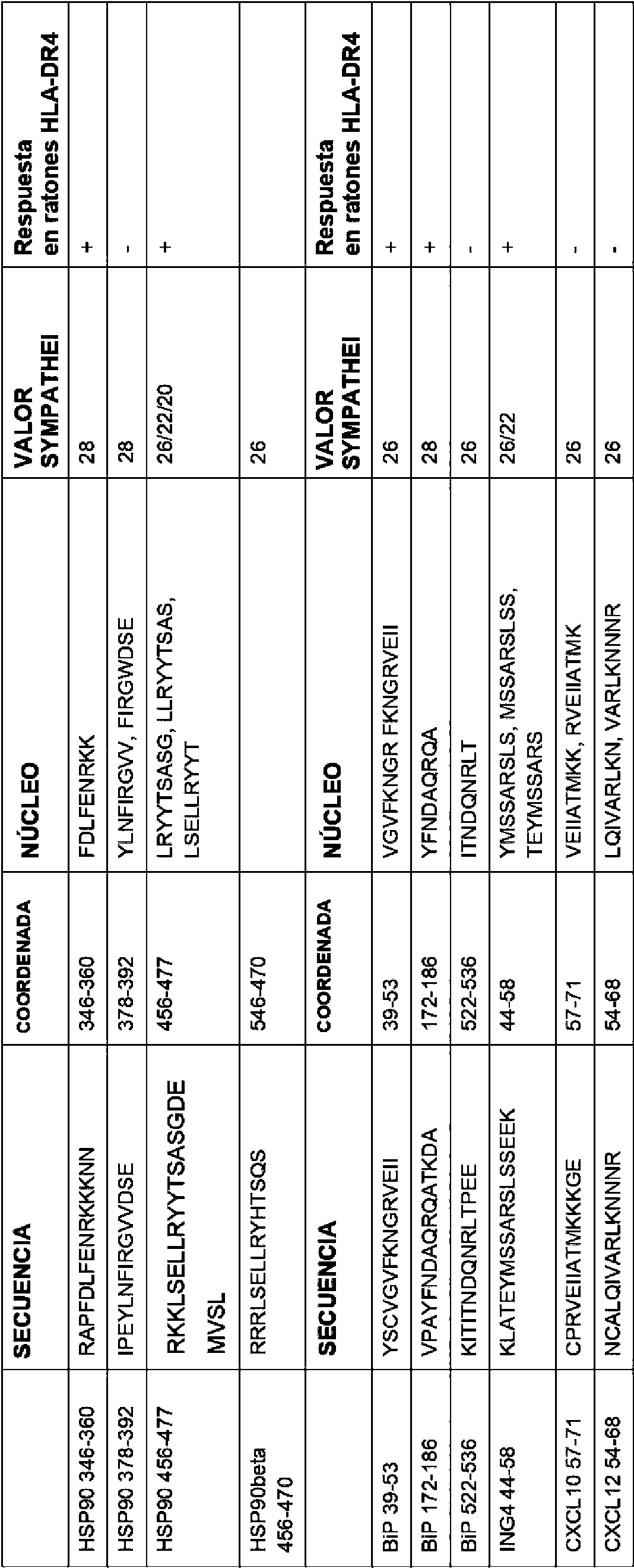
Los ratones transgénicos de HLA-DR4 se inmunizaron hasta en tres ocasiones con péptidos citrulinados y se evaluaron ex vivo las respuestas por elispot de IFNg contra péptidos citrulinados y no modificados pertinentes. La figura 34 muestra respuestas significativas a BiP 39-53, BiP 172-186, HSP90346-360, HSP90456-477 y ING444 58 citrulinados sobre aquellas contra el péptido no modificado o el control de fondo. Esto proporciona otro método eficaz para la selección de epítopos de linfocitos T citrulinados.HLA-DR4 transgenic mice were immunized up to three times with citrullinated peptides and IFNg elispot responses against relevant citrullinated and unmodified peptides were assessed ex vivo. Figure 34 shows significant responses to Citrullinated BiP 39-53, BiP 172-186, HSP90346-360, HSP90456-477 and ING444 58 over those against the unmodified peptide or background control. This provides another efficient method for the selection of citrullinated T lymphocyte epitopes.
Para demostrar que los linfocitos T reconocen los tumores, pueden cribarse para el reconocimiento de células diana tumorales, separarse con ácido para alentar el reciclado de MHC y privarse de suero para inducir autofagia (figura 22a y e). La función de la citrulinación y la autofagia puede confirmarse usando inhibidores de PAD y autofagia (figura 22a). Las respuestas antitumorales in vivo pueden medirse iniciando tumores, inmunizando con péptidos citrulinados y controlando el crecimiento del tumor como se muestra en las figuras (24-26).To demonstrate that T cells recognize tumors, they can be screened for tumor target cell recognition, acid-stripped to encourage MHC recycling, and serum starved to induce autophagy (Figure 22a and e). The function of citrullination and autophagy can be confirmed using inhibitors of PAD and autophagy (Figure 22a). In vivo antitumor responses can be measured by initiating tumors, immunizing with citrullinated peptides, and monitoring tumor growth as shown in Figures (24-26).
Ejemplo 11.Example 11.
Los estudios han demostrado que es posible determinar la diferenciación de células CD4 vírgenes en diferentes fenotipos auxiliares dependiendo de su medio de citocinas presente cuando se estimulan. Por el contrario, se ha demostrado por primera vez en la figura 15 que determinados epítopos pueden determinar la diferenciación de linfocitos T auxiliares a pesar del entorno de citocinas. Vim415 cit estimulaba un fenotipo Th1/IL-17 incluso cuando se inmunizaba en presencia del adyuvante Th2 adyuvante completo de Freund. Esto sugiere que la potencia del acoplamiento del receptor de linfocitos T CD4 con el péptido MHC puede determinar la diferenciación de linfocitos T. En este contexto, se ha demostrado que vim415 de tipo silvestre estimula una respuesta de IL-10 (figura 30; media de 580 manchas/millón de esplenocitos, p=0,0249) incluso en presencia de los adyuvantes Th1, CpG/MPLA. Sin embargo, no logró estimular una respuesta de IFNy o IL-17 significativa.Studies have shown that it is possible to determine the differentiation of naive CD4 cells into different helper phenotypes depending on their cytokine environment present when stimulated. In contrast, it has been shown for the first time in Figure 15 that certain epitopes can determine T helper lymphocyte differentiation despite the cytokine environment. Vim415 cit stimulated a Th1 / IL-17 phenotype even when immunized in the presence of Freund's complete adjuvant Th2 adjuvant. This suggests that the potency of CD4 T cell receptor coupling with the MHC peptide may determine T cell differentiation. In this context, wild-type vim415 has been shown to stimulate an IL-10 response (Figure 30; mean of 580 spots / million splenocytes, p = 0.0249) even in the presence of Th1 adjuvants, CpG / MPLA. However, it failed to stimulate a significant IFNy or IL-17 response.
ReferenciasReferences
1. Gao, F.G., et al., Antigen-specific CD4+ T-cell help is required to activate a memory CD8 + T cell to a fully functional tumor killer cell. Cancer Res, 2002. 62(22): págs. 6438-41.1. Gao, FG, et al., Antigen-specific CD4 + T-cell help is required to activate a memory CD 8 + T cell to a fully functional tumor killer cell. Cancer Res, 2002. 62 (22): pp. 6438-41.
2. Janssen, E.M., et al., CD4+ T cells are required for secondary expansion and memory in CD8 + T lymphocytes. Nature, 2003. 421(6925): págs. 852-6.2. Janssen, EM, et al., CD4 + T cells are required for secondary expansion and memory in CD 8 + T lymphocytes. Nature, 2003. 421 (6925): pp. 852-6.
3. Fearon, E.R., et al., Interleukin-2 production by tumor cells bypasses T helper function in the generation of an antitumor response. Cell, 1990. 60(3): pásg. 397-403.3. Fearon, E.R., et al., Interleukin-2 production by tumor cells bypasses T helper function in the generation of an antitumor response. Cell, 1990. 60 (3): p. 397-403.
4. Baxevanis, C.N., et al., Tumor-specific CD4+ T lymphocytes from cancer patients are required for optimal induction of cytotoxic T cells against the autologous tumor. J Immunol, 2000. 164(7): págs. 3902-12.4. Baxevanis, C.N., et al., Tumor-specific CD4 + T lymphocytes from cancer patients are required for optimal induction of cytotoxic T cells against the autologous tumor. J Immunol, 2000. 164 (7): pp. 3902-12.
5. Cella, M., et al., Ligation of CD40 on dendritic cells triggers production of high levels of interleukin-12 and enhances T cell stimulatory capacity: T-T help via APC activation. J. Exp. Med., 1996. 184(2): págs. 747-52. 6. Ridge, J.P., F. Di Rosa y P. Matzinger, A conditioned dendritic cell can be a temporal bridge between a CD4+ T-helper and a T-killer cell. Nature, 1998. 393(6684): págs. 474-8.5. Cella, M., et al., Ligation of CD40 on dendritic cells triggers production of high levels of interleukin-12 and enhances T cell stimulatory capacity: TT help via APC activation. J. Exp. Med., 1996. 184 (2): pp. 747-52. 6 . Ridge, JP, F. Di Rosa and P. Matzinger, A conditioned dendritic cell can be a temporal bridge between a CD4 + T-helper and a T-killer cell. Nature, 1998. 393 (6684): pp. 474-8.
7. Schoenberger, S.P., et al., T-cell help for cytotoxic T lymphocytes is mediated by CD40-CD40L interactions. Nature, 1998. 393(6684): págs. 480-3.7. Schoenberger, S.P., et al., T-cell help for cytotoxic T lymphocytes is mediated by CD40-CD40L interactions. Nature, 1998. 393 (6684): pp. 480-3.
8. Ayyoub, M., et al., An immunodominant SSX-2-derived epitope recognized by CD4+ T cells in association with HLA-DR. J Clin Invest, 2004. 113(8): págs. 1225-33. 8 . Ayyoub, M., et al., An immunodominant SSX-2-derived epitope recognized by CD4 + T cells in association with HLA-DR. J Clin Invest, 2004. 113 (8): pp. 1225-33.
9. Halder, T., et al., Isolation of novel HLA-DR restricted potential tumor-associated antigens from the melanoma cell line FM3. Cancer Res, 1997. 57(15): págs. 3238-44.9. Halder, T., et al., Isolation of novel HLA-DR restricted potential tumor-associated antigens from the melanoma cell line FM3. Cancer Res, 1997. 57 (15): pp. 3238-44.
10. Pardoll, D.M. y S.L. Topalian, The role of CD4+ T cell responses in antitumor immunity. Curr Opin Immunol, 1998. 10(5): págs. 588-94.10. Pardoll, D.M. and S.L. Topalian, The role of CD4 + T cell responses in antitumor immunity. Curr Opin Immunol, 1998. 10 (5): pp. 588-94.
11. Topalian, S.L., MHC class II restricted tumor antigens and the role of CD4+ T cells in cancer immunotherapy. Curr Opin Immunol, 1994. 6(5): págs. 741-5.11. Topalian, S.L., MHC class II restricted tumor antigens and the role of CD4 + T cells in cancer immunotherapy. Curr Opin Immunol, 1994. 6 (5): pp. 741-5.
12. Muranski, P., et al., Tumor-specific Th17-polarized cells eradicate large established melanoma. Blood, 2008.12. Muranski, P., et al., Tumor-specific Th17-polarized cells eradicate large established melanoma. Blood, 2008.
112(2): págs. 362-73.112 (2): pp. 362-73.
13. Paludan, C., et al., Epstein-Barr nuclear antigen 1-specific CD4(+) Th1 cells kill Burkitt's lymphoma cells. J Immunol, 2002. 169(3): págs. 1593-603.13. Paludan, C., et al., Epstein-Barr nuclear antigen 1-specific CD4 (+) Th1 cells kill Burkitt's lymphoma cells. J Immunol, 2002. 169 (3): pp. 1593-603.
14. Quezada, S.A., et al., Tumor-reactive CD4(+) T cells develop cytotoxic activity and eradicate large established melanoma after transfer into lymphopenic hosts. J. Exp. Med., 2010. 207(3): págs. 637-50.14. Quezada, S.A., et al., Tumor-reactive CD4 (+) T cells develop cytotoxic activity and eradicate large established melanoma after transfer into lymphopenic hosts. J. Exp. Med., 2010. 207 (3): pp. 637-50.
15. Xie, Y., et al., Naive tumor-specific CD4(+) T cells differentiated in vivo eradicate established melanoma. Journal of Experimental Medicine, 2010. 207(3): págs. 651-667.15. Xie, Y., et al., Naive tumor-specific CD4 (+) T cells differentiated in vivo eradicate established melanoma. Journal of Experimental Medicine, 2010. 207 (3): pp. 651-667.
16. Brandmaier, A.G., et al., High-avidity autoreactive CD4+ T cells induce host CTL, overcome T(regs) and mediate tumor destruction. J Immunother, 2009. 32(7): págs. 677-88.16. Brandmaier, A.G., et al., High-avidity autoreactive CD4 + T cells induce host CTL, overcome T (regs) and mediate tumor destruction. J Immunother, 2009. 32 (7): pp. 677-88.
17. Lauwen, M.M., et al., Self-tolerance does not restrict the CD4+ T-helper response against the p53 tumor antigen. Cancer Res, 2008. 68(3): págs. 893-900.17. Lauwen, M.M., et al., Self-tolerance does not restrict the CD4 + T-helper response against the p53 tumor antigen. Cancer Res, 2008. 68 (3): pp. 893-900.
18. Touloukian, C.E., et al., Identification of a MHC class II-restricted human gp100 epitope using DR4-IE transgenic mice. J Immunol, 2000. 164(7): págs. 3535-42.18. Touloukian, C.E., et al., Identification of a MHC class II-restricted human gp100 epitope using DR4-IE transgenic mice. J Immunol, 2000. 164 (7): pp. 3535-42.
19. Mohanan, S., et al., Potential role of peptidylarginine deiminase enzymes and protein citrullination in cancer pathogenesis. Biochem Res Int, 2012. 2012: págs. 895343.19. Mohanan, S., et al., Potential role of peptidylarginine deiminase enzymes and protein citrullination in cancer pathogenesis. Biochem Res Int, 2012. 2012: pp. 895343.
20. Nomura, K., Specificity and mode of action of the muscle-type protein-arginine deiminase. Arch Biochem Biophys, 1992. 293(2): págs. 362-9. 20. Nomura, K., Specificity and mode of action of the muscle-type protein-arginine deiminase. Arch Biochem Biophys, 1992. 293 (2): pp. 362-9.
21. Stensland, M.E., et al., Primary sequence, together with other factors, influence peptide deimination by peptidylarginine deiminase-4. Biol Chem, 2009. 390(2): págs. 99-107.21. Stensland, M.E., et al., Primary sequence, together with other factors, influence peptide deimination by peptidylarginine deiminase-4. Biol Chem, 2009. 390 (2): pp. 99-107.
22. Klareskog, L., et al., Immunity to citrullinated proteins in rheumatoid arthritis. Annual Review of Immunology, 2008. 26: págs. 651-675.22. Klareskog, L., et al., Immunity to citrullinated proteins in rheumatoid arthritis. Annual Review of Immunology, 2008. 26: pp. 651-675.
23. Migliorini, P., et al., The immune response to citrullinated antigens in autoimmune diseases. Autoimmunity Reviews, 2005. 4(8): págs. 561-564.23. Migliorini, P., et al., The immune response to citrullinated antigens in autoimmune diseases. Autoimmunity Reviews, 2005. 4 (8): pp. 561-564.
24. Nijenhuis, S., et al., Autoantibodies to citrullinated proteins in rheumatoid arthritis: clinical performance and biochemical aspects of an RA-specific marker. Clinica Chimica Acta, 2004. 350(1-2): págs. 17-34.24. Nijenhuis, S., et al., Autoantibodies to citrullinated proteins in rheumatoid arthritis: clinical performance and biochemical aspects of an RA-specific marker. Clinica Chimica Acta, 2004. 350 (1-2): pp. 17-34.
25. Sebbag, M., et al., Epitopes of human fibrin recognized by the rheumatoid arthritis-specific autoantibodies to citrullinated proteins. European Journal of Immunology, 2006. 36(8): págs. 2250-2263.25. Sebbag, M., et al., Epitopes of human fibrin recognized by the rheumatoid arthritis-specific autoantibodies to citrullinated proteins. European Journal of Immunology, 2006. 36 (8): pp. 2250-2263.
26. Vossenaar, E.R., et al., Rheumatoid arthritis specific anti-Sa antibodies target citrullinated vimentin. Arthritis Res Ther, 2004. 6(2): págs. R142-50.26. Vossenaar, E.R., et al., Rheumatoid arthritis specific anti-Sa antibodies target citrullinated vimentin. Arthritis Res Ther, 2004. 6 (2): pp. R142-50.
27. Vossenaar, E.R., et al., Expression and activity of citrullinating peptidylarginine deiminase enzymes in monocytes and macrophages. Annals of the Rheumatic Diseases, 2004. 63(4): págs. 373-381.27. Vossenaar, E.R., et al., Expression and activity of citrullinating peptidylarginine deiminase enzymes in monocytes and macrophages. Annals of the Rheumatic Diseases, 2004. 63 (4): pp. 373-381.
28. Birnboim, H.C., et al., Cutting edge: MHC class II-restricted peptides containing the inflammation-associated marker 3-nitrotyrosine evade central tolerance and elicit a robust cell-mediated immune response. J Immunol, 2003. 171(2): págs. 528-532.28. Birnboim, H.C., et al., Cutting edge: MHC class II-restricted peptides containing the inflammation-associated marker 3-nitrotyrosine evade central tolerance and elicit a robust cell-mediated immune response. J Immunol, 2003. 171 (2): pp. 528-532.
29. Herzog, J., et al., Activated antigen-presenting cells select and present chemically modified peptides recognized by unique CD4 T cells. Proceedings of the National Academy of Sciences of the United States of America, 2005. 102(22): págs. 7928-7933.29. Herzog, J., et al., Activated antigen-presenting cells select and present chemically modified peptides recognized by unique CD4 T cells. Proceedings of the National Academy of Sciences of the United States of America, 2005. 102 (22): pp. 7928-7933.
30. Bronte, V., et al., Boosting antitumor responses of T lymphocytes infiltrating human prostate cancers. Journal of Experimental Medicine, 2005. 201(8): págs. 1257-1268.30. Bronte, V., et al., Boosting antitumor responses of T lymphocytes infiltrating human prostate cancers. Journal of Experimental Medicine, 2005. 201 (8): pp. 1257-1268.
31. Arentz-Hansen, H., et al., The intestinal T cell response to alpha-gliadin in adult celiac disease is focused on a single deamidated glutamine targeted by tissue transglutaminase. J. Exp. Med., 2000. 191(4): págs. 603-12.31. Arentz-Hansen, H., et al., The intestinal T cell response to alpha-gliadin in adult celiac disease is focused on a single deamidated glutamine targeted by tissue transglutaminase. J. Exp. Med., 2000. 191 (4): pp. 603-12.
32. Pudney, V.A., et al., DNA vaccination with T-cell epitopes encoded within Ab molecules induces high-avidity anti-tumor CD8 (+) T cells. European Journal of Immunology, 2010. 40(3): págs. 899-910.32. Pudney, VA, et al., DNA vaccination with T-cell epitopes encoded within Ab molecules induces high-avidity anti-tumor CD 8 (+) T cells. European Journal of Immunology, 2010. 40 (3): pp. 899-910.
33. DURRANT, L.G.M., Rachael Louise; PUDNEY, Victoria Anne; , NUCLEIC ACIDS, 2008.33. DURRANT, L.G.M., Rachael Louise; PUDNEY, Victoria Anne; , NUCLEIC ACIDS, 2008.
34. Feitsma, A.L., et al., Identification of Citrullinated Vimentin Peptides as T Cell Epitopes in HLA-DR4-Positive Patients With Rheumatoid Arthritis. Arthritis and Rheumatism, 2010. 62(1): págs. 117-125.34. Feitsma, A.L., et al., Identification of Citrullinated Vimentin Peptides as T Cell Epitopes in HLA-DR4-Positive Patients With Rheumatoid Arthritis. Arthritis and Rheumatism, 2010. 62 (1): pp. 117-125.
35. Kryszke, M.H. y P. Vicart, Regulation of the expression of the human vimentin gene: Application to cellular immortalization. Pathologie Biologie, 1998. 46(1): págs. 39-45.35. Kryszke, M.H. and P. Vicart, Regulation of the expression of the human vimentin gene: Application to cellular immortalization. Pathologie Biologie, 1998. 46 (1): pp. 39-45.
36. Paulin, D., EXPRESSION OF THE GENES-CODING FOR THE HUMAN INTERMEDIATE FILAMENT PROTEINS. Pathologie Biologie, 1989. 37(4): págs. 277-282.36. Paulin, D., EXPRESSION OF THE GENES-CODING FOR THE HUMAN INTERMEDIATE FILAMENT PROTEINS. Pathologie Biologie, 1989. 37 (4): pp. 277-282.
37. Ramaekers, F.C., et al., Use of antibodies to intermediate filaments in the characterization of human tumors. Cold Spring Harb Symp Quant Biol, 1982. 46 Pt 1: págs. 331-9.37. Ramaekers, F.C., et al., Use of antibodies to intermediate filaments in the characterization of human tumors. Cold Spring Harb Symp Quant Biol, 1982. 46 Pt 1: pp. 331-9.
38. Thomas, P.A., et al., Association between keratin and vimentin expression, malignant phenotype, and survival in postmenopausal breast cancer patients. Clinical Cancer Research, 1999. 5(10): págs. 2698-2703.38. Thomas, P.A., et al., Association between keratin and vimentin expression, malignant phenotype, and survival in postmenopausal breast cancer patients. Clinical Cancer Research, 1999. 5 (10): pp. 2698-2703.
39. Conforti, G., et al., DIFFERENT VIMENTIN EXPRESSION IN 2 CLONES DERIVED FROM A HUMAN COLOCARCINOMA CELL-LINE (LOVO) SHOWING DIFFERENT SENSITIVITY TO DOXORUBICIN. British Journal of Cancer, 1995. 71(3): págs. 505-511.39. Conforti, G., et al., DIFFERENT VIMENTIN EXPRESSION IN 2 CLONES DERIVED FROM A HUMAN PLOCARCINOMA CELL-LINE (LOVO) SHOWING DIFFERENT SENSITIVITY TO DOXORUBICIN. British Journal of Cancer, 1995. 71 (3): pp. 505-511.
40. Moran, E., et al., Co-expression of MDR-associated markers, including P-170, MRP and LRP and cytoskeletal proteins, in three resistant variants of the human ovarian carcinoma cell line, OAW42. European Journal of Cancer, 1997. 33(4): págs. 652-660.40. Moran, E., et al., Co-expression of MDR-associated markers, including P-170, MRP and LRP and cytoskeletal proteins, in three resistant variants of the human ovarian carcinoma cell line, OAW42. European Journal of Cancer, 1997. 33 (4): pp. 652-660.
41. Gilles, C., et al., Vimentin expression in cervical carcinomas: association with invasive and migratory potential. J Pathol, 1996. 180(2): págs. 175-80.41. Gilles, C., et al., Vimentin expression in cervical carcinomas: association with invasive and migratory potential. J Pathol, 1996. 180 (2): pp. 175-80.
42. Williams, A.A., et al., CD 9 and vimentin distinguish clear cell from chromophobe renal cell carcinoma. BMC Clin Pathol, 2009. 9: pág. 9.42. Williams, A.A., et al., CD 9 and vimentin distinguish clear cell from chromophobe renal cell carcinoma. BMC Clin Pathol, 2009. 9: p. 9.
43. Gustmann, C., et al., CYTOKERATIN EXPRESSION AND VIMENTIN CONTENT IN LARGE CELL ANAPLASTIC LYMPHOMAS AND OTHER NON-HODGKINS-LYMPHOMAS. American Journal of Pathology, 1991. 138(6): pág. 1413-1422.43. Gustmann, C., et al., CYTOKERATIN EXPRESSION AND VIMENTIN CONTENT IN LARGE CELL ANAPLASTIC LYMPHOMAS AND OTHER NON-HODGKINS-LYMPHOMAS. American Journal of Pathology, 1991. 138 (6): p. 1413-1422.
44. Yamamoto, Y., K. Izumi y H. Otsuka, AN IMMUNOHISTOCHEMICAL STUDY OF EPITHELIAL MEMBRANE ANTIGEN, CYTOKERATIN, AND VIMENTIN IN PAPILLARY THYROID-CARCINOMA - RECOGNITION OF LETHAL AND FAVORABLE PROGNOSTIC TYPES. Cancer, 1992. 70(9): págs. 2326-2333.44. Yamamoto, Y., K. Izumi and H. Otsuka, AN IMMUNOHISTOCHEMICAL STUDY OF EPITHELIAL MEMBRANE ANTIGEN, CYTOKERATIN, AND VIMENTIN IN PAPILLARY THYROID-CARCINOMA - RECOGNITION OF LETHAL AND FAVORABLE PROGNOSTIC TYPES. Cancer, 1992. 70 (9): pp. 2326-2333.
45. Coppola, D., et al., Prognostic significance of p53, bcl-2, vimentin, and S100 protein-positive Langerhans cells in endometrial carcinoma. Human Pathology, 1998. 29(5): págs. 455-462.45. Coppola, D., et al., Prognostic significance of p53, bcl-2, vimentin, and S100 protein-positive Langerhans cells in endometrial carcinoma. Human Pathology, 1998. 29 (5): pp. 455-462.
46. Fuyuhiro, Y., et al., Clinical Significance of Vimentin-positive Gastric Cancer Cells. Anticancer Research, 2010. 30(12): págs. 5239-5243.46. Fuyuhiro, Y., et al., Clinical Significance of Vimentin-positive Gastric Cancer Cells. Anticancer Research, 2010. 30 (12): pp. 5239-5243.
47. Takemura, K., et al., EXPRESSION OF VIMENTIN IN GASTRIC-CANCER - A POSSIBLE INDICATOR FOR PROGNOSIS. Pathobiology, 1994. 62(3): pág. 149-154.47. Takemura, K., et al., EXPRESSION OF VIMENTIN IN GASTRIC-CANCER - A POSSIBLE INDICATOR FOR PROGNOSIS. Pathobiology, 1994. 62 (3): p. 149-154.
48. Ivaska, J., Vimentin: Central hub in EMT induction? Small Gtpases, 2011,2(1): págs. 51-53.48. Ivaska, J., Vimentin: Central hub in EMT induction? Small Gtpases, 2011,2 (1): pp. 51-53.
49. Vuoriluoto, K., et al., Vimentin regulates EMT induction by Slug and oncogenic H-Ras and migration by governing Axl expression in breast cancer. Oncogene, 2011,30(12): págs. 1436-1448.49. Vuoriluoto, K., et al., Vimentin regulates EMT induction by Slug and oncogenic H-Ras and migration by governing Axl expression in breast cancer. Oncogene, 2011,30 (12): pp. 1436-1448.
50. Hill, JA, et al., Cutting edge: The conversion of arginine to citrulline allows for a high-affinity peptide interaction with the rheumatoid arthritis-associated HLA-DRB1*0401 MHC class II molecule. J Immunol, 2003. 171(2): págs.50. Hill, JA, et al., Cutting edge: The conversion of arginine to citrulline allows for a high-affinity peptide interaction with the rheumatoid arthritis-associated HLA-DRB1 * 0401 MHC class II molecule. J Immunol, 2003. 171 (2): pp.
538-541.538-541.
51. Ireland, J., J. Herzog y E.R. Unanue, Cutting edge: Unique T cells that recognize citrullinated peptides are a feature of protein immunization. J Immunol, 2006. 177(3): págs. 1421-1425.51. Ireland, J., J. Herzog and ER Unanue, Cutting edge: Unique T cells that recognize citrullinated peptides are a feature of protein immunization. J Immunol, 2006. 177 (3): pp. 1421-1425.
52. Asaga, H., et al., Immunocytochemical localization of peptidylarginine deiminase in human eosinophils and neutrophils. Journal of Leukocyte Biology, 2001,70(1): págs. 46-51.52. Asaga, H., et al., Immunocytochemical localization of peptidylarginine deiminase in human eosinophils and neutrophils. Journal of Leukocyte Biology, 2001,70 (1): pp. 46-51.
53. Nagata, S. y T. Senshu, PEPTIDYLARGININE DEIMINASE IN RAT AND MOUSE HEMATOPOIETIC-CELLS. Experientia, 1990. 46(1): págs. 72-74.53. Nagata, S. and T. Senshu, PEPTIDYLARGININE DEIMINASE IN RAT AND MOUSE HEMATOPOIETIC-CELLS. Experientia, 1990. 46 (1): pp. 72-74.
54. Loos, T., et al., Citrullination of CXCL10 and CXCL11 by peptidylarginine deiminase: a naturally occurring posttranslational modification of chemokines and new dimension of immunoregulation. Blood, 2008. 112(7): pág.54. Loos, T., et al., Citrullination of CXCL10 and CXCL11 by peptidylarginine deiminase: a naturally occurring posttranslational modification of chemokines and new dimension of immunoregulation. Blood, 2008. 112 (7): p.
2648-2656.2648-2656.
55. Proost, P., et al., Citrullination of CXCL8 by peptidylarginine deiminase alters receptor usage, prevents proteolysis, and dampens tissue inflammation. Journal of Experimental Medicine, 2008. 205(9): pág. 2085-2097.55. Proost, P., et al., Citrullination of CXCL 8 by peptidylarginine deiminase alters receptor usage, prevents proteolysis, and dampens tissue inflammation. Journal of Experimental Medicine, 2008. 205 (9): p. 2085-2097.
56. Struyf, S., et al., Citrullination of CXCL12 Differentially Reduces CXCR4 and CXCR7 Binding with Loss of Inflammatory and Anti-HIV-1 Activity via CXCR4. J Immunol, 2009. 182(1): págs. 666-674.56. Struyf, S., et al., Citrullination of CXCL12 Differentially Reduces CXCR4 and CXCR7 Binding with Loss of Inflammatory and Anti-HIV-1 Activity via CXCR4. J Immunol, 2009. 182 (1): pp. 666-674.
57. Ireland, J.M. y E.R. Unanue, Autophagy in antigen-presenting cells results in presentation of citrullinated peptides to CD4 T cells. Journal of Experimental Medicine, 2011,208(13): págs. 2625-2632.57. Ireland, J.M. and E.R. Unanue, Autophagy in antigen-presenting cells results in presentation of citrullinated peptides to CD4 T cells. Journal of Experimental Medicine, 2011,208 (13): pp. 2625-2632.
58. Asaga, H., M. Yamada y T. Senshu, Selective deimination of vimentin in calcium ionophore-induced apoptosis of mouse peritoneal macrophages. Biochem Biophys Res Commun, 1998. 243(3): págs. 641-6.58. Asaga, H., M. Yamada and T. Senshu, Selective deimination of vimentin in calcium ionophore-induced apoptosis of mouse peritoneal macrophages. Biochem Biophys Res Commun, 1998. 243 (3): pp. 641-6.
59. Mor-Vaknin, N., et al., Vimentin is secreted by activated macrophages. Nature Cell Biology, 2003. 5(1): págs.59. Mor-Vaknin, N., et al., Vimentin is secreted by activated macrophages. Nature Cell Biology, 2003. 5 (1): pp.
59-63.59-63.
60. Vossenaar, E.R., et al., PAD, a growing family of citrullinating enzymes: genes, features and involvement in disease. Bioessays, 2003. 25(11): págs. 1106-18.60. Vossenaar, E.R., et al., PAD, a growing family of citrullinating enzymes: genes, features and involvement in disease. Bioessays, 2003. 25 (11): pp. 1106-18.
61. Kaufmann, S.H.E., The contribution of immunology to the rational design of novel antibacterial vaccines. Nature Reviews Microbiology, 2007. 5(7): págs. 491-504.61. Kaufmann, S.H.E., The contribution of immunology to the rational design of novel antibacterial vaccines. Nature Reviews Microbiology, 2007. 5 (7): pp. 491-504.
62. Kubilus, J., R.F. Waitkus y H.P. Baden, PARTIAL-PURIFICATION AND SPECIFICITY OF AN ARGININECONVERTING ENZYME FROM BOVINE EPIDERMIS. Biochimica Et Biophysica Acta, 1980. 615(1): pág. 246-251.62. Kubilus, J., R.F. Waitkus and H.P. Baden, PARTIAL-PURIFICATION AND SPECIFICITY OF AN ARGININECONVERTING ENZYME FROM BOVINE EPIDERMIS. Biochimica Et Biophysica Acta, 1980. 615 (1): p. 246-251.
63. Senshu, T., et al., Studies on specificity of peptidylarginine deiminase reactions using an immunochemical probe that recognizes an enzymatically deiminated partial sequence of mouse keratin K1. Journal of Dermatological Science, 1999. 21(2): págs. 113-126.63. Senshu, T., et al., Studies on specificity of peptidylarginine deiminase reactions using an immunochemical probe that recognizes an enzymatically deiminated partial sequence of mouse keratin K1. Journal of Dermatological Science, 1999. 21 (2): pp. 113-126.
64. Chang, X., et al., Localization of peptidylarginine deiminase 4 (PADI4) and citrullinated protein in synovial tissue of rheumatoid arthritis. Rheumatology, 2005. 44(1): págs. 40-50.64. Chang, X., et al., Localization of peptidylarginine deiminase 4 (PADI4) and citrullinated protein in synovial tissue of rheumatoid arthritis. Rheumatology, 2005. 44 (1): pp. 40-50.
65. Chang, X. y J. Han, Expression of peptidylarginine deiminase type 4 (PAD4) in various tumors. Mol Carcinog, 2006. 45(3): págs. 183-96.65. Chang, X. and J. Han, Expression of peptidylarginine deiminase type 4 (PAD4) in various tumors. Mol Carcinog, 2006. 45 (3): pp. 183-96.
6 6. Guo, Q. y W. Fast, Citrullination of inhibitor of growth 4 (ING4) by peptidylarginine deminase 4 (PAD4) disrupts the interaction between ING4 and p53. J Biol Chem, 2011,286(19): págs. 17069-78. 6 6 . Guo, Q. and W. Fast, Citrullination of inhibitor of growth 4 (ING4) by peptidylarginine deminase 4 (PAD4) disrupts the interaction between ING4 and p53. J Biol Chem, 2011,286 (19): pp. 17069-78.
67. Karlin, S. y S.F. Altschul, Applications and statistics for multiple high-scoring segments in molecular sequences. Proc Natl Acad Sci U S A, 1993. 90(12): págs. 5873-7.67. Karlin, S. and S.F. Altschul, Applications and statistics for multiple high-scoring segments in molecular sequences. Proc Natl Acad Sci U S A, 1993. 90 (12): pp. 5873-7.
6 8. Altschul, S.F., et al., Basic local alignment search tool. J Mol Biol, 1990. 215(3): págs. 403-10. 6 8 . Altschul, SF, et al., Basic local alignment search tool. J Mol Biol, 1990. 215 (3): pp. 403-10.
69. Altschul, S.F., et al., Gapped BLAST and PSI-BLAST: a new generation of protein database search programs. Nucleic Acids Res, 1997. 25(17): págs. 3389-402.69. Altschul, S.F., et al., Gapped BLAST and PSI-BLAST: a new generation of protein database search programs. Nucleic Acids Res, 1997. 25 (17): pp. 3389-402.
70. Myers, E.W. y W. Miller, Approximate matching of regular expressions. Bull Math Biol, 1989. 51(1): pág. 5-37.70. Myers, E.W. and W. Miller, Approximate matching of regular expressions. Bull Math Biol, 1989. 51 (1): p. 5-37.
71. Torelli, A. y C.A. Robotti, ADVANCE and ADAM: two algorithms for the analysis of global similarity between homologous informational sequences. Comput Appl Biosci, 1994. 10(1): págs. 3-5.71. Torelli, A. and C.A. Robotti, ADVANCE and ADAM: two algorithms for the analysis of global similarity between homologous informational sequences. Comput Appl Biosci, 1994. 10 (1): pp. 3-5.
72. Pearson, W.R. y D.J. Lipman, Improved tools for biological sequence comparison. Proc Natl Acad Sci U S A, 1988. 85(8): págs. 2444-8.72. Pearson, W.R. and D.J. Lipman, Improved tools for biological sequence comparison. Proc Natl Acad Sci U S A, 1988. 85 (8): pp. 2444-8.
73. Remington, R., Remington's pharmaceutical sciences. 16a ed. 1980: Mack Pub. Co.73. Remington, R., Remington's pharmaceutical sciences. 16th ed. 1980: Mack Pub. Co.
74. Stewart, Solid phase peptide synthesis. 2a ed. 1984, Rockford: Illinois Pierce Chemical Company.74. Stewart, Solid phase peptide synthesis. 2nd ed. 1984, Rockford: Illinois Pierce Chemical Company.
75. Bodanzsky, B., The practice of peptide synthesis. 1984, Nueva York: Springer Verlag.75. Bodanzsky, B., The practice of peptide synthesis. 1984, New York: Springer Verlag.
76. Sambrook, J., Fritsch y Maniatis, Molecular cloning: A laboratory manual. 2a ed. 1989: Cold Spring Harbor Laboratory Press.76. Sambrook, J., Fritsch and Maniatis, Molecular cloning: A laboratory manual. 2nd ed. 1989: Cold Spring Harbor Laboratory Press.
77. Ausubel, J., Short protocols in molecular biology. 1992: John Wiley y Sons77. Ausubel, J., Short protocols in molecular biology. 1992: John Wiley and Sons
78. Pluckthun, A., Antibody engineering: advances from the use of Escherichia coli expression systems. Biotechnology (N Y), 1991,9(6): págs. 545-51.78. Pluckthun, A., Antibody engineering: advances from the use of Escherichia coli expression systems. Biotechnology (N Y), 1991.9 (6): pp. 545-51.
79. Reff, M.E., High-level production of recombinant immunoglobulins in mammalian cells. Curr Opin Biotechnol, 1993. 4(5): pág. 573-6.79. Reff, M.E., High-level production of recombinant immunoglobulins in mammalian cells. Curr Opin Biotechnol, 1993. 4 (5): p. 573-6.
80. Trill, J.J., A.R. Shatzman y S. Ganguly, Production of monoclonal antibodies in COS and CHO cells. Curr Opin Biotechnol, 1995. 6(5): págs. 553-60.80. Trill, J.J., A.R. Shatzman and S. Ganguly, Production of monoclonal antibodies in COS and CHO cells. Curr Opin Biotechnol, 1995. 6 (5): pp. 553-60.
81. Palena, C., et al., Strategies to target molecules that control the acquisition of a mesenchymal-like phenotype by carcinoma cells. Exp Biol Med (Maywood), 2011,236(5): págs. 537-45.81. Palena, C., et al., Strategies to target molecules that control the acquisition of a mesenchymal-like phenotype by carcinoma cells. Exp Biol Med (Maywood), 2011,236 (5): pp. 537-45.
82. Denzin, L.K., et al., Assembly and intracellular transport of HLA-DM and correction of the class II antigenprocessing defect in T2 cells. Immunity, 1994. 1(7): págs. 595-606.82. Denzin, L.K., et al., Assembly and intracellular transport of HLA-DM and correction of the class II antigenprocessing defect in T2 cells. Immunity, 1994. 1 (7): pp. 595-606.
83. Kovats, S., et al., Presentation of abundant endogenous class II DR-restricted antigens by DM-negative B cell lines. Eur J Immunol, 1997. 27(4): págs. 1014-21.83. Kovats, S., et al., Presentation of abundant endogenous class II DR-restricted antigens by DM-negative B cell lines. Eur J Immunol, 1997. 27 (4): pp. 1014-21.
84. Metheringham, R.L., et al., Antibodies designed as effective cancer vaccines. MAbs, 2009. 1(1): págs. 71-85.84. Metheringham, R.L., et al., Antibodies designed as effective cancer vaccines. MAbs, 2009. 1 (1): pp. 71-85.
85. Quinn MJ BP, B.A., Kirby EA, Jones J, Cancer trends in england and wales, 1950-1999, en Londres: Stationery Office 2001. pásg. 206-207.85. Quinn MJ BP, B.A., Kirby EA, Jones J, Cancer trends in england and wales, 1950-1999, in London: Stationery Office 2001. p. 206-207.
86. NICE, Improving outcomes in colorectal cancers: Manual update, 2004: Londres: National Institute for Clinical Excellence. 86 . NICE, Improving outcomes in colorectal cancers: Manual update, 2004: London: National Institute for Clinical Excellence.
Ċ Ċ
Claims (2)
Applications Claiming Priority (1)
| Application Number | Priority Date | Filing Date | Title |
|---|---|---|---|
| GBGB1214007.5A GB201214007D0 (en) | 2012-08-07 | 2012-08-07 | Anti-tumour immune responses to modified self-epitopes |
Publications (1)
| Publication Number | Publication Date |
|---|---|
| ES2843777T3 true ES2843777T3 (en) | 2021-07-20 |
Family
ID=46934958
Family Applications (3)
| Application Number | Title | Priority Date | Filing Date |
|---|---|---|---|
| ES13750368T Active ES2681973T3 (en) | 2012-08-07 | 2013-08-07 | Anti-tumor response against modified autoepitopes |
| ES17194251T Active ES2833962T3 (en) | 2012-08-07 | 2013-08-07 | Antitumor response to modified self epitopes |
| ES17194252T Active ES2843777T3 (en) | 2012-08-07 | 2013-08-07 | Antitumor response against modified autoepitopes |
Family Applications Before (2)
| Application Number | Title | Priority Date | Filing Date |
|---|---|---|---|
| ES13750368T Active ES2681973T3 (en) | 2012-08-07 | 2013-08-07 | Anti-tumor response against modified autoepitopes |
| ES17194251T Active ES2833962T3 (en) | 2012-08-07 | 2013-08-07 | Antitumor response to modified self epitopes |
Country Status (18)
| Country | Link |
|---|---|
| US (4) | US10233220B2 (en) |
| EP (3) | EP3301447B1 (en) |
| JP (2) | JP6490581B2 (en) |
| KR (1) | KR102203096B1 (en) |
| CN (2) | CN104823054B (en) |
| AU (1) | AU2013301341B2 (en) |
| BR (1) | BR112015002574A8 (en) |
| CA (1) | CA2874515A1 (en) |
| DK (3) | DK3287787T3 (en) |
| ES (3) | ES2681973T3 (en) |
| GB (1) | GB201214007D0 (en) |
| HK (1) | HK1206427A1 (en) |
| IN (1) | IN2014DN10629A (en) |
| PL (2) | PL3287787T3 (en) |
| PT (3) | PT2883054T (en) |
| SG (2) | SG11201407804RA (en) |
| WO (1) | WO2014023957A2 (en) |
| ZA (1) | ZA201409095B (en) |
Families Citing this family (19)
| Publication number | Priority date | Publication date | Assignee | Title |
|---|---|---|---|---|
| EP3061462B1 (en) | 2007-07-02 | 2019-02-27 | Etubics Corporation | Methods and compositions for producing an adenovirus vector for use with multiple vaccinations |
| GB201214007D0 (en) | 2012-08-07 | 2012-09-19 | Scancell Ltd | Anti-tumour immune responses to modified self-epitopes |
| US9605276B2 (en) | 2012-08-24 | 2017-03-28 | Etubics Corporation | Replication defective adenovirus vector in vaccination |
| AU2015257929B2 (en) | 2014-05-05 | 2020-06-25 | Novio Th B.V. | Method for the serological diagnosis of rheumatoid arthritis. |
| CN104360070B (en) * | 2014-11-28 | 2017-02-22 | 山东新创生物科技有限公司 | Application of peptidylarginine deiminase 2 (PAD2) to preparation of reagent for clinical blood diagnosis of tumors |
| CN113456812A (en) | 2015-01-09 | 2021-10-01 | 埃图比克斯公司 | Methods and compositions for combination immunotherapy |
| EP3286213B1 (en) | 2015-04-20 | 2021-08-04 | Etubics Corporation | Methods and compositions for combination immunotherapy |
| AU2016260540B2 (en) | 2015-05-13 | 2021-01-07 | Agenus Inc. | Vaccines for treatment and prevention of cancer |
| GB201512703D0 (en) | 2015-07-20 | 2015-08-26 | Scancell Ltd | Anti-tumour immune responses to modified self-epitopes |
| GB201522667D0 (en) * | 2015-12-22 | 2016-02-03 | Immatics Biotechnologies Gmbh | Novel peptides and combination of peptides for use in immunotherapy against breast cancer and other cancers |
| SG10202111399YA (en) | 2015-12-22 | 2021-11-29 | Immatics Biotechnologies Gmbh | Peptides and combination of peptides for use in immunotherapy against breast cancer and other cancers |
| WO2018046648A1 (en) * | 2016-09-08 | 2018-03-15 | Katholieke Universiteit Leuven | Citrullinated peptides for diagnosis or treatment of diabetes |
| KR102184377B1 (en) * | 2018-02-19 | 2020-11-30 | 고려대학교 산학협력단 | Vaccine Comprising Epitopes of Heat Shock Protein and Its Uses |
| US11065317B2 (en) | 2018-04-26 | 2021-07-20 | Agenus Inc. | Heat shock protein-binding peptide compositions and methods of use thereof |
| GB201815041D0 (en) | 2018-09-14 | 2018-10-31 | Scancell Ltd | Epitopes |
| GB202005779D0 (en) | 2020-04-21 | 2020-06-03 | Scancell Ltd | Anti-tumour immune responses |
| CN112194719A (en) * | 2020-09-01 | 2021-01-08 | 中日友好医院(中日友好临床医学研究所) | Preparation and application of CRT antigen and MAGE-A1 antigen |
| GB202018395D0 (en) | 2020-11-23 | 2021-01-06 | Scancell Ltd | Immunotherapy |
| CN114404570B (en) * | 2022-01-20 | 2023-05-30 | 中国药科大学 | Novel anti-tumor metastasis polypeptide and application thereof |
Family Cites Families (29)
| Publication number | Priority date | Publication date | Assignee | Title |
|---|---|---|---|---|
| JP2002051785A (en) * | 2000-08-09 | 2002-02-19 | Hagiwara Yoshihide | Amino acid sequence of antigen epitope recognized by anticancer human monoclonal antibody cln- immunoglobulin g and dna nucleotide sequence encoding the same |
| WO2002034287A2 (en) * | 2000-10-27 | 2002-05-02 | Pharmexa A/S | Therapeutic vaccine formulations containing chitosan |
| US20030072737A1 (en) * | 2000-12-29 | 2003-04-17 | Michael Brines | Tissue protective cytokines for the protection, restoration, and enhancement of responsive cells, tissues and organs |
| WO2003024191A2 (en) * | 2001-09-21 | 2003-03-27 | Raven Biotechnologies, Inc. | Antibodies that bind to cancer-associated antigen cytokeratin 8 and methods of use thereof |
| AU2003207459A1 (en) * | 2002-01-03 | 2003-07-24 | The Scripps Research Institute | Cancer-associated epitope |
| US20040013649A1 (en) * | 2002-05-10 | 2004-01-22 | Inex Pharmaceuticals Corporation | Cancer vaccines and methods of using the same |
| ES2403932T3 (en) | 2002-08-27 | 2013-05-22 | Biokine Therapeutics Ltd. | CXCR4 antagonist and use of it |
| CA2498854A1 (en) * | 2002-10-02 | 2004-04-15 | Harald Kropshofer | Novel mhc ii associated peptides |
| US7670604B2 (en) * | 2002-12-13 | 2010-03-02 | Aurelium Biopharma, Inc. | Vimentin directed diagnostics and therapeutics for multidrug resistant neoplastic disease |
| DE10313819A1 (en) * | 2003-03-24 | 2004-10-07 | Immatics Biotechnologies Gmbh | Tumor-associated peptides binding to MHC molecules |
| EP1695089A1 (en) * | 2003-12-15 | 2006-08-30 | Aurelium Biopharma Inc. | Vimentin directed diagnostics and therapeutics for multidrug resistant neoplastic disease |
| EP1890724A2 (en) * | 2005-05-13 | 2008-02-27 | Oxxon Therapeutics Limited | Compositions for inducing an immune response |
| DE102005029845B4 (en) * | 2005-06-27 | 2017-03-23 | Orgentec Diagnostika Gmbh | Method for the diagnosis of rheumatic diseases |
| CN1712964A (en) * | 2005-07-18 | 2005-12-28 | 山东省医药生物技术研究中心 | Specific antigenic mark for rheumatoid arthritis and its use |
| US20070248628A1 (en) * | 2005-12-06 | 2007-10-25 | Keller Lorraine H | Immunogens in cancer stem cells |
| EA200801847A1 (en) * | 2006-02-17 | 2009-02-27 | Индиана Юниверсити Рисерч Энд Текнолоджи Корпорейшн | PEPTIDE INHIBITION OF CAPCNA INTERACTION IN CANCER |
| WO2007098093A2 (en) * | 2006-02-17 | 2007-08-30 | Five Prime Therapeutics, Inc. | Compositions and methods of use for modulators of polypeptides and polynucleotides in treating breast cancer and melanoma |
| AU2007243442A1 (en) * | 2006-04-24 | 2007-11-08 | Shantha West, Inc. | AgRM2 antigen |
| EP1882697B1 (en) * | 2006-07-24 | 2010-04-21 | Institut Pasteur | Antibodies, antibody fragments and scFv binding to post-translationally modified neurotrophins |
| JP5352586B2 (en) * | 2007-07-27 | 2013-11-27 | イマティクス バイオテクノロジーズ ゲーエムベーハー | Composition of tumor-related peptides and related anti-cancer vaccines |
| BRPI0906388A2 (en) * | 2008-02-08 | 2015-07-07 | Scripps Research Inst | Immune tolerance breakdown with a genetically encoded unnatural amino acid |
| BR122013006960A2 (en) * | 2009-03-30 | 2019-10-15 | Nestec S.A. | SYNTHETIC PEPTIDE THAT IS IMMUNOLOGICALLY REACTIVE WITH AN ANTI-CITRULINTED PROTEIN ANTIBODY, ITS USE AND ITS METHOD OF IDENTIFICATION, AS A KIT UNDERSTANDING SUCH PEPTIDE |
| EP3023788B1 (en) * | 2010-05-14 | 2020-02-12 | The General Hospital Corporation | Compositions of tumor specific neoantigens for use in treating tumours |
| US20130303460A1 (en) | 2011-01-10 | 2013-11-14 | Biokine Therapeutics Ltd. | Peptides and compositions for the treatment of neuroectodermal derived tumors and retinoblastoma |
| EP2667888A4 (en) | 2011-01-28 | 2015-11-11 | Univ Oregon Health & Science | Recombinant t cell ligands and antibodies that bind b cells for the treatment of autoimmune diseases |
| WO2012138294A1 (en) | 2011-04-05 | 2012-10-11 | Curara Ab | Novel peptides that bind to types of mhc class ii and their use on diagnosis and treatment |
| US20120295292A1 (en) * | 2011-05-19 | 2012-11-22 | University Of South Carolina | Detecting Protein Arginine Deiminase (PAD) Activity in Human Tissues and Sera |
| US20130274125A1 (en) * | 2012-04-16 | 2013-10-17 | Bio-Rad Laboratories Inc. | Multiplex immunoassay for rheumatoid arthritis and other autoimmune diseases |
| GB201214007D0 (en) | 2012-08-07 | 2012-09-19 | Scancell Ltd | Anti-tumour immune responses to modified self-epitopes |
-
2012
- 2012-08-07 GB GBGB1214007.5A patent/GB201214007D0/en not_active Ceased
-
2013
- 2013-08-07 DK DK17194251.9T patent/DK3287787T3/en active
- 2013-08-07 CA CA2874515A patent/CA2874515A1/en active Pending
- 2013-08-07 CN CN201380041701.3A patent/CN104823054B/en active Active
- 2013-08-07 ES ES13750368T patent/ES2681973T3/en active Active
- 2013-08-07 AU AU2013301341A patent/AU2013301341B2/en active Active
- 2013-08-07 ES ES17194251T patent/ES2833962T3/en active Active
- 2013-08-07 WO PCT/GB2013/052109 patent/WO2014023957A2/en active Application Filing
- 2013-08-07 PT PT13750368T patent/PT2883054T/en unknown
- 2013-08-07 CN CN201611090520.1A patent/CN107043414A/en active Pending
- 2013-08-07 KR KR1020157004947A patent/KR102203096B1/en active IP Right Grant
- 2013-08-07 PL PL17194251T patent/PL3287787T3/en unknown
- 2013-08-07 DK DK17194252.7T patent/DK3301447T3/en active
- 2013-08-07 EP EP17194252.7A patent/EP3301447B1/en active Active
- 2013-08-07 BR BR112015002574A patent/BR112015002574A8/en not_active Application Discontinuation
- 2013-08-07 PL PL17194252T patent/PL3301447T3/en unknown
- 2013-08-07 ES ES17194252T patent/ES2843777T3/en active Active
- 2013-08-07 IN IN10629DEN2014 patent/IN2014DN10629A/en unknown
- 2013-08-07 SG SG11201407804RA patent/SG11201407804RA/en unknown
- 2013-08-07 EP EP17194251.9A patent/EP3287787B1/en active Active
- 2013-08-07 DK DK13750368.6T patent/DK2883054T3/en active
- 2013-08-07 JP JP2015525944A patent/JP6490581B2/en active Active
- 2013-08-07 PT PT171942527T patent/PT3301447T/en unknown
- 2013-08-07 PT PT171942519T patent/PT3287787T/en unknown
- 2013-08-07 SG SG10201701001VA patent/SG10201701001VA/en unknown
- 2013-08-07 EP EP13750368.6A patent/EP2883054B1/en active Active
-
2014
- 2014-12-10 ZA ZA2014/09095A patent/ZA201409095B/en unknown
-
2015
- 2015-02-05 US US14/614,964 patent/US10233220B2/en active Active
- 2015-07-22 HK HK15106996.4A patent/HK1206427A1/en unknown
-
2018
- 2018-06-13 US US16/007,944 patent/US20180346537A1/en not_active Abandoned
- 2018-06-26 JP JP2018120859A patent/JP2018197236A/en active Pending
-
2020
- 2020-12-22 US US17/131,532 patent/US20210198334A1/en not_active Abandoned
-
2022
- 2022-08-01 US US17/878,533 patent/US20230192790A1/en active Pending
Also Published As
Similar Documents
| Publication | Publication Date | Title |
|---|---|---|
| ES2843777T3 (en) | Antitumor response against modified autoepitopes | |
| AU2016206329B2 (en) | Antigen-specific T cell receptors and T cell epitopes | |
| ES2758126T3 (en) | Compositions and methods for the treatment of diseases related to immunity | |
| ES2577532T3 (en) | New composition and methods for the treatment of diseases related to the immune system | |
| US20090214517A1 (en) | Compositions and methods of use for modulators of nectin 4, semaphorin 4b, igsf9, and kiaa0152 in treating disease | |
| KR20040041575A (en) | Compositions and methods for modulation of immune responses | |
| JP2010017187A (en) | Use of a33 antigen and jam-it | |
| US20220259285A1 (en) | CD80 and CD86 Binding Protein Compositions and Uses Thereof | |
| US8404810B2 (en) | Compositions and methods of use for modulators of polypeptides and polynucleotides in treating breast cancer and melanoma | |
| US20100028867A1 (en) | LRRTM1 Compositions and Methods of Their Use for the Diagnosis and Treatment of Cancer | |
| EP1337850B1 (en) | Method for diagnosing a tumor in a patient determining the concentration of pibf | |
| PT2190469E (en) | Polypeptides and polynucleotides, and uses thereof as a drug target for producing drugs and biologics | |
| DK2190469T3 (en) | POLYPEPTIDES AND POLYNUCLEOTIDES AND USES THEREOF AS a drug FOR THE PRODUCTION OF PHARMACEUTICAL AND BIOTECHNOLOGY PRODUCTS | |
| WO2015150815A1 (en) | Methods and materials relating to auto-immune disease | |
| WO2005037854A2 (en) | Immunogenic peptides |