ES2811127T3 - Compositions to achieve target plasma levels of glial growth factor 2 - Google Patents
Compositions to achieve target plasma levels of glial growth factor 2 Download PDFInfo
- Publication number
- ES2811127T3 ES2811127T3 ES16182139T ES16182139T ES2811127T3 ES 2811127 T3 ES2811127 T3 ES 2811127T3 ES 16182139 T ES16182139 T ES 16182139T ES 16182139 T ES16182139 T ES 16182139T ES 2811127 T3 ES2811127 T3 ES 2811127T3
- Authority
- ES
- Spain
- Prior art keywords
- ggf2
- myelination
- activation
- erk
- cell
- Prior art date
- Legal status (The legal status is an assumption and is not a legal conclusion. Google has not performed a legal analysis and makes no representation as to the accuracy of the status listed.)
- Active
Links
Classifications
-
- A—HUMAN NECESSITIES
- A61—MEDICAL OR VETERINARY SCIENCE; HYGIENE
- A61K—PREPARATIONS FOR MEDICAL, DENTAL OR TOILETRY PURPOSES
- A61K38/00—Medicinal preparations containing peptides
- A61K38/16—Peptides having more than 20 amino acids; Gastrins; Somatostatins; Melanotropins; Derivatives thereof
- A61K38/17—Peptides having more than 20 amino acids; Gastrins; Somatostatins; Melanotropins; Derivatives thereof from animals; from humans
- A61K38/18—Growth factors; Growth regulators
- A61K38/1883—Neuregulins, e.g.. p185erbB2 ligands, glial growth factor, heregulin, ARIA, neu differentiation factor
-
- A—HUMAN NECESSITIES
- A61—MEDICAL OR VETERINARY SCIENCE; HYGIENE
- A61K—PREPARATIONS FOR MEDICAL, DENTAL OR TOILETRY PURPOSES
- A61K38/00—Medicinal preparations containing peptides
- A61K38/16—Peptides having more than 20 amino acids; Gastrins; Somatostatins; Melanotropins; Derivatives thereof
- A61K38/17—Peptides having more than 20 amino acids; Gastrins; Somatostatins; Melanotropins; Derivatives thereof from animals; from humans
- A61K38/18—Growth factors; Growth regulators
-
- A—HUMAN NECESSITIES
- A61—MEDICAL OR VETERINARY SCIENCE; HYGIENE
- A61K—PREPARATIONS FOR MEDICAL, DENTAL OR TOILETRY PURPOSES
- A61K38/00—Medicinal preparations containing peptides
- A61K38/16—Peptides having more than 20 amino acids; Gastrins; Somatostatins; Melanotropins; Derivatives thereof
- A61K38/43—Enzymes; Proenzymes; Derivatives thereof
- A61K38/51—Lyases (4)
-
- A—HUMAN NECESSITIES
- A61—MEDICAL OR VETERINARY SCIENCE; HYGIENE
- A61K—PREPARATIONS FOR MEDICAL, DENTAL OR TOILETRY PURPOSES
- A61K45/00—Medicinal preparations containing active ingredients not provided for in groups A61K31/00 - A61K41/00
- A61K45/06—Mixtures of active ingredients without chemical characterisation, e.g. antiphlogistics and cardiaca
-
- A—HUMAN NECESSITIES
- A61—MEDICAL OR VETERINARY SCIENCE; HYGIENE
- A61K—PREPARATIONS FOR MEDICAL, DENTAL OR TOILETRY PURPOSES
- A61K9/00—Medicinal preparations characterised by special physical form
- A61K9/08—Solutions
-
- A—HUMAN NECESSITIES
- A61—MEDICAL OR VETERINARY SCIENCE; HYGIENE
- A61P—SPECIFIC THERAPEUTIC ACTIVITY OF CHEMICAL COMPOUNDS OR MEDICINAL PREPARATIONS
- A61P25/00—Drugs for disorders of the nervous system
-
- A—HUMAN NECESSITIES
- A61—MEDICAL OR VETERINARY SCIENCE; HYGIENE
- A61P—SPECIFIC THERAPEUTIC ACTIVITY OF CHEMICAL COMPOUNDS OR MEDICINAL PREPARATIONS
- A61P25/00—Drugs for disorders of the nervous system
- A61P25/02—Drugs for disorders of the nervous system for peripheral neuropathies
-
- A—HUMAN NECESSITIES
- A61—MEDICAL OR VETERINARY SCIENCE; HYGIENE
- A61P—SPECIFIC THERAPEUTIC ACTIVITY OF CHEMICAL COMPOUNDS OR MEDICINAL PREPARATIONS
- A61P25/00—Drugs for disorders of the nervous system
- A61P25/28—Drugs for disorders of the nervous system for treating neurodegenerative disorders of the central nervous system, e.g. nootropic agents, cognition enhancers, drugs for treating Alzheimer's disease or other forms of dementia
-
- G—PHYSICS
- G01—MEASURING; TESTING
- G01N—INVESTIGATING OR ANALYSING MATERIALS BY DETERMINING THEIR CHEMICAL OR PHYSICAL PROPERTIES
- G01N33/00—Investigating or analysing materials by specific methods not covered by groups G01N1/00 - G01N31/00
- G01N33/48—Biological material, e.g. blood, urine; Haemocytometers
- G01N33/50—Chemical analysis of biological material, e.g. blood, urine; Testing involving biospecific ligand binding methods; Immunological testing
- G01N33/68—Chemical analysis of biological material, e.g. blood, urine; Testing involving biospecific ligand binding methods; Immunological testing involving proteins, peptides or amino acids
- G01N33/6872—Intracellular protein regulatory factors and their receptors, e.g. including ion channels
-
- G—PHYSICS
- G01—MEASURING; TESTING
- G01N—INVESTIGATING OR ANALYSING MATERIALS BY DETERMINING THEIR CHEMICAL OR PHYSICAL PROPERTIES
- G01N33/00—Investigating or analysing materials by specific methods not covered by groups G01N1/00 - G01N31/00
- G01N33/48—Biological material, e.g. blood, urine; Haemocytometers
- G01N33/50—Chemical analysis of biological material, e.g. blood, urine; Testing involving biospecific ligand binding methods; Immunological testing
- G01N33/68—Chemical analysis of biological material, e.g. blood, urine; Testing involving biospecific ligand binding methods; Immunological testing involving proteins, peptides or amino acids
- G01N33/6893—Chemical analysis of biological material, e.g. blood, urine; Testing involving biospecific ligand binding methods; Immunological testing involving proteins, peptides or amino acids related to diseases not provided for elsewhere
- G01N33/6896—Neurological disorders, e.g. Alzheimer's disease
-
- G—PHYSICS
- G01—MEASURING; TESTING
- G01N—INVESTIGATING OR ANALYSING MATERIALS BY DETERMINING THEIR CHEMICAL OR PHYSICAL PROPERTIES
- G01N2333/00—Assays involving biological materials from specific organisms or of a specific nature
- G01N2333/435—Assays involving biological materials from specific organisms or of a specific nature from animals; from humans
- G01N2333/475—Assays involving growth factors
- G01N2333/4756—Neuregulins, i.e. p185erbB2 ligands, glial growth factor, heregulin, ARIA, neu differentiation factor
-
- G—PHYSICS
- G01—MEASURING; TESTING
- G01N—INVESTIGATING OR ANALYSING MATERIALS BY DETERMINING THEIR CHEMICAL OR PHYSICAL PROPERTIES
- G01N2800/00—Detection or diagnosis of diseases
- G01N2800/28—Neurological disorders
- G01N2800/285—Demyelinating diseases; Multipel sclerosis
Abstract
Factor de crecimiento glial 2 (GGF2) para su uso en la promocion de la remielinizacion celular en un paciente, donde el GGF2 se administra al paciente en una cantidad de 500 ng +/- 15% de GGF2 por kg de peso corporal.Glial growth factor 2 (GGF2) for use in promoting cellular remyelination in a patient, wherein GGF2 is administered to the patient in an amount of 500 ng +/- 15% GGF2 per kg of body weight.
Description
DESCRIPCIÓNDESCRIPTION
Composiciones para lograr niveles plasmáticos deseados del factor 2 de crecimiento glialCompositions to achieve target plasma levels of glial growth factor 2
Campo de la invenciónField of the invention
La presente invención se refiere a la administración del factor 2 de crecimiento glial (GGF2) a un paciente que lo necesite, para lograr niveles séricos de GGF2 dentro de una ventana terapéutica deseada, determinada en función de la enfermedad o del trastorno que afecta al paciente.The present invention refers to the administration of glial growth factor 2 (GGF2) to a patient in need, to achieve serum levels of GGF2 within a desired therapeutic window, determined based on the disease or disorder affecting the patient. .
AntecedentesBackground
Las Neuregulinas (NRG) y los receptores de NRG comprenden un sistema de tirosina quinasa de receptor de factor de crecimiento para la señalización de célula-célula que está involucrado en la organogénesis en nervios, músculos, epitelios y otros tejidos (Lemke, MoI. Cell. Neurosci. 7:247-262, 1996; Burden et al., Neuron 18: 847-855, 1997). La familia de NRG consta de tres genes que codifican numerosos ligandos que contienen un factor similar al factor de crecimiento epidérmico (EGF), inmunoglobulina (Ig) y otros dominios reconocibles. Numerosas isoformas secretadas y unidas a la membrana funcionan como ligandos en este sistema de señalización. Todos los receptores para NRG son miembros de la familia de receptores EGF (EGFR) e incluyen EGFR (o ErbB1), ErbB2, ErbB3 y ErbB4, también conocidos como HER1 a HER4, respectivamente, en humanos (Meyer et al., Development 124:3575-3586, 1997; Orr-Urtreger et al., Proc. Natl. Acad. Sci. USA 90: 1867-71, 1993; Marchionni et al., Nature 362: 312-8, 1993; Chen et al., J. Comp. Neurol 349:389-400, 1994; Corfas et al., Neuron 30-07-2020 13814:103 -115, 1995; Meyer et al., Proc. Natl. Acad. Sci. USA 91: 1064-1068, 1994; y Pinkas-Kramarski et al., Oncogene 15:2803-2815, 1997).Neuregulins (NRG) and NRG receptors comprise a growth factor receptor tyrosine kinase system for cell-cell signaling that is involved in organogenesis in nerves, muscles, epithelia, and other tissues (Lemke, MoI. Cell Neurosci. 7: 247-262, 1996; Burden et al., Neuron 18: 847-855, 1997). The NRG family consists of three genes that encode numerous ligands that contain epidermal growth factor-like factor (EGF), immunoglobulin (Ig), and other recognizable domains. Numerous secreted and membrane-bound isoforms function as ligands in this signaling system. All NRG receptors are members of the EGF receptor (EGFR) family and include EGFR (or ErbB1), ErbB2, ErbB3, and ErbB4, also known as HER1 to HER4, respectively, in humans (Meyer et al., Development 124: 3575-3586, 1997; Orr-Urtreger et al., Proc. Natl. Acad. Sci. USA 90: 1867-71, 1993; Marchionni et al., Nature 362: 312-8, 1993; Chen et al., J Comp. Neurol 349: 389-400, 1994; Corfas et al., Neuron 30-07-2020 13814: 103-115, 1995; Meyer et al., Proc. Natl. Acad. Sci. USA 91: 1064-1068 , 1994; and Pinkas-Kramarski et al., Oncogene 15: 2803-2815, 1997).
Los tres genes NRG, Nrg-1, Nrg-2 y Nrg-3, se asignan a loci cromosómicos distintos (Pinkas-Kramarski et al., Proc. Natl. Acad. Sci. Estados Unidos 91: 9387-91, 1994; Carraway et al., Nature 387:512-516, 1997; Chang et al., Nature 387:509-511, 1997; y Zhang et al., Proc. Natl. Acad. Sci. USA 94: 9562-9567, 1997), y codifican colectivamente un conjunto diverso de proteínas NRG. Los más estudiados hasta la fecha son los productos génicos de Nrg-1, que comprenden un grupo de aproximadamente 15 isoformas distintas relacionadas estructuralmente (Lemke, MoI. Cell. Neurosci. 7: 247-262, 1996 y Peles y Yarden, BioEssays 15:815-824, 1993). Las primeras isoformas identificadas de NRG-1 incluyeron el factor de diferenciación de Neu (Nd F; Peles et al., Cell 69, 205-216, 1992 y Wen et al., Cell 69, 559-572, 1992), Heregulin (HRG; Holmes et al., Science 256:1205-1210, 1992), inductor de actividad de receptor de acetilcolina (a RiA; Falls et al., Cell 72: 801-815, 1993), y los factores de crecimiento glial GGFl, GGF2 y GGF3 (Marchionni et al. al. Nature 362:312-8, 1993).The three NRG genes, Nrg-1, Nrg-2 and Nrg-3, are assigned to different chromosomal loci (Pinkas-Kramarski et al., Proc. Natl. Acad. Sci. USA 91: 9387-91, 1994; Carraway et al., Nature 387: 512-516, 1997; Chang et al., Nature 387: 509-511, 1997; and Zhang et al., Proc. Natl. Acad. Sci. USA 94: 9562-9567, 1997) , and collectively encode a diverse set of NRG proteins. The most studied to date are the Nrg-1 gene products, which comprise a group of approximately 15 different structurally related isoforms (Lemke, MoI. Cell. Neurosci. 7: 247-262, 1996 and Peles and Yarden, BioEssays 15: 815-824, 1993). The first identified isoforms of NRG-1 included Neu's differentiation factor (Nd F; Peles et al., Cell 69, 205-216, 1992 and Wen et al., Cell 69, 559-572, 1992), Heregulin ( HRG; Holmes et al., Science 256: 1205-1210, 1992), inducer of acetylcholine receptor activity (a RiA; Falls et al., Cell 72: 801-815, 1993), and glial growth factors GGFl , GGF2 and GGF3 (Marchionni et al. Al. Nature 362: 312-8, 1993).
El gen Nrg-2 se identificó por clonación de homología (Chang et al., Nature 387: 509-512, 1997; Carraway et al., Nature 387: 512-516, 1997; y Higashiyama et al., J. Biochem. 122: 675- 680, 1997) y mediante enfoques genómicos (Busfield et al., MoI. Cell. Biol. 17:4007-4014, 1997). Los ADNc de NRG-2 también se conocen como activadores derivados de neural y timo de ErbB quinasas (NTAK; Genbank Accession No. AB005060), divergente de 57 Neuregulina (Don-1) y factor de crecimiento derivado del cerebelo (CDGF; solicitud PCT WO 97/09425). La evidencia experimental muestra que las células que expresan ErbB4 o la combinación ErbB2/ErbB4 pueden mostrar probablemente una respuesta particularmente robusta a NRG-2 (Pinkas-Kramarski et al., MoI. Cell. Biol. 18:6090-6101, 1998). También se sabe que el producto del gen Nrg-3 (Zhang et al., Supra) se une y activa los receptores ErbB4 (Hijazi et al., Int. J. Oncol. 13:1061-1067, 1998).The Nrg-2 gene was identified by homology cloning (Chang et al., Nature 387: 509-512, 1997; Carraway et al., Nature 387: 512-516, 1997; and Higashiyama et al., J. Biochem. 122: 675-680, 1997) and by genomic approaches (Busfield et al., MoI. Cell. Biol. 17: 4007-4014, 1997). NRG-2 cDNAs are also known as Neural and Thymus Derived Activators of ErbB Kinases (NTAK; Genbank Accession No. AB005060), Neuregulin 57 Divergent (Don-1), and Cerebellum Derived Growth Factor (CDGF; PCT Application WO 97/09425). Experimental evidence shows that cells expressing ErbB4 or the ErbB2 / ErbB4 combination can probably show a particularly robust response to NRG-2 (Pinkas-Kramarski et al., MoI. Cell. Biol. 18: 6090-6101, 1998). The Nrg-3 gene product (Zhang et al., Supra) is also known to bind and activate ErbB4 receptors (Hijazi et al., Int. J. Oncol. 13: 1061-1067, 1998).
Un dominio similar a EGF está presente en el núcleo de todas las formas de NRGs, y es necesario para la unión y activación de los receptores ErbB. Las secuencias de aminoácidos deducidas de los dominios similares a EGF codificados en los tres genes son idénticas en aproximadamente 30-40% (comparaciones por pares). Además, parece haber al menos dos subformas de dominios similares a EGF en NRG-1 y NRG-2, que pueden conferir diferentes bioactividades y potencias específicas de tejido.An EGF-like domain is present in the nucleus of all forms of NRGs, and is necessary for the binding and activation of ErbB receptors. The deduced amino acid sequences of the EGF-like domains encoded in the three genes are approximately 30-40% identical (pairwise comparisons). Furthermore, there appear to be at least two subforms of EGF-like domains in NRG-1 and NRG-2, which can confer different bioactivities and tissue-specific potencies.
Las respuestas celulares a NRG están mediadas a través de las tirosinas quinasas de receptor NRG EGFR, ErbB2, ErbB3 y ErbB4 de la familia de receptores del factor de crecimiento epidérmico (Busfield et al., 1997, MoI Cell Biol.Cellular responses to NRG are mediated through the NRG receptor tyrosine kinases EGFR, ErbB2, ErbB3, and ErbB4 of the epidermal growth factor receptor family (Busfield et al., 1997, MoI Cell Biol.
17:4007-14; Carraway et al., 1997, Nature 387: 512-6; Chang et al., 1997, Nature 387: 509-12). La unión de alta afinidad de todos los NRG está mediada principalmente a través de ErbB3 o ErbB4 (Ferguson et al., 2000, EMBO J.17: 4007-14; Carraway et al., 1997, Nature 387: 512-6; Chang et al., 1997, Nature 387: 509-12). The high affinity binding of all NRGs is mediated primarily through ErbB3 or ErbB4 (Ferguson et al., 2000, EMBO J.
19: 4632-43). La unión de los ligandos NRG conduce a la dimerización con otras subunidades ErbB y a la transactivación por fosforilación en residuos de tirosina específicos (Honegger et al., 1990, MoI Cell Biol. 76 10:4035-44; Lemmon y Schlessinger, 1994, Trends Biochem Sci. 19:459-63; Heldin, 1995, Cell. 80:213-23; Hubbard et al., 1998, J Biol Chem. 273:11987-90). En determinados entornos experimentales, casi todas las combinaciones de receptores ErbB parecen ser capaces de formar dímeros en respuesta a la unión de las isoformas NRG-I. Sin embargo, ErbB2 parece ser una pareja de dimerización preferido que puede desempeñar un papel importante en la estabilización del complejo ligando-receptor.19: 4632-43). Binding of NRG ligands leads to dimerization with other ErbB subunits and transactivation by phosphorylation at specific tyrosine residues (Honegger et al., 1990, MoI Cell Biol. 76 10: 4035-44; Lemmon and Schlessinger, 1994, Trends Biochem Sci. 19: 459-63; Heldin, 1995, Cell. 80: 213-23; Hubbard et al., 1998, J Biol Chem. 273: 11987-90). In certain experimental settings, almost all ErbB receptor combinations appear to be capable of forming dimers in response to the binding of NRG-I isoforms. However, ErbB2 appears to be a preferred dimerization partner that may play an important role in stabilizing the ligand-receptor complex.
Se ha demostrado que GGF2 promueve la proliferación, diferenciación y protección de las células de Schwann (Goodearl et al., 1993, J Biol Chem. 268:18095-102; Minghetti et al., 1996 J Neurosci Res. 43:684-93). La expresión de NRG-I, ErbB2 y ErbB4 también es necesaria para la trabeculación del miocardio ventricular durante el desarrollo del ratón (Meyer y Birchmeier 1995, Nature 378:386-90; Gassmann et al., 1995, Nature 378:390-4; Kramer et al., 1996, Proc Natl Acad Sci USA 93:4833-8). También se ha demostrado que GGF2 promueve la proliferación y protección de las células de cardiomiocitos (Zhao et al., 1998, J Biol Chem 273:10261-10269). La neuroprotección mediada por GGF2 también se ha demostrado en modelos animales de accidente cerebrovascular, aunque los parámetros relacionados con la dosificación permanecen indefinidos.GGF2 has been shown to promote the proliferation, differentiation and protection of Schwann cells (Goodearl et al., 1993, J Biol Chem. 268: 18095-102; Minghetti et al., 1996 J Neurosci Res. 43: 684-93 ). The expression of NRG-I, ErbB2 and ErbB4 is also required for ventricular myocardial trabeculation during mouse development (Meyer and Birchmeier 1995, Nature 378: 386-90; Gassmann et al., 1995, Nature 378: 390-4 ; Kramer et al., 1996, Proc Natl Acad Sci USA 93: 4833-8). GGF2 has also been shown to promote the proliferation and protection of cardiomyocyte cells (Zhao et al., 1998, J Biol Chem 273: 10261-10269). GGF2-mediated neuroprotection has also been demonstrated in animal models of stroke, although dosage-related parameters remain undefined.
La divulgación en el presente documento promueve el uso de GGF2 con respecto a aplicaciones terapéuticas al presentar una guía para los procedimientos de administración de GGF2 que optimizan el beneficio terapéutico, al tiempo que limitan los efectos adversos. En el presente documento se definen ventanas terapéuticas objetivo para los niveles de concentración sérica de GGF2 que se especifican con respecto a afecciones de enfermedad particulares. The disclosure herein promotes the use of GGF2 with respect to therapeutic applications by presenting guidance for GGF2 administration procedures that optimize therapeutic benefit, while limiting adverse effects. Target therapeutic windows are defined herein for serum GGF2 concentration levels that are specified with respect to particular disease conditions.
El documento WO2004/110359 divulga composiciones capaces de usarse en el tratamiento de lesiones de la médula espinal y trastornos relacionados del sistema nervioso central (SNC). El documento WO94/04560 divulga un factor mitogénico de células de Schwann. Ogata et al 2004 24(30):6724-6732 revela información sobre la mielinización de células de Schwann. Maurel et al 2000 20(12):4635-4645 revela información sobre la regulación axonal de la proliferación y supervivencia de células de Schwann. Syed et al. 2010 30(17):6122-6131 revela información sobre neuregulina soluble.WO2004 / 110359 discloses compositions capable of being used in the treatment of spinal cord injuries and related disorders of the central nervous system (CNS). WO94 / 04560 discloses a Schwann cell mitogenic factor. Ogata et al 2004 24 (30): 6724-6732 discloses information on the myelination of Schwann cells. Maurel et al 2000 20 (12): 4635-4645 reveals information on axonal regulation of Schwann cell proliferation and survival. Syed et al. 2010 30 (17): 6122-6131 discloses information on soluble neuregulin.
ResumenResume
En un aspecto, la invención proporciona el factor 2 de crecimiento glial (GGF2) para su uso en la promoción de la remielinización celular en un paciente, en donde el GGF2 se administra al paciente en una cantidad de 500 ng /- 15% de GGF2 por kg de peso corporal.In one aspect, the invention provides glial growth factor 2 (GGF2) for use in promoting cellular remyelination in a patient, wherein GGF2 is administered to the patient in an amount of 500 ng / -15% GGF2. per kg of body weight.
En otro aspecto, la invención proporciona una composición farmacéutica que comprende el GGF2 para su uso de acuerdo con la invención y un inhibidor de la vía Mekl/Erk, en la que el inhibidor de la vía Mekl/Erk se selecciona del grupo que consiste en Arctigenin, PD 98059, SB202190, SB203580, SP600125, U0126, tipifarnib (Zarnestra®), sorafenib, ISIS 5132, CI-1040 y PD 0325901.In another aspect, the invention provides a pharmaceutical composition comprising GGF2 for use in accordance with the invention and an inhibitor of the Mekl / Erk pathway, wherein the inhibitor of the Mekl / Erk pathway is selected from the group consisting of Arctigenin, PD 98059, SB202190, SB203580, SP600125, U0126, tipifarnib (Zarnestra®), sorafenib, ISIS 5132, CI-1040 and PD 0325901.
En otro aspecto, la invención proporciona GGF2 para su uso según la invención o la composición farmacéutica según la invención, en donde la célula es una célula de Schwann.In another aspect, the invention provides GGF2 for use according to the invention or the pharmaceutical composition according to the invention, wherein the cell is a Schwann cell.
La divulgación en el presente documento se refiere a la administración de GGF2 a un paciente que lo necesite para alcanzar un nivel plasmático de GGF2 en suero dentro de una ventana terapéutica diana, determinada como efectiva en el tratamiento de una enfermedad o trastorno. De acuerdo con la divulgación, GGF2 puede administrarse en una composición farmacéutica.The disclosure herein refers to the administration of GGF2 to a patient in need of it to achieve a plasma level of serum GGF2 within a target therapeutic window, determined to be effective in treating a disease or disorder. According to the disclosure, GGF2 can be administered in a pharmaceutical composition.
De acuerdo con la descripción, se presenta un procedimiento para evitar la inhibición de la mielinización de células de Schwann después de la administración del factor 2 de crecimiento glial (GGF2) en un sujeto; dicho procedimiento comprende: proporcionar a un sujeto que necesita mielinización neuronal; proporcionar GGF2 en un vehículo farmacéuticamente aceptable; administrar el GGF2 al sujeto; y determinar que la cantidad de GGF2 es menor que la cantidad que inhibe la mielinización de las células de Schwann.In accordance with the disclosure, a method is presented to prevent inhibition of Schwann cell myelination after administration of glial growth factor 2 (GGF2) to a subject; said method comprises: providing a subject in need of neuronal myelination; providing GGF2 in a pharmaceutically acceptable carrier; administer the GGF2 to the subject; and determining that the amount of GGF2 is less than the amount that inhibits the myelination of Schwann cells.
Otra divulgación se refiere a un procedimiento para promover la mielinización en un paciente afectado por una enfermedad o trastorno asociado con niveles reducidos de mielinización; el procedimiento comprende: seleccionar al paciente afectado por una enfermedad o trastorno asociado con niveles reducidos de mielinización; administrar el factor 2 de crecimiento glial (GGF2) al paciente en una cantidad de aproximadamente 500 ng de GGF2 por kg de peso corporal, por lo cual se promueve la mielinización.Another disclosure relates to a method of promoting myelination in a patient affected by a disease or disorder associated with reduced levels of myelination; The method comprises: selecting the patient affected by a disease or disorder associated with reduced levels of myelination; administer glial growth factor 2 (GGF2) to the patient in an amount of approximately 500 ng of GGF2 per kg of body weight, thereby promoting myelination.
Otra divulgación más se refiere a un procedimiento para promover la mielinización en un paciente afectado por una enfermedad o trastorno asociado con niveles reducidos de mielinización; el procedimiento comprende: seleccionar un paciente afectado por una enfermedad o trastorno asociado con niveles reducidos de mielinización; y administrar el factor 2 de crecimiento glial (GGF2) al paciente en una cantidad que alcance un nivel plasmático de aproximadamente 0.01 nM GGF2.Yet another disclosure relates to a method of promoting myelination in a patient affected by a disease or disorder associated with reduced levels of myelination; The method comprises: selecting a patient affected by a disease or disorder associated with reduced levels of myelination; and administering glial growth factor 2 (GGF2) to the patient in an amount that reaches a plasma level of about 0.01 nM GGF2.
Una divulgación más se refiere a un procedimiento para ampliar el intervalo de dosis terapéuticas para GGF2 cuando se usa GGF2 para facilitar la mielinización; el procedimiento comprende:A further disclosure relates to a method of broadening the range of therapeutic doses for GGF2 when GGF2 is used to facilitate myelination; the procedure comprises:
seleccionar un sujeto con una enfermedad o trastorno asociado con niveles reducidos de mielinización; selecting a subject with a disease or disorder associated with reduced levels of myelination;
administrar GGF2 y un inhibidor de la vía Mekl/Erk al paciente, y, por lo que la mielinización mediada por GGF2 se produce a dosis más altas de GGF2 de lo que ocurriría en ausencia de administración del inhibidor de la vía Mekl/Erk. administering GGF2 and a Mekl / Erk pathway inhibitor to the patient, and, therefore, GGF2-mediated myelination occurs at higher doses of GGF2 than would occur in the absence of Mekl / Erk pathway inhibitor administration.
Otra divulgación se refiere a un procedimiento para determinar si una cantidad de GGF2 es una cantidad terapéuticamente efectiva para promover la mielinización; el procedimiento comprende: proporcionar a un sujeto que recibe terapia con GGF2; y medir los niveles de proteína c-Jun en el sujeto, por lo que un aumento en c-Jun en relación con los niveles basales de c-Jun indica que la cantidad de GGF2 está cerca de un umbral máximo de eficacia terapéutica para promover la mielinización.Another disclosure relates to a method of determining whether an amount of GGF2 is a therapeutically effective amount to promote myelination; The method comprises: providing a subject receiving GGF2 therapy; and measure c-Jun protein levels in the subject, so that an increase in c-Jun relative to baseline c-Jun levels indicates that the amount of GGF2 is close to a maximum threshold of therapeutic efficacy to promote myelination.
En una realización particular de la invención, GGF2 se administra a un mamífero usando un régimen de dosificación dirigido a lograr una ventana terapéutica objetivo estrecha de las concentraciones plasmáticas de GGF2. In a particular embodiment of the invention, GGF2 is administered to a mammal using a dosage regimen aimed at achieving a narrow target therapeutic window of GGF2 plasma concentrations.
Como se indica aquí, se sabe que GGF2 puede promover la proliferación, diferenciación y protección de las células de Schwann. También se ha demostrado que g GF2 promueve la remielinización y reduce los síntomas en modelos animales de esclerosis múltiple, incluida la encefalomielitis autoinmune experimental. Sin embargo, en algunas circunstancias (por ejemplo, a altas concentraciones de GGF2), el GGF2 puede prevenir la mielinización de las neuronas co-cultivadas con las células de Schwann.As noted herein, GGF2 is known to promote the proliferation, differentiation, and protection of Schwann cells. G GF2 has also been shown to promote remyelination and reduce symptoms in animal models of multiple sclerosis, including experimental autoimmune encephalomyelitis. However, in some circumstances (eg, at high concentrations of GGF2), GGF2 can prevent myelination of neurons co-cultured with Schwann cells.
Los datos presentados en este documento demuestran que GGF2 es realmente capaz de promover la mielinización de los nervios periféricos, pero enseña que se requiere una dosificación precisa de GGF2 a un mamífero que lo necesite para lograr la mielinización de los nervios periféricos promovida, mediada por GGF2. Como se enseña aquí, GGF2 se administra para que esté dentro de una ventana terapéutica de las concentraciones plasmáticas de GGF2 para promover la mielinización. En ausencia de los resultados presentados aquí, no se aprecia la estrecha ventana terapéutica de las concentraciones plasmáticas de GGF2 requeridas para promover la mielinización en un mamífero que lo necesita.The data presented in this document demonstrate that GGF2 is indeed capable of promoting myelination of peripheral nerves, but teaches that precise dosing of GGF2 is required to a mammal in need of it to achieve GGF2-mediated promoted peripheral nerve myelination. . As taught herein, GGF2 is administered to be within a therapeutic window of plasma GGF2 concentrations to promote myelination. In the absence of the results presented here, the narrow therapeutic window of plasma GGF2 concentrations required to promote myelination in a mammal in need is not appreciated.
Los datos presentados en este documento también demuestran que GGF2 es suficiente para promover la mielinización y rescatar el defecto de mielinización en axones deficientes en CRD-Nrg1. Sin embargo, a altas concentraciones, GGF2 inhibe la mielinización de una manera dependiente de Erk. Los resultados actuales demuestran que GGF2 es capaz de promover e inhibir la mielinización dependiendo de la concentración presentada a las células de Schwann. The data presented in this paper also demonstrate that GGF2 is sufficient to promote myelination and rescue the myelination defect in CRD-Nrg1 deficient axons. However, at high concentrations, GGF2 inhibits myelination in an Erk-dependent manner. Current results demonstrate that GGF2 is capable of promoting and inhibiting myelination depending on the concentration presented to Schwann cells.
Por consiguiente, la presente invención se refiere al sorprendente descubrimiento de que existe una correlación positiva hasta ahora no realizada entre la activación de la vía PI3-quinasa mediada por GGF2 y la promoción de la mielinización y existe una correlación negativa entre la activación de la vía Mekl/Erk mediada por GGF2 y la promoción de la mielinización. Enunciado de modo alternativo, los presentes inventores descubrieron que la administración de GGF2 puede ajustarse finamente para promover la mielinización mediante la evaluación de los niveles de activación de estas vías. De acuerdo con la presente divulgación, una ventana terapéutica objetivo para GGF2 con respecto a promover la mielinización en un sujeto es una cantidad de GGF2 que promueve la activación de la vía de PI3-quinasa (evaluada, por ejemplo, detectando Akt fosforilada) en ausencia de activación detectable de la vía de Mekl/Erk (evaluada, por ejemplo, mediante la detección de Erk fosforilada).Accordingly, the present invention relates to the surprising discovery that there is a hitherto unrealized positive correlation between the activation of the GGF2-mediated PI3-kinase pathway and the promotion of myelination and there is a negative correlation between the activation of the pathway. Mekl / Erk mediated by GGF2 and promoting myelination. Stated alternatively, the present inventors found that GGF2 administration can be finely tuned to promote myelination by evaluating the levels of activation of these pathways. According to the present disclosure, a target therapeutic window for GGF2 with respect to promoting myelination in a subject is an amount of GGF2 that promotes activation of the PI3-kinase pathway (assessed, for example, by detecting phosphorylated Akt) in the absence of detectable activation of the Mekl / Erk pathway (evaluated, for example, by detecting phosphorylated Erk).
Las formulaciones y composiciones de la presente invención exhiben un perfil de liberación específico deseado que maximiza el efecto terapéutico mientras minimiza los efectos secundarios adversos. El perfil de liberación deseado puede describirse en términos de la concentración plasmática máxima del fármaco o agente activo (Cmax) y la concentración plasmática del fármaco o agente activo en un intervalo de dosificación específico (Ctau). Una relación de Cmax a Ctau. (Cmax. Ctau) puede calcularse a partir de la Cmax y Ctau observadas. Un intervalo de dosificación (tau) es el tiempo transcurrido desde la última administración del fármaco o agente activo. En la presente solicitud, el intervalo de dosificación (tau) puede ser, por ejemplo, doce (12) horas, en cuyo caso Ctau es la concentración del fármaco o agente activo a las doce (12) horas desde la última administración.The formulations and compositions of the present invention exhibit a desired specific release profile that maximizes therapeutic effect while minimizing adverse side effects. The desired release profile can be described in terms of the maximum plasma concentration of the drug or active agent (Cmax) and the plasma concentration of the drug or active agent in a specific dosage range (Ctau). A ratio of Cmax to Ctau. (Cmax. Ctau) can be calculated from the observed Cmax and Ctau. A dosage interval (tau) is the time elapsed since the last administration of the drug or active agent. In the present application, the dosage interval (tau) can be, for example, twelve (12) hours, in which case Ctau is the concentration of the drug or active agent at twelve (12) hours from the last administration.
Además, las formulaciones y composiciones de la presente divulgación exhiben un perfil de liberación deseado que puede describirse en términos de la concentración plasmática máxima del fármaco o agente activo en estado estacionario (CmaxSS) y la concentración plasmática mínima del fármaco o agente activo en estado estacionario (CminSS). El estado estacionario se observa cuando la velocidad de administración (absorción) es igual a la velocidad de eliminación del fármaco o agente activo. Se puede calcular una relación de CmaxSS a CminSS (CmaxSS. CminSS) a partir de las CmaxSS y CminSS observadas. Además, las formulaciones y composiciones de la presente divulgación exhiben un perfil de liberación deseado que puede describirse en términos de la concentración plasmática máxima promedio del fármaco o agente activo en estado estacionario (CavSS).Furthermore, the formulations and compositions of the present disclosure exhibit a desired release profile that can be described in terms of the maximum plasma concentration of the drug or active agent at steady state (CmaxSS) and the minimum plasma concentration of the drug or active agent at steady state. (CminSS). Steady state is observed when the rate of administration (absorption) equals the rate of elimination of the drug or active agent. A ratio of CmaxSS to CminSS (CmaxSS. CminSS) can be calculated from the observed CmaxSS and CminSS. Furthermore, the formulations and compositions of the present disclosure exhibit a desired release profile that can be described in terms of the mean peak plasma concentration of the drug or active agent at steady state (CavSS).
En una forma de realización de la divulgación dirigida a un paciente que necesita remielinización, los niveles séricos máximos objetivo de GGF2 son aproximadamente 0.01 nM.In one embodiment of the disclosure directed to a patient in need of remyelination, the maximum target serum levels of GGF2 are approximately 0.01 nM.
En una forma de realización de la divulgación dirigida a un paciente que necesita remielinización, los niveles séricos máximos objetivo de GGF2 están en o alrededor de cualquiera de los siguientes valores, o varían entre los siguientes valores desde aproximadamente 0.001 a 0.01 ng/ml; 0.01 a 0.1 ng/ml; 0.1 a 1.0 ng/ml; 1.0 a 10 ng/ml; 10 a 100 ng/ml; o 100 a 1000 ng/ml. En una realización particular, el nivel sérico máximo objetivo es aproximadamente 1.0 ng/ml. In one embodiment of the disclosure directed to a patient in need of remyelination, the maximum target serum levels of GGF2 are at or around any of the following values, or range between the following values from about 0.001 to 0.01 ng / ml; 0.01 to 0.1 ng / ml; 0.1 to 1.0 ng / ml; 1.0 to 10 ng / ml; 10 to 100 ng / ml; or 100 to 1000 ng / ml. In a particular embodiment, the maximum target serum level is approximately 1.0 ng / ml.
En una forma de realización de la divulgación dirigida a un paciente que ha tenido un accidente cerebrovascular, los niveles séricos máximos objetivo de GGF2 están en o alrededor de cualquiera de los siguientes valores, o varían entre los siguientes valores de aproximadamente 0.00001 a 0.0001 ng/ml; 0.0001 a 0.001 ng/ml; 0.001 a 0.01 ng/ml; 0.001 a 0.01 ng/ml; 0.01 a 0.1 ng/ml; 0.1 a 1.0 ng/ml; 1.0 a 10 ng/ml; 10 a 100 ng/ml; 100 a 1000 ng/ml; 1000 a 10000 ng/ml; o 10000 a 100000 ng/ml. En una divulgación particular, el nivel sérico máximo objetivo es de aproximadamente 0.2 microgramos/ml.In one embodiment of the disclosure directed to a patient who has had a stroke, the maximum target serum levels of GGF2 are at or around any of the following values, or range between the following values of approximately 0.00001 to 0.0001 ng / ml; 0.0001 to 0.001 ng / ml; 0.001 to 0.01 ng / ml; 0.001 to 0.01 ng / ml; 0.01 to 0.1 ng / ml; 0.1 to 1.0 ng / ml; 1.0 to 10 ng / ml; 10 to 100 ng / ml; 100 to 1000 ng / ml; 1000 to 10000 ng / ml; or 10,000 to 100,000 ng / ml. In one particular disclosure, the maximum target serum level is approximately 0.2 micrograms / ml.
En una forma de realización de la divulgación dirigida a un paciente que tiene neuropatía, los niveles séricos máximos objetivo de GGF2 están en o alrededor de cualquiera de los siguientes valores, o varían entre los siguientes valores de aproximadamente 0.001 a 0.01 ng/ml; 0.01 a 0.1 ng/ml; 0.1 a 1.0 ng/ml; 1.0 a 10 ng/ml; 10 a 100 ng/ml; o 100 a 1000 ng/ml. En una divulgación particular, el nivel sérico máximo objetivo es aproximadamente 6.25 ng/ml. In one embodiment of the disclosure directed to a patient having neuropathy, the maximum target serum levels of GGF2 are at or around any of the following values, or range between the following values of about 0.001 to 0.01 ng / ml; 0.01 to 0.1 ng / ml; 0.1 to 1.0 ng / ml; 1.0 to 10 ng / ml; 10 to 100 ng / ml; or 100 to 1000 ng / ml. In a particular disclosure, the maximum target serum level is approximately 6.25 ng / ml.
En una forma de realización de la divulgación dirigida a un paciente que tiene insuficiencia cardíaca, los niveles séricos máximos objetivo de GGF2 están en o alrededor de cualquiera de los siguientes valores, o varían entre los siguientes valores de aproximadamente 0.001 a 0.01 ng/ml; 0.01 a 0.1 ng/ml; 0.1 a 1.0 ng/ml; 1.0 a 10 ng/ml; 10 a 100 ng/ml; o 100 a 1000 ng/ml. En una divulgación particular, el nivel sérico máximo objetivo es de aproximadamente 6,8 microgramos/ml.In one embodiment of the disclosure directed to a patient having heart failure, the maximum target serum levels of GGF2 are at or around any of the following values, or range between the following values of about 0.001 to 0.01 ng / ml; 0.01 to 0.1 ng / ml; 0.1 to 1.0 ng / ml; 1.0 to 10 ng / ml; 10 to 100 ng / ml; or 100 to 1000 ng / ml. In a particular disclosure, the maximum target serum level is approximately 6.8 micrograms / ml.
De acuerdo con la presente invención, las composiciones farmacéuticas que comprenden GGF2 para usar según la invención pueden administrarse a través de diferentes vías conocidas por los expertos en la técnica. Se puede emplear cualquier vía de administración apropiada, por ejemplo, intravenosa, parenteral, subcutánea, intramuscular, intracraneal, intraorbital, oftálmica, intraventricular, intracapsular, intraespinal, intracisternal, intraperitoneal, intranasal, aerosol, oral o tópica (por ejemplo, aplicando un parche adhesivo que lleva una formulación capaz de cruzar la dermis y entrar en el torrente sanguíneo). Se prevé que la administración oral incluya formas de dosificación oral de liberación sostenida que comprenden GGF2. Una composición farmacéutica de GGF2, como se describe en el presente documento, puede usarse para tratar individuos afectados con trastornos neurológicos en los que dicha composición farmacéutica maximiza el efecto terapéutico, mientras minimiza los efectos secundarios adversos.In accordance with the present invention, pharmaceutical compositions comprising GGF2 for use according to the invention can be administered via different routes known to those skilled in the art. Any appropriate route of administration may be employed, for example, intravenous, parenteral, subcutaneous, intramuscular, intracranial, intraorbital, ophthalmic, intraventricular, intracapsular, intraspinal, intracisternal, intraperitoneal, intranasal, aerosol, oral or topical (for example, applying a patch adhesive that carries a formulation capable of crossing the dermis and entering the bloodstream). Oral administration is envisioned to include sustained release oral dosage forms comprising GGF2. A GGF2 pharmaceutical composition, as described herein, can be used to treat individuals affected with neurological disorders in which said pharmaceutical composition maximizes therapeutic effect, while minimizing adverse side effects.
En una primera forma de realización de la presente divulgación, GGF2 se administra a un mamífero afectado por un trastorno neurológico asociado con la desmielinización, en donde el GGF2 se administra en un régimen de dosificación para lograr y mantener una ventana terapéutica objetivo estrecha de las concentraciones plasmáticas de GGF2. Como se enseña en el presente documento, es necesaria una dosificación precisa de g GF2 para lograr los niveles plasmáticos de GGF2 en suero requeridos para la eficacia terapéutica con respecto a inducir mielinización en un sujeto que lo necesita. Los ejemplos de trastornos desmielinizantes para los cuales es necesaria una dosificación adecuada de GGF2 para lograr la eficacia terapéutica incluyen el síndrome de Guillain-Barré, polineuropatía desmielinizante inflamatoria crónica, desmielinización periférica debido a lesión traumática, esclerosis múltiple, neuritis óptica, desmielinización central debido a lesión traumática, transversal mielitis, leucoencefalopatía multifocal progresiva, enfermedad de Devic (neuromielitis óptica), encefalomielitis aguda diseminada, adrenoleucodistrofia y adrenoleucooneuropatía.In a first embodiment of the present disclosure, GGF2 is administered to a mammal affected by a neurological disorder associated with demyelination, wherein GGF2 is administered in a dosage regimen to achieve and maintain a narrow target therapeutic window of concentrations. plasma levels of GGF2. As taught herein, precise dosing of g GF2 is necessary to achieve the plasma levels of serum GGF2 required for therapeutic efficacy with respect to inducing myelination in a subject in need thereof. Examples of demyelinating disorders for which adequate GGF2 dosing is necessary to achieve therapeutic efficacy include Guillain-Barré syndrome, chronic inflammatory demyelinating polyneuropathy, peripheral demyelination due to traumatic injury, multiple sclerosis, optic neuritis, central demyelination due to traumatic injury, transverse myelitis, progressive multifocal leukoencephalopathy, Devic's disease (neuromyelitis optica), acute disseminated encephalomyelitis, adrenoleukodystrophy and adrenoleukooneuropathy.
En una segunda forma de realización de la presente divulgación, GGF2 se administra a un mamífero afectado con un trastorno muscular cardíaco, tal como insuficiencia cardíaca congestiva, infarto de miocardio, lesión por reperfusión, cardiotoxicidad química, viral o idiopática, arritmias, en donde el GGF2 se administra en un régimen de dosificación para lograr una ventana terapéutica objetivo de las concentraciones plasmáticas de GGF2.In a second embodiment of the present disclosure, GGF2 is administered to a mammal affected with a cardiac muscle disorder, such as congestive heart failure, myocardial infarction, reperfusion injury, idiopathic, viral or chemical cardiotoxicity, arrhythmias, wherein the GGF2 is administered in a dosage regimen to achieve a target therapeutic window of plasma GGF2 concentrations.
En una tercera forma de realización de la presente divulgación, GGF2 se administra a un mamífero que ha sufrido un accidente cerebrovascular, lesión de la médula espinal o lesión cerebral traumática, en donde el GGF2 se administra en un régimen de dosificación para lograr una ventana terapéutica objetivo de las concentraciones plasmáticas de GGF2.In a third embodiment of the present disclosure, GGF2 is administered to a mammal that has suffered a stroke, spinal cord injury, or traumatic brain injury, wherein GGF2 is administered in a dosage regimen to achieve a therapeutic window. target plasma GGF2 concentrations.
Se apreciará que, para cualquiera de las aplicaciones detalladas en este documento, GGF2 puede administrarse en cualquier forma adecuada, o como un componente en una composición farmacéutica y por cualquier medio, todos los cuales se describen en este documento y/o se entienden en la técnica.It will be appreciated that, for any of the applications detailed herein, GGF2 can be administered in any suitable form, or as a component in a pharmaceutical composition and by any means, all of which are described herein and / or understood herein. technique.
Por consiguiente, la presente invención está dirigida a la identificación de una ventana terapéutica objetivo con respecto a un nivel plasmático terapéuticamente efectivo de GGF2. La ventana terapéutica objetivo varía dependiendo de la enfermedad o trastorno que afecta al paciente y la actividad deseada conferida al lograr el nivel plasmático de GGF2 terapéuticamente efectivo apropiado.Accordingly, the present invention is directed to the identification of a target therapeutic window with respect to a therapeutically effective plasma level of GGF2. The target therapeutic window varies depending on the disease or disorder affecting the patient and the desired activity conferred upon achieving the appropriate therapeutically effective GGF2 plasma level.
Un procedimiento para seleccionar individuos basado en la presentación de síntomas también se incluye aquí. También se incluye un procedimiento para seleccionar individuos basado en la capacidad de respuesta para lograr el nivel plasmático de GGF2 terapéuticamente efectivo, como se indica para cada aplicación, también se incluye en este documento.A procedure for selecting individuals based on presentation of symptoms is also included here. Also included is a procedure for selecting individuals based on responsiveness to achieve the therapeutically effective plasma level of GGF2, as indicated for each application, is also included in this document.
Además de los procedimientos de tratamiento establecidos anteriormente, la presente divulgación se extiende al uso de cualquiera de los compuestos divulgados aquí para la preparación de medicamentos o como medicamentos que pueden administrarse para tales tratamientos, así como a tales compuestos para tratamientos divulgados y especificados.In addition to the treatment methods set forth above, the present disclosure extends to the use of any of the compounds disclosed herein for the preparation of medicaments or as medicaments that can be administered for such treatments, as well as such disclosed and specified treatment compounds.
La presente divulgación también abarca una composición farmacéutica que comprende GGF2 o un dominio EGFL y un inhibidor de la vía Mekl/Erk y su uso en el tratamiento de un paciente afectado por una enfermedad o trastorno asociado con niveles reducidos de mielinización.The present disclosure also encompasses a pharmaceutical composition comprising GGF2 or an EGFL domain and an inhibitor of the Mekl / Erk pathway and its use in treating a patient affected by a disease or disorder associated with reduced levels of myelination.
Breve descripción de los dibujosBrief description of the drawings
La Figura 1A-C muestra (A) la activación de Akt y MAPK inducida por GGF2 en co-cultivos de neuronas DRG de células de Schwann. Los co-cultivos de células Schwann-DRG en condiciones mielinizantes se trataron con GGF (0.6 |jM) y 20 minutos después, se evaluaron los niveles de activación de Akt y MAPK mediante análisis de electrotransferencia Western. (B) Inhibición de la activación de MAPK inducida por GGF2 por U0125. Los cocultivos se pretrataron con dosis crecientes de U0125 durante 30 minutos y luego se estimularon con GGF2. Los cultivos de control se dejaron sin tratar. La activación de MAPK se evaluó 20 minutos después. (C) La inhibición de la activación de MAPK inducida por GGF2 por U0125 (1 y 3 |j M) revierte el efecto inhibidor de GGF2 sobre la mielinización. Los cocultivos se trataron conjuntamente con g GF2 y U0125 (1 y 3 j M) en condiciones mielinizantes. Diez a doce días después, los cultivos se fijaron y se inmunotiñeron para MBP para evaluar el nivel de mielinización.Figure 1A-C shows (A) GGF2-induced Akt and MAPK activation in Schwann cell DRG neuron co-cultures. Schwann-DRG cell co-cultures under myelinating conditions were treated with GGF (0.6 µM) and 20 minutes later, Akt and MAPK activation levels were evaluated by Western blot analysis. (B) Inhibition of GGF2-induced MAPK activation by U0125. Cocultures were pretreated with increasing doses of U0125 for 30 minutes and then stimulated with GGF2. Crops control were left untreated. Activation of MAPK was evaluated 20 minutes later. (C) Inhibition of GGF2-induced MAPK activation by U0125 (1 and 3 | jM) reverses the inhibitory effect of GGF2 on myelination. The cocultures were co-treated with g GF2 and U0125 (1 and 3 µM) under myelinating conditions. Ten to twelve days later, the cultures were fixed and immunostained for MBP to assess the level of myelination.
La Figura 2 muestra que GGF2 promueve la mielinización a bajas concentraciones. Los cocultivos se trataron con GGF2 a concentraciones que oscilaban entre 0.5 y 1000 pM (0.0005 a 1 nM) en condiciones mielinizantes. Diez a doce días después, la mielinización se evaluó mediante inmunotinción MBP. Más particularmente, las concentraciones de GGF2 de izquierda a derecha son las siguientes: NT, 0.5 pM, 1pM, 3 pM, 10 pM, 30 pM, 300 pM, 600 pM y 1,000 pM, respectivamente. Diez a doce días después, la mielinización se evaluó mediante inmunotinción MBP.Figure 2 shows that GGF2 promotes myelination at low concentrations. Cocultures were treated with GGF2 at concentrations ranging from 0.5 to 1000 pM (0.0005 to 1 nM) under myelinating conditions. Ten to twelve days later, myelination was assessed by MBP immunostaining. More particularly, the GGF2 concentrations from left to right are as follows: NT, 0.5 pM, 1 pM, 3 pM, 10 pM, 30 pM, 300 pM, 600 pM, and 1,000 pM, respectively. Ten to twelve days later, myelination was assessed by MBP immunostaining.
La Figura 3A-F muestra que un efecto inhibidor de GGF sobre la mielinización está mediado por la activación de Mekl/Erk. (A) Los cocultivos de células de Schwann DRG se trataron con GGF (0.01, 0.6 y 1 nM) y 45 minutos después se prepararon los lisados celulares y se determinaron los niveles de Erk activo (p-Erk) y Akt (p-Akt) por análisis de transferencia Western. A 1 nM (en recuadro), GGF indujo la activación de Erk y Akt. (B) Inhibición de la activación de Erk inducida por GGF en cocultivos. Los cocultivos de células Schwann-DRG se pretrataron con U0126 durante 30 minutos y luego se añadió GGF (0.6 nM) en presencia continua de U0126. Después de 45 minutos, se prepararon los lisados celulares y se determinó el nivel de p-Erk y p-Akt. El tratamiento con U0126 inhibió la activación de Erk tanto endógena como inducida por GGF sin afectar la activación de Akt. (C) Imágenes de segmentos de mielina MBP+ formados en cocultivos tratados con GGF o GGF+U0126 (1 nM). El tratamiento con UO 126 abolió el efecto inhibidor de GGF y la mielinización inducida. Los cultivos de control se mantuvieron sin ningún tratamiento (NT). Barra de escala: 100 jm . La cuantificación del resultado se muestra en (D). (E) La inhibición de la actividad de Erk endógena en cocultivos promueve la mielinización. Los cocultivos se trataron con una concentración creciente de U0126 (0.5, 1 y 3 nM) en condiciones mielinizantes y 11 días después se analizó la mielinización como anteriormente. Se observó un aumento significativo en la mielinización en cultivos tratados con U0126. Las barras de error indican ± SE (p <0.001). (F) La inhibición de la activación de Erk inducida por GGF se acompaña de una disminución en c-Jun y un aumento en la expresión de Krox20. Los cocultivos se mantuvieron en condiciones mielinizantes en presencia de GGF o GGF U0126 (0.5, 1 y 3 nM) durante 11 días y los lisados celulares se analizaron para determinar la expresión de MBP, c-Jun y Krox 20. El nivel de actina sirvió como control de carga. La expresión de c-Jun inducida por GGF se reguló a la baja con el tratamiento con U0126. El nivel de proteína Krox 20 pareció incrementado en cultivos tratados con U0126. Figure 3A-F shows that an inhibitory effect of GGF on myelination is mediated by Mekl / Erk activation. (A) The cocultures of Schwann DRG cells were treated with GGF (0.01, 0.6 and 1 nM) and 45 minutes later the cell lysates were prepared and the levels of active Erk (p-Erk) and Akt (p-Akt ) by Western blot analysis. At 1 nM (boxed), GGF induced the activation of Erk and Akt. (B) Inhibition of GGF-induced Erk activation in cocultures. Schwann-DRG cell cocultures were pretreated with U0126 for 30 minutes and then GGF (0.6 nM) was added in the continuous presence of U0126. After 45 minutes, cell lysates were prepared and the level of p-Erk and p-Akt was determined. Treatment with U0126 inhibited both endogenous and GGF-induced Erk activation without affecting Akt activation. (C) Images of MBP + myelin segments formed in cocultures treated with GGF or GGF + U0126 (1 nM). Treatment with UO 126 abolished the inhibitory effect of GGF and induced myelination. Control cultures were kept without any treatment (NT). Scale bar: 100 jm. The quantification of the result is shown in (D). (E) Inhibition of endogenous Erk activity in cocultures promotes myelination. The cocultures were treated with an increasing concentration of U0126 (0.5, 1 and 3 nM) under myelinating conditions and 11 days later the myelination was analyzed as before. A significant increase in myelination was observed in cultures treated with U0126. Error bars indicate ± SE (p <0.001). (F) Inhibition of GGF-induced Erk activation is accompanied by a decrease in c-Jun and an increase in Krox20 expression. The cocultures were maintained under myelinating conditions in the presence of GGF or GGF U0126 (0.5, 1 and 3 nM) for 11 days and the cell lysates were analyzed to determine the expression of MBP, c-Jun and Krox 20. The actin level served as load control. GGF-induced c-Jun expression was down-regulated with U0126 treatment. The Krox 20 protein level appeared to be increased in cultures treated with U0126.
La Figura 4A-C muestra que GGF promueve la mielinización a baja concentración. (A) Las células de Schwann se trataron con concentraciones variables de GGF que variaban de 0.0003 a 10 nM) y 20 minutos después se prepararon los lisados celulares y se analizaron los niveles de activación de Erk y Akt mediante transferencia Western (arriba) y análisis densitométrico (abajo). Un aumento en la activación de Akt apareció en un rango de concentración más bajo (en recuadro) en comparación con la activación de Erk. (B) Los cocultivos se trataron con diferentes concentraciones de GGF (0.0005, 0.001,0.003, 0.01,0.03, 0.3, 0.6 e InM) durante 11 días en condiciones mielinizantes, luego se fijaron y se inmuno-tiñeron para MBP y DAPI. Las imágenes del control y los cultivos tratados con 0.01nM de GGF se muestran junto con la cuantificación del resultado (derecha). Se muestra un claro efecto bifásico de GGF que promueve la mielinización a bajas concentraciones (0.0005 a 0.01 nM) mientras inhibe el proceso a una concentración más alta (0.3 nM y superior). (C) La baja concentración de GGF (0.01 nM) aumentó significativamente la mielinización en las neuronas CRD-Nrg1+/' (p = 0.003). Las barras de error muestran ± SEM. Los datos fueron analizados por ANOVA unidireccional (*: p <0.001)Figure 4A-C shows that GGF promotes myelination at low concentration. (A) Schwann cells were treated with varying concentrations of GGF ranging from 0.0003 to 10 nM) and 20 minutes later the cell lysates were prepared and the activation levels of Erk and Akt were analyzed by Western blotting (top) and analysis densitometric (below). An increase in Akt activation appeared in a lower concentration range (boxed) compared to Erk activation. (B) Cocultures were treated with different concentrations of GGF (0.0005, 0.001.0.003, 0.01,0.03, 0.3, 0.6 and InM) for 11 days under myelinating conditions, then they were fixed and immunostained for MBP and DAPI. Images of the control and the cultures treated with 0.01nM GGF are shown along with the quantification of the result (right). A clear biphasic effect of GGF is shown that promotes myelination at low concentrations (0.0005 to 0.01 nM) while inhibiting the process at a higher concentration (0.3 nM and above). (C) The low concentration of GGF (0.01 nM) significantly increased myelination in CRD-Nrg1 + / 'neurons (p = 0.003). Error bars show ± SEM. The data were analyzed by one-way ANOVA (*: p <0.001)
La Figura 5A-D muestra las secuencias de ácido nucleico y de aminoácidos de GGF2 de longitud completa.Figure 5A-D shows the full-length GGF2 nucleic acid and amino acid sequences.
Las figuras 6-11 muestran las secuencias de aminoácidos y ácidos nucleicos de los dominios 1-6 semejantes al factor de crecimiento epidérmico (EGFL).Figures 6-11 show the amino acid and nucleic acid sequences of domains 1-6 similar to epidermal growth factor (EGFL).
La Figura 12 muestra una tabla relacionada con la nomenclatura de neuregulina.Figure 12 shows a table related to the nomenclature of neuregulin.
Descripción detallada de la invenciónDetailed description of the invention
Los datos presentados en el presente documento demostraron que, para promover la mielinización de los nervios periféricos, GGF2 debe administrarse a un mamífero utilizando un régimen de dosificación dirigido a lograr una ventana terapéutica de, por ejemplo, concentraciones plasmáticas de GGF2 o dosis de GGF2.The data presented herein demonstrated that, to promote myelination of peripheral nerves, GGF2 must be administered to a mammal using a dosage regimen aimed at achieving a therapeutic window of, for example, plasma concentrations of GGF2 or doses of GGF2.
DefinicionesDefinitions
Los términos utilizados en este documento tienen significados reconocidos y conocidos por los expertos en la materia; sin embargo, por conveniencia e integridad, los términos particulares y sus significados se exponen a continuación The terms used in this document have meanings recognized and known to those of skill in the art; however, for convenience and completeness, the particular terms and their meanings are set out below
Como se usa en el presente documento "aproximadamente" significa un valor declarado más o menos otra cantidad; de esta manera se establece un intervalo de valores. En ciertas realizaciones preferidas, "aproximadamente" indica un intervalo relativo a un valor o cantidad base (o núcleo o referencia) más o menos hasta 15%, 14%, 13%, 12%, 11%, 10%, 9%, 8%, 7%, 6%, 5%, 4%, 3%, 2%, 1%, .75%, .5%, .25% o .1%. As used herein "approximately" means a declared value plus or minus another amount; in this way a range of values is established. In certain preferred embodiments, "about" indicates a range relative to a base (or core or reference) value or amount plus or minus up to 15%, 14%, 13%, 12%, 11%, 10%, 9%, 8 %, 7%, 6%, 5%, 4%, 3%, 2%, 1%, .75%, .5%, .25%, or .1%.
Por "dominio similar al factor de crecimiento epidérmico" o "dominio similar al EGF" se entiende un motivo de polipéptido codificado por el gen NRG-I, NRG-2 o NRG-3 que se une y activa ErbB2, ErbB3, ErbB4, o combinaciones del mismo, y tiene una similitud estructural con el dominio de unión al receptor de EGF como se describe en Holmes et al., Science 256: 1205-1210, 1992; Pat. estadounidense No. 5,530,109; Pat. estadounidense No. 5,716,930; U.S. Ser. No. 08/461.097; Hijazi et al., Int. J. Oncol. 13:1061-1067, 1998; Chang et al., Nature 387: 509-512, 1997; Carraway et al., Nature 351 387:512 -516, 1997; Higashiyama et al., J Biochem. 122: 675-680, 1997; y WO 97/09425). Véanse las Figuras 10-15 para las secuencias de aminoácidos y ácidos nucleicos de los dominios 1-6 semejantes al factor de crecimiento epidérmico (EGFL).By "epidermal growth factor-like domain" or "EGF-like domain" is meant a polypeptide motif encoded by the NRG-I, NRG-2, or NRG-3 gene that binds and activates ErbB2, ErbB3, ErbB4, or combinations thereof, and has a structural similarity to the EGF receptor binding domain as described in Holmes et al., Science 256: 1205-1210, 1992; Pat. US No. 5,530,109; Pat. US No. 5,716,930; U.S. Ser. No. 08 / 461,097; Hijazi et al., Int. J. Oncol. 13: 1061-1067, 1998; Chang et al., Nature 387: 509-512, 1997; Carraway et al., Nature 351 387: 512-516, 1997; Higashiyama et al., J Biochem. 122: 675-680, 1997; and WO 97/09425). See Figures 10-15 for the amino acid and nucleic acid sequences of epidermal growth factor-like domains 1-6 (EGFL).
Por "neuregulina" o "NRG" se entiende un polipéptido que está codificado por un gen o ácido nucleico NRG-I, NRG-2 o NRG-3 (por ejemplo, un ADNc), y se une y activa receptores de ErbB2, ErbB3 o ErbB4, o combinaciones de los mismos.By "neuregulin" or "NRG" is meant a polypeptide that is encoded by an NRG-I, NRG-2 or NRG-3 gene or nucleic acid (eg, a cDNA), and binds and activates ErbB2, ErbB3 receptors or ErbB4, or combinations thereof.
Por "neuregulina-1", "NRG-1", "heregulina", "GGF2" o "ligando p185erbB2" se entiende un polipéptido que se une directamente a o transactiva el receptor ErbB2 y está codificado por el gen del ligando p185erbB2 descrito en la patente estadounidense No. 5,530,109; patente estadounidense No. 5,716,930; y patente estadounidense 7,037,888. Véanse las Figuras 9A-D para las secuencias de aminoácidos y ácido nucleico de GGF2 de longitud completa. Véase la Figura 12 para una tabla relacionada con la nomenclatura de neuregulina.By "neuregulin-1", "NRG-1", "heregulin", "GGF2" or "p185erbB2 ligand" is meant a polypeptide that directly binds to or transactivates the ErbB2 receptor and is encoded by the p185erbB2 ligand gene described in US Patent No. 5,530,109; US Patent No. 5,716,930; and US Patent 7,037,888. See Figures 9A-D for full-length GGF2 nucleic acid and amino acid sequences. See Figure 12 for a table related to neuregulin nomenclature.
Los polipéptidos codificados por los genes NRG-1, NRG-2 y NRG-3 poseen dominios similares a EGF que les permiten unirse y activar los receptores ErbB. Holmes et al. (Science 256:1205-1210, 1992) han demostrado que el dominio similar a EGF solo es suficiente para unir y activar el receptor p185erbB2. En consecuencia, cualquier producto polipeptídico codificado por el gen NRG-1, NRG-2 o NRG-3, por ejemplo, un polipéptido que tiene un dominio similar a EGF codificado por un gen de neuregulina o ADNc (por ejemplo, un dominio similar a EGF, como se describe en la Patente de Estados Unidos No. 5.530.109, la Patente de Estados Unidos No. 5,716,930; la Patente de Estados Unidos 7,037,888, la Patente de Estados Unidos No. 7,135,456, y la Patente de Estados Unidos No. 7,319,019; o un dominio similar a EGF como se describe en el documento WO 97/09425) puede usarse en los procedimientos de la divulgación para lograr una ventana terapéutica en la que se logra un nivel plasmático eficaz de GGF2 en suero.The polypeptides encoded by the NRG-1, NRG-2, and NRG-3 genes possess EGF-like domains that allow them to bind and activate ErbB receptors. Holmes et al. (Science 256: 1205-1210, 1992) have shown that the EGF-like domain alone is sufficient to bind and activate the p185erbB2 receptor. Consequently, any polypeptide product encoded by the NRG-1, NRG-2 or NRG-3 gene, for example, a polypeptide having an EGF-like domain encoded by a neuregulin or cDNA gene (for example, a domain similar to EGF, as described in US Patent No. 5,530,109, US Patent No. 5,716,930, US Patent 7,037,888, US Patent No. 7,135,456, and US Patent No. 7,319,019; or an EGF-like domain as described in WO 97/09425) can be used in the methods of the disclosure to achieve a therapeutic window in which an effective plasma level of GGF2 is achieved in serum.
También se debe tener en cuenta que, como se usa en este documento y en las reivindicaciones adjuntas, las formas singulares "uno", "una" y "el" y "la" incluyen una referencia plural a menos que el contexto indique claramente lo contrario.It should also be noted that, as used herein and in the appended claims, the singular forms "an", "an" and "the" and "the" include a plural reference unless the context clearly indicates otherwise. contrary.
A menos que se defina lo contrario, todos los términos técnicos y científicos utilizados en este documento tienen los mismos significados que los entendidos comúnmente por un experto en la materia.Unless otherwise defined, all technical and scientific terms used in this document have the same meanings as those commonly understood by one of ordinary skill in the art.
"Administración local" significa administración directa por una vía no sistémica en o cerca del sitio de aflicción o trastorno."Local administration" means direct administration by a non-systemic route at or near the site of affliction or disorder.
Los términos "paciente" y "sujeto" se usan aquí para referirse a todos los animales, incluidos los mamíferos. Los ejemplos de pacientes o sujetos incluyen humanos, vacas, perros, gatos, cabras, ovejas y cerdos.The terms "patient" and "subject" are used herein to refer to all animals, including mammals. Examples of patients or subjects include humans, cows, dogs, cats, goats, sheep, and pigs.
El término "sales, ésteres, amidas y profármacos farmacéuticamente aceptables", como se usa en el presente documento, se refiere a aquellas sales de carboxilato, sales de adición de aminoácidos, ésteres, amidas y profármacos de los compuestos de la presente divulgación que dentro del alcance del buen juicio médico son adecuadas para su uso en contacto con los tejidos de pacientes sin toxicidad indebida, irritación, respuesta alérgica y similares, proporcionales a una relación beneficio/riesgo razonable, y eficaces para su uso previsto, así como las formas zwitteriónicas, cuando sea posible, de los compuestos de la divulgación.The term "pharmaceutically acceptable salts, esters, amides, and prodrugs," as used herein, refers to those carboxylate salts, amino acid addition salts, esters, amides, and prodrugs of the compounds of the present disclosure that within within the scope of sound medical judgment are suitable for use in contact with patient tissues without undue toxicity, irritation, allergic response and the like, proportionate to a reasonable benefit / risk ratio, and effective for their intended use, as well as zwitterionic forms , where possible, of the compounds of the disclosure.
El término "profármaco" se refiere a compuestos que se transforman rápidamente in vivo para producir los compuestos originales de la fórmula anterior, por ejemplo, por hidrólisis en sangre. Se proporciona una discusión exhaustiva en T. Higuchi y V. Stella, "Pro-drugs as Novel Delivery Systems", vol. 14 de la A.C.S. Symposium Series, y en Bioreversible Carriers in Drug Design, ed. Edward B. Roche, American Pharmaceutical Association y Pergamon Press, 1987.The term "prodrug" refers to compounds that are rapidly transformed in vivo to produce the parent compounds of the above formula, for example, by hydrolysis in blood. A comprehensive discussion is provided in T. Higuchi and V. Stella, "Pro-drugs as Novel Delivery Systems", vol. 14 of the A.C.S. Symposium Series, and in Bioreversible Carriers in Drug Design, ed. Edward B. Roche, American Pharmaceutical Association and Pergamon Press, 1987.
El término "sales" se refiere a las sales de adición de ácido inorgánico y orgánico relativamente no tóxicas de los compuestos de la presente divulgación. Estas sales se pueden preparar in situ durante el aislamiento final y la purificación de los compuestos o haciendo reaccionar por separado el compuesto purificado en su forma de base libre con un ácido orgánico o inorgánico adecuado y aislando la sal así formada. Las sales representativas incluyen sales de bromhidrato, clorhidrato, sulfato, bisulfato, nitrato, acetato, oxalato, valerato, oleato, palmitato, estearato, laurato, borato, benzoato, lactato, fosfato, tosilato, citrato, maleato, fumarato, succinato, tartrato, mesilato de naftilato, glucoheptonato, lactobionato y laurilsulfonato, y similares. Estas pueden incluir cationes a base de metales alcalinos y alcalinotérreos, como sodio, litio, potasio, calcio, magnesio y similares, así como amonio no tóxico, tetrametilamonio, tetraetilamonio, metilamina, dimetilamina, trimetilamina, trietilamina, etilamina, y similares. (Véase, por ejemplo, S. M. Barge et al., "Pharmaceutical Salts", J. Pharm. Sci., 1977, 66: 1-19.The term "salts" refers to the relatively non-toxic inorganic and organic acid addition salts of the compounds of the present disclosure. These salts can be prepared in situ during the final isolation and purification of the compounds or by separately reacting the purified compound in its free base form with a suitable organic or inorganic acid and isolating the salt thus formed. Representative salts include hydrobromide, hydrochloride, sulfate, bisulfate, nitrate, acetate, oxalate, valerate, oleate, palmitate, stearate, laurate, borate, benzoate, lactate, phosphate, tosylate, citrate, maleate, fumarate, succinate, tartrate, salts. naphthylate mesylate, glucoheptonate, lactobionate, and lauryl sulfonate, and the like. These can include cations based on alkali and alkaline earth metals, such as sodium, lithium, potassium, calcium, magnesium, and the like, as well as non-toxic ammonium, tetramethylammonium, tetraethylammonium, methylamine, dimethylamine, trimethylamine, triethylamine, ethylamine, and the like. (See, for example, S. M. Barge et al., "Pharmaceutical Salts", J. Pharm. Sci., 1977, 66: 1-19.
Una "cantidad terapéuticamente efectiva" es una cantidad suficiente para disminuir los síntomas asociados con una afección o enfermedad médica, para normalizar las funciones corporales en enfermedades o trastornos que dan lugar al deterioro de funciones corporales específicas, o para proporcionar una mejora en una o más de los parámetros clínicamente medidos de una enfermedad. Preferiblemente, una mejora en los síntomas asociados con la enfermedad asociada con una enfermedad desmielinizante, por ejemplo, que incluye velocidad de marcha, tono muscular de las extremidades inferiores, fuerza muscular de las extremidades inferiores o espasticidad. En relación con la presente solicitud, una cantidad terapéuticamente efectiva es una cantidad suficiente para reducir el dolor o la espasticidad asociada con el trastorno neurológico que se está tratando, o una cantidad suficiente para mejorar la función sexual, vesical o intestinal en sujetos que tienen un trastorno neurológico que deteriora la conducción nerviosa; o que dificulta las funciones sexuales, vesicales o intestinales normales.A "therapeutically effective amount" is an amount sufficient to decrease symptoms associated with a medical condition or disease, to normalize bodily functions in diseases or disorders that result in the impairment of specific bodily functions, or to provide an improvement in one or more of the parameters clinically measured disease. Preferably, an improvement in symptoms associated with disease associated with a demyelinating disease, for example, including gait speed, lower extremity muscle tone, lower extremity muscle strength, or spasticity. In connection with the present application, a therapeutically effective amount is an amount sufficient to reduce pain or spasticity associated with the neurological disorder being treated, or an amount sufficient to improve sexual, bladder or bowel function in subjects who have a neurological disorder that impairs nerve conduction; or that hinders normal sexual, bladder, or intestinal functions.
El "tratamiento" se refiere a la administración de medicamentos o la realización de procedimientos médicos con respecto a un paciente, para mejorar la condición clínica del paciente, lo que incluye una disminución de la duración de la enfermedad o la gravedad de la misma, o una mejora subjetiva en la calidad de vida del paciente o una supervivencia prolongada del paciente."Treatment" refers to the administration of medications or the performance of medical procedures with respect to a patient, to improve the clinical condition of the patient, including a decrease in the duration of the disease or the severity of the disease, or a subjective improvement in the quality of life of the patient or a prolonged survival of the patient.
Como se usa en este documento, el término "ventana terapéutica objetivo" se refiere al intervalo de dosis o intervalo de concentración sérica que logra los resultados terapéuticos deseados. Con respecto al GGF2, en una divulgación particular, la ventana terapéutica objetivo se refiere a una cantidad de GGF2 suficiente para inducir la mielinización de células de Schwann en un sujeto; tal cantidad es menor que la cantidad suficiente para inhibir la mielinización en un sujeto. En un descubrimiento sorprendente, los presentes inventores identificaron la ventana terapéutica objetivo para g GF2 con respecto a su capacidad para promover la mielinización mediante la determinación de los niveles relativos de activación de activación de la vía de PI3-quinasa y la activación de la vía de Mekl/Erk. Más particularmente, los presentes inventores descubrieron la correlación positiva hasta ahora no realizada entre la activación de la vía PI3-quinasa mediada por GGF2 y la promoción de la mielinización y una correlación negativa entre la activación de la vía Mekl/Erk mediada por GGF2 y la promoción de la mielinización. Enunciado de modo alternativo, los presentes inventores descubrieron que la administración de GGF2 puede ajustarse finamente para promover la mielinización mediante la evaluación de los niveles de activación de estas vías. Una ventana terapéutica objetivo para GGF2 con respecto a promover la mielinización en un sujeto se define como una cantidad de g GF2 que promueve la activación de la vía de PI3-quinasa (evaluada, por ejemplo, mediante la detección de Akt fosforilada) en ausencia de activación detectable de la vía de Mekl/Erk (evaluada, por ejemplo, detectando Erk fosforilado). La detección de Akt fosforilada y Erk fosforilada se puede lograr utilizando ensayos estándar conocidos en la técnica, que incluyen ELISA, (inmuno) electrotransferencia Western, inmunocitoquímica, ensayo de quinasa in vitro, LC/MS (cromatografía líquida/espectrometría de masas), MaldiTOF MS (desorción/ionización láser asistida por matriz: espectrometría de masas de tiempo de vuelo) u otros sistemas de proteínas conocidos en el campo, como LuminexAs used herein, the term "target therapeutic window" refers to the dose range or serum concentration range that achieves the desired therapeutic results. With respect to GGF2, in a particular disclosure, the target therapeutic window refers to an amount of GGF2 sufficient to induce myelination of Schwann cells in a subject; such an amount is less than an amount sufficient to inhibit myelination in a subject. In a surprising discovery, the present inventors identified the target therapeutic window for g GF2 with respect to its ability to promote myelination by determining the relative levels of activation of activation of the PI3-kinase pathway and activation of the PI3-kinase pathway. Mekl / Erk. More particularly, the present inventors discovered the hitherto unrealized positive correlation between GGF2-mediated activation of the PI3-kinase pathway and promotion of myelination and a negative correlation between GGF2-mediated activation of the Mekl / Erk pathway and promotion of myelination. Stated alternatively, the present inventors found that GGF2 administration can be finely tuned to promote myelination by evaluating the levels of activation of these pathways. A target therapeutic window for GGF2 with respect to promoting myelination in a subject is defined as an amount of GF2 g that promotes activation of the PI3-kinase pathway (assessed, for example, by detecting phosphorylated Akt) in the absence of detectable activation of the Mekl / Erk pathway (assessed, for example, by detecting phosphorylated Erk). Detection of phosphorylated Akt and phosphorylated Erk can be achieved using standard assays known in the art, including ELISA, (immuno) Western blot, immunocytochemistry, in vitro kinase assay, LC / MS (liquid chromatography / mass spectrometry), MaldiTOF MS (Matrix Assisted Laser Desorption / Ionization - Time-of-Flight Mass Spectrometry) or other protein systems known in the field, such as Luminex
Un experto en la materia apreciaría que otros marcadores intracelulares de la activación de la vía PI3-quinasa y la activación de la vía Mekl/Erk son conocidos y se usan de acuerdo con la presente divulgación. De acuerdo con la presente divulgación, se pueden usar otros indicadores de activación de la vía PI3-quinasa y activación de la vía Mekl/Erk para determinar la ventana terapéutica en la que GGF2 promueve la mielinización.One skilled in the art would appreciate that other intracellular markers of PI3-kinase pathway activation and Mekl / Erk pathway activation are known and used in accordance with the present disclosure. In accordance with the present disclosure, other indicators of activation of the PI3-kinase pathway and activation of the Mekl / Erk pathway can be used to determine the therapeutic window in which GGF2 promotes myelination.
Además, los compuestos de la presente divulgación pueden existir en formas no solvatadas, así como solvatadas con disolventes farmacéuticamente aceptables tales como agua, etanol y similares. En general, las formas solvatadas se consideran equivalentes a las formas no solvatadas para los fines de la presente divulgación.Furthermore, the compounds of the present disclosure can exist in unsolvated forms, as well as solvated with pharmaceutically acceptable solvents such as water, ethanol, and the like. In general, solvated forms are considered equivalent to unsolvated forms for the purposes of the present disclosure.
"Inhibidores de MAP quinasa""MAP kinase inhibitors"
Una lista no limitante de inhibidores de MAP quinasas que se pueden usar en la presente divulgación incluye: Arctigenina, que inhibe potentemente la actividad de MKK1 in vitro con un valor de IC50 de 1 nM y, por lo tanto, inhibe la fosforilación y activación de MAP quinasas ERK1/2, p38 quinasa y JNK y sus actividades en células Raw264.7 tratadas con l PS; PD 98059, que es un inhibidor potente, selectivo y permeable a las células de la MAP quinasa quinasa (también conocida como MAPK/ERK quinasa o MEK) que inhibe la fosforilación de MAP quinasa por MAP quinasa-quinasa, pero no inhibe la propia MAP quinasa. Los valores de IC50 para los efectos inducidos por PD 98059 están en el intervalo de 1-20 |jM para muchos ensayos; SB202190, que es un inhibidor altamente selectivo, potente y permeable a las células de p38 MAP quinasas que se une dentro del bolsillo ATP de la quinasa activa con un Kd de 38 nM medido en p38 humano recombinante e inhibe selectivamente las isoformas p38alfa y beta (valores IC50 son 50 y 100 nM para p38alpha/SAPK2alpha y p38beta2/SAPK2beta respectivamente); SB203580, que es un inhibidor altamente selectivo y permeable a las células de la proteína quinasa activada por mitógeno p38 con valores de IC50 de 50 y 500 nM para p38/SAPK2a y p38/SAPK2b respectivamente y también inhibe la proteína quinasa 1 dependiente de fosfoinositida (PDK1) a concentraciones 10 veces más altas (IC50-3-5 j M) (Muestra una selectividad de 100-500 veces sobre Lck, GSK3b y Akt/PKB); SL 327, que es un inhibidor selectivo de MEK1 y MEK2 con valores de IC50 de 180 y 220 nM, respectivamente. Bloquea la lTp del hipocampo in vitro y penetra en el cerebro in vivo, bloquea el condicionamiento al miedo y el aprendizaje en ratas y produce neuroprotección en ratones, después de la administración sistémica; SP600125, que es un inhibidor selectivo de la quinasa N-terminal c-Jun (JNK). Inhibe de forma competitiva y reversible JNK1, 2 y 3 (IC50 =40-90 nM) y se ha demostrado que tiene menos potencia inhibitoria en ERK2, p38b y un intervalo de otras quinasas y se sabe que es activo in vivo; y U0126, que es un inhibidor selectivo de las quinasas de proteínas quinasas activadas por mitógeno, MEK-1 y MEK-2, con una potencia 100 veces mayor que PD 98059 y es un inhibidor débil de PKC, Raf, ERK, JNK, MEKK, MKK-3, MKK-4/SEK, MKK-6, Abl, Cdk2 y Cdk4 e inhibe la transactivación de AP-1 en ensayos de reportero basados en células. A non-limiting list of MAP kinase inhibitors that can be used in the present disclosure includes: Arctigenin, which potently inhibits MKK1 activity in vitro with an IC50 value of 1 nM and therefore inhibits phosphorylation and activation of MAP kinases ERK1 / 2, p38 kinase and JNK and their activities in Raw264.7 cells treated with 1 PS; PD 98059, which is a potent, selective, and cell-permeable inhibitor of MAP kinase kinase (also known as MAPK / ERK kinase or MEK) that inhibits MAP kinase phosphorylation by MAP kinase-kinase, but does not inhibit MAP itself kinase. IC50 values for PD 98059 induced effects are in the range of 1-20 µM for many assays; SB202190, which is a highly selective, potent and cell-permeable inhibitor of p38 MAP kinases that binds within the ATP pocket of the active kinase with a Kd of 38 nM measured in recombinant human p38 and selectively inhibits the p38alpha and beta isoforms ( IC50 values are 50 and 100 nM for p38alpha / SAPK2alpha and p38beta2 / SAPK2beta respectively); SB203580, which is a highly selective and cell-permeable inhibitor of the mitogen-activated protein kinase p38 with IC50 values of 50 and 500 nM for p38 / SAPK2a and p38 / SAPK2b respectively and also inhibits phosphoinositide-dependent protein kinase 1 ( PDK1) at 10-fold higher concentrations (IC50-3-5 µM) (Shows 100-500-fold selectivity over Lck, GSK3b and Akt / PKB); SL 327, which is a selective MEK1 and MEK2 inhibitor with IC50 values of 180 and 220 nM, respectively. It blocks hippocampal lTp in vitro and penetrates the brain in vivo, blocks fear conditioning and learning in rats, and produces neuroprotection in mice, after systemic administration; SP600125, which is a selective inhibitor of the N-terminal c-Jun kinase (JNK). It competitively and reversibly inhibits JNK1, 2 and 3 (IC50 = 40-90 nM) and has been shown to have less inhibitory potency on ERK2, p38b and a range of other kinases and is known to be active in vivo; and U0126, which is a selective inhibitor of the mitogen-activated protein kinases, MEK-1 and MEK-2, with 100-fold potency than PD 98059 and is a weak inhibitor of PKC, Raf, ERK, JNK, MEKK , MKK-3, MKK-4 / SEK, MKK-6, Abl, Cdk2 and Cdk4 and inhibits AP-1 transactivation in cell-based reporter assays.
Otros inhibidores que se encuentran actualmente en el ensayo de fase de la FDA incluyen los inhibidores de farnesil transferasa (FTI). Zarnestra® (R115777, tipifarnib), por ejemplo, es la FTI que está más avanzada en su desarrollo. Una prueba de fase II de pacientes con cáncer de mama metastásico previamente tratado probó dos programas de dosificación diferentes: continua e intermitente. Las tasas de respuesta objetivas en los 2 grupos fueron del 10% y el 14%, con un 15% y un 9% adicionales que tenían enfermedad estable durante al menos 6 meses. Los principales efectos secundarios observados fueron la supresión de la médula ósea y la neuropatía, los cuales fueron menos en el grupo de dosificación intermitente que en el continuo. Se han realizado varios estudios de fase I de zarnestra y otras FTI en combinación con quimioterapia citotóxica y han demostrado la seguridad de estos regímenes combinados. Se están realizando ensayos de fase II en cáncer de mama, incluido uno que usa zarnestra en combinación con un inhibidor de aromatasa. La aprobación de la FDA para el uso de zarnestra en la leucemia mieloide aguda (LMA) está pendiente de datos de fase III, ya que el comité de la FDA votó en contra de la aprobación acelerada de zarnestra basada en datos de un ensayo de fase II de un solo brazo (grupo).Other inhibitors currently in the FDA phase trial include farnesyl transferase inhibitors (FTIs). Zarnestra® (R115777, tipifarnib), for example, is the FTI that is most advanced in its development. A phase II trial of previously treated metastatic breast cancer patients tested two different dosing schedules: continuous and intermittent. The objective response rates in the 2 groups were 10% and 14%, with an additional 15% and 9% having stable disease for at least 6 months. The main side effects observed were bone marrow suppression and neuropathy, which were less in the intermittent dosing group than in the continuous. Several phase I studies of zarnestra and other FTIs in combination with cytotoxic chemotherapy have been performed and have demonstrated the safety of these combination regimens. Phase II trials are underway in breast cancer, including one using zarnestra in combination with an aromatase inhibitor. FDA approval for use of zarnestra in acute myeloid leukemia (AML) is pending phase III data, as the FDA committee voted against accelerated approval of zarnestra based on data from a phase trial II single-arm (group).
Con respecto a Zarnestra®, para los ensayos clínicos de fase I, Zarnestra® se administra a 400 mg administrados por vía oral dos veces al día durante dos semanas; Para los ensayos clínicos de fase II, Zarnestra® se administra a 300 mg administrados por vía oral dos veces al día durante los primeros 21 días de cada ciclo de 28 días; Para los ensayos clínicos de fase III, Zarnestra® se administra a 600 mg administrados por vía oral dos veces al día durante los primeros 21 días de cada ciclo de 28 días.With respect to Zarnestra®, for phase I clinical trials, Zarnestra® is administered at 400 mg administered orally twice daily for two weeks; For phase II clinical trials, Zarnestra® is administered at 300 mg administered orally twice daily for the first 21 days of each 28-day cycle; For phase III clinical trials, Zarnestra® is administered at 600 mg taken orally twice daily for the first 21 days of each 28-day cycle.
Los inhibidores de Raf comprenden otros tipos de inhibidores que se encuentran actualmente en ensayos de fase de la FDA. Sorafenib (BAY 43-9006), por ejemplo, es el primer compuesto que se dirige no solo a la vía de señalización Raf/MEK/Erk, sino también a las vías VEGFR y PDGFR. En marzo de 2004, la FDA le otorgó a sorafenib el estado Fast Track (vía rápida) para el cáncer metastásico de células renales. En abril de 2005, sorafenib fue aceptado en el Programa Piloto 1, que está diseñado para terapias a las que se les ha otorgado el estado Fast T rack de la FDA y que tienen el potencial de proporcionar un beneficio significativo sobre la terapia estándar existente. También hay varios estudios clínicos grandes, internacionales, de fase III, de múltiples instituciones, de sorafenib en curso en pacientes con cánceres primarios avanzados de riñón e hígado, así como melanoma metastásico.Raf inhibitors comprise other types of inhibitors that are currently in FDA phase trials. Sorafenib (BAY 43-9006), for example, is the first compound to target not only the Raf / MEK / Erk signaling pathway, but also the VEGFR and PDGFR pathways. In March 2004, the FDA granted sorafenib Fast Track status for metastatic renal cell cancer. In April 2005, sorafenib was accepted into Pilot Program 1, which is designed for therapies that have been granted Fast T rack status from the FDA and that have the potential to provide significant benefit over existing standard therapy. There are also several large, international, phase III, multi-institution clinical trials of sorafenib underway in patients with advanced primary kidney and liver cancers, as well as metastatic melanoma.
Con respecto a Sorafenib, los ensayos clínicos de Fase I probaron dos niveles de dosis: Nivel de dosis 1: 200 mg de Sorafenib por vía oral dos veces al día durante un ciclo de 3 semanas o Nivel de dosis 2: 400 mg de Sorafenib por vía oral dos veces al día durante un ciclo de 3 semanas.Regarding Sorafenib, Phase I clinical trials tested two dose levels: Dose Level 1: Sorafenib 200 mg orally twice daily for a 3-week cycle or Dose Level 2: Sorafenib 400 mg per orally twice daily for a 3-week cycle.
Recientemente se presentaron los resultados de un análisis provisional planificado de un ensayo en fase III en curso en pacientes con cáncer de riñón avanzado (Escudier et al. J Natl Cancer Inst. 2008100: 1454-63). Entre 769 pacientes analizados, la supervivencia libre de progresión (SLP) se duplicó a un valor medio de 24 semanas con sorafenib, en comparación con 12 semanas con placebo. Los beneficios del sorafenib se observaron en todos los subgrupos de pacientes, independientemente de la edad, la duración de la enfermedad o las terapias previas. El control de la enfermedad se logró en el 80% de los pacientes que recibieron sorafenib: el 78% tenía enfermedad estable (en comparación con el 55% en el brazo (grupo) de placebo) y el 2% tenía respuesta parcial (en comparación con ninguno en el grupo de placebo). La tasa libre de progresión de 12 semanas fue del 79% para sorafenib frente al 50% para placebo. Además, sorafenib fue muy bien tolerado en 768 pacientes, y los efectos secundarios más comunes fueron hipertensión, fatiga, diarrea y erupción cutánea, incluida una erupción en la mano y el pie (síndrome de manos y pies). Los ensayos de eficacia de fase II están estudiando sorafenib como un agente único en los cánceres avanzados de pulmón, mama y otros. Los ensayos clínicos de fase I/II están investigando sorafenib en combinación con una gama de quimioterapéuticos estándar y otros agentes anticancerosos.The results of a planned interim analysis of an ongoing phase III trial in patients with advanced kidney cancer were recently presented (Escudier et al. J Natl Cancer Inst. 2008100: 1454-63). Among 769 patients analyzed, progression-free survival (PFS) doubled to a mean value of 24 weeks with sorafenib, compared to 12 weeks with placebo. The benefits of sorafenib were seen in all patient subgroups, regardless of age, disease duration, or prior therapies. Disease control was achieved in 80% of the patients receiving sorafenib: 78% had stable disease (compared to 55% in the placebo arm (group)) and 2% had a partial response (compared with none in the placebo group). The 12-week progression-free rate was 79% for sorafenib versus 50% for placebo. In addition, sorafenib was very well tolerated in 768 patients, and the most common side effects were hypertension, fatigue, diarrhea, and rash, including a rash on the hand and foot (hand-foot syndrome). Phase II efficacy trials are studying sorafenib as a single agent in advanced lung, breast, and other cancers. Phase I / II clinical trials are investigating sorafenib in combination with a range of standard chemotherapeutics and other anticancer agents.
ISIS 5132 es otro inhibidor de raf que ha mostrado una toxicidad aceptable en estudios de fase I. Los estudios de fase II ahora están en marcha en una variedad de tipos de cáncer.ISIS 5132 is another raf inhibitor that has shown acceptable toxicity in phase I studies. Phase II studies are now underway in a variety of cancer types.
Otros inhibidores que se encuentran actualmente en el ensayo de fase de la FDA incluyen los inhibidores de MEK. CI-1040, por ejemplo, es un inhibidor oral selectivo de moléculas pequeñas de MEK 1-2. Los estudios en animales y cultivos han demostrado la actividad de este agente en líneas celulares de cáncer de mama. Los estudios de fase I han encontrado efectos secundarios gastrointestinales y cutáneos leves. Desafortunadamente, un estudio de fase II en 67 pacientes con 4 tipos de tumores diferentes (cáncer colorrectal avanzado, NSCLC, cáncer de mama y pancreático) no encontró respuestas, aunque el tratamiento con CI-1040 fue bien tolerado.Other inhibitors currently in the FDA phase trial include MEK inhibitors. CI-1040, for example, is a selective oral small molecule inhibitor of MEK 1-2. Animal and culture studies have demonstrated the activity of this agent in breast cancer cell lines. Phase I studies have found mild skin and gastrointestinal side effects. Unfortunately, a phase II study in 67 patients with 4 different tumor types (advanced colorectal cancer, NSCLC, breast and pancreatic cancer) found no answers, although treatment with CI-1040 was well tolerated.
PD 0325901, un inhibidor de MEK de segunda generación, recientemente ha pasado a desarrollo clínico y parece tener propiedades farmacológicas notablemente mejores en comparación con CI-1040, que los investigadores esperan que se traduzca en una mejor eficacia contra el cáncer. Ha mostrado alguna respuesta parcial en pacientes con melanoma.PD 0325901, a second-generation MEK inhibitor, has recently entered clinical development and appears to have markedly better pharmacological properties compared to CI-1040, which the researchers hope will translate into better efficacy against cancer. It has shown some partial response in melanoma patients.
Con respecto a PD 0325901, los ensayos clínicos de fase I y fase II probaron niveles de dosis múltiples. Administrado oralmente una o dos veces al día; varios programas de dosificación evaluados; programa de dosificación actual 5 días con el medicamento, 2 días sin medicamento durante 3 semanas en un ciclo de 28 días. Las dosis evaluadas variaron de 1 mg una vez al día a 30 mg dos veces al día. Los ensayos clínicos se interrumpieron prematuramente debido a problemas de seguridad, específicamente la toxicidad ocular y neurológica presentada en dosis de 10 mg dos veces al día y más altas. Regarding PD 0325901, phase I and phase II clinical trials tested multiple dose levels. Orally administered once or twice a day; various dosing schedules evaluated; current dosing schedule 5 days on medication, 2 days off medication for 3 weeks on a 28-day cycle. The doses evaluated ranged from 1 mg once a day to 30 mg twice a day. Clinical trials were prematurely discontinued due to safety concerns, specifically neurological and ocular toxicity presented at doses of 10 mg twice daily and higher.
Administración: Las neuregulinas y polipéptidos que contienen dominios similares a EGF codificados por genes de neuregulina pueden administrarse a pacientes o animales experimentales con un diluyente, vehículo o excipiente farmacéuticamente aceptable, en forma de dosificación unitaria. La práctica farmacéutica convencional puede emplearse para proporcionar formulaciones o composiciones adecuadas para administrar tales composiciones a pacientes o animales experimentales. Se puede emplear cualquier vía de administración apropiada, por ejemplo, intravenosa, parenteral, subcutánea, intramuscular, intracraneal, intraorbital, oftálmica, intraventricular, intracapsular, intraespinal, intracisternal, intraperitoneal, intranasal, aerosol, oral o tópica (por ejemplo, aplicando un parche adhesivo que lleva una formulación capaz de cruzar la dermis y entrar en el torrente sanguíneo). Las formulaciones terapéuticas pueden estar en forma de soluciones o suspensiones líquidas; para la administración oral, las formulaciones pueden estar en forma de tabletas o cápsulas; y para formulaciones intranasales, en forma de polvos, gotas nasales o aerosoles. Cualquiera de las formulaciones anteriores puede estar en una formulación de liberación sostenida. Administration: Neuregulins and polypeptides containing EGF-like domains encoded by neuregulin genes can be administered to patients or experimental animals with a pharmaceutically acceptable diluent, carrier or excipient, in unit dosage form. Conventional pharmaceutical practice can be employed to provide suitable formulations or compositions for administering such compositions to patients or experimental animals. Any appropriate route of administration may be employed, for example, intravenous, parenteral, subcutaneous, intramuscular, intracranial, intraorbital, ophthalmic, intraventricular, intracapsular, intraspinal, intracisternal, intraperitoneal, intranasal, aerosol, oral or topical (for example, applying a patch adhesive that carries a formulation capable of crossing the dermis and entering the bloodstream). Therapeutic formulations can be in the form of liquid solutions or suspensions; for oral administration, the formulations may be in the form of tablets or capsules; and for intranasal formulations, in the form of powders, nasal drops or aerosols. Any of the above formulations can be in a sustained release formulation.
Los procedimientos bien conocidos en la técnica para preparar formulaciones se encuentran, por ejemplo, en "Remington's Pharmaceutical Sciences". Las formulaciones para administración parenteral pueden contener, por ejemplo, excipientes, agua estéril o solución salina, polialquilenglicoles tales como polietilenglicol, aceites de origen vegetal o naftalenos hidrogenados. El polímero de lactida biodegradable, biocompatible y de liberación sostenida, el copolímero de lactida/glicólido o los copolímeros de polioxietileno-polioxipropileno pueden usarse para controlar la liberación de los compuestos. Otros sistemas de suministro parenteral útiles para administrar moléculas, divulgados en el presente documento, incluyen partículas de copolímero de etileno-acetato de vinilo, bombas osmóticas, sistemas de infusión implantables y liposomas. Las formulaciones para inhalación pueden contener excipientes, por ejemplo, lactosa, o pueden ser soluciones acuosas que contienen, por ejemplo, éter de polioxietilen-9-laurilo, glicocolato y desoxicolato, o pueden ser soluciones oleosas para administración en forma de gotas nasales, o como gel.Procedures well known in the art for preparing formulations are found, for example, in "Remington's Pharmaceutical Sciences". Formulations for parenteral administration may contain, for example, excipients, sterile water or saline, polyalkylene glycols such as polyethylene glycol, oils of vegetable origin or hydrogenated naphthalenes. Sustained release, biocompatible, biodegradable lactide polymer, lactide / glycolide copolymer, or polyoxyethylene-polyoxypropylene copolymers can be used to control the release of the compounds. Other useful parenteral delivery systems for delivering molecules disclosed herein include ethylene vinyl acetate copolymer particles, osmotic pumps, implantable infusion systems, and liposomes. Formulations for inhalation may contain excipients, for example lactose, or they may be aqueous solutions containing, for example, polyoxyethylene-9-lauryl ether, glycocholate and deoxycholate, or they may be oily solutions for administration in the form of nasal drops, or like gel.
Por lo tanto, y como se indicó anteriormente, la presente divulgación incluye dentro de su alcance, y se extiende a, los procedimientos de tratamiento mencionados y al uso de tales compuestos para la preparación de medicamentos útiles para tales procedimientos.Therefore, and as indicated above, the present disclosure includes within its scope, and extends to, the aforementioned treatment procedures and the use of such compounds for the preparation of medicaments useful for such procedures.
Enfermedades desmielinizantes: las vainas de mielina cubren muchas fibras nerviosas en el sistema nervioso central y periférico. La presencia de vainas de mielina intactas acelera la transmisión axonal de los impulsos neurales. Los trastornos que afectan la mielina interrumpen la transmisión nerviosa y los síntomas de la enfermedad pueden reflejar déficits en cualquier parte del sistema nervioso.Demyelinating diseases: Myelin sheaths cover many nerve fibers in the central and peripheral nervous system. The presence of intact myelin sheaths accelerates axonal transmission of neural impulses. Disorders that affect myelin disrupt nerve transmission, and disease symptoms can reflect deficits in any part of the nervous system.
La mielina formada por oligodendroglia en el sistema nervioso central (SNC) difiere química e inmunológicamente de la formada periféricamente por las células de Schwann. Por lo tanto, algunos trastornos de mielina (por ejemplo, síndrome de Guillain-Barré, polineuropatía desmielinizante inflamatoria crónica y otras polineuropatías de nervios periféricos) tienden a afectar principalmente a los nervios periféricos, mientras que otros trastornos de mielina afectan principalmente al SNC. Las áreas más comúnmente afectadas en el SNC son el cerebro, la médula espinal y los nervios ópticos.Myelin formed by oligodendroglia in the central nervous system (CNS) differs chemically and immunologically from that formed peripherally by Schwann cells. Therefore, some myelin disorders (eg, Guillain-Barré syndrome, chronic inflammatory demyelinating polyneuropathy, and other peripheral nerve polyneuropathies) tend to primarily affect the peripheral nerves, while other myelin disorders primarily affect the CNS. The most commonly affected areas in the CNS are the brain, spinal cord, and optic nerves.
La desmielinización suele ser secundaria a un trastorno infeccioso, isquémico, metabólico o hereditario. En los trastornos desmielinizantes primarios, aunque se desconoce la causa o causas, se sospecha un mecanismo autoinmune porque el trastorno a veces sigue a una infección viral o vacunación viral.Demyelination is usually secondary to an infectious, ischemic, metabolic, or inherited disorder. In primary demyelinating disorders, although the cause or causes are unknown, an autoimmune mechanism is suspected because the disorder sometimes follows a viral infection or viral vaccination.
La desmielinización tiende a ser segmentaria o irregular, afectando múltiples áreas de forma simultánea o secuencial. Sin embargo, puede producirse remielinización con reparación, regeneración y recuperación completa de la función neural. Sin embargo, la pérdida extensa de mielina suele ir seguida de degeneración axonal y, a menudo, degeneración del cuerpo celular.Demyelination tends to be segmental or irregular, affecting multiple areas simultaneously or sequentially. However, remyelination can occur with repair, regeneration, and complete recovery of neural function. However, extensive myelin loss is often followed by axonal degeneration and often cell body degeneration.
La esclerosis múltiple (EM) se caracteriza por parches diseminados de desmielinización en el cerebro y la médula espinal. Los síntomas comunes incluyen anomalías visuales y oculomotoras, parestesias, debilidad, espasticidad, disfunción urinaria y deterioro cognitivo leve. Típicamente, los déficits neurológicos son múltiples, con remisiones y exacerbaciones que producen discapacidad gradualmente. El diagnóstico es por antecedentes de remisiones y exacerbaciones más signos clínicos, resultados de pruebas, lesiones observadas en la resonancia magnética (MRI) u otros criterios (según los síntomas) para demostrar objetivamente >2 anormalidades neurológicas separadas. El tratamiento generalmente incluye corticosteroides para las exacerbaciones agudas, medicamentos inmunomoduladores para prevenir las exacerbaciones y medidas de apoyo.Multiple sclerosis (MS) is characterized by scattered patches of demyelination in the brain and spinal cord. Common symptoms include visual and oculomotor abnormalities, paresthesias, weakness, spasticity, urinary dysfunction, and mild cognitive impairment. Typically, neurological deficits are multiple, with remissions and exacerbations gradually resulting in disability. Diagnosis is by a history of remissions and exacerbations plus clinical signs, test results, lesions seen on magnetic resonance imaging (MRI), or other criteria (based on symptoms) to objectively demonstrate> 2 separate neurologic abnormalities. Treatment generally includes corticosteroids for acute exacerbations, immunomodulatory medications to prevent exacerbations, and supportive measures.
En la EM, se producen áreas localizadas de desmielinización (placas), con destrucción de oligodendroglia, inflamación perivascular y cambios químicos en los constituyentes lipídicos y proteicos de la mielina en y alrededor de las placas. El daño axonal es posible, pero los cuerpos celulares y los axones tienden a estar relativamente bien conservados. La gliosis fibrosa se desarrolla en placas que se diseminan por todo el SNC, principalmente en la sustancia blanca, particularmente en las columnas laterales y posteriores (especialmente en las regiones cervicales), nervios ópticos y áreas periventriculares. Los tractos en el mesencéfalo, la protuberancia y el cerebelo también se ven afectados. La materia gris en el cerebro y la médula espinal puede verse afectada, pero en un grado mucho menor.In MS, localized areas of demyelination (plaques) occur, with destruction of oligodendroglia, perivascular inflammation, and chemical changes in the lipid and protein constituents of myelin in and around the plaques. Axonal damage is possible, but cell bodies and axons tend to be relatively well preserved. Fibrous gliosis develops in plaques that spread throughout the CNS, mainly in the white matter, particularly in the lateral and posterior columns (especially in the cervical regions), optic nerves, and periventricular areas. Tracts in the midbrain, pons, and cerebellum are also affected. Gray matter in the brain and spinal cord can be affected, but to a much lesser degree.
Enfermedad cardíaca Heart disease
La enfermedad cardíaca es un término general para varias enfermedades diferentes que afectan el corazón. Es la principal causa de muerte en muchos países industrializados, incluido Estados Unidos. Las siguientes categorías generales de enfermedades del corazón se presentan a modo de introducción. Las cardiomiopatías extrínsecas son cardiomiopatías, en las que la patología primaria se encuentra fuera del miocardio. La mayoría de las cardiomiopatías son extrínsecas, porque la causa más común de cardiomiopatía es la isquemia. Las cardiomiopatías intrínsecas derivan de una debilidad en el músculo cardíaco que no se debe a una causa externa identificable. La enfermedad cardiovascular, por otro lado, se refiere a cualquier cantidad de enfermedades específicas que afectan el corazón mismo y/o el sistema de vasos sanguíneos, especialmente las venas y arterias que conducen hacia y desde el corazón. La investigación sobre el dimorfismo de la enfermedad sugiere que las mujeres que padecen enfermedades cardiovasculares generalmente sufren de formas que afectan los vasos sanguíneos, mientras que los hombres generalmente sufren de formas que afectan el músculo cardíaco mismo. Las causas conocidas o asociadas de enfermedad cardiovascular incluyen diabetes mellitus, hipertensión, hiperhomocisteinemia e hipercolesterolemia. La cardiopatía isquémica es otra categoría de enfermedad del corazón en sí, caracterizada por la reducción del suministro de sangre al órgano. La enfermedad cardíaca hipertensiva es un término utilizado para referirse a la enfermedad cardíaca causada por la presión arterial alta, especialmente la presión arterial alta localizada. La enfermedad cardíaca inflamatoria implica la inflamación del músculo cardíaco y/o el tejido que lo rodea. La enfermedad cardíaca valvular es cualquier proceso de enfermedad que involucra una o más válvulas del corazón. Las válvulas en el lado derecho del corazón son la válvula tricúspide y la válvula pulmonar y las válvulas en el lado izquierdo del corazón son la válvula mitral y la válvula aórtica.Heart disease is a general term for several different diseases that affect the heart. It is the leading cause of death in many industrialized countries, including the United States. The following general categories of heart disease are presented by way of introduction. Extrinsic cardiomyopathies are cardiomyopathies, in which the primary pathology is outside the myocardium. Most cardiomyopathies are extrinsic, because the most common cause of cardiomyopathy is ischemia. Intrinsic cardiomyopathies result from a weakness in the heart muscle that is not due to an identifiable external cause. Cardiovascular disease, on the other hand, refers to any number of specific diseases that affect the heart itself and / or the blood vessel system, especially the veins and arteries leading to and from the heart. Research on the dimorphism of the disease suggests that women with cardiovascular disease generally suffer in ways that affect the blood vessels, while men generally suffer in ways that affect the heart muscle itself. Known or associated causes of cardiovascular disease include diabetes mellitus, hypertension, hyperhomocysteinemia, and hypercholesterolemia. Ischemic heart disease is another category of heart disease itself, characterized by reduced blood supply to the organ. Hypertensive heart disease is a term used to refer to heart disease caused by high blood pressure, especially localized high blood pressure. Inflammatory heart disease involves inflammation of the heart muscle and / or the tissue around it. Valvular heart disease is any disease process that involves one or more valves in the heart. The valves on the right side of the heart are the tricuspid valve and the pulmonary valve, and the valves on the left side of the heart are the mitral valve and the aortic valve.
La insuficiencia cardíaca congestiva, una de las principales causas de muerte en los países industrializados, es el resultado de una mayor carga de trabajo en el corazón y una disminución progresiva de su capacidad de bombeo. Puede ser el resultado de cualquier trastorno cardíaco estructural o funcional que afecte la capacidad del corazón para llenar o bombear una cantidad suficiente de sangre a través del cuerpo. Inicialmente, el aumento de la carga de trabajo que resulta de la presión arterial alta o la pérdida de tejido contráctil induce hipertrofia compensatoria de cardiomiocitos y engrosamiento de la pared ventricular izquierda, lo que mejora la contractilidad y mantiene la función cardíaca. Con el tiempo, sin embargo, la cámara ventricular izquierda se dilata, la función de la bomba sistólica se deteriora, los cardiomiocitos sufren la muerte celular apoptótica y la función miocárdica se deteriora progresivamente.Congestive heart failure, one of the leading causes of death in industrialized countries, is the result of an increased workload on the heart and a progressive decrease in its pumping capacity. It can be the result of any structural or functional heart disorder that affects the heart's ability to fill or pump a sufficient amount of blood through the body. Initially, the increased workload resulting from high blood pressure or loss of contractile tissue induces compensatory hypertrophy of cardiomyocytes and thickening of the left ventricular wall, which improves contractility and maintains cardiac function. Over time, however, the left ventricular chamber dilates, systolic pump function deteriorates, cardiomyocytes undergo apoptotic cell death, and myocardial function progressively deteriorates.
Los factores que subyacen tras la insuficiencia cardíaca congestiva incluyen presión arterial alta, cardiopatía isquémica, exposición a compuestos cardiotóxicos como los antibióticos de antraciclina y defectos genéticos conocidos por aumentar el riesgo de insuficiencia cardíaca.Factors underlying congestive heart failure include high blood pressure, ischemic heart disease, exposure to cardiotoxic compounds such as anthracycline antibiotics, and genetic defects known to increase the risk of heart failure.
Por "insuficiencia cardíaca congestiva" se entiende una función cardíaca deteriorada que hace que el corazón sea incapaz de mantener el gasto sanguíneo normal en reposo o con ejercicio, o de mantener un gasto cardíaco normal en el contexto de la presión de llenado cardíaco normal. Una fracción de eyección ventricular izquierda de aproximadamente 40% o menos es indicativa de insuficiencia cardíaca congestiva (a modo de comparación, una fracción de eyección de aproximadamente 60% es normal). Los pacientes con insuficiencia cardíaca congestiva muestran síntomas y signos clínicos bien conocidos, como taquipnea, derrames pleurales, fatiga en reposo o con ejercicio, disfunción contráctil y edema. La insuficiencia cardíaca congestiva se diagnostica fácilmente mediante procedimientos bien conocidos (ver, por ejemplo, "Recomendaciones de consenso para el tratamiento de la insuficiencia cardíaca crónica. "Am. J. Cardiol, 83(2A): lA-38-A, 1999).By "congestive heart failure" is meant impaired cardiac function that renders the heart unable to maintain normal blood output at rest or with exercise, or to maintain normal cardiac output in the context of normal cardiac filling pressure. A left ventricular ejection fraction of approximately 40% or less is indicative of congestive heart failure (for comparison, an ejection fraction of approximately 60% is normal). Patients with congestive heart failure show well-known clinical signs and symptoms, such as tachypnea, pleural effusions, fatigue at rest or with exercise, contractile dysfunction, and edema. Congestive heart failure is easily diagnosed by well-known procedures (see, for example, "Consensus Recommendations for the Treatment of Chronic Heart Failure." Am. J. Cardiol, 83 (2A): IA-38-A, 1999) .
La gravedad relativa y la progresión de la enfermedad se evalúan utilizando procedimientos bien conocidos, como el examen físico, la ecocardiografía, las imágenes de radionúclidos, la monitorización hemodinámica invasiva, la angiografía por resonancia magnética y la prueba de esfuerzo en cinta de correr junto con estudios de absorción de oxígeno.The relative severity and progression of the disease are assessed using well-known procedures such as physical examination, echocardiography, radionuclide imaging, invasive hemodynamic monitoring, magnetic resonance angiography, and treadmill stress test in conjunction with oxygen uptake studies.
Por "cardiopatía isquémica" se entiende cualquier trastorno resultante de un desequilibrio entre la necesidad miocárdica de oxígeno y la adecuación del suministro de oxígeno. La mayoría de los casos de cardiopatía isquémica resultan del estrechamiento de las arterias coronarias, como ocurre en la aterosclerosis u otros trastornos vasculares. By "ischemic heart disease" is meant any disorder resulting from an imbalance between the myocardial need for oxygen and the adequacy of oxygen supply. Most cases of ischemic heart disease result from narrowing of the coronary arteries, as occurs in atherosclerosis or other vascular disorders.
Por "infarto de miocardio" se entiende un proceso por el cual la enfermedad isquémica da como resultado que una región del miocardio sea reemplazada por tejido cicatricial.By "myocardial infarction" is meant a process by which ischemic disease results in a region of the myocardium being replaced by scar tissue.
Por "cardiotóxico" se entiende un compuesto que disminuye la función cardíaca al dirigir o dañar o matar indirectamente a los cardiomiocitos.By "cardiotoxic" is meant a compound that decreases cardiac function by indirectly targeting or damaging or killing cardiomyocytes.
Por "hipertensión" se entiende la presión arterial que un profesional médico (por ejemplo, un médico o enfermero) considera que es más alta de lo normal e implica un mayor riesgo de desarrollar insuficiencia cardíaca congestiva. By "hypertension" we mean blood pressure that a medical professional (for example, a doctor or nurse) considers to be higher than normal and carries an increased risk of developing congestive heart failure.
Por "tratamiento" se entiende que la administración de una neuregulina o un polipéptido similar a la neuregulina retarda o inhibe la progresión de la insuficiencia cardíaca congestiva durante el tratamiento, en relación con la progresión de la enfermedad que ocurriría en ausencia de tratamiento, de una manera estadísticamente significativa. Se pueden utilizar indicios bien conocidos, como la fracción de eyección del ventrículo izquierdo, el rendimiento del ejercicio y otras pruebas clínicas, así como las tasas de supervivencia y hospitalización para evaluar la progresión de la enfermedad. Si un tratamiento ralentiza o inhibe la progresión de la enfermedad de una manera estadísticamente significativa puede determinarse mediante procedimientos que son bien conocidos en la técnica (véase, por ejemplo, SOLVD Investigators, N. Engl. J. Med. 327: 685-691, 1992 y Cohn et al., N. Engl. J Med. 339:1810-1816, 1998). By "treatment" it is meant that the administration of a neuregulin or a neuregulin-like polypeptide slows or inhibits the progression of congestive heart failure during treatment, relative to the disease progression that would occur in the absence of treatment, of a statistically significant way. Well-known clues, such as left ventricular ejection fraction, exercise performance, and other clinical tests, as well as survival and hospitalization rates, can be used to assess disease progression. Whether a treatment statistically slows or inhibits disease progression Significant significance can be determined by procedures that are well known in the art (see, for example, SOLVD Investigators, N. Engl. J. Med. 327: 685-691, 1992 and Cohn et al., N. Engl. J Med. 339 : 1810-1816, 1998).
Por "disminución de la progresión del adelgazamiento miocárdico" se entiende el mantenimiento de la hipertrofia de los cardiomiocitos ventriculares de modo que se mantenga o aumente el grosor de la pared ventricular.By "slowing the progression of myocardial thinning" is meant the maintenance of hypertrophy of the ventricular cardiomyocytes so as to maintain or increase the thickness of the ventricular wall.
Por "inhibe la apoptosis miocárdica" se entiende que el tratamiento con neuregulina inhibe la muerte de cardiomiocitos en al menos 10%, más preferiblemente en al menos 15%, aún más preferiblemente en al menos 25%, incluso más preferiblemente en al menos 50%, aún más preferiblemente en al menos un 75%, y lo más preferiblemente en al menos un 90%, en comparación con los cardiomiocitos no tratados.By "inhibits myocardial apoptosis" is meant that neuregulin treatment inhibits cardiomyocyte death by at least 10%, more preferably by at least 15%, even more preferably by at least 25%, even more preferably by at least 50% , even more preferably at least 75%, and most preferably at least 90%, compared to untreated cardiomyocytes.
ApoplejíaApoplexy
Apoplejía o accidente cerebrovascular (ACV) es un término utilizado para referirse a la pérdida de funciones cerebrales en rápido desarrollo debido a una alteración en los vasos sanguíneos que suministran sangre al cerebro. Una apoplejía ocurre cuando el suministro de sangre a una parte del cerebro se interrumpe repentinamente o cuando explota un vaso sanguíneo en el cerebro, derramando sangre en los espacios que rodean las células cerebrales. Las células cerebrales mueren cuando ya no reciben oxígeno y nutrientes de la sangre o hay un sangrado repentino dentro o alrededor del cerebro. Los síntomas de una apoplejía incluyen entumecimiento o debilidad repentinos, especialmente en un lado del cuerpo; confusión repentina o dificultad para hablar o entender el habla; dificultad repentina para ver en uno o ambos ojos; problemas repentinos al caminar, mareos o pérdida de equilibrio o coordinación; o dolor de cabeza intenso y repentino sin causa conocida. Hay dos formas de apoplejía: isquémico, que se debe al bloqueo de un vaso sanguíneo que irriga el cerebro (por ejemplo, causado por trombosis o embolia); y hemorrágico, que resulta del sangrado dentro o alrededor del cerebro.Stroke or cerebrovascular accident (CVA) is a term used to refer to the loss of rapidly developing brain functions due to a disturbance in the blood vessels that supply blood to the brain. A stroke occurs when the blood supply to a part of the brain is suddenly cut off or when a blood vessel in the brain bursts, spilling blood into the spaces around brain cells. Brain cells die when they no longer receive oxygen and nutrients from the blood or there is sudden bleeding in or around the brain. Symptoms of a stroke include sudden numbness or weakness, especially on one side of the body; sudden confusion or trouble speaking or understanding speech; sudden trouble seeing in one or both eyes; sudden trouble walking, dizziness, or loss of balance or coordination; or sudden severe headache with no known cause. There are two forms of stroke: ischemic, which is due to a blockage of a blood vessel supplying the brain (for example, caused by thrombosis or embolism); and hemorrhagic, which results from bleeding in or around the brain.
Índice de ventana terapéuticaTherapeutic window index
Para cada aplicación de enfermedad descrita en este documento, se establece una ventana terapéutica objetivo para los niveles plasmáticos de GGF2 en suero. De acuerdo con los resultados experimentales presentados aquí, cuando GGF2 se administra a un mamífero afectado por un trastorno neurológico asociado con la desmielinización, GGF2 debe administrarse en un régimen de dosificación para lograr y mantener una ventana terapéutica objetivo estrecha de las concentraciones plasmáticas de GGF2. Como se enseña en el presente documento, es necesaria una dosificación precisa de GGF2 para lograr los niveles plasmáticos de GGF2 en suero requeridos para la eficacia terapéutica con respecto a inducir mielinización en un sujeto que lo necesita.For each disease application described herein, a target therapeutic window is established for plasma GGF2 levels in serum. In accordance with the experimental results presented here, when GGF2 is administered to a mammal affected by a neurological disorder associated with demyelination, GGF2 must be administered in a dosage regimen to achieve and maintain a narrow target therapeutic window of plasma GGF2 concentrations. As taught herein, precise GGF2 dosing is necessary to achieve the plasma levels of serum GGF2 required for therapeutic efficacy with respect to inducing myelination in a subject in need thereof.
En una forma de realización de la divulgación dirigida a factor 2 de crecimiento glial (GGF2) para usarse en la promoción de la remielinización en un paciente, en la cual el GGF2 se administra al paciente en una cantidad de 500 ng /- 15% de GGF2 por kg de peso corporal, el nivel plasmático de GGF2 en suero es de aproximadamente 0.01 nM. In one embodiment of the disclosure targeting glial growth factor 2 (GGF2) for use in promoting remyelination in a patient, in which GGF2 is administered to the patient in an amount of 500 ng / -15% of GGF2 per kg of body weight, the plasma level of GGF2 in serum is approximately 0.01 nM.
Las composiciones de la presente divulgación pueden usarse en el tratamiento de una afección en un paciente que incluye establecer una concentración terapéuticamente efectiva de GGF2 en el paciente. Cuando sea deseable, las composiciones de la presente divulgación pueden formularse para evitar picos grandes en la liberación inicial de GGF2. Las composiciones de la presente divulgación, cuando se administran a un paciente que lo necesita, proporcionan el tratamiento de las enfermedades indicadas anteriormente. Preferiblemente, las composiciones se administran para lograr un nivel de GGF2 en plasma sanguíneo terapéuticamente eficaz que se mantiene en el paciente durante un período de al menos 6 horas, preferiblemente al menos 8 horas, y más preferiblemente al menos aproximadamente 10-12.The compositions of the present disclosure can be used in the treatment of a condition in a patient including establishing a therapeutically effective concentration of GGF2 in the patient. Where desirable, the compositions of the present disclosure can be formulated to avoid large spikes in initial GGF2 release. The compositions of the present disclosure, when administered to a patient in need, provide the treatment of the diseases noted above. Preferably, the compositions are administered to achieve a therapeutically effective blood plasma GGF2 level that is maintained in the patient for a period of at least 6 hours, preferably at least 8 hours, and more preferably at least about 10-12 hours.
Ejemplos de referenciaReference examples
Materiales y procedimientosMaterials and procedures
AnticuerposAntibodies
Para el análisis de inmunofluorescencia, se usó anticuerpo monoclonal (SM194) frente a la proteína básica de mielina (MBP) (monoclonales de Sternberger) a una dilución 1:500. Para el análisis de transferencia Western, se obtuvieron anticuerpos policlonales frente a erbB2 (p-Neu/Tyr 1248), erbB2 y erbB3 activos de Santa Cruz y se usaron a una dilución 1:1000. El anticuerpo monoclonal frente a Akt fosforilado y el anticuerpo policlonal frente a MAPK fosforilado se adquirieron en Cell Signaling y se usaron en diluciones de 1:1000 y 1:500, respectivamente. Los anticuerpos policlonales contra Akt y MAPK (Promega) se usaron en diluciones de 1:1000 y 1:5000, respectivamente.For immunofluorescence analysis, monoclonal antibody (SM194) against myelin basic protein (MBP) (Sternberger monoclonal) was used at a 1: 500 dilution. For Western blot analysis, polyclonal antibodies to erbB2 (p-Neu / Tyr 1248), active erbB2 and erbB3 were obtained from Santa Cruz and used at a 1: 1000 dilution. Phosphorylated Akt monoclonal antibody and phosphorylated MAPK polyclonal antibody were purchased from Cell Signaling and used in dilutions of 1: 1000 and 1: 500, respectively. Polyclonal antibodies against Akt and MAPK (Promega) were used in dilutions of 1: 1000 and 1: 5000, respectively.
Neuregulina-1 tipo II y tipo IIINeuregulin-1 type II and type III
El factor de crecimiento glial humano recombinante II (rhGGF-II, Nrg1 tipo II) se obtuvo de Acorda Therapeutics, Inc. El factor derivado de neuronas sensoriales y motoras humanas recombinantes (rhSMDF, Nrg1 Tipo-Ill) se adquirió en R&D Systems. En el presente estudio, rhGGF-II y rhSMDF se denominan simplemente como GGF (o GGF2) y SMDF, respectivamente. El GGF era los residuos de aminoácidos 419 del extremo N que contenían el dominio EGF y el dominio de tipo Ig. En consecuencia, GGF es una proteína soluble que carece de dominios transmembrana y citoplasmáticos.Recombinant human glial growth factor II (rhGGF-II, Nrg1 type II) was obtained from Acorda Therapeutics, Inc. Recombinant human sensory and motor neuron derived factor (rhSMDF, Nrg1 Type-Ill) was purchased from R&D Systems. In the present study, rhGGF-II and rhSMDF are simply referred to as GGF (or GGF2) and SMDF, respectively. The GGF was the N-terminal amino acid residues 419 containing the EGF domain and the Ig-like domain. Consequently, GGF is a soluble protein that lacks transmembrane and cytoplasmic domains.
Cultivo primario de células de Schwann de rataPrimary culture of rat Schwann cells
Las células de Schwann se prepararon a partir de nervios ciáticos de ratas recién nacidas (1-2 días de edad) como se describió anteriormente (Brockes et al., Brain Res. 1979; 165:105-118). Para el cultivo de rutina, se cultivaron células de Schwann en medio Eagle modificado de Dulbecco (DMEM) con suero fetal bovino (FBS) al 10% suplementado con neuregulina-1 del dominio EGF (R&D Systems) (10 ng/ml) y forskolina (2 |jM). Las células entre el pasaje 2-4 se usaron en todos los experimentos descritos en el texto.Schwann cells were prepared from newborn rat sciatic nerves (1-2 days old) as previously described (Brockes et al., Brain Res. 1979; 165: 105-118). For routine culture, Schwann cells were cultured in Dulbecco's Modified Eagle's Medium (DMEM) with 10% fetal bovine serum (FBS) supplemented with EGF domain neuregulin-1 (R&D Systems) (10 ng / ml) and forskolin (2 | jM). Cells between passage 2-4 were used in all experiments described in the text.
Co-cultivo de neuronas del ganglio de la raíz dorsal (ORG)-células de SchwannCo-culture of dorsal root ganglion (ORG) neurons-Schwann cells
Se prepararon DRG disociados a partir de embriones de rata del día 14.5 embrionario como se describió anteriormente (Eldridge et al., J Cell Biol. 1987; 105(2):1023-34) y se colocaron en placas sobre colágeno (colágeno de cola de rata tipo 1), cubreobjetos de vidrio recubiertos de 12 mm a una densidad de 1.25 DRG/cubreobjetos. Cinco a seis horas después, los cultivos se inundaron con medio neurobasal (Cellgro,) suplementado con B27 (GIBCO), glucosa al 20%, NGF (50 ng/ml) y 5-fluorodesoxiuridina (FUdR, 10 j M) y se mantuvieron en el medio durante 2-3 días adicionales para eliminar las células proliferativas no neuronales. Luego, los cultivos se cambiaron a medio fresco sin FUdR y se mantuvieron hasta que los axones DRG alcanzaron la periferia de los cubreobjetos. Después de que se establecieron las redes axonales, las células de Schwann se colocaron en placas sobre las neuronas a una densidad de 100.000 células/cubreobjetos. Cuatro a cinco días después, los cultivos se cambiaron a medio mielinizante: Medio esencial mínimo (MEM) suplementado con FBS inactivado por calor al 10%, glucosa al 20%, NGF (50 ng/ml) y ácido ascórbico (50 jg/ml). Diez a once días después, se evaluó la mielinización mediante inmunotinción para MBP.Dissociated DRGs were prepared from embryonic day 14.5 rat embryos as described above (Eldridge et al., J Cell Biol. 1987; 105 (2): 1023-34) and plated on collagen (collagen collagen rat type 1), 12 mm coated glass coverslips at a density of 1.25 DRG / coverslip. Five to six hours later, the cultures were flooded with neurobasal medium (Cellgro,) supplemented with B27 (GIBCO), 20% glucose, NGF (50 ng / ml) and 5-fluorodeoxyuridine (FUdR, 10 µM) and kept in the medium for an additional 2-3 days to kill non-neuronal proliferating cells. The cultures were then changed to fresh medium without FUdR and maintained until the DRG axons reached the periphery of the coverslips. After the axonal networks were established, the Schwann cells were plated onto the neurons at a density of 100,000 cells / coverslip. Four to five days later, the cultures were switched to myelinating medium: Minimal Essential Medium (MEM) supplemented with 10% heat inactivated FBS, 20% glucose, NGF (50 ng / ml) and ascorbic acid (50 jg / ml ). Ten to eleven days later, myelination was assessed by immunostaining for MBP.
Co-cultivo de neuronas del ganglio cervical superior (SCG)- células de SchwannCo-culture of neurons of the superior cervical ganglion (SCG) - Schwann cells
Se prepararon SCG disociados a partir de ratas de 1-2 días postnatales como se describió anteriormente y se colocaron en placas sobre cubreobjetos de vidrio recubiertos con colágeno de 12 mm a una densidad de 0.8 SCG/cubreobjetos. Al día siguiente, los cultivos se inundaron con medio neurobasal suplementado con B27 (GIBCO), glucosa al 20%, NGF (50 ng/ml) y 5-fluorodesoxiuridina (FUdR, 10 j M) y se mantuvieron en el medio durante 2-3 días adicionales con el fin de eliminar la proliferación de células no neuronales. Los cultivos se volvieron a cambiar a medio fresco sin FudR y se mantuvieron hasta que los axones se extendieron hacia la periferia de los cubreobjetos. Las células de Schwann se colocaron en placas sobre las neuronas y se mantuvieron en medio neurobasal con suplementos hasta que las células de Schwann poblaron los axones (aproximadamente 7-10 días). La mielinización se inició colocando los cultivos en medio mielinizante como se describe para el cocultivo de células DRG-Schwann. Cuarenta días después, la mielinización se evaluó mediante inmunotinción MBP.Dissociated SCGs were prepared from 1-2 day postnatal rats as described above and plated on 12mm collagen coated glass coverslips at a density of 0.8 SCG / coverslips. The next day, the cultures were flooded with neurobasal medium supplemented with B27 (GIBCO), 20% glucose, NGF (50 ng / ml) and 5-fluorodeoxyuridine (FUdR, 10 µM) and kept in the medium for 2- 3 additional days in order to eliminate non-neuronal cell proliferation. The cultures were changed back to fresh medium without FudR and maintained until the axons extended to the periphery of the coverslips. Schwann cells were plated onto neurons and maintained in supplemented neurobasal medium until Schwann cells populated the axons (approximately 7-10 days). Myelination was initiated by placing the cultures in myelinating medium as described for DRG-Schwann cell coculture. Forty days later, myelination was assessed by MBP immunostaining.
Inmunoprecipitación y análisis por transferencia WesternImmunoprecipitation and Western Blot Analysis
Para preparar lisados celulares, se lavaron 90-95% de células Schwann de rata confluentes en placas de 60 mm o cocultivos dos veces en solución salina de pH regulado con fosfato (PBS) y luego se lisaron en 300 j l de regulador de pH de lisis helado (Tris HCl de 50 mM pH 7.4, NP-40 al 1%, desoxicolato de sodio al 0.25%, NaCl de 150 mM, EGTA de 1 mM, leupeptina de 10 jg/ml, aprotinina de 2 jg/ml, PMSF de 1 mM y ortovanadato de sodio de 0.5 mM). Los lisados se clarificaron por centrifugación durante 15 minutos a 14,000 rpm en frío y la concentración de proteínas de los sobrenadantes se determinó de acuerdo con las especificaciones del fabricante (Bio-Rad: Hercules, CA). Para el análisis de transferencia Western, se fraccionaron 50-70 jg de lisados de células de Schwann en geles de SDS-poliacrilamida al 10% y se transfirieron a membranas de PVDF. Después del bloqueo en leche al 5%, las membranas se incubaron con anticuerpos primarios apropiados preparados en solución de bloqueo. Después de incubar con anticuerpos secundarios conjugados con peroxidasa de rábano picante, las bandas de proteínas se visualizaron por quimioluminiscencia mejorada. Para la inmunoprecipitación, se incubaron 500 jg de lisados de células de Schwann con 0.6 jg de anticuerpo primario durante 3 horas a 4°C, luego se incubaron con 50 j l de perlas de Sepharose A durante 1 hora. Las perlas se lavaron 5 veces en el regulador de pH de lisis y las proteínas unidas a las perlas se fraccionaron en geles de SDS-poliacrilamida y se sometieron a análisis de transferencia Western.To prepare cell lysates, 90-95% confluent rat Schwann cells were washed in 60 mm plates or cocultures twice in phosphate-regulated saline (PBS) and then lysed in 300 µl of lysis pH buffer. ice cream (50 mM Tris HCl pH 7.4, 1% NP-40, 0.25% sodium deoxycholate, 150 mM NaCl, 1 mM EGTA, 10 μg / ml leupeptin, 2 μg / ml aprotinin, 1 mM and 0.5 mM sodium orthovanadate). The lysates were clarified by centrifugation for 15 minutes at 14,000 rpm in the cold and the protein concentration of the supernatants was determined according to the manufacturer's specifications (Bio-Rad: Hercules, CA). For Western blot analysis, 50-70 µg of Schwann cell lysates were fractionated on 10% SDS-polyacrylamide gels and transferred to PVDF membranes. After blocking in 5% milk, the membranes were incubated with appropriate primary antibodies prepared in blocking solution. After incubation with horseradish peroxidase-conjugated secondary antibodies, protein bands were visualized by enhanced chemiluminescence. For immunoprecipitation, 500 µg of Schwann cell lysates were incubated with 0.6 µg of primary antibody for 3 hours at 4 ° C, then incubated with 50 µl of Sepharose A beads for 1 hour. The beads were washed 5 times in the lysis buffer and the proteins bound to the beads were fractionated on SDS-polyacrylamide gels and subjected to Western blot analysis.
Tinción por inmunofluorescencia para MBPImmunofluorescence staining for MBP
Los cultivos de células DRG-Schwann o SCG-Schwann se enjuagaron en solución salina con pH regulado con fosfato (PBS) y luego se fijaron en paraformaldehído al 4% durante 20 minutos. Después de lavar con PBS, las muestras se permeabilizaron en metanol helado durante 25 minutos y luego se incubaron en solución de bloqueo (5% de suero de cabra normal 0.3% de Triton X) durante 1 hora a temperatura ambiente. Esto fue seguido por incubación con anticuerpo primario preparado en solución de bloqueo durante la noche. Después de lavar con p Bs , las muestras se incubaron con anticuerpo secundario conjugado de cabra-anti-ratón Alexa-488 durante 45 minutos. Los núcleos de las células se visualizaron mediante tinción con DAPI.DRG-Schwann or SCG-Schwann cell cultures were rinsed in phosphate-regulated pH saline (PBS) and then fixed in 4% paraformaldehyde for 20 minutes. After washing with PBS, the samples were permeabilized in ice cold methanol for 25 minutes and then incubated in blocking solution (5% normal goat serum 0.3% Triton X) for 1 hour at room temperature. This was followed by incubation with primary antibody prepared in blocking solution overnight. After washing with p Bs, samples were incubated with Alexa-488 goat-anti-mouse conjugated secondary antibody for 45 minutes. Cell nuclei were visualized by DAPI staining.
PCR cuantitativa en tiempo realQuantitative real-time PCR
Análisis estadístico Statistic analysis
Se realizó ANOVA de una vía utilizando el software de programación SAS con un nivel de significancia del 95%. One-way ANOVA was performed using SAS programming software with a significance level of 95%.
ResultadosResults
La función inhibitoria de GGF2 en la mielinización está mediada por la activación de MAPK. Un estudio anterior ha demostrado que Nrg1 tipo II (GGF2), cuando se agrega a los cocultivos de células neuronales de Schwann, inhibe la mielinización. También se ha informado que la activación de la vía Ras/Raf/MAPK inhibe la expresión génica asociada a la mielina en las células de Schwann, mientras que la activación de la vía PI3-quinasa promueve la mielinización, lo que lleva a la noción de que el estado de mielinización de la célula de Schwann está determinado por equilibrio entre las vías PI3-quinasa y Ras/Raf/MAPK (Ogata et al. J Neurosci 2004; 24:6724-32). Los presentes inventores predijeron que, si GGF2 actúa mediante la activación de MAPK para inhibir la mielinización, la inhibición de la activación de MAPK inducida por GGF2 revertiría el efecto inhibidor sobre la mielinización. Para evaluar la posibilidad de que el efecto inhibidor de GGF2 sobre la mielinización se deba a su capacidad para inducir una activación MAPK robusta en las células de Schwann, los presentes inventores utilizaron un sistema de cultivo de mielinización in vitro bien establecido en el que las células de Schwann se cultivan conjuntamente con neuronas del ganglio de la raíz dorsal (DRG) e inducidas a mielinizar los axones asociados mediante la adición de ácido ascórbico a los medios de cultivo. Primero, para determinar el efecto de GGF2 sobre la activación de MAPK en los cocultivos, se sembraron células primarias de Schwann en neuronas DRG y se les permitió propagar los axones. Una vez que los cultivos dejaron de proliferar, los cocultivos se estimularon con GGF2 a 0.6 nM. Veinte minutos después, se prepararon los lisados celulares y se determinó la activación de MAPK mediante análisis de transferencia Western. En los cocultivos de control, hubo un bajo nivel de MAPK activo. Como se muestra en la Figura 1, el tratamiento con GGF2 aumentó aún más el nivel de activación de MAPK. Para determinar si la activación de MAPK inducida por GGF2 podría bloquearse mediante el tratamiento con U0126, un inhibidor farmacológico de MAPK quinasa, se pretrataron los cocultivos con concentraciones crecientes (0.5, 1, 3 y 10 |jM) de U0126 durante 30 minutos antes de la estimulación con GGF2 y estas concentraciones se mantuvieron en el medio de cultivo. Los cultivos de control se trataron con el inhibidor en ausencia de tratamiento con GGF2. Tanto en cultivos de control como en cultivos experimentales, la inhibición de MAPK mediada por U0126 dependía de la concentración, como lo indica la disminución progresiva en los niveles de fosfo-MAPK. En cultivos tratados con GGF2 y U0126 a una concentración de 1 j M, el nivel de activación se redujo al nivel basal, mientras que a U0126 de 10 j M, la activación de MAPK en el cocultivo se abolió por completo. El U0126 no tuvo efecto sobre la activación de PI3quinasa inducida por GGF2.The inhibitory role of GGF2 in myelination is mediated by the activation of MAPK. A previous study has shown that Nrg1 type II (GGF2), when added to cocultures of neuronal Schwann cells, inhibits myelination. Activation of the Ras / Raf / MAPK pathway has also been reported to inhibit myelin-associated gene expression in Schwann cells, while activation of the PI3-kinase pathway promotes myelination, leading to the notion of that the state of myelination of the Schwann cell is determined by equilibrium between the PI3-kinase and Ras / Raf / MAPK pathways (Ogata et al. J Neurosci 2004; 24: 6724-32). The present inventors predicted that, if GGF2 acts through MAPK activation to inhibit myelination, inhibition of GGF2-induced MAPK activation would reverse the inhibitory effect on myelination. To evaluate the possibility that GGF2's inhibitory effect on myelination is due to its ability to induce robust MAPK activation in Schwann cells, the present inventors used a well-established in vitro myelination culture system in which cells Schwann's are co-cultured with dorsal root ganglion (DRG) neurons and induced to myelinate the associated axons by adding ascorbic acid to the culture media. First, to determine the effect of GGF2 on MAPK activation in cocultures, primary Schwann cells were seeded into DRG neurons and allowed to propagate axons. Once the cultures stopped proliferating, the cocultures were stimulated with GGF2 at 0.6 nM. Twenty minutes later, cell lysates were prepared and MAPK activation was determined by Western blot analysis. In control cocultures, there was a low level of active MAPK. As shown in Figure 1, GGF2 treatment further increased the level of MAPK activation. To determine whether GGF2-induced MAPK activation could be blocked by treatment with U0126, a pharmacological inhibitor of MAPK kinase, cocultures were pretreated with increasing concentrations (0.5, 1, 3, and 10 µM) of U0126 for 30 minutes before stimulation with GGF2 and these concentrations were maintained in the culture medium. Control cultures were treated with the inhibitor in the absence of GGF2 treatment. In both control and experimental cultures, U0126-mediated MAPK inhibition was concentration-dependent, as indicated by the progressive decrease in phospho-MAPK levels. In cultures treated with GGF2 and U0126 at a concentration of 1 µM, the activation level was reduced to the basal level, while at U0126 of 10 µM, the activation of MAPK in the coculture was completely abolished. U0126 had no effect on GGF2-induced PI3kinase activation.
Para evaluar el efecto de la inhibición de MAPK sobre la mielinización, los cocultivos se trataron con GGF2 en presencia o ausencia de U0126 al momento de iniciar la mielinización y se mantuvieron las mismas condiciones en la condición de mielinización descrita. Los cultivos de control no se trataron en la condición de mielinización descrita. Diez a once días después, los cultivos se fijaron y se inmunotiñeron para la proteína básica de mielina (MBP) para visualizar segmentos de mielina. En cultivos tratados con GGF2, hubo una marcada disminución en el número de segmentos de mielina como se mostró anteriormente, lo que reveló el efecto inhibidor de GGF2 sobre la mielinización. Sin embargo, en cultivos tratados conjuntamente con U0126, hubo un aumento de la mielinización dependiente de la dosis, lo que indica que el bloqueo de la activación de MAPK revirtió el efecto inhibidor de GGF2.To evaluate the effect of MAPK inhibition on myelination, the cocultures were treated with GGF2 in the presence or absence of U0126 at the time of initiation of myelination and the same conditions were maintained in the described myelination condition. Control cultures were not treated in the described myelination condition. Ten to eleven days later, the cultures were fixed and immunostained for myelin basic protein (MBP) to visualize myelin segments. In GGF2-treated cultures, there was a marked decrease in the number of myelin segments as shown above, revealing the inhibitory effect of GGF2 on myelination. However, in cultures co-treated with U0126, there was a dose-dependent increase in myelination, indicating that blocking MAPK activation reversed the inhibitory effect of GGF2.
GGF2 promueve la mielinización a bajas concentraciones: aunque el nivel de activación de MAPK aumentó constantemente en las células de Schwann tratadas con concentraciones crecientes de GGF2, los presentes inventores observaron que a bajas concentraciones por debajo de 0.01 j M, mientras que el nivel de activación de Akt aumentó significativamente por encima del nivel basal, no hubo un nivel detectable de activación de MAPK. Si el estado de mielinización de una célula de Schwann está determinado por el equilibrio entre Akt y activación de MAPK, los presentes inventores buscaron evaluar si el aumento en la activación de Akt en ausencia de actividad de MAPK a estas concentraciones está correlacionado con un efecto positivo sobre la mielinización. Para investigar esta potencialidad, los cocultivos se trataron con GGF2 a concentraciones que oscilaban entre 0.0005 y 0.03 nM al momento de iniciar la mielinización. Los cultivos fueron luego fijados e inmunoteñidos para MBP. Como se predijo con base en los hallazgos instantáneos, hubo un aumento en el nivel de mielinización en cultivos tratados con dosis bajas de GGF2, que van desde 0.0005 a 0.01 nM, en comparación con los cultivos de control no tratados. Cuando se cuantificó, el resultado demostró que hubo un aumento dependiente de la dosis en el número de segmentos de mielina (Figura 2): un aumento de 1.9, 2.7 y 3.5 veces en la mielinización en relación con el nivel de control, a GGF2 de 0.0005, 0.001 y 0.01 nM, respectivamente. A 0.03 nM, hubo una disminución drástica en el nivel de mielinización a un nivel cercano o ligeramente por debajo de los cultivos de control. Los aumentos posteriores en la cantidad de GGF2 dieron como resultado una disminución adicional de la mielinización. La mielinización sensible al GGF2 se inhibió por completo a 0.6 nM de GGF2. Esta concentración correspondió a la aparición de MAPK activa en los cocultivos como se muestra en la Figura 1. Estos resultados sugieren que GGF2 desempeña dos funciones durante la mielinización: una que promueve y la otra que inhibe la mielinización, y las dos funciones opuestas están determinadas por la dosis de g GF2 presentada a las células de Schwann.GGF2 promotes myelination at low concentrations: although the level of MAPK activation increased steadily in Schwann cells treated with increasing concentrations of GGF2, the present inventors observed that at low concentrations below 0.01 μM, while the activation level Akt increased significantly above baseline, there was no detectable level of MAPK activation. If the state of myelination of a Schwann cell is determined by the balance between Akt and MAPK activation, the present inventors sought to evaluate whether the increase in Akt activation in the absence of MAPK activity at these concentrations is correlated with a positive effect. on myelination. To investigate this potential, the cocultures were treated with GGF2 at concentrations ranging between 0.0005 and 0.03 nM at the time of initiating myelination. The cultures were then fixed and immunostained for MBP. As predicted based on the instantaneous findings, there was an increase in the level of myelination in cultures treated with low doses of GGF2, ranging from 0.0005 to 0.01 nM, compared to untreated control cultures. When quantified, the result demonstrated that there was a dose-dependent increase in the number of myelin segments (Figure 2): a 1.9, 2.7 and 3.5-fold increase in myelination relative to the control level, at GGF2 of 0.0005, 0.001 and 0.01 nM, respectively. At 0.03 nM, there was a drastic decrease in the level of myelination to close to or slightly below the control cultures. Subsequent increases in the amount of GGF2 resulted in a further decrease in myelination. GGF2-sensitive myelination was completely inhibited at 0.6 nM GGF2. This concentration corresponded to the appearance of active MAPK in the cocultures as shown in Figure 1. These results suggest that GGF2 performs two functions during myelination: one that promotes and the other that inhibits myelination, and the two opposite functions are determined by the dose of g GF2 presented to Schwann cells.
Las funciones opuestas de GGF2 están mediadas por la activación de Mek/Erk: para investigar más a fondo las funciones opuestas de GGF2, se realizaron experimentos adicionales. Estudios previos han implicado a Ras/Raf/Erk y PI-3 quinasa, respectivamente, como reguladores negativos y positivos de la mielinización, lo que sugiere que un equilibrio entre los dos está correlacionado con el estado de mielinización de las células de Schwann. Para delinear aún más los estados de activación de las vías inducidas por GGF2, los cocultivos se trataron con la proteína GGF2 soluble a 1 nM. Los presentes inventores determinaron que, a esta concentración, GGF2 inhibía efectivamente la mielinización. Los lisados celulares se prepararon 30 minutos después del tratamiento con GGF2 y la presencia de las proteínas fosforiladas se determinó mediante análisis de transferencia Western (Figura 3A). A 1 nM (figura 3 A, carriles en caja) GGF2 aumentó la activación de Akt por encima del nivel basal. También se observó un aumento en la activación de Erk en cultivos tratados con GGF2 a esta concentración. Se demostró que concentraciones de GGF2 tan bajas como 0.6 nM eran suficientes para la activación de Erk en cultivos tratados con GGF2.The opposite functions of GGF2 are mediated by Mek / Erk activation: to further investigate the opposite functions of GGF2, additional experiments were performed. Previous studies have implicated Ras / Raf / Erk and PI-3 kinase, respectively, as negative and positive regulators of myelination, suggesting that a balance between the two is correlated with the state of myelination of Schwann cells. To further delineate the activation states of the GGF2-induced pathways, the cocultures were treated with soluble GGF2 protein at 1 nM. The present inventors determined that, at this concentration, GGF2 effectively inhibited the myelination. Cell lysates were prepared 30 minutes after GGF2 treatment and the presence of phosphorylated proteins was determined by Western blot analysis (Figure 3A). At 1 nM (Figure 3A, boxed lanes) GGF2 increased Akt activation above baseline. An increase in Erk activation was also observed in cultures treated with GGF2 at this concentration. GGF2 concentrations as low as 0.6 nM were shown to be sufficient for Erk activation in GGF2-treated cultures.
Para corroborar los resultados anteriores e investigar más a fondo el vínculo correlativo entre la activación de Erk y el efecto inhibidor de GGF sobre la mielinización, se realizaron experimentos adicionales. En consecuencia, los cocultivos se trataron con GGF2 junto con concentraciones crecientes de U0126, el inhibidor específico descrito anteriormente de la vía Mekl/Erk. El análisis de transferencia Western presentado en la figura 3B muestra que U0126 inhibió la activación de Erk inducida por GGF2 de una manera dependiente de la dosis, mientras que no tuvo ningún efecto sobre la activación de Akt. El bajo nivel de actividad Erk endógena que normalmente se observa en el sistema de cocultivo también disminuyó con el tratamiento farmacológico.To corroborate the above results and to further investigate the correlative link between Erk activation and the inhibitory effect of GGF on myelination, additional experiments were performed. Consequently, the cocultures were treated with GGF2 in conjunction with increasing concentrations of U0126, the previously described specific inhibitor of the Mekl / Erk pathway. Western blot analysis presented in Figure 3B shows that U0126 inhibited GGF2-induced Erk activation in a dose-dependent manner, while it had no effect on Akt activation. The low level of endogenous Erk activity normally observed in the coculture system also decreased with drug treatment.
Los presentes inventores evaluaron adicionalmente el efecto de la inhibición de Mekl/Erk sobre la mielinización. Como se muestra en las Figuras 3C y 3D, la adición de GGF2 a alta concentración inhibió casi por completo la mielinización en los cocultivos. Sin embargo, en cultivos tratados conjuntamente con U0126, se revirtió el efecto inhibidor de GGF2, como lo indica el aumento dependiente de la dosis en el nivel de mielinización (figuras 3C y 3D). Este resultado proporciona evidencia directa de que el efecto inhibidor de GGF2 sobre la mielinización está mediado por la activación de Erk. Curiosamente, el tratamiento con U0126 en cocultivos en ausencia de GGF2 también dio lugar a un aumento en el nivel de mielinización (figura 3E), lo que indica que la actividad endógena Mekl/Erk funciona como un regulador negativo intrínseco de la mielinización.The present inventors further evaluated the effect of Mekl / Erk inhibition on myelination. As shown in Figures 3C and 3D, the addition of high concentration GGF2 almost completely inhibited myelination in the cocultures. However, in cultures co-treated with U0126, the inhibitory effect of GGF2 was reversed, as indicated by the dose-dependent increase in the level of myelination (Figures 3C and 3D). This result provides direct evidence that the inhibitory effect of GGF2 on myelination is mediated by Erk activation. Interestingly, treatment with U0126 in cocultures in the absence of GGF2 also resulted in an increase in the level of myelination (Figure 3E), indicating that endogenous Mekl / Erk activity functions as an intrinsic negative regulator of myelination.
El análisis de transferencia Western en lisados preparados a partir de los cocultivos también reveló que el tratamiento con GGF2 aumentó la expresión de la proteína c-Jun, un regulador negativo de la diferenciación y mielinización de las células de Schwann. La posterior inhibición de la actividad de Mekl/Erk inducida por GGF2 disminuyó los niveles de c-Jun, lo que, a su vez, estuvo acompañado por un aumento en la expresión de la proteína de mielina. A diferencia del efecto sobre c-Jun, el tratamiento con U0126 dio como resultado un aumento en la expresión de Krox20 en los cocultivos. Esto está de acuerdo con un informe reciente que sugiere una relación antagónica cruzada entre c-Jun y Krox20 en la regulación de la mielinización (Parkinson et al, 2008, Journal of Cell Biology 181:625-637).Western blot analysis on lysates prepared from the cocultures also revealed that GGF2 treatment increased the expression of the c-Jun protein, a negative regulator of Schwann cell differentiation and myelination. Subsequent inhibition of GGF2-induced Mekl / Erk activity decreased c-Jun levels, which, in turn, was accompanied by an increase in myelin protein expression. In contrast to the effect on c-Jun, treatment with U0126 resulted in an increase in the expression of Krox20 in the cocultures. This is in agreement with a recent report suggesting a cross-antagonistic relationship between c-Jun and Krox20 in the regulation of myelination (Parkinson et al, 2008, Journal of Cell Biology 181: 625-637).
GGF2 promueve la mielinización de las células de Schwann: para corroborar y ampliar los resultados presentados en este documento y evaluar aún más las funciones opuestas de GGF2, los presentes inventores evaluaron el efecto dependiente de la concentración de GGF2 en la activación de Ras/Raf/Erk y PI3-quinasa en células de Schwann. Las células se trataron con diversas concentraciones de GGF2 que variaban de 0.0003 a 10 nM y el nivel de activación de Erk y Akt se determinó mediante análisis de transferencia Western. Las imágenes y el aumento relativo en los niveles de activación se presentan en las figuras 4A y 4B. El nivel de Akt activo aumentó constantemente a partir de la dosis más baja probada, mientras que la activación de Erk requirió mayores concentraciones de GGF2. La activación diferencial de las dos vías a bajas concentraciones, como resultado, generó una ventana estrecha de dosis (0.003 a 0.01 nM, enmarcada en la Figura 4B) en la que Akt era la vía predominante activada en respuesta a GGF2. A continuación, se determinó el efecto de varias dosis de GGF2 sobre la mielinización en el sistema de cocultivo. En la ventana de baja concentración, GGF2 provocó un efecto promielinizante dependiente de la dosis: aumento de 1.5, 2.3, 2.2 y 2.8 veces en la mielinización en comparación con los cultivos de control a 0.0005, 0.001, 0.003 y 0.01 nM de GGF2, respectivamente (figura 4C). A medida que la concentración aumentó aún más, GGF2 comenzó a inhibir la mielinización, coincidiendo con la aparición de la activación de Erk. El efecto promielinizante de GGF2 también se demostró en cocultivos CRD-Nrg1+/- en los que dosis bajas de GGF2 rescataron el defecto de mielinización en los axones mutantes (figura 4D).GGF2 promotes myelination of Schwann cells: To corroborate and expand the results presented in this document and to further evaluate the opposite functions of GGF2, the present inventors evaluated the concentration-dependent effect of GGF2 on the activation of Ras / Raf / Erk and PI3-kinase in Schwann cells. Cells were treated with various concentrations of GGF2 ranging from 0.0003 to 10 nM and the level of activation of Erk and Akt was determined by Western blot analysis. The images and the relative increase in activation levels are presented in Figures 4A and 4B. The level of active Akt increased steadily from the lowest dose tested, while the activation of Erk required higher concentrations of GGF2. Differential activation of the two pathways at low concentrations, as a result, generated a narrow dose window (0.003 to 0.01 nM, outlined in Figure 4B) in which Akt was the predominant pathway activated in response to GGF2. The effect of various doses of GGF2 on myelination in the co-culture system was then determined. In the low concentration window, GGF2 elicited a dose-dependent promyelinating effect: 1.5, 2.3, 2.2, and 2.8-fold increase in myelination compared to control cultures at 0.0005, 0.001, 0.003, and 0.01 nM GGF2, respectively. (figure 4C). As the concentration increased further, GGF2 began to inhibit myelination, coinciding with the appearance of Erk activation. The promyelinating effect of GGF2 was also demonstrated in CRD-Nrg1 +/- cocultures in which low doses of GGF2 rescued the myelination defect in the mutant axons (Figure 4D).
Nrg1 soluble puede promover e inhibir la mielinización: elección binaria determinada por la concentración: en el sistema nervioso periférico (SNP), GGF2 se ha considerado como una isoforma de Nrg1 asociada con la respuesta a la lesión de células de Schwann. La expresión ectópica in vivo de GGF-p3 en células de Schwann mielinizantes estimula la proliferación celular e induce la desmielinización (Huijbregts et al. J Neurosci 2003; 23:7269-80). Además, se ha demostrado que la adición de altas concentraciones de g GF2 (por ejemplo, las que superan el GGF20.25 nM) a los cocultivos de neuronas DRG de células de Schwann inhibe la mielinización (Zanazzi et al. J Cell Biol 2001; 152:1289-99). Por lo tanto, un resultado inesperado del presente estudio fue el descubrimiento de que, a baja concentración, GGF2 exhibe efectos promotores de la mielinización. Sin embargo, el efecto promielinizante se limitó a un intervalo de concentración bajo y un aumento en la concentración de GGF2 de este intervalo da como resultado la inhibición de la mielinización como se describió anteriormente. Este es un hallazgo intrigante ya que demuestra que el GGF2 soluble puede provocar dos funciones biológicas contrastantes en el mismo contexto celular únicamente en función de la cantidad presentada a la célula. También sugiere que los niveles de umbral de GGF2 determinan la función promielinizante e inhibidora durante la mielinización. Como se demostró por primera vez en el presente estudio, esto puede explicarse por la activación diferencial dependiente de la concentración de los efectores de señalización aguas abajo del receptor. Más específicamente, los datos actuales muestran que la función promielinizante del GGF2 se observa a concentraciones que activan preferentemente Akt, mientras que la transición al papel inhibidor a concentraciones más altas coincide con la aparición de la activación de Erk a pesar del aumento continuo en el nivel de Akt activo. Este resultado también respalda la noción anterior de que el equilibrio entre la activación de PI3-quinasa y Ras/Raf/Erk es crucial para determinar el estado de mielinización en las células de Schwann (Ogata et al. J Neurosci 2004; 24:6724-32). Sin embargo, los hallazgos actuales proporcionan evidencia directa de que la activación de la vía Ras/Raf/Erk funciona como un regulador negativo de la mielinización.Soluble Nrg1 May Promote and Inhibit Myelination: Concentration-Determined Binary Choice: In the peripheral nervous system (SNP), GGF2 has been considered an isoform of Nrg1 associated with the response to Schwann cell injury. In vivo ectopic expression of GGF-p3 in myelinating Schwann cells stimulates cell proliferation and induces demyelination (Huijbregts et al. J Neurosci 2003; 23: 7269-80). In addition, the addition of high concentrations of g GF2 (eg, those exceeding GGF20.25 nM) to cocultures of Schwann cell DRG neurons has been shown to inhibit myelination (Zanazzi et al. J Cell Biol 2001; 152: 1289-99). Therefore, an unexpected result of the present study was the discovery that, at low concentration, GGF2 exhibits myelination-promoting effects. However, the promyelinating effect was limited to a low concentration range and an increase in GGF2 concentration from this range results in inhibition of myelination as described above. This is an intriguing finding as it demonstrates that soluble GGF2 can elicit two contrasting biological functions in the same cellular context solely based on the amount presented to the cell. It also suggests that GGF2 threshold levels determine promyelinating and inhibitory function during myelination. As first demonstrated in the present study, this may be explained by the concentration-dependent differential activation of signaling effectors downstream of the receptor. More specifically, current data show that the promyelinating function of GGF2 is observed at concentrations that preferentially activate Akt, while the transition to the inhibitory role at higher concentrations coincides with the appearance of Erk activation despite the continuous increase in the level. active Akt. This result also supports the earlier notion that the balance between PI3-kinase and Ras / Raf / Erk activation is crucial in determining the state of myelination in Schwann cells (Ogata et al. J Neurosci 2004; 24: 6724-32). However, the current findings provide direct evidence that activation of the Ras / Raf / Erk pathway functions as a negative regulator of myelination.
La función inhibitoria Nrg1 en la mielinización está mediada por la activación de Erk/Mekl: el papel inhibidor de la vía Ras/Raf/Erk en la mielinización ha sido sugerido previamente por estudios en los que la expresión de Mekl constitutivamente activo en las células de Schwann bloquea la expresión del gen de mielina inducida por forskolina, mientras que Ras dominante negativo bloquea la regulación negativa del gen de mielina inducida por Nrg1. Sin embargo, su efecto directo sobre la mielinización no se ha dilucidado antes de los presentes resultados. Como se demuestra en el presente documento, GGF2, cuando se usa por encima de una concentración umbral, inhibe la mielinización en los cocultivos. Los presentes inventores muestran aquí que la inhibición de la activación de Mek1/Erk1 restableció la mielinización en cocultivos tratados con GGF2, lo que demuestra que el papel inhibidor de GGF2 estaba mediado a través de su activación de Ras/Raf/Erk1. El mecanismo por el cual la activación de MekI/Erk inhibe la mielinización no está claro. Un posible mecanismo incluye la supresión de la expresión del gen de mielina como se describió anteriormente. En apoyo de esta sugerencia, los datos actuales revelaron que un aumento en la mielinización en cultivos tratados con U0126 estuvo acompañado por un aumento en la expresión de P0. También es posible que la vía Mekl/Erk pueda modular la expresión de los factores de transcripción implicados en la mielinización o la diferenciación de células de Schwann. Recientemente se ha demostrado que la expresión ectópica de c-Jun en las células de Schwann inhibe la mielinización, lo que sugiere que c-Jun funciona como un regulador negativo del programa de mielina. Los presentes hallazgos son consistentes con esta conclusión, ya que los presentes inventores determinaron que el tratamiento con GGF2 que inhibe la mielinización está acompañado por la inducción de c-Jun y, además, la inhibición de la actividad Mek1/Erk1 inducida por Nrg1 bloquea la expresión de c-Jun. Este resultado sugiere que la función inhibitoria de Mek1/Erk1 en la mielinización está en parte mediada por la inducción de c-Jun. The inhibitory role of Nrg1 in myelination is mediated by the activation of Erk / Mekl: the inhibitory role of the Ras / Raf / Erk pathway in myelination has been previously suggested by studies in which the expression of constitutively active Mekl in cells of Schwann blocks the expression of the forskolin-induced myelin gene, while dominant-negative Ras blocks the negative regulation of the Nrg1-induced myelin gene. However, its direct effect on myelination has not been elucidated prior to the present results. As demonstrated herein, GGF2, when used above a threshold concentration, inhibits myelination in cocultures. The present inventors show here that inhibition of Mek1 / Erk1 activation restored myelination in GGF2-treated cocultures, demonstrating that the inhibitory role of GGF2 was mediated through its Ras / Raf / Erk1 activation. The mechanism by which MekI / Erk activation inhibits myelination is unclear. One possible mechanism includes suppression of myelin gene expression as described above. In support of this suggestion, current data revealed that an increase in myelination in cultures treated with U0126 was accompanied by an increase in P0 expression. It is also possible that the Mekl / Erk pathway can modulate the expression of transcription factors involved in the myelination or differentiation of Schwann cells. Recently, ectopic expression of c-Jun in Schwann cells has been shown to inhibit myelination, suggesting that c-Jun functions as a negative regulator of the myelin program. The present findings are consistent with this conclusion, as the present inventors determined that GGF2 treatment that inhibits myelination is accompanied by induction of c-Jun and, furthermore, inhibition of Nrg1-induced Mek1 / Erk1 activity blocks the c-Jun expression. This result suggests that the inhibitory role of Mek1 / Erk1 in myelination is in part mediated by induction of c-Jun.
Otro hallazgo interesante del presente estudio es la presencia de una señal intrínseca dependiente de MekI/Erk en los cocultivos que sirve como regulador negativo de la mielinización. Esto se demostró en un experimento en el que el tratamiento de cocultivos mielinizantes normales con U0126 promovió la mielinización. La naturaleza de la señal que contribuye a la actividad Mek1/Erk1 durante la mielinización es actualmente desconocida, aunque es probable que sea de origen axonal, independiente del CRD-Nrg1 axonal. Los posibles candidatos son Ig-Nrg1 de tipo I y II que se expresan por las neuronas del PNS y luego se liberan de la membrana axonal por escisión proteolítica. Otro posible activador de Mek1/Erks es FGF-2, que se expresa en las neuronas del PNS y el receptor para el cual se expresa en las células de Schwann. El tratamiento con FGF-2 regula negativamente la expresión del gen de mielina e inhibe la mielinización in vitro. La pérdida de la expresión de FGF-2 da lugar a un aumento en el número de axones mielinizados durante la regeneración del nervio ciático. Las neuronas periféricas también expresan PDGF e IGF, con las correspondientes de tirosinas quinasas de receptor expresadas en las células de Schwann asociadas. Será de gran interés evaluar el papel regulador de estos factores de crecimiento durante la mielinización del SNP.Another interesting finding of the present study is the presence of an intrinsic signal dependent on MekI / Erk in the cocultures that serves as a negative regulator of myelination. This was demonstrated in an experiment in which treatment of normal myelinating cocultures with U0126 promoted myelination. The nature of the signal that contributes to Mek1 / Erk1 activity during myelination is currently unknown, although it is likely to be of axonal origin, independent of axonal CRD-Nrg1. Possible candidates are type I and II Ig-Nrg1 that are expressed by PNS neurons and then released from the axonal membrane by proteolytic cleavage. Another possible Mek1 / Erks activator is FGF-2, which is expressed on PNS neurons and the receptor for which it is expressed on Schwann cells. Treatment with FGF-2 negatively regulates myelin gene expression and inhibits myelination in vitro. Loss of FGF-2 expression results in an increase in the number of myelinated axons during regeneration of the sciatic nerve. Peripheral neurons also express PDGF and IGF, with corresponding receptor tyrosine kinases expressed in associated Schwann cells. It will be of great interest to evaluate the regulatory role of these growth factors during myelination of the SNP.
Uso terapéutico de GGF2: el trasplante experimental ha proporcionado una prueba abrumadora del potencial de reparar los nervios dañados mediante el trasplante de células gliales mielinizantes. Las células de Schwann son buenas candidatas para dicha terapia, ya que se expanden fácilmente en cultivo y ofrecen la posibilidad de un trasplante autólogo para promover la remielinización y restauración de la conducción nerviosa en las lesiones desmielinizadas no solo en el SNP sino también en el SNC. Sin embargo, la remielinización de las células de Schwann en los axones adultos en regeneración a menudo es incompleta, lo que da lugar a la formación de una vaina de mielina más delgada y un entrenudo más corto en comparación con los nervios normales. Por lo tanto, la presente demostración de la función promielinizante de GGF2 y la capacidad de evitar el obstáculo involuntario de la mielinización debido a los niveles de dosis de GGF2 es significativa ya que proporciona una estrategia terapéutica para el tratamiento de enfermedades desmielinizantes, así como para reconstruir la mielina después de una lesión nerviosa.Therapeutic use of GGF2: Experimental transplantation has provided overwhelming evidence of the potential to repair damaged nerves by transplantation of myelinating glial cells. Schwann cells are good candidates for such therapy, since they expand easily in culture and offer the possibility of an autologous transplant to promote remyelination and restoration of nerve conduction in demyelinated lesions not only in the PNS but also in the CNS . However, remyelination of Schwann cells in regenerating adult axons is often incomplete, resulting in the formation of a thinner myelin sheath and shorter internode compared to normal nerves. Therefore, the present demonstration of the promyelinating function of GGF2 and the ability to avoid the inadvertent hindrance of myelination due to GGF2 dose levels is significant as it provides a therapeutic strategy for the treatment of demyelinating diseases, as well as for rebuild myelin after nerve injury.
Listado de secuenciasSequence listing
<110> Kim, Haesun<110> Kim, Haesun
Caggiano, AnthonyCaggiano, Anthony
<120> PROCEDIMIENTO PARA LOGRAR NIVELES PLASMÁTICOS DESEADOS DE FACTOR DE CRECIMIENTO GLIAL 2<120> PROCEDURE TO ACHIEVE DESIRED PLASMA LEVELS OF GLIAL GROWTH FACTOR 2
<130> 1094-1-038PCT<130> 1094-1-038PCT
<140> PCT/US2009/001356<140> PCT / US2009 / 001356
<141 > 2009-03-02<141> 2009-03-02
<150> 61/067,589<150> 61 / 067,589
<151 > 2008-02-29<151> 2008-02-29
<160> 14 <160> 14
<170> FastSEQ for Windows Versión 4.0<170> FastSEQ for Windows Version 4.0
<210> 1<210> 1
<211> 2003<211> 2003
<212> ADN<212> DNA
<213> Homo sapiens<213> Homo sapiens
<400> 1<400> 1
ggaattcctt tttttttttt tttttttctt rrtttttttt tgcccttata cctcttcgcc 60 tttctgtggt tccatccact tcttccccct cctcctccca taaacaactc tcctacccct 120 gcacccccaa taaataaata aaaggaggag ggcaaggggg gaggaggagg agtggtgctg 180 cgaggggaag gaaaagggag gcagcgcgag aagagccggg cagagtccga accgacagcc 240 agaagcccgc acgcacctcg caccatgaga tggcgacgcg ccccgcgccg ctccgggcgt 300 cccggccccc gggcccagcg ccccggctcc gccgcccgct cgtcgccgcc gctgccgctg 360 ctgccactac tgctgctgct ggggaccgcg gccctggcgc cgggggcggc ggccggcaac 420 gaggcggctc ccgcgggggc ctcggtgtgc tactcgtccc cgcccagcgt gggatcggtg 480 caggagctag ctcagcgcgc cgcggtggtc atcgagggaa aggtgcaccc gcagcggcgg 540 cagcaggggg cactcgacag gaaggcggcg gcggcggcgg gcgaggcagg ggcgtggggc 600 ggcgatcgcg agccgccagc cgcgggccca cgggcgctgg ggccgcccgc cgaggagccg 660 ctgctcgccg ccaacgggac cgtgccctct tggcccaccg ccccggtgcc cagcgccggc 720 gagcccgggg aggaggcgcc ctatctggtg aaggtgcacc aggtgtgggc ggtgaaagcc 780 gggggcttga agaaggactc gctgctcacc gtgcgcctgg ggacctgggg ccaccccgcc 840 ttcccctcct gcgggaggct caaggaggac agcaggtaca tcttcttcat ggagcccgac 900 gccaacagca ccagccgcgc gccggccgcc ttccgagcct ctttcccccc tctggagacg 960 ggccggaacc tcaagaagga ggtcagccgg gtgctgtgca agcggtgcgc cttgcctccc 1020 caattgaaag agatgaaaag ccaggaatcg gctgcaggtt ccaaactagt ccttcggtgt 1080 gaaaccagtt ctgaatactc ctctctcaga ttcaagtggt tcaagaatgg gaatgaattg 1140 aatcgaaaaa acaaaccaca aaatatcaag atacaaaaaa agccagggaa gtcagaactt 1200 cgcattaaca aagcatcact ggctgattct ggagagtata tgtgcaaagt gatcagcaaa 1260 ttaggaaatg acagtgcctc tgccaatatc accatcgtgg aatcaaacgc tacatctaca 1320 tccaccactg ggacaagcca tcttgtaaaa tgtgcggaga aggagaaaac tttctgtgtg 1380 aatggagggg agtgcttcat ggtgaaagac ctttcaaacc cctcgagata cttgtgcaag 1440 tgcccaaatg agtttactgg tgatcgctgc caaaactacg taatggccag cttctacagt 1500 acgtccactc cctttctgtc tctgcctgaa taggagcatg ctcagttggt gctgctttct 1560 tgttgctgca tctcccctca gattccacct agagctagat gtgtcttacc agatctaata 1620 ttgactgcct ctgcctgtcg catgagaaca ttaacaaaag caattgtatt acttcctctg 1680 ttcgcgacta gttggctctg agatactaat aggtgtgtga ggctccggat gtttctggaa 1740 ttgatattga atgatgtgat acaaattgat agtcaatatc aagcagtgaa atatgataat 1800 aaaggcattt caaagtctca cttttattga taaaataaaa atcattctac tgaacagtcc 1860 atcttcttta tacaatgacc acatcctgaa aagggtgttg ctaagctgta accgatatgc 1920 acttgaaatg atggtaagtt aattttgatt cagaatgtgt tatttgtcac aaataaacat 1980 aataaaagga aaaaaaaaaa aaa 2003 ggaattcctt tttttttttt tttttttctt rrtttttttt tgcccttata cctcttcgcc 60 tttctgtggt tccatccact tcttccccct cctcctccca taaacaactc tcctacccct 120 taaataaata gcacccccaa aaaggaggag ggcaaggggg gaggaggagg agtggtgctg 180 cgaggggaag gaaaagggag gcagcgcgag aagagccggg cagagtccga accgacagcc 240 agaagcccgc acgcacctcg caccatgaga tggcgacgcg ccccgcgccg ctccgggcgt 300 cccggccccc gggcccagcg ccccggctcc gccgcccgct cgtcgccgcc gctgccgctg 360 ctgccactac tgctgctgct ggggaccgcg gccctggcgc cgggggcggc ggccggcaac 420 gaggcggctc ccgcgggggc ctcggtgtgc tactcgtccc cgcccagcgt gggatcggtg 480 caggagctag ctcagcgcgc cgcggtggtc atcgagggaa aggtgcaccc gcagcggcgg 540 cagcaggggg cactcgacag gaaggcggcg gcggcggcgg gcgaggcagg ggcgtggggc 600 ggcgatcgcg agccgccagc cgcgggccca cgggcgctgg ggccgcccgc cgaggagccg 660 ctgctcgccg ccaacgggac cgtgccctct tggcccaccg ccccggtgcc cagcgccggc 720 gagcccgggg aggaggcgcc ctatctggtg aaggtgcacc aggtgtgggc ggtgaaagcc 780 gggggcttga agaaggactc gctgctcacc gtgcgcctgg ggacctgggg ccaccccgcc 840 ttcccctcct gcgggaggct caaggaggac agcaggtaca tcttcttcat ggagcccgac 900 gccaacagca ccagccgcgc gccggccgcc ttccgagcct ctttcccccc tctggagacg 960 ggccggaacc tcaagaagga ggtcagccgg gtgctgtgca agcggtgcgc cttgcctccc 1020 caattgaaag agatgaaaag ccaggaatcg gctgcaggtt ccaaactagt ccttcggtgt 1080 gaaaccagtt ctgaatactc ctctctcaga ttcaagtggt tcaagaatgg gaatgaattg 1140 acaaaccaca aatcgaaaaa aaatatcaag atacaaaaaa agccagggaa gtcagaactt 1200 cgcattaaca aagcatcact ggctgattct ggagagtata tgtgcaaagt gatcagcaaa 1260 ttaggaaatg acagtgcctc tgccaatatc accatcgtgg aatcaaacgc tacatctaca 1320 ggacaagcca tccaccactg tcttgtaaaa tgtgcggaga aggagaaaac tttctgtgtg 1380 aatggagggg agtgcttcat ggtgaaagac ctttcaaacc cctcgagata cttgtgcaag 1440 tgcccaaatg agtttactgg tgatcgctgc caaaactacg taatggccag cttctacagt 1500 acgtccactc cctttctgtc tctgcctgaa taggagcatg ctcagttggt gctgctttct 1560 tctcccctca tgttgctgca gattccacct agagctagat gtgtcttacc agatctaata 1620 ttgactgcct ctgcctgtcg catgagaaca ttaacaaaag caattgtatt acttcctctg 1680 ttcgcgacta gttggctctg agatact aat aggtgtgtga ggctccggat gtttctggaa 1740 ttgatattga atgatgtgat acaaattgat agtcaatatc aagcagtgaa atatgataat 1800 caaagtctca aaaggcattt cttttattga taaaataaaa atcattctac tgaacagtcc 1860 atcttcttta tacaatgacc acatcctgaa aagggtgttg ctaagctgta accgatatgc 1920 acttgaaatg atggtaagtt aattttgatt cagaatgtgt tatttgtcac aaataaacat 1980 aaaaaaaaaa aataaaagga aaa 2003
<210>2<210> 2
<211> 422<211> 422
<212> PRT<212> PRT
<213> Homo sapiens<213> Homo sapiens
<400> 2 <400> 2
Claims (3)


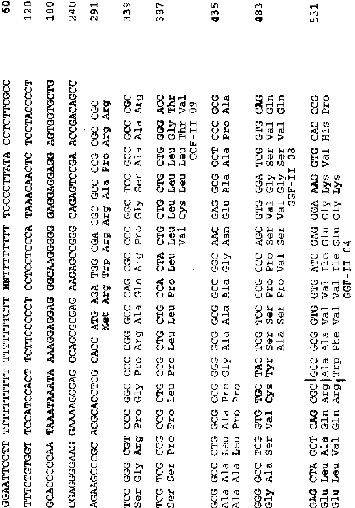


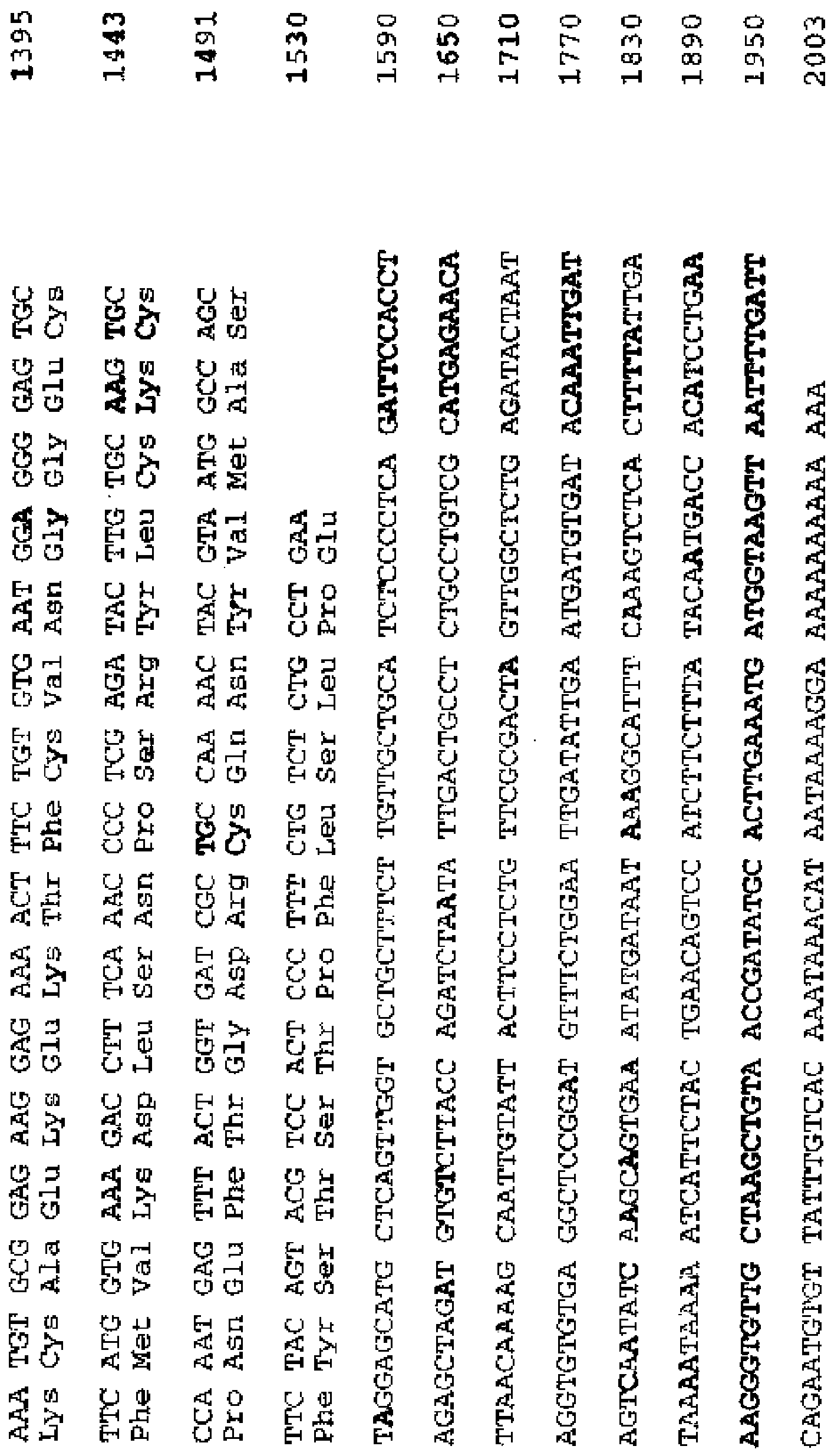


Applications Claiming Priority (1)
| Application Number | Priority Date | Filing Date | Title |
|---|---|---|---|
| US6758908P | 2008-02-29 | 2008-02-29 |
Publications (1)
| Publication Number | Publication Date |
|---|---|
| ES2811127T3 true ES2811127T3 (en) | 2021-03-10 |
Family
ID=41016663
Family Applications (2)
| Application Number | Title | Priority Date | Filing Date |
|---|---|---|---|
| ES16182139T Active ES2811127T3 (en) | 2008-02-29 | 2009-03-02 | Compositions to achieve target plasma levels of glial growth factor 2 |
| ES09715322.5T Active ES2613177T3 (en) | 2008-02-29 | 2009-03-02 | Method to achieve desired plasma levels of glial growth factor 2 |
Family Applications After (1)
| Application Number | Title | Priority Date | Filing Date |
|---|---|---|---|
| ES09715322.5T Active ES2613177T3 (en) | 2008-02-29 | 2009-03-02 | Method to achieve desired plasma levels of glial growth factor 2 |
Country Status (11)
| Country | Link |
|---|---|
| US (5) | US8410050B2 (en) |
| EP (3) | EP2262527B1 (en) |
| JP (2) | JP5697455B2 (en) |
| CN (2) | CN104645315A (en) |
| AU (1) | AU2009217606B2 (en) |
| BR (1) | BRPI0908271A2 (en) |
| CA (1) | CA2717193C (en) |
| ES (2) | ES2811127T3 (en) |
| PL (1) | PL2262527T4 (en) |
| RU (3) | RU2530650C2 (en) |
| WO (1) | WO2009108390A2 (en) |
Families Citing this family (9)
| Publication number | Priority date | Publication date | Assignee | Title |
|---|---|---|---|---|
| ES2811127T3 (en) | 2008-02-29 | 2021-03-10 | Acorda Therapeutics Inc | Compositions to achieve target plasma levels of glial growth factor 2 |
| US20110166068A1 (en) | 2008-07-17 | 2011-07-07 | Acorda Therapeutics, Inc. | Therapeutic Dosing of a Neuregulin or a Subsequence Thereof for Treatment or Pro-phylaxis of Heart Failure |
| CN104707130A (en) | 2008-08-15 | 2015-06-17 | 阿索尔达治疗公司 | Compositions and methods for treatment during non-acute periods following cns neurological injury |
| ES2569121T3 (en) | 2009-10-14 | 2016-05-06 | Acorda Therapeutics, Inc. | Use of a neuregulin to treat a cavernous nerve injury |
| EP2575862A2 (en) * | 2010-05-28 | 2013-04-10 | Mind-Nrg Sa | Neuregulin isoforms, neuregulin polypeptides and uses thereof |
| JP2013535507A (en) * | 2010-08-13 | 2013-09-12 | ジョージタウン ユニバーシティ | GGF2 and methods of use |
| PT3278811T (en) * | 2012-03-30 | 2021-03-03 | Acorda Therapeutics Inc | Use of ggf2 to treat peripheral nerve injury |
| WO2018056412A1 (en) * | 2016-09-23 | 2018-03-29 | 国立大学法人大阪大学 | Schwann cell differentiation promoter and peripheral nerve regeneration promoter |
| CN108653274B (en) * | 2017-04-01 | 2022-09-16 | 鲁南制药集团股份有限公司 | Application of arctigenin in preparation of medicine for treating bone marrow injury |
Family Cites Families (17)
| Publication number | Priority date | Publication date | Assignee | Title |
|---|---|---|---|---|
| US5530109A (en) | 1991-04-10 | 1996-06-25 | Ludwig Institute For Cancer Research | DNA encoding glial mitogenic factors |
| US7135456B1 (en) | 1991-04-10 | 2006-11-14 | Acorda Therapeutics, Inc. | Glial mitogenic factors, their preparation and use |
| US5716930A (en) * | 1991-04-10 | 1998-02-10 | Ludwig Institute For Cancer Research | Glial growth factors |
| US7319019B1 (en) | 1991-04-10 | 2008-01-15 | Acorda Therapeutics, Inc. | Glial mitogenic factors lacking an N-terminal signal sequence |
| US7037888B1 (en) | 1992-04-03 | 2006-05-02 | Acorda Therapeutics, Inc. | Methods for treating muscle diseases and disorders |
| GB9217316D0 (en) | 1992-08-14 | 1992-09-30 | Ludwig Inst Cancer Res | Schwann cell mitogenic factor,its preparation and use |
| US5912326A (en) | 1995-09-08 | 1999-06-15 | President And Fellows Of Harvard College | Cerebellum-derived growth factors |
| JP2003513995A (en) * | 1999-11-19 | 2003-04-15 | アクスクシーマ ファルマシューティカルス アクチェンゲゼルシャフト | Inhibitors of gastrointestinal diseases induced by Helicobacter pylori |
| US20030232761A1 (en) | 2002-03-28 | 2003-12-18 | Hinke Simon A. | Novel analogues of glucose-dependent insulinotropic polypeptide |
| ES2831031T3 (en) * | 2003-05-16 | 2021-06-07 | Acorda Therapeutics Inc | Fusion proteins for CNS treatment |
| WO2005079458A2 (en) * | 2004-02-19 | 2005-09-01 | The Regents Of The University Of California | ENHANCEMENT OF Th2-DEPENDENT AND INFLAMMATORY RESPONSE |
| JP2008528697A (en) | 2005-02-02 | 2008-07-31 | ネクサスファーマ・インコーポレイテッド | Compositions and methods containing amine derivatives for treating viral infections associated with cancer pathogenesis |
| US7776817B2 (en) | 2005-09-02 | 2010-08-17 | Morehouse School Of Medicine | Neuregulins for prevention and treatment of damage from acute assault on vascular and neuronal tissue and as regulators of neuronal stem cell migration |
| US20080003013A1 (en) * | 2006-06-30 | 2008-01-03 | Static Control Components, Inc. | Cartridge split rail clip |
| ES2811127T3 (en) | 2008-02-29 | 2021-03-10 | Acorda Therapeutics Inc | Compositions to achieve target plasma levels of glial growth factor 2 |
| CA2794084C (en) | 2010-04-01 | 2014-09-02 | The Procter & Gamble Company | Fabric softener based on a bis-(2-hydroxyethyl)-dimethylammonium chloride fatty acid ester |
| JP2013535507A (en) | 2010-08-13 | 2013-09-12 | ジョージタウン ユニバーシティ | GGF2 and methods of use |
-
2009
- 2009-03-02 ES ES16182139T patent/ES2811127T3/en active Active
- 2009-03-02 EP EP09715322.5A patent/EP2262527B1/en active Active
- 2009-03-02 EP EP16182139.2A patent/EP3120863B1/en active Active
- 2009-03-02 CN CN201410709391.4A patent/CN104645315A/en active Pending
- 2009-03-02 EP EP20173000.9A patent/EP3750551A1/en not_active Withdrawn
- 2009-03-02 PL PL09715322T patent/PL2262527T4/en unknown
- 2009-03-02 CA CA2717193A patent/CA2717193C/en active Active
- 2009-03-02 BR BRPI0908271A patent/BRPI0908271A2/en not_active IP Right Cessation
- 2009-03-02 JP JP2010548749A patent/JP5697455B2/en not_active Expired - Fee Related
- 2009-03-02 ES ES09715322.5T patent/ES2613177T3/en active Active
- 2009-03-02 RU RU2010139906/15A patent/RU2530650C2/en active
- 2009-03-02 AU AU2009217606A patent/AU2009217606B2/en not_active Ceased
- 2009-03-02 US US12/380,760 patent/US8410050B2/en active Active
- 2009-03-02 CN CN200980114836.1A patent/CN102026651B/en not_active Expired - Fee Related
- 2009-03-02 WO PCT/US2009/001356 patent/WO2009108390A2/en active Application Filing
-
2013
- 2013-03-29 US US13/853,386 patent/US9272015B2/en active Active
-
2014
- 2014-07-28 RU RU2014131265A patent/RU2687097C2/en active
- 2014-10-17 JP JP2014212193A patent/JP5964920B2/en not_active Expired - Fee Related
-
2016
- 2016-01-21 US US15/002,608 patent/US9744215B2/en active Active
-
2017
- 2017-07-25 US US15/658,967 patent/US10675331B2/en active Active
-
2019
- 2019-04-09 RU RU2019110452A patent/RU2019110452A/en unknown
-
2020
- 2020-05-18 US US16/876,702 patent/US20210060128A1/en not_active Abandoned
Also Published As
Similar Documents
| Publication | Publication Date | Title |
|---|---|---|
| ES2811127T3 (en) | Compositions to achieve target plasma levels of glial growth factor 2 | |
| Syed et al. | Pituitary adenylate cyclase-activating polypeptide (PACAP) potently dilates middle meningeal arteries: implications for migraine | |
| Li et al. | Angiotensin AT2 receptor protects against cerebral ischemia‐induced neuronal injury | |
| JP5905833B2 (en) | Composition comprising neuregulin for the prevention, treatment or delay of myocardial ischemia-reperfusion injury and use thereof | |
| US20220033517A1 (en) | ErbB2 SIGNALING AND NERVE REGENERATION | |
| US20030113326A1 (en) | Reducing myelin-mediated inhibition of axon regeneration | |
| Yin et al. | DLK silencing attenuated neuron apoptosis through JIP3/MA2K7/JNK pathway in early brain injury after SAH in rats | |
| Wang et al. | Chemogenetics-mediated acute inhibition of excitatory neuronal activity improves stroke outcome | |
| JP2017524727A (en) | SOCS1-derived peptides for use in chronic complications of diabetes | |
| KR101174333B1 (en) | Method of treatment of myocardial infarction | |
| Luo et al. | Multiple roles of apelin/APJ system in eye diseases | |
| AU2017203961B2 (en) | Method for achieving desired glial growth factor 2 plasma levels | |
| Wang et al. | Machrophage stimulating protein preserves blood brain barrier integrity after intracerebral hemorrhage through recepteur d'origine nantais dependent GAB1/Src/β-catenin pathway activation in a mouse model. |