ES2712303T3 - DNA binding domain of the CRISPR system for the production of non-fucosylated and partially fucosylated proteins - Google Patents
DNA binding domain of the CRISPR system for the production of non-fucosylated and partially fucosylated proteins Download PDFInfo
- Publication number
- ES2712303T3 ES2712303T3 ES15816521T ES15816521T ES2712303T3 ES 2712303 T3 ES2712303 T3 ES 2712303T3 ES 15816521 T ES15816521 T ES 15816521T ES 15816521 T ES15816521 T ES 15816521T ES 2712303 T3 ES2712303 T3 ES 2712303T3
- Authority
- ES
- Spain
- Prior art keywords
- cell
- crispr
- gmd
- dna
- sequence
- Prior art date
- Legal status (The legal status is an assumption and is not a legal conclusion. Google has not performed a legal analysis and makes no representation as to the accuracy of the status listed.)
- Active
Links
Classifications
-
- C—CHEMISTRY; METALLURGY
- C07—ORGANIC CHEMISTRY
- C07K—PEPTIDES
- C07K16/00—Immunoglobulins [IGs], e.g. monoclonal or polyclonal antibodies
- C07K16/06—Immunoglobulins [IGs], e.g. monoclonal or polyclonal antibodies from serum
- C07K16/065—Purification, fragmentation
-
- C—CHEMISTRY; METALLURGY
- C12—BIOCHEMISTRY; BEER; SPIRITS; WINE; VINEGAR; MICROBIOLOGY; ENZYMOLOGY; MUTATION OR GENETIC ENGINEERING
- C12Y—ENZYMES
- C12Y402/00—Carbon-oxygen lyases (4.2)
- C12Y402/01—Hydro-lyases (4.2.1)
- C12Y402/01047—GDP-mannose 4,6-dehydratase (4.2.1.47), i.e. GMD
-
- A—HUMAN NECESSITIES
- A61—MEDICAL OR VETERINARY SCIENCE; HYGIENE
- A61K—PREPARATIONS FOR MEDICAL, DENTAL OR TOILETRY PURPOSES
- A61K48/00—Medicinal preparations containing genetic material which is inserted into cells of the living body to treat genetic diseases; Gene therapy
- A61K48/0008—Medicinal preparations containing genetic material which is inserted into cells of the living body to treat genetic diseases; Gene therapy characterised by an aspect of the 'non-active' part of the composition delivered, e.g. wherein such 'non-active' part is not delivered simultaneously with the 'active' part of the composition
- A61K48/0016—Medicinal preparations containing genetic material which is inserted into cells of the living body to treat genetic diseases; Gene therapy characterised by an aspect of the 'non-active' part of the composition delivered, e.g. wherein such 'non-active' part is not delivered simultaneously with the 'active' part of the composition wherein the nucleic acid is delivered as a 'naked' nucleic acid, i.e. not combined with an entity such as a cationic lipid
-
- C—CHEMISTRY; METALLURGY
- C07—ORGANIC CHEMISTRY
- C07K—PEPTIDES
- C07K16/00—Immunoglobulins [IGs], e.g. monoclonal or polyclonal antibodies
-
- C—CHEMISTRY; METALLURGY
- C12—BIOCHEMISTRY; BEER; SPIRITS; WINE; VINEGAR; MICROBIOLOGY; ENZYMOLOGY; MUTATION OR GENETIC ENGINEERING
- C12N—MICROORGANISMS OR ENZYMES; COMPOSITIONS THEREOF; PROPAGATING, PRESERVING, OR MAINTAINING MICROORGANISMS; MUTATION OR GENETIC ENGINEERING; CULTURE MEDIA
- C12N15/00—Mutation or genetic engineering; DNA or RNA concerning genetic engineering, vectors, e.g. plasmids, or their isolation, preparation or purification; Use of hosts therefor
- C12N15/09—Recombinant DNA-technology
- C12N15/10—Processes for the isolation, preparation or purification of DNA or RNA
- C12N15/102—Mutagenizing nucleic acids
-
- C—CHEMISTRY; METALLURGY
- C12—BIOCHEMISTRY; BEER; SPIRITS; WINE; VINEGAR; MICROBIOLOGY; ENZYMOLOGY; MUTATION OR GENETIC ENGINEERING
- C12N—MICROORGANISMS OR ENZYMES; COMPOSITIONS THEREOF; PROPAGATING, PRESERVING, OR MAINTAINING MICROORGANISMS; MUTATION OR GENETIC ENGINEERING; CULTURE MEDIA
- C12N15/00—Mutation or genetic engineering; DNA or RNA concerning genetic engineering, vectors, e.g. plasmids, or their isolation, preparation or purification; Use of hosts therefor
- C12N15/09—Recombinant DNA-technology
- C12N15/11—DNA or RNA fragments; Modified forms thereof; Non-coding nucleic acids having a biological activity
- C12N15/113—Non-coding nucleic acids modulating the expression of genes, e.g. antisense oligonucleotides; Antisense DNA or RNA; Triplex- forming oligonucleotides; Catalytic nucleic acids, e.g. ribozymes; Nucleic acids used in co-suppression or gene silencing
- C12N15/1137—Non-coding nucleic acids modulating the expression of genes, e.g. antisense oligonucleotides; Antisense DNA or RNA; Triplex- forming oligonucleotides; Catalytic nucleic acids, e.g. ribozymes; Nucleic acids used in co-suppression or gene silencing against enzymes
-
- C—CHEMISTRY; METALLURGY
- C12—BIOCHEMISTRY; BEER; SPIRITS; WINE; VINEGAR; MICROBIOLOGY; ENZYMOLOGY; MUTATION OR GENETIC ENGINEERING
- C12N—MICROORGANISMS OR ENZYMES; COMPOSITIONS THEREOF; PROPAGATING, PRESERVING, OR MAINTAINING MICROORGANISMS; MUTATION OR GENETIC ENGINEERING; CULTURE MEDIA
- C12N5/00—Undifferentiated human, animal or plant cells, e.g. cell lines; Tissues; Cultivation or maintenance thereof; Culture media therefor
- C12N5/06—Animal cells or tissues; Human cells or tissues
- C12N5/0601—Invertebrate cells or tissues, e.g. insect cells; Culture media therefor
-
- C—CHEMISTRY; METALLURGY
- C12—BIOCHEMISTRY; BEER; SPIRITS; WINE; VINEGAR; MICROBIOLOGY; ENZYMOLOGY; MUTATION OR GENETIC ENGINEERING
- C12N—MICROORGANISMS OR ENZYMES; COMPOSITIONS THEREOF; PROPAGATING, PRESERVING, OR MAINTAINING MICROORGANISMS; MUTATION OR GENETIC ENGINEERING; CULTURE MEDIA
- C12N5/00—Undifferentiated human, animal or plant cells, e.g. cell lines; Tissues; Cultivation or maintenance thereof; Culture media therefor
- C12N5/10—Cells modified by introduction of foreign genetic material
- C12N5/12—Fused cells, e.g. hybridomas
- C12N5/16—Animal cells
- C12N5/163—Animal cells one of the fusion partners being a B or a T lymphocyte
-
- C—CHEMISTRY; METALLURGY
- C12—BIOCHEMISTRY; BEER; SPIRITS; WINE; VINEGAR; MICROBIOLOGY; ENZYMOLOGY; MUTATION OR GENETIC ENGINEERING
- C12Y—ENZYMES
- C12Y204/00—Glycosyltransferases (2.4)
- C12Y204/01—Hexosyltransferases (2.4.1)
- C12Y204/01068—Glycoprotein 6-alpha-L-fucosyltransferase (2.4.1.68), i.e. FUT8
-
- C—CHEMISTRY; METALLURGY
- C07—ORGANIC CHEMISTRY
- C07K—PEPTIDES
- C07K2317/00—Immunoglobulins specific features
- C07K2317/40—Immunoglobulins specific features characterized by post-translational modification
- C07K2317/41—Glycosylation, sialylation, or fucosylation
-
- C—CHEMISTRY; METALLURGY
- C12—BIOCHEMISTRY; BEER; SPIRITS; WINE; VINEGAR; MICROBIOLOGY; ENZYMOLOGY; MUTATION OR GENETIC ENGINEERING
- C12N—MICROORGANISMS OR ENZYMES; COMPOSITIONS THEREOF; PROPAGATING, PRESERVING, OR MAINTAINING MICROORGANISMS; MUTATION OR GENETIC ENGINEERING; CULTURE MEDIA
- C12N2310/00—Structure or type of the nucleic acid
- C12N2310/10—Type of nucleic acid
- C12N2310/20—Type of nucleic acid involving clustered regularly interspaced short palindromic repeats [CRISPRs]
Abstract
Un dominio de unión a ADN del sistema CRISPR, en donde el dominio de unión a ADN comprende una secuencia de ARN transcrita a partir de la secuencia de nucleótidos seleccionada del grupo que consiste en la SEQ ID NO: 41, la SEQ ID NO: 43 y la SEQ ID NO: 45.A DNA-binding domain of the CRISPR system, wherein the DNA-binding domain comprises an RNA sequence transcribed from the nucleotide sequence selected from the group consisting of SEQ ID NO: 41, SEQ ID NO: 43 and SEQ ID NO: 45.
Description
DESCRIPCIONDESCRIPTION
Dominio de union a ADN del sistema CRISPR para la produccion de protemas no fucosiladas y parcialmente fucosiladasDNA binding domain of the CRISPR system for the production of non-fucosylated and partially fucosylated proteins
Campo tecnicoTechnical field
La presente divulgacion pertenece al campo de la biotecnolog^a, la ingenieria genetica y la inmunologia. Particularmente, la presente divulgacion se refiere al desarrollo de lineas celulares en las que se modifican las v^as biologicas especificas. Tales modificaciones estan en las enzimas de la celula, en particular, las enzimas implicadas en la glucosilacion de proteinas. La presente divulgacion desarrolla sistemas de expresion de proteinas en los que se logra la modification de la cadena de glicano de la proteina. La modification especifica de la cadena de glicano produce proteinas parcialmente fucosiladas y no fucosiladas, que incluyen anticuerpos. Tales productos se usan en el desarrollo de farmacos y biomarcadores, y en diagnostico y pronostico de enfermedades. La presente divulgacion emplea la tecnologia de las repeticiones palindromicas cortas agrupadas y espaciadas de forma regular (CRISPR, del ingles Clustered Regularly Interspaced Short Palindromic Repeats). The present disclosure belongs to the field of biotechnology, genetic engineering and immunology. Particularly, the present disclosure refers to the development of cell lines in which the specific biological vines are modified. Such modifications are in the enzymes of the cell, in particular, the enzymes involved in the glycosylation of proteins. The present disclosure develops protein expression systems in which the modification of the glycan chain of the protein is achieved. The specific modification of the glycan chain produces partially fucosylated and non-fucosylated proteins, including antibodies. Such products are used in the development of drugs and biomarkers, and in the diagnosis and prognosis of diseases. The present disclosure uses the technology of short palindromic repeats grouped and spaced on a regular basis (CRISPR, from the English Clustered Regularly Interspaced Short Palindromic Repeats).
Antecedentes y tecnica anterior de la divulgacionBACKGROUND AND PRIOR ART OF DISCLOSURE
La glucosilacion en eucariotas se ha estudiado a fondo durante decadas como el mecanismo de modificacion covalente postraduccional de proteinas. Se predice que aproximadamente el 1-2 % del transcriptoma humano (aproximadamente 250-500 glucogenos) traduce las proteinas que son responsables de la glucosilacion (Campbell y Yarema 2005). La glucosilacion de las proteinas celulares desempena muchas funciones importantes tales como el plegamiento y la estabilidad de la proteina, el trafico intracelular e intercelular, la interaction celula-celula y celulamatriz.Glycosylation in eukaryotes has been studied extensively for decades as the mechanism of post-translational covalent modification of proteins. It is predicted that approximately 1-2% of the human transcriptome (approximately 250-500 glycogens) translates the proteins that are responsible for glycosylation (Campbell and Yarema 2005). The glycosylation of cellular proteins performs many important functions such as protein folding and stability, intracellular and intercellular trafficking, cell-cell and cell-matrix interaction.
Hay cuatro grupos diferentes de glucoproteinas: N-ligadas, O-ligadas, glucosaminoglicanos y proteinas ancladas a glucosilfosfatidilinositol. La glucosilacion N-ligada tiene lugar a traves del nitrogeno de la amida de la cadena lateral de los restos de asparagina, mientras que la glucosilacion O-ligada usa el atomo de oxigeno en la cadena lateral de los restos de serina o treonina. La glucosilacion N-ligada tiene lugar en la secuencia de aminoacidos de Asn-X-Ser/Thr, en donde X puede ser cualquier aminoacido salvo prolina y acido aspartico (Helenius y Aebi 2004).There are four different groups of glycoproteins: N-linked, O-linked, glycosaminoglycans and proteins anchored to glycosylphosphatidylinositol. The N-linked glycosylation takes place through the amide nitrogen of the side chain of the asparagine residues, while the O-linked glycosylation uses the oxygen atom in the side chain of the serine or threonine residues. The N-linked glycosylation takes place in the amino acid sequence of Asn-X-Ser / Thr, where X can be any amino acid except proline and aspartic acid (Helenius and Aebi 2004).
La fucosa (6-desoxi-L-galactosa) es un monosacarido que esta presente en muchas glucoproteinas y glucolipidos presentes en vertebrados, invertebrados, plantas y bacterias. La fucosilacion es el proceso de transferencia de un resto de fucosa a diversas proteinas y oligosacaridos. La fucosilacion esta regulada por varias moleculas, incluyendo fucosiltransferasas, enzimas sinteticas de guanosina difosfato (GDP)-fucosa, y transportador(es) de GDP-fucosa. Un gran numero de glucoproteinas fucosiladas son proteinas secretoras o proteinas de membrana sobre la superficie celular.Fucose (6-deoxy-L-galactose) is a monosaccharide that is present in many glycoproteins and glycolipids present in vertebrates, invertebrates, plants and bacteria. Fucosylation is the process of transferring a residue of fucose to various proteins and oligosaccharides. Fucosylation is regulated by several molecules, including fucosyltransferases, synthetic guanosine diphosphate (GDP) -fucose enzymes, and GDP-fucose transporter (s). A large number of fucosylated glycoproteins are secretory proteins or membrane proteins on the cell surface.
Existen 14,1 millones de casos nuevos de cancer, 8,2 millones de muertes por cancer y 32,6 millones de personas que viven con cancer (dentro de los 5 anos de diagnostico) en 2014 en todo el mundo. La elevada tasa de mortalidad del cancer sirve como un recordatorio de la necesidad de terapias mas eficaces. El cambio mas destacable en el desarrollo de farmacos de oncologia en los ultimos 20 anos ha sido el desplazamiento de los citotoxicos clasicos a farmacos que afectan a las vias de senalizacion implicadas en el cancer, conocidos como "anticuerpos monoclonales" o mAb. Hace una decada, solo habia dos mAb en el mercado y actualmente hay aproximadamente 30 mAb aprobados por la FDA de diversas modalidades terapeuticas, como Adalimumab, Infliximab, Rituximab etc. Los mAbs son el segmento de crecimiento mas rapido en la industria farmaceutica y se espera que esta rapida expansion continue. Hay mas de 100 farmacos biologicos basados en anticuerpos monoclonales en ensayos clinicos. Muchos de estos estan en ensayos de fase II y de fase III y se presentaran ante las agencias reguladoras para su aprobacion. La mejora de medicamentos de anticuerpos monoclonales a traves de las tecnologias descritas en el presente documento allanara el camino para un mejor resultado clinico para los pacientes.There are 14.1 million new cases of cancer, 8.2 million deaths from cancer and 32.6 million people living with cancer (within 5 years of diagnosis) in 2014 around the world. The high cancer mortality rate serves as a reminder of the need for more effective therapies. The most remarkable change in the development of oncology drugs in the last 20 years has been the displacement of the classic cytotoxins to drugs that affect the signaling pathways involved in cancer, known as "monoclonal antibodies" or mAb. A decade ago, there were only two mAb in the market and currently there are approximately 30 mAb approved by the FDA for various therapeutic modalities, such as Adalimumab, Infliximab, Rituximab etc. Mabs are the fastest growing segment in the pharmaceutical industry and it is expected that this rapid expansion will continue. There are more than 100 biological drugs based on monoclonal antibodies in clinical trials. Many of these are in phase II and phase III trials and will be submitted to regulatory agencies for approval. The improvement of monoclonal antibody drugs through the technologies described in this document will pave the way for a better clinical outcome for patients.
El anticuerpo IgG1 humano es una glucoproteina altamente fucosilada. Dos oligosacaridos biantenarios N-ligados que consisten en un nucleo de heptasacarido con adicion variable de fucosa, galactosa, N-acetilglucosamina de bisection y acido sialico estan presentes en el Asn-297 del IgG1. La glucosilacion del anticuerpo lleva a funciones biologicas unicas conocidas como "funciones efectoras" - citotoxicidad celular dependiente de anticuerpo (ADCC, del ingles Antibody Dependent Cellular Cytotoxicity) y citotoxicidad dependiente del complemento (CDC, del ingles Complement Dependent Cytotoxicity). La ADCC es un sistema inmunitario mediado por celulas en donde las celulas inmunitarias (como celulas citoliticas naturales) lisan las celulas diana identificadas a traves de anticuerpos frente a antigenos de superficie celular.The human IgG1 antibody is a highly fucosylated glycoprotein. Two N-linked biantennary oligosaccharides consisting of a heptasaccharide nucleus with variable addition of fucose, galactose, bisection N-acetylglucosamine and sialic acid are present in Asn-297 of IgG1. The glycosylation of the antibody leads to unique biological functions known as "effector functions" - antibody-dependent cellular cytotoxicity (ADCC ) and complement-dependent cytotoxicity (CDC, from the English Complement Dependent Cytotoxicity ). ADCC is a cell-mediated immune system where immune cells (such as natural cytolytic cells) lyse the target cells identified through antibodies to cell surface antigens.
La funcion efectora de la molecula de IgG se define mediante la interaccion de la region la region Fc del anticuerpo con receptores del leucocito, conocido como FcyRs, o interacciones con componentes del complemento. La composition de la estructura del oligosacarido es, de manera critica, importante para la funcion efectora a traves de la union de FcyR (Shields et al. 2002; Shinkawa et al. 2003; Niwa et al. 2004; Niwa, Shoji-Hosaka, et al. 2004; Yamane-Ohnuki et al. 2004;). El analisis de la estructura cristalina de la IgG 1 humana ha revelado la interaccion intrincada de las cadenas de oligosacaridos con el dominio CH2 (Harris et al. 1998; Radaev et al. 2001).The effector function of the IgG molecule is defined by the interaction of the region the Fc region of the antibody with leukocyte receptors, known as FcyRs, or interactions with complement components. The composition of the oligosaccharide structure is critically important for effector function through the binding of Fc and R (Shields et al., 2002; Shinkawa et al., 2003; Niwa et al., 2004; Niwa, Shoji- Hosaka, et al., 2004; Yamane-Ohnuki et al. 2004;). Analysis of the crystal structure of human IgG 1 has revealed the intricate interaction of the oligosaccharide chains with the CH2 domain (Harris et al., 1998, Radaev et al., 2001).
La eficacia del mecanismo de ADCC es considerablemente dependiente del nivel de fucosilacion del anticuerpo. Cuanto menor es la fucosilacion, mayor es la tasa de ADCC. Por lo tanto, la perdida de fucosilacion tiene consecuencias biologicas significativas. La perdida podria deberse a las enzimas no funcionales de fucosiltransferasa, dando como resultado la no fucosilacion de proteinas celulares. La ausencia de fucosa de la N-acetilgucosamina principal da como resultado el anticuerpo IgG1 que tiene una elevada afinidad de union por el receptor FcYRIIIa, con el consecuente aumento de 50 - 100 veces mayor eficacia de ADCC. La mejora de ADCC con IgG no fucosilada es directamente proporcional a la elevada afinidad por FcYRIIIa, lo que permite que la Fc de la IgG no fucosilada supere la competicion de las altas concentraciones de IgG fucosilado en suero normal. La razon plausible para la elevada afinidad de la Fc de la IgG no fucosilada por FcYRIIIa puede ser la reduccion o la ausencia de inhibicion esterica en la zona de contacto receptor-ligando (Harris, 1998; Radaev, 2001).The effectiveness of the ADCC mechanism is considerably dependent on the level of fucosylation of the antibody. The lower the fucosylation, the higher the ADCC rate. Therefore, the loss of fucosylation has significant biological consequences. The loss could be due to the non-functional enzymes of fucosyltransferase, resulting in the non-fucosylation of cellular proteins. The absence of fucose from the main N-acetylgucosamine results in the IgG1 antibody having a high binding affinity for the FcYRIIIa receptor, with the consequent increase of 50-100 times greater efficacy of ADCC. The improvement of ADCC with non-fucosylated IgG is directly proportional to the high affinity for FcYRIIIa, which allows the Fc of the non-fucosylated IgG to overcome the competition of high concentrations of fucosylated IgG in normal serum. The plausible reason for the high affinity of the Fc of the non-fucosylated IgG for FcYRIIIa may be the reduction or absence of steric inhibition in the receptor-ligand contact zone (Harris, 1998, Radaev, 2001).
En el sistema de expresion de mamiferos, la enzima a1,6-fucosiltransferasa codificada por el gen Fut8 es responsable de la transferencia del resto de fucosa desde la GDP-fucosa a la N-acetilglucosamina de la cadena de N-glicano en proteinas (Miyoshi, 1999). La alteracion de esta funcion genica a traves de diversos medios lleva a la produccion de proteinas no fucosiladas, que incluyen anticuerpos (Naoko Yamane-Ohnuki, 2004).In the expression system of mammals, the α1,6-fucosyltransferase enzyme encoded by the Fut8 gene is responsible for the transfer of the rest of the fucose from the GDP-fucose to the N-acetylglucosamine of the N-glycan chain in proteins (Miyoshi , 1999). The alteration of this genetic function through various means leads to the production of non-fucosylated proteins, which include antibodies (Naoko Yamane-Ohnuki, 2004).
La GDP-D-manosa 4,6-deshidratasa (GMD) es un miembro de la subfamilia modificadora de azucar de los nucleotidos de la familia de deshidrogenasa/reductasa de cadena corta (SDR) (Webb, Mulichak et al. 2004).The GDP-D-mannose 4,6-dehydratase (GMD) is a member of the sugar modifier subfamily of the nucleotides of the short chain dehydrogenase / reductase (SDR) family (Webb, Mulichak et al., 2004).
En sistemas de expresion en mamiferos, la GDP-fucosa, un sustrato esencial de la fucosilacion, se sintetiza en el citoplasma a traves de las vias de novo y de rescate. En la via de fucosilacion de novo, se sintetiza GDP-fucosa a traves de la conversion de GDP-manosa a GDP-4-ceto-6-desoxi-manosa, catalizado por la enzima GDP-manosa 4,6-deshidratasa (GMD). Esta GDP-fucosa se transporta despues a dentro del golgi y se usa como un sustrato para la fucosilacion de proteina por la enzima a1-6 fucosiltransferasa (FUT8). La enzima transfiere el resto de fucosa desde la GDP-fucosa a la N-acetilglucosamina de la cadena de N-glicano (Miyoshi, 1999). Estas enzimas criticas, GDP-manosa 4,6-deshidratasa y a,1-6 fucosiltransferasa estan codificadas por los genes GMD y FUT8, respectivamente.In expression systems in mammals, GDP-fucose, an essential substrate of fucosylation, is synthesized in the cytoplasm through de novo and rescue pathways. In the de novo fucosylation path, GDP-fucose is synthesized through the conversion of GDP-mannose to GDP-4-keto-6-deoxy-mannose, catalyzed by the enzyme GDP-mannose 4,6-dehydratase (GMD) . This GDP-fucose is then transported into the golgi and used as a substrate for protein fucosylation by the enzyme a1-6 fucosyltransferase (FUT8). The enzyme transfers the rest of the fucose from the GDP-fucose to the N-acetylglucosamine of the N-glycan chain (Miyoshi, 1999). These critical enzymes, GDP-mannose 4,6-dehydratase and 1-6 fucosyltransferase are encoded by the GMD and FUT8 genes, respectively.
Las formas no fucosiladas de los anticuerpos terapeuticos desarrollados en soportes de mamiferos, en los que la biosintesis de fucosa esta alterada, pueden tener una ventaja clinica sobre las formas fucosiladas debido a una eficacia mejorada de ADCC frente a las celulas tumorales diana.The non-fucosylated forms of the therapeutic antibodies developed in mammalian supports, in which the fucose biosynthesis is altered, may have a clinical advantage over the fucosylated forms due to an improved efficacy of ADCC against the target tumor cells.
Historicamente, los sistemas de inactivacion genica dependian por completo de la mutacion, delecion y/o insertion mediadas por recombination homologa (HR, del ingles homologous recombination). El sistema de HR, aunque es muy especifico, es altamente ineficaz, ya que se necesitan cribar miles de clones para encontrar un clon mutado. Por otra parte, la delecion de variaciones alelicas podria llevar mas tiempo e incluso un cribado mucho mayor. En la ultima decada han evolucionado multiples tecnologias para lograr una modification genica dirigida usando una combination de un dominio de reconocimiento de secuencia de ADN y un dominio nucleasa. Estos sistemas son altamente eficientes en la identification de sitios especificos de interes y la introduction posterior de roturas en la cadena de ADN. La rotura de ADN de cadena doble (DBS, del ingles DNA double-strand break) en el locus genomico dirigido activa la reparation del ADN, que se utiliza para la modificacion de genes. La respuesta al dano del ADN esta altamente conservada en celulas eucariotas. El concepto de diseno del genoma basado en DSB es altamente transferible entre organismos altamente diversos. La creation de una rotura de cadena doble aumenta la frecuencia de inactivacion genica en loci dirigidos en miles de veces a traves de mecanismos de recombinacion homologa y de union de extremos no homologos.Historically, the systems of genetic inactivation depended completely on the mutation, deletion and / or insertion mediated by homologous recombination (HR, of the English homologous recombination). The HR system, although it is very specific, is highly inefficient, since thousands of clones need to be screened to find a mutated clone. On the other hand, the deletion of allelic variations could take more time and even a much larger screening. In the last decade, multiple technologies have evolved to achieve a directed gene modification using a combination of a DNA sequence recognition domain and a nuclease domain. These systems are highly efficient in the identification of specific sites of interest and the subsequent introduction of breaks in the DNA chain. The DNA double-strand break (DBS ) at the targeted genomic locus activates DNA repair, which is used for gene modification. The DNA damage response is highly conserved in eukaryotic cells. The concept of genome design based on DSB is highly transferable among highly diverse organisms. The creation of a double chain break increases the frequency of genetic inactivation in loci directed thousands of times through mechanisms of homologous recombination and union of non-homologous ends.
La nucleasa con dedos de zinc (ZFN, del ingles Zinc Finger Nuclease) es una de las tecnicas mas frecuentemente usadas para la alteracion genica. Requiere tres bases a nivel de ADN para cada matriz en tandem de dedos de zinc. Por otra parte, el solapamiento del sitio diana y la interferencia entre los dedos individuales en una matriz de dedos de zinc complica considerablemente la produccion de ZFN especificos de secuencia. Adicionalmente, el principal inconveniente de los ZFN incluye un proceso de selection experimental elaborado y lento para identificar los motivos de ZFN para el reconocimiento de secuencias de ADN especificas.Zinc finger nuclease (ZFN ) is one of the most frequently used techniques for genetic alteration. It requires three bases at the DNA level for each tandem matrix of zinc fingers. On the other hand, the overlap of the target site and the interference between the individual fingers in a zinc finger matrix considerably complicate the production of sequence-specific ZFNs. Additionally, the main drawback of the ZFN includes an elaborate and slow experimental selection process to identify the motifs of ZFN for the recognition of specific DNA sequences.
Existen metodos en la tecnica anterior para la alteracion de los loci genomicos Fut8 y GMD. Sin embargo, ninguno de los metodos dirige la localization especifica en los loci genomicos de FUT8 y GMD mediante la tecnologia CRISPR.There are methods in the prior art for the alteration of the genomic loci Fut8 and GMD. However, none of the methods addresses the specific localization in the genomic loci of FUT8 and GMD through the CRISPR technology.
Ronda et al 2014 Biotechnology and Bioengineering 111: 1604-1616 trata una herramienta basada en la red para su uso en la modificacion del genoma en celulas CHO usando CRISPR Cas9 y CRISPy.Ronda et al 2014 Biotechnology and Bioengineering 111: 1604-1616 discusses a network-based tool for use in modifying the genome in CHO cells using CRISPR Cas9 and CRISPy.
Malphettes et al 2010 Biotechnology and Bioengineering 106: 774-783 trata la delecion de FUt8 en lineas celulares CHO usando nucleasas de dedos de zinc y la produccion de anticuerpos no fucosilados.Malphettes et al 2010 Biotechnology and Bioengineering 106: 774-783 treats the deletion of FUt8 in CHO cell lines using zinc finger nucleases and the production of non-fucosylated antibodies.
El documento WO 2013/013013 se refiere a composiciones y metodos para producir glucoproteinas que tienen una estructura de glucano alterada. WO 2013/013013 relates to compositions and methods for producing glycoproteins having an altered glucan structure.
El documento WO 03/035835 describe una variante de lmea celular de CHO, las celulas Lecl3, con capacidad reducida para unir fucosa a los hidratos de carbono enlazados a Asn(297).WO 03/035835 describes a cell line variant of CHO, Lecl3 cells, with reduced capacity to bind fucose to carbohydrates linked to Asn (297).
Kanda et al 2007 J Biotech 130: 300-310 trata el establecimiento de una linea celular de inactivacion de GMD.Kanda et al 2007 J Biotech 130: 300-310 discusses the establishment of a GMD inactivation cell line.
El documento WO 2015/010114 trata un metodo y composiciones que utilizan el sistema CRISPR para alterar un gen diana en celulas eucarioticas para producir inactivaciones dobles de alelos.WO 2015/010114 discusses a method and compositions that use the CRISPR system to alter a target gene in eukaryotic cells to produce double inactivations of alleles.
El documento WO 2015/052231 describe un sistema que permite la modificacion multiple de secuencias de acido nucleico tales como secuencias genomicas.WO 2015/052231 describes a system allowing the multiple modification of nucleic acid sequences such as genomic sequences.
La presente divulgacion supera las desventajas o limitaciones asociadas con los metodos de la tecnica anterior usando la tecnologia CRISpR para dirigir una localizacion especifica en el loci genomico FUT8 o el loci genomico GMD, que da como resultado una alteration completa del gen y la funcion relacionada, proporcionando una celula que produce proteinas no fucosiladas.The present disclosure overcomes the disadvantages or limitations associated with the prior art methods using the CRISpR technology to direct a specific location at the FUT8 genomic loci or the GMD genomic loci, which results in a complete alteration of the gene and related function, providing a cell that produces non-fucosylated proteins.
EXPOSICION DE LA DIVULGACIONEXHIBITION OF THE DISCLOSURE
La invencion es tal como se define en las reivindicaciones y proporciona:The invention is as defined in the claims and provides:
En un primer aspecto: Un dominio de union a ADN del sistema CRISPR, en el que el dominio de union a ADN comprende una secuencia de ARN transcrita a partir de la secuencia de nucleotidos seleccionada del grupo que consiste en la SEQ ID NO: 41, SEQ ID NO: 43, s Eq ID NO: 45 y combinaciones de las mismas;In a first aspect: A DNA binding domain of the CRISPR system, wherein the DNA binding domain comprises an RNA sequence transcribed from the nucleotide sequence selected from the group consisting of SEQ ID NO: 41, SEQ ID NO: 43, s Eq ID NO: 45 and combinations thereof;
En un segundo aspecto: Un complejo CRISPR-nucleasa que comprende el dominio de union a ADN de acuerdo con el primer aspecto y nucleasa;In a second aspect: A CRISPR-nuclease complex comprising the DNA binding domain according to the first aspect and nuclease;
En un tercer aspecto: Un vector que comprende la secuencia de nucleotidos que codifica el dominio de union a ADN de acuerdo con el primer aspecto;In a third aspect: A vector comprising the nucleotide sequence encoding the DNA binding domain according to the first aspect;
En un cuarto aspecto: Una celula que comprende un vector de acuerdo con el tercer aspecto;In a fourth aspect: A cell comprising a vector according to the third aspect;
En un quinto aspecto: Un procedimiento in vitro de obtencion de una celula de inactivacion de fucosa, comprendiendo dicho metodo las etapas de:In a fifth aspect: An in vitro procedure for obtaining a fucose inactivation cell, said method comprising the steps of:
Obtener una construction CRISPR-nucleasa que comprende la secuencia de nucleotidos que codifica el dominio de union a ADN de acuerdo con el primer aspecto; yObtain a CRISPR-nuclease construction comprising the nucleotide sequence encoding the DNA binding domain according to the first aspect; Y
Transfectar una celula con la construccion de la etapa (a) para obtener una celula de inactivacion de fucosa, en la que la construccion CRISPR-nucleasa proporciona el complejo CRISPR-nucleasa que comprende el dominio de union a ADN y nucleasa; y en donde el complejo escinde la secuencia del gen GMD en la celula;Transfecting a cell with the construction of step (a) to obtain a fucose inactivation cell, wherein the CRISPR-nuclease construct provides the CRISPR-nuclease complex comprising the DNA and nuclease binding domain; and wherein the complex cleaves the sequence of the GMD gene in the cell;
En un sexto aspecto: Un metodo in vitro de obtencion de proteina con fucosilacion que varia del 0 % al 100 %, comprendiendo dicho metodo las etapas de:In a sixth aspect: An in vitro method of obtaining protein with fucosylation ranging from 0% to 100%, said method comprising the steps of:
a) Cultivar la celula obtenida mediante el metodo de acuerdo con el quinto aspecto, en donde la celula tiene actividad de fucosilacion que varia del 0 % al 100 %; ya) Cultivating the cell obtained by the method according to the fifth aspect, wherein the cell has fucosylation activity ranging from 0% to 100%; Y
b) Obtener la proteina expresada por la celula de la etapa (a).b) Obtain the protein expressed by the cell of step (a).
La presente divulgacion se refiere a un dominio de union a ADN del sistema CRISPR, en donde el dominio de union a ADN comprende la secuencia seleccionada del grupo que consiste en la SEQ ID NO: 13, SEQ ID NO: 15, SEQ ID NO: 17 a la SEQ ID NO: 37, SEQ ID NO: 39, SEQ ID NO: 41, SEQ ID NO: 43, SEQ ID NO: 45, SEQ ID NO: 47 a la SEQ ID NO: 93 y combinaciones de los mismos; un complejo CRISPR-nucleasa que comprende el dominio de union a ADN tal y como se menciona anteriormente y nucleasa; un vector que comprende un dominio de union a ADN tal como se menciona anteriormente; una celula que comprende un vector tal como se menciono anteriormente; un metodo de obtencion de una celula de inactivacion de fucosa, comprendiendo dicho metodo las etapas de a) Obtener una construccion CRISPR-nucleasa, y b) Transfectar una celula con la construccion de la etapa (a) para obtener una celula de inactivacion de fucosa; un metodo de obtencion de proteina con fucosilacion que varia del 0 % al 100 %, comprendiendo dicho metodo las etapas de - a) Obtener una construccion CRISPR-nucleasa, b) Transfectar una celula con la construccion de la etapa (a) para obtener una celula con actividad de fucosilacion que varia del 0% al 100 % y c) Obtener la proteina expresada por la celula de la etapa (b); una proteina con fucosilacion del 0 % al 100 %, obtenido por el metodo tal como se menciona anteriormente; y una composition que comprende la proteina tal como se menciona anteriormente, opcionalmente junto con excipiente farmaceuticamente aceptable. Breve descripcion de las figuras adjuntas The present disclosure relates to a DNA binding domain of the CRISPR system, wherein the DNA binding domain comprises the sequence selected from the group consisting of SEQ ID NO: 13, SEQ ID NO: 15, SEQ ID NO: 17 to SEQ ID NO: 37, SEQ ID NO: 39, SEQ ID NO: 41, SEQ ID NO: 43, SEQ ID NO: 45, SEQ ID NO: 47 to SEQ ID NO: 93 and combinations thereof ; a CRISPR-nuclease complex comprising the DNA binding domain as mentioned above and nuclease; a vector comprising a DNA binding domain as mentioned above; a cell comprising a vector as mentioned above; a method for obtaining a fucose inactivation cell, said method comprising the steps of a) Obtaining a CRISPR-nuclease construct, and b) Transfecting a cell with the construction of step (a) to obtain a fucose inactivation cell; a method of obtaining protein with fucosylation ranging from 0% to 100%, said method comprising the steps of - a) Obtaining a CRISPR-nuclease construction, b) Transfecting a cell with the construction of step (a) to obtain a cell with fucosylation activity varying from 0% to 100%; and c) Obtaining the protein expressed by the cell of step (b); a protein with fucosylation from 0% to 100%, obtained by the method as mentioned above; and a composition comprising the protein as mentioned above, optionally together with pharmaceutically acceptable excipient. Brief description of the attached figures
La Figura 1A representa la secuencia codificante del gen Fut8 y la secuencia de la proteina. Figure 1A represents the coding sequence of the Fut8 gene and the sequence of the protein.
La Figura 1B representa la organizacion del gen GMD. Figure 1B represents the organization of the GMD gene.
La Figura 2A representa la secuencia de aminoacidos de Fut8 en CHOK1. Figure 2A depicts the amino acid sequence of Fut8 in CHOK1.
La Figura 2B representa la secuencia de aminoacidos completa del gen GMD. Figure 2B depicts the complete amino acid sequence of the GMD gene.
La Figura 3A representa el mapa de construccion para el ARNg de la construction del vector de CRISPR/Cas pD1401. Figure 3A depicts the construction map for gRNA from the construction of the CRISPR / Cas vector pD1401.
La Figura 3B representa la secuencia diana del exon 7 de Fut8. Figure 3B represents the target sequence of exon 7 of Fut8.
La Figura 4A representa la construccion CRISPR/Cas de GMD pD1401 (ARNg 167-207) que se dirige al Exon 3 del gen GMD. Figure 4A depicts the CRISPR / Cas construct of GMD pD1401 (RNAg 167-207) that targets Exon 3 of the GMD gene.
La Figura 4B representa la construccion CRISPR/Cas de GMD pD1301 (ARNg 404) que se dirige al Exon 4 del gen GMD. Figure 4B depicts the CRPPR / Cas construct of GMD pD1301 (RNAg 404) which targets Exon 4 of the GMD gene.
La Figura 4C representa la secuencia diana del exon 3 de GMD. Figure 4C depicts the target sequence of GMD exon 3.
La Figura 4D representa la secuencia diana del exon 4 de GMD. Figure 4D depicts the target sequence of GMD exon 4.
La Figura 5 representa las celulas CHOK1 de control y las lineas celulares clonales de CHOK1 transfectadas con la construccion CRISPR/Cas pD1401 (ARNg 514-553) que se dirige al gen Fut8, observadas en el dia 1. La Figura 6A representa las celulas CHOK1 de control y las lineas celulares de CHOK1 transfectadas con la construccion CRISPR/Cas pD1401 (ARNg 514-553) que se dirige al gen Fut8, observadas en el dia 4. Figure 5 depicts the CHOK1 control cells and clonal cell lines of CHOK1 transfected with the CRISPR / Cas construct pD1401 (RNAg 514-553) that targets the Fut8 gene, observed on day 1. Figure 6A depicts the CHOK1 cells control and cell lines of CHOK1 transfected with the CRISPR / Cas pD1401 construct (RNAg 514-553) that targets the Fut8 gene, observed on day 4.
La Figura 6B representa las lineas celulares de CHOK1 transfectadas con la construccion CRISPR/Cas pD1401 (ARNg 167-207) que se dirige al exon 3 del gen GMD. Figure 6B depicts cell lines of CHOK1 transfected with the CRISPR / Cas construct pD1401 (RNAg 167-207) which targets exon 3 of the GMD gene.
La Figura 7A representa el ensayo de citometria de flujo con LCA-FITC de las celulas CHOK1 clonales transfectadas con la construccion de CRISPR/Cas pD1401 (ARNg 514-553) que se dirige al exon 7 de FUT8. La Figura 7B representa el ensayo de citometria de flujo de las celulas CHOK1 clonales transfectadas con las construcciones de CRISPR/Cas pD1401 (ARNg 167-207) o pD1301 (ARNg 404) o pD1401 (ARNg 167-207) pD1301 (ARNg 404) que se dirigen al exon 3 y/o al exon 4 de GMD. Figure 7A depicts the LCA-FITC flow cytometry assay of clonal CHOK1 cells transfected with the CRISPR / Cas construct pD1401 (RNAg 514-553) which is directed to exon 7 of FUT8. Figure 7B depicts the flow cytometry assay of clonal CHOK1 cells transfected with the CRISPR / Cas constructions pD1401 (RNAg 167-207) or pD1301 (RNAg 404) or pD1401 (RNAg 167-207) pD1301 (RNAg 404) which they go to exon 3 and / or exon 4 of GMD.
La Figura 7C representa el ensayo de citometria de flujo con LCA-FITC de las celulas CHOK1 clonales transfectadas con la construccion de CRISPR/Cas pD1401 (ARNg 167-207) que se dirige al exon 3 de GMD. La Figura 8A representa el perfil de fluorescencia en el ensayo de citometria de flujo con LCA-FITC de las celulas CHOK1 clonales transfectadas con la construccion de CRISPR/Cas pD1401 (ARNg 514-553) que se dirige al exon 7 de FUT8. Figure 7C depicts the LCA-FITC flow cytometry assay of clonal CHOK1 cells transfected with the CRISPR / Cas construct pD1401 (RNAg 167-207) which targets GMD exon 3. Figure 8A depicts the fluorescence profile in the LCA-FITC flow cytometry assay of the clonal CHOK1 cells transfected with the CRISPR / Cas construct pD1401 (RNAg 514-553) which targets exon 7 of FUT8.
La Figura 8B representa el perfil de fluorescencia en el ensayo de citometria de flujo de las celulas CHOK1 clonales transfectadas con las construcciones de CRISPR/Cas pD1401 (ARNg 167-207) o pD1301 (ARNg 404) o pD1401 (ARNg 167-207) pD1301 (ARNg 404) que se dirigen al exon 3 y/o al exon 4 de GMD. Figure 8B depicts the fluorescence profile in the flow cytometry assay of the clonal CHOK1 cells transfected with the CRISPR / Cas constructions pD1401 (RNAg 167-207) or pD1301 (RNAg 404) or pD1401 (RNAg 167-207) pD1301 (RNAg 404) that are directed to exon 3 and / or to exon 4 of GMD.
La Figura 8C representa el perfil de fluorescencia en el ensayo de citometria de flujo con LCA-FITC de las celulas CHOK1 clonales transfectadas con la construccion de CRISPR/Cas pD1401 (ARNg 167-207) que se dirige al exon 3 de GMD. Figure 8C depicts the fluorescence profile in the LCA-FITC flow cytometry assay of the clonal CHOK1 cells transfected with the CRISPR / Cas construct pD1401 (mRNA 167-207) which targets GMD exon 3.
La Figura 9A representa el locus genomico del exon 7 de Fut 8, la respectiva secuencia de aminoacidos, motivos importantes de la enzima como la cadena beta 2 y la helice 3H2 y la secuencia de reconocimiento CRISPR. Figure 9A represents the genomic locus of Fut 8 exon 7, the respective amino acid sequence, important motifs of the enzyme such as the beta 2 chain and the 3H2 helix and the CRISPR recognition sequence.
La Figura 9B representa el locus genomico del exon 3 y el exon 4 de GMD, la correspondiente secuencia de aminoacidos y las secuencias de reconocimiento de CRISPR. Figure 9B depicts the genomic locus of exon 3 and exon 4 of GMD, the corresponding amino acid sequence and the CRISPR recognition sequences.
La Figura 10 representa el ensayo de citometria de flujo con LCA-FITC de las celulas CHOK1 clonales transfectadas con la construccion de CRISPR/Cas pD1401 (ARNg 514-553) que se dirige al exon 7 de FUT8. La Figura 11 representa el perfil de fluorescencia en el ensayo de citometria de flujo con LCA-FITC de las celulas CHOK1 clonales transfectadas con la construccion de CRISPR/Cas pD1401 (ARNg 514-553) que se dirige al exon 7 de FUT8. Figure 10 depicts the LCA-FITC flow cytometry assay of clonal CHOK1 cells transfected with the CRISPR / Cas construct pD1401 (RNAg 514-553) which targets exon 7 of FUT8. Figure 11 depicts the fluorescence profile in the LCA-FITC flow cytometry assay of clonal CHOK1 cells transfected with the CRISPR / Cas construct pD1401 (RNAg 514-553) which targets exon 7 of FUT8.
La Figura 12 representa el ensayo de citometria de flujo con LCA-FITC de las celulas CHOK1 clonales transfectadas con la construccion de CRISPR/Cas pD1401 (ARNg 514-553) que se dirige al exon 7 de FUT8. La Figura 13A a 13C representan la curva de crecimiento de las celulas CHOK1 clonales transfectadas con la construccion de CRISPR/Cas pD1401 (ARNg 514-553) que se dirige al exon 7 de FUT8. Figure 12 depicts the LCA-FITC flow cytometry assay of clonal CHOK1 cells transfected with the CRISPR / Cas construct pD1401 (RNAg 514-553) which targets exon 7 of FUT8. Figure 13A to 13C depict the growth curve of clonal CHOK1 cells transfected with the CRISPR / Cas construct pD1401 (RNAg 514-553) which targets exon 7 of FUT8.
La Figura 14 representa la comparacion de las celulas CHOK1 clonales transfectadas con la construccion de CRISPR/Cas pD1401 (ARNg 514-553) que se dirige al exon 7 de FUT8 del ensayo de citometria de flujo con LCA-FITC y Strep-FITC. Figure 14 depicts the comparison of clonal CHOK1 cells transfected with the CRISPR / Cas construct pD1401 (RNAg 514-553) which targets exon 7 of FUT8 of the flow cytometry assay with LCA-FITC and Strep-FITC.
La Figura 15A representa la figura representativa del producto amplificado por PCR del clon de Fut8 en CRISPR/Cas (CR1-KI-T1 n.° 022) cuando corre en gel de agarosa al 1 %. Figure 15A represents the representative figure of the PCR-amplified product of the Fut8 clone in CRISPR / Cas (CR1-KI-T1 # 022) when running on a 1% agarose gel.
La Figura 15B representa la figura representativa del producto amplificado por PCR del clon de GMD en CRISPR/Cas (GMD_1.12 y GMD_1.27) cuando corre en gel de agarosa al 1 %. Figure 15B represents the representative figure of the product amplified by PCR of the GMD clone in CRISPR / Cas (GMD_1.12 and GMD_1.27) when running on a 1% agarose gel.
La Figura 15C representa la ejecucion representativa en gel de agarosa al 1 % con amplification por PCR de ADN genomico de la linea celular clonal GMD 2.30 con cebadores especificos para el locus del exon 4 de GMD. Las Figuras 16A a 16C representan la digestion por enzimas de restriction representativas del producto amplificado por PCR en el vector pTZ57R/T para confirmar la presencia de inserciones de diferentes lineas celulares de inactivacion. Figure 15C represents the representative execution in 1% agarose gel with amplification by PCR of genomic DNA of the clonal cell line GMD 2.30 with specific primers for the locus of exon 4 of GMD. Figures 16A to 16C represent the digestion by representative restriction enzymes of the product amplified by PCR in the vector pTZ57R / T to confirm the presence of insertions of different inactivation cell lines.
Las Figuras 17A a 17G representan la alineacion representativa de la secuencia de ADN genomico en los clones de lineas celulares de inactivation de FUT8 que presentan deletion en la secuencia genica de FUT8 en el exon 7. Figures 17A to 17G represent the representative alignment of the genomic DNA sequence in clones of inactivation cell lines of FUT8 that present deletion in the FUT8 gene sequence in exon 7.
Las Figuras 17H a 17L representan las alineaciones de la secuencia de nucleotidos con lineas celulares clonales de inactivacion de GMD. Figures 17H to 17L depict the alignments of the nucleotide sequence with clonal cell lines of GMD inactivation.
Las Figuras 18A y 18B representan la construccion CRISPR/Cas de FUT8, pD1401 (ARNg 514-553), que da como resultado la delecion, codones de parada prematura en el locus diana del exon 7 de FUT8. Figures 18A and 18B depict the CRISPR / Cas construction of FUT8, pD1401 (RNAg 514-553), which gives as a result of the deletion, codons of premature stop at the target locus of exon 7 of FUT8.
La Figura 18C representa la construction CRISPR/Cas de GMD, pD1401 (ARNg 167-207), que da como resultado la delecion, codones de parada prematura y mutaciones de desplazamiento del marco en el locus diana del exon 3 de GMD. Figure 18C represents the construction CRISPR / Cas of GMD, pD1401 (RNAg 167-207), which results in the deletion, premature stop codons and frame shift mutations at the target locus of GMD exon 3.
La Figura 18D representa la construccion CRISPR/Cas de GMD, pD1401 (ARNg 167-207), que da como resultado la insertion, codones de parada prematura y mutaciones de desplazamiento del marco en el locus diana del exon 3 de GMD. Figure 18D depicts the CRISPR / Cas construct of GMD, pD1401 (RNAg 167-207), which results in insertion, premature stop codons and frame shift mutations at the target locus of GMD exon 3.
La Figura 18E representa la construccion CRISPR/Cas de GMD, pD1401 (ARNg 167-207), que da como resultado la insercion, codones de parada prematura en el locus diana del exon 3 de GMD. Figure 18E depicts the CRISPR / Cas construct of GMD, pD1401 (RNAg 167-207), which results in insertion, codons of premature stop at the target locus of GMD exon 3.
La Figura 18F representa la construccion CRISPR/Cas de GMD, pD1301 (ARNg 404), que da como resultado la insercion, codones de parada prematura y mutaciones de desplazamiento del marco en el locus diana del exon 4 de GMD. Figure 18F depicts the CRISPR / Cas construct of GMD, pD1301 (RNAg 404), which results in insertion, premature stop codons and frame shift mutations at the target locus of GMD exon 4.
La Figura 18G representa que la linea celular transfectada con ambas construcciones CRISPR/Cas de GMD, pD1301 (ARNg 404) y pD1401 (ARNg 167-207) revelan la delecion de los aminoacidos en el locus del exon 4 y el locus del exon 3 permanecio sin cambios. Figure 18G represents that the cell line transfected with both GMD CRISPR / Cas constructs, pD1301 (RNAg 404) and pD1401 (RNAg 167-207) reveal the deletion of the amino acids at the locus of exon 4 and the locus of exon 3 remained. without changes.
La Figura 19 representa la comparacion de la secuencia de aminoacidos de FUT8 en varios eucariotas. Figure 19 depicts the comparison of the amino acid sequence of FUT8 in several eukaryotes.
La Figura 20 representa la eficacia de la transfection de la linea celular CHOK1 usando diferentes protocolos. Figure 20 represents the efficiency of the transfection of the CHOK1 cell line using different protocols.
Descripcion detallada de la divulgacionDetailed description of the disclosure
La presente divulgacion se refiere a un dominio de union a ADN del sistema CRISPR, en donde el dominio de union a ADN comprende la secuencia seleccionada del grupo que consiste en la SEQ ID NO: 13, SEQ ID NO: 15, SEQ ID NO: 17 a la SEQ ID NO: 37, SEQ ID NO: 39, SEQ ID NO: 41, SEQ ID NO: 43, SEQ ID NO: 45, SEQ ID NO: 47 a la SEQ ID NO: 93 y combinaciones de las mismas.The present disclosure relates to a DNA binding domain of the CRISPR system, wherein the DNA binding domain comprises the sequence selected from the group consisting of SEQ ID NO: 13, SEQ ID NO: 15, SEQ ID NO: 17 to SEQ ID NO: 37, SEQ ID NO: 39, SEQ ID NO: 41, SEQ ID NO: 43, SEQ ID NO: 45, SEQ ID NO: 47 to SEQ ID NO: 93 and combinations thereof .
En una divulgacion, la SEQ ID NO: 13, SEQ ID NO: 15, SEQ ID NO: 39 y SEQ ID NO: 17 a la SEQ ID NO: 37 se unen a la secuencia del gen Fut8; y la SEQ ID NO: 41, SEQ ID NO: 43, SEQ ID NO: 45 y SEQ ID NO: 47 a la SEQ ID NO: 93 se unen a la secuencia del gen GMD.In one disclosure, SEQ ID NO: 13, SEQ ID NO: 15, SEQ ID NO: 39 and SEQ ID NO: 17 to SEQ ID NO: 37 bind to the sequence of the Fut8 gene; and SEQ ID NO: 41, SEQ ID NO: 43, SEQ ID NO: 45 and SEQ ID NO: 47 to SEQ ID NO: 93 bind to the GMD gene sequence.
En otra divulgacion, la SEQ ID NO: 13 transcribe a la SEQ ID NO: 14; la SEQ ID NO: 15 transcribe a la SEQ ID NO: 16; la SEQ ID NO: 37 transcribe a la SEQ ID NO: 38; la SEQ ID NO: 39 transcribe a la SEQ ID NO: 40; la SEQ ID NO: 41 transcribe a la SEQ ID NO: 42; la SEQ ID NO: 43 transcribe a la SEQ ID NO: 44 y la SEQ ID NO: 45 transcribe a la SEQ ID NO: 46.In another disclosure, SEQ ID NO: 13 transcribes to SEQ ID NO: 14; SEQ ID NO: 15 transcribes to SEQ ID NO: 16; SEQ ID NO: 37 transcribes to SEQ ID NO: 38; SEQ ID NO: 39 transcribes to SEQ ID NO: 40; SEQ ID NO: 41 transcribes to SEQ ID NO: 42; SEQ ID NO: 43 transcribed to SEQ ID NO: 44 and SEQ ID NO: 45 transcribed to SEQ ID NO: 46.
La presente divulgacion tambien se refiere a un complejo CRISPR-nucleasa que comprende el dominio de union a ADN tal y como se menciona anteriormente y nucleasa.The present disclosure also relates to a CRISPR-nuclease complex comprising the DNA binding domain as mentioned above and nuclease.
En una divulgacion, la nucleasa es endonucleasa Cas9.In one disclosure, the nuclease is Cas9 endonuclease.
En una divulgacion, la nucleasa es endonucleasa Cas9n.In one disclosure, the nuclease is Cas9n endonuclease.
La presente divulgacion tambien se refiere a un vector que comprende el dominio de union a ADN tal y como se menciona anteriormente.The present disclosure also relates to a vector comprising the DNA binding domain as mentioned above.
En una divulgacion, el vector comprende adicionalmente una secuencia que codifica la nucleasa.In one disclosure, the vector further comprises a sequence encoding the nuclease.
La presente divulgacion tambien se refiere a una celula que comprende un vector tal como se menciona anteriormente.The present disclosure also relates to a cell comprising a vector as mentioned above.
En una divulgacion, la celula se selecciona del grupo que consiste en COS, CHO-S, CHO-K1, CHO-K1 GS (-/-), CHO-DG44, CHO-DUXB11, CHO-DUKX, CHOK1SV, VERO, MDCK, W138, V79, B14AF28-G3, BHK, HaK, NSO, SP2/0-Ag14, HeLa, HEK293-F, HEK293-H, HEK293 -T, YB23HL.P2.G11.16Ag.20, perC6, celula de hibridoma productora de anticuerpos, celula madre embrionaria, celula Namalwa, linea celular de insecto de Spodoptera fugiperda (Sf), Pichia, Saccharomyces y Schizosaccharomyces.In one disclosure, the cell is selected from the group consisting of COS, CHO-S, CHO-K1, CHO-K1 GS (- / -), CHO-DG44, CHO-DUXB11, CHO-DUKX, CHOK1SV, VERO, MDCK , W138, V79, B14AF28-G3, BHK, HaK, NSO, SP2 / 0-Ag14, HeLa, HEK293-F, HEK293-H, HEK293 -T, YB23HL.P2.G11.16Ag.20, perC6, hybridoma cell antibody producer, embryonic stem cell, Namalwa cell, insect cell line Spodoptera fugiperda (Sf), Pichia, Saccharomyces and Schizosaccharomyces.
La presente divulgacion tambien se refiere a un metodo de obtencion de una celula de inactivation de fucosa, comprendiendo dicho metodo las etapas de:The present disclosure also relates to a method of obtaining a fucose inactivation cell, said method comprising the steps of:
a) Obtener una construccion de CRISPR-nucleasa; ya) Obtain a CRISPR-nuclease construct; Y
b) Transfectar una celula con la construccion de la etapa (a) para obtener una celula de inactivacion de fucosa. La presente divulgacion tambien se refiere a un metodo de obtencion de proteina con fucosilacion que varia del 0 % al 100 %, comprendiendo dicho metodo las etapas de:b) Transfecting a cell with the construction of step (a) to obtain a fucose inactivation cell. The present disclosure also relates to a method of obtaining protein with fucosylation ranging from 0% to 100%, said method comprising the steps of:
a) Obtener una construccion de CRISPR-nucleasa;a) Obtain a CRISPR-nuclease construct;
b) Transfectar una celula con la construccion de la etapa (a) para obtener una celula con actividad de fucosilacion que varia del 0% al 100 %; yb) Transfect a cell with the construction of stage (a) to obtain a cell with activity of fucosylation that varies from 0% to 100%; Y
c) Obtener la proteina expresada por la celula de la etapa (b).c) Obtain the protein expressed by the cell of step (b).
En una divulgacion, la construccion CRISPR-nucleasa proporciona el complejo tal como se menciona anteriormente; y el complejo escinde la secuencia genica en la celula, dicho gen seleccionado del grupo que consiste en Fut8, GMD y combinaciones de los mismos.In one disclosure, the CRISPR-nuclease construct provides the complex as mentioned above; and the complex clears the gene sequence in the cell, said gene selected from the group consisting of Fut8, GMD and combinations thereof.
En otra divulgacion, la secuencia del gen Fut8 que codifica la enzima a-1,6 Fucosiltransferasa se escinde en el Exon 7.In another disclosure, the sequence of the Fut8 gene encoding the α-1,6 Fucosyltransferase enzyme is cleaved in Exon 7.
En otra divulgacion mas, la secuencia del gen GMD que codifica la enzima a GDP-D-manosa 4,6-deshidratasa se escinde en el exon seleccionado del grupo que consiste en el Exon 3, el Exon 4 y una combinacion de los mismos. En otra divulgacion mas, la celula se selecciona del grupo que consiste en COS, CHO-S, CHO-K1, CHO-K1 GS (-/-), CHO-DG44, CHO -DUXB11, CHO-DUKX, CHOK1SV, VERO, MDCK, W138, V79, B14AF28-G3, BHK, HaK, NS0, SP2/0-Ag14, HeLa, HEK293-F, HEK293-H, HEK293-T, YB23HL.P2.G11.16Ag.20, perC6, celula de hibridoma productora de anticuerpos, celula madre embrionaria, celula Namalwa, linea celular de insecto de Spodoptera fugiperda (Sf), Pichia, Saccharomyces y Schizosaccharomyces.In yet another disclosure, the sequence of the GMD gene encoding the enzyme to GDP-D-mannose 4,6-dehydratase is cleaved in the exon selected from the group consisting of Exon 3, Exon 4 and a combination thereof. In yet another disclosure, the cell is selected from the group consisting of COS, CHO-S, CHO-K1, CHO-K1 GS (- / -), CHO-DG44, CHO-DUXB11, CHO-DUKX, CHOK1SV, VERO, MDCK, W138, V79, B14AF28-G3, BHK, HaK, NS0, SP2 / 0-Ag14, HeLa, HEK293-F, HEK293-H, HEK293-T, YB23HL.P2.G11.16Ag.20, perC6, cell antibody-producing hybridoma, embryonic stem cell, Namalwa cell, insect cell line of Spodoptera fugiperda (Sf), Pichia, Saccharomyces and Schizosaccharomyces.
En otra divulgacion mas, la proteina esta fucosilada al 0 %, y la proteina se obtiene por alteracion del gen Fut8 en la celula.In yet another disclosure, the protein is fucosylated at 0%, and the protein is obtained by alteration of the Fut8 gene in the cell.
En otra divulgacion mas, la proteina tiene del 0 % al 100 % de fucosilacion, y la proteina se obtiene por alteracion del gen GMD en la celula; y el metodo comprende ademas la adicion de L-Fucosa en medio de cultivo.In yet another disclosure, the protein has 0% to 100% fucosylation, and the protein is obtained by altering the GMD gene in the cell; and the method further comprises the addition of L-Fucose in culture medium.
En otra divulgacion mas, la proteina es un anticuerpo.In yet another disclosure, the protein is an antibody.
En otra divulgacion mas, el anticuerpo es un anticuerpo monoclonal.In yet another disclosure, the antibody is a monoclonal antibody.
En otra divulgacion mas, la celula produce una proteina endogena.In yet another disclosure, the cell produces an endogenous protein.
En otra divulgacion mas, el procedimiento comprende adicionalmente una etapa de introduccion de un gen que codifica una proteina en la celula y la obtencion de la proteina.In yet another disclosure, the method further comprises a step of introducing a gene encoding a protein into the cell and obtaining the protein.
La presente divulgacion tambien se refiere a una proteina con fucosilacion del 0 % al 100 %, obtenido por el metodo tal como se menciona anteriormente.The present disclosure also relates to a protein with 0% to 100% fucosylation, obtained by the method as mentioned above.
En una divulgacion, la proteina es un anticuerpo.In one disclosure, the protein is an antibody.
La presente divulgacion tambien se refiere a una composition que comprende la proteina tal y como se menciona anteriormente, opcionalmente junto con excipiente farmaceuticamente aceptable.The present disclosure also relates to a composition comprising the protein as mentioned above, optionally together with pharmaceutically acceptable excipient.
En una divulgacion, la proteina es un anticuerpo.In one disclosure, the protein is an antibody.
La presente divulgacion se refiere a la production de proteinas no fucosiladas, incluyendo anticuerpos no fucosilados, a partir de la celula.The present disclosure relates to the production of non-fucosylated proteins, including non-fucosylated antibodies, from the cell.
La presente divulgacion se refiere a la produccion de proteinas parcialmente fucosiladas, que incluye anticuerpos parcialmente fucosilados, a partir de la celula.The present disclosure relates to the production of partially fucosylated proteins, including partially fucosylated antibodies, from the cell.
La presente divulgacion tambien se refiere al direccionamiento y a la alteracion de genes aguas arriba y aguas abajo de las etapas bioquimicas clave que implican GDP-Fucosa.The present disclosure also relates to the targeting and alteration of genes upstream and downstream of the key biochemical stages involving GDP-Fucose.
La presente divulgacion emplea la tecnologia CRISPR para producir proteinas no fucosiladas.The present disclosure employs CRISPR technology to produce non-fucosylated proteins.
En la presente divulgacion, una celula sin actividad de fucosilacion tambien se denomina celula de "inactivation de fucosa" o celula "FKO".In the present disclosure, a cell without fucosylation activity is also referred to as a "fucose inactivation" cell or "FKO" cell.
El sistema CRISPR (repeticiones palindromicas cortas agrupadas y espaciadas de forma regular) es un mecanismo inmunitario de origen natural, adaptable, usado por muchas bacterias para protegerse a ellas mismas de acidos nucleicos extranos, tales como virus o plasmidos. Las CRISPR son segmentos de ADN procariota que contienen repeticiones cortas de secuencias de bases, seguido de segmentos cortos de "ADN espaciador". Este ADN espaciador es ADN extrano obtenido de exposiciones previas a virus o plasmido bacteriano. Un conjunto de enzimas denominadas enzimas Cas (del ingles CRISPR-associated proteins, proteinas asociadas a CRlSPR) estan en asociacion con estas secuencias CRISPR, y las Cas son nucleasas que pueden cortar ADN. The CRISPR system (short palindromic repeats grouped and spaced regularly) is an immune mechanism of natural origin, adaptable, used by many bacteria to protect themselves from foreign nucleic acids, such as viruses or plasmids. CRISPRs are segments of prokaryotic DNA that contain short repeats of base sequences, followed by short segments of "DNA spacer". This spacer DNA is extraneous DNA obtained from previous exposures to a bacterial virus or plasmid. A set of enzymes called enzymes Cas (the English C RISPR- as sociated proteins, proteins associated with CRlSPR) are in association with CRISPR these sequences, and Cas are nucleases that can cut DNA.
La bacteria copia el material genetico en cada ADN espaciador en una molecula de ARN. Las enzimas Cas luego toman una de las moleculas de ARN, que se denominan ARN gma (ARNg). En conjunto forman el sistema CRISPR-Cas. Cuando el sistema encuentra ADN de un virus que se empareja con el ARN de CRISPR, el ARN hibrida con la secuencia de ADN y la enzima Cas entonces escinde el ADN en dos, evitando que el virus se replique.The bacterium copies the genetic material in each DNA spacer into an RNA molecule. The Cas enzymes then take one of the RNA molecules, which are called RNA gma (gRNA). Together they form the CRISPR-Cas system. When the system finds DNA from a virus that is paired with CRISPR RNA, the RNA hybridizes to the DNA sequence and the Cas enzyme then cleaves the DNA in two, preventing the virus from replicating.
Hay diversas enzimas Cas que funcionan en conjunto con CRISPR, pero lo mejor conocido y empleado con frecuencia en ingenieria genetica es la nucleasa Cas9, que deriva de Streptococcus pyogenes. Conjuntamente, forman el sistema CRISPR/Cas9, llamado el sistema CRISPR de tipo II.There are several Cas enzymes that work in conjunction with CRISPR, but the best known and often used in genetic engineering is Cas9 nuclease, which is derived from Streptococcus pyogenes. Together, they form the CRISPR / Cas9 system, called the CRISPR type II system.
Se ha demostrado que Cas9 es un elemento clave en determinados mecanismos de CRISPR, especificamente, sistemas CRISPR de tipo II en donde solo se requiere una proteina Cas. En este sistema, la endonucleasa Cas9 participa en el procesamiento de los ARNcr que da como resultado la destruccion del ADN diana. La funcion Cas9 es dependiente de la presencia de dos dominios de nucleasa, un dominio de nucleasa de tipo RuvC localizado en el extremo amino terminal y un dominio de nucleasa de tipo HNH que reside en la region media de la proteina.It has been shown that Cas9 is a key element in certain CRISPR mechanisms, specifically, CRISPR type II systems where only one Cas protein is required. In this system, the Cas9 endonuclease participates in the processing of the rRNAs that result in the destruction of the target DNA. The Cas9 function is dependent on the presence of two nuclease domains, a nuclease domain of the RuvC type located at the amino terminus and a nuclease domain of the HNH type residing in the middle region of the protein.
Para el reconocimiento y escision de ADN especifico de sitio, la nucleasa Cas9 debe complejarse con dos secuencias de ARN, un ARNcr (ARN de CRISPR) y un ARNcr de transactivacion separado (ARNtracr o ARNtr), que es parcialmente complementario al ARNcr. El ARNtracr se requiere para la maduracion de ARNcr a partir de un transcrito primario que codifica multiples pre-ARNcr. Esto tiene lugar en presencia de RNasa III y Cas9. Durante la escision de ADN diana, los dominios de nucleasa de tipo HNH y RuvC de la nucleasa Cas9 cortan ambas cadenas de ADN, generando roturas de cadena doble (DSB). Los sitios de reconocimiento se definen por secuencia diana de 20 nucleotidos dentro de un transcrito de ARNcr asociado. El dominio HNH escinde la cadena complementaria, mientras que el dominio RuvC escinde la cadena no complementaria. La actividad de endonucleasa de cadena doble de Cas9 tambien requiere que una secuencia corta conservada, (2-5) conocida como motivo asociado al protoespaciador (PAM, del ingles Protospacer-Associated Motif), sigue inmediatamente en 3'- de la secuencia complementaria de ARNcr en el ADN diana. El requisito de la secuencia PAM es obligatorio para la funcion de CRISPR/Cas.For the recognition and cleavage of site-specific DNA, Cas9 nuclease must be complexed with two RNA sequences, a crRNA (CRISPR RNA) and a separate transactivation rRNA (arctracr or rRNA), which is partially complementary to the crRNA. The RNAtracr is required for the maturation of rRNA from a primary transcript encoding multiple pre-ARNcr. This takes place in the presence of RNase III and Cas9. During target DNA cleavage, the nuclease domains of HNH and RuvC type of Cas9 nuclease cut both DNA strands, generating double strand breaks (DSB). The recognition sites are defined by target sequence of 20 nucleotides within an associated rRNA-transcript. The HNH domain splits the complementary strand, while the RuvC domain splits the non-complementary strand. The double-stranded endonuclease activity of Cas9 also requires that a conserved short sequence, (2-5) known as the proto-spacer-associated motif (PAM ), follows immediately in 3'- of the complementary sequence of RNAcr in the target DNA. The requirement of the PAM sequence is mandatory for the CRISPR / Cas function.
En general, se usa un sistema de dos vectores para la modification de genes mediada por CRISPR, 1) una endonucleasa Cas9 y 2) un complejo de ARNcr (ARN de CRISPR) y ARNtracr (ARNcr de transactivacion). Cuando estas dos construcciones se coexpresan en celulas de mamifero, forman un complejo y se reclutan hacia la secuencia de ADN diana. El ARNcr y el ARNtracr se combinan para formar un ARN guia quimerico (ARNg) con la misma funcion - para guiar Cas9 hacia secuencias genicas diana.In general, a two-vector system is used for gene modification mediated by CRISPR, 1) an endonuclease Cas9 and 2) a complex of rRNA (CRISPR RNA) and tracer (rRNA transactivation). When these two constructions are coexpressed in mammalian cells, they form a complex and are recruited into the target DNA sequence. The rRNA and the RNArc combine to form a chimeric guide RNA (RNAg) with the same function - to guide Cas9 to target gene sequences.
Las tecnologias de modificacion genica mediada por recombination son las primeras de su tipo que se usan para la modificacion de genes. Sin embargo, es muy rara la frecuencia de eventos exitosos usando HR, 1 de cada 3x10⁴ celulas.The technologies of genetic modification mediated by recombination are the first of their kind that are used for the modification of genes. However, the frequency of successful events using HR is very rare, 1 out of 3x10⁴ cells.
En la actualidad, la nucleasa con dedos de zinc se esta volviendo popular, ya que permiten una alta especificidad de direccionamiento con mayor frecuencia de eventos mutantes con exito. Usa proteinas de union a ADN con actividad nucleasa que se unen al ADN y crean DSB especificos de sitio. Aunque son eficaces, estos metodos requieren potentes herramientas de ingenieria de proteinas para ser exitosas y por lo tanto, limitan la flexibilidad en el direccionamiento de secuencias del genoma del complejo. La adaptation de CRISPR para las celulas de mamifero ha revolucionado la modificacion genomica con una mayor precision y facilidad del diseno. A diferencia de la ZFN, la CRISPR/Cas no requiere el diseno genetico de proteinas para todos los genes que se direccionan.Currently, nuclease with zinc fingers is becoming popular, since they allow a high specificity of targeting with higher frequency of mutant events with success. Uses DNA-binding proteins with nuclease activity that bind to DNA and create site-specific DSBs. Although effective, these methods require powerful protein engineering tools to be successful and therefore limit the flexibility in targeting the genome sequences of the complex. The adaptation of CRISPR for mammalian cells has revolutionized the genomic modification with greater precision and ease of design. Unlike the ZFN, the CRISPR / Cas does not require the genetic design of proteins for all the genes that are directed.
El sistema CRISPR solo requiere unas pocas construcciones de ADN simple para codificar el ARNg y Cas9. Ademas, se direccionan multiples genes de manera simultanea. En la divulgation, el sistema CRISPR/Cas se aplica para direccionar dos genes separados, FUT8 y GMD, en la via de la biosintesis de la fucosa. Aunque se produce information para el desarrollo de la linea celular CHOK de inactivation con el complejo individual CRISPR/Cas para los genes FUT8 y GMD, esta claro que el complejo se podria usar junto con la inactivacion de manera simultanea de ambos genes en lineas celulares CHOK y otras lineas celulares relevantes.The CRISPR system only requires a few simple DNA constructs to encode the gRNA and Cas9. In addition, multiple genes are addressed simultaneously. In the disclosure, the CRISPR / Cas system is applied to address two separate genes, FUT8 and GMD, in the pathway of fucose biosynthesis. Although information is produced for the development of the CHOK cell line of inactivation with the individual CRISPR / Cas complex for the FUT8 and GMD genes, it is clear that the complex could be used together with the simultaneous inactivation of both genes in CHOK cell lines. and other relevant cell lines.
Aunque es raro para una secuencia de ARNg de 20 pb tener el 100 % de homologia en multiples sitios a lo largo del genoma, los complejos ARNsg-Cas9 son tolerantes a varios desajustes en sus dianas. Se ha documentado que Cas9 se une a multiples localizaciones en el genoma de forma no especifica, sin embargo solo crea rotura de cadena doble de ADN en un punado de esos sitios. Los datos experimentales tambien sugieren que determinados niveles de desajustes en el sitio diana de ADN permite la rotura de ADN de cadena doble. Por lo tanto, se siguen estrategias para aumentar la especificidad de CRISPR/Cas.Although it is rare for a 20 bp RNAg sequence to have 100% homology at multiple sites throughout the genome, the RNAg-Cas9 complexes are tolerant of several mismatches in their targets. It has been documented that Cas9 binds to multiple locations in the genome in a non-specific manner, however it only creates double-stranded DNA breaks in a handful of those sites. The experimental data also suggest that certain levels of mismatches at the DNA target site allow the breakage of double-stranded DNA. Therefore, strategies are followed to increase the specificity of CRISPR / Cas.
Una de estas observaciones es que una mutation puntual de Aspartato a Alanina (D10A) en el dominio catalitico RuvC dio como resultado roturas de cadena simple (incisiones) en lugar de roturas de cadena doble. La Cas9 mutante se conoce como Cas9n. El uso de Cas9n en dos sitios adyacente de diana de ADN permite la formation de incisiones de ADN en estrecha proximidad, y si los sitios diana estan separados de manera apropiada, genera una rotura de cadena doble. One of these observations is that a point mutation of Aspartate to Alanine (D10A) in the RuvC catalytic domain resulted in single chain breaks (incisions) instead of double-strand breaks. The mutant Cas9 is known as Cas9n. The use of Cas9n in two adjacent sites of DNA target allows the formation of DNA incisions in close proximity, and if the target sites are properly separated, it generates a double chain break.
Por lo tanto, la especificidad de la creacion de DSB es mayor, lo que eventualmente se repara mediante el mecanismo de NHEJ. La union inespedfica de Cas9n solo genera incisiones que generalmente se reparan a traves de la reparacion mediada por HR y raramente genera mutacion o efectos fuera de la diana. En la presente divulgacion, se usan Cas9n y CRISPR para inactivar los genes Fut8 y GMD. En uno de los locus diana de GMD, tambien se usa la endonucleasa Cas9 de tipo silvestre.Therefore, the specificity of the creation of DSB is greater, which is eventually repaired through the NHEJ mechanism. The inespedfica union of Cas9n only generates incisions that are usually repaired through repair mediated by HR and rarely generates mutation or effects outside the target. In the present disclosure, Cas9n and CRISPR are used to inactivate the Fut8 and GMD genes. In one of the GMD target loci, the wild-type Cas9 endonuclease is also used.
En la presente divulgacion, la construccion CRISPR-Cas tras la expresion en una celula proporciona el complejo CRISPR-Cas.In the present disclosure, the CRISPR-Cas construction after expression in a cell provides the CRISPR-Cas complex.
En la presente divulgacion, las expresiones complejo CRISPR-Cas y sistema CRISPR-Cas se usan de manera intercambiable.In the present disclosure, the expressions CRISPR-Cas complex and CRISPR-Cas system are used interchangeably.
La presente divulgacion se refiere a un metodo para la obtencion de proteinas no fucosiladas, mediante la alteracion o la inactivacion de la maquinaria fucosilante en una celula.The present disclosure relates to a method for obtaining non-fucosylated proteins, by altering or inactivating the fucosylating machinery in a cell.
La presente divulgacion se refiere a un metodo para la obtencion de proteinas parcialmente fucosiladas, mediante la alteracion o la inactivacion de la maquinaria fucosilante en una celula.The present disclosure relates to a method for obtaining partially fucosylated proteins, by altering or inactivating the fucosylating machinery in a cell.
En una divulgacion, la proteina es un anticuerpo.In one disclosure, the protein is an antibody.
En una divulgacion preferida pero no limitante, el anticuerpo es un anticuerpo monoclonal.In a preferred but non-limiting disclosure, the antibody is a monoclonal antibody.
En la presente divulgacion, las expresiones "anticuerpo no fucosilado", "anticuerpo afucosilado", "anticuerpo fucosilado al 0 %" y "anticuerpo no fucosilado al 100 %" se usan de manera intercambiable y tienen el mismo significado y alcance.In the present disclosure, the terms "non-fucosylated antibody", "afucosylated antibody", "0% fucosylated antibody" and "100% non-fucosylated antibody" are used interchangeably and have the same meaning and scope.
La presente divulgacion se refiere en particular a la alteracion o a la inactivacion del gen FUT8 o del gen GMD en una celula. Cualquier experto en la materia entiende que ambos genes FUT8 y GMD se podrian alterar juntos en la misma linea celular para lograr una linea celular de inactivacion de fucosa que usa las construcciones CRISPR/Cas descritas en la presente divulgacion.The present disclosure relates in particular to the alteration or inactivation of the FUT8 gene or the GMD gene in a cell. Any person skilled in the art understands that both FUT8 and GMD genes could be altered together in the same cell line to achieve a fucose inactivating cell line using the CRISPR / Cas constructs described in the present disclosure.
El gen FUT8 codifica la enzima a-1,6 fucosiltransferasa. El gen GMD codifica la GDP-D-manosa 4,6-deshidratasa. En una divulgacion, la celula es una celula que de manera natural produce una proteina.The FUT8 gene encodes the α-1,6 fucosyltransferase enzyme. The GMD gene encodes GDP-D-mannose 4,6-dehydratase. In a disclosure, the cell is a cell that naturally produces a protein.
En una divulgacion, la celula es una celula que de manera natural produce un anticuerpo.In one disclosure, the cell is a cell that naturally produces an antibody.
En una divulgacion, la celula es una celula que no produce de forma natura una proteina dada, y se introduce en la celula un gen que codifica la proteina.In a disclosure, the cell is a cell that does not naturally produce a given protein, and a gene encoding the protein is introduced into the cell.
En una divulgacion, la celula es una celula que no produce de forma natural un anticuerpo, y se introduce en la celula un gen que codifica un anticuerpo.In one disclosure, the cell is a cell that does not naturally produce an antibody, and a gene encoding an antibody is introduced into the cell.
En una divulgacion, la celula es una celula que produce de forma natural un anticuerpo, y se introduce en la celula un gen que codifica un anticuerpo.In one disclosure, the cell is a cell that naturally produces an antibody, and a gene encoding an antibody is introduced into the cell.
En una divulgacion, la celula es una celula eucariota.In a disclosure, the cell is a eukaryotic cell.
En una divulgacion, la celula es una celula de mamifero.In a dissemination, the cell is a mammal cell.
En una divulgacion, la celula es una celula de ovario de hamster chino.In one disclosure, the cell is a Chinese hamster ovary cell.
En una divulgacion, la celula es una celula de ovario de hamster chino K1 (CHOK1).In one disclosure, the cell is a Chinese hamster ovary cell K1 (CHOK1).
En una divulgacion, la celula CHOK1 es una celula productora de anticuerpos.In one disclosure, cell CHOK1 is an antibody producing cell.
En una divulgacion, el anticuerpo producido por el metodo de la presente divulgacion es un anticuerpo terapeutico. En una divulgacion, la celula CHOK1 no es una celula productora de anticuerpo, y en la celula se introduce un gen que codifica un anticuerpo.In one disclosure, the antibody produced by the method of the present disclosure is a therapeutic antibody. In one disclosure, the CHOK1 cell is not an antibody producing cell, and a gene encoding an antibody is introduced into the cell.
En algunas divulgaciones, la linea celular se selecciona del grupo que consiste en COS, CHO-S, CHO-K1, CHO-DG44, CHO -DUXB11, CHO-DUKX, CHOK1SV, VERO, MDCK, W138, V79, B14AF28-G3, BHK, HaK, NSO, SP2/0-Ag14, HeLa, HEK293-F, HEK293-H, HEK293-T, YB23HL.P2.G11.16Ag.20, perC6, celula de hibridoma que produce anticuerpos, celula madre embrionaria, celula Namalwa, linea celular de insecto de Spodoptera fugiperda (Sf), Pichia, Saccharomyces y Schizosaccharomyces. In some disclosures, the cell line is selected from the group consisting of COS, CHO-S, CHO-K1, CHO-DG44, CHO-DUXB11, CHO-DUKX, CHOK1SV, VERO, MDCK, W138, V79, B14AF28-G3, BHK, HaK, NSO, SP2 / 0-Ag14, HeLa, HEK293-F, HEK293-H, HEK293-T, YB23HL.P2.G11.16Ag.20, perC6, hybridoma cell that produces antibodies, embryonic stem cell, cell Namalwa, insect cell line of Spodoptera fugiperda (Sf), Pichia, Saccharomyces and Schizosaccharomyces.
En una divulgacion no limitante, la celula es una celula con inactivacion de glutamina sintetasa (GS-/-), preferentemente una celula CHOK1 con inactivacion de glutamina sintetasa (GS-/-).In a non-limiting disclosure, the cell is a cell with inactivation of glutamine synthetase (GS - / -), preferably a cell CHOK1 with inactivation of glutamine synthetase (GS - / -).
En una divulgacion, la celula se cita como una celula de "inactivacion de fucosa" o una celula "FKO" o un soporte de "inactivacion de fucosa" o un soporte "FKO".In one disclosure, the cell is referred to as a "fucose inactivation" cell or a "FKO" cell or a "fucose inactivation" support or "FKO" support.
En una divulgacion, la celula se cita como una celula recombinante.In one disclosure, the cell is cited as a recombinant cell.
En una divulgacion, el complejo CRISPR (repeticiones palindromicas cortas agrupadas y espaciadas de forma regular)-Cas se usa para alterar o inactivar la via de fucosilacion de una celula.In one disclosure, the CRISPR complex (short palindromic repeats grouped and spaced regularly) -Cas is used to alter or inactivate the fucosylation path of a cell.
En una divulgacion, el complejo CRISPR (repeticiones palindromicas cortas agrupadas y espaciadas de forma regular)-Cas se usa para alterar o inactivar uno o mas genes de la via de fucosilacion de una celula.In one disclosure, the CRISPR complex (short palindromic repeats grouped and spaced regularly) -Cas is used to alter or inactivate one or more genes in the cell fucosylation pathway.
En una divulgacion, el complejo CRISPR (repeticiones palindromicas cortas agrupadas y espaciadas de forma regular)-Cas se usa para alterar o inactivar o mutar el gen seleccionado del grupo que comprende a 1,6 Fucosil transferasa (gen Fut8), GDP manosa 4, 6 deshidratasa (gen GMD), GDP-ceto-6 desoximanosa 3,5 epimerasa 4-reductasa (gen FX), GDP-beta-L-fucosa pirofosforilasa (gen GEPP), y gen de Fucosa cinasa.In one disclosure, the CRISPR complex (short palindromic repeats grouped and spaced regularly) -Cas is used to alter or inactivate or mutate the gene selected from the group comprising 1,6 Fucosyl transferase (Fut8 gene), GDP mannose 4, 6 dehydratase (GMD gene), GDP-keto-6 deoxymeanose 3,5 epimerase 4-reductase (FX gene), GDP-beta-L-fucose pyrophosphorylase (GEPP gene), and Fucose kinase gene.
En una divulgacion, la presente divulgacion se refiere a la alteracion de una combinacion de gen Fut8 y gen GMD en una celula mediante el complejo CRISPR/Cas de la presente divulgacion.In one disclosure, the present disclosure relates to the alteration of a combination of the Fut8 gene and GMD gene in a cell by the CRISPR / Cas complex of the present disclosure.
En la via de fucosilacion de novo, se sintetiza GDP-fucosa a traves de la conversion de GDP-manosa a GDP-4-ceto-6-desoxi-manosa, catalizado por la enzima GDP-manosa 4,6-deshidratasa (GMD). Esta GDP-fucosa se transporta despues a dentro del golgi y se usa como un sustrato para la fucosilacion de proteina por la enzima a-(1-6) fucosiltransferasa. La enzima transfiere el resto de fucosa de la GDP-fucosa a la N-acetil glucosamina de la cadena de N-glicano.In the de novo fucosylation path , GDP-fucose is synthesized through the conversion of GDP-mannose to GDP-4-keto-6-deoxy-mannose, catalyzed by the enzyme GDP-mannose 4,6-dehydratase (GMD) . This GDP-fucose is then transported into the golgi and used as a substrate for protein fucosylation by the enzyme a- (1-6) fucosyltransferase. The enzyme transfers the rest of the fucose from the GDP-fucose to the N-acetyl glucosamine of the N-glycan chain.
En una divulgacion, el complejo CRISPR (repeticiones palindromicas cortas agrupadas y espaciadas de forma regular)-Cas se usa para alterar el gen Fut8 que codifica la enzima a-1,6 fucosiltransferasa.In one disclosure, the CRISPR complex (short palindromic repeats grouped and spaced on a regular basis) -Cas is used to alter the Fut8 gene encoding the α-1,6 fucosyltransferase enzyme.
En una divulgacion, el complejo CRISPR (repeticiones palindromicas cortas agrupadas y espaciadas de forma regular)-Cas se usa para alterar el gen GMD que codifica la enzima GDP-manosa 4, 6-deshidratasa. En una divulgacion, la region catalitica N-terminal de la enzima fucosil transferasa se direcciona mediante el complejo CRISPR/Cas.In one disclosure, the CRISPR complex (short, palindromic repeats grouped and spaced regularly) -Cas is used to alter the GMD gene encoding the GDP-mannose 4,6-dehydratase enzyme. In one disclosure, the N-terminal catalytic region of the fucosyl transferase enzyme is targeted by the CRISPR / Cas complex.
En una divulgacion, el sitio activo de la enzima GDP-manosa 4, 6-deshidratasa se direcciona mediante el complejo CRISPR/Cas.In one disclosure, the active site of the GDP-mannose 4,6-dehydratase enzyme is targeted by the CRISPR / Cas complex.
En una divulgacion particular, el exon 7 de la secuencia genica de Fut8 se direcciona mediante el complejo el complejo CRISPR (repeticiones palindromicas cortas agrupadas y espaciadas de forma regular)-Cas.In a particular disclosure, exon 7 of the Fut8 gene sequence is targeted by the complex CRISPR complex (short palindromic repeats grouped and spaced regularly) -Cas.
En una divulgacion, la enzima fucosiltransferasa se muta a una posicion de aminoacido seleccionada de las secuencias de aminoacidos en la hebra beta 2 y la region de la helice 3H2 codificada por la secuencia codificante del exon 7. Los clones resultantes pueden dar como resultado la parada prematura de la traduccion, y por lo tanto, la ausencia de secuencias aguas abajo tales como Arg-365, Arg-366, Asp-368, Lys-369, Glu-373, Tyr-382, Asp-409, Asp-410, Asp-453, Ser-469 y combinaciones de los mismos.In one disclosure, the fucosyltransferase enzyme is mutated to an amino acid position selected from the amino acid sequences in the beta 2 strand and the 3H2 helical region encoded by the coding sequence of exon 7. The resulting clones may result in stopping premature translation, and therefore, the absence of downstream sequences such as Arg-365, Arg-366, Asp-368, Lys-369, Glu-373, Tyr-382, Asp-409, Asp-410, Asp-453, Ser-469 and combinations thereof.
En una divulgacion particular, el exon 3 o el exon 4 de la secuencia genica de GMD se direcciona mediante construcciones CRISPR /Cas (repeticiones palindromicas cortas agrupadas y espaciadas de forma regular).In a particular disclosure, exon 3 or exon 4 of the GMD gene sequence is addressed by CRISPR / Cas constructs (short palindromic repeats grouped and spaced on a regular basis).
Las construcciones CRISPR/Cas estan disenadas como un sistema de dos vectores, en general. Una construccion codifica la expresion de endonucleasa Cas9 y el segundo vector expresa el ARNg - que esta hecho de ARNcr y ARNtracr. El ARNcr normalmente esta disenado como un fragmento de 20 nucleotidos de longitud que reconoce la secuencia diana en funcion del posicionamiento correcto del ARNtracr, la secuencia PAM y el complejo funcional de ARNcr-Cas9-ARNtracr. En determinados casos, un unico vector expresa tanto ARNg y la proteina Cas9 para una mayor actividad y facilidad de uso. La especificidad del reconocimiento de la diana proviene del diseno de ARNcr. En algunas divulgaciones, el dominio de union a ADN tambien se cita como el dominio de reconocimiento de ADN. En una divulgacion, tambien se proporcionan los polinucleotidos que codifican dicho complejo CRISPR/Cas, ya que son celulas que comprenden dichos polinucleotidos.The CRISPR / Cas constructions are designed as a two-vector system, in general. One construct encodes the Cas9 endonuclease expression and the second vector expresses the gRNA - which is made of rRNAr and tracRNA. The rRNA is normally designed as a fragment of 20 nucleotides in length that recognizes the target sequence as a function of the correct positioning of the RNAtracr, the PAM sequence and the functional complex of rRNA-Cas9-ARNtracr. In certain cases, a single vector expresses both RNAg and the Cas9 protein for greater activity and ease of use. The specificity of the recognition of the target comes from the design of ARNcr. In some disclosures, the DNA binding domain is also cited as the DNA recognition domain. In a disclosure, polynucleotides encoding said CRISPR / Cas complex are also provided, since they are cells comprising said polynucleotides.
En una divulgacion, se proporcionan los nucleotidos que codifican el dominio de union a ADN del complejo CRISPR/Cas9. En otra divulgacion, se proporcionan los nucleotidos que codifican el dominio de nucleasa del complejo CRISPR/Cas9. In one disclosure, the nucleotides encoding the DNA binding domain of the CRISPR / Cas9 complex are provided. In another disclosure, nucleotides encoding the nuclease domain of the CRISPR / Cas9 complex are provided.
En una divulgacion, la nucleasa es Cas9.In one disclosure, the nuclease is Cas9.
En otra divulgacion, la nucleasa es el mutante D10A de Cas9n (nickasa, de nick, incision).In another disclosure, the nuclease is the D10A mutant of Cas9n (nickase, nick, incision).
En una divulgacion, el complejo CRISPR/Cas reconoce el sitio diana en el gen FUT8 o el gen GMD. En una divulgacion, la nucleasa es una endonucleasa de asentamiento. En otra divulgacion, la nucleasa es una meganucleasa. Tambien se sabe que la especificidad de las endonucleasas de asentamiento y de las meganucleasas se pueden disenar para la union a sitios diana no naturales. Ademas, en divulgaciones a modo de ejemplo, las endonucleasas de asentamiento incluyen I-Scel, I-CeuI, Pl-PspI, Pl-Sce, I-ScelY, I-CsmI, I-PanI, I-SceII, I-Ppol, I-SceIII, I-CreI, I-TevI, I-TevII y I-TevIII. Se conocen sus secuencias de reconocimiento.In one disclosure, the CRISPR / Cas complex recognizes the target site in the FUT8 gene or the GMD gene. In one disclosure, nuclease is a settling endonuclease. In another disclosure, the nuclease is a meganuclease. It is also known that the specificity of the settling endonucleases and the meganucleases can be designed for binding to non-natural target sites. In addition, in exemplary disclosures, settling endonucleases include I-Scel, I-Ceu I, Pl-Psp I, Pl-Sce, I-Scel Y, I-Csm I, I-Pan I, I-Sce II, I-P pol, I-SceIII, I-CreI, I-TevI, I-TevII and I-TevIII. Their recognition sequences are known.
En una divulgacion, se usa una combinacion de una o mas de las nucleasas mencionadas anteriormente con el dominio de union a ADN del complejo de proteina CRISPR-Cas.In one disclosure, a combination of one or more of the aforementioned nucleases is used with the DNA binding domain of the CRISPR-Cas protein complex.
En una divulgacion, se usa la transfeccion para introducir un complejo CRISPR/Cas en una celula. Aunque se proporciona un protocolo de lipofeccion como una divulgacion a modo de ejemplo, cualquier metodo de transfeccion conocido para un experto en la materia es igualmente aplicable a los metodos de la presente divulgacion.In one disclosure, transfection is used to introduce a CRISPR / Cas complex into a cell. Although a lipofection protocol is provided as an exemplary disclosure, any transfection method known to a person skilled in the art is equally applicable to the methods of the present disclosure.
En otra divulgacion, la presente divulgacion proporciona metodologias para producir proteinas recombinantes en cualquier celula hospedadora en donde la celula hospedadora tiene expresion de gen FUT8 o GMD endogeno que se direcciona a traves de la tecnologia de CRISPR/Cas para alterar el gen FUT8 o GMD endogeno tal como se describe en el presente documento. La linea celular resultante es nula para la expresion del gen FUT8 o del gen GMD y se usa adicionalmente para la expresion del gen de interes.In another disclosure, the present disclosure provides methodologies for producing recombinant proteins in any host cell wherein the host cell has expression of the endogenous FUT8 or GMD gene that is targeted through the CRISPR / Cas technology to alter the gene FUT8 or endogenous GMD as described in this document. The resulting cell line is null for the expression of the FUT8 gene or the GMD gene and is additionally used for the expression of the gene of interest.
En la presente divulgacion, se crean diecisiete lineas celulares clonales de inactivacion de FUT8 a partir de un cribado de menos de 60 lineas celulares clonales generadas tras la transfeccion con el complejo CRISPR/Cas pD1401 (ARNg 514-553). En comparacion, solo tres lineas celulares FUT8-/- se podrian seleccionar a partir de aproximadamente 120.000 lineas celulares clonales tal como se documenta en la tecnica anterior.In the present disclosure, seventeen clonal cellular lines of inactivation of FUT8 are created from a screening of less than 60 clonal cell lines generated after transfection with the CRISPR / Cas pD1401 complex (RNAg 514-553). In comparison, only three FUT8 - / - cell lines could be selected from approximately 120,000 clonal cell lines as documented in the prior art.
En la presente divulgacion, se crean treinta lineas celulares clonales de inactivacion de GMD a partir de un cribado de menos de 200 lineas celulares clonales generadas tras la transfeccion con el complejo CRISPR/Cas pD1401 (ARNg 167-207) y pD1301 (ARNg 404).In the present disclosure, thirty clonal cellular lines of GMD inactivation are created from a screening of less than 200 clonal cell lines generated after transfection with the CRISPR / Cas complex pD1401 (RNAg 167-207) and pD1301 (RNAg 404) .
La especificidad, seguridad y simplicidad del protocolo son algunas de las ventajas ofrecidas por el complejo CRISPR/Cas y el metodo de la presente divulgacion sobre los metodos de la tecnica anterior. La alteration genica mediada por CRISPR proporciona una ventaja unica de especificidad del locus diana que permite que el complejo CRISPR/Cas hecho a medida reconozca la secuencia diana de cualquier complejidad definida por el usuario. El complejo CRISPR/Cas es mas eficaz que la ZFB en terminos de eficacia de modification del genoma y es significativamente menos toxico, lo que permite por lo tanto una mayor eficacia en la generation de clones mutantes frente a un locus particular. En la presente divulgacion, los loci genomicos de FUT8 y los loci genomicos de GMD se direccionan para la modificacion especifica de la secuencia a traves de los ARNg de CRISPR.The specificity, security and simplicity of the protocol are some of the advantages offered by the CRISPR / Cas complex and the method of the present disclosure on the methods of the prior art. The CRISPR-mediated genetic alteration provides a unique advantage of target locus specificity that allows the custom CRISPR / Cas complex to recognize the target sequence of any user-defined complexity. The CRISPR / Cas complex is more effective than ZFB in terms of genome modification efficiency and is significantly less toxic, thus allowing for greater efficiency in the generation of mutant clones against a particular locus. In the present disclosure, the FUT8 genomic loci and the GMD genomic loci are targeted for the specific modification of the sequence through the CRISPR gRNAs.
La metodologia descrita en el presente documento ha logrado una tasa de eficacia de mas del 28 % de exito de generacion de lineas celulares de inactivacion de FUT8 en CHOK1 (17 lineas celulares de inactivacion en CHOK1 de un cribado de menos de 60 poblaciones celulares clonales) y una tasa de exito del 15 % de generacion de lineas celulares de inactivacion de GMD en CHOK1 (30 lineas celulares de inactivacion de GMD en CHOK1 de 200 poblaciones de celulas clonales). Este logro imprevisto siguiendo la metodologia y las construcciones especificas de CRISPR de la presente divulgacion ha mejorado enormemente el desarrollo de lineas celulares de inactivacion de FUT8 y GMD.The methodology described in this document has achieved an efficiency rate of more than 28% in the generation of cellular lines of inactivation of FUT8 in CHOK1 (17 cellular lines of inactivation in CHOK1 of a screening of less than 60 clonal cell populations) and a 15% success rate of generation of GMD inactivation cell lines in CHOK1 (30 cell lines of GMD inactivation in CHOK1 of 200 populations of clonal cells). This unforeseen achievement following the methodology and specific constructions of CRISPR of the present disclosure has greatly improved the development of inactivation cell lines of FUT8 and GMD.
Ademas, la presente divulgacion ha usado solamente un conjunto de construcciones de CRISPR que se direccionan a una localization genomica muy especifica en la secuencia de ADN de FUT8 en CHOK1 y dos sitios separados en los loci genomicos de GMD en CHOK1. Sorprendentemente, el complejo CRISPR/Cas da como resultado no solo la alteracion de los aminoacidos direccionados sino que tambien produce deleciones largas que introducen mutaciones de desplazamiento del marco y codon de parada prematura. De este modo, la presente divulgacion ha logrado muchas lineas celulares de inactivacion de FUT8 en CHOK1 y multiples lineas celulares de inactivacion GMD con modificaciones muy pequenas del ADN en el locus diana asi como grandes modificaciones a nivel genomico en los loci de FUT8 y GMD direccionados. La generacion de tal numero grande de lineas celulares de inactivacion de FUT8 y GMD en CHOK1 es inesperada, considerando el pequeno numero de poblaciones clonales cribadas para el fenotipo de inactivacion de fucosa. Este logro sorprendente proporciona la selection de multiples lineas celulares de inactivacion de FUT8 en CHOK1 y lineas celulares de inactivacion de GMD para establecer las lineas celulares de mejor rendimiento para la sobreexpresion de anticuerpo monoclonal.In addition, the present disclosure has used only one set of CRISPR constructs that are directed to a very specific genomic location in the DNA sequence of FUT8 in CHOK1 and two separate sites in the GMD genomic loci in CHOK1. Surprisingly, the CRISPR / Cas complex results not only in the alteration of the targeted amino acids but also produces long deletions that introduce frameshift mutations and premature stop codons. Thus, the present disclosure has achieved many cell lines of inactivation of FUT8 in CHOK1 and multiple cell lines of GMD inactivation with very small modifications of the DNA in the target locus as well as large modifications at the genomic level in the loci of FUT8 and GMD targeted . The generation of such a large number of inactivation cell lines of FUT8 and GMD in CHOK1 is unexpected, considering the small number of clonal populations screened for the fucose inactivation phenotype. This surprising achievement provides the selection of multiple cell lines of inactivation of FUT8 in CHOK1 and cellular lines of inactivation of GMD to establish the cell lines of better performance for overexpression of monoclonal antibody.
En una divulgacion, el gen de interes se introduce en la linea celular resultante a traves de un vector de expresion que comprende secuencias de ADN que codifican la proteina de interes, produciendo de este modo la proteina recombinante.In a disclosure, the gene of interest is introduced into the resulting cell line through an expression vector comprising DNA sequences that encode the protein of interest, thereby producing the protein recombinant.
En otra divulgacion, la protema expresada de interes incluye anticuerpos, incluyendo anticuerpos monoclonales. En algunas divulgaciones, la inactivacion de un gen FUT8 da como resultado una lmea celular que produce proteinas recombinantes a mayores niveles.In another disclosure, the expressed protein of interest includes antibodies, including monoclonal antibodies. In some disclosures, inactivation of a FUT8 gene results in a cell line that produces recombinant proteins at higher levels.
En algunas divulgaciones, la inactivacion de un gen GMD da como resultado una linea celular que produce proteinas recombinantes a mayores niveles.In some disclosures, inactivation of a GMD gene results in a cell line that produces recombinant proteins at higher levels.
En determinadas divulgaciones, la inactivacion de un gen FUT8 proporciona una linea celular en la que aumenta una o mas actividades (funciones) de una proteina, en comparacion con proteinas producidas en celulas en donde el gen FUT8 no esta inactivado.In certain disclosures, inactivation of a FUT8 gene provides a cell line in which it increases one or more activities (functions) of a protein, as compared to proteins produced in cells where the FUT8 gene is not inactivated.
En determinadas divulgaciones, la inactivacion de un gen GMD proporciona una linea celular en la que aumenta una o mas actividades (funciones) de una proteina, en comparacion con proteinas producidas en celulas en donde el gen GMD no esta inactivado.In certain disclosures, inactivation of a GMD gene provides a cell line in which it increases one or more activities (functions) of a protein, as compared to proteins produced in cells where the GMD gene is not inactivated.
En una divulgacion, la proteina no fucosilada producida por la celula es un anticuerpo no fucosilado.In one disclosure, the non-fucosylated protein produced by the cell is a non-fucosylated antibody.
En una divulgacion no limitante, la proteina no fucosilada es un anticuerpo IgG1 no fucosilado, y preferentemente un anticuerpo IgG1 monoclonal no fucosilado.In a non-limiting disclosure, the non-fucosylated protein is a non-fucosylated IgG1 antibody, and preferably a non-fucosylated monoclonal IgG1 antibody.
En una divulgacion, el anticuerpo no fucosilado presenta una mayor funcion efectora que un anticuerpo fucosilado correspondiente.In one disclosure, the non-fucosylated antibody has a higher effector function than a corresponding fucosylated antibody.
En una divulgacion, el anticuerpo no fucosilado presenta propiedades terapeuticas mas eficaces que un anticuerpo fucosilado correspondiente.In one disclosure, the non-fucosylated antibody exhibits more effective therapeutic properties than a corresponding fucosylated antibody.
En una divulgacion, el anticuerpo no fucosilado presenta una mejor toxicidad celular dependiente de anticuerpos (ADCC) que un anticuerpo fucosilado correspondiente.In one disclosure, the non-fucosylated antibody exhibits better antibody-dependent cellular toxicity (ADCC) than a corresponding fucosylated antibody.
En la presente divulgacion, los metodos, la preparacion y el uso de las proteinas desveladas emplean, a menos que se indique otra cosa, tecnicas convencionales en biologia molecular, bioquimica, quimica computacional, cultivo celular, tecnologia del ADN recombinante, reaccion en cadena de la polimerasa (PCR) y tecnicas relacionadas. Estas tecnicas, sus principios y sus requisitos se explican en la bibliografia y son conocidas para un experto en la materia. Las tecnicas para determinar la identidad de secuencia del acido nucleico y de los aminoacidos son conocidas para un experto en la materia.In the present disclosure, the methods, preparation and use of the disclosed proteins employ, unless otherwise indicated, conventional techniques in molecular biology, biochemistry, computational chemistry, cell culture, recombinant DNA technology, chain reaction of polymerase (PCR) and related techniques. These techniques, their principles and their requirements are explained in the literature and are known to an expert in the field. Techniques for determining the sequence identity of nucleic acid and amino acids are known to one skilled in the art.
La celula con la maquinaria de fucosilacion alterada es una celula que produce anticuerpos de forma natural, o una celula en la que se introduce un gen que codifica un anticuerpo antes o despues de la alteracion de la fucosilacion. Un "fragmento funcional" de una proteina, polipeptido o acido nucleico es una proteina, polipeptido o acido nucleico cuya secuencia no es identica a la proteina, polipeptido o acido nucleico de longitud completa, todavia mantiene la misma funcion que la proteina, polipeptido o acido nucleico de longitud completa.The cell with the altered fucosylation machinery is a cell that produces antibodies naturally, or a cell in which a gene encoding an antibody is introduced before or after the alteration of the fucosylation. A "functional fragment" of a protein, polypeptide or nucleic acid is a protein, polypeptide or nucleic acid whose sequence is not identical to the protein, polypeptide or full-length nucleic acid, still maintains the same function as the protein, polypeptide or acid full length nucleic acid
El termino "anticuerpo" usado en el presente documento incluye tanto preparaciones de anticuerpo policlonal como monoclonal y tambien incluye lo siguiente: moleculas de anticuerpos quimericos, fragmentos F(ab')2 y F(ab), moleculas de Fv, moleculas Fv de cadena simple (scFv), fragmentos de anticuerpos dimericos y trimericos, minicuerpos, moleculas de anticuerpos monoclonales humanizados, anticuerpos humanos, proteinas de fusion que comprenden la region Fc del anticuerpo y cualquiera de los fragmentos funcionales que surgen de estas moleculas, en donde las moleculas derivadas mantienen la funcionalidad inmunologica de la molecula de anticuerpo parental. La expresion "anticuerpo monoclonal" en la presente divulgacion, se refiere a una composicion de anticuerpo que tiene una poblacion de anticuerpo homogenea. El anticuerpo no se limita a la especie o fuente del anticuerpo o por la manera en que se produce. El termino abarca inmunoglobulinas completas, asi como fragmentos tales como Fab, F(ab')2, Fv y otros fragmentos, asi como poblaciones homogeneas de anticuerpos quimericos y humanizados que presentan propiedades de union inmunologica de la molecula de anticuerpo monoclonal parental.The term "antibody" used herein includes both polyclonal and monoclonal antibody preparations and also includes the following: chimeric antibody molecules, F (ab ') 2 and F (ab) fragments, Fv molecules, chain Fv molecules simple (scFv), dimeric and trimeric antibody fragments, minibodies, humanized monoclonal antibody molecules, human antibodies, fusion proteins comprising the Fc region of the antibody and any of the functional fragments arising from these molecules, wherein the derived molecules maintain the immunological functionality of the parental antibody molecule. The term "monoclonal antibody" in the present disclosure refers to an antibody composition having a homogeneous antibody population. The antibody is not limited to the species or source of the antibody or by the manner in which it is produced. The term encompasses complete immunoglobulins, as well as fragments such as Fab, F (ab ') 2, Fv and other fragments, as well as homogenous populations of chimeric and humanized antibodies that exhibit immunological binding properties of the parent monoclonal antibody molecule.
Cabe destacar que los clones/celulas de la presente divulgacion se citan con terminos tales como CR1KOT1#06, CR1KOT1#23 etc., que son denominaciones internas y no representan ninguna caracteristica particular de la celula. Estas lineas celulares se desarrollan usando el complejo CRlSPR/Cas pD1401 (ARNg 514-553).It should be noted that the clones / cells of the present disclosure are cited with terms such as CR1KOT1 # 06, CR1KOT1 # 23 etc., which are internal designations and do not represent any particular characteristic of the cell. These cell lines are developed using the CRlSPR / Cas pD1401 complex (RNAg 514-553).
Cabe destacar que los clones/celulas de la presente divulgacion se citan con terminos tales como C1GMD1.12, C1GMD1.27, etc., que son denominaciones internas y no representan ninguna caracteristica particular de la celula. Estas lineas celulares se desarrollan usando el complejo CRISPR/Cas pD1401 (ARNg 167-207). It should be noted that the clones / cells of the present disclosure are cited with terms such as C1GMD1.12, C1GMD1.27, etc., which are internal designations and do not represent any particular characteristic of the cell. These cell lines are developed using the CRISPR / Cas pD1401 complex (RNAg 167-207).
Cabe destacar que los clones/celulas de la presente divulgacion se citan con terminos tales como CIGMD2.30, CIGMD2.34, etc., que son denominaciones internas y no representan ninguna caracteristica particular de la celula. Estas lineas celulares se desarrollan usando el complejo CRISPR/Cas pD1301 (ARNg 404).It should be noted that the clones / cells of the present disclosure are cited with terms such as CIGMD2.30, CIGMD2.34, etc., which are internal designations and do not represent any particular characteristic of the cell. These cell lines are developed using the CRISPR / Cas pD1301 complex (RNAg 404).
Cabe destacar que los clones/celulas de la presente divulgacion se citan con terminos tales como CIGMD3.36, CIGMD3.43, etc., que son denominaciones internas y no representan ninguna caracteristica particular de la celula. Estas lineas celulares se desarrollan usando una combinacion del complejo CRISPR/Cas pD1401 (ARNg 167-207) y pD1301 (ARNg 404).It should be noted that the clones / cells of the present disclosure are cited with terms such as CIGMD3.36, CIGMD3.43, etc., which are internal designations and do not represent any particular characteristic of the cell. These cell lines are developed using a combination of the CRISPR / Cas complex pD1401 (RNAg 167-207) and pD1301 (RNAg 404).
En una divulgacion, se proporciona una composicion que comprende el anticuerpo no fucosilado, opcionalmente junto con un vehiculo o aditivo o excipiente farmaceuticamente aceptable. El vehiculo o aditivo o excipiente farmaceuticamente aceptable se determina mediante la composicion que se esta administrando, asi como mediante el metodo particular usado para administrar la composicion y es conocido por un experto en la materia.In one disclosure, a composition comprising the non-fucosylated antibody is provided, optionally together with a pharmaceutically acceptable carrier or additive or excipient. The pharmaceutically acceptable carrier or additive or excipient is determined by the composition being administered, as well as by the particular method used to administer the composition and is known to one skilled in the art.
Todas las secuencias proporcionadas por la presente divulgacion se leen en la direccion 5' a 3', a menos que se indique lo contrario.All sequences provided by the present disclosure are read in the 5 'to 3' direction, unless otherwise indicated.
Los excipientes son importantes para lograr la estabilizacion de la proteina y mejorar otras cualidades de las biologias. Se anade varios excipientes a composiciones para estabilizar proteinas, actuar como agentes antimicrobianos, ayudar en la fabricacion de la forma de dosificacion, controlar o dirigir la administracion del farmaco, y minimizar el dolor tras la inyeccion.The excipients are important to achieve the stabilization of the protein and improve other qualities of biology. Several excipients are added to compositions to stabilize proteins, act as antimicrobial agents, assist in the manufacture of the dosage form, control or direct drug administration, and minimize pain upon injection.
Los excipientes se pueden dividir ampliamente en cinco categorias basandose en sus modos de accion:The excipients can be broadly divided into five categories based on their modes of action:
1. Estabilizantes de proteinas: Estos excipientes estabilizan la conformation natural de la proteina. Los ejemplos incluyen polioles, azucares, aminoacidos, aminas y sales de precipitation. La sacarosa y trehalosa son los azucares usados con mas frecuencia y los polioles largos son mejores estabilizantes que los polioles mas pequenos.1. Protein stabilizers: These excipients stabilize the natural conformation of the protein. Examples include polyols, sugars, amino acids, amines and precipitation salts. Sucrose and trehalose are the most commonly used sugars and long polyols are better stabilizers than smaller polyols.
2. Polimeros y proteinas: Los polimeros hidrofilos, tales como polietilenglicoles (PEG), polisacaridos y proteinas inertes, se usan de manera inespecifica para estabilizar proteinas y mejorar el ensamblaje de las proteinas. Los ejemplos incluyen dextrano, almidon hidroxietilico (HETA), PEG-4000 y gelatina.2. Polymers and proteins: Hydrophilic polymers, such as polyethylene glycols (PEG), polysaccharides and inert proteins, are used in a non-specific manner to stabilize proteins and improve the assembly of proteins. Examples include dextran, hydroxyethyl starch (HETA), PEG-4000 and gelatin.
3. Tensioactivos: Los tensioactivos no ionicos se usan ampliamente para estabilizar proteinas, suprimir la agregacion y ayudar en el replegamiento proteico. Polisorbato 80 y polisorbato 20, tambien conocidos como Tween 80 y Tween 20, respectivamente, se usan generalmente en agentes terapeuticos de mAb. otros ejemplos incluyen Brij 35, Triton X-l0, Pluronic F127, dodecilsulfato de sodio (SDS).3. Surfactants: Nonionic surfactants are widely used to stabilize proteins, suppress aggregation and aid in protein refolding. Polysorbate 80 and polysorbate 20, also known as Tween 80 and Tween 20, respectively, are generally used in mAb therapeutic agents. other examples include Brij 35, Triton X-l0, Pluronic F127, sodium dodecylsulfate (SDS).
4. Aminoacidos: Estos excipientes estabilizan las proteinas mediante varios mecanismos. Los ejemplos incluyen histidina, arginina y glicina. Otros aminoacidos usados como excipientes de formulation incluyen mezclas de metionina, prolina, lisina, acido glutamico y arginina.4. Amino acids: These excipients stabilize proteins by several mechanisms. Examples include histidine, arginine and glycine. Other amino acids used as excipients of formulation include mixtures of methionine, proline, lysine, glutamic acid and arginine.
5. Conservantes: Estos compuestos se incluyen en formulaciones para evitar el crecimiento microbiano. Los ejemplos incluyen alcohol bencilico, m-Cresol y fenol.5. Preservatives: These compounds are included in formulations to prevent microbial growth. Examples include benzyl alcohol, m-Cresol and phenol.
El material biologico usado en la presente divulgacion se obtiene de fuera de la India.The biological material used in the present disclosure is obtained from outside India.
Fundamentos para el direccionamiento de secuencia genomica especifica en el locus de FUT 8Fundamentals for the specific genome sequence addressing in the FUT 8 locus
FUT8 esta comprendido de tres dominios, un dominio de superhelice en N-terminal, un dominio catalitico y un dominio SH3 en C-terminal.FUT8 is comprised of three domains, a N-terminal superhelix domain, a catalytic domain and a SH3 domain in C-terminal.
La estructura de la proteina Fut8 se estudia a fondo para entender el dominio funcional de la secuencia de aminoacidos de la enzima. La estructura cristalina tridimensional de la enzima FUT8 revelo 15 hebras y 16 helices. Hay al menos tres regiones, el extremo N-terminal (restos 68-107), el extremo C-terminal (573-575) y los restos 368 372 que estan desordenados.The structure of the Fut8 protein is thoroughly studied to understand the functional domain of the amino acid sequence of the enzyme. The three-dimensional crystal structure of the FUT8 enzyme revealed 15 strands and 16 helices. There are at least three regions, the N-terminal end (residues 68-107), the C-terminal end (573-575) and the remains 368 372 that are disordered.
El presunto dominio catalitico de la enzima FUT8 consiste en dos dominios, un dominio alfa/beta de lamina abierta y el pliegue de Rossmann ampliamente conocido por la region de union a nucleotidos. El dominio alfa/beta consistio en cinco helices y tres cadenas beta, que son alfa 4, 3H1, 3H2, 3H3, y las cadenas beta 1, beta 2 y beta 3. El dominio se localiza en el extremo N-terminal de la secuencia de la proteina. No hay evidencias claras de como el dominio catalitico del extremo N-terminal es responsable de la funcionalidad de la enzima.The presumed catalytic domain of the FUT8 enzyme consists of two domains, an alpha / beta domain of open lamina and the Rossmann fold widely known for the nucleotide binding region. The alpha / beta domain consisted of five helices and three beta chains, which are alpha 4, 3H1, 3H2, 3H3, and the beta 1, beta 2 and beta 3 chains. The domain is located at the N-terminal end of the sequence of the protein. There is no clear evidence of how the catalytic domain of the N-terminus is responsible for the functionality of the enzyme.
El pliegue de Rossmann se localiza aguas abajo, en los restos 359-492 y contiene varias helices alfa y cadenas beta. Una serie de restos, Arg 365, Arg366, Asp-368, Lys-369, Glu-373, Tyr-382, Asp-409, Asp-410, Asp-453 y Ser-469 desempena un papel importante en el dominio catalitico de la enzima FUT8.The Rossmann fold is located downstream in residues 359-492 and contains several alpha helices and beta chains. A series of residues, Arg 365, Arg366, Asp-368, Lys-369, Glu-373, Tyr-382, Asp-409, Asp-410, Asp-453 and Ser-469 plays an important role in the catalytic domain of the enzyme FUT8.
Diez restos de aminoacidos, Arg365, Arg366, Asp-368, Lys-369, Glu-373, Tyr-382, Asp-409, Asp-410, Asp-453 y Ser-469 de la proteina enzimatica FUT8 humana se conservan entre diversas especies, que incluyen los vertebrados, insectos, nematodos y ascidias tal como se observa en la figura 19 de la presente divulgacion.Ten amino acid residues, Arg365, Arg366, Asp-368, Lys-369, Glu-373, Tyr-382, Asp-409, Asp-410, Asp-453 and Ser-469 of the human FUT8 enzyme protein are conserved among various species, which include vertebrates, insects, nematodes and ascidians as seen in Figure 19 of the present disclosure.
Para entender la contribucion de la secuencia de aminoacidos espedfica en el gen FUT8 en la actividad a 1,6 fucosiltransferasa, las regiones de la secuencia de aminoacidos de FUT8 se comparan entre multiples especies. El alineamiento muestra que las secuencias de la enzima constituyen restos de aminoacidos altamente conservados en la cadena beta 2 y en la region de helice 3H2. Por tanto, estas posiciones de aminoacidos son la diana del complejo CRISPR/Cas en el metodo de la presente divulgacion.To understand the contribution of the specific amino acid sequence in the FUT8 gene in the activity to 1,6 fucosyltransferase, the regions of the amino acid sequence of FUT8 are compared between multiple species. The alignment shows that the enzyme sequences are highly conserved amino acid residues in the beta 2 chain and in the 3H2 helical region. Therefore, these amino acid positions are the target of the CRISPR / Cas complex in the method of the present disclosure.
Fundamentos para el direccionamiento de los genes GMD y FUT8 en la linea celular CHOK1Fundamentals for the targeting of the GMD and FUT8 genes in the CHOK1 cell line
El soporte de inactivacion de fucosa es util para lograr el desarrollo de la molecula de anticuerpo monoclonal no fucosilado. En muchos casos, el desarrollo de un anticuerpo completamente no fucosilado es un resultado preferido y, por lo tanto, se preparan estrategias en la presente divulgacion para crear inactivaciones completas de los genes de la via de la biosintesis de la fucosa. En determinados casos, el producto de farmaco terapeutico de anticuerpo monoclonal puede requerir la fucosilacion parcial, que no esta disponible de forma natural. Para crear versiones disenadas de anticuerpos monoclonales fucosilados para fines terapeuticos, la linea celular CHOK1 de inactivacion de GMD es muy util.The fucose inactivation support is useful to achieve the development of the non-fucosylated monoclonal antibody molecule. In many cases, the development of a completely non-fucosylated antibody is a preferred result and, therefore, strategies are prepared in the present disclosure to create complete inactivations of the genes in the fucose biosynthesis pathway. In certain cases, the therapeutic drug product of monoclonal antibody may require partial fucosylation, which is not naturally available. To create designed versions of fucosylated monoclonal antibodies for therapeutic purposes, the CHOK1 cell line of GMD inactivation is very useful.
El gen GMD esta implicado en la via de fucosilacion, aguas arriba del gen FUT8 y es responsable de la sintesis de GDP-fucosa a traves de la conversion de GDP-manosa a GDP-4-ceto-6-desoxi-manosa. Esta etapa es una de las etapas criticas de la via de biosintesis de fucosa de novo. La GDP fucosa tambien se produce en celulas a traves de la via de rescate y se usa para la fucosilacion de proteinas celulares. En la via de rescate, las celulas captan fucosa del medio de crecimiento. La via de novo para la biosintesis de fucosa se detiene por completo si el gen GMD esta inactivo y completamente no funcional. La biosintesis de GDP-fucosa aun permanece activa a traves de la via de rescate si el medio de crecimiento se suplementa con fucosa. Por lo tanto, la via de biosintesis de fucosa y la fucosilacion de proteina celular aun permanece activa.The GMD gene is involved in the fucosylation pathway upstream of the FUT8 gene and is responsible for the synthesis of GDP-fucose through the conversion of GDP-mannose to GDP-4-keto-6-deoxy-mannose. This stage is one of the critical stages of the de novo fucose biosynthesis pathway. Fucose GDP is also produced in cells through the rescue route and is used for the fucosylation of cellular proteins. In the rescue route, the cells take up fucose from the growth medium. The de novo pathway for fucose biosynthesis stops completely if the GMD gene is inactive and completely non-functional. The GDP-fucose biosynthesis still remains active through the rescue route if the growth medium is supplemented with fucose. Therefore, the pathway of fucose biosynthesis and cellular protein fucosylation remains active.
Las lineas celulares CHOK1 de inactivacion de GMD proporcionan una ventaja unica si el anticuerpo monoclonal necesita estar desfucosilado al 100 %, se utiliza el soporte celular de inactivacion doble de GMD. En casos, en donde los anticuerpos monoclonales requieren un nivel especifico de fucosilacion, se utiliza la via de rescate para generar GDP-Fucosa a traves de la suplementacion del medio de crecimiento con L-fucosa. Esencialmente, el nivel de fucosilacion de anticuerpo monoclonal se logra a traves de niveles de titulacion de L-fucosa en medio de crecimiento. Por lo tanto, la estrategia de inactivacion de GMD proporciona un producto no fucosilado al 100 % a niveles variables de fucosilacion mediante titulacion simple de L-fucosa en medio de cultivo de CHOK1. Esta es una estrategia unica para controlar la fucosilacion de la produccion de anticuerpo monoclonal en celulas CHOK1.CHOK1 cell lines of GMD inactivation provide a unique advantage if the monoclonal antibody needs to be 100% desfucosylated, the GMD double inactivation cellular support is used. In cases, where the monoclonal antibodies require a specific level of fucosylation, the rescue route is used to generate GDP-Fucose through the supplementation of the growth medium with L-fucose. Essentially, the level of fucosylation of monoclonal antibody is achieved through L-fucose titration levels in growth medium. Therefore, the GMD inactivation strategy provides a 100% non-fucosylated product at varying levels of fucosylation by simple titration of L-fucose in CHOK1 culture medium. This is a unique strategy to control the fucosylation of monoclonal antibody production in CHOK1 cells.
Por otro lado, la enzima Fu8 funciona aguas abajo de la etapa de biosintesis de GDP-fucosa y es la ultima etapa enzimatica para la fucosilacion de proteinas celulares en el golgi. Los precursores de fucosilacion de la via de novo y de rescate usan la enzima FUT8 para la transferencia del resto a la fucosa final. Por lo tanto, la inactivacion del gen Fut8 esencialmente detiene tanto la via de novo como la de rescate de la fucosilacion de proteina celular. Esta estrategia da como resultado una desfucosilacion del 100 % de anticuerpos monoclonales producidos en la linea celular CHOK1 de inactivacion de Fut8.On the other hand, the Fu8 enzyme works downstream of the GDP-fucose biosynthesis stage and is the last enzymatic step for the fucosylation of cellular proteins in the golgi. The fucosylation precursors of the de novo and rescue route use the enzyme FUT8 for the transfer of the rest to the final fucose. Therefore, inactivation of the Fut8 gene essentially arrests both the de novo pathway and the rescue pathway of cellular protein fucosylation. This strategy results in a 100% desfucosylation of monoclonal antibodies produced in the CHOK1 cell line of Fut8 inactivation.
Direccionamiento del sitio activo de GMD:Addressing the active GMD site:
La enzima GDP-D-manosa 4,6-deshidratasa (GMD) cataliza la conversion de GDP-D-manosa al intermediario GDP-4-ceto-6-desoxi-D-manosa. Esto funciona como un punto de ramificacion para diferentes desoxihexosas, incluyendo GDP-D-ramnosa, GDP-L-fucosa, GDP-6-desoxi-D-talosa,y GDPdidesoxi aminoazucar GDP-D-perosamina. Entre estos, la GDP-L-fucosa es un intermediario importante en la via de la biosintesis de la fucosa. GMD es un miembro de la subfamilia modificadora de NDP-azucar de las deshidrogenasas/reductasas de cadena corta (SDR).The enzyme GDP-D-mannose 4,6-dehydratase (GMD) catalyzes the conversion of GDP-D-mannose to the intermediate GDP-4-keto-6-deoxy-D-mannose. This functions as a branch point for different deoxyhexoses, including GDP-D-rhamnose, GDP-L-fucose, GDP-6-deoxy-D-talose, and GDPdidesoxy aminoazucar GDP-D-perosamine. Among these, GDP-L-fucose is an important intermediate in the pathway of fucose biosynthesis. GMD is a member of the NDP-sugar modifier subfamily of short chain dehydrogenases / reductases (SDR).
Como miembro de esta subfamilia, GMD se une a su cofactor NADP(H) en la porcion de N-terminal de la molecula en la que esta presente una region comun rica en glicina. La triada catalitica se ha identificado como Tyr-XXX-Lys y Ser/Thr, que son todas importantes para la catalisis. Aunque hay una variabilidad significativa en la secuencia de aminoacidos en miembros de este grupo de enzimas, existen similitudes en la estructura tridimensional.As a member of this subfamily, GMD binds to its cofactor NADP (H) in the N-terminal portion of the molecule in which a common region rich in glycine is present. The catalytic triad has been identified as Tyr-XXX-Lys and Ser / Thr, which are all important for catalysis. Although there is significant variability in the sequence of amino acids in members of this group of enzymes, there are similarities in the three-dimensional structure.
El analisis estructural de GMD de E. coli sugiere que la molecula activa esta en configuracion dimerica. Mientras que el homologo de Arabidopsis thaliana es tetramerico, y el sitio de union de NADP(H) esta intimamente implicado en la creacion de la superficie de contacto del tetramero. Lo mas probable es que la forma funcional de la enzima GMD en eucariotas consista en la configuracion tetramerica. La GMD cristaliza con cuatro unidades monomericas y los monomeros interaccionan entre si para formar el dominio catalitico. Los monomeros opuestos interaccionan a traves de enlaces de hidrogeno entre Asn 163, Arg147, Glu166, Tyr145 y Arg47. La tetramerizacion de GMD da como resultado sitios de union de cofactor adecuados (NADPH) en la zona de contacto. Ser85 desempena un papel crucial en los enlaces de hidrogeno con pirofosfato en el sitio activo. Ademas, los hidroxilos de nicotinamida ribosa estan dentro de la distancia del enlace de hidrogeno con los restos cataliticos Tyr150 y Lys154, interacciones que estan altamente conservadas en enzimas SDR. The structural analysis of GMD of E. coli suggests that the active molecule is in dimeric configuration. While the homologue of Arabidopsis thaliana is tetrameric, and the binding site of NADP (H) is intimately involved in the creation of the contact surface of the tetramer. Most likely, the functional form of the GMD enzyme in eukaryotes consists of the tetrameric configuration. The GMD crystallizes with four monomeric units and the monomers interact with each other to form the catalytic domain. Opposite monomers interact through hydrogen bonds between Asn 163, Arg147, Glu166, Tyr145 and Arg47. The tetramerization of GMD results in suitable cofactor binding sites (NADPH) in the contact zone. Ser85 plays a crucial role in the hydrogen bonding with pyrophosphate in the active site. In addition, the nicotinamide ribose hydroxyls are within the distance of the hydrogen bond with the catalytic residues Tyr150 and Lys154, interactions that are highly conserved in SDR enzymes.
El bucle RR, un segmento de nueve restos (Ag35-Arg43), se estira en el monomero adyacente haciendo interacciones protema-protema y contactos con el cofactor adyacente. Las interacciones protema-protema incluyen enlaces de hidrogeno de Arg35 con Ser85 y Glu188. Para la union al sustrato, se ha documentado que la interaction de GDP-D-manosa podria depender de la capacidad para hacer posibles enlaces de hidrogeno con Thr126, Ser127 y Glu128. Ademas, los restos cataltticos Thr126 y Tyr150 as^ como Ser85 podrian hacer enlaces de hidrogeno con el hidroxilo 04 de la hexosa. El mecanismo catalitico propuesto para GMD implica unos pocos restos clave como Thr126, Ser127, Glu128, Tyr150 entre otros.The RR loop, a segment of nine residues (Ag35-Arg43), is stretched in the adjacent monomer by making protein-protein interactions and contacts with the adjacent cofactor. Protein-protein interactions include hydrogen bonds of Arg35 with Ser85 and Glu188. For binding to the substrate, it has been documented that the interaction of GDP-D-mannose could depend on the ability to make possible hydrogen bonds with Thr126, Ser127 and Glu128. In addition, the catalytic residues Thr126 and Tyr150 as well as Ser85 could make hydrogen bonds with the hydroxyl 04 of the hexose. The proposed catalytic mechanism for GMD involves a few key remnants such as Thr126, Ser127, Glu128, Tyr150 among others.
Considerando la importancia de estos restos, se han direccionado multiples complejos CRISPR/Cas que potencialmente alteran la configuration tetramerica de la enzima activa asi como afectan a la region de union al cofactor y a los motivos de interaccion con el sustrato. Se disena una construction de CRISPR en la zona de contacto dimerica propuesta en la secuencia de aminoacidos ADVDGVGTLRLL. Esta region es parte del Exon 4 del gen GMD. La construccion CRISPR se direcciona a la endonucleasa Cas9 para crear una rotura de ADN de cadena doble en el exon 4. El sitio de la rotura se localiza antes de los restos clave de aminoacidos en el motivo ADVDGVGTLRLL con el supuesto de que cualquier modification en estos aminoacido afecta directamente al mecanismo catalitico de la enzima GMD.Considering the importance of these residues, multiple CRISPR / Cas complexes have been addressed that potentially alter the tetramer configuration of the active enzyme as well as affect the cofactor binding region and the interaction motifs with the substrate. A construction of CRISPR is designed in the proposed dimer contact zone in the amino acid sequence ADVDGVGTLRLL. This region is part of Exon 4 of the GMD gene. The CRISPR construct is targeted to the Cas9 endonuclease to create a double-stranded DNA break in exon 4. The break site is located before the key amino acid residues in the ADVDGVGTLRLL motif with the assumption that any modification in these amino acid directly affects the catalytic mechanism of the GMD enzyme.
Un segundo conjunto de complejos CRISPR/Cas se disena en el exon 3 del gen GMD. Este diseno de CRISPR es unico para alta especificidad, cuando un mutante de Cas9, conocido como mutante de nickasa D10A Cas9 (Cas9n) se elige, provocando una rotura de ADN de cadena simple. Los dos complejos CRISPR/Cas disenados para dos roturas de ADN de cadena simple permiten un alto nivel de especificidad. Las construcciones se disenan en un motivo de secuencia de aminoacidos de zona de contacto tetramerica propuesto YGDLTDSTCLVK. Las dos roturas de cadena simple permiten la reparation del ADN por el mecanismo NHEJ e introducen mutaciones en esta region. Estas mutaciones afectan al resto importante Ser85 implicado en el mantenimiento de interacciones de los monomeros en la configuracion tetramerica.A second set of CRISPR / Cas complexes is designed in exon 3 of the GMD gene. This CRISPR design is unique for high specificity, when a Cas9 mutant, known as nickase mutant D10A Cas9 (Cas9n) is chosen, causing a breakage of single stranded DNA. The two CRISPR / Cas complexes designed for two single-stranded DNA breaks allow a high level of specificity. The constructs are designed in a proposed amino acid sequence motif of tetrameric contact zone YGDLTDSTCLVK. The two single chain breaks allow DNA repair by the NHEJ mechanism and introduce mutations in this region. These mutations affect the important Ser85 residue involved in the maintenance of monomer interactions in the tetrameric configuration.
La posicion de motivos estructurales importantes en el exon 3 y en el exon 4 del gen MD y en posiciones diana de CRISPR se ilustran en la figura 9B.The position of important structural motifs in exon 3 and exon 4 of the MD gene and in CRISPR target positions are illustrated in Figure 9B.
Ambos disenos de CRISPR/Cas son unicos y logran un alto potencial en la generation de lineas celulares CHOK1 de inactivation de fucosa.Both CRISPR / Cas designs are unique and achieve high potential in the generation of CHOK1 cell lines of fucose inactivation.
Direccionamiento del sitio activo de Fut8:Addressing the active site of Fut8:
Uno de los aspectos mas importantes de la presente divulgation es el direccionamiento del sitio catalitico de la enzima a 1,6-fucosiltransferasa, codificado por el gen Fut8. La estructura de la proteina Fut8 se estudia a fondo para entender el dominio funcional de la secuencia de aminoacidos de la enzima. La estructura cristalina tridimensional de la enzima FUT8 revelo 15 hebras y 16 helices. Hay al menos tres regiones, el extremo N-terminal (restos 68-107), el extremo C-terminal (573-575) y los restos 368-372 que estan desordenados.One of the most important aspects of the present disclosure is the targeting of the catalytic site of the enzyme to 1,6-fucosyltransferase, encoded by the Fut8 gene. The structure of the Fut8 protein is thoroughly studied to understand the functional domain of the amino acid sequence of the enzyme. The three-dimensional crystal structure of the FUT8 enzyme revealed 15 strands and 16 helices. There are at least three regions, the N-terminal end (residues 68-107), the C-terminal end (573-575) and the remains 368-372 that are disordered.
El presunto dominio catalitico de la enzima FUT8 consiste en dos dominios, un dominio alfa/beta de lamina abierta y el pliegue de Rossmann ampliamente conocido por la region de union a nucleotidos. El dominio alfa/beta consiste en cinco helices y tres cadenas beta, que son alfa 4, 3H1, 3H2, 3H3, y las cadenas beta 1, beta 2 y beta 3. El dominio se localiza en el extremo N-terminal de la secuencia de la proteina. No hay evidencias claras de como el dominio catalitico del extremo N-terminal es responsable de la funcionalidad de la enzima. Las secuencias diana CRISPR/Cas se direccionan en esta region. El locus genomico del exon 7 de Fut 8, la respectiva secuencia de aminoacidos y la position de los motivos estructurales importantes y las posiciones diana de CRISPR se ilustran en la figura 9A.The presumed catalytic domain of the FUT8 enzyme consists of two domains, an alpha / beta domain of open lamina and the Rossmann fold widely known for the nucleotide binding region. The alpha / beta domain consists of five helices and three beta chains, which are alpha 4, 3H1, 3H2, 3H3, and the beta 1, beta 2 and beta 3 chains. The domain is located at the N-terminal end of the sequence of the protein. There is no clear evidence of how the catalytic domain of the N-terminus is responsible for the functionality of the enzyme. The CRISPR / Cas target sequences are routed in this region. The genomic locus of Fut 8 exon 7, the respective amino acid sequence and the position of the important structural motifs and the CRISPR target positions are illustrated in Fig. 9A.
Este direccionamiento no es una selection aleatoria, pero se ha llegado a ella, en la presente divulgacion, mediante experimentation para determinar la posicion altamente especifica en el gen o enzima, cuya alteration asegura que la fucosilacion parcial que esta causada por la enzima truncada o parcialmente funcional se evita.This targeting is not a random selection, but has been reached in the present disclosure, by experimentation to determine the highly specific position in the gene or enzyme, whose alteration ensures that partial fucosylation that is caused by the enzyme truncated or partially functional is avoided.
El plegamiento Rossmann, por otro lado, se localiza aguas abajo en el resto 359-492 y contiene varias helices alfa y cadenas beta. Una serie de restos, Arg 365, Arg366, Asp-368, Lys-369, Glu-373, Tyr-382, Asp-409, Asp-410, Asp-453 y Ser-469 desempena un papel importante en el dominio catalitico de la enzima FUT8.The Rossmann folding, on the other hand, is located downstream in the remainder 359-492 and contains several alpha helices and beta chains. A series of residues, Arg 365, Arg366, Asp-368, Lys-369, Glu-373, Tyr-382, Asp-409, Asp-410, Asp-453 and Ser-469 plays an important role in the catalytic domain of the enzyme FUT8.
Por tanto, el direccionamiento de la region equivalente al sitio activo de la enzima asegura la alteracion completa del gen Fut8 y proporciona resultados eficaces en comparacion cualquier tecnica que es incapaz de direccionar una posicion concreta en el gen Fut8 o una tecnica que direcciona otra localization en el gen Fut8, que podria dar como resultado una alteracion parcial del gen Fut8 y de la actividad de la enzima. Una celula con una maquinaria fucosilada parcialmente funcional produce proteinas parcialmente fucosiladas, que muestran menores funciones terapeuticas en comparacion con las proteinas no fucosiladas. Las celulas producidas por el metodo de la presente divulgacion produce proteinas completamente o al 100 % no fucosiladas, incluyendo anticuerpos no fucosilados al 100 %. Thus, targeting the region equivalent to the active site of the enzyme ensures complete alteration of the Fut8 gene and provides effective results in comparison to any technique that is unable to address a specific position in the Fut8 gene or a technique that addresses another location in the the Fut8 gene, which could result in a partial alteration of the Fut8 gene and the activity of the enzyme. A cell with a partially functional fucosylated machinery produces partially fucosylated proteins, which show lower therapeutic functions compared to non-fucosylated proteins. The cells produced by the method of the present disclosure produce completely or 100% non-fucosylated proteins, including 100% non-fucosylated antibodies.
La presente divulgacion introduce mutaciones en posiciones criticas de aminoacidos en el sitio catalitico de la secuencia de codon de FUT8 a traves del complejo CRISPR/Cas. El diseno de CRISPR se centra principalmente en el direccionamiento del dominio catalttico de N-terminal, espedficamente la cadena beta 2 y la region de la helice 3H2 mediante la incorporacion de roturas de cadena simple. El sistema de reparacion de ADN celular introduce cambios de nucleotidos mientras lleva a cabo la reparacion de la cadena simple y crea una enzima FUT8 no funcional.The present disclosure introduces mutations in critical positions of amino acids at the catalytic site of the codon sequence of FUT8 through the CRISPR / Cas complex. The CRISPR design focuses mainly on the N-terminal catalytic domain targeting, specifically the beta 2 chain and the 3H2 helical region by the incorporation of single-strand breaks. The cellular DNA repair system introduces nucleotide changes while carrying out repair of the single strand and creates a non-functional FUT8 enzyme.
El sistema CRISPR es bien conocido por la delecion e insercion de manera localizada y por lo tanto crea una mutacion de desplazamiento del marco en el exon 7 direccionado e inserta codones de parada. La introduction de codones de parada asegura la finalization prematura de la traduction y el plegamiento Rossmann de aguas abajo se excluye de la estructura de la enzima, dando como resultado una enzima FUT8 no funcional.The CRISPR system is well known for deletion and insertion in a localized manner and therefore creates a mutation of frame shift in the addressed exon 7 and inserts stop codons. The introduction of stop codons ensures the premature termination of the translation and downstream Rossmann folding is excluded from the structure of the enzyme, resulting in a non-functional FUT8 enzyme.
En una divulgacion, el posterior analisis de ADN genomico de las lineas celulares de CHOK1 modificadas revela la delecion, insercion, el codon de parada asi como mutaciones de desplazamiento del marco. Por tanto, la presente divulgacion contempla la alteration del gen Fut8 y la enzima fucosiltransferasa mediante el direccionamiento de posiciones de aminoacidos en la cadena beta 2 y en la helice 3H2 a traves de deleciones, inserciones y/o mutaciones de desplazamiento del marco.In a disclosure, the subsequent genomic DNA analysis of the modified CHOK1 cell lines reveals the deletion, insertion, stop codon as well as frame shift mutations. Therefore, the present disclosure contemplates the alteration of the Fut8 gene and the fucosyltransferase enzyme by targeting amino acid positions in the beta 2 chain and helix 3H2 through deletions, insertions and / or frameshift mutations.
Los clones resultantes pueden dar como resultado la parada prematura de la traduccion, provocando de este modo grandes cambios en secuencias criticas aguas abajo, tales como Arg-365, Arg-366, Asp-368, Lys-369, Glu-373, Tyr-382, Asp-409, Asp-410, Asp-453, Ser-469 y combinaciones de los mismos.The resulting clones can result in premature shutdown of the translation, thereby causing large changes in critical downstream sequences, such as Arg-365, Arg-366, Asp-368, Lys-369, Glu-373, Tyr- 382, Asp-409, Asp-410, Asp-453, Ser-469 and combinations thereof.
La Figura 19 de la presente divulgacion ilustra la alineacion de la secuencia de aminoacidos de FUT8 de rata, ser humano, raton, vaca y hamster chino. Los aminoacidos en el plegamiento de Rossmann 365, 366, 368, 369 y 373 estan marcados con asteriscos. Los aminoacidos en el recuadro sombreado indican restos no alineados con la secuencia consenso. Los aminoacidos en la region del exon 7 tambien estan marcados. Las secuencias de reconocimiento de CRISPR estan marcadas por lineas gruesas.Figure 19 of the present disclosure illustrates the alignment of the amino acid sequence of rat, human, mouse, cow and Chinese hamster FUT8. The amino acids in the folding of Rossmann 365, 366, 368, 369 and 373 are marked with asterisks. The amino acids in the shaded box indicate residues not aligned with the consensus sequence. The amino acids in the region of exon 7 are also marked. The CRISPR recognition sequences are marked by thick lines.
En la presente divulgacion, se analiza la secuencia de aminoacidos de FUT8 de la base de datos genomica de CHOK1 y se confirma que estos aminoacidos criticos se conservan en el gen FUT8 derivado de la linea celular de CHOK1. Se diseno el complejo CRISPR/Cas especifico de secuencia, se direccionando las secuencias genicas aguas arriba de estos motivos de aminoacidos para introducir modificaciones genomicas. Se analizo como la alteracion de las secuencias de aminoacidos aguas arriba del dominio catalitico critico de la enzima FUT8 altera la funcion enzimatica.In the present disclosure, the amino acid sequence of FUT8 of the CHOK1 genomic database is analyzed and it is confirmed that these critical amino acids are conserved in the FUT8 gene derived from the cell line of CHOK1. The CRISPR / Cas specific sequence complex was designed, the genetic sequences upstream of these amino acid motifs being directed to introduce genomic modifications. It was analyzed how the alteration of the amino acid sequences upstream of the catalytic critical domain of the enzyme FUT8 alters the enzymatic function.
Se indica que la mutacion de estos aminoacidos criticos proporciona la alteracion completa de la funcionalidad del gen FUT8. El direccionamiento genico usando la tecnologia CRISPR/Cas es una nueva estrategia para crear un soporte de linea celular de inactivation de fucosa. Las celulas transfectadas por CRISPR/Cas se criban a traves de ensayos de funcionalidad del gen FUT8. Los clones seleccionados se confirman a traves de la secuenciacion de los loci de FUT8 genomicos para mutaciones. La linea celular mutante CHOK1 de inactivacion de fucosa se usa entonces para la expresion de proteinas terapeuticas no fucosiladas, incluyendo anticuerpos monoclonales terapeuticos no fucosilados o parte de los anticuerpos.It is indicated that the mutation of these critical amino acids provides the complete alteration of the functionality of the FUT8 gene. The genetic targeting using the CRISPR / Cas technology is a new strategy to create a cell line support for fucose inactivation. Cells transfected by CRISPR / Cas are screened through functional assays of the FUT8 gene. The selected clones are confirmed through the sequencing of genomic FUT8 loci for mutations. The CHOK1 mutant cell line of fucose inactivation is then used for the expression of non-fucosylated therapeutic proteins, including non-fucosylated therapeutic monoclonal antibodies or part of the antibodies.
Se disenan construcciones CRISPR/Cas que se dirigen especificamente a las secuencias de codones de aminoacidos en localizaciones genomicas y se clonan en vectores de expresion, por ejemplo, pD1401 o pD1301, en funcion del tipo de gen Cas9. El complejo CRISPR/Cas se transfecta de manera transitoria en celulas CHOK1; las celulas se colocan en placas de 96 pocillos para la generation de colonias individuales. Cada clon se criba despues para la fucosilacion de proteinas celulares usando el ensayo de aglutinina de Lens culinaris (LCA, del ingles Lens culinaris agglutinin). Los clones positivos para la alteracion del gen FUT8 o GMD se ensayan adicionalmente a traves de ensayos enzimaticos y el analisis cinetico de los alelos mutantes del gen FUT8 o del gen GMD. Por ultimo, se analiza cualquier mutacion llevada a cabo a traves de CRISPR/Cas en la secuencia genomica en los loci de FUT8 y GMD. Estas mutaciones implican deleciones o inserciones, que introducen de este modo mutaciones de desplazamiento de marco de la secuencia de codon de FUT8 y GMD, y produciendo la secuencia alterada y las enzimas no funcionales.CRISPR / Cas constructs are designed which specifically target amino acid codon sequences at genomic locations and are cloned into expression vectors, for example, pD1401 or pD1301, depending on the type of Cas9 gene. The CRISPR / Cas complex is transiently transfected in CHOK1 cells; the cells are placed in 96-well plates for the generation of individual colonies. Each clone was then sieved for the fucosylation of cellular proteins using the agglutinin assay of Lens culinaris (LCA, from the English Lens culinaris agglutinin). Clones positive for the alteration of the FUT8 or GMD gene are further tested through enzymatic assays and kinetic analysis of the mutant alleles of the FUT8 gene or GMD gene. Finally, any mutation carried out through CRISPR / Cas in the genomic sequence at the FUT8 and GMD loci is analyzed. These mutations involve deletions or insertions, thus introducing frame shift mutations of the codon sequence of FUT8 and GMD, and producing the altered sequence and the non-functional enzymes.
La linea celular CHOK1 de inactivacion de fucosa derivada del proceso mencionado anteriormente se usa como un soporte de linea celular para la expresion de proteinas, anticuerpos monoclonales, peptidos, proteinas de fusion de fines terapeuticos, desarrollo de biomarcadores, usos de diagnostico y de pronostico.The CHOK1 cell line of fucose inactivation derived from the aforementioned process is used as a cell line support for the expression of proteins, monoclonal antibodies, peptides, therapeutic fusion proteins, development of biomarkers, diagnostic and prognostic uses.
La presente divulgacion se describe adicionalmente con referencia a los siguiente ejemplos, que son solo de naturaleza ilustrativa y de no deberian interpretarse en ningun caso como limitantes del alcance de la presente divulgacion.The present disclosure is further described with reference to the following examples, which are only illustrative in nature and should not be construed in any way as limiting the scope of the present disclosure.
Preparation del reactivoPreparation of the reagent
Medio de crecimiento completo DMEM avanzado - 500 ml Advanced DMEM Advanced Growth Medium - 500 ml
1. Se anaden 50 ml de FBS (10 % de concentracion final) a la camara superior de la unidad de filtro de 500 ml.1. Add 50 ml of FBS (10% final concentration) to the upper chamber of the 500 ml filter unit.
2. Se anaden 10 ml de glutamina 200 mM (4 mM de concentracion final).2. Add 10 ml of 200 mM glutamine (4 mM final concentration).
3. Se anaden 5 ml de solucion Pen-strep a 100X (1X de concentracion final).3. Add 5 ml of Pen-strep solution to 100X (1X final concentration).
4. Se ajusta el volumen a 500 ml con medio DMEM avanzado.4. The volume is adjusted to 500 ml with advanced DMEM medium.
5. El medio completo se filtra a traves de un filtro de 0,22 |jm.5. The complete medium is filtered through a 0.22 μm filter.
6. La camara superior se desmonta y se cierra el reservorio o recipiente del medio.6. The upper chamber is removed and the reservoir or container of the medium is closed.
7. El medio se puede usar en 30 dias de preparacion.7. The medium can be used in 30 days of preparation.
8. El medio se almacena a 2 °C a 8 °C y lejos de la exposicion continua a la luz.8. The medium is stored at 2 ° C to 8 ° C and away from continuous exposure to light.
9. En los casos en los que se prepara el medio de seleccion de LCA, se mezclan 10 ml de 10 mg/ml de reactivo LCA del reservorio con 500 ml de medio DMEM preparado para lograr una concentracion final de LCA de 200 jg/ml en medio DMEM.9. In cases where the LCA selection medium is prepared, 10 ml of 10 mg / ml of LCA reagent from the reservoir is mixed with 500 ml of DMEM medium prepared to achieve a final LCA concentration of 200 jg / ml in DMEM medium.
Materiales y equipoMaterials and equipment
1. Cabina de bioseguridad1. Biosecurity cabinet
2. Centrifuga Sorvall ST 16R2. Centrifuge Sorvall ST 16R
3. Sistema de bano de agua3. Water bath system
4. Microscopio de contraste de fase inversa4. Reverse phase contrast microscope
5. Incubador de CO25. CO2 incubator
6. Citometro de flujo de mesa de trabajo Millipore GUAVA 8HT easyCyte6. Millipore work table flow meter GUAVA 8HT easyCyte
7. Analizador de viabilidad celular Vi-cell XR7. Vi-cell XR cell viability analyzer
8. Hemocitometro8. Hemocytometer
9. Refrigerador9. Refrigerator
10. Centrifugadora de Eppendorf Minispin10. Eppendorf Minispin Centrifuge
11. Micropipetas11. Micropipettes
12. Micropuntas12. Microtips
13. Placas de cultivo tisular de 96 pocillos13. 96-well tissue culture plates
14. placas de cultivo tisular de 12 pocillos 15. Placas de cultivo tisular de 6 pocillos14. 12-well tissue culture plates 15. 6-well tissue culture plates
16. Pipetas serologicas (de 10 ml, 25 ml y 50 ml)16. Serological pipettes (10 ml, 25 ml and 50 ml)
17. Unidad de filtracion de 1000 ml (tamano de poro de 0,22 jm)17. 1000 ml filtration unit (0.22 jm pore size)
18. Etanol al 70 % 18. 70 % Ethanol
19. DMEM avanzado19. Advanced DMEM
20. Solucion salina tamponada con fosfato de Dulbecco (DMEM)20. Dulbecco's phosphate buffered saline solution (DMEM)
21. Suero fetal bovino (FBS)21. Fetal bovine serum (FBS)
22. Penicilina-Estreptomicina (Pen-strep)22. Penicillin-Streptomycin (Pen-strep)
23. Glutamina23. Glutamine
24. Tripsina-EDTA al 0,05 %24. 0.05% Trypsin-EDTA
25. Azul de tripano al 0,4 %25. 0.4% trypan blue
26. Tubos de microfuga (de 1,5 ml y 2 ml)26. Microfuge tubes (1.5 ml and 2 ml)
27. Tubos Falcon (de 15 ml y 50 ml)27. Falcon tubes (15 ml and 50 ml)
28. Fraccion V de albumina de suero bovino28. Fraction V of bovine serum albumin
29. Aglutinina de Lens culinaris-Fluorescema (LCA-FITC)29. Lens culinaris-Fluorescema agglutinin (LCA-FITC)
30. Estreptavidina-Fluoresceina (Strep-FITC)30. Streptavidin-Fluorescein (Strep-FITC)
T l 1: R iv n l r n iv l i nT l 1: R iv n l r n iv l i n


T l 2: M i m n n l r n iv l i nT l 2: M i m n n l r n iv l i n

Ejemplo 1: DISENO DE CONSTRUCCIONES CRISPR/CASExample 1: DESIGN OF CONSTRUCTIONS CRISPR / CAS
El objetivo del presente ejemplo es disenar un complejo CRISPR/Cas para la inactivacion espedfica de los alelos de FUT8 y GMD.The objective of the present example is to design a CRISPR / Cas complex for the specific inactivation of the FUT8 and GMD alleles.
1.1 - Construcciones de CRISPR1.1 - Constructions of CRISPR
La CRISPR se basa en una clase de endonucleasas guiadas por ARN conocidas como Cas9 del sistema inmunitario adaptativo microbiano hallado en Streptococcus pyogenes. La nucleasa Cas9 se dirige a sitios espedficos en el genoma por ARN gma (ARNg). El complejo Cas9/ARNg se une a una secuencia diana de 20 pb que esta seguida por un motivo de activacion de protoespaciador (PAM) de 3 pb NGG o NAG en el gen especifico que se necesita modificar (Jinek, 2012; Mali, 2013). Por tanto, la union de este complejo completo genera roturas de cadena doble (DSB).CRISPR is based on a class of RNA-guided endonucleases known as Cas9 of the adaptive microbial immune system found in Streptococcus pyogenes. Cas9 nuclease is targeted to specific sites in the genome by gmRNA (gRNA). The Cas9 / RNAg complex binds to a 20 bp target sequence which is followed by a 3 bp NGG or NAG proto-spacer triggering (PAM) motif in the specific gene that needs to be modified (Jinek, 2012; Mali, 2013) . Therefore, the union of this complete complex generates double chain breaks (DSB).
Una etapa crucial en la modification dirigida del genoma en los loci genomicos que necesitan ser modificados, es la introduction de estas DSB. Una vez que se han introducido las DSB, se reparan bien mediante la union de extremos no homologos (NHEJ) o la reparation dirigida por homologia (HDR).A crucial step in the directed modification of the genome in the genomic loci that need to be modified is the introduction of these DSB. Once the DSBs have been introduced, they are repaired either by joining non-homologous ends (NHEJ) or homology-directed reparation (HDR).
Se sabe que la NHEJ introduce de forma eficaz mutaciones de insercion/delecion (indels) que, a su vez, provocan la alteration de un marco de lectura traduccional de la secuencia codificante diana o en sitios de union de factores de action en trans en promotores o potenciadores. Por otro lado, la reparacion mediada por HDR puede insertar mutaciones puntuales especificas o secuencias en el locus diana. Por tanto, la cotransfeccion de tipos celulares con vectores que expresan la nucleasa Cas9 y los ARNg dirigidos a un locus especifico del gen pueden reducir la expresion de genes diana. La frecuencia esperada de mutaciones en estos sitios especificos varia de >1 % al 50 % (Sander 2014).It is known that the NHEJ efficiently introduces insertion / deletion mutations (indels) which, in turn, cause the alteration of a translational reading frame of the target coding sequence or in binding sites of trans action factors in promoters. or enhancers. On the other hand, repair mediated by HDR can insert specific point mutations or sequences in the target locus. Thus, co-transfection of cell types with vectors expressing the Cas9 nuclease and the gRNAs targeted to a specific locus of the gene can reduce the expression of target genes. The expected frequency of mutations in these specific sites varies from> 1% to 50% (Sander 2014).
La selection de mutantes se realiza mediante cribado simple, usando secuenciacion, sin el uso de la selection de marcadores de resistencia a farmacos. Con el fin de aumentar la especificidad de la alteracion genica, la presente divulgacion usa el mutante de Cas9 (D10A) que esta guiado por dos ARN guia para un solo locus del gen y que introduce dos roturas de cadena simple o incisiones. Esto tambien reduce las oportunidades de uniones no especificas en otros sitios aleatorios. Se usa un vector que codifica la Cas9 D10A y los 2 ARNg para provocar la inactivacion eficaz del gen.The selection of mutants is carried out by simple screening, using sequencing, without the use of the selection of drug resistance markers. In order to increase the specificity of the genetic alteration, the present disclosure uses the Cas9 mutant (D10A) which is guided by two guide RNAs for a single locus of the gene and which introduces two single chain breaks or incisions. This also reduces opportunities for non-specific joins in other random sites. A vector encoding Cas9 D10A and the 2 RNAg is used to cause efficient inactivation of the gene.
Los loci genomicos de GMD y Fut8 se direccionan para deleciones especificas de secuencia a traves de la tecnologia CRISPR/CAS9 y generan sistemas de expresion de mamiferos defucosilados.The GMD and Fut8 genomic loci are targeted for specific sequence deletions through the CRISPR / CAS9 technology and generate defucosylated mammalian expression systems.
1.2 - El proceso completo de obtencion de la construction CRISPR se compone de las siguientes etapas: 1. Diseno de CRISPR. 1.2 - The complete process of obtaining the CRISPR construction is composed of the following stages: 1. CRISPR design.
2. Diseno del cebador.2. Primer design.
3. Sintesis de oligonucleotidos.3. Synthesis of oligonucleotides.
4. Transformation de construcciones de CRISPR pD1401 (ARNg 514-553), pD1401 (ARNg 167-207) y pD1301 (ARNg 404) en placa de LB (caldo Luria) ampicilina.4. Transformation of CRISPR constructs pD1401 (RNAg 514-553), pD1401 (RNAg 167-207) and pD1301 (RNAg 404) in LB plate (Luria broth) ampicillin.
5. Inoculation de celulas transformadas (construcciones de CRISPR) en caldo LB con ampicilina.5. Inoculation of transformed cells (CRISPR constructs) in LB broth with ampicillin.
6. Aislamiento del plasmido pD1401 (ARNg 514-553), pD1401 (ARNg 167-207) y pD1301 (ARNg 404) de celulas DH10B o DH5alpha.6. Isolation of plasmid pD1401 (RNAg 514-553), pD1401 (RNAg 167-207) and pD1301 (RNAg 404) of cells DH10B or DH5alpha.
7. Transfection en celulas CHOK1; cribado y seleccion por ensayo de LCA.7. Transfection in CHOK1 cells; Screening and selection by LCA test.
8. Aislamiento de ADN genomico de clones seleccionados usando el kit QIAGEN DNeasy Blood & Tissue.8. Genomic DNA isolation of selected clones using the QIAGEN DNeasy Blood & Tissue kit.
9. Cuantificacion mediante espectrofotometria.9. Quantification by spectrophotometry.
10. Optimization de la condition de la PCR.10. Optimization of the PCR condition.
11. Comprobacion cruzada de la muestra de ADN genomico mediante PCR.11. Cross-checking of the genomic DNA sample by PCR.
12. Electroforesis en gel de agarosa.12. Electrophoresis in agarose gel.
13. Amplification por PCR usando polimerasa Phusion y adicion de ADN por cola homopolimerica usando Taq polimerasa.13. Amplification by PCR using Phusion polymerase and addition of DNA by homopolymeric glue using Taq polymerase.
14. Elucion de gel de producto de PCR usando el kit QIAGEN. 14. Elution of gel of PCR product using the QIAGEN kit.
15. Clonacion de TA usando el vector pTZ57R/T.15. Cloning of TA using the vector pTZ57R / T.
16. Transformacion de muestra ligada pTZ57R/T+CRISPR(PCR) en celulas DH10B o DH5alpha.16. Transformation of bound sample pTZ57R / T + CRISPR (PCR) in cells DH10B or DH5alpha.
17. Inoculacion de celulas transformadas (pTZ57R/T+CRISPR(PCR)) en caldo LB con ampicilina.17. Inoculation of transformed cells (pTZ57R / T + CRISPR (PCR)) in LB broth with ampicillin.
18. Aislamiento de ADN plasm^dico (pTZ57R/T+CRISPR(PCR)) de celulas DH5alpha y Dh 10B usando el kit de aislamiento de ADN plasm^dico de QIAGEN.18. Isolation of plasmid DNA (pTZ57R / T + CRISPR (PCR)) from DH5alpha and Dh 10B cells using the QIAGEN plasmid DNA isolation kit.
19. Comprobacion cruzada de la presencia de inserciones por digestion de restriction (sitios).19. Cross-checking of the presence of insertions by restriction digestion (sites).
20. Secuenciacion de cebadores; y20. Sequencing of primers; Y
21. Confirmation de las INDEL mediante secuenciacion.21. Confirmation of the INDELs by sequencing.
La Figura 1A de la presente divulgation representa la secuencia codificante de Fut8 y la secuencia de la proteina. La secuencia genomica de FUT8 se analiza a partir de la secuencia de la secuencia de la base de datos, ID de secuencia NW 003613860. La secuencia genomica de FUT8 abarca 570171 - 731804 bases y contiene once exones representados como E1 a E11 en la figura. Tambien se indican las posiciones de los pares de bases para cada exon. E1, E2 y parte de E3 constituyen la region no traducida en la secuencia de aguas arriba, y parte de E11 es tambien parte de la region no traducida. Las regiones traducidas se describen como CDS 1 a CDS 9. La longitud de cada CDS se indica debajo del numero de la CDS. Las CD1 a CDS9 codifican las secuencias de aminoacidos que varian de 38 aminoacidos a 105 aminoacidos.Figure 1A of the present disclosure represents the coding sequence of Fut8 and the sequence of the protein. The genomic sequence of FUT8 is analyzed from the sequence of the sequence of the database, sequence ID NW 003613860. The genomic sequence of FUT8 covers 570171-731804 bases and contains eleven exons represented as E1 to E11 in the figure. The positions of the base pairs for each exon are also indicated. E1, E2 and part of E3 constitute the untranslated region in the upstream sequence, and part of E11 is also part of the untranslated region. The translated regions are described as CDS 1 to CDS 9. The length of each CDS is indicated below the CDS number. CD1 through CDS9 encode amino acid sequences ranging from 38 amino acids to 105 amino acids.
El ARNm de la fucosiltransferasa 8 (Fut8) (3126 pb) de Cricetulus griseus o de hamster chino deriva de la secuencia de referencia del NCBI: The mRNA of fucosyltransferase 8 (Fut8) (3126 bp) of Cricetulus griseus or Chinese hamster is derived from the NCBI reference sequence:
XM_003501735.1, tambien representada por la SEQ ID NO: 1 de la presente divulgacion.XM_003501735.1, also represented by SEQ ID NO: 1 of the present disclosure.
Los exones alternativos se representan en mayusculas y en minusculas.Alternative exons are represented in uppercase and lowercase.
La estructura de la proteina Fut8 se estudia a fondo para entender el dominio funcional de la enzima. La estructura cristalina tridimensional de la enzima FUT8 revelo 15 hebras y 16 helices.The structure of the Fut8 protein is studied thoroughly to understand the functional domain of the enzyme. The three-dimensional crystal structure of the FUT8 enzyme revealed 15 strands and 16 helices.
La secuencia de aminoacidos del gen FUT8 se proporciona en la Figura 2A.The amino acid sequence of the FUT8 gene is provided in Figure 2A.
Las regiones de union de CRISPR/Cas se disenan de tal forma que la especificidad del sitio de reconocimiento es alta y, al mismo tiempo, el complejo CRISPR/Cas lleva a cabo la rotura de cadena simple de ADN pretendida.The binding regions of CRISPR / Cas are designed in such a way that the specificity of the recognition site is high and, at the same time, the CRISPR / Cas complex carries out the intended single-strand DNA disruption.
En una divulgacion, se usa Cas9n (mutante D10A de la endonucleasa Cas9) para el complejo CRISPR/Cas. La endonucleasa Cas9n provoca la rotura de cadena simple de ADN. Los dos sitios de reconocimiento de CRISPR (sitio de reconocimiento 5' y sitio de reconocimiento 3' estan espaciados por una distancia de 5 pares de bases, lo que permite dos roturas de cadena simple en estrecha proximidad. Las roturas resultantes permiten el proceso de NHEJ de reparation de rotura de ADN que introduce mutaciones en esta region.In one disclosure, Cas9n (D10A mutant of the Cas9 endonuclease) is used for the CRISPR / Cas complex. Cas9n endonuclease causes the breakage of single strand DNA. The two CRISPR recognition sites (5 'recognition site and 3' recognition site) are spaced by a distance of 5 base pairs, allowing for two single-chain breaks in close proximity.The resulting breaks allow the NHEJ process of reparation of DNA breakage that introduces mutations in this region.
La construction CRISPR tiene dos unicas secuencias de reconocimiento de CRISPR de 20 pares de bases flanqueadas por andamiajes de ARNg en tandem con elementos promotores de U6 para la expresion eficaz de las secuencias de ARNg. El diseno unico permite que un solo vector exprese dos andamiajes de ARNg separados y dos secuencias unicas de reconocimiento de CRISPR en el ADN genomico.The CRISPR construct has two unique 20 base pair CRISPR recognition sequences flanked by RNAg scaffolds in tandem with U6 promoter elements for efficient expression of the gRNA sequences. The unique design allows a single vector to express two separate RNAg scaffolds and two unique CRISPR recognition sequences in the genomic DNA.
La secuencia de nucleotidos y de aminoacidos del gen Cas9 de tipo silvestre se proporciona en las SEQ ID NO: 3 y 4 respectivamente.The nucleotide and amino acid sequence of the wild type Cas9 gene is provided in SEQ ID NO: 3 and 4 respectively.
La secuencia de nucleotidos y de aminoacidos de la endonucleasa Cas9n de se proporciona en las SEQ ID NO: 5 y 6 respectivamente.The nucleotide and amino acid sequence of the Cas9n endonuclease is given in SEQ ID NO: 5 and 6 respectively.
El diseno de CRISPR/Cas se coloca unicamente en la cadena beta 2 diana y en la region de la helice 3H2 incorporando roturas de cadena simple. El diseno es compatible con dos roturas de cadena simple en estrecha proximidad, proporcionando de ese modo una mayor especificidad de reconocimiento de la diana ta que el mecanismo de reparacion de NHEJ solo tiene lugar en estas localizaciones genomicas. Las roturas de cadena simple no especificas, si se crean, normalmente se reparan mediante recombination homologa, que es precisa y raramente crea ninguna mutacion.The CRISPR / Cas design is placed only in the target beta 2 chain and in the region of the 3H2 helix incorporating single chain breaks. The design is compatible with two single-strand breaks in close proximity, thereby providing a greater target recognition specificity than the NHEJ repair mechanism only takes place in these genomic locations. Single-stranded non-specific breaks, if created, are usually repaired by homologous recombination, which is accurate and rarely creates any mutation.
El principal objetivo de la presente divulgacion es crear mutaciones en el dominio catalitico de N-terminal, la cadena beta 2 y la helice 3H2. La insertion y las deleciones a traves de CRISPR/Cas en esta position hace que la enzima FUT8 sea no funcional. Ademas, las mutaciones de desplazamiento del marco tambien provocan codones de parada prematura de la traduction, el plegamiento de Rossmann que esta aguas abajo de esta region no se expresa entonces. Los restos de aminoacidos en el plegamiento de Rossmann tales como Arg 365, Arg366, Asp-368, Lys-369, Glu-373, Tyr-382, Asp-409, Asp-410, Asp-453 y Ser-469 son muy importantes para la funcionalidad de FuT8. La enzima truncada sera no funcional y lleva a una linea celular de inactivation de fucosa.The main objective of the present disclosure is to create mutations in the N-terminal catalytic domain, the beta 2 chain and the 3H2 helix. The insertion and deletions through CRISPR / Cas in this position makes the FUT8 enzyme non-functional. In addition, frameshift mutations also cause premature stop codons of the translation, the Rossmann folding that is downstream from this region is not expressed then. The amino acid residues in the Rossmann fold such as Arg 365, Arg366, Asp-368, Lys-369, Glu-373, Tyr-382, Asp-409, Asp-410, Asp-453 and Ser-469 are very important for the functionality of FuT8. The truncated enzyme will be non-functional and leads to a cell line of fucose inactivation.
La Figura 2A de la presente divulgacion representa la secuencia de aminoacidos de Fut8 en CHOK1. Se proporciona la secuencia de aminoacidos completa del gen FUT8. La secuencia de aminoacidos de cada CDS se indica con puntas de flecha grandes. Las flechas pequenas indican los aminoacidos criticos presentes en el Exon 7 (CDS5) que se direccionan en el gen Fut8 por construcciones de CRISPR.Figure 2A of the present disclosure depicts the amino acid sequence of Fut8 in CHOK1. The complete amino acid sequence of the FUT8 gene is provided. The amino acid sequence of each CDS is indicated by large arrowheads. The small arrows indicate the critical amino acids present in Exon 7 (CDS5) that they are addressed in the Fut8 gene by CRISPR constructions.
Los datos de secuenciacion aleatoria del genoma celular completo de CHO con numero de registro NW 003613860 para el gen Fut8 se corresponde con un total de 161634 pb. El numero de registro de Pubmed para la region codificante de ARNm del gen Fut8 es XM_003501735.1. La secuencia de ARNm, tal como se muestra en la figura 1A, abarca la secuencia codificante completa para la expresion del producto del gen FUT8, que es a-1,6 fucosiltransferasa.The randomization data of the CHO complete cell genome with registration number NW 003613860 for the Fut8 gene corresponds to a total of 161634 bp. The Pubmed registry number for the coding region of Fut8 gene mRNA is XM_003501735.1. The mRNA sequence, as shown in Figure 1A, encompasses the entire coding sequence for the expression of the product of the FUT8 gene, which is α-1,6 fucosyltransferase.
La herramienta de alineacion Spidey (http://www.ncbi.nlm.nih.gov/spidey/spideyweb.cgi) se usa para identificar los exones en el ADN mediante alineacion de la secuencia de ARNm con la secuencia de ADN genomico. Se identificaron un total de 11 exones con los limites que se muestran en la Tabla 3.The Spidey alignment tool (http://www.ncbi.nlm.nih.gov/spidey/spideyweb.cgi) is used to identify exons in DNA by aligning the mRNA sequence with the genomic DNA sequence. A total of 11 exons were identified with the limits shown in Table 3.
T l : r riz i n ARNm FT l: r r i n F mRNA

Se han confirmado las funcionalidades de la enzima FUT8 a traves de estudios de mutagenesis de sitio dirigido de restos de aminoacidos criticamente importantes en el dominio catalitico.The functionalities of the FUT8 enzyme have been confirmed through site-directed mutagenesis studies of critically important amino acid residues in the catalytic domain.
Se observa una identidad del 100 % entre el ADN genomico y la secuencia de ARNm. La organizacion del gen Fut8 que muestra los 11 exones y la posicion de los ARNg que direccionan el exon 7 se muestra en la figura 1A de la presente divulgacion. Se disena la construccion con la nucleasa Cas9 mutante (Cas9n), que crea roturas de cadena simple (incision) en el sitio diana. Se disenan dos ARNg en estrecha proximidad en el exon 7 para crear dos incisiones para la reparacion eventual de ADN.A 100% identity is observed between the genomic DNA and the mRNA sequence. The organization of the Fut8 gene that shows the 11 exons and the position of the gRNAs that direct exon 7 is shown in Figure 1A of the present disclosure. The construction is designed with the mutant Cas9 nuclease (Cas9n), which creates single chain breaks (incision) at the target site. Two gRNAs are designed in close proximity in exon 7 to create two incisions for the eventual repair of DNA.
En la presente divulgacion, los sitios diana de la tecnologia de CRISPR/Cas9 se localizan en los primeros pocos exones del gen Fut8. Esto se hace para evitar la fucosilacion parcial que puede estar causada por una enzima truncada o parcialmente funcional.In the present disclosure, the target sites of the CRISPR / Cas9 technology are located in the first few exons of the Fut8 gene. This is done to avoid partial fucosylation that may be caused by a truncated or partially functional enzyme.
La secuencia de nucleotidos del exon 7 (CDS 5) de Fut8 esta representada por la SEQ ID NO: 7 de la presente divulgacion.The nucleotide sequence of exon 7 (CDS 5) of Fut8 is represented by SEQ ID NO: 7 of the present disclosure.

La secuencia de aminoacidos del exon 7 (CDS 5) de Fut8 de la celula CHO esta representada por la SEQ ID NO: 8 de la presente divulgacion. Las posiciones de aminoacidos direccionadas en la protema/secuencia del peptido estan subrayadas.The amino acid sequence of exon 7 (CDS 5) of Fut8 of the CHO cell is represented by SEQ ID NO: 8 of the present disclosure. The amino acid positions addressed in the protein / peptide sequence are underlined.

De forma similar a la estrategia descrita anteriormente para el gen Fut8, la herramienta de alineacion Spidey (http://www.ncbi.nlm.nih.gov/spidey/spideyweb.cgi) se usa para identificar los exones de GMD en el ADN genomico mediante alineacion de la secuencia de ARNm de GMD con la secuencia de ADN genomico. Se han identificado un total de 10 exones con una region no traducida en 5’ y una cola de poli A y se tabulan en la Tabla 4.Similar to the strategy described above for the Fut8 gene, the Spidey alignment tool (http://www.ncbi.nlm.nih.gov/spidey/spideyweb.cgi) is used to identify the GMD exons in the genomic DNA by aligning the GMD mRNA sequence with the genomic DNA sequence. A total of 10 exons with a 5 'untranslated region and a poly A tail have been identified and tabulated in Table 4.
La organizacion del gen GMD que muestra los 10 exones se proporciona en la Tabla 4 de la presente divulgacion. Las otras dianas de CRISPR/Cas en el gen GMD, que se consideran para el direccionamiento tambien se proporcionan en la Tabla 6.The organization of the GMD gene showing the 10 exons is given in Table 4 of the present disclosure. The other CRISPR / Cas targets in the GMD gene, which are considered for addressing are also provided in Table 6.
T l 4: r riz i n ARNm MDT l 4: r riz i n MD mRNA

La secuencia de nucleotidos del exon 3 de GMD esta representada por la SEQ ID NO: 9 de la presente divulgacion.The nucleotide sequence of GMD exon 3 is represented by SEQ ID NO: 9 of the present disclosure.

La secuencia de nucleotidos del exon 4 de GMD esta representada por la SEQ ID NO: 10 de la presente divulgacion.The nucleotide sequence of exon 4 of GMD is represented by SEQ ID NO: 10 of the present disclosure.

Las posiciones de aminoacidos direccionadas en la proteina/secuencia del peptido estan en negrita (figura 2B). La secuencia de aminoacidos del exon 3 de GMD esta representada por la SEQ ID NO: 11 de la presente divulgacion.The amino acid positions addressed in the protein / peptide sequence are in bold (Figure 2B). The amino acid sequence of GMD exon 3 is represented by SEQ ID NO: 11 of the present disclosure.
M K LH Y GDLTD STCL V K IIN E V K PTEIY N LG A Q SH V KM K LH Y GDLTD STCL V K IIN E V K PTEIY N LG A Q SH V K
La secuencia de aminoacidos del exon 4 de GMD esta representada por la SEQ ID NO: 12 de la presente divulgacion.The amino acid sequence of GMD exon 4 is represented by SEQ ID NO: 12 of the present disclosure.

1.3 - Secuencia de interes en el gen Fut8 para el direccionamiento usando CRISPR1.3 - Sequence of interest in the Fut8 gene for addressing using CRISPR
En el exon 7 del gen Fut8, las secuencias proporcionadas a continuacion se usan para la union a ADN diana.In exon 7 of the Fut8 gene, the sequences provided below are used for binding to target DNA.
La secuencia de reconocimiento 1 de CRISPR se representa por la SEQ ID NO 13.The CRISPR recognition sequence 1 is represented by SEQ ID NO 13.

El ARNg1 -esta representado por la SEQ ID NO: 14, RNAg1 - is represented by SEQ ID NO: 14,

La secuencia de reconocimiento 2 de CRISPR se representa por la SEQ ID NO 15.The CRISPR recognition sequence 2 is represented by SEQ ID NO 15.

El ARNg2 -esta representado por la SEQ ID NO: 16.RNAg2 - is represented by SEQ ID NO: 16.

Se han disenado multiples sitios posibles para CRISPR/Cas, todos a lo largo de las secuencias genomicas de FUT8 y GMD. La siguiente tabla indica sitios importantes en las secuencias genomicas de FUT8. Multiple possible sites for CRISPR / Cas have been designed, all along the genomic sequences of FUT8 and GMD. The following table indicates important sites in the genomic sequences of FUT8.


Todos estos sitios son unicos y se usan para crear una posible estrategia de inactivacion del gen en CHO y otras lineas celulares. Todas las secuencias proporcionadas en la tabla anterior se representan en la direction 5’ a 3’. La correspondiente secuencia diana espedfica de ARNcr de 20 pares de bases provendra de la secuencia de reconocimiento de CRISPR proporcionada en cada diseno mencionado en la tabla 5 anterior.All these sites are unique and are used to create a possible strategy of inactivation of the gene in CHO and other cell lines. All the sequences provided in the above table are represented in the direction 5 'to 3'. The corresponding 20-base pair rRNA-specific target sequence will come from the CRISPR recognition sequence provided in each design mentioned in Table 5 above.
La Tabla 5 de la presente divulgacion enumera diferentes secuencias diana de Fut8 que se consideran para el direccionamiento de inactivacion por CRISPR. Inicialmente se consideraron un total de veinte secuencias diferentes. Se asegura que ninguno de los ARNg se extienda a un limite exon-intron ya que esto puede producir la inactivacion de los ARNg. Basandose en esta tecnica, se elige un tramo de 57 pb en el exon 7 como la diana para el objetivo de inactivacion mediada por CRISPR/Cas. Este incluye dos ARNg, uno en cada cadena que provoca dos roturas de cadena simple.Table 5 of the present disclosure lists different Fut8 target sequences that are considered for CRISPR inactivation targeting. Initially, a total of twenty different sequences were considered. It is ensured that none of the RNAg extends to an exon-intron boundary as this may result in the inactivation of the gRNAs. Based on this technique, a 57 bp section in exon 7 is chosen as the target for the inactivation target mediated by CRISPR / Cas. This includes two RNAg, one in each chain that causes two single chain breaks.
La secuencia diana en el gen Fut8 que se usa en una divulgacion del presente metodo se muestra a continuacion en la figura 3B de la presente divulgacion como el ARNg 1120-1176. Los sitios de escision se indican con una flecha. La distancia entre los dos ARNg es de 5 bases. La secuencia de reconocimiento de ARNg 1120-1176 esta subrayada en la Figura 3B. El correspondiente fragmento sintetizado se incorpora en el vector pD1401 y se nombra como ARNg 514-553 de pD1401, cuyas caracteristicas se describen posteriormente en la divulgacion.The target sequence in the Fut8 gene that is used in a disclosure of the present method is shown below in Figure 3B of the present disclosure as the RNAg 1120-1176. The cleavage sites are indicated by an arrow. The distance between the two RNAg is 5 bases. The RNAse recognition sequence 1120-1176 is underlined in Figure 3B. The corresponding synthesized fragment is incorporated into vector pD1401 and is designated as gRNA 514-553 of pD1401, whose characteristics are described later in the disclosure.
Este metodo de la presente divulgacion usa Cas9n (mutante de nickasa) en el direccionamiento de la secuencia genomica de Fut8, exon 7 con el sistema CRISPR/Cas. La endonucleasa Cas9n genera roturas de cadena simple (SSB, del ingles single strand break) en la cadena opuesta de ADN. Las secuencias de reconocimiento de CRISPR/Cas en las cadenas superior e inferior estan subrayadas. Los correspondientes sitios de rotura de cadena simple se indican con las puntas de las flechas negras. Las tres secuencias de nucleotidos PAM se indican en negrita.This method of the present disclosure uses Cas9n (nickase mutant) in the targeting of the Fut8 genome sequence, exon 7 with the CRISPR / Cas system. Cas9n endonuclease generates single strand break (SSB ) in the opposite strand of DNA. The CRISPR / Cas recognition sequences in the upper and lower chains are underlined. The corresponding single chain break sites are indicated with the tips of the black arrows. The three PAM nucleotide sequences are indicated in bold.
En la presente divulgacion, se usa uno de los disenos para el direccionamiento en el ARNg 1120-1176 del exon 7. La construccion del vector CRISPR/Cas para este diseno se nombra como ARNg (514-553) de pD1401.In the present disclosure, one of the designs is used for targeting in the 1120-1176 gRNA of exon 7. The construction of the CRISPR / Cas vector for this design is named as gRNA (514-553) of pD1401.
La secuencia de reconocimiento de CRISPR de 5’ y 3’ se indica en pequeno y en cursiva, se reconocen dos sitios separados complementarios a esta secuencia de reconocimiento en la secuencia genomica de FUT8. La secuencia representada en negrita indica la secuencia de andamiaje de ARNg para la union del complejo CRISPR/Cas.The 5 'and 3' CRISPR recognition sequence is indicated in small and in italics, two separate sites complementary to this recognition sequence are recognized in the genomic sequence of FUT8. The sequence represented in bold indicates the gRNA scaffolding sequence for the CRISPR / Cas complex binding.
SEQ ID NO: 100 - ARNg andamiaje para el exon 7 de Fut8SEQ ID NO: 100 - gRNA scaffolding for exon 7 of Fut8

Secuencia de reconocimiento de CRISPR en 5’ en el ADN sintetizado a partir de ADN2.0 SEQ ID NO: 37 5 'CRISPR recognition sequence in DNA synthesized from DNA2.0 SEQ ID NO: 37

Secuencia de ARNcr especifica de diana (direccion 5’ a 3’): SEQ ID NO: 38RNAse specific target sequence (direction 5 'to 3'): SEQ ID NO: 38

Secuencia de reconocimiento de CRISPR en 3’ en el ADN sintetizado a partir de ADN2.0- SEQ ID NO: 39

Secuencia de ARNcr especifica de diana (direccion 5’ a 3’): SEQ ID NO: 40RNAse-specific target sequence (direction 5 'to 3'): SEQ ID NO: 40

ARNg de andamiaje de ADN2.0-ARNg of scaffolding of ADN2.0-

El mapa de construccion se proporciona en la figura 3A y se marcan las regiones importantes de la secuencia.The construction map is provided in Figure 3A and the important regions of the sequence are marked.
1.4 - DISENO DE LA CONSTRUCCION DE CRISPR DE GMD1.4 - DESIGN OF THE CONSTRUCTION OF GMD CRISPR
La Figura 9B de la presente divulgacion proporciona el locus genomico de GMD y las secuencias de reconocimiento de CRISPR.Figure 9B of the present disclosure provides the genomic GMD locus and the CRISPR recognition sequences.
Se obtuvieron de Pubmed los datos de secuenciacion aleatoria del genoma celular completo de CHO con numero de registro NW 003613635.1 para el locus del gen GMD que consiste en 442215 pb. El numero de registro de Pubmed para la region codificante de ARNm del gen GMD es NM 001246696.1.Pubmed data were obtained from randomization of the complete CHO cell genome with registration number NW 003613635.1 for the GMD gene locus consisting of 442215 bp. The Pubmed registry number for the coding region of GMD gene mRNA is NM 001246696.1.
GMD es un miembro de la subfamilia modificadora de NDP-azucar de las deshidrogenasas/reductasas de cadena corta (SDR). Como miembro de esta subfamilia, GMD se une a su cofactor NADP (H) en la parte de N-terminal de la molecula en la que esta presente una region comun rica en glicina. La triada catalitica se ha identificado como Tyr-XXX-Lys y Ser/Thr, que son todas importantes para la catalisis. El analisis estructural de GMD de E. coli sugiere que la molecula activa esta en configuracion dimerica. Mientras que el homologo de Arabidopsis thaliana es tetramerico, y el sitio de union de NADP(H) esta intimamente implicado en la creacion de la superficie de contacto del tetramero. Lo mas probable es que la forma funcional de la enzima GMD en eucariotas consista en la configuracion tetramerica. Las regiones de union de CRISPR/Cas se disenan de tal forma que la especificidad del sitio de reconocimiento es alta y, al mismo tiempo, el complejo CRISPR/Cas lleva a cabo la rotura de cadena simple de ADN pretendida.GMD is a member of the NDP-sugar modifier subfamily of short chain dehydrogenases / reductases (SDR). As a member of this subfamily, GMD binds to its cofactor NADP (H) in the N-terminal part of the molecule in which a common region rich in glycine is present. The catalytic triad has been identified as Tyr-XXX-Lys and Ser / Thr, which are all important for catalysis. The structural analysis of GMD of E. coli suggests that the active molecule is in dimeric configuration. While the homologue of Arabidopsis thaliana is tetrameric, and the binding site of NADP (H) is intimately involved in the creation of the contact surface of the tetramer. Most likely, the functional form of the GMD enzyme in eukaryotes consists of the tetrameric configuration. The binding regions of CRISPR / Cas are designed in such a way that the specificity of the recognition site is high and, at the same time, the CRISPR / Cas complex carries out the intended single-strand DNA disruption.
En una divulgacion, se usa Cas9n (mutante D10A de la endonucleasa Cas9) para el complejo CRISPR/Cas. La endonucleasa Cas9n provoca la rotura de cadena simple de ADN. Los dos sitios de reconocimiento de CRISPR (sitio de reconocimiento 5' y sitio de reconocimiento 3' estan espaciados por una distancia de 5 pares de bases, lo que permite dos roturas de cadena simple en estrecha proximidad. Las roturas resultantes permiten el proceso de NHEJ de reparacion de rotura de ADN que introduce mutaciones en esta region.In one disclosure, Cas9n (D10A mutant of the Cas9 endonuclease) is used for the CRISPR / Cas complex. Cas9n endonuclease causes the breakage of single strand DNA. The two CRISPR recognition sites (5 'recognition site and 3' recognition site) are spaced by a distance of 5 base pairs, allowing for two single-chain breaks in close proximity.The resulting breaks allow the NHEJ process of repair of DNA breakage that introduces mutations in this region.
La construccion CRISPR tiene dos unicas secuencias de reconocimiento de CRISPR de 20 pares de bases flanqueadas por andamiajes de ARNg en tandem con elementos promotores de U6 para la expresion eficaz de las secuencias de ARNg. El diseno unico permite que un solo vector exprese dos andamiajes de ARNg separados y dos secuencias unicas de reconocimiento de CRISPR en el ADN genomico.The CRISPR construct has two unique 20 base pair CRISPR recognition sequences flanked by RNAg scaffolds in tandem with U6 promoter elements for efficient expression of the gRNA sequences. The unique design allows a single vector to express two separate RNAg scaffolds and two unique CRISPR recognition sequences in the genomic DNA.
El diseno de CRISPR/Cas se coloca unicamente para dirigir el motivo YGDLTDSTCLVK y el motivo DLAEYT responsable de la zona de contacto del tetramero de la estructura proteica multimerica funcional de GMD. Las dos roturas de cadena simple inducidas por la endonucleasa Cas9n en esta region permite la reparacion de ADN mediada por NHEJ. Las mutaciones incorporadas durante la reparacion de ADN dan como resultado una mutacion de desplazamiento del marco, delecion, insertion asi como codones de parada prematura. Tal mutacion no solo altera el motivo critico para la tetramerizacion, sino que tambien crea mutaciones aguas abajo del resto Ser85, que esta implicado en el mantenimiento de interacciones de los monomeros en la configuracion tetramerica. El diseno es compatible con dos roturas de cadena simple en estrecha proximidad, proporcionando de ese modo una mayor especificidad de reconocimiento de la diana ta que el mecanismo de reparacion de NHEJ solo tiene lugar en estas localizaciones genomicas. Las roturas de cadena simple no especificas, si se crean, normalmente se reparan mediante recombination homologa, que es precisa y raramente crea ninguna mutacion.The design of CRISPR / Cas is placed only to address the YGDLTDSTCLVK motif and the DLAEYT motif responsible for the contact zone of the tetramer of the GMD functional multimeric protein structure. The two single chain breaks induced by the Cas9n endonuclease in this region allow the repair of DNA mediated by NHEJ. Mutations incorporated during DNA repair result in a frame shift, deletion, insertion mutation as well as premature stop codons. Such a mutation not only alters the critical motive for tetramerization, but also creates mutations downstream of the Ser85 moiety, which is involved in the maintenance of monomer interactions in the tetrameric configuration. The design is compatible with two single-strand breaks in close proximity, thereby providing a greater target recognition specificity than the NHEJ repair mechanism only takes place in these genomic locations. Single-stranded non-specific breaks, if created, are usually repaired by homologous recombination, which is accurate and rarely creates any mutation.
La Figura 1B de la presente divulgacion representa la organization genomica de GMD en CHOK1.Figure 1B of the present disclosure represents the genomic organization of GMD in CHOK1.
Se proporciona la secuencia de aminoacidos completa del gen GMD en la figura 2B. La secuencia de aminoacidos de cada CDS se indica con puntas de flecha grandes. Las dos regiones diana de CRISPR estan subrayadas en el exon 3 y en el exon 4, que se direccionan en el gen GMD. Cada exon esta representado con marcas de flechas. Las lineas finas indican los sitios de reconocimiento de CRISPR para la localization de roturas de cadena simple y la linea gruesa indica el sitio de reconocimiento de CRISPR para la localizacion de la rotura de cadena doble.The complete amino acid sequence of the GMD gene is provided in Figure 2B. The amino acid sequence of each CDS is indicated by large arrowheads. The two target regions of CRISPR are underlined in exon 3 and in exon 4, which are targeted in the GMD gene. Each exon is represented with arrow marks. The thin lines indicate the CRISPR recognition sites for the location of single chain breaks and the thick line indicates the CRISPR recognition site for the location of the double chain break.
Se usan os estrategias diferentes para la inactivation genica, una es el uso del mutante Cas9 (Cas9n) que genera roturas de cadena simple (SSB) y la segunda es el uso de Cas9 de tipo silvestre que genera roturas de cadena doble (DSB).Different strategies are used for genetic inactivation, one is the use of the Cas9 mutant (Cas9n) that generates single chain breaks (SSB) and the second is the use of wild type Cas9 that generates chain breaks. double (DSB).
En el locus del exon 3 de GMD,At the GMD exon 3 locus,
El metodo de la presente divulgacion usa Cas9n (mutante de nickasa) en el direccionamiento de la secuencia genomica de GMD, exon 3 con el sistema CRISPR/Cas. La endonucleasa Cas9n genera roturas de cadena simple (SSB, del ingles single strand break) en la cadena opuesta de ADN. La construccion se nombra como pD1401 (ARNg 167-207) y se representa por la Figura 4A de la presente divulgacion.The method of the present disclosure uses Cas9n (nickase mutant) in the targeting of the GMD genome sequence, exon 3 with the CRISPR / Cas system. Cas9n endonuclease generates single strand break (SSB ) in the opposite strand of DNA. The construct is named as pD1401 (RNAg 167-207) and is represented by Figure 4A of the present disclosure.
La secuencia de reconocimiento de CRISPR de 5’ y 3’ se indica en pequeno y en cursiva, se reconocen dos sitios separados complementarios a esta secuencia en la secuencia genomica de GMD. La secuencia representada en negrita indica la secuencia de andamiaje de ARNg para la union del complejo CRISPR/Cas.The 5 'and 3' CRISPR recognition sequence is indicated in small and in italics, two separate sites complementary to this sequence are recognized in the genomic GMD sequence. The sequence represented in bold indicates the gRNA scaffolding sequence for the CRISPR / Cas complex binding.
SEQ ID NO: 101 -SEQ ID NO: 101 -

Secuencia de reconocimiento de CRISPR en 5’ en el ADN sintetizado a partir de ADN2.0 = SEQ ID NO: 415 'CRISPR recognition sequence in the DNA synthesized from ADN2.0 = SEQ ID NO: 41

Secuencia de ARNcr especifica de diana (direccion 5’ a 3’): SEQ ID NO: 42RNAse-specific target sequence (direction 5 'to 3'): SEQ ID NO: 42

Secuencia de reconocimiento de CRISPR en 3’ en el ADN sintetizado a partir de ADN2.0 - SEQ ID NO: 43-
Secuencia de ARNcr especifica de diana (direccion 5’ a 3’): SEQ ID NO: 44Target-specific RNAcr sequence (5 'to 3' address): SEQ ID NO: 44

En el locus del exon 4 de GMD,At the locus of GMD exon 4,
el exon 4 del gen GMD se direcciona con la endonucleasa Cas9 de tipo silvestre. Esta Cas9 de tipo silvestre genera roturas de cadena doble (DSB) en el sitio diana. La construccion se nombra como pD1301 (ARNg 404) y se representa por la Figura 4B de la presente divulgacion.exon 4 of the GMD gene is targeted with the wild-type Cas9 endonuclease. This wild-type Cas9 generates double-stranded breaks (DSB) at the target site. The construct is named as pD1301 (RNAg 404) and is represented by Figure 4B of the present disclosure.
La secuencia de reconocimiento de CRISPR se indica en pequeno y en cursiva. La secuencia de ADN genomico de cadena doble se reconoce basandose en esta secuencia por el sistema CRISPR/Cas. La secuencia representada en negrita indica la secuencia de andamiaje de ARNg para la union del complejo CRISPR/Cas.The CRISPR recognition sequence is indicated in small and italic. The double-stranded genomic DNA sequence is recognized based on this sequence by the CRISPR / Cas system. The sequence represented in bold indicates the gRNA scaffolding sequence for the CRISPR / Cas complex binding.
SEQ ID NO: 102-SEQ ID NO: 102-

Secuencia de reconocimiento de CRISPR en el ADN sintetizado a partir de ADN2.0 - SEQ ID NO: 45-
Secuencia de ARNcr especifica de diana (direccion 5’ a 3’): SEQ ID NO: 46RNAse-specific target sequence (direction 5 'to 3'): SEQ ID NO: 46

Secuencias de reconocimiento de CRISPR para el gen GMDCRISPR recognition sequences for the GMD gene
La siguiente tabla representa secuencias de reconocimiento de CRISPR/Cas a lo largo de secuencias codificantes de GMD para posibles sitios de rotura de cadena simple. Cualquiera de estas secuencias de reconocimiento se usa para la rotura de cadena simple mediada por la endonucleasa Cas9n y la estrategia de reparacion del sistema CRISPR/Cas para inactivar el gen GMD.The following table represents CRISPR / Cas recognition sequences along GMD coding sequences for possible single chain break sites. Any of these recognition sequences is used for the simple strand break mediated by the Cas9n endonuclease and the repair strategy of the CRISPR / Cas system to inactivate the GMD gene.
L ^L ^


En este caso, se disenan un total de diecinueve secuencias diana en la secuencia del gen GMD para los sitios de reconocimiento de CRISPR. Todas las secuencias se representan en la direccion 5’ a 3’; la correspondiente secuencia diana especifica de ARNcr de 20 pares de bases provendra de la secuencia de reconocimiento de CRISPR proporcionada en cada diseno mencionado en la tabla 6 anterior.In this case, a total of nineteen target sequences are designed in the GMD gene sequence for the CRISPR recognition sites. All sequences are represented in the 5 'to 3' direction; the corresponding 20-base pair rRNA-specific target sequence will come from the CRISPR recognition sequence provided in each design mentioned in Table 6 above.
Uno de los disenos mencionados anteriormente, El ARNg 394-434, se uso para crear el complejo CRISPR/Cas pD1401 (ARNg 167-207) para la transfeccion de celulas CHOK1. El complejo CRISPR/Cas crea dos roturas de cadena simple en el ADN en la cadena complementaria de la secuencia codificante reconocida del gen GMD. La reparacion exitosa de ADN en el sitio diana crea un gen GMD no funcional y, por lo tanto, se desarrollan lineas celulares CHOK1 con inactivacion de fucosa. Las caracteristicas de pD1401 (ARNg 167-207) se describen posteriormente en la divulgacion.One of the designs mentioned above, RNAg 394-434, was used to create the CRISPR / Cas pD1401 complex (RNAg 167-207) for the transfection of CHOK1 cells. The CRISPR / Cas complex creates two single-strand breaks in the DNA in the complementary strand of the recognized coding sequence of the GMD gene. The successful repair of DNA at the target site creates a non-functional GMD gene and, therefore, CHOK1 cell lines develop with fucose inactivation. The characteristics of pD1401 (RNAg 167-207) are described later in the disclosure.
Aunque la presente divulgacion usa uno de los disenos, uno cualquiera de los sitios de reconocimiento de CRISPR mencionados anteriormente crea un gen GMD no funcional. Por lo tanto, cualquiera de estos posibles sitios solo o en combinacion se usa para el desarrollo de la linea celular CHOK1 con inactivacion de fucosa.Although the present disclosure uses one of the designs, any one of the CRISPR recognition sites mentioned above creates a non-functional GMD gene. Therefore, any of these possible sites alone or in combination is used for the development of the CHOK1 cell line with fucose inactivation.
La secuencia diana en el exon 3 de GMD que se usa en una divulgacion del presente metodo se menciona en la tabla 6 como ARNg 394-434. La Figura 4C describe la secuencia de reconocimiento de CRISPR. Los sitios de escision se indican con una flecha. La distancia entre los dos ARNg es de 6 bases. La secuencia de ARNg 394-434 esta subrayada en la Figura 4C. El correspondiente fragmento sintetizado se incorpora en el vector pD1401 y se nombra como ARNg 167-207 de pD1401, cuyas caracteristicas se describen posteriormente en la divulgacion. Este metodo de la presente divulgacion usa Cas9n (mutante de nickasa) en el direccionamiento de la secuencia genomica de GMD, exon 3 con el sistema CRISPR/Cas. La endonucleasa Cas9n genera roturas de cadena simple (SSB, del ingles single strand break) en la cadena opuesta de ADN.The target sequence in GMD exon 3 which is used in a disclosure of the present method is mentioned in Table 6 as RNAg 394-434. Figure 4C describes the CRISPR recognition sequence. The cleavage sites are indicated by an arrow. The distance between the two ARNg is 6 bases. The 394-434 gRNA sequence is underlined in Figure 4C. The corresponding synthesized fragment is incorporated in the vector pD1401 and is designated as gRNA 167-207 of pD1401, whose characteristics are described later in the disclosure. This method of the present disclosure uses Cas9n (nickase mutant) in the targeting of the GMD genome sequence, exon 3 with the CRISPR / Cas system. Cas9n endonuclease generates single strand break (SSB ) in the opposite strand of DNA.
La siguiente tabla representa secuencias de reconocimiento de CRISPR/Cas a lo largo de secuencias codificantes de GMD para posibles sitios de rotura de cadena doble. Cualquiera de estas secuencias de reconocimiento se usa para la rotura de cadena simple mediada por la endonucleasa Cas9 de tipo silvestre y la estrategia de reparacion del sistema CRISPR/Cas para inactivar el gen GMD.The following table represents CRISPR / Cas recognition sequences along GMD coding sequences for possible double-chain break sites. Any of these recognition sequences is used for the single-strand breakage mediated by the wild-type Cas9 endonuclease and the repair strategy of the CRISPR / Cas system to inactivate the GMD gene.
Tabla 7 - secuencias de reconocimiento de CRISPR/Cas a lo largo de secuencias codificantes de GMD para osibles sitios de rotura de cadena doble Table 7 - CRISPR / Cas recognition sequences along GMD coding sequences for possible double chain break sites


Se disenaron veintitres secuencias unicas (ARNg) de reconocimiento de CRISPR a lo largo de la secuencia del gen GMD. Todas las secuencias se representan en la direccion 5’ a 3’; la correspondiente secuencia diana espedfica de ARNcr de 20 pares de bases provendra de la secuencia de reconocimiento de CRISPR proporcionada en cada diseno mencionado en la tabla 7 anterior.Twenty-three unique sequences (RNAg) of CRISPR recognition were designed along the sequence of the GMD gene. All sequences are represented in the 5 'to 3' direction; the corresponding 20-base pair rRNA-specific target sequence will come from the CRISPR recognition sequence provided in each design mentioned in Table 7 above.
Uno de los disenos mencionados anteriormente, El ARNg 306, se uso para crear el complejo CRISPR/Cas pD1301 (ARNg 404) para la transfeccion de celulas CHOK1. El complejo CRISPR/Cas crea una rotura de cadena doble en el ADN en la secuencia codificante reconocida del gen GMD. La reparacion exitosa de ADN en el sitio diana crea un gen GMD no funcional y, por lo tanto, se desarrollan lineas celulares CHOK1 con inactivacion de fucosa.One of the above-mentioned designs, RNAg 306, was used to create the CRISPR / Cas pD1301 complex (RNAg 404) for the transfection of CHOK1 cells. The CRISPR / Cas complex creates a double-strand break in the DNA in the recognized coding sequence of the GMD gene. The successful repair of DNA at the target site creates a non-functional GMD gene and, therefore, CHOK1 cell lines develop with fucose inactivation.
Aunque uno de los disenos se usa en la presente divulgacion, uno cualquiera de los sitios de reconocimiento de CRISPR mencionados anteriormente crea un gen GMD no funcional. Por lo tanto, cualquiera de estos posibles sitios solo o en combinacion se usan para el desarrollo de la linea celular CHOK1 con inactivacion de fucosa.Although one of the designs is used in the present disclosure, any one of the CRISPR recognition sites mentioned above creates a non-functional GMD gene. Therefore, any of these possible sites alone or in combination are used for the development of the cell line CHOK1 with fucose inactivation.
La secuencia diana en el exon 4 de GMD que se usa en una divulgacion del presente metodo se muestra en la tabla 7 anterior como ARNg 306. La Figura 4D describe la secuencia de reconocimiento de CRISPR. Los sitios de escision se indican con una flecha. El correspondiente fragmento sintetizado se incorpora en el vector pD1301 y se nombra como ARNg 404 de pD1301, cuyas caracteristicas se describen posteriormente en la divulgacion. Este metodo de la presente divulgacion usa endonucleasa Cas9 de tipo silvestre en el direccionamiento de la secuencia genomica de GMD, exon 4 con el sistema CRISPR/Cas. La endonucleasa Cas9 de tipo silvestre genera roturas de cadena doble (DSB) en ambas cadenas de ADN.The target sequence in GMD exon 4 that is used in a disclosure of the present method is shown in Table 7 above as RNAg 306. Figure 4D describes the CRISPR recognition sequence. The cleavage sites are indicated by an arrow. The corresponding synthesized fragment is incorporated into vector pD1301 and is designated as gD 404 of pD1301, whose characteristics are described later in the disclosure. This method of the present disclosure uses wild-type Cas9 endonuclease in the targeting of the GMD genome sequence, exon 4 with the CRISPR / Cas system. The wild-type Cas9 endonuclease generates double-stranded breaks (DSB) in both strands of DNA.
1.5- Sintesis del complejo CRISPR/Cas1.5- Synthesis of the CRISPR / Cas complex
La tecnologia CRISPR se basa en una clase de endonucleasas guiadas por ARN conocidas como Cas9 del sistema inmunitario adaptativo microbiano hallado en Streptococcus pyogenes. La nucleasa Cas9 se dirige a sitios especificos en el genoma por ARN guia (ARNg). Se deben introducir y/o expresar dos componentes en celulas o en un organismo para realizar la modificacion del genoma basado en CRISPR: la nucleasa Cas9; y un "ARN guia" (ARNg).The CRISPR technology is based on a class of RNA-guided endonucleases known as Cas9 of the adaptive microbial immune system found in Streptococcus pyogenes. Cas9 nuclease is targeted to specific sites in the genome by guide RNA (RNAg). Two components must be introduced and / or expressed in cells or in an organism to carry out the modification of the CRISPR-based genome: Cas9 nuclease; and a "guide RNA" (RNAg).
Veinte secuencias de reconocimiento de nucleotidos en el extremo 5’ del ARNg dirigen la Cas9 a un sitio de diana de ADN especifico usando las reglas de alineacion de bases complementarias de ARN-ADN. Estos sitios diana deben caer inmediatamente en 5’ de una secuencia PAM que coincide con la forma convencional 5-NGG. Twenty nucleotide recognition sequences at the 5 'end of the gRNA direct the Cas9 to a specific DNA target site using the rules of complementary DNA-DNA base alignment. These target sites should fall immediately in 5 'of a PAM sequence that matches the conventional 5-NGG.
La presente divulgacion usa dos tipos diferentes de endonucleasa Cas9 en la presente divulgacion tal como se describe a continuation. En ambos casos se usa un solo vector de transfection que codifica el ARNg y la nucleasa, aumentando de este modo la eficacia de la transfeccion de las celulas CHOK1.The present disclosure uses two different types of Cas9 endonuclease in the present disclosure as described below. In both cases, a single transfection vector encoding the gRNA and nuclease is used, thereby increasing the efficiency of the transfection of the CHOK1 cells.
a) La nucleasa Cas9 de tipo silvestre se usa para el direccionamiento del gen GMD en el exon 4. La construction permite la rotura de cadena doble (DSB) en el sitio dirigido.a) The wild type Cas9 nuclease is used for the targeting of the GMD gene in exon 4. The construction allows double chain break (DSB) at the targeted site.
b) Una nucleasa Cas9 mutante (D10A), conocida como Cas9n se usa para dirigir el locus del exon 3 de GMD y el locus del exon 7 de Fut8, las construcciones crean roturas de cadena simple en lugar de una rotura de cadena doble en el ADN. Este diseno se centra en mejorar la especificidad de construcciones CRISPR/Cas.b) A mutant Cas9 nuclease (D10A), known as Cas9n, is used to direct the exon 3 locus of GMD and the locus of exon 7 of Fut8, the constructions create single-strand breaks rather than a double-strand break in the DNA This design focuses on improving the specificity of CRISPR / Cas constructions.
En el caso de roturas de cadena simple, se direccionan dos sitios diana en el ADN en estrecha proximidad en donde tiene lugar la rotura de cadena simple o incision en cadenas opuestas de ADN. De este modo, se recluta la maquinaria de reparation del ADN (NHEJ) para reparar el dano del ADN. El reclutamiento de dos complejos ARNg/Cas9n en un intervalo especifico para iniciar la reparacion de ADN mejora la especificidad hacia el sitio dirigido. La union no especifica de solo uno de los complejos ARNg/Cas9n a sitios no relacionados provoca incisiones que normalmente se reparan a traves de reparacion basada en la recombination homologa con una tasa de mutation muy baja. Por lo tanto, esta estrategia aumenta la especificidad del direccionamiento del gen Fut8 y GMD.In the case of single chain breaks, two target sites are targeted in the DNA in close proximity where the single chain break or incision in opposite strands of DNA occurs. In this way, the DNA repair machinery (NHEJ) is recruited to repair the DNA damage. The recruitment of two RNAg / Cas9n complexes in a specific range to initiate DNA repair improves site-directed specificity. The binding does not specify only one of the RNAg / Cas9n complexes to unrelated sites causes incisions that are usually repaired through repair based on homologous recombination with a very low mutation rate. Therefore, this strategy increases the specificity of the targeting of the Fut8 and GMD gene.
Las regiones unicas de ambos genes se direccionan basandose en la information estructural de la enzima de tal forma que anulan la funcion catalitica de la enzima o mediante la alteration de una estructura de mayor orden. The unique regions of both genes are directed based on the structural information of the enzyme in such a way that they cancel out the catalytic function of the enzyme or through the alteration of a higher order structure.
Las caracteristicas importantes de los vectores son,The important characteristics of the vectors are,
a) Cas9 - una nucleasa que primero se descubre como un componente del sistema CRISPR en Streptococcus pyogenes y se ha adaptado para su utilidad en celulas de mamifero. La Cas9 guiada por ARN es capaz de introducir de manera eficaz roturas precisas de cadena doble en loci genomicos endogenos en celulas de mamifero con alta eficacia. a) Cas9 - a nuclease that is first discovered as a component of the CRISPR system in Streptococcus pyogenes and has been adapted for use in mammalian cells. The RNA-guided Cas9 is capable of efficiently introducing precise double-stranded breaks at endogenous genomic loci in mammalian cells with high efficiency.
Cas9-D10A - Un mutante D10A de nucleasa Cas9 (Cas9n) hace incisiones en cadena simple y se combina con un par de ARN guia de compensation complementarios a las cadenas opuestas de los loci genomicos diana. Esto ayuda a reducir la actividad fuera de diana observada con la Cas9 de tipo silvestre. Cas9-D10A - A D10A mutant of Cas9 nuclease (Cas9n) makes single chain incisions and combines with a pair of compensatory RNAs complementary to the opposite strands of the target genomic loci. This helps reduce off-target activity observed with the wild-type Cas9.
b) Andamiaje de ARNg quimerico - El andamiaje de ARN guia quimerico (ARNg) consiste en una region complementaria especifica de diana de 20 nucleotidos, una estructura de ARN de union a Cas9 de 42 nucleotidos, y un finalizador de la transcription de 40 nucleotidos que proviene de S. pyogenes que dirige la nucleasa Cas9 al sitio diana para la modification del genoma. En este caso hay dos andamiajes de ARNg, uno para cada ARNg. b) Chimeric RNAg Scaffolding - The scaffold of chimeric guide RNA (gRNA) consists of a target-specific complementary region of 20 nucleotides, a Cas9-binding RNA structure of 42 nucleotides, and a terminator of the transcription of 40 nucleotides that comes from S. pyogenes that directs Cas9 nuclease to the target site for genome modification. In this case there are two gRNA scaffolds, one for each gRNA.
c) Kanamicina-r - Un agente bactericida eficaz que inhibe la translocation ribosomica provocando de este modo errores de codification. El gen que codifica para la resistencia a la Kanamicina es Neomicina fosfotransferasa II (NPT II/Neo). La E. coli transformada con el plasmido que contiene el gen de resistencia a la kanamicina se puede cultivar en medios que contienen 25 |jg/ml de kanamicina. c) Kanamycin-r - An effective bactericidal agent that inhibits ribosomal translocation thereby causing coding errors. The gene coding for Kanamycin resistance is Neomycin phosphotransferase II (NPT II / Neo). E. coli transformed with the plasmid containing the kanamycin resistance gene can be cultured in media containing 25 μg / ml kanamycin.
d) P CMV - El promotor de CMV es un promotor constitutivo de mamiferos y media una fuerte expresion en diversos sistemas celulares. d) P CMV - The CMV promoter is a constitutive promoter of mammals and mediates a strong expression in diverse cellular systems.
e) P_hU6.1 - Promotor central humano A de tipo 3 para la expresion de ARN. e) P_hU6.1 - Human central promoter A of type 3 for the expression of RNA.
1.6 - El proceso completo de obtencion de la construccion CRISPR se compone de las siguientes etapas: 1.6 - The complete process of obtaining the CRISPR construction is composed of the following stages:
22. Diseno de la diana de CRISPR.22. Design of the CRISPR target.
23. Construcciones de vector con dos estructuras principales del vector separadas, a saber, los vectores pD1401 y pD1301 con insertion de ARNg.23. Vector constructions with two separate vector main structures, namely, vectors pD1401 and pD1301 with insertion of gRNA.
24. Transformation de las construcciones de CRISPR pD1401 (ARNg 514-553), pD1401 (ARNg 167-207) y pD1301 (ARNg 404) en celulas competentes de E. coli (DH10B o DH5alpha) y colocacion en LB (Luria Bertani)-Agar suplementado con kanamicina.24. Transformation of the CRISPR constructs pD1401 (RNAg 514-553), pD1401 (RNAg 167-207) and pD1301 (RNAg 404) in competent E. coli cells (DH10B or DH5alpha) and placement in LB (Luria Bertani) - Agar supplemented with kanamycin.
25. Inoculation de celulas transformadas (construcciones de CRISPR) en caldo LB con kanamicina.25. Inoculation of transformed cells (CRISPR constructs) in LB broth with kanamycin.
26. Aislamiento del plasmido de ADN pD1401 (ARNg 514-553), pD1401 (ARNg 167-207) y pD1301 (ARNg 404) de celulas DH10B o DH5alpha.26. Isolation of the DNA plasmid pD1401 (RNAg 514-553), pD1401 (RNAg 167-207) and pD1301 (RNAg 404) from DH10B or DH5alpha cells.
27. Transfeccion en celulas CHOK1; cribado y selection por ensayo de LCA.27. Transfection in CHOK1 cells; screening and selection by LCA trial.
28. Aislamiento de ADN genomico de clones seleccionados usando el kit QIAGEN DNeasy Blood & Tissue. 29. Cuantificacion mediante espectrofotometria.28. Genomic DNA isolation of selected clones using the QIAGEN DNeasy Blood & Tissue kit. 29. Quantification by spectrophotometry.
30. Optimizacion de la condicion de la PCR.30. Optimization of the PCR condition.
31. Comprobacion cruzada de la muestra de ADN genomico mediante PCR.31. Cross-checking of the genomic DNA sample by PCR.
32. Electroforesis en gel de agarosa.32. Electrophoresis in agarose gel.
33. Amplification por PCR usando polimerasa Phusion y adicion de ADN por cola homopolimerica usando Taq polimerasa.33. Amplification by PCR using Phusion polymerase and addition of DNA by homopolymeric glue using Taq polymerase.
34. Elucion de gel de producto de PCR usando el kit QIAGEN.34. Gel elution of PCR product using the QIAGEN kit.
35. donation de TA usando el vector pTZ57R/T. 35. donation of TA using the vector pTZ57R / T.
36. Transformacion de muestra ligada pTZ57R/T+CRISPR(PCR) en celulas DH10B o DH5alpha.36. Transformation of bound sample pTZ57R / T + CRISPR (PCR) in cells DH10B or DH5alpha.
37. Inoculacion de celulas transformadas (pTZ57R/T+CRISPR(PCR)) en caldo LB con ampicilina.37. Inoculation of transformed cells (pTZ57R / T + CRISPR (PCR)) in LB broth with ampicillin.
38. Aislamiento de ADN plasmidico (pTZ57R/T+CRISPR(PCR)) de celulas DH5alpha y Dh 10B usando el kit de aislamiento de ADN plasmidico de QIAGEN.38. Isolation of plasmid DNA (pTZ57R / T + CRISPR (PCR)) from DH5alpha and Dh 10B cells using the QIAGEN plasmid DNA isolation kit.
39. Comprobacion cruzada de la presencia de inserciones por digestion de restriction.39. Cross-checking of the presence of insertions by restriction digestion.
40. Secuenciacion de cebadores; y40. Sequencing of primers; Y
41. Confirmation de las INDEL mediante secuenciacion.41. Confirmation of the INDELs by sequencing.
Ejemplo 2: TRANSFECCION DE CELULAS CON CONSTRUCCIONES TALENExample 2: TRANSFER OF CELLS WITH TALEN CONSTRUCTIONS
El presente ejemplo contiene un procedimiento para la transfection de celulas CHOK1 con construcciones CRISPR. Tambien se proporciona la selection y confirmacion de lineas celulares estables unicas para el desarrollo de lineas celulares CHOK1 de inactivation de FUT8 usando la tecnologia CRISPR y la seleccion de clones positivo por ensayo funcional basado en la citometria de flujo.The present example contains a procedure for the transfection of CHOK1 cells with CRISPR constructions. The selection and confirmation of unique stable cell lines for the development of CHOK1 cell lines of inactivation of FUT8 using CRISPR technology and the selection of positive clones by functional assay based on flow cytometry is also provided.
Protocolo de transfeccionTransfection protocol
La transfeccion se isoptimizo usando celulas CHOK1 de tipo adherente y de suspension. Se usaron reactivos de transfeccion mediada por liposoma y liposoma modificado, por ejemplo, Lipofectamina 2000, Lipofectamina 3000, Lipofectamina LTX con reactivo PlusTM, MIRUS TransIT X2, MIRUS TransIT 2020, MIRUS TransIT 293, kit de transfeccion en CHO MIRUS TransIT. Se ensayo una concentration de ADN que varia de 0,5 |jg a 5 |jg para diversos periodos de incubation, por ejemplo, 4 horas, 24 horas y 48 horas. Tambien se ensayaron multiples proporciones de ADN frente a reactivo de transfeccion (jg : jl). Se logra una eficacia de transfeccion optima usando una proportion de ADN frente a reactivo de transfeccion de 1:3, incubacion de 24 horas y Lipofectamina LTX con reactivo PlusTM. Se realizaron experimentos de optimization realizados con ADN plasmidico que expresa GFP. La Figura 20 representa la eficacia de transfeccion de la linea celular CHOK1 usando el protocolo descrito en la divulgation. La eficacia de transfeccion se determino usando una construction de plasmido que expresa proteina verde fluorescente. El numero de celulas verdes observadas tras la transfeccion en comparacion con el numero total de celulas viables determina la eficacia de transfeccion del protocolo establecido. El panel A representa la imagen en campo claro y el panel B representa el mismo campo del microscopio para el canal de fluorescencia rojo.The transfection is isoptimized using adherent and suspension type CHOK1 cells. Liposome-mediated transfection and modified liposome reagents were used, for example, Lipofectamine 2000, Lipofectamine 3000, Lipofectamine LTX with PlusTM reagent, MIRUS TransIT X2, MIRUS TransIT 2020, MIRUS TransIT 293, transfection kit in CHO MIRUS TransIT. A DNA concentration varying from 0.5 | jg to 5 | jg is tested for various periods of incubation, for example, 4 hours, 24 hours and 48 hours. Multiple proportions of DNA were also tested against transfection reagent (jg: jl). Optimal transfection efficiency is achieved using a ratio of DNA to transfection reagent of 1: 3, 24 hour incubation and Lipofectamine LTX with PlusTM reagent. Optimization experiments were performed with plasmid DNA expressing GFP. Figure 20 represents the transfection efficiency of the CHOK1 cell line using the protocol described in the disclosure. The transfection efficiency was determined using a plasmid construct expressing green fluorescent protein. The number of green cells observed after transfection in comparison with the total number of viable cells determines the transfection efficiency of the established protocol. Panel A represents the image in the bright field and panel B represents the same microscope field for the red fluorescence channel.
La eficacia de la transfeccion se calcula mediante la siguiente formula:The efficiency of the transfection is calculated by the following formula:
Eficacia de la transfeccion = (Numero de celulas que expresan GFP / Numero total de celulas)*100 La eficacia de transfeccion transitoria optimizada es del 40-50 % en celulas CHOK1.Efficacy of transfection = (Number of cells expressing GFP / Total number of cells) * 100 The efficiency of transient optimized transfection is 40-50% in CHOK1 cells.
Transfeccion:Transfection:
Las celulas CHOK1 se inocularon a mas del 90 % de viabilidad y a una densidad de 0,25 x 10⁶ celulas/pocillo en una placa de cultivo tisular de 6 pocillos y se permitio que se adheriesen durante 24 horas. Se usaron las construcciones de CRISPR pD1401 (ARNg 514-553), pD1401 (ARNg 167-207), pD1301 (ARNg 404), la combination de pD1401 (ARNg 167-207) pD1301 (ARNg 404) para la transfeccion usando Lipofectamina Lt X con reactivo PlusTM. Se usaron 2,5 jg de la construccion con una proporcion de ADN frente a reactivo de transfeccion de 1:3. Se incubaron las celulas durante 20-24 horas tras la transfeccion. Antes de la transfeccion, se estimo la cantidad y la calidad de ADN por espectrofotometria de UV. Un ADN de valor A260/280 representa calidad y contamination de proteina. La proporcion de absorbancia a 260nm y 280 nm se usa para evaluar la pureza del ADN. Generalmente, se acepta un A260/280 >1,8 como ADN "puro" o de buena calidad. Se colocaron 3-4 j l de muestra de ADN en la microcubeta y se estimo la concentracion de ADN usando el Biofotometro de Eppendorf D30 frente a un blanco adecuado.The CHOK1 cells were inoculated to more than 90% viability and at a density of 0.25 x 10 6 cells / well in a 6-well tissue culture plate and allowed to adhere for 24 hours. The constructs of CRISPR pD1401 (RNAg 514-553), pD1401 (RNAg 167-207), pD1301 (RNAg 404), the combination of pD1401 (RNAg 167-207) pD1301 (RNAg 404) for transfection using Lipofectamine Lt X were used with PlusTM reagent. 2.5 jg of the construct was used with a ratio of DNA to transfection reagent of 1: 3. The cells were incubated for 20-24 hours after transfection. Before transfection, the quantity and quality of DNA was estimated by UV spectrophotometry. A DNA of A260 / 280 value represents protein quality and contamination. The absorbance ratio at 260 nm and 280 nm is used to evaluate the purity of the DNA. Generally, A260 / 280> 1.8 is accepted as "pure" or good quality DNA. 3-4 μl of DNA sample was placed in the microcuvette and the DNA concentration was estimated using the Eppendorf D30 Biofhotometer against a suitable blank.
Tabla 8 - Dilucion de ADN de CRISPR:Table 8 - CRISPR DNA Dilution:

Dilution de Lipofectamina LTX:Lipofectamine LTX Dilution:

A las celulas se les proporciono un cambio del medio con medio sin suero, 1 hora antes de la transfeccion. Las construcciones de CRISPR y la solucion de Lipofectamina LTX se diluyeron, se mezclaron suavemente y se incubaron durante 5-10 minutos a 20-25 °C. Las diluciones de ADN y de reactivo de transfeccion (3 ml) se mezclan y se incuban durante 20-30 minutos a temperatura ambiente durante la formation del complejo. Se aspiro el medio de los pocillos. Se anaden gota a gota 1,5 ml de complejo de ADN y reactivo de transfeccion a las celulas colocadas en placa.The cells were given a medium change with serum-free medium, 1 hour before transfection. The CRISPR constructs and the Lipofectamine LTX solution were diluted, mixed gently and incubated for 5-10 minutes at 20-25 ° C. The dilutions of DNA and transfection reagent (3 ml) are mixed and incubated for 20-30 minutes at room temperature during formation of the complex. The medium of the wells was aspirated. 1.5 ml of DNA complex and transfection reagent are added dropwise to the cells plated.
Las celulas se incubaron durante 4 horas a 37 °C en una incubadora con CO2 al 5 %. El medio completo se anadio a 1,5 ml/pocillo y se incubo durante 20-24 horas a 37 °C en una incubadora de CO2 al 5 %. Despues de 20-24 horas de transfeccion, se tripsinizan las celulas y se prepara una dilution unica de celulas.The cells were incubated for 4 hours at 37 ° C in an incubator with 5% CO2. The complete medium was added at 1.5 ml / well and incubated for 20-24 hours at 37 ° C in a 5% CO2 incubator. After 20-24 hours of transfection, the cells are trypsinized and a single dilution of cells is prepared.
La dilucion unica de celulas se obtiene mediante dilucion de las celulas a una concentration de 0,5 celulas/100 |jl. Se hizo un recuento de celulas usando el hemocitometro. Se permitio que las celulas creciesen durante unos pocos dias a 37 °C en una incubadora con CO2 al 5 %. Se hizo el barrido de la placa para identificar colonias de celulas unicas en microscopio de contraste de fase inversa. Las celulas que crecieron en colonias individuales distintivamente pequenas se marcaron para una amplification posterior. Despues de 2-3 semanas, los clones de celulas individuales se amplificaron de un pocillo de la placa de 96 pocillos a un pocillo de la placa de 6 pocillos por tripsinizacion. Se permitio que crecieran las celulas durante 2-3 dias a 37 °C al 5 % de CO2 en un incubador de CO2. Las celulas se amplifican posteriormente de un pocillo a dos pocillos en una placa de 24 pocillos (colocation de la replica) para el cribado adicional.The single cell dilution is obtained by diluting the cells to a concentration of 0.5 cells / 100 | jl. A cell count was made using the hemocytometer. The cells were allowed to grow for a few days at 37 ° C in an incubator with 5% CO2. The plaque was scanned to identify colonies of single cells in a reverse phase contrast microscope. Cells that grew in distinctly small individual colonies were labeled for further amplification. After 2-3 weeks, individual cell clones were amplified from one well of the 96-well plate to a well of the 6-well plate by trypsinization. The cells were allowed to grow for 2-3 days at 37 ° C at 5% CO2 in a CO2 incubator. The cells are then amplified from one well to two wells in a 24-well plate (replication colocation) for further screening.
Ensayo de union a LCA-FITC (Aglutinina de Lens culinaris - isotiocianato de fluorescema)LCA-FITC binding assay (Lens culinaris agglutinin - fluorescema isothiocyanate)
El isotiocianato de fluoresceina (FITC) es un fluorocromo conjugado con LCA. Por lo tanto, la presencia de proteinas fucosiladas sobre la membrana celular de las celulas CHOK1 de control se reconoce mediante LCA conjugada con fluoresceina. Estas celulas fluorescen con mas brillo en el canal del citometro de flujo especifico. La fluorescencia observada se representa como unidad de fluorescencia. En las celulas en las que la via de la fucosa esta alterada, las lineas de inactivation no son capaces de producir proteinas celulares fucosiladas y, por lo tanto, las proteinas de la membrana celular no estan fucosiladas. El ensayo de estas celulas con el conjugado fluoresceina-LCA da como resultado una fluorescencia comparable con el fondo. Por lo tanto, las celulas de inactivacion de fucosa fluorescen a un nivel mucho mas bajo (menos de 100 UFR) en comparacion con la linea celular CHOK1 de control.Fluorescein isothiocyanate (FITC) is a fluorochrome conjugated to LCA. Therefore, the presence of fucosylated proteins on the cell membrane of control CHOK1 cells is recognized by LCA conjugated with fluorescein. These cells fluoresce with more brightness in the channel of the specific flow cytometer. The observed fluorescence is represented as a fluorescence unit. In cells in which the fucose pathway is altered, the inactivation lines are not capable of producing fucosylated cell proteins and, therefore, the proteins of the cell membrane are not fucosylated. Testing these cells with the fluorescein-LCA conjugate results in a fluorescence comparable to the background. Therefore, the fucose inactivating cells fluoresce at a much lower level (less than 100 UFR) as compared to the control CHOK1 cell line.
Las celulas se tripsinizaron, se transfirieron a un tubo de microfuga y se agitaron vorticialmente a 1500 rpm (revoluciones por minuto) durante 5 minutos usando la centrifugadora de Eppendorf Minispin. Se retiro el medio y se anadio medio nuevo a los tubos. Tanto las celulas CHOK1 transfectadas como las no transfectadas se procesaron de manera simultanea. Se ensayaron las celulas en analisis basado en citometria de flujo de LCA-FITC usando el citometro de flujo de mesa de trabajo "Millipore GUAVA 8HT easyCyte".The cells were trypsinized, transferred to a microfuge tube and vortexed at 1500 rpm (revolutions per minute) for 5 minutes using the Eppendorf Minispin centrifuge. The medium was removed and new medium was added to the tubes. Both transfected and non-transfected CHOK1 cells were processed simultaneously. The cells were tested in LCA-FITC flow cytometric analysis using the "Millipore GUAVA 8HT easyCyte" worktable flow cytometer.
Se seleccionaron 54 clones de la transfeccion con pD1401 (ARNg 514-553) con perfil de inactivacion de fucosa. De manera similar, se seleccionaron 200 clones de la transfeccion con pD1401 (ARNg 167-207) o pD1301 (ARNg 404) o una combination de pD1401 (ARNg 167-207) pD1301 (ARNg 404) con perfil de inactivacion de fucosa.54 transfection clones were selected with pD1401 (RNA 514-553) with fucose inactivation profile. Similarly, 200 clones of the transfection were selected with pD1401 (167-767 gRNA) or pD1301 (404 gRNA) or a combination of pD1401 (167-177 gRNA) pD1301 (gRNA 404) with fucose inactivation profile.
Se diluyeron 5 mg/ml de la solucion madre de aglutinina de Lens culinaris-Fluorescema (LCA-FITC) para obtener una concentracion final de 2 jg/ml en tampon de ensayo (DPBS que contiene BSA al 2 %). Se agitaron vorticialmente las celulas a 1500 rpm durante 5 minutos usando la centrifuga minispin de Eppendorf. Se aspiro el medio y se resuspendio el sedimento en 0,25-1 ml de tampon de ensayo que contiene 2 jg/ml de LCA-FITC. Se resuspendieron las celulas CHOK1 de control en 0,25-1 ml de tampon de ensayo solo (control sin tincion) y 0,25-1 ml de tampon de ensayo que contiene 2 jg/ml de LCA-FITC (control con tincion). Todas las muestras se diluyeron para obtener 0,1-0,2 x10⁶ celulas/ml en tampon de ensayo final. Despues se incubaron las muestras en la oscuridad y sobre hielo durante 30 minutos. Despues, 200 j l de cada muestra se dividieron en alicuotas en una placa de 96 pocillos. La placa se cargo despues en el citometro de flujo de mesa de trabajo Millipore GUAVA easyCyte 8HT para la adquisicion y el analisis de datos. El analisis de datos se hizo usando el programa informatico Incyte. En algunos experimentos se uso el citometro de flujo Accuri C6 para la adquisicion y el analisis de datos.5 mg / ml of the Lens culinaris-Fluorescema agglutinin stock solution (LCA-FITC) was diluted to obtain a final concentration of 2 jg / ml in assay buffer (DPBS containing 2% BSA). The cells were vortexed at 1500 rpm for 5 minutes using the Eppendorf minispin centrifuge. The medium was aspirated and the pellet resuspended in 0.25-1 ml of assay buffer containing 2 jg / ml of LCA-FITC. The CHOK1 control cells were resuspended in 0.25-1 ml of assay buffer alone (control without staining) and 0.25-1 ml of assay buffer containing 2 jg / ml of LCA-FITC (control with staining) . All samples were diluted to obtain 0.1-0.2 x 10 6 cells / ml in final assay buffer. The samples were then incubated in the dark and on ice for 30 minutes. Then, 200 μl of each sample was divided into aliquots in a 96-well plate. The plate was then loaded into the Millipore GUAVA easyCyte 8HT work table flow cytometer for data acquisition and analysis. The data analysis was done using the Incyte computer program. In some experiments, the Accuri C6 flow cytometer was used for the acquisition and analysis of data.
Tambien se realizo una tincion negativa de estreptavidina-fluoresceina (Strep-FITC). Se diluyeron 5 mg/ml de la solucion madre de aglutinina de Lens culinaris-Fluorescema (LCA-FITC) para obtener una concentracion final de 2 jg/ml en tampon de ensayo (DPBS que contiene BSA al 2 %). Se diluyeron 1 mg/ml de la solucion madre de estreptavidina-fluoresceina (Strep-FITC) para obtener una concentracion final de 2 jg/ml en tampon de ensayo (DPBS que contiene BSA al 2 %). Se tomaron suspensiones celulares por duplicado y se agitaron vorticialmente las celulas a 1500 rpm durante 5 minutos usando la centrifuga minispin de Eppendorf. Se aspiro el medio y, en un tubo, se resuspendio el sedimento en tampon de ensayo que contiene 2 jg/ml de LCA-FITC y la muestra duplicada en tampon de ensayo que contiene 2 jg/ml de Strep-FITC. Las celulas CHOK1 de control se resuspendieron en tampon de ensayo solo (control sin tincion), en tampon de ensayo que contiene 2 jg/ml de LCA-FITC (control con tincion) y tampon de ensayo que contiene 2 jg/ml de Strep-FITC.Negative streptavidin-fluorescein staining (Strep-FITC) was also performed. 5 mg / ml of the Lens culinaris-Fluorescema agglutinin stock solution (LCA-FITC) was diluted to obtain a final concentration of 2 jg / ml in assay buffer (DPBS containing 2% BSA). 1 mg / ml of the streptavidin-fluorescein stock solution (Strep-FITC) was diluted to obtain a final concentration of 2 jg / ml in assay buffer (DPBS containing 2% BSA). Cell suspensions were taken in duplicate and the cells were vortexed at 1500 rpm for 5 minutes using the Eppendorf minispin centrifuge. The medium was aspirated and, in one tube, the pellet was resuspended in assay buffer containing 2 μg / ml LCA-FITC and the duplicate sample in assay buffer containing 2 μg / ml Strep-FITC. CHOK1 control cells were resuspended in assay buffer alone (control without staining), in assay buffer containing 2 μg / ml LCA-FITC (control with staining) and assay buffer containing 2 μg / ml Strep- FITC.
Todas las muestras se diluyeron para obtener 0,1-0,2 x 10⁶ celulas/ml en 0,25-1 ml de tampon de ensayo. Despues se incubaron las muestras en la oscuridad y sobre hielo durante 30 minutos. Despues, 200 j l de cada muestra se dividieron en alicuotas en una placa de 96 pocillos. La placa se cargo en el citometro de flujo de mesa de trabajo Millipore GUAVA easyCyte 8HT para la adquisicion y el analisis de datos. El analisis de datos se hizo usando el programa informatico Incyte. En algunos experimentos se uso el citometro de flujo Accuri C6 para la adquisicion y el analisis de datos.All samples were diluted to obtain 0.1-0.2 x 10⁶ cells / ml in 0.25-1 ml of assay buffer. The samples were then incubated in the dark and on ice for 30 minutes. Then, 200 jl of each sample was divided into aliquots in a 96-well plate. The plate was loaded into the Millipore GUAVA easyCyte 8HT work table flow cytometer for data acquisition and analysis. The data analysis was done using the Incyte computer program. In some experiments, the Accuri C6 flow cytometer was used for the acquisition and analysis of data.
Las celulas CHOK1 se transfectaron con la construccion pD1401 (ARNg 514-553) que direcciona al locus Fut8 y los resultados se proporcionan a continuacion.The CHOK1 cells were transfected with the pD1401 construct (RNAg 514-553) which directs to the Fut8 locus and the results are given below.
Tabla 9Table 9

La UFR se refiere al valor medio de unidades de florescencia relativa.The UFR refers to the average value of relative fluorescence units.
Resultados -Los resultados graficos y el perfil de fluorescencia se proporcionan en la tabla anterior y tambien se representa en la figura 7A y en la figura 8A de la presente divulgacion. Las figuras representan el resultado grafico y el perfil de fluorescencia observado para las lineas celulares de CHOK1 CR1KOT1 n.° 6, CR1KOT1 n.° 18, CRi Ko t I n.° 22, CR1KOT1 n.° 23, CR1KOT1 n.° 26, CR1KOT1 n.° 31, CR1KOT1 n.° 34, CR1KOT1 n.° 37, en el ensayo de union a LCA-FITC basado en citometria de flujo. Este ensayo de citometria de flujo detecta las proteinas fucosiladas sobre la superficie celular. Por lo tanto, las celulas CHOK1 de control fluorescen altamente, ya que muchas proteinas fucosiladas estan presentes en la linea celular CHOK1 de control. En casos en los que el complejo CRISPR/Cas es capaz de alterar el gen FUT8 en lineas celulares transfectadas, la fluorescencia de LCA-FITC se minimiza, ya que no hay proteina fucosilada en la superficie de estas lineas celulares. La figura revela la perdida significativa de fluorescencia cuando las CR1KOT1 n.° 6, CR1KOT1 n.° 18, CR1KOT1 n.° 22, CR1KOT1 n.° 23, CR1KOT1 n.° 26, CR1KOT1 n.° 31, CR1KOT1 n.° 34 y CR1KOT1 n.° 37, se prueban en este ensayo, lo que indica que estas lineas celulares son lineas de CHOK1 con inactivacion de FUT8. Results - The graphical results and the fluorescence profile are given in the above table and also shown in Figure 7A and Figure 8A of the present disclosure. The figures represent the graphical result and the fluorescence profile observed for the cell lines of CHOK1 CR1KOT1 n ° 6, CR1KOT1 n ° 18, CR i K ot I n ° 22, CR1KOT1 n ° 23, CR1KOT1 n ° 26, CR1KOT1 No. 31, CR1KOT1 No. 34, CR1KOT1 No. 37, in the LCA-FITC binding assay based on flow cytometry. This flow cytometry assay detects fucosylated proteins on the cell surface. Therefore, control CHOK1 cells fluoresce highly, since many fucosylated proteins are present in the control CHOK1 cell line. In cases where the CRISPR / Cas complex is capable of altering the FUT8 gene in transfected cell lines, the fluorescence of LCA-FITC is minimized, since there is no fucosylated protein on the surface of these cell lines. The figure reveals the significant loss of fluorescence when CR1KOT1 n ° 6, CR1KOT1 n ° 18, CR1KOT1 n ° 22, CR1KOT1 n ° 23, CR1KOT1 n ° 26, CR1KOT1 n ° 31, CR1KOT1 n ° 34 and CR1KOT1 No. 37, are tested in this assay, which indicates that these cell lines are CHOK1 lines with inactivation of FUT8.
Las celulas CHOK1 se transfectaron con las construcciones pD1401 (ARNg 167-207), pD1301 (ARNg 404), pD1401 (ARNg 167-207)+ pD1301 (ARNg 404) que direccionan al locus GMD y los resultados se proporcionan en la tabla a continuacion.The CHOK1 cells were transfected with the constructs pD1401 (RNAg 167-207), pD1301 (RNAg 404), pD1401 (RNAg 167-207) + pD1301 (RNAg 404) that address the GMD locus and the results are given in the table below.
Tabla 10Table 10
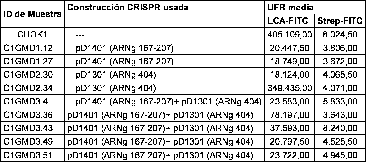
Los resultados graficos y el perfil de fluorescencia se proporcionan en la tabla anterior y tambien se representa en las figuras 7B y 8B de la presente divulgacion. Las figuras representan el resultado grafico y el perfil de fluorescencia observado para las CHOK1, C1GMD1.12, C1GMD1.27, C1GMD2.30, C1GMD2.34, C1GMD3.4, C1GMD3.36, C1GMD3.43, C1GMD3.49, C1GMD3.51 en el ensayo de union a LCA-FITC basado en citometria de flujo. Este ensayo de citometria de flujo detecta las proteinas fucosiladas sobre la superficie celular. Por lo tanto, las celulas CHOK1 de control fluorescen altamente, ya que muchas proteinas fucosiladas estan presentes en la linea celular CHOK1 de control. En casos en los que el complejo CRISPR/Cas es capaz de alterar el gen GMD en lineas celulares transfectadas, la fluorescencia de LCA-FITC se minimiza, ya que no hay proteina fucosilada en la superficie de estas lineas celulares. La figura revela la perdida significativa de fluorescencia cuando las C1GMD1.12, C1GMD1.27, C1GMD2.30, C1GMD3.4, C1GMD3.36, C1GMD3.43, C1GMD3.49, C1GMD3.51 se probaron en este ensayo, lo que indica que estas lineas celulares son posibles lineas celulares CHOK1 de inactivacion de GMD. Tambien se probo otro conjunto de transfeccion con el ensayo de fluorescencia de LCA-FITC. Los datos de la citometria de flujo se proporcionan en la tabla a continuacion.The graphical results and the fluorescence profile are given in the above table and also shown in Figures 7B and 8B of the present disclosure. The figures represent the graphical result and the observed fluorescence profile for CHOK1, C1GMD1.12, C1GMD1.27, C1GMD2.30, C1GMD2.34, C1GMD3.4, C1GMD3.36, C1GMD3.43, C1GMD3.49, C1GMD3. 51 in the LCA-FITC binding assay based on flow cytometry. This flow cytometry assay detects fucosylated proteins on the cell surface. Therefore, control CHOK1 cells fluoresce highly, since many fucosylated proteins are present in the control CHOK1 cell line. In cases in which the CRISPR / Cas complex is capable of altering the GMD gene in transfected cell lines, the LCA-FITC fluorescence is minimized, since there is no fucosylated protein on the surface of these cell lines. The figure reveals the significant loss of fluorescence when C1GMD1.12, C1GMD1.27, C1GMD2.30, C1GMD3.4, C1GMD3.36, C1GMD3.43, C1GMD3.49, C1GMD3.51 were tested in this test, which indicates that these cellular lines are possible CHOK1 cell lines of GMD inactivation. Another transfection set was also tested with the fluorescence assay of LCA-FITC. Flow cytometry data are provided in the table below.
Tabla 11Table 11

Los resultados graficos y el perfil de fluorescencia se proporcionan en la tabla anterior y tambien se representa en las figuras 7C y 8C de la presente divulgacion. Las figuras representan el resultado grafico y el perfil de fluorescencia observado para las CHOK1, C1GMD1.37, C1GMD1.4, C1GMD1.41, C1GMD1.43, y C1GMD1.44, en el ensayo de union a LCA-FITC basado en citometria de flujo. Este ensayo de citometria de flujo detecta las proteinas fucosiladas sobre la superficie celular. Por lo tanto, las celulas CHOK1 de control fluorescen altamente, ya que muchas proteinas fucosiladas estan presentes en la linea celular CHOK1 de control. En casos en los que el complejo CRISPR/Cas es capaz de alterar el gen GMD en lineas celulares transfectadas, la fluorescencia de LCA-FITC se minimiza, ya que no hay proteina fucosilada en la superficie de estas lineas celulares. La figura revela la perdida significativa de fluorescencia cuando las C1GMD1.37, C1GMD1.4, C1GMD1.41, C1GMD1.43, C1GMD1.44 se probaron en este ensayo, lo que indica que estas lineas celulares son posibles lineas celulares CHOK1 de inactivacion de GMD. Ensayo de seleccion de LCA (aglutinina de Lens culinaris) The graphical results and the fluorescence profile are given in the above table and also shown in figures 7C and 8C of the present disclosure. The figures represent the graphic result and the fluorescence profile observed for CHOK1, C1GMD1.37, C1GMD1.4, C1GMD1.41, C1GMD1.43, and C1GMD1.44, in the LCA-FITC binding assay based on cytometry of flow. This flow cytometry assay detects fucosylated proteins on the cell surface. Therefore, control CHOK1 cells fluoresce highly, since many fucosylated proteins are present in the control CHOK1 cell line. In cases in which the CRISPR / Cas complex is capable of altering the GMD gene in transfected cell lines, the LCA-FITC fluorescence is minimized, since there is no fucosylated protein on the surface of these cell lines. The figure reveals the significant loss of fluorescence when C1GMD1.37, C1GMD1.4, C1GMD1.41, C1GMD1.43, C1GMD1.44 were tested in this assay, indicating that these cell lines are possible cell lines CHOK1 inactivation of GMD. LCA selection assay (agglutinin from Lens culinaris)
Se separaron multiples poblaciones de lineas celulares clonales unicelulares en placas de replica tras la transfeccion con el complejo CRISPR/Cas. Estas lineas celulares se probaron despues con 200 |jg de reactivo LCA en el medio de cultivo. Se observaron las celulas cada d^ a para conformar la salud y la morfolog^a de las celulas y se tomaron fotografias en los puntos temporales apropiados. La Figura 5 indica las fotografias tomadas despues de un dia de cultivo tras el punto de partida de seleccion por LCA y la figura 6 indica las fotografias tomadas despues de 4 dias de cultivo. Las lmeas celulares indicadas en este caso muestran resistencia frente a LCA representadas por celulas resistentes en el dia uno que se han multiplicado y cultivado en grandes colonias de celulas tras el dia 4 de cultivo en presencia de reactivo LCA. Se observo la morfologia celular con microscopio y las observaciones se registraron en los dias 1 y 4.Multiple populations of unicellular clonal cell lines were separated in replicate plates after transfection with the CRISPR / Cas complex. These cell lines were then tested with 200 | jg of LCA reagent in the medium of culture. The cells were observed every day to shape the health and morphology of the cells and photographs were taken at the appropriate time points. Figure 5 indicates the photographs taken after a day of cultivation after the starting point of selection by LCA and figure 6 indicates the photographs taken after 4 days of cultivation. The cell lines indicated in this case show resistance to ACL represented by resistant cells on day one that have multiplied and grown in large cell colonies after day 4 of culture in the presence of LCA reagent. The cell morphology was observed with a microscope and the observations were recorded on days 1 and 4.
Las celulas se observaron regularmente con el microscopio de contraste de fase inversa y se controlo la morfologia de la colonia. Las fotografias tomadas en diferentes puntos temporales del ensayo de seleccion por LCA con 200 |jg/ml de LCA claramente muestran que las celulas CHOK1 de control estan completamente muertas en el dia 4 de cultivo, mientras que los clones seleccionados muestran un crecimiento celular continuo y morfologia celular sana incluso despues de 4 dias de cultivo.The cells were observed regularly with the reverse phase contrast microscope and the morphology of the colony was controlled. The photographs taken at different time points of the LCA selection assay with 200 | jg / ml of LCA clearly show that the CHOK1 control cells are completely dead on day 4 of culture, while the selected clones show continuous cell growth and Healthy cell morphology even after 4 days of culture.
De estas figuras se observa que los siguientes clones - CR1KOT1 n.° 44, CR1KOT1 n.° 52, CR1KOT1 n.° 55, CR1KOT1 n.° 61 y CR1KOT1 n.° 67, conservan la morfologia de la colonia incluso despues del tratamiento con 200 jg/ml de LCA. Por lo tanto, se considera que estos clones son posibles fenotipos de desactivacion de FUT 8.From these figures it is observed that the following clones - CR1KOT1 No. 44, CR1KOT1 No. 52, CR1KOT1 No. 55, CR1KOT1 No. 61 and CR1KOT1 No. 67, retain the morphology of the colony even after treatment with 200 jg / ml of LCA. Therefore, it is considered that these clones are possible phenotypes of FUT 8 deactivation.
Durante la seleccion por LCA de clones transfectados con pD1401 (ARNg 167-207), se comparo la viabilidad de los clones en el dia 1, el dia 4 y el dia 6, en la figura 6B de la presente divulgacion. De estas figuras se observa que los siguientes clones C1GMD1.12, C1GMD1.27, C1GMD1.40, C1GMD1.41, C1GMD1.43 y C1GMD1.44 conservan la morfologia de la colonia incluso despues del tratamiento con 200 jg/ml de LCA. Por lo tanto, se considera que estos clones son posibles fenotipos de desactivacion de FUT 8. Las lineas celulares indicadas en este caso muestran resistencia frente a LCA en el dia uno que se han multiplicado y cultivado en grandes colonias de celulas tras el dia 4 y el dia 6 de cultivo en presencia de reactivo LCA. Se observo la morfologia celular con microscopio y las observaciones se registraron en los dias 1, 4 y 6. Las celulas se observaron regularmente con el microscopio de contraste de fase inversa y se controlo la morfologia de la colonia. Las fotografias tomadas en diferentes puntos temporales del ensayo de seleccion por LCA con 200 jg/ml de LCA claramente muestran que las celulas CHOK1 de control estan completamente muertas en el dia 4 de cultivo, mientras que los clones seleccionados muestran un crecimiento celular continuo y morfologia celular sana incluso despues de 4 dias de cultivo.During the selection by LCA of clones transfected with pD1401 (RNAg 167-207), the viability of the clones was compared on day 1, day 4 and day 6, in figure 6B of the present disclosure. From these figures it is observed that the following clones C1GMD1.12, C1GMD1.27, C1GMD1.40, C1GMD1.41, C1GMD1.43 and C1GMD1.44 conserve the morphology of the colony even after treatment with 200 jg / ml of ACL. Therefore, it is considered that these clones are possible phenotypes of deactivation of FUT 8. The cell lines indicated in this case show resistance against ACL on day one that have multiplied and grown in large cell colonies after day 4 and on day 6 of culture in the presence of LCA reagent. The cell morphology was observed with microscope and the observations were recorded on days 1, 4 and 6. The cells were observed regularly with the reverse phase contrast microscope and the morphology of the colony was controlled. The photographs taken at different time points of the LCA selection trial with 200 jg / ml of LCA clearly show that the CHOK1 control cells are completely dead on day 4 of culture, while the selected clones show continuous cell growth and morphology. cell phone even after 4 days of cultivation.
Ensayo de union de LCA-FITC:LCA-FITC binding assay:
Para el segundo conjunto de ensayo de union de LCA-FITC, los siguientes clones han demostrado un perfil de citometria de flujo de inactivacion de fucosa: CR1KOT1 n.° 44, CR1KOT1 n.° 46, CR1KOT1 n.° 48, CR1KOT1 n.° 49, CR1KOT1 n.° 51, CR1KOT1 n.° 52, CR1KOT1 n.° 55, CR1KOT1 n.° 59, CR1KOT1 n.° 61, CR1KOT1 n.° 66, CR1KOT1 n.° 67 (Figura 10). Se diluyeron 5 mg/ml de la solucion madre de aglutinina de Lens culinaris-Fluorescema (LCA-FITC) para obtener una concentracion final de 2 jg/ml en tampon de ensayo (DPBS que contiene BSA al 2 %). Se agitaron vorticialmente las celulas a 1500 rpm durante 5 minutos usando la centrifuga minispin de Eppendorf. Se aspiro el medio y se resuspendio el sedimento en 0,25-1 ml de tampon de ensayo que contiene 2 jg/ml de LCA-FITC.For the second LCA-FITC binding assay set, the following clones have demonstrated a flow cytometry profile of fucose inactivation: CR1KOT1 # 44, CR1KOT1 # 46, CR1KOT1 # 48, CR1KOT1 n. ° 49, CR1KOT1 n ° 51, CR1KOT1 n ° 52, CR1KOT1 n ° 55, CR1KOT1 n ° 59, CR1KOT1 n ° 61, CR1KOT1 n ° 66, CR1KOT1 n ° 67 (Figure 10). 5 mg / ml of the Lens culinaris-Fluorescema agglutinin stock solution (LCA-FITC) was diluted to obtain a final concentration of 2 jg / ml in assay buffer (DPBS containing 2% BSA). The cells were vortexed at 1500 rpm for 5 minutes using the Eppendorf minispin centrifuge. The medium was aspirated and the pellet resuspended in 0.25-1 ml of assay buffer containing 2 jg / ml of LCA-FITC.
Se resuspendieron las celulas CHOK1 de control en 0,25-1 ml de tampon de ensayo solo (control sin tincion) y 0,25 1 ml de tampon de ensayo que contiene 2 jg/ml de LCA-FITC (control con tincion). Todas las muestras se diluyeron para obtener 0,1-0,2 x 10⁶ celulas/ml en tampon de ensayo final. Despues se incubaron las muestras en la oscuridad y sobre hielo durante 30 minutos. Despues, 200 j l de cada muestra se dividieron en alicuotas en una placa de 96 pocillos. La placa se cargo despues en el citometro de flujo de mesa de trabajo Millipore GUAVA easyCyte 8HT para la adquisicion y el analisis de datos. El analisis de datos se hizo usando el programa informatico Incyte.The CHOK1 control cells were resuspended in 0.25-1 ml of assay buffer alone (control without staining) and 0.25 ml of assay buffer containing 2 jg / ml of LCA-FITC (control with staining). All samples were diluted to obtain 0.1-0.2 x 10⁶ cells / ml in final assay buffer. The samples were then incubated in the dark and on ice for 30 minutes. Then, 200 μl of each sample was divided into aliquots in a 96-well plate. The plate was then loaded into the Millipore GUAVA easyCyte 8HT work table flow cytometer for data acquisition and analysis. The data analysis was done using the Incyte computer program.
Tabla 12Table 12


Los resultados proporcionados en la tabla anterior tambien se representan en la representacion grafica en la figura 10 y el perfil de fluorescencia en la figura 11 de la presente divulgacion.The results provided in the above table are also represented in the graphical representation in Figure 10 and the fluorescence profile in Figure 11 of the present disclosure.
La Figura 10 representa el resultado grafico observado para las lineas celulares de CHOK1 CR1KOT1 n.° 44, CR1KOT1 n.° 46, CR1KOT1 n.° 48, CR1KOT1 n.° 49, CR1KOT1 n.° 51, CR1KOT1 n.° 52, CR1KOT1 n.° 55, CR1KOT1 n.° 59, CR1KOT1 n.° 61, CR1KOT1 n.° 66, CR1KOT1 n.° 67 en el ensayo de union a LCA-FITC basado en citometria de flujo. La Figura 11 representa los perfiles de fluorescencia representativos observados en las lineas celulares CR1KOT1 n.° 44, CR1KOT1 n.° 49, CR1KOT1 n.° 51, CR1KOT1 n.° 52, CR1KOT1 n.° 55, CR1KOT1 n.° 59, CR1KOT1 n.° 61, CR1KOT1 n.° 67. Todos los clones se traspasaron durante los dias adicionales antes de ser analizados en el presente experimento. Este ensayo de citometria de flujo detecta las proteinas fucosiladas sobre la superficie celular. Por lo tanto, las celulas CHOK1 de control fluorescen altamente, ya que muchas proteinas fucosiladas estan presentes en la linea celular CHOK1 de control. En casos en los que el complejo CRISPR/Cas es capaz de alterar el gen FUT8 en lineas celulares transfectadas, la fluorescencia de LCA-FITC se minimiza, ya que no hay proteina fucosilada en la superficie de estas lineas celulares. La figura revela la perdida significativa de fluorescencia cuando las CR1KOT1 n.° 44, CR1KOT1 n.° 46, CR1KOT1 n.° 48, CR1KOT1 n.° 49, CR1KOT1 n.° 51, CR1KOT1 n.° 52, CR1KOT1 n.° 55, CR1KOT1 n.° 59, CR1KOT1 n.° 61, CR1KOT1 n.° 66, CR1KOT1 n.° 67 se probaron en este ensayo, lo que indica que estas lineas celulares son lineas celulares CHOK1 de inactivacion de FUT8.Figure 10 represents the graphical result observed for the cell lines of CHOK1 CR1KOT1 n ° 44, CR1KOT1 n ° 46, CR1KOT1 n ° 48, CR1KOT1 n ° 49, CR1KOT1 n ° 51, CR1KOT1 n ° 52, CR1KOT1 n ° 55, CR1KOT1 n ° 59, CR1KOT1 n ° 61, CR1KOT1 n ° 66, CR1KOT1 n ° 67 in the LCA-FITC binding assay based on flow cytometry. Figure 11 represents the representative fluorescence profiles observed in cell lines CR1KOT1 No. 44, CR1KOT1 No. 49, CR1KOT1 No. 51, CR1KOT1 No. 52, CR1KOT1 No. 55, CR1KOT1 No. 59, CR1KOT1 No. 61, CR1KOT1 No. 67. All the clones were transferred during the additional days before being analyzed in the present experiment. This flow cytometry assay detects fucosylated proteins on the cell surface. Therefore, control CHOK1 cells fluoresce highly, since many fucosylated proteins are present in the control CHOK1 cell line. In cases where the CRISPR / Cas complex is capable of altering the FUT8 gene in transfected cell lines, the fluorescence of LCA-FITC is minimized, since there is no fucosylated protein on the surface of these cell lines. The figure reveals the significant loss of fluorescence when the CR1KOT1 n ° 44, CR1KOT1 n ° 46, CR1KOT1 n ° 48, CR1KOT1 n ° 49, CR1KOT1 n ° 51, CR1KOT1 n ° 52, CR1KOT1 n ° 55, CR1KOT1 No. 59, CR1KOT1 No. 61, CR1KOT1 No. 66, CR1KOT1 No. 67 were tested in this assay, indicating that these cell lines are CHOK1 cell lines of inactivation of FUT8.
Conclusion - se han identificado 17 posibles candidatos para la linea celular CHOK1 de inactivacion de fut8. Estos 17 clones son como sigue:Conclusion - 17 possible candidates have been identified for the CHOK1 cell line of inactivation of fut8. These 17 clones are as follows:
CR1KOT1 n.° 006, CR1KOT1 n.° 018, CR1KOT1 n.° 022, CR1KOT1 n.° 023, CR1KOT1 n.° 026, CR1KOT1 n.° 031, CR1KOT1 n.° 034, CR1KOT1 n.° 036, CR1KOT1 n.° 037, CR1KOT1 n.° 044, CR1KOT1 n.° 049, CR1KOT1 n.° 051, CR1KOT1 n.° 052, CR1KOT1 n.° 055, CR1KOT1 n.° 059, CR1KOT1 n.° 061 y CR1KOT1 n.° 067.CR1KOT1 No. 006, CR1KOT1 No. 018, CR1KOT1 No. 022, CR1KOT1 No. 023, CR1KOT1 No. 026, CR1KOT1 No. 031, CR1KOT1 No. 034, CR1KOT1 No. 036, CR1KOT1 n 037, CR1KOT1 No. 044, CR1KOT1 No. 049, CR1KOT1 No. 051, CR1KOT1 No. 052, CR1KOT1 No. 055, CR1KOT1 No. 059, CR1KOT1 No. 061 and CR1KOT1 No. 067
Se han identificado 13 posibles candidatos para la linea celular CHOK1 de inactivacion GMD. Los clones son los siguientes:We have identified 13 possible candidates for the CHOK1 cell line of GMD inactivation. The clones are the following:
C1GMD1.12, C1GMD1.27, C1GMD1.37, C1GMD1.4, C1GMD1.41, C1GMD1.43, C1GMD1.44, C1GMD2.30, C1GMD3.4, C1GMD3.36, C1GMD3.43, C1GMD3.49, C1GMD3.51C1GMD1.12, C1GMD1.27, C1GMD1.37, C1GMD1.4, C1GMD1.41, C1GMD1.43, C1GMD1.44, C1GMD3.30, C1GMD3.49, C1GMD3.49, C1GMD3.49, C1GMD3.49, C1GMD3.49, C1GMD3.49, C1GMD3.49, C1GMD3.49, C1GMD3.49, C1GMD3.49, C1GMD3.49, C1GMD3.49. 51
Ejemplo 3: ENSAYO DE UNION A LCA-FITCExample 3: LCA-FITC UNION ASSAY
Las lineas celulares clonales CHOK1 de inactivacion de fucosa se probaron en experimentos de repeticion independientes usando el ensayo de union LCA-FITC:The CHOK1 clonal cell lines of fucose inactivation were tested in independent repeating experiments using the LCA-FITC binding assay:
Se probaron las siguientes lineas celulares clonales para la repetibilidad del ensayo de union a LCA-FITC.The following clonal cell lines were tested for the repeatability of the LCA-FITC binding assay.
CR1KOT1 n.° 006, CR1KOT1 n.° 018, CR1KOT1 n.° 022, CR1KOT1 n.° 023, CR1KOT1 n.° 026, CR1KOT1 n.° 031, CR1KOT1 n.° 034, CR1KOT1 n.° 036, CR1KOT1 n.° 037, CR1KOT1 n.° 044, CR1KOT1 n.° 049, CR1KOT1 n.° 051, CR1KOT1 n.° 052, CR1KOT1 n.° 055, CR1KOT1 n.° 059, CR1KOT1 n.° 061 y CR1KOT1 n.° 067.CR1KOT1 No. 006, CR1KOT1 No. 018, CR1KOT1 No. 022, CR1KOT1 No. 023, CR1KOT1 No. 026, CR1KOT1 No. 031, CR1KOT1 No. 034, CR1KOT1 No. 036, CR1KOT1 n 037, CR1KOT1 No. 044, CR1KOT1 No. 049, CR1KOT1 No. 051, CR1KOT1 No. 052, CR1KOT1 No. 055, CR1KOT1 No. 059, CR1KOT1 No. 061 and CR1KOT1 No. 067
Se diluyeron 5 mg/ml de la solucion madre de aglutinina de Lens culinaris-Fluorescema (LCA-FITC) para obtener una concentracion final de 2 |jg/ml en tampon de ensayo (DPBS que contiene BSA al 2 %). Se agitaron vorticialmente las celulas a 1500 rpm durante 5 minutos usando la centrifuga minispin de Eppendorf. Se aspiro el medio y se resuspendio el sedimento en 0,25-1 ml de tampon de ensayo que contiene 2 |jg/ml de LCA-FITC. Se resuspendieron las celulas CHOK1 de control en 0,25-1 ml de tampon de ensayo solo (control sin tincion) y 0,25-1 ml de tampon de ensayo que contiene 2 jg/ml de LCA-FITC (control con tincion). Todas las muestras se diluyeron para obtener 0,1-0,2 x 10⁶ celulas/ml en tampon de ensayo final. Despues se incubaron las muestras en la oscuridad y sobre hielo durante 30 minutos. Despues, 200 j l de cada muestra se dividieron en alicuotas en una placa de 96 pocillos. La placa se cargo despues en el citometro de flujo de mesa de trabajo Millipore GUAVA easyCyte 8HT para la adquisicion y el analisis de datos. El analisis de datos se hizo usando el programa informatico Incyte.5 mg / ml of the Lens culinaris-Fluorescema agglutinin stock solution (LCA-FITC) was diluted to obtain a final concentration of 2 μg / ml in assay buffer (DPBS containing 2% BSA). The cells were vortexed at 1500 rpm for 5 minutes using the Eppendorf minispin centrifuge. The medium and the pellet was resuspended in 0.25-1 ml of assay buffer containing 2 μg / ml LCA-FITC. The CHOK1 control cells were resuspended in 0.25-1 ml of assay buffer alone (control without staining) and 0.25-1 ml of assay buffer containing 2 jg / ml of LCA-FITC (control with staining) . All samples were diluted to obtain 0.1-0.2 x 10⁶ cells / ml in final assay buffer. The samples were then incubated in the dark and on ice for 30 minutes. Then, 200 jl of each sample was divided into aliquots in a 96-well plate. The plate was then loaded into the Millipore GUAVA easyCyte 8HT work table flow cytometer for data acquisition and analysis. The data analysis was done using the Incyte computer program.
Resultados:Results:
La siguiente tabla describe los datos de repetibilidad del ensayo de union de LCA-FITC para todos los clones seleccionados.The following table describes the repeatability data of the LCA-FITC binding assay for all selected clones.
Tabla 13Table 13

La representacion grafica del ensayo de union a LCA-FITC se presenta en la figura 12. El analisis de los datos sugiere una alta reproducibilidad del patron de union a LCA-FITC para las lineas celulares clonales CHOK1 de inactivacion de fucosa. Estas lineas celulares CHOK1 de inactivacion de fucosa se prueban adicionalmente para las caracteristicas del cultivo y se compararon con la linea celular CHOK1 parental no transfectada.The graphical representation of the LCA-FITC binding assay is presented in Figure 12. The analysis of the data suggests a high reproducibility of the LCA-FITC binding pattern for the CHOK1 clonal cell lines of fucose inactivation. These CHOK1 cell lines of fucose inactivation are further tested for culture characteristics and compared to the untransfected parental CHOK1 cell line.
Ejemplo 4: ENSAYO DE ESTREPTAVIDINA-FITCExample 4: ESTREPTAVIDINE-FITC TEST
Las lineas celulares clonales CHOK1 de inactivacion de fucosa se probaron con el conjugado de estreptavidina-FITC para asegurar la interaction especifica de la union a LCA-FITC:CHOK1 clonal cell lines of fucose inactivation were tested with the streptavidin-FITC conjugate to ensure the specific interaction of the LCA-FITC binding:
La tincion de estreptavidina conjugada con FITC (Strep-FITC) de los clones se llevo a cabo para asegurar que no hay union no especifica del colorante FITC. Las proteinas de la membrana celular no se unen al conjugado de estreptavidina-FITC, mientras que las proteinas de membrana fucosilada se unen de forma especifica al conjugado LCA-FITC. Las celulas CHOK1 de control se tineron tanto con LCA-FITC como con Strep-FITC en reacciones separadas para confirmar esta especificidad. Todos los clones se tineron de forma similar con conjugados de estreptavidina-FITC y LCA-FITC para determinar la union no especifica.Streptavidin-conjugated staining with FITC (Strep-FITC) of the clones was carried out to ensure that there is no non-specific binding of the FITC dye. The cell membrane proteins do not bind to the streptavidin-FITC conjugate, while the fucosylated membrane proteins bind specifically to the LCA-FITC conjugate. CHOK1 control cells were stained with both LCA-FITC and Strep-FITC in separate reactions to confirm this specificity. All clones were stained in a similar manner with streptavidin-FITC and LCA-FITC conjugates to determine non-specific binding.
La Figura 14 de la presente divulgation representa el ensayo de citometria de flujo de LCA, y la comparacion de los clones a traves del ensayo de LCA-FITC y Strep-FITC, respectivamente. Para los siguientes clones se observo el fenotipo de inactivacion de fucosa: CR1KOT1 n.° 006, CR1KOT1 n.° 018, CR1KOT1 n.° 022, CR1KOT1 n.° 023, CR1KOT1 n.° 026, CR1KOT1 n.° 031, CR1KOT1 n.° 034, CR1KOT1 n.° 036, CR1KOT1 n.° 037, CR1KOT1 n.° 044, CR1KOT1 n.° 049, CR1KOT1 n.° 051, CR1KOT1 n.° 052, CR1KOT1 n.° 055, CR1KOT1 n.° 059, CR1KOT1 n.° 061 y CR1KOT1 n.° 067. En todos los clones, se observo una reduction significativa de la fluorescencia en comparacion con el control y, por lo tanto, esto demuestra la ausencia de proteina fucosilada en la superficie celular para los clones. Estos datos proporcionan la evidencia funcional de que estas lineas funcionales carecen de la proteina fucosilada debido a la alteration del gen FUT8 llevada a cabo usando el complejo en el metodo de la presente divulgacion.Figure 14 of the present disclosure represents the LCA flow cytometry assay, and the comparison of the clones through the LCA-FITC and Strep-FITC assay, respectively. For the following clones, the fucose inactivation phenotype was observed: CR1KOT1 n. 006, CR1KOT1 n. 018, CR1KOT1 n. 022, CR1KOT1 n. 023, CR1KOT1 n. 026, CR1KOT1 n. 031, CR1KOT1 No. 034, CR1KOT1 No. 036, CR1KOT1 No. 037, CR1KOT1 No. 044, CR1KOT1 No. 049, CR1KOT1 No. 051, CR1KOT1 No. 052, CR1KOT1 No. 055, CR1KOT1 n. 059, CR1KOT1 n. 061 and CR1KOT1 n. 067. In all clones, a significant reduction of fluorescence was observed in comparison with the control and, therefore, this demonstrates the absence of fucosylated protein on the cell surface for the clones. These data provide the functional evidence that these functional lines lack the fucosylated protein due to the alteration of the FUT8 gene carried out using the complex in the method of the present invention. divulgation.
Se diluyeron 1 mg/ml de la solucion madre de estreptavidina-fluorescema (Strep-FITC) para obtener una concentracion final de 2 jg/ml en tampon de ensayo (DPBS que contiene BSA al 2 %). Se agitaron vorticialmente las celulas a 1500 rpm durante 5 minutos usando la centrifuga minispin de Eppendorf. Se aspiro el medio y se resuspendio el sedimento en 0,25-1 ml de tampon de ensayo que contiene 2 |jg/ml de Estreptavidina-FITC. Se resuspendieron las celulas CHOK1 de control en 0,25-1 ml de tampon de ensayo solo (control sin tincion) y 0,25-1 ml de tampon de ensayo que contiene 2 jg/ml de Estreptavidina-FITC (control con tincion). Todas las muestras se diluyeron para obtener 0,1-0,2 x 10⁶ celulas/ml en tampon de ensayo final. Despues se incubaron las muestras en la oscuridad y sobre hielo durante 30 minutos.1 mg / ml of the streptavidin-fluorescem stock solution (Strep-FITC) was diluted to obtain a final concentration of 2 jg / ml in assay buffer (DPBS containing 2% BSA). The cells were vortexed at 1500 rpm for 5 minutes using the Eppendorf minispin centrifuge. The medium was aspirated and the pellet resuspended in 0.25-1 ml of assay buffer containing 2 μg / ml Streptavidin-FITC. The CHOK1 control cells were resuspended in 0.25-1 ml of assay buffer alone (control without staining) and 0.25-1 ml of assay buffer containing 2 jg / ml Streptavidin-FITC (control with staining) . All samples were diluted to obtain 0.1-0.2 x 10⁶ cells / ml in final assay buffer. The samples were then incubated in the dark and on ice for 30 minutes.
Despues, 200 j l de cada muestra se dividieron en alicuotas en una placa de 96 pocillos. La placa se cargo despues en el citometro de flujo de mesa de trabajo Millipore GUAVA easyCyte 8HT para la adquisicion y el analisis de datos. El analisis de datos se hizo usando el programa informatico Incyte.Then, 200 μl of each sample was divided into aliquots in a 96-well plate. The plate was then loaded into the Millipore GUAVA easyCyte 8HT work table flow cytometer for data acquisition and analysis. The data analysis was done using the Incyte computer program.
Se llevo a cabo a comparacion con Estreptavidina-FITC para asegurar la interaccion especifica del conjugado LCA-FITC. Los datos sugieren solo fluorescencia de fondo observada con el conjugado Estreptavidina-FITC cuando se probo con la linea celular de control CHOK1 y cualquiera de las lineas celulares transfectadas con CRISPR/Cas. It was carried out in comparison with Streptavidin-FITC to ensure the specific interaction of the LCA-FITC conjugate. The data suggest only background fluorescence observed with the Estreptavidin-FITC conjugate when tested with the CHOK1 control cell line and any of the cell lines transfected with CRISPR / Cas.
La Figura 14 representa la especificidad del ensayo de citometria de flujo de LCA-FITC, desarrollado para cribar las lineas celulares CHOK1 de inactivacion de fucosa. Los experimentos de citometria de flujo se llevaron a cabo con el conjugado de Estreptavidina-FITC. La Estreptavidina-FITC no reconoce proteinas de superficie celular en la linea celular de CHOK1 de control, lo que indica la interaccion especifica del conjugado LCA-FITC. Adicionalmente, el conjugado de estreptavidina-FITC revela niveles similares de fluorescencia de fondo entre lineas celulares positivas y negativas identificadas por el conjugado LCA-FITC. La Figura 14 no muestra interaccion no especifica del conjugado de LCA-FITC usado en este estudio cuando se comparo con el conjugado de Estreptavidina-FITC.Figure 14 represents the specificity of the LCA-FITC flow cytometry assay, developed to screen the CHOK1 cell lines for fucose inactivation. The flow cytometry experiments were carried out with the Streptavidin-FITC conjugate. Streptavidin-FITC does not recognize cell surface proteins in the control CHOK1 cell line, indicating the specific interaction of the LCA-FITC conjugate. Additionally, the streptavidin-FITC conjugate reveals similar levels of background fluorescence between positive and negative cell lines identified by the LCA-FITC conjugate. Figure 14 shows no nonspecific interaction of the LCA-FITC conjugate used in this study when compared to the Streptavidin-FITC conjugate.
Determinacion de la curva de crecimiento para las lineas celulares CHOK1 de inactivacion de fucosa:Determination of the growth curve for the CHOK1 cell lines of fucose inactivation:
La determinacion de la curva de crecimiento de los clones seleccionados se realizo para asegurar que el perfil de crecimiento no se altera significativamente en comparacion con las celulas CHOK1 de tipo silvestre durante el proceso de desarrollo de la linea celular de inactivacion. Se inocularon 0,1 X 10⁶ celulas CHOK1 en placas de cultivo tisular de 6 pocillos. La inoculacion se hizo para 5 puntos temporales para cada clon. Para cada punto temporal, se hizo una inoculacion por triplicado (por ejemplo, 15 pocillo para 5 puntos temporales). En cada punto temporal, se hicieron recuentos celulares por triplicado. El recuento de celulas viables se realizo usando un hemocitometro o un analizador de viabilidad celular Vi-cell XR. Se generaron las respectivas curvas de crecimiento con EEM como barra de error. La Tabla 14 describe los datos de crecimiento representativos de una de las lineas celulares de inactivacion de FUT8.The determination of the growth curve of the selected clones was carried out to ensure that the growth profile was not significantly altered in comparison with the wild-type CHOK1 cells during the process of developing the inactivation cell line. 0.1 X 10 6 CHOK1 cells were inoculated into 6-well tissue culture plates. Inoculation was done for 5 time points for each clone. For each time point, an inoculation was done in triplicate (for example, 15 well for 5 temporary points). At each time point, cell counts were made in triplicate. The viable cell count was performed using a hemocytometer or a Vi-cell XR cell viability analyzer. The respective growth curves with EEM were generated as an error bar. Table 14 describes the representative growth data of one of the FUT8 inactivation cell lines.
Tabla 14Table 14


Resultados: El recuento de celulas viables para cada una de las lmeas celulares se probo y se uso para la determinacion de la curva de crecimiento. Las siguientes lmeas celulares CHOK1 de inactivacion de fucosa se usan para el desarrollo de la curva de crecimiento, CR1KOT1 n.° 006, CR1KOT1 n.° 018, CR1KOT1 n.° 022, CR1KOT1 n.° 023, CR1KOT1 n.° 026, CR1KOT1 n.° 031, CR1KOT1 n.° 034, CR1KOT1 n.° 036, CR1KOT1 n.° 037, CR1KOT1 n.° 044, CR1KOT1 n.° 049, CR1KOT1 n.° 051, CR1KOT1 n.° 052, CR1KOT1 n.° 055, CR1KOT1 n.° 061 y CR1KOT1 n.° 067.Results: The viable cell count for each of the cell lines was tested and used for the determination of the growth curve. The following CHOK1 cell lines of fucose inactivation are used for the development of the growth curve, CR1KOT1 # 006, CR1KOT1 # 018, CR1KOT1 # 022, CR1KOT1 # 023, CR1KOT1 # 026, CR1KOT1 No. 031, CR1KOT1 No. 034, CR1KOT1 No. 036, CR1KOT1 No. 037, CR1KOT1 No. 044, CR1KOT1 No. 049, CR1KOT1 No. 051, CR1KOT1 No. 052, CR1KOT1 n . ° 055, CR1KOT1 n. ° 061 and CR1KOT1 n. ° 067.
Los datos se analizaron y se representaron graficamente en la curva de crecimiento. Las curvas de crecimiento de las respectivas lineas celulares se proporcionan en las figuras 13A, 13B y 13C de la presente divulgacion.The data was analyzed and plotted on the growth curve. The growth curves of the respective cell lines are provided in Figures 13A, 13B and 13C of the present disclosure.
De las figuras se observa que la mayoria de las lineas celulares clonales tienen un potencial de crecimiento comparable con respecto a la linea celular parental de CHOK1. Estas lineas celulares clonales se san para la sobreexpresion de las proteinas y/o anticuerpos monoclonales terapeuticos. Se hizo un recuento de celulas viables todos los dias durante 5 dias en condiciones de cultivo optimas, usando el contador Vi-Cell. Unas pocas lineas celulares CHOK1 de inactivacion de fucosa, CR1KOT1 n.° 018, CR1KOT1 n.° 026, CR1KOT1 n.° 034, CR1KOT1 n.° 052 y CR1KOT1 n.° 055 han demostrado tener un potencial de crecimiento ligeramente mas lento en comparacion con otras lineas celulares.From the figures it is observed that the majority of the clonal cell lines have a growth potential comparable with respect to the parental cell line of CHOK1. These clonal cell lines are sanctioned for overexpression of the proteins and / or therapeutic monoclonal antibodies. A count of viable cells was made every day for 5 days under optimal culture conditions, using the Vi-Cell counter. A few CHOK1 cell lines of fucose inactivation, CR1KOT1 # 018, CR1KOT1 # 026, CR1KOT1 # 034, CR1KOT1 # 052 and CR1KOT1 # 055 have been shown to have a slightly slower growth potential in comparison with other cell lines.
Ejemplo 5: ENSAYOS DE SECUENCIACION GENOMICAExample 5: GENOMIC SEQUENCING TESTS
Los clones transfectados con CRISPR seleccionados a traves del ensayo funcional, es decir, se usaron ensayos de citometria de flujo con LCA-FITC para el analisis de secuencia genomica. El locus genomico de FUT8 en hamster chino esta bien documentado en la bibliografia (NW 003613860) y se usa como una secuencia de tipo silvestre para comprender el tipo de modificacion genica en cada clon de linea celular. De manera similar, el locus genomico de Gm D de hamster chino se obtuvo de la base de datos de la secuencia, NW_003613635.1, NP_001233625.1, NM_001246696.1 y se uso como secuencia de tipo silvestre para entender el tipo de modificacion genica en cada linea celular. El objetivo de este ejemplo es analizar los resultados de secuenciacion de ADN genomico obtenidos de las lineas celulares CHOK1 de inactivacion de FUT8 transfectadas con CRISPR/Cas y las lineas celulares CHOK1 de inactivacion de GMD. Todas las lineas celulares documentadas en este caso son lineas celulares clonales y se seleccionan del ensayo de selection del medio LCA y del ensayo de citometria de flujo con LCA-FITC.Clones transfected with CRISPR selected through the functional assay, i.e., flow cytometry assays with LCA-FITC were used for genomic sequence analysis. The FUT8 genomic locus in Chinese hamster is well documented in the literature (NW 003613860) and is used as a wild type sequence to understand the type of genetic modification in each cell line clone. Similarly, the Chinese hamster GmD genomic locus was obtained from the sequence database, NW_003613635.1, NP_001233625.1, NM_001246696.1 and used as a wild-type sequence to understand the type of genetic modification in each cell line. The objective of this example is to analyze the results of genomic DNA sequencing obtained from the CHOK1 cell lines of inactivation of FUT8 transfected with CRISPR / Cas and the CHOK1 cell lines of inactivation of GMD. All the cell lines documented in this case are clonal cell lines and are selected from the selection test of the LCA medium and the flow cytometry assay with LCA-FITC.
En resumen, las lineas celulares clonales seleccionadas se cultivan en condiciones de cultivo apropiadas para el aislamiento de ADN genomico, el ADN genomico purificado se uso para la amplification por PCR usando cebadores que flanquean los loci diana de FUT8 y GMD, el producto amplificado por PCR se purifica despues y se clona en un vector adecuado usando celulas E. coli competentes, las colonias de E. coli resistentes a ampicilina resultantes se seleccionan y se cultivan, los ADN plasmidicos se aislan de cada clon bacteriano, aproximadamente se prueban 5 10 colonias bacterianas individuales por lineas celulares clonales a traves de la secuenciacion automatica para comprender el tipo de modificacion en el locus genomico diana de FUT8.In summary, the selected clonal cell lines are cultured under appropriate culture conditions for the isolation of genomic DNA, the purified genomic DNA was used for PCR amplification using primers flanking the target loci of FUT8 and GMD, the product amplified by PCR then purified and cloned into a suitable vector using competent E. coli cells, the resulting ampicillin-resistant E. coli colonies are selected and cultured, the plasmid DNAs are isolated from each bacterial clone, approximately 10 bacterial colonies are tested. Individuals by clonal cell lines through automatic sequencing to understand the type of modification in the FUT8 target genomic locus.
Los siguientes reactivos y soluciones se usaron para llevar a cabo la secuenciacion genomica de los clones seleccionadosThe following reagents and solutions were used to carry out the genomic sequencing of the selected clones
Todo el protocolo de secuenciacion genomica se divide en los siguientes cuatro procesosThe whole genome sequencing protocol is divided into the following four processes
A. Aislamiento de ADN genomico de los clones seleccionadosA. Genomic DNA isolation of the selected clones
B. Estrategia de PCR para amplificar locus genomico especifico para cada linea celular.B. PCR strategy to amplify specific genomic loci for each cell line.
C. donation de los productos de PCR en vectores de secuenciacionC. donation of PCR products in sequencing vectors
D. Analisis de datos de secuencia e identification de INDELD. Analysis of sequence data and identification of INDEL
Aislamiento de ADN genomico de los clones seleccionadosIsolation of genomic DNA from the selected clones
Las lineas celulares CHOK1 clonales se cultivaron en medio DMEM avanzado con suero bovino fetal al 10 %, glutamina 4 mM, 100 unidades/ml de penicilina y 100 |jg/ml de estreptomicina en matraces T175 a 37 °C en presencia de CO2 al 5 % y humedad relativa del 75 % en incubadoras de condiciones controladas. El cultivo celular se observo cada dia y se controlo la viabilidad. Se recolectaron celulas con una confluencia del 80 % y mas del 95 % de viabilidad con tripsinizacion. El dia del aislamiento, se retiro el medio de cultivo y las celulas adherentes se lavaron primero con 10 ml de DPBS seguido por la adicion de 4 ml de solution de tripsina-EDTA al 0,05 % para la tripsinizacion. Se incubaron las celulas a 37 °C durante 2-3 minutos y se recolectaron. Despues se mezclaron las celulas con 10 ml de DPBS y se centrifugaron a 1500 rpm durante 5 minutos. El medio gastado se retiro y el sedimento celular se resuspendio en 10 ml de DPBS. Las celulas se lavaron de nuevo usando la centrifugation a 1500 rpm durante 5 minutos. El DPBS se retiro por completo mediante aspiration. El sedimento celular final se uso para el aislamiento del ADN genomico.Clonal CHOK1 cell lines were cultured in advanced DMEM medium with 10% fetal bovine serum, 4 mM glutamine, 100 units / ml penicillin and 100 μg / ml streptomycin in T175 flasks at 37 ° C in the presence of 5% CO 2. % and relative humidity of 75% in incubators of controlled conditions. Cell culture was observed every day and viability was monitored. Cells were collected with a confluence of 80% and more than 95% viability with trypsinization. On the day of isolation, the culture medium was removed and the adherent cells were first washed with 10 ml of DPBS followed by the addition of 4 ml of trypsin-EDTA solution at 0.05% for trypsinization. The cells were incubated at 37 ° C for 2-3 minutes and harvested. The cells were then mixed with 10 ml of DPBS and centrifuged at 1500 rpm for 5 minutes. The spent medium was removed and the cell pellet was resuspended in 10 ml of DPBS. The cells were washed again using the centrifugation at 1500 rpm for 5 minutes. The DPBS was completely removed by aspiration. The final cell pellet was used for the isolation of genomic DNA.
El ADN genomico se aislo de las celulas CHOK1 de control, y las lmeas celulares clonales CHOK1 transfectadas con CRISPR/Cas que presentan resistencia a LCA y se selecciono a traves del ensayo de citometria de flujo con LCA. El kit de extraccion de ADNg de QIAGEN comercialmente disponible se uso para el aislamiento del ADN genomico siguiendo el protocolo del fabricante.The genomic DNA was isolated from the control CHOK1 cells, and the CHOK1 clonal cell lines transfected with CRISPR / Cas that show resistance to ACL and was selected through the flow cytometry assay with LCA. The commercially available QIAGEN gDNA extraction kit was used for the isolation of genomic DNA following the manufacturer's protocol.
Diseno de estrategia de PCRPCR strategy design
La secuencia de ADN genomico de hamster chino se analizo a partir de la secuencia de la base de datos publicamente disponible NW_003613860. Las secuencias de ADN del exon 7 de FUT8 y la secuencia del intron parcial se uso para disenar la estrategia de PCR para amplificar el locus diana FUT8.The Chinese hamster genomic DNA sequence was analyzed from the sequence of the publicly available database NW_003613860. The DNA sequences of FUT8 exon 7 and the partial intron sequence were used to design the PCR strategy to amplify the FUT8 target locus.
Las secuencias de intron y de exon de GMD se obtuvieron de NW 003613635.1 y NM_001246696.1 y los analisis de secuencia se llevaron a cabo para el exon 3 y el exon 4.The intron and exon sequences of GMD were obtained from NW 003613635.1 and NM_001246696.1 and sequence analyzes were carried out for exon 3 and exon 4.
Los cebadores se disenaron basandose en la longitud del cebador, en la longitud del producto de PCR, en el contenido de GC, el la temperatura de fusion y en el potencial de formacion de homoduplex y de heteroduplex. Los cebadores se disenaron flanqueando el locus diana de FUT8 tal como se proporciona a continuation. El producto de PCR amplificado esta pensado para el analisis de mutation debido a la s Sb mediada por CRISPR y la posterior reparation de ADN. La siguiente secuencia de nucleotidos representa la region de interes con secuencias del cebador en negrita.The primers were designed based on the length of the primer, the length of the PCR product, the GC content, the melting temperature, and the homoduplex and heteroduplex formation potential. The primers were designed flanking the target locus of FUT8 as provided below. The amplified PCR product is designed for the analysis of mutation due to s Sb mediated by CRISPR and the subsequent DNA repair. The following nucleotide sequence represents the region of interest with bold primer sequences.
El exon 7 de Fut8 y las secuencias de intron asociadas se usaron para el diseno del cebador de PCR:Exon 7 of Fut8 and the associated intron sequences were used for the design of the PCR primer:

Los intrones estan representados desde la base 21 hasta la base 106 y desde la base 345 hasta la base 371 en minusculas. El exon 7 se representa desde la base 107 hasta la base 344 en mayusculas. Los sitios de union del cebador de izquierda y derecha estan subrayados.The introns are represented from base 21 to base 106 and from base 345 to base 371 in lower case. Exon 7 is represented from base 107 to base 344 in capital letters. The left and right primer binding sites are underlined.
El exon 3 y el exon 4 de GMD y las secuencias de intron asociadas se usaron para el diseno del cebador de PCR: Se usaron las siguientes secuencias.Exon 3 and exon 4 of GMD and the associated intron sequences were used for the design of the PCR primer: The following sequences were used.
Exon 3 de GMD e intrones circundantesExon 3 of GMD and surrounding introns

Los intrones estan representados desde la base 23 hasta la base 166 y desde la base 277 hasta la base 447 en minusculas. El exon 3 se representa desde la base 167 hasta la base 276 en mayusculas. Los sitios de union del cebador de izquierda y derecha estan subrayados.The introns are represented from base 23 to base 166 and from base 277 to base 447 in lower case. Exon 3 is represented from base 167 to base 276 in capital letters. The left and right primer binding sites are underlined.
Exon 4 de GMD e intrones circundantes Exon 4 of GMD and surrounding introns

Los intrones estan representados desde la base 21 hasta la base 187 y desde la base 381 hasta la base 432 en minusculas. El exon 4 se representa desde la base 188 hasta la base 380 en mayusculas. Los sitios de union del cebador de izquierda y derecha estan subrayados.The introns are represented from base 21 to base 187 and from base 381 to base 432 in lower case. Exon 4 is represented from base 188 to base 380 in capital letters. The left and right primer binding sites are underlined.
Diseno del cebador para la identificacion de INDEL por PCRPrimer design for the identification of INDEL by PCR
La PCR genomica se realizo usando el kit de extraccion de ADNg de QIAGEN usando los siguientes cebadores mencionados en la tabla 15.Genomic PCR was performed using the QIAGEN gDNA extraction kit using the following primers mentioned in Table 15.
T l 1T l 1

La siguiente seccion proporciona detalles experimentales para la generacion de producto de PCR a partir de ADN genomico de CHOK1 de lineas celulares de control y lineas celulares clonales seleccionadas de LCA, la clonacion de productos de PCR en celulas competentes de E. coli y secuenciacion de productos de PCR clonados.The following section provides experimental details for the generation of PCR product from CHOK1 genomic DNA from control cell lines and clonal cell lines selected from LCA, cloning of PCR products in competent E. coli cells and sequencing of products. of cloned PCR.
Optimizacion de las condiciones de PCR -Se disenaron los experimentos para estandarizar las condiciones de PCR. Los parametros probados incluyeron, la concentracion de ADN genomico (de 100 ng a 1000 ng), las concentraciones del cebador (2 nmol a 20 nmol), la temperatura de hibridacion de PCR (de 55,8 °C a 62,9 °C) y el tiempo (20 segundos a 50 segundos), el tiempo de extension del producto de PCR (de 30 seg a 60 seg) y el numero de ciclos de PCR se establece a 30 ciclos. La condicion optimizada de llegada se describe en la siguiente seccion. Optimization of PCR conditions - Experiments were designed to standardize PCR conditions. The parameters tested included genomic DNA concentration (100 ng to 1000 ng), primer concentrations (2 nmol to 20 nmol), PCR hybridization temperature (55.8 ° C to 62.9 ° C). ) and the time (20 seconds to 50 seconds), the extension time of the PCR product (from 30 sec to 60 sec) and the number of PCR cycles is set at 30 cycles. The optimized arrival condition is described in the next section.
Las reacciones de PCR se llevaron a cabo usando la prueba de lectura de la polimerasa Phusion para asegurar que las mutaciones mediadas por PCR estan limitadas. Tras los ciclos de amplificacion por PCR, se anade la enzima Taq polimerasa en la mezcla para la adicion de ADN por cola homopolimerica. La etapa de adicion de ADN por cola homopolimerica es importante, ya que las bases extra anadidas a los productos de PCR permiten la clonacion directa en el vector de secuenciacion descrito en la siguiente seccion. Con el fin de anadir proyecciones de dATP al producto de PCR para el vector de clonacion en TA, el producto amplificado de la polimerasa Phusion se incubo con la ADN polimerasa Taq durante 20 minutos a 72 °C.PCR reactions were carried out using the Phusion polymerase reading test to ensure that PCR-mediated mutations are limited. After PCR amplification cycles, the enzyme Taq polymerase is added to the mixture for the addition of DNA by homopolymeric glue. The stage of DNA addition by tail homopolymeric is important, since the extra bases added to the PCR products allow direct cloning into the sequencing vector described in the next section. In order to add dATP projections to the PCR product for the cloning vector in TA, the amplified product of the Phusion polymerase was incubated with the Taq DNA polymerase for 20 minutes at 72 ° C.
Comprobacion cruzada de la muestra de ADN genomico mediante PCR-Los productos de PCR de ADN genomico se analizaron en electroforesis en gel de agarosa y la longitud del producto se confirmo usando un patron de peso molecular. Las muestras de PCR con un perfil de amplificacion claro se usaron en la proxima etapa de procesamiento. Cross-checking of the genomic DNA sample by PCR- The genomic DNA PCR products were analyzed in agarose gel electrophoresis and the length of the product was confirmed using a molecular weight standard. PCR samples with a clear amplification profile were used in the next processing step.
Elucion de gel de producto de PCR usando el kit QIAGEN-Los productos de PCR amplificados se cargaron en gel de agarosa al 1 % de preparacion reciente y se sometieron a electroforesis a 100 V durante una hora para separar los productos de pCr amplificados de los cebadores no usados y cualquier otro dimero producido durante el proceso de amplificacion. Los productos amplificados se escindieron del gel y se eluyeron usando el kit de elucion en gel de Qiagen comercialmente disponible. El ADN se eluyo con agua de grado de biologia molecular altamente pura. Gel elution of PCR product using the QIAGEN kit - The amplified PCR products were loaded on freshly prepared 1% agarose gel and subjected to 100 V electrophoresis for one hour to separate the amplified pCr products from the primers unused and any other dimer produced during the amplification process. The amplified products were excised from the gel and eluted using the commercially available Qiagen gel eluting kit. The DNA eluted with water of highly pure molecular biology grade.
Clonacion de los productos de PCR en vectores de secuenciacion-Los productos amplificados por PCR y purificados en gel de agarosa se usaron despues para la clonacion en vector pTZ57R/T comercialmente disponible a traves de un proceso de ligamiento de ADN. Las condiciones para el ligamiento de ADN se estandarizaron previamente. Cloning of PCR products into sequencing vectors - PCR- amplified and agarose gel-purified products were then used for cloning into commercially available vector pTZ57R / T through a DNA ligation process. The conditions for DNA ligation were previously standardized.
Transformacion de muestra ligada pTZ57R/T+CRISPR(PCR) en celulas competentes de E.coli DH5alpha - El ADN ligado se transformo en celulas competentes de E. coli DH5alpha, comercialmente disponibles. Se siguio el protocolo de transformacion tal como se describe por el fabricante para lograr una eficacia de transformacion de alto nivel. Tras la transformacion, las celulas de E. coli se cultivaron en presencia de antibiotico ampicilina para el crecimiento de colonias transformadas.Transformation of ligated sample pTZ57R / T + CRISPR (PCR) into competent cells of E. coli DH5alpha - The ligated DNA was transformed into competent cells of E. coli DH5alpha, commercially available. The transformation protocol was followed as described by the manufacturer to achieve high level transformation efficiency. After transformation, the E. coli cells were cultured in the presence of antibiotic ampicillin for the growth of transformed colonies.
Inoculacion de celulas transformadas (pTZ57R/T+CRISPR(PCR)) en medio LB con ampicilina - Cada colonia separada se inoculo en caldo LB Ampicilina en un volumen de cultivo de 5 ml y se cultivo toda la noche para el aislamiento de ADN plasmidico.Inoculation of transformed cells (pTZ57R / T + CRISPR (PCR)) in LB medium with ampicillin - Each separate colony was inoculated into LB ampicillin broth in a culture volume of 5 ml and cultured overnight for the isolation of plasmid DNA.
Aislamiento de ADN plasmidico (pTZ57R/T+CRISPR(PCR) de celulas transformadas DH5alpha - se usaron 4,5 ml del cultivo de toda la noche para el aislamiento de ADN plasmidico usando el kit de aislamiento de ADN plasmidico de QIAGEN comercialmente disponible siguiendo el protocolo del fabricante. El ADN plasmidico se eluyo con agua de grado de biologia molecular altamente pura.Isolation of plasmid DNA (pTZ57R / T + CRISPR (PCR) from transformed DH5alpha cells - 4.5 ml of the overnight culture was used for the isolation of plasmid DNA using the commercially available QIAGEN plasmid DNA isolation kit following the manufacturer's protocol The plasmid DNA was eluted with water of highly pure molecular biology grade.
4.3.5 Comprobacion cruzada de los plasmidos para la presencia de inserciones - Se probo la presencia de inserciones en la preparacion de cada plasmido usando enzimas de restriction adecuadas seguido por la electroforesis en gel de agarosa. El tamano de la insertion se comparo con los patrones de peso molecular adecuados.4.3.5 Cross-checking of plasmids for the presence of inserts - The presence of inserts in the preparation of each plasmid was tested using suitable restriction enzymes followed by agarose gel electrophoresis. The size of the insertion was compared with the appropriate molecular weight standards.
Analisis de datos de secuencia e identification de INDELAnalysis of sequence data and identification of INDEL
Secuenciacion - Los plasmidos confirmados se secuenciaron despues con cebadores de secuenciacion especificos presentes en la estructura principal del vector pTZ57R/T. Los datos de la secuencia se generaron en instrumentos de secuenciacion automatica de ADN siguiendo los protocolos apropiados. La secuenciacion se llevo a cabo con cebadores de secuenciacion directos e inversos para asegurar la information de secuencia apropiada.Sequencing - The confirmed plasmids were then sequenced with specific sequencing primers present in the main structure of vector pTZ57R / T. The sequence data were generated in automatic DNA sequencing instruments following the appropriate protocols. Sequencing was carried out with forward and reverse sequencing primers to ensure appropriate sequence information.
Analisis de secuencia de ADN - Se analizaron los datos de secuenciacion de ADN de todos los plasmidos. Se comparo la secuencia de ADN de todos los ADN plasmidicos derivados de la linea celular CHOK1 de control y diversas lineas celulares clonales CHOK1 de inactivation de FUT8 mediada por CRISPR y lineas celulares clonales CHOK1 de inactivacion de GMD mediada por CRISPR y se identificaron las diferencias en las secuencias de ADN. De cada clon de linea celular CHOK1, se generaron productos de PCR y se clonaron en E. coli. Se secuenciaron multiples clones de E. coli para confirmar la modification de la secuencia de nucleotidos en el locus genomico diana. Se uso el analisis combinado de los datos de secuencia para identificar posibles lineas celulares CHOK1 de inactivacion de FUT8 y GMD en donde los loci genomicos de FUT8 y GMD se modifican a traves de deletion y/o inserciones (INDEL). Las secuencias de ADN se alinearon despues para mostrar diferencias distintivas. Las Figuras 17A a 17G proporcionan la alineacion de las secuencias de nucleotidos de la linea celular CHOK1 de control y FUT8 y las Figuras (17H a 17L) proporcionan las alineaciones de la secuencia de nucleotidos con las lineas celulares clonales de inactivacion de GMD u la linea celular CHOK1 de control. Se uso la informacion de la secuencia de ADN para atribuir la secuencia de aminoacidos del gen FUT8 (exon 7). Usando el patron de codones para el gen GMD, se analizaron las regiones de exon 3 y exon 4. Despues se analizaron las secuencias de aminoacidos para identificar deleciones, mutaciones de desplazamiento del marco, inserciones de codones de parada asi como sustituciones de aminoacidos en posiciones espedficas. Las figuras 17A a 17G representan el grado de modificacion de nucleotidos observado en las lmeas celulares CHOK1 de inactivacion de FUT8 y las figuras 17H a 17L en las lineas celulares CHOK1 de inactivacion de GMD cuando se compara con la linea celular CHOK1 de control. Los datos proporcionan una representacion de la organizacion del ADN genomico de FUT8 y GMD entre multiples lineas celulares CHOK1 de inactivacion de FUT8 y GMD.DNA sequence analysis - The DNA sequencing data of all the plasmids were analyzed. The DNA sequence of all the plasmid DNAs derived from the control CHOK1 cell line and various CHOK1 clonal cell lines of CRUTPR-mediated inactivation of FUT8 and CHOK1 clonal cell lines of GMD-inactivation mediated by CRISPR were compared and differences were identified in the DNA sequences. From each CHOK1 cell line clone, PCR products were generated and cloned in E. coli. Multiple clones of E. coli were sequenced to confirm the modification of the nucleotide sequence at the target genomic locus. The combined analysis of the sequence data was used to identify possible CHOK1 cell lines of inactivation of FUT8 and GMD where the genomic loci of FUT8 and GMD are modified through deletion and / or insertions (INDEL). The DNA sequences were then aligned to show distinctive differences. Figures 17A to 17G provide the alignment of the nucleotide sequences of the control cell line CHOK1 and FUT8 and the Figures (17H to 17L) provide the alignments of the nucleotide sequence with the clonal cell lines of GMD inactivation or the line CHOK1 cellular control. The DNA sequence information was used to attribute the amino acid sequence of the FUT8 gene (exon 7). Using the codon pattern for the GMD gene, the exon 3 and exon 4 regions were analyzed. The amino acid sequences were then analyzed to identify deletions, frameshift mutations, stop codon insertions as well as amino acid substitutions at specific positions. Figures 17A to 17G depict the degree of nucleotide modification observed in the CHOK1 cell lines of inactivation of FUT8 and Figures 17H to 17L in the CHOK1 cell lines of GMD inactivation when compared to the control CHOK1 cell line. The data provide a representation of the organization of the genomic DNA of FUT8 and GMD between multiple CHOK1 cell lines of inactivation of FUT8 and GMD.
Reaccion de PCRPCR reaction
En primer lugar, se desnaturalizo el molde de ADN de cadena doble a una temperatura elevada de 94 °C. Los cebadores especificos de secuencia mencionados en la Tabla 15 se hibridaron posteriormente (a 60,4 °C) con sitios que flanquean la secuencia diana. Una ADN polimerasa termoestable (polimerasa Phusion) prolongo (a 72 °C) los cebadores hibridados, duplicando de este modo la cantidad de secuencia de ADN original. Este producto recien sintetizado llego entonces a ser un molde adicional para posteriores ciclos de amplificacion. Estas tres etapas se repitieron durante 30 ciclos, dando como resultado un aumento de 109 veces en la concentracion de ADN diana. Con el fin de anadir proyecciones de dATP al producto de PCR para el vector de clonacion en TA, el producto amplificado por PCR de la polimerasa Phusion se incubo con la polimerasa Taq durante 20 minutos a 72 °C.First, the double stranded DNA template was denatured at an elevated temperature of 94 ° C. The specific sequence primers mentioned in Table 15 were subsequently hybridized (at 60.4 ° C) to sites flanking the target sequence. A thermostable DNA polymerase (Phusion polymerase) extended (at 72 ° C) the annealed primers, thereby doubling the amount of the original DNA sequence. This newly synthesized product then became an additional mold for subsequent amplification cycles. These three steps were repeated for 30 cycles, resulting in a 109-fold increase in the target DNA concentration. In order to add dATP projections to the PCR product for the cloning vector in TA, the PCR-amplified product of the Phusion polymerase was incubated with the Taq polymerase for 20 minutes at 72 ° C.
Tabla 16Table 16

T l 17 - n i i n P RT l 17 - n i i n P R

La Figura 15A de la presente divulgacion representa la figura representativa del producto amplificado por PCR del clon de Fut8 en CRISPR/Cas (CR1-KI-T1 n.° 022) cuando corre en gel de agarosa al 1 %. El a Dn genomico se aisla y se amplifica con los cebadores CRP_P1_Directo y CRP_P01_Inverso en condiciones de PCR estandarizadas. El producto amplificado se somete a electroforesis en gel de agarosa al 1 %. El carril 1 proporciona ADN genomico del clon CR1-KO-T1 n.° 022 amplificado con cebadores especificos y el carril 2 proporciona un patron de peso molecular de ADN.Figure 15A of the present disclosure represents the representative figure of the PCR-amplified product of the Fut8 clone in CRISPR / Cas (CR1-KI-T1 # 022) when running on a 1% agarose gel. The genomic D n is isolated and amplified with primers CRP_P01_Inverso CRP_P1_Directo and PCR conditions standardized. The amplified product is subjected to electrophoresis in 1% agarose gel. Lane 1 provides genomic DNA from clone CR1-KO-T1 # 022 amplified with specific primers and lane 2 provides a DNA molecular weight standard.
El resultado revela el tamano del producto esperado del producto amplificado. El producto amplificado por PCR se purifica en gel y se clona en clones bacterianos y se secuencia para confirmar el estado del FUT8 genomico. La Figura 15B de la presente divulgacion representa la figura representativa del producto amplificado por PCR de los clones de GMD con CRISPR/Cas representativos (GMD_1.12 y GMD_1.27) cuando corren en gel de agarosa al 1 %. El ADN genomico se aislo y se amplifico con el conjunto de cebadores GMD_P01_Directo y GMD_P01_Inverso y el conjunto de cebadores GMD_P03_Directo e GMD_P03_Inverso, en condiciones de PCR estandarizadas. El producto amplificado se somete a electroforesis en gel de agarosa al 1 %. El resultado revela el tamano del producto esperado del producto amplificado. El producto amplificado por PCR se purifica en gel y se clona en clones bacterianos y se secuencia para confirmar el estado de las secuencias del exon 3 y el exon 4 de GMD.The result reveals the size of the expected product of the amplified product. The product amplified by PCR is gel purified and cloned into bacterial clones and sequenced to confirm the status of the genomic FUT8. Figure 15B of the present disclosure represents the representative figure of the product amplified by PCR of the GMD clones with representative CRISPR / Cas (GMD_1.12 and GMD_1.27) when running on a 1% agarose gel. The genomic DNA was isolated and amplified with the set of primers GMD_P01_Directo and GMD_P01_Inverso and the set of primers GMD_P03_Directo e GMD_P03_Inverso, under standardized PCR conditions. The amplified product is subjected to electrophoresis in 1% agarose gel. The result reveals the size of the expected product of the amplified product. The PCR amplified product is gel purified and cloned into bacterial clones and sequenced to confirm the status of the exon 3 and exon 4 GMD sequences.
En la Figura 15B, el carril 1 representa el ADN genomico del clon GMD_1.12 amplificado con cebadores especificos, el carril 2 representa el ADN genomico del clon GMD_1.27 amplificado con cebadores especificos, el carril 3 representa el patron de peso molecular de ADN del marcador de ADN de 1 kb y el carril 4 representa el patron de peso molecular de ADN del marcador de ADN de 100 pb.In Figure 15B, lane 1 represents the genomic DNA of clone GMD_1.12 amplified with specific primers, lane 2 represents the genomic DNA of clone GMD_1.27 amplified with specific primers, lane 3 represents the molecular weight pattern of DNA of the 1 kb DNA marker and lane 4 represents the DNA molecular weight standard of the 100 bp DNA marker.
Esta figura representativa describe la amplificacion por PCR de los loci genomicos FUT8 y GMD usando las secuencias de cebador en la Tabla 15 y la polimerasa Phusion. El producto de PCR se modifica adicionalmente con la ADN polimerasa Taq para la adicion de ADN por cola homopolimerica. El producto final de la PCR se somete despues a electroforesis en gel de agarosa para la elucion del fragmento amplificado.This representative figure describes the PCR amplification of the FUT8 and GMD genomic loci using the primer sequences in Table 15 and the Phusion polymerase. The PCR product is further modified with Taq DNA polymerase for the addition of DNA by homopolymeric glue. The final product of the PCR is then subjected to agarose gel electrophoresis for the elution of the amplified fragment.
La figura 15C representa la ejecucion representativa en gel de agarosa al 1 % con amplificacion por PCR de ADN genomico de la linea celular clonal GMD 2.30 con cebadores especificos para el locus del exon 4 de GMD. el carril 1 representa el patron de peso molecular de ADN con el marcador de ADN de 1 kb, el carril 2 representa el patron de peso molecular de ADN del marcador de ADN de 100 pb y el carril 3 representa el ADN genomico del clon GMD2.30 amplificado con cebadores especificos.Figure 15C represents the representative execution in 1% agarose gel with PCR amplification of genomic DNA of the GMD 2.30 clonal cell line with specific primers for the GMD exon 4 locus. lane 1 represents the molecular weight standard of DNA with the 1 kb DNA marker, lane 2 represents the DNA molecular weight standard of the 100 bp DNA marker and lane 3 represents the genomic DNA of the GMD2 clone. 30 amplified with specific primers.
Las figuras 15A, 15B y 15C revelan productos amplificados por PCR de tamano apropiado y sometidos a electroforesis para elucion en gel. Se aplico el mismo proceso para amplificar el producto amplificado por PCR de las lineas celulares CHOK1 de control y de los clones CHOK1 de inactivacion de Fut8 y de inactivacion de GMD, que se extrajeron en gel usando el kit de extraccion en gel QIAEX II.Figures 15A, 15B and 15C reveal products amplified by PCR of appropriate size and subjected to electrophoresis for gel elution. The same process was applied to amplify the PCR amplified product of the CHOK1 control cell lines and the CHOK1 clones of Fut8 inactivation and GMD inactivation, which were gel extracted using the QIAEX II gel extraction kit.
LigamientoLinkage
Los productos amplificados por PCR y eluidos en gel se unen en el vector comercialmente disponible pTZ57R/T. El protocolo de ligamiento se describe tal como sigueThe PCR-amplified and gel eluted products bind in the commercially available vector pTZ57R / T. The linkage protocol is described as follows
T l 1 - M z l li mi nT l 1 - M z l li n

La mezcla de ligamiento anterior se incubo a 4 °C toda la noche y el 50 % de la mezcla de ligamiento se transformo en celulas competentes de E. coli DH5alpha mediante el metodo del choque termico.The above ligation mixture was incubated at 4 ° C overnight and 50% of the ligation mixture was transformed into competent E. coli DH5alpha cells by the heat shock method.
Transformacion de la muestra ligada en celulas bacterianas mediante el metodo del choque termicoTransformation of the sample bound in bacterial cells by the method of thermal shock
El fin de transformar celulas bacterianas es clonar y propagar el ADN plasmidico. Se tomaron alicuotas de 20 |jl de E. coli competentes (DH5alpha) del congelador a -80 °C y se descongelaron en hielo durante 5 minutos. El 50 % de la muestra ligada (pTZ57R/T+CRISPR(PCR) se anadio a las celulas competentes y se mezclo suavemente y se incubo en hielo durante 20 minutos. El tubo que contiene la mezcla se coloco en bano de agua/ bano seo a 42 °C durante 50 segundos. El tubo se volvio a colocar en hielo durante 2 minutos. Se incubaron 0,950 ml de caldo LB calentado a 37 °C (sin antibiotico ampicilina), se incubo a 37 °C, a 220 rpm durante 1 hora, en el agitador. Se diseminaron 100 j l del cultivo resultante en placas de cultivo de LB caliente ampicilina. Las placas se incubaron durante la noche a 37 °C.The purpose of transforming bacterial cells is to clone and propagate plasmid DNA. Aliquots of 20 μl of competent E. coli (DH5alpha) were taken from the freezer at -80 ° C and thawed on ice for 5 minutes. 50% of the bound sample (pTZ57R / T + CRISPR (PCR) was added to the competent cells and mixed gently and incubated on ice for 20 minutes.The tube containing the mixture was placed in water bath / bath seo at 42 ° C for 50 seconds The tube was placed back on ice for 2 minutes, 0.950 ml of LB broth heated at 37 ° C (without antibiotic ampicillin) was incubated, incubated at 37 ° C, at 220 rpm for 1 hour. One hour, in the agitator, 100 μl of the resulting culture was disseminated in warm LB ampicillin culture dishes.The plates were incubated overnight at 37 ° C.
Aislamiento de ADN plasmidico de celulas bacterianas usando QIAPrep spin miniprepIsolation of plasmid DNA from bacterial cells using QIAPrep spin miniprep
El fin de este procedimiento es crecer/cultivar bacterias que contienen un ADN plasmidico especifico, que se usa en los siguientes experimentos. Se anadieron 5 ml de caldo LB ampicilina en tubos esterilizados por autoclave, las colonias bacterianas aisladas se inocularon de las placas de cultivo a tubos de cultivo con caldo LB ampicilina. Los tubos se incubaron a 220 rpm, a 37 °C toda la noche (aproximadamente 16-18 horas, en funcion del crecimiento de la bacteria). El cultivo de toda la noche de 4,5 ml se centrifugo a 13 rpm durante 1 minuto. El ADN plasmidico se aislo usando el kit de aislamiento de plasmido de QIAGEN. El ADN plasmidico se eluyo con agua de grado de biologia molecular altamente pura y se almaceno en un congelador a -20 °C hasta su uso posterior.The purpose of this procedure is to grow / grow bacteria that contain a specific plasmid DNA, which is used in the following experiments. 5 ml of LB ampicillin broth were added in sterilized tubes by autoclaving, isolated bacterial colonies were inoculated from the culture plates to culture tubes with LB ampicillin broth. The tubes were incubated at 220 rpm, at 37 ° C overnight (approximately 16-18 hours, depending on the growth of the bacteria). The overnight 4.5 ml culture was centrifuged at 13 rpm for 1 minute. Plasmid DNA was isolated using the QIAGEN plasmid isolation kit. The plasmid DNA was eluted with water of high pure molecular biology grade and stored in a freezer at -20 ° C until further use.
Clones positivos seleccionados usando la digestion por restriccion con enzimas EcoR I-HFy Hind III-HF Selected positive clones using restriction digestion with enzymes EcoR I-HF and Hind III-HF
El ADN plasmidico aislado de este modo se probo para la presencia de inserciones, en este caso, el fragmento amplificado por PCR. El vector pTZ57R/T contiene multiples sitios de enzimas de restriccion que flanquean el producto de PCR clonado. Los sitios de restriccion EcoRI y H in d i se seleccionan para la digestion por restriccion tal como se describe en la tabla a continuacion. La reaccion se llevo a cabo a 37 °C durante 2 horas para la digestion completa de ADN plasmidico. Tras la digestion por restriccion, la mezcla se sometio a electroforesis en gel de agarosa al 1 % durante 1 hora. La insercion del producto de PCR, si esta presente, esta separada de la estructura principal del vector pTZ57R/T y los clones bacterianos confirmados se usaron para la secuenciacion de ADN. The plasmid DNA isolated in this way was tested for the presence of insertions, in this case, the fragment amplified by PCR. The pTZ57R / T vector contains multiple restriction enzyme sites that flank the cloned PCR product. The restriction sites EcoRI and H in di are selected for restriction digestion as described in the table below. The reaction was carried out at 37 ° C for 2 hours for the complete digestion of plasmid DNA. After restriction digestion, the mixture was subjected to electrophoresis in 1% agarose gel for 1 hour. The insertion of the PCR product, if present, is separated from the main structure of vector pTZ57R / T and the confirmed bacterial clones were used for DNA sequencing.
Tab - - ionTab - - ion

Las figuras 16 A, 16B y 16C de la presente divulgacion representan la digestion por enzimas de restriccion representativas del producto amplificado por PCR en el vector pTZ57R/T para confirmar la presencia de inserciones de diferentes lineas celulares de inactivacion. Las preparaciones de ADN plasmidico de clones bacterianos independientes se digirieron con las enzimas de restriccion EcoRI y HindIII que flanquean el fragmento de PCR clonado en el vector y la mezcla se sometio a electroforesis en gel de agarosa al 1 %. El tamano de los fragmentos de ADN resultantes se estimo a partir de los patrones de peso molecular de ADN.Figures 16A, 16B and 16C of the present disclosure represent the digestion by representative restriction enzymes of the product amplified by PCR in the vector pTZ57R / T to confirm the presence of insertions of different inactivation cell lines. Plasmid DNA preparations from independent bacterial clones were digested with the restriction enzymes EcoRI and HindIII flanking the PCR fragment cloned into the vector and the mixture was subjected to electrophoresis on a 1% agarose gel. The size of the resulting DNA fragments was estimated from the DNA molecular weight standards.
La figura 16A representa la ejecucion representativa en gel de agarosa al 1 % de digestion por restriccion del plasmido pTZ57R/T con inserciones del producto de PCR. La digestion por restriccion con EcoRI y HindIII da como resultado una insercion de 500 pb. La insercion representa el producto de PCR obtenido de la amplificacion del ADN genomico de la linea celular CR1-KO-T1 n.° 022 con cebadores especificos para el locus del exon 7 de FUT8.Figure 16A depicts the representative 1% agarose gel run of restriction digestion of plasmid pTZ57R / T with insertions of the PCR product. Restriction digestion with EcoRI and HindIII results in an insertion of 500 bp. The insert represents the PCR product obtained from the amplification of the genomic DNA of the cell line CR1-KO-T1 No. 022 with specific primers for the locus of exon 7 of FUT8.
En la figura,In the figure,
Carril 1 - Marcador de ADN de 100 pbLane 1 - 100 bp DNA Marker
Carril 2- pTZ57R/T+ CR1-KO-T1 n.°022 a [EcoRI-HF y HindIII-HF]Lane 2- pTZ57R / T + CR1-KO-T1 # 022 to [EcoRI-HF and HindIII-HF]
Carril 3- pTZ57R/T+ CR1-KO-T1 n.° 022 b [EcoRI-HF y HindIII-HF]Lane 3- pTZ57R / T + CR1-KO-T1 # 022 b [EcoRI-HF and HindIII-HF]
Carril 4- pTZ57R/T+ CR1-KO-T1 n.° 022 c [EcoRI-HF y HindIII-HF]Lane 4- pTZ57R / T + CR1-KO-T1 # 022 c [EcoRI-HF and HindIII-HF]
Carril 5- pTZ57R/T+ CR1-KO-T1 n.° 022 d [EcoRI-HF y HindIII-HF]Lane 5- pTZ57R / T + CR1-KO-T1 # 022 d [EcoRI-HF and HindIII-HF]
Carril 6- pTZ57R/T+ CR1-KO-T1 n.° 022 d [Sin corte]Lane 6- pTZ57R / T + CR1-KO-T1 n. ° 022 d [Without cut]
La figura 16B representa la ejecucion representativa en gel de agarosa al 1 % de digestion por restriccion del plasmido pTZ57R/T con inserciones del producto de PCR. La digestion por restriccion con EcoRI y HindIII da como resultado una insercion de 500 pb. La insercion representa el producto de PCR obtenido de la amplificacion del ADN genomico de la linea celular GMD1.27 con cebadores especificos para el locus del exon 3 de Gm D.Figure 16B depicts the representative execution on 1% agarose gel of restriction digestion of plasmid pTZ57R / T with insertions of the PCR product. Restriction digestion with EcoRI and HindIII results in an insertion of 500 bp. The insert represents the PCR product obtained from the amplification of the genomic DNA of the cell line GMD1.27 with specific primers for the Gm D exon 3 locus.
En la figura,In the figure,
Carril 1- Marcador de ADN de 1 kb GeneRuler (Thermoscientific)Lane 1- 1 kb DNA marker GeneRuler (Thermoscientific)
Carril 2- pTZ57R/T+(CHO_GMD_1.27) a [BamHI-HF y XbaI]Lane 2- pTZ57R / T + (CHO_GMD_1.27) to [BamHI-HF and XbaI]
Carril 3- pTZ57R/T+(CHO_GMD_1.27) b [BamHI-HF y XbaI]Lane 3- pTZ57R / T + (CHO_GMD_1.27) b [BamHI-HF and XbaI]
Carril 4- pTZ57R/T+(CHO_GMD_1.27) c [BamHI-HF y XbaI]Lane 4- pTZ57R / T + (CHO_GMD_1.27) c [BamHI-HF and XbaI]
Carril 5- pTZ57R/T+(CHO_GMD_1.27) d [BamHI-HF y XbaI]Lane 5- pTZ57R / T + (CHO_GMD_1.27) d [BamHI-HF and XbaI]
Carril 6- pTZ57R/T+(CHO_GMD_1.27) d [Sin corte]Lane 6- pTZ57R / T + (CHO_GMD_1.27) d [Without cut]
La figura 16C representa la ejecucion representativa en gel de agarosa al 1 % de digestion por restriccion del plasmido pTZ57R/T con inserciones del producto de PCR. La digestion por restriccion con EcoRI y HindIII da como resultado una insercion de 500 pb. La insercion representa el producto de PCR obtenido de la amplificacion del ADN genomico de la linea celular GMD 2.30 con cebadores especificos para el locus del exon 4 de GMD.Figure 16C depicts the representative 1% agarose gel digestion of restriction plasmid pTZ57R / T with insertions of the PCR product. Restriction digestion with EcoRI and HindIII results in an insertion of 500 bp. The insert represents the PCR product obtained from the amplification of the genomic DNA of the cell line GMD 2.30 with specific primers for the locus of exon 4 of GMD.
En la figura,In the figure,
Carril 1- pTZ57R/T+( GMD_2.30) a [Sin corte]Lane 1- pTZ57R / T + (GMD_2.30) to [No cut]
Carril 2- pTZ57R/T+(GMD_2.30) a [BamHI-HF y XbaI]Lane 2- pTZ57R / T + (GMD_2.30) to [BamHI-HF and XbaI]
Carril 3- pTZ57R/T+(GMD_2.30) b [BamHI-HF y XbaI]Lane 3- pTZ57R / T + (GMD_2.30) b [BamHI-HF and XbaI]
Carril 4- pTZ57R/T+(GMD_2.30) c [BamHI-HF y XbaI]Lane 4- pTZ57R / T + (GMD_2.30) c [BamHI-HF and XbaI]
Carril 5- pTZ57R/T+(GMD_2.30) d [BamHI-HF y XbaI]Lane 5- pTZ57R / T + (GMD_2.30) d [BamHI-HF and XbaI]
Carril 6- Marcador de ADN de 1 kbLane 6- DNA marker of 1 kb
Los resultados revelan que todos los clones que portaban inserciones de producto de PCR de longitud prolongada. La estructura principal del vector pTZ57R/T se representa mediante el fragmento observado a una posicion de banda de aproximadamente 5,4 Kb. Basandose en estos datos, se seleccionaron muestras individuales de ADN plasmidico y se usaron para la secuenciacion de ADN. Se aplico el mismo proceso a todos los productos de PCR clonados en el vector pTZ57R/T y los clones confirmados se seleccionaron para la secuenciacion de ADN. El resultado indica la presencia de inserciones que se secuenciaron con los cebadores de secuenciacion presentes en la estructura principal del vector. The results reveal that all clones carrying inserts of long-length PCR product. The main structure of vector pTZ57R / T is represented by the fragment observed at a band position of approximately 5.4 Kb. Based on these data, individual samples of plasmid DNA were selected and used for DNA sequencing. The same process was applied to all the PCR products cloned in the pTZ57R / T vector and the confirmed clones were selected for DNA sequencing. The result indicates the presence of inserts that were sequenced with the sequencing primers present in the main structure of the vector.
Ejemplo 6: CONFIRMACION DE LAS INDEL POR SECUENCIACIONExample 6: CONFIRMATION OF THE INDEL BY SEQUENCING
La secuenciacion del ADN plasmidico bacteriano seleccionado se realizo con cebadores de secuenciacion aguas arriba y aguas abajo localizados en la estructura principal del vector pTZ57R/T. Los datos de secuenciacion se recopilaron usando ambos cebadores y se analizaron para determinar la informacion de secuencia de ADN adecuada. Se secuenciaron multiples plasmidos bacterianos y se secuenciaron para generar informacion combinada de secuencias de ADN en los loci genomicos diana de FUT8 y GMD para la linea celular CHOK1 de control y las lineas celulares clonales CHOK1 de inactivacion de FUT8 y las lineas celulares clonales de inactivacion de GMD logradas a traves del complejo CRISPR/Cas.The sequencing of the selected bacterial plasmid DNA was carried out with upstream and downstream sequencing primers located in the main structure of vector pTZ57R / T. Sequencing data were collected using both primers and analyzed for the appropriate DNA sequence information. Multiple bacterial plasmids were sequenced and sequenced to generate combined DNA sequence information at the target genomic loci of FUT8 and GMD for the control CHOK1 cell line and the CHOK1 clonal cell lines for inactivation of FUT8 and the clonal cell lines for inactivation of GMD achieved through the CRISPR / Cas complex.
A continuacion se proporcionan secuencias de ADN genomico de la linea celular CHOK1 de control y las lineas celulares clonales ChOk i de inactivacion de FUT8 y GMD, lo que confirma la presencia de mutaciones de insercion y/o delecion en el gen Fut8 y en el gen GMD, respectivamente, mediante construcciones de CRISPR, de acuerdo con el metodo de la presente divulgacion. El locus genomico dirigido amplificado de cada linea celular que incluye la linea celular CHOK1 de control se clono como productos de PCR en multiples clone bacterianos independientes. La verificacion de la secuencia se llevo a cabo tanto con cebadores de secuenciacion directos e inversos de multiples clones bacterianos independientes (que varian de 5-15) para entender la variabilidad alelica de los loci diana FUT8 y GMD. Los datos de secuencia de ADN a continuacion son representativos de secuencias genomicas en el locus dirigido FUT8 de diversas lineas celulares de inactivacion de FUT8.Next, the genomic DNA sequences of the control cell line CHOK1 and the clonal cell lines C h O ki of inactivation of FUT8 and GMD are provided, confirming the presence of insertion and / or deletion mutations in the Fut8 gene and in the GMD gene, respectively, by CRISPR constructions, according to the method of the present disclosure. The amplified directed genomic locus of each cell line that includes the control CHOK1 cell line was cloned as PCR products in multiple independent bacterial clones. Sequence verification was carried out with both direct and inverse sequencing primers of multiple independent bacterial clones (ranging from 5-15) to understand the allelic variability of the target loci FUT8 and GMD. The DNA sequence data below are representative of genomic sequences in the FUT8-directed locus of various FUT8 inactivation cell lines.
Analisis de secuencia de ADNDNA sequence analysis
Linea celular CHOK1 de control (tipo silvestre) - la secuencia del Exon 7 del gen FUT8 esta en mayusculas. La secuencia del intron esta en minusculas y subrayado.Control CHOK1 cell line (wild type) - the sequence of Exon 7 of the FUT8 gene is in upper case. The intron sequence is in lowercase and underlined.

Las secuencias de la linea celular clonal CHOK1 de inactivacion de FUT8 se proporcionan a continuacion. Se observa que la secuencia del exon 7 esta mutada en las lineas celulares.The sequences of the CHUT1 clonal cell line of inactivation of FUT8 are given below. It is observed that the sequence of exon 7 is mutated in the cell lines.
CR1KOT1 n.° 023CR1KOT1 No. 023

CR1KOT1 n.° 018CR1KOT1 No. 018

CR1KOT1 n.° 055 CR1KOT1 No. 055

CR1KOT1 n.° 044CR1KOT1 No. 044

CR1KOT1 n.° 022CR1KOT1 # 022

CR1KOT1 n.° 036CR1KOT1 No. 036

CR1KOT1 n.° 037CR1KOT1 No. 037

CR1KOT1 n.° 051 CR1KOT1 No. 051

CR1KOT1 n.° 052CR1KOT1 No. 052

CR1KOT1 n.° 059CR1KOT1 No. 059
C 

CR1KOT1 n.° 067CR1KOT1 No. 067

En la figura 17 de la presente divulgacion se proporciona la alineacion representativa de secuencia de ADN genomico en clones de la linea celular de inactivacion de FUT8 que muestra delecion en la secuencia del gen FUT8. Las figuras 17A a 17G representan el analisis de secuencia de nucleotidos en el locus diana del exon 7 deFut8. El ADN genomico de los clones CHOK1 de inactivacion de FUT8 transfectados con CRISPR/Cas y las lineas celulares CHOK1 de control se usaron para amplificar por PCR el locus FUT8 genomico direccionado. Los datos de la secuencia se recolectaron del analisis de 5-15 clones bacterianos independientes secuenciados con cebadores de secuenciacion directos e inversos. Los datos de la secuenciacion sugieren deleciones de longitudes variables en multiples clones en comparacion con la linea celular CHOK1 de control. La delecion de bases mas grande se observo en el clon CR1KOT1 n.° 061 y la delecion mas pequena es de solo 2 bases en el clon CR1KOT1 n.° 055. Todas las deleciones se localizaron en el sitio diana de CRISPR/Cas. In Figure 17 of the present disclosure the representative alignment of genomic DNA sequence in clones of the FUT8 inactivation cell line showing deletion in the sequence of the FUT8 gene is provided. Figures 17A to 17G represent the analysis of nucleotide sequence at the target locus of exon 7 of Fut8. The genomic DNA of the CHOK1 clones of FUT8 inactivation transfected with CRISPR / Cas and the control CHOK1 cell lines were used to PCR amplify the targeted FUT8 genomic locus. Sequence data were collected from the analysis of 5-15 independent bacterial clones sequenced with forward and reverse sequencing primers. Sequencing data suggest deletions of variable lengths in multiple clones as compared to control cell line CHOK1. The largest base deletion was observed in clone CR1KOT1 # 061 and the smallest deletion was only 2 bases in clone CR1KOT1 # 055. All deletions were located in the CRISPR / Cas target site.
En la figura 17A, los sitios de union a ADN de CRISPR/Cas a la izquierda y a la derecha se indican mediante recuadros abiertos. Las figuras 17B a 17G indican multiples clones en donde los datos de secuencia revelaron insercion de nueva secuencia de ADN en comparacion con la linea celular CHOK1 de control. La Figura 17B y la Figura 17F que representan los numeros de clones CR1KOT1 n.° 023 y CR1KOT1 n.° 052 relevaron tanto insercion como deletion de secuencias en comparacion con la secuencia genomica de CHOK1. El clon numero CR1KOT1 n.° 037 (figura 17E) revelo una insercion unica y un extenso error de emparejamiento de bases en comparacion con las secuencia genomica de CHOK1. La insercion de bases tambien vario en longitud tal como se muestra en las figuras 17B a 17G.In Figure 17A, the CRISPR / Cas DNA binding sites on the left and on the right are indicated by open boxes. Figures 17B to 17G indicate multiple clones in which the sequence data revealed insertion of new DNA sequence as compared to the control CHOK1 cell line. Figure 17B and Figure 17F representing the numbers of clones CR1KOT1 # 023 and CR1KOT1 # 052 relieved both insertion and deletion of sequences compared to the genomic sequence of CHOK1. Clone number CR1KOT1 # 037 (Figure 17E) revealed a single insertion and extensive base pairing error as compared to the genomic sequence of CHOK1. The insertion of bases also varied in length as shown in Figures 17B to 17G.
Los datos sugieren diversos INDEL presentes en el locus genomico FUT8 en las lineas celulares CHOK1 de inactivation de FUT8. En muchos casos, se observo que hay modificaciones muy especificas en las bases direccionadas, y en otros casos, los cambios son amplios e implican tramos mas largos de ADN. Tal diversidad de modification genomica a lo largo del complejo CRISPR/Cas es posible debido a las roturas de cadena simple de ADN endogeno en estrecha cercania y a la reparation a traves de la union de extremos no homologos. Todas estas lineas celulares se seleccionaron a traves de un ensayo de cribado funcional, a saber, un ensayo de citometria de flujo con LCA-FITC. Los resultados tambien implican una alta eficacia de los ensayos funcionales para aislar e identificar la linea celular CHOK1 de inactivacion de FUT8.The data suggest various INDELs present in the FUT8 genomic locus in the CHOK1 cell lines of inactivation of FUT8. In many cases, it was observed that there are very specific modifications in the addressed bases, and in other cases, the changes are broad and involve longer stretches of DNA. Such diversity of genomic modification along the CRISPR / Cas complex is possible due to single chain breaks of endogenous DNA in close proximity and to repair through the union of non-homologous ends. All these cell lines were selected through a functional screening assay, namely, a flow cytometry assay with LCA-FITC. The results also imply a high efficacy of the functional assays to isolate and identify the CHOK1 cell line of inactivation of FUT8.
Tambien se revela que el diseno del complejo CRISPR/Cas representado en la presente divulgation es unico, ya que este par de complejos CRISPR/Cas con la endonucleasa Cas9n proporciona una alteration de secuencia genica altamente especifica en el locus direccionado FUT8 de las lineas celulares CHOK1.It is also revealed that the design of the CRISPR / Cas complex represented in the present disclosure is unique, since this pair of CRISPR / Cas complexes with the Cas9n endonuclease provides a highly specific genetic sequence alteration in the FUT8-directed locus of the CHOK1 cell lines. .
Analisis de secuencia de ADN de celulas CHOK1 transfectadas con el complejo CRISPR/Cas pD1401 (ARNg 167-207) para el direccionamiento del exon 3 del gen GMDDNA sequence analysis of CHOK1 cells transfected with the CRISPR / Cas pD1401 complex (RNAg 167-207) for the targeting of exon 3 of the GMD gene
Linea celular CHOK1 de control (tipo silvestre) - la secuencia del Exon 3 del gen GMD esta en mayusculas. La secuencia del intron esta en minusculas y subrayado.CONTROL CHOK1 cell line (wild type) - the Exon 3 sequence of the GMD gene is in upper case. The intron sequence is in lowercase and underlined.

Las secuencias de la linea celular clonal CHOK1 de inactivacion de GMD se proporcionan a continuation. Se observa que la secuencia del exon 3 esta mutada en las lineas celulares.The sequences of the CHOK1 clonal cell line of GMD inactivation are given below. It is observed that the sequence of exon 3 is mutated in the cell lines.
GMD_1.12GMD_1.12

GMD_1.27GMD_1.27

GMD_1.37GMD_1.37

GMD_1.41GMD_1.41

GMD_1.43GMD_1.43

GMD 1.44GMD 1.44

Analisis de secuencia de ADN de celulas CHOK1 transfectadas con el complejo CRISPR/Cas pD1301 (ARNg 404) para el direccionamiento del exon 4 del gen GMDDNA sequence analysis of CHOK1 cells transfected with the CRISPR / Cas pD1301 complex (RNA 404) for the targeting of exon 4 of the GMD gene
Lmea celular CHOK1 de control (tipo silvestre) - la secuencia del Exon 4 del gen GMD esta en mayusculas. La secuencia del intron esta en minusculas y las localizaciones del cebador estan subrayadas.Control CHOK1 cell line (wild type) - the Exon 4 sequence of the GMD gene is in upper case. The intron sequence is in lowercase and the primer locations are underlined.

La secuencia de la linea celular clonal CHOK1 de inactivacion de GMD se proporciona a continuacion. Se observa que la secuencia del exon 4 esta mutada en la linea celular clonal.The sequence of the CHOK1 clonal cell line of GMD inactivation is given below. It is observed that the sequence of exon 4 is mutated in the clonal cell line.
GMD 2.30GMD 2.30

Analisis de secuencia de ADN de celulas CHOK1 transfectadas con el complejo CRISPR/Cas pD1401 (ARNg 167-207) y pD1301 (ARNg 404) para el direccionamiento del Exon 3 y el Exon 4 del gen GMD DNA sequence analysis of CHOK1 cells transfected with the CRISPR / Cas complex pD1401 (RNAg 167-207) and pD1301 (RNAg 404) for the targeting of Exon 3 and Exon 4 of the GMD gene
La secuencia de la lmea celular clonal CHOK1 de inactivacion de GMD se proporciona a continuacion. Se observa que aunque se transfectaron ambos complejos CRISPR/Cas, solo se observaron mutaciones de nucleotidos en el exon 4. El analisis de la secuencia del Exon 3 revelo el Exon 3 de tipo silvestre en la linea celular clonal de inactivacion.The sequence of the CHOK1 clonal cell line of GMD inactivation is given below. It is observed that although both CRISPR / Cas complexes were transfected, only nucleotide mutations were observed in exon 4. The analysis of the Exon 3 sequence revealed the wild-type Exon 3 in the clonal cell line of inactivation.
GMD_3.51GMD_3.51
Secuencia del Exon 3 -Sequence of the Exon 3 -

Secuencia del Exon 4 -Sequence of the Exon 4 -
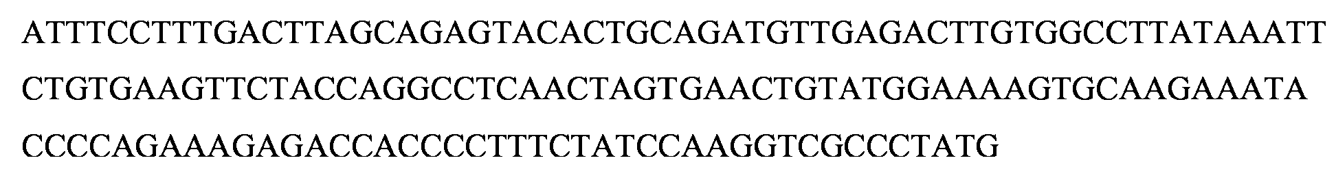
Tabla que muestra e listado de secuencias de las lineas celulares de inactivacion de GMD desarrolladas mediante transfeccion con el complejo CRISPR/Cas pD1401 (ARNg 167-207).Table showing the sequence listing of the GMD inactivation cell lines developed by transfection with the CRISPR / Cas pD1401 complex (RNAg 167-207).
Tabla 20Table 20


Tabla que muestra e listado de secuencias de las lineas celulares de inactivacion de GMD desarrolladas mediante transfeccion con el complejo CRISPR/Cas pD1301 (ARNg 404).Table showing the sequence listing of the GMD inactivation cell lines developed by transfection with the CRISPR / Cas pD1301 complex (RNAg 404).
Tabla 21Table 21
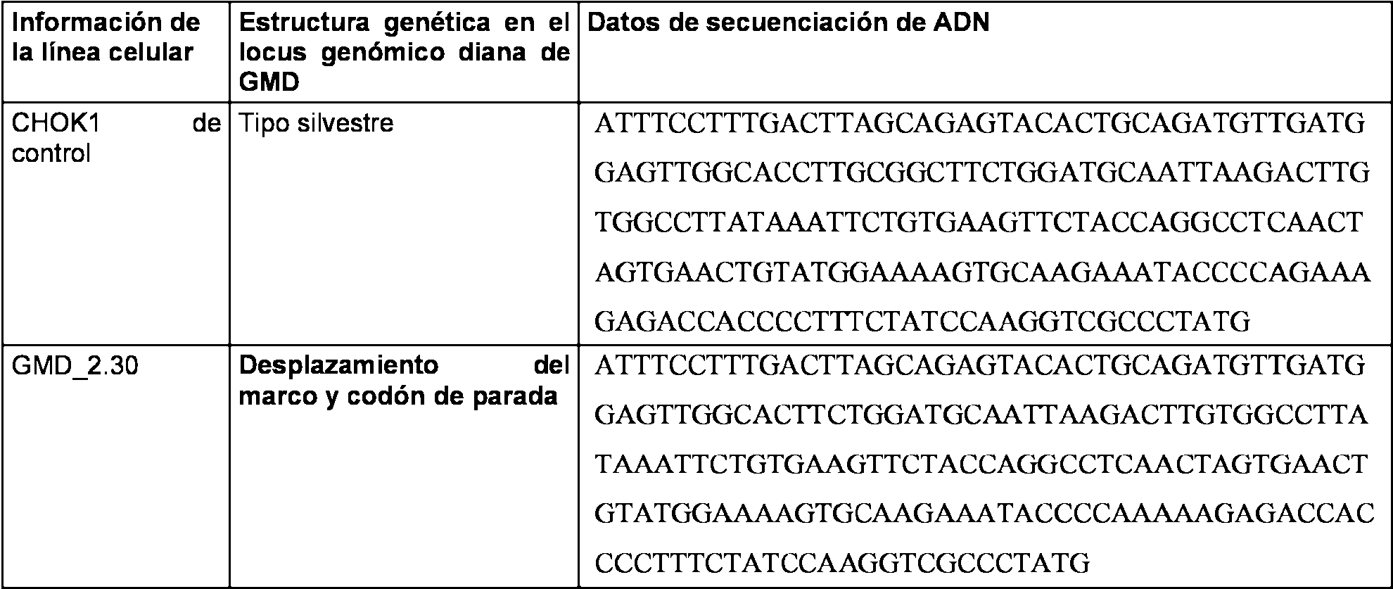
Tabla que muestra e listado de secuencias de las lineas celulares de inactivacion de GMD desarrolladas mediante transfeccion con el complejo CRISPR/Cas pD1401 (ARNg 167-207) y pD1301 (ARNg 404).Table showing the sequence listing of the GMD inactivation cell lines developed by transfection with the CRISPR / Cas complex pD1401 (RNAg 167-207) and pD1301 (RNAg 404).
Tabla 22Table 22


En la figura 17H a17L de la presente divulgacion se proporciona la alineacion representativa de secuencia de ADN genomico en clones de la linea celular CHOK1 de inactivacion de GMD que muestran delecion en la secuencia del gen GMD. Las figuras 17H a 17L representan el analisis de secuencia de nucleotidos en el locus diana de GMD. El ADN genomico de los clones CHOK1 de inactivacion de GMD transfectados con CRISPR/Cas y las lineas celulares CHOK1 de control se usaron para amplificar por PCR los loci de GMD genomicos direccionados, tanto las secuencias del exon 3 como del exon 4. Los datos de la secuencia se recolectaron del analisis de 5-15 clones bacterianos independientes secuenciados con cebadores de secuenciacion directos e inversos. Los datos de la secuenciacion sugieren deleciones de longitudes variables en multiples clones en comparacion con la linea celular CHOK1 de control. La delecion de bases mas grande se observo en el clon GMD1.41 y la delecion mas pequena es de solo 8 bases en el clon GMD 2.30. Todas las deleciones se localizaron en el sitio diana de CRISPR/Cas.In FIG. 17H to 17L of the present disclosure the representative alignment of genomic DNA sequence is provided in clones of the GMD inactivation CHOK1 cell line showing deletion in the GMD gene sequence. Figures 17H to 17L depict nucleotide sequence analysis at the target GMD locus. The genomic DNA of the CHOK1 clones of GMD inactivation transfected with CRISPR / Cas and the control CHOK1 cell lines were used to amplify the targeted genomic GMD loci by PCR, both exon 3 and exon 4 sequences. The sequence was collected from the analysis of 5-15 independent bacterial clones sequenced with direct and inverse sequencing primers. Sequencing data suggest deletions of variable lengths in multiple clones as compared to control cell line CHOK1. The largest deletion of bases was observed in clone GMD1.41 and the smallest deletion is only 8 bases in clone GMD 2.30. All deletions were located at the CRISPR / Cas target site.
En la figura 17H, los sitios de union a ADN de CRISPR/Cas a la izquierda y a la derecha se indican mediante recuadros abiertos. En la figura 17K, el sitio de union a CRISPR/Cas se indica mediante un recuadro abierto. Las figuras 17I a 17J indican clones en donde los datos de secuencia revelaron insercion de nueva secuencia de ADN en comparacion con la linea celular CHOK1 de control. La figura 17K releva una pequena delecion en el sitio diana del exon 4 de GMD con insercion de CRISPR/Cas pD1301 (ARNg 404) que es el resultado de una rotura de ADN de cadena doble. La figura 17L representa los datos de secuencia de la linea celular CHOK1 de inactivacion de GMD transfectada con dos construcciones de CRISPR/Cas, pD1401 (ARNg 167-207) y pD1301 (ARNg 404) en donde solo la secuencia del exon 4 de GMD se modifica con una delecion de 36 pares de bases.In Figure 17H, the CRISPR / Cas DNA binding sites on the left and on the right are indicated by open boxes. In Figure 17K, the CRISPR / Cas binding site is indicated by an open box. Figures 17I to 17J indicate clones in which the sequence data revealed insertion of new DNA sequence as compared to the control CHOK1 cell line. Figure 17K reveals a small deletion at the target site of GMD exon 4 with insertion of CRISPR / Cas pD1301 (RNAg 404) which is the result of a double-stranded DNA break. Figure 17L depicts the sequence data of the CHOK1 cell line of GMD inactivation transfected with two CRISPR / Cas constructs, pD1401 (167-7g RNAg) and pD1301 (404gRNA) where only the GMD exon 4 sequence is modify with a deletion of 36 base pairs.
Los datos sugieren diversos INDEL presentes en el locus genomico GMD en las lineas celulares CHOK1 de inactivacion de GMD. En muchos casos, se observo que hay modificaciones muy especificas en las bases direccionadas, y en otros casos, los cambios son amplios e implican tramos mas largos de ADN. Tal diversidad de modificacion genomica a lo largo del complejo CRISPR/Cas es posible debido a las roturas de cadena simple de ADN y roturas de cadena doble de ADN y la posterior reparacion de ADN. Todas estas lineas celulares se seleccionaron a traves de un ensayo de cribado funcional, a saber, un ensayo de citometria de flujo con LCA-FITC. Los resultados tambien implican una alta eficacia de los ensayos funcionales para aislar e identificar la linea celular CHOK1 de inactivacion de GMD.The data suggest various INDELs present in the GMD genomic locus in the CHOK1 cell lines of GMD inactivation. In many cases, it was observed that there are very specific modifications in the addressed bases, and in other cases, the changes are broad and involve longer stretches of DNA. Such diversity of genomic modification along the CRISPR / Cas complex is possible due to single strand breaks of DNA and DNA double strand breaks and subsequent DNA repair. All these cell lines were selected through a functional screening assay, namely, a flow cytometry assay with LCA-FITC. The results also imply a high efficacy of the functional assays to isolate and identify the CHOK1 cell line of GMD inactivation.
Tambien se revela que el diseno del complejo CRISPR/Cas representado en la presente divulgacion es unico, ya que este par de complejos CRISPR/Cas con la endonucleasa Cas9n proporciona una alteracion de secuencia genica altamente especifica en el locus direccionado de GMD de las lineas celulares CHOK1.It is also revealed that the design of the CRISPR / Cas complex represented in the present disclosure is unique, since this pair of CRISPR / Cas complexes with the Cas9n endonuclease provides a highly specific genetic sequence alteration at the GMD targeted locus of the cell lines CHOK1
Analisis de secuencia de aminoacidos de las lineas celulares CHOK1 de inactivacion de FUT8Analysis of amino acid sequence of CHOK1 cell lines of inactivation of FUT8
Las secuencias de ADN genomico de FUT8 de las lineas celulares CHOK1 de control y CJOK1 de inactivacion de FUT8 se analizaron posteriormente para comprender el impacto de la INDEL de la secuencia de ADN sobre el estado de la proteina FUT8. Las secuencias de ADN en el locus de FUT8 se traducen a secuencias de aminoacidos que usan el sesgo de codon de vertebrados. Se estudio de cerca la secuencia de aminoacidos de la region del exon 7 y los resultados se resumen en la tabla 23. Cuando se comparo con la linea celular CHOK1 de control, las lineas celulares de inactivacion de FUT8 revelaron modificaciones que implican deleciones e insercion de aminoacidos asi como introduction de codones de parada y mutaciones de desplazamiento del marco. Se observo que se obtenian deleciones de 10 aminoacidos o de tramos mas largos de secuencias de aminoacidos en comparacion con la secuencia de la proteina FUT8 de CHOK1.The genomic DNA sequences of FUT8 of the control CHOK1 and CUT1 inhibitor cell lines of FUT8 were subsequently analyzed to understand the impact of the INDEL on the DNA sequence on the status of the FUT8 protein. The DNA sequences at the FUT8 locus are translated into amino acid sequences that use vertebrate codon bias. The amino acid sequence of the exon 7 region was studied closely and the results are summarized in Table 23. When compared to the CHOK1 control cell line, the FUT8 inactivation cell lines revealed modifications involving deletions and insertion of amino acids as well as introduction of stop codons and frameshift mutations. It was observed that deletions of 10 amino acids or longer stretches of amino acid sequences were obtained in comparison with the sequence of the FUT8 protein of CHOK1.
En muchos casos, se observaron mutaciones de desplazamiento del marco, que alteran la region de C-terminal de la proteina FUT8 para hacer no funcional a la enzima. Ademas, en varios casos, se introdujo un codon de parada como un efecto de la mutation de desplazamiento del marco y, por lo tanto, la proteina FUT8 era truncada y no funcional en estos clones. In many cases, frameshift mutations were observed, which alter the C-terminal region of the FUT8 protein to render the enzyme non-functional. In addition, in several cases, a stop codon was introduced as an effect of the frame shift mutation and, therefore, the FUT8 protein was truncated and non-functional in these clones.
T l 2T l 2

Asimismo, se observo que la seleccion de los aminoacidos diana en la secuencia de la protema FUT8 es altamente eficaz. El direccionamiento de los aminoacidos conservados en las posiciones de la proteina FUT8 de tipo silvestre con solo un par de complejos CRISPR/Cas ha creado mutaciones en el locus dirigido en multiples lineas celulares de inactivacion.Likewise, it was observed that the selection of the target amino acids in the sequence of the FUT8 protein is highly effective. The targeting of conserved amino acids at the positions of the wild-type FUT8 protein with only a pair of CRISPR / Cas complexes has created mutations in the targeted locus in multiple inactivation cell lines.
La alineacion de secuencias de aminoacidos representativas en las lineas celulares CHOK1 de control y CHOK1 transfectada con CRISPR/Cas que presentan la delecion en la secuencia genica de FUT8 se proporcionan en la Figura 18A y 18B de la presente divulgacion. La secuencia de aminoacidos traducida se predijo usando el patron de codones. Los datos indicaron diversos efectos sobre la secuencia de aminoacidos de FUT8 debido a la delecion y/o insercion de nucleotidos observada. Los clones CR1KOT1 n.° 018, CR1KOT1 n.° 044, CR1KOT1 n.° 061, CR1KOT1 n.° 055, CR1KOT1 n.° 067 y CR1KOT1 n.° 051 revelaron la delecion dirigida de posiciones especificas de aminoacidos. En los numeros de clones CR1KOT1 n.° 052, CR1KOT1 n.° 022, CR1KOT1 n.° 036, CR1KOT1 n.° 059, CR1KOT1 n.° 023 y CR1KOT1 n.° 037, se observa la adicion de aminoacidos en la region dirigida (figura 18B). Algunos de los clones revelaron delecion seguida por mutaciones de desplazamiento de marco en codones de parada temprana. Todas estas modificaciones indicaron proteina FUT8 no funcional en las lineas celulares CHOK1 de inactivacion de FUT8 transfectadas pro CRISPR/Cas.Alignment of representative amino acid sequences in the control CHOK1 and CHOK1 cell lines transfected with CRISPR / Cas having the deletion in the FUT8 gene sequence are provided in Figure 18A and 18B of the present disclosure. The translated amino acid sequence was predicted using the codon pattern. The data indicated various effects on the amino acid sequence of FUT8 due to the deletion and / or insertion of nucleotides observed. Clones CR1KOT1 # 018, CR1KOT1 # 044, CR1KOT1 # 061, CR1KOT1 # 055, CR1KOT1 # 067 and CR1KOT1 # 051 revealed the targeted deletion of specific amino acid positions. In the numbers of clones CR1KOT1 No. 052, CR1KOT1 No. 022, CR1KOT1 No. 036, CR1KOT1 No. 059, CR1KOT1 No. 023 and CR1KOT1 No. 037, the addition of amino acids in the region is observed directed (figure 18B). Some of the clones revealed deletion followed by frame shift mutations in early stop codons. All these modifications indicated non-functional FUT8 protein in the CHOK1 cell lines of inactivation of transfected FUT8 pro CRISPR / Cas.
Ademas, el complejo CRISPR/Cas crea mutaciones de desplazamiento del marco seguidas por codones de parada que alteran la region de C-terminal de la enzima FUT8 que contiene un motivo importante II y un motivo III en el plegamiento de Rossmann. Las posiciones de los aminoacidos especificos Tyr-382, Asp-409, Asp-410, Asp-453 y Ser-469 que estan implicados en el dominio catalitico de la enzima FUT8, por lo tanto, no se expresan en estas versiones truncadas del gen FUT8. El resultado final de estas mutaciones criticas es una enzima a-1,6 fucosiltransferasa no funcional, el producto de proteina del gen FUT8 en las lineas celulares CHOK1 de inactivacion de FUT8.In addition, the CRISPR / Cas complex creates frame shift mutations followed by stop codons that alter the C-terminal region of the FUT8 enzyme that contains an important motif II and a motif III in the Rossmann fold. The positions of the specific amino acids Tyr-382, Asp-409, Asp-410, Asp-453 and Ser-469 that are involved in the catalytic domain of the enzyme FUT8, therefore, are not expressed in these truncated versions of the gene FUT8. The final result of these critical mutations is a non-functional a-1,6 fucosyltransferase enzyme, the protein product of the FUT8 gene in the CHOK1 cell lines of inactivation of FUT8.
Analisis del locus del exon 3 del gen GMD en lineas celulares clonales transfectadas con el complejo CRISPR/Cas pD1401 (ARNg 167-207).Analysis of the exon 3 locus of the GMD gene in clonal cell lines transfected with the CRISPR / Cas pD1401 complex (RNAg 167-207).
Las lineas clonales de inactivacion revelaron diferentes tipos de mutaciones en la secuencia de proteina del gen GMD en la region diana. La tabla de a continuacion enumera todas las mutaciones observadas en las lineas celulares clonales de inactivacion de GMD.The cloning lines of inactivation revealed different types of mutations in the protein sequence of the GMD gene in the target region. The table below lists all the mutations observed in the clonal cell lines of GMD inactivation.
Tabla 24Table 24

Analisis del locus del exon 4 del gen GMD en lineas celulares clonales transfectadas con el complejo CRISPR/Cas pD1301 (ARNg 404):Analysis of the locus of exon 4 of the GMD gene in clonal cell lines transfected with the CRISPR / Cas pD1301 complex (RNAg 404):
Las lineas celulares clonales de inactivacion revelaron mutaciones en la secuencia de proteina del gen GMD en la region diana. La tabla de a continuacion enumera todas las mutaciones observadas en la linea celular clonal de inactivacion de GMD.The clonal inactivation cell lines revealed mutations in the protein sequence of the GMD gene in the target region. The table below lists all the mutations observed in the clonal cell line of inactivation of GMD.
Tabla 25Table 25

Analisis de los loci del exon 3 y el exon 4 del gen GMD en la linea celular clonal transfectada con el complejo CRISPR/Cas pD1401 (ARNg 167-207) y pD1301 (ARNg 404):Analysis of the loci of exon 3 and exon 4 of the GMD gene in the clonal cell line transfected with the CRISPR / Cas complex pD1401 (RNAg 167-207) and pD1301 (RNAg 404):
La lmea celular clonal de inactivacion revelo mutaciones solo en la secuencia de proteina del exon 4 del gen GMD entre las regiones diana. La tabla de a continuacion enumera todas las mutaciones observadas en la linea celular clonal de inactivacion de GMD.The clonal inactivation cell line revealed mutations only in the protein sequence of exon 4 of the GMD gene between the target regions. The table below lists all the mutations observed in the clonal cell line of GMD inactivation.
Tabla 26Table 26

La Figura 18C representa el analisis de aminoacidos de los clones CHOK1 de inactivacion de GMD generados mediante el uso de pD1401 (ARNg 167-207). Se observan multiples tipos de mutaciones en el locus diana del exon 3 de GMD que incluyen la delecion de restos de aminoacidos, la sustitucion asi como codones de parada prematura. Tales modificaciones producen el gen GMD no funcional y, por lo tanto, dan como resultado lineas celulares de inactivacion de fucosa. Los clones numero GMD 1.12 y GMD 1.37 revelaron la insercion de restos de aminoacidos y mutaciones de desplazamiento del marco que introdujeron codones de parada prematura (figura 18D y 18E). En el caso del clon GMD 2.30, cuando se uso pD1301 (ARNg 404) para la introduccion de rotura de cadena doble de ADN se revelo la insercion y mutaciones de desplazamiento del marco en el locus diana del exon 4 de GMD (figura 18 F). De manera similar, en el clon GMD 3.51 (figura 18G) los datos revelaron delecion de aminoacidos solo en el exon 4 de GMD aunque la linea celular se genero mediante transfeccion con las construcciones CRISPR/Cas pD1401 (ARNg 167-207) y pD1301 (ARNg 404).Figure 18C depicts amino acid analysis of the CHOK1 clones of GMD inactivation generated by the use of pD1401 (167-7g gRNA). Multiple types of mutations are observed in the target locus of GMD exon 3 that include deletion of amino acid residues, substitution as well as codons of premature arrest. Such modifications produce the non-functional GMD gene and, therefore, result in cell lines of fucose inactivation. Clones number GMD 1.12 and GMD 1.37 revealed the insertion of amino acid residues and frameshift mutations that introduced premature stop codons (Figure 18D and 18E). In the case of GMD clone 2.30, when pD1301 (404g RNA) was used for the introduction of DNA double-strand breaks, the insertion and frameshift mutations were revealed at the target locus of GMD exon 4 (Figure 18F) . Similarly, in the GMD clone 3.51 (Figure 18G) the data revealed deletion of amino acids only in exon 4 of GMD although the cell line was generated by transfection with the CRISPR / Cas constructions pD1401 (RNAg 167-207) and pD1301 ( RNAg 404).
Estos datos revelan que el diseno de CRISPR/Cas hecho para direccionar dos sitios diana del exon especificos del gen GMD es muy especifico y ambas construcciones son eficaces en el direccionamiento especifico. La linea celular CHOK1 de inactivacion de GMD desarrollada de este modo se uso para el desarrollo de anticuerpos monoclonales no fucosilados.These data reveal that the design of CRISPR / Cas made to address two specific exon target sites of the GMD gene is very specific and both constructions are effective in specific targeting. The CHOK1 cell line of GMD inactivation developed in this way was used for the development of non-fucosylated monoclonal antibodies.
Ejemplo 7 - USO DE LiNEA CELULAR DE INACTIVACION DE FUCOSA PARA PRODUCIR ANTICUERPOS PARCIALMENTE FUCOSILADOS Y NO FUCOSILADOS Example 7 - USE OF CELLULAR LINE OF FUCKING INACTIVATION TO PRODUCE PARTIALLY FUCOSILATED AND NON-FUCOSILATED ANTIBODIES
El soporte de expresion de celulas CHOK1 de inactivacion de fucosa se uso para la expresion de anticuerpos no fucosilados, en particular, anticuerpos monoclonales parcialmente no fucosilados. Los genes de anticuerpos que codifican la cadena pesada y la cadena ligera de los anticuerpos monoclonales se clonaron en los plasmidos adecuados de expresion de genes y se transfectaron en el soporte celular CHOK1 de inactivacion de fucosa descrito en los ejemplos anteriores. El anticuerpo monoclonal producido usando este soporte/metodo se expreso como un anticuerpo no fucosilado. El producto se purifico siguiendo los protocolos y las directrices establecidas para desarrollar un producto de anticuerpo monoclonal biomejorado para uso terapeutico. El anticuerpo no fucosilado biomejorado producido usando este soporte da como resultado un nivel mayor de ADCC y, por lo tanto, mejores resultados terapeuticos.The cell expression support CHOK1 of fucose inactivation was used for the expression of non-fucosylated antibodies, in particular, partially non-fucosylated monoclonal antibodies. The antibody genes encoding the heavy chain and the light chain of the monoclonal antibodies were cloned into the appropriate gene expression plasmids and transfected into the cell support CHOK1 of fucose inactivation described in the previous examples. The monoclonal antibody produced using this support / method was expressed as a non-fucosylated antibody. The product was purified following the protocols and guidelines established to develop a biomaterial monoclonal antibody product for therapeutic use. The bi-enhanced non-fucosylated antibody produced using this support results in a higher level of ADCC and, therefore, better therapeutic results.
Los datos de citometria de flujo con LCA-FITC y los posteriores experimentos de secuenciacion de la presente divulgacion confirman que las lineas FKO son incapaces de fucosilar proteinas de membrana. Por tanto, la celula obtenida en la presente divulgacion produce proteinas no fucosiladas, especificamente anticuerpos o fucosilados. Los aspectos caracteristicos y las ventajas terapeuticas de los anticuerpos no fucosilados, tales como mayor ADCC, con conocidas para un experto en la materia.The flow cytometry data with LCA-FITC and the subsequent sequencing experiments of the present disclosure confirm that the FKO lines are incapable of fucosylating membrane proteins. Therefore, the cell obtained in the present disclosure produces non-fucosylated proteins, specifically antibodies or fucosylated. Characteristic aspects and therapeutic benefits of non-fucosylated antibodies, such as increased ADCC, are known to one skilled in the art.
Las lineas celulares CHOK1 de inactivacion de GMD son utiles en aplicaciones unicas en programas de desarrollo de anticuerpos monoclonales no fucosilados. El gen GMD esta aguas arriba e la etapa critica de GDP-Fucosa en la via de la biosintesis de fucosa. Se puede producir GDP-fucosa en celulas CHOK1 bien por la via de novo que depende por completo de la funcion del gen GMD o a traves de la via de rescate que es independiente de la funcion del gen GMD pero requiere la presencia de L-Fucosa en medio de cultivo. Por lo tanto, es posible lograr una regulacion condicional de la fucosilacion de anticuerpos monoclonales producidos en las lineas celulares de inactivacion de GMD.The CHOK1 cell lines of GMD inactivation are useful in unique applications in non-fucosylated monoclonal antibody development programs. The GMD gene is upstream of the critical phase of GDP-Fucose in the pathway of fucose biosynthesis. GDP-fucose can be produced in CHOK1 cells either via the de novo pathway that depends entirely on the function of the GMD gene or through the rescue pathway that is independent of the function of the GMD gene but requires the presence of L-Fucose in culture medium Therefore, it is possible to achieve a conditional regulation of the fucosylation of monoclonal antibodies produced in the cell lines of GMD inactivation.
Supuesto 1: Expresion genica de anticuerpos monoclonales en celulas CHOK1 de inactivacion de GMD sin L-Fucosa en el medio de cultivo. El anticuerpo monoclonal producido esta afucosilado al 100 %. En este caso, tanto la via de la biosintesis de fucosa de novo como la de rescate son no funcionales.Assumption 1: Genetic expression of monoclonal antibodies in CHOK1 cells of GMD inactivation without L-Fucose in the culture medium. The monoclonal antibody produced is 100% afucosylated. In this case, both the pathway of de novo fucose biosynthesis and the rescue route are non-functional.
Supuesto 2: Expresion genica de anticuerpos monoclonales en celulas CHOK1 de inactivacion de GMD con L-Fucosa optima en el medio de cultivo. El anticuerpo monoclonal producido esta fucosilado al 100 %. En este caso, la via de novo esta completamente bloqueada pero la via de rescate es funcional. Esto permite la fucosilacion completa del gen del anticuerpo monoclonal producido en la linea celular CHOK1 de inactivacion de GMDAssumption 2: Genetic expression of monoclonal antibodies in CHOK1 cells of inactivation of GMD with optimal L-Fucose in the culture medium. The monoclonal antibody produced is 100% fucosylated. In this case, the de novo way is completely blocked but the rescue way is functional. This allows the complete fucosylation of the monoclonal antibody gene produced in the CHOK1 cell line of GMD inactivation
Supuesto 3: Expresion genica de anticuerpos monoclonales en celulas CHOK1 de inactivacion de GMD con diversos niveles de L-Fucosa en el medio de cultivo. El anticuerpo monoclonal producido en esta condicion esta parcialmente fucosilado. La dosificacion de L-Fucosa en el medio de crecimiento determina el nivel de fucosilacion del anticuerpo monoclonal. Esta dosificacion se titula durante la condicion del cultivo para asegurar el nivel de fucosilacion de anticuerpo monoclonal y, a continuacion, se titula de nuevo para afinar para lograr niveles criticos de fucosilacion de anticuerpos monoclonales diana.Assumption 3: Genetic expression of monoclonal antibodies in CHOK1 cells of inactivation of GMD with various levels of L-Fucose in the culture medium. The monoclonal antibody produced in this condition is partially fucosylated. The dosage of L-Fucose in the growth medium determines the level of fucosylation of the monoclonal antibody. This dosage is titrated during the culture condition to ensure the level of fucosylation of monoclonal antibody and then titrated again to fine tune to achieve critical levels of fucosylation of target monoclonal antibodies.
Esto es una ventaja unica con la linea celular CHOK1 de inactivacion del gen GMD y esta caracteristica se describe unicamente en la presente divulgacion.This is a unique advantage with the CHOK1 cell line of inactivation of the GMD gene and this feature is described only in the present disclosure.
Aunque la divulgacion y la ejemplificacion se han proporcionado a modo de ilustraciones y ejemplos con el proposito de claridad y comprension, es evidente para un experto en la materia que pueden practicarse diversos cambios y modificaciones sin apartarse del alcance de la divulgacion. Por consiguiente, las descripciones y ejemplos anteriores no deben interpretarse como limitantes del alcance de la presente divulgacion.Although the disclosure and the exemplification have been provided by way of illustrations and examples for the purpose of clarity and understanding, it is evident to one skilled in the art that various changes and modifications may be practiced without departing from the scope of the disclosure. Accordingly, the foregoing descriptions and examples should not be construed as limiting the scope of the present disclosure.
Se pretende que el alcance de la divulgacion no este limitado por la presente descripcion detallada, sino mas bien por las reivindicaciones adjuntas. Tambien debe entenderse que las siguientes reivindicaciones pretenden cubrir todas las caracteristicas genericas y especificas de la divulgacion descrita en este documento.It is intended that the scope of the disclosure is not limited by the present detailed description, but rather by the appended claims. It should also be understood that the following claims are intended to cover all the generic and specific features of the disclosure described in this document.
Son posibles muchas modificaciones y variaciones a la luz de las ensenanzas anteriores. Por lo tanto, debe entenderse que la materia reivindicada puede practicarse de otra manera que la descrita especificamente. Cualquier referencia a elementos reivindicados en singular, por ejemplo, usando los articulos "uno", "una", "el", "la", o "dicho" no debe interpretarse como limitante de la divulgacion.Many modifications and variations are possible in light of the above teachings. Therefore, it should be understood that the claimed material may be practiced other than that specifically described. Any reference to elements claimed in the singular, for example, using the articles "a", "an", "the", "the", or "said" should not be construed as limiting the disclosure.
A lo largo de la presente divulgacion, la palabra "comprender" o variaciones tales como "comprende" o "que comprende", donde se use, debe entenderse que implican la inclusion de un elemento indicado, un numero entero o etapa, o grupo de elementos, numeros enteros o etapas, pero no la exclusion de cualquier otro elemento, un numero entero o etapa, o grupo de elementos, numeros enteros o etapas.Throughout the present disclosure, the word "comprise" or variations such as "comprises" or "comprising", where used, should be understood to imply the inclusion of an indicated element, an integer or stage, or group of elements, whole numbers or stages, but not the exclusion of any other element, a whole number or stage, or group of elements, whole numbers or stages.
Cuando se indique un limite numerico o un intervalo en el presente documento, los puntos extremos se incluyen. Ademas, los valores y subintervalos dentro de un limite o intervalo numerico se incluyen especificamente como si se escribieran explicitamente. When a numerical limit or interval is indicated in the present document, endpoints are included. In addition, values and subintervals within a numerical range or range are specifically included as if they were written explicitly.
Claims (13)
Applications Claiming Priority (2)
| Application Number | Priority Date | Filing Date | Title |
|---|---|---|---|
| IN5767CH2014 | 2014-11-15 | ||
| PCT/IB2015/058777 WO2016075662A2 (en) | 2014-11-15 | 2015-11-13 | Dna-binding domain, non-fucosylated and partially fucosylated proteins, and methods thereof |
Publications (1)
| Publication Number | Publication Date |
|---|---|
| ES2712303T3 true ES2712303T3 (en) | 2019-05-10 |
Family
ID=55024178
Family Applications (1)
| Application Number | Title | Priority Date | Filing Date |
|---|---|---|---|
| ES15816521T Active ES2712303T3 (en) | 2014-11-15 | 2015-11-13 | DNA binding domain of the CRISPR system for the production of non-fucosylated and partially fucosylated proteins |
Country Status (6)
| Country | Link |
|---|---|
| US (2) | US10752674B2 (en) |
| EP (2) | EP3218490B1 (en) |
| JP (2) | JP7090421B2 (en) |
| DK (1) | DK3218490T3 (en) |
| ES (1) | ES2712303T3 (en) |
| WO (1) | WO2016075662A2 (en) |
Families Citing this family (30)
| Publication number | Priority date | Publication date | Assignee | Title |
|---|---|---|---|---|
| WO2013066438A2 (en) | 2011-07-22 | 2013-05-10 | President And Fellows Of Harvard College | Evaluation and improvement of nuclease cleavage specificity |
| US9163284B2 (en) | 2013-08-09 | 2015-10-20 | President And Fellows Of Harvard College | Methods for identifying a target site of a Cas9 nuclease |
| US9359599B2 (en) | 2013-08-22 | 2016-06-07 | President And Fellows Of Harvard College | Engineered transcription activator-like effector (TALE) domains and uses thereof |
| US9228207B2 (en) | 2013-09-06 | 2016-01-05 | President And Fellows Of Harvard College | Switchable gRNAs comprising aptamers |
| US9322037B2 (en) | 2013-09-06 | 2016-04-26 | President And Fellows Of Harvard College | Cas9-FokI fusion proteins and uses thereof |
| US9526784B2 (en) | 2013-09-06 | 2016-12-27 | President And Fellows Of Harvard College | Delivery system for functional nucleases |
| US20150165054A1 (en) | 2013-12-12 | 2015-06-18 | President And Fellows Of Harvard College | Methods for correcting caspase-9 point mutations |
| US10077453B2 (en) | 2014-07-30 | 2018-09-18 | President And Fellows Of Harvard College | CAS9 proteins including ligand-dependent inteins |
| WO2016075662A2 (en) | 2014-11-15 | 2016-05-19 | Zumutor Biologics, Inc. | Dna-binding domain, non-fucosylated and partially fucosylated proteins, and methods thereof |
| US11267899B2 (en) | 2015-05-13 | 2022-03-08 | Zumutor Biologics Inc. | Afucosylated protein, cell expressing said protein and associated methods |
| EP3365357B1 (en) | 2015-10-23 | 2024-02-14 | President and Fellows of Harvard College | Evolved cas9 proteins for gene editing |
| WO2017079165A1 (en) * | 2015-11-02 | 2017-05-11 | Genentech, Inc. | Methods of making fucosylated and afucosylated forms of a protein |
| AU2017306676B2 (en) | 2016-08-03 | 2024-02-22 | President And Fellows Of Harvard College | Adenosine nucleobase editors and uses thereof |
| AU2017308889B2 (en) | 2016-08-09 | 2023-11-09 | President And Fellows Of Harvard College | Programmable Cas9-recombinase fusion proteins and uses thereof |
| US11542509B2 (en) | 2016-08-24 | 2023-01-03 | President And Fellows Of Harvard College | Incorporation of unnatural amino acids into proteins using base editing |
| SG11201903089RA (en) | 2016-10-14 | 2019-05-30 | Harvard College | Aav delivery of nucleobase editors |
| WO2018119359A1 (en) | 2016-12-23 | 2018-06-28 | President And Fellows Of Harvard College | Editing of ccr5 receptor gene to protect against hiv infection |
| WO2018165504A1 (en) | 2017-03-09 | 2018-09-13 | President And Fellows Of Harvard College | Suppression of pain by gene editing |
| US11542496B2 (en) | 2017-03-10 | 2023-01-03 | President And Fellows Of Harvard College | Cytosine to guanine base editor |
| IL306092A (en) | 2017-03-23 | 2023-11-01 | Harvard College | Nucleobase editors comprising nucleic acid programmable dna binding proteins |
| WO2018209320A1 (en) | 2017-05-12 | 2018-11-15 | President And Fellows Of Harvard College | Aptazyme-embedded guide rnas for use with crispr-cas9 in genome editing and transcriptional activation |
| JP2020534795A (en) | 2017-07-28 | 2020-12-03 | プレジデント アンド フェローズ オブ ハーバード カレッジ | Methods and Compositions for Evolving Base Editing Factors Using Phage-Supported Continuous Evolution (PACE) |
| WO2019139645A2 (en) | 2017-08-30 | 2019-07-18 | President And Fellows Of Harvard College | High efficiency base editors comprising gam |
| US11795443B2 (en) | 2017-10-16 | 2023-10-24 | The Broad Institute, Inc. | Uses of adenosine base editors |
| KR102061251B1 (en) | 2017-10-31 | 2019-12-31 | 주식회사 에이치유비바이오텍 | Recombinant cell and method for production of endogenous polypeptide |
| EP3797160A1 (en) * | 2018-05-23 | 2021-03-31 | The Broad Institute Inc. | Base editors and uses thereof |
| JP2022526908A (en) | 2019-03-19 | 2022-05-27 | ザ ブロード インスティテュート,インコーポレーテッド | Editing Methods and Compositions for Editing Nucleotide Sequences |
| AU2021267940A1 (en) | 2020-05-08 | 2022-12-08 | President And Fellows Of Harvard College | Methods and compositions for simultaneous editing of both strands of a target double-stranded nucleotide sequence |
| CN113061605B (en) * | 2020-07-13 | 2022-11-29 | 中山大学 | Construction method and application of fucose transferase 8 (FUT 8) function-deficient cell strain |
| AR126089A1 (en) | 2021-06-07 | 2023-09-13 | Amgen Inc | USE OF FUCOSIDASE TO CONTROL THE LEVEL OF AFFUCOSYLATION OF GLUCOSYLATED PROTEINS |
Family Cites Families (9)
| Publication number | Priority date | Publication date | Assignee | Title |
|---|---|---|---|---|
| EP1443961B1 (en) * | 2001-10-25 | 2009-05-06 | Genentech, Inc. | Glycoprotein compositions |
| EP1705251A4 (en) | 2003-10-09 | 2009-10-28 | Kyowa Hakko Kirin Co Ltd | PROCESS FOR PRODUCING ANTIBODY COMPOSITION BY USING RNA INHIBITING THE FUNCTION OF a1,6-FUCOSYLTRANSFERASE |
| WO2005035563A1 (en) * | 2003-10-09 | 2005-04-21 | Kyowa Hakko Kogyo Co., Ltd. | Process for producing antithrombin iii composition |
| US20060223147A1 (en) | 2004-08-05 | 2006-10-05 | Kyowa Hakko Kogyo Co., Ltd., | Process for producing glycoprotein composition |
| ES2687771T3 (en) * | 2011-03-06 | 2018-10-29 | Merck Serono Sa | Cell lines with low level of fucose and its uses |
| WO2013013013A2 (en) * | 2011-07-21 | 2013-01-24 | Alnylam Pharmaceuticals, Inc. | Compositions and methods for producing modified glycoproteins |
| CN105392885B (en) * | 2013-07-19 | 2020-11-03 | 赖瑞克斯生物科技公司 | Methods and compositions for generating double allele knockouts |
| WO2015052231A2 (en) * | 2013-10-08 | 2015-04-16 | Technical University Of Denmark | Multiplex editing system |
| WO2016075662A2 (en) | 2014-11-15 | 2016-05-19 | Zumutor Biologics, Inc. | Dna-binding domain, non-fucosylated and partially fucosylated proteins, and methods thereof |
-
2015
- 2015-11-13 WO PCT/IB2015/058777 patent/WO2016075662A2/en active Application Filing
- 2015-11-13 US US15/526,971 patent/US10752674B2/en active Active
- 2015-11-13 EP EP15816521.7A patent/EP3218490B1/en active Active
- 2015-11-13 DK DK15816521.7T patent/DK3218490T3/en active
- 2015-11-13 EP EP18192111.5A patent/EP3467110A1/en active Pending
- 2015-11-13 JP JP2017545006A patent/JP7090421B2/en active Active
- 2015-11-13 ES ES15816521T patent/ES2712303T3/en active Active
-
2020
- 2020-07-06 US US16/921,491 patent/US11548937B2/en active Active
- 2020-09-28 JP JP2020162750A patent/JP7134207B2/en active Active
Also Published As
| Publication number | Publication date |
|---|---|
| JP2021007397A (en) | 2021-01-28 |
| EP3467110A1 (en) | 2019-04-10 |
| WO2016075662A3 (en) | 2016-07-07 |
| US11548937B2 (en) | 2023-01-10 |
| US20190112358A1 (en) | 2019-04-18 |
| DK3218490T3 (en) | 2019-02-18 |
| WO2016075662A2 (en) | 2016-05-19 |
| JP7090421B2 (en) | 2022-06-24 |
| JP2018502595A (en) | 2018-02-01 |
| EP3218490A2 (en) | 2017-09-20 |
| US20200339666A1 (en) | 2020-10-29 |
| EP3218490B1 (en) | 2018-10-31 |
| JP7134207B2 (en) | 2022-09-09 |
| US10752674B2 (en) | 2020-08-25 |
Similar Documents
| Publication | Publication Date | Title |
|---|---|---|
| ES2712303T3 (en) | DNA binding domain of the CRISPR system for the production of non-fucosylated and partially fucosylated proteins | |
| US11267899B2 (en) | Afucosylated protein, cell expressing said protein and associated methods | |
| EP3022304B1 (en) | Methods and compositions for producing double allele knock outs | |
| ES2738641T3 (en) | Production of therapeutic proteins in genetically modified mammalian cells | |
| US9670271B2 (en) | Production of recombinant proteins with simple glycoforms | |
| JP2017506903A (en) | Virus resistant cells and uses thereof | |
| JP6790057B2 (en) | Cells producing glycoproteins with modified N- and O-glycosylation patterns and methods and uses thereof | |
| ES2901128T3 (en) | Non-fucosylated protein and methods thereof | |
| US11060083B2 (en) | Methods and compositions for producing double allele knock outs | |
| Amiri et al. | CRISPR‐interceded CHO cell line development approaches | |
| ES2659118T3 (en) | N-glycosylation in transformed Phaeodactylum tricornutum | |
| JP2020202832A (en) | Non-fucosylated protein and methods |