EP4582040A2 - Systeme und verfahren zur navigation in bildgeführten medizinischen verfahren - Google Patents
Systeme und verfahren zur navigation in bildgeführten medizinischen verfahren Download PDFInfo
- Publication number
- EP4582040A2 EP4582040A2 EP25178554.9A EP25178554A EP4582040A2 EP 4582040 A2 EP4582040 A2 EP 4582040A2 EP 25178554 A EP25178554 A EP 25178554A EP 4582040 A2 EP4582040 A2 EP 4582040A2
- Authority
- EP
- European Patent Office
- Prior art keywords
- model
- image data
- operator
- anatomical
- dimensional image
- Prior art date
- Legal status (The legal status is an assumption and is not a legal conclusion. Google has not performed a legal analysis and makes no representation as to the accuracy of the status listed.)
- Pending
Links
Images


Classifications
-
- A—HUMAN NECESSITIES
- A61—MEDICAL OR VETERINARY SCIENCE; HYGIENE
- A61B—DIAGNOSIS; SURGERY; IDENTIFICATION
- A61B8/00—Diagnosis using ultrasonic, sonic or infrasonic waves
- A61B8/08—Clinical applications
- A61B8/0833—Clinical applications involving detecting or locating foreign bodies or organic structures
- A61B8/0841—Clinical applications involving detecting or locating foreign bodies or organic structures for locating instruments
-
- A—HUMAN NECESSITIES
- A61—MEDICAL OR VETERINARY SCIENCE; HYGIENE
- A61B—DIAGNOSIS; SURGERY; IDENTIFICATION
- A61B17/00—Surgical instruments, devices or methods
- A61B17/00234—Surgical instruments, devices or methods for minimally invasive surgery
-
- A—HUMAN NECESSITIES
- A61—MEDICAL OR VETERINARY SCIENCE; HYGIENE
- A61B—DIAGNOSIS; SURGERY; IDENTIFICATION
- A61B34/00—Computer-aided surgery; Manipulators or robots specially adapted for use in surgery
- A61B34/10—Computer-aided planning, simulation or modelling of surgical operations
-
- A—HUMAN NECESSITIES
- A61—MEDICAL OR VETERINARY SCIENCE; HYGIENE
- A61B—DIAGNOSIS; SURGERY; IDENTIFICATION
- A61B34/00—Computer-aided surgery; Manipulators or robots specially adapted for use in surgery
- A61B34/20—Surgical navigation systems; Devices for tracking or guiding surgical instruments, e.g. for frameless stereotaxis
-
- A—HUMAN NECESSITIES
- A61—MEDICAL OR VETERINARY SCIENCE; HYGIENE
- A61B—DIAGNOSIS; SURGERY; IDENTIFICATION
- A61B34/00—Computer-aided surgery; Manipulators or robots specially adapted for use in surgery
- A61B34/25—User interfaces for surgical systems
-
- A—HUMAN NECESSITIES
- A61—MEDICAL OR VETERINARY SCIENCE; HYGIENE
- A61B—DIAGNOSIS; SURGERY; IDENTIFICATION
- A61B34/00—Computer-aided surgery; Manipulators or robots specially adapted for use in surgery
- A61B34/30—Surgical robots
- A61B34/35—Surgical robots for telesurgery
-
- A—HUMAN NECESSITIES
- A61—MEDICAL OR VETERINARY SCIENCE; HYGIENE
- A61B—DIAGNOSIS; SURGERY; IDENTIFICATION
- A61B6/00—Apparatus or devices for radiation diagnosis; Apparatus or devices for radiation diagnosis combined with radiation therapy equipment
- A61B6/12—Arrangements for detecting or locating foreign bodies
-
- A—HUMAN NECESSITIES
- A61—MEDICAL OR VETERINARY SCIENCE; HYGIENE
- A61B—DIAGNOSIS; SURGERY; IDENTIFICATION
- A61B90/00—Instruments, implements or accessories specially adapted for surgery or diagnosis and not covered by any of the groups A61B1/00 - A61B50/00, e.g. for luxation treatment or for protecting wound edges
- A61B90/08—Accessories or related features not otherwise provided for
-
- A—HUMAN NECESSITIES
- A61—MEDICAL OR VETERINARY SCIENCE; HYGIENE
- A61B—DIAGNOSIS; SURGERY; IDENTIFICATION
- A61B90/00—Instruments, implements or accessories specially adapted for surgery or diagnosis and not covered by any of the groups A61B1/00 - A61B50/00, e.g. for luxation treatment or for protecting wound edges
- A61B90/36—Image-producing devices or illumination devices not otherwise provided for
- A61B90/37—Surgical systems with images on a monitor during operation
-
- G—PHYSICS
- G06—COMPUTING OR CALCULATING; COUNTING
- G06T—IMAGE DATA PROCESSING OR GENERATION, IN GENERAL
- G06T15/00—3D [Three Dimensional] image rendering
-
- A—HUMAN NECESSITIES
- A61—MEDICAL OR VETERINARY SCIENCE; HYGIENE
- A61B—DIAGNOSIS; SURGERY; IDENTIFICATION
- A61B17/00—Surgical instruments, devices or methods
- A61B17/00234—Surgical instruments, devices or methods for minimally invasive surgery
- A61B2017/00292—Surgical instruments, devices or methods for minimally invasive surgery mounted on or guided by flexible, e.g. catheter-like, means
- A61B2017/00296—Surgical instruments, devices or methods for minimally invasive surgery mounted on or guided by flexible, e.g. catheter-like, means mounted on an endoscope
-
- A—HUMAN NECESSITIES
- A61—MEDICAL OR VETERINARY SCIENCE; HYGIENE
- A61B—DIAGNOSIS; SURGERY; IDENTIFICATION
- A61B34/00—Computer-aided surgery; Manipulators or robots specially adapted for use in surgery
- A61B34/10—Computer-aided planning, simulation or modelling of surgical operations
- A61B2034/101—Computer-aided simulation of surgical operations
- A61B2034/105—Modelling of the patient, e.g. for ligaments or bones
-
- A—HUMAN NECESSITIES
- A61—MEDICAL OR VETERINARY SCIENCE; HYGIENE
- A61B—DIAGNOSIS; SURGERY; IDENTIFICATION
- A61B34/00—Computer-aided surgery; Manipulators or robots specially adapted for use in surgery
- A61B34/10—Computer-aided planning, simulation or modelling of surgical operations
- A61B2034/107—Visualisation of planned trajectories or target regions
-
- A—HUMAN NECESSITIES
- A61—MEDICAL OR VETERINARY SCIENCE; HYGIENE
- A61B—DIAGNOSIS; SURGERY; IDENTIFICATION
- A61B34/00—Computer-aided surgery; Manipulators or robots specially adapted for use in surgery
- A61B34/20—Surgical navigation systems; Devices for tracking or guiding surgical instruments, e.g. for frameless stereotaxis
- A61B2034/2046—Tracking techniques
- A61B2034/2051—Electromagnetic tracking systems
-
- A—HUMAN NECESSITIES
- A61—MEDICAL OR VETERINARY SCIENCE; HYGIENE
- A61B—DIAGNOSIS; SURGERY; IDENTIFICATION
- A61B34/00—Computer-aided surgery; Manipulators or robots specially adapted for use in surgery
- A61B34/20—Surgical navigation systems; Devices for tracking or guiding surgical instruments, e.g. for frameless stereotaxis
- A61B2034/2046—Tracking techniques
- A61B2034/2055—Optical tracking systems
- A61B2034/2057—Details of tracking cameras
-
- A—HUMAN NECESSITIES
- A61—MEDICAL OR VETERINARY SCIENCE; HYGIENE
- A61B—DIAGNOSIS; SURGERY; IDENTIFICATION
- A61B34/00—Computer-aided surgery; Manipulators or robots specially adapted for use in surgery
- A61B34/20—Surgical navigation systems; Devices for tracking or guiding surgical instruments, e.g. for frameless stereotaxis
- A61B2034/2046—Tracking techniques
- A61B2034/2061—Tracking techniques using shape-sensors, e.g. fiber shape sensors with Bragg gratings
-
- A—HUMAN NECESSITIES
- A61—MEDICAL OR VETERINARY SCIENCE; HYGIENE
- A61B—DIAGNOSIS; SURGERY; IDENTIFICATION
- A61B34/00—Computer-aided surgery; Manipulators or robots specially adapted for use in surgery
- A61B34/20—Surgical navigation systems; Devices for tracking or guiding surgical instruments, e.g. for frameless stereotaxis
- A61B2034/2046—Tracking techniques
- A61B2034/2065—Tracking using image or pattern recognition
-
- A—HUMAN NECESSITIES
- A61—MEDICAL OR VETERINARY SCIENCE; HYGIENE
- A61B—DIAGNOSIS; SURGERY; IDENTIFICATION
- A61B34/00—Computer-aided surgery; Manipulators or robots specially adapted for use in surgery
- A61B34/20—Surgical navigation systems; Devices for tracking or guiding surgical instruments, e.g. for frameless stereotaxis
- A61B2034/2068—Surgical navigation systems; Devices for tracking or guiding surgical instruments, e.g. for frameless stereotaxis using pointers, e.g. pointers having reference marks for determining coordinates of body points
-
- A—HUMAN NECESSITIES
- A61—MEDICAL OR VETERINARY SCIENCE; HYGIENE
- A61B—DIAGNOSIS; SURGERY; IDENTIFICATION
- A61B34/00—Computer-aided surgery; Manipulators or robots specially adapted for use in surgery
- A61B34/25—User interfaces for surgical systems
- A61B2034/254—User interfaces for surgical systems being adapted depending on the stage of the surgical procedure
-
- A—HUMAN NECESSITIES
- A61—MEDICAL OR VETERINARY SCIENCE; HYGIENE
- A61B—DIAGNOSIS; SURGERY; IDENTIFICATION
- A61B6/00—Apparatus or devices for radiation diagnosis; Apparatus or devices for radiation diagnosis combined with radiation therapy equipment
- A61B6/02—Arrangements for diagnosis sequentially in different planes; Stereoscopic radiation diagnosis
- A61B6/03—Computed tomography [CT]
- A61B6/032—Transmission computed tomography [CT]
-
- G—PHYSICS
- G16—INFORMATION AND COMMUNICATION TECHNOLOGY [ICT] SPECIALLY ADAPTED FOR SPECIFIC APPLICATION FIELDS
- G16H—HEALTHCARE INFORMATICS, i.e. INFORMATION AND COMMUNICATION TECHNOLOGY [ICT] SPECIALLY ADAPTED FOR THE HANDLING OR PROCESSING OF MEDICAL OR HEALTHCARE DATA
- G16H30/00—ICT specially adapted for the handling or processing of medical images
- G16H30/40—ICT specially adapted for the handling or processing of medical images for processing medical images, e.g. editing
Definitions
- imaging registration techniques may be utilized to relate at least modality of preoperative or intraoperative imaging to the position and/or orientation of an inserted minimally invasive medical instrument to navigate and positioned the instrument with respect to the target tissue location within the patient.
- the operator or other operator may be able to more accurately direct and control the operation of the minimally invasive medical instruments.
- a method includes receiving, by a medical imaging system having at least one processing device, three-dimensional image data of at least a portion of patient anatomy; identifying, by the processing device, a portion of the three-dimensional image data that is associated with the portion of patient anatomy; receiving, at the processing device, input from an operator input device, the input including navigational directions for virtual movement within a space defined by the three-dimensional image data; tracking the virtual movement; and generating a model of the portion of patient anatomy based on the tracked virtual movements.
- Other embodiments include corresponding computer systems, apparatus, and computer programs recorded on one or more computer storage devices, each configured to perform the actions of the methods.
- a system for processing medical images may include a memory storing a set of three-dimensional image data of at least a portion of patient anatomy and a processing device in communication with the memory, the processing device configured to execute instructions to perform operations.
- the operations may include receiving three-dimensional image data of at least a portion of patient anatomy, identifying a portion of the three-dimensional image data, and receiving input from an operator input device.
- the input may define a pathway within an image space defined by the portion of the three-dimensional image data.
- the operations may further include generating a model of the portion of patient anatomy based on the pathway within the image space defined by the three-dimensional image data.
- Other embodiments of this aspect include corresponding computer systems, apparatus, and computer programs recorded on one or more computer storage devices, each configured to perform the actions of the methods.
- a system for displaying and interacting with medical images may include a memory storing a set of three-dimensional image data of at least a portion of patient anatomy and a processing device in communication with the memory.
- the processing device configured to execute instructions, stored in the memory, to perform operations.
- the operations may include rendering a graphical user interface in a display in communication with the processing device, receiving three-dimensional image data of at least a portion of patient anatomy, and identifying a portion of the three-dimensional image data that is associated with a portion of patient anatomy.
- the operations may further include receiving input from an operator input device, the input defining a pathway within an image space defined by the three-dimensional image data.
- the system also includes generating a model of the portion of patient anatomy based on the pathway within the image space defined by the three-dimensional image data.
- Other embodiments of this aspect include corresponding computer systems, apparatus, and computer programs recorded on one or more computer storage devices, each configured to perform the actions of the methods.
- position refers to the location of an object or a portion of an object in a three-dimensional space (e.g., three degrees of translational freedom along Cartesian x-, y-, and z-coordinates).
- orientation refers to the rotational placement of an object or a portion of an object (three degrees of rotational freedom - e.g., roll, pitch, and yaw).
- the term “pose” refers to the position of an object or a portion of an object in at least one degree of translational freedom and to the orientation of that object or portion of the object in at least one degree of rotational freedom (up to six total degrees of freedom).
- the term “shape” refers to a set of poses, positions, or orientations measured along an object.
- FIG. 1 is a simplified diagram of a teleoperated medical system 100 according to some embodiments.
- teleoperated medical system 100 may be suitable for use in, for example, surgical, diagnostic, therapeutic, or biopsy procedures.
- medical system 100 generally includes a teleoperational manipulator assembly 102 for operating a medical instrument 104 in performing various procedures on a patient P.
- Teleoperational manipulator assembly 102 is mounted to or near an operating table T.
- a master assembly 106 allows an operator O (e.g., a surgeon, a clinician, or a physician as illustrated in FIG. 1 ) to view the interventional site and to control teleoperational manipulator assembly 102.
- O e.g., a surgeon, a clinician, or a physician as illustrated in FIG. 1
- Master assembly 106 may be located at an operator's console which is usually located in the same room as operating table T, such as at the side of a surgical table on which patient P is located. However, it should be understood that operator O can be located in a different room or a completely different building from patient P. Master assembly 106 generally includes one or more control devices for controlling teleoperational manipulator assembly 102.
- the control devices may include any number of a variety of input devices, such as joysticks, trackballs, data gloves, trigger-guns, hand-operated controllers, voice recognition devices, body motion or presence sensors, and/or the like.
- the control devices may be provided with the same degrees of freedom as the associated medical instrument 104. In this manner, the control devices provide operator O with telepresence or the perception that the control devices are integral with medical instruments 104.
- control devices may have more or fewer degrees of freedom than the associated medical instrument 104 and still provide operator O with telepresence.
- the control devices may optionally be manual input devices which move with six degrees of freedom, and which may also include an actuatable handle for actuating instruments (for example, for closing grasping jaws, applying an electrical potential to an electrode, delivering a medicinal treatment, and/or the like).
- Control system 112 may optionally further include a virtual visualization system to provide navigation assistance to operator O when controlling medical instrument 104 during an image-guided surgical procedure.
- Virtual navigation using the virtual visualization system may be based upon reference to an acquired preoperative or intraoperative dataset of anatomic passageways.
- the virtual visualization system processes images of the surgical site imaged using imaging technology such as computerized tomography (CT), magnetic resonance imaging (MRI), fluoroscopy, thermography, ultrasound, optical coherence tomography (OCT), thermal imaging, impedance imaging, laser imaging, nanotube X-ray imaging, and/or the like.
- Software which may be used in combination with manual inputs, is used to convert the recorded images into segmented two dimensional or three dimensional composite representation of a partial or an entire anatomic organ or anatomic region.
- a correspondingly low Hounsfield value filter may be applied to the image data to effectively isolate the air in the anatomical passageways of the lungs.
- the processing device may identify, within the image data, the boundaries of the anatomical passageways within the lungs.
- one or more threshold values may be applied adaptively, such that different areas of the three-dimensional image data are subjected to different thresholds. For example, in order to identify structures within the image data, a first threshold value may be applied that identifies major airways within the image data. This first threshold value may be applied during a first pass of the data. Thereafter, a second pass of a data filter may be applied. The second pass may include a second threshold value that better identifies smaller branches in the airways included in the three-dimensional image data. In this and other ways, an adaptive airway threshold may be used to identify the anatomical passageways in the image data. In some embodiments, the second threshold value may be applied based on the terminal voxels identified in the first pass. In other embodiments, duplicate data sets including the three-dimensional image data may be subjected to different thresholds and then combined together. Such a process may resolve some amount of noise occurring in the image data.
- the processing device may filter the image data to identify the tissues that form actual walls of the bronchial passageways of the lungs or the blood vessels that lie just outside the bronchial walls of the lungs.
- user input may be received in a request to display specific types of tissue, or as a request to adapt a Hounsfield value filter with a specific setting or specific adjustment.
- Some other types of tissues or materials that may be identified and selectively displayed include: bones, muscles, blood vessels, bronchial walls, the pleura, tumors, lesions, and fluids, such as blood.
- organs other than the lungs may be analyzed using the features and processed described herein, such that other tissues and materials may be displayed.
- the filtered image data may be presented to the operator O in a display, such as the display system 110, and the operator O may interact with the control system 112 to adjusting one or more filters applied to the data.
- the processing device may receive input from an operator input device, such as one of the input devices 130 of FIG. 1 .
- the input may define navigational directions for virtual movement within the image space of the filtered image data.
- FIG. 8A illustrates the image space of the filtered image data that the operator may view while navigating and providing the input to the operator input device.
- the operator O may manipulate the filtered image data such that a perspective of the image data is centered upon the upper opening of the trachea, which the operator O may visually identify in the image data.
- the operator O may use the input devices 130 to move within the filtered image data with the display being updated to show the new perspective of the filtered image data after each input is received.
- the operator O may use a keyboard having arrow keys, a mouse, a scroll wheel, a trackball, a three-dimensional input device, and/or any other suitable input device to navigate within the filtered image data.
- the processing device may receive input from the operator input device designating a portion of the image data as a target (e.g. target 800 in FIG. 9B-D ).
- the processing device may track the input as virtual movement within the image space. For example, the processing device may generate a list or history of received commands relative to the image space, such that a pathway defined by the input received from one or more of the input devices 130 may be generated by the processing device 114. As the operator O moves within the image space and the processing device tracks the virtual movements, the virtual movements may provide information for a model of the anatomical passageways being virtually navigated.
- the tracked pathway may be used to generate a model of the navigated portion of the patient anatomy, at operation 510. For example, the tracked pathway may form a linear model having one or more lines in three-dimensional space.
- the operator O may generate these lines within the three-dimensional image space.
- the lines or pathways may then define a model similar to a centerline model which would result from a segmentation process. However, the navigational pathways are obtained without using segmentation of the image data.
- the operator O may interact with the input devices 130 to indicate whether the navigational pathways are approximately centered within the anatomical passageways or are disposed close to the bottom or top edge of the anatomical passageways.
- FIG. 8B illustrates a line-based navigational path model 604.
- One or more models may be produced from the navigational pathways using a diameter associated with the navigated passageway.
- the processing device may provide guidance information to help guide the operator) to the designated target (e.g. target 800 in FIG. 9B-D ).
- FIG. 5B is a flowchart of a method 520 for generating a hybrid model or a portion of a model from three-dimensional image data with some portions of the model generated with a segmentation process and some portions of the model generated without performing a segmentation process. Some embodiments of the method 520 may be understood as embodiments of the operation 404 of FIG. 4 .
- the method 520 is depicted as a series of enumerated operations.
- Embodiments of the method 520 may include additional or alternative operations before, after, in between, or as part of the method 520 as shown in FIG. 5B . Some embodiments of the method 520 may omit one or more of the enumerated operations. Additionally, some embodiments of the method 520 include a set of instructions stored on a computer readable medium, like the instructions 120 stored in memory 116 of FIG. 1 . The processing device may execute the set of instructions to cause a medical system, like system 100 of FIG. 1 or a component thereof, to perform the operations of the method 500.
- the method 520 may begin at the operation 502, as previously described for method 500.
- a segmentation algorithm may be used to segment the three dimensional image data. Segmentation identifies certain features of a model, such as the curvature of a bronchial passageway in the lung to extract the curvature.
- the features extracted from the segmentation process are used to generate a centerline model and/or surface model (e.g., a mesh model) representing the bronchial passageway.
- a segmentation algorithm may be used to generate a centerline model defining the trachea and the primary bronchii of the lungs.
- the centerline model may be displayed along with the CT image data in the display system 110.
- the processing device may receive input from an operator input device navigating through or along the model generated by segmentation. For example, the operator O may navigate along the centerline model until the distal end of the centerline model portion is reached.
- a termination point for the model generated by segmentation is identified and may serve as a starting point for generating a model based on user input.
- the segmentation algorithm may be used to generate a centerline model of a portion of the patient anatomy then the input provided by the operator O may be used to, continue the model, enhance the model, or add missing portions of the patient anatomy to the model.
- the method may continue to the operation 506, as previously described, in which the processing device may receive input from an operator input device, such as one of the input devices 130 of FIG. 1 .
- the input may define navigational directions for virtual movement within the image space of the filtered image data.
- the processing device may detect the end of the centerline model and may automatically begin tracking navigational movements of the operator O as the operator navigates beyond the centerline model within the filtered image data.
- segmentation may fail when image quality degrades below a threshold. For example, low-dose CT image data may provide adequate information to a segmentation algorithm for larger passageways within the lungs, but may fail as the passageways narrow and as the number or resolution of voxels defining more distal passageways decreases.
- the operator O may be able to determine the approximate boundaries of these distal passageways visually in the filtered image data shown in the display system 110.
- the operator O may virtually navigate (i.e., navigate within the image space) through the image data to define pathways that may be used to augment the segmented model where the segmentation has failed.
- the processing device may track the input as virtual movement within the image space.
- the tracked navigational movement of the virtual driving by the operator beyond the trachea and the primary bronchii of the lungs in this example, may provide for input data that can be used to expand the centerline model to secondary and further generations in the lung, providing for a more complete model of the patient anatomy.
- a hybrid model is generated by the segmented data and the tracked pathway may, at operation 510.
- hybrid models may include at least one centerline derived from segmentation and at least one navigational pathway derived from virtual navigation inputs received from the operator.
- one exemplary model may include a proximal centerline model portion derived from segmentation having a distal end connected to a navigational pathway model portion, which in turn has a distal end connected to a distal centerline model portion derived from segmentation.
- unique aspects of a portion of an anatomical passageway may cause failure of segmentation at that particular portion.
- a lesion, tumor, blockage, or wound may be present at that portion of the anatomical passageway and distort the anatomical passageways in a way that cannot be resolved or is difficult to resolve by the segmentation algorithm.
- the operator O may virtually navigate through the particular portion that was not segmented. The operator O may then request through the input devices 130 that operation of a segmentation algorithm may be resumed based upon a distal end of the pathway defined by the virtual navigation. Because the problem that caused the failed segmentation may not be present distally from the particular portion, the segmentation algorithm may be able to continue after the operator O has navigated beyond the particular problematic portion.
- FIGS. 6A, 6B , and 6C shown therein are renderings of exemplary medical image data that may be used in a method (e.g. method 500, 520) for generating a model without performing a segmentation process.
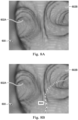
- FIG. 6A presents an anterior view of the chest region of patient P taken in an axial plane;
- FIG. 6B is a lateral view of the chest region taken in a coronal plane, and
- FIG. 6C is a frontal view of the chest region taken in a sagittal plane.
- the views shown in FIGS. 6A-C are cross-sectional views obtained from three-dimensional data. These views are "slices" showing two-dimensional planes within the three-dimensional image data. The air in the lungs in FIGS. 6A-C is depicted in black.
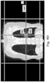
- FIGS. 7A, 7B , and 7C depict multiple on-axis views of filtered image data, obtained by filtering the image data of FIGS. 6A-C .
- FIGS. 7A-C highlight voxels 700 that have a Hounsfield value that may be associated with the air in the anatomical passageways of the lungs.
- the air within the lungs of patient P may be the least dense portions of the patient P and may be filtered by density value to isolate the anatomical passageways.
- FIGS. 7A-C are cross-sectional slices that depict the voxels having the density of air within the trachea and other passageways in the lungs of the patient P.
- voxels may be identified by filtering according to the Hounsfield value and rendered differently than other voxels so as to highlight the anatomic passageways to the operator O.
- the image data may be filtered to show the air in the anatomical passageways of the lungs as a distinctive color so as to provide a model of the passageways.
- FIGS. 7D , 7E, and 7F are orthographic views of the image data, shown in FIGS. 7A-C , being further filtered so as to highlight or depict specific aspects of the patient anatomy included in the image data.
- FIG. 7D is filtered based on Hounsfield values to depict portions of bone 702 the vasculature 704 surrounds the bronchial walls 706 of the lungs of patient P.
- the operator O may select specific tissues from a menu of selectable tissues for rendering in the user interface and vasculature 704.
- FIG. 7E may be presented to the position.
- FIG. 7E shows the vasculature 704 and the bronchial walls 706.
- the operator O may also interact with the user interface to select only the bronchial walls 706 for display as seen in FIG. 7F .
- the use of active, selective filtering of the imaging data based on a characteristic of each voxel other than its location may provide the operator O with a desired rendering.
- the computing resources CPU and/or GPU resources
- the operator O may interact with user interface elements to selectively apply and modify filters to the image data.
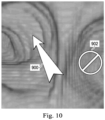
- the image data is filtered to render the air transparent while rendering the voxels having Hounsfield values of the lung walls 600.
- the interior surface of the lung walls 600 is presented to the operator O to enable the operator O to virtually navigate within the walls 600.
- the perspective shown in FIG. 8A is indicated by vertical and horizontal guidelines shown in FIGS. 6A-C and 7A-C , which intersect at the perspective point of FIG. 8A , and is directed toward the main carina of the lungs of the patient P.
- FIG. 8A looking distally from the trachea, there is the left bronchus 602A and the right bronchus 602B.
- image data similar to that shown in FIGS. 6A-C and FIGS. 7A-F , such as CT image data that includes data corresponding to a target 800 of a medical procedure.
- the target 800 may be a tumor such as a lung tumor.
- filtering the CT data by density may facilitate identification of the location and shape of the tumor.
- the processing device 114 may identify the target 800 or make a preliminary identification of the target 800.
- an input may be received via a user interface from the operator O designating a portion of the image data as the target 800.
- the operator O may provide input on a display screen 110 displaying each of the two-dimensional slices of FIGS. 8B-8D.
- the inputs received on each of the three views may be used to produce a target identification zone being centered around a position in the three-dimensional image space shown in three-dimensional perspective in FIG. 8A .
- the target identification zone may further include a three-dimensional shape formed around the central position.
- the three-dimensional shape 802 may include as shown in FIG. 8A , an oval, a cube, etc.
- the operator O may interact with a UI element to control the size and shape of the three-dimensional shape 802.
- the three-dimensional shape 802 may be used as the target 800 or the processing device may identify the voxels within the shape 802 that make up the target 800.
- the processing device may determine that some modeled passageways do not provide access to the target. For example, the processing device may determine that certain passageways do not have a point within a pre-determined threshold distance from the target 800. As another example, the processing device may determine a subset of the modeled passageways that does have at least one point within a threshold distance from the target 800.
- the threshold distance may be in a range from about 0.5 to about 5 cm. In some embodiments, the threshold distance may be in a range from about 1.5 to about 3 cm.
- the processing device may prevent navigation down any of the passageways that do not provide access to the target 800. As shown in FIG.
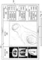
- the centerline segmented model 1110 includes several branch points, some of which are highlighted for visibility.
- the branch points A, B, C, D, and E are shown at each of several of the branch points.
- the branch point A may represent the point in the model 1110 at which the trachea divides into the left and right principal bronchii.
- the right principal bronchus may be identified in the centerline segment model 1110 as being located between branch points A and B.
- secondary bronchii are identified by the branch points B and C and between the branch points B and E. Another generation may be defined between branch points C and D.
- Combinations of these approaches may be used to generate a complete or partial model of the anatomic passageways based user interactions with the image data 1102.
- combinations of segmentation-based modeling techniques and non-segmentation-based modeling techniques may be used to generate a model, like the augmented model 1112, which can then be registered to a medical instrument inserted within the patient anatomy during a medical procedure.
- the medical instrument such as the elongate device 202 of FIGS. 2A and 2B
- the image data associated with the model 1112 and the elongate device 202 may be brought into a common frame of reference for use in the image-guided medical procedure.
- a graphical representation of the registered elongate device 202 may be presented in the display system 110 along with the registered image data.
- the segmented model 1106 may be used to produce the centerline segment model 1110 or another suitable model including a cloud, set, or collection of points.
- the segmented model 1106 comprises a mesh representing the internal surfaces of one or more passageways
- a subset of vertices of a mesh as represented in a stored data file including the model 1106 may be used.
- a geometric center of the mesh or surface may be used to represent volumes or the passageways in the segmented model 1106.
- the processing device may apply a filter to the three-dimensional image data to alter a rendering of one or more voxels of the three-dimensional image data. For example, when the image data is displayed from a perspective of the distal end of the medical instrument, some portions of the image data may be filtered to provide a desired presentation of the image data. For example, the image data may be filtered according to the Hounsfield value associated with the voxel.
- the operator O may selectively cause, or the control system 112 may automatically cause, certain tissues to be displayed entirely transparently or partially transparently to enable certain aspects to be visualized more easily.
- the processing device may render the three-dimensional image data in a display from a perspective associated with the medical instrument. As noted, this may be done in a first portion of a display screen or in a first display screen, while live video from the same perspective or from a different perspective is provided in a second portion of the display screen or in a second display screen.
- the CT data may be used by the operator O for continued navigation. Additionally, the operator O may make comparisons between the tissues, fluids, etc., shown in the live view and the same subjects as included in the image data. Other imaging modalities, intra-operative or pre-operative, may be compared with the CT image data in other embodiments.
- the operator O may make selections in a user interface like the user interface 1200 of FIG. 12 to provide selections of tissue types and/or to receive rendering settings associated with each of the tissue types to the control system 112.
- the user interface 1200 may present additional user interface options, such as sliders or entry fields, whereby the operator O may enter a transparency setting and/or a color setting associated with the bronchial walls element 1232A.
- the settings may then be applied to the voxel's associated with the Hounsfield value of the bronchial walls.
- the perspective from which the image data is viewed maybe updated to reflect the change in position of the medical instrument, such that the navigation commands transmitted to steer/position the medical instrument are simultaneously utilized for virtual navigation of the image data.
- Embodiments of the present disclosure may provide for significant improvements in image-guided medical procedures. For example, some embodiments may permit the use of lower resolution CT scans to be used in generating a model of patient anatomy. By improving the usefulness of such lower resolution CT scans, patient exposure to the radioactive imaging agents used to obtain the image data may be decreased. Some embodiments may permit use of both segmentation and user input to generate a hybrid model of patient anatomy, which may result in improved efficiency and accuracy in generating such anatomic models that permit registration. Additionally, embodiments of the present disclosure may facilitate simultaneously viewing the image data along with live video data during a medical procedure.
- Embodiments of the methods and systems described herein include computer readable storage media, such as CD-ROMs, DVDs, flash memory, and other storage medium, having machine-readable instructions stored thereon.
- the processing device such as a processing device included in the control system 112 of FIG. 1 .
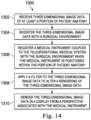
- Additional embodiments of the present disclosure include systems, such as a workstation, having one or more processing devices (such as a CPU and GPU) and a memory storing instructions that, when read by the processing device, cause the system to perform an embodiment of one of the methods 400, 500, and 1300.
- features described with respect to the various embodiments of the present disclosure may be combined. For example, operations discussed with respect to method 1300 may be included in embodiments of the method 500.
Landscapes
- Health & Medical Sciences (AREA)
- Life Sciences & Earth Sciences (AREA)
- Engineering & Computer Science (AREA)
- Surgery (AREA)
- Medical Informatics (AREA)
- Nuclear Medicine, Radiotherapy & Molecular Imaging (AREA)
- Public Health (AREA)
- Veterinary Medicine (AREA)
- Biomedical Technology (AREA)
- Molecular Biology (AREA)
- Animal Behavior & Ethology (AREA)
- General Health & Medical Sciences (AREA)
- Heart & Thoracic Surgery (AREA)
- Robotics (AREA)
- Pathology (AREA)
- Radiology & Medical Imaging (AREA)
- Physics & Mathematics (AREA)
- Oral & Maxillofacial Surgery (AREA)
- Human Computer Interaction (AREA)
- Biophysics (AREA)
- High Energy & Nuclear Physics (AREA)
- Optics & Photonics (AREA)
- Gynecology & Obstetrics (AREA)
- Computer Graphics (AREA)
- General Physics & Mathematics (AREA)
- Theoretical Computer Science (AREA)
- Apparatus For Radiation Diagnosis (AREA)
Applications Claiming Priority (3)
| Application Number | Priority Date | Filing Date | Title |
|---|---|---|---|
| US201662431696P | 2016-12-08 | 2016-12-08 | |
| PCT/US2017/065162 WO2018106950A1 (en) | 2016-12-08 | 2017-12-07 | Systems and methods for navigation in image-guided medical procedures |
| EP17879065.5A EP3551114B1 (de) | 2016-12-08 | 2017-12-07 | Systeme und verfahren zur erzeugung eines modells der patientenanatomie mit anatomischen gängen |
Related Parent Applications (2)
| Application Number | Title | Priority Date | Filing Date |
|---|---|---|---|
| EP17879065.5A Division EP3551114B1 (de) | 2016-12-08 | 2017-12-07 | Systeme und verfahren zur erzeugung eines modells der patientenanatomie mit anatomischen gängen |
| EP17879065.5A Division-Into EP3551114B1 (de) | 2016-12-08 | 2017-12-07 | Systeme und verfahren zur erzeugung eines modells der patientenanatomie mit anatomischen gängen |
Publications (2)
| Publication Number | Publication Date |
|---|---|
| EP4582040A2 true EP4582040A2 (de) | 2025-07-09 |
| EP4582040A3 EP4582040A3 (de) | 2025-10-01 |
Family
ID=62492338
Family Applications (2)
| Application Number | Title | Priority Date | Filing Date |
|---|---|---|---|
| EP25178554.9A Pending EP4582040A3 (de) | 2016-12-08 | 2017-12-07 | Systeme und verfahren zur navigation in bildgeführten medizinischen verfahren |
| EP17879065.5A Active EP3551114B1 (de) | 2016-12-08 | 2017-12-07 | Systeme und verfahren zur erzeugung eines modells der patientenanatomie mit anatomischen gängen |
Family Applications After (1)
| Application Number | Title | Priority Date | Filing Date |
|---|---|---|---|
| EP17879065.5A Active EP3551114B1 (de) | 2016-12-08 | 2017-12-07 | Systeme und verfahren zur erzeugung eines modells der patientenanatomie mit anatomischen gängen |
Country Status (4)
| Country | Link |
|---|---|
| US (2) | US11547490B2 (de) |
| EP (2) | EP4582040A3 (de) |
| CN (2) | CN109922753B (de) |
| WO (1) | WO2018106950A1 (de) |
Families Citing this family (61)
| Publication number | Priority date | Publication date | Assignee | Title |
|---|---|---|---|---|
| US8218847B2 (en) | 2008-06-06 | 2012-07-10 | Superdimension, Ltd. | Hybrid registration method |
| US9603668B2 (en) | 2014-07-02 | 2017-03-28 | Covidien Lp | Dynamic 3D lung map view for tool navigation inside the lung |
| US9633431B2 (en) | 2014-07-02 | 2017-04-25 | Covidien Lp | Fluoroscopic pose estimation |
| US9986983B2 (en) | 2014-10-31 | 2018-06-05 | Covidien Lp | Computed tomography enhanced fluoroscopic system, device, and method of utilizing the same |
| US10674982B2 (en) | 2015-08-06 | 2020-06-09 | Covidien Lp | System and method for local three dimensional volume reconstruction using a standard fluoroscope |
| US10702226B2 (en) | 2015-08-06 | 2020-07-07 | Covidien Lp | System and method for local three dimensional volume reconstruction using a standard fluoroscope |
| US10716525B2 (en) | 2015-08-06 | 2020-07-21 | Covidien Lp | System and method for navigating to target and performing procedure on target utilizing fluoroscopic-based local three dimensional volume reconstruction |
| US11793579B2 (en) | 2017-02-22 | 2023-10-24 | Covidien Lp | Integration of multiple data sources for localization and navigation |
| CN110325138B (zh) * | 2017-03-22 | 2023-06-06 | 直观外科手术操作公司 | 用于智能种子配准的系统和方法 |
| EP3613057B1 (de) * | 2017-04-18 | 2025-06-04 | Intuitive Surgical Operations, Inc. | Grafische benutzeroberfläche zur planung eines verfahrens |
| EP3622528A1 (de) * | 2017-05-09 | 2020-03-18 | Boston Scientific Scimed, Inc. | Betriebsraumvorrichtungen, -verfahren und -systeme |
| US10699448B2 (en) | 2017-06-29 | 2020-06-30 | Covidien Lp | System and method for identifying, marking and navigating to a target using real time two dimensional fluoroscopic data |
| CN111163697B (zh) | 2017-10-10 | 2023-10-03 | 柯惠有限合伙公司 | 用于在荧光三维重构中识别和标记目标的系统和方法 |
| CN110809453B (zh) * | 2017-12-18 | 2023-06-06 | 奥瑞斯健康公司 | 用于腔网络内的器械跟踪和导航的方法和系统 |
| US10930064B2 (en) | 2018-02-08 | 2021-02-23 | Covidien Lp | Imaging reconstruction system and method |
| US10893842B2 (en) | 2018-02-08 | 2021-01-19 | Covidien Lp | System and method for pose estimation of an imaging device and for determining the location of a medical device with respect to a target |
| US10905498B2 (en) | 2018-02-08 | 2021-02-02 | Covidien Lp | System and method for catheter detection in fluoroscopic images and updating displayed position of catheter |
| US10685439B2 (en) * | 2018-06-27 | 2020-06-16 | General Electric Company | Imaging system and method providing scalable resolution in multi-dimensional image data |
| US11705238B2 (en) | 2018-07-26 | 2023-07-18 | Covidien Lp | Systems and methods for providing assistance during surgery |
| US11071591B2 (en) | 2018-07-26 | 2021-07-27 | Covidien Lp | Modeling a collapsed lung using CT data |
| US11944388B2 (en) | 2018-09-28 | 2024-04-02 | Covidien Lp | Systems and methods for magnetic interference correction |
| US11877806B2 (en) | 2018-12-06 | 2024-01-23 | Covidien Lp | Deformable registration of computer-generated airway models to airway trees |
| US11045075B2 (en) | 2018-12-10 | 2021-06-29 | Covidien Lp | System and method for generating a three-dimensional model of a surgical site |
| US11617493B2 (en) | 2018-12-13 | 2023-04-04 | Covidien Lp | Thoracic imaging, distance measuring, surgical awareness, and notification system and method |
| US11801113B2 (en) | 2018-12-13 | 2023-10-31 | Covidien Lp | Thoracic imaging, distance measuring, and notification system and method |
| US11357593B2 (en) | 2019-01-10 | 2022-06-14 | Covidien Lp | Endoscopic imaging with augmented parallax |
| US11625825B2 (en) | 2019-01-30 | 2023-04-11 | Covidien Lp | Method for displaying tumor location within endoscopic images |
| US11564751B2 (en) | 2019-02-01 | 2023-01-31 | Covidien Lp | Systems and methods for visualizing navigation of medical devices relative to targets |
| US11925333B2 (en) | 2019-02-01 | 2024-03-12 | Covidien Lp | System for fluoroscopic tracking of a catheter to update the relative position of a target and the catheter in a 3D model of a luminal network |
| US11744643B2 (en) | 2019-02-04 | 2023-09-05 | Covidien Lp | Systems and methods facilitating pre-operative prediction of post-operative tissue function |
| US11819285B2 (en) | 2019-04-05 | 2023-11-21 | Covidien Lp | Magnetic interference detection systems and methods |
| US12089902B2 (en) | 2019-07-30 | 2024-09-17 | Coviden Lp | Cone beam and 3D fluoroscope lung navigation |
| US11269173B2 (en) | 2019-08-19 | 2022-03-08 | Covidien Lp | Systems and methods for displaying medical video images and/or medical 3D models |
| US12059281B2 (en) | 2019-08-19 | 2024-08-13 | Covidien Lp | Systems and methods of fluoro-CT imaging for initial registration |
| US11864935B2 (en) | 2019-09-09 | 2024-01-09 | Covidien Lp | Systems and methods for pose estimation of a fluoroscopic imaging device and for three-dimensional imaging of body structures |
| US11931111B2 (en) | 2019-09-09 | 2024-03-19 | Covidien Lp | Systems and methods for providing surgical guidance |
| JP7444569B2 (ja) * | 2019-09-18 | 2024-03-06 | ザイオソフト株式会社 | 鏡視下手術支援装置、鏡視下手術支援方法、及びプログラム |
| US11627924B2 (en) | 2019-09-24 | 2023-04-18 | Covidien Lp | Systems and methods for image-guided navigation of percutaneously-inserted devices |
| US12144564B2 (en) * | 2019-11-08 | 2024-11-19 | Intuitive Surgical Operations, Inc. | Systems and methods for registering an instrument to an image using change in instrument position data |
| US12102298B2 (en) | 2019-12-10 | 2024-10-01 | Covidien Lp | Lymphatic system tracking |
| US11977723B2 (en) * | 2019-12-17 | 2024-05-07 | Palantir Technologies Inc. | Image tiling and distributive modification |
| EP3838159A1 (de) * | 2019-12-17 | 2021-06-23 | Koninklijke Philips N.V. | Navigation der bronchialwege |
| US11380060B2 (en) | 2020-01-24 | 2022-07-05 | Covidien Lp | System and method for linking a segmentation graph to volumetric data |
| US11847730B2 (en) | 2020-01-24 | 2023-12-19 | Covidien Lp | Orientation detection in fluoroscopic images |
| EP4138715A4 (de) * | 2020-04-19 | 2023-10-11 | Xact Robotics Ltd. | Algorithmusbasierte verfahren zur vorhersage und/oder erkennung eines klinischen zustands im zusammenhang mit der einführung eines medizinischen instruments in ein internes ziel |
| US12064191B2 (en) | 2020-06-03 | 2024-08-20 | Covidien Lp | Surgical tool navigation using sensor fusion |
| DE102020208325A1 (de) * | 2020-07-02 | 2022-01-05 | Siemens Healthcare Gmbh | Verfahren und System zur Erstellung eines Navigationsplans für einen Katheter mit Roboter |
| US11950950B2 (en) | 2020-07-24 | 2024-04-09 | Covidien Lp | Zoom detection and fluoroscope movement detection for target overlay |
| EP3944834A1 (de) * | 2020-07-30 | 2022-02-02 | Koninklijke Philips N.V. | Navigationsoperationsanweisungen |
| US12256923B2 (en) | 2020-08-13 | 2025-03-25 | Covidien Lp | Endoluminal robotic systems and methods for suturing |
| US12383352B2 (en) | 2020-08-13 | 2025-08-12 | Covidien Lp | Endoluminal robotic (ELR) systems and methods |
| US12161309B2 (en) | 2020-09-24 | 2024-12-10 | Covidien Lp | Articulating mechanism for the laparoscopic ablation device for blunt dissection |
| CN112259199B (zh) * | 2020-10-29 | 2024-08-13 | 西交利物浦大学 | 医疗图像分类模型的训练方法、系统、存储介质及医疗图像处理装置 |
| WO2022123577A1 (en) | 2020-12-10 | 2022-06-16 | Magnisity Ltd. | Dynamic deformation tracking for navigational bronchoscopy |
| WO2022216716A1 (en) * | 2021-04-07 | 2022-10-13 | Intuitive Surgical Operations, Inc. | Systems, methods and medium containing instruction for connecting model structures representing anatomical pathways |
| WO2022266587A1 (en) * | 2021-06-16 | 2022-12-22 | Gyrus Acmi, Inc. D/B/A Olympus Surgical Technologies America | Lung analysis and reporting system |
| US11515041B1 (en) * | 2021-09-02 | 2022-11-29 | Omniscient Neurotechnology Pty Limited | Display of subset brain graph by shading nodes |
| US12303220B2 (en) | 2022-01-26 | 2025-05-20 | Covidien Lp | Autonomous endobronchial access with an EM guided catheter |
| US12257082B2 (en) | 2022-06-30 | 2025-03-25 | Covidien Lp | Cone beam computed tomography integration for creating a navigation pathway to a target in the lung and method of navigating to the target |
| CN120659587A (zh) * | 2022-12-29 | 2025-09-16 | 直观外科手术操作公司 | 用于生成用于医疗过程的3d导航界面的系统和方法 |
| DE102023133232B4 (de) * | 2023-11-28 | 2025-09-11 | Karl Storz Se & Co. Kg | Medizinisches System, medizinisches Bildgebungsinstrument und Verfahren zur Bildgebung mittelseines medizinischen Systems für das Airway-Management eines Patienten |
Citations (4)
| Publication number | Priority date | Publication date | Assignee | Title |
|---|---|---|---|---|
| US4705604A (en) | 1984-07-06 | 1987-11-10 | Solvay & Cie. (Societe Anonyme) | Process for extracting poly-beta-hydroxybutyrates by means of a solvent from an aqueous suspension of microorganisms |
| US6380732B1 (en) | 1997-02-13 | 2002-04-30 | Super Dimension Ltd. | Six-degree of freedom tracking system having a passive transponder on the object being tracked |
| US6389187B1 (en) | 1997-06-20 | 2002-05-14 | Qinetiq Limited | Optical fiber bend sensor |
| US7316681B2 (en) | 1996-05-20 | 2008-01-08 | Intuitive Surgical, Inc | Articulated surgical instrument for performing minimally invasive surgery with enhanced dexterity and sensitivity |
Family Cites Families (34)
| Publication number | Priority date | Publication date | Assignee | Title |
|---|---|---|---|---|
| US6113946A (en) * | 1992-04-03 | 2000-09-05 | The Regents Of The University Of California | Self-assembling polynucleotide delivery system comprising dendrimer polycations |
| US5611025A (en) * | 1994-11-23 | 1997-03-11 | General Electric Company | Virtual internal cavity inspection system |
| US6246784B1 (en) * | 1997-08-19 | 2001-06-12 | The United States Of America As Represented By The Department Of Health And Human Services | Method for segmenting medical images and detecting surface anomalies in anatomical structures |
| US7998062B2 (en) * | 2004-03-29 | 2011-08-16 | Superdimension, Ltd. | Endoscope structures and techniques for navigating to a target in branched structure |
| EP1689290A2 (de) * | 2003-10-21 | 2006-08-16 | The Board of Trustees of The Leland Stanford Junior University | Systeme und verfahren für das intraoperative targetting |
| US7772541B2 (en) | 2004-07-16 | 2010-08-10 | Luna Innnovations Incorporated | Fiber optic position and/or shape sensing based on rayleigh scatter |
| US20060013523A1 (en) | 2004-07-16 | 2006-01-19 | Luna Innovations Incorporated | Fiber optic position and shape sensing device and method relating thereto |
| GB2416944A (en) * | 2004-07-30 | 2006-02-08 | Voxar Ltd | Classifying voxels in a medical image |
| US20070092864A1 (en) * | 2005-09-30 | 2007-04-26 | The University Of Iowa Research Foundation | Treatment planning methods, devices and systems |
| US7907772B2 (en) | 2006-03-30 | 2011-03-15 | Accuray Incorporated | Delineation on three-dimensional medical image |
| AU2007350982A1 (en) * | 2006-11-10 | 2008-10-23 | Dorian Averbuch | Adaptive navigation technique for navigating a catheter through a body channel or cavity |
| US9037215B2 (en) * | 2007-01-31 | 2015-05-19 | The Penn State Research Foundation | Methods and apparatus for 3D route planning through hollow organs |
| US10039613B2 (en) * | 2007-03-01 | 2018-08-07 | Surgical Navigation Technologies, Inc. | Method for localizing an imaging device with a surgical navigation system |
| US20090105579A1 (en) | 2007-10-19 | 2009-04-23 | Garibaldi Jeffrey M | Method and apparatus for remotely controlled navigation using diagnostically enhanced intra-operative three-dimensional image data |
| US9259274B2 (en) | 2008-09-30 | 2016-02-16 | Intuitive Surgical Operations, Inc. | Passive preload and capstan drive for surgical instruments |
| US8819591B2 (en) | 2009-10-30 | 2014-08-26 | Accuray Incorporated | Treatment planning in a virtual environment |
| US8900131B2 (en) | 2011-05-13 | 2014-12-02 | Intuitive Surgical Operations, Inc. | Medical system providing dynamic registration of a model of an anatomical structure for image-guided surgery |
| US9452276B2 (en) | 2011-10-14 | 2016-09-27 | Intuitive Surgical Operations, Inc. | Catheter with removable vision probe |
| EP2785270B1 (de) | 2011-12-03 | 2019-05-01 | Koninklijke Philips N.V. | Automatisches tiefen-scrollen und ausrichtungsanpassung für halbautomatische wegplanung |
| US8920368B2 (en) * | 2011-12-22 | 2014-12-30 | St. Jude Medical, Atrial Fibrillation Division, Inc. | Multi-user touch-based control of a remote catheter guidance system (RCGS) |
| US10249036B2 (en) * | 2012-02-22 | 2019-04-02 | Veran Medical Technologies, Inc. | Surgical catheter having side exiting medical instrument and related systems and methods for four dimensional soft tissue navigation |
| US20140188440A1 (en) * | 2012-12-31 | 2014-07-03 | Intuitive Surgical Operations, Inc. | Systems And Methods For Interventional Procedure Planning |
| EP2953532B1 (de) * | 2013-02-08 | 2020-01-15 | Covidien LP | System zur lungendenervierung |
| US9301723B2 (en) * | 2013-03-15 | 2016-04-05 | Covidien Lp | Microwave energy-delivery device and system |
| US9639666B2 (en) * | 2013-03-15 | 2017-05-02 | Covidien Lp | Pathway planning system and method |
| WO2014176207A1 (en) * | 2013-04-22 | 2014-10-30 | University Of Washington Through Its Center For Commercialization | Patient-specific guides to improve point registration accuracy in surgical navigation |
| US10433911B2 (en) * | 2013-09-18 | 2019-10-08 | iMIRGE Medical INC. | Optical targeting and visualization of trajectories |
| BR112016021761A2 (pt) * | 2014-03-22 | 2017-08-15 | Endocare Inc | Sistemas para auxiliar um cirurgião na colocação de pelo menos um dispositivo de ablação e para orientar e executar um procedimento de ablação e método de desenvolvimento de um plano de tratamento |
| US9770216B2 (en) * | 2014-07-02 | 2017-09-26 | Covidien Lp | System and method for navigating within the lung |
| WO2016033599A1 (en) * | 2014-08-29 | 2016-03-03 | Cardioinsight Technologies, Inc. | Localization and tracking of an object |
| US10373719B2 (en) * | 2014-09-10 | 2019-08-06 | Intuitive Surgical Operations, Inc. | Systems and methods for pre-operative modeling |
| EP3240492B1 (de) * | 2014-12-31 | 2020-03-25 | Covidien LP | System zur behandlung von copd und emphysemen |
| CN107660134B (zh) | 2015-05-22 | 2021-06-29 | 直观外科手术操作公司 | 图像引导手术记录的系统和方法 |
| WO2017030913A2 (en) | 2015-08-14 | 2017-02-23 | Intuitive Surgical Operations, Inc. | Systems and methods of registration for image-guided surgery |
-
2017
- 2017-12-07 CN CN201780067272.5A patent/CN109922753B/zh active Active
- 2017-12-07 EP EP25178554.9A patent/EP4582040A3/de active Pending
- 2017-12-07 WO PCT/US2017/065162 patent/WO2018106950A1/en not_active Ceased
- 2017-12-07 EP EP17879065.5A patent/EP3551114B1/de active Active
- 2017-12-07 US US16/349,073 patent/US11547490B2/en active Active
- 2017-12-07 CN CN202310225582.2A patent/CN116421309A/zh active Pending
-
2022
- 2022-11-23 US US17/993,227 patent/US20230088056A1/en active Pending
Patent Citations (4)
| Publication number | Priority date | Publication date | Assignee | Title |
|---|---|---|---|---|
| US4705604A (en) | 1984-07-06 | 1987-11-10 | Solvay & Cie. (Societe Anonyme) | Process for extracting poly-beta-hydroxybutyrates by means of a solvent from an aqueous suspension of microorganisms |
| US7316681B2 (en) | 1996-05-20 | 2008-01-08 | Intuitive Surgical, Inc | Articulated surgical instrument for performing minimally invasive surgery with enhanced dexterity and sensitivity |
| US6380732B1 (en) | 1997-02-13 | 2002-04-30 | Super Dimension Ltd. | Six-degree of freedom tracking system having a passive transponder on the object being tracked |
| US6389187B1 (en) | 1997-06-20 | 2002-05-14 | Qinetiq Limited | Optical fiber bend sensor |
Also Published As
| Publication number | Publication date |
|---|---|
| WO2018106950A1 (en) | 2018-06-14 |
| CN109922753B (zh) | 2023-04-14 |
| US11547490B2 (en) | 2023-01-10 |
| EP3551114A1 (de) | 2019-10-16 |
| EP4582040A3 (de) | 2025-10-01 |
| US20230088056A1 (en) | 2023-03-23 |
| CN116421309A (zh) | 2023-07-14 |
| EP3551114B1 (de) | 2025-07-16 |
| EP3551114A4 (de) | 2020-07-22 |
| US20190350659A1 (en) | 2019-11-21 |
| CN109922753A (zh) | 2019-06-21 |
Similar Documents
| Publication | Publication Date | Title |
|---|---|---|
| US20230088056A1 (en) | Systems and methods for navigation in image-guided medical procedures | |
| US20240041531A1 (en) | Systems and methods for registering elongate devices to three-dimensional images in image-guided procedures | |
| US12243233B2 (en) | Systems and methods for using registered fluoroscopic images in image-guided surgery | |
| EP3576598B1 (de) | System zur registrierung für bildgeführte verfahren | |
| US20210259783A1 (en) | Systems and Methods Related to Registration for Image Guided Surgery | |
| US20230030727A1 (en) | Systems and methods related to registration for image guided surgery | |
| US20220142714A1 (en) | Systems for enhanced registration of patient anatomy | |
| EP3930616B1 (de) | Systeme zur registrierung von patientenanatomie | |
| US20240164853A1 (en) | User interface for connecting model structures and associated systems and methods | |
| EP4663093A2 (de) | Systeme zur registrierung der anatomie eines patienten |
Legal Events
| Date | Code | Title | Description |
|---|---|---|---|
| PUAI | Public reference made under article 153(3) epc to a published international application that has entered the european phase |
Free format text: ORIGINAL CODE: 0009012 |
|
| STAA | Information on the status of an ep patent application or granted ep patent |
Free format text: STATUS: THE APPLICATION HAS BEEN PUBLISHED |
|
| AC | Divisional application: reference to earlier application |
Ref document number: 3551114 Country of ref document: EP Kind code of ref document: P |
|
| AK | Designated contracting states |
Kind code of ref document: A2 Designated state(s): AL AT BE BG CH CY CZ DE DK EE ES FI FR GB GR HR HU IE IS IT LI LT LU LV MC MK MT NL NO PL PT RO RS SE SI SK SM TR |
|
| REG | Reference to a national code |
Ref country code: DE Ref legal event code: R079 Free format text: PREVIOUS MAIN CLASS: A61B0034100000 Ipc: A61B0034200000 |
|
| PUAL | Search report despatched |
Free format text: ORIGINAL CODE: 0009013 |
|
| AK | Designated contracting states |
Kind code of ref document: A3 Designated state(s): AL AT BE BG CH CY CZ DE DK EE ES FI FR GB GR HR HU IE IS IT LI LT LU LV MC MK MT NL NO PL PT RO RS SE SI SK SM TR |
|
| RIC1 | Information provided on ipc code assigned before grant |
Ipc: A61B 34/20 20160101AFI20250827BHEP Ipc: A61B 6/03 20060101ALI20250827BHEP Ipc: A61B 90/00 20160101ALI20250827BHEP Ipc: A61B 34/10 20160101ALI20250827BHEP Ipc: A61B 6/12 20060101ALI20250827BHEP Ipc: A61B 8/08 20060101ALI20250827BHEP |