EP4579682A1 - Verfahren und system zur risikostratifizierung asymptomatischer patienten mit karotidstenose - Google Patents
Verfahren und system zur risikostratifizierung asymptomatischer patienten mit karotidstenose Download PDFInfo
- Publication number
- EP4579682A1 EP4579682A1 EP23220881.9A EP23220881A EP4579682A1 EP 4579682 A1 EP4579682 A1 EP 4579682A1 EP 23220881 A EP23220881 A EP 23220881A EP 4579682 A1 EP4579682 A1 EP 4579682A1
- Authority
- EP
- European Patent Office
- Prior art keywords
- plaque
- model
- carotid
- risk
- wall
- Prior art date
- Legal status (The legal status is an assumption and is not a legal conclusion. Google has not performed a legal analysis and makes no representation as to the accuracy of the status listed.)
- Pending
Links
Images







Classifications
-
- G—PHYSICS
- G16—INFORMATION AND COMMUNICATION TECHNOLOGY [ICT] SPECIALLY ADAPTED FOR SPECIFIC APPLICATION FIELDS
- G16H—HEALTHCARE INFORMATICS, i.e. INFORMATION AND COMMUNICATION TECHNOLOGY [ICT] SPECIALLY ADAPTED FOR THE HANDLING OR PROCESSING OF MEDICAL OR HEALTHCARE DATA
- G16H50/00—ICT specially adapted for medical diagnosis, medical simulation or medical data mining; ICT specially adapted for detecting, monitoring or modelling epidemics or pandemics
- G16H50/30—ICT specially adapted for medical diagnosis, medical simulation or medical data mining; ICT specially adapted for detecting, monitoring or modelling epidemics or pandemics for calculating health indices; for individual health risk assessment
-
- G—PHYSICS
- G16—INFORMATION AND COMMUNICATION TECHNOLOGY [ICT] SPECIALLY ADAPTED FOR SPECIFIC APPLICATION FIELDS
- G16H—HEALTHCARE INFORMATICS, i.e. INFORMATION AND COMMUNICATION TECHNOLOGY [ICT] SPECIALLY ADAPTED FOR THE HANDLING OR PROCESSING OF MEDICAL OR HEALTHCARE DATA
- G16H30/00—ICT specially adapted for the handling or processing of medical images
- G16H30/40—ICT specially adapted for the handling or processing of medical images for processing medical images, e.g. editing
-
- G—PHYSICS
- G16—INFORMATION AND COMMUNICATION TECHNOLOGY [ICT] SPECIALLY ADAPTED FOR SPECIFIC APPLICATION FIELDS
- G16H—HEALTHCARE INFORMATICS, i.e. INFORMATION AND COMMUNICATION TECHNOLOGY [ICT] SPECIALLY ADAPTED FOR THE HANDLING OR PROCESSING OF MEDICAL OR HEALTHCARE DATA
- G16H50/00—ICT specially adapted for medical diagnosis, medical simulation or medical data mining; ICT specially adapted for detecting, monitoring or modelling epidemics or pandemics
- G16H50/20—ICT specially adapted for medical diagnosis, medical simulation or medical data mining; ICT specially adapted for detecting, monitoring or modelling epidemics or pandemics for computer-aided diagnosis, e.g. based on medical expert systems
-
- G—PHYSICS
- G16—INFORMATION AND COMMUNICATION TECHNOLOGY [ICT] SPECIALLY ADAPTED FOR SPECIFIC APPLICATION FIELDS
- G16H—HEALTHCARE INFORMATICS, i.e. INFORMATION AND COMMUNICATION TECHNOLOGY [ICT] SPECIALLY ADAPTED FOR THE HANDLING OR PROCESSING OF MEDICAL OR HEALTHCARE DATA
- G16H50/00—ICT specially adapted for medical diagnosis, medical simulation or medical data mining; ICT specially adapted for detecting, monitoring or modelling epidemics or pandemics
- G16H50/50—ICT specially adapted for medical diagnosis, medical simulation or medical data mining; ICT specially adapted for detecting, monitoring or modelling epidemics or pandemics for simulation or modelling of medical disorders
Definitions
- the present invention provides methods and apparatuses for the improved stratification of asymptomatic patients with carotid artery disease.
- Carotid artery disease constitutes one of the primary causes of ischaemic cerebrovascular events and accounts for an estimated 150,000 deaths per year from stroke in Europe and 130,000 in the USA, and an even higher burden in morbidity and healthcare costs, due to long term disability.
- Atherosclerotic plaques in the carotid artery bifurcations cause progressive narrowing of the vessel lumen, which erode or rupture and subsequently cause thromboembolism in the cerebropetal circulation leading to cerebral infarction in 80% of patients while in 20% the cause of stroke is hemodynamic disturbances of cerebral perfusion.
- the determining factors for carotid artery disease management are currently the degree of stenosis, the presence and recency of symptoms.
- Patients with 70% stenosis in their carotid artery are considered to be at high risk of cerebrovascular events and are therefore directed to surgical intervention (carotid endarterectomy - CEA or carotid stenting- CAS).
- patients with ⁇ 70% stenosis are considered at low-intermediate risk when asymptomatic, and unless other confounding factors exist, they are subjected to medical treatment alone.
- the cut-off of 50% stenosis is used instead.
- this stratification is rather generic, leading to high levels of unnecessary surgical treatment (estimated as high as 95% for asymptomatic patients and costing annually to the European healthcare systems >2 billion €).
- vanLammeren GW et al. (Circulation. 2014;129(22):2269-76 ) have noted a 50% reduction of lipid-rich plaques and intraplaque haemorrhage in symptomatic and asymptomatic carotid lesions. Conversely, an increasing rate of non-ruptured lesions has been observed that are likely to "erode” and cause thromboembolic complications, through different mechanisms than previously suggested. Notably, these plaques are twice more frequent in women than in men, indicating an important gender dimension.
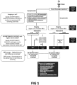
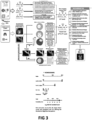
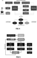
- the invention described herein provides a method for multi-level computational method for the risk stratification of asymptomatic patients with carotid artery disease. Specifically, said method comprises the following steps:
- the 3D arterial geometry is used as input to the Blood flow modeling module to provide as output patient specific blood flow features such as blood velocities, blood pressure drop, wall shear stress (WSS) distribution, time-averaged wall shear stress (TAWSS), Oscillatory shear index (OSI) and Relative Residence Time (RRT) along the vessel's inner wall.
- WSS wall shear stress
- TWSS time-averaged wall shear stress
- OSI Oscillatory shear index
- RRT Relative Residence Time
- This module models the basic parameters of the atherosclerotic process by solving several differential equations and providing as output the concentrations of LDL, HDL, OxLDL, OxHDL, monocytes, microphages, Foam Cells, T-Cells, Cytokines, smooth muscle cells (SMCs), Collagen and plaque volume over time.
- the output of the module provides prediction of plaque volume evolution over time with an updated 3D arterial geometry.
- the new predicted 3D geometry is utilized by the blood flow modeling module to provide updated WSS distribution values.
- Initial (baseline) and predicted 3D vessel geometries are provided as input to the plaque rupture module to provide plaque burden and plaque composition features as well as biomechanical features and stresses (e.g. max tensile/principal Plaque Structural Stress (PSS) and WSS thresholds) that are collectively exploited to provide risk prediction for plaque rupture.
- biomechanical features and stresses e.g. max tensile/principal Plaque Structural Stress (PSS) and WSS thresholds
- Image segmentation model One of the most important components of the methodology described in the previous section is the segmentation component, which is used to isolate the pixels of interest from each MRI series (lumen from the Time-of-Flight (TOF) series and outer arterial wall from the T1W (T1-weighted) image series).
- TOF Time-of-Flight
- T1W T1-weighted
- the acquisition parameters for the MRI sequences were as follows: (i) TOF images: repetition time: 23 ms, effective echo time: 3.2 ms, field of view [FOV]: 160 mm, section thickness: 1 mm; (ii) fast-spin echo double-inversion recovery prepared sequences (T1W): repetition time: 1428.57 ms, effective echo time: 7.672 ms, FOV: 100 mm, section thickness: 3 mm; The acquired data were stored in DICOM format.
- Training dataset preparation Two specialists annotated the luminal wall on the TOF frames and the outer arterial wall on the T1W frames. Based on these annotations, masked images were created which were used as input for the training phase of the U-net models (one model for segmenting the lumen and one model for segmenting the outer arterial wall). This process resulted in two datasets, one for the luminal wall with 29 patients ⁇ 100 (TOF frames) ⁇ 100 ⁇ 100 pixels and one for the outer arterial wall with 29 patients ⁇ 10 (T1W frames) ⁇ 100 ⁇ 100 pixels.
- This deep neural network was implemented with PyTorch functional API. Output from the network is a 100 ⁇ 100 pixels image which represents the segmented image. The binary cross-entropy loss function was selected.
- the parameters selected for each model component are the following: rectified linear unit (ReLU) activation function (for all layers but last), sigmoid activation function (for the last layer), He distribution for the kernel, Adam optimization algorithm.
- the two U-Net models have been trained for 7 epochs, where 300 batches (steps) per epoch were applied. As far as the loss function is concerned, the binary cross-entropy function has been used.
- the prior art approaches for carotid artery MRI image segmentation mainly involve active contours (with various objective functions, like snakes or level sets) both in 2D and in 3D domains and ML approaches, which are produced by training different models (NNs, SVMs, etc.) with annotated data.
- the disclosed methodology contributes beyond the state-of-the-art by 1. Training and deploying a deep learning model (U-net) for the image segmentation phase, 2. Utilizing an iterative approach, according to which the produced model is refined over several iterations, aiming to reach certain quality criteria, based on the anatomic properties of the specific body area, and 3. Validating the proposed models over an extensive dataset of ground truth annotated dataset.
- Plaque Characterization using MRI modalities In order to refine the produced 3D artery model, information related to the plaque tissue components which produce the stenosis of the diseased arteries was integrated into the model. To this end, hybrid models of image processing methods together with supervised ML techniques are explored, to improve plaque type identification of the carotid arteries.
- the methodology exploits TOF, T1W, T2 weighted (T2W) and Proton Density weighted (PDW) MRI for more accurate identification of LRNC, IPH, calcification and fibrous tissue including thin or ruptured fibrous cap.
- the proposed methodology mimics that of the manual review and includes the steps presented in Figure 10 .
- Ground truth datasets In order to support the aforementioned methodology, a dataset to act as ground truth for the ML models is required, the aim of the ML models being to detect and segment the plaque regions on every MRI frame. For this, two experts annotated once a dataset obtained from a total of 91 patients at baseline which have been enrolled in the prospective clinical study. For each case, approximately 18 frames per series have been annotated (TOF, T1W, T2W and PDW) using an annotation software tool. Lumen and outer wall border as well as plaque areas (calcifications, hemorrhages, fibrous cap, lipid cores) have been annotated on specific pre-defined image series. This process resulted in a dataset of 2421 annotated images.
- Supervised learning In the supervised setting an algorithm is trained with a training set in which every example has a ground truth. Since the goal is to perform pixel-wise segmentation, every pixel is an example that needs to be evaluated by the algorithm. It is thus also necessary to have a ground truth for each pixel (pixel-wise annotations).
- the supervised learning method used was Convolutional Neural Networks (CNNs). Other methods that may also be used are Linear Discriminant Analysis, Support Vector Machines, Random Forest and Neural Networks. For the ML methods only the features from the region between the lumen and vessel wall are evaluated.
- the following features were obtained: the normalized MRI intensity values, scale space features such as gradient magnitude, Laplacian on three different scales, distance from lumen, distance from outer wall, texture features such as: local range, local entropy, local standard deviation, and local binary patterns. From the vessel segmentations the distance to the vessel wall and lumen were calculated as well as the multiplication between these two.
- Evaluating Performance is not a trivial problem. Especially in the medical field where the data sets are often imbalanced. Diseased tissue, the target class, is often a minority class compared to healthy tissue, the background class. However, the background class (fibrous tissue) still contains a vast majority of pixels. In such imbalanced data classifying every example to the majority class can result in an overall accuracy over 90%. For this reason, metrics are calculated over all the classes individually and other metrics are used to evaluate the performance of the algorithms on the imbalanced data set. All the metrics are derived from the confusion matrix. The metrics used are:
- Figure 11 summarizes the detailed proposed methodology for plaque characterization.
- the first step of the proposed methodology invokes the MRI 3D reconstruction module, which provides as input the geometrical characteristics of the vessel. More specifically, the borders of the lumen and the outer wall are calculated and provided as input to the plaque characterization module. Using these borders, the area between them is isolated and passed as input to the next step.
- the next step involves the segmentation and characterization of the plaque regions.
- This problem is treated as a four-class classification problem, where the first class refers to "healthy" (non-plaque) pixels, the second class to pixels depicting calcified areas, the third class to pixels depicting fibrotic areas and the fourth class to pixels depicting lipid cores.
- the training dataset described in the previous section is utilized, to train ML algorithms. The most important features calculated for the supervised learning approach are presented below, however additional features may also be used.
- plaque characterization is a challenging research field, open for more than twenty years now.
- Many works have been proposed from both academia and industry, all aiming to facilitate the workflow of radiologists and medical doctors.
- all the proposed algorithms and methodologies are usually "tied" to the MRI acquisition protocol, the characteristics of the MRI scanner and other in-house protocols.
- This heterogeneous environment prevents the direct comparison of different approaches, as they are related to the quality of the MR image acquisition and the utilized MRI series.
- MRI examinations were provided from four different centers, allowing the design of both data driven approaches and knowledge-based models which are more generalized and more widely applied than most reported similar works.
- the validation scheme on the collected dataset provides a robust method with the capacity to be applied to a wide range of medical centers.
- Carotid artery stenosis is a major consequence of carotid artery disease and its early detection is very important because if not adequately treated, it may potentially have deteriorating effects.
- Visualizing and analyzing the morphological structure of carotid bifurcations are important for understanding the etiology of carotid atherosclerosis, which is a major cause of stroke and TIA.
- US is usually the initial recommended CAS diagnostic examination. For delineation of vasculatures in the carotid artery, US examinations have been widely employed as they constitute a noninvasive procedure without ionizing radiation.
- 2D US imaging is a widely available tool for the diagnosis and management of carotid plaque atherosclerosis.
- conventional 2D US imaging has technical limitations in observing the complicated 3D shapes and asymmetric vasodilation of bifurcations.
- 2D ultrasound can only provide a limited number of views (longitudinal and transverse) the quality of which and information contained within them can vary since it is an operator-dependent technique.
- images of other planes can no longer be extracted. This may affect the accuracy of plaque characterization since information is only sampled from one section, which is then assumed to be representative of the entire plaque.
- there is accumulating evidence that plaque composition, morphology and size may significantly impact plaque vulnerability.
- the 2D approach may not be sufficient to evaluate these features accurately for entire plaque volumes since many carotid plaques have variable compositions with some of their components being more echo-lucent than others. Thus, in heterogeneous carotid plaques, 2D US imaging may not provide an accurate assessment of the composition or size, with obvious implications in risk stratification.
- the development of angio-MRI and angio-CT introduced a new era in volumetric vascular imaging. Following the use of highly sophisticated reconstruction software and the acquisition of images containing volume information in three dimensions, the visualization of vascular structures in different planes from different angles is possible. Thus, a more holistic evaluation of the plaque properties can be achieved.
- the first step of the preprocessing is the automated isolation of the image region which contains the arterial tree under reconstruction. This is performed by selecting a static 512x512 pixels window for both arterial models, left and right. Special attention is paid to the window coordinates in order for the whole arterial tree to be visible in the region. After this step, all images are labeled thus creating two datasets with labeled regions, one for the lumen and one for the wall. Images in the dataset are extremely diversified in terms of colors, brightness/contrast, frames and tables. Significantly lower brightness/contrast of the figure caused an overall dark appearance of the figure, where dark zones occupy a lot more space than on other figures.
- each decoder block 2 ⁇ 2 upconvolution and skip connection are followed by three more convolutional layers with 3 ⁇ 3 filters, and the last decoder block produces the segmentation mask with 1 ⁇ 1 convolution and sigmoid activation function. All convolutional layers are padded so that the resulting activation map preserves the same height and width. In this way, the resulting segmentation map has the same resolution as the input image.
- the variant U-Net uses batch normalization after each convolutional layer and all batch normalization layers are followed by a ReLU activation. The model is trained with a combination of binary cross-entropy and soft dice coefficient ( Anbeek P et al., Neuroimage.
- Python V3.6.7 is used as the programming language and related code is implemented using Keras framework, which regards Tensorflow framework as the backend.
- Model deployment A deployment procedure of the developed deep learning models for US image segmentation task of carotid artery was realized by using FLASK and Docker technologies. This methodology can be applied for any other dataset of US images.
- FLASK and Docker technologies This methodology can be applied for any other dataset of US images.
- two separate FLASK applications are developed, one for the transversal US images projection and the second one for the longitudinal US images of carotid artery.
- two Docker images (python:3.7.10-slim-stretch) are developed, one image is determined only for the transversal and the second one only for the longitudinal carotid artery projection.
- a set of python scripts is applied through API.
- the percentage of stenosis is measured on the ICA transversal cut, as the percentage of the current area of the lumen compared with the average area of the lumen for the ICA branch.
- the plaque length is determined from the ICA longitudinal image, as the length of the ICA where the stenosis is present.
- the values of these quantities are calculated for the reconstructed models to validate the segmentation and reconstruction modules of the proposed methodology.
- the diameters of the ICA are calculated from the segmented longitudinal images. The diameters are measured approximately on every 2 mm. Overall 20 patients were considered for the validation of the methodology.
- the overall pipeline methodology is shown in Figure 17A .
- An extended algorithm for the 3D reconstruction of carotid arteries atherosclerotic plaques using CTA imaging modality was developed and calibrated based on the CTA images quality, the CTA scanners and the acquisition dose protocols.
- the 3D reconstruction algorithm aims to segment the inner wall, the outer wall of the carotid arteries and to identify the calcified plaques (CP) and the noncalcified plaques (NCP) into the ROI.
- the utilized segmentation methodology is a 3D level set methodology as far as the inner wall, outer wall and CP segmentation is concerned, whereas the NCP pixels are identified based on dynamic thresholding technique.
- the main concept of the proposed methodology is that it is adapted in all acquisition dose protocols and CTA images derived by different CTA scanners.
- the proposed methodology includes different steps: 1) estimation of intensity membership functions for the inner wall, outer wall and CP, 2) carotid artery centerline extraction, 3) inner wall segmentation, 4) outer wall segmentation, 5) CP segmentation, 6) NCP segmentation and 7) 3D model construction.
- three different sigmoidal membership functions are estimated for the three components (the inner wall, the outer wall and the CP). These membership functions aim to correspond its pixel to an inner wall, outer wall or CP pixel and are estimated in two different ways.
- the overall concept of these membership functions use is to adapt the intensity range for each component on the value of mean lumen intensity pixels.
- the proposed methodology is an extension of an already existing methodology for the 3D reconstruction of coronary arteries ( Kigka VI et al., Biomed. Signal Process. Control. 2018;40:286-294 ) and is calibrated for the carotid arteries.
- the overall methodology has been incorporated into a software tool, which is easily used and requires minimum interaction with the user (only the starting and ending point of each vessel).
- the proposed methodology is able to provide not only carotid segment, but also carotid bifurcations which are high risk regions for atherosclerotic plaque growth.
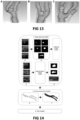
- Figures 17B , C and D demonstrate (B) the 3D reconstruction of a carotid artery bifurcation, (C) the 2D CTCA slice without segmentation and (D) the 2D segmentation of the inner wall and outer wall.
- Results & Validation The aim of the validation process is to assess the accuracy and the quality of the proposed methodology, using as gold standard a medical expert's annotations. It is known that the lack of expert-annotated datasets remains one of the main challenges in medical image processing. Generating high-quality expert-derived annotations in CTA images is time-consuming and requires a specialized in CTA imaging field medical expert. More specifically, a medical expert annotated slice per slice of the axial CTA image the inner wall, the outer wall and both types of carotid atherosclerotic plaques, the CP and the NCP ones of 16 patients. Two different validation strategies were followed.
- the detection of both the inner and the outer wall of the carotid artery constitutes the novel aspect of the proposed methodology, since both layers have a clinical significance.
- the degree of stenosis, calculated by the segmentation of the inner wall is considered as one of the most significant geometric features of the carotid artery and its value contributes to the clinical decision and the patient management.
- the quantification of the overall plaque burden region, computed by the detection of both the inner and outer wall is a significant measurement as far as the carotid artery disease prediction and progression is concerned.
- Another advantage of the disclosed methodology is its incorporation in a user-friendly software tool, which provides the 2D segmentation in each 2D CT axial slice, the 3D rendering of both the inner and outer wall of the carotid arteries and the geometrical based metrics.
- the first level of care of risk stratification tool includes five different clinical problems and each one is developed using only non-imaging based data.
- the clinical problems' outputs are: 1. Diagnosis of ⁇ 50% and >50% Stenosis, 2. Diagnosis of vulnerable plaque components, 3. Diagnosis of SBLs, 4. Evolution of Brain Structural Changes, 5. Prognosis of Cerebral outcomes/events.
- Machine learning based models are trained for each clinical problem, using as input either retrospective datasets or the input dataset of the prospective clinical study.
- datasets include demographics (sex, age), clinical characteristics (smoking, blood pressure, pulse rate etc.), medical history (Diabetes mellitus, hypertension, hypercholesterolemia, stroke, etc.), medication therapy, hematologic features (Hemoglobin, Hematocrit), lipid biomarkers (cholesterol, LDL-cholesterol, HDL-cholesterol, etc), inflammatory biomarkers (C-reactive protein, Osteoprotegerin, etc.), biochemical biomarkers (creatinin, urea, GFR, etc.), Imaging Color duplex ultrasonography (Degree of stenosis, etc), and MRI imaging data (Diffusion weighted imaging, acute ischemic brain lesions, intimal thickening, lipid or necrotic core, haemorrhage, calcification).
- the first problem aims to identify individuals at high risk for the presence of at least one of the carotid arteries with CAS 50%.
- the development of these models is based on retrospective datasets, with different clinical views of input features.
- the detection of these individuals has been formulated as a multivariate 2 class classification problem based on the carotid DS, as it is defined by carotid US.
- High risk group (Class 1) included the individuals whose at least one of the carotid arteries (common, internal or external, left or right) has a DS above 50%
- low risk group Class 0
- high risk plaques were defined based either on the histological composition of atherosclerotic plaques or on the presence of symptoms.
- the carotid artery disease risk stratification problem based on the vulnerability of plaques has been formulated as multivariate 2-class classification problem of the presence of high-risk plaques.
- five subproblems were defined depending on the datasets used: 1) composition of total collagen, 2) composition of SMCs, 3) composition of lipid, 4) composition of neutrophils, 5) composition of macrophages.
- the aim of the third problem is to identify those individuals with SBLs.
- An expert neuroradiologist annotates the cerebral MRI images at the different time steps of the clinical study.
- brain MRI images it was observed that individuals with the presence of SBL may be asymptomatic.
- the main idea of this third problem is to identify those individuals whose brain lesions are related to the carotid territory and are of high risk for a cerebral event.
- the aim of the fourth problem is to identify those individuals who have a progression in their brain lesions, either these lesions are symptomatic or asymptomatic.
- the annotation of brain MRI images has to be performed at the baseline step and the follow-up steps.
- the goal of this supervised classification problem is to obtain an approximation F(x) of the function F *(x) mapping the input x to output Y.
- Plaque Growth Model can predict real patient carotid atherosclerotic progress, with the potential to identify areas of risk. This helps predict new sites of plaque formation and evolution and ultimately provides input to the plaque rupture module to predict potential sites of plaque rupture.
- plaque size With plaque size becoming larger and lumen stenosis increasing, high WSS values are observed at the top of the plaque and low WSS values at the shoulders of the plaque. Disturbed flow and low WSS regions at the distal shoulder further increase endothelial dysfunction and plaque growth while supraphysiological high WSS levels at the top of the plaque have been associated with plaque destabilization and IPH. Both supraphysiological high WSS and increased tensile or PSS due to destabilized plaque components have been linked with the site of plaque rupture.
- plaque burden metrics plaque size parameters such as volume, cross-sectional area, thickness
- LRNC plaque tissue components
- IPH plaque tissue components
- thin or ruptured FC plaque tissue components
- Carotid arterial WSS and PSS levels can be estimated in real patients via 3D reconstruction of their arteries and atherosclerotic tissue components from imaging (e.g., MRI), and subsequent performance of FEM analysis, whereas plaque burden metrics and plaque tissue components and size, can be measured by the automated plaque characterization tools disclosed herein.
- the proposed methodology exploits real patient carotid artery geometries (e.g from MRI, CT, US) and personalized parameters.
- the model utilized as a first step the concentrations of LDL, HDL, and monocytes that enter the arterial wall in a WSS-dependent manner based on Kedem-Katchalsky Equations (plasma flow, LDL, HDL) and experimental data (monocyte transendothelial migration).
- plasma flow lipid species
- LDL lipid species
- Mass transport is influenced by blood flow in the vessel lumen and the transmural flow in the arterial wall.
- lipoproteins such as LDL and HDL
- the flux is based mainly on convective transport rather than diffusive transport.
- Blood velocity derived from the Navier-Stokes equations solution, plays the most significant role in the mass transfer.
- the next step is the solution of the advection-diffusion equation.
- Concerning the boundary conditions of the problem a concentration profile is applied at the inlet and outlet boundary. Suitable boundary conditions are also imposed at the wall.
- the overall information flow of the developed plaque growth model is presented in Figure 19 .
- the patient MRI data are 3D reconstructed using the disclosed 3D reconstruction tool to obtain the domains of lumen, arterial wall and plaques.
- the reconstructed geometries along with the clinical data of each patient are used as input to the novel plaque growth model, which is performed in a 4-step procedure.
- the first step includes a steady-state simulation of the blood flow resulting to the evaluation of the WSS distribution of the endothelium.
- the resulted flow-induced endothelial pressure is used as an initial condition to a static structural analysis of the arterial deformation to evaluate the arterial wall strain distribution.
- Both distributions of WSS and strain are used as input to the next step, which is the simulation of the mass transport and the interactions within the arterial wall. During this step, several equations are solved that account for the species dynamics, resulting to the evaluation of the plaque-induced strain distribution within the arterial wall. Finally, a static structural analysis of the arterial deformation is performed using the strain distribution as an initial condition, which provides the final wall thickening and lumen stenosis.
- 2nd step - Flow-induced strain of the arterial wall.
- the second step of the analysis considers the flow-induced strain to the arterial wall.
- a linear stress-strain equation is implemented.
- Boundary conditions A frictionless support is applied to the adventitia and the arterial wall side areas.
- the third step includes the modeling of the main mechanisms of plaque formation.
- Species transport is influenced by the transmural flow of plasma in the arterial wall, which is modeled by the modified Navier-Stokes that account for the decreased flow space and the momentum losses due to arterial wall's porosity.
- the momentum losses are due to the permeability decrease, according to Darcy's law, as well as due to the velocity direction changes, which are neglected in this case, because of the low plasma velocity within the arterial wall.
- all the considered species are described by the modified convection-diffusion-reaction considering the porosity effects.
- This equation derives from the mass conservation of the studied substance, and includes a transient term, an advection term, a diffusion term and a source or sink term.
- the blood concentration of the substances is patient-specific and depends on several endogenous and exogenous factors. However, they can also infiltrate into the arterial wall, participating in the atherosclerotic process. Within the wall, their dynamics is described by the convection-diffusion-reaction equation, which is modified to account for flow in porous media.
- ROS reactive oxygen species
- na ⁇ ve CD4 + and CD8 + T cells are activated by IL12.
- the activated CD4 + T cells produce IL2 and IFN- ⁇ .
- the kinetics of naive CD4 + and CD8 + T cells, active CD4 + and CD8 + T cells, IL2, IL12 and TGF ⁇ are also described by Convection-Diffusion-Reaction equations.
- the activated CD8 + T cells attack and kill SMCs within the inflamed fibroatheromatic tissue, simulating in this way the process of the fibrous cap thinning and destabilization of the plaque tissue.
- Cytotoxic CD8 + T cells are increased more than 3-fold in unstable versus stable human carotid plaques. Atherosclerotic plaques having a thin fibrous cap become unstable, and more vulnerable to rupture under the biomechanical forces (i.e. high PSS) applied by the pulsatile blood flow and may result ultimately in thromboembolism manifested as stroke.
- Apoptotic bodies from SMCs have been shown experimentally to calcify within 21 days following their formation, a period used to simulate the generation of calcified atherosclerotic plaque tissue from apoptotic SMCs.
- Elegant studies have revealed an inverse relationship between microcalcification area and collagen content within the fibrous cap of human carotid plaques, while microcalcifications are predicted to produce significant fibrous cap stress mainly localized in regions of low collagen content.
- Similar findings today indicate that atherosclerotic plaques with dense calcifications (macrocalcification) are more likely to represent stable plaques, while small, spotty, or fragmented calcified regions (microcalcifications) are associated with early-stage plaques or unstable lesions prone to rupture or erosion.
- TGF- ⁇ is mainly produced by SMCs and macrophages, it is localized in the shoulder region and the periphery of the lipid core and it is more abundant in the asymptomatic than in the symptomatic plaques.
- TGF- ⁇ signaling in T-cells increases the inflammatory cell content and activation in atherosclerotic lesions and decreases SMC and collagen content consistent with a more vulnerable and unstable plaque phenotype.
- TGF- ⁇ levels counter-regulate the activation of na ⁇ ve CD4 + T cells by promoting their differentiation to Foxp3 + T-regulatory cells (Tregs).
- This key anti-inflammatory T-cell population has been identified in the fibrous cap, shoulders and lipid core regions of human carotid endarterectomy lesions presenting a 3.5 fold decrease in unstable compared to stable plaques.
- the suppressive capacity of Tregs is simulated by the inhibition of the CD8 + and CD4 + T cell proliferation which has been shown experimentally to be dependent by the Treg/T cell target ratio.
- ⁇ ⁇ c Treg ⁇ t + ⁇ ⁇ x i K ⁇ U j c Treg D T cells ⁇ ⁇ x i K ⁇ ⁇ ⁇ x j c Treg ⁇ k d _ Treg c Treg + c N CD 4 F 4 c TGFB F 5 c IL 2
- k d_Treg is the degradation rate of Tregs.
- Foam cells (lipid core), synthetic SMCs with collagen (fibrotic tissue) and calcified apoptotic bodies (calcified tissue) comprise the atherosclerotic plaque volume while the size and location of each plaque tissue component together with the changing hemodynamic environment determine the plaque vulnerability to rupture under the biomechanical stresses applied by the pulsatile blood flow.
- Ratio Plaque volume Finite Volume c Foam cells ⁇ F _ volume + c Synthetic SMCs ⁇ S _ volume + c collagen ⁇ G _ volume
- F_volume, S_volume and G_volume are the volumes of one foam cell, one syvthetic SMC cell and the specific volume of collagen, respectively.
- the volume of Foam cells is calculated by their average radius.
- the SMCs are cylindrical cells with an average and therefore their volume is calculated using their average radius and their average length.
- the average specific volume of collagen is the inverse of its density.
- the endothelial fluxes are defined by the Kedem-Katchalsky equations, which evaluate the fluxes through bio-membranes, based on the pressure and concentration differences across them.
- the endothelial monocyte and T-cell fluxes cannot be described by them.
- the monocyte and T cell transendothelial migration is based both on the inflammatory signaling, which is more intense in case of a high OxLDL concentration, and on the magnitude of the endothelial shear stresses, which do not allow easy cell infiltration within the arterial wall in areas with high WSSs.
- the arterial wall thickening and lumen narrowing is evaluated using a linear stress-strain equation.
- the rationale is that the simulated plaque volume causes the thickening of the arterial wall, and therefore causes the development of volumetric strains, which can be used by the stress-strain equation to predict the wall's thickening.
- the resulted timestep of 0.5 day was tested, by evaluating the results of three different simulations of a patient's atherosclerotic process, which had a time step of 0.2-, 0.5- and 1-day periods. Finally, all simulations presented minor differences, and therefore the time-step of 0.5 day was selected for the implementation of this model to the available patients.
- RCCA Mass Flow Rate
- LCCA LCCA
- Systolic Blood Pressure Diastolic Blood Pressure
- Hematocrit Blood LDL
- Blood HDL Blood Glucose
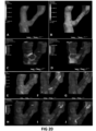
- FIGS 20 C, D show the predicted arterial wall strain distribution of the same 3D reconstructed carotid segment, as in Figures 20 A, B , and specifically the endothelial strain distribution (C) and the arterial wall strain distribution (D).
- the model's third step includes the transient simulation of all species' dynamics within the arterial wall. Initially, in this step, a steady state analysis is performed to evaluate the accurate plasma dynamics within the arterial wall, considering the effects of arterial wall's porosity, as well as the peculiarities of the endothelial layer ( Figure 20E ).
- Agent Based Model The problem of atherosclerosis includes components that contribute to plaque formation and progression, each with proper characteristics, behaviors, and sets of rules. The interaction between these components and the environment they develop determines plaque progression. For this reason, the inventors chose the Agent Based Model (ABM) as the right approach to reproduce the evolution of plaque progression and arterial reshaping by simulating the behavior of agents.
- the key components of agent-based models are agents, rules, environment and time-scale.
- ABM provides a way to capture the complexity and heterogeneity of atherosclerosis by representing individual components and their interactions. This approach allows researchers to explore emergent phenomena, understand system dynamics, and test hypotheses in a simulated environment.
- the WSS values are calculated with CFD simulation, while the ABM has been used for the arterial wall remodeling.
- all geometry changes from the ABM were transformed to fluid domain which cause blood flow calculation and again WSS determination for next step to update the ABM.
- Each geometrical change computed by the ABM in 2D cross-sections affects the fluid dynamics domain. For that reason, the ABM module is coupled to the CFD module and by this coupling, the WSS distribution is always updated.
- the 2D ABM replicates remodeling of the arterial wall by simulating cell mitosis, production or degradation of the ECM and lipid infiltration in the intima. Also, different vessel structures and compositions were implemented, as well as dealing with new cellular events.
- the ABM behavior was validated in an environment with atherogenic conditions.
- CFD-ABM coupling was performed to initialize the ABM with hemodynamic input.
- 2D circular cross-section composed of 3 concentric layers (tunica intima, media and adventitia) made initial position.
- the 2D ABM is fed with an initial WSS profile Bentzon et al., Circ Res. 114(12), 1852-1866 ).
- low WSS affects endothelial function by reducing atheroprotective genes and increasing the atherogenic ones, which leads to atherosclerotic plaque formation.
- WSS c values were obtained from the 3D CFD simulation.
- a level of endothelial dysfunction D i was computed as given in Eq. (122).
Landscapes
- Health & Medical Sciences (AREA)
- Engineering & Computer Science (AREA)
- Medical Informatics (AREA)
- Public Health (AREA)
- Epidemiology (AREA)
- General Health & Medical Sciences (AREA)
- Primary Health Care (AREA)
- Biomedical Technology (AREA)
- Data Mining & Analysis (AREA)
- Databases & Information Systems (AREA)
- Pathology (AREA)
- Radiology & Medical Imaging (AREA)
- Nuclear Medicine, Radiotherapy & Molecular Imaging (AREA)
- Measuring And Recording Apparatus For Diagnosis (AREA)
Priority Applications (1)
| Application Number | Priority Date | Filing Date | Title |
|---|---|---|---|
| EP23220881.9A EP4579682A1 (de) | 2023-12-31 | 2023-12-31 | Verfahren und system zur risikostratifizierung asymptomatischer patienten mit karotidstenose |
Applications Claiming Priority (1)
| Application Number | Priority Date | Filing Date | Title |
|---|---|---|---|
| EP23220881.9A EP4579682A1 (de) | 2023-12-31 | 2023-12-31 | Verfahren und system zur risikostratifizierung asymptomatischer patienten mit karotidstenose |
Publications (1)
| Publication Number | Publication Date |
|---|---|
| EP4579682A1 true EP4579682A1 (de) | 2025-07-02 |
Family
ID=89474260
Family Applications (1)
| Application Number | Title | Priority Date | Filing Date |
|---|---|---|---|
| EP23220881.9A Pending EP4579682A1 (de) | 2023-12-31 | 2023-12-31 | Verfahren und system zur risikostratifizierung asymptomatischer patienten mit karotidstenose |
Country Status (1)
| Country | Link |
|---|---|
| EP (1) | EP4579682A1 (de) |
-
2023
- 2023-12-31 EP EP23220881.9A patent/EP4579682A1/de active Pending
Non-Patent Citations (33)
| Title |
|---|
| ANBEEK P ET AL., NEUROIMAGE, vol. 27, no. 4, 2005, pages 795 - 804 |
| ANNU INT CONF IEEE ENG MED BIOL SOC., vol. 2020, 2020, pages 2808 - 2811 |
| BENTZON ET AL., CIRC RES, vol. 114, no. 12, pages 1852 - 1866 |
| BRANKO ARSIC: "Atherosclerotic Plaque Characterization Using Deep Learning Methods", AORTA, 10 June 2022 (2022-06-10), pages 1 - 2, XP093167203, Retrieved from the Internet <URL:https://www.thieme-connect.de/products/ejournals/abstract/10.1055/s-0042-1750994> [retrieved on 20240527], DOI: 10.1055/s-0042-1750994 (http://dx.doi.org/10.1055/s-0042-1750994 * |
| BURKE AP ET AL., CIRCULATION, vol. 103, no. 7, 2001, pages 934 - 40 |
| CORTI ET AL., COMPUT. BIOL. MED., vol. 118, pages 103623 |
| GARBEY ET AL., J. THEOR. BIOL., vol. 429, 2017, pages 149 - 163 |
| HEART, vol. 82, no. 3, September 1999 (1999-09-01), pages 265 - 8 |
| KERKTOLD K ET AL., J. BIOMECH., vol. 24, no. 6, 1991, pages 409 - 420 |
| KIGKA VASSILIKI I ET AL: "A Machine Learning Model for the Identification of High risk Carotid Atherosclerotic Plaques", 2021 43RD ANNUAL INTERNATIONAL CONFERENCE OF THE IEEE ENGINEERING IN MEDICINE & BIOLOGY SOCIETY (EMBC), IEEE, 1 November 2021 (2021-11-01), pages 2266 - 2269, XP034043401, DOI: 10.1109/EMBC46164.2021.9630654 * |
| KIGKA VASSILIKI I ET AL: "Detection of Asymptomatic Carotid Artery Stenosis through Machine Learning", 2022 44TH ANNUAL INTERNATIONAL CONFERENCE OF THE IEEE ENGINEERING IN MEDICINE & BIOLOGY SOCIETY (EMBC), IEEE, 11 July 2022 (2022-07-11), pages 1041 - 1044, XP034183143, DOI: 10.1109/EMBC48229.2022.9870927 * |
| KIGKA VASSILIKI I ET AL: "Early Diagnosis of Carotid Artery Disease based on non-imaging data", 2023 IEEE EMBS SPECIAL TOPIC CONFERENCE ON DATA SCIENCE AND ENGINEERING IN HEALTHCARE, MEDICINE AND BIOLOGY, IEEE, 7 December 2023 (2023-12-07), pages 129 - 130, XP034533139, DOI: 10.1109/IEEECONF58974.2023.10404873 * |
| KIGKA VI ET AL., BIOMED. SIGNAL PROCESS. CONTROL, vol. 40, 2018, pages 286 - 294 |
| KONTUSH A., CURR OPIN LIPIDOL., vol. 21, no. 4, 2010, pages 312 - 8 |
| MANTZARIS MICHALIS D ET AL: "Computational modeling of atherosclerotic plaque progression in carotid lesions with moderate degree of stenosis*", 2021 43RD ANNUAL INTERNATIONAL CONFERENCE OF THE IEEE ENGINEERING IN MEDICINE & BIOLOGY SOCIETY (EMBC), IEEE, 1 November 2021 (2021-11-01), pages 4209 - 4212, XP034041578, DOI: 10.1109/EMBC46164.2021.9630376 * |
| MANTZARIS MICHALIS ET AL: "A Multimodal Advanced Approach for the Stratification of Carotid Artery Disease", 2019 IEEE 19TH INTERNATIONAL CONFERENCE ON BIOINFORMATICS AND BIOENGINEERING (BIBE), IEEE, 28 October 2019 (2019-10-28), pages 706 - 709, XP033680509, DOI: 10.1109/BIBE.2019.00133 * |
| METZT ET AL., MEDICAL PHYSICS., vol. 36, 2009, pages 5568 - 5579 |
| NAYLOR AR ET AL., EUR J VASC ENDOVASC SURG., vol. 55, 2018, pages 3e81 |
| PASTERKAMP G ET AL., NAT REV CARDIOL., vol. 14, no. l, pages 21 - 29 |
| PEZOULAS ET AL., IEEE OPEN J. ENG. MED. BIOL., vol. 1, 2020, pages 83 - 90 |
| PLEOURAS DIMITRIOS S ET AL: "Prediction of the atherosclerotic plaque development in carotid arteries; the effect of T-cells", 2022 44TH ANNUAL INTERNATIONAL CONFERENCE OF THE IEEE ENGINEERING IN MEDICINE & BIOLOGY SOCIETY (EMBC), IEEE, 11 July 2022 (2022-07-11), pages 1590 - 1593, XP034182014, DOI: 10.1109/EMBC48229.2022.9871632 * |
| SAKELLARIOS ET AL., AM. J. PHYSIOL. HEART CIRC. PHYSIOL., vol. 304, no. 11, 2013, pages H1455 - 1470 |
| SAMADY ET AL., CIRCULATION, vol. 124, no. 7, 2011, pages 779 - 788 |
| SAVVAS KYRIAKIDIS: "An All-in-One Tool for 2D Atherosclerotic Disease Assessment and 3D Coronary Artery Reconstruction", JOURNAL OF CARDIOVASCULAR DEVELOPMENT AND DISEASE, vol. 10, no. 3, 19 March 2023 (2023-03-19), pages 130, XP093166784, ISSN: 2308-3425, DOI: 10.3390/jcdd10030130 * |
| STONE ET AL., CIRCULATION, vol. 126, 2012, pages 172 - 181 |
| STONE GW ET AL., N ENGL J MED, vol. 364, no. 3, 20 January 2011 (2011-01-20), pages 226 - 35 |
| TENG Z ET AL., ACTA BIOMATER, vol. 10, no. 12, 2014, pages 5055 - 5063 |
| TSAKANIKAS VASSILIS D ET AL: "A deep learning oriented method for automated 3D reconstruction of carotid arterial trees from MR imaging", 2020 42ND ANNUAL INTERNATIONAL CONFERENCE OF THE IEEE ENGINEERING IN MEDICINE & BIOLOGY SOCIETY (EMBC), IEEE, 20 July 2020 (2020-07-20), pages 2408 - 2411, XP033816515, DOI: 10.1109/EMBC44109.2020.9176532 * |
| TSAKANIKAS VASSILIS D ET AL: "TAXINOMISIS: A cloud - based platform for risk profiling and patient specific management of the carotid artery disease", 2023 45TH ANNUAL INTERNATIONAL CONFERENCE OF THE IEEE ENGINEERING IN MEDICINE & BIOLOGY SOCIETY (EMBC), IEEE, 24 July 2023 (2023-07-24), pages 1 - 4, XP034487899, DOI: 10.1109/EMBC40787.2023.10340947 * |
| TSOMPOU PANAGIOTA I ET AL: "Computational modeling of atherosclerotic plaque progression through an efficient 3D agent-based modeling approach", 2022 IEEE-EMBS INTERNATIONAL CONFERENCE ON BIOMEDICAL AND HEALTH INFORMATICS (BHI), IEEE, 27 September 2022 (2022-09-27), pages 1 - 4, XP034219338, DOI: 10.1109/BHI56158.2022.9926888 * |
| VANLAMMEREN GW ET AL., CIRCULATION, vol. 129, no. 22, 2014, pages 2269 - 76 |
| VUKICEVIC AM ET AL., MED. BIOL. ENG. COMPUT., vol. 52, 2014, pages 539 - 556 |
| VUKICEVIC AM ET AL., SCI REP, vol. 8, 2018, pages 1711 |
Similar Documents
| Publication | Publication Date | Title |
|---|---|---|
| JP7542578B2 (ja) | 定量的イメージングを利用するための方法およびシステム | |
| Li et al. | Automatic coronary artery segmentation and diagnosis of stenosis by deep learning based on computed tomographic coronary angiography | |
| Retson et al. | Machine learning and deep neural networks in thoracic and cardiovascular imaging | |
| Slomka et al. | Cardiac imaging: working towards fully-automated machine analysis & interpretation | |
| US20210098113A1 (en) | Medical image identification and interpretation | |
| CN109615636B (zh) | Ct影像的肺叶段分割中的血管树构造方法、装置 | |
| He et al. | Automatic segmentation and quantification of epicardial adipose tissue from coronary computed tomography angiography | |
| CN112686899A (zh) | 医学图像分析方法和装置、计算机设备及存储介质 | |
| Yagis et al. | Deep learning for 3D vascular segmentation in hierarchical phase contrast tomography: a case study on kidney | |
| Dong et al. | A novel end‐to‐end deep learning solution for coronary artery segmentation from CCTA | |
| Al-Kadi et al. | Empowering brain cancer diagnosis: harnessing artificial intelligence for advanced imaging insights | |
| Xiang et al. | ADSeg: A flap-attention-based deep learning approach for aortic dissection segmentation | |
| Liu et al. | Deep learning based retinal vessel segmentation and hypertensive retinopathy quantification using heterogeneous features cross-attention neural network | |
| Wang et al. | Role of artificial intelligence in medical image analysis | |
| Chakshu et al. | Automating fractional flow reserve (FFR) calculation from CT scans: A rapid workflow using unsupervised learning and computational fluid dynamics | |
| Li et al. | An automatic pipeline for segmentation and quantification of intravascular ultrasound images | |
| Chacón et al. | Computational assessment of stomach tumor volume from multi-slice computerized tomography images in presence of type 2 cancer | |
| EP4579682A1 (de) | Verfahren und system zur risikostratifizierung asymptomatischer patienten mit karotidstenose | |
| Peng et al. | A windowing-based multi-view u-net for tumor segmentation in cone-beam breast CT | |
| Mahmoudi et al. | Fully Automated Aortic Root Localization and Tilt Alignment in Cardiac Computed Tomography | |
| Müller | Frameworks in medical image analysis with deep neural networks | |
| Kozhamkulova et al. | TransAneu-Net: A Hybrid Radiomics and Contrastive Deep Learning Framework for Automated Brain Aneurysm Diagnosis. | |
| Tsakanikas et al. | TAXINOMISIS: A cloud–based platform for risk profiling and patient specific management of the carotid artery disease | |
| WO2026055511A1 (en) | Systems and methods for use of generative artificial intelligence (ai) in cardiac patient care | |
| Ntetska et al. | From Pixels to Prognosis: Deep Learning Methods in Abdominal Aortic Aneurysm Imaging |
Legal Events
| Date | Code | Title | Description |
|---|---|---|---|
| PUAI | Public reference made under article 153(3) epc to a published international application that has entered the european phase |
Free format text: ORIGINAL CODE: 0009012 |
|
| STAA | Information on the status of an ep patent application or granted ep patent |
Free format text: STATUS: THE APPLICATION HAS BEEN PUBLISHED |
|
| AK | Designated contracting states |
Kind code of ref document: A1 Designated state(s): AL AT BE BG CH CY CZ DE DK EE ES FI FR GB GR HR HU IE IS IT LI LT LU LV MC ME MK MT NL NO PL PT RO RS SE SI SK SM TR |
|
| STAA | Information on the status of an ep patent application or granted ep patent |
Free format text: STATUS: THE APPLICATION IS DEEMED TO BE WITHDRAWN |