EP4559486A1 - Vorrichtung für chirurgische reparatur von zylindrischen organen, insbesondere gerissenen sehnen, und verfahren zur herstellung solch einer vorrichtung - Google Patents
Vorrichtung für chirurgische reparatur von zylindrischen organen, insbesondere gerissenen sehnen, und verfahren zur herstellung solch einer vorrichtung Download PDFInfo
- Publication number
- EP4559486A1 EP4559486A1 EP23211132.8A EP23211132A EP4559486A1 EP 4559486 A1 EP4559486 A1 EP 4559486A1 EP 23211132 A EP23211132 A EP 23211132A EP 4559486 A1 EP4559486 A1 EP 4559486A1
- Authority
- EP
- European Patent Office
- Prior art keywords
- peo
- scaffolds
- tendon
- tendons
- fibers
- Prior art date
- Legal status (The legal status is an assumption and is not a legal conclusion. Google has not performed a legal analysis and makes no representation as to the accuracy of the status listed.)
- Withdrawn
Links
Images


Classifications
-
- A—HUMAN NECESSITIES
- A61—MEDICAL OR VETERINARY SCIENCE; HYGIENE
- A61L—METHODS OR APPARATUS FOR STERILISING MATERIALS OR OBJECTS IN GENERAL; DISINFECTION, STERILISATION OR DEODORISATION OF AIR; CHEMICAL ASPECTS OF BANDAGES, DRESSINGS, ABSORBENT PADS OR SURGICAL ARTICLES; MATERIALS FOR BANDAGES, DRESSINGS, ABSORBENT PADS OR SURGICAL ARTICLES
- A61L31/00—Materials for other surgical articles, e.g. stents, stent-grafts, shunts, surgical drapes, guide wires, materials for adhesion prevention, occluding devices, surgical gloves, tissue fixation devices
- A61L31/04—Macromolecular materials
- A61L31/041—Mixtures of macromolecular compounds
-
- A—HUMAN NECESSITIES
- A61—MEDICAL OR VETERINARY SCIENCE; HYGIENE
- A61L—METHODS OR APPARATUS FOR STERILISING MATERIALS OR OBJECTS IN GENERAL; DISINFECTION, STERILISATION OR DEODORISATION OF AIR; CHEMICAL ASPECTS OF BANDAGES, DRESSINGS, ABSORBENT PADS OR SURGICAL ARTICLES; MATERIALS FOR BANDAGES, DRESSINGS, ABSORBENT PADS OR SURGICAL ARTICLES
- A61L27/00—Materials for grafts or prostheses or for coating grafts or prostheses
- A61L27/14—Macromolecular materials
- A61L27/18—Macromolecular materials obtained otherwise than by reactions only involving carbon-to-carbon unsaturated bonds
-
- A—HUMAN NECESSITIES
- A61—MEDICAL OR VETERINARY SCIENCE; HYGIENE
- A61L—METHODS OR APPARATUS FOR STERILISING MATERIALS OR OBJECTS IN GENERAL; DISINFECTION, STERILISATION OR DEODORISATION OF AIR; CHEMICAL ASPECTS OF BANDAGES, DRESSINGS, ABSORBENT PADS OR SURGICAL ARTICLES; MATERIALS FOR BANDAGES, DRESSINGS, ABSORBENT PADS OR SURGICAL ARTICLES
- A61L27/00—Materials for grafts or prostheses or for coating grafts or prostheses
- A61L27/14—Macromolecular materials
- A61L27/26—Mixtures of macromolecular compounds
-
- A—HUMAN NECESSITIES
- A61—MEDICAL OR VETERINARY SCIENCE; HYGIENE
- A61L—METHODS OR APPARATUS FOR STERILISING MATERIALS OR OBJECTS IN GENERAL; DISINFECTION, STERILISATION OR DEODORISATION OF AIR; CHEMICAL ASPECTS OF BANDAGES, DRESSINGS, ABSORBENT PADS OR SURGICAL ARTICLES; MATERIALS FOR BANDAGES, DRESSINGS, ABSORBENT PADS OR SURGICAL ARTICLES
- A61L27/00—Materials for grafts or prostheses or for coating grafts or prostheses
- A61L27/50—Materials characterised by their function or physical properties, e.g. injectable or lubricating compositions, shape-memory materials, surface modified materials
- A61L27/58—Materials at least partially resorbable by the body
-
- A—HUMAN NECESSITIES
- A61—MEDICAL OR VETERINARY SCIENCE; HYGIENE
- A61L—METHODS OR APPARATUS FOR STERILISING MATERIALS OR OBJECTS IN GENERAL; DISINFECTION, STERILISATION OR DEODORISATION OF AIR; CHEMICAL ASPECTS OF BANDAGES, DRESSINGS, ABSORBENT PADS OR SURGICAL ARTICLES; MATERIALS FOR BANDAGES, DRESSINGS, ABSORBENT PADS OR SURGICAL ARTICLES
- A61L31/00—Materials for other surgical articles, e.g. stents, stent-grafts, shunts, surgical drapes, guide wires, materials for adhesion prevention, occluding devices, surgical gloves, tissue fixation devices
- A61L31/04—Macromolecular materials
- A61L31/06—Macromolecular materials obtained otherwise than by reactions only involving carbon-to-carbon unsaturated bonds
-
- A—HUMAN NECESSITIES
- A61—MEDICAL OR VETERINARY SCIENCE; HYGIENE
- A61L—METHODS OR APPARATUS FOR STERILISING MATERIALS OR OBJECTS IN GENERAL; DISINFECTION, STERILISATION OR DEODORISATION OF AIR; CHEMICAL ASPECTS OF BANDAGES, DRESSINGS, ABSORBENT PADS OR SURGICAL ARTICLES; MATERIALS FOR BANDAGES, DRESSINGS, ABSORBENT PADS OR SURGICAL ARTICLES
- A61L31/00—Materials for other surgical articles, e.g. stents, stent-grafts, shunts, surgical drapes, guide wires, materials for adhesion prevention, occluding devices, surgical gloves, tissue fixation devices
- A61L31/14—Materials characterised by their function or physical properties, e.g. injectable or lubricating compositions, shape-memory materials, surface modified materials
- A61L31/148—Materials at least partially resorbable by the body
-
- A—HUMAN NECESSITIES
- A61—MEDICAL OR VETERINARY SCIENCE; HYGIENE
- A61L—METHODS OR APPARATUS FOR STERILISING MATERIALS OR OBJECTS IN GENERAL; DISINFECTION, STERILISATION OR DEODORISATION OF AIR; CHEMICAL ASPECTS OF BANDAGES, DRESSINGS, ABSORBENT PADS OR SURGICAL ARTICLES; MATERIALS FOR BANDAGES, DRESSINGS, ABSORBENT PADS OR SURGICAL ARTICLES
- A61L2430/00—Materials or treatment for tissue regeneration
- A61L2430/10—Materials or treatment for tissue regeneration for reconstruction of tendons or ligaments
Definitions
- the present invention relates to a device for repair surgery of cylindrical organs, particularly ruptured tendons. Moreover, the invention relates to a method of producing such a device.
- Tendon ruptures constitute a major part of musculoskeletal injuries, with Achilles tendon ruptures as one of the most frequently ruptured tendons in the human body [A1]. Functional loss of the repaired tendons is due to two major drawbacks, (i) adhesion formation to the surrounding tissue, resulting in a reduced range of motion and (ii) insufficient mechanical strength acquired during initial tendon healing, leading to re-rupture [A1]. Due to limited tendon vascularity and innervation, the natural healing of the tendon is inefficient [A2] and therapeutic repair options include autografts, allografts, xenografts, suture techniques and tendon prostheses [A3].
- PDGF-BB platelet-derived growth factor - BB
- This approach aims to provide a bioactive scaffold that can locally promote and accelerate tendon healing.
- Administration of biological molecules like growth factors has been proposed and studied in aiding the recapitulation of the native tendon function after injury [A8]. More specifically, PDGF-BB has been shown to affect matrix remodeling, increase collagen synthesis and cell proliferation [A9], thus its local delivery might also affect the biomechanical strength of the repaired tendons.
- WO 2017/137602 A1 discloses a device for repair surgery of cylindrical organs, particularly ruptured tendons, is configured as a tubular sheath (T) made of a biocompatible and biodegradable polymer.
- the tubular sheath comprises an elastic fiber mesh formed by electrospinning of said polymer and has an inner wall surface and an outer wall surface substantially parallel thereto.
- One of said wall surfaces is comparatively rough (W R ) and the other one of said wall surfaces is comparatively smooth (Ws), with the tubular sheath having a Young elasticity modulus of about 0.1 to about 4 MPa and an elongation at break of about 50 to about 1'000 %.
- the device is configured as a tubular sheath (T) made of a mesh of elastic fibers formed by electrospinning biocompatible and biodegradable polymers, said tubular sheath having a Young elasticity modulus of about 0.1 to about 4 MPa and a strain at break of about 50 to about 1'000 %.
- the tubular sheath has a first, inner wall surface (W I ) and a second, outer wall surface (Wo) substantially parallel thereto.
- the first, inner wall surface is comparatively rough (W R ) and the second, outer wall surface is comparatively smooth (Ws).
- the device may be used for repair surgery of a whole variety of cylindrical organs such as nerves, blood vessels and certain muscles in humans or other animals, particularly mammals, it is particularly useful for repair surgery of ruptured tendons in humans.
- cylindrical shall be understood as "having substantially cylindrical symmetry" in line with the fact that human and animal organs do not exhibit perfect cylindricity in the geometrical sense both because of inherent slight irregularities of their shape and variations in cross sectional size, but also because of their deformability.
- polymer with an admixture of a therapeutic agent for stimulating regrowth processes shall be understood in a broad sense so as to include various types of loading the therapeutic agent onto or into a polymeric fiber.
- biocompatible and biodegradable are generally known in the field of surgery. Synthetic biocompatible and bioresorbable polymers are becoming increasingly popular for surgical applications either as tissue engineered artificial grafts and/ or as bioactive carrier devices delivering growth factors [A10], cytokines and other bioactive substances [A11], [A12], [A13]. Tendon grafts can also be seeded with stem cells improving the early healing process [A14]. The advantage of such polymers is that their mechanical properties as well as their degradation rates can be controlled and adjusted for specific medical applications [A15], [A16], [A5], [A17]. Moreover, tissue integration can be regulated by porosity and architecture of the material used for grafting [A18].
- a tubular sheath comprising an elastic polymeric fiber mesh by electrospinning
- the resulting sheath has a comparatively smooth surface on its inner wall adjacent the target and a comparatively rough outer surface on its outer wall.
- the smooth surface will have a dynamic friction coefficient of about 0.85 whereas the rough surface will have a dynamic friction coefficient of about 1.05.
- this difference in surface roughness can be exploited in repair surgery by first everting the tubular sheath so as to have the rough surface as the inner wall. This allows for firm contact of the sheath with the cylindrical organ to be repaired and reduces the friction between the outer wall and any surrounding tissue, thereby improving mobility of the repaired organ, for example gliding of a tendon in the tendon sheath.
- the device of the present invention provides for a substantively improved healing process.
- HA high molecular weight hyaluronic acid
- PEO polyethylene oxide
- the tubular sheath By having a Young elasticity modulus of about 0.1 to about 4 MPa and a strain at break of about 50 to about 1'000 %, the tubular sheath can be readily expanded when being placed on the repaired region and it can subsequently provide a long lasting radially inward directed pressure that contributes to the improved healing process.
- the diameter of the tubular sheath will be selected according to the size of the organ to be repaired. In general, it will be chosen to be slightly narrower than the outer diameter of the organ to be repaired, so that the tubular sheath can be applied onto the relevant organ region after slight radial expansion.
- the tubular shell will have a diameter in the range of about 1 to about 5 mm.
- the first polymeric fibers are formed from degrapol (DP) with an admixture of a therapeutic agent (GA) for stimulating regrowth processes of a predetermined cylindrical organ.
- DP degrapol
- GA therapeutic agent
- the first tubular layer and the second tubular layer each form about one half of the tubular sheath's wall thickness.
- the biocompatible and biodegradable polymers used to form the first fibers and the second fibers, respectively may be different polymeric species.
- the polymer used to form the first fibers henceforth also called “first polymer”
- first polymer may be selected to optimize the release of any therapeutic agent to be delivered and to optimize mechanical properties of the device.
- second polymer may be selected to optimize the anti-adhesive properties of the device.
- the first polymer is a biodegradable polyester urethane block copolymer with poly-hydroxy-butyrate as a hard segment and ⁇ -caprolactone as a soft segment.
- Such polymers are known and can be purchased as DegraPol ® from ab medica s.p.a., Italy. Degrapol has been shown to be biocompatible for fibroblasts, osteoblasts and tenocytes in vitro; moreover, it is biodegradable as well as cyto- and hemocompatible [A15], [A5]. The degradation rate can be adjusted in the range from a few weeks up to a few years. It is contemplated that different subtypes of the above mentioned polyester urethane block copolymer can be used to form the first and second polymer fibers.
- a particularly preferred embodiment involves a selected type of the above mentioned polyester urethane block copolymer, characterized by a soft segment with an average molecular weight of about 900g/mol to about 1'250 g/mol, a relative content of said soft segment of about 60 to about 75 parts by weight and a relative content of said hard segment of about 40 to about 25 parts by weight.
- relative content is used here because, as will be generally known, formation of such block copolymers further requires the addition of an appropriate coupling agent. In the present case, this is an isocyanate coupler.
- average molecular weights reported here are number averaged molecular weights M n , which can be determined e.g. by means of gel permeation chromatography (GPC).
- the tubular sheath has a Young elasticity modulus of about 0.4 to about 2.5 MPa and a strain at break of about 200 to about 1000 %.
- the device of the present invention is generally intended for repair surgery of organs with substantially cylindrical symmetry.
- This also includes configurations with variable diameter.
- the tubular sheath is of substantially frustoconical shape, i.e. it has a diameter that monotonously decreases in one axial direction.
- the half-aperture angle i.e. the angle between the wall surface and the longitudinal axis will be in the range of about 1 to about 10°.
- the useful tubular shapes also include configurations with a nonlinear diameter variation, i.e. having a curved longitudinal section. The specific shape will be selected according to the shape of the organ to be repaired. As will be understood, the manufacture of tubular sheaths with such specific shapes by electrospinning is readily achieved by selecting an appropriately formed deposition target.
- the first fibers are heterogeneous filaments having included cavities filled with said therapeutic agent.
- Such fibers can be produced by the method of emulsion electrospinning, in which the process is applied to an emulsion of the polymer and an aqueous solution containing the therapeutic agent.
- the agent filled cavities will have varying sizes and will be distributed randomly within the fiber. The release of therapeutic agent will begin as soon as there has been sufficient polymer degradation to expose some of the filled cavities.
- the first fibers are hollow filaments having a central core filled with said therapeutic agent.
- the release of therapeutic agent will begin in a nearly step-like manner once the polymer forming the tube wall has sufficiently degraded.
- the first tubular region may form about 5 to about 95 percent of the wall thickness, with the remainder being formed by the second tubular region. In this manner one can optimize the amount of therapeutic agent that is ultimately released by the device and the mechanical properties. According to one embodiment, the first tubular region and the second tubular region each form about one half of the sheath's wall thickness.
- the device of the present invention is suitable for delivery of a large variety of therapeutic agents, which term shall be interpreted in a broad sense and include any agents that could have a beneficial effect on the healing process after repair surgery.
- the therapeutic agent can be selected from the group consisting of growth hormones, pharmaceutical agents and growth promoting cells, including stem cells.
- the therapeutic agent is platelet-derived growth factor - BB or insulin-like growth factor-1 [A19].
- a method of producing a device as defined above in which method said elastic fibers are formed by solution electrospinning.
- This technique is generally known.
- solution electrospinning is based on using a solution of the respective polymeric species in a volatile solvent. The solution is driven from a reservoir through a nozzle or needle, after the exit of which the solvent evaporates and a thread of first polymer fibers is formed.
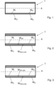
- the device for repair surgery of cylindrical organs, particularly ruptured tendons, as shown in Fig. 1 is configured as a tubular sheath T made of a biocompatible and biodegradable polymer.
- the tubular sheath T is approximately cylindrically shaped along a longitudinal axis A. It comprises an elastic fiber mesh formed by electrospinning of said polymers and has an inner wall surface W I and an outer wall surface Wo substantially parallel thereto.
- One of said wall surfaces - in the presently shown case it is the inner wall surface W I - is comparatively rough W R whereas the other one of said wall surfaces - in the presently shown case it is the outer wall surface Wo - is comparatively smooth Ws .
- the configuration shown in Fig. 1 is the one to be used for the intended surgical application.
- this configuration is obtained from a freshly produced electrospun tubular sheath by everting the same, i.e. by switching the inner and outer side thereof.
- the outer layer comprises fibers of polyethylene oxide and fibers of hyaluronic acid.
- the inner layer consists of neat degrapol.
- the inner layer consists of neat degrapol including a therapeutic agent GA.
- a 2-layered implant was made by electrospinning. It was characterized with respect to surface characteristics (fiber thickness and wall thick-ness by SEM). In addition, the water contact angle has been determined. Moreover, it was implanted around a fully transected rabbit Achilles tendon that was sutured. After 3 weeks, histological analysis of the adhesion extent revealed negligible adhesion - on the same level as non-treated naive Achilles tendons, which is a highly promising result. In the same model without such a tube, we had approximately 40 - 50 % of adhesion. The rabbit Achilles tendon full transection model is used because the tendons have approximately the same strength as typical human tendons of the hand.
- tubular implant acts anti-adhesive in an excellent manner (0-5 % of adhesion as the natural tendon). There was no adhesion formation, which was caused by the anti-adhesive effects that are attributed to the bio-lubricant hyaluronic acid in the layer facing the surrounding tissue.
- Tendons consist of dense viscoelastic, fibrous connective tissue and transfer muscle force to bones enabling joint motion and stabilization. They are hypovascular [1, 2] and hypocellular with a low metabolic activity leading to a limited natural healing capacity, frequently resulting in the formation of fibrovascular scar tissue and inferior mechanical and functional properties [3]. Accidents or overuse in daily life or in sports, but also age-related degeneration or adiposity [4], may be risk factors for tendon disease affecting four millions new patients per year worldwide [1, 5-7]. Tendons heal in three overlapping phases: Immediately after injury, an inflammatory response occurs including intrinsic and extrinsic pathways lasting for about 3 - 7 days [8].
- the injury site becomes hypercellular characterized by an increase in myofibroblasts and deposition of new extracellular matrix (ECM) material.
- ECM extracellular matrix
- the remodeling phase begins replacing the randomly orientated collagen III fibers by collagen I and orientating them in longitudinal structures [9-11].
- the third phase can last up to one year, quality of the healed tendon remains inferior due to still decreased fiber orientation and formation of scar tissue leading to worse biomechanical properties.
- Several animal models have confirmed these findings [12, 13] documenting that repaired tendons reach only 40-70 % of uninjured tendon strength [14-16]. Re-rupture of the repaired tendons, joint stiffness and adhesion formation are common clinical problems leading to secondary surgeries, persisting functional disability and impairments in patients daily lives [17].
- Rabbit Achilles tendon (AT) and human hand flexor tendons show similar biomechanical properties with nearly the same ultimate failure load [19, 20] and although not having a synovial sheath rabbit AT are inclined to develop adhesion formation [20, 21]. This makes the rabbit AT a good model to gain knowledge for improving hand injury treatments, an important field for plastic surgeons as hand injuries represent 20 % of all injuries treated in emergency [6, 22].
- An effective method to reduce adhesion formation after surgery is the use of physical barriers, limiting the contact between the tendon and the surrounding tissue but allowing cytokines, growth factors and metabolic waste to pass without disturbing tendon movement, mechanical properties and the healing process [8, 18, 23].
- Synthetic and biological biomaterials such as polymers based on hydroxy-butyrate and ⁇ -caprolactone, a biodegradable and biocompatible polyester urethane [21, 24], chitosanbased scaffolds [25], decellularized extracellular matrix (ECM) [26] or Type I collagen-based scaffolds [18, 23] have been used in animal models amongst others.
- the polyester urethane DegraPol ® 15 (DP) utilized in this study has been shown to be a good scaffold material for tenocytes [27] and previous work demonstrated that cellular response in a rabbit full laceration AT model was not impaired while adhesion formation could be reduced by about 20 % [28, 29].
- the elasticity of the electrospun DP tube is an advantage for the surgery because the material has a high strain at break compared to other commonly used materials.
- electrospun scaffolds have an ECM-like morphology with a wide range of pore diameter distribution and a high surface area-to-volume ratio and are often used as carriers for therapeutic agents [30-33].
- As adhesion extent in rabbit AT full laceration model has been shown to be reduced with pure DP implants [29] we aimed to improve the electrospun implant by adding hyaluronic acid (HA) to reduce adhesion formation even further.
- HA hyaluronic acid
- Hyaluronic acid is a major component of the ECM, playing essential roles in cell signaling, wound healing, tissue regeneration and matrix organization [34, 35]. It is an important lubricating component of the synovial fluid and supplies the tendon with nutrients [36]. Macrophages and fibroblast-like cells synthesize HA in the synovial membrane [37, 38] and its production increases after injuries, which improves cell viability and cell proliferation, and reduces inflammation [34, 39-41]. Thanks to the biocompatible and biodegradable properties, HA is often applied in medical therapies and experimental studies [34, 35, 42, 43]. Injections of HA have shown positive effects regarding tendon maturity, strength, stiffness and joint function in animal models [41, 44, 45] possibly induced by a reduced inflammation stage.
- Hyaluronic acid is an unbranched glycosaminoglycan with hydrophilic properties [34]. Effects of HA depend on its molecular weight ranging from 5 ⁇ 10 4 -2 ⁇ 10 8 Da [33] and its concentration influencing signaling pathways and differential macrophage activation [34, 39, 47] but also viscosity and viscoelasticity [34]. High molecular weight (HMW) HA (> 10 6 Da) has been shown to have immunosuppressive, anti-angiogenic and anti-inflammatory effects [48], while low molecular weight (LMW) HA shows opposite effects [49, 50].
- HMW high molecular weight
- LMW low molecular weight
- HA was the first time added to DP electrospun scaffolds, material properties were analyzed with SEM, Fourier-transformed infrared Spectroscopy (FTIR), Differential Scanning Calorimetry (DSC), Water Contact Angle (WCA), and by testing the mechanical qualities ( Fig. 4D ).
- FTIR Fourier-transformed infrared Spectroscopy
- DSC Differential Scanning Calorimetry
- WCA Water Contact Angle
- DegraPol ® 15 a biodegradable polyester urethane block copolymer, was kindly provided by Ab Medica, Italy. High molecular weight hyaloronic acid (HA, 1.01 - 1.8 MDa) was ordered from Lifecore Biomedical (Lifecore Biomedical #HA 15M, Chaska, USA).
- Chloroform (#132950), 1,1,1,3,3,3-hexa fluoro-2-propanol (HFP) (#105228), polyethylene glycol (PEG) (in average 35'000 g/mol) (#81310) and polyethylene oxide (PEO) (in average 600'0000 g/mol) (#182028) were purchased from Sigma-Aldrich (Buchs, Switzerland). Solutions for electrospinning were filled in a 5 mL glass syringe (Huberlab, #3.7102.33, Aesch, Switzerland).
- gentamycine (# L0011), Ham's F12 (# L0135-500) and FBS (# S1830-500) were bought from Biowest (Nuaillé, France) and amphotericin B (# P06-01100) from Pan Biotech (Aidenbach, Germany).
- Penicillin/streptomycin (# 15140122) and GlutaMAX TM (# 35050038) were delivered from ThermoFisher scientific (Basel, Switzerland) and phosphate buffered saline (PBS) (# D8537) from Sigma-Aldrich (Merck, Buchs, Switzerland).
- DAKO Autostainer Link48
- Target retrieval solution # GV80411
- washing buffer # K800721
- Hydrogen peroxide H 2 O 2 was bought from Sigma-Aldrich (Merck, Buchs, Switzerland, # 1.07209.0250) and Pertex from HistoLab (Biosystems, Muttenz, Switzerland #00801-EX) was used to fix coverslips.
- a mouse monoclonal primary antibody was purchased from Sigma-Aldrich (Sigma-Aldrich, Merck, Buchs, Switzerland, # F0791; 1:200 dilution) and for ⁇ -SMA staining a mouse monoclonal primary antibody from DAKO was applied (Agilent Technologies, Basel, Switzerland, # IR611; 1:2 dilution).
- HMDS 1,1,1,3,3,3-hexamethyldisilazan
- a 12 wt % DP polymer solution was prepared by dissolving DP in a mixture of chloroform/hexafluoropropylene (HFP) (80:20 wt/wt) at room temperature the day before electrospinning.
- HFP chloroform/hexafluoropropylene
- HA/PEO solutions (2 wt %) were prepared at 1:1 and 1:4 weight ratios and dissolved in MilliQ water, under stirring at 500 rpm for 48 hours at room temperature.
- PEG (30 wt %) was dissolved in chloroform at room temperature and used for electrospinning the next day.
- Electrospinning was carried out with an in-house assembled electrospinning device consisting of a DC high voltage supply (Glassman High Voltage Inc., High Bridge, NJ, US), a spinning head with a blunt end and a stainless steel tube (1 mm inner diameter and 0.3 mm wall thickness, Angst & Pfister AG, Switzerland).
- the spinning head was fixed on a transporter and connected via a Teflon hose with a syringe pump (SP210cZ, WPI, Germany).
- the solutions were filled in a 5 mL glass syringe and the electrospun fibers were collected on a metal rod with a diameter of 6 mm and a length of 450-550 mm fixed to a rotary motor (Euro Star B rotary motor, IKA Labortechnik) rotating with 500 rpm. Flow rate was set at 1 mL/h for all solutions.
- a rotary motor Euro Star B rotary motor, IKA Labortechnik
- Flow rate was set at 1 mL/h for all solutions.
- PEG and DP solutions 12.5 kV and a working distance of 18.5 cm were used while layers containing HA/PEO were produced with 20 kV and a working distance of 14 cm.
- the electrospinning was performed at room temperature (22-25 °C) with an air humidity between 21-35 % for PEG and DP, and 21-26 % for HA/PEO solutions.
- the first layer consisted of a thin PEG layer facilitating the detachment of the tube from the metal rod using 50 % ethanol.
- the Teflon hose was rinsed with chloroform and dried with air between using different solutions.
- a first layer of HA/PEO was electrospun followed by a pure DP layer and tubes were flipped before implantation.
- three layered tubes were produced with a DP layer in the middle flanked by a HA/PEO layer on each side to ensure cell seeding on a HA/PEO surface.
- Static and dynamic water contact angles on scaffold surfaces were measured by a video-based optical contact angle measuring instrument (OCA 35 Dataphysics, Germany).
- Ultimate tensile stress (UTS), fracture strain and the Young's modulus were determined as the peak stress to failure, strain at failure, and the slope of a linear fit of the stress-stain curve up to 20 % strain respectively. Calculation of mechanical parameters was performed with MATLAB (Release 2021a, The MathWorks, Inc., Natick, MA, USA). Stress was defined as force over specimen cross sectional area and strain as the percent change in length from the gage length. For the HA containing tubes, 3 tubes per material were used and 6 measurements per tube were carried out. For the pure DP tubes, 2 samples with 6 repetitions were measured.
- Rabbit tenocytes were isolated from ATs of three New Zealand White rabbits using the cell migration method (Approval by the veterinary office of Canton Zurich, reference number ZH 080/2021; 33530). Briefly, tendons were extracted from the animals and washed with PBS supplemented with 200 ⁇ g/mL gentamicin and 2.5 ⁇ g/mL amphotericin B. Tendons were cleaned from the surrounding tissue and the central part of the tendons was cut into very small pieces ( ⁇ 2 mm) and washed three times in PBS buffer.
- tissue pieces were placed into a tissue culture plate and a drop of cell culture medium was added onto each tissue piece (Ham's F12, 10 % FBS, 100 U/mL penicillin, 100 ⁇ g/mL streptomycin and 1 % GlutaMAX). Tissues were allowed to attach onto the cell culture plates for 2 hours at 37 °C and 5 % CO 2 before adding 10 mL of cell culture media into each plate. The plates with the tissues were not moved for the first 5 days, to decrease tissue detachment upon plate movement and to allow cells to start migrating out from the tissues. The first medium change was done after 5 days, and subsequently, the culture medium was changed every third day.
- Tenocyte seeded scaffolds were embedded in paraffin according to commonly established protocols and cut into 3 ⁇ m thick slices before deparaffinization with xylene and subsequent rehydration.
- Immunocytochemical staining for collagen I, fibronectin and ⁇ -SMA was carried out with standardized protocols, using Autostainer Link48. Shortly, after antigen retrieval samples were exposed to 3 % H 2 O 2 for 10 min. After washing with washing buffer, samples were blocked with serum for 30 min and primary antibody was added for 1 h. After the next washing step, samples were treated with HRP for 20 min., washed again and exposed to DAB for 10 min. Then samples were stained with hematoxylin for 10 min. and rinsed with washing buffer before they were transferred into water and dehydrated with ethanol. Slides were imaged for qualitative analysis at 100 ⁇ , 200 ⁇ and 400 ⁇ magnification using a confocal microscope (Leica AF 6000B).
- tenocyte seeded scaffolds were washed once in PBS and dehydrated in a series of ascending concentration of ethanol (30 %, 50 %, 70 %, 80 %, 95 % (5 min each step) and 100 % (2 times 10 min). Subsequently, scaffolds were chemically dried in HMDS/ethanol mix (1:3, 1:1, 3:1) and pure HMDS, each step for 15 min). HMDS was allowed to evaporate overnight, and the samples were mounted on SEM stubs. For SEM imaging see preceding section 2.2.3.
- the tubes were sterilized with H 2 O 2 (plasma sterilization) before implantation and flipped over the wound so that the HA containing layer was facing the surrounding tissue. Afterwards the wound was closed with a running suture (using a USP 6.0 polypropylene fibre) and a well-padded cast was applied with an angle of 180° at the ankle.
- the rabbits got a Durogesic Matrix patch after surgery (Janssen-Cilag AG, Switzerland) with 4.2 mg Fentanyl per patch to provide analgesia for about 72 h with 25 ⁇ g/h Fentanyl.
- the rabbits were euthanized three weeks later in deep anaesthesia (100 mg/kg Ketamine and 4 mg/kg Xylazine) with 80 mg/kg Pentobarbital (Esconarkon ad us. vet., Switzerland) and the tendons were removed. Surgery was performed on one hind leg while the counter hind leg was not treated (NT) and served as control. The extracted tendons were immediately frozen and stored at -20 °C in a gauze moistened with 0.9 % NaCl-solution.
- the tendons were thawed overnight at 4 °C and warmed to RT before they were dehydrated and embedded in paraffin according to commonly established protocols.
- Cros sections of 5 ⁇ m in the wound region (perpendicular to the Achilles tendon) were deparaffinized with xylene and rehydrated prior histological staining with Hematoxylin-Eosin (H&E), Alcian Blue (AB) and Picrosirius Red (PS) according to commonly established procedures.
- H&E Hematoxylin-Eosin
- AB Alcian Blue
- PS Picrosirius Red
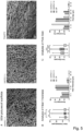
- Hyaluronic acid containing scaffolds were compared to pure DP scaffolds using SEM images ( Fig. 5A ), showing a network of random fibers with a diameter of about 6 ⁇ m in average in every material (HA/PEO (1:1) 6.1 ⁇ m ⁇ 2.5 ⁇ m, HA/PEO (1:4) 6.7 ⁇ m ⁇ 3.0 ⁇ m, DP 6.7 ⁇ m ⁇ 2.8 ⁇ m) ( Fig. 5B ).
- Pore sizes of scaffolds containing HA were significantly larger than in pure DP scaffolds; with HA/PEO (1:1) 14.5 ⁇ m ⁇ 6.3 ⁇ m, HA/PEO (1:4) 15.8 ⁇ m ⁇ 6.8 ⁇ m and DP 9.6 ⁇ m ⁇ 6.2 ⁇ m ( Fig. 5C ). Nearly 60 % of the pores in HA containing scaffolds showed a diameter of 10-20 ⁇ m, three times more pores than in DP scaffolds in which about 70 % of the pores were smaller than 10 ⁇ m ( Fig. 5C ).
- the static WCA provides information about the wettability of materials and is lower than 90° for hydrophilic surfaces, whereas hydrophobic surfaces show WCAs higher than 90°.
- HA is highly hydrophilic
- CAH Contact angle hysteresis
- the peaks at 2940 cm -1 and 2860 cm -1 correspond to stretching vibrations of -CH 2 and CH 3 groups, while the small peak at about 3380 cm -1 is associated with NH stretching. In contrast, the peak at 2360 cm -1 results from free CO 2 in the device during measurement.
- Thermograms to determine glass transition temperature (T g ) of amorphous material and melting point (T m ) of crystalline components were assessed using DSC [67] ( Fig. 6C ).
- Addition of HA and PEO did not change the typical thermal pattern of DP with a T g at about -40 °C and a T m at around 130 °C.
- the prominent endothermic peak at about 56 °C corresponds to T m of PEG, used as first layer to facilitate removal of the tube from the metal, or to PEO respectively, enabling electrospinning of HA.
- Polyethylene oxide and PEG are both ethylene oxide polymers differing only in their molecular weight (PEG in average 35'000 g/mol; PEO in average 600'000 g/mol).
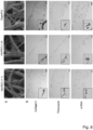
- HA/PEO containing DP tubes with a ratio 1:1 in the full laceration AT model was evaluated by histological analysis of the near contact region between the tendon and the surrounding tissue 3 weeks after surgery ( Fig. 9 ). Results were compared with native tendons and with earlier results from repaired tendons with or without DP tube implantation [29]. Large areas of non-adhesive tissue can be seen on sections of tendons with a DP/HA/PEO tube and although HA/PEO layer is directed towards the surroundings, anti-adhesive effects can be observed as well on the inner tube layer facing the tendon tissue ( Fig. 9A ).
- Tendon healing is a long-lasting process, and natural regeneration capacity of tendons is limited due to their hypovascularization, the dense connective tissue, low cell density and slow cell metabolism.
- re-rupture and adhesion formation are common clinical problems after surgery [22, 68, 69].
- Adhesion results from the generation of fibrotic tissue between the tendon and the surroundings and may impair tendon gliding, leading to joint stiffness and pain in up to 30 % of flexor tendon injuries [11].
- the detailed mechanism of adhesion formation is still unclear, but persistent inflammation is known to activate fibrogenesis leading to an excessive accumulation of ECM components, such as collagen and alpha smooth muscle actin ( ⁇ -SMA) [70, 71].
- ECM components such as collagen and alpha smooth muscle actin ( ⁇ -SMA)
- Electrospun tubes of polycaprolactone alone [76] or in combination with chitosan [32] or acrylate endcapped urethane precursor [77], Zein [31, 78] or PEO [79] have been used in combination with HA, showing promising results, but demonstrating that further investigation is necessary.
- Another successfully electrospun and commercially available material is DegraPol, a biodegradable, biocompatible and surgeon friendly polyester urethane. No adverse reactions of the tendon tissue following tube implantation in a full laceration rabbit AT model was observed [21, 24], but adhesion extent was reduced by about 20 % [20, 29].
- Wall thickness of HA/PEO containing DP tubes have about the same thickness as the pure DP tube (HA/PEO (1:1) 487 ⁇ m ⁇ 53 ⁇ m, HA/PEO (1:4) 436 ⁇ m ⁇ 63 ⁇ m), demonstrating the impact of HA/PEO on material properties.
- Pore size influences mechanical properties as well, depending on the scaffold material and the range of their size. For instance, in titanium scaffolds having pore sizes from 45-500 ⁇ m larger pore sizes reduced mechanical strength [86], but in hyaluronan-collagen scaffolds having pores ranging from 300-500 ⁇ m larger pores increased mechanical rupture stresses and Young's Moduli [87].
- Polyethylene oxide a water-soluble synthetic polymer [88] improving electrospinning processability of aqueous solutions [78] has been shown to improve mechanical properties of commonly used biodegradable polymer electrospun scaffolds [89] and to result in increased UTS with increasing PEO concentrations [90].
- addition of HA/PEO increased the mechanical properties of the scaffolds significantly, which facilitates the application of the implants during tendon surgery when material stretchability is needed mainly in the transverse direction.
- mechanical strength of the implants is too low to be used as reinforcement of injured tendons after surgery or in tendinopathy patients.
- UTS of rabbit AT are in the range of 30 MPa and Young's modulus about 100 MPa [21] showing similar strength as human flexor tendons, while HA/PEO containing scaffolds showed for both, UTS and Young's modulus, values in the range of 1.5-2 MPa.
- HA/PEO containing tubes are envisioned to reduce adhesion extent after surgery and not to act as tendon reinforcement, their low mechanical strength compared to tendon tissue is not relevant for this purpose.
- HA/PEO (1:1) 100.1° ⁇ 7.5°
- HA/PEO (1:4) 99.5° ⁇ 5.1°
- DP 107.6° ⁇ 4.7°
- CAH contact angle hysteresis
- Fig. 6B The roughness of the surfaces might explain the hydrophobic characteristics as contact angles can be increased with the surface roughness and air pockets at the water-scaffold interface [92].
- FTIR spectra of all tested scaffolds look very similar because the amount of electrospun HA or PEO respectively, is very low (HA/PEO (1:1) about 5 % w/v each; HA/PEO (1:4) about 1.2 % w/v HA and 4.8 % w/v PEO) and therefore typical peaks of HA or PEO are superimposed by peaks of DP [32, 67, 80]. This result confirms data from Hu et al.
- the FTIR spectra indicate that no new bonds, between the DP polymer and HA or PEO respectively, were formed as peak pattern from pure DP scaffolds are identical to patterns of HA and PEO containing scaffolds.
- same peak intensity of pure DP tubes and HA/PEO/DP tubes as well as the missing of typical HA and PEO peaks suggest that relative amount of HA and PEO is small, and therefore these components might not be detectable with FTIR.
- the second DSC heating cycle was analyzed to determine the glass transition temperature (T g ), at which amorphous, solid material gets rubbery and viscous, and the melting point (T m ) of crystalline components [67] ( Fig. 6C ).
- T g glass transition temperature
- T m melting point
- HA and PEO supplementation did not change the typical thermal pattern of DP with a T g at about -40 °C and a T m at around 130 °C consistent with published data [57, 80].
- the prominent endothermic peak at about 56 °C corresponds to T m of PEG or PEO, respectively.
- the peak in the pure DP scaffolds results from incomplete washing of PEG that was used as first layer to facilitate taking off the tube from the metal rod [80].
- the peak at 56 °C is smaller in pure DP scaffolds than in HA/PEO containing scaffolds and peak in HA/PEO scaffolds in a ratio 1:4 is slightly higher than in scaffolds with a ratio 1:1, it cannot be distinguished between PEG contamination and PEO integration in HA/PEO containing DP tubes.
- the rabbit animal model has often been used to examine tendon healing and to enable translation of therapeutic assessments into clinical care [96]. For instance, cellular response towards DP implants [24], ultrasound studies [97] and adhesion formation [29, 98] were studied using this model.
- the effect of HA/PEO containing DP tubes with a ratio 1:1 was evaluated regarding adhesion prevention in the rabbit full laceration AT model by histological analysis of the near contact region between the tendon and the surrounding tissue 3 weeks after surgery ( Fig. 9 ). Results were compared with native tendons and with earlier results from repaired tendons with or without DP tube implantation [29].
- anti-adhesive effects of HA could be observed as well on the inner tube layer of pure DP, facing the tendon tissue. This side effect may be caused by HA diffusion and the strong lubricating properties of HA even in small amounts.
- the anti-adhesive effect of both tube sides might be reduced by adding more DP layers, eventually with incorporated growth factors [80], to enhance tendon regeneration as has been previously shown by Evrova et al. [57].
Landscapes
- Health & Medical Sciences (AREA)
- Public Health (AREA)
- General Health & Medical Sciences (AREA)
- Veterinary Medicine (AREA)
- Epidemiology (AREA)
- Life Sciences & Earth Sciences (AREA)
- Animal Behavior & Ethology (AREA)
- Chemical & Material Sciences (AREA)
- Surgery (AREA)
- Vascular Medicine (AREA)
- Heart & Thoracic Surgery (AREA)
- Dermatology (AREA)
- Medicinal Chemistry (AREA)
- Oral & Maxillofacial Surgery (AREA)
- Transplantation (AREA)
- Chemical Kinetics & Catalysis (AREA)
- Materials For Medical Uses (AREA)
Priority Applications (2)
| Application Number | Priority Date | Filing Date | Title |
|---|---|---|---|
| EP23211132.8A EP4559486A1 (de) | 2023-11-21 | 2023-11-21 | Vorrichtung für chirurgische reparatur von zylindrischen organen, insbesondere gerissenen sehnen, und verfahren zur herstellung solch einer vorrichtung |
| PCT/EP2024/082611 WO2025108856A1 (en) | 2023-11-21 | 2024-11-15 | A device for repair surgery of cylindrical organs, particularly ruptured tendons, and method of producing such device |
Applications Claiming Priority (1)
| Application Number | Priority Date | Filing Date | Title |
|---|---|---|---|
| EP23211132.8A EP4559486A1 (de) | 2023-11-21 | 2023-11-21 | Vorrichtung für chirurgische reparatur von zylindrischen organen, insbesondere gerissenen sehnen, und verfahren zur herstellung solch einer vorrichtung |
Publications (1)
| Publication Number | Publication Date |
|---|---|
| EP4559486A1 true EP4559486A1 (de) | 2025-05-28 |
Family
ID=88920819
Family Applications (1)
| Application Number | Title | Priority Date | Filing Date |
|---|---|---|---|
| EP23211132.8A Withdrawn EP4559486A1 (de) | 2023-11-21 | 2023-11-21 | Vorrichtung für chirurgische reparatur von zylindrischen organen, insbesondere gerissenen sehnen, und verfahren zur herstellung solch einer vorrichtung |
Country Status (2)
| Country | Link |
|---|---|
| EP (1) | EP4559486A1 (de) |
| WO (1) | WO2025108856A1 (de) |
Citations (4)
| Publication number | Priority date | Publication date | Assignee | Title |
|---|---|---|---|---|
| US20110039101A1 (en) | 2008-04-18 | 2011-02-17 | Shanghai Institute Of Ceramics, Chinese Academy Of Sciences | Electrospun fiber tubular material and preparation method thereof |
| WO2013026937A1 (en) | 2011-08-25 | 2013-02-28 | Universität Zürich Prorektorat Mnw | A device for repair surgery of cylindrical organs, particularly ruptured tendons |
| WO2017137602A1 (en) | 2016-02-12 | 2017-08-17 | Universität Zürich | A device for repair surgery of cylindrical organs, particularly ruptured tendons, comprising a therapeutic agent for stimulating regrowth, and method of producing such device |
| CN115381593A (zh) * | 2022-08-17 | 2022-11-25 | 上海市第六人民医院 | 一种仿生腱鞘膜及制备方法 |
-
2023
- 2023-11-21 EP EP23211132.8A patent/EP4559486A1/de not_active Withdrawn
-
2024
- 2024-11-15 WO PCT/EP2024/082611 patent/WO2025108856A1/en active Pending
Patent Citations (6)
| Publication number | Priority date | Publication date | Assignee | Title |
|---|---|---|---|---|
| US20110039101A1 (en) | 2008-04-18 | 2011-02-17 | Shanghai Institute Of Ceramics, Chinese Academy Of Sciences | Electrospun fiber tubular material and preparation method thereof |
| WO2013026937A1 (en) | 2011-08-25 | 2013-02-28 | Universität Zürich Prorektorat Mnw | A device for repair surgery of cylindrical organs, particularly ruptured tendons |
| US9351820B2 (en) * | 2011-08-25 | 2016-05-31 | University Of Zurich | Device for repair surgery of cylindrical organs, particularly ruptured tendons |
| WO2017137602A1 (en) | 2016-02-12 | 2017-08-17 | Universität Zürich | A device for repair surgery of cylindrical organs, particularly ruptured tendons, comprising a therapeutic agent for stimulating regrowth, and method of producing such device |
| US20190070340A1 (en) * | 2016-02-12 | 2019-03-07 | Universitaet Zuerich | A device for repair surgery of cylindrical organs, particularly ruptured tendons, comprising a therapeutic agent for stimulating regrowth, and method of producing such device |
| CN115381593A (zh) * | 2022-08-17 | 2022-11-25 | 上海市第六人民医院 | 一种仿生腱鞘膜及制备方法 |
Non-Patent Citations (126)
| Title |
|---|
| A. K. S. CHONG, A. D. ANG, J. C. H. GOH, J. H. P. HUI, A. Y. T. LIM, E. H. LEE: "Bone and Joint Surgery", AMERICAN VOLUME, vol. 89A, 2007, pages 74 |
| ABATE, M.C. SCHIAVONEV. SALINI: "The use of hyaluronic acid after tendon surgery and in tendinopathies", BIOMED RES INT, vol. 2014, 2014, pages 783632 |
| AL-MUNAJJED, A.A. ET AL.: "Influence of pore size on tensile strength, permeability and porosity of hyaluronan-collagen scaffolds", J MATER SCI MATER MED, vol. 19, no. 8, 2008, pages 2859 - 64, XP019607881 |
| ASEFNEJAD, A., NANOMEDICINE, vol. 6, 2011, pages 2375 - 84 |
| AYA, K.L. AND R. STERN: "Hyaluronan in wound healing: rediscovering a major player. Wound", REPAIR REGEN, vol. 22, no. 5, 2014, pages 579 - 93 |
| B. SAAD, Y. KUBOKI, M. WELTI, G. K. UHLSCHMID, P. NEUENSCHWANDER, U. W. SUTER: "12th World Congress of the International-Society-for-Artificial-Organs/26th Congress of the European-Society-for-Artificial-Organs", 1999, BLACKWELL SCIENCE INC., article "DegraPol-Foam: A degradable and highly porous polyesterurethane foam as a new substrate for bone formation" |
| BAKER, B.M. ET AL.: "The potential to improve cell infiltration in composite fiber-aligned electrospun scaffolds by the selective removal of sacrificial fibers", BIOMATERIALS, vol. 29, no. 15, 2008, pages 2348 - 58, XP022526907, DOI: 10.1016/j.biomaterials.2008.01.032 |
| BALAZS, E.A. ET AL.: "Hyaluronic acid in synovial fluid. I. Molecular parameters of hyaluronic acid in normal and arthritis human fluids", ARTHRITIS RHEUM, vol. 10, no. 4, 1967, pages 357 - 76 |
| BARTLET, K.: "Antibacterial activity on superhydrophobic titania nanotube arrays. ", SURF B BIOINTERFACES, vol. 166, 2018, pages 179 - 186, XP085382200, DOI: 10.1016/j.colsurfb.2018.03.019 |
| BURGISSER, G.M. ET AL.: "Electrospun tube reduces adhesion in rabbit Achilles tendon 12 weeks post-surgery without PAR-2 overexpression", SCI REP, vol. 11, no. 1, 2021, pages 23293 |
| BUSCHMANN, J. ET AL.: "Cellular response of healing tissue to DegraPol tube implantation in rabbit Achilles tendon rupture repair: an in vivo histomorphometric study", J TISSUE ENG REGEN, vol. 7, no. 5, 2013, pages 413 - 20 |
| BUSCHMANN, J. ET AL.: "Correspondence of high-frequency ultrasound and histomorphometry of healing rabbit Achilles tendon tissue", CONNECT TISSUE RES, vol. 55, no. 2, 2014, pages 123 - 31 |
| BUSCHMANN, J. ET AL.: "Small hook thread (Quill) and soft felt internal splint to increase the primary repair strength of lacerated rabbit Achilles tendons: biomechanical analysis and considerations for hand surgery", CLIN BIOMECH (BRISTOL, AVON, vol. 26, no. 6, 2011, pages 626 - 31 |
| C.F. MAURUSM. K. J. SCHNEIDERD. SCHMIDTG. ZUNDJ.D. SEEBACH, TRANSPLANTATION, vol. 81, 2006, pages 1204 |
| CARPENTER, J.E. ET AL.: "Rotator cuff defect healing: a biomechanical and histologic analysis in an animal model", J SHOULDER ELBOW SURG, vol. 7, no. 6, 1998, pages 599 - 605 |
| CASTRO, K.C.M.G.N. CAMPOSL.H.I. MEI: "Hyaluronic acid electrospinning: Challenges, applications in wound dressings and new perspectives", INT J BIOL MACROMOL, vol. 173, 2021, pages 251 - 266 |
| CHANDA, A. ET AL.: "Electrospun chitosan/polycaprolactone-hyaluronic acid bilayered scaffold for potential wound healing applications", INT J BIOL MACROMOL, vol. 116, 2018, pages 774 - 785 |
| CHARTIER, C. ET AL.: "Tendon: Principles of Healing and Repair", SEMIN PLAST SURG, vol. 35, no. 3, 2021, pages 211 - 215 |
| CHEN CHIH-HAO ET AL: "Hyaluronic acid/platelet rich plasma-infused core-shell nanofiber membrane to prevent postoperative tendon adhesion and promote tendon healing", INTERNATIONAL JOURNAL OF BIOLOGICAL MACROMOLECULES, ELSEVIER BV, NL, vol. 231, 18 January 2023 (2023-01-18), XP087271484, ISSN: 0141-8130, [retrieved on 20230118], DOI: 10.1016/J.IJBIOMAC.2023.123312 * |
| CHEN, C.H. ET AL.: "Hyaluronic acid/platelet rich plasma-infused core-shell nanofiber membrane to prevent postoperative tendon adhesion and promote tendon healing", INT J BIOL, vol. 231, 2023, pages 123312, XP087271484, DOI: 10.1016/j.ijbiomac.2023.123312 |
| CHEN, C.T. ET AL.: "Ibuprofen-Loaded Hyaluronic Acid Nanofibrous Membranes for Prevention of Postoperative Tendon Adhesion through Reduction of Inflammation", INT J MOL SCI, vol. 20, no. 20, 2019 |
| CHEN, E. ET AL.: "An asymmetric chitosan scaffold for tendon tissue engineering: In vitro and in vivo evaluation with rat tendon stem/progenitor cells", ACTA BIOMATER, vol. 73, 2018, pages 377 - 387 |
| CHEN, S. ET AL.: "RelA/p65 inhibition prevents tendon adhesion by modulating inflammation, cell proliferation, and apoptosis", CELL DEATH DIS, vol. 8, no. 3, 2017, pages e2710 |
| CHEN, Y.G.Z. TANY. ZHOU: "Effects of Viscosities and Solution Composition on Core-Sheath Electrospun Polycaprolactone(PCL) Nanoporous Microtubes", POLYMERS, no. 21, 2021 |
| CLAYTON, R.AC.M. COURT-BROWN: "The epidemiology of musculoskeletal tendinous and ligamentous injuries", INJURY, vol. 39, no. 12, 2008, pages 1338 - 44, XP025744512, DOI: 10.1016/j.injury.2008.06.021 |
| CONNECT TISSUE RES, vol. 60, no. 1, 2019, pages 10 - 20 |
| D. ELLIOTT. GIESEN, HAND CLIN, vol. 29, 2013, pages 191 |
| D'AGOSTINO, A. ET AL.: "Is molecular size a discriminating factor in hyaluronan interaction with human cells", CARBOHYDR POLYM, vol. 157, 2017, pages 21 - 30, XP029848564, DOI: 10.1016/j.carbpol.2016.07.125 |
| DE JONG, J.P. ET AL.: "The incidence of acute traumatic tendon injuries in the hand and wrist: a 10-year population-based study", CLIN ORTHOP SURG, vol. 6, no. 2, 2014, pages 196 - 202 |
| DOCHEVA, D. ET AL.: "Biologies for tendon repair", ADV DRUG DELIV REV, vol. 84, 2015, pages 222 - 39 |
| ELLIOT, D., T. GIESEN: "Primary flexor tendon surgery: the search for a perfect result", CLIN, vol. 29, no. 2, 2013, pages 191 - 206 |
| ERAL, H.B.T MANNETJED.J.C.M.OH, J.M.: "Contact angle hysteresis: a review of fundamentals and applications", COLLOID AND 663 POLYMER SCIENCE, vol. 291, no. 2, 2013, pages 247 - 260, XP035166476, DOI: 10.1007/s00396-012-2796-6 |
| EVROVA, O ET AL.: "Elastic and surgeon friendly electrospun tubes delivering PDGF-BB positively impact tendon rupture healing in a rabbit Achilles tendon model", BIOMATERIALS, vol. 232, 2020, pages 119722 |
| EVROVA, O.: " Supporting Cell-Based Tendon Therapy: Effect of PDGF-BB and Ascorbic Acid on Rabbit Achilles Tenocytes in Vitro.", INT J MOL SCI, vol. 21, no. 2, 2020 |
| EVROVA, O.: "Bioactive, Elastic, and Biodegradable Emulsion Electrospun DegraPol Tube Delivering PDGF-BB for Tendon Rupture Repair", MACROMOL BIOSCI, vol. 16, no. 7, 2016, pages 1048 - 63, XP055349140, DOI: 10.1002/mabi.201500455 |
| EXTRAND, C.WY. KUMAGAI: "An Experimental Study of Contact Angle Hysteresis", J COLLOID INTERFACE SCI, vol. 191, no. 2, 1997, pages 378 - 83 |
| FALLACARA, A. ET AL.: "Hyaluronic Acid in the Third Millennium", POLYMERS (BASEL, vol. 10, no. 7, 2018, XP055587625, DOI: 10.3390/polym10070701 |
| FIGUEIRA, D.R.: " Production and characterization of polycaprolactone- hyaluronic acid/chitosan- zein electrospun bilayer nanofibrous membrane for tissue regeneration. ", BIOL MACROMOL, vol. 93, 2016, pages 1100 - 1110, XP029790707, DOI: 10.1016/j.ijbiomac.2016.09.080 |
| G. M. BURGISSER M. CALCAGNI, MULLER A, E. BONAVOGLIA, G FESSEL, J. G. SNEDEKER, P. GIOVANOLI, J. BUSCHMANN, BIOMED. RES. INT, vol. 2014, 2014, pages 656240 |
| GAIDA, J.E.J.L. COOKS.L. BASS: "Adiposity and tendinopathy", DISABIL REHABIL, vol. 30, no. 20-22, 2008, pages 1555 - 62 |
| GALATZ, L.M. ET AL.: "Tendon regeneration and scar formation: The concept of scarless healing", J ORTHOP RES, vol. 33, no. 6, 2015, pages 823 - 31 |
| GAO, Y.: "A Low Molecular Weight Hyaluronic Acid Derivative Accelerates Excisional Wound Healing by Modulating Pro-Inflammation, Promoting Epithelialization and Neovascularization, and Remodeling Collagen", INT J MOL SCI, vol. 20, no. 15, 2019 |
| GOODMAN, H.JJ. CHOUEKA: "Biomechanics of the flexor tendons", HAND CLIN, vol. 21, no. 2, 2005, pages 129 - 49 |
| HODGE, JC. QUINT: "The improvement of cell infiltration in an electrospun scaffold with multiple synthetic biodegradable polymers using sacrificial PEO microparticles", J BIOMED MATER RES A, vol. 107, no. 9, 2019, pages 1954 - 1964, XP055643946, DOI: 10.1002/jbm.a.36706 |
| HONDA, H. ET AL.: "Hyaluronic Acid Accelerates Tendon-to-Bone Healing After Rotator Cuff Repair", AM J SPORTS MED, vol. 45, no. 14, 2017, pages 3322 - 3330 |
| HU, J. ET AL.: "Emulsion electrospinning of polycaprolactone: influence of surfactant type towards the scaffold properties", J BIOMATER SCI POLYM ED, vol. 26, no. 1, 2015, pages 57 - 75 |
| HU, M. ET AL.: "Nanozymes in Nanofibrous Mats with Haloperoxidase-like Activity To Combat Biofouling", ACS APPL MATER INTERFACES, vol. 10, no. 51, 2018, pages 44722 - 44730 |
| IMERE, A. ET AL.: "Engineering a cell-hydrogel-fibre composite to mimic the structure and function of the tendon synovial sheath", ACTA BIOMATER, vol. 119, 2021, pages 140 - 154, XP086405092, DOI: 10.1016/j.actbio.2020.11.017 |
| J. A. HENRYK. BURUGAPALLIP. NEUENSCHWANDERA. PANDIT, ACTA BIOMATERIALIA, 29 May 2009 (2009-05-29) |
| J. BUSCHMANN, G. MEIER-BURGISSER, E. BONAVOGLIA, P. NEUENSCHWANDER, V. MILLERET, P. GIOVANOLI, M. CALCAGNI, REGEN. MED., vol. 7, 2013, pages 413 |
| J. BUSCHMANNG. PUIPPEG. M. BURGISSERE. BONAVOGLIAP. GIOVANOLIM. CALCAGNI, CONNECT. TISSUE RES, vol. 55, 2014, pages 123 |
| J. BUSCHMANNM. CALCAGNIG. M. BURGISSERE. BONAVOGLIAP. NEUENSCHWANDERV. MILLERETP. GIOVANOLI, J. TISSUE ENG. REGEN. MED, vol. 9, 2015, pages 584 |
| J. C. GOHH. W. OUYANGS. H. TEOHC. K. CHANE. H. LEE, TISSUE ENG, vol. 9, 2003, pages S31 |
| J. RIEBERMEIER-BIIRGISSER, G.MIESCHER, I.WEBER, F. E.WOLINT, P.YAO, Y.ONGINI, E.MILIONIS, A.SNEDEKER, J. G.CALCAGNI, M. ET AL., INT. J. MOL. SCI, vol. 24, no. 12, 2023 |
| JUNCOSA-MELVIN, N. ET AL.: "Effects of mechanical stimulation on the biomechanics and histology of stem cell-collagen sponge constructs for rabbit patellar tendon repair", TISSUE ENG, vol. 12, no. 8, 2006, pages 2291 - 300 |
| K. A. CORSIE. M. SCHWARZD. J. MOONEYJ. HUARD, JOURNAL OF ORTHOPAEDIC RESEARCH, vol. 25, 2007, pages 1261 |
| KARABEKMEZ, F.EC. ZHAO: "Surface treatment of flexor tendon autograft and allograft decreases adhesion without an effect of graft cellularity: a pilot study", CLIN ORTHOP RELAT RES, vol. 470, no. 9, 2012, pages 2522 - 7, XP035096487, DOI: 10.1007/s11999-012-2437-x |
| KAUX, J.F.A. SAMSONJ.M. CRIELAARD: "Hyaluronic acid and tendon lesions", MUSCLES LIGAMENTS TENDONS J, vol. 5, no. 4, 2015, pages 264 - 9 |
| KOBAYASHI, T., T. CHANMEE, AND N. ITANO: "Hyaluronan: Metabolism and Function.", BIOMOLECULES, vol. 10, no. 11, 2020 |
| KUMBAR, S.G. ET AL.: "Electrospun nanofiber scaffolds: engineering soft tissues", BIOMED MATE, vol. 3, no. 3, 2008, pages 034002, XP002522845, DOI: 10.1088/1748-6041/3/3/034002 |
| L. DURSELEN, M. DAUNER, H. HIERLEMANN, H. PLANCK, L. E. CLAES, A. IGNATIUS, BIOMEDICAL MATERIALS RESEARCH, vol. 58, 2001, pages 666 |
| L. V. GULOTTA, S. A.RODEO, CLIN. SPORTS MED, vol. 28, 2009, pages 13 |
| LAVIN, D.M. ET AL.: "Effects of protein molecular weight on the intrinsic material properties and release kinetics of wet spun polymeric microfiber delivery systems", ACTA BIOMATER, vol. 9, no. 1, 2013, pages 4569 - 78 |
| LEONG, N.L.: "Tendon and Ligament Healing and Current Approaches to Tendon and Ligament Regeneration.", J ORTHOP RES, vol. 38, no. 1, 2020, pages 7 - 12 |
| LI, H.Y. CHENS. CHEN: "Enhancement of rotator cuff tendon-bone healing using bone marrow-stimulating technique along with hyaluronic acid", J ORTHOP TRANSLAT, vol. 17, 2019, pages 96 - 102 |
| LIANG, J.I. ET AL.: "The effect of tenocyte/hyaluronic acid therapy on the early recovery of healing Achilles tendon in rats", J MATER SCI MATER MED, vol. 25, no. 1, 2014, pages 217 - 27 |
| LINDERMAN, S.W. ET AL.: "Cell and Biologic-Based Treatment of Flexor Tendon Injuries", OPER TECH ORTHOP, vol. 26, no. 3, 2016, pages 206 - 215, XP029722049, DOI: 10.1053/j.oto.2016.06.011 |
| LIPAR, M. ET AL.: "Extracellular matrix supports healing of transected rabbit Achilles tendon", HELIYON, vol. 4, no. 9, 2018, pages e00781 |
| LOEBEL, C. ET AL.: "Fabrication of cell-compatible hyaluronan hydrogels with a wide range of biophysical properties through high tyramine functionalization", J MATER CHEM B, vol. 5, no. 12, 2017, pages 2355 - 2363 |
| LOISELLE, A.E.: "Remodeling of murine intrasynovial tendon adhesions following injury:MMP and neotendon gene expression", J ORTHOP RES, vol. 27, no. 6, 2009, pages 833 - 40 |
| M. A. COSTAC. WUB.V. PHAMA. K. S. CHONGH. M. PHAMJ. CHANG, TISSUE ENGINEERING, vol. 12, 2006, pages 1937 |
| M. A. SANDREY, J. ATHL. TRAIN, vol. 49, 2014, pages 428 |
| MARIAN, M.: "Exploring the lubrication mechanisms of synovial fluids for joint longevity - A perspective", COLLOIDS SURF B BIOINTERFACES, vol. 206, 2021, pages 111926 |
| MEIER BIIRGISSER, G.: "Prevention of peritendinous adhesions using an electrospun DegraPol polymer tube: a histological, ultrasonographic, and biomechanical study in rabbits.", BIOMED RES INT, vol. 2014, 2014, pages 656240 |
| MEIER BURGISSER, G. ET AL.: "Rabbit Achilles tendon full transection model - wound healing, adhesion formation and biomechanics at 3, 6 and 12 weeks post-surgery", BIOL OPEN, vol. 5, no. 9, 2016, pages 1324 - 33 |
| MEMIC, A.: "Latest Progress in Electrospun Nanofibers for Wound Healing Applications.", ACS APPL BIO MATER, vol. 2, no. 3, 2019, pages 952 - 969 |
| MIESCHER, I. ET AL.: "Impact of High-Molecular-Weight Hyaluronic Acid on Gene Expression in Rabbit Achilles Tenocytes In Vitro", INT J MOL SCI, vol. 23, no. 14, 2022 |
| MURRAY, E.: "Effectiveness of Sodium Hyaluronate and ADCON-T/N for the Prevention of Adhesions in Hand Flexor Tendon Surgery: A Systematic Review and Meta-Analysis.", HAND SURG AM, vol. 47, no. 9, 2022, pages 1 - 20 |
| N. BACHL, W. DERMAN, L. ENGEBRETSEN, G. GOLDSPINK, M. KINZLBAUER, H. TSCHAN, P. VOLPI, D. VENTER, B. WESSNER, J. SPORTS MED. PHYS. FITNESS, vol. 49, 2009, pages 346 |
| OLIVA, F. ET AL.: "The Impact of Hyaluronic Acid on Tendon Physiology and Its Clinical Application in Tendinopathies", CELLS, vol. 10, no. 11, 2021 |
| OSTI, L.: "Hyaluronic acid increases tendon derived cell viability and collagen type I expression in vitro: Comparative study of four different Hyaluronic acid preparations bymolecular weight.", BMC MUSCULOSKELET DISORD, vol. 16, 2015, pages 284, XP021229339, DOI: 10.1186/s12891-015-0735-7 |
| PAREKH, S.G. ET AL.: "Achilles: Failed Acute Repair", FOOT ANKLE CLIN, vol. 27, no. 2, 2022, pages 415 - 430 |
| PIEN, N. ET AL.: "Design and development of a reinforced tubular electrospun construct for the repair of ruptures of deep flexor tendons", MATER SCI ENG C MATER BIOL APPL, vol. 119, 2021, pages 111504 |
| PROCTOR, C.S.D.W. JACKSONT.M. SIMON: "Characterization of the repair tissue after removal of the central one-third of the patellar ligament. An experimental study in a goat model", J BONE JOINT SURG AM, vol. 79, no. 7, 1997, pages 997 - 1006 |
| R. BULLOUGHT. FINNIGANA. KAYN. MAFFULLIN. R. FORSYTH, DISABILITY AND REHABILITATION, vol. 30, 2008, pages 1746 |
| RAYAHIN, J.E. ET AL.: "High and low molecular weight hyaluronic acid differentially influence macrophage activation", ACS BIOMATER SCI ENG, vol. 1, no. 7, 2015, pages 481 - 493, XP093079035, DOI: 10.1021/acsbiomaterials.5b00181 |
| REED, R.K. ET AL.: "Removal rate of [3H]hyaluronan injected subcutaneously in rabbits", AM J PHYSIOL, vol. 259, 1990, pages H532 - 5, XP008052807 |
| REN, Z.: "Instantaneous self-healing and strongly adhesive self-adaptive hyaluronic acid-based hydrogel for controlled drug release to promote tendon wound healing. ", MACROMOL, vol. 242, 2023, pages 125001 |
| RIEBER, J. ET AL.: "Bioactive and Elastic Emulsion Electrospun DegraPol Tubes Delivering IGF-1 for Tendon Rupture Repair", INT J MOL SCI, vol. 24, no. 12, 2023 |
| S. A. RIBOLDI, M. SAMPAOLESI, P. NEUENSCHWANDER, G.COSSU, S. MANTERO, BIOMATERIALS, vol. 26, 2005, pages 4606 |
| S. SAHOOL.T.A.J.C. GOHS.L. TOH: "Growth factor delivery through electrospun nanofibers in scaffolds for tissue engineering applications", J. BIOMED. MATER, vol. 93, 2010, pages 1539 - 1550, XP002721020, DOI: 10.1002/jbm.a.32645 |
| S. SAHOOS. L. TOHJ. C. H. GOH, JOURNAL OF BIOMEDICAL MATERIALS RESEARCH PART B-APPLIED, vol. 95, 2010, pages 19 |
| S. THOMOPOULOS, R. DASM. J. SILVAS. SAKIYAMA-ELBERTF. L. HARWOODE. ZAMPIAKISH. M. KIMD. AMIELR. H. GELBERMAN, J. ORTHOP. RES, vol. 27, 2009, pages 1209 |
| S. THOMOPOULOSM. ZAEGELR. DASF. L. HARWOODM. J. SILVAD. AMIELS. SAKIYAMA-ELBERTR. H. GELBERMAN, J. ORTHOP. RES, vol. 25, 2007, pages 1358 |
| S. THOMOPOULOSW. C. PARKSD. B. RIFKINK. A. DERWIN, J. ORTHOP. RES, vol. 33, 2015, pages 832 |
| SAAD, B. ET AL.: "Multiblock copolyesters as biomaterials: in vitro biocompatibility testing", J MATER SCI MATER MED, vol. 8, no. 8, 1997, pages 497 - 505 |
| SALAM, A. ET AL.: "In-vitro assessment of appropriate hydrophilic scaffolds by coelectrospinning of poly(1,4 cyclohexane isosorbide terephthalate)/polyvinyl alcohol", SCI REP, vol. 10, no. 1, 2020, pages 19751 |
| SCHULZE-TANZIL, G.G. ET AL.: "Tendon healing: a concise review on cellular and molecular mechanisms with a particular focus on the Achilles tendon", BONE JOINT RES, vol. 11, no. 8, 2022, pages 561 - 574 |
| SERODIO, R. ET AL.: "Ultrasound sonication prior to electrospinning tailors silk fibroin/PEO membranes for periodontal regeneration", MATER SCI ENG C MATER BIOL APPL, vol. 98, 2019, pages 969 - 981, XP085611503, DOI: 10.1016/j.msec.2019.01.055 |
| SHALUMON, K.T.: "Multi-functional electrospun antibacterial core-shell nanofibrous membranes for prolonged prevention of post-surgical tendon adhesion and inflammation. ", BIOMATER, vol. 72, 2018, pages 121 - 136, XP085393079, DOI: 10.1016/j.actbio.2018.03.044 |
| SHARMA, PN. MAFFULLI: "Biology of tendon injury: healing, modeling and remodeling", J MUSCULOSKELET NEURONAL INTERACT, vol. 6, no. 2, 2006, pages 181 - 90 |
| SMITH, L.AP.X. MA: "Nano-fibrous scaffolds for tissue engineering", COLLOIDS SURF B BIOINTERFACES, vol. 39, no. 3, 2004, pages 125 - 31, XP004649329 |
| SNETKOV, P.: "Hyaluronan-Based Nanofibers: Fabrication, Characterization and Application.", POLYMERS, vol. 11, no. 12, 2019 |
| TAN, V. ET AL.: "Effects of nonsteroidal anti-inflammatory drugs on flexor tendon adhesion", J HAND SURG AM, vol. 35, no. 6, 2010, pages 941 - 7, XP027062633, DOI: 10.1016/j.jhsa.2010.02.033 |
| TANG, J.B.: "Clinical outcomes associated with flexor tendon repair", HAND CLIN, vol. 21, no. 2, 2005, pages 199 - 210 |
| TANG, J.B.E.A.: "Basic FGF or VEGF gene therapy corrects insufficiency in the intrinsic healing capacity of tendons", SCI. REP., vol. 6, 2016, pages 20643 |
| TAYLOR, S.H. ET AL.: "Tendon is covered by a basement membrane epithelium that is required for cell retention and the prevention of adhesion formation", PLOS ONE, vol. 6, no. 1, 2011, pages e16337 |
| TERCIC, DB. BOZIC: "The basis of the synovial fluid analysis", CLIN CHEM LAB MED, vol. 39, no. 12, 2001, pages 1221 - 6 |
| THOMOPOULOS, S. ET AL.: "Mechanisms of tendon injury and repair", J ORTHOP RES, vol. 33, no. 6, 2015, pages 832 - 9 |
| TISSUE ENG REGEN MED, vol. 9, no. 5, 2015, pages 584 - 94 |
| TITAN, A.L.: " Flexor Tendon: Development, Healing, Adhesion Formation, and Contributing Growth Factors.", PLAST RECONSTR SURG, vol. 144, no. 4, 2019, pages 639e - 647e |
| TORRES-SANCHEZ, C. ET AL.: "The effect of pore size and porosity on mechanical properties and biological response of porous titanium scaffolds", MATER SCI ENG C MATER BIOL APPL, vol. 77, 2017, pages 219 - 228, XP085028595, DOI: 10.1016/j.msec.2017.03.249 |
| TORRES-SANCHEZ, C.: "The impact of multimodal pore size considered independently from porosity on mechanical performance and osteogenic behaviour of titanium scaffolds.", SCI ENG C MATER BIOL APPL, vol. 124, 2021, pages 112026, XP086558745, DOI: 10.1016/j.msec.2021.112026 |
| UGGEN, J. DINES, M.MCGARRY, D. GRANDE, T. LEE, O.LIMPISVASTI, ARTHROSCOPY, vol. 26, 2010, pages 1456 |
| V. MILLERETB. SIMONAP. NEUENSCHWANDERH. HALL, EUR. CELL MATER, vol. 21, 2011, pages 286 |
| V. MILLERETM. SIMONETA. G. BITTERMANNP. NEUENSCHWANDERH. HALL, J. BIOMED. MAT, vol. 91, 2009, pages 109 |
| VASSALLO, V. ET AL.: "Unsulfated biotechnological chondroitin by itself as well as in combination with high molecular weight hyaluronan improves the inflammation profile in osteoarthritis in vitro model", J CELL BIOCHEM, vol. 122, no. 9, 2021, pages 1021 - 36 |
| VERONESI, F. ET AL.: "Evaluation of a new collagen-based medical device (ElastiCoO) for the treatment of acute Achilles tendon injury and prevention of peritendinous adhesions: An in vitro biocompatibility and in vivo investigation", J TISSUE ENG REGEN MED, vol. 14, no. 8, 2020, pages 1113 - 1125 |
| VITKOVA, L.: " Electrospinning of Hyaluronan Using Polymer Coelectrospinning and Intermediate Solvent", POLYMERS (BASEL, vol. 11, 2019, pages 9 |
| VOLETI, P.B., M.R. BUCKLEY, AND L.J. SOSLOWSKY: "Tendon healing: repair and regeneration.", ANNU REV BIOMED ENG, vol. 14, 2012, pages 47 - 71 |
| WALDEN, G. ET AL.: "A Clinical, Biological, and Biomaterials Perspective into Tendon Injuries and Regeneration", TISSUE ENG PART B REV, vol. 23, no. 1, 2017, pages 44 - 58 |
| WANG, Y.P. LIL. KONG: "Chitosan-modified PLGA nanoparticles with versatile surface for improved drug delivery", AAPS PHARMSCITECH, vol. 14, no. 2, 2013, pages 585 - 92 |
| Y. QIANZ. ZHANGL. ZHENGR. SONGY. ZHAO, J. NANOMAT, vol. 2014, 2014, pages 964621 |
| Y.C. JUNG, B.B.: "Contact angle, adhesion and friction properties of micro- and nanopatterned polymers for superhydrophobicity", NANOTECHNOLOGY, vol. 17, 2006, pages 4970 - 4980, XP020104140, DOI: 10.1088/0957-4484/17/19/033 |
| YAO, C.X. LIT. SONG: "Fabrication of zein/hyaluronic acid fibrous membranes by electrospinning", J BIOMATER SCI POLYM ED, vol. 18, no. 6, 2007, pages 731 - 42 |
| ZHAO, H. ET AL.: "Collagen membrane alleviates peritendinous adhesion in the rat Achilles tendon injury model", CHIN MED J (ENGL, vol. 126, no. 4, 2013, pages 729 - 33 |
Also Published As
| Publication number | Publication date |
|---|---|
| WO2025108856A1 (en) | 2025-05-30 |
Similar Documents
| Publication | Publication Date | Title |
|---|---|---|
| Liu et al. | Composite poly (lactic acid)/chitosan nanofibrous scaffolds for cardiac tissue engineering | |
| Yucel et al. | Polyester based nerve guidance conduit design | |
| Chen et al. | Prevention of peritendinous adhesions with electrospun polyethylene glycol/polycaprolactone nanofibrous membranes | |
| Sheng et al. | Electrospun PCL/Gel-aligned scaffolds enhance the biomechanical strength in tendon repair | |
| Lee et al. | The effect of gelatin incorporation into electrospun poly (l-lactide-co-ɛ-caprolactone) fibers on mechanical properties and cytocompatibility | |
| Li et al. | Antimicrobial eugenol-loaded electrospun membranes of poly (ε-caprolactone)/gelatin incorporated with REDV for vascular graft applications | |
| Briganti et al. | A composite fibrin-based scaffold for controlled delivery of bioactive pro-angiogenetic growth factors | |
| Sankaran et al. | Axially aligned 3D nanofibrous grafts of PLA–PCL for small diameter cardiovascular applications | |
| Birhanu et al. | An improved surface for enhanced stem cell proliferation and osteogenic differentiation using electrospun composite PLLA/P123 scaffold | |
| Santoro et al. | Poly (lactic acid) nanofibrous scaffolds for tissue engineering | |
| Huang et al. | Electrospun collagen–chitosan–TPU nanofibrous scaffolds for tissue engineered tubular grafts | |
| Chen et al. | Prevention of peritendinous adhesions with electrospun chitosan-grafted polycaprolactone nanofibrous membranes | |
| Alvarez-Perez et al. | Influence of gelatin cues in PCL electrospun membranes on nerve outgrowth | |
| Chen et al. | The effect of surface area on the degradation rate of nano-fibrous poly (L-lactic acid) foams | |
| US20190070339A1 (en) | Electro-mechanically stretched micro fibers and methods of use thereof | |
| EP3337464B1 (de) | In-situ-herstellung von verbundmaterial zur gewebewiederherstellung | |
| Cheng et al. | Promoting osteogenic differentiation in pre-osteoblasts and reducing tibial fracture healing time using functional nanofibers | |
| Hu et al. | Electrospun Tri‐layer core‐sheath nanofibrous coating for sequential treatment of postoperative complications in orthopedic implants | |
| Zhang et al. | Silk fibroin-based scaffolds with controlled delivery order of VEGF and BDNF for cavernous nerve regeneration | |
| CN107073169A (zh) | 用于组织修复的复合材料 | |
| JP2015532845A (ja) | 心臓修復パッチの新しいスキャフォールド | |
| Du et al. | Enhanced biocompatibility of poly (l‑lactide‑co‑epsilon‑caprolactone) electrospun vascular grafts via self-assembly modification | |
| Zhai et al. | Coaxial electrospinning of P (LLA‐CL)/heparin biodegradable polymer nanofibers: Potential vascular graft for substitution of femoral artery | |
| US9351820B2 (en) | Device for repair surgery of cylindrical organs, particularly ruptured tendons | |
| Mendibil et al. | Bioresorbable and mechanically optimized nerve guidance conduit based on a naturally derived medium chain length polyhydroxyalkanoate and poly (ε-caprolactone) blend |
Legal Events
| Date | Code | Title | Description |
|---|---|---|---|
| PUAI | Public reference made under article 153(3) epc to a published international application that has entered the european phase |
Free format text: ORIGINAL CODE: 0009012 |
|
| STAA | Information on the status of an ep patent application or granted ep patent |
Free format text: STATUS: THE APPLICATION HAS BEEN PUBLISHED |
|
| AK | Designated contracting states |
Kind code of ref document: A1 Designated state(s): AL AT BE BG CH CY CZ DE DK EE ES FI FR GB GR HR HU IE IS IT LI LT LU LV MC ME MK MT NL NO PL PT RO RS SE SI SK SM TR |
|
| STAA | Information on the status of an ep patent application or granted ep patent |
Free format text: STATUS: THE APPLICATION IS DEEMED TO BE WITHDRAWN |
|
| 18D | Application deemed to be withdrawn |
Effective date: 20251129 |