EP4537798A2 - Intraocular drug delivery and filter device and methods of using same - Google Patents
Intraocular drug delivery and filter device and methods of using same Download PDFInfo
- Publication number
- EP4537798A2 EP4537798A2 EP25152586.1A EP25152586A EP4537798A2 EP 4537798 A2 EP4537798 A2 EP 4537798A2 EP 25152586 A EP25152586 A EP 25152586A EP 4537798 A2 EP4537798 A2 EP 4537798A2
- Authority
- EP
- European Patent Office
- Prior art keywords
- eye
- target molecule
- substrate
- vegf
- capturing
- Prior art date
- Legal status (The legal status is an assumption and is not a legal conclusion. Google has not performed a legal analysis and makes no representation as to the accuracy of the status listed.)
- Withdrawn
Links
Images



Classifications
-
- A—HUMAN NECESSITIES
- A61—MEDICAL OR VETERINARY SCIENCE; HYGIENE
- A61L—METHODS OR APPARATUS FOR STERILISING MATERIALS OR OBJECTS IN GENERAL; DISINFECTION, STERILISATION OR DEODORISATION OF AIR; CHEMICAL ASPECTS OF BANDAGES, DRESSINGS, ABSORBENT PADS OR SURGICAL ARTICLES; MATERIALS FOR BANDAGES, DRESSINGS, ABSORBENT PADS OR SURGICAL ARTICLES
- A61L31/00—Materials for other surgical articles, e.g. stents, stent-grafts, shunts, surgical drapes, guide wires, materials for adhesion prevention, occluding devices, surgical gloves, tissue fixation devices
- A61L31/02—Inorganic materials
- A61L31/026—Ceramic or ceramic-like structures, e.g. glasses
-
- A—HUMAN NECESSITIES
- A61—MEDICAL OR VETERINARY SCIENCE; HYGIENE
- A61F—FILTERS IMPLANTABLE INTO BLOOD VESSELS; PROSTHESES; DEVICES PROVIDING PATENCY TO, OR PREVENTING COLLAPSING OF, TUBULAR STRUCTURES OF THE BODY, e.g. STENTS; ORTHOPAEDIC, NURSING OR CONTRACEPTIVE DEVICES; FOMENTATION; TREATMENT OR PROTECTION OF EYES OR EARS; BANDAGES, DRESSINGS OR ABSORBENT PADS; FIRST-AID KITS
- A61F9/00—Methods or devices for treatment of the eyes; Devices for putting in contact-lenses; Devices to correct squinting; Apparatus to guide the blind; Protective devices for the eyes, carried on the body or in the hand
- A61F9/0008—Introducing ophthalmic products into the ocular cavity or retaining products therein
-
- A—HUMAN NECESSITIES
- A61—MEDICAL OR VETERINARY SCIENCE; HYGIENE
- A61F—FILTERS IMPLANTABLE INTO BLOOD VESSELS; PROSTHESES; DEVICES PROVIDING PATENCY TO, OR PREVENTING COLLAPSING OF, TUBULAR STRUCTURES OF THE BODY, e.g. STENTS; ORTHOPAEDIC, NURSING OR CONTRACEPTIVE DEVICES; FOMENTATION; TREATMENT OR PROTECTION OF EYES OR EARS; BANDAGES, DRESSINGS OR ABSORBENT PADS; FIRST-AID KITS
- A61F9/00—Methods or devices for treatment of the eyes; Devices for putting in contact-lenses; Devices to correct squinting; Apparatus to guide the blind; Protective devices for the eyes, carried on the body or in the hand
- A61F9/0008—Introducing ophthalmic products into the ocular cavity or retaining products therein
- A61F9/0017—Introducing ophthalmic products into the ocular cavity or retaining products therein implantable in, or in contact with, the eye, e.g. ocular inserts
-
- A—HUMAN NECESSITIES
- A61—MEDICAL OR VETERINARY SCIENCE; HYGIENE
- A61F—FILTERS IMPLANTABLE INTO BLOOD VESSELS; PROSTHESES; DEVICES PROVIDING PATENCY TO, OR PREVENTING COLLAPSING OF, TUBULAR STRUCTURES OF THE BODY, e.g. STENTS; ORTHOPAEDIC, NURSING OR CONTRACEPTIVE DEVICES; FOMENTATION; TREATMENT OR PROTECTION OF EYES OR EARS; BANDAGES, DRESSINGS OR ABSORBENT PADS; FIRST-AID KITS
- A61F9/00—Methods or devices for treatment of the eyes; Devices for putting in contact-lenses; Devices to correct squinting; Apparatus to guide the blind; Protective devices for the eyes, carried on the body or in the hand
- A61F9/007—Methods or devices for eye surgery
- A61F9/00736—Instruments for removal of intra-ocular material or intra-ocular injection, e.g. cataract instruments
-
- A—HUMAN NECESSITIES
- A61—MEDICAL OR VETERINARY SCIENCE; HYGIENE
- A61F—FILTERS IMPLANTABLE INTO BLOOD VESSELS; PROSTHESES; DEVICES PROVIDING PATENCY TO, OR PREVENTING COLLAPSING OF, TUBULAR STRUCTURES OF THE BODY, e.g. STENTS; ORTHOPAEDIC, NURSING OR CONTRACEPTIVE DEVICES; FOMENTATION; TREATMENT OR PROTECTION OF EYES OR EARS; BANDAGES, DRESSINGS OR ABSORBENT PADS; FIRST-AID KITS
- A61F9/00—Methods or devices for treatment of the eyes; Devices for putting in contact-lenses; Devices to correct squinting; Apparatus to guide the blind; Protective devices for the eyes, carried on the body or in the hand
- A61F9/007—Methods or devices for eye surgery
- A61F9/008—Methods or devices for eye surgery using laser
-
- A—HUMAN NECESSITIES
- A61—MEDICAL OR VETERINARY SCIENCE; HYGIENE
- A61F—FILTERS IMPLANTABLE INTO BLOOD VESSELS; PROSTHESES; DEVICES PROVIDING PATENCY TO, OR PREVENTING COLLAPSING OF, TUBULAR STRUCTURES OF THE BODY, e.g. STENTS; ORTHOPAEDIC, NURSING OR CONTRACEPTIVE DEVICES; FOMENTATION; TREATMENT OR PROTECTION OF EYES OR EARS; BANDAGES, DRESSINGS OR ABSORBENT PADS; FIRST-AID KITS
- A61F9/00—Methods or devices for treatment of the eyes; Devices for putting in contact-lenses; Devices to correct squinting; Apparatus to guide the blind; Protective devices for the eyes, carried on the body or in the hand
- A61F9/007—Methods or devices for eye surgery
- A61F9/008—Methods or devices for eye surgery using laser
- A61F9/00802—Methods or devices for eye surgery using laser for photoablation
-
- A—HUMAN NECESSITIES
- A61—MEDICAL OR VETERINARY SCIENCE; HYGIENE
- A61K—PREPARATIONS FOR MEDICAL, DENTAL OR TOILETRY PURPOSES
- A61K41/00—Medicinal preparations obtained by treating materials with wave energy or particle radiation ; Therapies using these preparations
- A61K41/0028—Disruption, e.g. by heat or ultrasounds, sonophysical or sonochemical activation, e.g. thermosensitive or heat-sensitive liposomes, disruption of calculi with a medicinal preparation and ultrasounds
-
- A—HUMAN NECESSITIES
- A61—MEDICAL OR VETERINARY SCIENCE; HYGIENE
- A61K—PREPARATIONS FOR MEDICAL, DENTAL OR TOILETRY PURPOSES
- A61K9/00—Medicinal preparations characterised by special physical form
- A61K9/0012—Galenical forms characterised by the site of application
- A61K9/0048—Eye, e.g. artificial tears
- A61K9/0051—Ocular inserts or implants
-
- A—HUMAN NECESSITIES
- A61—MEDICAL OR VETERINARY SCIENCE; HYGIENE
- A61L—METHODS OR APPARATUS FOR STERILISING MATERIALS OR OBJECTS IN GENERAL; DISINFECTION, STERILISATION OR DEODORISATION OF AIR; CHEMICAL ASPECTS OF BANDAGES, DRESSINGS, ABSORBENT PADS OR SURGICAL ARTICLES; MATERIALS FOR BANDAGES, DRESSINGS, ABSORBENT PADS OR SURGICAL ARTICLES
- A61L31/00—Materials for other surgical articles, e.g. stents, stent-grafts, shunts, surgical drapes, guide wires, materials for adhesion prevention, occluding devices, surgical gloves, tissue fixation devices
- A61L31/02—Inorganic materials
- A61L31/028—Other inorganic materials not covered by A61L31/022 - A61L31/026
-
- A—HUMAN NECESSITIES
- A61—MEDICAL OR VETERINARY SCIENCE; HYGIENE
- A61L—METHODS OR APPARATUS FOR STERILISING MATERIALS OR OBJECTS IN GENERAL; DISINFECTION, STERILISATION OR DEODORISATION OF AIR; CHEMICAL ASPECTS OF BANDAGES, DRESSINGS, ABSORBENT PADS OR SURGICAL ARTICLES; MATERIALS FOR BANDAGES, DRESSINGS, ABSORBENT PADS OR SURGICAL ARTICLES
- A61L31/00—Materials for other surgical articles, e.g. stents, stent-grafts, shunts, surgical drapes, guide wires, materials for adhesion prevention, occluding devices, surgical gloves, tissue fixation devices
- A61L31/14—Materials characterised by their function or physical properties, e.g. injectable or lubricating compositions, shape-memory materials, surface modified materials
- A61L31/146—Porous materials, e.g. foams or sponges
-
- A—HUMAN NECESSITIES
- A61—MEDICAL OR VETERINARY SCIENCE; HYGIENE
- A61L—METHODS OR APPARATUS FOR STERILISING MATERIALS OR OBJECTS IN GENERAL; DISINFECTION, STERILISATION OR DEODORISATION OF AIR; CHEMICAL ASPECTS OF BANDAGES, DRESSINGS, ABSORBENT PADS OR SURGICAL ARTICLES; MATERIALS FOR BANDAGES, DRESSINGS, ABSORBENT PADS OR SURGICAL ARTICLES
- A61L31/00—Materials for other surgical articles, e.g. stents, stent-grafts, shunts, surgical drapes, guide wires, materials for adhesion prevention, occluding devices, surgical gloves, tissue fixation devices
- A61L31/14—Materials characterised by their function or physical properties, e.g. injectable or lubricating compositions, shape-memory materials, surface modified materials
- A61L31/16—Biologically active materials, e.g. therapeutic substances
-
- A—HUMAN NECESSITIES
- A61—MEDICAL OR VETERINARY SCIENCE; HYGIENE
- A61F—FILTERS IMPLANTABLE INTO BLOOD VESSELS; PROSTHESES; DEVICES PROVIDING PATENCY TO, OR PREVENTING COLLAPSING OF, TUBULAR STRUCTURES OF THE BODY, e.g. STENTS; ORTHOPAEDIC, NURSING OR CONTRACEPTIVE DEVICES; FOMENTATION; TREATMENT OR PROTECTION OF EYES OR EARS; BANDAGES, DRESSINGS OR ABSORBENT PADS; FIRST-AID KITS
- A61F9/00—Methods or devices for treatment of the eyes; Devices for putting in contact-lenses; Devices to correct squinting; Apparatus to guide the blind; Protective devices for the eyes, carried on the body or in the hand
- A61F9/007—Methods or devices for eye surgery
- A61F9/008—Methods or devices for eye surgery using laser
- A61F2009/00885—Methods or devices for eye surgery using laser for treating a particular disease
-
- A—HUMAN NECESSITIES
- A61—MEDICAL OR VETERINARY SCIENCE; HYGIENE
- A61L—METHODS OR APPARATUS FOR STERILISING MATERIALS OR OBJECTS IN GENERAL; DISINFECTION, STERILISATION OR DEODORISATION OF AIR; CHEMICAL ASPECTS OF BANDAGES, DRESSINGS, ABSORBENT PADS OR SURGICAL ARTICLES; MATERIALS FOR BANDAGES, DRESSINGS, ABSORBENT PADS OR SURGICAL ARTICLES
- A61L2300/00—Biologically active materials used in bandages, wound dressings, absorbent pads or medical devices
- A61L2300/10—Biologically active materials used in bandages, wound dressings, absorbent pads or medical devices containing or releasing inorganic materials
- A61L2300/112—Phosphorus-containing compounds, e.g. phosphates, phosphonates
Definitions
- the present disclosure provides an implantable device comprising a substrate capable of capturing a target molecule present intraocularly (e.g., an intraocular target molecule).
- the device is capable of being regenerated in situ.
- the method further comprises, after the step of regenerating the device in situ, capturing the target molecule from the eye and/or from fluid of the eye.
- fluid from the eye may be shunted outside the eye.
- fluid may be shunted from the eye using a glaucoma shunting device or similar apparatus.
- a filtering device as described herein may be placed in the reservoir of the shunting device to remove angiogenic proteins from the fluid before the fluid is returned to the eye.
- the present disclosure provides a method of treatment of an ocular disorder comprising implanting a device according to the present disclosure in an eye of a subject.
- ocular disorder herein refers to any disease or disorder of the eye or related tissues (i.e. retina, macula, retinal blood vessels, etc.) or any symptom thereof.
- Non-limiting examples of ocular disorders include macular degeneration (e.g., exudative and non- exudative age-related macular degeneration), bull's eye maculopathy, cataract, central serous retinopathy, chorioretinal scars, chorioretinitis, chorioretinitis from toxoplasma, chorioretinitis from tuberculous, choroid, choroidal (central areolar, choroidal atrophy, choroidal degeneration, choroidal detachment, choroidal haemorrhage, choroidal haemorrhage and rupture, choroidal neovascularization, choroidal sclerosis, choroideremia, choroiditis, cystoid macular edema, detachment of retinal pigment epithelium, diabetic retinopathy, dystrophy, epiretinal membrane, generalized, gyrate atrophy, glaucoma, Harada's disease, hereditary
- the present disclosure provides an implantable device comprising a substrate capable of capturing a target molecule present in the eye and/or from fluid of the eye (e.g., an intraocular target molecule).
- a substrate capable of capturing a target molecule present in the eye and/or from fluid of the eye (e.g., an intraocular target molecule).
- substrates suitable for use in a device consistent with this disclosure have large surface areas and high affinities for an angiogenic compound, such as VEGF, or other proteins associated with ocular disease.
- the substrate comprises hydroxyapatite, a ceramic (e.g., a bioceramic), tricalcium phosphate, bioglass, glass, bone, calcium phosphate, metallic alloys, a membrane or a combination thereof.
- the absorbed heat modifies the target molecule (e.g., denatures a protein) such that in its modified form it is characterized by a modified biological activity (e.g., a reduced level of its original biological activity, a different biological activity, or no biological activity).
- a modified biological activity e.g., a reduced level of its original biological activity, a different biological activity, or no biological activity.
- the device is capable of adsorbing and/or absorbing VEGF and, after exposure to a thermal source (e.g., a thermal laser such as an argon laser, a diode, a femtosecond laser, a neodymium-doped yttrium aluminum garnet (Nd:Yag) laser, a photodynamic laser, a photodisruptive laser, or a combination thereof), is capable of releasing denatured VEGF.
- a thermal source e.g., a thermal laser such as an argon laser, a diode, a femtosecond laser, a neodymium-doped yttrium aluminum garnet (Nd:Yag) laser, a photodynamic laser, a photodisruptive laser, or a combination thereof
- a thermal source e.g., a thermal laser such as an argon laser, a diode, a femtosecond laser,
- the device 100 has an anchor portion 110 with which the device can be attached to a portion of the eye tissue, for example by suture.
- an implantable ocular device 200 comprises an anchoring portion 210 and an immersed or immersible portion 220.
- device 200 includes one or more anchoring features (240a, 240b) which allow a practitioner to secure device 200 to a tissue of the eye.
- anchoring feature(s) 240a-b may comprise holes through which a suture can be passed.
- the substrate may be incorporated into other implants which are commonly placed in the eye such as glaucoma drainage devices, aqueous shunting devices, and intraocular lenses.
- indicator portion 230 is configured to allow a needle to pass therethrough, for example to allow removal, replacement, or exchange of materials inside immersed or immersible portion 220 without removing device 200 from the eye.
- indicator portion 230 comprises a self-sealing material, for example to prevent fluid from passing therethrough after penetration by a needle.
- the implantable device is further capable of delivering a drug to the subject.
- the drug is an anti-VEGF compound such as ranibizumab, bevacizumab or pegaptanib, or a steroid.
- the drug may be loaded into the substrate by forced pressure or vacuum techniques, filling the porous cavities of the device with the intended therapeutic agent, whether solid, powder, liquid, or gas.
- the method further comprises, after regenerating the device in situ, capturing an additional amount of the target molecule from fluid of the eye.
- a method of treating an ocular disorder in a subject on intraocular injection therapy comprises, consists essentially of, or consists of optionally identifying the subject as being on intraocular injection therapy; implanting into an eye of the subject, and in contact with fluid of the eye, a device comprising a substrate capable of capturing a target molecule present in fluid of the eye; capturing the target molecule from the fluid; and optionally after capturing the target molecule from the fluid, regenerating the device in situ.
- the intraocular injection therapy comprises administration of ranibizumab, bevacizumab and/or pegaptanib.
- the target molecule is an angiogenic compound, optionally VEGF.
- the optional step of regenerating the device in situ comprises contacting the device with a thermal laser to denature and/or decompose at least a portion of the captured target molecule.
- a method of the present disclosure comprises replacing existing intraocular injection therapeutic regimen with a second treatment regimen comprising, consisting essentially of, or consisting of implanting into an eye of the subject, and in contact with fluid of the eye, a device comprising a substrate capable of capturing a target molecule present in fluid of the eye; capturing the target molecule from the fluid; and optionally after capturing the target molecule from the fluid, regenerating the device in situ.
- the existing intraocular injection therapy is discontinued before, concomitantly with, or after implanting the device into the eye of the subject.
- the target molecule is an angiogenic compound, optionally VEGF.
- the optional step of regenerating the device in situ comprises contacting the device with a thermal laser to denature and/or decompose at least a portion of the captured target molecule.
- the substrate is a solid, a porous matrix, a gel, a sheet, a membrane, a colloid, a microparticle, a nanoparticle, or does not include a polyethylene glycol-conjugated oligonucleotide.
- the implantable device of paragraph 57 wherein the device has a higher affinity for the target molecule compared to a corresponding thermally denatured target molecule.
- the ceramic comprises mesoporous hydroxyapatite (MFIA).
- vascular endothelial growth factor VEGF
- the angiogenic compound comprises VEGF 165 .
- step of regenerating the device comprises exposing the device to light from a laser or to a source capable of inducing a thermal reaction.
- the laser is an argon laser, a diode, a femtosecond laser, a Nd:Yag laser, a photodynamic laser, or a photodisruptive laser.
- the ocular disorder is selected from the group consisting of macular degeneration (e.g., exudative age-related macular degeneration), bull's eye maculopathy, central serous retinopathy, horioretinal scars, chorioretinitis, chorioretinitis, chorioretinitis, chorioretinitis, chorioretinitis from toxoplasma, chorioretinitis from tuberculous, choroid, choroidal (central areolar, choroidal atrophy, choroidal degeneration, choroidal detachment, choroidal haemorrhage, choroidal haemorrhage and rupture, choroidal neovascularization, choroidal sclerosis, choroideremia, choroiditis, choroiditis, detachment of retinal pigment epithelium, diabetic retinopathy,
- macular degeneration e.g., exudative age-related
- the device further comprises an indicator portion configured to provide a practitioner information about a component, feature or recommended use of the device.
- the device further comprises an indicator portion configured to provide a practitioner information about a component, feature or recommended use of the device.
Landscapes
- Health & Medical Sciences (AREA)
- Life Sciences & Earth Sciences (AREA)
- General Health & Medical Sciences (AREA)
- Veterinary Medicine (AREA)
- Public Health (AREA)
- Animal Behavior & Ethology (AREA)
- Ophthalmology & Optometry (AREA)
- Heart & Thoracic Surgery (AREA)
- Vascular Medicine (AREA)
- Chemical & Material Sciences (AREA)
- Engineering & Computer Science (AREA)
- Epidemiology (AREA)
- Surgery (AREA)
- Biomedical Technology (AREA)
- Medicinal Chemistry (AREA)
- Pharmacology & Pharmacy (AREA)
- Nuclear Medicine, Radiotherapy & Molecular Imaging (AREA)
- Inorganic Chemistry (AREA)
- Physics & Mathematics (AREA)
- Optics & Photonics (AREA)
- Dispersion Chemistry (AREA)
- Molecular Biology (AREA)
- Ceramic Engineering (AREA)
- Medicines That Contain Protein Lipid Enzymes And Other Medicines (AREA)
- Medicinal Preparation (AREA)
- Prostheses (AREA)
- Pharmaceuticals Containing Other Organic And Inorganic Compounds (AREA)
Abstract
Description
- The present application claims priority to
U.S. provisional patent application serial no. 61/874,759, filed on September 6, 2013 - Many ocular disorders are caused by or associated with one or more proteins; some such proteins are vasoactive compounds like vascular endothelial growth factor (VEGF), complement factors, and inflammatory factors. Current treatments typically attempt to decrease levels and effects of VEGF. Studies have shown that existing treatments are costly and rely on repeat injections. Further, endogenous proteins may be associated with progressive retinal degeneration, such as non-exudative macular degeneration. A need exists for improved treatments for ocular disorders, and particularly long-term treatment regimens that do not require repeat intraocular injections.
- In various embodiments, the present disclosure provides an implantable device comprising a substrate capable of capturing a target molecule present intraocularly (e.g., an intraocular target molecule). In some embodiments, the device is capable of being regenerated in situ.
- In various embodiments, the present disclosure provides an implantable device comprising a substrate comprising one or more of hydroxyapatite and a ceramic (e.g., a bioceramic), and a captured angiogenic compound.
- In various embodiments, the present disclosure provides a method of treating an ocular disorder in a subject, the method comprising implanting into an eye of the subject a device comprising a substrate capable of capturing a target molecule present in the eye or in fluid of the eye (e.g., an intraocular target molecule); and capturing the target molecule from the eye or from fluid of the eye. In some embodiments, the device is implanted into the eye such that the device extends into the vitreous cavity and/or the anterior chamber of the eye. In some embodiments, the method further comprises, after capturing the target molecule from the eye or from fluid of the eye, regenerating the device in situ. In some embodiments, the method further comprises, after the step of regenerating the device in situ, capturing the target molecule from the eye and/or from fluid of the eye. In another embodiment, fluid from the eye may be shunted outside the eye. For example and without limitation, fluid may be shunted from the eye using a glaucoma shunting device or similar apparatus. In one embodiment, a filtering device as described herein may be placed in the reservoir of the shunting device to remove angiogenic proteins from the fluid before the fluid is returned to the eye.
- These and other embodiments of the present invention will be disclosed in further detail herein below.
-
-
FIG. 1 depicts a device of the present disclosure implanted in an eye of a subject. -
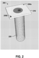
FIG. 2 depicts a portion of one embodiment of a device of the present disclosure having a generally cylindrical cross section. -
FIG. 3 shows ELISA concentration results when complement Factor D (CFD) was incubated in vials containing polyacrylonitrile (PAN) dialysis fibers, a control containing polypropylene fibers, and a negative control. The solution remaining was then examined using spectrophotometry. The results indicate that PAN is taken up in very high amounts by the PAN membranes compared to the controls. -
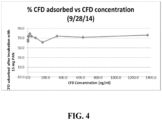
FIG 4 . demonstrates that when CFD is incubated with PAN membranes, the protein is taken up by the membrane well, and even at very high concentrations does not show saturation, indicating that the membrane can adsorb very high levels of the protein. -
FIG. 5 shows results of an in vitro VEGF adsorption assasy and in particular that VEGF levels remaining in solution were significantly lower in the HA implant group, indicating that VEGF is strongly adsorbed by the hydroxyapatite implant. - While the present invention is capable of being embodied in various forms, the description below of several embodiments is made with the understanding that the present disclosure is to be considered as an exemplification of the invention, and is not intended to limit the invention to the specific embodiments illustrated. Headings are provided for convenience only and are not to be construed to limit the invention in any manner. Embodiments illustrated under any heading may be combined with embodiments illustrated under any other heading.
- The use of numerical values in the various quantitative values specified in this application, unless expressly indicated otherwise, are stated as approximations as though the minimum and maximum values within the stated ranges were both preceded by the word "about." Also, the disclosure of ranges is intended as a continuous range including every value between the minimum and maximum values recited as well as any ranges that can be formed by such values. Also disclosed herein are any and all ratios (and ranges of any such ratios) that can be formed by dividing a disclosed numeric value into any other disclosed numeric value. Accordingly, the skilled person will appreciate that many such ratios, ranges, and ranges of ratios can be unambiguously derived from the numerical values presented herein and in all instances such ratios, ranges, and ranges of ratios represent various embodiments of the present invention.
- The term "treatment" in relation a given disease or disorder, includes, but is not limited to, inhibiting the disease or disorder, for example, arresting the development of the disease or disorder; relieving the disease or disorder, for example, causing regression of the disease or disorder; or relieving a condition caused by or resulting from the disease or disorder, for example, relieving, preventing or treating symptoms of the disease or disorder. The term "prevention" in relation to a given disease or disorder means: preventing the onset of disease development if none had occurred, preventing the disease or disorder from occurring in a subject that may be predisposed to the disorder or disease but has not yet been diagnosed as having the disorder or disease, and/or preventing further disease/disorder development if already present.
- In one embodiment, the present disclosure provides a method of treatment of an ocular disorder comprising implanting a device according to the present disclosure in an eye of a subject. The term "ocular disorder" herein refers to any disease or disorder of the eye or related tissues (i.e. retina, macula, retinal blood vessels, etc.) or any symptom thereof. Non-limiting examples of ocular disorders include macular degeneration (e.g., exudative and non- exudative age-related macular degeneration), bull's eye maculopathy, cataract, central serous retinopathy, chorioretinal scars, chorioretinitis, chorioretinitis from toxoplasma, chorioretinitis from tuberculous, choroid, choroidal (central areolar, choroidal atrophy, choroidal degeneration, choroidal detachment, choroidal haemorrhage, choroidal haemorrhage and rupture, choroidal neovascularization, choroidal sclerosis, choroideremia, choroiditis, cystoid macular edema, detachment of retinal pigment epithelium, diabetic retinopathy, dystrophy, epiretinal membrane, generalized, gyrate atrophy, glaucoma, Harada's disease, hereditary choroidal dystrophy, diabetic macular edema, cystoid macular edema, hereditary retinal dystrophy, hypertensive retinopathy, macula scars of posterior pole (postinflammatory or post-traumatic), macular edema, or peripapillary, pars planitis, papillitis, peripheral retinal degeneration, posterior cyclitis, retinal detachment, retinal haemorrhage, retinal neovascularization, retinal vascular occlusions, retinitis, retinitis, retinitis, retinitis pigmentosa, retinochoroiditis, retinochoroiditis, retinochoroiditis, retinopathy, retinopathy of prematurity, retinoschisis, separation of retinal layers, solar retinopathy, syphilitic chorioretinitis, , infectious and non-infectious uveitis, retinal artery occlusion, retinal vein occlusion, retinal and choroidal angiogenesis or neovascularization, retinal and choroidal ischemia, and other ocular events.
- In one embodiment, the present disclosure provides an implantable device comprising a substrate capable of capturing a target molecule present in the eye and/or from fluid of the eye (e.g., an intraocular target molecule). Generally, substrates suitable for use in a device consistent with this disclosure have large surface areas and high affinities for an angiogenic compound, such as VEGF, or other proteins associated with ocular disease. In some embodiments, the substrate comprises hydroxyapatite, a ceramic (e.g., a bioceramic), tricalcium phosphate, bioglass, glass, bone, calcium phosphate, metallic alloys, a membrane or a combination thereof. As used herein, the term "hydroxyapatite" refers to a mineral having a formula Ca10(PO4)6(OH)2. In some embodiments, the substrate comprises, consists essentially of, or consists of hydroxyapatite. In some embodiments, the substrate comprises, consists essentially of, or consists of a ceramic such as mesoporous hydroxyapatite (MHA). In some embodiments, the substrate does not include a polyethylene glycol-conjugated oligonucleotide. In some embodiments, the substrate comprises, consists essentially of, or consists of polyacrylonitrile, biomedical polymers, polystyrene, polyvinyl chloride, poly(D, L-lactide), polymethyl methacrylate (PMMA), and poly(2-hydroxyethyl methacrylate)PHEMA, acrylic, silicone, dextran, bisacrylamide, alkyl chains, agarose, polyacrylamide, silica, nanoparticles, shape memory polymers, alumina, silicon, graphite, grapheme, gold, DMPC, phospholipid membranes, chitosan, collagen, and/or glycosaminoglycan. In other embodiments, the substrate may be a shape memory polymer, plastic, acrylic, nylon, or a combination of various materials. In some embodiments, the substrate comprises, consists essentially of, or consists of a solid, a porous matrix, a gel, a sheet, a membrane, a colloid, a microparticle, or a nanoparticle. In some embodiments the substrate is durable. In other embodiments, the substrate is dissolvable and/or biodegradable. In some embodiments, the substrate comprises biodegradable pellets that can be injected intravitreally and that subsequently degrade or dissolve. In one embodiment, the device is coated with antibodies specific to a target protein, molecule, or moiety, including those belonging to inflammatory, angiogenic, or infectious etiologies.
- In some embodiments, the device is capable of being implanted through an incision. In some embodiments, the device is capable of being implanted by injection. In some embodiments, the device is capable of being implanted in and/or through (e.g., is in contact with) tissue of an eye, for example in or through the pars plana.
- Vascular endothelial growth factor (VEGF) is a protein that promotes vasculogenesis and angiogenesis, and is known to mediate retinal neovascularization. Overexpression (among other factors) can therefore lead to ocular disorders such as macular degeneration or age-related macular degeneration. In some embodiments, the target molecule is a protein. In some embodiments, the target molecule is an angiogenic compound, such as VEGF. In some embodiments, the angiogenic compound comprises a human isoform of VEGF, such as one or more of VEGF121, VEGF121b, VEGF145, VEGF165, VEGF165, VEGF165b, VEGF189, or VEGF206. In some embodiments, the target molecule is adversely associated with retinal physiology, such as PEDF. In other embodiments, the target molecule is selected from: Bone morphogenetic protein-2 (BMP2), Hypoxia-inducible factor-1α (HIF-1α), P2X2, Taurine, Advance glycation end products, Claudin-5, Occludin and JAM-A, microRNAs, RhoJ Pathway, Caveolin-1 (Cav-1), Apelin-13, Exendin-4 and GLP-1, Vascular adhesion protein-1, Angiopoietin-like protein 4 (ANGPTL4), IL-6, IL-8, CXCR3, (CCL2?), MMP-2, 7, and/or 9, TIMP (MMP inhibitors) 1, 2, and/or 3, Cathepsins D, Cystatins, High Temperature required factor A (HtrA) 4, HtrA1 SNP rs11200638, Urokinase-type plasminogen activator, Tissue-type plasminogen activator, IL-6 , IL-8, and/or IL-17, IL-2 and TNFa, Pigment epithelium derived growth factor, Placental growth factor, Fibroblast growth factor, Heat shock proteins (HSP27), Human factor H-related protein 2 -(CFHR2), IGF-1 and IGFBP-2, INFgamma and TNFa, Norrin, Neural cell adhesion molecule (N-CAM), Erythropoietin (EPO) and IGF-1, Angiopoietin-1, IL-1B, Nox and RAAS, Apelin/APJ system, 15-lipoxygenase-1 (15-LOX-1), Prolactine and vasoinhibins, TNF, MCP-1, MCP-2, any molecule involved in the complement cascade, oxygen free radicals, apolipoproteins, lipofuscin, and/or inflammatory proteins or those of an infectious etiology.
- In some embodiments, the substrate has a relatively high affinity for a target molecule. In some embodiments, the substrate has a relatively high affinity for an angiogenic compound. In some embodiments, the substrate has a higher affinity for an angiogenic compound (e.g., VEGF) than for a VEGF antagonist such as ranibizumab, bevacizumab or pegaptanib.
- In some embodiments, the adsorptive property of the device is capable of being regenerated in situ. As used herein, the phrase "capable of being regenerated in situ" indicates that the device is configured such that at least some of the target molecule captured in (or on) the device can be released or expelled from the device in a modified form without the need to remove the device from the subject, so that an additional amount of the target molecule can be captured by the device without implanting a second device in the subject. For example, in one embodiment the device can be regenerated by absorbing energy (e.g., heat) from a source external to the subject. In such an embodiment, the absorbed heat modifies the target molecule (e.g., denatures a protein) such that in its modified form it is characterized by a modified biological activity (e.g., a reduced level of its original biological activity, a different biological activity, or no biological activity). As one example, in one embodiment, the device is capable of adsorbing and/or absorbing VEGF and, after exposure to a thermal source (e.g., a thermal laser such as an argon laser, a diode, a femtosecond laser, a neodymium-doped yttrium aluminum garnet (Nd:Yag) laser, a photodynamic laser, a photodisruptive laser, or a combination thereof), is capable of releasing denatured VEGF. In another embodiment, the device is regenerated by applying cryotherapy to the device. Without wishing to be bound by theory, in such an embodiment it is believed that the decrease or increase in temperature of the adherent proteins causes denaturation and inactivation of biological activity. Alternatively, electrical current or electromagnetic energy may be passed through the device to cause protein denaturation. Further, changing the local pH, dessication, radiation, or doping with elements may all be used to interfere or degrade the biological function of the target protein. Photodynamic therapy, or other light-sensitive materials, may be used to inactivate the adherent proteins. Additionally, the process of laser induced surface plasmon resonance such as with quantum dots may be used to generate a local thermal reaction. Accordingly, in some embodiments, the device has a higher affinity for the target molecule compared to its corresponding modified target molecule (e.g., its thermally denatured target molecule). In some embodiments, the modified target molecule comprises one or more degradation products of the target molecule. Further, the substrate may be treated with fluorophores or other chemical moieties such that the amount of protein adsorbed to the surface is visible by color change, perceptible by indirect ophthalmoscopy, direct visualization, or using confocal scanning laser technology, filters, or other means. The implant may be translucent or clear, allowing the practitioner to laser all surfaces by means of adjusting the laser's focal point. Further, solid implants may be capable of rotating along the major axis, allowing the practitioner to visualize and apply laser energy to the surface in its entirety.
- In some embodiments, the present disclosure provides an implantable device comprising a substrate comprising one or more of hydroxyapatite and a ceramic, and a captured angiogenic compound. In some embodiments, the device is capable of releasing the captured angiogenic compound (and/or modified angiogenic compounds, degradation products of the angiogenic compound, etc.) after denaturation of at least a portion of the captured angiogenic compound, for example by exposure to a laser. In some embodiments, the device is capable of capturing an additional amount of the angiogenic compound after exposure to the laser. In some embodiments, the angiogenic compound comprises VEGF (e.g., a human isoform of VEGF) from vitreous fluid of a subject.
- In some embodiments, such as the embodiment shown in
FIG. 1 , at least a portion of theimplantable device 100 has a size and shape that imparts a relatively high surface area to the device. For example, in oneembodiment device 100 comprises an anchoringportion 110 and an immersed or immersible portion 120, wherein the anchoringportion 110 is configured to be in contact with and secured at one or more tissues of the eye, and wherein immersed or immersible portion 120 is configured to be in contact withvitreous fluid 18. In some embodiments, immersed or immersible portion 120 defines a size and shape different from that of an anchoringportion 110 ofdevice 100. Immersed or immersible portion 120 of thedevice 100 may have a polygon shape, or a shape of a cylinder, sphere, partial sphere, cone, truncated cone, or a combination thereof. The device may also be composed of multiple hollow tubes, similar to dialysis tubing, which can be bundled together. - In some embodiments, the
device 100 has ananchor portion 110 with which the device can be attached to a portion of the eye tissue, for example by suture. - In some embodiments, the
device 100 has anindicator portion 130 which may be used, for example, to provide a practitioner with information about the type of material(s) used indevice 100. In other embodiments,indicator portion 130 may provide the practitioner a target zone or information about where a regenerating laser may be focused. - In other embodiments, for example as shown in
FIG. 2 , an implantableocular device 200 comprises an anchoringportion 210 and an immersed orimmersible portion 220. In some embodiments,device 200 includes one or more anchoring features (240a, 240b) which allow a practitioner to securedevice 200 to a tissue of the eye. For example, anchoring feature(s) 240a-b may comprise holes through which a suture can be passed. In some embodiments, the substrate may be incorporated into other implants which are commonly placed in the eye such as glaucoma drainage devices, aqueous shunting devices, and intraocular lenses. - In some embodiments, immersed or
immersible portion 220 ofdevice 200, may have a generally cylindrical, tapered cylinder, or conical cross section. Further, it may be spherical, tubular, or in a sheet-like configuration. In some embodiments,device 200 has an indicator portion 230 which may be used, for example, to provide a practitioner with information about the type of material(s) used indevice 200. In other embodiments, indicator portion 230 may provide the practitioner a target zone or information about where a regenerating laser may be focused. In some embodiments, indicator portion 230 is applied to or formed as part of anchoringportion 210. In some embodiments, indicator portion 230 is configured to allow a needle to pass therethrough, for example to allow removal, replacement, or exchange of materials inside immersed orimmersible portion 220 without removingdevice 200 from the eye. In some embodiments, indicator portion 230 comprises a self-sealing material, for example to prevent fluid from passing therethrough after penetration by a needle. - In some embodiments at least a portion of the implantable device (e.g., immersed or immersible portion 120, 220) has a porous or microporous surface. In some embodiments, the surface of at least a portion of device has an average pore diameter of less than 1 mm, for example about 1 mm, about 0.95 mm, about 0.9 mm, about 0.85 mm, about 0.8 mm, about 0.75 mm, about 0.7 mm, about 0.65 mm, about 0.6 mm, about 0.55 mm, about 0.5 mm, about 0.45 mm, about 0.4 mm, about 0.35 mm, about 0.3 mm, about 0.25 mm, about 0.2 mm, about 0.15 mm, about 0.1 mm, about 0.05 mm, or less than about 0.05 mm.
- In some embodiments, the implantable device is further capable of delivering a drug to the subject. In some embodiments, the drug is an anti-VEGF compound such as ranibizumab, bevacizumab or pegaptanib, or a steroid. The drug may be loaded into the substrate by forced pressure or vacuum techniques, filling the porous cavities of the device with the intended therapeutic agent, whether solid, powder, liquid, or gas.
- In one embodiment, the present disclosure provides a method of treating an ocular disorder in a subject, the method comprising implanting into an eye of the subject, and in contact with fluid of the eye, a device as disclosed herein; and thereafter capturing the target molecule from the fluid. In one embodiment, the eye has previously undergone vitrectomy or other ocular surgery.
- One embodiment of a method of the present disclosure is depicted in
FIG. 1 . A device of thepresent disclosure 100 is implanted in theeye 10 of the subject. In some embodiments, thedevice 100 is implanted through (e.g., is in contact with) one or more of: the sclera 16, thechoroid 14, and/or theretina 12. In some embodiments, thedevice 100 is implanted through (e.g., is in contact with) the pars plana portion of thechoroid 14. - In some embodiments, the method further comprises, after capturing the target molecule from fluid of the eye, regenerating the adsorptive properties of the device in situ. In some embodiments, the step of regenerating the device in situ comprises exposing the device to an energy source, such as a laser, for example from a laser indirect ophthalmoscope.
- In some embodiments, the method further comprises, after regenerating the device in situ, capturing an additional amount of the target molecule from fluid of the eye.
- In some embodiments, the implanted device is left in place for a period of time sufficient to reduce or alleviate one or more symptoms of the ocular disorder, for example about one month, about 2 months, about 3 months, about 4 months, about 5 months, about 6 months, about 7 months, about 8 months, about 9 months, about 10 months, about 11 months, about 12 months, or more than 12 months. In some embodiments, the device is regenerated at least once, at least twice, at least 3 times, at least 4 times, at least 5 times, at least 6 times, at least 7 times, at least 8 times, at least 9 times, at least 10 times, at least 11 times, at least 12 times, or more than 12 times before removal from the subject. In an alternative embodiment, the device is removed after a period of time sufficient for it to become saturated with the target molecule, for example after about 1 week, about 2 weeks, about 3 weeks, about 4 weeks, about 5 weeks, about 6 weeks, about 7 weeks, about 8 weeks, about 9 weeks, about 10 weeks, about 11 weeks, about 12 weeks, about 4 months, about 5 months, about 6 months, about 7 months, about 8 months, about 9 months, about 10 months, about 11 months, about 12 months.
- In some embodiments, a method of treating an ocular disorder in a subject on intraocular injection therapy comprises, consists essentially of, or consists of optionally identifying the subject as being on intraocular injection therapy; implanting into an eye of the subject, and in contact with fluid of the eye, a device comprising a substrate capable of capturing a target molecule present in fluid of the eye; capturing the target molecule from the fluid; and optionally after capturing the target molecule from the fluid, regenerating the device in situ. In some embodiments, the intraocular injection therapy comprises administration of ranibizumab, bevacizumab and/or pegaptanib. In some embodiments, the target molecule is an angiogenic compound, optionally VEGF. In some embodiments, the optional step of regenerating the device in situ comprises contacting the device with a thermal laser to denature and/or decompose at least a portion of the captured target molecule.
- In some embodiments, a method of the present disclosure comprises replacing existing intraocular injection therapeutic regimen with a second treatment regimen comprising, consisting essentially of, or consisting of implanting into an eye of the subject, and in contact with fluid of the eye, a device comprising a substrate capable of capturing a target molecule present in fluid of the eye; capturing the target molecule from the fluid; and optionally after capturing the target molecule from the fluid, regenerating the device in situ. In some embodiments, the existing intraocular injection therapy is discontinued before, concomitantly with, or after implanting the device into the eye of the subject. In some embodiments, the target molecule is an angiogenic compound, optionally VEGF. In some embodiments, the optional step of regenerating the device in situ comprises contacting the device with a thermal laser to denature and/or decompose at least a portion of the captured target molecule.
- Twenty white cross rabbits are divided into three groups: control, active implant (bioceramic), and inactive implant (standard plastic). Baseline photographs, electroretinograms, and intravitreal VEGF levels are performed on all subjects. The active and inactive implant groups undergo surgery and two weeks later all three groups receive in the right eye a standardized dose of VEGF in a polymer pellet which has a sustained release profile sufficient to induce vasoproliferation in a reliable and repeatable manner. Repeat examinations, fundus photography, fluorescein angiography, and intravitreal VEGF sampling are performed at 24 hrs, 48 hrs, 4 days, 7 days, 14 days, 21 days, and 28 days post-surgery. Prior to harvesting the eyes for histological examination, final electroretinograms are performed. Subsequent grading of observed neovascularization is performed by a masked observer using the system described by Ozaki et al., "Intravitreal sustained release of VEGF causes retinal neovascularization in rabbits and breakdown of the blood-retinal barrier in rabbits and primates," Exp. Eye Res., vol. 64(4), pages 505-17 (1997).
- A subset of subjects from Example 1 are observed for an additional 9 months in order to determine long-term stability and biocompatibility of the implanted device. Exams, fundus photography, angiography, electroretinography and VEGF sampling are performed each month.
- Bioceramic discs composed of porous hydroxyapatite were soaked overnight in 50 µL of deionized water containing 50 ng of VEGF, each in a separate well of a 96-well plate. The discs soaked for 24 hours with periodic gentle agitation, after which the excess solution was removed. The amount of VEGF in this solution was determined using a human VEGF ELISA kit. The estimated amount of VEGF loaded into the implant was determined by subtracting the amount in the solution from the loading amount. The hydroxyapatite discs adsorbed an average of 14 ng of VEGF, compared to less than 1 ng for a control group consisting of plastic beads (p < 0.05).
- Next, half of the hydroxyapatite discs were exposed to argon laser (240 mW power, 0.2 ms duration, 100 spots over 2 minutes), sufficient to induce a rise in the surface temperature of the discs of 8 °C, as measured by an infrared thermometer. The other half of the pellets were exposed to light for 2 minutes, without exposure to laser and without surface temperature change. All the pellets were then placed in separate wells of another 96-well plate and allowed to soak 48 hours in DI water with gentle agitation. The amount of VEGF released from each pellet was then measured again using the VEGF ELISA kit. The discs exposed to light released about 30% of the initial loading dose of VEGF, compared to about 5% in the lasered group (p < 0.05). These data demonstrate that the bioceramic material can sequester VEGF, which can be further inactivated by thermal laser.
- An experiment was conducted to assess adsorption of complement Factor D by PAN fibers as compared to polypropylene capillaries according to the below protocol.
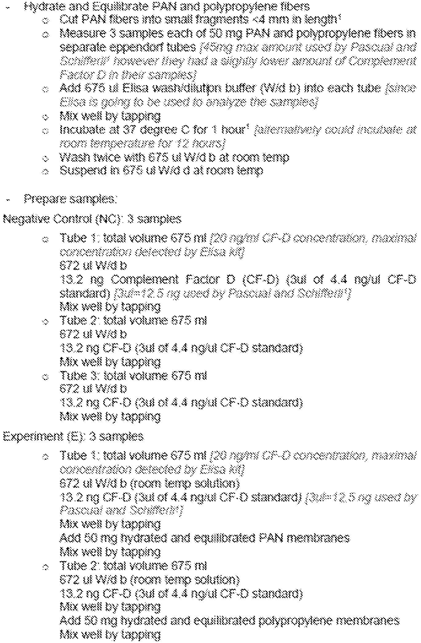


- Results are shown in
Figures 3 and4 . Overall, these experiments demonstrate that a type of membrane (polyacrylonitrile) will adsorb high amounts of a protein called Complement Factor D (CFD), which is thought to be a major therapeutic target in dry macular degeneration. - An in vitro adsorption experiment was conducted according to the following protocol:

- Results are shown in
FIG. 5 and demonstrate that VEGF levels remaining in solution were significantly lower in the HA implant group, indicating that VEGF is adsorbed by the hydroxyapatite implant. - An implantable device comprising a substrate capable of capturing an intraocular target molecule.
- The implantable device of paragraph 44, wherein the substrate comprises one or more of hydroxyapatite, a ceramic, tricalcium phosphate, bioglass, bone, calcium phosphate, metallic alloys, or a combination thereof.
- The implantable device of paragraph 44 or paragraph 45, wherein the substrate comprises hydroxyapatite.
- The implantable device of paragraph 44 or paragraph 45, wherein the substrate comprises a ceramic.
- The implantable device of any one of paragraphs 44, 45 or 47, wherein the ceramic comprises mesoporous hydroxyapatite (MHA).
- The implantable device of any one of paragraphs 44-48, wherein the substrate is a solid, a porous matrix, a gel, a sheet, a membrane, a colloid, a microparticle, a nanoparticle, or does not include a polyethylene glycol-conjugated oligonucleotide.
- The implantable device of any one of paragraphs 44-49, wherein the device is capable of being implanted through an incision.
- The implantable device of any one of paragraphs 44-50, wherein the device is capable of being implanted by injection.
- The implantable device of any one of paragraphs 44-51 wherein the target molecule is a protein.
- The implantable device of any one of paragraphs 44-52, wherein the target molecule is an angiogenic compound.
- The implantable device of paragraph 53, wherein the angiogenic compound comprises vascular endothelial growth factor (VEGF).
- The implantable device of paragraph 54, wherein the angiogenic compound comprises VEGF165.
- The implantable device of any one of paragraphs 44-55, wherein the target molecule comprises an angiogenic compound but not ranibizumab, bevacizumab or pegaptanib.
- The implantable device of any one of paragraphs 44-56, wherein the device is capable of being regenerated in situ.
- The implantable device of paragraph 57, wherein the device has a higher affinity for the target molecule compared to a corresponding thermally denatured target molecule.
- An implantable device comprising a substrate comprising one or more of hydroxyapatite and a ceramic, and a captured angiogenic compound.
- The implantable device of paragraph 59, wherein the device is capable of releasing the captured angiogenic compound after exposure to a laser.
- The implantable device of paragraph 60, wherein the device is capable of capturing the angiogenic compound after exposure to the laser.
- The implantable device of any one of paragraphs 59-91, wherein the angiogenic compound comprises VEGF from vitreous fluid of a subject.
- The implantable device of paragraph 62, wherein the angiogenic compound comprises VEGF165.
- A method of treating an ocular disorder in a subject, the method comprising implanting into an eye of the subject, and in contact with a tissue, cavity and/or fluid of the eye, a device comprising a substrate capable of capturing a target molecule present in the eye; and capturing the target molecule from the eye.
- The method of paragraph 64, wherein the substrate comprises hydroxyapatite and/or a ceramic.
- The method of paragraph 64, wherein the substrate comprises a ceramic.
- The method of paragraph 64, wherein the ceramic comprises mesoporous hydroxyapatite (MFIA).
- The method of any one of paragraphs 64-67, wherein the substrate does not include a polyethylene glycol-conjugated oligonucleotide.
- The method of any one of paragraphs 64-68, wherein the device is implanted through an incision in the eye.
- The method of any one of paragraphs 64-69, wherein the device is implanted by injecting the device into or through tissue of the eye.
- The method of any one of paragraphs 64-70, wherein the target molecule is an angiogenic compound.
- The method of paragraph 71, wherein the angiogenic compound comprises vascular endothelial growth factor (VEGF).
- The method of paragraph 72, wherein the angiogenic compound comprises VEGF165.
- The method of any one of paragraphs 64-73, wherein the target molecule comprises an angiogenic compound but not ranibizumab, bevacizumab or pegaptanib.
- The method of any one of paragraphs 64-74, the method further comprising, after capturing the target molecule from the eye, regenerating the device in situ.
- The method of paragraph 75, wherein the step of regenerating the device comprises exposing the device to light from a laser or to a source capable of inducing a thermal reaction.
- The method of paragraph 76, wherein the laser is an argon laser, a diode, a femtosecond laser, a Nd:Yag laser, a photodynamic laser, or a photodisruptive laser.
- The method of paragraph 76 or 77 further comprising, after regenerating the device in situ, capturing the target molecule from the eye.
- The method of any one of paragraphs 64-78, wherein the ocular disorder is selected from the group consisting of macular degeneration (e.g., exudative age-related macular degeneration), bull's eye maculopathy, central serous retinopathy, horioretinal scars, chorioretinitis, chorioretinitis, chorioretinitis, chorioretinitis, chorioretinitis from toxoplasma, chorioretinitis from tuberculous, choroid, choroidal (central areolar, choroidal atrophy, choroidal degeneration, choroidal detachment, choroidal haemorrhage, choroidal haemorrhage and rupture, choroidal neovascularization, choroidal sclerosis, choroideremia, choroiditis, choroiditis, choroiditis, detachment of retinal pigment epithelium, diabetic retinopathy, dystrophy, epiretinal membrane, generalized, gyrate atrophy, Harada's disease, hereditary choroidal dystrophy, diabetic macular edema, cystoid macular edema, hereditary retinal dystrophy, hypertensive retinopathy, macula scars of posterior pole (postinflammatory or post-traumatic), macular edema, or peripapillary, pars planitis, papillitis, peripheral retinal degeneration, posterior cyclitis, retinal detachment, retinal haemorrhage, retinal neovascularization, retinal vascular occlusions, retinitis, retinitis, retinitis, retinitis pigmentosa, retinochoroiditis, retinochoroiditis, retinochoroiditis, retinopathy, retinopathy of prematurity, retinoschisis, separation of retinal layers, solar retinopathy, and syphilitic chorioretinitis.
- The implantable device of any one of paragraphs 44-63, wherein the device further comprises an indicator portion configured to provide a practitioner information about a component, feature or recommended use of the device.
- The method of any one of paragraphs 64-79, wherein the device further comprises an indicator portion configured to provide a practitioner information about a component, feature or recommended use of the device.
- From the foregoing, it will be appreciated that specific embodiments of the invention have been described herein for purposes of illustration, but that various modifications may be made without deviating from the scope of the invention.
Claims (10)
- Polyacrylonitrile for use in the treatment of dry macular degeneration, wherein the polyacrylonitrile is in the form of a substrate that is a constituent of an implantable device (200).
- The polyacrylonitrile for use in the treatment of dry macular degeneration of claim 1, wherein the treatment comprises: implanting into an eye of a subject the device comprising the polyacrylonitrile substrate, wherein the polyacrylonitrile substrate is capable of capturing a target molecule from the eye or in fluid of the eye; and capturing the target molecule from the eye or from fluid of the eye.
- The polyacrylonitrile for use in the treatment of dry macular degeneration of claim 1 or claim 2, wherein the polyacrylonitrile is in the form of a substrate that is capable of capturing complement factor D from the eye or from fluid of the eye.
- An implantable device (200), comprising a substrate that is capable of capturing a target molecule that is present intraocularly wherein the substrate comprises, consists essentially of, or consists of polyacrylonitrile.
- The implantable device (200), that is capable of capturing Complement Factor D.
- The implantable device (200) of claim 4 or claim 5, comprising: an anchoring portion (110, 210) configured to be in contact with and secured at one or more tissues of the eye and an immersible portion (120, 220) configured to be in contact with the vitreous fluid (18).
- The implantable device (200) of claim 6, wherein the immersible portion (120, 220) has a porous or microporous surface.
- The implantable device (200) of any one of claims 4 to 7, wherein the device is configured to extend into the vitreous cavity and/or the anterior chamber of the eye.
- The implantable device (200) of any one of claims 4 to 8, wherein the device is configured such that at least some of the target molecule captured in the device can be released or expelled from the device in a modified form without the need to remove the device from a subject, so that an additional amount of the target molecule can be captured by the device without implanting a second device in the subject.
- The polyacrylonitrile for use in the treatment of dry macular degeneration of any one of claims 1 to 3, wherein the substrate is a constituent of the implantable device (200) of any one of claims 4 to 9.
Applications Claiming Priority (3)
| Application Number | Priority Date | Filing Date | Title |
|---|---|---|---|
| US201361874759P | 2013-09-06 | 2013-09-06 | |
| EP14842818.8A EP3041524A4 (en) | 2013-09-06 | 2014-09-08 | Intraocular drug delivery and filter device and methods of using same |
| PCT/US2014/054531 WO2015035296A2 (en) | 2013-09-06 | 2014-09-08 | Intraocular drug delivery and filter device and methods of using same |
Related Parent Applications (1)
| Application Number | Title | Priority Date | Filing Date |
|---|---|---|---|
| EP14842818.8A Division EP3041524A4 (en) | 2013-09-06 | 2014-09-08 | Intraocular drug delivery and filter device and methods of using same |
Publications (2)
| Publication Number | Publication Date |
|---|---|
| EP4537798A2 true EP4537798A2 (en) | 2025-04-16 |
| EP4537798A3 EP4537798A3 (en) | 2025-06-18 |
Family
ID=52629090
Family Applications (2)
| Application Number | Title | Priority Date | Filing Date |
|---|---|---|---|
| EP14842818.8A Ceased EP3041524A4 (en) | 2013-09-06 | 2014-09-08 | Intraocular drug delivery and filter device and methods of using same |
| EP25152586.1A Withdrawn EP4537798A3 (en) | 2013-09-06 | 2014-09-08 | Intraocular drug delivery and filter device and methods of using same |
Family Applications Before (1)
| Application Number | Title | Priority Date | Filing Date |
|---|---|---|---|
| EP14842818.8A Ceased EP3041524A4 (en) | 2013-09-06 | 2014-09-08 | Intraocular drug delivery and filter device and methods of using same |
Country Status (3)
| Country | Link |
|---|---|
| US (2) | US10518002B2 (en) |
| EP (2) | EP3041524A4 (en) |
| WO (1) | WO2015035296A2 (en) |
Families Citing this family (2)
| Publication number | Priority date | Publication date | Assignee | Title |
|---|---|---|---|---|
| US11219552B2 (en) | 2013-09-06 | 2022-01-11 | The Regents Of The University Of Colorado, A Body Corporate | Intraocular filter device and methods of using same |
| EP3041524A4 (en) * | 2013-09-06 | 2017-06-14 | The Regents of the University of Colorado, a body corporate | Intraocular drug delivery and filter device and methods of using same |
Family Cites Families (15)
| Publication number | Priority date | Publication date | Assignee | Title |
|---|---|---|---|---|
| ATE404140T1 (en) * | 2000-08-30 | 2008-08-15 | Univ Johns Hopkins | DEVICE FOR INTRAOCULAR ADMINISTRATION OF ACTIVE INGREDIENTS |
| US8182527B2 (en) * | 2001-05-07 | 2012-05-22 | Cordis Corporation | Heparin barrier coating for controlled drug release |
| JP2004208898A (en) * | 2002-12-27 | 2004-07-29 | Japan Science & Technology Agency | Aqueous humor drainage implant for glaucoma treatment |
| US20060110428A1 (en) * | 2004-07-02 | 2006-05-25 | Eugene Dejuan | Methods and devices for the treatment of ocular conditions |
| CN101180086A (en) | 2005-04-08 | 2008-05-14 | 苏尔莫迪克斯公司 | Sustained-release implants for subretinal delivery |
| US7625404B2 (en) * | 2005-12-24 | 2009-12-01 | Mohsen Shahinpoor | Surgical correction of ptosis by polymeric artificial muscles |
| US20080147021A1 (en) * | 2006-12-15 | 2008-06-19 | Jani Dharmendra M | Drug delivery devices |
| RU2009144142A (en) * | 2007-04-30 | 2011-06-10 | Алькон Рисерч, Лтд. (Us) | TREATMENT OF AGE YELLOW SPOT DEGENERATION WITH APPLICATION OF INHIBITORS OF FACTOR D OF COMPLEMENT |
| US8889168B2 (en) | 2008-08-07 | 2014-11-18 | Bioactive Surgical Inc. | Stem cell capture and immobilization coatings for medical devices and implants |
| US8317737B2 (en) * | 2009-02-25 | 2012-11-27 | The Invention Science Fund I, Llc | Device for actively removing a target component from blood or lymph of a vertebrate subject |
| US8657804B2 (en) * | 2009-10-22 | 2014-02-25 | On Demand Therapeutics, Inc. | Visual indication of rupture of drug reservoir |
| WO2011063152A1 (en) * | 2009-11-18 | 2011-05-26 | Affinergy, Inc. | Methods and compositions for soft tissue repair |
| WO2012149468A2 (en) * | 2011-04-29 | 2012-11-01 | University Of Southern California | Instruments and methods for the implantation of cell-seeded substrates |
| EP2726016B1 (en) * | 2011-06-28 | 2023-07-19 | ForSight Vision4, Inc. | An apparatus for collecting a sample of fluid from a reservoir chamber of a therapeutic device for the eye |
| EP3041524A4 (en) | 2013-09-06 | 2017-06-14 | The Regents of the University of Colorado, a body corporate | Intraocular drug delivery and filter device and methods of using same |
-
2014
- 2014-09-08 EP EP14842818.8A patent/EP3041524A4/en not_active Ceased
- 2014-09-08 WO PCT/US2014/054531 patent/WO2015035296A2/en not_active Ceased
- 2014-09-08 EP EP25152586.1A patent/EP4537798A3/en not_active Withdrawn
- 2014-09-08 US US14/916,659 patent/US10518002B2/en active Active
-
2019
- 2019-12-06 US US16/705,989 patent/US11160908B2/en active Active
Non-Patent Citations (1)
| Title |
|---|
| OZAKI ET AL.: "Intravitreal sustained release of VEGF causes retinal neovascularization in rabbits and breakdown of the blood-retinal barrier in rabbits and primates", EXP. EYE RES., vol. 64, no. 4, 1997, pages 505 - 17, XP000909325, DOI: 10.1006/exer.1996.0239 |
Also Published As
| Publication number | Publication date |
|---|---|
| US20160193391A1 (en) | 2016-07-07 |
| US10518002B2 (en) | 2019-12-31 |
| US11160908B2 (en) | 2021-11-02 |
| WO2015035296A2 (en) | 2015-03-12 |
| EP3041524A2 (en) | 2016-07-13 |
| WO2015035296A3 (en) | 2015-11-19 |
| EP3041524A4 (en) | 2017-06-14 |
| EP4537798A3 (en) | 2025-06-18 |
| US20200129672A1 (en) | 2020-04-30 |
Similar Documents
| Publication | Publication Date | Title |
|---|---|---|
| JP5996526B2 (en) | Method and apparatus for drug delivery to ocular tissue using microneedles | |
| AU2011285637B2 (en) | Subconjunctival implant for posterior segment drug delivery | |
| AU2018253472B2 (en) | Trans-sclera portal for delivery of therapeutic agents | |
| DK2207529T3 (en) | PHARMACEUTICAL cores for the sustained release of therapeutic agents | |
| JP6980671B2 (en) | Ophthalmic composition | |
| JP2016166208A (en) | Implantable therapeutic device | |
| ES2856886T3 (en) | Intraocular lens comprising drug-containing microspheres | |
| CA3240136A1 (en) | Methods and devices for the treatment of ocular diseases in human subjects | |
| WO2010088548A1 (en) | Posterior segment drug delivery | |
| WO2017053733A1 (en) | Biodegradable microsphere-hydrogel ocular drug delivery system | |
| US11160908B2 (en) | Intraocular drug delivery and filter device and methods of using same | |
| US11219552B2 (en) | Intraocular filter device and methods of using same | |
| US11173130B2 (en) | Drug delivery system and methods of use | |
| JP6856948B2 (en) | Ophthalmic drug sustained release device | |
| Al-Shohani | Hydrogel formulations for ophthalmic delivery | |
| US20220249281A1 (en) | Continual drug release device that can be replenished with drugs | |
| US20110054445A1 (en) | Devices and Methods for Treatment of Eye Disease | |
| RU2557925C1 (en) | Biodegradable multilayer implant for administering drug substances into vitreal cavity of eye |
Legal Events
| Date | Code | Title | Description |
|---|---|---|---|
| PUAI | Public reference made under article 153(3) epc to a published international application that has entered the european phase |
Free format text: ORIGINAL CODE: 0009012 |
|
| STAA | Information on the status of an ep patent application or granted ep patent |
Free format text: STATUS: THE APPLICATION HAS BEEN PUBLISHED |
|
| AC | Divisional application: reference to earlier application |
Ref document number: 3041524 Country of ref document: EP Kind code of ref document: P |
|
| AK | Designated contracting states |
Kind code of ref document: A2 Designated state(s): AL AT BE BG CH CY CZ DE DK EE ES FI FR GB GR HR HU IE IS IT LI LT LU LV MC MK MT NL NO PL PT RO RS SE SI SK SM TR |
|
| REG | Reference to a national code |
Ref country code: DE Ref legal event code: R079 Free format text: PREVIOUS MAIN CLASS: A61F0009000000 Ipc: A61L0027540000 |
|
| PUAL | Search report despatched |
Free format text: ORIGINAL CODE: 0009013 |
|
| AK | Designated contracting states |
Kind code of ref document: A3 Designated state(s): AL AT BE BG CH CY CZ DE DK EE ES FI FR GB GR HR HU IE IS IT LI LT LU LV MC MK MT NL NO PL PT RO RS SE SI SK SM TR |
|
| RIC1 | Information provided on ipc code assigned before grant |
Ipc: A61F 9/00 20060101ALI20250512BHEP Ipc: A61L 27/54 20060101AFI20250512BHEP |
|
| STAA | Information on the status of an ep patent application or granted ep patent |
Free format text: STATUS: THE APPLICATION IS DEEMED TO BE WITHDRAWN |
|
| 18D | Application deemed to be withdrawn |
Effective date: 20250718 |