EP4530359A1 - Multiplexierte einzelmolekül-rna fische unter verwendung von dna nanostrukturen - Google Patents
Multiplexierte einzelmolekül-rna fische unter verwendung von dna nanostrukturen Download PDFInfo
- Publication number
- EP4530359A1 EP4530359A1 EP23200449.9A EP23200449A EP4530359A1 EP 4530359 A1 EP4530359 A1 EP 4530359A1 EP 23200449 A EP23200449 A EP 23200449A EP 4530359 A1 EP4530359 A1 EP 4530359A1
- Authority
- EP
- European Patent Office
- Prior art keywords
- dna
- seq
- probe
- targ
- nanostructure
- Prior art date
- Legal status (The legal status is an assumption and is not a legal conclusion. Google has not performed a legal analysis and makes no representation as to the accuracy of the status listed.)
- Withdrawn
Links
Images


Classifications
-
- C—CHEMISTRY; METALLURGY
- C12—BIOCHEMISTRY; BEER; SPIRITS; WINE; VINEGAR; MICROBIOLOGY; ENZYMOLOGY; MUTATION OR GENETIC ENGINEERING
- C12Q—MEASURING OR TESTING PROCESSES INVOLVING ENZYMES, NUCLEIC ACIDS OR MICROORGANISMS; COMPOSITIONS OR TEST PAPERS THEREFOR; PROCESSES OF PREPARING SUCH COMPOSITIONS; CONDITION-RESPONSIVE CONTROL IN MICROBIOLOGICAL OR ENZYMOLOGICAL PROCESSES
- C12Q1/00—Measuring or testing processes involving enzymes, nucleic acids or microorganisms; Compositions therefor; Processes of preparing such compositions
- C12Q1/68—Measuring or testing processes involving enzymes, nucleic acids or microorganisms; Compositions therefor; Processes of preparing such compositions involving nucleic acids
- C12Q1/6813—Hybridisation assays
- C12Q1/6841—In situ hybridisation
-
- C—CHEMISTRY; METALLURGY
- C12—BIOCHEMISTRY; BEER; SPIRITS; WINE; VINEGAR; MICROBIOLOGY; ENZYMOLOGY; MUTATION OR GENETIC ENGINEERING
- C12Q—MEASURING OR TESTING PROCESSES INVOLVING ENZYMES, NUCLEIC ACIDS OR MICROORGANISMS; COMPOSITIONS OR TEST PAPERS THEREFOR; PROCESSES OF PREPARING SUCH COMPOSITIONS; CONDITION-RESPONSIVE CONTROL IN MICROBIOLOGICAL OR ENZYMOLOGICAL PROCESSES
- C12Q2525/00—Reactions involving modified oligonucleotides, nucleic acids, or nucleotides
- C12Q2525/10—Modifications characterised by
- C12Q2525/197—Modifications characterised by incorporating a spacer/coupling moiety
-
- C—CHEMISTRY; METALLURGY
- C12—BIOCHEMISTRY; BEER; SPIRITS; WINE; VINEGAR; MICROBIOLOGY; ENZYMOLOGY; MUTATION OR GENETIC ENGINEERING
- C12Q—MEASURING OR TESTING PROCESSES INVOLVING ENZYMES, NUCLEIC ACIDS OR MICROORGANISMS; COMPOSITIONS OR TEST PAPERS THEREFOR; PROCESSES OF PREPARING SUCH COMPOSITIONS; CONDITION-RESPONSIVE CONTROL IN MICROBIOLOGICAL OR ENZYMOLOGICAL PROCESSES
- C12Q2563/00—Nucleic acid detection characterized by the use of physical, structural and functional properties
- C12Q2563/179—Nucleic acid detection characterized by the use of physical, structural and functional properties the label being a nucleic acid
Definitions
- the invention lies in the field of molecular biology and particularly in the field of RNA fluorescence in situ hybridization (RNA FISH) technology.
- RNA FISH RNA fluorescence in situ hybridization
- the goal of the present invention is to provide a multiplexed single molecule RNA FISH using DNA nanotechnology and a method performed using such multiplexed single molecule RNA FISH.
- Measurement of gene expression in individual cells in the spatial context of cells or tissue is becoming increasingly relevant not only to academic research, but also to industrial applications and medical molecular diagnostics.
- Bulk measurements of gene expression such as RT-qPCR, microarray technology, bulk RNA sequencing (RNASeq) averages abundance of RNA in all measured cells, which covers up individual differences in gene expression as well as obscures low-level gene expression of crucial genes.
- This approach results in relevant information being lost, for example, in the case with transcription factors such as GATA6, which in about 50% of cases is more lowly expressed than measurable via RT-qPCR or microarrays (PMID 32156747).
- the corresponding spatial information of where the gene is expressed i.e., where the transcripts are found in the cell, are lost in bulk measurements of gene expression.
- tissue level differentiated expression of genes in different cell types and tissue polarization
- subcellular level spatialal compartmentalization of gene expression within cells, enrichment of RNAs at the endoplasmatic reticulum (ER), retainment in the nucleus (PMID 31501331)).
- RNA Fluorescence in situ Hybridization also called single molecule FISH, smFISH
- FISH RNA Fluorescence in situ Hybridization
- smFISH single molecule FISH
- the number of simultaneously measurable RNA species detectable by this method is limited by the number of clearly distinguishable fluorescence spectra which can be recorded simultaneously by a fluorescence microscope; usually 3-4 RNAs in one hybridization, greatly limiting the scalability of the method.
- EP 3 133 170 A1 relates to a probe set for in situ hybridization that enables detection of individual mRNA molecules, comprising at least 12 non-overlapping nucleic acid hybridization probes singly labelled with the same first fluorophore label of a first color, wherein the probes have sequences complementary to a target sequence, wherein said probes are 7-30 nucleotides in length.
- EP 3 541 956 A1 relates to methods for spatial tagging of nucleic acid molecules in a biological specimen and in particular to a method comprising: providing a solid substrate on which multiple species of capture probes are immobilized; contacting a solid substrate with a biological specimen; and releasing capture probes from the surface of the solid substrate under conditions that allow nucleic acids of the biological specimen to hybridize.
- EP 3 916 108 A1 relates to a method for spatially tagging nucleic acid molecules in a biological specimen.
- the method involves using a solid substrate with multiple species of capture probes immobilized in distinct positions, wherein the capture probes have three components: a cleavage domain for releasing them from the substrate, a positional domain corresponding to their location on the substrate, and a capture domain.
- EP 3 916 108 A1 discloses to three main steps: (a) placing the solid substrate with capture probes in contact with the biological specimen, (b) allowing the capture probes to be released from the substrate, and (c) simultaneously or subsequently extending the released capture probes using nucleic acids from the biological specimen as templates.
- the present invention relates to methods and products for localized or spatial detection and/or analysis of RNA in a tissue sample or a portion thereof, comprising: (a) providing an object substrate on which at least one species of capture probe, comprising a capture domain, is directly or indirectly immobilized such that the probes are oriented to have a free 3' end to enable said probe to function as a reverse transcriptase (RT) primer; (b) contacting said substrate with a tissue sample and allowing RNA of the tissue sample to hybridize to the capture probes; (c) generating cDNA molecules from the captured RNA molecules using said capture probes as RT primers; (d) labelling the cDNA molecules generated in step (c), wherein said labelling step may be contemporaneous with, or subsequent to, said generating step; (e) detecting a signal from the labelled cDNA molecules; and optionally (f) imaging the tissue sample, wherein the tissue sample is imaged before or after step (c).
- RT reverse transcriptase
- US 6 485 944 B1 relates to methods of making and using immobilized arrays of nucleic acids, particularly methods for producing replicas of such arrays, including methods for producing high density arrays of nucleic acids and replicas of such arrays, as well as methods for preserving the resolution of arrays through rounds of replication.
- US 8 865 404 B2 relates to a method for determining the nucleic acid sequence of a polynucleotide that is at least 1500 nucleotides in length comprising, inter alia, providing a polynucleotide; cleaving the polynucleotide into a plurality of oligonucleotide sequences; immobilizing only one end of each of the plurality of oligonucleotide; arranging the plurality of oligonucleotide sequences; attaching a label to a non-immobilized end of each of the plurality of oligonucleotide sequences; cleaving the plurality of oligonucleotide sequences to generate a plurality of labelled oligonucleotide fragments; collecting the plurality of oligonucleotide fragments; sequencing the plurality of oligonucleotide fragments to obtain oligonucleotide reads; assembling the oligonucleo
- the invention relates to a probe for RNA fluorescence in situ hybridization (FISH) comprising: a marker molecule, a targeting molecule comprising 22 to 55 nt comprising a spacer sequence, and a target binding sequence capable of binding to a target molecule, wherein the marker molecule may be connected to the targeting molecule via the spacer sequence.
- FISH RNA fluorescence in situ hybridization
- spacer sequence may also be referred to as spacer molecule, or simply as spacer.
- the probe may comprise a marker molecule binding sequence comprising 20 to 40 nt.
- the marker molecule may be bound to the marker molecule binding sequence, the marker molecule binding sequence may be connected to the spacer sequence, and the spacer sequence may be connected to the target binding sequence
- the probe may comprise an oligonucleotide molecule comprising the marker molecule binding sequence, the spacer sequence, and the target binding sequence.
- the spacer sequence may be less than 50% complementary to the target binding sequence.
- the marker molecule may comprise a DNA-nanostructure. Additionally or alternative, the DNA-nanostructure may comprise at least one of: a 2D DNA-nanostructure, and a 3D DNA-nanostructure.
- the marker molecule may comprise a protein. In another embodiment, the marker molecule may comprise an antibody. In a further embodiment, the marker molecule may comprise a nanoparticle. The nanoparticle may comprise gold particles. In an even further embodiment, the marker molecule may comprise a peptide. Moreover, the peptide may be a polypeptide such as a myc tag.
- the target molecule may comprise an RNA to be detected comprising an RNA target sequence.
- the probe may comprise a DNA oligonucleotide comprising a length between 57 and 92 nt.
- the DNA oligonucleotide may comprise the target binding sequence, wherein 20 to 55 nt of the DNA oligonucleotide may comprise the target binding sequence.
- the targeting molecule may comprise a nucleotide sequence complementary to the RNA target sequence of the RNA to be detected.
- the marker molecule comprising the at least one fluorophore may comprise at least 10 fluorophore molecules, preferably at least 30 molecules, more preferably at least 40 fluorophore molecules incorporated in its structure.
- the marker molecule comprising the at least one fluorophore may comprise at most 80 fluorophore molecules, preferably at most 70 fluorophore molecules incorporated in its structure, such as 63 fluorophore molecules.
- the probe may comprise anywhere between 10 and 63 fluorophore molecules in marker molecule.
- probes with lower intensity level may be possible, for example, probes with 10 fluorophores
- the marker molecule may comprise the at least one fluorophore comprising the DNA-nanostructure comprising the at least one fluorophore incorporated in its structure.
- the DNA-nanostructure may be at least one of: the 2D DNA-nanostructure, and the 3D DNA-nanostructure.
- the protein may comprise a fluorescent protein.
- the fluorescent protein may comprise a fluorescent antibody.
- the DNA-nanostructure may comprise a DNA design comprising a sequence of staples, wherein the sequence of staples may be configured to bind to a DNA scaffold to form the DNA-nanostructure.
- the sequence of staples configured to bind to the DNA scaffold forms the 2D DNA-nanostructure.
- sequence of staples configured to bind to the DNA scaffold forms the 3D DNA-nanostructure.
- the spacer sequence as part of the targeting molecule may be designed to directly and fixedly integrate into the DNA-nanostructure.
- 3D DNA-nanostructure constructs can indeed be considered thermostable, but this may be a relative term.
- the stability of the 3D DNA-nanostructure construct depends greatly on the conditions in which it's maintained.
- 3D DNA-nanostructure structures may be stable within a range of temperatures from 4°C to 60°C.
- 3D DNA-nanostructure may be prepared in buffers containing monovalent (Na + , K + ) and multivalent cations (Mg 2+ , Ca 2+ ), which help stabilize the structure by screening the negative charges on the DNA backbone and facilitating the correct hybridization of the staples with the scaffold.
- DNA hybridization may be a dynamic process that can technically be reversed under certain conditions, such as high temperature or low salt concentrations, where the DNA strands can denature and the staples can dissociate from the scaffold. Therefore, it would not be strictly accurate to describe this process as "irreversible.”
- the 3D DNA-nanostructure structure may be relatively stable under appropriate conditions.
- the stability of the structure may be not just due to the individual binding of each staple strand to the scaffold, but also to the overall architecture of the structure, where every staple strand helps to stabilize its neighbors. In this sense, you could describe the process as being "kinetically trapped," where the properly folded structure may be a low-energy state that the system doesn't readily escape from.
- the hybridization of two complementary 20-nucleotide single-stranded DNAs may be a simpler process and more susceptible to changes in conditions like temperature and salt concentration. This may be because it involves fewer base pairs and lacks the collective stability provided by the overall architecture in a 3D DNA-nanostructure structure.
- the 3D DNA-nanostructure structure may be generally more stable than a single double-stranded DNA molecule.
- At least one of: the target binding sequence, the spacer sequence, and the marker molecule binding sequence may be annealed to a complementary sequence of the DNA scaffold.
- a subsequence of at least one of: the target binding sequence, the spacer sequence, and the marker molecule binding sequence may be annealed to a complementary sequence of the DNA scaffold.
- the molecule annealed to the complementary sequence of the DNA scaffold may be molecule-DNA-scaffold annealed structure, wherein the molecule-DNA-scaffold annealed structure may be incorporated into the DNA-nanostructure. This may be particularly beneficial as the thermic and chemical stability of the annealing/binding may be elevated over the stability of a simple double-stranded DNA oligonucleotide.
- the set of probes may be linked to the marker molecule, wherein the marker molecule may be larger than a single fluorescence dye molecule moiety.
- the structure comprised of several oligonucleotides may comprise at least one branched DNA tree.
- the target binding sequence may comprise a complementarity to the subsequence of the RNA to be detected of at least 75%, preferably at least 85%, more preferably at least 95%.
- the targeting molecule may be complementary to a subsequence on one side of the DNA scaffold.
- the probe may comprise at least 2 targeting molecules, preferably at least 5 targeting molecules, wherein at least 2 of the targeting molecules may be complementary to a different sub-sequence of the DNA scaffold, preferably wherein at least 5 of the targeting molecules may be complementary to a different sub-sequence of the DNA scaffold.
- the probe may comprise 10 targeting molecules, wherein each of the 10 targeting molecules may be complementary to a different sub-sequence of the DNA scaffold.
- the targeting molecule may be complementary to at least 20 nt and up to 40 nt of the 3D DNA-nanostructure structure.
- the target binding sequence may be complementary to at least 20 nt and up to 40 nt of the DNA scaffold.
- incorporation of the targeting molecule into the structure of the DNA-nanostructure take place via Watson-Crick base pairing of a subsequence of the targeting molecule or the marker molecule binding sequence to the DNA scaffold or staple strands. This may be particularly beneficial, as it results in a binding that may be "kinetically trapped” and more stable compared to simple Watson-Crick base pairing of two double-stranded DNA oligonucleotides.
- the probe may comprise the marker molecule comprising the fluorescent DNA-nanostructure bound to the oligonucleotide molecule.
- the 3D DNA-nanostructure may be a DNA origami.
- the probe may be configured to bind in situ to at least one sequence of the target molecule.
- the probe may be for multiplexed detection of the target molecule.
- the multiplexing may be based on spectral barcoding of the target molecule.
- the multiplexing may be based on intensity barcoding of the target molecule.
- the probe may be for detecting microRNA.
- the marker molecule may be directly bound to the marker molecule binding sequence.
- the marker molecule binding sequence may be directly connected to the spacer molecule.
- the spacer molecule may be directly connected to the target binding sequence
- the probe may comprise the marker molecule comprising the fluorescent DNA-nanostructure bound to the oligonucleotide molecule.
- At least one of: the marker molecule binding sequence, and the spacer sequence may be allocated between the marker molecule and the target binding sequence.
- the marker molecule binding sequence may be directly connected to the spacer sequence, and wherein the marker molecule binding sequence directly connected to the spacer sequence may be allocated between the marker molecule and the target binding sequence.
- the targeting molecule may comprise at least one of: the marker molecule binding sequence, the spacer sequence and the target binding sequence.
- the targeting molecule may comprise the marker molecule binding sequence, the spacer sequence and the target binding sequence.
- the invention in a second aspect, relates to a composition for RNA FISH comprising a set of probes for RNA FISH.
- the set of probes may comprise at least 1 probe for RNA FISH, preferably at least 5 probes for RNA FISH, more preferably at least 10 probes for RNA FISH.
- the set of probes may comprise at least one probe according to any of the preceding probe embodiments.
- the set of probes may comprise at least one probe binding to a sub-sequence of a target RNA sequence to result in at least one bound probe, wherein the at least one bound probe may be detectable using fluorescence microscopy.
- the set of probes may comprise at least two probes binding to the sub-sequence of the target RNA sequence to result in the at least two bound probes, wherein the at least two bound probes may be detectable using fluorescence microscopy.
- the set of probes may comprise at least three probes binding to the sub-sequence of the target RNA sequence to result in the at least three bound probes, wherein the at least three bound probes may be detectable using fluorescence microscopy.
- the set of probes may comprise at least four probes binding to the subsequence of the target RNA sequence to result in the at least four bound probes, wherein the at least four bound probes may be detectable using fluorescence microscopy.
- the sub-sequence of the target RNA sequence may be located within an open loop structure when folding the target RNA sequence under hybridization conditions.
- the invention relates to a method for designing a probe for RNA FISH for analyzing a sample.
- the method may comprise providing a probe for RNA FISH, wherein the probe may be according to any of the preceding probe embodiments.
- the method further may comprise detecting at least one target molecule in situ in the sample using the probe.
- the at least one target molecule may comprise an RNA to be detected.
- the sample may comprise at least one of: cell, fixed cells, fixed mammalian tissues, fixed reptile tissues, ficed fish tissues, fresh-frozen tissue section, Formalin-Fixed Paraffin-Embedded (FFPE) section, and cryosection of a tissue.
- FFPE Formalin-Fixed Paraffin-Embedded
- the method may comprise permantly linking the probe to a marker molecule.
- the method may comprise combining at least one flurophore molecule comprising a single color with one marker molecule linked to the probe with the target molecule, wherein the single color may comprise at least two intensity levels of occurrence, wherein each of the at least two intensity levels of occurrence direclty correlates to the number of fluorophore molecules per marker molecule.
- the method may comprise combining at least one fluorophore molecule comprising at least one color with one marker molecule linked to the probe with the target molecule, wherein the at least one color may comprise at least two different types of fluorophores.
- the method may comprise combining the at least one fluorophore molecule comprising two colors with one marker molecule linked to the probe with the target molecule, wherein each of the two colors may comprise a different type of fluorophores. This may be particularly advantageous, as it allows each of the two colors to occur only once.
- each of the at least one color or each of the two colors may comprise at least two intensity levels of occurrence, wherein each of the at least two intensity levels of occurrence directly correlates to the number of fluorophore molecules per marker molecule.
- This may be notably advantegeous, as it leads, for instance, to 52 possible combinations for 4 different possible colors, in particular, for the two colors in one single probe, and it leads to 30 possible combinations, for instance, for 3 different colors in 4 different intensity levels, as well as for 4 different colors in 3 intensity levels.
- the method may comprise binding two probes to the target molecule, wherein the method may comprise labelling each of the two probes with the marker molecule comprising different colors, each color comprising a different type of fluorophore.
- Each of the two colors may comprise at least two intensity levels of occurrence, wherein each of the at least two intensity levels of occurrence directly correlates to the number of fluorophore molecules per marker molecule. This may be notably advantegeous, as it leads to 48 possible combinations for different possible colors, in particular, for the three colors.
- the least one target molecule may be bound to the probe, wherein the method may comprise detecting the least one target molecule to probe.
- the method may comprise: imaging the sample, collecting at least one z-slice image, removing a background from the at least one z-slice image, identifying a number of individual, dot-like signals in a given channel, and counting the number of individual, dot-like signals identified in the given channel.
- the at least one z-slice image may comprise a plurality of images in a z distance fitting a resolution limit of a microscope.
- removing the background may comprise removing the background from the at least one z-slice image by at least one background removal method.
- the at least one background removal method may comprise thresholding.
- the method may comprise recording in the given channel a signal from a single type of fluorophore comprising a distinct excitation and emission wavelength.
- designing the probe may comprise comprising: defining via search in an mRNA repository a coding sequence (CDS) of a RNA of interest comprising a target RNA sequence, folding the target RNA sequence under hybridization conditions, and analyzing the RNA of interest for an open loop structure when folding the target RNA sequence under hybridization conditions.
- the mRNA repository may for example be a reference sequence database such as RefSeq.
- the method may comprise further at least one of: determining a probe length of n nucleotides, calculating a score for each n nucleotide sub-sequence, and estimating an average probability of a long sub-sequence to be an open loop.
- the calculating step may be performed iteratively in the CDS in single nucleotide increments for each stretch of n nucleotides.
- the method may comprise: selecting between 5 and 10 sub-sequences with the highest scores as probe targets, expanding the selected sub-sequence by 10 to 200 nt on each side to output stretched nucleotides, computationally probing the stretched nucleotides for matches of 22 nt to 50 nt probes, preferably at least 22 nt and less than 41 nt, more preferably less than 30 nt, and selecting matching sub-sequences based on thermodynamics comprising low likelihood of structure under buffer conditions of Na + 300 mM and Mg 2+ 11 mM.
- the method may comprise detecting microRNA.
- the invention relates to a process for producing a composition for RNA FISH, wherein the process may comprise producing at least one of: a probe recited herein, or a composition as recited herein.
- the process may comprise using any of method steps as recited herein.
- the invention in a fifth aspect, relates to a kit for use in a molecular analysis, the comprising: a probe as recited herein, or a composition as recited herein.
- the probe may be in a solution. Further, the probe may be freeze-dried.
- the composition may comprise at least one probe as recited herein, wherein the at least one probe may be in a solution.
- composition may comprise at least one probe as recited herein, wherein the at least one probe may be freeze-dried.
- the kit may comprise at least one buffer for hybridization comprising at least one of: fixation buffer component, wash buffer, and hybridization buffer.
- the invention relates to a system configured to produce a probe for RNA FISH.
- the probe may be as recited herein.
- system may be configured to produce a composition as recited herein.
- system may be configured to produce a kit as recited herein.
- system may be configured to carry out any steps of the method as recited herein.
- system may be configured to carry out any steps of the process as recited herein.
- the invention relates to use of the probe according as recited herein for quality control in gene therapy.
- the invention may comprise the use of the probe according as recited herein for quality control in siRNA/RNAi applications.
- the invention may comprise the use of the probe according as recited herein for determination of a cancer therapy for a specific tumor.
- the determination of the cancer therapy for the specific tumor may be based on at least one of: genetics, epigenetics, expression pattern, tumor-microenvironment, internal signal, and external signals.
- the invention may comprise the use of the probe according as recited herein for determination of number and tissue/cell type/subcellular distribution of multiple RNAs simultaneously, without sequential hybridization steps.
- the invention may comprise the use of the probe according as recited herein in research to co-detect a set of at least one of: RNAs and proteins.
- the invention may comprise the use of the probe according as recited herein for in research to co-detect a set of at least one of: mRNAs, and miRNAs.
- the invention may comprise the use of the composition as recited herein for quality control in gene therapy.
- the invention may comprise the use of the composition as recited herein for quality control in siRNA/RNAi applications.
- the invention may comprise the use of the composition as recited herein for determination of a cancer therapy for a specific tumor.
- the determination of the cancer therapy for the specific tumor may be all integrated in expression landscape of a tissue, wherein the determination may be based on at least one of: genetics, epigenetics, expression pattern, tumor-microenvironment, internal signal, and external signals.
- the invention may comprise the use of the composition as recited herein for determination of number and tissue/cell type/subcellular distribution of multiple RNAs simultaneously, without sequential hybridization steps
- the invention may comprise the use of the composition as recited herein in research to co-detect a set of at least one of: RNAs and proteins.
- the invention may comprise the use of the composition as recited herein in research to co-detect a set of at least one of: mRNAs, and miRNAs.
- some of the surprisingly advantageous features of the invention include: the possibility to link the target binding sequence to different types of marker molecules using the spacer molecule, so that different marker molecules can be deployed with the same target binding sequence; the length and composition of the target binding sequence, which enhances specificity and binding temperature (Tm, melting temperature) under hybridization conditions and thus allows for specific binding; the ability to bind only one to four probe as recited herein to a target DNA or RNA molecule, with this being sufficient for detection and visualization using fluorescent microscopy, and still achieving the required specificity and sensitivity, while conventional RNA FISH methods require 20-50 probes labelled with a single fluorescent moiety to bind to a target DNA or RNA molecule to make it detectable; use of a combination of marker molecules with different fluorophore species i.e., colors, per target RNA sequence.
- Tm specificity and binding temperature
- This combination can be achieved by combining several different fluorophore species within one marker molecule carrying multiple fluorophore molecules. Alternatively, this may be achieved by applying several different marker molecules binding to different subsequences of the same target RNA, in which case each marker molecule carries several molecules of one fluorophore species that differs from the fluorophore species in the other marker molecule(s) targeting the same RNA, in fluorescence emission. Alternatively, this may also be achieved by applying several different marker molecules which each carry fluorophore molecules of two or more different fluorophore species. Regardless of the way to achieve this, the goal is to achieve a color combination that is distinct and can be distinguished from other color combinations.
- the approach of the present invention is notably advantageous over the prior art, as it allows detecting and measuring up to 48 RNAs simultaneously, without sequential rounds of hybridization and washing.
- the approach of the method of the present invention comprises multiplexing based on spectral barcoding and intensity barcoding.
- the unique molecular composition of the probes of the present invention comprises a plurality of fluorophore-containing structures incorporated in their structure such as fluorophore-conjugated oligonucleotides on the inside of the structure.
- This is notably and surprisingly advantageous, as it enables to perform a spectral barcoding and/or intensity barcoding in the context of RNA FISH.
- the approach of the present invention is surprisingly advantageous, as it lowers the requirements for analytical methods, for example, the state of the art could only achieve spectral bar coding by using super-resolution microscopy methods.
- the approach of the present invention is also notably advantageous, as it merely requires identification of a type of sample, such as fixed cells, tissue, on which the probe of the present invention would be applied on; identification of the CDS and transcript version of a target RNA that is to be detected and the number of RNAs to be detected; and finding out an approximated abundance of the transcript expected in the target sample such as low, medium or high abundance or housekeeping gene.
- the approach of the present invention is also surprisingly advantageous, as it allows to identify the ideal fluorophore color combination for each RNA to be detected such as weaker fluorophores that have low photon yield or appear in channels with typically high autofluorescence signal for high-abundance RNA targets.
- the invention allows to choose a desired probe sequence, color(s) and/or color combination according to the expected abundance.
- the invention allows to output, in short time, a customized probe for RNA FISH.
- a customized probe can be delivered, for example, as a freeze-dried composition, which is readable readily used for analysis of samples, for instance by dissolving the freeze-dried probe is buffer solutions, as recited herein.
- This invention is also notably advantageous, as it allows, for instance, DNA probes permanently linked to a marker molecule that is larger than a single fluorescence dye molecule moiety such as a fluorescent 2D or 3D DNA-nanostructure with over 10, even over 100 dye molecules incorporated into its structure, or a fluorescently labelled antibody, or other comparable structures.
- a marker molecule that is larger than a single fluorescence dye molecule moiety such as a fluorescent 2D or 3D DNA-nanostructure with over 10, even over 100 dye molecules incorporated into its structure, or a fluorescently labelled antibody, or other comparable structures.
- the invention allows to produce DNA probe structure comprising between 22 and 55 nucleotides target binding sequence with at least 95% complementarity to a subsequence within the target RNA sequence; a spacer molecule comprising between 4 and 40 nucleotides which are less than 50% complementary to a sub-sequence within the target RNA sequence, and a marker molecule binding sequence, which links the probe to the marker molecule.
- the sequence is complementary to at least 20 and up to 40 nucleotides within the DNA origami structure, so that it is incorporated into that structure permanently via Watson-Crick-base pairing.
- the present invention permits that only 1-4 probes for RNA FISH may be required to bind to a sub-sequence of the target RNA sequence in order to give enough signal with a sufficient signal-to-noise ratio (SNR) for it to be detected using fluorescence microscopy. Binding of many more probes per target RNA is possible though, to massively increase signal. Furthermore, the present invention also allows locating the sub-sequence of the target RNA sequence on an open loop structure when folding the target RNA sequence under hybridization conditions.
- SNR signal-to-noise ratio
- the present invention also allows use fluorescent DNA origami as marker molecules for use within a FISH probe to detect mRNA molecules within fixed cells.
- probe embodiments will be discussed. These embodiments are abbreviated by the letter “P” followed by a number. When reference is herein made to a probe embodiment, those embodiments are meant.
- composition embodiments will be discussed. These embodiments are abbreviated by the letter “C” followed by a number. When reference is herein made to a composition embodiment, those embodiments are meant.
- kit embodiments will be discussed. These embodiments are abbreviated by the letter “K” followed by a number. When reference is herein made to a kit embodiment, those embodiments are meant.
- Fig. 1 conceptually depicts a typical RNA FISH protocol compared to the probe according to embodiment of the present invention.
- the probe according to embodiments of the present invention enables measuring several RNAs (up to 52) simultaneously in fixed cell or tissue samples with spatial context.
- the tissue sample or cells are fixed and permeabilized. These contain both DNA and RNA, of which in RNA FISH only the RNA will be detected.
- RNA FISH probes with a binding sequence partially complementary to the target RNA to be measured are added to the sample, followed by hybridization over 10-16 hours at a specific temperature (e.g., 30°C) in hybridization buffer.
- the rightmost panels show the difference between conventional RNA FISH and the probe of the present invention.
- Upper panel Conventional RNA FISH probes using 20-50 short DNA oligonucleotides modified with one fluorophore molecule per oligonucleotide. These allow detection of only one RNA per fluorescence channel. In a typical epifluorescence microscope, this limits the number of RNAs to be detected simultaneously to 3-4.
- Lower Panel probe according to embodiments of the present invention can use e.g., two DNA oligonucleotide probes using e.g., a fluorescent 3D DNA Origami as a fluorescent marker molecule. By combining different fluorescent markers with divergent excitation and emission spectra, and a specific number of fluorophore molecules within the fluorescent marker molecule, up to 52 RNAs can be measured simultaneously in one hybridization.
- Figs. 2-4 schematically depicts the makeup of a probe according to embodiments of the present invention.
- the probe comprises a (a) marker molecule (e.g., a fluorescent DNA origami), connected via a (c) spacer sequence on the targeting molecule to the (b) target binding sequence.
- the spacer sequence and target binding sequence are comprised by the targeting molecule.
- Fig. 3 conceptually depicts a probe using a (d) fluorescent DNA origami as a marker molecule, which is bound to an (e) marker molecule binding sequence, connected to a (g) spacer sequence, connected to (f) the target binding sequence.
- the targeting molecule comprises (e)-(g).
- Fig. 4 depicts a detailed view of Fig. 3 .
- the targeting molecule comprises (j)-(I).
- Fig. 2 schematically depicts a probe for RNA FISH according to embodiments of the present invention.
- the probe for RNA FISH may also referred to simply a probe.
- the probe comprises: a marker molecule, and a targeting molecule.
- the targeting molecule comprises at least a spacer sequence and a target binding sequence.
- the marker molecule may be directly connected to the spacer sequence on a first end of the targeting molecule.
- the targeting molecule comprises a spacer sequence connected to a target binding sequence on a second end of the targeting molecule.
- the probe may comprise the targeting molecule with the spacer sequence arranged between the marker molecule and the target binding sequence.
- the marker may comprise a fluorescent structure such as a fluorescent DNA-nanostructure.
- the DNA-nanostructure may be a 2D and/or 3D DNA-nanostructure.
- the marker molecule may also comprise a DNA origami.
- Fig. 2 depicts a makeup of a probe for RNA FISH comprising: a marker molecule such a fluorescent 3D DNA-nanostructure, connected via a spacer sequence to a target binding sequence.
- Fig. 3 schematically depicts a probe for RNA comprising a DNA-nanostructure according to embodiments of the present invention.
- Fig. 3 depicts the probe of Fig. 2 comprising a fluorescent 3D DNA-nanostructure as a marker molecule, which is bound to a marker molecule binding sequence connected to a spacer molecule which is connected to the target binding sequence.
- Fig. 3 also intends to conceptually depict one oligonucleotide which comprises the marker molecule binding sequence, the spacer sequence, and the target binding sequence.
- This nucleotide may also be referred to as the targeting molecule.
- the target binding sequence when such one oligonucleotide is referred to, only the last part of it, i.e., the target binding sequence, actually binds to the target RNA and therefore has to be complementary to it.
- Fig. 4 schematically depicts a detailed view of Fig. 3 .
- a single-stranded 3D DNA-nanostructure scaffold which is bound via Watson-Crick pairing and integrated into the 3D structure by the marker molecule binding sequence linked to the spacer molecule and the target binding sequence.
- the probe may comprise a DNA-nanostructure as a marker molecule, for instance, a DNA origami.
- the probe should be designed to determine which target binding sequences are best for a desired target RNA.
- a coding sequence of a target RNA sequence may be input into a software, which then may out put a ranked list of potential binding sequences.
- the most suitable binding sequence may be selected, which may be acquired as single-stranded DNA oligonucleotides.
- the selected binding sequence may be dissolved in RNAse-free H 2 O.
- Such a sequence may also be referred to as handles.
- the dissolved handles are mixed with a pre-made master mix for the desired, e.g., DNA origami as marker molecule.
- This master mix may include the scaffold, as well as an abundance of short ssDNA oligonucleotides which also be referred to as staples, which be divided into colorless staples without dye molecules ("core mix”), and staples with a fluorescence dye molecule attached at the 3' end of the ssDNA oligonucleotide ("dye mix").
- This mix may also include a folding buffer with 22 mM MgCl 2 (FoB22), which consists of 5 mM Tris, 1 mM EDTA, 22 mM MgCl 2 , 5 mM NaCl, and RNAse-free water. It should be understood that these concentrations are final concentrations in the mix.
- the buffer may, usually, be made as a 10x stock. All of the reagents mentioned must be RNAse-free.
- Each mix may, e.g., be created at 50 ⁇ l scale, but it should be understood that this may be scaled up.
- DNA origamis-handle-buffer mix is subjected to an annealing temperature gradient in a PCR machine.
- the now folded probes in this example DNA origamis + handles, may be purified, for instance, by either using a 2% agarose gel electrophoresis, or preferably, using Amicon 100 kDa filtration columns.
- Table 1.1 Example of Buffer B Component Stock cone. Volume / amount Final cone. Tris (pH8) RNAse free 1M 1M 250 ⁇ l 5 mM EDTA RNAse free 0.5M 0.5M 100 ⁇ l 1 mM MgCl 2 RNAse free 1M 1M 500 ⁇ l 10 mM H 2 O RNAse free fill to 50 ml Total: 50 ml
- the resulting probe may be then quality-controlled by running an aliquot of the prepurification and post-purification probe on a 2% analytical agarose gel and verifying band size and clarity.
- the resulting liquid can then be provided to a user, ideally frozen or at 4°C or freeze-dried.
- Example 2 exemplary steps on an experiment on a sample type comprising fixed cells from cell culture.
- the cells required are seeded at a low to medium density in a suitable vessel, for example, Ibidi ⁇ -Slide or a glass-bottom 96-well plate suitable for high resolution microscopy.
- the cells are then fixed after growing for a certain number of days, e.g., via methanol or formaldehyde fixation.
- pre-chill 70% (v/v) EtOH in RNAse-free H 2 O at -20°C or -80°C once cells have grown to the desired density and at least 24h have passed from seeding, aspirate the growth medium, wash once with 1X PBS, add 150 ⁇ L of fixation solution to each well containing cells and incubate at room temperature for 10 min.
- the hybridization may comprise a plurality of steps, for instance, washing with a buffer, storage after the washing, a hybridization with a buffer, a second washing and imaging.
- the hybridization comprises preparing a wash buffer (freshly prepare for a day), preparing a hybridization solution which may, for instance, comprise a dilute probe for RNA as desired with a buffer B, then mixing with 2x hybridization buffer 1+1. Removing the 70% ethanol from the sample, adding 150 ⁇ l of the wash buffer (e.g., same formamide (FA) % as subsequent Hybridization Buffer) and let standing for approximately 5min at, for example, 30°C.
- the wash buffer e.g., same formamide (FA) % as subsequent Hybridization Buffer
- wash buffer is removed and 150 ⁇ l hybridization solution to each well is added, and covered with Seal Ibidi slide with parafilm and tin foil to protect from light. Subsequently, Incubate at hybridization temperature, e.g., 30°C, possibly 30-65°C, overnight in the dark such as in an incubator or slide incubator.
- the hybridization solution is removed and 150 ⁇ l of wash buffer (same FA % as Hyb solution) is added and swirled for a short time and removed. This comprises further wrapping tin foil around the hybridized sample, incubating it for, for example, 30 mint at 30°C in the dark. Afterwards, it takes place removing the wash buffer, adding a Hoechst solution such 0.5 ⁇ L Hoechst in 5mL of wash buffer, 1:10.000), wrapping in tin foil and incubating at 30°C for 30min. Then, the removing and adding of 150 ⁇ L sterile-filtered Sodium Citrate-Sodium Chloride Solution (SSC).
- 1x intends to refer to the 1x concentrated buffer (final use concentration). For instance, SSC buffer is usually prepared at a higher concentration, such as 20x, which means that it has to be diluted 1:20 in order to reach a 1x concentration.
- Table 2.1 Components for hybridization Component Stock conc.
- Supplier Cat. No. Volume / amount Final conc. Where Formaldehyde 37% (v/v) 100 ⁇ l 10% PBS RNAse-free 10x 100 ⁇ l 1x Component Stock cone.
- Supplier Cat. No. Volume / amount Final conc. Where RNASe-free water - 800 800 ⁇ l - Total: 1 ml
- Table 2.2 Wash buffer Component Stock cone.
- the hybridization buffer may contain a higher SSC concentration - which would create a higher Na + concentration - and a higher dextran sulfate concentration than normally used for RNA FISH, and also includes a high MgCl 2 concentration. Both serve to stabilize the probes as well as possible marker molecules such as a DNA origami marker molecule.
- These higher concentrations may be, but not limited to, exemplified as follows: Table 2.5 Hybridization buffer (2x) - Higher concentrations Stock Concentration Final concentration Dextran sulfate 60% 20% E. coli tRNA 40 mg/ml 1,00 mg/mL Vanadyl Ribon.
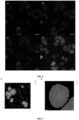
- Fig. 5 depicts a multiplex detection of three cancer biomarkers (KRT18, GATA3 and ERBB2 (Her2/neu)) using single-color probe according to embodiments of the present inventions in breast cancer cell lines.
- Fig. 5A depicts an overlay images of nuclear Hoechst stain (blue), KRT18 mRNA (cyan), GATA3 mRNA (green) und ERBB2/Her2/neu mRNA (red) in the Her2/neu-wild type cell line MCF7, und the Her2/neu-overexpressing cell line SKBR3.
- Fig. 5B single channel images of the overlays shown in Fig. 5A.
- Fig. 5C depicts an absolute quantification of the detected RNAs using the open-source software, FISH-quant v2. Scale bar 30 ⁇ m. * p ⁇ 0.05, ** p ⁇ 0.01.
- Fig. 6 depicts a multiplex detection of 5 cancer biomarker RNAs with one probe according to embodiments of the present invention each, containing one or two different types of fluorophores (KRT19 detected with Atto488 in cyan, GAPDH with Atto565 in green, ERBB2 with Atto565 and Atto647N in green and red, GATA3 with Atto488 and Atto565 in cyan and green, and KRT18 with Atto488 and Atto647N in cyan and red).
- Fig. 6A depicts a signal in the red channel detecting Atto647N.
- Fig. 6B depicts a signal in the green channel detecting Atto565.
- FIG. 6C depicts a signal in the cyan channel detecting Atto488.
- Fig. 6D Overlay image of Figs. 6A-6C with Hoechst nuclear stain (blue).
- Dual-color probe according to embodiments of the present inventions contain 50% of the fluorophore molecules per fluorophore type, compared to a single-color probe according to embodiments of the present invention.
- the signal of RNAs labelled with one probe according to embodiments of the present invention with two fluorophore types is extracted based on the intensity difference of that signal compared to the single-color signal.
- Fig. 7 depicts images of an example of false-color labelling of multi-color probe according to embodiments of the present invention.
- Fig. 7A depicts an example microscopy image of the experiment portrayed in Figure 4 .
- Each signal from one of the 5 used single- or dual-color probe according to embodiments of the present inventions has been assigned to and replaced by a false-color circle, which encodes the signal of a single RNA species.
- the legend on the right of the image gives the color code for assigning the false-color circles to the respective RNA species.
- Fig. 7B depicts a zoomed-in view of one cell from Fig. 7A .
- St. str. for DNA nanostructure GGGAGAAAGGCTTTCATTCAACTAATGCCAGTCAG 31 56 DNA Art. seq. St. str. for DNA nanostructure CTTAGTGGCAACTACTGTAGCAGTAAAAGAGTCTGCGCCAGCTGGCAGAAGCCCTA 32 56 DNA Art. seq. St. str. for DNA nanostructure TCTTCTAGTTGATTCATTTTTATACTTCTTTGATTTCAAACTTCTGGCCAATATTT 33 28 DNA Art. seq. St. str. for DNA nanostructure GTAAGAATGAACCTACCCTCATTGAGTA 34 28 DNA Art. seq. St. str. for DNA nanostructure GTCTTTATGAGAGCAAGGAATTCGTATT 35 28 DNA Art. seq. St. str.
- ACTB v01 200 64 DNA Human p. seq. 10 p. targ. ACTB v01 201 67 DNA Human p. seq. 1 p. targ. ACTB v01 202 67 DNA Human p. seq. 2 p. targ. ACTB v01 203 67 DNA Human p. seq. 3 p. targ. ACTB v01 204 67 DNA Human p. seq. 4 p. targ. ACTB v01 205 67 DNA Human p. seq. 5 p. targ. ACTB v01 206 67 DNA Human p. seq. 6 p. targ.
- ACTB v01 207 67 DNA Human p. seq. 7 p. targ.
- ACTB v01 208 67 DNA Human p. seq. 8
- p. targ. ACTB v01 209 67 DNA Human p. seq. 9
- ACTB v01 210 67 DNA Human p. seq. 10
- ACTB v03 212 67 DNA Human p. seq. 2 p. targ.
- ACTB v03 213 67 DNA Human p. seq. 3 p. targ.
- ACTB v03 214 67 DNA Human p. seq. 4 p. targ. ACTB v03 215 67 DNA Human p. seq. 5 p. targ. ACTB v03 216 67 DNA Human p. seq. 6 p. targ. ACTB v03 217 67 DNA Human p. seq. 7 p. targ. ACTB v03 218 67 DNA Human p. seq. 8 p. targ. ACTB v03 219 67 DNA Human p. seq. 9 p. targ. ACTB v03 220 67 DNA Human p. seq. 10 p. targ.
- EZR v01 228 67 DNA Human p. seq. 8 p. targ. EZR v01 229 67 DNA Human p. seq. 9 p. targ. EZR v01 230 67 DNA Human p. seq. 10 p. targ. EZR v01 231 67 DNA Human p. seq. 1 p. targ. EZR v02 232 67 DNA Human p. seq. 2 p. targ. EZR v02 233 67 DNA Human p. seq. 3 p. targ. EZR v02 234 67 DNA Human p. seq. 4 p. targ.
- EZR v03 242 67 DNA Human p. seq. 2 p. targ. EZR v03 243 67 DNA Human p. seq. 3 p. targ. EZR v03 244 67 DNA Human p. seq. 4 p. targ. EZR v03 245 67 DNA Human p. seq. 5 p. targ. EZR v03 246 67 DNA Human p. seq. 6 p. targ. EZR v03 247 67 DNA Human p. seq. 7 p. targ. EZR v03 248 67 DNA Human p. seq. 8 p. targ.
- EZR v03 249 67 DNA Human p. seq. 9 p. targ. EZR v03 250 67 DNA Human p. seq. 10 p. targ. EZR v03 251 67 DNA Human p. seq. 1 p. targ. GATA3 v01 252 67 DNA Human p. seq. 2 p. targ. GATA3 v01 253 67 DNA Human p. seq. 3 p. targ. GATA3 v01 254 67 DNA Human p. seq. 4 p. targ. GATA3 v01 255 67 DNA Human p. seq. 5 p. targ.
- GATA3 v02 263 67 DNA Human p. seq. 3 p. targ.
- GATA3 v02 264 67 DNA Human p. seq. 4 p. targ.
- GATA3 v02 265 67 DNA Human p. seq. 5 p. targ.
- GATA3 v02 266 67 DNA Human p. seq. 6
- GATA3 v02 267 DNA Human p. seq. 7 p. targ.
- GATA3 v02 268 67 DNA Human p. seq. 8 p. targ.
- GATA3 v02 269 67 DNA Human p. seq. 9 p. targ.
- GATA3 v02 270 67 DNA Human p. seq. 10 p. targ.
- GATA3 v02 271 67 DNA Human p. seq. 1 p. targ.
- ESR1 v01 272 67 DNA Human p. seq. 2 p. targ.
- ESR1 v01 273 67 DNA Human p. seq. 3 p. targ.
- ESR1 v01 274 67 DNA Human p. seq. 4 p. targ.
- ESR1 v01 275 67 DNA Human p. seq. 5 p. targ.
- ESR1 v01 276 67 DNA Human p. seq. 6 p. targ.
- ESR1 v01 277 67 DNA Human p. seq. 7 p. targ.
- ESR1 v01 278 67 DNA Human p. seq. 8 p. targ.
- ESR1 v01 279 67 DNA Human p. seq. 9 p. targ.
- ESR1 v01 280 67 DNA Human p. seq. 10
- ESR1 v01 281 67 DNA Human p. seq. 1 p. targ.
- ESR1 v02 282 67 DNA Human p. seq. 2 p. targ.
- ESR1 v02 283 67 DNA Human p. seq. 3 p. targ.
- ESR1 v02 284 67 DNA Human p. seq. 4 p. targ.
- ESR1 v02 285 67 DNA Human p. seq. 5 p. targ.
- ESR1 v02 286 67 DNA Human p. seq. 6
- ESR1 v02 287 67 DNA Human p. seq. 7
- ESR1 v02 288 67 DNA Human p. seq. 8
- ESR1 v02 289 67 DNA Human p. seq. 9 p. targ.
- ESR1 v02 290 67 DNA Human p. seq. 10 p. targ.
- GAPDH v01 298 67 DNA Human p. seq. 8 p. targ.
- GAPDH v01 299 67 DNA Human p. seq. 9 p. targ.
- GAPDH v01 300 67 DNA Human p. seq. 10 p. targ.
- GAPDH v02 301 67 DNA Human p. seq. 1 p. targ.
- GAPDH v02 302 67 DNA Human p. seq. 2 p. targ.
- GAPDH v02 303 67 DNA Human p. seq. 3 p. targ.
- GAPDH v02 304 67 DNA Human p. seq. 4 p. targ.
- GAPDH v02 305 67 DNA Human p. seq. 5 p. targ.
- GAPDH v02 306 67 DNA Human p. seq. 6 p. targ.
- GAPDH v02 307 67 DNA Human p. seq. 7 p. targ.
- GAPDH v02 308 67 DNA Human p. seq. 8 p. targ.
- GAPDH v02 309 67 DNA Human p. seq. 9 p. targ.
- GAPDH v02 310 67 DNA Human p. seq. 10 p. targ.
- GAPDH v02 311 67 DNA Human p. seq. 1 p. targ.
- KRT18 v01 312 67 DNA Human p. seq. 2 p. targ. KRT18 v01 313 67 DNA Human p. seq. 3 p. targ. KRT18 v01 314 67 DNA Human p. seq. 4 p. targ. KRT18 v01 315 67 DNA Human p. seq. 5 p. targ. KRT18 v01 316 67 DNA Human p. seq. 6 p. targ. KRT18 v01 317 67 DNA Human p. seq. 7 p. targ. KRT18 v01 318 67 DNA Human p. seq. 8 p. targ.
- KRT18 v01 319 67 DNA Human p. seq. 9 p. targ. KRT18 v01 320 67 DNA Human p. seq. 10 p. targ. KRT18 v01 321 67 DNA Human p. seq. 1 p. targ. KRT18 v02 322 67 DNA Human p. seq. 2 p. targ. KRT18 v02 323 67 DNA Human p. seq. 3 p. targ. KRT18 v02 324 67 DNA Human p. seq. 4 p. targ. KRT18 v02 325 67 DNA Human p. seq. 5 p. targ.
- KRT18 v02 326 67 DNA Human p. seq. 6 p. targ. KRT18 v02 327 67 DNA Human p. seq. 7 p. targ. KRT18 v02 328 67 DNA Human p. seq. 8 p. targ. KRT18 v02 329 67 DNA Human p. seq. 9 p. targ. KRT18 v02 330 67 DNA Human p. seq. 10 p. targ. KRT18 v02 331 67 DNA Human p. seq. 1 p. targ. KRT19 v01 332 67 DNA Human p. seq. 2 p. targ.
- KRT19 v01 340 67 DNA Human p. seq. 10 p. targ. KRT19 v01 341 67 DNA Human p. seq. 1 p. targ. KRT19 v02 342 67 DNA Human p. seq. 2 p. targ. KRT19 v02 343 67 DNA Human p. seq. 3 p. targ. KRT19 v02 344 67 DNA Human p. seq. 4 p. targ. KRT19 v02 345 67 DNA Human p. seq. 5 p. targ. KRT19 v02 346 67 DNA Human p. seq. 6 p. targ.
- KRT19 v02 347 67 DNA Human p. seq. 7 p. targ. KRT19 v02 348 67 DNA Human p. seq. 8 p. targ. KRT19 v02 349 67 DNA Human p. seq. 9 p. targ. KRT19 v02 350 67 DNA Human p. seq. 10 p. targ. KRT19 v02 351 67 DNA Human p. seq. 1 p. targ. NR3C3 v01 352 67 DNA Human p. seq. 2 p. targ. NR3C3 v01 353 67 DNA Human p. seq. 3 p. targ.
- NR3C3 v01 354 67 DNA Human p. seq. 4 p. targ. NR3C3 v01 355 67 DNA Human p. seq. 5 p. targ. NR3C3 v01 356 67 DNA Human p. seq. 6 p. targ. NR3C3 v01 357 67 DNA Human p. seq. 7 p. targ. NR3C3 v01 358 67 DNA Human p. seq. 8 p. targ. NR3C3 v01 359 67 DNA Human p. seq. 9 p. targ. NR3C3 v01 360 67 DNA Human p. seq. 10 p.
- GATA6 v02 382 67 DNA Human p. seq. 2 p. targ.
- GATA6 v02 383 67 DNA Human p. seq. 3 p. targ.
- GATA6 v02 384 67 DNA Human p. seq. 4 p. targ.
- GATA6 v02 385 67 DNA Human p. seq. 5 p. targ.
- GATA6 v02 386 67 DNA Human p. seq. 6 p. targ.
- GATA6 v02 387 67 DNA Human p. seq. 7 p. targ.
- GATA6 v02 388 67 DNA Human p. seq. 8 p. targ.
- GATA6 v02 389 67 DNA Human p. seq. 9 p. targ.
- GATA6 v02 390 67 DNA Human p. seq. 10 p. targ.
- GATA6 v02 391 67 DNA Human p. seq. 1 p. targ.
- LPAR5 v01 392 67 DNA Human p. seq. 2 p. targ.
- LPAR5 v01 393 67 DNA Human p. seq. 3 p. targ.
- LPAR5 v01 394 67 DNA Human p. seq. 4 p. targ. LPAR5 v01 395 67 DNA Human p. seq. 5 p. targ.
- LPAR5 v01 396 67 DNA Human p. seq. 6 p. targ. LPAR5 v01 397 67 DNA Human p. seq. 7 p. targ. LPAR5 v01 398 67 DNA Human p. seq. 8 p. targ. LPAR5 v01 399 67 DNA Human p. seq. 9 p. targ. LPAR5 v01 400 67 DNA Human p. seq. 10 p. targ. LPAR5 v01 401 67 DNA Human p. seq. 1 p. targ. LPAR5 v02 402 67 DNA Human p. seq. 2 p. targ.
- LPAR5 v02 403 67 DNA Human p. seq. 3 p. targ.
- LPAR5 v02 404 67 DNA Human p. seq. 4 p. targ.
- LPAR5 v02 405 67 DNA Human p. seq. 5 p. targ.
- LPAR5 v02 406 67 DNA Human p. seq. 6
- LPAR5 v02 407 67 DNA Human p. seq. 7 p. targ.
- LPAR5 v02 408 67 DNA Human p. seq. 8 p. targ.
- LPAR5 v02 409 67 DNA Human p. seq. 9 p. targ.
- LPAR5 v02 410 67 DNA Human p. seq. 10 p. targ. LPAR5 v02 411 67 DNA Human p. seq. 1 p. targ. ERBB2 v01 412 67 DNA Human p. seq. 2 p. targ. ERBB2 v01 413 67 DNA Human p. seq. 3 p. targ. ERBB2 v01 414 67 DNA Human p. seq. 4 p. targ. ERBB2 v01 415 67 DNA Human p. seq. 5 p. targ. ERBB2 v01 416 67 DNA Human p. seq. 6 p. targ.
- ERBB2 v02 424 67 DNA Human p. seq. 4 p. targ. ERBB2 v02 425 67 DNA Human p. seq. 5 p. targ. ERBB2 v02 426 67 DNA Human p. seq. 6 p. targ. ERBB2 v02 427 67 DNA Human p. seq. 7 p. targ. ERBB2 v02 428 67 DNA Human p. seq. 8 p. targ. ERBB2 v02 429 67 DNA Human p. seq. 9 p. targ. ERBB2 v02 430 67 DNA Human p. seq. 10 p. targ.
- ERBB2 v02 431 67 DNA Human p. seq. 1 p. targ. TP53 v01 432 67 DNA Human p. seq. 2 p. targ. TP53 v01 433 67 DNA Human p. seq. 3 p. targ. TP53 v01 434 67 DNA Human p. seq. 4 p. targ. TP53 v01 435 67 DNA Human p. seq. 5 p. targ. TP53 v01 436 67 DNA Human p. seq. 6 p. targ. TP53 v01 437 67 DNA Human p. seq. 7 p. targ.
- TP53 v01 438 67 DNA Human p. seq. 8 p. targ. TP53 v01 439 67 DNA Human p. seq. 9 p. targ. TP53 v01 440 67 DNA Human p. seq. 10 p. targ. TP53 v01 441 67 DNA Human p. seq. 1 p. targ. PDX1 v01 442 67 DNA Human p. seq. 2 p. targ. PDX1 v01 443 67 DNA Human p. seq. 3 p. targ. PDX1 v01 444 67 DNA Human p. seq. 4 p. targ.
- PDX1 v01 445 67 DNA Human p. seq. 5 p. targ. PDX1 v01 446 67 DNA Human p. seq. 6 p. targ. PDX1 v01 447 67 DNA Human p. seq. 7 p. targ. PDX1 v01 448 67 DNA Human p. seq. 8 p. targ. PDX1 v01 449 67 DNA Human p. seq. 9 p. targ. PDX1 v01 450 67 DNA Human p. seq. 10 p. targ. PDX1 v01 451 67 DNA Human p. seq. 1 p. targ.
- PDX1 v02 452 67 DNA Human p. seq. 2 p. targ. PDX1 v02 453 67 DNA Human p. seq. 3 p. targ. PDX1 v02 454 67 DNA Human p. seq. 4 p. targ. PDX1 v02 455 67 DNA Human p. seq. 5 p. targ. PDX1 v02 456 67 DNA Human p. seq. 6 p. targ. PDX1 v02 457 67 DNA Human p. seq. 7 p. targ. PDX1 v02 458 67 DNA Human p. seq. 8 p. targ.
- PDX1 v02 459 DNA Human p. seq. 9 p. targ. PDX1 v02 460 67 DNA Human p. seq. 10 p. targ. PDX1 v02 461 67 DNA Human p. seq. 1 p. targ. PDX1 v03 462 67 DNA Human p. seq. 2 p. targ. PDX1 v03 463 67 DNA Human p. seq. 3 p. targ. PDX1 v03 464 67 DNA Human p. seq. 4 p. targ. PDX1 v03 465 67 DNA Human p. seq. 5 p. targ.
- PDX1 v03 466 DNA Human p. seq. 6 p. targ. PDX1 v03 467 67 DNA Human p. seq. 7 p. targ. PDX1 v03 468 67 DNA Human p. seq. 8 p. targ. PDX1 v03 469 67 DNA Human p. seq. 9 p. targ. PDX1 v03 470 67 DNA Human p. seq. 10 p. targ. PDX1 v03 471 67 DNA Human p. seq. 1 p. targ. PDX1 v04 472 67 DNA Human p. seq. 2 p. targ.
- PDX1 v04 473 67 DNA Human p. seq. 3 p. targ. PDX1 v04 474 67 DNA Human p. seq. 4 p. targ. PDX1 v04 475 67 DNA Human p. seq. 5 p. targ. PDX1 v04 476 67 DNA Human p. seq. 6 p. targ. PDX1 v04 477 67 DNA Human p. seq. 7 p. targ. PDX1 v04 478 67 DNA Human p. seq. 8 p. targ. PDX1 v04 479 67 DNA Human p. seq. 9 p. targ.
- PDX1 v04 480 67 DNA Human p. seq. 10 p. targ. PDX1 v04 481 67 DNA Human p. seq. 1 p. targ. PDX1 v05 482 67 DNA Human p. seq. 2 p. targ. PDX1 v05 483 67 DNA Human p. seq. 3 p. targ. PDX1 v05 484 67 DNA Human p. seq. 4 p. targ. PDX1 v05 485 67 DNA Human p. seq. 5 p. targ. PDX1 v05 486 67 DNA Human p. seq. 6 p. targ.
- PDX1 v05 487 67 DNA Human p. seq. 7 p. targ. PDX1 v05 488 67 DNA Human p. seq. 8 p. targ. PDX1 v05 489 67 DNA Human p. seq. 9 p. targ. PDX1 v05 490 67 DNA Human p. seq. 10 p. targ. PDX1 v05
- step (X) preceding step (Z) encompasses the situation that step (X) is performed directly before step (Z), but also the situation that (X) is performed before one or more steps (Y1), ..., followed by step (Z).
- step (Z) encompasses the situation that step (X) is performed directly before step (Z), but also the situation that (X) is performed before one or more steps (Y1), ..., followed by step (Z).
Landscapes
- Chemical & Material Sciences (AREA)
- Organic Chemistry (AREA)
- Life Sciences & Earth Sciences (AREA)
- Zoology (AREA)
- Wood Science & Technology (AREA)
- Proteomics, Peptides & Aminoacids (AREA)
- Health & Medical Sciences (AREA)
- Engineering & Computer Science (AREA)
- Microbiology (AREA)
- Immunology (AREA)
- Physics & Mathematics (AREA)
- Molecular Biology (AREA)
- Biotechnology (AREA)
- Biophysics (AREA)
- Analytical Chemistry (AREA)
- Biochemistry (AREA)
- Bioinformatics & Cheminformatics (AREA)
- General Engineering & Computer Science (AREA)
- General Health & Medical Sciences (AREA)
- Genetics & Genomics (AREA)
- Measuring Or Testing Involving Enzymes Or Micro-Organisms (AREA)
Priority Applications (2)
| Application Number | Priority Date | Filing Date | Title |
|---|---|---|---|
| EP23200449.9A EP4530359A1 (de) | 2023-09-28 | 2023-09-28 | Multiplexierte einzelmolekül-rna fische unter verwendung von dna nanostrukturen |
| PCT/EP2024/077372 WO2025068573A1 (en) | 2023-09-28 | 2024-09-27 | Multiplexed single molecule rna fish using dna nanostructures |
Applications Claiming Priority (1)
| Application Number | Priority Date | Filing Date | Title |
|---|---|---|---|
| EP23200449.9A EP4530359A1 (de) | 2023-09-28 | 2023-09-28 | Multiplexierte einzelmolekül-rna fische unter verwendung von dna nanostrukturen |
Publications (1)
| Publication Number | Publication Date |
|---|---|
| EP4530359A1 true EP4530359A1 (de) | 2025-04-02 |
Family
ID=88291326
Family Applications (1)
| Application Number | Title | Priority Date | Filing Date |
|---|---|---|---|
| EP23200449.9A Withdrawn EP4530359A1 (de) | 2023-09-28 | 2023-09-28 | Multiplexierte einzelmolekül-rna fische unter verwendung von dna nanostrukturen |
Country Status (2)
| Country | Link |
|---|---|
| EP (1) | EP4530359A1 (de) |
| WO (1) | WO2025068573A1 (de) |
Citations (8)
| Publication number | Priority date | Publication date | Assignee | Title |
|---|---|---|---|---|
| US6485944B1 (en) | 1997-10-10 | 2002-11-26 | President And Fellows Of Harvard College | Replica amplification of nucleic acid arrays |
| US8865404B2 (en) | 2010-11-05 | 2014-10-21 | President And Fellows Of Harvard College | Methods for sequencing nucleic acid molecules |
| US20150232935A1 (en) * | 2014-02-14 | 2015-08-20 | The General Hospital Corporation | Methods for diagnosing igg4-related disease |
| EP3133170A1 (de) | 2008-09-10 | 2017-02-22 | Rutgers, the State University of New Jersey | Abbildung einzelner mrna-moleküle mithilfe mehrerer individuell markierter sonden |
| EP3511423A1 (de) | 2012-10-17 | 2019-07-17 | Spatial Transcriptomics AB | Verfahren und produkt zur optimierung einer lokalisierten oder räumlichen detektion einer genexpression in einer gewebeprobe |
| EP3541956A1 (de) | 2016-11-17 | 2019-09-25 | Spatial Transcriptomics AB | Verfahren zur räumlichen markierung und analyse von nukleinsäuren in einer biologischen probe |
| US20220282319A1 (en) * | 2021-03-03 | 2022-09-08 | 10X Genomics, Inc. | Analyte detection in situ using nucleic acid origami |
| EP3913068B1 (de) * | 2015-10-12 | 2023-08-02 | Advanced Cell Diagnostics, Inc. | In-situ-detektion von nukleotidvarianten bei proben mit hohem rauschen sowie zugehörige zusammensetzungen und verfahren |
-
2023
- 2023-09-28 EP EP23200449.9A patent/EP4530359A1/de not_active Withdrawn
-
2024
- 2024-09-27 WO PCT/EP2024/077372 patent/WO2025068573A1/en active Pending
Patent Citations (9)
| Publication number | Priority date | Publication date | Assignee | Title |
|---|---|---|---|---|
| US6485944B1 (en) | 1997-10-10 | 2002-11-26 | President And Fellows Of Harvard College | Replica amplification of nucleic acid arrays |
| EP3133170A1 (de) | 2008-09-10 | 2017-02-22 | Rutgers, the State University of New Jersey | Abbildung einzelner mrna-moleküle mithilfe mehrerer individuell markierter sonden |
| US8865404B2 (en) | 2010-11-05 | 2014-10-21 | President And Fellows Of Harvard College | Methods for sequencing nucleic acid molecules |
| EP3511423A1 (de) | 2012-10-17 | 2019-07-17 | Spatial Transcriptomics AB | Verfahren und produkt zur optimierung einer lokalisierten oder räumlichen detektion einer genexpression in einer gewebeprobe |
| US20150232935A1 (en) * | 2014-02-14 | 2015-08-20 | The General Hospital Corporation | Methods for diagnosing igg4-related disease |
| EP3913068B1 (de) * | 2015-10-12 | 2023-08-02 | Advanced Cell Diagnostics, Inc. | In-situ-detektion von nukleotidvarianten bei proben mit hohem rauschen sowie zugehörige zusammensetzungen und verfahren |
| EP3541956A1 (de) | 2016-11-17 | 2019-09-25 | Spatial Transcriptomics AB | Verfahren zur räumlichen markierung und analyse von nukleinsäuren in einer biologischen probe |
| EP3916108A1 (de) | 2016-11-17 | 2021-12-01 | Spatial Transcriptomics AB | Verfahren zur räumlichen markierung und analyse von nukleinsäuren in einer biologischen probe |
| US20220282319A1 (en) * | 2021-03-03 | 2022-09-08 | 10X Genomics, Inc. | Analyte detection in situ using nucleic acid origami |
Non-Patent Citations (2)
| Title |
|---|
| HAUPTMANN GISELBERT ET AL: "Detection and signal amplification in zebrafish RNA FISH", METHODS, ACADEMIC PRESS, NL, vol. 98, 26 January 2016 (2016-01-26), pages 50 - 59, XP029471301, ISSN: 1046-2023, DOI: 10.1016/J.YMETH.2016.01.012 * |
| HERSHBERG ELLIOT A. ET AL: "PaintSHOP enables the interactive design of transcriptome- and genome-scale oligonucleotide FISH experiments", BIORXIV, 6 July 2020 (2020-07-06), XP093126066, Retrieved from the Internet <URL:https://www.biorxiv.org/content/10.1101/2020.07.05.188797v1.full.pdf> [retrieved on 20240131], DOI: 10.1101/2020.07.05.188797 * |
Also Published As
| Publication number | Publication date |
|---|---|
| WO2025068573A1 (en) | 2025-04-03 |
Similar Documents
| Publication | Publication Date | Title |
|---|---|---|
| US20210340527A1 (en) | Encoding of dna vector identity via iterative hybridization detection of a barcode transcript | |
| EP2459743B1 (de) | Satz oligonukleotidsonden sowie damit zusammenhängende verfahren und verwendungen | |
| EP2992115B1 (de) | Multiplexmarkierung von molekülen durch barcoding mit sequenzieller hybridisierung | |
| EP3133170B1 (de) | Abbildung einzelner mrna-moleküle mithilfe mehrerer individuell markierter sonden | |
| CN109072205A (zh) | 核酸的检测 | |
| EP3610039B1 (de) | Markierung von oligonukleotidsonden durch mehrwegligation | |
| JP6247934B2 (ja) | 単一細胞において染色体構造および遺伝子発現を同時に検出するための方法 | |
| Van der Ploeg | Cytochemical nucleic acid research during the twentieth century | |
| US20030198983A1 (en) | Methods of genetic analysis of human genes | |
| EP4530359A1 (de) | Multiplexierte einzelmolekül-rna fische unter verwendung von dna nanostrukturen | |
| EP3093342B1 (de) | Rna-mikroarray für den nachweis einer wechselwirkung zwischen proteinen und rna, welcher eine structur höherer ordnung enthält | |
| US20230159989A1 (en) | Multiplexed fluorescence in situ hybridization method capable of rapid detection of billions of targets | |
| US20260074017A1 (en) | Method for indexing a cellular sample | |
| US20030082596A1 (en) | Methods of genetic analysis of probes: test3 | |
| HK1169452B (en) | A set of oligonucleotide probes as well as methods and uses related thereto |
Legal Events
| Date | Code | Title | Description |
|---|---|---|---|
| PUAI | Public reference made under article 153(3) epc to a published international application that has entered the european phase |
Free format text: ORIGINAL CODE: 0009012 |
|
| STAA | Information on the status of an ep patent application or granted ep patent |
Free format text: STATUS: THE APPLICATION HAS BEEN PUBLISHED |
|
| AK | Designated contracting states |
Kind code of ref document: A1 Designated state(s): AL AT BE BG CH CY CZ DE DK EE ES FI FR GB GR HR HU IE IS IT LI LT LU LV MC ME MK MT NL NO PL PT RO RS SE SI SK SM TR |
|
| P01 | Opt-out of the competence of the unified patent court (upc) registered |
Free format text: CASE NUMBER: APP_26522/2025 Effective date: 20250604 |
|
| STAA | Information on the status of an ep patent application or granted ep patent |
Free format text: STATUS: THE APPLICATION IS DEEMED TO BE WITHDRAWN |
|
| 18D | Application deemed to be withdrawn |
Effective date: 20251003 |