EP4527445A1 - Dispositif de stimulation électrique - Google Patents
Dispositif de stimulation électrique Download PDFInfo
- Publication number
- EP4527445A1 EP4527445A1 EP24201917.2A EP24201917A EP4527445A1 EP 4527445 A1 EP4527445 A1 EP 4527445A1 EP 24201917 A EP24201917 A EP 24201917A EP 4527445 A1 EP4527445 A1 EP 4527445A1
- Authority
- EP
- European Patent Office
- Prior art keywords
- electrical stimulation
- organism
- stimulation signal
- electrical
- stimulation device
- Prior art date
- Legal status (The legal status is an assumption and is not a legal conclusion. Google has not performed a legal analysis and makes no representation as to the accuracy of the status listed.)
- Pending
Links
- 230000000638 stimulation Effects 0.000 title claims abstract description 247
- 108010037462 Cyclooxygenase 2 Proteins 0.000 claims abstract description 50
- 102000010907 Cyclooxygenase 2 Human genes 0.000 claims abstract 6
- 208000002193 Pain Diseases 0.000 claims description 33
- 229940077737 brain-derived neurotrophic factor Drugs 0.000 claims description 31
- 102000004219 Brain-derived neurotrophic factor Human genes 0.000 claims description 30
- 108090000715 Brain-derived neurotrophic factor Proteins 0.000 claims description 30
- 102100024304 Protachykinin-1 Human genes 0.000 claims description 24
- QDZOEBFLNHCSSF-PFFBOGFISA-N (2S)-2-[[(2R)-2-[[(2S)-1-[(2S)-6-amino-2-[[(2S)-1-[(2R)-2-amino-5-carbamimidamidopentanoyl]pyrrolidine-2-carbonyl]amino]hexanoyl]pyrrolidine-2-carbonyl]amino]-3-(1H-indol-3-yl)propanoyl]amino]-N-[(2R)-1-[[(2S)-1-[[(2R)-1-[[(2S)-1-[[(2S)-1-amino-4-methyl-1-oxopentan-2-yl]amino]-4-methyl-1-oxopentan-2-yl]amino]-3-(1H-indol-3-yl)-1-oxopropan-2-yl]amino]-1-oxo-3-phenylpropan-2-yl]amino]-3-(1H-indol-3-yl)-1-oxopropan-2-yl]pentanediamide Chemical compound C([C@@H](C(=O)N[C@H](CC=1C2=CC=CC=C2NC=1)C(=O)N[C@@H](CC(C)C)C(=O)N[C@@H](CC(C)C)C(N)=O)NC(=O)[C@@H](CC=1C2=CC=CC=C2NC=1)NC(=O)[C@H](CCC(N)=O)NC(=O)[C@@H](CC=1C2=CC=CC=C2NC=1)NC(=O)[C@H]1N(CCC1)C(=O)[C@H](CCCCN)NC(=O)[C@H]1N(CCC1)C(=O)[C@H](N)CCCNC(N)=N)C1=CC=CC=C1 QDZOEBFLNHCSSF-PFFBOGFISA-N 0.000 claims description 22
- 101800003906 Substance P Proteins 0.000 claims description 22
- 230000002401 inhibitory effect Effects 0.000 claims description 14
- 108090000623 proteins and genes Proteins 0.000 claims description 11
- 102000004169 proteins and genes Human genes 0.000 claims description 11
- 230000001537 neural effect Effects 0.000 claims description 10
- 238000006243 chemical reaction Methods 0.000 claims description 9
- 208000024891 symptom Diseases 0.000 claims description 8
- 230000014509 gene expression Effects 0.000 claims description 7
- 230000003962 neuroinflammatory response Effects 0.000 claims description 6
- 230000035945 sensitivity Effects 0.000 claims description 6
- 230000001186 cumulative effect Effects 0.000 claims description 5
- 230000007830 nerve conduction Effects 0.000 claims description 4
- 238000011282 treatment Methods 0.000 description 58
- 102100038280 Prostaglandin G/H synthase 2 Human genes 0.000 description 44
- 241000700159 Rattus Species 0.000 description 36
- 210000005036 nerve Anatomy 0.000 description 36
- 238000012360 testing method Methods 0.000 description 34
- 210000002569 neuron Anatomy 0.000 description 17
- 208000028389 Nerve injury Diseases 0.000 description 15
- 230000000694 effects Effects 0.000 description 15
- 230000008764 nerve damage Effects 0.000 description 15
- 102000004232 Mitogen-Activated Protein Kinase Kinases Human genes 0.000 description 14
- 108090000744 Mitogen-Activated Protein Kinase Kinases Proteins 0.000 description 14
- 102100038895 Myc proto-oncogene protein Human genes 0.000 description 14
- 101710135898 Myc proto-oncogene protein Proteins 0.000 description 14
- 101710150448 Transcriptional regulator Myc Proteins 0.000 description 14
- 108010091047 neurofilament protein H Proteins 0.000 description 14
- KISWVXRQTGLFGD-UHFFFAOYSA-N 2-[[2-[[6-amino-2-[[2-[[2-[[5-amino-2-[[2-[[1-[2-[[6-amino-2-[(2,5-diamino-5-oxopentanoyl)amino]hexanoyl]amino]-5-(diaminomethylideneamino)pentanoyl]pyrrolidine-2-carbonyl]amino]-3-hydroxypropanoyl]amino]-5-oxopentanoyl]amino]-5-(diaminomethylideneamino)p Chemical compound C1CCN(C(=O)C(CCCN=C(N)N)NC(=O)C(CCCCN)NC(=O)C(N)CCC(N)=O)C1C(=O)NC(CO)C(=O)NC(CCC(N)=O)C(=O)NC(CCCN=C(N)N)C(=O)NC(CO)C(=O)NC(CCCCN)C(=O)NC(C(=O)NC(CC(C)C)C(O)=O)CC1=CC=C(O)C=C1 KISWVXRQTGLFGD-UHFFFAOYSA-N 0.000 description 12
- 238000010586 diagram Methods 0.000 description 12
- 102000047918 Myelin Basic Human genes 0.000 description 11
- 101710107068 Myelin basic protein Proteins 0.000 description 11
- 108010071563 Proto-Oncogene Proteins c-fos Proteins 0.000 description 11
- 102000007568 Proto-Oncogene Proteins c-fos Human genes 0.000 description 11
- 210000001519 tissue Anatomy 0.000 description 11
- 238000003125 immunofluorescent labeling Methods 0.000 description 10
- 238000003556 assay Methods 0.000 description 9
- 238000000034 method Methods 0.000 description 9
- 210000003497 sciatic nerve Anatomy 0.000 description 9
- 208000004454 Hyperalgesia Diseases 0.000 description 8
- 230000008569 process Effects 0.000 description 8
- 102000004005 Prostaglandin-endoperoxide synthases Human genes 0.000 description 7
- 108090000459 Prostaglandin-endoperoxide synthases Proteins 0.000 description 7
- 208000004296 neuralgia Diseases 0.000 description 7
- 208000021722 neuropathic pain Diseases 0.000 description 7
- 230000001105 regulatory effect Effects 0.000 description 7
- 210000003594 spinal ganglia Anatomy 0.000 description 7
- 238000010186 staining Methods 0.000 description 7
- 206010061218 Inflammation Diseases 0.000 description 6
- 241000283973 Oryctolagus cuniculus Species 0.000 description 6
- 208000027418 Wounds and injury Diseases 0.000 description 6
- 230000006378 damage Effects 0.000 description 6
- 230000004054 inflammatory process Effects 0.000 description 6
- 208000014674 injury Diseases 0.000 description 6
- 108090000189 Neuropeptides Proteins 0.000 description 5
- 230000002051 biphasic effect Effects 0.000 description 5
- 210000004027 cell Anatomy 0.000 description 5
- 230000003247 decreasing effect Effects 0.000 description 5
- 229940079593 drug Drugs 0.000 description 5
- 239000003814 drug Substances 0.000 description 5
- 239000007850 fluorescent dye Substances 0.000 description 5
- 210000000278 spinal cord Anatomy 0.000 description 5
- 102000043136 MAP kinase family Human genes 0.000 description 4
- 108091054455 MAP kinase family Proteins 0.000 description 4
- 241001465754 Metazoa Species 0.000 description 4
- 229940035676 analgesics Drugs 0.000 description 4
- 239000000730 antalgic agent Substances 0.000 description 4
- YZXBAPSDXZZRGB-DOFZRALJSA-N arachidonic acid Chemical compound CCCCC\C=C/C\C=C/C\C=C/C\C=C/CCCC(O)=O YZXBAPSDXZZRGB-DOFZRALJSA-N 0.000 description 4
- 230000002314 neuroinflammatory effect Effects 0.000 description 4
- 230000002829 reductive effect Effects 0.000 description 4
- 241000283707 Capra Species 0.000 description 3
- 208000036110 Neuroinflammatory disease Diseases 0.000 description 3
- 102100038277 Prostaglandin G/H synthase 1 Human genes 0.000 description 3
- 108050003243 Prostaglandin G/H synthase 1 Proteins 0.000 description 3
- 238000000692 Student's t-test Methods 0.000 description 3
- 210000003050 axon Anatomy 0.000 description 3
- 230000033228 biological regulation Effects 0.000 description 3
- 210000004556 brain Anatomy 0.000 description 3
- 230000001684 chronic effect Effects 0.000 description 3
- 230000003959 neuroinflammation Effects 0.000 description 3
- 229940021182 non-steroidal anti-inflammatory drug Drugs 0.000 description 3
- 229940094443 oxytocics prostaglandins Drugs 0.000 description 3
- 210000001428 peripheral nervous system Anatomy 0.000 description 3
- 150000003180 prostaglandins Chemical class 0.000 description 3
- 230000009467 reduction Effects 0.000 description 3
- 210000004116 schwann cell Anatomy 0.000 description 3
- 238000012453 sprague-dawley rat model Methods 0.000 description 3
- 238000007619 statistical method Methods 0.000 description 3
- PXGPLTODNUVGFL-BRIYLRKRSA-N (E,Z)-(1R,2R,3R,5S)-7-(3,5-Dihydroxy-2-((3S)-(3-hydroxy-1-octenyl))cyclopentyl)-5-heptenoic acid Chemical compound CCCCC[C@H](O)C=C[C@H]1[C@H](O)C[C@H](O)[C@@H]1CC=CCCCC(O)=O PXGPLTODNUVGFL-BRIYLRKRSA-N 0.000 description 2
- RZVAJINKPMORJF-UHFFFAOYSA-N Acetaminophen Chemical compound CC(=O)NC1=CC=C(O)C=C1 RZVAJINKPMORJF-UHFFFAOYSA-N 0.000 description 2
- 238000002965 ELISA Methods 0.000 description 2
- 101000831616 Homo sapiens Protachykinin-1 Proteins 0.000 description 2
- 239000004677 Nylon Substances 0.000 description 2
- 238000009825 accumulation Methods 0.000 description 2
- 235000021342 arachidonic acid Nutrition 0.000 description 2
- 229940114079 arachidonic acid Drugs 0.000 description 2
- 210000000133 brain stem Anatomy 0.000 description 2
- 210000001217 buttock Anatomy 0.000 description 2
- 210000003169 central nervous system Anatomy 0.000 description 2
- 201000010099 disease Diseases 0.000 description 2
- 208000037265 diseases, disorders, signs and symptoms Diseases 0.000 description 2
- 210000000609 ganglia Anatomy 0.000 description 2
- 230000028709 inflammatory response Effects 0.000 description 2
- 230000003902 lesion Effects 0.000 description 2
- 239000003550 marker Substances 0.000 description 2
- CWWARWOPSKGELM-SARDKLJWSA-N methyl (2s)-2-[[(2s)-2-[[2-[[(2s)-2-[[(2s)-2-[[(2s)-5-amino-2-[[(2s)-5-amino-2-[[(2s)-1-[(2s)-6-amino-2-[[(2s)-1-[(2s)-2-amino-5-(diaminomethylideneamino)pentanoyl]pyrrolidine-2-carbonyl]amino]hexanoyl]pyrrolidine-2-carbonyl]amino]-5-oxopentanoyl]amino]-5 Chemical compound C([C@@H](C(=O)NCC(=O)N[C@@H](CC(C)C)C(=O)N[C@@H](CCSC)C(=O)OC)NC(=O)[C@H](CC=1C=CC=CC=1)NC(=O)[C@H](CCC(N)=O)NC(=O)[C@H](CCC(N)=O)NC(=O)[C@H]1N(CCC1)C(=O)[C@H](CCCCN)NC(=O)[C@H]1N(CCC1)C(=O)[C@@H](N)CCCN=C(N)N)C1=CC=CC=C1 CWWARWOPSKGELM-SARDKLJWSA-N 0.000 description 2
- 201000001119 neuropathy Diseases 0.000 description 2
- 230000007823 neuropathy Effects 0.000 description 2
- 229920001778 nylon Polymers 0.000 description 2
- 210000000578 peripheral nerve Anatomy 0.000 description 2
- 208000033808 peripheral neuropathy Diseases 0.000 description 2
- 230000035807 sensation Effects 0.000 description 2
- 239000012128 staining reagent Substances 0.000 description 2
- ADNPLDHMAVUMIW-CUZNLEPHSA-N substance P Chemical compound C([C@@H](C(=O)NCC(=O)N[C@@H](CC(C)C)C(=O)N[C@@H](CCSC)C(N)=O)NC(=O)[C@H](CC=1C=CC=CC=1)NC(=O)[C@H](CCC(N)=O)NC(=O)[C@H](CCC(N)=O)NC(=O)[C@H]1N(CCC1)C(=O)[C@H](CCCCN)NC(=O)[C@H]1N(CCC1)C(=O)[C@@H](N)CCCN=C(N)N)C1=CC=CC=C1 ADNPLDHMAVUMIW-CUZNLEPHSA-N 0.000 description 2
- 230000002459 sustained effect Effects 0.000 description 2
- 210000001186 vagus nerve Anatomy 0.000 description 2
- 241000700199 Cavia porcellus Species 0.000 description 1
- 208000000094 Chronic Pain Diseases 0.000 description 1
- 102000004190 Enzymes Human genes 0.000 description 1
- 108090000790 Enzymes Proteins 0.000 description 1
- 206010020853 Hypertonic bladder Diseases 0.000 description 1
- 206010065390 Inflammatory pain Diseases 0.000 description 1
- 208000008238 Muscle Spasticity Diseases 0.000 description 1
- 102000003797 Neuropeptides Human genes 0.000 description 1
- 208000009722 Overactive Urinary Bladder Diseases 0.000 description 1
- 108091000080 Phosphotransferase Proteins 0.000 description 1
- 206010037660 Pyrexia Diseases 0.000 description 1
- 206010070834 Sensitisation Diseases 0.000 description 1
- 206010047141 Vasodilatation Diseases 0.000 description 1
- 230000001154 acute effect Effects 0.000 description 1
- 208000005298 acute pain Diseases 0.000 description 1
- 230000000202 analgesic effect Effects 0.000 description 1
- 229940124599 anti-inflammatory drug Drugs 0.000 description 1
- 230000003110 anti-inflammatory effect Effects 0.000 description 1
- 210000001130 astrocyte Anatomy 0.000 description 1
- 210000003403 autonomic nervous system Anatomy 0.000 description 1
- 230000008901 benefit Effects 0.000 description 1
- 239000012472 biological sample Substances 0.000 description 1
- 230000000903 blocking effect Effects 0.000 description 1
- 210000004369 blood Anatomy 0.000 description 1
- 239000008280 blood Substances 0.000 description 1
- 210000000601 blood cell Anatomy 0.000 description 1
- 210000000170 cell membrane Anatomy 0.000 description 1
- 230000001413 cellular effect Effects 0.000 description 1
- 230000015271 coagulation Effects 0.000 description 1
- 238000005345 coagulation Methods 0.000 description 1
- 210000002808 connective tissue Anatomy 0.000 description 1
- 210000003792 cranial nerve Anatomy 0.000 description 1
- 230000003111 delayed effect Effects 0.000 description 1
- 238000001514 detection method Methods 0.000 description 1
- 238000011161 development Methods 0.000 description 1
- 230000003467 diminishing effect Effects 0.000 description 1
- XEYBRNLFEZDVAW-ARSRFYASSA-N dinoprostone Chemical compound CCCCC[C@H](O)\C=C\[C@H]1[C@H](O)CC(=O)[C@@H]1C\C=C/CCCC(O)=O XEYBRNLFEZDVAW-ARSRFYASSA-N 0.000 description 1
- 238000001647 drug administration Methods 0.000 description 1
- 230000009977 dual effect Effects 0.000 description 1
- 230000002526 effect on cardiovascular system Effects 0.000 description 1
- 230000005684 electric field Effects 0.000 description 1
- 210000002919 epithelial cell Anatomy 0.000 description 1
- 230000003628 erosive effect Effects 0.000 description 1
- 238000011156 evaluation Methods 0.000 description 1
- 238000000605 extraction Methods 0.000 description 1
- 239000012530 fluid Substances 0.000 description 1
- 230000002496 gastric effect Effects 0.000 description 1
- 210000004392 genitalia Anatomy 0.000 description 1
- 238000001631 haemodialysis Methods 0.000 description 1
- 210000003128 head Anatomy 0.000 description 1
- 230000000322 hemodialysis Effects 0.000 description 1
- 210000000548 hind-foot Anatomy 0.000 description 1
- 210000002865 immune cell Anatomy 0.000 description 1
- 230000001900 immune effect Effects 0.000 description 1
- 238000001727 in vivo Methods 0.000 description 1
- 230000002757 inflammatory effect Effects 0.000 description 1
- 210000003141 lower extremity Anatomy 0.000 description 1
- 238000012423 maintenance Methods 0.000 description 1
- 210000000260 male genitalia Anatomy 0.000 description 1
- 238000005259 measurement Methods 0.000 description 1
- 230000007246 mechanism Effects 0.000 description 1
- 210000001617 median nerve Anatomy 0.000 description 1
- 230000001404 mediated effect Effects 0.000 description 1
- 230000002025 microglial effect Effects 0.000 description 1
- 210000004877 mucosa Anatomy 0.000 description 1
- 210000001107 musculocutaneous nerve Anatomy 0.000 description 1
- 210000003007 myelin sheath Anatomy 0.000 description 1
- 210000000653 nervous system Anatomy 0.000 description 1
- 230000004031 neuronal differentiation Effects 0.000 description 1
- 230000006576 neuronal survival Effects 0.000 description 1
- 239000002858 neurotransmitter agent Substances 0.000 description 1
- 210000004248 oligodendroglia Anatomy 0.000 description 1
- 235000020665 omega-6 fatty acid Nutrition 0.000 description 1
- 229940033080 omega-6 fatty acid Drugs 0.000 description 1
- 239000000014 opioid analgesic Substances 0.000 description 1
- 229940005483 opioid analgesics Drugs 0.000 description 1
- 208000020629 overactive bladder Diseases 0.000 description 1
- 230000037392 palmar hyperhidrosis Effects 0.000 description 1
- 229960005489 paracetamol Drugs 0.000 description 1
- 208000035824 paresthesia Diseases 0.000 description 1
- 230000008506 pathogenesis Effects 0.000 description 1
- 230000037361 pathway Effects 0.000 description 1
- 210000003899 penis Anatomy 0.000 description 1
- 102000020233 phosphotransferase Human genes 0.000 description 1
- 230000001817 pituitary effect Effects 0.000 description 1
- 230000010287 polarization Effects 0.000 description 1
- 229920001184 polypeptide Polymers 0.000 description 1
- 206010036596 premature ejaculation Diseases 0.000 description 1
- 102000004196 processed proteins & peptides Human genes 0.000 description 1
- 108090000765 processed proteins & peptides Proteins 0.000 description 1
- PXGPLTODNUVGFL-UHFFFAOYSA-N prostaglandin F2alpha Natural products CCCCCC(O)C=CC1C(O)CC(O)C1CC=CCCCC(O)=O PXGPLTODNUVGFL-UHFFFAOYSA-N 0.000 description 1
- 238000011002 quantification Methods 0.000 description 1
- 210000003124 radial glial cell Anatomy 0.000 description 1
- 210000002979 radial nerve Anatomy 0.000 description 1
- 206010038464 renal hypertension Diseases 0.000 description 1
- 238000010079 rubber tapping Methods 0.000 description 1
- 210000003296 saliva Anatomy 0.000 description 1
- 230000008313 sensitization Effects 0.000 description 1
- 210000001057 smooth muscle myoblast Anatomy 0.000 description 1
- 239000007787 solid Substances 0.000 description 1
- 230000000392 somatic effect Effects 0.000 description 1
- 208000018198 spasticity Diseases 0.000 description 1
- 230000004936 stimulating effect Effects 0.000 description 1
- 210000002784 stomach Anatomy 0.000 description 1
- 230000008961 swelling Effects 0.000 description 1
- 230000000946 synaptic effect Effects 0.000 description 1
- 238000011287 therapeutic dose Methods 0.000 description 1
- 238000002560 therapeutic procedure Methods 0.000 description 1
- RZWIIPASKMUIAC-VQTJNVASSA-N thromboxane Chemical compound CCCCCCCC[C@H]1OCCC[C@@H]1CCCCCCC RZWIIPASKMUIAC-VQTJNVASSA-N 0.000 description 1
- 238000002646 transcutaneous electrical nerve stimulation Methods 0.000 description 1
- 230000026683 transduction Effects 0.000 description 1
- 238000010361 transduction Methods 0.000 description 1
- 210000003901 trigeminal nerve Anatomy 0.000 description 1
- 210000001364 upper extremity Anatomy 0.000 description 1
- 210000002700 urine Anatomy 0.000 description 1
- 230000024883 vasodilation Effects 0.000 description 1
- 210000000707 wrist Anatomy 0.000 description 1
Images


Classifications
-
- A—HUMAN NECESSITIES
- A61—MEDICAL OR VETERINARY SCIENCE; HYGIENE
- A61N—ELECTROTHERAPY; MAGNETOTHERAPY; RADIATION THERAPY; ULTRASOUND THERAPY
- A61N1/00—Electrotherapy; Circuits therefor
- A61N1/18—Applying electric currents by contact electrodes
- A61N1/32—Applying electric currents by contact electrodes alternating or intermittent currents
- A61N1/36—Applying electric currents by contact electrodes alternating or intermittent currents for stimulation
- A61N1/3605—Implantable neurostimulators for stimulating central or peripheral nerve system
- A61N1/3606—Implantable neurostimulators for stimulating central or peripheral nerve system adapted for a particular treatment
- A61N1/36071—Pain
-
- A—HUMAN NECESSITIES
- A61—MEDICAL OR VETERINARY SCIENCE; HYGIENE
- A61N—ELECTROTHERAPY; MAGNETOTHERAPY; RADIATION THERAPY; ULTRASOUND THERAPY
- A61N1/00—Electrotherapy; Circuits therefor
- A61N1/18—Applying electric currents by contact electrodes
- A61N1/32—Applying electric currents by contact electrodes alternating or intermittent currents
- A61N1/36—Applying electric currents by contact electrodes alternating or intermittent currents for stimulation
- A61N1/36014—External stimulators, e.g. with patch electrodes
- A61N1/3603—Control systems
- A61N1/36034—Control systems specified by the stimulation parameters
-
- A—HUMAN NECESSITIES
- A61—MEDICAL OR VETERINARY SCIENCE; HYGIENE
- A61N—ELECTROTHERAPY; MAGNETOTHERAPY; RADIATION THERAPY; ULTRASOUND THERAPY
- A61N1/00—Electrotherapy; Circuits therefor
- A61N1/18—Applying electric currents by contact electrodes
- A61N1/32—Applying electric currents by contact electrodes alternating or intermittent currents
- A61N1/36—Applying electric currents by contact electrodes alternating or intermittent currents for stimulation
- A61N1/3605—Implantable neurostimulators for stimulating central or peripheral nerve system
- A61N1/36128—Control systems
- A61N1/36146—Control systems specified by the stimulation parameters
- A61N1/36167—Timing, e.g. stimulation onset
- A61N1/36171—Frequency
-
- A—HUMAN NECESSITIES
- A61—MEDICAL OR VETERINARY SCIENCE; HYGIENE
- A61N—ELECTROTHERAPY; MAGNETOTHERAPY; RADIATION THERAPY; ULTRASOUND THERAPY
- A61N1/00—Electrotherapy; Circuits therefor
- A61N1/18—Applying electric currents by contact electrodes
- A61N1/32—Applying electric currents by contact electrodes alternating or intermittent currents
- A61N1/36—Applying electric currents by contact electrodes alternating or intermittent currents for stimulation
- A61N1/3605—Implantable neurostimulators for stimulating central or peripheral nerve system
- A61N1/36128—Control systems
- A61N1/36146—Control systems specified by the stimulation parameters
- A61N1/36167—Timing, e.g. stimulation onset
- A61N1/36178—Burst or pulse train parameters
-
- A—HUMAN NECESSITIES
- A61—MEDICAL OR VETERINARY SCIENCE; HYGIENE
- A61N—ELECTROTHERAPY; MAGNETOTHERAPY; RADIATION THERAPY; ULTRASOUND THERAPY
- A61N1/00—Electrotherapy; Circuits therefor
- A61N1/18—Applying electric currents by contact electrodes
- A61N1/32—Applying electric currents by contact electrodes alternating or intermittent currents
- A61N1/36—Applying electric currents by contact electrodes alternating or intermittent currents for stimulation
- A61N1/36014—External stimulators, e.g. with patch electrodes
- A61N1/36021—External stimulators, e.g. with patch electrodes for treatment of pain
Definitions
- the present invention relates to the technical field of electrical stimulation, particularly to an electrical stimulation device.
- Cyclooxygenase is an important enzyme involved in the inflammatory reaction.
- COX is an important enzyme involved in the inflammatory reaction.
- PGE2 prostaglandin E2
- PPF2 ⁇ prostaglandin F2 ⁇
- thromboxane thromboxane
- COX includes COX-1 and COX-2.
- COX-1 is present in various tissues of an organism and is responsible for regulating normal cellular activities, such as protecting the stomach wall and platelets from coagulation, whereas COX-2 is induced only when the body is inflamed.
- Analgesics can be classified into three major groups, from weak to strong, according to their analgesic strength, acetaminophen, nonsteroidal anti-inflammatory drugs (NSAIDs) and opioid analgesics.
- NSAIDs nonsteroidal anti-inflammatory drugs
- opioid analgesics When inflammation occurs in vivo, activated cyclooxygenase converts arachidonic acid into prostaglandins and further produces more inflammatory factors, causing inflammation and pain.
- NASIDs mainly achieve the effects of diminishing inflammation, relieving pain and/or relieving fever and pain by inhibiting COX and further blocking the process of converting arachidonic acid through COX.
- analgesics of NSAIDs capable of selectively inhibiting COX-2 without affecting COX-1 at the therapeutic dose.
- the drugs have the advantage of low incidence of gastrointestinal mucosa injury.
- the accumulation of the drugs may cause problems such as side effects.
- the present disclosure provides an electrical stimulation device, which can reduce COX-2 without causing nerve injury.
- an electrical stimulation device includes an electrode assembly and an electrical stimulator.
- the electrical stimulator is coupled to the electrode assembly and generates an electrical stimulation signal.
- the electrical stimulation signal is transmitted to a target region of an organism through the electrode assembly.
- the electrical stimulation signal contains a plurality of burst signals, and the burst signals have the burst frequency between 0.1 Hz and 1,000 Hz.
- Each burst signal contains a plurality of pulses, and the pulses have the pulse frequency between 1 kHz (kilohertz) and 1,000 kHz.
- the electrical stimulation signal reduces a level of cyclooxygenase-2 of the organism.
- the electrical stimulation signal reduces the level of the cyclooxygenase-2 by inhibiting a protein expression of the cyclooxygenase-2.
- reducing the level of the cyclooxygenase-2 of the organism occurs within 24 hours after receiving the electrical stimulation signal.
- the electrical stimulator generates the electrical stimulation signal for a cumulative time less than or equal to 12 hours per day.
- the electrical stimulation signal further reduces the levels of brain-derived neurotrophic factor (BDNF) and/or substance P of the organism.
- BDNF brain-derived neurotrophic factor
- the electrical stimulator uses the electrical stimulation signal to inhibit neuroinflammatory response of the organism.
- the electrical stimulation signal inhibits and/or relieves at least a part of pain on a nerve conduction path of the target region.
- the electrical stimulation signal relieves pain of the organism, relieves symptoms of the organism, reduces neural sensitivity of the organism or relieves overactive reaction of the organism and maintains for at least 1 hour.
- the electrical stimulation signal reduces the level of the cyclooxygenase-2 by inhibiting a protein expression of the cyclooxygenase-2.
- reducing the level of the cyclooxygenase-2 of the organism occurs within 24 hours of receiving the electrical stimulation signal at the target region.
- the electrical stimulator generates the electrical stimulation signal for a cumulative time less than or equal to 12 hours per day.
- the electrical stimulation signal relieves pain of the organism, relieves symptoms of the organism, reduces neural sensitivity of the organism or relieves overactive reaction of the organism and maintains for at least 1 hour.
- the electrical stimulation signal further inhibits the levels of brain-derived neurotrophic factor and/or substance P of the organism, inhibits neuroinflammatory response of the organism or inhibits and/or relieves at least a part of pain on a nerve conduction path of the target region.
- the electrical stimulation device has the effect of reducing COX-2 without causing the nerve injury.
- the electrical stimulation device has the effects of reducing the level of the brain-derived neurotrophic factor, reducing the level of the substance P, inhibiting the neuroinflammatory response, inhibiting and/or relieving pain, relieving symptoms, reducing nerve sensitivity, or relieving the overactive reaction without causing the nerve injury.
- the electrical stimulation device has an effect of regulating nerves by regulating protein and gene expression of the protein.
- electrical stimulation signal refers to an electrical signal transmitted by an electrical stimulator through an electrode assembly to an organism.
- the electrical stimulation signal may be quantified into voltage, current, electric field, power, joule energy or other electrical measurement values.
- target region refers to a neural tissue and/or non-neural tissue that receives the electrical stimulation signal.
- the neural tissue includes neurons.
- the non-neural tissue includes neurogliocytes, myelin sheaths, immune cells, connective tissues, epithelial cells, cardiovascular cells and/or blood cells and the like.
- the neurogliocytes include astrocytes, oligodendrocytes, ependymal cells, radial glial cells, Schwann cells, satellite cells, microglial cells and/or pituitary cells and the like.
- organism refers to an individual, which may be a human or an animal, receiving the electrical stimulation signal, but is not limited thereto.
- An electrical stimulation device 10 includes an electrode assembly 11 and an electrical stimulator 12.
- the electrical stimulator 12 is coupled to the electrode assembly 11 and generates an electrical stimulation signal ES.
- the electrode assembly 11 receives the electrical stimulation signal ES and transmits it to a target region T of an organism 20.
- the electrical stimulation signal ES is transmitted to the target region T through the electrode assembly 11.
- the electrical stimulation device 10 may be of transcutaneous, transcranial, partially invasive, minimally invasive or implantable.
- the target region T is where to be electrically stimulated by the electrical stimulation device 10, such as a central nervous system and its tissue or a peripheral nervous system and its tissue.
- the central nervous system and its tissue contain brain, brain stem and spinal cord; and the peripheral nervous system and its tissue contain nerves, nerve roots and root ganglia other than the brain, the brain stem and the spinal cord.
- the peripheral nervous system and its tissue are functionally classified as an autonomic nervous system and a somatic nervous system, containing, for example, cranial nerves, vagus nerves, occipital nerves, trigeminal nerves, hypoglossal nerves, sphenopalatine ganglia, sacral nerves, lumbar nerves, pudendal nerves, sacral nerves, coccygeal nerves and the like.
- the electrical stimulation device 10 may be a spinal cord electrical stimulation (SCS) device, a transcutaneous electrical nerve stimulation (TENS) device, a peripheral nerve electrical stimulation (PNS) device, a deep brain electrical stimulation (DBS) device or a vagus nerve electrical stimulation (VNS) device and the like.
- SCS spinal cord electrical stimulation
- TESS transcutaneous electrical nerve stimulation
- PNS peripheral nerve electrical stimulation
- DBS deep brain electrical stimulation
- VNS vagus nerve electrical stimulation
- the electrode assembly 11 and the electrical stimulator 12 may be separable elements connected to each other or integrally formed.
- the separable electrode assembly 11 and electrical stimulator 12 are directly or indirectly coupled (e.g., clamped, snap-fit, pluggable or electrically coupled).
- one or more electrical stimulation devices 10 may be positioned on the target region T.
- a user may use multiple electrical stimulation devices 10 simultaneously to apply an electrical stimulation to the same target region T.
- a plurality of the electrical stimulation devices 10 may be positioned on the same organism 20 to apply an electrical stimulation to different target regions T.
- the electrode assembly 11 contains an electrode of paddle-shaped, cuff-shaped, spiral, wire-shaped, thin-probe-shaped, cylindrical or the like; and the electrode assembly 11 may be linear, spiral, patch-shaped, cable-shaped, needle-shaped or the like.
- the electrode of the electrode assembly 11 is close to a target nerve N of the target region T (see FIG. 4 ).
- the distance between the electrode of the electrode assembly 11 and the target nerve N is within 2 cm, even within 1.5 cm or within 1 cm.
- the electrical stimulation device 10 may be a transcutaneous electrical stimulation device 10a, and the electrode assembly 11 is embedded in a surface of the electrical stimulator 12.
- the electrical stimulator 12 is positioned on a skin 21 of the organism 20 and the surface having the electrode assembly 11 is attached to the skin 21.
- both the electrical stimulator 12 and the electrode assembly 11 of the electrical stimulation device 10a are positioned outside a body of the organism.
- the electrical stimulation signal ES generated by the electrical stimulator 12 stimulates the skin 21 and superficial nerves under the skin 21 via the electrode assembly 11.
- the target region T contains the skin 21 and the superficial nerves under the skin 21.
- the skin 21 may be the surface of head, upper limbs, lower limbs, trunk, genitals or the like, and the superficial nerves under the skin 21 may be nerves within about 1, 2, 3 or 4 cm subcutaneously.
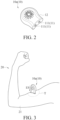
- the electrode assembly 11 contains dual electrodes or a plurality of electrodes such that the electrical stimulation is bipolar or tripolar. In other embodiments, the electrode assembly 11 contains a working electrode and a reference electrode such that the electrical stimulation is monopolar. As shown in FIG. 2 , in a bipolar example, the electrode assembly 11 contains an electrode pair 111. In the electrical stimulation device 10a, the electrode pair 111 is positioned on the surface (i.e., a casing) of the electrical stimulator 12.
- the electrical stimulation device 10a is attached to the skin 21 of an arm by the electrode pair 111 and is activated to output the electrical stimulation signal ES to the electrode pair 111, so as to electrically stimulate the skin 21 of the arm and the superficial nerves under the skin 21 via the electrode pair 111.
- the superficial nerves under the skin 21 of the arm may be a musculocutaneous nerve located in biceps brachii, a radial nerve located in triceps brachii, a dorsal nerve of the penis (DPN) located in the male genitalia or a median nerve located near a wrist.
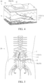
- the electrical stimulation device 10 may be a minimally invasive electrical stimulation device 10b. After the electrode assembly 11 is implanted, one end thereof is wirelessly or wiredly connected to the electrical stimulator 12 and the other end is implanted in the organism 20. In use, the electrical stimulator 12 is positioned on the skin 21 of the organism 20 and the electrode assembly 11 is at least partially implanted under the skin 21 of the organism 20 in a minimally invasive manner. In other words, the electrical stimulator 12 of the electrical stimulation device 10b is positioned outside the body of the organism, while the electrode assembly 11 of the electrical stimulation device 10b is positioned inside the body of the organism.
- the electrical stimulation signal ES generated by the electrical stimulator 12 wirelessly or wiredly stimulates the target nerve N near a terminal (one end of the electrode assembly 11 not connected to the electrical stimulator 12) of the electrode assembly 11 via the electrode assembly 11.
- the target region T is the target nerve N near the terminal of the electrode assembly 11.
- the electrical stimulation device 10b may be a peripheral nerve electrical stimulation device
- the electrode assembly 11 may be a lead 112
- the lead 112 contains a plurality of ring electrodes 113.
- the lead 112 is wirelessly connected to the electrical stimulator 12.
- the electrical stimulation device 10b is attached to the skin 21 of the organism 20 with the electrical stimulator 12, and the lead 112 is implanted under the skin 21 of the organism 20 in a minimally invasive manner.
- the electrical stimulator 12 wirelessly outputs the electrical stimulation signal ES to the lead 112, thereby electrically stimulating a nerve (i.e., the target nerve N) under the skin 21 of the organism 20 and located near the ring electrodes 113 of the lead 112 via the lead 112.
- a nerve i.e., the target nerve N
- one end of the lead 112 may be implanted near the target nerve N and the other end may be positioned subcutaneously to enable the electrical stimulator 12 to be wirelessly connected and powered (as shown in FIG. 4 ).
- the other end of the lead 112 may extend over the surface of the skin 21 to connect to the electrical stimulator 12 and be powered by a wired connection.
- the target region T is a nerve or neural structure around the ring electrodes 113 on the lead 112.
- the target region T is the target nerve N around the ring electrodes 113 on the lead 112.
- FIG. 5 is a use schematic diagram of another example of the electrical stimulation device of FIG. 1 .
- the electrical stimulation device 10 is an implantable electrical stimulation device 10c, such as a spinal cord electrical stimulation device or a sacral nerve electrical stimulation device
- the electrode assembly 11 may be a lead 112
- the electrode assembly 11 is connected to the electrical stimulator 12.
- both the electrical stimulator 12 and electrode assembly 11 are implanted in the organism 20, the ring electrodes 113 of the electrode assembly 11 are positioned at the terminal of the lead 112 (one end of the electrode assembly 11 not connected to the electrical stimulator 12), and the ring electrodes 113 corresponds to the position of the target region T of the organism 20.
- the electrical stimulation signal ES generated by the electrical stimulator 12 stimulates the nerve near the ring electrodes 113 via the ring electrodes 113 on the electrode assembly 11.
- the electrical stimulator 12 can be implanted at any position around the target region T of the organism 20 according to the placement position and length of the ring electrodes 113 of the lead 112.
- the electrical stimulator 12 is connected to one end of the lead 112 and implanted in the epidural space of the spine or near the sacral nerve around the buttocks of the organism 20. Therefore, when the electrical stimulator 12 outputs the electrical stimulation signal ES to the lead 112, the electrical stimulation signal ES can electrically stimulate the spinal cord or the sacral nerve around the buttocks of the organism 20 via the lead 112.
- the electrical stimulation signal ES may be a biphasic electrical stimulation signal ES. In other embodiments, the electrical stimulation signal ES may be a monophasic electrical stimulation signal ES.
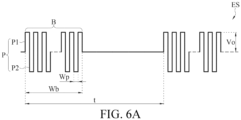
- FIG. 6A is a schematic diagram of an example of the electrical stimulation signal generated by the electrical stimulator of FIG. 1 .
- the waveform of the electrical stimulation signal ES generated by the electrical stimulation device 10 in the period time t is for example, biphasic, square-wave, symmetric and charge-balanced.
- the waveform of the electrical stimulation signal ES may be a monophasic signal, a triangular wave, a sine wave, an asymmetric signal, or a charge-unbalanced signal or the like.
- the electrical stimulation signal ES contains a plurality of burst signals B, and the burst signals B have the burst frequency between 0.1 Hz and 1,000 Hz.
- Each burst signal B contains a plurality of pulses P, and the pulses P have the pulse frequency between 1,000 Hz and 1,000 kHz.
- the burst signals B have the burst frequency between 0.1 Hz and 500 Hz, for example, 0.1 Hz-400 Hz, 0.1 Hz-200 Hz, 0.1 Hz-100 Hz or 0.1 Hz-20 Hz.
- the pulses P have the pulse frequency between 1 kHz and 750 kHz, for example, 400 kHz-600 kHz.
- the wave width Wb of the burst signals B is between 1 ms and 10 s, and the pulse width Wp of the pulses P is between 0.5 ⁇ s and 1 ms.
- the wave width Wb of the burst signals B is between 2 ms and 500 ms, for example, between 10 ms and 50 ms.
- the pulse width Wp of the pulses P is between 1 ⁇ s and 1 ms, for example, between 1 ⁇ s and 100 ⁇ s.
- the current intensity of the electrical stimulation signal ES may be between 0.1 mA and 120 mA, and the voltage intensity Vo of the electrical stimulation signal ES may be between 0.05 V and 60 V Preferably, the current intensity is between 6 mA and 40 mA. Preferably, the voltage intensity Vo is between 3 V and 20 V
- the pulses P are biphasic and successive of opposite polarity.
- the pulses P contained in the biphasic pulses include positive pulses P1 and negative pulses P2, the positive pulses P1 and the negative pulses P2 alternately appear, and preferably, the positive pulses P1 and the negative pulses P2 are charge balanced.
- the pulses P may be a square wave, a sine wave, a triangular wave or a combination thereof.
- the burst signals B may be a square wave, a sine wave, a triangle wave, a symmetric wave, an asymmetric wave or a combination thereof.
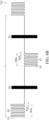
- FIG. 6B is a schematic diagram of one other example of the electrical stimulation signal generated by the electrical stimulator of FIG. 1 .
- the electrical stimulation signal ES generated by the electrical stimulation device 10 is an electrical stimulation signal ES2
- the electrical stimulation signal ES2 is exemplified by containing two alternate burst signals B1 and B2, wherein the burst signals B1 are the same as the burst signals B in FIG. 6A , and the waveform thereof is illustrated by a solid block, which is not described herein again.
- the burst frequency of the burst signals B2 is also between 0.1 Hz and 1,000 Hz, and preferably, the burst frequency of the burst signals B2 is less than or equal to that of the burst signals B1.
- the burst signals B1 and B2 alternately appear at 1:1 in time.
- the burst signals B2 contain a plurality of positive pulses P3 or a plurality of negative pulses P4.
- the electrical stimulation signal ES2 contains two burst signals B2, first burst signals B2 contain a plurality of positive pulses P3 and second burst signals B2 contain a plurality of negative pulses P4, the two burst signals B2 have the same waveform but opposite phases.
- the pulse frequency of the positive pulses P3 and the negative pulses P4 is in the range of 1 kHz to 1,000 kHz, and preferably the pulse frequency of the positive pulses P3 and the negative pulses P4 is in the range of 1 kHz to 300 kHz.
- the pulse frequency of the positive pulse P3 or the negative pulse P4 contained in the burst signals B2 is smaller than that of the pulses P contained in the burst signals B1.
- the pulse frequency of the positive pulse P3 or the negative pulse P4 contained in the burst signals B2 is smaller than 1/3 of that of the pulses P contained in the burst signals B1.
- the wave width Wb2 of the burst signals B2 is also between 1 ms and 10 s, and preferably, the wave width Wb2 of the burst signals B2 is between 2 ms and 500 ms, for example, between 5 ms and 100 ms.
- the pulse widths Wp1 and Wp2 of the positive pulses P3 and the negative pulses P4 are also between 1 ⁇ s and 1 ms, and preferably, the pulse widths Wp1 and Wp2 of the positive pulses P3 and the negative pulses P4 are between 3.3 ⁇ s and 1 ms, for example, 1 ⁇ s.
- the pulse frequencies and the pulse widths Wp1 and Wp2 of the positive pulses P3 and the negative pulses P4 may be the same or different.
- the charge of the positive pulses P3 is equal to that of the negative pulses P4 in a unit time, thereby achieving a delayed charge balance and reducing erosion of an electrode due to polarization.
- the organism 20 when the accumulated charge of the positive pulses P3 or the negative pulses P4 of the burst signals B2 is greater than or equal to 20 ⁇ C (microcoulomb), it can be sensible by the organism 20 (a user using the electrical stimulation device 10), such as a tapping sensation, a shaking sensation or a tingling sensation, such that the organism 20 can know that the electrical stimulation is in progress.
- FIG. 6C is a schematic diagram of another example of the electrical stimulation signal generated by the electrical stimulator of FIG. 1 .
- the electrical stimulation device 10 has two output ends CH1 and CH2.
- the two output ends CH1 and CH2 respectively output the electrical stimulation signals ES2, and the output times of the electrical stimulation signals ES2 at the two output ends CH1 and CH2 are not overlapped in each burst signal or each pulse on period in the period time t.
- the on periods of the burst signals B1 and burst signals B2 at the output end CH1 do not overlap with those of the burst signals B1 and burst signals B2 at the output end CH2.
- the electrical stimulation device 10c may contain two leads 112 ( FIG. 5 only shows one lead 112 and the other lead 112 is not shown), which respectively correspond to the two output ends CH1 and CH2 to output the electrical stimulation signal ES2 to the plurality of ring electrodes 113 on the two leads 112 for bipolar output of the electrical stimulation signal ES2.
- the electrical stimulation signal ES generated by the electrical stimulation device 10 may reduce pain related neuropeptide, neuroinflammatory information and/or neuropathic pain signals of the organism 20.
- the electrical stimulation signal ES generated by the electrical stimulation device 10 reduces the level of COX-2 of the organism 20. In some embodiments, the electrical stimulation signal ES generated by the electrical stimulation device 10 inhibits the level of the COX-2 of the organism 20. In some embodiments, the electrical stimulation signal ES generated by the electrical stimulation device 10 reduces the level of the COX-2 by inhibiting the COX-2 protein or the gene expression of the COX-2 protein. In some embodiments, reducing the level of the COX-2 of the organism 20 in the body occurs within 24 hours after receiving the electrical stimulation signal ES at the target region T. In some embodiments, the electrical stimulator 12 generates the electrical stimulation signal ES for a cumulative time less than or equal to 12 hours per day so as to reduce the level of the COX-2 of the organism 20.
- the electrical stimulation signal ES generated by the electrical stimulation device 10 reduces the levels of brain-derived neurotrophic factor (BDNF) and/or substance P of the organism 20.
- BDNF brain-derived neurotrophic factor
- the electrical stimulator 12 uses the electrical stimulation signal ES to inhibit the neuroinflammatory response of the organism 20 via the electrode assembly 11.
- the electrical stimulator 12 uses the electrical stimulation signal ES to inhibit and/or relieve pain of the organism 20. In some embodiments, the electrical stimulator 12 uses the electrical stimulation signal ES to inhibit and/or relieve the generation of the neuropathic pain signals of the organism 20 via the electrode assembly 11. In some embodiments, the electrical stimulation signal ES generated by the electrical stimulation device 10 inhibits and/or relieves at least a part of pain on a nerve conduction path of the target region T. In some embodiments, the pain of the organism 20 may be acute or chronic.
- the electrical stimulation device 10 generates the electrical stimulation signal ES for a duration time of at least 3 minutes to 60 minutes. In some embodiments, the electrical stimulation device 10 generates the electrical stimulation signal ES for a duration time of 5 minutes to 60 minutes, 5 minutes to 30 minutes, or 5 minutes to 20 minutes. Preferably, the electrical stimulation device 10 generates the electrical stimulation signal ES for a duration time of 15 minutes.
- the electrical stimulation device 10 generates the electrical stimulation signal ES with a cumulative time per day of less than or equal to 12 hours, preferably less than 10 hours, 8 hours, 6 hours, 4 hours, 2 hours, or 1 hour so as to relieve pain, relieve symptoms, desensitize overactive nerves, or relieve overactive reaction.
- the electrical stimulation signal ES relieves the pain of the organism 20, relieves the symptoms of the organism 20, reduces the overactive neural sensitivity of the organism 20 or relieves the overactive reaction of the organism 20.
- the electrical stimulator 12 uses the electrical stimulation signal ES to inhibit and/or relieve the pain of the organism 20 sustaining for a particular time via the electrode assembly 11.
- the particular time is at least one hour, one hour to 4 days, one hour to 5 days; at least 1 day, 1 day to 4 days, or 1 day to 5 days.
- the target region T is a dorsal root ganglion.
- the electrical stimulation signal ES generated by the electrical stimulation device 10 has effects of inhibiting and/or relieving acute pain, chronic pain, local pain, spasticity, renal hypertension, overactive bladder, palmar hyperhidrosis, premature ejaculation, or diseases or symptoms caused by nerve sensitization.
- Test I Animal test-Mechanical allodynia evaluation test
- the sciatic nerves of the left hind paws of normal rats were ligated with 5-0 nylon wires so as to establish a model of chronic constriction injury (CCI) through controllable forces (6 g rope tension) monitored by a computer.
- CCI chronic constriction injury
- Compressive neuropathy of the CCI model had marked and sustained mechanical allodynia and increased neuroinflammation.
- the CCI model treated with the nerve injury was a CCI group, which was divided into a CCI-UHF group and a CCI+UHF group with 6 rats in each group.
- the CCI model can be established according to the reference of Chen et al (Chen et al, 2020).
- an electrical stimulation device (StimOn GM2439; Gimer Medical Co., Ltd., Taiwan) was placed on the skin above the sciatic nerves of the rats in the CCI+UHF group and the rats were continuously treated with an ultrahigh frequency electrical stimulation signal for 15 minutes.
- An electrode pad of the electrical stimulation device was firmly attached to the skin above the sciatic nerves (lesions).
- the ultrahigh frequency electrical stimulation signal contained a plurality of burst signals, and the burst signals contained a plurality of pulses.
- the burst frequency of the burst signals was 2 Hz
- the pulse frequency of the pulses was 500 kHz
- the voltage intensity of the electrical stimulation signal was about ⁇ 6.6 V
- the duration time of the electrical stimulation signal was 15 minutes.
- the rats in the CCI-UHF group was not subjected to the first ultrahigh frequency electrical stimulation treatment T1.
- the rats in the CCI+UHF group were subjected to a second ultrahigh frequency electrical stimulation treatment T2.
- the treatment process was the same as that of the first ultrahigh frequency electrical stimulation treatment T1 in (2), which was not described herein again.
- the rats in the CCI-UHF group was not subjected to the second ultrahigh frequency electrical stimulation treatment T2.
- a monofilament tactile pain test (von Frey test) was used to measure the threshold forces (withdrawal thresholds) required to cause the withdrawal of the paws of the rats in the CCI-UHF group and CCI+UHF group to obtain the plantar tolerance of the rats, thereby evaluating the mechanical allodynia of the normal rats.
- the underside of the rat hind paws was poked with a Semmes-Weinstein monofilament (SWM; North Coast Medical Inc., USA).
- SWM Semmes-Weinstein monofilament
- each rat was subjected to two monofilament tactile pain tests, and the interval time of the two monofilament tactile pain tests was at least 10-15 minutes.
- the rats in the CCI-UHF group and CCI+UHF group were respectively subjected to the mechanical allodynia assessment test.
- the withdrawal threshold measured before the CCI model was established was the withdrawal threshold at day -7.
- the withdrawal threshold measured 7 days after the CCI model was established, but the electrical stimulation was not performed was the withdrawal threshold at day 0.
- the withdrawal thresholds measured after the first ultrahigh frequency electrical stimulation treatment T1 were the withdrawal thresholds at 30 minutes, 1 day, 2 days, 3 days, 4 days, and 5 days after the first ultrahigh frequency electrical stimulation treatment T1.
- the withdrawal thresholds measured after the second ultrahigh frequency electrical stimulation treatment T2 were the withdrawal thresholds at 30 minutes, 1 day, 2 days, 3 days, 4 days, and 5 days after the second ultrahigh frequency electrical stimulation treatment T2.
- the withdrawal thresholds between groups were compared by a Student's t-test and analyzed.
- the withdrawal thresholds were all expressed as mean standard deviation. **** indicated that compared to the CCI-UHF group, the CCI+UHF group was statistically significant, p ⁇ 0.0001; and ++++ indicated that compared to the CCI+UHF group at day 0, the CCI+UHF group at different time points was statistically significant, p ⁇ 0.0001.
- the withdrawal threshold of the CCI-UHF group was 5.77 ⁇ 1.39 g, while the withdrawal threshold of the CCI+UHF group was 6.86 ⁇ 1.40 g.
- the withdrawal threshold of the CCI-UHF group was 0.45 ⁇ 0.068 g, while the withdrawal threshold of the CCI+UHF group was 0.48 ⁇ 0.087 g.
- the withdrawal threshold of the CCI+UHF group was 8.64 ⁇ 0.73 g.
- the withdrawal threshold of the CCI+UHF group was 10.25 ⁇ 2.68 g.
- the withdrawal threshold of the CCI+UHF group was 0.55 ⁇ 0.20 g.
- the second ultrahigh frequency electrical stimulation treatment T2 was performed 4 days after the first ultrahigh frequency electrical stimulation treatment T1.
- the withdrawal threshold of the CCI+UHF group was 8.53 ⁇ 0.89 g.
- the withdrawal threshold of the CCI+UHF group was 0.34 ⁇ 0.17 g.
- the withdrawal threshold of the CCI-UHF group was consistently maintained at about 0.45 ⁇ 0.068 g for 2 weeks.
- the ultrahigh frequency electrical stimulation treatment can relieve the mechanical allodynia caused by the nerve injury treatment.
- the first ultrahigh frequency electrical stimulation treatment T1 may increase the decreased withdrawal threshold continuously for 3 days, while the second ultrahigh frequency electrical stimulation treatment T2 may increase the decreased withdrawal threshold continuously for 4 days.
- the ultrahigh frequency electrical stimulation treatment inhibits and/or relieves the mechanical allodynia for at least about 4 days.
- the electrical stimulation signal generated by the electrical stimulator has the effect of relieving and/or inhibiting neuropathic pain caused by the nerve injury treatment.
- Test II Animal test-neuronal injury test
- Normal rats (Sprague Dawley, SD) without injury treatment were a control group (Ctrl group) which was divided into a Ctrl-UHF group (4 rats) and a Ctrl+UHF group (3 rats).
- the electrical stimulation device (StimOn GM2439; Gimer Medical Co., Ltd., Taiwan) was placed on the skin above the sciatic nerves of the rats in the Ctrl+UHF group and the rats were continuously treated with an ultrahigh frequency electrical stimulation signal for 15 minutes.
- An electrode pad of the electrical stimulation device was firmly attached to the skin above the sciatic nerves.
- the ultrahigh frequency electrical stimulation signal contained a plurality of burst signals, and the burst signals contained a plurality of pulses.
- the burst frequency of the burst signals was 2 Hz
- the pulse frequency of the pulses was 500 kHz
- the voltage intensity of the electrical stimulation signal was about ⁇ 6.6 V
- the duration time of the electrical stimulation signal was 15 minutes.
- the pulses were a symmetric biphasic sine wave.
- the rats in the Ctrl-UHF group was not subjected to the first ultrahigh frequency electrical stimulation treatment T1.
- the rats in the Ctrl+UHF group were subjected to a second ultrahigh frequency electrical stimulation treatment T2.
- the treatment process was the same as that of the first ultrahigh frequency electrical stimulation treatment T1 in (1), which was not described herein again.
- the rats in the Ctrl-UHF group was not subjected to the second ultrahigh frequency electrical stimulation treatment T2.
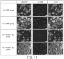
- the immunofluorescent staining assay was performed according to the general immunofluorescent (IF) staining test process.
- Staining reagents contained a mouse anti-neurofilament heavy polypeptide antibody (mouse anti-NF200, 1: 200; product number N0142, Sigma-Aldrich, USA) and a rabbit anti-MBP antibody (1:200; product number GTX133108, GeneTex, USA) as primary antibodies, and a goat anti-mouse IgG antibody (1:200; GeneTex, product number GTX213111-05) as a secondary antibody of NF200.
- IF general immunofluorescent
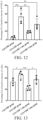
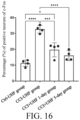
- the levels of the MBP and the NF200 and the fluorescent staining results of the MBP and the NF200 among groups were compared by a Student's t-test and analyzed.
- the levels of the MBP and the NF200 and the fluorescent staining results of the MBP and the NF200 were expressed as mean standard deviation (SD). ns indicated that there was no significant difference between the Ctrl+UHF and Ctrl-UHF groups.
- the number of myelinated axons of the uninjured nerves of the rats treated with the ultrahigh frequency electrical stimulation treatment was not significantly different from that of the uninjured nerves of the rats not treated with the ultrahigh frequency electrical stimulation treatment.
- the ultrahigh frequency electrical stimulation treatment did not injure the axons and Schwann cells in the uninjured nerves.
- the sciatic nerves of the left hind paws of normal rats were ligated with 5-0 nylon wires so as to establish a model of chronic constriction injury (CCI) through controllable forces (6 g rope tension) monitored by a computer.
- CCI chronic constriction injury
- Compressive neuropathy of the CCI model had marked and sustained mechanical allodynia and increased neuroinflammation.
- the CCI model treated with the nerve injury was a CCI group and the normal rats not treated with the injury was a control group (Ctrl group).
- the CCI group was divided into a CCI-UHF group and a CCI+UHF group.
- the Ctrl group was a Ctrl-UHF group.
- the CCI model can be established according to the reference of Chen et al (Chen et al, 2020).
- an electrical stimulation device (StimOn GM2439; Gimer Medical Co., Ltd., Taiwan) was placed on the skin above the sciatic nerves of the rats in the CCI+UHF group and the rats were continuously treated with an ultrahigh frequency electrical stimulation signal for 15 minutes.
- An electrode pad of the electrical stimulation device was firmly attached to the skin above the sciatic nerves (lesions).
- the ultrahigh frequency electrical stimulation signal contained a plurality of burst signals, and the burst signals contained a plurality of pulses.
- the burst frequency of the burst signals was 2 Hz
- the pulse frequency of the pulses was 500 kHz
- the voltage intensity of the electrical stimulation signal was about ⁇ 6.6 V
- the duration time of the electrical stimulation signal was 15 minutes.
- the rats in the CCI-UHF group and the Ctrl-UHF group were not subjected to the ultrahigh frequency electrical stimulation treatment.
- the immunofluorescent staining assay was performed according to the general immunofluorescent (IF) staining test process.
- the staining reagents contained a rabbit anti-c-FOS antibody (purchased from GeneTex), a rabbit anti-brain-derived neurotrophic factor (BDNF) antibody (purchased from Elabscience), a rabbit anti-COX-2 antibody (purchased from Elabscience), a rabbit anti-c-Myc antibody (purchased from GeneTex), a guinea pig anti-substance P (SP) antibody (purchased from GeneTex) and a rabbit anti-MEK antibody (purchased from GeneTex) as primary antibodies, and a goat anti-guinea pig IgG antibody (purchased from GeneTex) as a secondary antibody of SP and a goat anti-rabbit IgG antibody (purchased from GeneTex) as secondary antibodies of BDNF, COX-2, c-Myc, MEK and c-FOS.
- BDNF rabbit anti-brain-derived neurotrophic factor
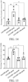
- the percentage of positive neurons of the BDNF in the CCI-UHF group was 29.98% ⁇ 3.79%
- the percentage of positive neurons of the BDNF in the CCI+UHF 1-day group was 12.95% ⁇ 1.71%
- the percentage of positive neurons of the BDNF in the CCI+UHF 5-day group was 24.66% ⁇ 13.85%.
- the level of the BDNF was significantly increased by the nerve injury treatment, but significantly decreased in the CCI+UHF 1-day group after the ultrahigh frequency electrical stimulation treatment.
- the level of the BDNF in the CCI+UHF 5-day group returned to that in the CCI-UHF group (subjected to the nerve injury, but not subjected to the electrical stimulation treatment), and had no significant difference with that in the CCI-UHF group.
- c-Myc and COX-2 both showed the same trend except for the BDNF.
- the levels of the c-Myc and the COX-2 can be significantly increased by the nerve injury treatment, but the levels of the c-Myc and the COX-2 can be significantly reduced by the ultrahigh frequency electrical stimulation treatment and can be continuously reduced for at least 4 days.
- substance P (SP), MEK and c-Fos also showed similar trends. After the nerve injury treatment, the levels of the substance P (SP), MEK and c-Fos were all significantly increased. However, after the ultrahigh frequency electrical stimulation treatment, the levels of the substance P (SP), MEK and c-Fos in the CCI+UHF 1-day group were significantly decreased and can be continuously reduced for at least 5 days after the ultrahigh frequency electrical stimulation treatment.
- the ultrahigh frequency electrical stimulation treatment can reduce the levels of the pain related neuropeptides BDNF, COX-2 and c-Myc continuously for at least 4 days.
- the ultrahigh frequency electrical stimulation treatment can reduce the levels of the neuroinflammatory information SP, MEK and c-FOS continuously for at least 5 days.
- the electrical stimulation signal generated by the electrical stimulator had the effects of reducing the pain related neuropeptides, and relieving the inflammation information and neuropathic pain.
- the BDNF is critical for neuronal survival, differentiation and synaptic strength regulation and is involved in inflammatory pain mechanisms.
- the BDNF is considered as a neuroregulator and plays an important role in spinal plasticity.
- the levels of the BDNF and downstream signal proteins (such as MEK, c-Myc and c-FOS) thereof were down-regulated after the ultrahigh frequency electrical stimulation treatment.
- the electrical stimulation signal generated by the electrical stimulator can inhibit and/or relieve neuroinflammation and inhibit and/or relieve the neuropathic pain by regulating a BDNF/MAPK-mediated pain transduction pathway.
- the level of the COX-2 is related with the development and maintenance of the neuropathic pain. Prostaglandins synthesized by the COX-2 are involved in the pathogenesis of the inflammatory response and neuropathic pain.
- the electrical stimulation signal generated by the electrical stimulator can inhibit and/or relieve pain or inhibit and/or relieve the inflammatory response by inhibiting the generation of the COX-2 and/or reducing the level of the COX-2.
- the reduction in the level of the COX-2 could mean quantitative reduction in the level of the COX-2 or mean conversion of the COX-2 into inactive state.
- the reduction in the level of the COX-2 is not limited to the location of the DRG, but anywhere the level of the COX-2 is detectable, such as by extraction from blood, saliva, urine, stool, spinal fluid, and other biological samples that may be sampled.
- a detection method may include enzyme-linked immunosorbent assay (ELISA), immunofluorescent staining, or other immunological methods.
- SP can bind to its specific receptor neurokinin-1 (NK-1) which sensitizes neurons and produces sense of pain.
- NK-1 specific receptor neurokinin-1
- the electrical stimulation signal generated by the electrical stimulator can inhibit and/or relieve pain by inhibiting the generation of the SP.
- the electrical stimulation device has the effect of reducing COX-2 without causing the nerve injury.
- the electrical stimulation device has the effects of reducing the level of the brain-derived neurotrophic factor, reducing the level of the substance P, inhibiting the neuroinflammatory response, relieving and/or inhibiting pain, relieving symptoms, reducing nerve sensitivity, or relieving the overactive reaction without causing the nerve injury.
- the electrical stimulation device has an effect of regulating nerves by regulating protein and gene expression of the protein to reduce the level of the protein.
Landscapes
- Health & Medical Sciences (AREA)
- Life Sciences & Earth Sciences (AREA)
- Animal Behavior & Ethology (AREA)
- General Health & Medical Sciences (AREA)
- Biomedical Technology (AREA)
- Nuclear Medicine, Radiotherapy & Molecular Imaging (AREA)
- Radiology & Medical Imaging (AREA)
- Veterinary Medicine (AREA)
- Public Health (AREA)
- Engineering & Computer Science (AREA)
- Neurology (AREA)
- Neurosurgery (AREA)
- Pain & Pain Management (AREA)
- Biophysics (AREA)
- Heart & Thoracic Surgery (AREA)
- Electrotherapy Devices (AREA)
Applications Claiming Priority (2)
| Application Number | Priority Date | Filing Date | Title |
|---|---|---|---|
| TW112136944A TWI851432B (zh) | 2022-12-27 | 2023-09-25 | 電刺激裝置 |
| TW113119906A TW202513113A (zh) | 2022-12-27 | 2024-05-29 | 電刺激裝置 |
Publications (1)
| Publication Number | Publication Date |
|---|---|
| EP4527445A1 true EP4527445A1 (fr) | 2025-03-26 |
Family
ID=92895426
Family Applications (1)
| Application Number | Title | Priority Date | Filing Date |
|---|---|---|---|
| EP24201917.2A Pending EP4527445A1 (fr) | 2023-09-25 | 2024-09-23 | Dispositif de stimulation électrique |
Country Status (2)
| Country | Link |
|---|---|
| US (1) | US20250099766A1 (fr) |
| EP (1) | EP4527445A1 (fr) |
Citations (4)
| Publication number | Priority date | Publication date | Assignee | Title |
|---|---|---|---|---|
| US20190240486A1 (en) * | 2009-03-20 | 2019-08-08 | Electrocore, Inc. | Non-invasive treatment of neurodegenerative diseases |
| AU2020279753A1 (en) * | 2019-05-22 | 2021-12-16 | Scuola Internazionale Superiore Di Studi Avanzati - Sissa | Transcutaneous electrical spinal cord neuromodulator and uses thereof |
| US20220044828A1 (en) * | 2017-12-22 | 2022-02-10 | Electrocore, Inc. | Systems and methods for treating patients with diseases associated with viruses |
| US20230071154A1 (en) * | 2021-08-27 | 2023-03-09 | Leonhardt Ventures Llc | Modulation of brain-derived neurotrophic factor (bdnf) |
-
2024
- 2024-09-16 US US18/886,469 patent/US20250099766A1/en active Pending
- 2024-09-23 EP EP24201917.2A patent/EP4527445A1/fr active Pending
Patent Citations (4)
| Publication number | Priority date | Publication date | Assignee | Title |
|---|---|---|---|---|
| US20190240486A1 (en) * | 2009-03-20 | 2019-08-08 | Electrocore, Inc. | Non-invasive treatment of neurodegenerative diseases |
| US20220044828A1 (en) * | 2017-12-22 | 2022-02-10 | Electrocore, Inc. | Systems and methods for treating patients with diseases associated with viruses |
| AU2020279753A1 (en) * | 2019-05-22 | 2021-12-16 | Scuola Internazionale Superiore Di Studi Avanzati - Sissa | Transcutaneous electrical spinal cord neuromodulator and uses thereof |
| US20230071154A1 (en) * | 2021-08-27 | 2023-03-09 | Leonhardt Ventures Llc | Modulation of brain-derived neurotrophic factor (bdnf) |
Also Published As
| Publication number | Publication date |
|---|---|
| US20250099766A1 (en) | 2025-03-27 |
Similar Documents
| Publication | Publication Date | Title |
|---|---|---|
| US20230142770A1 (en) | Treatment of Inflammatory Disorders | |
| US20250235694A1 (en) | Transcutaneous electrical spinal cord neuromodulator and uses thereof | |
| US20210393949A1 (en) | Neuromodulation device | |
| Garrison et al. | Decreased activity of spontaneous and noxiously evoked dorsal horn cells during transcutaneous electrical nerve stimulation (TENS) | |
| Ahmed et al. | Strategies for precision vagus neuromodulation | |
| US20220161042A1 (en) | Magnetic stimulation of the spinal cord to restore control of bladder and/or bowel | |
| Meyerson et al. | Mechanisms of spinal cord stimulation in neuropathic pain | |
| US7251529B2 (en) | Spinal cord stimulation as treatment for functional bowel disorders | |
| US10201706B2 (en) | Systems and methods of improving an inflammatory disorder | |
| Tator et al. | Spinal cord stimulation: therapeutic benefits and movement generation after spinal cord injury | |
| US20170100605A1 (en) | Systems and methods of improving an immune disorder | |
| US20170100604A1 (en) | Systems and methods of improving metabolic syndrome | |
| US20170100589A1 (en) | Systems and methods of improving infections by neuromodulation of immune function | |
| US20170100588A1 (en) | Systems and methods of improving cancer symptoms by neuromodulation of immune function | |
| WO2018118860A1 (fr) | Systèmes et méthodes de soulagement d'un trouble inflammatoire | |
| Malone et al. | Closed-loop, cervical, epidural stimulation elicits respiratory neuroplasticity after spinal cord injury in freely behaving rats | |
| WO2016197039A1 (fr) | Procédé et dispositif de stimulation du nerf sacré | |
| WO2018118857A1 (fr) | Systèmes et procédés d'atténuation d'un trouble immunitaire | |
| EP4527445A1 (fr) | Dispositif de stimulation électrique | |
| TW202513113A (zh) | 電刺激裝置 | |
| CN119680102A (zh) | 电刺激装置 | |
| Xi et al. | Interactions between hypocretinergic and GABAergic systems in the control of activity of neurons in the cat pontine reticular formation | |
| WO2025128813A1 (fr) | Systèmes et méthodes de traitement de la douleur | |
| Mills et al. | Use of Transcranial Magnetic Stimulation in the Investigation of Spinal and Transcortical | |
| Daneshgari et al. | Bladder sensation testing: Where are we? |
Legal Events
| Date | Code | Title | Description |
|---|---|---|---|
| PUAI | Public reference made under article 153(3) epc to a published international application that has entered the european phase |
Free format text: ORIGINAL CODE: 0009012 |
|
| STAA | Information on the status of an ep patent application or granted ep patent |
Free format text: STATUS: THE APPLICATION HAS BEEN PUBLISHED |
|
| AK | Designated contracting states |
Kind code of ref document: A1 Designated state(s): AL AT BE BG CH CY CZ DE DK EE ES FI FR GB GR HR HU IE IS IT LI LT LU LV MC ME MK MT NL NO PL PT RO RS SE SI SK SM TR |