EP4502941A1 - Lung region segmentation method and apparatus - Google Patents
Lung region segmentation method and apparatus Download PDFInfo
- Publication number
- EP4502941A1 EP4502941A1 EP24185117.9A EP24185117A EP4502941A1 EP 4502941 A1 EP4502941 A1 EP 4502941A1 EP 24185117 A EP24185117 A EP 24185117A EP 4502941 A1 EP4502941 A1 EP 4502941A1
- Authority
- EP
- European Patent Office
- Prior art keywords
- region
- lung
- mask
- lung region
- medical image
- Prior art date
- Legal status (The legal status is an assumption and is not a legal conclusion. Google has not performed a legal analysis and makes no representation as to the accuracy of the status listed.)
- Withdrawn
Links
Images




Classifications
-
- G—PHYSICS
- G06—COMPUTING OR CALCULATING; COUNTING
- G06T—IMAGE DATA PROCESSING OR GENERATION, IN GENERAL
- G06T7/00—Image analysis
- G06T7/0002—Inspection of images, e.g. flaw detection
- G06T7/0012—Biomedical image inspection
-
- A—HUMAN NECESSITIES
- A61—MEDICAL OR VETERINARY SCIENCE; HYGIENE
- A61B—DIAGNOSIS; SURGERY; IDENTIFICATION
- A61B6/00—Apparatus or devices for radiation diagnosis; Apparatus or devices for radiation diagnosis combined with radiation therapy equipment
- A61B6/52—Devices using data or image processing specially adapted for radiation diagnosis
- A61B6/5211—Devices using data or image processing specially adapted for radiation diagnosis involving processing of medical diagnostic data
-
- A—HUMAN NECESSITIES
- A61—MEDICAL OR VETERINARY SCIENCE; HYGIENE
- A61B—DIAGNOSIS; SURGERY; IDENTIFICATION
- A61B6/00—Apparatus or devices for radiation diagnosis; Apparatus or devices for radiation diagnosis combined with radiation therapy equipment
- A61B6/50—Apparatus or devices for radiation diagnosis; Apparatus or devices for radiation diagnosis combined with radiation therapy equipment specially adapted for specific body parts; specially adapted for specific clinical applications
-
- G—PHYSICS
- G06—COMPUTING OR CALCULATING; COUNTING
- G06T—IMAGE DATA PROCESSING OR GENERATION, IN GENERAL
- G06T7/00—Image analysis
- G06T7/10—Segmentation; Edge detection
- G06T7/11—Region-based segmentation
-
- G—PHYSICS
- G06—COMPUTING OR CALCULATING; COUNTING
- G06T—IMAGE DATA PROCESSING OR GENERATION, IN GENERAL
- G06T2207/00—Indexing scheme for image analysis or image enhancement
- G06T2207/10—Image acquisition modality
- G06T2207/10116—X-ray image
-
- G—PHYSICS
- G06—COMPUTING OR CALCULATING; COUNTING
- G06T—IMAGE DATA PROCESSING OR GENERATION, IN GENERAL
- G06T2207/00—Indexing scheme for image analysis or image enhancement
- G06T2207/20—Special algorithmic details
- G06T2207/20081—Training; Learning
-
- G—PHYSICS
- G06—COMPUTING OR CALCULATING; COUNTING
- G06T—IMAGE DATA PROCESSING OR GENERATION, IN GENERAL
- G06T2207/00—Indexing scheme for image analysis or image enhancement
- G06T2207/20—Special algorithmic details
- G06T2207/20084—Artificial neural networks [ANN]
-
- G—PHYSICS
- G06—COMPUTING OR CALCULATING; COUNTING
- G06T—IMAGE DATA PROCESSING OR GENERATION, IN GENERAL
- G06T2207/00—Indexing scheme for image analysis or image enhancement
- G06T2207/30—Subject of image; Context of image processing
- G06T2207/30004—Biomedical image processing
- G06T2207/30061—Lung
-
- G—PHYSICS
- G06—COMPUTING OR CALCULATING; COUNTING
- G06T—IMAGE DATA PROCESSING OR GENERATION, IN GENERAL
- G06T2207/00—Indexing scheme for image analysis or image enhancement
- G06T2207/30—Subject of image; Context of image processing
- G06T2207/30004—Biomedical image processing
- G06T2207/30101—Blood vessel; Artery; Vein; Vascular
Definitions
- the disclosure relates to a method and apparatus for segmenting a lung region, and more particularly, to a method and apparatus for segmenting a peripheral lung region to diagnose a lung disease.
- Medical images such as magnetic resonance imaging (MRI) images, computed tomography (CT) images, X-ray images, etc., are used to diagnose diseases.
- Three-dimensional (3D) medical images such as MRI images, CT images, etc., may enable accurate diagnosis of lesions by capturing exact images inside human bodies, but require a lot of imaging time or imaging costs.
- X-ray images may be easily captured, but it is difficult to accurately diagnose lesions using these images due to overlapping between images of several organs.
- 3D medical images such as MRI images, CT images, etc., are mainly used to diagnose lung diseases such as pulmonary hypertension or lung cancer because it is difficult to identify pulmonary hypertension, etc., from X-ray images.
- a method and apparatus for segmenting a peripheral lung region to diagnose a lung disease from a two-dimensional (2D) medical image is provided.
- a lung region segmentation method includes receiving a two-dimensional (2D) medical image, extracting a lung region from the 2D medical image, adjusting a size of a mask resembling the lung region, and extracting a peripheral region by removing a region corresponding to the mask from the lung region.
- 2D two-dimensional
- a lung region segmentation apparatus includes an input unit configured to receive a two-dimensional (2D) medical image, a lung extraction unit configured to extract a lung region from the 2D medical image, a mask adjustment unit configured to adjust a size of a mask resembling the lung region, and a periphery extraction unit configured to extract a peripheral region by removing a region corresponding to the mask from the lung region.
- 2D two-dimensional
- FIG. 1 is a view showing an example of a lung region segmentation apparatus according to an embodiment of the disclosure.
- a lung region segmentation apparatus 100 may segment a peripheral lung region 120 from a two-dimensional (2D) medical image 110 and output the same.
- the 2D medical image 110 may be an X-ray image.
- the 2D medical image 110 in the current embodiments may be of various types.
- the peripheral lung region 120 may be used to diagnose pulmonary hypertension or lung cancer.
- an area size of blood vessels of the peripheral lung region 120 may have a significant value for diagnosing a lung disease.
- FIG. 2 is a view showing an example of a lung region segmentation method according to an embodiment of the disclosure.
- a lung region segmentation apparatus may receive a 2D medical image in operation S200.
- the apparatus may segment a lung region from the 2D medical image in operation S210.
- the apparatus may use various conventional segmentation algorithms for lung region segmentation.
- the 2D medical image is an X-ray image
- a plurality of organs are overlappingly displayed.
- various organs such as ribs, etc., as well as lung tissues may be overlappingly displayed in the lung region segmented from the 2D medical image.
- lung tissues may be overlappingly displayed in the lung region segmented from the 2D medical image. From the 2D medical image, only a pure lung region may be segmented without overlapping display of other organs, which will be discussed again with reference to FIGS. 3 and 4 .
- the apparatus may adjust a size of a mask resembling the lung region in operation S220.
- the apparatus may generate the mask based on an outline of the lung region and reduce the size of the mask.
- An example of a specific method for generating the mask and adjusting the size thereof will be described again with reference to FIGS. 5 and 6 .
- the apparatus may extract a peripheral region corresponding to a peripheral lung region from the lung region, by using the size-adjusted mask, in operation S230. For example, the apparatus may remove the mask region from the lung region and output the remaining region as the peripheral lung region. The apparatus may output an image of the extracted peripheral region. By adjusting the size of the mask, the size of the peripheral region extracted from the lung region may be adjusted variously.
- the apparatus may further segment a vascular region from the peripheral region.
- the apparatus may obtain a ratio of a size of the vascular region (i.e., the entire area of blood vessels) segmented from the entire lung region (or a central lung region) to a size of the vascular region segmented from the peripheral region.
- the apparatus may obtain and output a size ratio of the central lung region to the peripheral lung region.
- FIGS. 3 and 4 are views showing an example of a lung region segmentation method according to an embodiment of the disclosure.
- a lung region 320 may be segmented from a 2D medical image 310 by using an artificial intelligence (AI) model 300.
- the AI model 300 may be implemented as various types of artificial neural networks such as a convolutional neural network, etc.
- the AI model 300 may be generated to segment the lung region through a training process.
- a dataset 440 for training the AI model 300 may be generated from a 3D medical image 400.
- the 3D medical image 400 may be a magnetic resonance imaging (MRI) image or a computed tomography (CT) image.
- MRI magnetic resonance imaging
- CT computed tomography
- the apparatus may two-dimensionally project the 3D medical image 400 to generate a 2D first training image 420.
- each voxel of the 3D medical image 400 may be projected to a 2D plane to generate an image in which a plurality of tissues overlap, like an X-ray image. That is, by two-dimensionally projecting the 3D medical image 400, a virtual X-ray image may be obtained.
- the apparatus may segment a 3D lung region 410 from the 3D medical image 400.
- the apparatus may segment the lung region 410 from the 3D medical image 400 based on a brightness value (i.e., a hounsfield unit (HU)) of a voxel.
- a brightness value i.e., a hounsfield unit (HU)
- HU hounsfield unit
- the apparatus may two-dimensionally project the 3D lung region 410 to generate a 2D second training image 430. That is, the second training image 430 may be a 2D image including a lung region.
- the apparatus may train the AI model 300 by using the dataset 440 including the first training image 420 and the second training image 430.
- the apparatus may generate the dataset 440 including a plurality of first training images 420 and a plurality of second training images 430 by using a plurality of 3D medical images 400.
- the apparatus may label the second training image 430 as ground truth to train the AI model 300 using a supervised training method.
- the AI model 300 may perform a training process by outputting a predicted image in which the lung region is segmented from the first training image 420 upon input of the first training image 420 of the dataset 440, and comparing the predicted image with the second training image 430 that is the ground truth of the dataset 440 to reduce an error.
- FIG. 5 is a view showing an example of a method of generating a mask, according to an embodiment of the disclosure.
- a mask 520 may include a first region 510 including an outline of a left lung and a second region 512 including an outline of a right lung.
- the mask 520 may include one enclosed space by connecting an empty space 514 between the first region 510 and the second region 512.
- the apparatus may connect the highest and lowest height points of the first region 510 to the highest and lowest height points of the second region 512 with line segments, respectively, to fill the empty space 514 between the two regions 510 and 512, thus generating the mask 520 including one enclosed region.
- a description will be made based on the mask 520 including one region.
- FIG. 6 is a view showing an example of a method of adjusting a size of a mask, according to an embodiment of the disclosure.
- the apparatus may adjust the size of the mask 520 with respect to a center point 530 of the mask 520.
- examples of adjusting sizes of masks 602, 612, and 622 to 80 %, 70 %, and 60 % are shown in FIG. 6 . That is, the apparatus may adjust the sizes of the masks 602, 612, and 622 from lung region images 600, 610, and 620 with respect to the center 530 of the mask 520 while maintaining the shape of the mask 520.
- the apparatus may identify an internally diving point that divides each point of the outline of the mask 520 and a center point 530 at m:n (m and n are natural numbers), and connect the dividing points to generate the size-adjusted masks 602, 612, and 622.
- m:n are natural numbers
- Various methods for changing a mask size may be applied to the current embodiment and are not limited to a specific one.
- the apparatus may extract, as peripheral lung regions, the other regions than regions of the masks 602, 612, and 622 from the lung region images 600, 610, and 620.
- the apparatus may also extract, as central lung regions 604, 614, and 624, regions corresponding to masks from lung regions. In FIG. 6 , extraction examples of the central lung regions 604, 614, and 624 are shown.
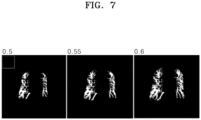
- FIG. 7 shows a vascular region segmented from a peripheral lung region, according to an embodiment of the disclosure.
- the apparatus may extract an edge area of the lung region as a peripheral lung region by adjusting the size of a mask.
- the apparatus may segment a vascular region from the peripheral lung region.
- Conventional various segmentation algorithms for segmenting a blood vessel from a human tissue may be applied to the current embodiment.
- an AI model trained to segment a blood vessel from a lung tissue may be used in the current embodiment.
- FIG. 8 shows an example of a ratio of a central lung to a peripheral lung for each lung disease, according to an embodiment of the disclosure.
- an optimal ratio of a central lung to a peripheral lung for disease diagnosis may differ with a type of a lung disease.
- a ratio 810 of a central lung to a peripheral lung used for diagnosis of a first disease 800 may be 5:5, and a ratio of the central lung to the peripheral lung used for diagnosis of a second disease may be 6:4. That is, to diagnose the first disease, the apparatus may adjust the size of the mask to 50 % of the original size to extract a peripheral lung from the lung region and extract a vascular region of the peripheral lung.
- the ratio of the central lung to the peripheral lung for each lung disease in the current embodiment may be identified through statistical analysis of existing clinical information of patients with lung diseases.
- the apparatus may extract the lung region from the 2D medical image or the 3D medical image of a patient group of the first disease and segment the lung region into the central lung and the peripheral lung at various ratios, thereby identifying a ratio optimized for identifying the corresponding disease.
- FIG. 9 is a block diagram of an example of a lung region segmentation apparatus according to an embodiment of the disclosure.
- the lung region segmentation apparatus 100 may include an input unit 900, a lung extraction unit 910, a mask adjustment unit 920, a periphery extraction unit 930, a vessel identification unit 940, an AI model 950, and a training unit 960.
- the lung region segmentation apparatus 100 may be implemented as a computing device including a memory, a processor, and an input/output device. In this case, each component may be implemented with software (program) and loaded on the memory, and then may be executed by the processor.
- the lung region segmentation apparatus 100 may omit at least one of the vessel identification unit 940, the AI model 950, and the training unit 960.
- the input unit 900 may receive a 2D medical image.
- An example of the 2D medical image may include an X-ray image.
- the lung extraction unit 910 may extract a lung region from the 2D medical image.
- the lung extraction unit 910 may segment the lung region from the 2D medical image by using an AI model 950.
- An example of segmenting the lung region by using the AI model 950 is shown in FIG. 3 .
- a training method of the training unit 960 training the AI model 950 is shown in FIG. 4 .
- the mask adjustment unit 920 may adjust the size of a mask resembling the lung region. For example, the mask adjustment unit 920 may generate the mask including a region formed with the outline of the lung region, and adjust the size of the mask based on the center of the mask. Examples of mask generation and size adjustment are shown in FIGS. 5 and 6 .
- the periphery extraction unit 930 may extract a peripheral region by removing a region corresponding the mask from the lung region. In another embodiment, the periphery extraction unit 930 may extract the region corresponding to the mask as a central lung region from the lung region. That is, the periphery extraction unit 930 may segment the lung region into the central lung region and the peripheral lung region.
- the vessel identification unit 940 may identify a vascular region from the peripheral region. In another embodiment, the vessel identification unit 940 may calculate and output a ratio of the size of the vascular region of the lung region (or the central lung region) to the size of the vascular region of the peripheral region.
- the present disclosure may also be implemented as a computer-readable program code on a computer-readable recording medium.
- the computer-readable recording medium may include all types of recording devices in which data that is readable by a computer system is stored. Examples of the computer-readable recording medium may include read-only memory (ROM), random access memory (RAM), compact-disc ROM (CD-ROM), a magnetic tape, a floppy disk, an optical data storage device, etc.
- the computer-readable recording medium may be distributed over computer systems connected through a network to store and execute a computer-readable code in a distributed manner.
- a peripheral lung region may be segmented from a 2D medical image such as an X-ray image, etc.
- the peripheral lung region may be used for accurate diagnosis of a lung disease such as pulmonary hypertension, lung cancer, etc.
- a size ratio or a vascular area ratio of a central lung to a peripheral lung may be calculated for each lung disease type.
Landscapes
- Engineering & Computer Science (AREA)
- Health & Medical Sciences (AREA)
- Life Sciences & Earth Sciences (AREA)
- Physics & Mathematics (AREA)
- Medical Informatics (AREA)
- Computer Vision & Pattern Recognition (AREA)
- General Physics & Mathematics (AREA)
- Theoretical Computer Science (AREA)
- General Health & Medical Sciences (AREA)
- Radiology & Medical Imaging (AREA)
- Nuclear Medicine, Radiotherapy & Molecular Imaging (AREA)
- Molecular Biology (AREA)
- Public Health (AREA)
- Optics & Photonics (AREA)
- Pathology (AREA)
- Biophysics (AREA)
- Biomedical Technology (AREA)
- Heart & Thoracic Surgery (AREA)
- High Energy & Nuclear Physics (AREA)
- Surgery (AREA)
- Animal Behavior & Ethology (AREA)
- Veterinary Medicine (AREA)
- Oral & Maxillofacial Surgery (AREA)
- Dentistry (AREA)
- Quality & Reliability (AREA)
- Apparatus For Radiation Diagnosis (AREA)
- Image Processing (AREA)
Abstract
Provided is a lung region segmentation method and apparatus. The lung region segmentation apparatus extracts a lung region from a two-dimensional (2D) medical image upon input of the 2D medical image, adjusts a size of a mask resembling the lung region, and extracts a peripheral region by removing a region corresponding to the mask from the lung region. The lung region segmentation apparatus further performs a process of extracting a vascular region from the peripheral region.
Description
- This application is based on and claims priority under 35 U.S.C. §119 to
Korean Patent Application No. 10-2023-0101745, filed on August 3, 2023 - The disclosure relates to a method and apparatus for segmenting a lung region, and more particularly, to a method and apparatus for segmenting a peripheral lung region to diagnose a lung disease.
- Medical images such as magnetic resonance imaging (MRI) images, computed tomography (CT) images, X-ray images, etc., are used to diagnose diseases. Three-dimensional (3D) medical images such as MRI images, CT images, etc., may enable accurate diagnosis of lesions by capturing exact images inside human bodies, but require a lot of imaging time or imaging costs. On the other hand, X-ray images may be easily captured, but it is difficult to accurately diagnose lesions using these images due to overlapping between images of several organs. Recently, a method of diagnosing a lesion from a medical image by using an artificial intelligence model has been proposed. 3D medical images such as MRI images, CT images, etc., are mainly used to diagnose lung diseases such as pulmonary hypertension or lung cancer because it is difficult to identify pulmonary hypertension, etc., from X-ray images.
- Provided is a method and apparatus for segmenting a peripheral lung region to diagnose a lung disease from a two-dimensional (2D) medical image.
- Additional aspects will be set forth in part in the description which follows and, in part, will be apparent from the description, or may be learned by practice of the presented embodiments of the disclosure.
- According to an aspect of the disclosure, a lung region segmentation method includes receiving a two-dimensional (2D) medical image, extracting a lung region from the 2D medical image, adjusting a size of a mask resembling the lung region, and extracting a peripheral region by removing a region corresponding to the mask from the lung region.
- According to another aspect of the disclosure, a lung region segmentation apparatus includes an input unit configured to receive a two-dimensional (2D) medical image, a lung extraction unit configured to extract a lung region from the 2D medical image, a mask adjustment unit configured to adjust a size of a mask resembling the lung region, and a periphery extraction unit configured to extract a peripheral region by removing a region corresponding to the mask from the lung region.
- The above and other aspects, features, and advantages of certain embodiments of the disclosure will be more apparent from the following description taken in conjunction with the accompanying drawings, in which:
-
FIG. 1 is a view showing an example of a lung region segmentation apparatus according to an embodiment of the disclosure; -
FIG. 2 is a view showing an example of a lung region segmentation method according to an embodiment of the disclosure; -
FIGS. 3 and 4 are views showing an example of a lung region segmentation method according to an embodiment of the disclosure; -
FIG. 5 is a view showing an example of a method of generating a mask, according to an embodiment of the disclosure; -
FIG. 6 is a view showing an example of a method of adjusting a size of a mask, according to an embodiment of the disclosure; -
FIG. 7 shows a vascular region segmented from a peripheral lung region, according to an embodiment of the disclosure; -
FIG. 8 shows an example of a ratio of a central lung to a peripheral lung for each lung disease, according to an embodiment of the disclosure; and -
FIG. 9 is a block diagram of an example of a lung region segmentation apparatus according to an embodiment of the disclosure. - Reference will now be made in detail to embodiments, examples of which are illustrated in the accompanying drawings, wherein like reference numerals refer to like elements throughout. In this regard, the present embodiments may have different forms and should not be construed as being limited to the descriptions set forth herein. Accordingly, the embodiments are merely described below, by referring to the figures, to explain aspects of the present description. Expressions such as "at least one of," when preceding a list of elements, modify the entire list of elements and do not modify the individual elements of the list.
- Hereinafter, a lung region segmentation method and apparatus according to an embodiment of the disclosure will be described in detail with reference to the accompanying drawings.
-
FIG. 1 is a view showing an example of a lung region segmentation apparatus according to an embodiment of the disclosure. - Referring to
FIG. 1 , a lungregion segmentation apparatus 100 may segment aperipheral lung region 120 from a two-dimensional (2D)medical image 110 and output the same. The 2Dmedical image 110 may be an X-ray image. In addition, the 2Dmedical image 110 in the current embodiments may be of various types. - The
peripheral lung region 120 may be used to diagnose pulmonary hypertension or lung cancer. For example, an area size of blood vessels of theperipheral lung region 120 may have a significant value for diagnosing a lung disease. -
FIG. 2 is a view showing an example of a lung region segmentation method according to an embodiment of the disclosure. - Referring to
FIG. 2 , a lung region segmentation apparatus (hereinafter, referred to as an 'apparatus') may receive a 2D medical image in operation S200. The apparatus may segment a lung region from the 2D medical image in operation S210. The apparatus may use various conventional segmentation algorithms for lung region segmentation. - When the 2D medical image is an X-ray image, a plurality of organs are overlappingly displayed. For example, various organs such as ribs, etc., as well as lung tissues may be overlappingly displayed in the lung region segmented from the 2D medical image. From the 2D medical image, only a pure lung region may be segmented without overlapping display of other organs, which will be discussed again with reference to
FIGS. 3 and 4 . - The apparatus may adjust a size of a mask resembling the lung region in operation S220. For example, the apparatus may generate the mask based on an outline of the lung region and reduce the size of the mask. An example of a specific method for generating the mask and adjusting the size thereof will be described again with reference to
FIGS. 5 and6 . - The apparatus may extract a peripheral region corresponding to a peripheral lung region from the lung region, by using the size-adjusted mask, in operation S230. For example, the apparatus may remove the mask region from the lung region and output the remaining region as the peripheral lung region. The apparatus may output an image of the extracted peripheral region. By adjusting the size of the mask, the size of the peripheral region extracted from the lung region may be adjusted variously.
- The apparatus may further segment a vascular region from the peripheral region. In an embodiment, the apparatus may obtain a ratio of a size of the vascular region (i.e., the entire area of blood vessels) segmented from the entire lung region (or a central lung region) to a size of the vascular region segmented from the peripheral region. In another embodiment, the apparatus may obtain and output a size ratio of the central lung region to the peripheral lung region.
-
FIGS. 3 and 4 are views showing an example of a lung region segmentation method according to an embodiment of the disclosure. - Referring to
FIG. 3 , alung region 320 may be segmented from a 2Dmedical image 310 by using an artificial intelligence (AI)model 300. TheAI model 300 may be implemented as various types of artificial neural networks such as a convolutional neural network, etc. TheAI model 300 may be generated to segment the lung region through a training process. - Referring to
FIG. 4 , an example of the training process of theAI model 300 is shown. Adataset 440 for training theAI model 300 may be generated from a 3Dmedical image 400. The 3Dmedical image 400 may be a magnetic resonance imaging (MRI) image or a computed tomography (CT) image. - The apparatus may two-dimensionally project the 3D
medical image 400 to generate a 2Dfirst training image 420. For example, each voxel of the 3Dmedical image 400 may be projected to a 2D plane to generate an image in which a plurality of tissues overlap, like an X-ray image. That is, by two-dimensionally projecting the 3Dmedical image 400, a virtual X-ray image may be obtained. - The apparatus may segment a
3D lung region 410 from the 3Dmedical image 400. The apparatus may segment thelung region 410 from the 3Dmedical image 400 based on a brightness value (i.e., a hounsfield unit (HU)) of a voxel. In addition, conventional various segmentation algorithms for segmenting a human organ from the 3Dmedical image 400 may be applied to the current embodiment. - The apparatus may two-dimensionally project the
3D lung region 410 to generate a 2Dsecond training image 430. That is, thesecond training image 430 may be a 2D image including a lung region. - The apparatus may train the
AI model 300 by using thedataset 440 including thefirst training image 420 and thesecond training image 430. The apparatus may generate thedataset 440 including a plurality offirst training images 420 and a plurality ofsecond training images 430 by using a plurality of 3Dmedical images 400. In an embodiment, the apparatus may label thesecond training image 430 as ground truth to train theAI model 300 using a supervised training method. TheAI model 300 may perform a training process by outputting a predicted image in which the lung region is segmented from thefirst training image 420 upon input of thefirst training image 420 of thedataset 440, and comparing the predicted image with thesecond training image 430 that is the ground truth of thedataset 440 to reduce an error. -
FIG. 5 is a view showing an example of a method of generating a mask, according to an embodiment of the disclosure. - Referring to
FIG. 5 , the apparatus may extract an outline from an image of the lung region segmented from the 2D medical image. In an embodiment, amask 520 may include afirst region 510 including an outline of a left lung and asecond region 512 including an outline of a right lung. In another embodiment, themask 520 may include one enclosed space by connecting anempty space 514 between thefirst region 510 and thesecond region 512. For example, the apparatus may connect the highest and lowest height points of thefirst region 510 to the highest and lowest height points of thesecond region 512 with line segments, respectively, to fill theempty space 514 between the tworegions mask 520 including one enclosed region. Hereinbelow, for convenience, a description will be made based on themask 520 including one region. -
FIG. 6 is a view showing an example of a method of adjusting a size of a mask, according to an embodiment of the disclosure. - Referring to
FIGS. 5 and6 , the apparatus may adjust the size of themask 520 with respect to acenter point 530 of themask 520. For example, examples of adjusting sizes ofmasks FIG. 6 . That is, the apparatus may adjust the sizes of themasks lung region images center 530 of themask 520 while maintaining the shape of themask 520. - Based on coordinates of the
center point 530 of themask 520 and coordinates of the outline of themask 520, the apparatus may identify an internally diving point that divides each point of the outline of themask 520 and acenter point 530 at m:n (m and n are natural numbers), and connect the dividing points to generate the size-adjustedmasks - The apparatus may extract, as peripheral lung regions, the other regions than regions of the
masks lung region images central lung regions FIG. 6 , extraction examples of thecentral lung regions -
FIG. 7 shows a vascular region segmented from a peripheral lung region, according to an embodiment of the disclosure. - Referring to
FIG. 7 , the apparatus may extract an edge area of the lung region as a peripheral lung region by adjusting the size of a mask. The apparatus may segment a vascular region from the peripheral lung region. Conventional various segmentation algorithms for segmenting a blood vessel from a human tissue may be applied to the current embodiment. In an embodiment, an AI model trained to segment a blood vessel from a lung tissue may be used in the current embodiment. - In the current embodiment are shown pictures obtained by adjusting a size of a mask to 50 %, 55 %, and 60 % of the original size, extracting a peripheral region from each lung region, and segmenting a vascular region from the peripheral region.
-
FIG. 8 shows an example of a ratio of a central lung to a peripheral lung for each lung disease, according to an embodiment of the disclosure. - Referring to
FIG. 8 , an optimal ratio of a central lung to a peripheral lung for disease diagnosis may differ with a type of a lung disease. For example, aratio 810 of a central lung to a peripheral lung used for diagnosis of afirst disease 800 may be 5:5, and a ratio of the central lung to the peripheral lung used for diagnosis of a second disease may be 6:4. That is, to diagnose the first disease, the apparatus may adjust the size of the mask to 50 % of the original size to extract a peripheral lung from the lung region and extract a vascular region of the peripheral lung. - The ratio of the central lung to the peripheral lung for each lung disease in the current embodiment may be identified through statistical analysis of existing clinical information of patients with lung diseases. For example, the apparatus may extract the lung region from the 2D medical image or the 3D medical image of a patient group of the first disease and segment the lung region into the central lung and the peripheral lung at various ratios, thereby identifying a ratio optimized for identifying the corresponding disease.
-
FIG. 9 is a block diagram of an example of a lung region segmentation apparatus according to an embodiment of the disclosure. - Referring to
FIG. 9 , the lungregion segmentation apparatus 100 may include aninput unit 900, alung extraction unit 910, amask adjustment unit 920, aperiphery extraction unit 930, avessel identification unit 940, anAI model 950, and atraining unit 960. In an embodiment, the lungregion segmentation apparatus 100 may be implemented as a computing device including a memory, a processor, and an input/output device. In this case, each component may be implemented with software (program) and loaded on the memory, and then may be executed by the processor. In another embodiment, the lungregion segmentation apparatus 100 may omit at least one of thevessel identification unit 940, theAI model 950, and thetraining unit 960. - The
input unit 900 may receive a 2D medical image. An example of the 2D medical image may include an X-ray image. - The
lung extraction unit 910 may extract a lung region from the 2D medical image. In an embodiment, thelung extraction unit 910 may segment the lung region from the 2D medical image by using anAI model 950. An example of segmenting the lung region by using theAI model 950 is shown inFIG. 3 . A training method of thetraining unit 960 training theAI model 950 is shown inFIG. 4 . - The
mask adjustment unit 920 may adjust the size of a mask resembling the lung region. For example, themask adjustment unit 920 may generate the mask including a region formed with the outline of the lung region, and adjust the size of the mask based on the center of the mask. Examples of mask generation and size adjustment are shown inFIGS. 5 and6 . - The
periphery extraction unit 930 may extract a peripheral region by removing a region corresponding the mask from the lung region. In another embodiment, theperiphery extraction unit 930 may extract the region corresponding to the mask as a central lung region from the lung region. That is, theperiphery extraction unit 930 may segment the lung region into the central lung region and the peripheral lung region. - The
vessel identification unit 940 may identify a vascular region from the peripheral region. In another embodiment, thevessel identification unit 940 may calculate and output a ratio of the size of the vascular region of the lung region (or the central lung region) to the size of the vascular region of the peripheral region. - The present disclosure may also be implemented as a computer-readable program code on a computer-readable recording medium. The computer-readable recording medium may include all types of recording devices in which data that is readable by a computer system is stored. Examples of the computer-readable recording medium may include read-only memory (ROM), random access memory (RAM), compact-disc ROM (CD-ROM), a magnetic tape, a floppy disk, an optical data storage device, etc. The computer-readable recording medium may be distributed over computer systems connected through a network to store and execute a computer-readable code in a distributed manner.
- So far, embodiments have been described for the disclosure. It would be understood by those of ordinary skill in the art that the present disclosure may be implemented in a modified form within a scope without departing from the essential characteristics of the disclosure. Therefore, the disclosed embodiments should be considered in a descriptive sense rather than a restrictive sense. The scope of the present specification is not described above, but in the claims, and all the differences in a range equivalent thereto should be interpreted as being included in the disclosure.
- According to an embodiment of the disclosure, a peripheral lung region may be segmented from a 2D medical image such as an X-ray image, etc. The peripheral lung region may be used for accurate diagnosis of a lung disease such as pulmonary hypertension, lung cancer, etc. In another embodiment, a size ratio or a vascular area ratio of a central lung to a peripheral lung may be calculated for each lung disease type.
- It should be understood that embodiments described herein should be considered in a descriptive sense only and not for purposes of limitation. Descriptions of features or aspects within each embodiment should typically be considered as available for other similar features or aspects in other embodiments. While one or more embodiments have been described with reference to the figures, it will be understood by those of ordinary skill in the art that various changes in form and details may be made therein without departing from the spirit and scope of the disclosure as defined by the following claims.
- The invention could be inter alia defined by the following examples:
- 1. A lung region segmentation method comprising:
- receiving a two-dimensional (2D) medical image;
- extracting a lung region from the 2D medical image;
- adjusting a size of a mask resembling the lung region; and
- extracting a peripheral region by removing a region corresponding to the mask from the lung region.
- 2. The lung region segmentation method of example 1, further comprising extracting a vascular region from the peripheral region.
- 3. The lung region segmentation method of example 2, further comprising calculating a ratio of a size of a vascular region of the lung region to a size of a vascular region of the peripheral region.
- 4. The lung region segmentation method of any one of the preceding examples, wherein the 2D medical image comprises an X-ray image.
- 5. The lung region segmentation method of any one of the preceding examples, wherein the extracting of the lung region comprises extracting the lung region by using an artificial intelligence (AI) model trained to extract the lung region from the 2D medical image.
- 6. The lung region segmentation method of example 5, wherein the AI model is trained according to a supervised training method by using a dataset comprising a first training image obtained by two-dimensionally projecting a three-dimensional (3D) medical image and a second training image obtained by two-dimensionally projecting a lung region segmented from the 3D medical image.
- 7. The lung region segmentation method of any one of the preceding examples, wherein the adjusting of the size of the mask comprises:
- extracting an outline from the lung region;
- generating a mask comprising a first region comprising an outline of a left lung, a second region comprising an outline of a right lung, and an empty space between the first region and the second region; and
- reducing the mask by a specific ratio with respect to a central point of the mask.
- 8. The lung region segmentation method of example 7, wherein the generating of the mask comprises generating the mask comprising one region enclosed by connecting a highest height point and a lowest height point of the first region to a highest height point and a lowest height point of the second region with line segments, respectively.
- 9. A lung region segmentation apparatus comprising:
- an input unit configured to receive a two-dimensional (2D) medical image;
- a lung extraction unit configured to extract a lung region from the 2D medical image;
- a mask adjustment unit configured to adjust a size of a mask resembling the lung region; and
- a periphery extraction unit configured to extract a peripheral region by removing a region corresponding to the mask from the lung region.
- 10. The lung region segmentation apparatus of example 9, further comprising a vessel identification unit configured to identify a vascular region from the peripheral region.
- 11. A computer-readable recording medium having recorded thereon a computer program for executing the lung region segmentation method of any one of examples 1 to 8.
Claims (11)
- A lung region segmentation method comprising:receiving a two-dimensional (2D) medical image;extracting a lung region from the 2D medical image;adjusting a size of a mask resembling the lung region; andextracting a peripheral region by removing a region corresponding to the mask from the lung region.
- The lung region segmentation method of claim 1, further comprising extracting a vascular region from the peripheral region.
- The lung region segmentation method of claim 2, further comprising calculating a ratio of a size of a vascular region of the lung region to a size of a vascular region of the peripheral region.
- The lung region segmentation method of claim 1, wherein the 2D medical image comprises an X-ray image.
- The lung region segmentation method of claim 1, wherein the extracting of the lung region comprises extracting the lung region by using an artificial intelligence (AI) model trained to extract the lung region from the 2D medical image.
- The lung region segmentation method of claim 5, wherein the AI model is trained according to a supervised training method by using a dataset comprising a first training image obtained by two-dimensionally projecting a three-dimensional (3D) medical image and a second training image obtained by two-dimensionally projecting a lung region segmented from the 3D medical image.
- The lung region segmentation method of claim 1, wherein the adjusting of the size of the mask comprises:extracting an outline from the lung region;generating a mask comprising a first region comprising an outline of a left lung, a second region comprising an outline of a right lung, and an empty space between the first region and the second region; andreducing the mask by a specific ratio with respect to a central point of the mask.
- The lung region segmentation method of claim 7, wherein the generating of the mask comprises generating the mask comprising one region enclosed by connecting a highest height point and a lowest height point of the first region to a highest height point and a lowest height point of the second region with line segments, respectively.
- A lung region segmentation apparatus comprising:an input unit configured to receive a two-dimensional (2D) medical image;a lung extraction unit configured to extract a lung region from the 2D medical image;a mask adjustment unit configured to adjust a size of a mask resembling the lung region; anda periphery extraction unit configured to extract a peripheral region by removing a region corresponding to the mask from the lung region.
- The lung region segmentation apparatus of claim 9, further comprising a vessel identification unit configured to identify a vascular region from the peripheral region.
- A computer-readable recording medium having recorded thereon a computer program for executing the lung region segmentation method of claim 1.
Applications Claiming Priority (1)
| Application Number | Priority Date | Filing Date | Title |
|---|---|---|---|
| KR1020230101745A KR20250020174A (en) | 2023-08-03 | 2023-08-03 | Lung region segmentation method and apparatus |
Publications (1)
| Publication Number | Publication Date |
|---|---|
| EP4502941A1 true EP4502941A1 (en) | 2025-02-05 |
Family
ID=91737698
Family Applications (1)
| Application Number | Title | Priority Date | Filing Date |
|---|---|---|---|
| EP24185117.9A Withdrawn EP4502941A1 (en) | 2023-08-03 | 2024-06-27 | Lung region segmentation method and apparatus |
Country Status (3)
| Country | Link |
|---|---|
| US (1) | US20250045915A1 (en) |
| EP (1) | EP4502941A1 (en) |
| KR (1) | KR20250020174A (en) |
Citations (3)
| Publication number | Priority date | Publication date | Assignee | Title |
|---|---|---|---|---|
| US20190197688A1 (en) * | 2017-03-10 | 2019-06-27 | Fujitsu Limited | Similar case image search program, similar case image search apparatus, and similar case image search method |
| US20220022818A1 (en) * | 2020-07-21 | 2022-01-27 | Siemens Healthcare Gmbh | Assessment of abnormality patterns associated with covid-19 from x-ray images |
| KR20230101745A (en) | 2021-12-29 | 2023-07-06 | (주)커넥티드 인사이트 | Flying vision alignment system and flying vision alignment method using the same |
-
2023
- 2023-08-03 KR KR1020230101745A patent/KR20250020174A/en not_active Ceased
-
2024
- 2024-06-27 EP EP24185117.9A patent/EP4502941A1/en not_active Withdrawn
- 2024-07-02 US US18/762,627 patent/US20250045915A1/en active Pending
Patent Citations (3)
| Publication number | Priority date | Publication date | Assignee | Title |
|---|---|---|---|---|
| US20190197688A1 (en) * | 2017-03-10 | 2019-06-27 | Fujitsu Limited | Similar case image search program, similar case image search apparatus, and similar case image search method |
| US20220022818A1 (en) * | 2020-07-21 | 2022-01-27 | Siemens Healthcare Gmbh | Assessment of abnormality patterns associated with covid-19 from x-ray images |
| KR20230101745A (en) | 2021-12-29 | 2023-07-06 | (주)커넥티드 인사이트 | Flying vision alignment system and flying vision alignment method using the same |
Non-Patent Citations (2)
| Title |
|---|
| CHEN X ET AL: "AUTOMATED SELECTION OF REGIONS OF INTEREST FOR QUANTITATIVE ANALYSIS OF LUNG TEXTURES IN DIGITAL CHEST RADIOGRAPHS", MEDICAL PHYSICS, AIP, MELVILLE, NY, US, vol. 20, no. 4, 1 July 1993 (1993-07-01), pages 975 - 982, XP000388375, ISSN: 0094-2405, DOI: 10.1118/1.596979 * |
| FETITA CATALIN ET AL: "Linking CT and SPECT based analysis for quantitative follow-up of vascular perfusion defects in COVID-19", 20230407, vol. 12465, 7 April 2023 (2023-04-07), pages 124650E - 124650E, XP060178517, ISSN: 1605-7422, ISBN: 978-1-5106-6035-9, DOI: 10.1117/12.2651945 * |
Also Published As
| Publication number | Publication date |
|---|---|
| KR20250020174A (en) | 2025-02-11 |
| US20250045915A1 (en) | 2025-02-06 |
Similar Documents
| Publication | Publication Date | Title |
|---|---|---|
| KR101981202B1 (en) | Method and apparatus for reconstructing medical image | |
| US12423838B2 (en) | Medical image registration method and apparatus | |
| US12361543B2 (en) | Automated detection of tumors based on image processing | |
| US10607114B2 (en) | Trained generative network for lung segmentation in medical imaging | |
| CN104700397B (en) | The medical image-processing apparatus of structure segmentation in medical imaging | |
| JP7463575B2 (en) | Information processing device, information processing method, and program | |
| KR102387928B1 (en) | Method for analyzing human tissue based on medical image and apparatus therefor | |
| JP7457011B2 (en) | Anomaly detection method, anomaly detection program, anomaly detection device, server device, and information processing method | |
| US20160129637A1 (en) | Semantic medical image to 3d print of anatomic structure | |
| US8385614B2 (en) | Slice image display apparatus, method and recording-medium having stored therein program | |
| CN107545584A (en) | The method, apparatus and its system of area-of-interest are positioned in medical image | |
| CN114512221B (en) | Dental arch line image generation method and device and computer equipment | |
| US11854190B2 (en) | Similarity determination apparatus, similarity determination method, and similarity determination program | |
| US9129391B2 (en) | Semi-automated preoperative resection planning | |
| Rubin et al. | CT-To-MR conditional generative adversarial networks for ischemic stroke lesion segmentation | |
| WO2020085336A1 (en) | Weighted image generation device, method, and program, classifier learning device, method, and program, region extraction device, method, and program, and classifier | |
| JP2006034585A (en) | Picture display device and picture display method, and program thereof | |
| EP4462358A1 (en) | Method and apparatus for improving performance of deep learning model | |
| JP2007526033A (en) | Apparatus and method for registering an image of a structured object | |
| US20250045915A1 (en) | Lung region segmentation method and apparatus | |
| KR102136107B1 (en) | Apparatus and method for alignment of bone suppressed chest x-ray image | |
| WO2020246192A1 (en) | Correction instructed region displaying device, method and program | |
| US20220405425A1 (en) | Three-dimensional medical image anonymization method and apparatus | |
| JP5846368B2 (en) | Medical image processing apparatus, method, and program | |
| Lachi et al. | Multi-stage Synthetic Image Generation for the Semantic Segmentation |
Legal Events
| Date | Code | Title | Description |
|---|---|---|---|
| PUAI | Public reference made under article 153(3) epc to a published international application that has entered the european phase |
Free format text: ORIGINAL CODE: 0009012 |
|
| STAA | Information on the status of an ep patent application or granted ep patent |
Free format text: STATUS: REQUEST FOR EXAMINATION WAS MADE |
|
| 17P | Request for examination filed |
Effective date: 20240627 |
|
| AK | Designated contracting states |
Kind code of ref document: A1 Designated state(s): AL AT BE BG CH CY CZ DE DK EE ES FI FR GB GR HR HU IE IS IT LI LT LU LV MC ME MK MT NL NO PL PT RO RS SE SI SK SM TR |
|
| STAA | Information on the status of an ep patent application or granted ep patent |
Free format text: STATUS: THE APPLICATION IS DEEMED TO BE WITHDRAWN |
|
| 18D | Application deemed to be withdrawn |
Effective date: 20250806 |