EP4502142A1 - Method for determining properties of light - Google Patents
Method for determining properties of light Download PDFInfo
- Publication number
- EP4502142A1 EP4502142A1 EP23779128.0A EP23779128A EP4502142A1 EP 4502142 A1 EP4502142 A1 EP 4502142A1 EP 23779128 A EP23779128 A EP 23779128A EP 4502142 A1 EP4502142 A1 EP 4502142A1
- Authority
- EP
- European Patent Office
- Prior art keywords
- light
- relationship
- wavelength
- irradiance
- cell
- Prior art date
- Legal status (The legal status is an assumption and is not a legal conclusion. Google has not performed a legal analysis and makes no representation as to the accuracy of the status listed.)
- Pending
Links
Images






Classifications
-
- A—HUMAN NECESSITIES
- A61—MEDICAL OR VETERINARY SCIENCE; HYGIENE
- A61B—DIAGNOSIS; SURGERY; IDENTIFICATION
- A61B3/00—Apparatus for testing the eyes; Instruments for examining the eyes
- A61B3/10—Objective types, i.e. instruments for examining the eyes independent of the patients' perceptions or reactions
-
- C—CHEMISTRY; METALLURGY
- C12—BIOCHEMISTRY; BEER; SPIRITS; WINE; VINEGAR; MICROBIOLOGY; ENZYMOLOGY; MUTATION OR GENETIC ENGINEERING
- C12N—MICROORGANISMS OR ENZYMES; COMPOSITIONS THEREOF; PROPAGATING, PRESERVING, OR MAINTAINING MICROORGANISMS; MUTATION OR GENETIC ENGINEERING; CULTURE MEDIA
- C12N5/00—Undifferentiated human, animal or plant cells, e.g. cell lines; Tissues; Cultivation or maintenance thereof; Culture media therefor
- C12N5/06—Animal cells or tissues; Human cells or tissues
- C12N5/0602—Vertebrate cells
- C12N5/0618—Cells of the nervous system
- C12N5/0621—Eye cells, e.g. cornea, iris pigmented cells
-
- C—CHEMISTRY; METALLURGY
- C12—BIOCHEMISTRY; BEER; SPIRITS; WINE; VINEGAR; MICROBIOLOGY; ENZYMOLOGY; MUTATION OR GENETIC ENGINEERING
- C12N—MICROORGANISMS OR ENZYMES; COMPOSITIONS THEREOF; PROPAGATING, PRESERVING, OR MAINTAINING MICROORGANISMS; MUTATION OR GENETIC ENGINEERING; CULTURE MEDIA
- C12N2533/00—Supports or coatings for cell culture, characterised by material
- C12N2533/30—Synthetic polymers
-
- C—CHEMISTRY; METALLURGY
- C12—BIOCHEMISTRY; BEER; SPIRITS; WINE; VINEGAR; MICROBIOLOGY; ENZYMOLOGY; MUTATION OR GENETIC ENGINEERING
- C12N—MICROORGANISMS OR ENZYMES; COMPOSITIONS THEREOF; PROPAGATING, PRESERVING, OR MAINTAINING MICROORGANISMS; MUTATION OR GENETIC ENGINEERING; CULTURE MEDIA
- C12N2535/00—Supports or coatings for cell culture characterised by topography
- C12N2535/10—Patterned coating
Definitions
- the present invention relates to a method of determining the characteristics of light.
- PTL 1 describes the influence of light on retinal pigment epithelial cells (RPE).
- RPE apoptosis is described to be extremely induced in 10 nm bandwidths having 420, 430, 440, or 450 nm (415 to 455 nm) at the center.
- prevention of the transmission of light having a wavelength in a harmful bandwidth with selective filtering means is proposed.
- the present inventors studied symptoms, diseases and the like that are caused by an abnormality in a lens epithelial cell such as a secondary cataract.
- the secondary cataract is as described below.
- a treatment for patients suffering from cataracts is a cataract surgery.
- a crystalline lens that has become cloudy is removed from the inside of the incised lens capsule (phacoemulsification), and an intraocular lens is inserted instead into the lens capsule.
- the intraocular lens is covered with the partially-incised lens capsule.
- the lens epithelial cell or the like that remains in the lens capsule proliferates in the gap between the rear surface (the surface in the eye that is opposite to an object in front of the eye) of the intraocular lens and the lens capsule, whereby the lens capsule becomes cloudy (posterior capsule opacification), the posterior capsule opacification further progresses to lower the translucency into the eye, and the visual function deteriorates, which is referred to as "secondary cataract."
- a typical treatment method in this case is the removal of turbidity using a YAG radar.
- cataract surgery In Japan, around the year of 2020, surgeries to treat secondary cataracts accounted for about 30% of all cataract surgeries (lens reconstruction surgeries), which has been considered as a problem. In addition, abnormalities of the lens epithelial cell are also considered to relate to other symptoms and diseases regarding crystalline lenses.
- lens epithelial cells which are one cause of secondary cataracts
- the present inventors found that it is extremely meaningful to learn what characteristics of light that is incident in the living environment of each individual (hereinafter, also referred to as "subject"), that is, on the subject's eye cause a change in the lens epithelial cell or how the lens epithelial cell changes.
- the present inventors investigated the influence of the characteristics (for example, the wavelength and irradiance of light) of light that enters an eye on the lens epithelial cells. As a result, it was found that, depending on the characteristics of light that enters the eye, a morphological change or irregular morphology can be caused in the lens epithelial cell, and the migration of the lens epithelial cell can be changed (for example, degraded). Furthermore, it was found that the information can also be applied to eye-related cells other than the lens epithelial cell (hereinafter, referred to as "ocular cells"; specific examples of the ocular cells will be described below).
- eye-related cells other than the lens epithelial cell
- An object of one embodiment of the present invention is to determine the characteristics of light that is made incident on an eye based on the relation between the morphological change and a change in the migration of the ocular cells and the characteristics of light.
- a first aspect of the present invention is a method of determining characteristics of light, including:
- a second aspect of the present invention is the method of determining characteristics of light according to the first aspect
- a third aspect of the present invention is the method of determining characteristics of light according to the second aspect
- a fourth aspect of the present invention is the method of determining characteristics of light according to the third aspect, in which, in addition to the plot 2, a plot 3 that further classifies the region where a change of the cell migration is caused in the regions 2 depending on a degree of the change of the cell migration is further obtained in the relationship acquisition step.
- a fifth aspect of the present invention is the method of determining characteristics of light according to any one of the second to fourth aspects, in which the plots 1 and 2 are created based on regional information where the subject resides.
- a sixth aspect of the present invention is the method of determining characteristics of light according to any one of the first to fifth aspects, in which the ocular cell is a lens epithelial cell, a cell retinal pigment epithelial cell, a corneal endothelial cell, a corneal epithelial stem cell, or a subconjunctival fibroblast.
- the ocular cell is a lens epithelial cell, a cell retinal pigment epithelial cell, a corneal endothelial cell, a corneal epithelial stem cell, or a subconjunctival fibroblast.
- An eighth aspect of the present invention is the method of determining characteristics of light according to the seventh aspect
- a ninth aspect of the present invention is the method of determining characteristics of light according to the seventh or eighth aspect, in which, in the relationship acquisition step, at least the relationship 3 is obtained in advance, and in the relationship selection step, at least the relationship 3 is selected.
- a tenth aspect of the present invention is the method of determining characteristics of light according to the seventh or eighth aspect, in which, in the relationship acquisition step, the relationships 1 to 3 are obtained in advance, and in the relationship selection step, the relationships 1 to 3 are selected.
- An eleventh aspect of the present invention is the method of determining characteristics of light according to the tenth aspect
- a twelfth aspect of the present invention is the method of determining characteristics of light according to the eleventh aspect, in which, in addition to the plot 2, a plot 3 that further classifies the region where a change of the cell migration is caused in the regions 2 depending on a degree of the change of the cell migration is further obtained in the relationship acquisition step.
- a thirteenth aspect of the present invention is the method of determining characteristics of light according to the twelfth aspect, in which, in the relation between the wavelength and irradiance of the light, the plot 4 is on the short wavelength side of the plot 3 and is on the long wavelength side of the plot 2.
- a fourteenth aspect of the present invention is the method of determining characteristics of light according to the tenth aspect, in which at least the plots 1, 2 and 4 are created based on regional information where the subject resides.
- a fifteenth aspect of the present invention is the method of determining characteristics of light according to any one of the seventh to fourteenth aspect, in which the ocular cell is a lens epithelial cell, a cell retinal pigment epithelial cell, a corneal endothelial cell, a corneal epithelial stem cell, or a subconjunctival fibroblast.
- the ocular cell is a lens epithelial cell, a cell retinal pigment epithelial cell, a corneal endothelial cell, a corneal epithelial stem cell, or a subconjunctival fibroblast.
- the characteristics of light based on the relation between the morphological change and a change in the migration of the ocular cell and the characteristics of light.
- the kinds of lens epithelial cells to be used in the test are determined to investigate a relationship between the wavelength and irradiance of light that decides whether or not the lens epithelial cells cause a change in all of the above-described steps.
- combinations of the wavelength and irradiance of light will be referred to as "irradiation conditions.”
- human primary cells have the following defects: the properties vary with donors, the number of times of cell division is limited, and the properties vary with the number of times of cell division, which makes it difficult to secure the reproducibility of evaluation results (scientific problems); cells themselves and reagents and the like necessary for cell culture are expensive as a whole (economic problems); and furthermore, difficult procurement.
- animal cells Even in this case, it is preferable to examine the adequacy using human cells after the relationships are acquired. In the case of selecting the kind of the animal cells, it is preferable to select the kind having similar properties to those of humans.
- human lens epithelial cells SRA01/04 (RCB1591, RIKEN BRC) were used.
- Reagents are as described below.
- the cells were sub-cultured when having become 80% to 100% confluent.
- a culture medium in a 60 mm dish was removed with an aspirator and washed using the PBS (-) solution. After that, 1 mL of the Trypsin/EDTA solution was added thereto, the culture medium was placed still at 37°C under 5% CO 2 for three minutes to exfoliate the cells from the dish.
- the reaction of the Trypsin/EDTA solution was stopped by adding a culture medium and centrifuged for five minutes (an apparatus used was LY3690-A000, Kubota). The supernatant was removed using the aspirator, and a culture medium was added to produce a cell suspension. Cells were counted, and 5.0 ⁇ 10 4 cells/mL of the cells were sowed in a 35 mm or 60 mm dish. The medium was exchanged every two or three days.
- the content of light irradiation is as described below.
- Irradiance conditions are as described below.
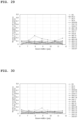
- Fig. 1 is a plot showing the spectral irradiance (vertical axis: the unit is W/m 2 /nm) and wavelength (horizontal axis: the unit is nm) of sunlight (AM1.5 and after corneal penetration) on the front surface of a crystalline lens.
- Fig. 2 is a plot showing the spectral irradiance (vertical axis: the unit is W/m 2 ) and wavelength (horizontal axis: the unit is nm) of each LED light (half width of about 20 nm) used in the experiment. Sunlight on the front surface of the crystalline lens for each bandwidth under the same conditions is also shown in the plots for reference.
- AM is referred to as air mass and is the ratio of the path of the sunlight to the path of sunlight in the case of being incident perpendicularly on the ground, which is a reference (AM1), and it is normal to employ AM1.5 in the latitude of Japan.
- the wavelengths were commonly set to 430 nm, and only the irradiance was changed.
- the irradiance used is the LED controller intensities of 100% (about 9 W/m 2 ), 75% (about 7.2 W/m 2 ) , 50% (about 5.1 W/m 2 ) , and 25% (about 2.7 W/m 2 ).
- the light irradiation time was set to 24 hours.
- the irradiance was set to 2.4 to 2.7 W/m 2 , and the lengths of the wavelengths were changed.
- the wavelengths used are five wavelengths of 430 nm, 450 nm, 470 nm, 505 nm, and 590 nm.
- the light irradiation time was set to 24 hours for the wavelengths of 430 nm and 450 nm and set to 48 hours for the wavelengths of 470 nm, 505 nm, and 590 nm.
- the relationship 1 between the wavelength and irradiance of light that decides whether or not a morphological change of an ocular cell (hereinafter, a lens epithelial cell will be exemplified) is caused for a subject was investigated.
- the relationship 1 may be a plot 1 that decides whether or not a morphological change of the ocular cell is caused in the relation between the wavelength and irradiance of the light.
- the relationship 1 is not limited to a relationship specified in the form of a plot and may be procured as data.
- Cell membrane staining was performed after light irradiation at a predetermined wavelength and a predetermined irradiance.
- the supernatant was removed using an aspirator, 0.5 mL of 0.1% CellMask Orange Plasma Membrane Stains (C10045, Life Technologies) was added to a culture medium, and the culture medium was placed still for 10 minutes under an atmospheric atmosphere in which the temperature was set to 37°C and the concentration of CO 2 was set to 5%. After that, the supernatant was removed with the aspirator, 2 mL of the medium was added thereto, and similarly, the culture medium was placed still for 10 minutes under an atmospheric atmosphere in which the temperature was set to 37°C and the concentration of CO 2 was set to 5%. This operation was repeated three times. After that, the cells were observed.
- Fig. 3 is photographs showing the observation results in the qualitative evaluation.
- the light irradiation conditions in Fig. 3(a) are a wavelength of 430 nm and an irradiance of 9.0 W/m 2
- the light irradiation conditions in Fig. 3(b) are a wavelength of 430 nm and an irradiance of 5.1 W/m 2
- the light irradiation conditions in Fig. 3(c) are a wavelength of 450 nm and an irradiance of 4.6 W/m 2 .
- the lens epithelial cell in Fig. 3(a) has been fragmented and shrank.
- the lens epithelial cell in Fig. 3(b) has been shrank. That is, in the lens epithelial cells in Fig. 3(a) and Fig. 3(b) , apoptosis has progressed.
- the morphology of the lens epithelial cell in Fig. 3(c) remains unchanged.
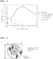
- Fig. 4 is a plot in which the morphological change of the lens epithelial cell is summarized by aspect in the relation between the irradiance (vertical axis: the unit is W/m 2 ) and the wavelength (horizontal axis: the unit is nm) of each LED light (half width of about 20 nm) used in the experiment. Sunlight on the front surface of the crystalline lens for each bandwidth under the same conditions is also shown in the plots for reference.
- a lens epithelial cell separately prepared from the lens epithelial cell used in the qualitative evaluation was cultured under the same conditions as those under which the lens epithelial cell for observation had been obtained in the qualitative evaluation, and a lens epithelial cell for the quantitative evaluation was obtained.
- a quantitative analysis of the cell peripheral shape was performed on this lens epithelial cell.
- the shape was measured using image analysis software (ImageJ) from a stained image of the cell captured using FSX100. Thirty cells were arbitrarily selected, the contour of each cell membrane was extracted manually, and the shape was measured.
- ImageJ image analysis software
- the area, circularity, and aspect ratio of the cell membrane were calculated.
- Fig. 5 is a view for describing the circularity and aspect ratio of the lens epithelial cell.
- S area
- L circumferential length
- L the circularity is indicated by 2 ⁇ S/L 2 in the present specification.
- a major axis of the lens epithelial cell elliptically approximated with ImageJ
- b minor axis
- metrics for evaluating whether or not a morphological change is caused are determined. That is, since the morphology of a cell that is used in a test varies with the kind, it is preferable to evaluate and determine the metrics for the morphological change in accordance with the kind of the cell.
- candidates of the following measurement items can be considered, and a plurality of these candidates can also be combined together.
- the microscopic image or the like is scaled into about two to five levels.
- the area and outer circumferential length of the cell and the distances between two points are measured by recognizing the difference between the cell portion and the background. At this time, measurement is performed on microscopic images in a plurality of arbitrary fields of view in consideration of a variation in the morphology of the cell.
- Measurement items to be used are selected based on the results (results in the initial state of the experiment) obtained by the initial preliminary feasibility study. At that time, measurement items with which a change can be acutely perceived based on the results obtained by the initial test light irradiation condition experiment in an ordinarily-known abnormal state are selected. Furthermore, the abnormal state in the selected measurement items is defined and used as the metrics. Based on the results of the initial light irradiation condition experiment, the light irradiation time for observing a morphological change, the downtime, the observation timing and the like are also determined.
- Table 2 below shows the invested rates of morphologically irregular cells in dark places (negative control).
- the state when morphologically irregular cells were 20%, the state was regarded as a state different from dark place conditions, that is, a state where phototoxicity occurs.
- the relationship 1 between the wavelength and irradiance of light that decides whether or not a morphological change of the lens epithelial cell is caused is clarified using the metrics.
- changes in the morphology are observed at a determined timing under conditions of different wavelengths and irradiance of light, and whether or not irregular morphology has occurred at each point is plotted.
- Such a plot is referred to as action spectrum.
- a range where irregular morphology is caused and a range where irregular morphology is not caused are roughly determined by the plot.
- a boundary between those ranges is temporarily determined, and a boundary is determined from the group of points that are positioned in the boundary.
- One example of a determination method is the least squares method or the like. This boundary is a plot showing the relationship 1.
- the boundary can be determined as described above since there is a principle that a stronger irregularity is caused as the wavelength of light is shorter (a chemical change of a substance is caused even when the energy of one photon having such a wavelength is large and the irradiance is low) and as the irradiance is higher.
- the evaluation has been described from the viewpoint of "whether or not irregular morphology is caused" compared with (based on) a state of no light irradiation; however, additionally, there are cases where there are light irradiation conditions under which a state where the morphology is estimated to be more favorable compared with that in the state of no light irradiation (a state of a favorable circularity of the cell or the like) is formed, and there is the relationship 1 under which no irregularity is caused and a more desirable state can be obtained.
- Quantitative evaluation was performed using the action spectrum.
- Fig. 6 is bar graphs showing the results of the quantitative evaluation
- Fig. 6(a) is a graph of the area S
- Fig. 6(b) is a graph of the circularity
- Fig. 6(c) is a graph of the aspect ratio.
- the results of the quantitative evaluation show that, as the irradiance was increased, the area of the lens epithelial cell increased in a plurality of samples, but decreased in another plurality of samples.
- the circularity increased as the irradiance increased.
- both the cell area and the circularity decrease due to apoptosis.
- the aspect ratio decreases or remains constant. Therefore, a change in the area or circularity in the quantitative evaluation described in the upper paragraphs is clearly distinctive from a behavior that is caused by apoptosis, which can be generally considered.
- the present inventors obtained information that an increase in the irradiance makes the active extension and contraction function of the lens epithelial cell deteriorate.
- the present inventors further advanced studies and investigated the relation between the irradiance and a change in the migration of the lens epithelial cell.
- the relationship 2 between the wavelength and irradiance of light that decides whether or not the migration of the lens epithelial cell is changed is also determined by the same steps as in the relationship 1. That is, metrics are determined first using the kind of the cell that has been determined to be used in all of the steps at first, and the relationship is then studied.
- the cell migration refers to the migration distance per time when a cell migrates on a polystyrene culture surface for tissue culture in the culture solution within a certain time (hereinafter, also referred to as "migrates in the culture medium").
- migrations in the culture medium the culture conditions for a cell under which the amount of migration of the cell can be observed or the measurement conditions of the migration distance are determined, and the cell is then observed over time to determine the cell migration per time.
- the inside of the medium is divided into blocks with a partition or the like, and a place where the cell has been sowed and a place where the cell is not sowed (gap) are made.
- a linear non-sowed region having a specific width, a round non-sowed region, and the like are formed.
- the partition or the like is removed in such a state, the measurement begins from that time, and the movement of the cell is then observed.
- the cell migration is appropriate, the cell migrates, and the cell non-sowed region (gap) decreases and is then blocked.
- the migration distance per time until the non-sowed region decreases and is blocked is referred to as the cell migration rate under the culture conditions.
- a cell non-sowed region (gap) having a predetermined width is formed on a surface having a large number of cells.
- the influence of light irradiation on the migration of the cell was investigated based on the blocking process of this gap.
- Fig. 7 is a schematic plan view of wells used to form a gap (scratch) having a predetermined width on a surface having a large number of cells.
- the culture-insert 2 well was removed one day after the sowing of the cells, and light irradiation was initiated after the formation of the gap was confirmed.
- the gap was captured using the inverted microscope at a magnification of four times immediately after the initiation of the light irradiation, six hours later, 12 hours later, 24 hours later and 48 hours later, and the gap was observed.
- the distances between the edges of the captured gap were measured using image analysis software (ImageJ). The measurement was performed on 10 arbitrary places at each time after the light irradiation, and the average value thereof was obtained and regarded as the distance between the edges of the gap.
- Fig. 8 is views showing the appearances of the blocking of the gap when the wavelength was 430 nm and the irradiance was 9.0 W/m 2
- Fig. 8(a) is a photograph showing the appearance of the gap after 0 hours (immediately after the initiation of the light irradiation)
- Fig. 8(b) is a photograph showing the appearance of the gap after 24 hours from the initiation of the light irradiation
- Fig. 8(c) is a photograph showing the appearance of the gap after 48 hours from the initiation of the light irradiation.
- Fig. 9 is views showing the appearances of the blocking of the gap when the wavelength was 430 nm and the irradiance was 5.1 W/m 2
- Fig. 9(a) is a photograph showing the appearance of the gap after 0 hours (immediately after the initiation of the light irradiation)
- Fig. 9(b) is a photograph showing the appearance of the gap after 24 hours from the initiation of the light irradiation
- Fig. 9(c) is a photograph showing the appearance of the gap after 48 hours from the initiation of the light irradiation.
- Fig. 10 is views showing the appearances of the blocking of the gap when the wavelength was 430 nm and the irradiance was 2.7 W/m 2
- Fig. 10(a) is a photograph showing the appearance of the gap after 0 hours (immediately after the initiation of the light irradiation)
- Fig. 10(b) is a photograph showing the appearance of the gap after 24 hours from the initiation of the light irradiation
- Fig. 10(c) is a photograph showing the appearance of the gap after 48 hours from the initiation of the light irradiation.
- Fig. 11 is views showing the appearances of the blocking of the gap when the wavelength was 470 nm and the irradiance was 2.5 W/m 2
- Fig. 11(a) is a photograph showing the appearance of the gap after 0 hours (immediately after the initiation of the light irradiation)
- Fig. 11(b) is a photograph showing the appearance of the gap after 24 hours from the initiation of the light irradiation
- Fig. 11(c) is a photograph showing the appearance of the gap after 48 hours from the initiation of the light irradiation.
- the size of the scale (horizontal line) in the photographs of Fig. 8 to Fig. 11 are 500 nm.
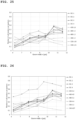
- Fig. 12 is a plot in which the relation between the distance (unit: ⁇ m) between the edges of the gap and the time from the initiation of the light irradiation (horizontal axis: the unit is h) is summarized for each wavelength.
- the slope of the plot is distance/time and indicates the migration rate of the lens epithelial cell.
- the present inventors found from the observation of the appearance of the gap blocking in each drawing that the decrease rate of the cell migration can be divided into several groups.
- Fig. 13 is a histogram showing the distribution of the decrease rates of the cell migration during the gap blocking.
- Fig. 14 is a plot in which the aspects of the morphological changes and the aspects of the cell migration changes of lens epithelial cells are grouped together in the relation between the irradiance (vertical axis: the unit is W/m 2 ) and wavelength (horizontal axis: the unit is nm) of each light source (half width: 10 nm) used in the experiment. Sunlight is also shown in the plots for reference.
- the leftmost dotted line plot in Fig. 14 is the relationship 1 between the wavelength and irradiance of light that decides whether or not a morphological change of the ocular cell is caused for the subject, and is specifically the plot 1 that decides whether or not a morphological change of the ocular cell is caused in the relation between the wavelength and irradiance of the light.
- the rightmost dotted line plot in Fig. 14 is the relationship 2 between the wavelength and irradiance of light that decides whether or not the migration of the ocular cell is changed for the subject, and is specifically a plot 2 that decides whether or not the migration of the ocular cell is changed in the relation between the wavelength and irradiance of the light.
- the plot 1 is on the short wavelength side of the plot 2,
- the center dotted line plot in Fig. 14 is a plot 3 that further classifies the region where a change in the cell migration is caused in the regions 2 depending on the degree of the change of the cell migration.
- the plots 1 to 3 are approximate curves that divide the respective regions. The approximate curve may be created by selecting two points closest to the boundary between individual regions adjacent to each other from each region, and in this case, the approximate curve becomes the boundary from a total of the four points.
- the plots in Fig. 14 are preferably obtained in advance before one embodiment of the present invention is carried out. That is, one example of the present invention may not include a step of creating the plots in Fig. 14 from the scratch. In addition, each plot in Fig. 14 becomes a straight line (approximate straight line) when expressed in logarithm.
- Fig. 14 What is found from Fig. 14 is that, for a certain person (subject), when light that is incident on the eye has a certain wavelength and certain irradiance, the light has a certain influence on ocular cells. As shown in Fig. 14 , if no light having a short wavelength is incident on the eyes, the influence on ocular cells is extremely small. Incidentally, the fact that no light having a short wavelength is incident on the eyes can be considered as the fact that no visible light having a short wavelength is incident on the eyes.
- a relationship acquisition step of acquiring the relationship 1 between the wavelength and irradiance of light that decides whether or not a morphological change of the ocular cell is caused and the relationship 2 between the wavelength and irradiance of light that decides whether or not the migration of the ocular cell is changed is performed (preferably in advance) on the subject.
- a relationship selection step of selecting at least any of the relationship 1 and the relationship 2 depending on a degree 1 that is required for the morphological change and a degree 2 that is required for the cell migration is performed.
- a determination step of determining a wavelength and irradiance of light to be incident on the subject's eye based on the selected relationship is performed. This determination step may also be referred to as a spectrum irradiance range determination step.
- the characteristics of light that is made incident on the eyes are determined by each of the above-described steps.
- Each of the above-described steps made light irradiation conditions under which a cell causes a certain abnormal state plotted and relationships thereof clarified. Next, which relationship to be based on is determined to determine the characteristics of light that is made incident on the eye.
- a thought that serves as the foundation of determination is whether an abnormal state that is caused in a cell is harmful or not to the subject. At a glance, it is considered to be most desirable that the abnormal state is never harmful and light under harmful irradiation conditions is fully removed. However, in actual cases, light having diverse and wide wavelength widths needs to be incident, and there is a basic principle that it is more desirable not to change ambient light unless it is obvious that the ambient light is clearly harmful in order to avoid taking a different undiscovered risk.
- the degree of which relationship is desirably based on in consideration of the degree 1 of the morphological change and the degree 2 that is required for the cell migration.
- the wavelength and irradiance of light to be incident on the eyes of the examinee are determined based on the selected relationship. In this determination, for example, which region in Fig. 14 the wavelength and irradiance of light need to belong to may be used as a reference.
- the plot 1 and the plot 2 are preferably selected.
- a region selection step of selecting one region from the regions 1 and selecting one region from the regions 2 is further provided after the relationship selection step.
- the region selection step it is preferable to perform at least (1) of the following (1) and (2).
- the wavelength and irradiance of light commonly belonging to the individual selected regions are obtained as the wavelength and irradiance of light that is to be incident on the subject's eyes.
- the plots 1 and 2 are preferably created based on regional information where the subject resides.
- "Regional information” may be, for example, information regarding the latitude such as AM1.5 or may be information regarding the weather.
- Information regarding the weather may be, for example, the annual average sunshine duration per day.
- the information may be the annual average global solar radiation or the like obtained by combining the above-described information.
- the ocular cell may be a lens epithelial cell, a retinal pigment epithelial cell, a corneal endothelial cell, a corneal epithelial stem cell, or a subconjunctival fibroblast.
- the lens epithelial cell has high phototoxicity sensitivity and consequently enables the effect of the present invention to be particularly significantly obtained.
- the subject is not limited and may be a cataract patient or may not be a cataract patient.
- the plot 3 that further classifies the region where a change of the cell migration is not caused instead of the region where the change is caused in the regions 2, depending on the degree of the change of the cell migration may be further obtained in the relationship acquisition step.
- Embodiment 1 For contents that will not be described in Embodiment 2 below, the content of Embodiment 1 is employed.
- Embodiment 2 in addition to the following two relationships for the subject, which have been described in Embodiment 1,
- a relationship acquisition step of obtaining at least any relationship out of the relationships 1 to 3 is performed (preferably in advance). It is preferable to obtain at least two relationships out of the relationships 1 to 3 since the characteristics of more preferable light can be determined.
- a relationship selection step of selecting at least any of the relationship 1, the relationship 2, and the relationship 3 depending on at least any of the degree 1 that is required for the morphological change, the degree 2 that is required for the cell migration, and the degree 3 that is required for the invasion is performed.
- a determination step of determining the wavelength and irradiance of light that is to be incident on the subject's eyes based on the selected relationship is performed. This determination step may also be referred to as a spectrum irradiance range determination step.
- the relationship 3 may be a plot 4 that decides whether or not the invasion of the ocular cell is increased in the relation between the wavelength and irradiance of the light.
- At least the relationship 3 may be obtained in advance, and in the relationship selection step, at least the relationship 3 may be selected. That is, the relationship 3 and the relationship 1 may be employed, or the relationship 3 and the relationship 2 may be employed.
- the relationships 1 to 3 may be obtained in advance, and in the relationship selection step, the relationships 1 to 3 may be selected.
- the plot 4 classifies a region into a plurality of regions 4 composed of a region where an increase of the invasion is caused and a region where an increase of the invasion is not caused,
- the plot 4 may be on the short wavelength side of the plot 3 and may be on the long wavelength side of the plot 2.
- Embodiment 2 invasion specific to Embodiment 2 will be described in detail.
- the contents regarding the morphological change and the cell migration described in Embodiment 1 may be ascertained and region-selected as the relationships together with the following content of the invasion, which is preferable.
- human lens epithelial cells SRA01/04 (RCB1591, RIKEN BRC) were used.
- Reagents are the culture solutions used in Embodiment 1.
- the cell density was set to 2 ⁇ 10 4 cells/dish.
- a device used for the light irradiation is a xenon light source (MAX-350, ASAHI SPECTRA). Light irradiation conditions are as follows.
- Light irradiation was performed 15 cm above the cell culture surface under a cell culture environment (under an atmospheric atmosphere in which the air temperature was set to 37°C and the concentration of CO 2 was set to 5%). At that time, it was expected that the influence of the light irradiation on the cells varied with the intensity of irradiation light even when the wavelength was the same. Therefore, individual experiments were performed with the intensity changed for individual experiments with the controller of the light source while using a plurality of band path filters as the xenon light source. The conditions for irradiance at each wavelength are shown in Table 3. The light irradiation time was set to 24 hours.
- the relationship 3 between the wavelength and irradiance of light that decides whether or not the invasion of the lens epithelial cell is increased is determined by the same steps as in the relationship 1. That is, metrics are determined first using the kind of the cell that has been determined to be used in all of the steps at first, and the relationship is then studied.
- a mold in which a convex stripe that is a straight line in a plan view has been patterned is prepared.
- Polydimethylsiloxane (PDMS) is poured into this mold, and a groove that corresponds to the convex stripe of the mold is formed in the PDMS.
- the PDMS is released from the mold, and a PDMS substrate on which the groove has been formed is obtained.
- An ocular cell is disposed in front of the groove on the PDMS substrate, and the fact that the ocular cell intrudes into the groove from the inlet of the groove (hereinafter, referred to as "opening portion") is regarded as invasion in the present specification.
- a plurality of grooves having an equal width are provided on the PDMS substrate.
- the following widths of grooves were prepared for each PDMS substrate.
- the depth of the grooves and the groove disposition intervals were set as follows.
- the ocular cells were classified into the following four categories.
- Fig. 15 is plan views showing the appearances of the ocular cells passing through the opening portions at the time of no light irradiation NC
- Fig. 15(a) is a photograph showing the appearance after 0 hours (immediately after the initiation of light irradiation)
- Fig. 15(b) is a photograph showing the appearance after 10 minutes from the initiation of light irradiation
- Fig. 15(c) is a photograph showing the appearance after 20 minutes from the initiation of light irradiation
- Fig. 15(d) is a photograph showing the appearance after 30 minutes from the initiation of light irradiation
- Fig. 15(e) is a photograph showing the appearance after 40 minutes from the initiation of light irradiation
- Fig. 15(f) is a photograph showing the appearance after 50 minutes from the initiation of light irradiation.
- Fig. 16 is plan views showing the appearances of the ocular cells invading (into the grooves) at the time of no light irradiation NC
- Fig. 16(a) is a photograph showing the appearance after 0 hours (immediately after the initiation of light irradiation)
- Fig. 16(b) is a photograph showing the appearance after 10 minutes from the initiation of light irradiation
- Fig. 16(c) is a photograph showing the appearance after 20 minutes from the initiation of light irradiation
- Fig. 16(d) is a photograph showing the appearance after 30 minutes from the initiation of light irradiation
- Fig. 16(e) is a photograph showing the appearance after 40 minutes from the initiation of light irradiation
- Fig. 16(f) is a photograph showing the appearance after 50 minutes from the initiation of light irradiation.
- Fig. 17 is plan views showing the appearances of the ocular cells performing the incomplete invasion behavior (failed invasion) at the time of no light irradiation NC
- Fig. 17(a) is a photograph showing the appearance after 0 hours (immediately after the initiation of light irradiation)
- Fig. 17(b) is a photograph showing the appearance after 10 minutes from the initiation of light irradiation
- Fig. 17(c) is a photograph showing the appearance after 20 minutes from the initiation of light irradiation
- Fig. 17(d) is a photograph showing the appearance after 30 minutes from the initiation of light irradiation
- Fig. 17 is plan views showing the appearances of the ocular cells performing the incomplete invasion behavior (failed invasion) at the time of no light irradiation NC
- Fig. 17(a) is a photograph showing the appearance after 0 hours (immediately after the initiation of light irradiation)
- Fig. 17(b) is
- FIG. 17(e) is a photograph showing the appearance after 40 minutes from the initiation of light irradiation
- Fig. 17(f) is a photograph showing the appearance after 50 minutes from the initiation of light irradiation
- Fig. 17(g) is a photograph showing the appearance after 60 minutes from the initiation of light irradiation
- Fig. 17(h) is a photograph showing the appearance after 70 minutes from the initiation of light irradiation.
- Fig. 18 is plan views showing the appearances of the ocular cells invading (and crawling from the grooves) at the time of no light irradiation NC
- Fig. 18(a) is a photograph showing the appearance after 0 hours (immediately after the initiation of light irradiation)
- Fig. 18(b) is a photograph showing the appearance after 10 minutes from the initiation of light irradiation
- Fig. 18(c) is a photograph showing the appearance after 20 minutes from the initiation of light irradiation
- Fig. 18(d) is a photograph showing the appearance after 30 minutes from the initiation of light irradiation
- Fig. 18(a) is a photograph showing the appearance after 0 hours (immediately after the initiation of light irradiation)
- Fig. 18(b) is a photograph showing the appearance after 10 minutes from the initiation of light irradiation
- Fig. 18(c) is a photograph showing the appearance after 20
- FIG. 18(e) is a photograph showing the appearance after 40 minutes from the initiation of light irradiation
- Fig. 18(f) is a photograph showing the appearance after 50 minutes from the initiation of light irradiation
- Fig. 18(g) is a photograph showing the appearance after 60 minutes from the initiation of light irradiation.
- Fig. 19 is a view showing plots NC-1, NC-2, NC-3 and NC-4 wherein the number of cells invading (into grooves) is indicated along the vertical axis and the width w ( ⁇ m) of the groove is indicated along the horizontal axis.
- Fig. 20 is a view showing the plots NC-1, NC-2, NC-3 and NC-4 and the results at wavelengths of 430 to 460 nm when the number of cells invading (into grooves) is indicated along the vertical axis and the width w ( ⁇ m) of the groove is indicated along the horizontal axis.
- Fig. 21 is a view showing the plots NC-1, NC-2, NC-3 and NC-4 and the results at a wavelength of 480 nm when the number of cells invading (into grooves) is indicated along the vertical axis and the width w ( ⁇ m) of the groove is indicated along the horizontal axis.
- Fig. 22 is a view showing the plots NC-1, NC-2, NC-3 and NC-4 and the results at a wavelength of 500 nm when the number of cells invading (into grooves) is indicated along the vertical axis and the width w ( ⁇ m) of the groove is indicated along the horizontal axis.
- Fig. 23 is a view showing the plots NC-1, NC-2, NC-3 and NC-4 and the results at a wavelength of 520 nm when the number of cells invading (into grooves) is indicated along the vertical axis and the width w ( ⁇ m) of the groove is indicated along the horizontal axis.
- Fig. 24 is a view showing the plots NC-1, NC-2, NC-3 and NC-4 and the results at a wavelength of 540 nm when the number of cells invading (into grooves) is indicated along the vertical axis and the width w ( ⁇ m) of the groove is indicated along the horizontal axis.
- the following views are results obtained by arranging the numbers of cells invading (into grooves) according to the irradiance.
- Fig. 25 is a view showing the results when the number of cells invading (into grooves) is indicated along the vertical axis and the width w ( ⁇ m) of the groove is indicated along the horizontal axis in the case of low irradiance of 1.4 to 1.5 W/m 2 .
- Fig. 26 is a view showing the results when the number of cells invading (into grooves) is indicated along the vertical axis and the width w ( ⁇ m) of the groove is indicated along the horizontal axis in the case of intermediate irradiance of 3.2 to 5.5 W/m 2 .
- Fig. 27 is a view showing the results when the number of cells invading (into grooves) is indicated along the vertical axis and the width w ( ⁇ m) of the groove is indicated along the horizontal axis in the case of high irradiance of 7.5 W/m 2 or more.
- the following views are results obtained by arranging the influence of light irradiation with attention paid to the number of cells passing and the number of cells of incomplete invasion.
- Fig. 28 is a view showing the results when the number of cells passing is indicated along the vertical axis and the width w ( ⁇ m) of the groove is indicated along the horizontal axis.
- Fig. 29 is a view showing the results when the number of cells of incomplete invasion (failed invasion) is indicated along the vertical axis and the width w ( ⁇ m) of the groove is indicated along the horizontal axis.
- Fig. 30 is a view showing the results when the number of cells invading (crawling from grooves) is indicated along the vertical axis and the width w ( ⁇ m) of the groove is indicated along the horizontal axis.
- the invasion can be increased at a slightly low irradiance of 20.1 W/m 2 , and it is thus preferable to determine the irradiance at a wavelength of 540 nm in the above-described characteristics to be low.
- the width w ( ⁇ m) of the groove into which cells are likely to invade varies depending on the size or hardness (easiness of deformation).
- the relationship between the wavelength and irradiance of light that decides whether or not the invasion is increased is preferably evaluated with respect to a plurality of grooves having a width smaller than the diameter of a cell, which is the evaluation subject, in a state where the cell has not been deformed, for example, a floating state.
- Embodiment 3 For contents that will not be described in Embodiment 3 below, the contents of Embodiments 1 and 2 are employed.
- Embodiment 3 in addition to the human cell line (LEC, cell line) in Embodiments 1 and 2, experiment results with respect to human primary lens epithelial cells (LECs, primary) and retinal pigment epithelial cells (RPEs) will be described.
- Fig. 31 is plots of aspects of changes in the invasion of a lens epithelial cell in the relation between the irradiance (vertical axis: the unit is W/m 2 ) and the wavelength (horizontal axis: the unit is nm) of each light source (half width: 10 nm) used in the experiments. Sunlight is also shown in the plots for reference.
- Fig. 31A is a view when the width w of the groove was set to 8.9 ⁇ m
- Fig. 31B is a view when the width w of the groove was set to 9.6 ⁇ m.
- the upper side of the V-like boundary in Fig. 31 shows the increase, and the lower side shows no changes in the invasion or the suppression.
- the boundary is a boundary between the increase and the low effect in each NC plot and is a line obtained by the least squares method.
Landscapes
- Health & Medical Sciences (AREA)
- Life Sciences & Earth Sciences (AREA)
- Engineering & Computer Science (AREA)
- Biomedical Technology (AREA)
- General Health & Medical Sciences (AREA)
- Ophthalmology & Optometry (AREA)
- Veterinary Medicine (AREA)
- Heart & Thoracic Surgery (AREA)
- Medical Informatics (AREA)
- Molecular Biology (AREA)
- Surgery (AREA)
- Animal Behavior & Ethology (AREA)
- Biophysics (AREA)
- Public Health (AREA)
- Physics & Mathematics (AREA)
- Biotechnology (AREA)
- Wood Science & Technology (AREA)
- Organic Chemistry (AREA)
- Chemical & Material Sciences (AREA)
- Bioinformatics & Cheminformatics (AREA)
- Genetics & Genomics (AREA)
- Zoology (AREA)
- Neurosurgery (AREA)
- Microbiology (AREA)
- Cell Biology (AREA)
- Biochemistry (AREA)
- General Engineering & Computer Science (AREA)
- Neurology (AREA)
- Prostheses (AREA)
- Investigating Or Analysing Biological Materials (AREA)
Applications Claiming Priority (2)
| Application Number | Priority Date | Filing Date | Title |
|---|---|---|---|
| JP2022056705 | 2022-03-30 | ||
| PCT/JP2023/007185 WO2023189098A1 (ja) | 2022-03-30 | 2023-02-28 | 光の特性の決定方法 |
Publications (1)
| Publication Number | Publication Date |
|---|---|
| EP4502142A1 true EP4502142A1 (en) | 2025-02-05 |
Family
ID=88200506
Family Applications (1)
| Application Number | Title | Priority Date | Filing Date |
|---|---|---|---|
| EP23779128.0A Pending EP4502142A1 (en) | 2022-03-30 | 2023-02-28 | Method for determining properties of light |
Country Status (5)
| Country | Link |
|---|---|
| US (1) | US20250176823A1 (https=) |
| EP (1) | EP4502142A1 (https=) |
| JP (1) | JPWO2023189098A1 (https=) |
| CN (1) | CN118984869A (https=) |
| WO (1) | WO2023189098A1 (https=) |
Families Citing this family (3)
| Publication number | Priority date | Publication date | Assignee | Title |
|---|---|---|---|---|
| WO2024204157A1 (ja) * | 2023-03-27 | 2024-10-03 | ホヤ レンズ タイランド リミテッド | 眼鏡レンズの供給システム、眼鏡レンズの供給方法、眼鏡レンズの設計方法、眼鏡レンズの製造方法、及び眼鏡レンズの供給システム用のプログラム |
| CN120917370A (zh) * | 2023-03-27 | 2025-11-07 | 豪雅镜片泰国有限公司 | 眼镜透镜的供给系统、眼镜透镜的供给方法以及眼镜透镜的供给系统用程序 |
| JPWO2024204159A1 (https=) * | 2023-03-27 | 2024-10-03 |
Family Cites Families (4)
| Publication number | Priority date | Publication date | Assignee | Title |
|---|---|---|---|---|
| EP2602653B1 (en) * | 2011-12-08 | 2020-09-16 | Essilor International | Method of determining the configuration of an ophthalmic filter |
| ES2478693B1 (es) * | 2012-12-21 | 2015-04-29 | Universidad Complutense De Madrid | Elemento bloqueante de longitudes de onda corta en fuentes de iluminación de tipo led |
| EP3191040B1 (en) * | 2014-09-09 | 2020-07-15 | Lumithera, Inc. | Multi-wavelength phototherapy devices for the non-invasive treatment of damaged or diseased tissue |
| JP7337360B2 (ja) * | 2020-02-21 | 2023-09-04 | イリスコミュニケーション株式会社 | 視覚特性の検査方法、光学フィルタの特性決定方法、光学フィルタ、視覚特性検査用の光学素子セット、および視覚特性の検査用画像 |
-
2023
- 2023-02-28 EP EP23779128.0A patent/EP4502142A1/en active Pending
- 2023-02-28 WO PCT/JP2023/007185 patent/WO2023189098A1/ja not_active Ceased
- 2023-02-28 US US18/843,411 patent/US20250176823A1/en active Pending
- 2023-02-28 CN CN202380026204.XA patent/CN118984869A/zh active Pending
- 2023-02-28 JP JP2024511502A patent/JPWO2023189098A1/ja active Pending
Also Published As
| Publication number | Publication date |
|---|---|
| WO2023189098A1 (ja) | 2023-10-05 |
| JPWO2023189098A1 (https=) | 2023-10-05 |
| CN118984869A (zh) | 2024-11-19 |
| US20250176823A1 (en) | 2025-06-05 |
Similar Documents
| Publication | Publication Date | Title |
|---|---|---|
| EP4502142A1 (en) | Method for determining properties of light | |
| McLaren et al. | Objective assessment of the corneal endothelium in Fuchs' endothelial dystrophy | |
| Pollreisz et al. | Visualizing melanosomes, lipofuscin, and melanolipofuscin in human retinal pigment epithelium using serial block face scanning electron microscopy | |
| DOUGHTY | Toward a quantitative analysis of corneal endothelial cell morphology: a review of techniques and their application | |
| Nubile et al. | In vivo confocal microscopy in diagnosis of limbal stem cell deficiency | |
| Wilkerson et al. | Melanocytes and iris color: light microscopic findings | |
| Curcio et al. | A whole mount method for sequential analysis of photoreceptor and ganglion cell topography in a single retina | |
| Lagali et al. | Stage-related central corneal epithelial transformation in congenital aniridia-associated keratopathy | |
| Dorey et al. | Lipofuscin in aged and AMD eyes | |
| Taurone et al. | Age related changes seen in human cornea in formalin fixed sections and on biomicroscopy in living subjects: A comparison | |
| Pedrotti et al. | In vivo confocal microscopy 1 year after autologous cultured limbal stem cell grafts | |
| von der Emde et al. | Histologic cell shape descriptors for the retinal pigment epithelium in age-related macular degeneration: A comparison to unaffected eyes | |
| Meleppat et al. | In situ morphologic and spectral characterization of retinal pigment epithelium organelles in mice using multicolor confocal fluorescence imaging | |
| Safi et al. | Semiquantitative criteria in the eye bank that correlate with cornea guttata in donor corneas | |
| Reichenbach et al. | Development of the rabbit retina. IV. Tissue tensility and elasticity in dependence on topographic specializations | |
| Simon et al. | Prolonged lifetimes of histologic autofluorescence in ectopic retinal pigment epithelium in age-related macular degeneration | |
| Borderie et al. | New parameters in assessment of human donor corneal stroma | |
| Costello et al. | Ultrastructural analysis of damage to nuclear fiber cell membranes in advanced age-related cataracts from India | |
| Pande et al. | Correlations between aqueous flare and cells and lens surface cytology in eyes with poly (methyl methacrylate) and heparin-surface-modified intraocular lenses | |
| Acquart et al. | Endothelial morphometry by image analysis of corneas organ cultured at 31 C | |
| Gisselberg et al. | A quantitative evaluation of Fourier components in transparent and opaque calf cornea | |
| Blades et al. | Pilot study on the use of impression cytology specimens for quantitative assessment of the surface area of bulbar conjunctival cells | |
| CN109614855B (zh) | 基于图像灰度值计算分析的后发性白内障分析装置及方法 | |
| Chylack Jr | Classification of human cataractous change by the American Cooperative Cataract Research Group method | |
| RU2326582C1 (ru) | Способ диагностики возрастной катаракты |
Legal Events
| Date | Code | Title | Description |
|---|---|---|---|
| STAA | Information on the status of an ep patent application or granted ep patent |
Free format text: STATUS: THE INTERNATIONAL PUBLICATION HAS BEEN MADE |
|
| PUAI | Public reference made under article 153(3) epc to a published international application that has entered the european phase |
Free format text: ORIGINAL CODE: 0009012 |
|
| STAA | Information on the status of an ep patent application or granted ep patent |
Free format text: STATUS: REQUEST FOR EXAMINATION WAS MADE |
|
| 17P | Request for examination filed |
Effective date: 20240812 |
|
| AK | Designated contracting states |
Kind code of ref document: A1 Designated state(s): AL AT BE BG CH CY CZ DE DK EE ES FI FR GB GR HR HU IE IS IT LI LT LU LV MC ME MK MT NL NO PL PT RO RS SE SI SK SM TR |
|
| DAV | Request for validation of the european patent (deleted) | ||
| DAX | Request for extension of the european patent (deleted) |