EP4498375A2 - Systeme und verfahren zur verwendung neuronaler netzwerke für keimbahn- und somatische variantenaufrufe - Google Patents
Systeme und verfahren zur verwendung neuronaler netzwerke für keimbahn- und somatische variantenaufrufe Download PDFInfo
- Publication number
- EP4498375A2 EP4498375A2 EP24180495.4A EP24180495A EP4498375A2 EP 4498375 A2 EP4498375 A2 EP 4498375A2 EP 24180495 A EP24180495 A EP 24180495A EP 4498375 A2 EP4498375 A2 EP 4498375A2
- Authority
- EP
- European Patent Office
- Prior art keywords
- tumor
- sequence
- normal
- data
- matrix
- Prior art date
- Legal status (The legal status is an assumption and is not a legal conclusion. Google has not performed a legal analysis and makes no representation as to the accuracy of the status listed.)
- Pending
Links
Images


Classifications
-
- G—PHYSICS
- G16—INFORMATION AND COMMUNICATION TECHNOLOGY [ICT] SPECIALLY ADAPTED FOR SPECIFIC APPLICATION FIELDS
- G16B—BIOINFORMATICS, i.e. INFORMATION AND COMMUNICATION TECHNOLOGY [ICT] SPECIALLY ADAPTED FOR GENETIC OR PROTEIN-RELATED DATA PROCESSING IN COMPUTATIONAL MOLECULAR BIOLOGY
- G16B20/00—ICT specially adapted for functional genomics or proteomics, e.g. genotype-phenotype associations
- G16B20/20—Allele or variant detection, e.g. single nucleotide polymorphism [SNP] detection
-
- G—PHYSICS
- G06—COMPUTING OR CALCULATING; COUNTING
- G06N—COMPUTING ARRANGEMENTS BASED ON SPECIFIC COMPUTATIONAL MODELS
- G06N3/00—Computing arrangements based on biological models
- G06N3/02—Neural networks
- G06N3/04—Architecture, e.g. interconnection topology
- G06N3/0464—Convolutional networks [CNN, ConvNet]
-
- G—PHYSICS
- G06—COMPUTING OR CALCULATING; COUNTING
- G06N—COMPUTING ARRANGEMENTS BASED ON SPECIFIC COMPUTATIONAL MODELS
- G06N3/00—Computing arrangements based on biological models
- G06N3/02—Neural networks
- G06N3/08—Learning methods
-
- G—PHYSICS
- G06—COMPUTING OR CALCULATING; COUNTING
- G06N—COMPUTING ARRANGEMENTS BASED ON SPECIFIC COMPUTATIONAL MODELS
- G06N3/00—Computing arrangements based on biological models
- G06N3/02—Neural networks
- G06N3/08—Learning methods
- G06N3/09—Supervised learning
-
- G—PHYSICS
- G16—INFORMATION AND COMMUNICATION TECHNOLOGY [ICT] SPECIALLY ADAPTED FOR SPECIFIC APPLICATION FIELDS
- G16B—BIOINFORMATICS, i.e. INFORMATION AND COMMUNICATION TECHNOLOGY [ICT] SPECIALLY ADAPTED FOR GENETIC OR PROTEIN-RELATED DATA PROCESSING IN COMPUTATIONAL MOLECULAR BIOLOGY
- G16B30/00—ICT specially adapted for sequence analysis involving nucleotides or amino acids
- G16B30/10—Sequence alignment; Homology search
-
- G—PHYSICS
- G16—INFORMATION AND COMMUNICATION TECHNOLOGY [ICT] SPECIALLY ADAPTED FOR SPECIFIC APPLICATION FIELDS
- G16B—BIOINFORMATICS, i.e. INFORMATION AND COMMUNICATION TECHNOLOGY [ICT] SPECIALLY ADAPTED FOR GENETIC OR PROTEIN-RELATED DATA PROCESSING IN COMPUTATIONAL MOLECULAR BIOLOGY
- G16B40/00—ICT specially adapted for biostatistics; ICT specially adapted for bioinformatics-related machine learning or data mining, e.g. knowledge discovery or pattern finding
- G16B40/20—Supervised data analysis
Definitions
- Embodiments of the invention relate generally to systems and methods for variant calling, and more particularly to using machine learning algorithms for variant calling.
- Identifying genetic mutations using sequencing data is a crucial task in bioinformatics that has implications in diagnosis, prognosis, and treatment of multiple diseases including cancer.
- the task is a non-trivial one, especially when the sequencing technology used to generate the sequencing data has a high-error rate (which is common to single-molecule sequencing technologies) or when the mutations occur at low frequency (which is particularly true for cancer mutations) or in complex genomic regions.
- the low frequency of cancer mutations in a sequencing sample could be due to sample contamination (since tumor samples may contain some DNA from normal cells) or due to tumor heterogeneity.
- Deep learning is a machine learning technique that uses multiple data layers to implicitly capture the data signatures as well as the statistical model, as long as the right training data is available. This makes deep learning a potentially appealing solution in terms of generalizability for the mutation detection problem.
- Several deep learning solutions for classifying germline mutations are known.
- Poplin, R. et al. (“Creating a Universal SNP and Small Indel Variant Caller With Deep Neural Networks", bioRxiv, 092890 (2016 )), proposes a method where candidate variant locations are first identified by the genome scan, and then a pileup image of reads aligned around each identified candidate are provided as inputs into a convolutional neural network (CNN), which then predicts the genotype of the candidate variants.
- CNN convolutional neural network
- Luo, R. et al. (“Clairvoyante: a Multi-task Convolutional Deep Neural Network For Variant Calling In Single Molecule Sequencing. bioRxiv, 310458 (2018 )) uses a data summarization approach, which includes scanning the genome to identify the candidate variant sites, and preparing for each candidate position multiple input matrices, one matrix for each type of variant. A similar technique is discussed in Chin, J.
- Torracinta R. et al.
- Adaptive somatic mutations calls with deep learning and semi-simulated data. bioRxiv, 79087 (2016 )
- a system having fully-connected layers only cannot leverage the power provided by convolutional neural networks, which is to learn feature representations directly from the raw sequence data using patterns seen in local genomic context of candidate variants.
- fully-connected networks are more complex, that approach allows for less generalizability and scalability than that enabled by the use of CNNs.
- the present invention relates generally systems and methods for variant calling, and more particularly to using machine learning algorithms for variant calling.
- a method for germline variant calling can include obtaining a reference sequence, a plurality of sequence reads, and a position of a candidate variant within the sequence reads; obtaining augmented sequence reads by inserting one or more spaces in one or more sequence reads; obtaining an augmented reference sequence by inserting one or more spaces in the reference sequence; converting a segment of the augmented sequence reads around the candidate variant into a sample matrix; converting a segment of the augmented reference sequence around the candidate variant into a reference matrix; providing the sample matrix and the reference matrix to a trained neural network; and obtaining, at the output of the trained neural network, prediction data related to a variant within the plurality of sequence reads.
- the method further includes detecting one or more inserted bases within the plurality of sequence reads, wherein augmenting the sequence reads and the reference sequence includes: for each inserted base detected in any of the sequence reads, inserting a space in the reference sample at the position of the inserted base.
- the method further includes: for each inserted base detected in any of the sequence reads, inserting a space at the position of the inserted base in every sequence read for which no insertions were detected at the position of the inserted base.
- the sample matrix includes at least four lines representing four types of nucleotide bases, each line representing the number of bases of the respective nucleotide base type at different positions within the segment of the augmented sequence reads; and at least one line representing the number of inserted spaces at different positions within the segment of the augmented sequence reads.
- the reference matrix has the same dimensions as the sample matrix and wherein the reference matrix provides a complete representation of the locations of different nucleotide bases and spaces within the augmented reference sequence.
- the trained neural network comprises a trained convolutional neural network.
- the method further includes providing to the trained neural network at least one of: a variant position matrix representing the candidate variant's position within the segment of the augmented sequence reads; a coverage matrix representing coverage or depth of the segment of the augmented sequence reads; an alignment feature matrix representing an alignment feature of the augmented sequence reads; a knowledgeable base matrix representing information about publicly known information about one or more variants.
- the prediction data related to the variant comprises at least one of: a predicted type of the variant; a predicted position of the variant; a predicted length of the variant; and a predicted genotype of the variant.
- the prediction data related to the variant includes a predicted type of the variant
- the neural network is configured to produce one of a plurality of values for predicted type of the variant, the plurality of values including: a first value indicating a probability that the variant is a false positive; a second value indicating a probability that the variant is a single-nucleotide- polymorphism variant; a third value indicating a probability that the variant is a deletion variant; and a fourth value indicating a probability that the variant is an insertion variant.
- a method for somatic variant calling can include obtaining a plurality of normal sequence reads and a plurality of tumor sequence reads; converting a segment of the normal sequence reads and a segment of the tumor sequence reads into a normal sample matrix and a tumor sample matrix, respectively; feeding the normal sample matrix and the tumor sample matrix into a trained convolutional neural network; and obtaining, at the output of the trained convolutional neural network, a predicted type of a somatic variant within the plurality of tumor sequence reads.
- the plurality of tumor sequence reads represent genetic information of a patient's tumor sample
- the plurality of normal sequence reads represent genetic information of the patient's normal sample
- converting the segment of the normal sequence reads into the normal sample matrix includes augmenting the segment of the normal sequence reads by inserting one or more spaces in one or more normal sequence reads; and converting the segment of the tumor sequence reads into the tumor sample matrix comprises augmenting the segment of the tumor sequence reads by inserting one or more spaces in one or more tumor sequence reads.
- the tumor sample matrix includes at least one line for each nucleotide base type, each line representing the number of occurrences of the respective nucleotide base type at each position within the segment of the tumor sequence reads; and at least one line representing the number of inserted spaces at each position within the segment of the tumor sequence reads.
- the method further includes providing to the trained convolutional neural network one or more matrices representing one or more features obtained from one or more other variant callers that have analyzed the plurality of tumor sequence reads and/or the plurality of normal sequence reads.
- the method further includes obtaining a reference sequence; converting the reference sequence into a reference matrix; and feeding the reference matrix into the trained convolutional matrix along with the normal sample matrix and the tumor sample matrix.
- a non-transitory computer-readable medium including instructions which, when executed by one or more processors of a computing system, causes the computing system to perform operations including: obtaining a plurality of normal sequence reads and a plurality of tumor sequence reads; converting a segment of the normal sequence reads and a segment of the tumor sequence reads into a normal sample matrix and a tumor sample matrix, respectively; feeding the normal sample matrix and the tumor sample matrix into a trained convolutional neural network; and obtaining, at the output of the trained convolutional neural network, a predicted type of a somatic variant within the plurality of normal sequence reads.
- a computing system including one or more processors and coupled to one or more non- transitory computer-readable memories storing instructions which, when executed by the computing system, cause the computing system to perform operations including: obtaining a plurality of tumor sequence reads; obtaining augmented tumor sequence reads by inserting one or more spaces in one or more tumor sequence reads; converting a segment of the tumor sequence reads into a tumor sample matrix; feeding the normal sample matrix and the tumor sample matrix into a trained neural network; and obtaining, at the output of the trained neural network, a predicted type of a somatic variant within the plurality of tumor sequence reads.
- a method for variant calling can include: obtaining a reference sequence and a plurality of sequence reads; optionally performing a first alignment of the plurality of sequence reads with the reference sequence, unless the obtained plurality of sequence reads and reference sequence are obtained in an already aligned configuration; identifying a candidate variant position from the aligned sequence reads and reference sequence; augmenting the sequence reads and/or the reference sequence around the candidate variant position to achieve a second alignment of the plurality of sequence reads with the reference sequence; generating a reference matrix for the candidate variant position from the augmented reference sequence and a sample matrix for the candidate variant position from the plurality of augmented sequence reads; inputting the reference matrix and the sample matrix into a neural network; and determining with the neural network whether a variant type exists at the candidate variant position.
- the step of augmenting the sequence reads and/or the reference sequence includes introducing one or more spaces to the sequence reads and/or the reference sequence to account for insertions and/or deletions in the sequence reads.
- the method further includes generating a plurality of training matrices from a training dataset, wherein the training matrices have a structure that corresponds to the sample matrix and the reference matrix, wherein the training dataset includes sequence data that comprises a plurality of mutations, the mutations including single nucleotide variants, insertions, and deletions; and training the neural network with the plurality of training matrices.
- the training dataset includes a plurality of subsets, wherein each subset includes a tumor purity level ranging from 0% to 100%, wherein at least two of the subsets each has a different tumor purity level.
- At least three of the subsets each has a different tumor purity level.
- the plurality of subsets includes a first subset with a tumor purity level less than about 30%, a second subset with a tumor purity level between about 30% and 70%, and a third subset with a third tumor purity level of at least about 70%.
- the plurality of subsets includes a first subset with a tumor purity level less than about 40%, a second subset with a tumor purity level between about 40% and 60%, and a third subset with a tumor purity level of at least about 60%.
- the plurality of subsets includes a subset with a tumor purity level of less than about 10%.
- the plurality of subsets includes a subset with a tumor purity level of less than about 5%.
- the training dataset includes synthetic data.
- the synthetic data includes artificially generated mutations, wherein the artificially generated mutations comprise single nucleotide variants, insertions, and deletions.
- the training dataset includes real data, wherein the real data includes real mutations, wherein the real mutations include single nucleotide variants, insertions, and deletions.
- the training dataset includes a plurality of subsets, wherein each subset includes a variant allele frequency ranging from 0% to 100%, wherein at least two of the subsets each has a different variant allele frequency level.
- At least three of the subsets each has a different variant allele frequency level.
- At least one of the subsets has a variant allele frequency of at least 2.5%.
- At least one of the subsets has a variant allele frequency of at least 5%.
- At least one of the subsets has a variant allele frequency of at least 10%.
- the method further includes inputting at least one prediction from at least one mutation calling algorithm into the neural network.
- the at least one prediction includes at least three predictions from at least three separate mutation calling algorithms.
- the at least one prediction includes at least five predictions from at least five separate mutation calling algorithms.
- the training dataset includes a mixture of synthetic data and real data.
- the training dataset includes at least 5% synthetic data.
- the training dataset includes at least 10% synthetic data.
- the training dataset includes whole genome sequencing data.
- the training dataset includes whole exome sequencing data.
- the training dataset includes targeted sequencing data.
- the training dataset includes data obtained from a formalin-fixed paraffin-embedded sample.
- the training dataset includes at least two of whole genome sequencing data, whole exome sequencing data, targeted sequencing data, and data obtained from a formalin-fixed paraffin-embedded sample.
- the training dataset includes at least three of whole genome sequencing data, whole exome sequencing data, targeted sequencing data, and data obtained from a formalin-fixed paraffin-embedded sample.
- the training dataset includes whole genome sequencing data, whole exome sequencing data, targeted sequencing data, and data obtained from a formalin-fixed paraffin-embedded sample.
- a method for variant calling can include obtaining a reference sequence, a plurality of tumor sequence reads, and a plurality of normal sequence reads; optionally performing a first alignment of the plurality of tumor sequence reads and the plurality of normal sequence reads with the reference sequence, unless the obtained plurality of tumor sequence reads and the plurality of normal sequence reads and the reference sequence are obtained in an already aligned configuration; identifying a candidate variant position from the aligned tumor sequence reads, normal sequence reads, and reference sequence; augmenting the tumor sequence reads and/or the normal sequence reads, and/or the reference sequence around the candidate variant position to achieve a second alignment of the plurality of tumor sequence reads and the plurality of normal sequence reads with the reference sequence; generating a reference matrix for the candidate variant position from the augmented reference sequence and a tumor matrix for the candidate variant position from the plurality of augmented tumor sequence reads and a normal matrix for the candidate variant position from the plurality of augmented normal sequence reads; inputting the
- the method further includes generating a plurality of training matrices from a training dataset, wherein the training matrices have a structure that corresponds to the tumor matrix, normal matrix and the reference matrix, wherein the training dataset includes tumor sequence data and normal sequence data; and training the neural network with the plurality of training matrices.
- both the tumor sequence data and the normal sequence data include a plurality of mutations, the mutations including single nucleotide variants, insertions, and deletions.
- the normal sequence data includes up to 5% tumor sequence data.
- the normal sequence data includes up to 10% tumor sequence data.
- the tumor sequence data includes a tumor purity level between about 10% to 100%.
- the training dataset includes a plurality of tumor sequence data subsets, wherein each tumor sequence data subset comprises a tumor purity level ranging from 10% to 100%, wherein at least two of the tumor sequence data subsets each has a different tumor purity level.
- At least three of the tumor sequence data subsets each has a different tumor purity level.
- the plurality of tumor sequence data subsets includes a first tumor sequence data subset with a tumor purity level less than about 30%, a second tumor sequence data subset with a tumor purity level between about 30% and 70%, and a third tumor sequence data subset with a tumor purity level of at least about 70%.
- the plurality of tumor sequence data subsets includes a first tumor sequence data subset with a tumor purity level less than about 40%, a second tumor sequence data subset with a tumor purity level between about 40% and 60%, and a third tumor sequence data subset with a tumor purity level of at least about 60%.
- the training dataset includes synthetic data.
- the synthetic data includes artificially generated mutations, wherein the artificially generated mutations comprise single nucleotide variants, insertions, and deletions.
- the training dataset includes real data, wherein the real data includes real mutations, wherein the real mutations include single nucleotide variants, insertions, and deletions.
- the training dataset includes whole genome sequencing data.
- the training dataset includes whole exome sequencing data.
- the training dataset includes targeted sequencing data.
- the training dataset includes data obtained from a formalin-fixed paraffin-embedded sample.
- a system is provided.
- the system can include a processor configured to perform the steps recited in any of claims 18-64.
- the present disclosure describes, among other things, a method for germline variant calling, which can include obtaining a reference sequence, a plurality of sequence reads, and a position of a candidate variant within the sequence reads; obtaining augmented sequence reads by inserting one or more spaces in one or more sequence reads; obtaining an augmented reference sequence by inserting one or more spaces in the reference sequence; converting a segment of the augmented sequence reads around the candidate variant into a sample matrix; converting a segment of the augmented reference sequence around the candidate variant into a reference matrix; providing the sample matrix and the reference matrix to a trained neural network; and obtaining, at the output of the trained neural network, prediction data related to a variant within the plurality of sequence reads.
- the disclosed systems and methods allow for capturing of important variant signals directly from the raw data and to consistently achieve high accuracy for different sequencing technologies, sample purities, and sequencing strategies such as whole-genome vs. target enrichment.
- FIG. 1 sets forth an illustrative system 100 including a sequencing device 110 communicatively coupled to a computing system 102.
- Sequencing device 110 can be coupled to computing system 102 either directly (e.g., through one or more communication cables) or through network 130, which may be the Internet or any other combination of wide-area, local-area, wired, and/or wireless networks.
- computing system 102 may be included in or integrated with the sequencing device 110.
- sequencing device 110 may sequence a sample containing genetic material and produce resulting sequencing data.
- the sequencing data can be sent to computing system 102 (e.g., through network 130) or stored on a storage device and at a later stage transferred to computing system 102 (e.g., through network 130).
- computing system 102 may or may not include a display 108 and one or more input devices (not illustrated) for receiving commands from a user or operator (e.g. a technician or a geneticist).
- computing system 102 and/or sequencing device 110 can be accessed by users or other devices remotely through network 130.
- various methods discussed herein may be run remotely on computing system 102.
- Computing system 102 may include one computing device or a combination of a number of computing devices of any type, such as personal computers, laptops, network servers (e.g., local servers or servers included on a public/private/hybrid cloud), mobile devices, etc., where some or all of the devices can be interconnected.
- Computing system 102 may include one or more processors (not illustrated), each of which can have one or more cores.
- processors not illustrated
- computing system 102 can include one or more general-purpose processors (e.g., CPUs), special-purpose processors such as graphics processors (GPUs), digital signal processors, or any combination of these and other types of processors.
- processors in computing system can be implemented using customized or customizable circuitry, such as application specific integrated circuits (ASICs) or field programmable gate arrays (FPGAs).
- Computing system 102 can also in some embodiments retrieve and execute non-transitory computer-readable instructions stored in one or more memories or storage devices (not illustrated) integrated into or otherwise communicatively coupled to computing system 102.
- the memory/storage devices can include any combination of non-transitory computer readable storage media including semiconductor memory chips of various types (DRAM, SRAM, SDRAM, flash memory, programmable read-only memory) and so on. Magnetic and/or optical disks can also be used.
- the memories/storage devices can also include removable storage media that can be readable and/or writeable; examples of such media include compact disc (CD), read-only digital versatile disc (e.g., DVD-ROM, dual-layer DVD-ROM), read-only and recordable Blu-ray ® disks, ultra-density optical disks, flash memory cards (e.g., SD cards, mini-SD cards, micro-SD cards, etc.), and so on.
- data and other information e.g. sequencing data
- FIG. 2a illustrates an exemplary method 200 of germline variant calling, in accordance with some embodiments.
- Method 200 may be implemented, for example, in the form of software (i.e., a set of instructions stored in one or more non-transitory mediums accessible and executable by one or more processors, e.g., of computing system 102), or in the form of firmware, hardware, or any combination thereof.
- Method 200 may begin at step 210 where a reference sequence may be obtained.
- the reference sequence may be obtained, for example, from one or more private or public repositories such as the Reference Sequence (RefSeq), an open-access, annotated and curated collection of nucleotide base sequences built by the National Center for Biotechnology Information (NCBI) or the NCBI Genomes FTP site storing a set of complete genomes of different organisms.
- RefSeq Reference Sequence
- NCBI National Center for Biotechnology Information
- NCBI Genomes FTP site storing a set of complete genomes of different organisms.
- a specific copy of the reference sequence may be stored locally (e.g., in a memory of computing system 102), while in other embodiments the reference sequence may be obtained from a remote server, e.g., through network 130.
- the entire reference sequence may be obtained while in other embodiments one or more sections of the reference sequence may be obtained - e.g., only the section(s) that is/are associated with a particular assay.
- a "reference sequence" as used herein refers generally to one or more sections of the reference sequence, which may or may not include the entire reference sequence.
- a plurality of sequence reads corresponding to a genetic sample e.g., a sample comprising a patient's DNA or RNA material
- sequenced by sequencing device 110 can be obtained.
- the sequence reads can be obtained either directly from sequencing device 110, or from one or more local or remote volatile or non-volatile memories, storage devices, or databases communicatively coupled to computing system 102.
- the obtained sequence reads can be already pre-processed (e.g., pre-aligned) or they can be "raw," in which case method 200 may also include a preprocessing (e.g., pre-aligning) step (not illustrated).
- obtaining a sequence read refers generally to obtaining one or more sections of one or more (e.g., adjacent) sequence reads.
- a plurality of candidate variant positions within the sequence read can be obtained. In some embodiments, this includes scanning the plurality of obtained sequence reads, comparing them to the obtained reference sequence, and determining a set of one or more positions within the sequence read that appear to include variants of some type (e.g., insertion or deletion variants (INDELs), single-nucleotide variants (SNVs), or structural variants (SVs)). Finding the candidate variant position can include, for example, checking all positions in the reference and determining positions for which at least one of the sequence reads differ from the reference.
- INDELs insertion or deletion variants
- SNVs single-nucleotide variants
- SVs structural variants
- some filters can also be used, for example filters on the number or the percentage of reads that must different from the reference for a particular position to be considered a candidate variant position.
- the sequence reads could have already been scanned and analyzed (e.g., by a separate software and/or hardware module), in which case the plurality of candidate positions can be obtained from a memory or a database coupled to computing system 102.
- the method can proceed to the next candidate variant position from the plurality of identified candidate variant positions.
- step 250 the sequence reads and the reference sequence around the current candidate variant position are augmented to achieve a precise multiple sequence alignment (MSA).
- MSA multiple sequence alignment
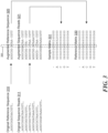
- FIG. 3 shows a plurality of exemplary sequence reads 311, an exemplary reference sequence 310, and an exemplary candidate variant position 350.
- augmenting reference sequence 310 and sequence reads 311 may include inserting one or more gaps or spaces (e.g., spaces 325) within reference sequence 310 and/or within one or more sequence reads 311, thereby producing an augmented reference sequence 320 and an augmented plurality of sequence reads 321 precisely aligned to augmented reference sequence 320.
- the augmentation may include: determining or detecting an inserted base in a particular sequence read; inserting a space at the position of the inserted base in the reference sample; inserting a space at the same position in every sequence read in which no insertion was detected at the same position (which in some cases corresponds to all sequence reads other than the particular sequence read where insertion was detected); and repeating the process for each detected inserted base in each sequence read. It is appreciated that in places where a potential insertion of two bases or longer is detected, two or more spaces can be inserted, respectively.
- MSA multiple sequence alignment
- the augmentation can also include detecting a deleted base in a particular sequence read and inserting a space in that sequence read at the position of the potential deletion variant. It is appreciated that in the case of a detected deleted base, the reference sequence need not be augmented.
- "inserting spaces” may or may not include an actual modification of the memory where the sequence reads and the reference sequences are stored, and may instead be an inherent or implied part of other operations (e.g., other steps of method 200) achieving the same end result.
- the sequence reads and the reference sequence can be selectively copied from the original location into a separate memory location in a manner that would cause the new copy to store spaces in all the right positions.
- "Storing a space” as used herein can mean storing any random or predefined value (e.g., 0) other than the four values corresponding to the four types of nucleotide bases (A, C, T, and G).
- spaces may not be physically inserted at any step, but may be algorithmically accounted for when the sequence reads and the reference sequence are converted into count matrices, as described below.
- augmenting the sequence reads and the reference sequence around the candidate variant position can mean augmenting only a portion of a variable or predefined length that includes the candidate variant position. In other embodiments, however, the entire length of available sequence reads and reference sequence can be augmented at once, in which case step 250 can be performed only once (e.g., before step 240 or even before step 230) and not be repeated for every candidate variant position.
- a segment (i.e., a window) of the augmented sequence reads can be converted (or "summarized") into a sample matrix.
- the segment can be of a fixed length and may or may not place the candidate variant position in the middle. For instance, in the example illustrated in FIG. 3 , the augmented segment is 16 columns (positions) long, and includes 7 columns to the left and 8 columns to the right of candidate variant position 350, placing candidate variant position 350 substantially in the middle of the augmented segment.
- the sample matrix can be a count matrix having the same number of columns as the segment being converted.
- the matrix can include one row for each type of nucleotide base (A, C, G, and T), each row representing the number of times the respective nucleotide base appeared at each position (column) within the augmented sequence reads.
- row "A" in sample matrix 331 includes values "6000000306520000,” indicating that base "A" appeared 6 times at the first position within the segment of augmented reads; 3 times at the eighth position; 6 times at the tenth position; 5 times at the eleventh position; twice at the twelfth position; and 0 times (did not appear) at any other position within the segment.
- the sample matrix can also include a row representing the number of times a space (represented in FIG. 3 as "-") appeared at each position within the augmented sequence reads.
- the first row of sample matrix 331 (marked as "-") includes values "0000014000140000,” indicating that the augmented sequence reads include 1, 4, 1, and 4 spaces at positions 6, 7, 11, and 12 within the segment, respectively.
- the space row "-" is shown as the first row of the matrix in the example of FIG. 3 , in other embodiments, the space row can be placed at other places (e.g., last) within the matrix.
- sample matrix could have 5 columns (A, C, T, G, -) and the number of rows corresponding to the length of the segment. Accordingly, the matrix can be generally described as having at least five "lines” (rows or columns), one line representing counts of each nucleotide type, and at least one line representing space counts within the augmented sequence reads.
- a segment of the augmented reference sequence corresponding to the segment of sequence reads can be similarly converted into a reference matrix having the same dimensions as the sample matrix.
- augmented reference 320 is converted into reference matrix 330.
- reference matrix 330 can have four rows indicating the number of occurrences of each type of nucleotide at each position within the augmented reference sequence, and a space row indicating the number of spaces at each position within the augmented reference sequence. It is appreciated, however, that in other embodiments, reference matrix 330 can have a different summary/representation of the different bases of the reference sequence, than the one illustrated in FIG. 3 .
- reference matrix 320 is also normalized to have the same range of values as sample matrix 331.
- the normalization may include multiplying each count by a normalization factor, such as the total number of sequence reads (in this example, by 6). It is appreciated that in other embodiments, instead of normalizing the reference matrix, normalization can be performed on the sample matrix, or on both matrices. In yet other embodiments, the matrices may not need to be normalized at all. It is also appreciated that step 270 does not need to follow step 260 and can in some embodiments be performed before or in parallel to step 260.
- the reference matrix and the sample matrix can be provided as inputs into a trained deep neural network, and at step 290, an output of the trained deep neural network can be obtained, where the output can include various predictions related to a variant included in the sequence reads represented by the sample matrix, as discussed in more detail below.
- FIG. 2b illustrates another embodiment where at step 210', a reference sequence and a plurality of corresponding sequence reads are obtained.
- the reference sequence and the plurality of corresponding sequence reads can be obtained in any order (i.e., sequentially or simultaneously).
- step 220' the reference sequence and the plurality of corresponding sequence reads are aligned.
- the reference sequence and the plurality of corresponding sequence reads may be obtained in an aligned format, which may eliminate the need for performing the first alignment step.
- a candidate variant position is identified from the aligned sequence reads and reference sequence.
- the sequence reads and/or the reference sequence are augmented around the candidate variant position to achieve a second alignment of the plurality of sequence reads with the reference sequence, in the same manner as described above (i.e. by inserting gaps to account for insertions and/or deletions).
- a window of about 2 to 5, 10, 15, 20, 25, 30, 35, 40, 45, 50, 55, 60, 65, 70, 75, 80, 85, 90, 95, or 100 bases on either of the candidate variant position can be augmented and aligned.
- a window of at least about 5, 10, 15, 20, 25, 30, 35, 40, 45, 50, 55, 60, 65, 70, 75, 80, 85, 90, 95, or 100 bases on either side of the candidate variant position can be augmented and aligned.
- a window of no more than about 5, 10, 15, 20, 25, 30, 35, 40, 45, 50, 55, 60, 65, 70, 75, 80, 85, 90, 95, or 100 bases on either side of the candidate variant position can be augmented and aligned.
- a reference matrix for the candidate variant position from the augmented reference sequence and a sample matrix for the candidate variant position from the plurality of augmented sequence reads are then generated.
- the matrices include the frequencies of each base or gap (i.e. A, C, G, T, or gap) at each sequence position.
- the reference matrix and the sample matrix are input into a trained neural network.
- the neural network determines whether a variant type exists at the candidate variant position.
- the neural network can comprise a plurality of convolutional layers that process the reference matrix and the sample matrix and generate an output that is processed by one or more classifiers and regressors (i.e. a mutation type of classifier, and length classifier, and a position regressor) that determine the variant type, size, and position.
- the neural network can include up to about 5, 10, 15, 20, 25, 30, 35, 40, 45, 50, 55, 60, 65, 70, 75, 80, 85, 90, 95, or 100 layers.
- the convolutional layers can be structured into blocks. In some embodiments, the number of blocks is about half the number of layers.
- steps 230' to 260' can be repeated for each variant candidate position.
- steps 210' to 260' can be repeated by obtaining additional reference sequences and additional corresponding sequence reads.
- the embodiments described in the somatic variant calling section may also be adapted for the germline variant calling.
- the plurality of sequence reads described in FIG. 2b can be subdivided into two groups, normal sequence reads and tumor sequence reads.
- the normal sequence reads can be obtained from a sample of normal tissue from the subject, while the tumor sequence reads can be obtained from a sample of tumor tissue from the subject.
- a normal matrix and a tumor matrix would be generated along with the reference matrix. The rest of the method would proceed in a similar manner.
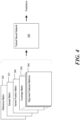
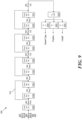
- FIG. 4 illustrates an exemplary embodiment in which reference matrix 330 and sample matrix 331 (along with other optional inputs discussed below) are fed into a trained deep neural network 350, which then outputs one or more predictions related to the sequence reads represented by sample matrix 331.
- network 350 can be a convolutional neural network (CNN), an illustrative example of which is shown in FIG. 5 .
- CNN convolutional neural network
- network 350 may include a plurality of serially connected convolutional layers (510A-510I) eventually feeding into a first fully connected layer 520A, the output of which feeds into four separate fully connected layers 520A-520C that output various predictions.
- different convolutional layers 510 may have different filter sizes (e.g., 1x3, 3x3, or 5x5) and may be interconnected through additional processing layers (not explicitly shown), such as Rectifying Linear Units (ReLUs), pooling layers, batch normalization (BN) layers, etc.
- ReLUs Rectifying Linear Units
- BN batch normalization
- an input to a certain layer can sometimes be connected via an identity shortcut connection 515 to an output of a subsequent layer in order to help maintain the signal in deeper layers of the network.
- network 350 can output one or more predictions associated with a candidate variant within the segment of augmented sequence reads. As illustrated in the example of FIG. 5 , one of such prediction can be obtained at the output of a fully connected layer 520B and can indicate whether the candidate variant position corresponded to a variant and if so the type of that variant. More specifically, in some embodiments, the prediction can include at least four probability values associated with four possible types of variants: NONE (no variant, i.e., a false-positive call); SNP/SNV (single nucleotide polymorphism/variant); INS (insertion variant); and DEL (deletion variant). Based on these probability values it can be determined which type of variant is the most likely, as well as the confidence level of the particular prediction.
- neural network 350 can also output (e.g., at the output of fully connected layer 520C) the predicted position of the variant within the segment of augmented sequence reads represented by sample matrix 331. In other embodiments, this output can be omitted and it can be assumed that the position of the variant is known (e.g., at the center of the segment) based on the manner in which the segment of sequence reads is selected for a given candidate variant, as described above.
- neural network 350 can also output (e.g., at the output of fully connected layer 520D) a predicted length of the variant. For example, a length of "1" can be outputted if the predicted variant is one base long (e.g., a SNP variant or a one-base DEL or INS variant); a length of "2" can be outputted if the predicted variant is a two-base long DEL or INS variant; and so forth. In some embodiments, if the output indicates that a variant is 2 bases long or longer, a post processing step can resolve the exact sequence being inserted or deleted.
- neural network 350 can also output (e.g., at the output of fully connected layer 520E) a predicted genotype associated with the variant, representing, for example, the probabilities with the variant being 1) a homozygous reference (a non-variant); 2) a heterozygous variant (where only one of maternal or paternal copies has a variant); 3) a homozygous variant (where both copies have the same variant); or 4) other (where each copy has a different variant). It is appreciated that in other embodiments, some of these outputs can be omitted and/or additional outputs can be added.
- additional inputs may include, for example, a variant position matrix 340, a coverage matrix 343, and one or more alignment feature matrices 344.
- all input matrices can be provided into network 350 as one large three-dimensional matrix.
- the dimensions of the matrix can be, for example, 5 ⁇ s ⁇ k, where 5 corresponds to the number of row/columns (-, A, C, T, G); s corresponds to the length of the segment (e.g., 16); and k corresponds to the number of different two-dimensional (e.g., 5 ⁇ s) matrices described above.
- k can be as high as 30, or even higher.
- Variant position matrix 340 may comprise a two dimensional representation of the candidate variant position within the reference and sample matrices.
- the matrix can have five rows (-, A, C, T, G) and each column can represent a position within the segment.
- variant position matrix 340 may include one value (e.g., 1) in all rows in the column corresponding to the candidate variant position, and another value (e.g., 0) in all rows of all other columns.
- Coverage matrix 343 may represent the coverage or depth of the particular segment of the sequence reads.
- coverage matrix 343 may include the same value in all of its elements, the value representing the coverage/depth (e.g., the average coverage/depth) of the different reads within the segment.
- coverage matrix 343 may include different values at different columns, each value representing the respective coverage/depth at each column.
- Alignment feature matrices 344 can represent various metrics associated with the quality of the sequence reads and their alignment. Such metrics can include, for example, base qualities, mapping qualities, strand bias, clipping information, and so forth.
- Additional inputs to network 350 may include various data related to known variants. Such data can be obtained, for example, from public and/or private knowledge bases such as dbSNP, COSMIC, ExAC, etc.
- FIG. 5 shows only one exemplary configuration of network 350 and that neural networks having other suitable configurations/architectures can be used to analyze augmented sequence reads (represented by the sample matrix) and to predict the type of variant and its other characteristics, without departing from the scope and spirit of the present disclosure.
- Such alternative configurations may include fewer layers, additional layers, layers having different parameters, additional inputs or outputs, fewer inputs or outputs, and so forth).
- network 350 may not be a convolutional neural network (CNN), but may instead be another type of a deep neural network (DNN), i.e., another type of artificial neural network (ANN) that has multiple layers between the input and output layers, without departing from the scope and spirit of the present disclosure.
- CNN convolutional neural network
- DNN deep neural network
- ANN artificial neural network
- neural network 350 can also be trained to perform somatic variant calling.
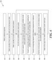
- FIG. 6 illustrates an exemplary method 600 for somatic variant calling using trained neural network 350.
- method 600 includes obtaining a reference sequence, for example, in a manner similar to that described above in connection with step 210 of method 200.
- method 600 includes obtaining a plurality of tumor sequence reads and a plurality of corresponding normal (non-tumor) sequence reads.
- tumor sequence reads can include sequencing results of a tumor (cancerous) tissue of a patient
- normal sequence reads can include sequencing results of a normal (non-cancerous) tissue taken from the same patient.
- the normal sequence reads can include sequencing results of a normal tissue taken from a different patient. Both types of sequence reads can be obtained, for example, in a manner similar to that described above in connection with step 220 of method 200.
- a plurality of candidate somatic variant positions can be obtained - either by obtaining pre-determined variant positions from a memory or a public/private database, a set of whitelist candidate positions, or by performing a comparison between the tumor sequence reads and the reference sequence, for example, in the manner described above in connection with step 230 of method 200.
- the method proceeds to one of the plurality of candidate somatic variant positions.
- a segment of the normal sequence reads around the candidate somatic variant position is converted into a normal sample matrix.
- a segment of the tumor sequence reads around the candidate somatic variant position is converted into a tumor sample matrix.
- a segment of the reference sequence around the candidate somatic variant position is converted into a reference matrix.
- each of them can be augmented using the augmentation technique described above in connection with step 250 of method 200.
- a different type of augmentation technique can be performed on the segments prior to their conversion into matrices; for example, a k-mer collapsing approach can be used where homopolymers of lengths L > k are collapsed to length k, where k is any number greater or equal to one.
- the augmentation step can be omitted altogether, and the original, unprocessed sequence reads and reference sequence can be converted into their respective matrices directly.
- segments of reference sequence 310, tumor sequence reads 311-A, and normal sequence reads 311-B are first augmented to produce augmented reference sequence 320, augmented tumor sequence reads 321-A, and augmented normal sequence reads 321-B, respectively, and are then converted into reference matrix 330, tumor sample matrix 331-A, and normal sample matrix 331-B, respectively.
- the three matrices are fed (provided as inputs) into a trained neural network, and at step 690, an output of the trained neural network is obtained, where the output includes at least a prediction (a probabilistic estimate) of the somatic variant type contained in the tumor sequence read (e.g., at the candidate somatic variant position or its vicinity).
- FIG. 8 shows an exemplary diagram of trained neural network 350 being used for somatic variant calling.
- network 350 receives as its input reference matrix 330, tumor sample matrix 331-A, and normal sample matrix 331-B.
- FIG. 8 shows all three types of matrices being inputted into network 350
- reference matrix 330 and/or normal sample matrix 331-B can be omitted and not be provided to network 350, and network 350 could still perform somatic variant calling.
- obtaining normal sequence reads can be omitted from step 620 of method 600, and step 660 of converting the normal sequence reads into a matrix can be omitted altogether.
- the network in order to further improve the accuracy of prediction and/or to expand the amount of information that can be obtained at the network's output, the network can also receive as inputs additional matrices such as somatic variant position matrix 340 representing the position of the candidate somatic variant; tumor coverage matrix 343-A and normal coverage matrix 343-B representing the respective coverages of the tumor sequence reads and normal sequence reads segments; and one or more alignment features matrices 344, the matrices representing various features of the sequence reads in a manner similar to that described above in connection with FIG. 4 .
- data related to known variants can be obtained from public and/or private knowledge bases such as dbSNP, COSMIC, ExAC, etc., and provided as input(s) to network 350.
- neural network 350 can also be provided one or more other callers' features matrices 345 at its input.
- These matrices can represent (e.g., summarize) one or more features obtained from one or more other (e.g., third-party) variant calling applications or algorithms that have already processed the same segment of sequence reads.
- Such features can include variant types, lengths, and positions predicted by other applications/algorithms, as well as quality scores, mapping scores, variant significance scores, and other alignment features.
- all input matrices being fed into network 350 can be "combined" and provided as one large three-dimensional matrix.
- FIG. 9 shows an exemplary trained neural network 350 receiving the various inputs described above and outputting predictions indicating at least the variant type, position, and length of a somatic variant contained within a segment of somatic sequence reads represented by somatic sample matrix 331-A at its input.
- network 350 can also include fully connected layer 520E (shown in FIG. 5 ) that outputs the variant's genotype, but in case of a somatic variant calling the genotype is not important and can therefore be omitted from the network.
- network 350 having the same or substantially the same architecture can be used to predict both germline and somatic variants, depending on the type of data the network has been trained on (as discussed below) and depending on the type of inputs provided to it.
- the architecture of network 350 can be modified and optimized for either germline or somatic variant calling.
- neural network 350 could start performing accurate germline or somatic variant calling, it first needs to be trained on germline or somatic training data (i.e., training sequences), respectively.
- the training can include, for example, performing all of the steps of methods 200 or 600 on a large number training sequences, but then also providing to the network "ground truth" data (e.g., actual known variant types and their positions, lengths, genotypes, etc.) to enable the network to gradually minimize its output errors by adjusting its trainable filters and other trainable parameters after every run through the process known as "back propagation.”
- ground truth data e.g., actual known variant types and their positions, lengths, genotypes, etc.
- network 350 can be trained on genomes having well characterized ground truth variants, such as the NA12878 genome.
- various simulation based strategies can be used to train the network. For example, to train the network for germline variant calling, synthetic samples with a given set of variants can be simulated using the method described in Mu, J. C. et al. ("VarSim: a high-fidelity simulation and validation framework for high-throughput genome sequencing with cancer applications.” Bioinformatics 31, 1469-1471 (2015 )).
- VarSim a high-fidelity simulation and validation framework for high-throughput genome sequencing with cancer applications. Bioinformatics 31, 1469-1471 (2015 )
- to train the network for somatic variant calling it can be fed normal samples in which random variants have been spiked, e.g., using the method described in Eving, A.
- Somatic mutations are critical signatures in cancer genesis, progression, and treatment. Accurate detection of somatic mutations is challenging due to tumor-normal cross contamination, tumor heterogeneity, sequencing artifacts, and coverage. In general, effectively filtering false-positive calls, which are introduced by the aforementioned issues, and precisely keeping hard-to-catch true-positive calls, which may occur with low allele-frequency (AF) or occur in low-complexity regions, are crucial for an accurate somatic mutation detection algorithm.
- AF allele-frequency
- the machine-learning backbone used in SomaticSeq relies on a set of extracted features for the mutations' locations. As a result, it cannot fully capture the raw information in the genomic contexts of the somatic mutations to further distinguish true somatic mutations from background errors, limiting its performance in challenging situations, such as low-complexity regions and low tumor purity.
- CNNs have recently shown significant performance in classification problems from different domains including germline variant calling 11,12,13 and skin cancer classification 14 . Even so, applying CNNs to the challenging problem of somatic mutation detection has not been explored.
- the only previous deep learning based attempt 15 was to apply a six-layer fully connected neural network to a set of manually extracted features. This approach lacks the power provided by the CNN architecture, which is to learn feature representations directly from the raw data using patterns seen in local regions. Besides, due to the complexity of fully connected networks, it has less generalizability and scalability as seen in CNNs.
- NeuSomatic the first CNN-based approach for somatic mutation detection that can effectively leverage signals derived from sequence alignment, as well as from other methods to accurately identify somatic mutations. Unlike other deep learning based methods that are focused on germline variants, NeuSomatic is addressing a bigger unmet need in terms of accuracy due to the complexity of tumor samples. It can effectively capture important mutation signals directly from the raw data and consistently achieve high accuracy for different sequencing technologies, sample purities, and sequencing strategies such as whole-genome vs. target enrichment.
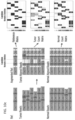
- the inputs to NeuSomatic's network are candidate somatic mutations identified by scanning the sequence alignments for the tumor sample as well as the matched normal sample ( FIGS. 10a-10d ). Somatic mutations reported by other methods can also be included in this list of candidates.
- a 3-dimensional feature matrix M (of size k ⁇ 5 ⁇ 32), consisting of k channels each of size 5 ⁇ 32, to capture signals from the region centered around that locus.
- Each channel of the matrix M has five rows representing the four nucleotide bases as well as the gapped base ( ⁇ -'), and 32 columns representing the alignment columns around the candidate location.
- FIG. 10a illustrates a toy example of input matrix preparation for a given candidate somatic SNV.
- Sequence alignment information in a window of seven bases around the candidate somatic mutation is extracted.
- the reference sequence is then augmented by adding gaps to account for insertions in the reads.
- the augmented alignment is then summarized into the reference matrix, the tumor count matrix, and the normal count matrix.
- the count matrices record the number of A/C/G/T and gap ( ⁇ - ⁇ ) characters in each column of the alignment, while the reference matrix records the reference bases in each column.
- the count matrices are then normalized by coverage to reflect base frequencies in each column. Separate channels are reserved to record the tumor and normal coverages.
- FIG. 10b illustrates the input 3-dimensional matrix and the proposed NeuSomatic network architecture.
- the input matrix consists of reference channel, tumor and normal-frequency channels, coverage and position channels, followed by several channels summarizing the alignment features.
- NeuSomatic When used in ensemble mode, NeuSomatic also includes additional channels for other individual methods features.
- NeuSomatic network architecture consists of nine convolutional layers structured in four blocks with shortcut identity connections. We use two softmax classifiers and one regressor on the final layer to predict the mutation type, size, and position.
- FIG. 10c illustrates a toy example of input matrix preparation for a given candidate somatic deletion.
- Sequence alignment information in a window of 7 bases around the candidate somatic mutation is extracted.
- the reference sequence is then augmented by adding gaps to account for insertions in the reads.
- the augmented alignment is then summarized into the reference matrix, the tumor count matrix, and the normal count matrix.
- the count matrices record the number of A/C/G/T and gap (' -') characters in each column of the alignment, while the reference matrix records the reference bases in each column.
- the count matrices are then normalized by coverage to reflect base frequencies in each column. Separate channels are reserved to record the tumor and normal coverages.
- FIG. 10d illustrates a toy example of input matrix preparation for a given candidate somatic insertion.
- Sequence alignment information in a window of 7 bases around the candidate somatic mutation is extracted.
- the reference sequence is then augmented by adding gaps to account for insertions in the reads.
- the augmented alignment is then summarized into the reference matrix, the tumor count matrix, and the normal count matrix.
- the count matrices record the number of A/C/G/T and gap (' -') characters in each column of the alignment, while the reference matrix records the reference bases in each column.
- the count matrices are then normalized by coverage to reect base frequencies in each column. Separate channels are reserved to record the tumor and normal coverages.
- the first three channels are the reference, tumor-frequency, and normal-frequency channels that summarize the reference bases around the candidate locus, as well as the frequency of different bases in that region.
- each column of tumor and normal-frequency matrices represents the frequency of A/C/G/T/gap bases in the corresponding multiple sequence alignment (MSA) column of the tumor and normal samples, respectively.
- MSA multiple sequence alignment
- NeuSomatic can use the necessary information in tumor, normal, and reference to differentiate difficult to catch somatic mutations with low AF from germline variants, as well as sequencing errors.
- This design also enables the use of convolutional filters in the CNN to capture contextual patterns in the sub-blocks of the matrix.
- DeepVariant 11 uses read pileup as the input for germline variant calling. In contrast, we use base frequency summary for each column as the input to our network. This simplifies the CNN structure, allowing a substantially more efficient implementation. For example, Deep Variant takes -1000 CPU core-hours to call germline variants for a 30 ⁇ whole-genome sample 16 , whereas the stand-alone version of NeuSomatic can detect somatic mutations from 30 ⁇ tumor-normal pair samples in -156 CPU core-hours, despite handling two (tumor-normal) samples instead of one and looking for candidates at lower somatic AFs than germline 50 or 100% AF.
- Clairvoyante 12 uses three channels to summarize base counts for allele counts, deletions, and insertions at the center of the window. In contrast, we summarize all these events in a single base frequency matrix using the reference augmentation approach described earlier, which can clearly represent all the insertion and deletion (INDEL) events across the window.
- INDEL insertion and deletion
- NeuSomatic employs a novel CNN structure that predicts the type and length of a candidate somatic mutation given the feature matrix M ( FIG. 10b ).
- the proposed CNN consists of nine convolutional layers structured in four blocks with shortcut identity connections inspired by ResNet 17 but with a different formation to adapt to the proposed input structure.
- the first classifier identifies whether the candidate is a non-somatic call, SNV, insertion, or deletion.
- the second classifier predicts the length of the somatic mutation with four classes (0 indicating non-somatic, or lengths from 1, 2, or greater than 2), and the regressor predicts the location of the somatic mutation.
- NeuSomatic-S can be used in stand-alone and ensemble modes, while reserving NeuSomatic to denote the ensemble mode.
- NeuSomatic and NeuSomatic-S against the state-of-the-art somatic mutation detection methods including MuTect2 1 , MuSE 2 , SomaticSniper 6 , Strelka2 5 , VarDict 3 , and VarScan2 4 , and against the ensemble approach, SomaticSeq 10 .
- reducing normal purity from 100 to 95% had minor impact on NeuSomatic's performance ( ⁇ 0.3%), whereas it caused ⁇ 3% decrease in SomaticSeq accuracy.
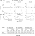
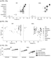
- FIGS. 11a and 11b are charts for the performance analysis of the Platinum two sample mixture dataset.
- FIG. 11a are graphs for a precision-recall analysis: the confidence or quality scores are used to derive the precision-recall curves. The highest F1-score achieved by each algorithm is printed on the curve and marked with a solid circle.
- FIG. 1 1b are graphs for a performance analysis of INDEL accuracy (F1-score) for different INDEL sizes.
- FIG. 11c is a table that shows the performance of different somatic mutation detection methods on Platinum two sample mix dataset. For each method we report the precision, recall and F1-score for the quality score threshold in precision-recall curve which achieves highest F1. (RC: Recall, PR: Precision, F1: F1-score).
- NeuSomatic the ensemble mode clearly outperformed both SomaticSeq and NeuSomatic-S, even though NeuSomatic-S still outperformed SomaticSeq in more challenging scenarios, such as SNVs in the 25:75 mixture and INDELs in the 25:75 and 50:50 mixtures.
- NeuSomatic yielded up to 96.2 and 93.5% F1-scores for SNVs and INDELs, respectively, overall and an improvement of up to 34.6% over the best method in the lowest sample purity.
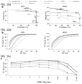
- FIGS. 12a and 12b are graphs for performance analysis of the DREAM Stage 3 dataset.
- five tumor and normal purity scenarios (100% T:100% N, 50% T:100% N, 70% T:95% N, 50% T:95% N, and 25% T:95% N) are used.
- FIG. 12a are graphs for precision-recall analysis: the confidence or quality scores are used to derive the precision-recall curves. The highest F1-score achieved by each algorithm is printed on the curve and marked with a solid circle.
- Fig. 12b are graphs for performance analysis of INDEL accuracy (F1-score) for different INDEL sizes.
- FIG. 12c is a table that shows the performance of different somatic mutation detection methods on Dream Challenge Stage 3 dataset. For each method we report the precision, recall and F1-score for the quality score threshold in precision-recall curve which achieves highest F1. (RC: Recall, PR: Precision, F1: F1-score).
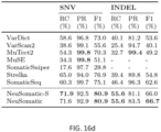
- FIGS. 13a and 13b are graphs for performance analysis of the DREAM Stage 4 dataset.
- five tumor and normal purity scenarios (100% T:100% N, 50% T:100% N, 70% T:95% N, 50% T:95% N, and 25% T:95% N) are used.
- FIG. 13a are graphs for precision-recall analysis: the confidence or quality scores are used to derive the precision-recall curves. The highest F1-score achieved by each algorithm is printed on the curve and marked with a solid circle.
- FIG. 13b are graphs for performance analysis of INDEL accuracy (F1-score) for different INDEL sizes.
- FIG. 13c is a table that shows the performance of different somatic mutation detection methods on Dream Challenge Stage 4 dataset. For each method we report the precision, recall and F1 score for the quality score threshold in precision-recall curve which achieves highest F1. (RC: Recall, PR: Precision, F1: F1-score).
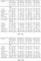
- FIG. 16a shows a precision-recall analysis where the confidence or quality scores are used to derive the precision-recall curves. The highest F1-score achieved by each algorithm is printed on the curve and marked with a solid circle.
- FIG. 16b shows a performance analysis for different AFs.
- FIG. 16c shows a performance analysis of INDEL accuracy (F1-score) for different INDEL sizes.
- FIG. 16d shows for each method the precision, recall and F1-score for the quality score threshold in precision-recall curve which achieves highest F1. (RC: Recall, PR: Precision, F1: F1-score).
- FIG. 17a illustrates the performance analysis of various methods on the exome sample mixture.
- four tumor and normal purity scenarios (50%T:100%N, 70%T:95%N, 50%T:95%N and 25%T:95%N) are used.
- the confidence or quality scores are used to derive the precision-recall curves.
- the highest F1-score achieved by each algorithm is printed on the curve and marked with a solid circle.
- the training is on exome data for NeuSomatic, NeuSomatic-S, and SomaticSeq.
- FIG. 17b illustrates the performance analysis of various methods on the Target panel sample mixture.
- four tumor and normal purity scenarios (50% T: 100%N, 70%T:95%N, 50%T:95%N and 25%T:95%N) are used.
- the confidence or quality scores are used to derive the precision-recall curves.
- the highest F1-score achieved by each algorithm is printed on the curve and marked with a solid circle.
- the training is on exome data for NeuSomatic, NeuSomatic-S, and SomaticSeq.
- FIG. 17c illustrates the performance of different somatic mutation detection methods on whole-exome sample mix dataset. For each method we report the precision, recall and F1-score for the quality score threshold in precision-recall curve which achieves highest F1. (RC: Recall, PR: Precision, F1: F1-score).
- FIG. 17d illustrates the performance of different somatic mutation detection methods on targeted panel dataset. For each method we report the precision, recall and F1-score for the quality score threshold in precision-recall curve which achieves highest F1. (RC: Recall, PR: Precision, F1: F1-score).
- FIG. 18a illustrates the performance analysis of using models trained on whole-genome (Platinum data, genome mixture) and whole-exome (HG003-HG004 exome mixture) to test on exome mixture dataset.
- the confidence or quality scores are used to derive the precision-recall curves.
- the highest F1-score achieved by each algorithm is indicated in the legend and marked with a solid circle on the curve.
- FIG. 18b illustrates the performance analysis of using models trained on whole-genome (Platinum data, genome mixture) and whole-exome (HG003-HG004 exome mixture) to test on target panel mixture dataset.
- the confidence or quality scores are used to derive the precision-recall curves.
- the highest F1-score achieved by each algorithm is indicated in the legend and marked with a solid circle on the curve.
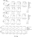
- FIGS. 14a and 14b are graphs for performance analysis of different somatic mutation dectection methods on the PacBio dataset.
- three tumor and normal purity scenarios (70% T:95% N, 50% T:95% N, and 25% T:95% N) are used.
- FIG. 14a are graphs for precision-recall analysis: the confidence or quality scores are used to derive the precision-recall curves. The highest F1-score achieved by each algorithm is printed on the curve and marked with a solid circle.
- FIG. 14b are graphs for performance analysis of INDEL accuracy (F1-score) for different INDEL sizes.
- FIG. 14c shows for each method the precision, recall and F1-score for the quality score threshold in precision-recall curve which achieves highest F1.
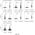
- FIG. 19 illustrates the size distribution of ground truth INDELs in Dream Stage 3, Dream Stage 4, Platinum two sample mixture, Platinum tumor spike, PacBio, and exome datasets. Negative sizes correspond to deletions.
- FIG. 20a illustrates the performance analysis of INDELs based on position and type of the predicted somatic mutations (while ignoring the accuracy of the exact predicted INDEL sequence) for PacBio dataset on three tumor purity scenarios (50%, 30% and 20%) and 95% normal purity.
- the confidence or quality scores are used to derive the precision-recall curves.
- the highest F1- score achieved by each algorithm is printed on the curve and marked with a solid circle.
- FIG. 20b illustrates the performance analysis of INDELs based on position and type of the predicted somatic mutations (while ignoring the accuracy of the exact predicted INDEL sequence) for Dream Stage 3, Dream Stage 4, Platinum two sample mixture, whole-exome, and Platinum tumor spike datasets.
- three tumor purity scenarios (70%, 50% and 25%) are used while normal sample has 95% purity.
- the confidence or quality scores are used to derive the precision-recall curves.
- the highest F1- score achieved by each algorithm is printed on the curve and marked with a solid circle.
- FIG. 15a are graphs for performance analysis of the sequence coverage impact on the whole-exome sample mixture dataset.
- tumor has 50% purity and normal has 95% purity.
- Y-axis illustrates the highest F1-score achieved by each algorithm for sample alignments coverages ranging from 20 ⁇ to 100 ⁇ .
- FIG. 15b are graphs for performance analysis of the sequence coverage impact on the whole-exome sample mixture dataset.
- tumor has 50% purity and normal has 95% purity.
- Tumor and normal alignments coverages are ranging from 20x to 100x.
- the confidence or quality scores are used to derive the precision-recall curves.
- the highest F1-score achieved by each algorithm is indicated in the legend and marked with a solid circle on the curve.
- FIG. 21 shows that performance degrades only marginally even when we trained and tested on very different tumor purities.
- FIG. 21 shows that training using data aggregated from multiple tumor purities was as good as training on the target tumor purity ( FIG. 21 ). This suggests that a training set incorporating multiple tumor purities is sufficient to get a model, which is robust to tumor purity variation.
- FIG. 21 illustrates the performance analysis of cross sample training for Dream Challenge Stage 3 dataset.
- the confidence or quality scores are used to derive the precision-recall curves.
- the highest F1-score achieved by each algorithm is printed in the legend and marked with a solid circle on the curve.
- CLL1 23 a chronic lymphocytic leukemia patient whole-genome data with 961 validated somatic SNVs
- COLO-829 24,25 an immortal metastatic malignant melanoma cell line-derived whole-genome dataset with 454 validated somatic SNVs.
- FIGS. 22a and 22b NeuSomatic achieved the highest extrapolated F1-score of 99.7 and 93.2%, respectively, for the COLO-829 malignant melanoma sample and the CLL1 chronic lymphocytic leukemia sample.
- FIG. 22a shows the performance of different somatic mutation detection methods on real dataset COLO-829.
- FIG. 22b shows the performance of different somatic mutation detection methods on real dataset CLL1.
- FIG. 22c shows the performance of different somatic mutation detection methods on real dataset TCGA-AZ-6601.
- NeuSomatic not only can be used on different sequencing technologies or sequencing strategies but also can be run on a variety of compute platforms including a local HPC cluster and on an elastic cloud compute infrastructure.
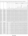
- FIG. 23 is a list of 261 TCGA cancer samples used for Microsoft Azure experiment. Samples are taken across three cancer types: colorectal adenocarcinoma (COAD), ovarian serus adenocarcinoma (OV), and cervical squamous cell carcinoma and endocervical adenocarcinoma (CESC).
- COAD colorectal adenocarcinoma
- OV ovarian serus adenocarcinoma
- CEC cervical squamous cell carcinoma and endocervical adenocarcinoma
- NeuSomatic is the first deep learning based framework for somatic mutation detection, which is high-performing and universal. While using the same CNN architecture, it achieves the best accuracy for varying tumor purities across multiple datasets ranging from synthetic to real, across multiple sequencing strategies ranging from whole genome to targeted as well as across multiple sequencing technologies ranging from short reads to high-error long reads. Specifically, for low tumor purities and low allelic frequencies, NeuSomatic significantly outperforms other state-of-the-art somatic mutation detection methods, thus demonstrating its capability in addressing the hard problem. NeuSomatic utilizes an efficient implementation of convolutional neural networks for solving the somatic mutation detection problem with speed and accuracy.
- Stage 3 data consist of a normal sample and a tumor sample constructed by computationally spiking 7,903 SNVs and 7,604 INDELs mutations into a healthy genome of the same normal sample with three different AFs of 50, 33, and 20% to create synthetic but realistic tumoral normal pairs.
- Stage 4 data have similar formation, but with 16,268 SNVs and 14,194 INDELs in two subclones of 30 and 15% AF.
- We also constructed four tumor mixtures by mixing tumor and normal, respectively, at 100:0, 70:30, 50:50, and 25:75 ratios.
- the somatic mutations across these four tumor mixture ratios have AFs ranging from 5 to 50% for Stage 3 dataset, and 3.75 to 30% for Stage 4 dataset.
- NA12878 we use the heterozygous and homozygous variants in NA12878, which are reference calls in NA12877 and are at least five bases apart from NA12877 variants and 300 base apart from each other as the truth set for the training and evaluation steps (1,103,285 SNVs and 174,754 INDELs).
- somatic mutations across these three tumor mixture ratios have AFs ranging from 12.5 to 70%.
- COLO-829 dataset consists of 80 ⁇ whole-genome sequencing tumor sample and its matched normal blood COLO-829BL sample at 60 ⁇ , with 454 validated somatic SNVs.
- CLL1 has a whole-genome sequencing tumor sample and a matched normal, respectively, at 53 ⁇ and 42 ⁇ coverage, with 961 published somatic SNVs.
- the TCGA-AZ-6601 26,27 dataset is a whole-exome sequencing of a colon adenocarcinoma tumor sample and its matched normal tissue from TCGA.
- the tumor and normal samples were sequenced at depths of 145 ⁇ and 165 ⁇ , respectively.
- extrapolated precision as the percentage of predicted somatic mutations that have been called by at least two stand-alone methods, or have been reported as verified somatic mutations in at least two samples of the same cancer type in COSMIC database.
- extrapolated F1-score based on the harmonic mean of recall and this extrapolated precision.
- NGMI,R v0.2.6 35 to align the sequences.
- each candidate position we prepare a 3-dimensional matrix M with k channels of size 5 ⁇ 32 ( FIG. 10a , 10c , and 10d ).
- the five rows in each channel corresponds to four DNA bases A, C, G, and T, and the gap character ('-').
- Each of the 32 columns of the matrix represents one column of the alignment.
- channel C1 we record the reference base (or gap) in each column.
- Channels Ci (4 ⁇ i ⁇ k) record other alignment signals in tumor and normal samples, such as coverage, base quality, mapping quality, strands, clipping information, edit distance, alignment score, and paired-end information.
- base quality channel we have a matrix of size 5 ⁇ 32 for each sample, which records the average base quality of reads that have a given base (for a given row) in each column.
- the proposed CNN ( FIG. 10b ) consists of nine convolutional layers structured as follows.
- the input matrices are fed into the first convolution layer with 64 output channels, 1 ⁇ 3 kernel size and Relu activation followed by a batch normalization and a max-pooling layer.
- the output of this layer is then fed to a set of four blocks with shortcut identity connection similar to ResNet structure.
- These blocks consist of a convolution layer with 3 ⁇ 3 kernels followed by batch normalization and a convolution layer with 5 ⁇ 5 kernels. Between these shortcut blocks, we use batch normalization and max-pooling layers.
- the output of final block is fed to a fully connected layer of size 240.
- the resulting feature vector is then fed to two softmax classifiers and a regressor.
- the first classifier is a 4-way classifier that predicts the mutation type from the four classes of non-somatic, SNV, insertion, and deletion.
- the second classifier predicts the length of the predicted mutation from the four categories of 0, 1, 2, and ⁇ 3.
- Non-somatic calls are annotated as zero size mutations
- SNVs and 1-base INDELs are annotated as size 1
- 2-base and ⁇ 3 size INDELs are, respectively, annotated as 2 and ⁇ 3 size mutations.
- the regressor predicts the column of the mutations in the matrix, to assure the prediction is targeted the right position and is optimized using a smooth L1 loss function.
- the CNN has less than 900 K parameters, which enables us to have a highly efficient implementation by using large batch sizes.
- the whole-genome training process took ⁇ 8 h on a machine with 8 Tesla K80 Nvidia GPU's.
- the DREAM Challenge dataset has 15,507 somatic mutations for Stage 3 and 30,462 somatic mutations for Stage 4.
- FIGS. 24a-24e we further explored different network architectures such as the pre-activation ResNet architecture with 4 to 16 ResNet blocks (including ResNet-18 and ResNet-34 architectures) ( FIGS. 24a-24e ), as well as some variants of the proposed residual NeuSomatic architecture ( FIGS. 24f-24m ).
- FIG. 25 compares these architectures in terms of accuracy, number of network parameters, memory usage, and speed. In general, all these networks can obtain relatively high accuracy compared to the conventional somatic mutation detection approaches.
- FIGS. 24a-24m illustrate the different network architectures tested.
- FIGS. 24a-24e illustrate ResNet architectures with different number of pre-activation residual blocks with default 3x3 conv layers.
- strided convolutions are used with channel expansions.
- FIGS. 24f and 24g illustrate multiple customized residual blocks with 3x3 and 5x5 conv layers and some dilated convolutions.
- strided convolutions are used with channel expansions.
- FIG. 24h illustrates four customized residual blocks with 3x3 and 5x5 conv layers and some dilated convolutions.
- no strided convolutions are used.
- FIGS. 24i-24m illustrate NeuSomatic residual architecture with different residual blocks and fully-connected sizes.
- FIG. 25 shows the performance analysis of the different network architectures shown in FIGS. 24a-24m .
- all the networks are assessed with batch size of 1000 and after 600 epochs training.
- VarDict as an alternative approach to NeuSomatic on PacBio data.
- VarDict To enable detecting somatic mutations on high-error rate long reads, we used VarDict with " -m 10000 -Q 1 -q 5 -X 1 -c 1 -S 2 -E 3 -g 4 -k 0 " parameter settings.
- AF ⁇ 0.1 for SNVs and AF ⁇ 0.15 for INDELs we used as in NeuSomatic.
- somatic mutations were sorted based on the confidence or quality scores assigned by each tool.
- MuSE we used the tier assignments as the sorting criterion.
- VarDict VarScan2, MuTect2, Strelka2
- SomaticSniper we, respectively, used SSF, SSC, TLOD, SomaticEVS, and SSC values reported in the VCF file for sorting.
- SomaticSeq and NeuSomatic we used the somatic mutation quality score in the QUAL field. NeuSomatic reports quality scores for the predicted somatic mutations based on the probability of predictions by CNN.
- scanning 30x tumor and normal alignments to find candidates, extracting features, and preparing the input matrices can take ⁇ 3.9 h on a dual-14 core Intel Xeon CPU E5-2680 v4 2.40 GHz machine.
- the whole-genome training process can take ⁇ 8 h on a machine with 8 Tesla K80 Nvidia GPU's ( ⁇ 90 s per epoch of size 580,000).
- computing the network predictions for the candidate mutations on a 30 ⁇ whole-genome data can take ⁇ 35 min (with AF cutoff of 0.05, 3.9 M candidates) to ⁇ 100 min (with AF cutoff of 0.03, 11.5 M candidates) with 8 Tesla K80 Nvidia GPUs.
- the whole scanning, preparation, and computing the network predictions can take ⁇ 30 min.
- the end-to-end run times for predicting somatic mutations on a 125 ⁇ whole-exome dataset and a 30 ⁇ whole-genome dataset using NeuSomatic ensemble and stand-alone approaches (in CPU-only mode) were compared with other somatic mutation detection techniques in FIGS. 26a and 26b .
- FIG. 26a illustrates a run-time comparison of different somatic mutation detection algorithms.
- CPU core-hours are shown for predicting somatic mutations on a 125x whole-exome sequencing dataset.
- FIG. 26b illustrates a run-time comparison of different somatic mutation detection algorithms.
- CPU core-hours are shown for predicting somatic mutations on a 30x whole-genome sequencing dataset.
- NeuSomatic is written in Python and C++. Its deep learning framework is implemented using PyTorch 0.3.1 to enable GPU application for training/testing.
- the source code is available at https://github.com/bioinform/neusomatic under the Creative Commons Attribution-NonCommercial-ShareAlike 4.0 International license. The results in this paper were based on NeuSomatic v0.1.3.
- the proposed strategies achieved high robustness across multiple sequencing technologies such as WGS, WES, AmpliSeq target sequencing for fresh and FFPE DNA input, varying tumor/normal purities, and different coverages (ranging from 10 ⁇ - 2000 ⁇ ).
- NeuSomatic significantly outperformed conventional detection approaches in general, as well as in challenging situations such as low coverage, low mutation frequency, DNA damage, and difficult genomic regions.
- Somatic mutations are critical cancer drivers. Accurate somatic mutation detection enables precise diagnosis, prognosis, and treatment of cancer patients 1 .
- Several tools have been developed to identify somatic mutations from next-generation sequencing technology 2-11 . In general, conventional techniques pinpoint somatic mutations from background noise, germline variants, and/or cross-contaminations through various statistical/algorithmic modeling approaches employing a set of hand-crafted sequencing features extracted in the tumor-normal paired sequencing data 2-10 . Those manually designed approaches are not easily generalizable beyond the specific cancer types, sample types, or sequencing strategies for which they were developed.
- NeuSomatic uses a convolutional neural network (CNN) to learn feature representations directly from raw data 11 .
- CNN convolutional neural network
- NeuSomatic uses a novel summarization of tumor/normal alignment information as a set of input matrices that can be adopted to efficiently train models that learn how to effectively differentiate true somatic mutations from artifacts.
- the network models trained by NeuSomatic can capture important mutation signals directly from read alignment and genomic-context without manual intervention.
- NeuSomatic thus, provides a framework that can easily be applied to different problem statements including diverse sequencing technologies, cancer types, tumor and normal purities, and mutation allele frequencies, through training on proper data.
- SEQC-II Somatic Mutation Working Group in the FDA-led Sequencing Quality Control Phase II
- SEQC-II Somatic Mutation Working Group in the FDA-led Sequencing Quality Control Phase II
- HCC1395 human triple-negative breast cancer cell line
- HCC1395BL matched B lymphocyte derived normal cell line
- SEQC-II consortium has developed a well-defined "Gold Set” of somatic Single Nucleotide Variants (SNVs) and insertion/deletions (INDELs) for HCC1395.
- SNVs Single Nucleotide Variants
- INDELs insertion/deletions
- this dataset along with the accompanying sequencing data prepared on multiple sites and technologies provides a unique resource to achieve two important purposes.
- the WGS and WES data were derived from Formalin-Fixed Paraffin-Embedded (FFPE) and fresh DNA, using three library preparation protocols with various input amounts, and from multiple platforms/sequencing sites.
- FFPE Formalin-Fixed Paraffin-Embedded
- the proposed strategies to train and implement the deep learning framework in NeuSomatic achieved high robustness across all the aforementioned real scenarios, and significantly outperformed conventional paired tumor-normal somatic mutation detection approaches.
- HCC1395 and HCC1395BL whole-genome characterized reference tumor-normal paired breast cancer cell lines
- HCC1395 and HCC1395BL were used by the Somatic Mutation Working Group of the SEQC-II consortium 12,13 .
- high-confidence somatic mutations 39,536 SNVs and 2,020 INDELs
- FIG. 27 illustrates the VAF distribution of the ground truth SNV and INDEL somatic mutations in the super set of calls for HCC1395 classified by SEQC-II consortium to four confidence levels (High, Med, Low, and Unclassified). High and medium confidence calls are grouped together as the "truth set" of somatic mutations.
- FIG. 27 illustrates VAF distribution of the ground truth SNV and INDEL somatic mutations in the super set of calls for HCC1395 classified by SEQC-II consortium to four confidence levels (High, Med, Low, and Unclassified). High and medium confidence calls are grouped together as the "truth set" of somatic mutations.
- FIGS. 28a-28e show NeuSomatic's overall performance on 123 replicates in SEQC-II dataset.
- FIGS. 28a shows the NeuSomatic model trained on DREAM challenge stage 3 dataset outperformed other techniques when applied to SEQC-II data.
- FIG. 28b shows that training different NeuSomatic models using SEQC-II reference samples resulted in additional absolute 4-5% improvement in the mean F1-score.
- FIG. 28c shows that using a model trained on SEQC-II data, NeuSomatic achieves consistent superiority over other techniques across diverse set of replicates of different purities/coverages in WGS, WES, FFPE, AmpliSeq, and different library-prep datasets.
- the heatmaps illustrate the absolute difference between the F1-score of any of the somatic mutation detection approaches to the best F1-score.
- the mean F1-score is shown for each approach across 123 replicates.
- FIG. 28d compares the performance of different techniques across 123 replicates in six datasets for SNVs and INDELs.
- the heatmaps illustrates the absolute difference between the F1-score of any of the somatic mutation detection approaches to the best F1-sScore.
- the mean F1-score is shown for each approach across 123 replicates.
- the first model (SEQC-WGS-Spike) was trained on a set of WGS pairs of in silico tumor-normal replicates, where the in silico tumors were designed by spiking mutations into distinct normal replicates.
- the second model (SEQC-WGS-GT-50) was trained using the ground truth somatic mutations in HCC1395 on a set of real WGS tumor-normal replicate pairs on 50% of the genome.
- the third model (SEQC-WGS-GT50-SpikeWGS 10) was prepared by adding 10% of the training data from the first model to those for the second model to take advantage of both a high number of spike-in mutations and the realistic somatic mutations.
- SEQC-WGS-GT-ALL For the specific datasets like FFPE and WES, we also prepared six additional specialized models using a set of synthetic pairs of in silico tumor and normal replicates. For all models, we evaluated the performance on the 50% held-out region of the genome (not used for the SEQC-WGS-GT-50 model). We also trained a model (SEQC-WGS-GT-ALL) using all ground truth mutations similar to the SEQC-WGS-GT-50 model but on the whole genome. Rather than being directly applicable to our WGS dataset where it has seen all true mutations, SEQC-WGS-GT-ALL will be beneficial for performance analysis on other datasets or samples other than HCC1395.
- the NeuSomatic SEQC-WGS-GT50-SpikeWGS10 model performed consistently better than other schemes with minor variations across replicates, demonstrating robustness and reproducibility ( FIG. 29a ).
- Precision-recall analysis revealed that the high precision of NeuSomatic compared to other techniques drove this superiority ( FIG. 29b ).
- the models trained on the ground truth variants yielded the highest advantage over the DREAM3 model, in general, mainly due to the higher precision ( FIGS. 30c and 30g ).
- the proposed models derived from SEQC-II reference samples were shown to reduce false positives for both SNVs and INDELs, improve insertion calling accuracies, and enhance somatic detection on difficult genomic regions by learning correct mutation signals.
- NeuSomatic models trained on WGS performed equally well on targeted sequencing data.
- TNscope employs a machine learning (random forest) model, while Lancet uses localized colored de Bruijn graphs approach to detect mutations. Both tools were designed for genome-wide somatic mutation detection and thus are not suitable for targeted sequencing (typically covers less than 1 MB of the genome).
- FIG. 42 shows the violin-plot comparison of the VAF distribution of private FN calls on WGS dataset.
- each subfigure we compared one of the conventional somatic mutation detection schemes against NeuSomatic.
- X FN the set of ground truth SNVs which were missed by algorithm X (FN in X) in at least 11 out of 21 WGS replicates.
- Y FN the set of ground truth SNVs which were missed by algorithm Y (FN in Y) in at least 11 out of 21 WGS replicates.
- the figure shows the VAF distribution of private FN calls for X and Y.
- the violin-plot shows the VAF distribution of calls in the set X FN /Y FN in blue and the VAF distribution of calls in the set Y FN /X FN in red.
- the private FNs which were correctly predicted by NeuSomatic, had low VAF which revealed their inferiority of such approaches in detecting low VAF mutations.
- the number of private FNs for X and Y are reported on the top.
- SEQC-II reference matched samples a human triple-negative breast cancer cell line (HCC1395) and a matched B lymphocyte derived normal cell line (HCC1395BL) in our analysis.
- HCC1395BL a human triple-negative breast cancer cell line
- HCC1395BL a matched B lymphocyte derived normal cell line
- the SEQC-II somatic mutation working group has established a gold standard truth set of somatic mutations for these samples 13 ( FIG. 27 ).
- the truth set was defined using multiple tumor-normal sequencing replicates from different sequencing centers, and orthogonal mutation detection bioinformatics pipelines.
- the SomaticSeq 10 machine learning framework was implemented to train a set of classifiers on in silico tumor-normal replicate pairs generated by spiking synthetic mutations into an HCC1395BL alignment file, matched with another HCC1395BL for each replicate pair.
- mutation calls were categorized into four confidence-levels (HighConf, MedConf, LowConf, and Unclassified) based on cross-aligner and cross-sequencing-center reproducibility.
- HighConf and MedConf calls were grouped together as the "truth set" of somatic mutations (v1.0), which contains a total of 39,536 SNVs and 2,020 INDELs.
- the Stage 3 dataset consists of a normal sample and a tumor sample constructed by computationally spiking 7,903 SNVs and 7,604 INDELs mutations into a healthy genome of the same normal sample with three different AFs of 50%, 33%, and 20% to create synthetic but realistic tumoral normal pairs.
- An additional ⁇ 95K SNVs and ⁇ 95K INDELs were spike-in into the tumor sample 11 using BAMSurgeon 17 with similar AF distributions to the original DREAM data for better network training.
- This model was trained by combining training data (in 50% of the genome) from five different tumor-normal purity setting of 100T:100N, 50T:100N, 70T:95N, 50T:95N, and 25T:95N11.
- NeuSomatic and NeuSomatic-S were trained on ⁇ 29.2M candidate mutations identified in these five replicate pairs, which includes ⁇ 450K candidates with somatic SNV/INDEL labels and ⁇ 28.7M candidates, labeled as non-somatic.
- TCGA model developed recently 11 by training on a set of 12 TCGA samples 18 .
- the tumor and normal alignments for each of these samples were mixed and split into two equal alignments. One alignment was treated as the normal replicate and the other alignment was used to spike in mutations to construct the tumor replicate.
- ⁇ 88K SNVs and ⁇ 44K INDELs were spiked in to generate a synthetic tumor sample for training.
- NeuSomatic and NeuSomatic-S were trained on ⁇ 5.9M candidate mutations identified in these 12 replicate pairs, which includes ⁇ 1.5M candidates with somatic SNV/INDEL labels and ⁇ 4.4M candidates, labeled as non-somatic.
- Genome-in-a-Bottle consortium 19 We used a set of difficult genomic regions derived by the Genome-in-a-Bottle consortium 19 . These regions included tandem repeats, in two different categories of less than 50bp and larger than 50bp repeats, and segmental duplication regions. We evaluated different techniques on these regions to illustrate robustness to genomic context complexity.
- references to a structure or feature that is disposed "adjacent" another feature may have portions that overlap or underlie the adjacent feature.
- the device may be otherwise oriented (rotated 90 degrees or at other orientations) and the spatially relative descriptors used herein interpreted accordingly.
- the terms “upwardly”, “downwardly”, “vertical”, “horizontal” and the like are used herein for the purpose of explanation only unless specifically indicated otherwise.
- a numeric value may have a value that is +/- 0.1% of the stated value (or range of values), +/- 1% of the stated value (or range of values), +/- 2% of the stated value (or range of values), +/- 5% of the stated value (or range of values), +/- 10% of the stated value (or range of values), etc.
- Any numerical values given herein should also be understood to include about or approximately that value, unless the context indicates otherwise. For example, if the value "10" is disclosed, then “about 10" is also disclosed. Any numerical range recited herein is intended to include all sub-ranges subsumed therein.
Landscapes
- Engineering & Computer Science (AREA)
- Physics & Mathematics (AREA)
- Life Sciences & Earth Sciences (AREA)
- Health & Medical Sciences (AREA)
- Theoretical Computer Science (AREA)
- General Health & Medical Sciences (AREA)
- Medical Informatics (AREA)
- Biophysics (AREA)
- Bioinformatics & Cheminformatics (AREA)
- Data Mining & Analysis (AREA)
- Bioinformatics & Computational Biology (AREA)
- Spectroscopy & Molecular Physics (AREA)
- Evolutionary Biology (AREA)
- Biotechnology (AREA)
- Software Systems (AREA)
- Artificial Intelligence (AREA)
- Evolutionary Computation (AREA)
- Molecular Biology (AREA)
- Proteomics, Peptides & Aminoacids (AREA)
- Analytical Chemistry (AREA)
- Chemical & Material Sciences (AREA)
- Databases & Information Systems (AREA)
- Mathematical Physics (AREA)
- Public Health (AREA)
- Epidemiology (AREA)
- General Physics & Mathematics (AREA)
- General Engineering & Computer Science (AREA)
- Computing Systems (AREA)
- Biomedical Technology (AREA)
- Computer Vision & Pattern Recognition (AREA)
- Bioethics (AREA)
- Computational Linguistics (AREA)
- Genetics & Genomics (AREA)
- Measuring Or Testing Involving Enzymes Or Micro-Organisms (AREA)
- Apparatus Associated With Microorganisms And Enzymes (AREA)
Applications Claiming Priority (4)
| Application Number | Priority Date | Filing Date | Title |
|---|---|---|---|
| US201862718338P | 2018-08-13 | 2018-08-13 | |
| US201962855541P | 2019-05-31 | 2019-05-31 | |
| PCT/EP2019/071580 WO2020035446A1 (en) | 2018-08-13 | 2019-08-12 | Systems and methods for using neural networks for germline and somatic variant calling |
| EP19753337.5A EP3837690B1 (de) | 2018-08-13 | 2019-08-12 | Systeme und verfahren zur verwendung neuronaler netze für keimbahn- und somatisches variant-calling |
Related Parent Applications (1)
| Application Number | Title | Priority Date | Filing Date |
|---|---|---|---|
| EP19753337.5A Division EP3837690B1 (de) | 2018-08-13 | 2019-08-12 | Systeme und verfahren zur verwendung neuronaler netze für keimbahn- und somatisches variant-calling |
Publications (2)
| Publication Number | Publication Date |
|---|---|
| EP4498375A2 true EP4498375A2 (de) | 2025-01-29 |
| EP4498375A3 EP4498375A3 (de) | 2025-04-09 |
Family
ID=67659862
Family Applications (2)
| Application Number | Title | Priority Date | Filing Date |
|---|---|---|---|
| EP19753337.5A Active EP3837690B1 (de) | 2018-08-13 | 2019-08-12 | Systeme und verfahren zur verwendung neuronaler netze für keimbahn- und somatisches variant-calling |
| EP24180495.4A Pending EP4498375A3 (de) | 2018-08-13 | 2019-08-12 | Systeme und verfahren zur verwendung neuronaler netzwerke für keimbahn- und somatische variantenaufrufe |
Family Applications Before (1)
| Application Number | Title | Priority Date | Filing Date |
|---|---|---|---|
| EP19753337.5A Active EP3837690B1 (de) | 2018-08-13 | 2019-08-12 | Systeme und verfahren zur verwendung neuronaler netze für keimbahn- und somatisches variant-calling |
Country Status (5)
| Country | Link |
|---|---|
| US (1) | US20210257050A1 (de) |
| EP (2) | EP3837690B1 (de) |
| JP (1) | JP7166434B2 (de) |
| CN (1) | CN113168886B (de) |
| WO (1) | WO2020035446A1 (de) |
Families Citing this family (14)
| Publication number | Priority date | Publication date | Assignee | Title |
|---|---|---|---|---|
| US11443832B2 (en) * | 2019-03-07 | 2022-09-13 | Nvidia Corporation | Genetic mutation detection using deep learning |
| US11210554B2 (en) | 2019-03-21 | 2021-12-28 | Illumina, Inc. | Artificial intelligence-based generation of sequencing metadata |
| EP4127232A1 (de) * | 2020-03-30 | 2023-02-08 | Grail, LLC | Krebsklassifizierung mit synthetischen spike-in-trainingsproben |
| US20220084631A1 (en) * | 2020-09-17 | 2022-03-17 | Korea Advanced Institute Of Science And Technology | Method and apparatus for machine learning based identification of structural variants in cancer genomes |
| CN113111803B (zh) * | 2021-04-20 | 2022-03-22 | 复旦大学 | 一种小样本字符与手绘草图识别方法及装置 |
| CN113223612B (zh) * | 2021-04-30 | 2024-08-02 | 阿里巴巴达摩院(杭州)科技有限公司 | 基因组的特征提取方法 |
| EP4138003A1 (de) * | 2021-08-20 | 2023-02-22 | Dassault Systèmes | Neuronales netzwerk für variantenaufruf |
| CN113963746B (zh) * | 2021-09-29 | 2023-09-19 | 西安交通大学 | 一种基于深度学习的基因组结构变异检测系统及方法 |
| CN114708906B (zh) * | 2022-03-11 | 2023-05-09 | 至本医疗科技(上海)有限公司 | 用于预测体细胞变异真假的方法、电子设备和存储介质 |
| CN114664379A (zh) * | 2022-04-12 | 2022-06-24 | 桂林电子科技大学 | 一种基于深度学习的第三代测序数据的自校正纠错方法 |
| CN114649055B (zh) * | 2022-04-15 | 2022-10-21 | 北京贝瑞和康生物技术有限公司 | 用于检测单核苷酸变异和插入缺失的方法、设备和介质 |
| CN116110493B (zh) * | 2023-03-20 | 2023-06-20 | 电子科技大学长三角研究院(衢州) | 用于g-四链体预测模型的数据集构建方法及其预测方法 |
| WO2025006874A1 (en) * | 2023-06-30 | 2025-01-02 | Illumina, Inc. | Machine-learning model for recalibrating genotype calls corresponding to germline variants and somatic mosaic variants |
| CN120356517B (zh) * | 2025-06-24 | 2025-09-16 | 上海金福康制药工程技术有限公司 | 测序数据污染识别模型的训练方法、识别方法及电子设备 |
Family Cites Families (10)
| Publication number | Priority date | Publication date | Assignee | Title |
|---|---|---|---|---|
| US20030134278A1 (en) * | 2000-09-07 | 2003-07-17 | Karpen Gary H. | Chromosome inheritance modifiers and their uses |
| JP3943937B2 (ja) | 2002-01-18 | 2007-07-11 | 国立がんセンター総長 | 基準配列抽出フィルターを利用した塩基挿入欠失部位を持つ塩基配列波形データ選別法 |
| JP2015035212A (ja) | 2013-07-29 | 2015-02-19 | アジレント・テクノロジーズ・インクAgilent Technologies, Inc. | ターゲットシークエンシングパネルから変異を見つける方法 |
| KR101400947B1 (ko) | 2013-12-27 | 2014-05-29 | 한국과학기술정보연구원 | 변이 유전체 시퀀스 예측 방법, 장치 및 변이 유전체 시퀀스 예측 프로그램을 저장하는 저장매체 |
| WO2015173222A1 (en) * | 2014-05-12 | 2015-11-19 | Roche Diagnostics Gmbh | Rare variant calls in ultra-deep sequencing |
| WO2016080750A1 (ko) * | 2014-11-18 | 2016-05-26 | 사회복지법인 삼성생명공익재단 | 암 유전체 돌연변이 검출용 유전자 패널 |
| CN106282320B (zh) * | 2015-05-20 | 2019-10-18 | 广州华大基因医学检验所有限公司 | 检测体细胞突变的方法和装置 |
| US20170175169A1 (en) * | 2015-12-18 | 2017-06-22 | Min Lee | Clinical decision support system utilizing deep neural networks for diagnosis of chronic diseases |
| WO2018094360A2 (en) | 2016-11-18 | 2018-05-24 | Nantomics, Llc | Methods and systems for predicting dna accessibility in the pan-cancer genome |
| CN108119316B (zh) * | 2017-11-16 | 2019-11-08 | 云南电网有限责任公司电力科学研究院 | 基于暂态录波数据的风力发电机运行故障新类型发现方法 |
-
2019
- 2019-08-12 EP EP19753337.5A patent/EP3837690B1/de active Active
- 2019-08-12 CN CN201980067395.8A patent/CN113168886B/zh active Active
- 2019-08-12 EP EP24180495.4A patent/EP4498375A3/de active Pending
- 2019-08-12 JP JP2021507667A patent/JP7166434B2/ja active Active
- 2019-08-12 WO PCT/EP2019/071580 patent/WO2020035446A1/en not_active Ceased
-
2021
- 2021-02-12 US US17/248,939 patent/US20210257050A1/en active Pending
Non-Patent Citations (48)
| Title |
|---|
| "Achieving reproducibility and accuracy in cancer mutation detection with whole-genome and whole-exome sequencing", THE SOMATIC MUTATION WORKING GROUP OF SEQC-II CONSORTIUM, 2019, Retrieved from the Internet <URL:https://doi.org/10.1101/626440> |
| ALIOTO, T. S. ET AL.: "A comprehensive assessment of somatic mutation detection in cancer using whole-genome sequencing", NAT. COMMUN, vol. 6, 2015, pages 10001, XP055342075, DOI: 10.1038/ncomms10001 |
| BOLGER, A. M.LOHSE, M.USADEL, B.: "Trimmomatic: a flexible trimmer for Illumina sequence data", BIOINFORMATICS, vol. 30, 2014, pages 2114 - 2120, XP055862121, DOI: 10.1093/bioinformatics/btu170 |
| CHAISSON, M. J. ET AL.: "Resolving the complexity of the human genome using single-molecule sequencing", NATURE, vol. 517, 2015, pages 608 |
| CHIN, J., SIMPLE CONVOLUTIONAL NEURAL NETWORK FOR GENOMIC VARIANT CALLING WITH TENSORFLOW, 16 July 2017 (2017-07-16), Retrieved from the Internet <URL:https://towardsdatascience.com/simple-convolution-neural-network-for-genomic-variant-calling-with-tensorflow-c085dbc2026f> |
| CIBULSKIS, K. ET AL.: "Sensitive detection of somatic point mutations in impure and heterogeneous cancer samples", NAT. BIOTECHNOL., vol. 31, 2013, pages 213 |
| EBERLE, M. A. ET AL.: "A reference data set of 5.4 million phased human variants validated by genetic inheritance from sequencing a three-generation 17-member pedigree", GENOME RES, vol. 27, 2017, pages 157 - 164 |
| ESTEVA, A. ET AL.: "Dermatologist-level classification of skin cancer with deep neural networks", NATURE, vol. 542, 2017, pages 115 - 118, XP055560219, DOI: 10.1038/nature21056 |
| EVING, A.: "Combining tumor genome simulation with crowd sourcing to benchmark somatic single-nucleotide-variant detection", NATURE METHODS, 2015 |
| EWING, A. D. ET AL.: "Combining tumor genome simulation with crowd sourcing to benchmark somatic single-nucleotide-variant detection", NAT. METHODS, vol. 12, 2015, pages 623 |
| FAN, Y. ET AL.: "MuSE: accounting for tumor heterogeneity using a sample-specific error model improves sensitivity and specificity in mutation calling from sequencing data", GENOME BIOL., vol. 17, 2016, pages 178 |
| FANG, L. T. ET AL., ESTABLISHING REFERENCE SAMPLES FOR DETECTION OF SOMATIC MUTATIONS AND GERMLINE VARIANTS WITH NGS TECHNOLOGIES, 2019, Retrieved from the Internet <URL:https://doi.org/10.1101/625624> |
| FANG, L. T. ET AL.: "An ensemble approach to accurately detect somatic mutations using SomaticSeq", GENOME BIOL., vol. 16, 2015, pages 197, XP055531382, DOI: 10.1186/s13059-015-0758-2 |
| FORBES, S. A. ET AL.: "COSMIC: somatic cancer genetics at high-resolution", NUCLEIC ACIDS RES., vol. 45, 2016, pages D777 - D783, XP055458076, DOI: 10.1093/nar/gkw1121 |
| FREED, D.PAN, R.ALDANA, R.T: "Nscope: Accurate Detection of Somatic Mutations with Haplotype-based Variant Candidate Detection and Machine Learning Filtering", BIORXIV 250647 |
| GRASSO, C. S. ET AL.: "Genetic mechanisms of immune evasion in colorectal cancer", CANCER DISCOV, vol. 8, 2018, pages 730 - 749, XP055834504, DOI: 10.1158/2159-8290.CD-17-1327 |
| GROSSMAN, R. L. ET AL.: "Toward a shared vision for cancer genomic data", N. ENGL. J. MED., vol. 375, 2016, pages 1109 - 1112 |
| HE, K.ZHANG, X.REN, S.SUN, J.: "Deep residual learning for image recognition", PROC. IEEE CONF. COMPUT. VIS. PATTERN RECOGNIT, 2016, pages 770 - 778, XP055536240, Retrieved from the Internet <URL:http://openaccess.thecvf.com/content_cvpr_2016/html/He_Deep_Residual_Learning_CVPR_2016_paper.html> DOI: 10.1109/CVPR.2016.90 |
| KIM, S. ET AL.: "Strelka2: fast and accurate calling of germline and somatic variants", NAT. METHODS, vol. 15, 2018, pages 591 - 594, XP036559399, DOI: 10.1038/s41592-018-0051-x |
| KOBOLDT, D. C. ET AL.: "VarScan 2: somatic mutation and copy number alteration discovery in cancer by exome sequencing", GENOME RES., vol. 22, 2012, pages 568 - 576, XP055364674, DOI: 10.1101/gr.129684.111 |
| KRUSCHEPETERLEN TRIGGPAUL C. BOUTROSCHRISTOPHER E. MASONM. FRANCISCOBENJAMIN L. MOOREMAR GONZALEZ-PORTA ET AL.: "Best practices for benchmarking germline small-variant calls in human genomes.", NATURE BIOTECHNOLOGY, vol. 1, 2019 |
| LAI, Z. ET AL.: "VarDict: a novel and versatile variant caller for next-generation sequencing in cancer research", NUCLEIC ACIDS RES., vol. 44, 2016, pages e108 - e108, XP055701286, DOI: 10.1093/nar/gkw227 |
| LARSON, D. E. ET AL.: "SomaticSniper: identification of somatic point mutations in whole genome sequencing data", BIOINFORMATICS, vol. 28, 2011, pages 311 - 317, XP055257171, DOI: 10.1093/bioinformatics/btr665 |
| LAU, B. ET AL.: "LongISLND: in silico sequencing of lengthy and noisy datatypes", BIOINFORMATICS, vol. 32, 2016, pages 3829 - 3832 |
| LI, H, ALIGNING SEQUENCE READS, CLONE SEQUENCES AND ASSEMBLY CONTIGS WITH BWA-MEM, 2013, Retrieved from the Internet <URL:https://arxiv.org/abs/1303.3997> |
| LUO, R. ET AL.: "Clairvoyante: a Multi-task Convolutional Deep Neural Network For Variant Calling In Single Molecule Sequencing", BIORXIV, vol. 310458, 2018 |
| LUO, R.SEDLAZECK, F. J.LAM, T.-WSCHATZ, M., CLAIRVOYANTE: A MULTI-TASK CONVOLUTIONAL DEEP NEURAL NETWORK FOR VARIANT CALLING IN SINGLE MOLECULE SEQUENCING, 2018, Retrieved from the Internet <URL:https://www.biorxiv.org/content/early/2018/04/28/310458> |
| MORSE, H. G.MOORE, G. E.: "Cytogenetic homogeneity in eight independent sites in a case of malignant melanoma", CANCER GENET. CYTOGENET, vol. 69, 1993, pages 108 - 112, XP025213533, DOI: 10.1016/0165-4608(93)90083-X |
| MU, J. C. ET AL.: "VarSim: a high-fidelity simulation and validation framework for high-throughput genome sequencing with cancer applications", BIOINFORMATICS, vol. 31, 2014, pages 1469 - 1471 |
| MU, J. C.: "VarSim: a high-fidelity simulation and validation framework for high-throughput genome sequencing with cancer applications.", BIOINFORMATICS, vol. 31, 2015, pages 1469 - 1471 |
| NARZISI, G. ET AL.: "Lancet: genome-wide somatic variant calling using localized colored DeBruijn graphs", COMMUNICATIONS BIOLOGY, vol. 1, 2018, pages 20 |
| NETWORK, C. G. A. ET AL.: "Comprehensive molecular characterization of human colon and rectal cancer", NATURE, vol. 487, 2012, pages 330 |
| PLEASANCE, E. D. ET AL.: "A comprehensive catalogue of somatic mutations from a human cancer genome", NATURE, vol. 463, 2010, pages 191, XP055065443, DOI: 10.1038/nature08658 |
| POPLIN, R. ET AL.: "A universal SNP and small-indel variant caller using deep neural networks", NAT. BIOTECHNOL., vol. 36, 2018, pages 983 |
| POPLIN, R. ET AL.: "Creating a Universal SNP and Small Indel Variant Caller With Deep Neural Networks", BIORXIV, 2016, pages 092890 |
| PUENTE, X. S. ET AL.: "Whole-genome sequencing identifies recurrent mutations in chronic lymphocytic leukaemia", NATURE, vol. 475, 2011, pages 101 |
| ROBERTS, N. D. ET AL.: "A comparative analysis of algorithms for somatic SNV detection in cancer", BIOINFORMATICS, vol. 29, 2013, pages 2223 - 2230 |
| SAHRAEIAN, S. M. E.LIU, R.LAU, B.PODESTA, K.MOHIYUDDIN, M.LAM, H. Y.: "Deep convolutional neural networks for accurate somatic mutation detection", NATURE COMMUNICATIONS, vol. 10, 2019, pages 1041, XP055636900, DOI: 10.1038/s41467-019-09027-x |
| SEDLAZECK, F. J. ET AL.: "Accurate detection of complex structural variations using single-molecule sequencing", NAT. METHODS, vol. 15, 2018, pages 461 - 468, XP036517999, DOI: 10.1038/s41592-018-0001-7 |
| SHERRY, S. T. ET AL.: "dbSNP: the NCBI database of genetic variation", NUCLEIC ACIDS RES., vol. 29, 2001, pages 308 - 311, XP055125042, DOI: 10.1093/nar/29.1.308 |
| TORRACINTA, R. ET AL., ADAPTIVE SOMATIC MUTATIONS CALLS WITH DEEP LEARNING AND SEMI-SIMULATED DATA, 2016, Retrieved from the Internet <URL:https://www.biorxiv.org/content/early/2016/10/04/079087> |
| TORRACINTA, R. ET AL.: "Adaptive somatic mutations calls with deep learning and semi-simulated data", BIORXIV, 2016, pages 79087 |
| VAN DER AUWERA, G. A. ET AL.: "From FastQ data to high-confidence variant calls: the genome analysis toolkit best practices pipeline", CURR. PROTOC. BIOINFORMA., vol. 43, 2013, pages 10 - 11 |
| WANG, Q. ET AL.: "Detecting somatic point mutations in cancer genome sequencing data: a comparison of mutation callers", GENOME MED, vol. 5, 2013, pages 91, XP021167429, DOI: 10.1186/gm495 |
| XU, C. A: "review of somatic single nucleotide variant calling algorithms for next-generation sequencing data", COMPUT. STRUCT. BIOTECHNOL. J., vol. 16, 2018, pages 15 - 24, XP055636960, DOI: 10.1016/j.csbj.2018.01.003 |
| XU, C.: "A review of somatic single nucleotide variant calling algorithms for next-generation sequencing data", COMPUT. STRUCT. BIOTECHNOL. J., vol. 16, 2018, pages 15 - 24, XP055636960, DOI: 10.1016/j.csbj.2018.01.003 |
| ZOOK, J. M. ET AL.: "Extensive sequencing of seven human genomes to characterize benchmark reference materials", SCI. DATA, vol. 3, 2016, pages 160025 |
| ZOOK, J. M. ET AL.: "Integrating human sequence data sets provides a resource of benchmark SNP and indel genotype calls", NAT. BIOTECHNOL., vol. 32, 2014, pages 246 |
Also Published As
| Publication number | Publication date |
|---|---|
| US20210257050A1 (en) | 2021-08-19 |
| CN113168886B (zh) | 2025-01-21 |
| WO2020035446A9 (en) | 2020-04-09 |
| JP2021534492A (ja) | 2021-12-09 |
| EP3837690B1 (de) | 2024-06-26 |
| EP4498375A3 (de) | 2025-04-09 |
| JP7166434B2 (ja) | 2022-11-07 |
| WO2020035446A1 (en) | 2020-02-20 |
| EP3837690A1 (de) | 2021-06-23 |
| CN113168886A (zh) | 2021-07-23 |
Similar Documents
| Publication | Publication Date | Title |
|---|---|---|
| EP3837690B1 (de) | Systeme und verfahren zur verwendung neuronaler netze für keimbahn- und somatisches variant-calling | |
| Mallory et al. | Methods for copy number aberration detection from single-cell DNA-sequencing data | |
| US11756652B2 (en) | Systems and methods for analyzing sequence data | |
| Rakocevic et al. | Fast and accurate genomic analyses using genome graphs | |
| US20240013921A1 (en) | Generalized computational framework and system for integrative prediction of biomarkers | |
| Lee et al. | Genomic dark matter: the reliability of short read mapping illustrated by the genome mappability score | |
| Huang et al. | Tissue-guided LASSO for prediction of clinical drug response using preclinical samples | |
| Li et al. | An NGS workflow blueprint for DNA sequencing data and its application in individualized molecular oncology | |
| Van Kampen et al. | Taking bioinformatics to systems medicine | |
| Salari et al. | Inference of tumor phylogenies with improved somatic mutation discovery | |
| Llinares-López et al. | Genome-wide genetic heterogeneity discovery with categorical covariates | |
| Jiménez-Sánchez et al. | Transcriptomic plasticity is a hallmark of metastatic pancreatic cancer | |
| Marko et al. | Why is there a lack of consensus on molecular subgroups of glioblastoma? Understanding the nature of biological and statistical variability in glioblastoma expression data | |
| Livne et al. | Pathway metrics accurately stratify T cells to their cells states | |
| Nielsen et al. | MorphoITH: A Framework for Deconvolving Intra-Tumor Heterogeneity Using Tissue Morphology | |
| Xiong et al. | Multimodal integration of single cell ATAC-seq data enables highly accurate delineation of clinically relevant tumor cell subpopulations | |
| Emmert-Streib | Statistical diagnostics for cancer: analyzing high-dimensional data | |
| Sinha et al. | ENLIGHTENMENT: A Scalable Annotated Database of Genomics and NGS-Based Nucleotide Level Profiles | |
| Bollas | Genome sequencing applications in precision medicine: From ancestry prediction to RNA variant calling | |
| Jaratlerdsiri et al. | African‑specific prostate cancer molecular taxonomy | |
| Haas | Bioinformatic tool developments with applications to RNA-seq data analysis and clinical cancer research | |
| Villa et al. | Comprehensive analysis of mutational processes across 20 000 adult and pediatric tumors | |
| Hayes et al. | African-specific prostate cancer molecular taxonomy | |
| Patruno | Computational strategies for single-cell multi-omics data analysis and integration | |
| Cantini et al. | Stabilized Independent Component Analysis outperforms other methods in finding reproducible signals in tumoral transcriptomes |
Legal Events
| Date | Code | Title | Description |
|---|---|---|---|
| PUAI | Public reference made under article 153(3) epc to a published international application that has entered the european phase |
Free format text: ORIGINAL CODE: 0009012 |
|
| STAA | Information on the status of an ep patent application or granted ep patent |
Free format text: STATUS: THE APPLICATION HAS BEEN PUBLISHED |
|
| AC | Divisional application: reference to earlier application |
Ref document number: 3837690 Country of ref document: EP Kind code of ref document: P |
|
| AK | Designated contracting states |
Kind code of ref document: A2 Designated state(s): AL AT BE BG CH CY CZ DE DK EE ES FI FR GB GR HR HU IE IS IT LI LT LU LV MC MK MT NL NO PL PT RO RS SE SI SK SM TR |
|
| REG | Reference to a national code |
Ref country code: DE Ref legal event code: R079 Free format text: PREVIOUS MAIN CLASS: G16B0030100000 Ipc: G16B0020200000 |
|
| PUAL | Search report despatched |
Free format text: ORIGINAL CODE: 0009013 |
|
| AK | Designated contracting states |
Kind code of ref document: A3 Designated state(s): AL AT BE BG CH CY CZ DE DK EE ES FI FR GB GR HR HU IE IS IT LI LT LU LV MC MK MT NL NO PL PT RO RS SE SI SK SM TR |
|
| RIC1 | Information provided on ipc code assigned before grant |
Ipc: G16B 30/10 20190101ALN20250305BHEP Ipc: G16B 40/20 20190101ALI20250305BHEP Ipc: G16B 20/20 20190101AFI20250305BHEP |
|
| STAA | Information on the status of an ep patent application or granted ep patent |
Free format text: STATUS: REQUEST FOR EXAMINATION WAS MADE |
|
| 17P | Request for examination filed |
Effective date: 20251008 |