EP4481672A2 - Computertomographie zur fluoroskopieregistrierung mit segmentierten eingaben - Google Patents
Computertomographie zur fluoroskopieregistrierung mit segmentierten eingaben Download PDFInfo
- Publication number
- EP4481672A2 EP4481672A2 EP24211136.7A EP24211136A EP4481672A2 EP 4481672 A2 EP4481672 A2 EP 4481672A2 EP 24211136 A EP24211136 A EP 24211136A EP 4481672 A2 EP4481672 A2 EP 4481672A2
- Authority
- EP
- European Patent Office
- Prior art keywords
- anatomical
- image
- images
- elements
- anatomical elements
- Prior art date
- Legal status (The legal status is an assumption and is not a legal conclusion. Google has not performed a legal analysis and makes no representation as to the accuracy of the status listed.)
- Granted
Links
Images


Classifications
-
- G—PHYSICS
- G06—COMPUTING OR CALCULATING; COUNTING
- G06T—IMAGE DATA PROCESSING OR GENERATION, IN GENERAL
- G06T5/00—Image enhancement or restoration
- G06T5/70—Denoising; Smoothing
-
- G—PHYSICS
- G06—COMPUTING OR CALCULATING; COUNTING
- G06T—IMAGE DATA PROCESSING OR GENERATION, IN GENERAL
- G06T7/00—Image analysis
- G06T7/10—Segmentation; Edge detection
- G06T7/174—Segmentation; Edge detection involving the use of two or more images
-
- G—PHYSICS
- G06—COMPUTING OR CALCULATING; COUNTING
- G06T—IMAGE DATA PROCESSING OR GENERATION, IN GENERAL
- G06T7/00—Image analysis
- G06T7/30—Determination of transform parameters for the alignment of images, i.e. image registration
- G06T7/33—Determination of transform parameters for the alignment of images, i.e. image registration using feature-based methods
-
- G—PHYSICS
- G06—COMPUTING OR CALCULATING; COUNTING
- G06T—IMAGE DATA PROCESSING OR GENERATION, IN GENERAL
- G06T7/00—Image analysis
- G06T7/30—Determination of transform parameters for the alignment of images, i.e. image registration
- G06T7/38—Registration of image sequences
-
- G—PHYSICS
- G06—COMPUTING OR CALCULATING; COUNTING
- G06T—IMAGE DATA PROCESSING OR GENERATION, IN GENERAL
- G06T2207/00—Indexing scheme for image analysis or image enhancement
- G06T2207/10—Image acquisition modality
- G06T2207/10072—Tomographic images
-
- G—PHYSICS
- G06—COMPUTING OR CALCULATING; COUNTING
- G06T—IMAGE DATA PROCESSING OR GENERATION, IN GENERAL
- G06T2207/00—Indexing scheme for image analysis or image enhancement
- G06T2207/10—Image acquisition modality
- G06T2207/10072—Tomographic images
- G06T2207/10081—Computed x-ray tomography [CT]
-
- G—PHYSICS
- G06—COMPUTING OR CALCULATING; COUNTING
- G06T—IMAGE DATA PROCESSING OR GENERATION, IN GENERAL
- G06T2207/00—Indexing scheme for image analysis or image enhancement
- G06T2207/10—Image acquisition modality
- G06T2207/10072—Tomographic images
- G06T2207/10088—Magnetic resonance imaging [MRI]
-
- G—PHYSICS
- G06—COMPUTING OR CALCULATING; COUNTING
- G06T—IMAGE DATA PROCESSING OR GENERATION, IN GENERAL
- G06T2207/00—Indexing scheme for image analysis or image enhancement
- G06T2207/10—Image acquisition modality
- G06T2207/10116—X-ray image
- G06T2207/10121—Fluoroscopy
-
- G—PHYSICS
- G06—COMPUTING OR CALCULATING; COUNTING
- G06T—IMAGE DATA PROCESSING OR GENERATION, IN GENERAL
- G06T2207/00—Indexing scheme for image analysis or image enhancement
- G06T2207/10—Image acquisition modality
- G06T2207/10132—Ultrasound image
-
- G—PHYSICS
- G06—COMPUTING OR CALCULATING; COUNTING
- G06T—IMAGE DATA PROCESSING OR GENERATION, IN GENERAL
- G06T2207/00—Indexing scheme for image analysis or image enhancement
- G06T2207/10—Image acquisition modality
- G06T2207/10132—Ultrasound image
- G06T2207/10136—3D ultrasound image
-
- G—PHYSICS
- G06—COMPUTING OR CALCULATING; COUNTING
- G06T—IMAGE DATA PROCESSING OR GENERATION, IN GENERAL
- G06T2207/00—Indexing scheme for image analysis or image enhancement
- G06T2207/20—Special algorithmic details
- G06T2207/20172—Image enhancement details
- G06T2207/20192—Edge enhancement; Edge preservation
-
- G—PHYSICS
- G06—COMPUTING OR CALCULATING; COUNTING
- G06T—IMAGE DATA PROCESSING OR GENERATION, IN GENERAL
- G06T2207/00—Indexing scheme for image analysis or image enhancement
- G06T2207/30—Subject of image; Context of image processing
- G06T2207/30004—Biomedical image processing
- G06T2207/30008—Bone
- G06T2207/30012—Spine; Backbone
Definitions
- the present technology generally relates to medical imaging, and relates more particularly to registration of medical images.
- the determining comprises detecting a gradient line within a boundary of the second anatomical element in at least one of the plurality of fluoroscopy images.
- each of the expressions “at least one of A, B and C”, “at least one of A, B, or C”, “one or more of A, B, and C", “one or more of A, B, or C" and "A, B, and/or C” means A alone, B alone, C alone, A and B together, A and C together, B and C together, or A, B and C together.
- each one of A, B, and C in the above expressions refers to an element, such as X, Y, and Z, or class of elements, such as X 1 -X n , Y 1 -Y m , and Z 1 -Z o
- the phrase is intended to refer to a single element selected from X, Y, and Z, a combination of elements selected from the same class (e.g., X 1 and X 2 ) as well as a combination of elements selected from two or more classes (e.g., Y 1 and Z o ).
- Computer-readable media may include non-transitory computer-readable media, which corresponds to a tangible medium such as data storage media (e.g., RAM, ROM, EEPROM, flash memory, or any other medium that can be used to store desired program code in the form of instructions or data structures and that can be accessed by a computer).
- data storage media e.g., RAM, ROM, EEPROM, flash memory, or any other medium that can be used to store desired program code in the form of instructions or data structures and that can be accessed by a computer.
- processors such as one or more digital signal processors (DSPs), general purpose microprocessors (e.g., Intel Core i3, i5, i7, or i9 processors; Intel Celeron processors; Intel Xeon processors; Intel Pentium processors; AMD Ryzen processors; AMD Athlon processors; AMD Phenom processors; Apple A10 or 10X Fusion processors; Apple A11, A12, A12X, A12Z, or A13 Bionic processors; or any other general purpose microprocessors), graphics processing units (e.g., Nvidia GeForce RTX 2000-series processors, Nvidia GeForce RTX 3000-series processors, AMD Radeon RX 5000-series processors, AMD Radeon RX 6000-series processors, or any other graphics processing units), application specific integrated circuits (ASICs), field programmable logic arrays (FPGAs), or other equivalent integrated or discrete logic circuitry.
- DSPs digital signal processors
- images such as Computed Tomography (CT) and intraoperative fluoroscopy images may be noisy and may depict or reflect multiple stacked anatomical elements along a line between an X-ray source and detector used to capture the images.
- a fluoroscopy image of the spine may also depict some or all of the patient's rib cage and/or any other anatomical elements that are positioned along a line between the X-ray source and detector, such that one or more aspects of the patient's spine may be obscured or less clear in the image.
- BMI Body Mass Index
- the noise and the stacked view in the image may increase the difficulty of image registration (as it may be difficult to precisely identify matching gradients or other features across multiple images), and may create additional time costs, frustrate users, and/or lead to postponed or cancelled surgeries.
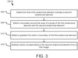
- Figs. 4-5 depict example 2D fluoroscopy images depicting overlapping anatomy of a patient, such as may be obtained intraoperatively in preparation for a registration process.
- a rib 408 overlaps a bone 404, resulting in a gradient 412 within the boundary of the bone 404 that does not correspond to the bone 404.
- each rib 408 in the image overlaps with a vertebra 416 in the image, creating a gradient 420 located within an outer boundary of the corresponding vertebra 416.
- the gradients 412 and/or the gradients 420 may increase the time required to complete the registration (by increasing the number of gradients in the image that must be analyzed and considered for a possible match) while reducing registration accuracy (if, for example, a gradient 412 and/or a gradient 420 is mistakenly identified as corresponding to an edge of a vertebra 416).
- the gradients 412 and/or the gradients 420 may be removed using one or more of the methods (and/or one or more aspects thereof) described herein (e.g., a method 200 and/or a method 300).
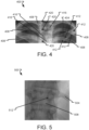
- Fig. 5 depicts another 2D fluoroscopy image 500 showing overlap between a rib 504 and a vertebra 508 in accordance with at least one embodiment of the present disclosure.
- the overlap between the rib 504 and the vertebra 508 in the image 500 creates a gradient 512 that falls within an outer boundary of the vertebra 508.
- the appearance of the gradient 512 can negatively affect the registration process, particularly where the registration process uses a gradient-matching technique to align corresponding vertebra in the 2D fluoroscopy image and a preoperative CT scan or other image.
- One or more methods (and/or one or more aspects thereof) disclosed herein may be used to remove or lessen the appearance of the gradient 512 in the image 500, thus enabling improved registration.
- Embodiments of the present disclosure permit a surgeon to better confirm a registration, improve initial guesses for registration, reduce the number of registration iterations sufficient to complete a surgery or surgical task, and reduce the amount of time needed for and frustration caused by the registration process.
- one or more vertebrae are segmented in each of a CT scan and one or more fluoroscopy images.
- Portions of patient anatomy that overlap the vertebrae (e.g., ribs) in the images may also be segmented.
- the portions may then be removed from the images (yielding, for example, cleaned images), and a registration between the vertebrae in the CT image and the vertebrae in the fluoroscopy images may be made (using the cleaned images).
- a surgeon may then approve or reject the segmented version of the registration.
- Embodiments of the present disclosure provide technical solutions to one or more of the problems of cleaning noisy intraoperative surgical images; improving registration accuracy for autonomous, semi-autonomous, and/or other image-guided surgeries or surgical procedures; reducing the time needed to complete registration in the operating room (and thus conserving operating room resources); reducing registration failure rate; and/or improving a surgeon visibility into the registration process.
- a block diagram of a system 100 according to at least one embodiment of the present disclosure is shown.
- the system 100 may be used to facilitate registration for a surgery or surgical procedure; to clean one or more images (e.g., remove noise or image artifacts in the one or more images); and/or carry out one or more other aspects of one or more of the methods disclosed herein.
- the system 100 comprises a computing device 102, one or more imaging devices 112, a robot 114, a navigation system 118, a database 130, and/or a cloud or other network 134.
- Systems according to other embodiments of the present disclosure may comprise more or fewer components than the system 100.
- the system 100 may not include the imaging device 112, the robot 114, the navigation system 118, one or more components of the computing device 102, the database 130, and/or the cloud 134.
- the computing device 102 comprises a processor 104, a memory 106, a communication interface 108, and a user interface 110.
- Computing devices according to other embodiments of the present disclosure may comprise more or fewer components than the computing device 102.
- the processor 104 of the computing device 102 may be any processor described herein or any similar processor.
- the processor 104 may be configured to execute instructions stored in the memory 106, which instructions may cause the processor 104 to carry out one or more computing steps utilizing or based on data received from the imaging device 112, the robot 114, the navigation system 118, the database 130, and/or the cloud 134.
- the memory 106 may be or comprise RAM, DRAM, SDRAM, other solid-state memory, any memory described herein, or any other tangible, non-transitory memory for storing computer-readable data and/or instructions.
- the memory 106 may store information or data useful for completing, for example, any step of the methods 200 and/or 300 described herein, or of any other methods.
- the memory 106 may store, for example, one or more image processing algorithms 120, one or more segmentation algorithms 122, one or more detection algorithms 124, and/or one or more registration algorithms 128.
- Such instructions or algorithms may, in some embodiments, be organized into one or more applications, modules, packages, layers, or engines.
- the algorithms and/or instructions may cause the processor 104 to manipulate data stored in the memory 106 and/or received from or via the imaging device 112, the robot 114, the database 130, and/or the cloud 134.
- the computing device 102 may also comprise a communication interface 108.
- the communication interface 108 may be used for receiving image data or other information from an external source (such as the imaging device 112, the robot 114, the navigation system 118, the database 130, the cloud 134, and/or any other system or component not part of the system 100), and/or for transmitting instructions, images, or other information to an external system or device (e.g., another computing device 102, the imaging device 112, the robot 114, the navigation system 118, the database 130, the cloud 134, and/or any other system or component not part of the system 100).
- an external source such as the imaging device 112, the robot 114, the navigation system 118, the database 130, the cloud 134, and/or any other system or component not part of the system 100.
- the communication interface 108 may comprise one or more wired interfaces (e.g., a USB port, an ethernet port, a Firewire port) and/or one or more wireless transceivers or interfaces (configured, for example, to transmit and/or receive information via one or more wireless communication protocols such as 802.1 1a/b/g/n, Bluetooth, NFC, ZigBee, and so forth).
- the communication interface 108 may be useful for enabling the device 102 to communicate with one or more other processors 104 or computing devices 102, whether to reduce the time needed to accomplish a computing-intensive task or for any other reason.
- the computing device 102 may also comprise one or more user interfaces 110.
- the user interface 110 may be or comprise a keyboard, mouse, trackball, monitor, television, screen, touchscreen, and/or any other device for receiving information from a user and/or for providing information to a user.
- the user interface 110 may be used, for example, to receive a user selection or other user input regarding any step of any method described herein. Notwithstanding the foregoing, any required input for any step of any method described herein may be generated automatically by the system 100 (e.g., by the processor 104 or another component of the system 100) or received by the system 100 from a source external to the system 100.
- the user interface 110 may be useful to allow a surgeon or other user to modify instructions to be executed by the processor 104 according to one or more embodiments of the present disclosure, and/or to modify or adjust a setting of other information displayed on the user interface 110 or corresponding thereto.
- the computing device 102 may utilize a user interface 110 that is housed separately from one or more remaining components of the computing device 102.
- the user interface 110 may be located proximate one or more other components of the computing device 102, while in other embodiments, the user interface 110 may be located remotely from one or more other components of the computing device 102.
- the robotic arm 116 may comprise a first robotic arm and a second robotic arm, though the robot 114 may comprise more than two robotic arms. In some embodiments, one or more of the robotic arms 116 may be used to hold and/or maneuver the imaging device 112. In embodiments where the imaging device 112 comprises two or more physically separate components (e.g., a transmitter and receiver), one robotic arm 116 may hold one such component, and another robotic arm 116 may hold another such component. Each robotic arm 116 may be positionable independently of the other robotic arm. The robotic arms may be controlled in a single, shared coordinate space, or in separate coordinate spaces.
- the navigation system 118 may provide navigation for a surgeon and/or a surgical robot during an operation.
- the navigation system 118 may be any now-known or future-developed navigation system, including, for example, the Medtronic StealthStation TM S8 surgical navigation system or any successor thereof.
- the navigation system 118 may include one or more cameras or other sensor(s) for tracking one or more reference markers, navigated trackers, or other objects within the operating room or other room in which some or all of the system 100 is located.
- the one or more cameras may be optical cameras, infrared cameras, or other cameras.
- the navigation system may comprise one or more electromagnetic sensors.
- the system of the nineteenth further embodiment is provided, wherein the removing the first anatomical object further comprises subtracting pixels corresponding to the first anatomical object from the at least one of the one or more 2D images.
Landscapes
- Engineering & Computer Science (AREA)
- Physics & Mathematics (AREA)
- General Physics & Mathematics (AREA)
- Theoretical Computer Science (AREA)
- Computer Vision & Pattern Recognition (AREA)
- Apparatus For Radiation Diagnosis (AREA)
- Image Processing (AREA)
Applications Claiming Priority (4)
| Application Number | Priority Date | Filing Date | Title |
|---|---|---|---|
| US202163152656P | 2021-02-23 | 2021-02-23 | |
| US17/590,010 US12190526B2 (en) | 2021-02-23 | 2022-02-01 | Computed tomography to fluoroscopy registration using segmented inputs |
| PCT/IL2022/050197 WO2022180624A1 (en) | 2021-02-23 | 2022-02-20 | Computer tomography to fluoroscopy registration using segmented inputs |
| EP22712457.5A EP4298604B1 (de) | 2021-02-23 | 2022-02-20 | Computertomographie zur fluoroskopieregistrierung mit segmentierten eingaben |
Related Parent Applications (2)
| Application Number | Title | Priority Date | Filing Date |
|---|---|---|---|
| EP22712457.5A Division-Into EP4298604B1 (de) | 2021-02-23 | 2022-02-20 | Computertomographie zur fluoroskopieregistrierung mit segmentierten eingaben |
| EP22712457.5A Division EP4298604B1 (de) | 2021-02-23 | 2022-02-20 | Computertomographie zur fluoroskopieregistrierung mit segmentierten eingaben |
Publications (3)
| Publication Number | Publication Date |
|---|---|
| EP4481672A2 true EP4481672A2 (de) | 2024-12-25 |
| EP4481672A3 EP4481672A3 (de) | 2025-03-12 |
| EP4481672B1 EP4481672B1 (de) | 2026-04-01 |
Family
ID=80933737
Family Applications (2)
| Application Number | Title | Priority Date | Filing Date |
|---|---|---|---|
| EP22712457.5A Active EP4298604B1 (de) | 2021-02-23 | 2022-02-20 | Computertomographie zur fluoroskopieregistrierung mit segmentierten eingaben |
| EP24211136.7A Active EP4481672B1 (de) | 2021-02-23 | 2022-02-20 | Computertomographie zur fluoroskopieregistrierung mit segmentierten eingaben |
Family Applications Before (1)
| Application Number | Title | Priority Date | Filing Date |
|---|---|---|---|
| EP22712457.5A Active EP4298604B1 (de) | 2021-02-23 | 2022-02-20 | Computertomographie zur fluoroskopieregistrierung mit segmentierten eingaben |
Country Status (3)
| Country | Link |
|---|---|
| US (1) | US20250095160A1 (de) |
| EP (2) | EP4298604B1 (de) |
| WO (1) | WO2022180624A1 (de) |
Families Citing this family (7)
| Publication number | Priority date | Publication date | Assignee | Title |
|---|---|---|---|---|
| US11980507B2 (en) | 2018-05-02 | 2024-05-14 | Augmedics Ltd. | Registration of a fiducial marker for an augmented reality system |
| US11382712B2 (en) | 2019-12-22 | 2022-07-12 | Augmedics Ltd. | Mirroring in image guided surgery |
| EP4387552A4 (de) | 2021-08-18 | 2025-04-30 | Augmedics Ltd. | Chirurgisches system mit erweiterter realität mit tiefenmessung |
| EP4511809A4 (de) | 2022-04-21 | 2026-03-11 | Augmedics Ltd | Systeme und verfahren zur visualisierung medizinischer bilder |
| EP4587881A1 (de) | 2022-09-13 | 2025-07-23 | Augmedics Ltd. | Brille der erweiterten realität für bildgeführte medizinische intervention |
| CN121889826A (zh) * | 2023-09-20 | 2026-04-17 | 直观外科手术操作公司 | 解剖对象的模型的计算机辅助配准和跟踪 |
| DE102024200705A1 (de) | 2024-01-26 | 2025-07-31 | Siemens Healthineers Ag | Verfahren zum Betreiben einer Röntgenvorrichtung, Röntgenvorrichtung, Recheneinrichtung, Computerprogramm und elektronisch lesbarer Datenträger |
Family Cites Families (1)
| Publication number | Priority date | Publication date | Assignee | Title |
|---|---|---|---|---|
| EP3474228A1 (de) * | 2017-10-20 | 2019-04-24 | Koninklijke Philips N.V. | Segmentierung eines bildes |
-
2022
- 2022-02-20 EP EP22712457.5A patent/EP4298604B1/de active Active
- 2022-02-20 WO PCT/IL2022/050197 patent/WO2022180624A1/en not_active Ceased
- 2022-02-20 EP EP24211136.7A patent/EP4481672B1/de active Active
-
2024
- 2024-11-27 US US18/962,314 patent/US20250095160A1/en active Pending
Also Published As
| Publication number | Publication date |
|---|---|
| EP4298604B1 (de) | 2024-12-11 |
| US20250095160A1 (en) | 2025-03-20 |
| EP4481672A3 (de) | 2025-03-12 |
| EP4481672B1 (de) | 2026-04-01 |
| WO2022180624A1 (en) | 2022-09-01 |
| EP4298604A1 (de) | 2024-01-03 |
Similar Documents
| Publication | Publication Date | Title |
|---|---|---|
| US12190526B2 (en) | Computed tomography to fluoroscopy registration using segmented inputs | |
| EP4481672A2 (de) | Computertomographie zur fluoroskopieregistrierung mit segmentierten eingaben | |
| EP4264548B1 (de) | Registrierung von zeitgetrennten röntgenbildern | |
| US20250152261A1 (en) | Systems and methods for registering one or more anatomical elements | |
| US20250127572A1 (en) | Methods and systems for planning a surgical procedure | |
| US12400360B2 (en) | Systems and methods for single image registration update | |
| US12361557B2 (en) | Systems and methods for monitoring one or more anatomical elements | |
| WO2025046505A1 (en) | Systems and methods for patient registration using 2d image planes | |
| EP4473481A1 (de) | Systeme, verfahren und vorrichtungen zur verfolgung eines oder mehrerer objekte | |
| CN116917942A (zh) | 使用所分割的输入进行计算机断层扫描到透视的配准 | |
| US12156705B2 (en) | Systems and methods for generating multiple registrations | |
| US20250315955A1 (en) | Systems and methods for monitoring one or more anatomical elements | |
| WO2026028196A1 (en) | Validation using registered fiducial | |
| WO2025109596A1 (en) | Systems and methods for registration using one or more fiducials | |
| WO2025186761A1 (en) | Systems and methods for determining a pose of an object relative to an imaging device | |
| WO2024180545A1 (en) | Systems and methods for registering a target anatomical element | |
| WO2025229497A1 (en) | Systems and methods for generating one or more reconstructions | |
| WO2025122777A1 (en) | Self-calibration of a multi-sensor system | |
| CN121127178A (zh) | 手术定位方法和用于确定经受辐射的区域的方法 | |
| EP4712888A1 (de) | Systeme und verfahren zur erzeugung und aktualisierung eines chirurgischen plans | |
| CN116762095A (zh) | 有时间间隔的x射线图像的配准 |
Legal Events
| Date | Code | Title | Description |
|---|---|---|---|
| PUAI | Public reference made under article 153(3) epc to a published international application that has entered the european phase |
Free format text: ORIGINAL CODE: 0009012 |
|
| STAA | Information on the status of an ep patent application or granted ep patent |
Free format text: STATUS: REQUEST FOR EXAMINATION WAS MADE |
|
| 17P | Request for examination filed |
Effective date: 20241106 |
|
| AC | Divisional application: reference to earlier application |
Ref document number: 4298604 Country of ref document: EP Kind code of ref document: P |
|
| AK | Designated contracting states |
Kind code of ref document: A2 Designated state(s): AL AT BE BG CH CY CZ DE DK EE ES FI FR GB GR HR HU IE IS IT LI LT LU LV MC MK MT NL NO PL PT RO RS SE SI SK SM TR |
|
| REG | Reference to a national code |
Ref country code: DE Ref legal event code: R079 Free format text: PREVIOUS MAIN CLASS: G06T0005000000 Ipc: G06T0007730000 Ref country code: DE Ref legal event code: R079 Ref document number: 602022033820 Country of ref document: DE Free format text: PREVIOUS MAIN CLASS: G06T0005000000 Ipc: G06T0007730000 |
|
| PUAL | Search report despatched |
Free format text: ORIGINAL CODE: 0009013 |
|
| AK | Designated contracting states |
Kind code of ref document: A3 Designated state(s): AL AT BE BG CH CY CZ DE DK EE ES FI FR GB GR HR HU IE IS IT LI LT LU LV MC MK MT NL NO PL PT RO RS SE SI SK SM TR |
|
| RIC1 | Information provided on ipc code assigned before grant |
Ipc: G06T 5/00 20240101ALI20250204BHEP Ipc: G06T 7/73 20170101AFI20250204BHEP |
|
| GRAP | Despatch of communication of intention to grant a patent |
Free format text: ORIGINAL CODE: EPIDOSNIGR1 |
|
| STAA | Information on the status of an ep patent application or granted ep patent |
Free format text: STATUS: GRANT OF PATENT IS INTENDED |
|
| RIC1 | Information provided on ipc code assigned before grant |
Ipc: G06T 7/73 20170101AFI20250912BHEP Ipc: G06T 5/70 20240101ALI20250912BHEP Ipc: G06T 7/33 20170101ALI20250912BHEP |
|
| INTG | Intention to grant announced |
Effective date: 20250926 |
|
| GRAS | Grant fee paid |
Free format text: ORIGINAL CODE: EPIDOSNIGR3 |
|
| GRAA | (expected) grant |
Free format text: ORIGINAL CODE: 0009210 |
|
| STAA | Information on the status of an ep patent application or granted ep patent |
Free format text: STATUS: THE PATENT HAS BEEN GRANTED |
|
| AC | Divisional application: reference to earlier application |
Ref document number: 4298604 Country of ref document: EP Kind code of ref document: P |
|
| AK | Designated contracting states |
Kind code of ref document: B1 Designated state(s): AL AT BE BG CH CY CZ DE DK EE ES FI FR GB GR HR HU IE IS IT LI LT LU LV MC MK MT NL NO PL PT RO RS SE SI SK SM TR |
|
| REG | Reference to a national code |
Ref country code: CH Ref legal event code: F10 Free format text: ST27 STATUS EVENT CODE: U-0-0-F10-F00 (AS PROVIDED BY THE NATIONAL OFFICE) Effective date: 20260401 Ref country code: GB Ref legal event code: FG4D |
|
| REG | Reference to a national code |
Ref country code: DE Ref legal event code: R096 Ref document number: 602022033820 Country of ref document: DE |
|
| REG | Reference to a national code |
Ref country code: IE Ref legal event code: FG4D |