EP4467136A2 - Antagonisten von kalziumkanälen vom typ cav2.3 r zur behandlung von neurodegenerativen erkrankungen - Google Patents
Antagonisten von kalziumkanälen vom typ cav2.3 r zur behandlung von neurodegenerativen erkrankungen Download PDFInfo
- Publication number
- EP4467136A2 EP4467136A2 EP24188598.7A EP24188598A EP4467136A2 EP 4467136 A2 EP4467136 A2 EP 4467136A2 EP 24188598 A EP24188598 A EP 24188598A EP 4467136 A2 EP4467136 A2 EP 4467136A2
- Authority
- EP
- European Patent Office
- Prior art keywords
- neurons
- antagonist
- type calcium
- calcium channel
- activity
- Prior art date
- Legal status (The legal status is an assumption and is not a legal conclusion. Google has not performed a legal analysis and makes no representation as to the accuracy of the status listed.)
- Pending
Links
Images


Classifications
-
- A—HUMAN NECESSITIES
- A61—MEDICAL OR VETERINARY SCIENCE; HYGIENE
- A61K—PREPARATIONS FOR MEDICAL, DENTAL OR TOILETRY PURPOSES
- A61K31/00—Medicinal preparations containing organic active ingredients
- A61K31/13—Amines
- A61K31/135—Amines having aromatic rings, e.g. ketamine, nortriptyline
- A61K31/137—Arylalkylamines, e.g. amphetamine, epinephrine, salbutamol, ephedrine or methadone
-
- A—HUMAN NECESSITIES
- A61—MEDICAL OR VETERINARY SCIENCE; HYGIENE
- A61P—SPECIFIC THERAPEUTIC ACTIVITY OF CHEMICAL COMPOUNDS OR MEDICINAL PREPARATIONS
- A61P25/00—Drugs for disorders of the nervous system
- A61P25/28—Drugs for disorders of the nervous system for treating neurodegenerative disorders of the central nervous system, e.g. nootropic agents, cognition enhancers, drugs for treating Alzheimer's disease or other forms of dementia
-
- G—PHYSICS
- G01—MEASURING; TESTING
- G01N—INVESTIGATING OR ANALYSING MATERIALS BY DETERMINING THEIR CHEMICAL OR PHYSICAL PROPERTIES
- G01N33/00—Investigating or analysing materials by specific methods not covered by groups G01N1/00 - G01N31/00
- G01N33/48—Biological material, e.g. blood, urine; Haemocytometers
- G01N33/50—Chemical analysis of biological material, e.g. blood, urine; Testing involving biospecific ligand binding methods; Immunological testing
- G01N33/68—Chemical analysis of biological material, e.g. blood, urine; Testing involving biospecific ligand binding methods; Immunological testing involving proteins, peptides or amino acids
- G01N33/6872—Intracellular protein regulatory factors and their receptors, e.g. including ion channels
-
- G—PHYSICS
- G01—MEASURING; TESTING
- G01N—INVESTIGATING OR ANALYSING MATERIALS BY DETERMINING THEIR CHEMICAL OR PHYSICAL PROPERTIES
- G01N2500/00—Screening for compounds of potential therapeutic value
-
- G—PHYSICS
- G01—MEASURING; TESTING
- G01N—INVESTIGATING OR ANALYSING MATERIALS BY DETERMINING THEIR CHEMICAL OR PHYSICAL PROPERTIES
- G01N2800/00—Detection or diagnosis of diseases
- G01N2800/28—Neurological disorders
- G01N2800/2814—Dementia; Cognitive disorders
-
- G—PHYSICS
- G01—MEASURING; TESTING
- G01N—INVESTIGATING OR ANALYSING MATERIALS BY DETERMINING THEIR CHEMICAL OR PHYSICAL PROPERTIES
- G01N2800/00—Detection or diagnosis of diseases
- G01N2800/28—Neurological disorders
- G01N2800/2835—Movement disorders, e.g. Parkinson, Huntington, Tourette
-
- G—PHYSICS
- G01—MEASURING; TESTING
- G01N—INVESTIGATING OR ANALYSING MATERIALS BY DETERMINING THEIR CHEMICAL OR PHYSICAL PROPERTIES
- G01N2800/00—Detection or diagnosis of diseases
- G01N2800/28—Neurological disorders
- G01N2800/285—Demyelinating diseases; Multipel sclerosis
Definitions
- metabolic stress seems to constitute an essential common converging downstream factor for most - if not all - neurodegenerative diseases.
- increasing age is the major risk factor for PD - as well as for most other neurodegenerative diseases (e.g. Alzheimer's disease, the most common neurodegenerative disease).
- dopamine-mimetics like L-DOPA the blood-brain-barrier permissive precursor of dopamine, dopamine receptor agonists, or monoamine oxidase inhibitors, like MAO-B).
- these therapies have considerable side effects (e.g., dyskinesia, hallucinations, and impulsive / compulsive syndrome ICB) that become more and more severe over time of treatment.
- a novel potentially neuroprotective PD therapy is currently in a phase III clinical trial: it aims at voltage gated calcium channels called L-type (more precisely Cav1.3 or Cav1.2), and its pharmacological inhibition by drugs, like the dihydropiridin DHP isradipine ( Parkinson Study Group, 2013, PMID: 24123224, phase III clinical trial ClinicalTrials.gov Identifier: NCT02168842 ) .
- the invention relates in its first aspect to a Cav2.3 R-type calcium channel antagonist for the treatment of neurodegenerative diseases.
- the invention relates in its second aspect to a Cav2.3 R-type calcium channel antagonist for use in the treatment of neurodegenerative diseases.
- Cav2.3 R-type calcium channels are Cav2.3 R-type voltage gated calcium channels (VGCC) and also relate to variants thereof, such as CACNA1E, also Cacnl1a6, Cach6, Cchra1, alpha1E and splicevariants a-f. Consequently, the invention relates also to antagonists to these variants.
- VGCC voltage gated calcium channels
- Cav2.3 R-type calcium channels also relate to subunits thereof, defined by a protein / protein complex that contains at least one subunit of Cav1.3 R-type calcium channels.
- the invention relates to neuroprotective therapies of neurodegenerative diseases and predominantly of Parkinson's disease (PD).
- the invention relates to the prevention and/or reduction of the preferential progressive degeneration of the highly-vulnerable SN DA neurons in the midbrain.
- SN DA Substantia nigra
- PD Parkinson's disease
- SN DA neurons release dopamine from their axonal terminals within the dorsal striatum, and also from their cell bodies, and dendrites within the midbrain, in a calcium- and activity-dependent manner.
- Voltage-gated Ca 2+ channels especially the Cav1.3 L-type, received particular attention, as they not only modulate activity-pattern and dopamine release of SN DA neurons, but also generate an activity-related, stressful dendritic Ca 2+ load that could trigger neurodegeneration and PD (Guzman et al., 2010, PMID: 21068725; Surmeier et al., 2017, PMID: 28104909).
- Cav1.3 LTCCs serving as a Ca 2+ source for NCS-1 in SN DA neurons, supporting its Ca 2+ dependent binding to D2-ARs, and preventing their desensitization ( Dragicevic et al., PMID: 24934288 ) and likely counteracts activity-dependent oscillatory elevated metabolic stress levels and cell death ( Guzman et al., 2010, PMID: 21068725 ). But it was observed only transiently in mouse SN DA neurons in response to elevated dopamine levels, e.g. due to in vivo administration of L-DOPA (still the gold-standard in PD-therapy).
- the inventors have surprisingly found that another type of voltage gated calcium channel, the R-type or Cav2.3 channel (or the CACNA1E gene, coding for all Cav2.3 variants), is significantly contributing to the high vulnerability of SN DA neurons to PD-stressors and to degeneration.
- Cav2.3 as the "stressful" calcium channel in highly vulnerable SN DA neurons, that is however not downregulated in human SN DA neurons in PD, is supported by a Cav2.3 downregulation in response to a PD-stressor (PARK1) in mouse dorsal vagal motor-neurons, as these neurons display a similar stressful calcium based pacemaker, but for unknown reason are much more resistant than SN DA neurons to degeneration in PD ( Goldberg et al., 2012 PMID: 22941107; Lasser-Katz et al., 2017, PMID: 28053029 ).
- PARK1 PD-stressor
- Suitable substances i.e. antagonists which inhibit functional activity Cav2.3 calcium channels or their functional expression, effects a slow down or an hold of the progressive loss of SN DA neurons.
- suitable substances i.e. antagonists
- Such suitable substances according to the present invention relates to antagonists of Cav2.3 R-type calcium channels.
- antagonist relates to substances which modulate Cav2.3 R-Type calcium channels either by influencing and modulating its activity (e.g., drugs, small molecules, enzymes, antibodies), or by influencing and modulating its expression (e.g., siRNA, CRISP/CAS9 approaches).
- antagonist and “inhibitor” are used in the same meaning as the term "antagonist”.
- modulate means a negative modulation and has to be understood as partially or fully inhibiting or impeding one or more biological activities of the Cav.2 R-type calcium channel and/or partially or fully inhibiting or impeding its functional expression, e.g. a downregulation of transcription of one or all genes that code for parts/subunits of functional Cav.2.3 R-type calcium channel complexes.
- a modulation of the expression has to be understood as to comprise modulations on transcription as well as translation levels, and of Cav2.3 channel trafficking.
- a preferred embodiment of the invention relates to a Cav2.3 R-type calcium channel antagonist, wherein the agonist effects a modulation of the Cav2.3 R-type calcium channel.
- a further preferred embodiment relates to a Cav2.3 R-type calcium channel antagonist, wherein the antagonist modulates the activity of the Cav2.3 R-type calcium channel.
- the antagonist reduces the activity of the Cav2.3 R-type calcium channel.
- the term "reduce" the activity of a Cav2.3 R-type calcium channel means that the functional activity is less than under normal conditions, i.e. without any modulation of the Cav2.3 R-type calcium channel.
- the activity can be fully or partly inhibited or blocked or interfered.
- the functional activity can be reduced by binding of an antagonist to at least one subunit of the Cav2.3 R-type calcium channel.
- This subunit can be a pore forming alpha subunit of Cav2.3 channels, or its regulatory beta or alpha2delta or gamma subunits.
- any extent by which the functional activity or the functional expression of the Cav2.3 R-type calcium channel is reduced means a modulation according to the invention.
- the functional activity is reduced up to 10%, preferably up to 30%, more preferably up to 50%, even more preferably up to 70% and most preferably up to 100% compared to the normal activity of the Cav2.3 R-type calcium channel.
- the term "downregulate / downregulation” means that the functional expression occurs to a lesser extent than under normal conditions, i.e. without any modulation of the Cav2.3 R-type calcium channel.
- the expression can be fully or partly inhibited or blocked or interfered.
- the downregulation of the expression according to the invention comprises a downregulation on transcription and/or translation level and/or trafficking.
- the expression can be downregulated by binding of an antagonist of the invention to the DNA (transcription level) or by interfering with protein folding (translation level), or with protein trafficking and insertion into the cellular plasmamembrane.
- Another preferred embodiment of the invention relates to Cav2.3 R-type calcium channel antagonist, wherein the antagonist is selected from the group consisting of antibodies, small molecules, macromolecules, siRNA, miRNA, Crispr/Cas9 constructs, hormons, protein kinases, protein phosphatases, SNX-482 and SNX-482 derivates.
- Crispr/Cas9 constructs according to the invention effect down-regulation, modulation or prevention of expression.
- Subjects of the present invention are preferably mammals, more preferably humans.
- the area of neurodegenerative diseases includes PD but also a wide range of other indications.
- Neurodegenerative diseases including, but not limited to Parkinson's disease, Alzheimer's disease, amyothrophic lateral sclerosis, and Huntington's disease occur as a result of neurodegenerative processes. Such diseases are currently incurable, and result in progressive degeneration and/or death of neurons.
- many similarities appear that relate these diseases to one another on a molecular level, in particular to altered intracellular calcium homeostasis and (partially related) elevated metabolic stress levels.
- the invention thus encompasses not only the treatment of PD but also other neurodegenerative diseases by the antagonists according to the invention.
- the invention relates to a Cav2.3 R-type calcium channel antagonist for the treatment of neurodegenerative diseases, wherein the neurodegenerative disease is selected from the group consisting of Parkinson's disease, Alzheimer's disease, Huntington's disease, amyotropic lateral sclerosis, and age-related neurodegeneration.
- the invention relates to Cav2.3 R-type calcium channel antagonist, wherein the antagonist is used in combination with NCS-1 stimulators, dopamine mimetics (which have to be understood as all substances which regulate/modulate the dopamine metabolism), such as L-DOPA and dopamine receptor agonists and/or monaminoxidase inhibitors (MAO inhibitors), or other agents that interfere with the synthesis, release or metabolism of dopamine.
- dopamine mimetics which have to be understood as all substances which regulate/modulate the dopamine metabolism
- L-DOPA and dopamine receptor agonists and/or monaminoxidase inhibitors (MAO inhibitors) or other agents that interfere with the synthesis, release or metabolism of dopamine.
- the pharmaceutical composition of the invention is for use in the treatment of neurodegenerative diseases.
- the Cav2.3 R-type calcium channel antagonist of the pharmaceutical composition of the invention effects a modulation of the Cav2.3 R-type calcium channel function.
- the Cav2.3 R-type calcium channel antagonist of the pharmaceutical composition of the invention modulates the activity of the Cav2.3 R-type calcium channel, preferably, the antagonist reduces the activity of the Cav2.3 R-type calcium channel.
- the Cav2.3 R-type calcium channel antagonist of the pharmaceutical composition of the invention modulates the functional expression of the Cav2.3 R-type calcium channel.
- the antagonist downregulates the expression of the Cav2.3 R-type calcium channel.
- the Cav2.3 R-type calcium channel antagonist of the pharmaceutical composition of the invention is selected from the group consisting of antibodies, small molecules, macro molecules, siRNA, miRNA, Crispr/Cas9 constructs, hormons, enzymes, protein kinases, protein phosphatases SNX-482 and SNX 482 derivates.
- Crispr/Cas9 constructs according to the invention effect down-regulation, modulation or prevention of expression.
- pharmaceutically acceptable excipients refers to excipients that do not produce an adverse, allergic or other untoward reaction when administered to an animal, preferably a human. It includes any and all solvents, dispersion media, coatings, antibacterial and antifungal agents, isotonic and absorption delaying agents and the like. For human administration, preparations should meet sterility, pyrogenicity, and general safety and purity standards.
- Such pharmaceutically acceptable excipients are inherently nontoxic and nontherapeutic.
- excipients include ion exchangers, alumina, aluminum stearate, lecithin, serum proteins, such as human serum albumin, buffer substances such as phosphates, glycine, sorbic acid, potassium sorbate, partial glyceride mixtures of saturated vegetable fatty acids, water, salts, or electrolytes such as protamine sulfate, disodium hydrogen phosphate, potassium hydrogen phosphate, sodium chloride, zinc salts, colloidal silica, magnesium trisilicate, polyvinyl pyrrolidone, cellulose-based substances, and polyethylene glycol.
- Excipients for topical or gel-based forms of antagonist include polysaccharides such as sodium carboxymethylcellulose or methylcellulose, polyvinylpyrrolidone, polyacrylates, polyoxyethylene-polyoxypropylene-block polymers, polyethylene glycol, and wood wax alcohols.
- conventional depot forms are suitably used. Such forms include, for example, microcapsules, nano-capsules, liposomes, sublingual tablets, and sustained-release preparations.
- the pharmaceutical composition of the invention may be administered by several routes of administration.
- routes of administration include, but are not limited to intramuscular (i.m.), subcutaneous, intravenous, intraocular, transdermal, topical, parenteral, intranasal and oral administration.
- Parenteral administration may be by intravenous (IV) injection, subcutaneous (s.c.) injection, intramuscular (i.m) injection, intra-arterial injection, intrathecal (i.t.) injection, intra-peritoneal (i.p.) injection, or direct injection or other administration to the subject.
- IV intravenous
- s.c. subcutaneous
- intramuscular i.m
- intra-arterial injection intra-arterial injection
- intrathecal (i.t.) injection intra-peritoneal (i.p.) injection
- Preferred routes of administration are oral and parenteral.
- the pharmaceutical composition of the invention may be designed to be short-acting, fast-releasing, long-acting, or sustained-releasing as described herein.
- the pharmaceutical formulations may also be formulated for controlled release or for slow release.
- the pharmaceutical composition preferably comprises a delivery system that controls the release of the composition.
- suitable carriers for sustained or delayed release include, but are not limited to, gelatin, gum Arabic, xanthane polymers, thermoplastic resins such as, for example polyvinyl halides, polyvinyl esters, polyvinylidene halides and halogenated polyolefins, elastomers such as, for example, brasiliensis, polydienes, and halogenated natural and synthetic rubbers, and flexible thermoset resins such as polyurethanes, epoxy resins, biodegradable polymers and the like.
- the blood-brain barrier is a physical barrier and system of cellular transport mechanisms between the blood vessels in the central nervous system (CNS) and most areas of the CNS itself.
- the BBB maintains homeostasis by restricting the entry of potentially harmful chemicals from the blood, and by allowing the entry of essential nutrients.
- the BBB can pose a barrier to delivery of pharmacological agents to the CNS for treatment of disorders.
- the pharmaceutical composition of the invention may preferably be used in conjunction with delivery systems that facilitate delivery of the agents to the central nervous system.
- various blood brain barrier (BBB) permeability enhancers may be used to transiently and reversibly increase the permeability of the blood brain barrier to a treatment agent.
- BBB permeability enhancers include but are not limited to leukotrienes, bradykinin agonists, histamine, tight junction disruptors (e.g., zonulin, zot), hyperosmotic solutions (e.g., mannitol), cytoskeletal contracting agents, and short chain alkylglycerols (e.g., 1-O-pentylglycerol).
- Oral, sublingual, parenteral, implantation, nasal and inhalational routes can provide delivery of the active agent to the central nervous system.
- the compounds of the present invention may be administered to the central nervous system with minimal effects on the peripheral nervous system.
- the appropriate dosage of antagonist will depend on the type of disease to be treated, as defined above, the severity and course of the disease, whether the antagonists (e.g. antibodies) are administered for preventive or therapeutic purposes, previous therapy, the patient's clinical history and response to the antagonist, and the discretion of the attending physician.
- the antagonist is suitably administered to the patient at one time or over a series of treatments.
- the invention relates to a pharmaceutical composition of the invention, wherein the antagonist is selected from the group consisting of antibodies, small molecules, macro molecules, siRNA, miRNA, CRISPR/Cas9 constructs, hormones, enzymes, protein kinases, protein phosphatases SNX-482 and SNX-482 derivates.
- the antagonist is selected from the group consisting of antibodies, small molecules, macro molecules, siRNA, miRNA, CRISPR/Cas9 constructs, hormones, enzymes, protein kinases, protein phosphatases SNX-482 and SNX-482 derivates.
- the invention relates to a pharmaceutical composition of the invention, wherein the antagonist is used in combination with NCS-1 stimulators, dopamine mimetics (which have to be understood as all substances which regulate/modulate the dopamine metabolism), like L-DOPA and dopamine receptor agonists and/or monaminoxidase inhibitors (MAO inhibitors), or other agents that interfere with the synthesis, release or metabolism of dopamine.
- NCS-1 stimulators dopamine mimetics
- dopamine mimetics which have to be understood as all substances which regulate/modulate the dopamine metabolism
- L-DOPA dopamine receptor agonists and/or monaminoxidase inhibitors (MAO inhibitors)
- MAO inhibitors monaminoxidase inhibitors
- the invention relates in another aspect to an in vitro method for identifying an antagonist of a Cav2.3 R-type calcium channel, comprising detecting said antagonist in a biological sample, tissue, cell, cell line or fraction thereof that includes at least one subunit of Cav2.3 R-type calcium channel.
- the invention relates in another aspect to a method to determine the effect of a test compound on the modulation of activity of a Cav2.3 R-type calcium channel comprising:
- test compound comprises any antagonist as defined herein.
- Such a test compound relates to any substance or molecule eventually able to bind and/or modulate the Cav2.3 R-type calcium channel of the invention.
- a “biological sample” relates to tissue, primary cells, cell-lines that are endogenously expressing the Cav2.3 ion channel or cell lines that are transiently or stably transfected or stimulated to express the Cav2.3 ion channel or any fractions of it.
- the activity of the Cav2.3 channels can be investigated by biophysical techniques (for example but not limited to electrophysiological, conductance, impedance or spectral methods), by radiometric or fluorescence displacement, or by signals that are related to an activity of the Cav2.3 channel as for example, but not limited to membrane potential, conductance, or changes of calcium or other charge carriers detected by optical, biophysical, electrical or analytical methods.
- the measuring of the inhibition of the Cav2.3 R-type calcium channel can further be performed by using standard electrophysiological, analytical, biophysical, binding, fluorescence or luminescence methods as described in the Assay Guidance Manual as updated on March 31, 2017 (https://www.ncbi.nlm.nih.gov/books/NBK100915/ ). This includes methods to:
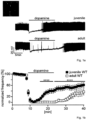
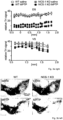
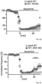
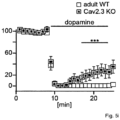
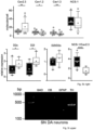
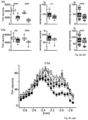
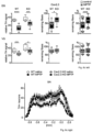
- VGCCs did not substantially alter sensitized dopamine-autoinhibition of SN DA neurons (Fig. 1d,g, Fig.5e,g). This confirms that the functional coupling of Cav2.3, NCS-1 and D2-AR signaling is crucial for maintaining constantly sensitized inhibitory responses to dopamine in mature SN DA neurons. Consistent with a prominent role of Cav2.3 channels in SN DA neurons, inventors cell-specific quantitative RT-qPCR results ( Fig.
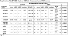
- Cav2.3 (more precisely the Cav2.3e splice variant) is the major VGCC isoform in SN DA neurons from adult mice; Cav2.3e mRNA levels (as well as those of D2-AR, short and long splice variants) increase with postnatal maturation, while those of Cav1.3 and Cav1.2 LTCCs decrease. This is in line with a respective age-dependent reduction of LTCC currents and the contribution of Cav1.3 for D2 AR sensitization in juvenile but not in adult SN DA neurons ( Branch et al, 2016 PMID: 27053209 ).
- Sensitized D2-AR responses inhibit pacemaker activity (and associated metabolic stress) to a greater extent, and their Cav2.3 dependency points to a protective inhibitory feedback-loop, as Cav2.3 activity could also contribute to activity-dependent Ca2+ entry and hence to stressful activity, Ca2+ load, and a high metabolic burden in adult SN DA neurons.
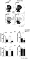
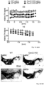
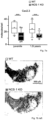
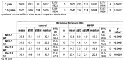
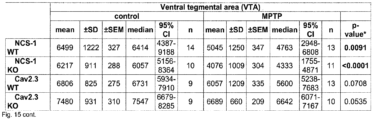
- the inventor applied a chronic neurotoxin PD-model (MPTP/probenecid) to NCS-1 KO, Cav2.3 KO, and WT mice, and compared pattern and degree of induced DA neuron loss in striatum and midbrain by tyrosine hydroxylase (TH) staining and stereology ( Fig. 3 & E3).
- TH tyrosine hydroxylase
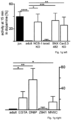
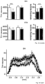
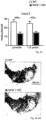
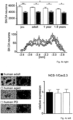
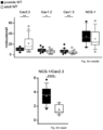
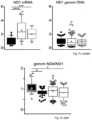
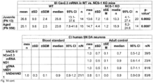
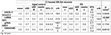
- NCS-1 and Cav2.3 are significantly lower in young and aged SN DA neurons from NCS-1 KO mice ( Fig. 4a ), and NCS-1 KO mice displayed about 30% fewer SN DA neurons (with no additional loss upon aging like in WT), while numbers of VTA DA neurons were not different from WT ( Fig. 4b , Fig.7b , Tab. S8A ).
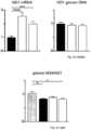
- human SN DA neurons from healthy adult ( ⁇ 42 years) or aged individuals ( ⁇ 71 years), and from aged PD patients ( ⁇ 80 years) displayed similar NCS-1/Cav2.3 mRNA ratios ( Fig. 4c left, Fig 7c left ) .
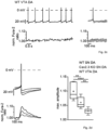
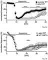
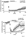
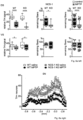
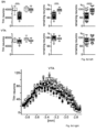
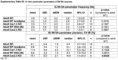
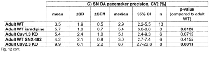
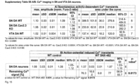
- Fig 4d the inventor shows that Cav2.3 channels are active during pacemaker activity and substantially contribute to oscillatory elevated free Ca 2+ levels in the soma of mouse SN DA neurons that display a particularly high vulnerability to PD-stressors and preferentially degenerate in PD.
- the inventor demonstrates that Cav2.3 channel activity continuously stimulates inhibitory NCS-1/D2-AR signaling in mature SN DA neurons and that loss of NCS-1 activity further increases high vulnerability of SN DA neurons to degeneration.
- the inventor shows that in the cell-bodies of SN DA neurons Ca 2+ oscillations are mediated prominently by Cav2.3 VGCCs, and evidence has been provided from mouse models und human post mortem brains for an antagonizing functional interplay of Cav2.3 channels and NCS-1 in SN DA neurons ( Fig 17 ): the more NCS-1 activity the more stressful Cav2.3 activity (and to a lesser extent Cav1.3 VGCC activity) SN DA neurons might tolerate.
- SN DA neurons Due to their VGCCs stimulated pacemaker-activity, and related Ca 2+ -stimulation of ATP-generation and metabolic stress, SN DA neurons are energetically " living on the edge " ( Bolam and Pissadaki, 2012, PMID: 23008164; Duda et al., 2016, PMID: 26865375 ), and additional PD-stressors (like PARK mutations, environmental factors or simply aging, all causing as an unifying event metabolic stress) further add to their delicate balance and could " tip them over the edge ", Fig4d ( Bolam and Pissadaki, 2012, PMID: 23008164; Duda et al., 2016, PMID: 26865375; Surmeier et al., 2017, PMID: 28104909 ).
- Plasma-levels that are reached with the maximal tolerable dosis of 10 mg isradipine/day might not be sufficient to inhibit Cav1.3 (and Cav1.2) channels in SN DA neurons 2017, PMID: 28592699 ), and inventors previous studies would predict that Cav1.3 inhibition might be protective only under conditions that lead to transient high dopamine levels, like dopamine-replacement therapy (e.g.
- CACNA1E that codes for all Cav2.3 splice-variants offer an alternative complementary, gene-therapy-based novel approach for neuroprotective therapy for PD and related neurodegenerative disorders.
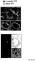
- the inventor designed and successfully tested suitable Crispr/Cas9 constructs, and a protocol for its viral delivery into SN DA neurons of adult WT mice (Fig. 7e), as a proof-of-principle for a Crisp/Cas9 based neuroprotective gene-therapy approach that reduces Cav2.3 channel expression in highly vulnerable neurons, in particular SN DA neurons for PD.
- this invention offers with Cav2.3 channels attractive targets for neuroprotective therapeutical intervention not only for PD and SN DA neurons but also for other neurodegenerative diseases with related pathomechanisms.
- MPP+ induces metabolic stress and selective loss of SN DA neurons but rather not of VTA DA neurons, and Parkinsonism into mice and men.
- Probenecid retards renal excretion of MPP+ and thus biological variations ( Meredith, G. E. & Rademacher, D. J. MPTP mouse models of Parkinson's disease: an update. J Parkinsons Dis 1, 19-33, doi:10.3233/JPD-2011-11023 (2011 )).
- Control mice were injected with saline and probenecid only. One week after the last injections mice were sacrificed, and perfused with PFA for immunohistochemistry.
- Example 3 In vivo retrograde tracing
- Coordinates Bregma (y-axis) 0.98 & -0.1 mm, lateral (x-axis) 1.9 & 2.70 mm, ventral (z-axis) -3.2 mm, 2x 60 nl) or bilaterally into the ventral striatum (Nucleus accumbens, NAc core and medial shell. Coordinates: Bregma (y-axis) 1.6 mm, lateral (x-axis) 0.8 mm, ventral (z-axis,) -4.0 mm, 60 nl) of anaesthetized mice.
- Retrobead-injection was performed under stereotactic control (Kopf Instruments) with a NanoFil syringe attached to a micropump (UMP3 with SYS-Micro4 Controller; World Precision Instruments) at a rate of 50 nl/min for SN tracing and 30 nl/min for VTA tracing.
- Animals were sacrificed 7 days (CPu) or 14 days (NAc) after retrobead injection for calcium imaging (VTA DA) or UV-LMD and RT-qPCR analysis (SN DA neurons). Injection sites were verified according to the mouse brain atlas ( Paxinos, G. & Franklin, K. The Mouse Brain in Stereotaxic Coordinates (Academic. New York). (2007 )) as described in Krabbe S. et al. (ref. as above).
- Example 4 In vitro electrophysiological recordings from juvenile and adult mouse brain slices
- SN DA neurons were identified according to their anatomical location, morphology, and typical electrophysiological fingerprint. Activity patterns were recorded in the presence of 10 ⁇ M DNQX (6,7-dinitroquinoxaline-2,3-dione, Tocris) and 10 ⁇ M gabazine (SR95531 hydrobromide, Tocris) for inhibition of fast synaptic transmission, in cell-attached or perforated patch (100 ⁇ g/ml gramicidine, Sigma) to maintain intracellular signaling (or whole-cell for 0.1 mM EGTA, Sigma).
- 10 ⁇ M DNQX 6,7-dinitroquinoxaline-2,3-dione, Tocris
- 10 ⁇ M gabazine SR95531 hydrobromide, Tocris
- dopamine auto-inhibition after recording of a minimum of 5 min stable baseline activity, 100 ⁇ M dopamine hydrochloride (Sigma) in aCSF was applied via bath-perfusion for 15 min, followed by 20 min washout. Mean frequency of each minute was normalized to the respective baseline frequency for each cell.
- slices were preincubated with 10 ⁇ M DNIP, a D2/NCS-1 interaction preventing peptide ( Dragicevic, E. et al .see ref. above; Scab, B. J. et al. NCS-1 in the dentate gyrus promotes exploration, synaptic plasticity, and rapid acquisition of spatial memory.
- Example 5 Combined calcium Imaging and electrophysiology from adult mouse brain slices
- the imaging setup consisted of an Imago/SensiCam CCD camera with a 640x480 chip (Till Photonics) and a Polychromator IV (Till Photonics) coupled via an optical fiber into the upright microscope.
- Fura-2 was excited at 340 nm, 360 nm or 380 nm (410 nm dichroic mirror; DCLP410, Chroma).
- Emitted fluorescence was detected through a 440 nm long-pass filter.
- Data were acquired as 80x60 frames using 8x8 on-chip binning. Images were recorded in analog-to-digital units (ADUs) and stored and analyzed as 12-bit grayscale images.
- ADUs analog-to-digital units
- fura-2 was loaded into the neurons by electroporation (1 V with 1 ms pulse duration at 65 Hz for 10 - 15 s).
- pairs of frames excited with 340 nm and 380 nm were taken at 25 Hz (pacemaker activity) or 10 Hz (current induced action potentials, APs). The mean ADU value within a region of interest (ROI) from the soma was used.
- ROI region of interest
- the ROI was adjusted for each cell. For 'background subtraction' for the whole-time series an adjacent, second ROI was chosen. Data were analyzed as normalized fura-2 F340/F380 ratios. Mean amplitudes of 20 oscillations were calculated for each analyzed neuron. Since AP-associated Ca2+ dynamics are strongly frequency dependent, the AP frequency was adjusted for Ca2+ imaging in all recorded neurons to a similar value of -1.5 Hz. As SN DA neurons from (+/+) and C57BU6 mice showed no significant difference, data were pooled. For pharmacology, 100 nM SNX-482 (Tocris) was bath applied (in aCSF) for at least 30 mins before and during recordings..
- Example 6 Cryo-sectioning, UV laser microdissection and RT qPCR analysis
- Coronal (mice) and horizontal (human) 12 ⁇ m cryosections were cut and mounted on PEN-membrane slides (Mirodissect) essentially as previously described (see Krabbe S. et al. (ref. as above); see also Schlaudraff, F. et al. Orchestrated increase of dopamine and PARK mRNAs but not miR-133b in dopamine neurons in Parkinson's disease. Neurobiol Aging 35, 2302-2315, doi:10.1016/j.neurobiolaging.2014.03.016 (2014 ) ) .
- UV-Laser-microdissection of UV-LMD samples from cresyl-violet-stained midbrain sections from juvenile mice and human tissue, or from retrogradely traced adult mouse midbrain sections, was carried out using an LMD7000 system (Leica Microsystems). UV-LMD, cell lysis, cDNA synthesis, precipitation, multiplex nested PCR (for marker-gene analysis of murine samples) and quantitative real-time PCR (qPCR) of UV-LMD samples were performed as described (see Krabbe S. et al. (ref. as above); Schlaudraff, F. et al. (ref. as above); Duda, J., Fouler, M., Grundemann, J.
- human samples only neuromelanin positive neurons were collected, and only tyrosine hydroxylase (TH) positive UV-LMD samples were further analyzed.
- TH tyrosine hydroxylase
- mouse-derived samples only samples expressing a correct marker gene profile were used for qPCR analysis: positive for TH, and negative for calbindin d28k (CB), glial fibrillary acidic protein (GFAP), and I-glutamate decarboxylase (GAD 65/67 ).
- CB calbindin d28k
- GFAP glial fibrillary acidic protein
- GID 65/67 I-glutamate decarboxylase
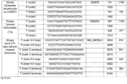
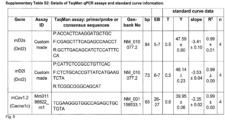
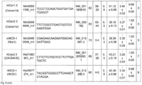
- mCav1.2 forward TCACCACTCTGCTGCAGTTC, reverse: GACGAAACCCACGAAGATGT (amplicon size: 392 bp);
- mCav1.3 forward TCGGGACTGGTCTATTCTGG, reverse: TACTTCCCCACCAGTCCTTG (480 bp);
- mCav2.3 forward TTGGATCTGCTTGTGCCCATG, mCav2.3 reverse: GGAATTTGAACGCTTATCCCGAA (amplicon size: 543 bp or 414 bp depending on splice variant expression);
- mNCS-1 forward CCGAGCATGGGGAAATCCAAC, mNCS-1 reverse: TCATGGCAAAGATCCGGTCCAC (472 bp).
- PCR products were purified (QIAquick Gel Extraction Kit), and quantified (with a Qubit 3.0 fluorometer). DNA standards containing 1.000.000 molecules down to 1 molecule (in 10-fold diluted steps) were used to generate absolute cDNA standard curves.
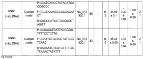
- Mitochondrial genomic DNA was isolated from human SN DA neurons after UV-LMD with the QiaAmp DNA Micro Kit (Qiagen) as previously described ( Muhling, T., Duda, J., Weisberger, J. H., Ludolph, A. C. & Liss, B. Elevated mRNA-levels of distinct mitochondrial and plasma membrane Co(2+) transporters in individual hypoglossal motor neurons of endstage SOD1 transgenic mice. Front Cell Neurosci 8, 353, doi:10.3389/fnce1.2014.00353 (2014 ) ). DNA was eluted in 30 ⁇ l molecular biology grade water and stored at 4°C until qPCR.
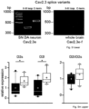
- Outer duplex primer pairs were chosen using Oligo7.60 software (possible PCR amplicon sizes mCav2.3 II-III loop: 816 bp, 795 bp and 759 bp; mCav2.3 C-terminus: 770 bp and 641 bp).
- primer sequences from23 were used (possible PCR amplicon sizes mCav2.3 II-III loop: 420 bp, 399 bp and 363 bp; mCav2.3 C-terminus: 498 bp and 369 bp).
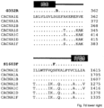
- Nested PCR products were separated in a 4% agarose gel (MetaPhor agarose, Biozym), and expressed splice variants (mouse whole brain tissue as positive control, and in individual SN DA neurons) were identified according to the amplicon sizes of the nested PCR products: II-III loop nested PCR fragment C-terminus nested PCR fragment Cav2.3a 363 bp 369 bp Cav2.3b 399 bp 369 bp Cav2.3c 420 bp 369 bp Cav2.3d 420 bp 498 bp Cav2.3e 363 bp 498 bp Cav2.3f 399 bp 498 bp Mouse Cav2.3 R-type calcium channel (Cacnale) F (outer II-III loop) CCATCTACTTCATTGTGCTCACC NM_009782.3 2043 816 R (outer II-III loop) CCCCTTCATCCAGACTCCG 2858 F (outer C-terminus) GACAGCAGCT
- Free floating slices were washed three times in PBS for 10 min each with shaking (300 rpm, microplate shaker, VWR) and treated for 2 h with a blocking solution (10% normal goat serum, 0.2% BSA and 0.5% Triton X-100 in PBS). After washing the slices with PBS, rabbit anti-TH (1:5000, Merck Millipore) was applied in a carrier solution (1% goat serum, 0.2% BSA and 0.5% Triton X-100 in PBS) and incubated overnight at room temperature while shaking (300 rpm, microplate shaker, VWR).
- a blocking solution 10% normal goat serum, 0.2% BSA and 0.5% Triton X-100 in PBS.
- Example 8 Optical densiometry and stereological analysis
- sampling fraction i.e. 0.44
- ssf section sampling fraction (i.e. 1 for SN and 2 for VTA).
- Sampling grid dimensions were 75 x 75 ⁇ m (x,y-axes) and counting frame size were 50 x 50 ⁇ m (x,y-axes).
- CE s 2 + VAR SRS s 2
- CE values were all ⁇ 0.05 for all analyzed animals.
- Caudorostral axes were generated by plotting the mean relative optical densities, or the mean absolute counted number of neurons for each analyzed section for each animal.
- CACNA1E/Cav2.3 NP_001192222
- CACNA1A/Cav2.1 NP_075461
- CACNA1B/Cav2.2 NP_000709
- CACNA1S/Cav1.1 NP_000060
- CACNA1C/Cav1.2 NP_955630
- CACNA1D/Cav1.3 NP_000711
- CACNA1F/Cav1.4 NP_005174.
- Example 10 Crispr/Cas9 mediated knock-down of Cav2.3 channels via viral delivery to SN DA neurons.
- AAV2 adeno-associated virus 2
- AdCas9 C-terminally AU1-tagged Staphylococcus aureus CRISPR associated protein 9 (SaCas9) ( Ran et al., 2015, PMID: 25830891 ) driven by the neuron-specific human synapsin 1 gene (hSyn)-promoter, as well as the guide RNA sequence (gRNA, T2 or T3) followed by the SaCas9-specific scaffold sequence and driven by the U6-promoter.
- the construct was designed for knock-down via non-homologous end-joining (NHEJ).
- Targeted sequences situated in exon 1 of Cacna1e, were: CCTTGTCGGTTCCTGCTCTGGT CCGAGT (T2), and CCGCCGGCCCCGAGGCCGGGAC GGGGGT (T3), whereas the underlined sequences in bold demarcate the Protospacer Adjacent Motifs (PAM) and the non-underlined sequences are reverse complementary to the target Cacna1e and correspond to the gRNA. Constructs were synthesized and amplified by GenScript (NJ, USA) and packaged into AAV2 capsids by Vigene Biosciences (MD, USA).
- Titers were 2.7 (T2) or 4.2 (T3) * 1013 IU/ml, respectively, and suspensions were injected either undiluted or diluted to an approximate titer of 5 x 10¹² IU/ml in sterile saline.
- AP anteroposterior
- ML mediolateral
- 500nl each were injected at 4.2 and 4.0 mm from pia.
- AP -2.0 and ML 1.5 relative to bregma 500nl each were injected at 1.3 and 0.8 mm from pia (similar procedure as in Example 3).
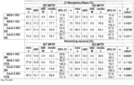
- % of remaining TH-positive fibers / neurons in the MPTP treated groups were calculated in respect to the mean of the corresponding saline treated group (particularly important for NCS-1 KO, as KO controls had lower numbers of SN DA neurons than WT, compare Fig. 2e ) and given in Fig. 3 and Supplementary Table S8C, Fig. 15 .
- each individual MPTP value was compared to each individual saline value of the respective groups for calculation of % of remaining TH-signal intensity and neuron numbers (see Fig.6 , and Supplementary Table S8D, Fig. 15 ).
Landscapes
- Health & Medical Sciences (AREA)
- Life Sciences & Earth Sciences (AREA)
- Engineering & Computer Science (AREA)
- Chemical & Material Sciences (AREA)
- Biomedical Technology (AREA)
- Molecular Biology (AREA)
- General Health & Medical Sciences (AREA)
- Medicinal Chemistry (AREA)
- Urology & Nephrology (AREA)
- Veterinary Medicine (AREA)
- Bioinformatics & Cheminformatics (AREA)
- Immunology (AREA)
- Hematology (AREA)
- Cell Biology (AREA)
- Pharmacology & Pharmacy (AREA)
- Neurosurgery (AREA)
- Animal Behavior & Ethology (AREA)
- Neurology (AREA)
- Public Health (AREA)
- Biotechnology (AREA)
- Food Science & Technology (AREA)
- Hospice & Palliative Care (AREA)
- Proteomics, Peptides & Aminoacids (AREA)
- Chemical Kinetics & Catalysis (AREA)
- Nuclear Medicine, Radiotherapy & Molecular Imaging (AREA)
- Psychiatry (AREA)
- Microbiology (AREA)
- General Chemical & Material Sciences (AREA)
- Organic Chemistry (AREA)
- Physics & Mathematics (AREA)
- Analytical Chemistry (AREA)
- Biochemistry (AREA)
- General Physics & Mathematics (AREA)
- Pathology (AREA)
- Emergency Medicine (AREA)
- Epidemiology (AREA)
- Medicines That Contain Protein Lipid Enzymes And Other Medicines (AREA)
- Pharmaceuticals Containing Other Organic And Inorganic Compounds (AREA)
Priority Applications (1)
| Application Number | Priority Date | Filing Date | Title |
|---|---|---|---|
| EP24188598.7A EP4467136A3 (de) | 2017-06-14 | 2017-06-14 | Antagonisten von kalziumkanälen vom typ cav2.3 r zur behandlung von neurodegenerativen erkrankungen |
Applications Claiming Priority (3)
| Application Number | Priority Date | Filing Date | Title |
|---|---|---|---|
| EP24188598.7A EP4467136A3 (de) | 2017-06-14 | 2017-06-14 | Antagonisten von kalziumkanälen vom typ cav2.3 r zur behandlung von neurodegenerativen erkrankungen |
| PCT/EP2017/064644 WO2018228692A1 (en) | 2017-06-14 | 2017-06-14 | Antagonists of cav2.3 r-type calcium channels for the treatment of neurodegenerative diseases |
| EP17737488.1A EP3638283B1 (de) | 2017-06-14 | 2017-06-14 | Antagonisten von calciumkanälen vom typ cav2.3 r zur behandlung von neurodegenerativen erkrankungen |
Related Parent Applications (1)
| Application Number | Title | Priority Date | Filing Date |
|---|---|---|---|
| EP17737488.1A Division EP3638283B1 (de) | 2017-06-14 | 2017-06-14 | Antagonisten von calciumkanälen vom typ cav2.3 r zur behandlung von neurodegenerativen erkrankungen |
Publications (2)
| Publication Number | Publication Date |
|---|---|
| EP4467136A2 true EP4467136A2 (de) | 2024-11-27 |
| EP4467136A3 EP4467136A3 (de) | 2025-02-26 |
Family
ID=59313195
Family Applications (3)
| Application Number | Title | Priority Date | Filing Date |
|---|---|---|---|
| EP24188598.7A Pending EP4467136A3 (de) | 2017-06-14 | 2017-06-14 | Antagonisten von kalziumkanälen vom typ cav2.3 r zur behandlung von neurodegenerativen erkrankungen |
| EP17737488.1A Active EP3638283B1 (de) | 2017-06-14 | 2017-06-14 | Antagonisten von calciumkanälen vom typ cav2.3 r zur behandlung von neurodegenerativen erkrankungen |
| EP18733210.1A Withdrawn EP3638285A1 (de) | 2017-06-14 | 2018-06-14 | Antagonisten von kalziumkanälen des typs cav2.3 r zur behandlung von neurodegenerativen erkrankungen |
Family Applications After (2)
| Application Number | Title | Priority Date | Filing Date |
|---|---|---|---|
| EP17737488.1A Active EP3638283B1 (de) | 2017-06-14 | 2017-06-14 | Antagonisten von calciumkanälen vom typ cav2.3 r zur behandlung von neurodegenerativen erkrankungen |
| EP18733210.1A Withdrawn EP3638285A1 (de) | 2017-06-14 | 2018-06-14 | Antagonisten von kalziumkanälen des typs cav2.3 r zur behandlung von neurodegenerativen erkrankungen |
Country Status (3)
| Country | Link |
|---|---|
| EP (3) | EP4467136A3 (de) |
| ES (1) | ES2992748T3 (de) |
| WO (2) | WO2018228692A1 (de) |
Families Citing this family (7)
| Publication number | Priority date | Publication date | Assignee | Title |
|---|---|---|---|---|
| GB202117127D0 (en) | 2021-11-26 | 2022-01-12 | Epidarex Exeed Ltd | Compounds |
| GB202117129D0 (en) | 2021-11-26 | 2022-01-12 | Epidarex Exeed Ltd | Compounds |
| GB202117126D0 (en) | 2021-11-26 | 2022-01-12 | Epidarex Exeed Ltd | Compounds |
| GB202308165D0 (en) | 2023-05-31 | 2023-07-12 | Lario Therapeutics Ltd | Compounds |
| GB202308164D0 (en) | 2023-05-31 | 2023-07-12 | Lario Therapeutics Ltd | Compounds |
| GB202308160D0 (en) | 2023-05-31 | 2023-07-12 | Lario Therapeutics Ltd | Compounds |
| WO2025158150A1 (en) | 2024-01-24 | 2025-07-31 | Lario Therapeutics Limited | Heterocyclic compounds as modulators of cav2.3 |
Family Cites Families (3)
| Publication number | Priority date | Publication date | Assignee | Title |
|---|---|---|---|---|
| FR2700117B1 (fr) * | 1993-01-07 | 1995-02-03 | Rhone Poulenc Rorer Sa | Application d'anticonvulsivants dans le traitement de la maladie de Parkinson et des syndromes parkinsoniens. |
| US9867837B2 (en) * | 2011-03-01 | 2018-01-16 | Pharnext | Compositions for treating neurological disorders |
| US20120321594A1 (en) * | 2011-05-06 | 2012-12-20 | New York University | Methods of controlling axon or dendrite development of neuronal cells |
-
2017
- 2017-06-14 ES ES17737488T patent/ES2992748T3/es active Active
- 2017-06-14 EP EP24188598.7A patent/EP4467136A3/de active Pending
- 2017-06-14 WO PCT/EP2017/064644 patent/WO2018228692A1/en not_active Ceased
- 2017-06-14 EP EP17737488.1A patent/EP3638283B1/de active Active
-
2018
- 2018-06-14 EP EP18733210.1A patent/EP3638285A1/de not_active Withdrawn
- 2018-06-14 WO PCT/EP2018/065893 patent/WO2018229230A1/en not_active Ceased
Non-Patent Citations (29)
Also Published As
| Publication number | Publication date |
|---|---|
| EP3638283A1 (de) | 2020-04-22 |
| ES2992748T3 (en) | 2024-12-17 |
| EP3638283C0 (de) | 2024-07-17 |
| EP3638283B1 (de) | 2024-07-17 |
| WO2018228692A9 (en) | 2023-11-09 |
| WO2018228692A1 (en) | 2018-12-20 |
| EP3638285A1 (de) | 2020-04-22 |
| WO2018229230A1 (en) | 2018-12-20 |
| EP4467136A3 (de) | 2025-02-26 |
Similar Documents
| Publication | Publication Date | Title |
|---|---|---|
| EP3638283B1 (de) | Antagonisten von calciumkanälen vom typ cav2.3 r zur behandlung von neurodegenerativen erkrankungen | |
| Acquarone et al. | Synaptic and memory dysfunction induced by tau oligomers is rescued by up-regulation of the nitric oxide cascade | |
| Basurto-Islas et al. | Therapeutic benefits of a component of coffee in a rat model of Alzheimer's disease | |
| Kamat et al. | Okadaic acid-induced Tau phosphorylation in rat brain: role of NMDA receptor | |
| US10758545B2 (en) | Methods to treat neurological diseases | |
| Duffney et al. | Shank3 deficiency induces NMDA receptor hypofunction via an actin-dependent mechanism | |
| Hao et al. | An evolutionarily conserved mechanism for cAMP elicited axonal regeneration involves direct activation of the dual leucine zipper kinase DLK | |
| Ruggieri et al. | Complete loss of the DNAJB6 G/F domain and novel missense mutations cause distal-onset DNAJB6 myopathy | |
| Wu et al. | microRNA-592 blockade inhibits oxidative stress injury in Alzheimer's disease astrocytes via the KIAA0319-mediated Keap1/Nrf2/ARE signaling pathway | |
| Liu et al. | Phosphodiesterase 2A as a therapeutic target to restore cardiac neurotransmission during sympathetic hyperactivity | |
| Ranek et al. | Muscarinic 2 receptors modulate cardiac proteasome function in a protein kinase G-dependent manner | |
| Nutter et al. | Choroid plexus mis-splicing and altered cerebrospinal fluid composition in myotonic dystrophy type 1 | |
| Yanagisawa et al. | EAAT1 variants associated with glaucoma | |
| US20200246429A1 (en) | Compositions and methods for the treatment or prevention of neurodegenerative disorders | |
| Chen et al. | HTRA2 variations in Taiwanese Parkinson’s disease | |
| US20240197760A1 (en) | Activators of integrated stress response pathway for protection against ferroptosis | |
| Song et al. | Neuronal FGF13 Inhibits Mitochondria‐Derived Damage Signals to Prevent Neuroinflammation and Neurodegeneration in a Mouse Model of Parkinson's Disease | |
| Liu et al. | Modulation of calcineurin activity in mouse brain by chronic oral administration of cyclosporine A | |
| Yu et al. | Transcriptional regulation of human FE65, a ligand of Alzheimer's disease amyloid precursor protein, by Sp1 | |
| Zhang et al. | TFP5 prevents 1-methyl-4-phenyl pyridine ion-induced neurotoxicity in mouse cortical neurons | |
| Fang et al. | Significance of the C-terminal globular domain and the extra tail of the calmodulin-like protein (Pinctada fucata) in subcellular localization and protein–protein interaction | |
| Saraiva | Mechanisms Underlying the Physiological Role of Amyloid Precursor Protein Glutamatergic Synapses | |
| Yang et al. | METTL14 integrates tumor-derived SAM to drive parabrachial epigenetic rewiring in pancreatic cancer | |
| Tirado-Class | Functional studies of DCAF14 in replication fork stability and genome integrity | |
| Cassidy et al. | Physiotherapy management of the ataxias towards best clinical practice: 2016 guideline update |
Legal Events
| Date | Code | Title | Description |
|---|---|---|---|
| PUAI | Public reference made under article 153(3) epc to a published international application that has entered the european phase |
Free format text: ORIGINAL CODE: 0009012 |
|
| STAA | Information on the status of an ep patent application or granted ep patent |
Free format text: STATUS: THE APPLICATION HAS BEEN PUBLISHED |
|
| AC | Divisional application: reference to earlier application |
Ref document number: 3638283 Country of ref document: EP Kind code of ref document: P |
|
| AK | Designated contracting states |
Kind code of ref document: A2 Designated state(s): AL AT BE BG CH CY CZ DE DK EE ES FI FR GB GR HR HU IE IS IT LI LT LU LV MC MK MT NL NO PL PT RO RS SE SI SK SM TR |
|
| REG | Reference to a national code |
Ref country code: DE Ref legal event code: R079 Free format text: PREVIOUS MAIN CLASS: A61K0031137000 Ipc: A61K0038160000 |
|
| PUAL | Search report despatched |
Free format text: ORIGINAL CODE: 0009013 |
|
| AK | Designated contracting states |
Kind code of ref document: A3 Designated state(s): AL AT BE BG CH CY CZ DE DK EE ES FI FR GB GR HR HU IE IS IT LI LT LU LV MC MK MT NL NO PL PT RO RS SE SI SK SM TR |
|
| RIC1 | Information provided on ipc code assigned before grant |
Ipc: G01N 33/68 20060101ALI20250121BHEP Ipc: A61K 31/137 20060101ALI20250121BHEP Ipc: A61P 25/28 20060101ALI20250121BHEP Ipc: A61K 45/06 20060101ALI20250121BHEP Ipc: A61K 38/16 20060101AFI20250121BHEP |
|
| STAA | Information on the status of an ep patent application or granted ep patent |
Free format text: STATUS: REQUEST FOR EXAMINATION WAS MADE |
|
| 17P | Request for examination filed |
Effective date: 20250822 |