EP4461802A1 - Cell dissociation device and method - Google Patents
Cell dissociation device and method Download PDFInfo
- Publication number
- EP4461802A1 EP4461802A1 EP24172687.6A EP24172687A EP4461802A1 EP 4461802 A1 EP4461802 A1 EP 4461802A1 EP 24172687 A EP24172687 A EP 24172687A EP 4461802 A1 EP4461802 A1 EP 4461802A1
- Authority
- EP
- European Patent Office
- Prior art keywords
- cell
- range
- shear
- cells
- component
- Prior art date
- Legal status (The legal status is an assumption and is not a legal conclusion. Google has not performed a legal analysis and makes no representation as to the accuracy of the status listed.)
- Pending
Links
Images
Classifications
-
- C—CHEMISTRY; METALLURGY
- C12—BIOCHEMISTRY; BEER; SPIRITS; WINE; VINEGAR; MICROBIOLOGY; ENZYMOLOGY; MUTATION OR GENETIC ENGINEERING
- C12M—APPARATUS FOR ENZYMOLOGY OR MICROBIOLOGY; APPARATUS FOR CULTURING MICROORGANISMS FOR PRODUCING BIOMASS, FOR GROWING CELLS OR FOR OBTAINING FERMENTATION OR METABOLIC PRODUCTS, i.e. BIOREACTORS OR FERMENTERS
- C12M45/00—Means for pre-treatment of biological substances
- C12M45/02—Means for pre-treatment of biological substances by mechanical forces; Stirring; Trituration; Comminuting
-
- C—CHEMISTRY; METALLURGY
- C12—BIOCHEMISTRY; BEER; SPIRITS; WINE; VINEGAR; MICROBIOLOGY; ENZYMOLOGY; MUTATION OR GENETIC ENGINEERING
- C12M—APPARATUS FOR ENZYMOLOGY OR MICROBIOLOGY; APPARATUS FOR CULTURING MICROORGANISMS FOR PRODUCING BIOMASS, FOR GROWING CELLS OR FOR OBTAINING FERMENTATION OR METABOLIC PRODUCTS, i.e. BIOREACTORS OR FERMENTERS
- C12M47/00—Means for after-treatment of the produced biomass or of the fermentation or metabolic products, e.g. storage of biomass
- C12M47/04—Cell isolation or sorting
-
- C—CHEMISTRY; METALLURGY
- C12—BIOCHEMISTRY; BEER; SPIRITS; WINE; VINEGAR; MICROBIOLOGY; ENZYMOLOGY; MUTATION OR GENETIC ENGINEERING
- C12N—MICROORGANISMS OR ENZYMES; COMPOSITIONS THEREOF; PROPAGATING, PRESERVING, OR MAINTAINING MICROORGANISMS; MUTATION OR GENETIC ENGINEERING; CULTURE MEDIA
- C12N5/00—Undifferentiated human, animal or plant cells, e.g. cell lines; Tissues; Cultivation or maintenance thereof; Culture media therefor
- C12N5/0062—General methods for three-dimensional culture
-
- C—CHEMISTRY; METALLURGY
- C12—BIOCHEMISTRY; BEER; SPIRITS; WINE; VINEGAR; MICROBIOLOGY; ENZYMOLOGY; MUTATION OR GENETIC ENGINEERING
- C12N—MICROORGANISMS OR ENZYMES; COMPOSITIONS THEREOF; PROPAGATING, PRESERVING, OR MAINTAINING MICROORGANISMS; MUTATION OR GENETIC ENGINEERING; CULTURE MEDIA
- C12N5/00—Undifferentiated human, animal or plant cells, e.g. cell lines; Tissues; Cultivation or maintenance thereof; Culture media therefor
- C12N5/06—Animal cells or tissues; Human cells or tissues
- C12N5/0602—Vertebrate cells
-
- C—CHEMISTRY; METALLURGY
- C12—BIOCHEMISTRY; BEER; SPIRITS; WINE; VINEGAR; MICROBIOLOGY; ENZYMOLOGY; MUTATION OR GENETIC ENGINEERING
- C12N—MICROORGANISMS OR ENZYMES; COMPOSITIONS THEREOF; PROPAGATING, PRESERVING, OR MAINTAINING MICROORGANISMS; MUTATION OR GENETIC ENGINEERING; CULTURE MEDIA
- C12N2509/00—Methods for the dissociation of cells, e.g. specific use of enzymes
- C12N2509/10—Mechanical dissociation
-
- C—CHEMISTRY; METALLURGY
- C12—BIOCHEMISTRY; BEER; SPIRITS; WINE; VINEGAR; MICROBIOLOGY; ENZYMOLOGY; MUTATION OR GENETIC ENGINEERING
- C12N—MICROORGANISMS OR ENZYMES; COMPOSITIONS THEREOF; PROPAGATING, PRESERVING, OR MAINTAINING MICROORGANISMS; MUTATION OR GENETIC ENGINEERING; CULTURE MEDIA
- C12N2521/00—Culture process characterised by the use of hydrostatic pressure, flow or shear forces
-
- C—CHEMISTRY; METALLURGY
- C12—BIOCHEMISTRY; BEER; SPIRITS; WINE; VINEGAR; MICROBIOLOGY; ENZYMOLOGY; MUTATION OR GENETIC ENGINEERING
- C12N—MICROORGANISMS OR ENZYMES; COMPOSITIONS THEREOF; PROPAGATING, PRESERVING, OR MAINTAINING MICROORGANISMS; MUTATION OR GENETIC ENGINEERING; CULTURE MEDIA
- C12N2527/00—Culture process characterised by the use of mechanical forces, e.g. strain, vibration
Definitions
- a method for dissociating a mammalian cell agglomeration in the range of between greater than (>30) ⁇ m to 300 ⁇ m after harvest into single cells or cell oligomers in the range of 8-30 ⁇ m comprising the steps of
- cell dissociation device (1) for dissociating a mammalian cell agglomeration in the range of between greater than (>)30 ⁇ m to 300 ⁇ m into single cells or cell oligomers in the range of 8-30 ⁇ m comprising
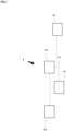
- Fig. 1 depicts an exemplary cell dissociation device (1) for dissociating mammalian cell agglomerations in the range of between greater than (>) 30 ⁇ m to 300 ⁇ m into single cells or cell oligomers in the range of 8-30 ⁇ m comprising a shear component (2) exerting a maximal shear in the range of between 2 Pa and 500 Pa, a recirculation loop (3) with a volume in the range of between 100 ml and 1000 ml, a recirculation component (4), an inlet (5) and an outlet (6).
- a shear component exerting a maximal shear in the range of between 2 Pa and 500 Pa
- a recirculation component (4) an inlet (5) and an outlet (6).
- a method for dissociating a mammalian cell agglomeration in the range of between greater than (>)30 ⁇ m to 300 ⁇ m after harvest into single cells or cell oligomers in the range of 8-30 ⁇ m comprising the steps of
- This method ensures a defined dissociation thereby providing a consistent cell quality as well as sufficient cell quantity. Hence via using the method described herein cells suitable for cell therapy applications can be reliably provided.
- shear component (2) refers to an active device or a passive structure that exerts a maximal shear in the range of between 2 Pa and 500 Pa on the cells to be dissociated while limiting an impact on cell viability.
- a given device is suitable as shear component in a given setting methods known in the art can be employed.
- Villiger et al. 2015 Villiger et al AIChEJ 61-2015 ) describe how the shear can be assessed using CFD simulations in case of contract nozzles which at certain flow rates and diameters exert a shear stress within the required range of between 2 Pa and 500 Pa (cf. Table 1 of Villiger et al.
- Villiger et al. 2018 Villiger et al . 2018 Biochemical Engineering Journal Volume 131, 15 March 2018, Pages 84-94 ) describe how shear was CFD modeled for different bioreactor systems exerting between 1 and 20 Pa shear (cf. Fig. 2c of Villiger et al. 2018) and these CFD model calculations were compared to measured shear values obtained in experiments employing the PMMA particles thereby correlating the size of the PMMA aggregates to the corresponding shear. It was found that the predicted values of maximum stress correlated well with those calculated by CFD (Villiger et al.
- the method described herein could also be worded as a method for dissociating a mammalian cell agglomeration in the range of between greater than (>)30 ⁇ m to 300 ⁇ m after harvest into single cells or cell oligomers in the range of 8-30 ⁇ m comprising the steps of
- a cell agglomeration is meant to also comprise the plural i.e. cell agglomerations.
- the maximal shear exerted by the least one shear component (2) is in the range of between 2 Pa and 500 Pa, preferably in the range of between 2 Pa and 100 Pa, more preferably in the range between 2 Pa and 50 Pa, even more preferably in the range of between 2 Pa and 25 Pa, most preferably in the range of 2,5 Pa to 25 Pa or in the range of 2,4 Pa to 25 Pa or in the range of 6 Pa to 25 Pa.
- cell viability remains in the range of between 70% to 100% (accordingly, the loss of cell viability in this embodiment is in the range of between 0% and 30% compared to the cell viability of cells and cell agglomerations that were not subjected to additional shear stress). It is preferred that the loss of cell viability in this embodiment is in the range of between 0% and 25% or in the range of between 1% to 20 % or in the range of between 1% to 13%. This loss of cell viability is measured as described below in example 3.
- the cell debris volume as determined using a FlowCam (8000 Analyzer, Fluid Imaging Technologies Inc,. A 10x objective) with the respective flow cell and setting as described in example 3 does not exceed 2% i.e. is in the range of 0,01% to 2%.
- cell viability remains in the range of between 70% to 100% and the cell debris volume as determined by FlowCam does not exceed 2% i.e. is in the range of 0,01% to 2%.
- loss of cell viability is in the range of between 0% and 30% compared to the cell viability before harvest the cell debris volume as determined by FlowCam does not exceed 2% i.e. is in the range of 0,01% to 2%.
- the loss of cell viability is in the range of between 1% and 13% compared to the cell viability before harvest the cell debris volume as determined by FlowCam does not exceed 2% i.e. is in the range of 0,1% to 1%.
- the method is a method for dissociating a mammalian cell agglomeration in the range of between greater than (>)30 ⁇ m to 300 ⁇ m after harvest into single cells or cell oligomers in the range of 8-30 ⁇ m comprising the steps of
- the method is a method for dissociating a mammalian cell agglomeration in the range of between greater than (>)30 ⁇ m to 300 ⁇ m after harvest into single cells or cell oligomers in the range of 8-30 ⁇ m comprising the steps of
- the method is a method for dissociating a mammalian cell agglomeration in the range of between greater than (>)30 ⁇ m to 300 ⁇ m after harvest into single cells or cell oligomers in the range of 8-30 ⁇ m comprising the steps of
- the shear component (2) is a passive structure and selected from the group consisting of
- the shear component (2) is an active device and selected from the group consisting of a pump, an ultrasonic device and a Completely Stirred Tank Reactor (CSTR).
- the shear component (2) is especially preferred to be an non-positive displacement, i.e., dynamic pump such as a Levitronix Puralev pump as in this case the device can be simplified by employing the at least one at least one recirculation component (4) also as shear component (2).
- the device (1) only comprises one non-positive displacement, i.e., dynamic pump which acts as both the at least one recirculation component (4) and as shear component (2).
- shear components (2) can be combined and that there are embodiment in which the least one recirculation component (4) can also act as shear component (2).
- Exemplary shear devices are pumps, rheometers allowing for defined shear rates as well as plate/plate, plate/cone and rotational cylinder devices.
- cell agglomeration refers to a plurality of cells in which an association between the cells is caused by cell-cell interaction (e.g., by biologic attachments to one another).
- Biological attachment may be, for example, through surface proteins, such integrins, immunoglobulins, cadherins, selectins, or other cell adhesion molecules.
- cells may spontaneously associate in suspension and form cell-cell attachments (e.g., self-assembly), thereby forming aggregates, or cells may be forced to aggregate..
- a cell agglomeration/ aggregate/oligomer may be substantially homogeneous (i.e., mostly containing cells of the same type). In some embodiments, a cell agglomeration/ aggregate/oligomer may be heterogeneous, (i.e., containing cells of more than one type).
- the average diameter of each cell agglomerations is in the range of between greater than (>)30 ⁇ m and 800 ⁇ m - i.e. in the range of between 30.1 ⁇ m and 800 ⁇ m - whereas single cells and cell oligomers i.e. cell duplets or triplets have diameters in the range of 8-30 ⁇ m.
- single cells and cell oligomers i.e. cell duplets or triplets have diameters in the range of 8-30 ⁇ m.
- cell oligomers i.e. cell duplets or triplets could be larger.
- small cell agglomerations at the lower end of the diameter range e.g. of around 30 ⁇ m are further dissociated, but that the overall diameter range of all cell aggregates is more uniform.
- the average diameter of cell agglomerations is in the range of between greater than (>) 30 ⁇ m and 300 ⁇ m e.g. 50 ⁇ m to 300 ⁇ m. This can be measured via microscopy e.g. using a FlowCam.
- Passage and “passaging” refer to the process of sub-culturing adherent cells, in which cell adhesion is disrupted and the cell density (number of cells per unit volume or area) is reduced by addition of fresh medium. Passaging is a technique that may be used to expand the number of cells in a culture and/or prolong the life of a cell line or the cells in culture.
- expansion refers to the proliferation of a cell, with or without differentiation, and may include no passaging, one passage or more than one passage and/or serial passages.
- dissociate and “dissociation” refer to a process of separating aggregated cells from one another. For example, during dissociation, the cell-cell interaction between cells and between cells and extracellular matrix in the aggregate may be disrupted, thereby breaking apart the cells in the aggregate.
- dissociated and dissociated aggregate refer to single cells, or cell aggregates or clusters that are smaller than the original cell aggregates (i.e., smaller than a pre-dissociation aggregate).
- a dissociated aggregate may comprise about 50% or less surface area, volume, or diameter relative to a pre-dissociation cell aggregate.
- the method is a method for dissociating a mammalian cell agglomerations in the range of between greater than (>)30 ⁇ m to 300 ⁇ m after harvest into single cells or cell oligomers in the range of 8-30 ⁇ m comprising the steps of
- dissociation reagent refers to a solution comprising one or more agents that separate cells from one another, such as, for example, enzyme(s) and/or chelating agent(s).
- a dissociation reagent may break the bonds between cells and/or between extracellular matrix proteins and cells, thereby disrupting the aggregation of cells in suspension.
- a dissociation reagent may cause sequestration of a molecule to weaken or break bond formation between cell adhesion proteins (e.g.
- chelation to disrupt calcium dependent adhesion molecules it may cause cleavage of proteins (e.g., serine proteases such as trypsin, collagenases, dispase, or papain) and/or other extracellular matrix components (e.g., hyaluronidase) to which cell surface molecules bind or within which cells becomes entrapped, and/or cleave such binding molecules from the cell surface (e.g., cleavage of integrins by serine proteases).
- proteins e.g., serine proteases such as trypsin, collagenases, dispase, or papain
- extracellular matrix components e.g., hyaluronidase
- the treatment before, after or during the method for dissociating with a dissociation reagents comprises the possibility that the treatment with the dissociation reagent takes place before harvest.
- the term "recirculation loop" refers to a device which allows cycling of the cells in suspension i.e. if cycled twice on average each cell of a cell suspension should pass a given point of the device twice .
- the recirculation loop does not comprise a bioreactor.
- the cells i.e. the single cells or cell oligomers in the range of 8 ⁇ m to 30 ⁇ m are subjected to an additional wash step after cell dissociation.
- This additional wash step has proved beneficial since it has been unexpectedly found that on large scale mechanical and enzymatic dissociation is not sufficient - unlike described in WO2017127921A1 - as structures termed "strings" appear that hinder the cells from dissociating.
- this additional wash step is especially preferred if the method and the device described herein are employed on large scale i.e. for volumes in the range of 1l - 200l.
- the cells are concentrated prior this additional wash step.
- the additional wash step can include exposure to DNAse.
- the method further also comprising a chemical dissociation step and/or increasing the temperature e.g. via an inline heat exchanger for heating the dissociation media and/or solution with dissociating cells.
- a chemical dissociation step and/or increasing the temperature e.g. via an inline heat exchanger for heating the dissociation media and/or solution with dissociating cells.
- the chemical dissociation is carried out after or while the shear component (2) exerts the shear stress in the range of between 2 Pa and 500 Pa, preferably 2,4 Pa to 25 Pa,on the cells to be dissociated thereby reducing the time cells must spend in a single cell state, which decreases their viability.
- the recirculation ratio is in the range between 3-10 (cf. example 4)
- the recycle (recirculation) ratio is the volume of cell suspension returned to the shear device entrance divided by the volume leaving the system, i.e. the volume processed by (2) and leaving the system (5).
- the method is continuous or semi-continuous. If the method is continuous and “one way” this ensures that each cell (aggregate) is only stressed once.
- the shear component is a nozzle with an inner diameter in the range of between 0.5mm - 2 mm, preferably 0.7mm -1.5mm.
- the shear component is a nozzle with a inner diameter of 1.5 mm and the cells to be dissociated pass the nozzle at a flow rate of rate of 19.3 - 62.5 (mL/min) and a velocity of between 0.18 and 0.59 (m/s).
- the shear component is a nozzle with a inner diameter of 1 mm and the cells to be dissociated pass the nozzle at a flow rate of rate of 41.1 (mL/min) and a velocity of 0.87.
- the cells to be dissociated are mammalian cells.
- the cells to be dissociated are chosen from the group consisting of
- cell dissociation device (1) for dissociating a mammalian cell agglomeration in the range of between greater than (>)30 ⁇ m to 300 ⁇ m into single cells or cell oligomers in the range of 8-30 ⁇ m comprising
- the cell dissociation device (1) for dissociating cell a mammalian agglomeration in the range of between greater than (>)30 ⁇ m to 300 ⁇ m into single cells or cell oligomers in the range of 8-30 ⁇ m comprises
- the combination of recirculation loop and the defined shear component reduces the variability of shear stress encountered by cells and still ensured a defined dissociation at an adequate shear value required to provide a consistent cell quality as well as sufficient cell quantity. Hence via using the device and the method described herein cells suitable for cell therapy applications can be provided on a large scale.
- the at least one recirculation component (4) is at the same time also the at least one shear component (2).
- the at least one recirculation component (4) is at the same time also the at least one shear component (2)
- the at least one recirculation component/ at least one shear component (2 &4) is a non-positive displacement, i.e., dynamic pump.
- the at least one recirculation component (4) is at the same time also the at least one shear component (2)
- the at least one recirculation component/ at least one shear component (2 &4) is a Levitronix PuraLev pump.
- the at least one recirculation loop can be made of single use tubing and preferably has a tubing length in the range of between 0.3 m and 10 m with inner tubing diameters between 3mm and 3 cm. It can comprise a bubble trap and/or a single use reactor.
- the other components especially the at least one shear component (2) exerting a maximal shear in the range of between 2 Pa and 500 Pa preferably 2,4 Pa to 25 Pa the at least one recirculation loop (3) with a volume in the range of between 100 ml and 1000 ml the at least one recirculation component (4) can be single-use.
- the Levitronix PuraLev pump is a single use pump which can be employed as shear component (2) and/or as recirculation component (4)
- the recirculation loop (3) is has a small volume of no more than the volume supplied via inlet (5) more preferred less than 20%, better less than 10% of the volume supplied via inlet (5) thereby further reducing the variability of the total shear stress encountered by each cell in a cell suspension.
- the volume of (cell culture) fluid within the recirculation shear device is smaller than 100 mL.
- the at least one recirculation component (4) is a pump.
- the at least one device (1) can further comprise
- the cell dissociation device comprises at least one cell retention device. This allows for the removal of single cells prior to dissociation as the single cells do not have to be dissociated further.
- cell retention devices are cell settlers, sedimentation devices, hydrocline devices, centrifuges and ultrasonic separation devices and filtration devices.
- the cell dissociation device comprises at least one additional inlet e.g. to add a dissociation reagent.
- all components of the device (1) are single-use components.
- the device (1) comprises a rheomicroscope as this allows to consistently quantify which shear stress was applied to cell agglomerations.
- a filling device and filling process of cell therapy products such as for example single cells or cell oligomers in the range of 8-30 ⁇ m employing a recirculation loop in combination with a non-positive displacement, i.e., dynamic pump.
- a non-positive displacement i.e., dynamic pump.
- this combination surprisingly was beneficial for cell viability and single cell content compared to using a positive displacement pump.
- a Levitronix Puralev pump is used.
- the cell dissociation device (1) described herein and the cell filling device are one and the same device.
- what is described herein relates to another embodiment of the filling process of cell therapy products such as for example single cells or cell oligomers in the range of 8-30 ⁇ m.
- a tube for filling cell therapy products where the tube had a inner diameter in the range of between 0,85 mm and 1,8 mm, preferably 1,2 mm i.e. a ratio of tube inner diameter to cell therapy product diameter in the range of between 28:1 and 60:1 was advantageous.
- a (flow) velocity in the range of between 0.45 (m/s) and 1 (m/s) exerting a corresponding shear stress was advantageous .
- it was more advantageous to use a tube for the filling process of cell therapy products such as for example single cells or cell oligomers in the range of 8-30 ⁇ m that is as short as possible.
- the tube for the filling process of cell therapy products such as for example single cells or cell oligomers in the range of 8-30 ⁇ m is a single use tube.
- a recirculation loop in combination with a non-positive displacement pump can for example be combined with the a tube for filling cell therapy products described above.
- Examples 1 and 2 were conducted to correlate a given shear stress to a corresponding particle size and vice versa so that a person skilled in the art can determine whether a given device, exerts the required shear stress.
- PMMA particles were prepared as described in Villiger 2015 .
- the experimental setting was as described in example 3 below.
- a contracting nozzle was designed and (Custom made, Sausen Maschinenbau GmbH) connected to two serological pipettes (50 mL Costar, Corning Inc.) using a silicon tubing, 7 cm in length/ 4.8 mm in Inner-diameter (FisherScientific))
- a liquid suspension comprising the PMMA particles in demineralized water with Kolliphor ® P 188 Geismar (0.01 g/L) and Ex-cell ® Antifoam gamma irradiated (0.0045 g/L) was prepared.
- the nozzle as well as both serological pipettes were placed in a double-walled 5 L bioreactors (bbi-biotech GmbH) connected to a Thermostat (CF41, Julabo) in order to maintain a defined temperature of the liquid suspension.
- One serological pipette was connected to a syringe pump (Syrdos, HiTec Zang)to adjust the flow rate accurately through the nozzle.
- the nozzle as well as both serological pipettes were placed in a double-walled 5 L bioreactors (bbi-biotech GmbH) connected to a Thermostat (CF41, Julabo) in order to maintain a defined temperature of the liquid suspension/cell suspension.
- One serological pipette was connected to a syringe pump (Syrdos, HiTec Zang) to adjust the flow rate accurately through the nozzle.
- the complete liquid suspension comprising he cells was pumped through the nozzle 200 times as Villiger et al. 2015 ( Villiger et al AIChEJ 61-2015 ) showed in Fig. 4 that around 200 passes are required for PMMA particles to achieve a steady aggregate size.
- Cell damage was measured in terms of cell debris Biovolume (%) using a FlowCam (8000 Analyzer, Fluid Imaging Technologies Inc,. A 10x objective) with the respective flow cell.
- Cells were diluted with PBS buffer to 100.000 cells/mL.
- the sample volume was 150 ⁇ L.
- Everything smaller than 11 ⁇ m and with a circle fit* of ⁇ 0.8 was considered debris.
- Everything below 7 ⁇ m was not be detected.
- Nucleocounter NC-202 from Chemometec according to the manufacturer's instructions i.e. measuring Acridin Orange (AO), which stained nuclei and membranes. Everything that deviated (non-ring structures or without center AO-peak) were classified as debris.
- AO Acridin Orange
- the cell debris consisted of cell fragments derived from necrotic cells which in this experiment were predominantly the result of the shear stress and hence cell debris was taken as measure of shear stress.
- the cell debris biovolume was used to normalize the generated cell debris by dividing the volume of cell debris by the total volume of objects detected with the FlowCam Analyzer assuming all particles and debris had a spherical shape.
- Table 5 shows that the loss of cell viability in the shear stress range of between 0 and 50 Pa remains in the range of the loss of cell viability in the sample that was not subjected to shear stress .
- Table 5 Loss of cell viability in relation to shear stress after 200 nozzle passes Shear stress in Pa 0 2,4 6 14 25 50 Loss of cell viability (%) 12,8 13,3 9 8,8 9,9 11,8 Viable cell density 1,2 ⁇ 10 6 9,86 ⁇ 10 5 1.01 ⁇ 10 6 4,52 ⁇ 10 5 5,3 ⁇ 10 5 7,77 ⁇ 10 5
- Table 5 Dissociation in relation to shear stress after 200 nozzle passes Shear stress in Pa 0 2,4 6 14 25 50 100 300 1000 Single cells In % Biovolume 3 10 13 20 18 12 30 40 51 Cell aggregates greater than (>) 30 ⁇ m in % Biovolume 78 63 54 36 41 58 20 6 4 Ratio single cells: cell aggregates greater than (>) 30 ⁇ m in % Biovolume 1:26 1:6,3 1:4,2 1:1,8 1:2,3 1:4,8 1:1,5 1:0,15 1:0,08
- Table 5 demonstrates that in order to dissociate nearly all cell agglomerations into single cells at 200 nozzle passes shear stress values of 50 Pa and above were necessary (single cells were defined as cells without attachment to other cells). However, as Table 4 showed that shear stress values of around 50 Pa already considerably increase the cell debris volume it was determined in Example 4 how many nozzle passes at a shear below 50 Pa were necessary to reach an acceptable ratio of single cells to cell aggregates greater than (>) 30 ⁇ m.
- the experimental set up was the same as described for example 3 except that the flow rate was not varied but kept constant at 62.5 mL/min to achieve a shear of 13.9 Pa.
- the nozzle with the diameter of 1.5 mm was used and the cells were passed several times (cf. Table 6) through the nozzle.
- Aggregate size was measured by a FlowCam [FlowCam 8000Analyzer, Fluid Imaging Technologies Inc] . A 10x objective with the respective flow cell was used.
- Cells were diluted with PBS buffer to 100.000 cells/mL.
- the sample volume was 150 ⁇ L. Single cells were defined as cells without attachment to other cells.
- Example 5 The at least one recirculation component (4) also acting as shear component (2)
- the set-up of this experiment comprised a source bag with a cell suspension (here 70 ml iPSC at a concentration of 13,3 6 cells at 94 % viability suspended in a 1:1 mixture of mTeSR cultivation media (STEMCELL Technolgies) and Cryostor10 cryoformulation buffer (STEMCELL Technologies), a recirculation pump - either a non-positive displacement, i.e., dynamic pump (here a Levitronix PuraLev i30 SU) or a positive displacement pump (here a peristaltic pump with a Cytoflow pump head with 3 rollers)- a sampling port (here a Luer lock sampling valve from Eppendorf) and a recirculation loop.
- a non-positive displacement i.e., dynamic pump (here a Levitronix PuraLev i30 SU) or a positive displacement pump (here a peristaltic pump with a Cytoflow pump head with 3 rollers)- a sampling port
- the recirculation loop in the experimental setup of the Levitronic pump with a total length of 62 cm consisted of a 3.2mm inner diameter C-flex tubing (St. Gobain) whereas the recirculation loop in the experimental setup of the Cytoflow pump consisted of a 3.2mm inner diameter C-flex tubing (St. Gobain) and a 4.8 mm inner diameter PharMed BPT LS25 with a combined length of 65 cm .
- the pump rate was 500 mL/min .
- the experiment was conducted at room temperature for 120 min.
- the amount of single cells was analyzed for both pump types using a Flowcam with a 10x objective with the respective flow cell (FlowCam 8000 Analyzer, Fluid Imaging Technologies Inc.).
- the at least one recirculation component can also act as shear component which simplifies the device.
- the recirculation component can also act as shear component it was found that a non-positive displacement, i.e., dynamic pump leads to higher percentage of single cells than a positive displacement pump.
- the set-up described in this experiment can be employed for the filling process of cell therapy products to avoid variations of the cell concentration in vials to be filled. This is the case as the recirculation loop ensures constant motion of the cell suspension thereby avoiding settling of cells and the employed dynamic pump in contrast to the positive displacement peristaltic pump ensured high cell viability and high single cell content.
- the set-up of this experiment was a follows: either a cell suspension of hiPSCs at a concentration of 5-20 ⁇ 10 6 cells per ml of CyroStore or StemCellbanker formulation medium with an average cells size of 16 ⁇ m or a suspension comprising polystyrene (PS) particles at a concentration of 1 Mio per ml in formulation media (e.g. CyroStore or StemCellbanker) with an average size of 15 or 20 ⁇ m were provided in a source bag with a volume between 125 and 1000 mL. It had been shown previously that the PS particles behave in a similar fashion as the hiPSC cells (data not shown).
- PS polystyrene
- Tubes from Saint-Gobain and/or Watson-Marlow with different inner diameters - as given in Table 9 - were used to aseptically connect the source bag with an automatic cell filling system based on the L1 or M1 AT filler from Aseptic Technologies.
- the cells or PS particles were pumped at a pump rate of 400 RPM into vials [mL per vial] using a Flexicon PF7+ dosing pump from Watson-Marlow.
- the experiment was conducted at room temperature. After priming and calibration of the pump the pump was paused for 8 - 12 minutes to mimic inherent processes pauses of the AT filler (e.g. a vial rack exchange). Thereafter the experiment was continued for another 2-5 min.
Landscapes
- Life Sciences & Earth Sciences (AREA)
- Engineering & Computer Science (AREA)
- Health & Medical Sciences (AREA)
- Organic Chemistry (AREA)
- Chemical & Material Sciences (AREA)
- Bioinformatics & Cheminformatics (AREA)
- Wood Science & Technology (AREA)
- Zoology (AREA)
- Biomedical Technology (AREA)
- Biotechnology (AREA)
- Genetics & Genomics (AREA)
- Biochemistry (AREA)
- Microbiology (AREA)
- General Engineering & Computer Science (AREA)
- General Health & Medical Sciences (AREA)
- Cell Biology (AREA)
- Molecular Biology (AREA)
- Sustainable Development (AREA)
- Mechanical Engineering (AREA)
- Micro-Organisms Or Cultivation Processes Thereof (AREA)
- Apparatus Associated With Microorganisms And Enzymes (AREA)
Abstract
What is described herein relates to a method and a device for dissociating cell agglomerations in the range of between >30 µm to 300 µm after harvest into single cells or cell oligomers in the range of 8-30 µm comprising the steps of
• providing cell agglomerations in the range of between >30 µm to 300 µm
• passing said cell agglomerations in the range of between >30 µm to 300 µm through a recirculation loop
• bringing said cell agglomerations in the range of between >30 µm to 300 µm into contact with a shear component exerting a maximal shear in the range of between 2 Pa and 500 Pa thereby dissociating the cell agglomerations in the range of between >30 µm to 300 µm into single cells or cell oligomers in the range of 8-30 µm.
• providing cell agglomerations in the range of between >30 µm to 300 µm
• passing said cell agglomerations in the range of between >30 µm to 300 µm through a recirculation loop
• bringing said cell agglomerations in the range of between >30 µm to 300 µm into contact with a shear component exerting a maximal shear in the range of between 2 Pa and 500 Pa thereby dissociating the cell agglomerations in the range of between >30 µm to 300 µm into single cells or cell oligomers in the range of 8-30 µm.
Description
- An established way of culturing various cell types such as induced pluripotent stem cells, embryoid bodies and embryonic stem cells is suspension culture, where single cells or small aggregates of cells grow and multiply in a growth medium. Dissociation of the cell aggregates is often necessary in order to e.g. reduce aggregate size, reach a uniform aggregate size, achieve a single cell suspension for cell counting, reinoculation or further processing. In general enzymatic or mechanical means or a combination of both are used for dissociation. With both approaches minimizing cell damage has to be balanced out with dissociation efficiency. If the cells are to be employed in cell therapy applications mechanical dissociation means are preferred in order to avoid any residues of enzymatic dissociation components in the final cell therapy product. However, existing mechanical cell dissociation technologies often fail to deliver a consistent cell quality. One reason being that the dissociation is carried out manually - e.g. via pipetting up-and down - and differences between the manual handlers exist. Another cause for varying cell quality is that if means such sonication are used the mechanical forces negatively impact cell viability. Hence a need exists for a cell dissociation which delivers consistent cell quality.
- This need is met by a method for dissociating a mammalian cell agglomeration in the range of between greater than (>) 30 µm to 300 µm after harvest into single cells or cell oligomers in the range of 8-30 µm comprising the steps of
- providing a mammalian cell agglomeration in the range of between greater than (>) 30 µm to 300 µm
- passing said mammalian cell agglomeration in the range of between greater than (>) 30 µm to 300 µm through a recirculation loop
- bringing said mammalian cell agglomerations in the range of between greater than (>)30 µm to 300 µm into contact with a shear component (2) exerting a maximal shear in the range of between 2 Pa and 500 Pa
- In a first aspect what is described herein relates to a method for dissociating a mammalian cell agglomeration in the range of between greater than (>30) µm to 300 µm after harvest into single cells or cell oligomers in the range of 8-30 µm comprising the steps of
- providing a mammalian cell agglomeration in the range of between greater than (>)30 µm to 300 µm
- passing said mammalian cell agglomeration in the range of between greater than (>)30 µm to 300 µm through a recirculation loop
- bringing said mammalian cell agglomeration in the range of between greater than (>)30 µm to 300 µm into contact with a shear component (2) exerting a maximal shear in the range of between 2 Pa and 500 Pa
- In a second aspect what is described herein relates to cell dissociation device (1) for dissociating a mammalian cell agglomeration in the range of between greater than (>)30 µm to 300 µm into single cells or cell oligomers in the range of 8-30µm comprising
- at least one shear component (2) exerting a maximal shear in the range of between 2 Pa and 500 Pa
- at least one recirculation loop (3) with a volume in the range of between 100 ml and 1000 ml
- at least one recirculation component (4)
- at least one inlet (5)
- and at least one outlet (6),
-
Fig. 1 depicts an exemplary cell dissociation device (1) for dissociating mammalian cell agglomerations in the range of between greater than (>) 30 µm to 300 µm into single cells or cell oligomers in the range of 8-30µm comprising a shear component (2) exerting a maximal shear in the range of between 2 Pa and 500 Pa, a recirculation loop (3) with a volume in the range of between 100 ml and 1000 ml, a recirculation component (4), an inlet (5) and an outlet (6). - In a first aspect what is described herein relates to a method for dissociating a mammalian cell agglomeration in the range of between greater than (>)30 µm to 300 µm after harvest into single cells or cell oligomers in the range of 8-30 µm comprising the steps of
- providing a mammalian cell agglomeration in the range of between greater than (>)30 µm to 300 µm
- passing said mammalian cell agglomeration in the range of between greater than (>)30 µm to 300 µm through a recirculation loop
- bringing said mammalian cell agglomeration in the range of between greater than (>)30 µm to 300 µm into contact with a shear component (2) exerting a maximal shear in the range of between 2 Pa and 500 Pa
- This method ensures a defined dissociation thereby providing a consistent cell quality as well as sufficient cell quantity. Hence via using the method described herein cells suitable for cell therapy applications can be reliably provided.
- As used herein the term shear component (2) refers to an active device or a passive structure that exerts a maximal shear in the range of between 2 Pa and 500 Pa on the cells to be dissociated while limiting an impact on cell viability. In order to determine whether a given device is suitable as shear component in a given setting methods known in the art can be employed. For example Villiger et al. 2015 (Villiger et al AIChEJ 61-2015) describe how the shear can be assessed using CFD simulations in case of contract nozzles which at certain flow rates and diameters exert a shear stress within the required range of between 2 Pa and 500 Pa (cf. Table 1 of Villiger et al. 2015) also shear stress sensitive Poly(methyl methacrylate (PMMA) particles were developed. Villiger et al. 2018 (Villiger et al . 2018 Biochemical Engineering Journal Volume 131, 15 March 2018, Pages 84-94) describe how shear was CFD modeled for different bioreactor systems exerting between 1 and 20 Pa shear (cf. Fig. 2c of Villiger et al. 2018) and these CFD model calculations were compared to measured shear values obtained in experiments employing the PMMA particles thereby correlating the size of the PMMA aggregates to the corresponding shear. It was found that the predicted values of maximum stress correlated well with those calculated by CFD (Villiger et al. 2018) hence both methods can be employed to determine whether a given device can be used as shear component exerting a shear in the range of between 2-500 Pa. 3. In 2022 Srom et al. verified that the PMMA method can be used to determine the shear in a 250ml ambr250® bioreactor system (Srom et al Biochemical Engineering Journal 177 (2022). Hence in general a person skilled in the art can employ various methods, especially CFD simulation and the PMMA method, to determine the shear exerted by a given device in a given setting. If one method has to be chosen e.g. to exactly compare different system the PMMA method is to be used.
- To a skilled person it is clear that the method described herein could also be worded as a method for dissociating a mammalian cell agglomeration in the range of between greater than (>)30 µm to 300 µm after harvest into single cells or cell oligomers in the range of 8-30 µm comprising the steps of
- providing a mammalian cell agglomeration in the range of between greater than (>)30 µm to 300 µm
- passing said mammalian cell agglomeration in the range of between greater than (>)30 µm to 300 µm through a recirculation loop
- exerting a maximal shear in the range of between 2 Pa and 500 Pa on said mammalian cell agglomeration in the range of between greater than (>)30 µm to 300 µm wherein the said shear is generated by a shear component (2)
- Moreover, the term a cell agglomeration is meant to also comprise the plural i.e. cell agglomerations.
- The maximal shear exerted by the least one shear component (2) is in the range of between 2 Pa and 500 Pa, preferably in the range of between 2 Pa and 100 Pa, more preferably in the range between 2 Pa and 50 Pa, even more preferably in the range of between 2 Pa and 25 Pa, most preferably in the range of 2,5 Pa to 25 Pa or in the range of 2,4 Pa to 25 Pa or in the range of 6 Pa to 25 Pa.
- In an embodiment of the method described herein cell viability remains in the range of between 70% to 100% (accordingly, the loss of cell viability in this embodiment is in the range of between 0% and 30% compared to the cell viability of cells and cell agglomerations that were not subjected to additional shear stress). It is preferred that the loss of cell viability in this embodiment is in the range of between 0% and 25% or in the range of between 1% to 20 % or in the range of between 1% to 13%. This loss of cell viability is measured as described below in example 3.
- In another embodiment of the method described herein the cell debris volume as determined using a FlowCam (8000 Analyzer, Fluid Imaging Technologies Inc,. A 10x objective) with the respective flow cell and setting as described in example 3 does not exceed 2% i.e. is in the range of 0,01% to 2%.
- In an embodiment of the method described herein cell viability remains in the range of between 70% to 100% and the cell debris volume as determined by FlowCam does not exceed 2% i.e. is in the range of 0,01% to 2%.
- In a further embodiment of the method described herein loss of cell viability is in the range of between 0% and 30% compared to the cell viability before harvest the cell debris volume as determined by FlowCam does not exceed 2% i.e. is in the range of 0,01% to 2%. Preferably in this embodiment the loss of cell viability is in the range of between 1% and 13% compared to the cell viability before harvest the cell debris volume as determined by FlowCam does not exceed 2% i.e. is in the range of 0,1% to 1%.
- Hence, in a preferred embodiment of the method for dissociating a mammalian cell agglomeration the method is a method for dissociating a mammalian cell agglomeration in the range of between greater than (>)30 µm to 300 µm after harvest into single cells or cell oligomers in the range of 8-30 µm comprising the steps of
- providing a mammalian cell agglomeration in the range of between greater than (>)30 µm to 300 µm
- passing said mammalian cell agglomeration in the range of between greater than (>)30 µm to 300 µm through a recirculation loop
- bringing said mammalian cell agglomeration in the range of between greater than (>)30 µm to 300 µm into contact with a shear component exerting a maximal shear in the range of between 2 Pa and 500 Pa, preferably 2,4 Pa to 25 Pa
- The finding that the cell agglomerations in the range of between greater than (>)30 µm to 300 µm could be subjected to a maximal shear in the range of between 2 Pa and 500 Pa was surprising as the state of the art already taught adverse effect at a shear rate of 2 Pa. In this respect Matsuura et al. (Regenerative Therapy 12 (2019) 6e13) reported that 2 Pa could have adverse effects on iPSC cells as their dissociation device which delivered a maximum shear of 2 Pa (" 28mm diameter device at 1500 rpm") was found to decrease Lin28 expression following dissociation thereby "reduce the risk of remaining undifferentiated iPSC" in iPSC derived cardiomyocytes. Hence a shear rate of 2 Pa was thought to be too high for dissociating cultures of undifferentiated iPSC cells.
- Moreover, also dispersion methods in other fields (cf.
WO 2011/043731 A1 ) employed much lower shear rates ranging from 0,001 to 0,126 Pa. - In another preferred embodiment of the method for dissociating a mammalian cell agglomeration the method is a method for dissociating a mammalian cell agglomeration in the range of between greater than (>)30 µm to 300 µm after harvest into single cells or cell oligomers in the range of 8-30 µm comprising the steps of
- providing a mammalian cell agglomeration in the range of between greater than (>)30 µm to 300 µm
- passing said mammalian cell agglomeration in the range of between greater than (>)30 µm to 300 µm through a recirculation loop
- bringing said mammalian cell agglomeration in the range of between greater than (>)30 µm to 300 µm into contact with a shear component exerting a maximal shear in the range of between 2 Pa and 500 Pa, preferably 2,4 Pa to 25 Pa
- In a further preferred embodiment of the method for dissociating a mammalian cell agglomeration the method is a method for dissociating a mammalian cell agglomeration in the range of between greater than (>)30 µm to 300 µm after harvest into single cells or cell oligomers in the range of 8-30 µm comprising the steps of
- providing a mammalian cell agglomeration in the range of between greater than (>)30 µm to 300 µm
- passing said mammalian cell agglomeration in the range of between greater than (>)30 µm to 300 µm through a recirculation loop
- bringing said mammalian cell agglomeration in the range of between greater than (>)30 µm to 300 µm into contact with a shear component exerting a maximal shear in the range of between 2 Pa and 500 Pa, preferably 2,4 Pa to 25 Pa
- In a preferred embodiment the shear component (2) is a passive structure and selected from the group consisting of
- a nozzle,
- a filter,
- a strainer,
- a static mixer,
- an inline tube decreasing the inner diameter of the at least one recirculation loop (3),
- a mesh membrane.
- In an alternative preferred embodiment the shear component (2) is an active device and selected from the group consisting of a pump, an ultrasonic device and a Completely Stirred Tank Reactor (CSTR). In one example of this alternative preferred embodiment the shear component (2) is especially preferred to be an non-positive displacement, i.e., dynamic pump such as a Levitronix Puralev pump as in this case the device can be simplified by employing the at least one at least one recirculation component (4) also as shear component (2). In other words in this example the device (1) only comprises one non-positive displacement, i.e., dynamic pump which acts as both the at least one recirculation component (4) and as shear component (2).
- A skilled person readily understandings that more than one shear component can be employed if suitable for a given process.
- To a person skilled in the art it is clear that different shear components (2) can be combined and that there are embodiment in which the least one recirculation component (4) can also act as shear component (2).
- Exemplary shear devices are pumps, rheometers allowing for defined shear rates as well as plate/plate, plate/cone and rotational cylinder devices.
- As used herein, the terms "cell agglomeration", "cell aggregate" and "cell oligomer" refer to a plurality of cells in which an association between the cells is caused by cell-cell interaction (e.g., by biologic attachments to one another). Biological attachment may be, for example, through surface proteins, such integrins, immunoglobulins, cadherins, selectins, or other cell adhesion molecules. For example, cells may spontaneously associate in suspension and form cell-cell attachments (e.g., self-assembly), thereby forming aggregates, or cells may be forced to aggregate.. In some embodiments, a cell agglomeration/ aggregate/oligomer may be substantially homogeneous (i.e., mostly containing cells of the same type). In some embodiments, a cell agglomeration/ aggregate/oligomer may be heterogeneous, (i.e., containing cells of more than one type).
- The average diameter of each cell agglomerations is in the range of between greater than (>)30 µm and 800µm - i.e. in the range of between 30.1 µm and 800 µm - whereas single cells and cell oligomers i.e. cell duplets or triplets have diameters in the range of 8-30 µm. To a skilled person it is clear that depending on the cell type the diameter range of 8µm-30µm for single cells, cell oligomers i.e. cell duplets or triplets could be larger. Depending on the application and the cell type it is not necessary that small cell agglomerations at the lower end of the diameter range e.g. of around 30µm are further dissociated, but that the overall diameter range of all cell aggregates is more uniform.
- Preferably the average diameter of cell agglomerations is in the range of between greater than (>) 30 µm and 300µm e.g. 50µm to 300µm. This can be measured via microscopy e.g. using a FlowCam.
- As used herein, the terms "passage" and "passaging" refer to the process of sub-culturing adherent cells, in which cell adhesion is disrupted and the cell density (number of cells per unit volume or area) is reduced by addition of fresh medium. Passaging is a technique that may be used to expand the number of cells in a culture and/or prolong the life of a cell line or the cells in culture.
- As used herein, the term "expansion" refers to the proliferation of a cell, with or without differentiation, and may include no passaging, one passage or more than one passage and/or serial passages.
- As used herein, the terms "dissociate" and "dissociation" refer to a process of separating aggregated cells from one another. For example, during dissociation, the cell-cell interaction between cells and between cells and extracellular matrix in the aggregate may be disrupted, thereby breaking apart the cells in the aggregate.
- As used herein, the terms "dissociated" and "dissociated aggregate" refer to single cells, or cell aggregates or clusters that are smaller than the original cell aggregates (i.e., smaller than a pre-dissociation aggregate). For example, a dissociated aggregate may comprise about 50% or less surface area, volume, or diameter relative to a pre-dissociation cell aggregate.
- In a further preferred embodiment of the method for dissociating a mammalian cell agglomeration the method is a method for dissociating a mammalian cell agglomerations in the range of between greater than (>)30 µm to 300 µm after harvest into single cells or cell oligomers in the range of 8-30 µm comprising the steps of
- providing a mammalian cell agglomeration in the range of between greater than (>)30 µm to 300 µm
- passing said mammalian cell agglomeration in the range of between greater than (>)30 µm to 300 µm through a recirculation loop
- bringing said mammalian cell agglomeration in the range of between greater than (>)30 µm to 300 µm into contact with a shear component exerting a maximal shear in the range of between 2 Pa and 500 Pa, preferably 2,4 Pa to 25 Pa
- As used herein, the term "dissociation reagent" refers to a solution comprising one or more agents that separate cells from one another, such as, for example, enzyme(s) and/or chelating agent(s). For example, a dissociation reagent may break the bonds between cells and/or between extracellular matrix proteins and cells, thereby disrupting the aggregation of cells in suspension. For example, a dissociation reagent may cause sequestration of a molecule to weaken or break bond formation between cell adhesion proteins (e.g. chelation to disrupt calcium dependent adhesion molecules), it may cause cleavage of proteins (e.g., serine proteases such as trypsin, collagenases, dispase, or papain) and/or other extracellular matrix components (e.g., hyaluronidase) to which cell surface molecules bind or within which cells becomes entrapped, and/or cleave such binding molecules from the cell surface (e.g., cleavage of integrins by serine proteases).
- To a person skilled in the art it is clear that the treatment before, after or during the method for dissociating with a dissociation reagents comprises the possibility that the treatment with the dissociation reagent takes place before harvest.
- As used herein the term "recirculation loop" refers to a device which allows cycling of the cells in suspension i.e. if cycled twice on average each cell of a cell suspension should pass a given point of the device twice . In some embodiments the recirculation loop does not comprise a bioreactor.
- In a preferred embodiment of the method of dissociating mammalian cells described herein wherein the cells i.e. the single cells or cell oligomers in the range of 8µm to 30µm are subjected to an additional wash step after cell dissociation. This additional wash step has proved beneficial since it has been unexpectedly found that on large scale mechanical and enzymatic dissociation is not sufficient - unlike described in
WO2017127921A1 - as structures termed "strings" appear that hinder the cells from dissociating. Hence, this additional wash step is especially preferred if the method and the device described herein are employed on large scale i.e. for volumes in the range of 1l - 200l. Preferably the cells are concentrated prior this additional wash step. Optionally the additional wash step can include exposure to DNAse. - In a preferred embodiment of the method of dissociating mammalian cells described herein the method further also comprising a chemical dissociation step and/or increasing the temperature e.g. via an inline heat exchanger for heating the dissociation media and/or solution with dissociating cells. Preferably the chemical dissociation is carried out after or while the shear component (2) exerts the shear stress in the range of between 2 Pa and 500 Pa, preferably 2,4 Pa to 25 Pa,on the cells to be dissociated thereby reducing the time cells must spend in a single cell state, which decreases their viability.
- In a preferred embodiment of the method of dissociating cells described herein the recirculation ratio is in the range between 3-10 (cf. example 4)
The recycle (recirculation) ratio is the volume of cell suspension returned to the shear device entrance divided by the volume leaving the system, i.e. the volume processed by (2) and leaving the system (5). - In a preferred embodiment of the method of dissociating cells described herein the method is continuous or semi-continuous. If the method is continuous and "one way" this ensures that each cell (aggregate) is only stressed once.
- In a preferred embodiment the method of dissociating cells described herein the shear component is a nozzle with an inner diameter in the range of between 0.5mm - 2 mm, preferably 0.7mm -1.5mm.
- In a further preferred embodiment the method of dissociating cells described herein the shear component is a nozzle with a inner diameter of 1.5 mm and the cells to be dissociated pass the nozzle at a flow rate of rate of 19.3 - 62.5 (mL/min) and a velocity of between 0.18 and 0.59 (m/s).
- In a further preferred embodiment the method of dissociating cells described herein the shear component is a nozzle with a inner diameter of 1 mm and the cells to be dissociated pass the nozzle at a flow rate of rate of 41.1 (mL/min) and a velocity of 0.87.
- Preferably the cells to be dissociated are mammalian cells.
- Preferably the cells to be dissociated are chosen from the group consisting of
- Pluripotent stem cells e.g. iPSC cells, human embryonic stem cells, mouse embryonic stem cells
- Chinese Hamster Ovary cells
- Baby Hamster Kidney (BHK) cells
- Differentiated cells, such as germ cells, ectoderm derivatives, endoderm derivatives and mesoderm derivatives.
- In a second aspect what is described herein relates to cell dissociation device (1) for dissociating a mammalian cell agglomeration in the range of between greater than (>)30 µm to 300 µm into single cells or cell oligomers in the range of 8-30µm comprising
- at least one shear component (2) exerting a maximal shear in the range of between 2 Pa and 500 Pa, preferably 2,4 Pa to 25 Pa
- at least one recirculation loop (3) with a volume in the range of between 100 ml and 1000 ml
- at least one recirculation component (4)
- at least one inlet (5)
- and at least one outlet (6).
- In one embodiment of the second aspect the cell dissociation device (1) for dissociating cell a mammalian agglomeration in the range of between greater than (>)30 µm to 300 µm into single cells or cell oligomers in the range of 8-30µm comprises
- at least one shear component (2) exerting a maximal shear in the range of between 2 Pa and 500 Pa, preferably 2,4 Pa to 25 Pa
- at least one recirculation loop (3) with a volume in the range of between 100 ml and 1000 ml
- at least one recirculation component (4)
- at least one inlet (5)
- and at least one outlet (6).
- The combination of recirculation loop and the defined shear component reduces the variability of shear stress encountered by cells and still ensured a defined dissociation at an adequate shear value required to provide a consistent cell quality as well as sufficient cell quantity. Hence via using the device and the method described herein cells suitable for cell therapy applications can be provided on a large scale.
- In one embodiment of the second aspect the at least one recirculation component (4) is at the same time also the at least one shear component (2). In a preferred embodiment where the at least one recirculation component (4) is at the same time also the at least one shear component (2) the at least one recirculation component/ at least one shear component (2 &4) is a non-positive displacement, i.e., dynamic pump. In an especially preferred embodiment where the at least one recirculation component (4) is at the same time also the at least one shear component (2) the at least one recirculation component/ at least one shear component (2 &4) is a Levitronix PuraLev pump.
- The at least one recirculation loop can be made of single use tubing and preferably has a tubing length in the range of between 0.3 m and 10 m with inner tubing diameters between 3mm and 3 cm. It can comprise a bubble trap and/or a single use reactor. Moreover, also the other components especially the at least one shear component (2) exerting a maximal shear in the range of between 2 Pa and 500 Pa preferably 2,4 Pa to 25 Pa the at least one recirculation loop (3) with a volume in the range of between 100 ml and 1000 ml the at least one recirculation component (4) can be single-use. For example the Levitronix PuraLev pump is a single use pump which can be employed as shear component (2) and/or as recirculation component (4)
- In a preferred embodiment the recirculation loop (3) is has a small volume of no more than the volume supplied via inlet (5) more preferred less than 20%, better less than 10% of the volume supplied via inlet (5) thereby further reducing the variability of the total shear stress encountered by each cell in a cell suspension. For example if the cells were cultured in a total culture volume of 1,3 liter a maximum recirculation volume of 200 mL would be used. In a preferred embodiment of the method of dissociating cells described herein the volume of (cell culture) fluid within the recirculation shear device is smaller than 100 mL.
- Preferably the at least one recirculation component (4) is a pump.
- The at least one device (1) can further comprise
- at least one a bubble trap
- At least one further pump
- At least one sensor, e.g. such as flow sensor, pressure sensor, temperature sensor,
- At least one additional vessel.
- In another preferred embodiment the cell dissociation device comprises at least one cell retention device. This allows for the removal of single cells prior to dissociation as the single cells do not have to be dissociated further. Examples for cell retention devices are cell settlers, sedimentation devices, hydrocline devices, centrifuges and ultrasonic separation devices and filtration devices.
- In another preferred embodiment the cell dissociation device comprises at least one additional inlet e.g. to add a dissociation reagent.
- Preferably all components of the device (1) are single-use components.
- Preferably the device (1) comprises a rheomicroscope as this allows to consistently quantify which shear stress was applied to cell agglomerations.
- In a further aspect what is described herein relates to a filling device and filling process of cell therapy products such as for example single cells or cell oligomers in the range of 8-30 µm employing a recirculation loop in combination with a non-positive displacement, i.e., dynamic pump. As shown in example 5 this combination surprisingly was beneficial for cell viability and single cell content compared to using a positive displacement pump. Preferably in this aspect a Levitronix Puralev pump is used. In one embodiment of this aspect the cell dissociation device (1) described herein and the cell filling device are one and the same device.
- In yet another aspect what is described herein relates to another embodiment of the filling process of cell therapy products such as for example single cells or cell oligomers in the range of 8-30µm. As described in example 6 it was found that employing a tube for filling cell therapy products where the tube had a inner diameter in the range of between 0,85 mm and 1,8 mm, preferably 1,2 mm i.e. a ratio of tube inner diameter to cell therapy product diameter in the range of between 28:1 and 60:1 was advantageous. In other words it was found that a (flow) velocity in the range of between 0.45 (m/s) and 1 (m/s) exerting a corresponding shear stress was advantageous . In addition it was found that it was more advantageous to use a tube for the filling process of cell therapy products such as for example single cells or cell oligomers in the range of 8-30µm that is as short as possible.
- Preferably the tube for the filling process of cell therapy products such as for example single cells or cell oligomers in the range of 8-30µm is a single use tube.
- To a person skilled in the art it is clear that the different aspects can be combined the filling device and filling process of cell therapy products a recirculation loop in combination with a non-positive displacement pump can for example be combined with the a tube for filling cell therapy products described above.
- Examples 1 and 2 were conducted to correlate a given shear stress to a corresponding particle size and vice versa so that a person skilled in the art can determine whether a given device, exerts the required shear stress.
- PMMA particles were prepared as described in Villiger 2015 . The experimental setting was as described in example 3 below. Hence based on the publication of Soos et al. 2010 (
Langmuir 2010, 26, 1, 10-18 - Different nozzles diameters and different flow rates were used according to Table 1 and the PMMA particle dissociation was measured using a FlowCam 8000 Analyzer, Fluid Imaging Technologies Inc,. A 10x objective with the respective flow cell was used. The PMMA particles were diluted to 100.000 particles/mL. The sample volume was 150 µL. Using a calibration curve the PMMA clustersize was correlated to the hydrodynamic stress i.e. the shear stress.
Table 1 Tested parameters using PMMA Nozzle ID (mm) Flow rate (mL/min) Re nozzle (-) Velocity (m/s) Particle size (µm) 1.5 19.3 392 0.18 60,26 1.5 35.6 724 0.34 28,51 1.5 62.5 1271 0.59 14,35 1 41.1 1254 0.87 8,89 1.5 147.2 2991 1.39 5,05 1.5 234.0 5983 2.21 2,86 - In parallel to the PMMA experiments, it was elucidated whether the nozzle geometry could also be simulated via Computational Fluid Dynamics (CFD). In detail, a two-dimensional CFD model was created, exploiting rotational symmetry to describe the physical shear stress inside of a nozzle with defined geometry and corresponding flow field.. The incompressible, laminar flow was predicted by means of steady-state simulations. Similar to Soos et al. (2010) ). The maximum shear stress parts of the aggregates were exposed to the shear during a single nozzle pass. Turbulent origin of shear stress was found to be minor compared to the laminar shear. By varying the nozzle throat diameter between 0.7 to 1.5 mm and the liquid throat velocity in the range of 0.1 to 3.5 m/s different maximal shear stress values were modelled to be exerted to a fluid suspension as summarized in Table 2. In Table 2 the velocity results from the nozzle inner diameter and the flow rate whereas the shear stress results in the CFD simulation depending on the velocity.
Table 2 Simulated geometry and resulting simulated shear stress Nozzle ID (mm) Flow rate (mL/min) Re nozzle (-) Velocity (m/s) Stress (Pa) 1.5 19.3 392 0.18 2.4 1.5 35.6 724 0.34 6 1.5 62.5 1271 0.59 13.9 1 41.1 1254 0.87 25 1.5 147.2 2991 1.39 50 1.5 234.0 5983 2.21 100 1 216.8 6611 4.60 300 0.7 237.7 10354 10.29 1000 - Via combining the velocity corresponding to a given shear stress and the particle size corresponding to the same velocity in the PMMA experiment (cf. Example 1 above) it could be determined which shear stress for a given nozzle geometry and a given velocity results in which particle size. Thus for each particle size now the corresponding shear stress was known. Hence e.g. in a different shear stress device if a certain particle size was measured e.g. via PMMA method then the shear stress that had caused the particle size could be deducted. Hence the parallel CFD and PMMA experiments described in Examples 1 and 2 do not have to be repeated again but for a different shear device it is sufficient to use one method to determine which shear stress is expected as the shear stress and the particle size have already been correlated.
- After conducting the PMMA experiments and CFD simulations as described above in examples 1 and 2, respectively it was determined how much shear was required to dissociate cells. In order to do so a contracting nozzle was custom made by Sausen Maschinenbau GmbH. The nozzle was connected to two serological pipettes (50 mL Costar, Corning Inc.) using a Silicon tubing, 7 cm in length/ 4.8 mm in inner-diameter (FisherScientific) . Moreover a liquid suspension consisting of DMEM/F12 Cell Culture Medium, ThermoFisher Scientific and iPS cells (0.5 - 1.5×10⁶ Cells/mL) in form of aggregates of a size in the range of between 30 µm to 300 µmwas prepared. The nozzle as well as both serological pipettes were placed in a double-walled 5 L bioreactors (bbi-biotech GmbH) connected to a Thermostat (CF41, Julabo) in order to maintain a defined temperature of the liquid suspension/cell suspension. One serological pipette was connected to a syringe pump (Syrdos, HiTec Zang) to adjust the flow rate accurately through the nozzle. The complete liquid suspension comprising he cells was pumped through the nozzle 200 times as Villiger et al. 2015 (Villiger et al AIChEJ 61-2015) showed in Fig. 4 that around 200 passes are required for PMMA particles to achieve a steady aggregate size.
- Different nozzles diameters and different flow rates were tested according to Tab. 3.
Table 3 Experimental Conditions for Nozzle Shear experiments Nozzle ID (mm) Flow rate (mL/min) Re nozzle (-) Velocity (m/s) Stress (Pa) 1.5 19.3 392 0.18 2.4 1.5 35.6 724 0.34 6 1.5 62.5 1271 0.59 13.9 1 41.1 1254 0.87 25 1.5 147.2 2991 1.39 50 1.5 234.0 5983 2.21 100 1 216.8 6611 4.60 300 0.7 237.7 10354 10.29 1000 - Cell damage was measured in terms of cell debris Biovolume (%) using a FlowCam (8000 Analyzer, Fluid Imaging Technologies Inc,. A 10x objective) with the respective flow cell. Cells were diluted with PBS buffer to 100.000 cells/mL. The sample volume was 150 µL. Everything smaller than 11 µm and with a circle fit* of <0.8 was considered debris. Everything below 7 µm was not be detected. In addition loss of viability and cell number were measured using a Nucleocounter NC-202 from Chemometec according to the manufacturer's instructions i.e. measuring Acridin Orange (AO), which stained nuclei and membranes. Everything that deviated (non-ring structures or without center AO-peak) were classified as debris.
- The cell debris consisted of cell fragments derived from necrotic cells which in this experiment were predominantly the result of the shear stress and hence cell debris was taken as measure of shear stress. The cell debris biovolume was used to normalize the generated cell debris by dividing the volume of cell debris by the total volume of objects detected with the FlowCam Analyzer assuming all particles and debris had a spherical shape.
- The results given in Table 4 showed that at a shear stress above 25 Pa considerably more cell debris appeared.
Table 4 Cell debris amount in relation to shear stress after 200 nozzle passes Shear stress in Pa 2,4 6 14 25 50 100 300 1000 Cell debris In % Biovolume 0,5 0,7 0,5 0,4 0,85 1,2 1,5 2 - Table 5 shows that the loss of cell viability in the shear stress range of between 0 and 50 Pa remains in the range of the loss of cell viability in the sample that was not subjected to shear stress .
Table 5: Loss of cell viability in relation to shear stress after 200 nozzle passes Shear stress in Pa 0 2,4 6 14 25 50 Loss of cell viability (%) 12,8 13,3 9 8,8 9,9 11,8 Viable cell density 1,2×10⁶ 9,86×10⁵ 1.01×10⁶ 4,52×10⁵ 5,3×10⁵ 7,77×10⁵ - In detail it can be seen that in the range of between 2 and 25 Pa corresponding to a nozzle diameter of 1,5 mm at a flow rate of 19.3 - 62.5 and a velocity of between 0.18 and 0.59 or a nozzle diameter of 1 mm at a flow rate of 41.1 and a velocity of 0.87, the percentage of biovolume representing cell debris substantially remains the same indicating that a shear in this range does not adversely affect the cells. In contrast, if a shear in the range of 30- 1000 Pa is applied the cell debris percentage of the biovolume increases noticeably indicating that this shear adversely affects the cells.
Table 5 Dissociation in relation to shear stress after 200 nozzle passes Shear stress in Pa 0 2,4 6 14 25 50 100 300 1000 Single cells In % Biovolume 3 10 13 20 18 12 30 40 51 Cell aggregates greater than (>) 30µm in % Biovolume 78 63 54 36 41 58 20 6 4 Ratio single cells: cell aggregates greater than (>) 30µm in % Biovolume 1:26 1:6,3 1:4,2 1:1,8 1:2,3 1:4,8 1:1,5 1:0,15 1:0,08 - Table 5 demonstrates that in order to dissociate nearly all cell agglomerations into single cells at 200 nozzle passes shear stress values of 50 Pa and above were necessary (single cells were defined as cells without attachment to other cells). However, as Table 4 showed that shear stress values of around 50 Pa already considerably increase the cell debris volume it was determined in Example 4 how many nozzle passes at a shear below 50 Pa were necessary to reach an acceptable ratio of single cells to cell aggregates greater than (>) 30 µm.
- Apart from determining the shear necessary to dissociate cells agglomerations without damaging the cells also the optimal recycle ratio was assessed. The experimental set up was the same as described for example 3 except that the flow rate was not varied but kept constant at 62.5 mL/min to achieve a shear of 13.9 Pa. The nozzle with the diameter of 1.5 mm was used and the cells were passed several times (cf. Table 6) through the nozzle. Aggregate size was measured by a FlowCam [FlowCam 8000Analyzer, Fluid Imaging Technologies Inc] . A 10x objective with the respective flow cell was used. Cells were diluted with PBS buffer to 100.000 cells/mL. The sample volume was 150 µL. Single cells were defined as cells without attachment to other cells.
Table 6 Number of nozzle passes Nozzle passes 0 3 10 50 100 200 Single cells In % Biovolume 8 17 20 20 17 23 Cell aggregates greater than (>) 30µm in % Biovolume In % Biovolume 72 46 39 40 49 36 Duration of experiment - 2 min 6 min 32 min 64 min 128 min - It can be seen that increasing number of nozzle passes promoted dissociation with between 3-10 nozzle passes being enough for most distinct effects.
- Therefore, using the method and the device as described herein with 3-10 nozzle passes allowed for using a shear component exerting less stress by reducing the time the cells are exposed to the shear to a comparatively short time while still achieving the required dissociation of cell agglomerations of greater than (>)30 µm to 300 µm into single cells or cell oligomers in the range of 8µm to 30µm.
- The set-up of this experiment comprised a source bag with a cell suspension (here 70 ml iPSC at a concentration of 13,36 cells at 94 % viability suspended in a 1:1 mixture of mTeSR cultivation media (STEMCELL Technolgies) and Cryostor10 cryoformulation buffer (STEMCELL Technologies), a recirculation pump - either a non-positive displacement, i.e., dynamic pump (here a Levitronix PuraLev i30 SU) or a positive displacement pump (here a peristaltic pump with a Cytoflow pump head with 3 rollers)- a sampling port (here a Luer lock sampling valve from Eppendorf) and a recirculation loop. The recirculation loop in the experimental setup of the Levitronic pump with a total length of 62 cm consisted of a 3.2mm inner diameter C-flex tubing (St. Gobain) whereas the recirculation loop in the experimental setup of the Cytoflow pump consisted of a 3.2mm inner diameter C-flex tubing (St. Gobain) and a 4.8 mm inner diameter PharMed BPT LS25 with a combined length of 65 cm . The pump rate was 500 mL/min . The experiment was conducted at room temperature for 120 min. The amount of single cells was analyzed for both pump types using a Flowcam with a 10x objective with the respective flow cell (FlowCam 8000 Analyzer, Fluid Imaging Technologies Inc.). Viability and cell concentration were measured using a Nucleocounter NC-202 from Chemometec. In addition total cell count was determined to remain around 1,3×10⁶ cells/ml in all samples. The other results are given in Tables 7 and 8 below:
Table 7 Cell viability in % 0 min 15 min 30 min 45 min 90 min 120 min Cytoflow pump, 94 91 86 83 66 54 Levitronix pump 94 93 92 91 91 89 Table 8 Single cell content in % 0 min 45 min Cytoflow pump, 61 76 Levitronix pump 61 92 - The results show that the at least one recirculation component can also act as shear component which simplifies the device. In addition in this embodiment where the recirculation component can also act as shear component it was found that a non-positive displacement, i.e., dynamic pump leads to higher percentage of single cells than a positive displacement pump.
- Moreover the set-up described in this experiment can be employed for the filling process of cell therapy products to avoid variations of the cell concentration in vials to be filled. This is the case as the recirculation loop ensures constant motion of the cell suspension thereby avoiding settling of cells and the employed dynamic pump in contrast to the positive displacement peristaltic pump ensured high cell viability and high single cell content.
- The set-up of this experiment was a follows: either a cell suspension of hiPSCs at a concentration of 5-20 ×10 6 cells per ml of CyroStore or StemCellbanker formulation medium with an average cells size of 16µm or a suspension comprising polystyrene (PS) particles at a concentration of 1 Mio per ml in formulation media (e.g. CyroStore or StemCellbanker) with an average size of 15 or 20 µm were provided in a source bag with a volume between 125 and 1000 mL. It had been shown previously that the PS particles behave in a similar fashion as the hiPSC cells (data not shown). Tubes from Saint-Gobain and/or Watson-Marlow with different inner diameters - as given in Table 9 -were used to aseptically connect the source bag with an automatic cell filling system based on the L1 or M1 AT filler from Aseptic Technologies. The cells or PS particles were pumped at a pump rate of 400 RPM into vials [mL per vial] using a Flexicon PF7+ dosing pump from Watson-Marlow. The experiment was conducted at room temperature. After priming and calibration of the pump the pump was paused for 8 - 12 minutes to mimic inherent processes pauses of the AT filler (e.g. a vial rack exchange). Thereafter the experiment was continued for another 2-5 min. Cell numbers or particle numbers in the vials were counted using an Nucleocounter 202 from Chemometec in case of cell number and an Cedex HiRes from Roche or an Multisizer from Beckmann in case of PS particle numbers according to the manufacturer's instructions.
Table 9 Comparison of tube specifications No of first 20 vials with ± 20% percent deviation in cell count Tube inner diameter Cells PS- Particles 3,2 mm 20 20 1,6 mm 6 6 1,2 mm 0 3 - It was found that the smallest diameter resulted in the most consistent cell number in the first 20 vials. Applied (flow) velocity was calculated as given in Table 10 for the different tube segments with the respective diameters based on the based on the volume flow of 60 mL/min used for filling the vials.
Table 10 Flow velocity experienced by cells 3.2 mm 0.12 m/s 1.6 mm 0.50 m/s 1.2 mm 0.88 m/s - Moreover, based on the above described experiments it was extrapolated that at a velocity of 0,5 m/s and a flow rate of 53 mL/min the cells experienced a shear stress of around 11 Pa in tubing with an inner diameter of 1.6 mm.
- In a further experiment it was tested whether the velocity could be reduced. The setting was as described above but the pump rate was set to 200 rpm giving a volume flow of 32.4 mL/min. It was found that a comparable consistent cell number in the first 20 vials can be achieved as when using 400 rpm (cf. Table 11). In this setting the flow velocity experienced by cells in the tubing with an inner diameter of 1.6 mm was around 0.27 m/s.
Table 11 No of first 20 vials with ± 20% percent deviation in cell count Tube inner diameter Cells PS- Particles 1,6 mm 7 7
as from what is described herein it is clear that the mammalian cell agglomeration do not have to be in (direct) contact into with the shear component (2) for the shear component (2) to exert a maximal shear in the range of between 2 Pa and 500 Pa thereby dissociating the mammalian cell agglomerations in the range of between greater than (>)30 µm to 300 µm into single cells or cell oligomers in the range of 8-30 µm.
Claims (9)
- Method for dissociating mammalian cell agglomerations in the range of between greater than (>)30 µm to 300 µm after harvest into single cells or cell oligomers in the range of 8-30 µm comprising the steps of• providing a mammalian cell agglomeration in the range of between greater than (>)30 µm to 300 µm• passing said mammalian cell agglomeration in the range of between greater than (>)30 µm to 300 µm through a recirculation loop• bringing said mammalian cell agglomeration in the range of between greater than (>)30 µm to 300 µm into contact with a shear component exerting a maximal shear in the range of between 2 Pa and 500 Pathereby dissociating the mammalian cell agglomeration in the range of between greater than (>)30 µm to 300 µm into single cells or cell oligomers in the range of 8-30 µm.
- Cell dissociation device (1) for dissociating a mammalian cell agglomeration in the range of between greater than (>)30 µm to 300 µm into single cells or cell oligomers in the range of 8-30µm comprising• at least one shear component (2) exerting a maximal shear in the range of between 2 Pa and 500 Pa• at least one recirculation loop (3) with a volume in the range of between 200 ml and 1000 ml• at least one recirculation component (4)• at least one inlet (5)• and at least one outlet (6).
- Method according to claim 1 wherein the loss of cell viability is in the range of between 0%-25% and/or the cell debris volume as determined by FlowCam is in the range of between 0,01% to 2%.
- Method according to claim 1 or device according to claim 2, wherein the shear component (2) exerting a maximal shear in the range of between 2 Pa and 500 Pa is the at least one recirculation component (4).
- Method according to claim 1 or device according to claim 2 wherein the maximal shear exerted by the least one shear component (2) is in range of between 2,4 Pa and 25 Pa.
- Method according to claim 1, wherein the mammalian cell agglomerations have an average diameter of 100-200µm.
- Method according to claim 1, wherein the recirculation ratio is in the range between 3-10.
- Method according to claim 1 or device according to claim 2 wherein the shear component (2) is a nozzle with a inner diameter in the range of between 0.5mm - 2 mm.
- Device according to claim 2 wherein the cell dissociation device (1) comprises at least one cell retention device.
Applications Claiming Priority (4)
| Application Number | Priority Date | Filing Date | Title |
|---|---|---|---|
| EP23171292 | 2023-05-03 | ||
| EP23171290.2A EP4458943A1 (en) | 2023-05-03 | 2023-05-03 | Cell dissociation device and method |
| EP23177209 | 2023-06-05 | ||
| EP23213055 | 2023-11-29 |
Publications (1)
| Publication Number | Publication Date |
|---|---|
| EP4461802A1 true EP4461802A1 (en) | 2024-11-13 |
Family
ID=90809922
Family Applications (1)
| Application Number | Title | Priority Date | Filing Date |
|---|---|---|---|
| EP24172687.6A Pending EP4461802A1 (en) | 2023-05-03 | 2024-04-26 | Cell dissociation device and method |
Country Status (2)
| Country | Link |
|---|---|
| US (1) | US20240368525A1 (en) |
| EP (1) | EP4461802A1 (en) |
Citations (6)
| Publication number | Priority date | Publication date | Assignee | Title |
|---|---|---|---|---|
| US20070148756A1 (en) * | 2003-09-22 | 2007-06-28 | Battelle Memorial Institute | Tissue dissociation device |
| WO2011043731A1 (en) | 2009-10-09 | 2011-04-14 | Georgia Tech Research Corporation | Method and device for dispersion of assemblies of biological material |
| US20130149724A1 (en) * | 2010-10-08 | 2013-06-13 | Ashok Chander | Systems, devices and methods for microfluidic culturing, manipulation and analysis of tissues and cells |
| US20150368619A1 (en) * | 2014-06-20 | 2015-12-24 | The Board Of Trustees Of The University Of Alabama | Systems and methods of dissociating aggregate spheres of cells |
| WO2017127921A1 (en) | 2016-01-26 | 2017-08-03 | Centre For Commercialization Of Regenerative Medicine | Method for dissociating cell aggregates |
| US20170355950A1 (en) * | 2014-12-04 | 2017-12-14 | The Arizona Board Of Regents On Behalf Of The University Of Arizona | Systems for dissociation of biological tissues |
-
2024
- 2024-04-26 EP EP24172687.6A patent/EP4461802A1/en active Pending
- 2024-05-01 US US18/652,720 patent/US20240368525A1/en active Pending
Patent Citations (6)
| Publication number | Priority date | Publication date | Assignee | Title |
|---|---|---|---|---|
| US20070148756A1 (en) * | 2003-09-22 | 2007-06-28 | Battelle Memorial Institute | Tissue dissociation device |
| WO2011043731A1 (en) | 2009-10-09 | 2011-04-14 | Georgia Tech Research Corporation | Method and device for dispersion of assemblies of biological material |
| US20130149724A1 (en) * | 2010-10-08 | 2013-06-13 | Ashok Chander | Systems, devices and methods for microfluidic culturing, manipulation and analysis of tissues and cells |
| US20150368619A1 (en) * | 2014-06-20 | 2015-12-24 | The Board Of Trustees Of The University Of Alabama | Systems and methods of dissociating aggregate spheres of cells |
| US20170355950A1 (en) * | 2014-12-04 | 2017-12-14 | The Arizona Board Of Regents On Behalf Of The University Of Arizona | Systems for dissociation of biological tissues |
| WO2017127921A1 (en) | 2016-01-26 | 2017-08-03 | Centre For Commercialization Of Regenerative Medicine | Method for dissociating cell aggregates |
Non-Patent Citations (5)
| Title |
|---|
| MATSUURA ET AL., REGENERATIVE THERAPY, vol. 12, 2019, pages 13 |
| SOOS ET AL., LANGMUIR, vol. 26, no. 1, 2010, pages 10 - 18, Retrieved from the Internet <URL:https://doi.org/10.1021/la903982n> |
| SROM ET AL., BIOCHEMICAL ENGINEERING JOURNAL, 2022, pages 177 |
| VILLIGER ET AL., AICHEJ, pages 61 - 2015 |
| VILLIGER ET AL., BIOCHEMICAL ENGINEERING JOURNAL, vol. 131, 15 March 2018 (2018-03-15), pages 84 - 94 |
Also Published As
| Publication number | Publication date |
|---|---|
| US20240368525A1 (en) | 2024-11-07 |
Similar Documents
| Publication | Publication Date | Title |
|---|---|---|
| JP7510247B2 (en) | Methods for dissociating cell clumps | |
| Hultström et al. | Proliferation and viability of adherent cells manipulated by standing-wave ultrasound in a microfluidic chip | |
| Cunha et al. | Filtration methodologies for the clarification and concentration of human mesenchymal stem cells | |
| Chisti | Hydrodynamic damage to animal cells | |
| CN112275336A (en) | Multi-channel integrated micro-fluidic chip and method for preparing monodisperse gel microspheres by using same in high throughput | |
| US20240367172A1 (en) | Microfluidic Delivery Method Utilizing An Electric Field | |
| CN111440697B (en) | Microfluidic channel, microfluidic chip and method for processing cells | |
| EP3307436B1 (en) | Microfluidic device | |
| Tang et al. | On-chip cell–cell interaction monitoring at single-cell level by efficient immobilization of multiple cells in adjustable quantities | |
| Elias et al. | Role of hydrodynamic shear on activity and structure of proteins | |
| JP7124475B2 (en) | Cell culture system and cell culture method | |
| JP2018501804A (en) | Proliferation and passage of pluripotent stem cells using a rocking platform bioreactor | |
| WO1995010619A2 (en) | Methods of increasing rates of infection by directing motion of vectors | |
| Poncelet et al. | Shear breakage of nylon membrane microcapsules in a turbine reactor | |
| CN111133314B (en) | Efficient delivery of cargo across cell membranes using a continuous flow fluidics system | |
| US20060134772A1 (en) | System for locating cells and for cellular analysis | |
| Kompala et al. | Optimization of high cell density perfusion bioreactors | |
| US20240368525A1 (en) | Cell dissociation device and method | |
| CN113518818A (en) | Cell culture device and method of using the same | |
| EP4458943A1 (en) | Cell dissociation device and method | |
| Pelz et al. | Upstream processing for viral vaccines–general aspects | |
| SK43498A3 (en) | Method of disrupting cultured cells using an impinging jet device | |
| CN117772300A (en) | Microfluidic chip and method for generating and collecting tumor microsphere array | |
| CN209816168U (en) | Multistage continuous system applied to large-scale cell culture | |
| Wu et al. | Pulsatile flow filtration of yeast cell debris: influence of preincubation on performance |
Legal Events
| Date | Code | Title | Description |
|---|---|---|---|
| PUAI | Public reference made under article 153(3) epc to a published international application that has entered the european phase |
Free format text: ORIGINAL CODE: 0009012 |
|
| STAA | Information on the status of an ep patent application or granted ep patent |
Free format text: STATUS: THE APPLICATION HAS BEEN PUBLISHED |
|
| AK | Designated contracting states |
Kind code of ref document: A1 Designated state(s): AL AT BE BG CH CY CZ DE DK EE ES FI FR GB GR HR HU IE IS IT LI LT LU LV MC ME MK MT NL NO PL PT RO RS SE SI SK SM TR |
|
| STAA | Information on the status of an ep patent application or granted ep patent |
Free format text: STATUS: REQUEST FOR EXAMINATION WAS MADE |
|
| 17P | Request for examination filed |
Effective date: 20250513 |
|
| RAP3 | Party data changed (applicant data changed or rights of an application transferred) |
Owner name: BAYER AKTIENGESELLSCHAFT |