EP4421084A1 - Chimäre proteine und ihre verwendung als antigennachweissystem - Google Patents
Chimäre proteine und ihre verwendung als antigennachweissystem Download PDFInfo
- Publication number
- EP4421084A1 EP4421084A1 EP23382158.6A EP23382158A EP4421084A1 EP 4421084 A1 EP4421084 A1 EP 4421084A1 EP 23382158 A EP23382158 A EP 23382158A EP 4421084 A1 EP4421084 A1 EP 4421084A1
- Authority
- EP
- European Patent Office
- Prior art keywords
- protein
- domain
- antigen
- chimeric
- intimin
- Prior art date
- Legal status (The legal status is an assumption and is not a legal conclusion. Google has not performed a legal analysis and makes no representation as to the accuracy of the status listed.)
- Withdrawn
Links
Images












Classifications
-
- C—CHEMISTRY; METALLURGY
- C07—ORGANIC CHEMISTRY
- C07K—PEPTIDES
- C07K14/00—Peptides having more than 20 amino acids; Gastrins; Somatostatins; Melanotropins; Derivatives thereof
- C07K14/195—Peptides having more than 20 amino acids; Gastrins; Somatostatins; Melanotropins; Derivatives thereof from bacteria
- C07K14/24—Peptides having more than 20 amino acids; Gastrins; Somatostatins; Melanotropins; Derivatives thereof from bacteria from Enterobacteriaceae (F), e.g. Citrobacter, Serratia, Proteus, Providencia, Morganella, Yersinia
- C07K14/245—Escherichia (G)
-
- C—CHEMISTRY; METALLURGY
- C07—ORGANIC CHEMISTRY
- C07K—PEPTIDES
- C07K14/00—Peptides having more than 20 amino acids; Gastrins; Somatostatins; Melanotropins; Derivatives thereof
- C07K14/195—Peptides having more than 20 amino acids; Gastrins; Somatostatins; Melanotropins; Derivatives thereof from bacteria
- C07K14/24—Peptides having more than 20 amino acids; Gastrins; Somatostatins; Melanotropins; Derivatives thereof from bacteria from Enterobacteriaceae (F), e.g. Citrobacter, Serratia, Proteus, Providencia, Morganella, Yersinia
-
- C—CHEMISTRY; METALLURGY
- C12—BIOCHEMISTRY; BEER; SPIRITS; WINE; VINEGAR; MICROBIOLOGY; ENZYMOLOGY; MUTATION OR GENETIC ENGINEERING
- C12Q—MEASURING OR TESTING PROCESSES INVOLVING ENZYMES, NUCLEIC ACIDS OR MICROORGANISMS; COMPOSITIONS OR TEST PAPERS THEREFOR; PROCESSES OF PREPARING SUCH COMPOSITIONS; CONDITION-RESPONSIVE CONTROL IN MICROBIOLOGICAL OR ENZYMOLOGICAL PROCESSES
- C12Q1/00—Measuring or testing processes involving enzymes, nucleic acids or microorganisms; Compositions therefor; Processes of preparing such compositions
- C12Q1/02—Measuring or testing processes involving enzymes, nucleic acids or microorganisms; Compositions therefor; Processes of preparing such compositions involving viable microorganisms
-
- C—CHEMISTRY; METALLURGY
- C12—BIOCHEMISTRY; BEER; SPIRITS; WINE; VINEGAR; MICROBIOLOGY; ENZYMOLOGY; MUTATION OR GENETIC ENGINEERING
- C12Q—MEASURING OR TESTING PROCESSES INVOLVING ENZYMES, NUCLEIC ACIDS OR MICROORGANISMS; COMPOSITIONS OR TEST PAPERS THEREFOR; PROCESSES OF PREPARING SUCH COMPOSITIONS; CONDITION-RESPONSIVE CONTROL IN MICROBIOLOGICAL OR ENZYMOLOGICAL PROCESSES
- C12Q1/00—Measuring or testing processes involving enzymes, nucleic acids or microorganisms; Compositions therefor; Processes of preparing such compositions
- C12Q1/66—Measuring or testing processes involving enzymes, nucleic acids or microorganisms; Compositions therefor; Processes of preparing such compositions involving luciferase
-
- G—PHYSICS
- G01—MEASURING; TESTING
- G01N—INVESTIGATING OR ANALYSING MATERIALS BY DETERMINING THEIR CHEMICAL OR PHYSICAL PROPERTIES
- G01N33/00—Investigating or analysing materials by specific methods not covered by groups G01N1/00 - G01N31/00
- G01N33/48—Biological material, e.g. blood, urine; Haemocytometers
- G01N33/50—Chemical analysis of biological material, e.g. blood, urine; Testing involving biospecific ligand binding methods; Immunological testing
- G01N33/53—Immunoassay; Biospecific binding assay; Materials therefor
- G01N33/536—Immunoassay; Biospecific binding assay; Materials therefor with immune complex formed in liquid phase
- G01N33/542—Immunoassay; Biospecific binding assay; Materials therefor with immune complex formed in liquid phase with steric inhibition or signal modification, e.g. fluorescent quenching
-
- C—CHEMISTRY; METALLURGY
- C07—ORGANIC CHEMISTRY
- C07K—PEPTIDES
- C07K2319/00—Fusion polypeptide
- C07K2319/01—Fusion polypeptide containing a localisation/targetting motif
- C07K2319/03—Fusion polypeptide containing a localisation/targetting motif containing a transmembrane segment
-
- C—CHEMISTRY; METALLURGY
- C07—ORGANIC CHEMISTRY
- C07K—PEPTIDES
- C07K2319/00—Fusion polypeptide
- C07K2319/01—Fusion polypeptide containing a localisation/targetting motif
- C07K2319/035—Fusion polypeptide containing a localisation/targetting motif containing a signal for targeting to the external surface of a cell, e.g. to the outer membrane of Gram negative bacteria, GPI- anchored eukaryote proteins
-
- C—CHEMISTRY; METALLURGY
- C07—ORGANIC CHEMISTRY
- C07K—PEPTIDES
- C07K2319/00—Fusion polypeptide
- C07K2319/20—Fusion polypeptide containing a tag with affinity for a non-protein ligand
- C07K2319/21—Fusion polypeptide containing a tag with affinity for a non-protein ligand containing a His-tag
-
- C—CHEMISTRY; METALLURGY
- C07—ORGANIC CHEMISTRY
- C07K—PEPTIDES
- C07K2319/00—Fusion polypeptide
- C07K2319/40—Fusion polypeptide containing a tag for immunodetection, or an epitope for immunisation
- C07K2319/41—Fusion polypeptide containing a tag for immunodetection, or an epitope for immunisation containing a Myc-tag
-
- C—CHEMISTRY; METALLURGY
- C07—ORGANIC CHEMISTRY
- C07K—PEPTIDES
- C07K2319/00—Fusion polypeptide
- C07K2319/60—Fusion polypeptide containing spectroscopic/fluorescent detection, e.g. green fluorescent protein [GFP]
Definitions
- the present invention relates to a chimeric protein and a system of bacterial receptors in the outer membrane that would enable the binding of an extracellular target antigen and the generation of a signal upon recognition.
- the present invention belongs to the Biotechnology field, particularly, to the detection of antigens using chimeric/fusion proteins.
- Engineered bacteria offer the potential to dramatically transform and improve approaches to biosensing, diagnostics, and therapeutics.
- Bacterial biosensors have been widely used for environmental monitoring and remediation, and are now being translated into the field of healthcare to detect pathological biomarkers and diagnose diseases. Bacteria also hold great promise for in vivo diagnostics and therapies. For example, using mouse models, researchers engineered bacteria to detect cancer metastasis, monitor gut inflammation, or specifically target cancer cells. In addition, microbiome engineering could help prevent or treat infectious and autoimmune diseases.
- transcription factor specificity can be switched to other ligands by directed evolution. While useful, these sensors are limited to recognizing structurally similar molecules and by the difficulty to preserve allosteric control.
- Synthetic receptors have also been engineered using conditionally stable ligand-binding domains (LBDs) that are degraded in the absence of their ligands.
- LBDs conditionally stable ligand-binding domains
- synthetic metabolic pathways can transform nondetectable molecules into ligands recognized by known transcription factors.
- the inventors have developed a chimeric protein which, once it is expressed in Gram negative bacteria, is anchored to the outer membrane (OM) of bacteria, displaying an antigen binding protein in the bacterium surface and a reporter protein in the periplasm.
- OM outer membrane
- the bacteria can function as a biosensor, producing a different activity of the reporter protein that can be monitored (e.g. with a bioluminescence signal, a fluorescence signal, a colorimetric signal, etc.) in the presence of antigen and in a dose-dependent manner. Therefore, the inventors have developed a bacteria system useful as an in situ antigen detector.
- the inventors engineered different types of chimeric OM protein (OMP) receptors, based on the ⁇ -barrel of proteins belonging to the type V secretion system (T5SS) such as intimin, invasin and EhaA, which simultaneously display a nanobody on the bacterial cell surface and the protein fragments of a reporter system in the bacterial periplasm.
- T5SS type V secretion system
- functional chimeric proteins that display a nanobody binding the human Epidermal Growth Factor Receptor (EGFR), a tumor-associated antigen, and that express in the periplasm the two fragments of a split luciferase reporter (NanoBiT) or peptides for covalent protein fusions (SpyTag and SnoopTag) were obtained.
- intimin- and invasin-based chimeric receptors having the anti-EGFR nanobody and the NanoBiT fragments, it is demonstrated that Gram negative bacteria, such as E. coli, can function as a biosensor, decreasing its bioluminescence signal in the presence of soluble EGFR and in a dose-dependent manner.
- the technology described in this invention uses bacterial cells expressing chimeric protein fusions in the OM for detection of antigens, making unnecessary purification of Abs/Nbs or further conjugation steps.
- this antigen detection system is simple to produce, requiring only the cultivation of the bacteria (e.g. E. coli) that directly produce the chimeric antigen receptors in the OM.
- This is a clear advantage over other systems based on purified Abs.
- the modularity of the chimeric proteins in which the system relies is also advantageous, since it allows for easy substitution of the sequence of the Ab/Nb for a different one, enabling the detection of new antigens as needed. This same modularity allows the substitution of the reporter protein for other reporters based on protein complementation.
- the activity of the linked reporter protein can be measured directly adding the tested sample on multiwell immunoplates containing these bacteria. Washing steps are not needed to remove the unbound antigen or Ab/Nb before measuring antigen binding signals in a microplate reader.
- the chimeric receptors in the OM of E. coli bacteria reported in this invention facilitate the detection of extracellular antigens, both large and small molecules. They are advantageous for detection of extracellular antigens either soluble or bound to particles (e.g. viruses, exosomes, vesicles), bacteria, cells from pathogens (fungal, protozoa, etc.), tumors, blood, immune system, etc.
- the invention relates to chimeric proteins expressed in Gram negative bacteria (e.g. E. coli ) that enable the detection of extracellular antigens that are specifically recognized by these chimeric proteins, and more particularly, to bacterial antigen receptor (BAR) chimeric proteins (chimeric BAR) that comprise three modular regions or functional domains: i) a receptor domain located on the surface of the bacterium and capable of specifically recognize an antigen based on single-domain antibody (e.g. nanobody) or on non-immunoglobulin domains with high affinity for the antigen (e.g.
- BAR bacterial antigen receptor
- an enzyme and/or protein reporter e.g. luciferase, protease, ⁇ -lactamase, fluorescent protein, etc.
- chimeric BARs of the present invention localize to the outer membrane, with the receptor domain (e.g. nanobody) on the surface and the sensor/reporter domain (e.g. peptide fragment) in the periplasm.
- the receptor domain e.g. nanobody
- the sensor/reporter domain e.g. peptide fragment
- the activity produced by the sensor domains of the chimeric BARs can be detected directly with intact bacteria using different assays (e.g. bioluminescence on multiwell plates, tubes, etc).
- Chimeric BARs are modular and can be applied for detection of different antigens (e.g. proteins, polysaccharides, etc.) by exchange of the receptor domain (e.g. nanobody) that binds them.
- the chimeric BAR could be developed for in vitro assays detecting specific antigen(s) from viruses, bacteria, tumors, immune and inflammatory responses, etc. These antigens can be detected in samples of different origins, such as from human and animal bodies (eg saliva, blood, plasma, urine, milk etc) or environmental samples (eg water, soil, plants, seeds, etc.).
- the present invention relates to a chimeric protein, hereinafter "chimeric protein of the invention", comprising
- chimeric protein and “fusion protein” are equivalent and can be used equally throughout the present description.
- chimeric protein refers to proteins created through the joining of two or more genes which are originally coded for separate or same proteins; translation of this fusion gene results in a single polypeptide with functional properties derived from each of the original proteins.
- the chimeric protein of the invention comprises three domains.
- domain refers to a set of amino acids or region of a protein's polypeptide chain that is self-stabilizing and that folds independently from the rest. Each domain forms a compact folded three-dimensional structure.
- one of the three domains is an antigen binding domain.
- antigen binding domain refers to any moiety, construct, or molecule (typically proteinaceous) that can specifically recognize and bind to a target antigen.
- specifically binds refers to an association or union of a binding domain, or a molecule containing the binding domain to a target molecule (i.e., the target antigen) without significantly binding or associating with distinct antigens.
- binding affinity i.e., an equilibrium association constant of a particular binding interaction with units of 1/M
- Binding affinity can be classified as “high affinity” or “low affinity”.
- “High affinity” refers to a binding affinity with a K a of at least 10 7 M -1 , at least 10 8 M -1 , at least 10 9 M -1 , at least 10 10 M -1 , at least 10 11 M -1 , at least 10 12 M -1 , or at least 10 13 M -1 .
- Low affinity refers to a binding affinity with a K a of up to 10 7 M, up to 10 6 M -1 , up to 10 5 M -1 .
- binding affinity can be defined as an equilibrium dissociation constant (K) of a particular binding interaction with units of M.
- the antigen binding domain can have a binding affinity within a range characterized by a K D from about 50nM (lower binding affinity) to about 0.001 nM (higher binding affinity).
- antigen binding domains encompassed by the present invention include, without being limited to, aptamers and antibodies, and antigen-binding fragments or derivatives thereof.
- antibody encompasses immunoglobulin molecules and antigen binding antibody derivatives and fragments thereof, derived from any antibody-producing animals (e.g., cartilaginous fish, mouse, rat, rabbit, camelids, and primate including human), that specifically bind to an antigen of interest.
- Exemplary antibodies include monoclonal antibodies, chimeric antibodies (e.g., mouse-rabbit, mouse-human, mouse-primate, primate-human monoclonal antibodies), and humanized antibodies.
- an antibody “derivative” encompasses fragments, modifications, fusions, or other antibody-related constructs that incorporate structure of at least part of an antibody molecule, typically at least the antigen-binding domain of the antibody molecule.
- An antigen-binding antibody derivative or fragment will typically contain at least a portion of the complementarity determining regions (CDRs) of the original antibody sufficient to bind to the antigen of interest.
- Illustrative examples of antibody fragments and derivatives encompassed by the present invention include, without being limited to, Fab, Fab', F(ab)2, F(ab')2 and Fv fragments, linear antibodies, single-chain antibody molecules, multi-specific antibodies formed from antibody fragments, and the like.
- Single-chain antibodies include single-chain variable fragments (scFv) and single-chain Fab fragments (scFab).
- a "single-chain Fv” or “scFv” antibody fragment for example, comprises the VJJ and Vp domains of an antibody, wherein these domains are present in a single polypeptide chain.
- the Fv polypeptide can further comprise a polypeptide linker between the VJJ and Vp domains, which enables the scFv to form the desired structure for antigen.
- Single-chain antibodies can also include diabodies, triabodies, and the like.
- heterologous affinity reagent is a "single-domain” antibody (sdAb), also referred to herein as a “nanobody”, or an antigen-binding fragment or derivative thereof.
- Single-domain antibodies are antibody fragments that contain a single monomeric variable domain that can bind to target antigens with high specificity. Single-domain antibodies are characteristic of camelids and cartilaginous fishes.
- camelid sdAbs are called V h H fragments and have a single variable domain (V H ) of a heavy chain antibody molecule.
- Design and production of binding domains based on camelid sdAbs widely known in the state of the art.
- Fish sdAbs are called V AAR ("new antigen receptor" variable) fragments, that similarly have a single variable domain joined with a number of constant domains.
- the sdAbs of the present invention can be VHH or V AAR fragments or be derived therefrom. These sdAbs are able to bind to antigens without associated light chain variable domains.
- sdAbs can be engineered from more conventional or canonical antibody structures, such as by monomerizing the traditional antibody domains by removing or replacing lipophilic residues or by introducing premature stop codons, and screening for binding affinity of the single domain chains.
- the antigen binding domain is selected from a single domain antibody, a single-chain Fv (scFv) or a fragment thereof, and an engineering protein binder with affinity for an antigen.
- engineering protein binder with affinity for an antigen include, without being limited to, proteins based on ankyrin repeat domains (DARPins) and lipocalins (anticalins) ( Sha, F. et al. 2017. Protein Science, 26(5): 940-924 ).
- the single domain antibody is a nanobody.
- the binding domain e.g., sdAbs and other antibody fragments and derivatives
- the binding domain comprise the variable domain or domains
- the antigen binding domain is an antigen monomeric binding domain.
- the binding domain can comprise antigen binding molecules other than antibody-based binding domains, such as peptidobodies, antigen-binding scaffolds (e.g., DARPins, HEAT repeat proteins, ARM repeat proteins, tetratricopeptide repeat proteins, and other scaffolds based on naturally occurring repeat proteins, etc., which include a functional binding domain or antigen-binding fragment thereof.
- antigen binding molecules other than antibody-based binding domains, such as peptidobodies, antigen-binding scaffolds (e.g., DARPins, HEAT repeat proteins, ARM repeat proteins, tetratricopeptide repeat proteins, and other scaffolds based on naturally occurring repeat proteins, etc., which include a functional binding domain or antigen-binding fragment thereof.
- the antigen binding domain comprises, or consists of, an amino acid sequence having an identity of, at least, 70, 71, 72, 73, 74, 75, 76, 77, 78, 79, 80, 81, 82, 83, 84, 85, 86, 87, 88, 89, 90, 91, 92, 93, 94, 95, 96, 97, 98, 99% with the sequence SEQ ID NO: 1.
- the antigen binding domain comprises, or consists of, an amino acid sequence having an identity of 100% with the sequence SEQ ID NO: 1.
- the term "identity” or “sequence identity” is understood to mean the degree of similarity between two nucleotide or amino acid sequences obtained by aligning the two sequences. Depending on the number of common residues between the aligned sequences, a different degree of identity, expressed as a percentage, will be obtained.
- the degree of identity between two amino acid sequences may be determined by conventional methods, for example, by standard sequence alignment algorithms known in the state of the art, such as, for example, BLAST [ Altschul S.F. et al. Basic local alignment search tool. J Mol Biol. 1990 Oct 5; 215(3): 403-10 ].
- the BLAST programmes for example, BLASTN, BLASTX, and TBLASTX, BLASTP and TBLASTN, are in the public domain at The National Center for Biotechonology Information (NCBI) website.
- the term “functionally equivalent variant”, as used herein, means an enzyme which is derived from a native enzyme (SEQ ID NO: 1 in the present invention) by one or more deletions, insertions and/or substitutions of one or more amino acids at site(s) within its amino acid sequence and performs the same activity, i.e.
- variants can act as antigen binding domain and specifically recognize and bind to a target antigen.
- Variants can be prepared using any mutagenesis procedure known in the art, such as site-directed mutagenesis, synthetic gene construction, semi-synthetic gene construction, random mutagenesis, shuffling, etc. All proteins having a sequence identity of at least 80% with the amino acid sequence of SEQ ID NO: 1 and capable of specifically recognize and bind to a target antigen are considered functionally equivalent variants in the context of the invention.
- An example of an assay to check if a given protein is a functionally equivalent variant of the binding domain with sequence SEQ ID NO: 1 is ELISA.
- the present invention also encompasses functionally equivalent fragments of the binding domain with sequence SEQ ID NO: 1.
- the term "functionally equivalent fragment” means a polypeptide/protein having one or more (e.g., several) amino acids absent from the amino and/or carboxy terminus of a native protein (in the present invention the sequence SEQ ID NO: 1) and shows the same activity/function that the native protein (in the present invention, capable of specifically recognize and bind to a target antigen).
- An example of an assay to check if a fragment of the protein of the invention is a functionally equivalent fragment of the SEQ ID NO: 1 is ELISA.
- ⁇ -barrel refers to a ⁇ -sheet comprised of tandem repeats that twists and coils to form a closed toroidal structure in which the first strand is bonded to the last strand (hydrogen bond), and it is inserted in the outer membrane (OM) by the ⁇ -barrel assembly machinery (BAM) complex, so that the ⁇ -strands are arranged in the folded domain inserted in the bacterial OM in an antiparallel fashion forming a ⁇ -barrel structure.
- BAM ⁇ -barrel assembly machinery
- the ⁇ -barrel domain in the chimeric protein of the invention may be the ⁇ -barrel domain from a bacteria type V autotransporter protein (or T5SS from Type V Secretion System).
- bacteria type V autotransporter protein include, but without limiting to, intimin protein, invasin protein, and EhaA autotransporter.
- the ⁇ -barrel domain in the chimeric protein of the invention may be the ⁇ -barrel domain from an intimin protein.
- intimin protein refers to the inverse autotransporter (AT) intimin found in various diarrheagenic bacterial species, such as the enteropathogenic and enterohemorrhagic E. coli (EPEC and EHEC), or Citrobacterrodentium.
- intimin protein is from enterohemorrhagic E. coli (EHEC O157:H7).
- the ⁇ -barrel domain from an intimin protein comprises, or consist of, an amino acid sequence having an identity of, at least, 70, 71, 72, 73, 74, 75, 76, 77, 78, 79, 80, 81, 82, 83, 84, 85, 86, 87, 88, 89, 90, 91, 92, 93, 94, 95, 96, 97, 98, 99% with the sequence SEQ ID NO: 2.
- coli with a nucleotide sequence encoding the given protein, to culture the transformed E.coli to express the nucleotide sequence, to isolate the protein from the OM fraction of the bacteria and to determine the structure of the ⁇ -barrel of the protein by structural methods (e.g. X-ray crystallography or cryoelectron microscopy).
- structural methods e.g. X-ray crystallography or cryoelectron microscopy.
- Another way of checking if a given protein is a functionally equivalent variant or fragment of the binding domain with sequence SEQ ID NO: 2 is by the analysis of its primary sequence by homology (e.g. BLAST programs of the NCBI), specific ⁇ -barrel membrane protein prediction methods (e.g. 3DBMPP) or by machine-learning algorithms (e.g. AlphaFold).
- the ⁇ -barrel domain from an intimin protein comprises, or consists of, an amino acid sequence having an identity of 100% with the sequence SEQ ID NO: 2.
- the ⁇ -barrel domain in the chimeric protein of the invention may also be the ⁇ -barrel domain from an invasin protein.
- invasin protein refers to the AT intimin found in the enteropathogenic Yersinia species such as Y. enterocolitica and Y. pseudotuberculosis.
- invasin protein is from Yersinia pseudotuberculosis.
- the ⁇ -barrel domain from an invasin protein comprises, or consist of, an amino acid sequence having an identity of, at least, 70, 71, 72, 73, 74, 75, 76, 77, 78, 79, 80, 81, 82, 83, 84, 85, 86, 87, 88, 89, 90, 91, 92, 93, 94, 95, 96, 97, 98, 99% with the sequence SEQ ID NO: 3.
- the ⁇ -barrel domain from an invasin protein comprises, or consists of, an amino acid sequence having an identity of 100% with the sequence SEQ ID NO: 3.
- the ⁇ -barrel domain in the chimeric protein of the invention may also be the ⁇ -barrel domain from an EhaA autotransporter protein.
- EhaA autotransporter protein refers to the EhaA autotransporter protein found in the enterohemorrhagic E. coli O157:H7 and other Shiga toxin-producing E. coli strains of various serotypes.
- EhaA autotransporter protein is from enterohemorrhagic E. coli O157:H7
- the ⁇ -barrel domain from a EhaA autotransporter protein comprises, or consist of, an amino acid sequence having an identity of, at least, 70, 71, 72, 73, 74, 75, 76, 77, 78, 79, 80, 81, 82, 83, 84, 85, 86, 87, 88, 89, 90, 91, 92, 93, 94, 95, 96, 97, 98, 99% with the sequence SEQ ID NO: 4.
- the ⁇ -barrel domain from EhaA autotransporter comprises, or consists of, an amino acid sequence having an identity of 100% with the sequence SEQ ID NO: 4.
- the ⁇ -barrel domain from an autotransporter comprises, or consists of, an amino acid sequence having an identity of 100% with a sequence found in Pfam family PF03797 (Pfam Autotransporter beta-domain)
- Another domain of the chimeric protein of the invention is a reporter protein domain comprising a partner of a split protein luminescence system.
- the reporter protein domain of the chimeric protein of the invention is localized at the bacteria periplasm.
- the terms "reporter domain”, “reporter protein domain” or “sensor domain” are equivalents, and they refer to a detectable marker whose presence in the cell is readily observed in the presence of a stimulus.
- the reporter domain of the chimeric protein comprises a partner of a split protein luminescence system. In this way, the chimeric protein of the invention may be used in Protein-fragment complementation assays (PCAs).
- PCAs Protein-fragment complementation assays
- a partner of split protein luminescence system refers to one fragment of a full-length reporter protein (or system) that has an enzymatic activity (e.g. luciferase, b-lactamase, protease, etc.) or intrinsic fluorescence (e.g. green fluorescent protein, etc.), and which is complementary to a second fragment of the reporter enzyme.
- enzymatic activity e.g. luciferase, b-lactamase, protease, etc.
- intrinsic fluorescence e.g. green fluorescent protein, etc.
- PCAs are particularly well suited to detect protein interactions in the cell. They are based on the formation of a bimolecular complex when two non-active fragments of a reporter protein are brought together due to an interaction between bait and prey (where both are fused to the split domains of the reporter). The interaction between bait and prey proteins brings the split reporter fragments close enough to enable their non-covalent and specific reassembly followed by the recovery of its native structure and activity ( Morell, M.; Ventura, S.; Avilés, F.X. (2009). FEBS Letters 583, 1684-1691 ).
- DHFR dihydrofolate reductase
- ⁇ -lactamase Galarneau, A., et al. (2002) Nat. Biotechnol. 20, 619-622
- TEV protease Wehr, M.C.,
- FPs fluorescent proteins
- BIFC bimolecular fluorescence complementation
- split-fluorescent protein systems include, without limited to, are listed below.
- Table 1 split-fluorescent proteins.
- Requires presence of heme oxygenase for chromophore formation (abundant in eukaryotes) iRFP GAF domains from miRFP709 and miRFP670, pair with common PAS domain. Requires presence of heme oxygenase for chromophore formation (abundant in eukaryotes)
- Multiple FAST Fluorescence-activating and absorption shifting tag (FAST) for use with green-yellow or orange-red fluorogen HaloTag TagBiFC allows exogenous labeling for single-molecule imaging
- the reporter protein domain is a luciferase; the ⁇ -lactamase ( Verma, V. (2015) Reson 20, 816-821 ); the TEV protease ( Wehr, M., et al. (2006) Nat Methods 3, 985-993 ); or the AMD fluorescent proteins ( Dammeyer T, Tinnefeld P. (2012) Comput Struct Biotechnol J. 22; 3:e201210013 ) such as GFP ( Ishikawa H, et al. (2012) Protein Eng Des Sel. 25(12):813-20 ). Functionally equivalent fragments of these proteins can also be used in the present invention as long as the functionally of the original protein remains.
- luciferase proteins include, without being limited to, firefly luciferase, renilla luciferase, luciferase coming from Oploforus gracilirostris [( Hall, M.P. et al. (2012) ACS Chem. Biol. 7, 11, 1848-1857 ), NanoLuc ® ], click beettle luciferase, Italian firefly luciferase, gaussian luciferase, Cypridina, Metridia and Rail worn.
- the reporter protein domain comprises, or consist of, an amino acid sequence having an identity of, at least, 70, 71, 72, 73, 74, 75, 76, 77, 78, 79, 80, 81, 82, 83, 84, 85, 86, 87, 88, 89, 90, 91, 92, 93, 94, 95, 96, 97, 98, 99% with the sequence SEQ ID NO: 6 or 7.
- the chimeric protein of the invention comprises three domains, (i) an antigen binding domain, (ii) at least one ⁇ -barrel domain, and (iii) a reporter domain, which are arranged in a particular order, i.e. domain (ii) is bound to domain (i) by one end and to domain (iii) by the other end.
- domain (ii) is bound to domain (i) by one end and to domain (iii) by the other end.
- the chimeric protein of the invention may further comprise one or more linker regions, such as poly-Glycine linkers or the like.
- the linker can be used, e.g., to link the different domains of the chimeric protein. It is also envisaged that two or more linkers are used in the chimeric protein e.g., one linker between the ⁇ -barrel domain and the antigen binding domain, and a further linker between the ⁇ -barrel domain and the reporter protein domain.
- the linker can be the same or be different.
- the terms “bound to”, “couple to” and “linked to” are considered equivalents and can be used interchangeably throughout the present description.
- linker denotes a short segment of an amino acid sequence. Any “linker” group is optional. When present, its chemical structure is not critical, since it serves primarily as a spacer.
- the linker is preferably made up of amino acids linked together by peptide bonds. Thus, in preferred embodiments, the linker is made up of from 1 to 20 amino acids linked by peptide bonds, wherein the amino acids are selected from the 20 naturally occurring amino acids. Some of these amino acids may be glycosylated, as is well understood by those in the art. In a more preferred embodiment, the 1 to 20 amino acids are selected from glycine, alanine, proline, asparagine, glutamine, and lysine.
- a linker is made up of a majority of amino acids that are sterically unhindered, such as glycine and alanine. In order to ensure proper folding of the protein, these linkers should not contain truncated ⁇ -helices or ⁇ -sheets. Many linkers are known from the state of the art and any of them can be used in the context of the present invention. Suitable linker sequences encoded by a corresponding DNA sequence are widely known in the state of the art. Besides the basic role in linking the different domains together (as in flexible or rigid linkers), linkers may offer many other advantages for the production of chimeric proteins, such as improving biological activity, increasing expression yield, and achieving desirable pharmacokinetic profiles.
- the chimeric protein of the invention may comprise a signal peptide bound to the amino-terminal end, so that it can be transported to the OM after its translation by the ribosomes.
- the chimeric protein further comprises an N-terminal signal peptide for translocation through the bacterial membrane.
- the signal peptide bound to the antigen binding domain may comprise a signal peptide bound to the amino-terminal end, so that it can be transported to the OM after its translation by the ribosomes.
- the chimeric protein further comprises an N-terminal signal peptide for translocation through the bacterial membrane.
- the signal peptide bound to the antigen binding domain alone or combination with all or each one of all the previous particular embodiments, the signal peptide bound to the antigen binding domain.
- signal peptide also called “secretion signal” is a peptide sequence, usually present in the N-terminal end of secretory proteins (i.e., which are secreted or exported from the place they are produced) or membrane proteins, involved in the passage of the protein across the cell membrane (in bacteria). Practically any signal peptide may be used in the instant invention.
- Illustrative, non-limitative, examples of said signal peptide directing the passage of the chimeric protein of the invention include the signal peptide of PeIB, the signal peptide of OmpA, the signal peptide of protein 3 of bacteriophage M13, the signal peptide of the maltose binding protein (MBP), or any other signal peptide of those normally used with autotransporter systems.
- said signal peptide is the signal peptide of invasin or intimin proteins, or PelB.
- the signal peptide of invasin protein comprises, or consists of, the amino acid sequence SEQ ID NO: 7, or the signal peptide of intimin protein comprises, or consists of, the amino acid sequence SEQ ID NO: 8, or the signal peptide of PelB comprises, or consists of, the amino acid sequence SEQ ID NO: 9.
- the chimeric protein of the invention may encompass suitable tags, e.g., FLAG-tags, Myc-tags, His-tags, HA-tags or GST-tags which allow, e.g., for efficient isolation and/or purification of the tagged chimeric protein.
- suitable tags e.g., FLAG-tags, Myc-tags, His-tags, HA-tags or GST-tags which allow, e.g., for efficient isolation and/or purification of the tagged chimeric protein.
- the tag(s) can be attached to the chimeric protein via a linker, if appropriate.
- the chimeric protein may be manufactured by chemical synthesis or recombinant molecular biology techniques well known for the skilled artisan.
- the method of manufacturing the chimeric protein typically comprises (a) culturing a host cell comprising a polynucleotide encoding the chimeric protein of the invention and (b) obtaining from the said host cell the chimeric protein.
- the chimeric protein comprises, or consists of, the amino acid sequence SEQ ID NO: 10, SEQ ID NO: 11, SEQ ID NO: 12, SEQ ID NO: 13, SEQ ID NO: 14 or SEQ ID NO: 15.
- the present invention relates to a polynucleotide, hereinafter "polynucleotide of the invention", comprising a nucleotide sequence encoding the chimeric protein of the invention as defined above in previous paragraphs.
- polynucleotide or “nucleic acid (molecule)” as used herein refers to single- or double-stranded DNA molecules as well as to RNA molecules. Encompassed by the said term is genomic DNA, cDNA, hnRNA, mRNA as well as all naturally occurring or artificially modified derivatives of such molecular species.
- the polynucleotide may be, in an aspect, a linear or circular molecule.
- the polynucleotide sequence codes for the chimeric protein of the invention.
- a polynucleotide as used herein may comprise additional sequences required for proper transcription and/or translation such as 5' or 3' UTR sequences.
- the nucleic acid sequences encoding the chimeric protein of the present invention can be derived from the amino acid sequences by a skilled artisan without undue burden. In light of the degeneracy of the genetic code, optimized codons may be used in the nucleic acid sequences encoding the chimeric protein of the invention. Thereby, optimal expression can be achieved.
- the nucleotide sequence encoding the chimeric protein of the invention is 'codon optimised' for expression in E. coli.
- the polynucleotide comprises, or consists of, the nucleotide sequence SEQ ID NO: 16, SEQ ID NO: 17, SEQ ID NO: 18, SEQ ID NO: 19, SEQ ID NO: 20 or SEQ ID NO: 21.
- the polynucleotide of the invention may be introduced into a vector.
- the present invention relates to a vector, hereinafter "vector of the invention", comprising the polynucleotide of the invention.
- vector preferably, encompasses phage, plasmid, viral or retroviral vectors as well as artificial chromosomes, such as bacterial artificial chromosomes. Moreover, the term also relates to targeting constructs which allow for random or site-directed integration of the targeting construct into genomic DNA. Such target constructs, preferably, comprise DNA of sufficient length for either homologous or heterologous recombination.
- the vector encompassing the polynucleotide of the invention may further comprise selectable markers for propagation and/or selection in a host or host cell.
- the vector may be incorporated into a host cell by various techniques well known in the art.
- a plasmid vector can be introduced in a precipitate such as a calcium phosphate precipitate or rubidium chloride precipitate, or in a complex with a charged lipid or in carbon-based clusters.
- a plasmid vector may be introduced by heat shock or electroporation techniques.
- the vector may be packaged in vitro using an appropriate packaging cell line prior to application to host cells.
- Retroviral vectors may be replication competent or replication defective. In the latter case, viral propagation generally will occur only in complementing host/cells.
- the polynucleotide may be operatively linked to expression control sequences allowing expression in prokaryotic or eukaryotic host cells or isolated fractions thereof in the said vector.
- Expression of the polynucleotide comprises transcription of the polynucleotide into a translatable mRNA.
- Regulatory elements ensuring expression in host cells are well known in the art. They comprise regulatory sequences ensuring initiation of transcription and/or poly-A signals ensuring termination of transcription and stabilization of the transcript. Additional regulatory elements may include transcriptional as well as translational enhancers. Possible regulatory elements permitting expression in prokaryotic host cells comprise, e.g., the lac-, trp- or tac- promoter in E. coli.
- inducible expression control sequences may be used in an expression vector encompassed by the present invention.
- Inducible expression systems and suitable expression control sequences are well known in the art.
- the tetracycline - responsive regulatory system for transcriptional transactivation among others.
- Such inducible vectors may comprise tet or lac operator sequences or sequences inducible by heat shock or other environmental factors are described in the art.
- two commonly used inducible expression systems are Tet-Off and Tet-On; They consist of a fusion of the Tet repressor and a VP 16 activation domain to create a transcriptional activator protein (transactivator) rather than a repressor.
- Gene expression is activated as a result of binding of the Tet-Off or Tet-On protein to tetracycline response elements (TREs) located within an inducible promoter.
- TREs tetracycline response elements
- Beside elements which are responsible for the initiation of transcription such regulatory elements may also comprise transcription termination signals, such as the SV40-poly-A site or the tk-poly-A site, downstream of the polynucleotide.
- suitable expression vectors are known in the art such as Okayama-Berg cDNA expression vector pcDVI (Pharmacia), pBluescript (Stratagene), pCDM8, pRc/CMV, pcDNAI, pcDNA3, pcDNA3.1 (Invitrogen) or pSPORTI (Invitrogen) or baculovirus-derived vectors.
- the vector of the invention can be used to transform, transfect or infect cells susceptible of being transformed, transfected or infected by said vector.
- the present invention relates to a host cell, hereinafter "host cell of the invention", comprising
- the host cell is a bacterium, preferably, a Gram negative bacteria.
- Gram negative bacteria include, without being limited to, Escherichia sp., Acinetobacter sp., Klebsiella sp., Pseudomonas sp., Providencia sp., Serratia sp., Salmonella sp., Yersinia sp., Shigella sp., and Citrobactersp..
- the host cell is Escherichia coli, Acinetobacter baumannii, Klebsiella pneumoniae, Pseudomonas putida, Pseudomonas aeruginosa, Pseudomonas luteola, Pseudomaonas cloacae, Pseudomonas fluorescens, Pseudomonas oryzihabitants, Providencia rettgeri, Serratia liquefaciens, Citrobacter freundii, Lebsiella oxytoca, Shigella flexneri, Chrysibacterium meningosepticum, Salmonella enteritidis, Salmonella infantis, and Yersinia enterocolitica.
- the host cell is E. coli.
- the host cell comprises at least two chimeric proteins of the invention, wherein domain (iii) of each chimeric protein (i.e.: the reporter domain) is complementary to each other.
- domain (iii) of the one chimeric protein is a fragment of a luciferase and domain (iii) of the other chimeric protein is a second luciferase fragment complementary to the one in the previous chimeric protein.
- the at least two chimeric proteins of the invention are complementary each other" when the signal produced by the reporter system is only generated when both proteins interact closely with each other, allowing the interaction between the reporter fragments and thus the restoration of their signaling activity, which cannot be produced by any of the reporter fragments in isolation.
- the reporter domain (iii) of the one chimeric protein comprises, or consists of, an amino acid sequence having an identity of, at least, 70, 71, 72, 73, 74, 75, 76, 77, 78, 79, 80, 81, 82, 83, 84, 85, 86, 87, 88, 89, 90, 91, 92, 93, 94, 95, 96, 97, 98, 99% with the sequence SEQ ID NO: 5, and the reporter domain (iii) of the other chimeric protein comprises, or consist of, an amino acid sequence having an identity of, at least, 70, 71, 72, 73, 74, 75, 76, 77, 78, 79, 80, 81, 82, 83, 84, 85, 86, 87, 88, 89, 90, 91, 92, 93,
- the chimeric protein comprises, or consists of, the amino acid sequence SEQ ID NO: 10, SEQ ID NO: 11, SEQ ID NO: 12, SEQ ID NO: 13, SEQ ID NO: 14 or SEQ ID NO: 15.
- the host cell comprises, or consists of, at least two chimeric proteins of the invention, wherein
- the present invention relates to the use of a host cell according to the present invention comprising at least two chimeric proteins of the invention as an in vitro antigen detection/quantification system (hereinafter, in vitro antigen detection system of the invention), wherein the "reporter protein domain" of the at least two chimeric proteins of the invention are complementary each other.
- in vitro antigen detection system of the invention an in vitro antigen detection/quantification system
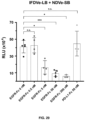
- the present invention relates to an in vitro method for detecting/quantifying antigens, hereinafter method of the invention", comprising (a) contacting an isolated sample with a host cell of the invention comprising at least two chimeric proteins of the invention, wherein the reporter protein domain of the at least two chimeric proteins of the invention are complementary each other, and (b) detecting and quantifying the signal emitted by the protein reporter, wherein the intensity of the signal is inversely proportional to the concentration of antigen present in the sample.
- the term "antigen" relates to a molecule or molecular structure or any foreign particulate matter that can trigger an immune response in the animal body. Any antigen can be detected by the method of the invention.
- the antigen is a viral antigen, parasite antigen, a bacteria antigen, a tumoral antigen, or an inflammation protein antigen.
- viral antigens include, without being limited to, antigens derived from HIV-1, such as gag, p17, p24, p41, p40, nef, pol, RT, p66, env, gp120 or gp160, gp40, p24, gag, vif, vpr, vpu, rev; from human herpes viruses, such as gH, gL gM gB gC gK gE or gD or derivatives thereof or Immediate Early protein such as ICP27, ICP 47, IC P 4, ICP36 from HSV1 or HSV2; from cytomegalovirus, especially Human, (such as gB or derivatives thereof); from Epstein Barr virus (such as gp350 or derivatives thereof), from Varicella Zoster Virus (such as gpl, II, III and IE63), or from a hepatitis virus such as hepatitis B virus (for example Hepati
- bacteria antigens include, without limiting to, antigens derived from bacterial pathogens such as Neisseria spp, including N. gonorrhea and N . meningitidis, eg, transferrin-binding proteins, lactoferrin binding proteins, PiIC, adhesins); S . pyogenes (for example M proteins or fragments thereof, C5A protease, S . agalactiae, S. mutans; H, ducreyi; Moraxella spp, including M.
- antigens derived from bacterial pathogens such as Neisseria spp, including N. gonorrhea and N . meningitidis, eg, transferrin-binding proteins, lactoferrin binding proteins, PiIC, adhesins); S . pyogenes (for example M proteins or fragments thereof, C5A protease, S . agalactiae, S.
- catarrhalis also known as Branhamella catarrhalis (for example high and low molecular weight adhesins and invasins); Bordetella spp, including B. pertussis (for example pertactin, pertussis toxin or derivatives thereof, filamenteous hemagglutinin, adenylate cyclase, fimbriae), B. parapertussis and B. bronchiseptica; Mycobacterium spp., including M .
- B. pertussis for example pertactin, pertussis toxin or derivatives thereof, filamenteous hemagglutinin, adenylate cyclase, fimbriae
- B. parapertussis and B. bronchiseptica
- Mycobacterium spp. including M .
- tuberculosis for example ESAT6, Antigen 85A, -B or -C, MPT 44, MPT59, MPT45, HSP10,HSP65, HSP70, HSP 75, HSP90, PPD 19kDa [Rv3763], PPD 38kDa [Rv0934] ), M. bovis, M. leprae, M. avium, M. paratuberculosis, M. smegmatis; Legionella spp, including L. pneumophila; Escherichia spp, including enterotoxic E . coli (for example colonization factors, heat-labile toxin or derivatives thereof, heat-stable toxin or derivatives thereof), enterohemorragic E .
- ESAT6 Antigen 85A, -B or -C
- E coli enteropathogenic E . coli (for example shiga toxin-like toxin or derivatives thereof); Vibrio spp, including V. cholera (for example cholera toxin or derivatives thereof); Shigella spp, including S. sonnei, S. dysenteriae, S. flexnerii; Yersinia spp, including Y. enterocolitica (for example a Yop protein), Y. pestis, Y. pseudotuberculosis; Campylobacter spp, including C. jejuni (for example toxins, adhesins and invasins) and C. coli; Salmonella spp, including S. typhi, S. paratyphi, S.
- Vibrio spp including V. cholera (for example cholera toxin or derivatives thereof); Shigella spp, including S. sonnei, S. dysenteriae, S. flexnerii; Yersinia
- choleraesuis S. enteritidis
- Listeria spp. including L. monocytogenes
- Helicobacter spp including H. pylori (for example urease, catalase, vacuolating toxin)
- Pseudomonas spp including P. aeruginosa
- Staphylococcus spp. including S . aureus, S. epidermidis
- Enterococcus spp. including E. faecalis, E. faecium
- Clostridium spp. including C. tetani (for example tetanus toxin and derivative thereof), C.
- botulinum for example botulinum toxin and derivative thereof
- C. difficile for example clostridium toxins A or B and derivatives thereof
- Bacillus spp. including B. anthracis (for example botulinum toxin and derivatives thereof); Corynebacterium spp., including C. diphtheriae (for example diphtheria toxin and derivatives thereof); Borrelia spp., including B. burgdorferi (for example OspA, OspC, DbpA, DbpB), B. garinii (for example OspA, OspC, DbpA, DbpB), B.
- afzelii for example OspA, OspC, DbpA, DbpB
- B. andersonii for example OspA, OspC, DbpA, DbpB
- B. hermsii for example OspA, OspC, DbpA, DbpB
- Ehrlichia spp. including E. equi and the agent of the Human Granulocytic Ehrlichiosis
- Rickettsia spp including R. rickettsii
- Chlamydia spp. including C. trachomatis (for example MOMP, heparin-binding proteins), C. pneumoniae (for example MOMP, heparin-binding proteins), C.
- Leptospira spp. including L. interrogans
- Treponema spp. including T. pallidum (for example the rare outer membrane proteins), T. denticola, T. hyodysenteriae; M.
- tuberculosis such as Rv2557, Rv2558, RPFs: Rv0837c, Rv1884c, Rv2389c, Rv2450, Rv1009, aceA (Rv0467), PstSI, (Rv0932), SodA (Rv3846), Rv2031c 16kDal., Tb Ra12, Tb H9, Tb Ra35, Tb38-1, Erd 14, DPV, MTI, MSL, mTTC2 and hTCC1 (V1lO 99/51748); Streptococcus spp, including S.
- pneumoniae PsaA, PspA, streptolysin, choline-binding proteins
- H. influenzae type 8 for example PRP and conjugates thereof
- non-typeable H. influenzae for example OMP26, high molecular weight adhesins, P5, P6, protein D and lipoprotein D, and fimbrin and fimbrin derived peptides.
- parasite antigens include, without limited to, antigens derived from parasites such as Plasmodium spp., including P. falciparum (for example RTS, S, TRAP MSP1, AMAI, MSP3, EBA, GLURP, RAPI, RAP2, Sequestrin, PfEMP1, Pf332, LSA1, LSA3, STARP, SALSA, PfEXP1, Pfs25, Pfs28, PFS27/25, Pfs16, Pfs48/45, Pfs230 and their analogues in Plasmodium spp.); Toxoplasma spp., including T.
- P. falciparum for example RTS, S, TRAP MSP1, AMAI, MSP3, EBA, GLURP, RAPI, RAP2, Sequestrin, PfEMP1, Pf332, LSA1, LSA3, STARP, SALSA, PfEXP1, Pfs25, Pfs28,
- gondii for example SAG2, SAG3, Tg34
- Entamoeba spp. including E. histolytica
- Babesia spp. including B. microti
- Trypanosoma spp. including T. cruzi
- Giardia spp. including G. lamblia
- leishmania spp. including L. major
- Pneumocystis spp. including P. carinii
- Trichomonas spp. including T. vaginalis
- Schisostoma spp. including S. mansoni, or derived from yeast such as Candida spp., including C. albicans
- Cryptococcus spp. including C. neoformans.
- tumoral antigens include, without being limited to, tumour rejection antigens such as those for prostrate, breast, colorectal, lung, pancreatic, renal or melanoma cancers.
- exemplary antigens include MAGE 1, 3 and MAGE 4 or other MAGE antigens, PRAME, BAGE, Lü (also known as NY Eos 1) SAGE and HAGE or GAGE. Indeed these antigens are expressed in a wide range of tumour types such as melanoma, lung carcinoma, sarcoma and bladder carcinoma.
- Prostate antigens include, without being limited to, Prostate specific antigen (PSA), PAP, PSCA, PSMA or antigen known as Prostase, and P501 S.
- Breast cancer antigens such as Muc- 1, Muc-2, EpCAM, her 2/ Neu, mammaglobin.
- inflammation-related antigens include, without being limited to, Interleukin 1alpha and 1beta (IL-1 ⁇ , IL1 ⁇ ), Interleukin 2 (IL-2), Interleukin 6 (IL-6), Interleukin 10 (IL-10), Interleukin 11 (IL-11), Interleukin 12 (IL-12), Interleukin 17 (IL-17), Interleukin 18 (IL-18), Interleukin 22 (IL-22), Tumor necrosis factor alpha (TNF- ⁇ ), Interferon gamma (IFN- ⁇ ), C-reactive protein (CRP), procalcitonin (PCT).
- IL-1 ⁇ , IL1 ⁇ Interleukin 2
- IL-6 Interleukin 6
- IL-10 Interleukin 10
- IL-11 Interleukin 11
- IL-12 Interleukin 12
- IL-17 Interleukin 17
- Interleukin 18 Interleukin 18
- IL-22 Interleukin 22
- TNF- ⁇ Tumor necrosis factor
- the first step of the method of the invention comprises contacting an isolated sample with a host cell of the invention comprising at least two chimeric proteins of the invention, wherein the reporter protein domain of the at least two chimeric proteins of the invention are complementary each other
- sample refers to a small part or quantity of something intended to show what the whole is like.
- samples include, without being limited to, the sample is a food- or feed-product sample, a pharmaceutical, veterinary or biotechnological product sample, a water system sample, a fluid sample, a soil sample, a plant sample, or a clinical sample such as saliva, blood, plasma, urine or faeces.
- the clinical sample may come from a subject, both animal and human.
- subject in the context of the present disclosure is a mammal, e.g., a primate, preferably a higher primate, e.g., a human.
- the sample is a saliva sample, a urine sample, a plasma sample, a water sample or a food sample. It is common general knowledge how to obtain these kinds of samples for their analysis.
- isolated will mean material removed from its original environment (e.g., the natural environment in which the material occurs), and thus it is “altered by the hand of man” from its natural environment.
- the method of the invention comprises a second step comprising detecting and quantifying the signal emitted by the protein reporter.
- Tools and techniques for detecting and quantifying the signal emitted by a protein reporter are widely known in the state of the art, and any of them can be used in the context of the present invention. Examples of these tools include, without being limited to, droplet, test tube and microplate spectrophotometers of ultraviolet and visible light spectra, including colorimeters, fluorimeters and luminescence readers.
- the intensity of the signal is inversely proportional to the concentration of antigen present in the sample.
- the present invention also relates to a kit comprising a chimeric protein, the polynucleotide, the vector or the host cell of the invention.
- Kit refers to a product containing the different reagents necessary to carry out the method of the invention, which are packaged to enable them to be transported and stored.
- the kit may further include, without any limitation, buffers, agents to prevent contamination, inhibitors of protein degradation, etc.
- Suitable materials for packaging the components of the kit include glass, plastic (polyethylene, polypropylene, polycarbonate and the like), bottles, vials, paper, sachets and the like. Additionally, the kits of the invention may contain instructions for simultaneous, sequential or separate use of the different components in the kit.
- Said instructions may be in the form of printed material or in the form of an electronic medium capable of storing instructions so that they can be read by a subject, such as electronic storage media (magnetic disks, tapes and the like), optical media (CD-ROM, DVD) and the like.
- electronic storage media magnetic disks, tapes and the like
- optical media CD-ROM, DVD
- chimeric protein the polynucleotide, the vector or the host cell of the invention have been defined and explained previously throughout the present description. Said definitions and explanations, as well as all the particular embodiments, are all applicable to the kit of the invention.
- a system of bacterial receptors in the OM that would enable the binding of an extracellular target antigen and the generation of a signal upon recognition is disclosed below.
- E. coli strains used in this work can be found in Table 2. Table 2. Bacterial strains used in this invention. ( Blattner, F.R., et al., 1997. Science 277, 1453-1462 ; Datsenko, K.A., Wanner, B.L., 2000. Proceedings of the National Academy of Sciences 97, 6640-6645 ; Salema, V., et al., 2013. PLoS ONE 8, e75126 ; Durfee, et al., 2008.
- kanamycin at 50 ⁇ g/mL
- chloramphenicol Cm
- ampicillin Amp
- aTc anhydrotetracycline
- IPTG isopropyl- ⁇ -D-thiogalactopyranoside
- L-ara L-arabinose
- Diaminopimelic acid (DAPA) at 0.3 mM was added to cultures of the dapA mutant E. coli MFDpir strain. Plasmids carrying the ⁇ -dependent R6K origin of replication (Stalker et al., 1982), such as pGE and pGEC derivatives, were cloned, propagated and isolated using E. coli BW25141. E. coli DH10BT1 R was used as a host for the rest of plasmids.
- E. coli EcM1 was freshly transformed with the corresponding plasmid and individual colonies harvested from plates were used to start pre-inoculate cultures.
- Pre-inoculums were grown at 30 oC, with glucose 2% (w/v) for repression of the P l ac promoter and static conditions overnight (O/N).
- pre-inoculum cultures were centrifuged (3 minutes, 4000 g) and washed using fresh LB medium twice to remove the glucose and finally diluted to an O.D. of 0.5 at 600 nm (OD 600 ).
- the cultures were later induced using IPTG for 3 h at 30 oC and 160 rpm.
- each promoter (P lldP , P lldP *, P uof P hmp and P norV ,) controlling the GFP TCD using the T7 RBS were integrated into the csgCG locus of EcM1.
- Two different clones from the resulting strains were grown O/N from individual colonies. After growth, cultures were diluted and grown up to 0.4 OD 600 for their induction.
- Plasmids used in this work can be found in Table 3. Table 3 - Plamids used in this invention. (Veiga, E., et al., 1999. Molecular Microbiology 33, 1232-1243; Salema et al., 2013, cited ad supra; Salema et al., 2016b; Salema, V. et al., 2016b. mAbs 8, 1286-1301; Silva-Rocha et al., 2013. Nucleic Acids Res 41, D666-675).
- This invention pF330VeLB pF330Ve with the SmBiT fragment fused to the C-terminal end through the complete OmpA linker (residues 192-208).
- This invention pF487Ve pAK vector expressing a fusion of the pelB signal peptide, a VHH anti-GFP, the E-tag and flu (Ag43) residues 553-1039
- This invention pIVe pAK vector expressing a fusion of invasin residues 1- 596, E-tag, a VHH
- This invention plFVe plVe with the L352F mutation This invention plFDVe plFVe without the D1 Ig-like domain (residues 503-596)
- This invention pSEVA221 KmR, RK2-origin Silva-Rocha et al., 2013
- This invention pSEVA221 -Ptet-NDVeSB pSEVA221 with a Ptet promoter for expressing the NDVeSB construct.
- This invention pSEVA221 -Ptet-IFDVe pSEVA221 with a Ptet promoter for expressing the IFDVe construct.
- This invention pSEVA221 -Ptet-IFDVeSB pSEVA221 with a P tet promoter for expressing the IFDVeSB construct.
- DNA constructs were produced by standard methods of DNA manipulation, digestion, ligation and modification (Ausubel et al., 2002) or one-step isothermal Gibson assembly (Gibson et al., 2009). DNA synthesis of the tested promoters, the intimin sequence lacking LysM and the LgBiT fragment was performed by ThermoFisher Scientific GeneArt. Oligonucleotides were purchased from Merck or Integrated DNA Technologies. Relevant oligonucleotides can be found in Table 4. Designs for integrase targets were kindly provided by Dr. Jércons Bonnet based on previous works (Bonnet et al., 2013, 2012; Guiziou et al., 2019).
- PCR Polymerase Chain Reaction
- Taq DNA polymerase NZYTaq II 2X Green Master Mix
- Herculase II Fusion DNA polymerase Agilent Technologies
- Replicative plasmids and Gibson assembly reactions were transformed into the appropiate E. coli strains using the Transformation and Storage Solution (TSS) method ( Chung et al., 1989. Proc Natl Acad Sci U S A 86, 2172-2175 ). DNA ligations and suicide pGE plasmids were electroporated into electrocompetent bacteria. Conjugation of pGECs plasmids was performed using E. coli MFDpir as the donor strain.
- TSS Transformation and Storage Solution
- Detection of chimeric proteins with Nb on the bacterial surface and binding to soluble antigen were analyzed by flow cytometry. After induction, a volume of the cultures equivalent to 1 OD 600 was centrifuged (4000 g, 3 min), washed 3 times with 500 ⁇ l of PBS and resuspended in 400 ⁇ l of the same buffer. Next, 180 ⁇ l of the cell resuspension were incubated for 1 h at room temperature with 20 ⁇ l of the primary antibody (for display detection) or biotinylated EGFR fused to Fc (50 nM).
- the primary antibody was a mouse anti-c-myc monoclonal antibody (1:500; 9B11 clone; Cell Signaling, Ref: 2276) unless indicated, for autotransporter constructs, mouse anti-E-tag antibody (1:500; Phadia) was used.
- bacteria were washed 3 times with PBS and incubated in the same manner with the secondary reagent: for display detection, a goat anti-mouse IgG-Alexa 488 conjugated polyclonal antibody (1:500; Life technologies, Ref: A11029), and for detection of the biotinylated antigen, Streptavidin-APC (1:100; Beckman Coulter, Ref: 733001).
- samples were washed 3 times with PBS and resuspended in 500 ⁇ l of the same buffer for analysis in a Gallios cytometer (Beckman Coulter, Inc).
- Electrophoresis in denaturing conditions using polyacrylamide gels containing SDS was performed following standard methods. A volume equivalent to 1 OD 600 of an induced culture was centrifuged (4000 g, 3 min) and resuspended in 100 ⁇ L of Tris-HCl 10 mM pH 8. Next, 100 ⁇ L of SDS loading buffer 2x (120 mM Tris-HCl pH 6.8, 2% (p/v) SDS, 2% (v/v) ⁇ -mercaptoethanol (2-ME), 10% glycerol and 0.01% (p/v) bromophenol blue) were added.
- SDS loading buffer 2x 120 mM Tris-HCl pH 6.8, 2% (p/v) SDS, 2% (v/v) ⁇ -mercaptoethanol (2-ME), 10% glycerol and 0.01% (p/v) bromophenol blue
- Samples were either boiled (100 oC, 10 min) or not, as specified, and centrifuged (21.000 g, 10 min) to eliminate any insoluble material (e.g., the peptidoglycan). 100 ⁇ L of the supernatants were transferred to new tubes from which up to 20 ⁇ L were loaded into 8% polyacrylamide gels for intimin, invasin and flu constructs and 10% polyacrylamide gels for EhaA constructs. Electrophoresis was performed in a Miniprotean III chamber (Bio-Rad).
- samples containing these proteins were prepared in 2x urea-SDS loading buffer (120 mM Tris-HCl pH 6.8, 4% (p/v) SDS, 10 mM EDTA, 8 M urea, 2% (v/v) 2-ME, 10% glycerol and 0.01% (p/v) bromophenol blue). When boiled, these samples were kept at 100 oC for 30 min. For Western blot analysis gels were transferred to a polyvinylidene difluoride membrane (PVDF, Immobilon-P, Merck-Millipore) in semi-dry conditions as previously reported ( Bodelon et al., 2009.
- PVDF polyvinylidene difluoride membrane
- Membranes were developed by measure of chemiluminescence using Clarity Western ECL Substrate (Bio-Rad ref. 1705061) by ChemiDoc Touch system (Bio-Rad). Precision Plus Protein Dual Color Standards (Bio-Rad) was used as molecular weight marker.
- the resuspended culture was then lysed by sonication (7 pulses of 30 sec, with the tip being cooled between pulses) and the resulting extract was centrifuged to eliminate non-lysed bacteria (1500 g, 10 min, 4 oC).
- the supernatant was then ultracentrifuged (100.000 g, 1 h, 4 oC, Beckman Optima-Max XP) and the pellet, corresponding to the totality of the membranes, was resuspended in 2 mL TN Buffer with 1.5 % (v/v) Triton-X-100 (Sigma) ( Schnaitman, 1971, J Bacteriol 108, 545-552 ) in order to eliminate the fraction corresponding to the inner membrane proteins.
- Resuspension was aided by using an ultrasonic bath for 10 sec. After a second ultracentrifugation the resulting pellet correspond to the outer membrane and insoluble remains. To obtain the outer membrane proteins, the pellet was resuspended in 1 mL TN buffer with 1% (p/v) Zwittergent 3-14 (Calbiochem) by incubating at 4 oC using a rotator for 1 h. A final ultracentrifugation results in the outer membrane proteins recovered in the supernatant.
- the proteins extracted from the outer membrane were subjected to non-denaturing electrophoresis in polyacrylamide gels using Coomassie Blue G-250 a negative charge provider (Blue Native-PAGE).
- Glycerol (Merck) and Coomassie Blue G-250 (Bio-Rad) were added to the outer membrane protein extracts to a final concentration of 8.7% (v/v) and 0.5%, respectively, and 20 ⁇ L of each sample were loaded into a 4-20% polyacrylamide gradient, Tris-Glycine native gel (Invitrogen).
- the cathode buffer composition was 50 mM tricine (Fluka), 15 mM Bis/Tris/HCl pH 7 (Fluka) and 0.002% Coomassie Blue G-250, while the anode buffer was 50 mM Bis/Tris/HCl pH 7.
- Native molecular weight markers (66-669 kDa; GE Bioscience) were resuspended to a final concentration of 2.5 mg/ml in 50 mM Bis/Tris/HCl ph7 and 750 mM aminocaproic acid, and 5 ⁇ L were loaded into the gel.
- the electrophoresis was first run at 100 V for 45 min, until samples have successfully penetrated the gel, and later at 500 V until the dye front has reached the gel's border.
- the gel is transferred to a PVDF membrane following standard methods. To unfold the proteins and facilitate antibody access, after transferring the membrane was incubated in 8% acetic acid (Panreac) for 15 min, rinsed in de-ionized water and air dried, after which it was re-wet with methanol (MERCK) for 1 min to remove the excess of Coomassie which helped visualizing the molecular weight markers. The membrane was rinsed in PBS with 0.05% (v/v) Tween 20 (PBS-Tween) before continuing with the standard Western blot protocol.
- the plate was placed in an orbital shaker at 37 oC for 5 min and 25 ⁇ l of the Nano-Glo ® Live Cell Reagent (Promega) were added to each well immediately before measuring luminescence (full spectrum, 2 sec integration time) and OD 600 using a Spectramax iD5 Multi-mode Microplate Reader (Molecular Devices). Plate was shaken for 3 sec prior to the measurements.
- CFSSP Chou and Fasman Secondary Structure Prediction server .
- Cytometry analysis of integrases experiments used Flow-Jo (Treestar, Inc.), while promoters and adhesins experiments were analyzed using Kaluza (Beckman Coulter, Inc.).
- Statistical analysis was performed using GraphPad Prism 9.4 (GraphPad Software, LLC.) AlphaFold predictions were kindly provided by Dr. Florencio Pazos from the Computational Systems Biology group (CNB-CSIC), and the results were visualized using Mol* Viewer ( Sehnal et al., 2021 Nucleic Acids Research 49, W431-W437 ).
- Example 1 Engineering intimin for outer membrane sensors.
- the outer membrane anchoring fragment of a synthetic adhesin is based on the protein intimin, from its N-terminus up to the D0 Ig-like domain. Intimin has been reported to form homodimers and the generation of this dimers has been attributed to the periplasmic segment containing the LysM domain followed by a short peptide spacer. It was first evaluated whether deletion of the LysM and spacer region enabled the production of a monomeric intimin-nanobody fusion. As a model, the plasmid pNVEGFR2, encoding a synthetic adhesin with a nanobody binding the human epidermal growth factor receptor (hEGFR) was used.
- hEGFR2 human epidermal growth factor receptor
- the LysM domain includes residues 63 to 112 of intimin and the present deletion comprised residues 54 through 153 to also delete the spacer.
- an EcoRI restriction site was placed at position 44 and a Sacll site at position 54 of intimin.
- the resulting plasmid was termed pNDVe.
- the periplasmic region of intimin also contains two ⁇ -helices that are connected to the ⁇ -barrel. The role of these ⁇ -helices in dimerization and/or folding of intimin has not been reported.
- a second construct deleting the LysM domain, the spacer and these two ⁇ -helices was also generated.
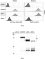
- intimin-nanobody constructs were tested for their capacity to be displayed on the bacteria surface, as well as for the functionality of the nanobody to bind its antigen (i.e., hEGFR-Fc) ( Figure 2 ).
- Flow cytometry analysis of the transformed bacteria showed that the deletion of LysM-spacer in pNDVe did not affect the display nor the binding of the nanobody, with values almost identical to the non-modified construct pNVEGFR2.
- deletion comprising LysM-spacer and the ⁇ -helices severely hindered the capacity for both display and antigen-binding of the bacteria, which were close to the negative control (pAK-Not).
- the unmodified intimin-nanobody fusion showed an intense band of high mobility and a faint band of low mobility on SDS-PAGE.
- the only band present for this parental fusion was that of low mobility, which corresponds to the fully denatured polypeptide.
- This mobility shift is characteristic of outer membrane proteins containing stable ⁇ -barrels and indicate that, without boiling, the intimin ⁇ -barrel remains folded even in the presence of SDS-urea.
- the unboiled sample of the LysM-deletion mutant presented additional protein bands between those of high and low mobility, which indicated the presence of some partial unfolded states of the ⁇ -barrel.
- the oligomerization state of intimin constructs with and without the LysM-spacer region was investigated.
- the constructs having the 6 ⁇ His peptide tag were used.
- a crosslinking assay was performed using bacteria expressing these intimin fusions. Different concentrations of the dithiobis(succinimidyl propionate) (DSP) crosslinker agent were used on the intact bacteria, and the resulting samples were subjected to SDS-PAGE and Western blot.
- DSP dithiobis(succinimidyl propionate)
- intimin-based proteins cannot be used as the only components of an outer membrane sensor relying on antigen-driven dimerization, as the endogenous dimer formation of intimin would trigger a constant activation of the system.
- intimin constructs could still be used in combination with other outer membrane proteins able to display nanobodies.
- intimin-nanobody construct lacking LysM was chosen as the basis of the sensor constructs of the present invention. It is important to remind here that LysM-deletion did not affect the surface display of the intimin construct nor the antigen binding activity of the nanobody.
- NanoBiT is a protein complementation assay based on a split NanoLuc luciferase with two protein fragments: the small SmBiT, a 1.3 kDa-peptide, and the large LgBiT, an 18 kDa-polypeptide. NanoBiT is able to emit light once the two protein fragments interact to reconstitute the Nanoluc luciferase.
- Both SmBiT and LgBiT were cloned into pNDVe plasmid in frame between the amino acids 43 and 154 of intimin positions, using the inserted EcoRI and Sacll restriction sites. These fusions placed the NanoBiT fragments in the periplasmic domain of the intimin-nanobody fusion, between the N-terminal signal peptide and the periplasmic ⁇ -helices. Two Gly-Ser flexible peptide linkers (GGGS) were placed at both ends of the NanoBiT fragments.
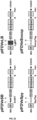
- the plasmid carrying the SmBiT was named pNDVeSB and the one carrying the LgBiT was named pNDVeLB ( Figure 7 ).
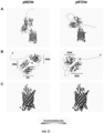
- the deep-learning algorithm AlphaFold ( Jumper et al., 2021. Nature 596, 583-589 .) was used to predict the structure of the protein fusion to the LgBiT fragment.
- the prediction estimated a correct folding of all the domains, including the LgBiT, which was placed in the periplasm and linked to the ⁇ -barrel of the adhesin through highly flexible helices ( Figure 10 ).
- LysM-deleted intimin-nanobody construct NDVe can be fused to small peptide fusions in its periplasmic region without alterations in surface display or antigen-binding levels, but larger polypeptide fragments are not well tolerated and reduce the expression levels and binding activity of the fusion.
- the presence of large folded fragments in the periplasm might be interfering with the process of insertion of the ⁇ -barrel in the outer membrane through the BAM complex and/or the translocation of its passenger (nanobody). Therefore, the inventors thought of an alternative that could allow the covalent linkage of large protein reporters once the protein fusion was already correctly inserted in the outer membrane.
- the SpyTag-SpyCatcher and SnoopTag-SnoopCatcher are homologous but orthogonal systems that allow the post-translational fusion of proteins ( Hatlem et al., 2019. IJMS 20, 2129 ). Both are derived from fimbrial subunits from Streptococcus species with an intradomain isopeptide bond. These fimbrial domains can be split into a small peptide (the Tag) that form a covalent isopeptide bond with the large fragment of the domain (the Catcher), which is catalytically active. Each of these components of a pair can be fused to different proteins to covalently link them through an isopeptide bond.
- the Tag-Catcher partners could be used to covalently attach a large fragment of a reporter enzyme in the periplasm fused to a Catcher domain if a small Tag peptide is placed in the periplasmic domain of intimin.
- a small Tag peptide is placed in the periplasmic domain of intimin.
- intimin-nanobody fusions that, although maintain the dimeric nature of intimin after deletion of the LysM domain, were able to simultaneously display a functional nanobody on the bacterial surface and a peptide or polypeptide fragment in the periplasm.
- Example 2 Engineering autotransporters for outer membrane sensors.
- the nanobody binding human EGFR fused to the C-terminal ⁇ -barrel domain of EhaA was cloned.
- the anti-GFP nanobody found in a similar fusion, encoded by pVgfpA ( Salema et al., 2013. PLoS ONE 8, e75126 ), was replaced, generating the plasmid derivative pVeA.
- the surface display and antigen binding levels of this construct were found low compared to those of pNDVe (compare Figure 2 with Figure 15 ).
- the outer membrane protein OmpA from E. coli is composed of an N-terminal ⁇ -barrel embedded in the outer membrane, with a terminal F residue in the last ⁇ -strand, followed by a C-terminal periplasmic domain. These two domains are connected through an unstructured 17-aa linker (GQGEAAPVVAPAPAP, SEQ D NO: 67) that functions as a peptidoglycan clamp.
- the first three aa of the linker were chosen and used them to connect the C-terminal end of EhaA ⁇ -barrel to the NanoBit domains preceded by a flexible GGGS linker, obtaining the plasmids pVeASB and pVeALB ( Figure 14 ).
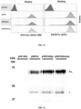
- Flow cytometry analysis of these constructs showed that, in a similar situation to intimin-based fusions, the small peptide SmBiT did not hinder the display nor the antigen binding of the nanobody, but the LgBiT fragment made the display and binding signals drop to values close to the negative control.
- Western blot analysis of whole-cell protein extracts from the induced cultures showed that both constructs were expressed, being the LgBiT chimera even expressed at higher levels than the SmBiT fusion.
- LOA was capable of restoring the surface display and antigen binding activity to the fusion carrying LgBiT, showing values similar to the positive control pVeA.
- Western blot analysis of bacteria from these cultures showed the expected pattern of protein bands for these constructs according to their mass, with no signs of higher proteolysis ( Figure 16 ). In fact, higher expression levels in the protein fusions carrying LgBiT and LOA were observed.
- AlphaFold was used to predict the sequence of these constructs.
- the algorithm predicted that the ⁇ -barrel is correctly folded in all constructs, and expected a flexible unstructured region for the peptides SmBiT and LOA.
- AlphaFold predicted a highly disorganized LgBiT domain in the case of pVeALB, with limited confidence in the result.
- the prediction of a folded LgBiT domain improved once the LOA was used to increase the distance between the ⁇ -barrel and the LgBiT fragment ( Figure 17 ).
- EHaa-Based fusion could be engineering to display a nanobody while simultaneously carrying a periplasmic reporter.
- Example 3 Engineering of invasin for outer membrane sensors.
- Invasins are inverted autotransporters proteins found in Yersinia species that present a high homology to intimin in their overall structure and ⁇ -domains. In most cases, invasins do not present a periplasmic LysM domain.
- invasin invA
- Yersinia pseudotuberculosis we chose the invasin ( invA ) from Yersinia pseudotuberculosis to generate a new set of constructs by replacing the intimin Neae fragment encoding the ⁇ -domain. The crystal structure of the ⁇ -barrel of this invasin has been solved and was reported to be monomeric.
- oligomerization state of the IFDVe construct was tested.
- the outer membrane proteins were extracted from induced E. coli bacteria carrying pIFVDVe and the samples were subjected to BN-PAGE and Western blot with anti-myc mAb ( Figure 21 ).
- Migration of this fusion protein produced a protein band of -140 kDa, which is roughly double the expected size for the monomer, leading us to think that this invasion-based construct was also behaving as a dimer similar to intimin. Due to its dimeric nature, for its functioning in a dimerization sensor, this fusion would likely require its co-expression with the intimin or EhaA partners.
- AlphaFold was used to predict the structure of the invasin constructs.
- the algorithm predicted the correct folding of the invasin ⁇ -barrel and of the extracellular domains as well as the presence of flexible ⁇ -helices in the periplasm.
- the LgBiT domain was predicted to be correctly folded ( Figure 25 ).
- AlphaFold also predicted the presence of an immunoglobulin-like domain between the ⁇ -barrel and the D1 domain of invasin, in a similar case to the D00 domain of intimin ( Figure 26 ).
- invasin-based constructs have the capacity to display a functional nanobody on the surface of E. coli while having a peptide and/or protein domain anchored in the periplasmic side.
- Example 4 Detection of extracellular antigen by bacteria co-expressing intimin and invasin protein sensors in the outer membrane.
- EGFR-Fc extracellular protein antigen
- the new plasmids (pSEVA221-Ptet-NDVe, pSEVA221-Ptet-NDVeSB, pSEVA221-Ptet-IFDVe and pSEVA221-Ptet-IFDVeLB), were co-transformed with either pNDVeLB or plFDVeLB, into E. coli EcM1 strain.
- both the IFDVeLB (SEQ ID NO: 12) + IFDVeSB (SEQ ID NO 13) and IFDVeLB (SEQ ID NO: 12) + NDVeSB (SEQ ID NO: 11) pairs showed light emission, with values ⁇ 10-fold below the luminescence found for the full-length NanoLuc in the periplasm (positive control). From the two combinations, the homodimer of invasin ⁇ -barrels pair showed higher luminescence. Unexpectedly, both chimeric protein pairs produced higher bioluminescence in the absence of the specific antigen than when EGFR-Fc was added. While this behavior was opposed to the original hypothesis, its potential for generating a biosensing platform was further investigated.
- Detection of tumor associated protein antigens is also a potential way to specifically trigger diagnostic and therapeutic responses in engineered bacteria.
- the present invention discloses a whole-cell biosensor E. coli that is able to detect the presence of a tumor-associated protein, hEGFR-Fc, through the display in the outer membrane of chimeric proteins pairs consisting in an exposed nanobody fused to a membrane anchored T5SS ⁇ -barrel that carries a split enzyme fragment (NanoBiT).
- the engineered chimeric proteins of intimin and invasin allowed the inventors to generate a whole-cell biosensor for detecting the presence of soluble hEGFR-Fc in a specific and dosage-dependent manner.
- the biosensor disclosed herein behaves in the opposite manner to what was expected: the inventors thought that the intimin and invasin constructs would not interact unless a dimeric antigen forced their dimerization, but luminescence was measurable in the absence of antigen and adding the antigen reduced the luminescence instead of increasing it.
- Nanobodies confer to the system the specificity for the detected protein.
- the ⁇ -barrel which acts as scaffold and OM anchor for the nanobody, and the reporter system.
- Three different barrels were engineered for their use in our biosensor based on invasin, intimin and the EhaA Ats, and their functionality in the system is demonstrated herein.
- BAR bacterial antigen receptors
- the final module (or domain), the reporter system, is based on the interaction of a split enzyme.
- the split version of the NanoLuc luciferase, NanoBiT was chosen due to its fast, simple and sensitive detection method.
- PCA protein complementation assays
Landscapes
- Chemical & Material Sciences (AREA)
- Health & Medical Sciences (AREA)
- Life Sciences & Earth Sciences (AREA)
- Organic Chemistry (AREA)
- Engineering & Computer Science (AREA)
- Molecular Biology (AREA)
- Proteomics, Peptides & Aminoacids (AREA)
- Immunology (AREA)
- General Health & Medical Sciences (AREA)
- Biochemistry (AREA)
- Wood Science & Technology (AREA)
- Zoology (AREA)
- Genetics & Genomics (AREA)
- Biophysics (AREA)
- Physics & Mathematics (AREA)
- Biotechnology (AREA)
- Medicinal Chemistry (AREA)
- Analytical Chemistry (AREA)
- Microbiology (AREA)
- Urology & Nephrology (AREA)
- Hematology (AREA)
- Bioinformatics & Cheminformatics (AREA)
- General Engineering & Computer Science (AREA)
- Biomedical Technology (AREA)
- Gastroenterology & Hepatology (AREA)
- Pathology (AREA)
- General Physics & Mathematics (AREA)
- Cell Biology (AREA)
- Food Science & Technology (AREA)
- Peptides Or Proteins (AREA)
Priority Applications (2)
| Application Number | Priority Date | Filing Date | Title |
|---|---|---|---|
| EP23382158.6A EP4421084A1 (de) | 2023-02-21 | 2023-02-21 | Chimäre proteine und ihre verwendung als antigennachweissystem |
| PCT/EP2024/054448 WO2024175675A1 (en) | 2023-02-21 | 2024-02-21 | Chimeric proteins and their use as an antigen detecting system |
Applications Claiming Priority (1)
| Application Number | Priority Date | Filing Date | Title |
|---|---|---|---|
| EP23382158.6A EP4421084A1 (de) | 2023-02-21 | 2023-02-21 | Chimäre proteine und ihre verwendung als antigennachweissystem |
Publications (1)
| Publication Number | Publication Date |
|---|---|
| EP4421084A1 true EP4421084A1 (de) | 2024-08-28 |
Family
ID=85382702
Family Applications (1)
| Application Number | Title | Priority Date | Filing Date |
|---|---|---|---|
| EP23382158.6A Withdrawn EP4421084A1 (de) | 2023-02-21 | 2023-02-21 | Chimäre proteine und ihre verwendung als antigennachweissystem |
Country Status (2)
| Country | Link |
|---|---|
| EP (1) | EP4421084A1 (de) |
| WO (1) | WO2024175675A1 (de) |
Cited By (1)
| Publication number | Priority date | Publication date | Assignee | Title |
|---|---|---|---|---|
| CN121558712A (zh) * | 2026-01-26 | 2026-02-24 | 四川大学 | 基于荧光互补筛选细菌膜蛋白折叠机器靶向抑制剂的方法 |
-
2023
- 2023-02-21 EP EP23382158.6A patent/EP4421084A1/de not_active Withdrawn
-
2024
- 2024-02-21 WO PCT/EP2024/054448 patent/WO2024175675A1/en not_active Ceased
Non-Patent Citations (34)
| Title |
|---|
| ALTSCHUL S.F. ET AL.: "Basic local alignment search tool", J MOL BIOL., vol. 215, no. 3, 5 October 1990 (1990-10-05), pages 403 - 10, XP002949123, DOI: 10.1006/jmbi.1990.9999 |
| ANDREW S. DIXON ET AL: "NanoLuc Complementation Reporter Optimized for Accurate Measurement of Protein Interactions in Cells", ACS CHEMICAL BIOLOGY, vol. 11, no. 2, 19 February 2016 (2016-02-19), pages 400 - 408, XP055715353, ISSN: 1554-8929, DOI: 10.1021/acschembio.5b00753 * |
| AZAD TAHA ET AL: "Split-luciferase complementary assay: applications, recent developments, and future perspectives", ANALYTICAL AND BIOANALYTICAL CHEMISTRY, SPRINGER BERLIN HEIDELBERG, BERLIN/HEIDELBERG, vol. 406, no. 23, 8 July 2014 (2014-07-08), pages 5541 - 5560, XP035377889, ISSN: 1618-2642, [retrieved on 20140708], DOI: 10.1007/S00216-014-7980-8 * |
| BODELON ET AL., J BACTERIOL, vol. 191, 2009, pages 5169 - 79 |
| CHUNG ET AL., PROC NATL ACAD SCI U S A, vol. 86, 1989, pages 2172 - 2175 |
| DAMMEYER TTINNEFELD P, COMPUT STRUCT BIOTECHNOL J, vol. 22, no. 3, 2012, pages e201210013 |
| DIXON ET AL., ACS CHEM. BIOL., vol. 11, 2016, pages 400 - 408 |
| GALARNEAU, A. ET AL., NAT. BIOTECHNOL., vol. 20, 2002, pages 619 - 622 |
| GALDZICKI ET AL., NAT BIOTECHNOL, vol. 32, 2014, pages 545 - 550 |
| GARNIER ET AL., JOURNAL OF MOLECULAR BIOLOGY, vol. 120, 1978, pages 97 - 120 |
| HALL, M.P., ACS CHEM. BIOL., vol. 7, no. 11, 2012, pages 1848 - 1857 |
| HATLEM ET AL., IJMS, vol. 20, 2019, pages 2129 |
| HERRING ET AL., GENE, vol. 311, 2003, pages 153 - 163 |
| HU, C.D.KERPPOLA, T.K., NAT. BIOTECHNOL., vol. 21, 2003, pages 539 - 545 |
| ISHIKAWA H ET AL., PROTEIN ENG DES SEL, vol. 25, no. 12, 2012, pages 813 - 20 |
| JUMPER ET AL., NATURE, vol. 596, 2021, pages 583 - 589 |
| LE CALVEZ ET AL., GENE, vol. 170, 1996, pages 51 - 55 |
| MORELL, M.VENTURA, S.AVILES, F.X., FEBS LETTERS, vol. 583, 2009, pages 1684 - 1691 |
| PELLETIER, J.N.CAMPBELL-VALOIS, F.X.MICHNICK, S.W., PROC. NATL. ACAD. SCI. USA, vol. 95, 1998, pages 12141 - 12146 |
| POSFAI ET AL., NUCLEIC ACIDS RES, vol. 27, 1999, pages 4409 - 4415 |
| POSFAI ET AL., SCIENCE, vol. 312, 2006, pages 1044 - 1046 |
| SALEMA ET AL., PLOS ONE, vol. 8, 2013, pages e75126 |
| SALEMAET, MABS, vol. 8, 2016, pages 1286 - 1301 |
| SCHNAITMAN, J BACTERIOL, vol. 108, 1971, pages 545 - 552 |
| SEHNAL ET AL., NUCLEIC ACIDS RESEARCH, vol. 49, 2021, pages W431 - W437 |
| SHA, F. ET AL., PROTEIN SCIENCE, vol. 26, no. 5, 2017, pages 940 - 924 |
| THANABALU ET AL., THE EMBO JOURNAL, vol. 17, 1998, pages 6487 - 6496 |
| THIE ET AL., NEW BIOTECHNOLOGY, vol. 25, 2008, pages 49 - 54 |
| VALENCIO SALEMA ET AL: "Selection of Single Domain Antibodies from Immune Libraries Displayed on the Surface of E. coli Cells with Two Beta-Domains of Opposite Topologies", PLOS ONE, vol. 8, no. 9, 23 September 2013 (2013-09-23), pages e75126, XP055123932, ISSN: 1932-6203, DOI: 10.1371/journal.pone.0075126 * |
| VERMA, V., RESON, vol. 20, 2015, pages 816 - 821 |
| VILLALOBOS VICTOR ET AL: "Current state of imaging protein-protein interactions in vivo with genetically encoded reporters", ANNUAL REVIEW OF BIOMEDICAL ENGINEERING, ANNUAL REVIEWS, US, vol. 9, 1 January 2007 (2007-01-01), pages 321 - 349, XP002479376, ISSN: 1523-9829, [retrieved on 20070426], DOI: 10.1146/ANNUREV.BIOENG.9.060906.152044 * |
| VILLALOBOS, V. ET AL., ANNU. REV. BIOMED. ENG., vol. 9, 2007, pages 321 - 349 |
| WEHR, M. ET AL., NAT METHODS, vol. 3, 2006, pages 985 - 993 |
| WEHR, M.C. ET AL., BMC BIOTECHNOL, vol. 8, 2008, pages 55 |
Cited By (1)
| Publication number | Priority date | Publication date | Assignee | Title |
|---|---|---|---|---|
| CN121558712A (zh) * | 2026-01-26 | 2026-02-24 | 四川大学 | 基于荧光互补筛选细菌膜蛋白折叠机器靶向抑制剂的方法 |
Also Published As
| Publication number | Publication date |
|---|---|
| WO2024175675A1 (en) | 2024-08-29 |
Similar Documents
| Publication | Publication Date | Title |
|---|---|---|
| JP7252278B2 (ja) | 活性化可能な結合ポリペプチドおよびその同定方法ならびに使用 | |
| Shi et al. | Display of green fluorescent protein on Escherichia coli cell surface | |
| US8877898B2 (en) | Fluorescent fusion polypeptides and methods of use | |
| AU2016213702B2 (en) | Activatable binding polypeptides and methods of identification and use thereof | |
| Kaserer et al. | Insight from TonB hybrid proteins into the mechanism of iron transport through the outer membrane | |
| CN111269324A (zh) | 高斯荧光素酶和地高辛单链抗体的融合蛋白及其应用 | |
| EP4421084A1 (de) | Chimäre proteine und ihre verwendung als antigennachweissystem | |
| AU2013202755B2 (en) | Activatable binding polypeptides and methods of identification and use thereof | |
| EP1655371A1 (de) | System zur herstellung dimerer proteine auf grundlage des hämolysin-transportsystems von escherichia coli | |
| Fernández et al. | Chimeric proteins and their use as an antigen detecting system | |
| Jose et al. | “Cystope tagging” for labeling and detection of recombinant protein expression | |
| Little | The Type IV pilus secretin BfpB: structural analysis and binding interactions | |
| CN114276447B (zh) | 一种牛源抑制金黄色葡萄球菌生长的单链抗体及其制备方法和用途 | |
| Malik | Understanding the molecular basis for adhesion by Haemophilus Surface Fibril | |
| Agboh | Immobilizing strategies for membranes to screen against antibody mimetics with phage display | |
| HK40009931A (en) | Activatable binding polypeptides and methods of identification and use thereof | |
| Chivers | Investigating high-affinity non-covalent protein-ligand interaction via variants of streptavidin | |
| Salerno | Multivalent Display of Single-Domain Antibodies on Curli Fibers to Neutralize Enteric Pathogens |
Legal Events
| Date | Code | Title | Description |
|---|---|---|---|
| PUAI | Public reference made under article 153(3) epc to a published international application that has entered the european phase |
Free format text: ORIGINAL CODE: 0009012 |
|
| STAA | Information on the status of an ep patent application or granted ep patent |
Free format text: STATUS: THE APPLICATION HAS BEEN PUBLISHED |
|
| AK | Designated contracting states |
Kind code of ref document: A1 Designated state(s): AL AT BE BG CH CY CZ DE DK EE ES FI FR GB GR HR HU IE IS IT LI LT LU LV MC ME MK MT NL NO PL PT RO RS SE SI SK SM TR |
|
| STAA | Information on the status of an ep patent application or granted ep patent |
Free format text: STATUS: THE APPLICATION IS DEEMED TO BE WITHDRAWN |
|
| 18D | Application deemed to be withdrawn |
Effective date: 20250301 |