EP4418280A2 - System und verfahren zur diagnose und beurteilung einer kardiovaskulären erkrankung durch vergleich der arteriellen versorgungskapazität mit dem endorganbedarf - Google Patents
System und verfahren zur diagnose und beurteilung einer kardiovaskulären erkrankung durch vergleich der arteriellen versorgungskapazität mit dem endorganbedarf Download PDFInfo
- Publication number
- EP4418280A2 EP4418280A2 EP24186125.1A EP24186125A EP4418280A2 EP 4418280 A2 EP4418280 A2 EP 4418280A2 EP 24186125 A EP24186125 A EP 24186125A EP 4418280 A2 EP4418280 A2 EP 4418280A2
- Authority
- EP
- European Patent Office
- Prior art keywords
- patient
- blood
- demand
- coronary
- supply
- Prior art date
- Legal status (The legal status is an assumption and is not a legal conclusion. Google has not performed a legal analysis and makes no representation as to the accuracy of the status listed.)
- Pending
Links
Images


Classifications
-
- A—HUMAN NECESSITIES
- A61—MEDICAL OR VETERINARY SCIENCE; HYGIENE
- A61B—DIAGNOSIS; SURGERY; IDENTIFICATION
- A61B34/00—Computer-aided surgery; Manipulators or robots specially adapted for use in surgery
- A61B34/10—Computer-aided planning, simulation or modelling of surgical operations
-
- A—HUMAN NECESSITIES
- A61—MEDICAL OR VETERINARY SCIENCE; HYGIENE
- A61B—DIAGNOSIS; SURGERY; IDENTIFICATION
- A61B5/00—Measuring for diagnostic purposes; Identification of persons
- A61B5/0033—Features or image-related aspects of imaging apparatus, e.g. for MRI, optical tomography or impedance tomography apparatus; Arrangements of imaging apparatus in a room
- A61B5/004—Features or image-related aspects of imaging apparatus, e.g. for MRI, optical tomography or impedance tomography apparatus; Arrangements of imaging apparatus in a room adapted for image acquisition of a particular organ or body part
- A61B5/0044—Features or image-related aspects of imaging apparatus, e.g. for MRI, optical tomography or impedance tomography apparatus; Arrangements of imaging apparatus in a room adapted for image acquisition of a particular organ or body part for the heart
-
- A—HUMAN NECESSITIES
- A61—MEDICAL OR VETERINARY SCIENCE; HYGIENE
- A61B—DIAGNOSIS; SURGERY; IDENTIFICATION
- A61B5/00—Measuring for diagnostic purposes; Identification of persons
- A61B5/02—Detecting, measuring or recording for evaluating the cardiovascular system, e.g. pulse, heart rate, blood pressure or blood flow
- A61B5/02028—Determining haemodynamic parameters not otherwise provided for, e.g. cardiac contractility or left ventricular ejection fraction
-
- A—HUMAN NECESSITIES
- A61—MEDICAL OR VETERINARY SCIENCE; HYGIENE
- A61B—DIAGNOSIS; SURGERY; IDENTIFICATION
- A61B5/00—Measuring for diagnostic purposes; Identification of persons
- A61B5/02—Detecting, measuring or recording for evaluating the cardiovascular system, e.g. pulse, heart rate, blood pressure or blood flow
- A61B5/026—Measuring blood flow
- A61B5/0263—Measuring blood flow using NMR
-
- A—HUMAN NECESSITIES
- A61—MEDICAL OR VETERINARY SCIENCE; HYGIENE
- A61B—DIAGNOSIS; SURGERY; IDENTIFICATION
- A61B6/00—Apparatus or devices for radiation diagnosis; Apparatus or devices for radiation diagnosis combined with radiation therapy equipment
- A61B6/02—Arrangements for diagnosis sequentially in different planes; Stereoscopic radiation diagnosis
- A61B6/03—Computed tomography [CT]
- A61B6/032—Transmission computed tomography [CT]
-
- A—HUMAN NECESSITIES
- A61—MEDICAL OR VETERINARY SCIENCE; HYGIENE
- A61B—DIAGNOSIS; SURGERY; IDENTIFICATION
- A61B6/00—Apparatus or devices for radiation diagnosis; Apparatus or devices for radiation diagnosis combined with radiation therapy equipment
- A61B6/02—Arrangements for diagnosis sequentially in different planes; Stereoscopic radiation diagnosis
- A61B6/03—Computed tomography [CT]
- A61B6/037—Emission tomography
-
- A—HUMAN NECESSITIES
- A61—MEDICAL OR VETERINARY SCIENCE; HYGIENE
- A61B—DIAGNOSIS; SURGERY; IDENTIFICATION
- A61B6/00—Apparatus or devices for radiation diagnosis; Apparatus or devices for radiation diagnosis combined with radiation therapy equipment
- A61B6/50—Apparatus or devices for radiation diagnosis; Apparatus or devices for radiation diagnosis combined with radiation therapy equipment specially adapted for specific body parts; specially adapted for specific clinical applications
- A61B6/503—Apparatus or devices for radiation diagnosis; Apparatus or devices for radiation diagnosis combined with radiation therapy equipment specially adapted for specific body parts; specially adapted for specific clinical applications for diagnosis of the heart
-
- A—HUMAN NECESSITIES
- A61—MEDICAL OR VETERINARY SCIENCE; HYGIENE
- A61B—DIAGNOSIS; SURGERY; IDENTIFICATION
- A61B6/00—Apparatus or devices for radiation diagnosis; Apparatus or devices for radiation diagnosis combined with radiation therapy equipment
- A61B6/50—Apparatus or devices for radiation diagnosis; Apparatus or devices for radiation diagnosis combined with radiation therapy equipment specially adapted for specific body parts; specially adapted for specific clinical applications
- A61B6/504—Apparatus or devices for radiation diagnosis; Apparatus or devices for radiation diagnosis combined with radiation therapy equipment specially adapted for specific body parts; specially adapted for specific clinical applications for diagnosis of blood vessels, e.g. by angiography
-
- A—HUMAN NECESSITIES
- A61—MEDICAL OR VETERINARY SCIENCE; HYGIENE
- A61B—DIAGNOSIS; SURGERY; IDENTIFICATION
- A61B6/00—Apparatus or devices for radiation diagnosis; Apparatus or devices for radiation diagnosis combined with radiation therapy equipment
- A61B6/50—Apparatus or devices for radiation diagnosis; Apparatus or devices for radiation diagnosis combined with radiation therapy equipment specially adapted for specific body parts; specially adapted for specific clinical applications
- A61B6/507—Apparatus or devices for radiation diagnosis; Apparatus or devices for radiation diagnosis combined with radiation therapy equipment specially adapted for specific body parts; specially adapted for specific clinical applications for determination of haemodynamic parameters, e.g. perfusion CT
-
- A—HUMAN NECESSITIES
- A61—MEDICAL OR VETERINARY SCIENCE; HYGIENE
- A61B—DIAGNOSIS; SURGERY; IDENTIFICATION
- A61B6/00—Apparatus or devices for radiation diagnosis; Apparatus or devices for radiation diagnosis combined with radiation therapy equipment
- A61B6/52—Devices using data or image processing specially adapted for radiation diagnosis
- A61B6/5211—Devices using data or image processing specially adapted for radiation diagnosis involving processing of medical diagnostic data
- A61B6/5217—Devices using data or image processing specially adapted for radiation diagnosis involving processing of medical diagnostic data extracting a diagnostic or physiological parameter from medical diagnostic data
-
- A—HUMAN NECESSITIES
- A61—MEDICAL OR VETERINARY SCIENCE; HYGIENE
- A61B—DIAGNOSIS; SURGERY; IDENTIFICATION
- A61B8/00—Diagnosis using ultrasonic, sonic or infrasonic waves
- A61B8/06—Measuring blood flow
- A61B8/065—Measuring blood flow to determine blood output from the heart
-
- A—HUMAN NECESSITIES
- A61—MEDICAL OR VETERINARY SCIENCE; HYGIENE
- A61B—DIAGNOSIS; SURGERY; IDENTIFICATION
- A61B8/00—Diagnosis using ultrasonic, sonic or infrasonic waves
- A61B8/12—Diagnosis using ultrasonic, sonic or infrasonic waves in body cavities or body tracts, e.g. by using catheters
-
- A—HUMAN NECESSITIES
- A61—MEDICAL OR VETERINARY SCIENCE; HYGIENE
- A61B—DIAGNOSIS; SURGERY; IDENTIFICATION
- A61B8/00—Diagnosis using ultrasonic, sonic or infrasonic waves
- A61B8/52—Devices using data or image processing specially adapted for diagnosis using ultrasonic, sonic or infrasonic waves
- A61B8/5215—Devices using data or image processing specially adapted for diagnosis using ultrasonic, sonic or infrasonic waves involving processing of medical diagnostic data
- A61B8/5223—Devices using data or image processing specially adapted for diagnosis using ultrasonic, sonic or infrasonic waves involving processing of medical diagnostic data for extracting a diagnostic or physiological parameter from medical diagnostic data
-
- G—PHYSICS
- G16—INFORMATION AND COMMUNICATION TECHNOLOGY [ICT] SPECIALLY ADAPTED FOR SPECIFIC APPLICATION FIELDS
- G16H—HEALTHCARE INFORMATICS, i.e. INFORMATION AND COMMUNICATION TECHNOLOGY [ICT] SPECIALLY ADAPTED FOR THE HANDLING OR PROCESSING OF MEDICAL OR HEALTHCARE DATA
- G16H50/00—ICT specially adapted for medical diagnosis, medical simulation or medical data mining; ICT specially adapted for detecting, monitoring or modelling epidemics or pandemics
- G16H50/20—ICT specially adapted for medical diagnosis, medical simulation or medical data mining; ICT specially adapted for detecting, monitoring or modelling epidemics or pandemics for computer-aided diagnosis, e.g. based on medical expert systems
-
- G—PHYSICS
- G16—INFORMATION AND COMMUNICATION TECHNOLOGY [ICT] SPECIALLY ADAPTED FOR SPECIFIC APPLICATION FIELDS
- G16H—HEALTHCARE INFORMATICS, i.e. INFORMATION AND COMMUNICATION TECHNOLOGY [ICT] SPECIALLY ADAPTED FOR THE HANDLING OR PROCESSING OF MEDICAL OR HEALTHCARE DATA
- G16H50/00—ICT specially adapted for medical diagnosis, medical simulation or medical data mining; ICT specially adapted for detecting, monitoring or modelling epidemics or pandemics
- G16H50/50—ICT specially adapted for medical diagnosis, medical simulation or medical data mining; ICT specially adapted for detecting, monitoring or modelling epidemics or pandemics for simulation or modelling of medical disorders
-
- G—PHYSICS
- G16—INFORMATION AND COMMUNICATION TECHNOLOGY [ICT] SPECIALLY ADAPTED FOR SPECIFIC APPLICATION FIELDS
- G16H—HEALTHCARE INFORMATICS, i.e. INFORMATION AND COMMUNICATION TECHNOLOGY [ICT] SPECIALLY ADAPTED FOR THE HANDLING OR PROCESSING OF MEDICAL OR HEALTHCARE DATA
- G16H50/00—ICT specially adapted for medical diagnosis, medical simulation or medical data mining; ICT specially adapted for detecting, monitoring or modelling epidemics or pandemics
- G16H50/70—ICT specially adapted for medical diagnosis, medical simulation or medical data mining; ICT specially adapted for detecting, monitoring or modelling epidemics or pandemics for mining of medical data, e.g. analysing previous cases of other patients
-
- A—HUMAN NECESSITIES
- A61—MEDICAL OR VETERINARY SCIENCE; HYGIENE
- A61B—DIAGNOSIS; SURGERY; IDENTIFICATION
- A61B34/00—Computer-aided surgery; Manipulators or robots specially adapted for use in surgery
- A61B34/10—Computer-aided planning, simulation or modelling of surgical operations
- A61B2034/101—Computer-aided simulation of surgical operations
- A61B2034/105—Modelling of the patient, e.g. for ligaments or bones
-
- A—HUMAN NECESSITIES
- A61—MEDICAL OR VETERINARY SCIENCE; HYGIENE
- A61B—DIAGNOSIS; SURGERY; IDENTIFICATION
- A61B2576/00—Medical imaging apparatus involving image processing or analysis
- A61B2576/02—Medical imaging apparatus involving image processing or analysis specially adapted for a particular organ or body part
- A61B2576/023—Medical imaging apparatus involving image processing or analysis specially adapted for a particular organ or body part for the heart
-
- A—HUMAN NECESSITIES
- A61—MEDICAL OR VETERINARY SCIENCE; HYGIENE
- A61B—DIAGNOSIS; SURGERY; IDENTIFICATION
- A61B5/00—Measuring for diagnostic purposes; Identification of persons
- A61B5/0033—Features or image-related aspects of imaging apparatus, e.g. for MRI, optical tomography or impedance tomography apparatus; Arrangements of imaging apparatus in a room
- A61B5/0035—Features or image-related aspects of imaging apparatus, e.g. for MRI, optical tomography or impedance tomography apparatus; Arrangements of imaging apparatus in a room adapted for acquisition of images from more than one imaging mode, e.g. combining MRI and optical tomography
-
- A—HUMAN NECESSITIES
- A61—MEDICAL OR VETERINARY SCIENCE; HYGIENE
- A61B—DIAGNOSIS; SURGERY; IDENTIFICATION
- A61B5/00—Measuring for diagnostic purposes; Identification of persons
- A61B5/0033—Features or image-related aspects of imaging apparatus, e.g. for MRI, optical tomography or impedance tomography apparatus; Arrangements of imaging apparatus in a room
- A61B5/004—Features or image-related aspects of imaging apparatus, e.g. for MRI, optical tomography or impedance tomography apparatus; Arrangements of imaging apparatus in a room adapted for image acquisition of a particular organ or body part
- A61B5/0042—Features or image-related aspects of imaging apparatus, e.g. for MRI, optical tomography or impedance tomography apparatus; Arrangements of imaging apparatus in a room adapted for image acquisition of a particular organ or body part for the brain
-
- A—HUMAN NECESSITIES
- A61—MEDICAL OR VETERINARY SCIENCE; HYGIENE
- A61B—DIAGNOSIS; SURGERY; IDENTIFICATION
- A61B5/00—Measuring for diagnostic purposes; Identification of persons
- A61B5/02—Detecting, measuring or recording for evaluating the cardiovascular system, e.g. pulse, heart rate, blood pressure or blood flow
- A61B5/02007—Evaluating blood vessel condition, e.g. elasticity, compliance
-
- A—HUMAN NECESSITIES
- A61—MEDICAL OR VETERINARY SCIENCE; HYGIENE
- A61B—DIAGNOSIS; SURGERY; IDENTIFICATION
- A61B5/00—Measuring for diagnostic purposes; Identification of persons
- A61B5/72—Signal processing specially adapted for physiological signals or for diagnostic purposes
- A61B5/7271—Specific aspects of physiological measurement analysis
- A61B5/7275—Determining trends in physiological measurement data; Predicting development of a medical condition based on physiological measurements, e.g. determining a risk factor
-
- A—HUMAN NECESSITIES
- A61—MEDICAL OR VETERINARY SCIENCE; HYGIENE
- A61B—DIAGNOSIS; SURGERY; IDENTIFICATION
- A61B8/00—Diagnosis using ultrasonic, sonic or infrasonic waves
- A61B8/08—Clinical applications
- A61B8/0883—Clinical applications for diagnosis of the heart
-
- A—HUMAN NECESSITIES
- A61—MEDICAL OR VETERINARY SCIENCE; HYGIENE
- A61B—DIAGNOSIS; SURGERY; IDENTIFICATION
- A61B8/00—Diagnosis using ultrasonic, sonic or infrasonic waves
- A61B8/08—Clinical applications
- A61B8/0891—Clinical applications for diagnosis of blood vessels
-
- Y—GENERAL TAGGING OF NEW TECHNOLOGICAL DEVELOPMENTS; GENERAL TAGGING OF CROSS-SECTIONAL TECHNOLOGIES SPANNING OVER SEVERAL SECTIONS OF THE IPC; TECHNICAL SUBJECTS COVERED BY FORMER USPC CROSS-REFERENCE ART COLLECTIONS [XRACs] AND DIGESTS
- Y02—TECHNOLOGIES OR APPLICATIONS FOR MITIGATION OR ADAPTATION AGAINST CLIMATE CHANGE
- Y02A—TECHNOLOGIES FOR ADAPTATION TO CLIMATE CHANGE
- Y02A90/00—Technologies having an indirect contribution to adaptation to climate change
- Y02A90/10—Information and communication technologies [ICT] supporting adaptation to climate change, e.g. for weather forecasting or climate simulation
Definitions
- Various embodiments of the present disclosure relate generally to disease assessment, treatment planning, and related methods. More specifically, particular embodiments of the present disclosure relate to systems and methods for assessing cardiovascular disease by comparing arterial supply capacity to end-organ demand.
- Coronary artery disease is a common ailment that affects millions of people. Coronary artery disease may cause the blood vessels providing blood to the heart to develop lesions, such as a stenosis (abnormal narrowing of a blood vessel). As a result, blood flow to the heart may be restricted.
- a patient suffering from coronary artery disease may experience chest pain, referred to as chronic stable angina, during physical exertion or unstable angina when the patient is at rest. A more severe manifestation of disease may lead to myocardial infarction, or heart attack.
- Significant strides have been made in the treatment of coronary artery disease including both medical therapy (e.g.
- statins or surgical alternatives (e.g., percutaneous coronary intervention (PCI) and coronary artery bypass graft surgery (CABG)).
- PCI percutaneous coronary intervention
- CABG coronary artery bypass graft surgery
- Invasive assessments are commonly used to assess the type of treatment a patient may receive.
- indirect or noninvasive assessments for formulating a patient treatment are being explored and developed.
- Heart disease is typically viewed as resulting from vessel disease, in particular, narrowing or blockage inside vessel lumens in a way that impacts blood flow.
- treatment assessment takes into account such intraluminal factors.
- systems and methods are disclosed for using a relationship between arterial blood supply and organ or tissue demand to guide diagnosis or treatment of cardiovascular disease.
- One method and/or system includes steps of receiving a patient-specific model of vessel geometry of at least a portion of a coronary artery, wherein the model may be based on patient-specific image data of at least a portion of a patient's heart having myocardium; determining a coronary blood supply based on the patient-specific model; determining at least a portion of the myocardium receiving blood from the coronary artery; determining a myocardial blood demand based on either a mass or a volume of the portion of the myocardium, or based on perfusion imaging of the portion of the myocardium; and determining a relationship between the coronary blood supply and the myocardial blood demand.
- Other methods and systems may further comprise evaluating the patient based upon the determined relationship between the coronary blood supply and the myocardial blood demand.
- Other methods and systems may further comprise determining whether a mismatch exists between the coronary blood supply and the myocardial blood demand based on the determined relationship between the coronary blood supply and the myocardial blood demand.
- Other methods and systems may further comprise, based on the determination of whether the mismatch exists, modifying at least one parameter of a patient-specific simulation of blood flow through at least the portion of the coronary artery.
- Other methods and systems may further comprise comparing the relationship to a reference value.
- Other methods and systems may determine the reference value from a population of patients.
- Other methods and systems may further comprise receiving a second patient-specific model representing coronary arterial vasculature downstream from the portion of the coronary artery.
- the mass of the portion of the myocardium may be calculated by measuring or assuming a tissue density of the portion of the myocardium, and multiplying the tissue density by the volume of the portion of the myocardium.
- the patient-specific model may be generated by modifying a generic model of vessel geometry.
- Coronary artery disease is a common ailment, by which blood flow to the heart may be restricted. While significant strides have been made in the treatment of coronary artery disease, the treatment is often misplaced or excessive. For example, patients often undergo invasive surgical treatments when medication may suffice. Patients are sometimes subjected to treatments that may not change their condition. In some situations, patients even undergo treatments that ultimately worsen their condition. Thus, a need exists to accurately assess the severity of cardiovascular disease in selecting a course of treatment.
- the caliber of the arterial tree is sized appropriately to meet the demands of the tissue and organ supplied.
- the caliber of the arterial tree is sized appropriately to meet the demands of the tissue and organ supplied.
- the coronary arterial tree large arteries on the epicardial surface of the heart, the epicardial coronary arteries, are assumed to conduct flow to the heart muscle (myocardium) through the smaller arteries, arterioles and capillaries with only minimal resistance to flow and, as a result, small gradients in pressure.
- myocardial ischemia a lack of blood flow to the muscle of the heart, is caused by either focal or diffuse atherosclerosis in the epicardial coronary arteries or microvascular dysfunction, i.e., an inability of the microcirculation to dilate in response to an increased demand for flow.
- FIG. 1 depicts a block diagram of an exemplary system 100 and network for assessing a patient based on analysis of blood supply and organ or tissue demand, according to an exemplary embodiment.
- FIG. 1 depicts a plurality of physicians 102 and third party providers 104, any of whom may be connected to an electronic network 101, such as the Internet, through one or more computers, servers, and/or handheld mobile devices.
- Physicians 102 and/or third party providers 104 may create or otherwise obtain images of one or more patients' anatomy.
- the physicians 102 and/or third party providers 104 may also obtain any combination of patient-specific information, such as age, medical history, blood pressure, blood viscosity, patient activity or exercise level, etc.
- Physicians 102 and/or third party providers 104 may transmit the anatomical images and/or patient-specific information to server systems 106 over the electronic network 101.
- Server systems 106 may include storage devices for storing images and data received from physicians 102 and/or third party providers 104.
- Server systems 106 may also include processing devices for processing images and data stored in the storage devices.
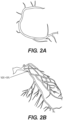
- FIGs. 2A and 2B are images of coronary arteries obtained from patients using an imaging device.
- the image data of FIGs. 2A and 2B may be captured, processed, and/or stored by server systems 106.
- the image data may be based on information, images, and/or data received from physicians 102 and/or third party providers 104 over electronic network 101.
- anatomic data may be obtained noninvasively using, for example, coronary computed tomographic angiography (CCTA).
- CCTA may be used for imaging of patients with chest pain and involves using computed tomography (CT) technology to image the heart and the coronary arteries following an intravenous infusion of a contrast agent.
- CT computed tomography
- CCTA also cannot provide direct information on the functional significance of coronary lesions, e.g., whether the lesions affect blood flow.
- CCTA is purely a diagnostic test, it cannot be used to predict changes in coronary blood flow, pressure, or myocardial perfusion under other physiologic states, e.g., exercise, nor can it be used to predict outcomes of interventions.
- Diagnostic cardiac catheterization may include performing conventional coronary angiography (CCA) to gather anatomic data on coronary lesions by providing a doctor with an image of the size and shape of the arteries.
- CCA coronary angiography
- CCA does not provide data for assessing the functional significance of coronary lesions. For example, a doctor may not be able to diagnose whether a coronary lesion is harmful without determining whether the lesion is functionally significant.
- a doctor may insert a stent because, as shown in FIG 2B , a portion of an artery appears that it has a substantial degree of stenosis (DS), for example, the degree of stenosis is greater than 50% of the vessel lumen.
- DS stenosis
- CCA has led to what has been referred to as an "oculostenotic reflex" of some interventional cardiologists to insert a stent for every lesion found with CCA regardless of whether the lesion is functionally significant.
- CCA may lead to unnecessary operations on the patient, which may pose added risks to patients and may result in unnecessary heath care costs for patients.
- Techniques presented herein may remedy one or more of these problems by determining a relationship between blood supply and organ or tissue demand.
- FIG. 3 is a block diagram of an exemplary method of determining a relationship between coronary blood supply and organ or tissue blood demand.
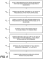
- FIG. 4 is a block diagram of an exemplary method of comparing a patient-specific relationship between arterial blood supply and organ or tissue blood demand to that of a population of prior patients.
- FIG. 5 is a block diagram of an exemplary method of determining a relationship between arterial blood supply and organ or tissue blood demand.
- FIG. 6 is a block diagram of an exemplary method of determining a relationship between arterial blood supply and organ or tissue blood demand to update a simulation of blood flow and pressure.
- FIG. 7 is a block diagram of an exemplary method of determining a coronary supply and myocardial demand.
- FIG. 3 is a block diagram of an exemplary method of determining a relationship between coronary blood supply and myocardial blood demand, according to an exemplary embodiment of the present disclosure.
- the method of FIG. 3 may be performed by server systems 106, based on information, images, and data received from physicians 102 and/or third party providers 104 over electronic network 101.
- step 305 may include receiving patient-specific imaging data of an organ or tissue.
- the coronary arteries and the muscle of the heart, the myocardium may both be imaged.
- a model of one or more vessels that supply blood to at least a portion of the organ or tissue may be generated based on the patient-specific imaging data.
- Techniques disclosed herein may extract the geometry of the epicardial coronary arteries from the coronary CT angiography data, and append a theoretical model representing the small arteries and arterioles that cannot be imaged in vivo.
- the theoretical model may be based on prior patient data, and/or may be selected based on the patient-specific imaging data.
- a blood supply may be determined based on the model of one or more vessels.
- the total volume of the coronary arterial tree may be computed.
- the above-mentioned theoretical model may represent the arteries and arterioles which are smaller than a known imaging threshold for the imaging device used to obtain patient-specific imaging data, and may be included in the determination of blood supply.
- Blood flow through the coronary arteries may be determined, and may be assumed to be related to the total coronary volume to the 3 ⁇ 4 power, although another mathematical relationship may be used.
- an organ or tissue demand may be determined.
- demand may be determined based on a mass or volume of the at least a portion of the organ or tissue. This may be the portion of the organ or tissue which is supplied with blood by the vessels corresponding to the model determined above.
- the determined flow through the coronary arteries may be assumed to be related to the myocardial mass or volume to the 3 ⁇ 4 power, although the exact power or mathematical relationship used may vary.
- the ratio of coronary volume to myocardial mass may be used to determine a mismatch between coronary supply and myocardial demand indicative of disease or a small caliber coronary arterial tree relative to the tissue mass that needs to be perfused.
- the demand for blood of at least a portion of an organ or tissue may be determined.
- demand for an organ or tissue may be determined based on a mass or volume of at least a portion of an organ or tissue.
- the organ or tissue demand such as the demand of at least a portion of the myocardium, may be determined based on perfusion imaging.
- the patient may be evaluated based on a comparison of the blood supply determined at step 315, and the organ or tissue demand determined at steps 320 and/or 325. For example, the ratio of blood supply to corresponding organ or tissue blood demand may indicate a presence or lack of ischemia.
- Imaging techniques may vary.
- the anatomic data to extract an arterial model may be obtained using 2D conventional angiography, 3D rotational angiography, magnetic resonance imaging, or 2D or 3D ultrasound imaging.
- Organ volume may be obtained using magnetic resonance imaging, or 2D or 3D ultrasound imaging, for example.
- Organ or tissue demand may be defined from organ or tissue volume, or organ or tissue mass (which may be computed using the volume data and a measured or assumed organ or tissue density). Organ or tissue demand may also be assessed directly using perfusion imaging from CT, MR, PET, or SPECT, or indirectly from CT, MRI or echocardiographic wall motion data using a model relating cardiac dynamics and work to blood flow demand.
- FIG. 4 is a block diagram of an exemplary method of comparing a patient-specific relationship between arterial blood supply and organ or tissue blood demand to that of a population of prior patients, according to an exemplary embodiment of the present disclosure.
- the method of FIG. 4 may be performed by server systems 106, based on information, images, and data received from physicians 102 and/or third party providers 104 over electronic network 101.
- a three-dimensional patient-specific arterial or anatomic model may be extracted from patient-specific imaging data, such as imaging data from an imaging device.
- a second model of arteries not included in the three-dimensional patient-specific arterial model may be generated.
- the model may be determined using branching laws originating from the terminal vessels extracted from the image data, or by generating vessels to fit within the boundaries of the supplied tissue extracted from the imaging data.
- a volume or mass of the relevant tissue of the organ supplied may also be extracted from imaging data.
- a volume or mass of the tissue or organ supplied by the three-dimensional patient-specific arterial model and the second model may be extracted.
- the volume or mass may be extracted using image processing methods where the organ surfaces are extracted.
- the volume or mass of the tissue or organ may be used to determine a tissue or organ demand. This may be done by relating the mass or volume of the tissue or organ to a physiologic parameter using form-function relations.
- total coronary artery blood flow is related to myocardial mass to determine demand of the heart for blood.
- a measure of the blood supply to the tissue or organ may be generated based on the three-dimensional patient-specific arterial model and the second model.
- a relationship between the tissue or organ demand and the measure of the blood supply may be determined. For example, the ratio of coronary arterial lumen volume to myocardial mass may be calculated.
- the relationship may be compared with a population of prior patients for patient evaluation purposes. For example, a relationship between a measure of the supplying arteries to a measure of the organ demanding blood may be calculated, reported, and compared to a normal reference value derived from a population of prior patients using statistical methods or machine learning.
- This comparison of the derived metric from the individual to the expected value from a population may then be used clinically to diagnose disease in the individual patient.
- the ratio of coronary arterial lumen volume to myocardial mass may be predictive of limitations in coronary artery blood flow to the heart muscle, which may for example cause chest pain.
- Techniques presented herein may use metrics related to a measure of the capacity of the supplying arteries to a measure of the organ demanding blood to refine the physiologic boundary conditions for an individual patient for use in patient-specific modeling of blood flow.
- the ratio of vascular volume to organ mass could be calculated for an individual patient, compared to data from a population of patients, and used to increase or decrease the resistance to flow under baseline, hyperemic, or exercise conditions. This could be applied to the calculation of noninvasive fractional flow reserve or coronary flow reserve to improve the accuracy of these methods for an individual patient.
- machine learning methods could be used together with information on the coronary artery lumen volume to myocardial mass ratio and measured FFR values in different patients to identify how the resistance boundary conditions could be adjusted to improve the accuracy of predictions of computed FFR.
- FIG. 5 is a block diagram of an exemplary method of determining a relationship between arterial blood supply and organ or tissue blood demand, according to an exemplary embodiment of the present disclosure.
- the method of FIG. 5 may be performed by server systems 106, based on information, images, and data received from physicians 102 and/or third party providers 104 over electronic network 101.
- a first patient-specific anatomic model may be received.
- the model may correspond to arteries of a patient, and may be received from an imaging device, or an electronic storage device (e.g., a hard drive, network drive, etc.).
- a second patient-specific model may be generated for vessels not included in the first patient-specific anatomic model.
- the second patient-specific model may include small blood vessels not observable in the image due to the limits of image resolution, image quality or limitations of the data collection technique.
- one or more patient-specific anatomic models of tissue or an organ supplied by the arteries of the first and second patient-specific models may be received or generated.
- the one or more models may be received from an electronic storage device (e.g., a hard drive, network drive, etc.).
- the arterial supply may be determined based on the first and second patient specific models.
- organ or tissue demand may be determined based on the patient-specific anatomic model of the organ or tissue.
- the organ or tissue mass may be calculated by measuring or assuming a tissue density and multiplying it by the tissue/organ volume.
- tissue or organ demand may be computed using methods described above.
- a relationship between arterial supply and organ demand may be determined based on a metric.
- this metric could be the ratio of the volume of the first patient-specific model to the volume or mass of the tissue/organ for the patient, or the ratio of the sum of the first and second patient-specific volumes to the volume or mass of the tissue/organ for that same patient.
- information may be provided on one or more parameters describing the relationship between arterial supply and tissue/organ demand. This information may be displayed to a user through a report, visual display or written to an electronic storage device (e.g., hard disk, network drive, cloud storage, smart phone, tablet, etc.).
- an electronic storage device e.g., hard disk, network drive, cloud storage, smart phone, tablet, etc.
- the supply-to-demand metric(s) computed for an individual patient above may be compared to data from a population of patients to provide additional information as to whether the patient data is within the normal range for an appropriate demographic.
- the normal supply-to-demand metric(s) may also depend on patient characteristics such as age, gender, blood pressure etc. This relationship can be inferred from data that relates all of these characteristics, including the supply-to-demand metrics, to whether the patient is healthy or diseased.
- the supply-to-demand metric(s) computed for an individual patient above may be utilized to update the physiologic model for that individual patient and/or compute blood flow and pressure, total or regional tissue perfusion, Fractional Flow Reserve (FFR), Coronary Flow Reserve (CFR), Index of Microcirculatory Resistance (IMR), territory at risk, plaque rupture risk, and/or plaque stress.
- Machine learning methods may be used to learn how the supply-to-demand metric(s) could be factored into boundary conditions assigned to compute coronary flow and pressure. For example, values of the supply-to-demand metrics that indicate that blood flow is too large to compute FFR accurately, may be used to change the boundary conditions in the calculation to decrease the flow.
- a particular supply-to-demand ratio may be indicative of a certain resistance to flow, as will be discussed further below.
- the resistance may be used to configure a patient-specific model which may be used to simulate blood flow in a patient's organs and/or tissues.
- FIG. 6 is a block diagram of an exemplary method of determining a relationship between arterial blood supply and organ or tissue blood demand to update a simulation of blood flow and pressure, according to an exemplary embodiment of the present disclosure.
- the method of FIG. 6 may be performed by server systems 106, based on information, images, and data received from physicians 102 and/or third party providers 104 over electronic network 101.
- one or more patient-specific anatomical models may be created from patient-specific images.
- Techniques presented herein may construct the patient-specific anatomic model from two-dimensional (e.g. coronary angiography, biplane angiography) or three-dimensional (e.g. 3D rotational angiography, coronary computed tomographic angiography (CCTA), magnetic resonance angiography (MRA)) model.
- This step may include methods to directly segment the image data and create a patient-specific three-dimensional anatomic model of the patient's arteries, or may involve modifying a previously-constructed "generic" model to customize it for that patient and create a patient-specific model.
- the patient-specific anatomic model may include some or all information related to the arteries of interest, including the length of each segment, diameter along the length of a segment (or any other geometrical description of the segment), branching patterns, presence of disease and/or characteristics of disease including composition of atherosclerotic plaques.
- the representation of the model may be defined by a surface enclosing a three-dimensional volume, a one-dimensional model where the centerline of the vessels is defined together with cross-sectional area information along the length, or could be an implicit representation of the vessel surface.
- the anatomic model may represent many different kinds of anatomy, such as coronary arteries, peripheral arteries, cerebral arteries, visceral arteries, hepatic vessels, renal arteries, etc. The model may also be received prior to using the methods and systems described herein.
- a model of the arterial tree beyond the anatomic model discussed above may be created.
- an anatomic model of the coronary arteries may be created downstream of the outlets of the model created above based on the theoretical anatomy of the coronary arteries by, for example, using data from the literature on coronary artery branching patterns.
- this model could also use the measured organ volume and boundaries to constrain the generated network of vessels as described in U.S. Patents 8,386,188 and 8,315,814 , both of which are incorporated herein by reference.
- a patient-specific anatomic model of the tissue or organ supplied by the arteries of the first and second patient-specific models may be generated.
- this is a model of the entire heart, the individual chamber tissue volumes, or the left ventricle myocardium extracted from CCTA imaging data. This model may be used to estimate organ demand for blood.
- a metric relating vascular supply to organ demand may be defined or determined.
- this metric is the ratio of the epicardial coronary artery volume (calculated in the step above) or the total coronary arterial volume (calculated in steps above) to myocardial mass, i.e., volume/mass.
- a low volume to mass ratio may be associated with presence of ischemia, whereas a higher volume to mass ratio may be associated with absence of ischemia.
- a ratio of volume/mass, using units mm 3 /g, of 30 or above may be associated with absence of ischemia.
- a ratio of below 30, and especially below 15, may be associated with presence of ischemia.
- a ratio above 30 may be classified as non-ischemic, 30-15 as moderately ischemic, and below 15 as ischemic.
- the specific thresholds, number and type of categorizations may vary. This metric may be determined in a similar manner using any vessels, organs or tissue.
- the supply-to-demand metrics may also be useful in predicting coronary flow reserve (CFR). For example, it is expected that low values of CFR would be observed in patients with low values of supply-to-demand metrics.
- a ratio of volume/mass, using units mm 3 /g, of below 30, and especially below 15, may be associated with low coronary flow reserve.
- Another step of techniques presented herein may be to report the above-calculated metric to a patient, physician or health care provider.
- a simulation of blood flow and pressure may be updated using the above calculated metric in comparison to population-based data to refine the physiologic model. This may be performed by adjusting values for microvascular resistance based on the above calculated metric.
- While one embodiment is related to more accurately computing blood flow and pressure in the human coronary arteries, other embodiments may include computing blood flow and pressure in the extracranial and intracranial cerebral arteries, the lower extremity arteries including the iliac, superficial femoral, common femoral, tibial, popliteal, peroneal, pedal arteries in patients with peripheral arterial disease, the renal arteries, the mesenteric arteries, and/or other vascular beds. This may be used to improve the methods described in U.S. Patent Nos. 8,386,188 and 8,315,814 , incorporated by reference in their entirety, which relate to simulating perfusion in the heart and brain, respectively.
- FIG. 7 is a block diagram of an exemplary method of determining a coronary supply and myocardial demand, according to an exemplary embodiment.
- the method of FIG. 7 includes steps of many of the prior-described embodiments, and applies those steps specifically to the heart and coronary arteries. Any of the details of corresponding steps in earlier-described embodiments may be used in the FIG. 7 method.
- the method of FIG. 7 may be performed by server systems 106, based on information, images, and data received from physicians 102 and/or third party providers 104 over electronic network 101.
- patient-specific image data obtained using an imaging device may be received, wherein at least a portion of the patient-specific image data corresponds to at least a portion of a patient's heart.
- a first patient-specific model of vessel geometry of at least a first portion of a coronary artery may be received, wherein the model is based on the received patient-specific image data.
- a second patient-specific model representing at least a second portion of a coronary artery may be received, wherein the second portion is associated with and smaller than the first portion of the coronary artery, and wherein the second portion of the coronary artery is below an imaging threshold of the imaging device.
- a coronary supply based on the first patient-specific model and the second patient-specific model may be determined.
- at least a portion of the myocardium corresponding to the coronary artery may be determined.
- a myocardial demand based on a mass or volume of the portion of the myocardium, or based on perfusion imaging of the portion of the myocardium may be determined.
- a relationship between the coronary supply and myocardial demand may be determined.
- Techniques presented herein describe methods which may determine a relationship between blood supply to an organ or tissue, and blood demand from that organ or tissue. These techniques provide significant insight into the overall disease burden of patients with atherosclerosis which would have prognostic value.
- Novel approaches described herein include determining a relationship between blood supply and blood demand in relationship to a given organ or tissue. Such a determination may be made for patients under resting, hyperemic and/or exercise conditions. These techniques may apply to the coronary arteries, but also to simulations of blood flow and pressure in any arterial tree including, but not limited to, the carotid, cerebral, renal, and lower extremity arteries.
- Techniques presented herein may calculate the ratio of blood flow and resistance based on vascular volume to that based on myocardial volume or mass, and may be implemented and included in an FFRCT platform. Methods to compute and display the resting flow mismatch or update the set of physiologic conditions and boundary conditions of the patient using this data may also be performed.
- a computer-implemented method determining a blood supply and a blood demand comprising:
- the method described in paragraph [070] may further comprise: evaluating the patient based upon the determined relationship between the coronary blood supply and the myocardial blood demand.
- the method described in paragraph [070] may further comprise: determining whether a mismatch exists between the coronary blood supply and the myocardial blood demand based on the determined relationship between the coronary blood supply and the myocardial blood demand.
- the method described in paragraph [072] may further comprise: based on the determination of whether the mismatch exists, modifying at least one parameter of a patient-specific simulation of blood flow through at least the portion of the coronary artery.
- the method described in paragraph [070] may further comprise: comparing the relationship to a reference value.
- the reference value may be determined from a population of patients.
- paragraph [070] may further comprise:
- the second patient-specific model may be generated based upon boundaries of a tissue associated with at least a portion of the coronary artery.
- the mass of the portion of the myocardium may be calculated by measuring or assuming a tissue density of the portion of the myocardium, and multiplying the tissue density by the volume of the portion of the myocardium.
- the patient-specific model may be generated by modifying a generic model of vessel geometry.
- a system for image processing to determine a blood supply and a blood demand comprising:
- the system described in paragraph [080] may be further configured for: evaluating the patient based upon the determined relationship between the coronary blood supply and the myocardial blood demand.
- the system described in paragraph [080] may be further configured for: determining whether a mismatch exists between the coronary blood supply and the myocardial blood demand based on the determined relationship between the coronary blood supply and the myocardial blood demand.
- the system described in paragraph [082] may be further configured for: based on the determination of whether the mismatch exists, modifying at least one parameter of a patient-specific simulation of blood flow through at least the portion of the coronary artery.
- the system described in paragraph [080] may be further configured for: comparing the relationship to a reference value.
- the reference value may be determined from a population of patients.
- the system described in paragraph [080] may be further configured for:
- the second patient-specific model may be generated based upon boundaries of a tissue associated with at least a portion of the coronary artery.
- the mass of the portion of the myocardium may be calculated by measuring or assuming a tissue density of the portion of the myocardium, and multiplying the tissue density by the volume of the portion of the myocardium.
- a non-transitory computer readable medium for use on a computer system containing computer-executable programming instructions for performing a method of determining a blood supply and a blood demand that comprises:
Landscapes
- Health & Medical Sciences (AREA)
- Life Sciences & Earth Sciences (AREA)
- Engineering & Computer Science (AREA)
- Medical Informatics (AREA)
- Public Health (AREA)
- Biomedical Technology (AREA)
- General Health & Medical Sciences (AREA)
- Pathology (AREA)
- Surgery (AREA)
- Heart & Thoracic Surgery (AREA)
- Molecular Biology (AREA)
- Animal Behavior & Ethology (AREA)
- Veterinary Medicine (AREA)
- Biophysics (AREA)
- Physics & Mathematics (AREA)
- Nuclear Medicine, Radiotherapy & Molecular Imaging (AREA)
- Radiology & Medical Imaging (AREA)
- High Energy & Nuclear Physics (AREA)
- Optics & Photonics (AREA)
- Cardiology (AREA)
- Data Mining & Analysis (AREA)
- Physiology (AREA)
- Oral & Maxillofacial Surgery (AREA)
- Dentistry (AREA)
- Databases & Information Systems (AREA)
- Epidemiology (AREA)
- Primary Health Care (AREA)
- Computer Vision & Pattern Recognition (AREA)
- Hematology (AREA)
- Pulmonology (AREA)
- Theoretical Computer Science (AREA)
- Vascular Medicine (AREA)
- Robotics (AREA)
- Apparatus For Radiation Diagnosis (AREA)
- Image Analysis (AREA)
- Magnetic Resonance Imaging Apparatus (AREA)
- Ultra Sonic Daignosis Equipment (AREA)
- Measuring And Recording Apparatus For Diagnosis (AREA)
Applications Claiming Priority (4)
| Application Number | Priority Date | Filing Date | Title |
|---|---|---|---|
| US201562236707P | 2015-10-02 | 2015-10-02 | |
| US15/192,286 US10517678B2 (en) | 2015-10-02 | 2016-06-24 | System and method for diagnosis and assessment of cardiovascular disease by comparing arterial supply capacity to end-organ demand |
| EP16753512.9A EP3355762B1 (de) | 2015-10-02 | 2016-07-29 | System und verfahren zur diagnose und beurteilung von herz-kreislauf-erkrankungen durch vergleich von arterieller versorgungskapazität mit dem endorganbedarf |
| PCT/US2016/044895 WO2017058351A1 (en) | 2015-10-02 | 2016-07-29 | System and method for diagnosis and assessment of cardiovascular disease by comparing arterial supply capacity to end-organ demand |
Related Parent Applications (2)
| Application Number | Title | Priority Date | Filing Date |
|---|---|---|---|
| EP16753512.9A Division EP3355762B1 (de) | 2015-10-02 | 2016-07-29 | System und verfahren zur diagnose und beurteilung von herz-kreislauf-erkrankungen durch vergleich von arterieller versorgungskapazität mit dem endorganbedarf |
| EP16753512.9A Division-Into EP3355762B1 (de) | 2015-10-02 | 2016-07-29 | System und verfahren zur diagnose und beurteilung von herz-kreislauf-erkrankungen durch vergleich von arterieller versorgungskapazität mit dem endorganbedarf |
Publications (2)
| Publication Number | Publication Date |
|---|---|
| EP4418280A2 true EP4418280A2 (de) | 2024-08-21 |
| EP4418280A3 EP4418280A3 (de) | 2024-11-06 |
Family
ID=56694228
Family Applications (2)
| Application Number | Title | Priority Date | Filing Date |
|---|---|---|---|
| EP24186125.1A Pending EP4418280A3 (de) | 2015-10-02 | 2016-07-29 | System und verfahren zur diagnose und beurteilung einer kardiovaskulären erkrankung durch vergleich der arteriellen versorgungskapazität mit dem endorganbedarf |
| EP16753512.9A Active EP3355762B1 (de) | 2015-10-02 | 2016-07-29 | System und verfahren zur diagnose und beurteilung von herz-kreislauf-erkrankungen durch vergleich von arterieller versorgungskapazität mit dem endorganbedarf |
Family Applications After (1)
| Application Number | Title | Priority Date | Filing Date |
|---|---|---|---|
| EP16753512.9A Active EP3355762B1 (de) | 2015-10-02 | 2016-07-29 | System und verfahren zur diagnose und beurteilung von herz-kreislauf-erkrankungen durch vergleich von arterieller versorgungskapazität mit dem endorganbedarf |
Country Status (5)
| Country | Link |
|---|---|
| US (4) | US10517678B2 (de) |
| EP (2) | EP4418280A3 (de) |
| JP (2) | JP6832920B2 (de) |
| CA (1) | CA2999437A1 (de) |
| WO (1) | WO2017058351A1 (de) |
Cited By (8)
| Publication number | Priority date | Publication date | Assignee | Title |
|---|---|---|---|---|
| US12144669B2 (en) | 2022-03-10 | 2024-11-19 | Cleerly, Inc. | Systems, devices, and methods for non-invasive image-based plaque analysis and risk determination |
| US12245882B2 (en) | 2020-01-07 | 2025-03-11 | Cleerly, Inc. | Systems, methods, and devices for medical image analysis, diagnosis, risk stratification, decision making and/or disease tracking |
| US12283046B2 (en) | 2020-01-07 | 2025-04-22 | Cleerly, Inc. | Systems, methods, and devices for medical image analysis, diagnosis, risk stratification, decision making and/or disease tracking |
| US12299885B2 (en) | 2022-03-10 | 2025-05-13 | Cleerly, Inc. | Systems, devices, and methods for non-invasive image-based plaque analysis and risk determination |
| US12324695B2 (en) | 2020-01-07 | 2025-06-10 | Cleerly, Inc. | Systems, methods, and devices for medical image analysis, diagnosis, risk stratification, decision making and/or disease tracking |
| US12380560B2 (en) | 2022-03-10 | 2025-08-05 | Cleerly, Inc. | Systems, methods, and devices for image-based plaque analysis and risk determination |
| US12440180B2 (en) | 2022-03-10 | 2025-10-14 | Cleerly, Inc. | Systems, devices, and methods for non-invasive image-based plaque analysis and risk determination |
| US12499539B2 (en) | 2022-12-30 | 2025-12-16 | Cleerly, Inc. | Systems, methods, and devices for medical image analysis, diagnosis, risk stratification, decision making and/or disease tracking |
Families Citing this family (28)
| Publication number | Priority date | Publication date | Assignee | Title |
|---|---|---|---|---|
| US10210956B2 (en) | 2012-10-24 | 2019-02-19 | Cathworks Ltd. | Diagnostically useful results in real time |
| US9135381B2 (en) | 2013-05-10 | 2015-09-15 | Stenomics, Inc. | Modeling and simulation system for optimizing prosthetic heart valve treatment |
| US9092743B2 (en) | 2013-10-23 | 2015-07-28 | Stenomics, Inc. | Machine learning system for assessing heart valves and surrounding cardiovascular tracts |
| JP2018515167A (ja) | 2015-04-02 | 2018-06-14 | ハートフロー, インコーポレイテッド | 血管網と潅流組織の機能的関連性を特定し可視化するシステム及び方法 |
| EP4241694A3 (de) | 2016-05-16 | 2023-12-20 | Cathworks Ltd. | Auswahl von blutgefässpfaden aus bildern |
| EP3457930B1 (de) | 2016-05-16 | 2023-11-15 | Cathworks Ltd. | System zur beurteilung von blutgefässen |
| WO2018138635A1 (en) * | 2017-01-24 | 2018-08-02 | Spectrum Dynamics Medical Limited | Systems and methods for computation of functional index parameter values for blood vessels |
| US11918291B2 (en) * | 2017-03-31 | 2024-03-05 | Koninklijke Philips N.V. | Simulation of transcatheter aortic valve implantation (TAVI) induced effects on coronary flow and pressure |
| WO2019002510A1 (en) | 2017-06-30 | 2019-01-03 | Koninklijke Philips N.V. | DIGITAL TOMOGRAPHY FFR SPECTRAL WITH AUTOMATIC LEARNING |
| EP3489893B1 (de) * | 2017-11-22 | 2020-06-24 | Siemens Healthcare GmbH | Verfahren und system zur bewertung eines hämodynamischen parameters |
| US11871995B2 (en) | 2017-12-18 | 2024-01-16 | Hemolens Diagnostics Sp. Z O.O. | Patient-specific modeling of hemodynamic parameters in coronary arteries |
| JP7467026B2 (ja) * | 2018-10-10 | 2024-04-15 | キヤノンメディカルシステムズ株式会社 | 医用情報処理装置、医用情報処理プログラム、医用情報処理システム |
| CN111227821B (zh) * | 2018-11-28 | 2022-02-11 | 苏州润迈德医疗科技有限公司 | 基于心肌血流量和ct图像的微循环阻力指数计算方法 |
| CN111227822B (zh) * | 2018-11-28 | 2022-02-11 | 苏州润迈德医疗科技有限公司 | 基于心肌血流量和ct图像的冠状动脉血流储备分数计算方法 |
| CN113412082B (zh) * | 2018-12-11 | 2024-12-13 | 辛纳吉A.I.有限公司 | 利用人工神经网络的器官体积测量方法及其装置 |
| US10813612B2 (en) | 2019-01-25 | 2020-10-27 | Cleerly, Inc. | Systems and method of characterizing high risk plaques |
| WO2020201942A1 (en) | 2019-04-01 | 2020-10-08 | Cathworks Ltd. | Methods and apparatus for angiographic image selection |
| JP7227400B2 (ja) * | 2019-04-15 | 2023-02-21 | 上海博動医療科技股▲分▼有限公司 | 冠血流量及び血流速度の取得方法並びに装置 |
| US11707325B2 (en) * | 2019-05-17 | 2023-07-25 | Heartflow, Inc. | System and methods for estimation of blood flow using response surface and reduced order modeling |
| US12039685B2 (en) | 2019-09-23 | 2024-07-16 | Cathworks Ltd. | Methods, apparatus, and system for synchronization between a three-dimensional vascular model and an imaging device |
| WO2021193020A1 (ja) * | 2020-03-27 | 2021-09-30 | テルモ株式会社 | プログラム、情報処理方法、情報処理装置及びモデル生成方法 |
| US20220102010A1 (en) * | 2020-09-25 | 2022-03-31 | Koninklijke Philips N.V. | Systems and methods for modelling a human subject |
| EP4020390A1 (de) * | 2020-12-22 | 2022-06-29 | Koninklijke Philips N.V. | Periphere perfusionsmessung |
| US12315076B1 (en) | 2021-09-22 | 2025-05-27 | Cathworks Ltd. | Four-dimensional motion analysis of a patient's coronary arteries and myocardial wall |
| WO2023152688A1 (en) | 2022-02-10 | 2023-08-17 | Cathworks Ltd. | System and method for machine-learning based sensor analysis and vascular tree segmentation |
| WO2024103115A1 (en) * | 2022-11-18 | 2024-05-23 | CFI Surgery Pty Ltd | Method of modelling anastomosis of channels |
| US20240387045A1 (en) * | 2023-05-17 | 2024-11-21 | Heartflow, Inc. | Systems and methods for processing electronic images to quantify coronary microvascualar disease |
| US12446965B2 (en) | 2023-08-09 | 2025-10-21 | Cathworks Ltd. | Enhanced user interface and crosstalk analysis for vascular index measurement |
Citations (2)
| Publication number | Priority date | Publication date | Assignee | Title |
|---|---|---|---|---|
| US8157742B2 (en) | 2010-08-12 | 2012-04-17 | Heartflow, Inc. | Method and system for patient-specific modeling of blood flow |
| US8311748B2 (en) | 2010-08-12 | 2012-11-13 | Heartflow, Inc. | Method and system for patient-specific modeling of blood flow |
Family Cites Families (19)
| Publication number | Priority date | Publication date | Assignee | Title |
|---|---|---|---|---|
| AU3071992A (en) | 1991-11-13 | 1993-06-15 | Gensia, Inc. | Methods for increasing the sensitivity and decreasing the side effects of pharmacological stress testing for coronary artery disease |
| US6217525B1 (en) | 1998-04-30 | 2001-04-17 | Medtronic Physio-Control Manufacturing Corp. | Reduced lead set device and method for detecting acute cardiac ischemic conditions |
| US6908435B1 (en) | 1999-11-05 | 2005-06-21 | Scimed Life Systems, Inc. | Method and monitor for enhancing angiogenesis in the heart by exercise follow-up |
| US8903472B2 (en) | 2007-01-23 | 2014-12-02 | Dtherapeutics, Llc | Applications of scaling laws of tree structures |
| US9119540B2 (en) * | 2010-09-16 | 2015-09-01 | Siemens Aktiengesellschaft | Method and system for non-invasive assessment of coronary artery disease |
| JP5946127B2 (ja) | 2012-05-11 | 2016-07-05 | 富士通株式会社 | シミュレーション方法、シミュレーション装置、およびシミュレーションプログラム |
| TW201401292A (zh) * | 2012-06-28 | 2014-01-01 | Hon Hai Prec Ind Co Ltd | 硬碟供電系統 |
| KR101939778B1 (ko) * | 2012-07-27 | 2019-01-18 | 삼성전자주식회사 | 필요 혈류량 결정 방법 및 장치, 혈류 영상 생성 방법 및 장치, 심근 관류 영상 처리 방법 및 장치 |
| US10398386B2 (en) * | 2012-09-12 | 2019-09-03 | Heartflow, Inc. | Systems and methods for estimating blood flow characteristics from vessel geometry and physiology |
| US9675301B2 (en) * | 2012-10-19 | 2017-06-13 | Heartflow, Inc. | Systems and methods for numerically evaluating vasculature |
| WO2014084382A1 (ja) | 2012-11-30 | 2014-06-05 | 株式会社 東芝 | 医用画像処理装置 |
| JP5953438B2 (ja) * | 2012-12-11 | 2016-07-20 | コーニンクレッカ フィリップス エヌ ヴェKoninklijke Philips N.V. | 冠状動脈を通る血流量を決定する方法 |
| US9805463B2 (en) * | 2013-08-27 | 2017-10-31 | Heartflow, Inc. | Systems and methods for predicting location, onset, and/or change of coronary lesions |
| US9501622B2 (en) | 2014-03-05 | 2016-11-22 | Heartflow, Inc. | Methods and systems for predicting sensitivity of blood flow calculations to changes in anatomical geometry |
| JP6262027B2 (ja) * | 2014-03-10 | 2018-01-17 | 東芝メディカルシステムズ株式会社 | 医用画像処理装置 |
| US9785746B2 (en) | 2014-03-31 | 2017-10-10 | Heartflow, Inc. | Systems and methods for determining blood flow characteristics using flow ratio |
| JP6553099B2 (ja) * | 2014-06-30 | 2019-07-31 | コーニンクレッカ フィリップス エヌ ヴェKoninklijke Philips N.V. | 血流予備量比値を算出するための機器 |
| WO2016030744A1 (ko) * | 2014-08-29 | 2016-03-03 | 강원대학교산학협력단 | 환자별 혈관 정보 결정 방법 |
| CN104921713B (zh) * | 2015-05-28 | 2017-08-11 | 中国科学院合肥物质科学研究院 | 一种基于心率与脉搏的心肌血氧供需状况动态监测系统及预警方法 |
-
2016
- 2016-06-24 US US15/192,286 patent/US10517678B2/en active Active
- 2016-07-29 EP EP24186125.1A patent/EP4418280A3/de active Pending
- 2016-07-29 CA CA2999437A patent/CA2999437A1/en active Pending
- 2016-07-29 WO PCT/US2016/044895 patent/WO2017058351A1/en not_active Ceased
- 2016-07-29 JP JP2018516445A patent/JP6832920B2/ja active Active
- 2016-07-29 EP EP16753512.9A patent/EP3355762B1/de active Active
-
2019
- 2019-11-21 US US16/691,266 patent/US10973583B2/en active Active
- 2019-12-27 JP JP2019237759A patent/JP7416617B2/ja active Active
-
2021
- 2021-03-10 US US17/197,271 patent/US11660143B2/en active Active
-
2023
- 2023-04-21 US US18/304,655 patent/US12329462B2/en active Active
Patent Citations (6)
| Publication number | Priority date | Publication date | Assignee | Title |
|---|---|---|---|---|
| US8157742B2 (en) | 2010-08-12 | 2012-04-17 | Heartflow, Inc. | Method and system for patient-specific modeling of blood flow |
| US8311748B2 (en) | 2010-08-12 | 2012-11-13 | Heartflow, Inc. | Method and system for patient-specific modeling of blood flow |
| US8315814B2 (en) | 2010-08-12 | 2012-11-20 | Heartflow, Inc. | Method and system for patient-specific modeling of blood flow |
| US8386188B2 (en) | 2010-08-12 | 2013-02-26 | Heartflow, Inc. | Method and system for patient-specific modeling of blood flow |
| US8594950B2 (en) | 2010-08-12 | 2013-11-26 | Heartflow, Inc. | Method and system for patient-specific modeling of blood flow |
| US8734357B2 (en) | 2010-08-12 | 2014-05-27 | Heartflow, Inc. | Method and system for patient-specific modeling of blood flow |
Cited By (11)
| Publication number | Priority date | Publication date | Assignee | Title |
|---|---|---|---|---|
| US12245882B2 (en) | 2020-01-07 | 2025-03-11 | Cleerly, Inc. | Systems, methods, and devices for medical image analysis, diagnosis, risk stratification, decision making and/or disease tracking |
| US12283046B2 (en) | 2020-01-07 | 2025-04-22 | Cleerly, Inc. | Systems, methods, and devices for medical image analysis, diagnosis, risk stratification, decision making and/or disease tracking |
| US12324695B2 (en) | 2020-01-07 | 2025-06-10 | Cleerly, Inc. | Systems, methods, and devices for medical image analysis, diagnosis, risk stratification, decision making and/or disease tracking |
| US12396695B2 (en) | 2020-01-07 | 2025-08-26 | Cleerly, Inc. | Systems, methods, and devices for medical image analysis, diagnosis, risk stratification, decision making and/or disease tracking |
| US12144669B2 (en) | 2022-03-10 | 2024-11-19 | Cleerly, Inc. | Systems, devices, and methods for non-invasive image-based plaque analysis and risk determination |
| US12299885B2 (en) | 2022-03-10 | 2025-05-13 | Cleerly, Inc. | Systems, devices, and methods for non-invasive image-based plaque analysis and risk determination |
| US12324696B2 (en) | 2022-03-10 | 2025-06-10 | Cleerly, Inc. | Systems, devices, and methods for non-invasive image-based plaque analysis and risk determination |
| US12380560B2 (en) | 2022-03-10 | 2025-08-05 | Cleerly, Inc. | Systems, methods, and devices for image-based plaque analysis and risk determination |
| US12406365B2 (en) | 2022-03-10 | 2025-09-02 | Cleerly, Inc. | Systems, devices, and methods for non-invasive image-based plaque analysis and risk determination |
| US12440180B2 (en) | 2022-03-10 | 2025-10-14 | Cleerly, Inc. | Systems, devices, and methods for non-invasive image-based plaque analysis and risk determination |
| US12499539B2 (en) | 2022-12-30 | 2025-12-16 | Cleerly, Inc. | Systems, methods, and devices for medical image analysis, diagnosis, risk stratification, decision making and/or disease tracking |
Also Published As
| Publication number | Publication date |
|---|---|
| US20170095292A1 (en) | 2017-04-06 |
| US20200085502A1 (en) | 2020-03-19 |
| US12329462B2 (en) | 2025-06-17 |
| US10973583B2 (en) | 2021-04-13 |
| CA2999437A1 (en) | 2017-04-06 |
| EP4418280A3 (de) | 2024-11-06 |
| JP7416617B2 (ja) | 2024-01-17 |
| JP6832920B2 (ja) | 2021-02-24 |
| US20210196391A1 (en) | 2021-07-01 |
| US11660143B2 (en) | 2023-05-30 |
| JP2020044449A (ja) | 2020-03-26 |
| US10517678B2 (en) | 2019-12-31 |
| EP3355762B1 (de) | 2024-09-04 |
| JP2018534970A (ja) | 2018-11-29 |
| WO2017058351A1 (en) | 2017-04-06 |
| US20230277247A1 (en) | 2023-09-07 |
| EP3355762A1 (de) | 2018-08-08 |
Similar Documents
| Publication | Publication Date | Title |
|---|---|---|
| US12329462B2 (en) | Systems and methods for processing electronic images to assess end-organ demand | |
| US20250025061A1 (en) | Systems and methods for processing images to analyze perfusion | |
| US12048490B2 (en) | Systems and methods for correction of artificial deformation in anatomic modeling | |
| EP3281135B1 (de) | System und verfahren zur gefässbaumerzeugung mithilfe patientenspezifischer struktureller und funktionaler daten und gemeinsamer vorheriger informationen |
Legal Events
| Date | Code | Title | Description |
|---|---|---|---|
| PUAI | Public reference made under article 153(3) epc to a published international application that has entered the european phase |
Free format text: ORIGINAL CODE: 0009012 |
|
| STAA | Information on the status of an ep patent application or granted ep patent |
Free format text: STATUS: REQUEST FOR EXAMINATION WAS MADE |
|
| 17P | Request for examination filed |
Effective date: 20240702 |
|
| AC | Divisional application: reference to earlier application |
Ref document number: 3355762 Country of ref document: EP Kind code of ref document: P |
|
| AK | Designated contracting states |
Kind code of ref document: A2 Designated state(s): AL AT BE BG CH CY CZ DE DK EE ES FI FR GB GR HR HU IE IS IT LI LT LU LV MC MK MT NL NO PL PT RO RS SE SI SK SM TR |
|
| REG | Reference to a national code |
Ref country code: DE Ref legal event code: R079 Free format text: PREVIOUS MAIN CLASS: G16H0050500000 Ipc: A61B0005000000 |
|
| PUAL | Search report despatched |
Free format text: ORIGINAL CODE: 0009013 |
|
| AK | Designated contracting states |
Kind code of ref document: A3 Designated state(s): AL AT BE BG CH CY CZ DE DK EE ES FI FR GB GR HR HU IE IS IT LI LT LU LV MC MK MT NL NO PL PT RO RS SE SI SK SM TR |
|
| RIC1 | Information provided on ipc code assigned before grant |
Ipc: A61B 8/08 20060101ALN20240930BHEP Ipc: A61B 8/06 20060101ALN20240930BHEP Ipc: G16H 50/70 20180101ALI20240930BHEP Ipc: A61B 8/12 20060101ALI20240930BHEP Ipc: G16H 50/50 20180101ALI20240930BHEP Ipc: A61B 6/50 20240101ALI20240930BHEP Ipc: A61B 6/03 20060101ALI20240930BHEP Ipc: G16H 50/20 20180101ALI20240930BHEP Ipc: A61B 5/02 20060101ALI20240930BHEP Ipc: A61B 6/00 20240101ALI20240930BHEP Ipc: A61B 5/00 20060101AFI20240930BHEP |