EP4406475A2 - Système et procédé de balayage d'empreinte intrabuccale et d'empreinte combinés - Google Patents
Système et procédé de balayage d'empreinte intrabuccale et d'empreinte combinés Download PDFInfo
- Publication number
- EP4406475A2 EP4406475A2 EP24180979.7A EP24180979A EP4406475A2 EP 4406475 A2 EP4406475 A2 EP 4406475A2 EP 24180979 A EP24180979 A EP 24180979A EP 4406475 A2 EP4406475 A2 EP 4406475A2
- Authority
- EP
- European Patent Office
- Prior art keywords
- scan data
- oral
- impression
- data
- area
- Prior art date
- Legal status (The legal status is an assumption and is not a legal conclusion. Google has not performed a legal analysis and makes no representation as to the accuracy of the status listed.)
- Granted
Links
Images





Classifications
-
- A—HUMAN NECESSITIES
- A61—MEDICAL OR VETERINARY SCIENCE; HYGIENE
- A61B—DIAGNOSIS; SURGERY; IDENTIFICATION
- A61B5/00—Measuring for diagnostic purposes; Identification of persons
- A61B5/0059—Measuring for diagnostic purposes; Identification of persons using light, e.g. diagnosis by transillumination, diascopy, fluorescence
- A61B5/0082—Measuring for diagnostic purposes; Identification of persons using light, e.g. diagnosis by transillumination, diascopy, fluorescence adapted for particular medical purposes
- A61B5/0088—Measuring for diagnostic purposes; Identification of persons using light, e.g. diagnosis by transillumination, diascopy, fluorescence adapted for particular medical purposes for oral or dental tissue
-
- A—HUMAN NECESSITIES
- A61—MEDICAL OR VETERINARY SCIENCE; HYGIENE
- A61C—DENTISTRY; APPARATUS OR METHODS FOR ORAL OR DENTAL HYGIENE
- A61C9/00—Impression cups, i.e. impression trays; Impression methods
- A61C9/004—Means or methods for taking digitized impressions
- A61C9/0046—Data acquisition means or methods
- A61C9/0053—Optical means or methods, e.g. scanning the teeth by a laser or light beam
-
- A—HUMAN NECESSITIES
- A61—MEDICAL OR VETERINARY SCIENCE; HYGIENE
- A61B—DIAGNOSIS; SURGERY; IDENTIFICATION
- A61B5/00—Measuring for diagnostic purposes; Identification of persons
-
- A—HUMAN NECESSITIES
- A61—MEDICAL OR VETERINARY SCIENCE; HYGIENE
- A61B—DIAGNOSIS; SURGERY; IDENTIFICATION
- A61B5/00—Measuring for diagnostic purposes; Identification of persons
- A61B5/0059—Measuring for diagnostic purposes; Identification of persons using light, e.g. diagnosis by transillumination, diascopy, fluorescence
- A61B5/0062—Arrangements for scanning
-
- A—HUMAN NECESSITIES
- A61—MEDICAL OR VETERINARY SCIENCE; HYGIENE
- A61C—DENTISTRY; APPARATUS OR METHODS FOR ORAL OR DENTAL HYGIENE
- A61C13/00—Dental prostheses; Making same
- A61C13/0003—Making bridge-work, inlays, implants or the like
- A61C13/0004—Computer-assisted sizing or machining of dental prostheses
-
- A—HUMAN NECESSITIES
- A61—MEDICAL OR VETERINARY SCIENCE; HYGIENE
- A61C—DENTISTRY; APPARATUS OR METHODS FOR ORAL OR DENTAL HYGIENE
- A61C9/00—Impression cups, i.e. impression trays; Impression methods
- A61C9/004—Means or methods for taking digitized impressions
- A61C9/0046—Data acquisition means or methods
-
- G—PHYSICS
- G06—COMPUTING OR CALCULATING; COUNTING
- G06T—IMAGE DATA PROCESSING OR GENERATION, IN GENERAL
- G06T7/00—Image analysis
- G06T7/30—Determination of transform parameters for the alignment of images, i.e. image registration
-
- G—PHYSICS
- G06—COMPUTING OR CALCULATING; COUNTING
- G06T—IMAGE DATA PROCESSING OR GENERATION, IN GENERAL
- G06T2207/00—Indexing scheme for image analysis or image enhancement
- G06T2207/30—Subject of image; Context of image processing
- G06T2207/30004—Biomedical image processing
- G06T2207/30036—Dental; Teeth
Definitions
- the present disclosure relates to a combined oral and impression scanning system and method, and more particularly, to a combined oral and impression scanning system and method for obtaining one final digital datum merged by aligning impression scan data with oral scan data in a process of obtaining the impression scan data after obtaining the oral scan data by using an oral scanner.
- 3-D scanning is used in various industry fields, such as reverse engineering, measurement, tests, content creation, and a CAD/CAM.
- 3-D three-dimensional
- a structured light method of projecting a structured pattern onto an object to be measured by using an optical projection device, obtaining an image of the object through an imaging device, and computing 3-D data by analyzing the obtained image is chiefly used.
- An object of the present disclosure is to provide a combined oral and impression scanning system and method, which enable elaborate prosthetics to be fabricated using obtained digital data by obtaining one final scan datum merged by aligning impression scan data with oral scan data in a process of obtaining the impression scan data after obtaining the oral scan data by using an oral scanner.
- a combined oral and impression scanning system may include an oral scanner configured to scan an oral object, a storage unit configured to store oral scan data obtained from the oral scanner and selectively store impression scan data of the oral object according to a preset determination condition, a determination unit configured to determine whether the oral scan data corresponds to the determination condition and guide selective storage of the impression scan data, and a control unit configured to control a selection area corresponding to the determination condition, among the impression scan data, to be partially applied to a corresponding area of the oral scan data.
- the determination condition may include at least one of a plurality of conditions, including the unclear indication of a margin line, the presence of a post area or the presence of an area including metal in the oral scan data.
- control unit may include a simulation unit configured to receive, from the storage unit, information on areas neighboring the selection area and to automatically simulate the selection area before the selection area is applied to be replaced.
- a combined oral and impression scanning method may include an oral scan data storage step of storing oral scan data by scanning an oral object, an impression mode change step of changing a mode of the oral scan data into an impression mode so that the oral scan data corresponds to selectively stored impression scan data, a selection area replacement application step of partially obtaining scan data of a selection area among the impression scan data and applying the obtained scan data to the oral scan data having the changed impression mode, and a final scan datum acquisition step of changing again, into an oral mode which is an initial mode, the impression mode of the oral scan data to which the impression scan data has been applied in the selection area and obtaining the final scan data.
- the method may further include a prosthetics production step of transmitting the final data to a design program so that prosthetics is fabricated.
- the oral object may include an oral model or an inside of a mouth of a patient.
- the method may further include an impression scan data storage determination step of selectively storing the impression scan data depending on whether the oral scan data includes a preset determination condition after the oral scan data storage step.
- the determination condition may include at least one of a plurality of conditions, including the unclear indication of a margin line, the presence of a post area or the presence of an area including metal in the oral scan data.
- the impression scan data may include data stored by selectively scanning only a portion corresponding to a portion of oral scan data corresponding to the determination condition in an impression model.
- the impression mode change step may include indicating a margin line in the impression scan data by using an impression material when it is determined that the indication of the margin line is unclear in the oral scan data in the oral scan data storage step.
- the margin line may be applied to the oral scan data and indicated when the final scan data is obtained.
- the oral scan data storage step may include a post area scan step of converting a mode of the post area into an impression mode when it is determined that the selection area is a post area by scanning the oral object and applying the selection area by replacing previously stored impression scan data.
- the post area scan step may include a display step of displaying the post area, an impression mode change step of selectively obtaining oral scan data of the post area, converting a mode of the oral scan data into the impression mode, and obtaining impression scan data, and an embossing scan data application step of obtaining embossing scan data of the post area by changing the impression mode and applying the embossing scan data to the impression scan data by partially replacing the impression scan data.
- a combined oral and impression scanning method may include an oral scan data acquisition step of obtaining, by a scanner, oral scan data of an oral object including a post area, an impression mode change step of changing a mode of the oral scan data into an impression mode so that the oral scan data is reversed, a selection area designation step of designating the post area as an object area for an impression scan, an impression scan data acquisition step of obtaining impression scan data of the selection area designated as the object area for the impression scan, and a data aligned step of aligning the oral scan data and the obtained impression scan data of the selection area.
- the impression scan data may be obtained by scanning an impression model which is a frame of an oral model through a scanner.
- the post area may be obtained in an embossing form.
- the method may further include a re-reverse step of reversing again the oral scan data and impression scan data aligned in the data aligned step when the impression mode is released.
- the method may further include a final scan datum acquisition step of replacing the oral scan data with the impression scan data of the selection area in the re-reverse step and obtaining final scan data by combining the oral scan data and the impression scan data.
- a combined oral and impression scanning method may include a first oral scan data storage step of storing first oral scan data by scanning an outer surface of an oral object including an upper jaw model and a lower jaw model, a second oral scan data storage step of storing second oral scan data by scanning an inside of the oral object in a state in which the upper jaw model and the lower jaw model have been isolated from each other, a selection area impression scan data comparison step of obtaining impression scan data of a selection area of the oral object including an occlusion state of the upper jaw model and the lower jaw model and comparing the first oral scan data and the second oral scan data, and a selection area impression scan data replacement application step of applying the impression scan data to the first oral scan data and the second oral scan data by partially replacing the first oral scan data and the second oral scan data.
- the upper jaw model and the lower jaw model may face each other in a way to be occluded.
- the present disclosure has an effect in that elaborate prosthetics can be fabricated using obtained digital data in a way to obtain one final scan datum merged by aligning impression scan data with oral scan data in real time in a process of obtaining the impression scan data after obtaining the oral scan data by using the oral scanner.
- the present disclosure has an effect in that more accurate digital data can be obtained by expressing a margin line in a process of obtaining an impression when it is difficult to measure the margin line because the margin line is covered with a gum (gingival) tissue in an environment within a mouth.
- the present disclosure has an effect in that it can solve a conventional problem in which it is difficult or impossible to obtain data by using the oral scanner due to the depth of a post areahaving an engraved form, in such a way to enable a portion whose scanning is impossible to be scanned by obtaining, in an embossing form, the post area actually having the engraved form by obtaining impression scan data.
- the present disclosure has an effect in that a teeth portion whose accurate scanning is difficult due to a material characteristic can be scanned by separately obtaining an impression of the teeth portion covered with a crown made of metal and using such impression scan data for occlusion and alignment necessary to obtain the final scan data.
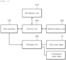
- FIG. 1 is a diagram schematically illustrating a combined oral and impression scanning system according to the present disclosure.
- the combined oral and impression scanning system according to the present disclosure includes an oral scanner 100, a storage unit 200, a determination unit 300 and a control unit 400.
- the oral scanner 100 may correspond to any one of a model scanner having a table form or a three-dimensional (3-D) scanner having a handheld form, for photographing an oral object (in the present disclosure, an oral object may be a frame whose impression has been taken or an oral model obtained by pouring plaster into a frame whose impression has been taken, or may be the inside of the mouth of an actual patient).
- the oral scanner 100 may include photographing means for photographing a movement.
- the storage unit 200 is formed to store oral scan data obtained by the oral scanner 100 and to also selectively store impression scan data of the oral object according to a preset determination condition.
- the storage unit 200 stores the oral scan data, and also stores the impression scan data of the oral object when the determination unit 300 determines that the stored scan data corresponds to the preset determination condition. That is, the determination unit 300 determines whether the oral scan data corresponds to the preset determination condition.
- the selective storage of the impression scan data in the storage unit 200 may be guided based on a result of the determination of the determination unit 300.
- the determination condition includes at least one of a plurality of conditions, including that the indication of a margin line is not clearly indicated in the oral scan data because the indication of the margin line is covered with gums, etc., a post area P is present or an area including a part made of metal, etc. is present.
- a portion is set as a selection area. More specifically, a surface of the oral object is scanned using the oral scan data. If a margin line whose measurement is difficult because the margin line is covered with a gum tissue in an environment within a mouth, a post area having an engraved portion, or a metal area filled with amalgam or resin is present, accurate scan data for such a portion cannot be secured. Accordingly, if at least one of a plurality of such determination conditions is included, the determination unit 300 enables the impression scan data of the oral object including the selection area to be scanned by the oral scanner 100 and to be selectively stored in the storage unit 200.
- the control unit 400 determines that accurate final scan data of the oral object cannot be obtained because the margin line, the post area, the metal area, etc. is present, and controls a selection area, including impression scan data and corresponding to the determination condition, to be partially applied to the oral scan data.
- to partially apply the impression scan data to the oral scan data may mean that data values of the oral scan data formed in the selection area are merged by performing a process of supplementing or replacing the data values of the oral scan data based on data values of the impression scan data.
- to partially apply the impression scan data to the oral scan data may mean that the impression scan data replaces the oral scan data with respect to the selection area.
- control unit 400 controls not only the oral scan data, but the impression scan data to be selectively obtained independently and to be stored in the storage unit 200, and controls the stored data to be divided into a portion to be retained in the oral scan data and a portion to be replaced with the impression scan data, that is, as a selection area and to be partially aligned and merged.
- scan data is obtained by the oral scanner 100, a portion not clearly represented based on only oral scan data can be selectively represented in the same level as an actual level. As a result, accurate final scan datum which may be used in a design task for an oral object can be effectively secured.
- control unit 400 may be connected to a simulation unit 410, and may share information with the simulation unit 410.
- the simulation unit 410 is formed to receive, from the storage unit 200, information on areas neighboring a selection area, that is, portions before/after the selection area, in addition to the selection area included in the impression scan data and to automatically simulate the selection area before oral scan data is applied by replacing the selection area. Accordingly, the simulation unit 410 can prevent a problem in that a selection area is erroneously replaced with a wrong area in a process of automatically replacing the selection area with a corresponding area of oral scan data. As a result, a corresponding area can be accurately applied by replacing the selection area.
- FIG. 2 is a flowchart sequentially illustrating a combined oral and impression scanning method according to a first embodiment of the present disclosure.
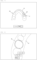
- FIG. 3 is a diagram illustrating the acquisition of oral scan data and impression scan data in the combined oral and impression scanning method according to the first embodiment of the present disclosure.
- FIG. 4 is a diagram illustrating a change into an impression mode in the combined oral and impression scanning method according to the first embodiment of the present disclosure.
- FIG. 5 is a diagram illustrating a selection area in the combined oral and impression scanning method according to the first embodiment of the present disclosure.
- FIG. 6 is a diagram illustrating the replacement of oral scan data with impression scan data in a selection area in the combined oral and impression scanning method according to the first embodiment of the present disclosure.
- FIG. 7 is a diagram illustrating the final scan data in the combined oral and impression scanning method according to the first embodiment of the present disclosure.
- the combined oral and impression scanning method according to the present embodiment is sequentially described as follows.
- an oral object is scanned using the oral scanner 100, and corresponding oral scan data 10 is stored in the storage unit 200 (S100).
- the determination unit 300 determines whether an area corresponding to a preset determination condition is present by checking the oral scan data 10 (S200), and also enables impression scan data 20 of the oral object to be selectively stored (S210). That is, as the oral scan data 10 is stored in the storage unit 200, when it is determined that an area corresponding to the preset determination condition is present by checking the stored oral scan data 10 (S200), the impression scan data 20 of the oral object is selectively stored (S210).
- the oral object may be a frame whose impression has been taken or may be an oral model obtained by pouring plaster into a frame whose impression has been taken. Alternatively, the oral object may be the inside of the mouth of an actual patient.
- the determination condition includes at least one of a plurality of conditions, including that the indication of a margin line in the oral scan data 10 is covered (the unclear indication of the margin line), a post area is present or an area including metal is present. Such a portion is set as a selection area. If it is confirmed that an area corresponds to the determination condition is not present by checking the oral scan data 10, the process is terminated by considering the oral scan data 10 to be the final scan datum 30 (S300).
- a mode of the oral scan data 10 is changed into an impression mode so that the oral scan data 10 corresponding to the selectively stored impression scan data 20 as described above (S220).
- a normal of the oral scan data 10 is changed, that is, a model of the oral scan data 10 becomes a negative model. Accordingly, the oral scan data 10 is represented as reverse data 10' of the oral scan data, and the oral scan data 10 and the impression scan data 20 are compared and merged in a one-to-one manner.
- scan data of a selection area A among the converted impression scan data 20 is partially obtained.
- the obtained scan data is applied by replacing the oral scan data 10 having the changed impression mode (S230).
- information on the scan data of the selection area A to be alternatively applied further includes scan data information of partial areas neighboring scan data information of the selection area A, that is, before and after the selection area A. Accordingly, the information on the scan data of the selection area A can be alternatively applied to an accurate area because the selection area A is automatically simulated before the scan data of the selection area A is alternatively applied to the oral scan data 10 of the selection area A.
- the selection area A corresponds to a margin line M in which measurement is difficult because the margin line is covered with a gum tissue in an environment within a mouth, a post area P formed of an engraved portion, or a metal area filled with amalgam or resin. For this reason, the selection area A needs to be replaced with information on a corresponding area of the impression scan data 20 because accurate scan data of such a portion cannot be secured.
- a mode of the oral scan data 10 having the state in which the selection area A has been alternatively applied and having the changed impression mode is changed into an oral mode again, that is, an initial mode (S240).
- the final scan datum 30 is obtained, and the process is terminated (S300).
- the one final scan datum 30 in which the oral scan data 10 and the impression scan data 20 have been merged can be obtained because a process of aligning the impression scan data 20 with the oral scan data 10 is performed in real time by continuously changing a mode of the selection area A into the impression mode and the oral mode in a process of obtaining the impression scan data 20 after obtaining the oral scan data 10.
- the oral scan data 10 is converted into the reverse data 10' of the oral scan data in the impression mode.
- the impression scan data 20 in the selection area A may be applied to replace some of the reverse data 10' of the oral scan data. Accordingly, the one final scan datum 30 merged by changing the impression mode into the oral mode again can be obtained.
- the oral scan data 10 and the impression scan data 20 are independently obtained.
- the obtained data is divided into a portion to be maintained in the oral scan data 10 and a portion to be replaced with the impression scan data 20, that is, the selection area A.
- the portions are partially aligned and merged. Accordingly, when scan data is obtained by the oral scanner, a portion not clearly represented using only the oral scan data 10 can be represented in the same level as an actual oral object by selectively supplementing the portion by using the impression scan data 20. As a result, the accurate final scan datum 30 which may be used in a task for designing the oral object can be effectively secured.
- the final scan datum 30 obtained as described above may be output in the form of a file having a common format without a separate additional task.
- the final scan datum 30 may be transmitted to a design program, so that prosthetics for an oral object having the same level as an actual mouth can be fabricated (S400).
- FIG. 8 is a diagram illustrating the indication of a margin line in a combined oral and impression scanning method according to a second embodiment of the present disclosure.
- FIG. 9 is a diagram illustrating the replacement and application of a margin line in the combined oral and impression scanning method according to the second embodiment of the present disclosure.
- the margin line M may be represented through scanning in the state in which an impression material enabling the impression scan data 20 to be obtained is filled between the gums and teeth. That is, it is difficult to obtain the margin line M by using the oral scanner.
- the margin line M may be obtained using the impression material filled between the gums and the teeth.
- the obtained margin line M more specifically, the margin line M indicated in the impression scan data 20 may be applied to the oral scan data 10 and indicated when the final scan datum 30 is obtained in the final scan data acquisition step S300.
- the margin line M whose measurement is difficult because the margin line is covered with a gum tissue in a mouth is applied to the oral scan data 10 by using an impression material and indicated in the final scan datum 30. Accordingly, prosthetics having high precision can be fabricated because data having more abundant and clearer information is delivered to a design program.
- FIG. 10 is a diagram illustrating a post area P in a combined oral and impression scanning method according to a third embodiment of the present disclosure.
- FIG. 11 is a diagram illustrating the state in which a selection area for the post area P is indicated in the combined oral and impression scanning method according to the third embodiment of the present disclosure.
- FIG. 12 is a diagram illustrating that embossing scan data is applied to the post area in the combined oral and impression scanning method according to the third embodiment of the present disclosure.
- a mode of the post area P may be changed into the impression mode and applied by replacing previously stored impression scan data.
- Such a post area scan step may be performed in an order to be described later.
- the post area P is indicated with respect to an oral object. That is, as illustrated in FIG. 10 , the indicated post area P is indicated in the state in which the post area P has been perforated due to its depth.
- the oral scan data 10 of the post area P is selectively obtained and indicated.
- a mode of the oral object including the post area P is changed into the impression mode in order to obtain the impression scan data 20.
- the step of determining whether to store the impression scan data is performed by confirming whether the presence of the post area P corresponds to the preset determination condition after the step of storing the oral scan data 10. Only a portion corresponding to a portion of the oral scan data corresponding to the determination condition, among the impression scan data, may be selectively scanned and stored.
- the impression scan data 20 of the post area P can be obtained by changing the mode of the oral object into the impression mode.
- the post area P may also be incorporated into the final scan datum 30 when the impression mode is changed into the oral mode again in the final scan data acquisition step S300.
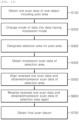
- FIG. 13 is a diagram sequentially illustrating a combined oral and impression scanning method according to a fourth embodiment of the present disclosure.
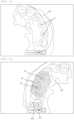
- FIG. 14 is a diagram illustrating a post area P in the combined oral and impression scanning method according to the fourth embodiment of the present disclosure.
- FIG. 15 is a diagram illustrating the state in which a selection area A for the post area is indicated in the combined oral and impression scanning method according to the fourth embodiment of the present disclosure.
- FIG. 16 is a diagram illustrating that embossing scan data is applied to the post area P in the combined oral and impression scanning method according to the fourth embodiment of the present disclosure.
- FIG. 17 is a diagram illustrating the final scan data obtained using the combined oral and impression scanning method according to the fourth embodiment of the present disclosure.
- an oral scan data acquisition step S102 of obtaining oral scan data 10 by scanning an oral object through the scanner is performed.
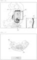
- the oral object includes a post and core task area (hereinafter referred to as a post area P) for a crown or implant task. More specifically, the oral object has the post area P as a part of the object. From FIG. 14 , it may be seen that the oral scan data 10 appears and the post area P has been incompletely scanned.
- data of the post area P may be supplemented in the impression mode. That is, this means that data reversed by changing a mode of the data having the oral mode into data having the impression mode (S202) is supplemented using newly obtained data having an impression form.
- the obtained oral scan data 10 may be represented as reverse data 10' of the oral scan data by clicking on an impression mode change button 42.
- the incompletely scanned post area P may be designated as a selection area A (S302).
- the selection area A may be designated by clicking on an area selection button 44 disposed close to the impression mode change button 42 and including the post area P among the reverse data 10' of the oral scan data.
- the selection area A is shaded, and impression scan data 20 corresponding to a corresponding portion may be obtained.
- impression scan data 20 corresponding to the selection area A may be obtained by additional scanning using the scanner (S402). Meanwhile, the impression scan data 20 is obtained by scanning an impression model, that is, a frame of an oral model, through the scanner. When the impression model is scanned, a current state of the scanning is displayed on a scan screen 22, and a process of obtaining corresponding data is displayed in the scan area 12.
- the post area P obtained as the impression scan data 20 actually has an engraved form. However, in the impression mode, the post area P may also be obtained in an embossing form because the oral scan data 10 is reversed.
- the obtained impression scan data 20 is aligned with a portion corresponding to the selection area A among the reverse data 10' of the oral scan data (S502). As the impression scan data 20 is aligned, the impression scan data 20 can supplement an insufficient scan portion of the oral scan data 10. Meanwhile, the oral scan data 10 of the selection area A including the post area P may be applied to be replaced with relatively precise impression scan data 20.
- the impression mode may be released by clicking on the impression mode change button 42. That is, the scan mode may return from the impression mode to the oral mode.
- the data is reversed again by releasing the impression mode (S602). More specifically, the reverse data 10' of the oral scan data is indicated as the oral scan data 10 again.
- the impression scan data 20 is indicated as reverse data 20' of the impression scan data. Meanwhile, since the impression scan data 20 is a negative model, the reverse data 20' of the impression scan data may be used to obtain the final scan datum 30. Meanwhile, the impression scan data 20 and the reverse data 20' of the impression scan data may be formed to have a normal in which the impression scan data 20 and the reverse data 20' face each other.
- a data value of the selection area A of the oral scan data 10 may be changed due to the impression scan data 20.
- the change of the data value may mean that a data value in the existing selection area A is replaced or merged with or supplemented by a data value of the impression scan data 20.
- impression scan data obtained by performing an impression scan may replace the reverse data 10' of oral scan data corresponding to a selection area (in particular, an area including a post area).
- one final scan datum 30 is obtained by combining the oral scan data 10 and the impression scan data 20 (S702).
- the combined scan data has high reliability, so that precise prosthetics can be fabricated.
- FIG. 18 is a diagram sequentially illustrating a combined oral and impression scanning method according to a fifth embodiment of the present disclosure.
- FIG. 19 is a diagram illustrating the state in which an upper jaw and a lower jaw face each other in the combined oral and impression scanning method according to the fifth embodiment of the present disclosure.
- FIG. 20 is a diagram illustrating a covered state of an impression material in the combined oral and impression scanning method according to the fifth embodiment of the present disclosure.
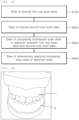
- a method of directly scanning the occlusion state in a mouth may have a difficulty in terms of its precision. Accordingly, scan data of the occlusion state may be precisely secured by merging oral scan data and impression scan data. To this end, as illustrated in FIG. 18 and FIG. 19 , a method of securing accurate scan data of the occlusion state is described as follows.
- first oral scan data is stored by scanning an outer surface of an oral object in the state in which the upper jaw 1 and lower jaw 2 of the oral object face each other in a way to be occluded (S104). Furthermore, second oral scan data is stored by scanning the inside of the oral object in the state in which the upper jaw 1 and the lower jaw 2 are widened in the state in which the upper jaw 1 and the lower jaw 2 face each other (S204).
- impression scan data of a selection area of the oral object including the occlusion state of the upper jaw 1 and the lower jaw 2 is obtained and compared with the first oral scan data and the second oral scan data (S304). That is, as illustrated in FIG. 18 and FIG. 20 , for example, when molar portions on both side of the oral object are covered with an impression material 3 and the upper jaw 1 and the lower jaw 2 face each other, the states in which the upper jaw 1 and the lower jaw 2 are aligned with upper and lower parts of the impression material 3, respectively, are indicated. Furthermore, a difference between the locations of the upper jaw 1 and the lower jaw 2, that is, the occlusion state of the upper jaw 1 and the lower jaw 2, may also be indicated. Accordingly, an accurate occlusion state of the oral object can be checked by scanning information on the indicated impression material 3 and comparing corresponding scan data with the previously stored first oral scan data and second oral scan data.
- impression scan data corresponding to corresponding locations that is, the molar portions on both sides described as an example, among the first oral scan data and the second oral scan data, are partially replaced (S404), so that the final scan data including information on an accurate occlusion state of the oral object is obtained.
- the reason for this is that since the impression scan data obtained using the impression material is more precise than the scan data obtained by scanning the inside and outside of the oral object through the oral scanner, the first oral scan data and the second oral scan data can be applied by partially replacing the impression scan data and accurate occlusion state information can be obtained.
- the present disclosure has an effect in that elaborate prosthetics can be fabricated using obtained digital data in a way to obtain one final scan datum merged by aligning impression scan data with oral scan data in real time in a process of obtaining the impression scan data after obtaining the oral scan data by using the oral scanner.
- the present disclosure has an effect in that more accurate digital data can be obtained by expressing a margin line in a process of obtaining an impression when it is difficult to measure the margin line because the margin line is covered with a gum tissue in an environment within a mouth.
- the present disclosure has an effect in that it can solve a conventional problem in which it is difficult or impossible to obtain data by using the oral scanner due to the depth of a post area having an engraved form, in such a way to enable a portion whose scanning is impossible to be scanned by obtaining, in an embossing form, the post area actually having the engraved form by obtaining impression scan data.
- the present disclosure has an effect in that a teeth portion whose accurate scanning is difficult due to a material characteristic can be scanned by separately obtaining an impression of the teeth portion covered with a crown made of metal and using such impression scan data for occlusion and alignment necessary to obtain the final scan data.
- the present disclosure provides the combined oral and impression scanning system and method for obtaining one final scan datum merged by aligning impression scan data with oral scan data in a process of obtaining the impression scan data after obtaining the oral scan data.
- Preferred Embodiments of the invention are defined in paras 1 to 19
Landscapes
- Health & Medical Sciences (AREA)
- Life Sciences & Earth Sciences (AREA)
- General Health & Medical Sciences (AREA)
- Veterinary Medicine (AREA)
- Public Health (AREA)
- Animal Behavior & Ethology (AREA)
- Physics & Mathematics (AREA)
- Oral & Maxillofacial Surgery (AREA)
- Dentistry (AREA)
- Engineering & Computer Science (AREA)
- Epidemiology (AREA)
- Molecular Biology (AREA)
- Medical Informatics (AREA)
- Surgery (AREA)
- Heart & Thoracic Surgery (AREA)
- Biomedical Technology (AREA)
- Biophysics (AREA)
- Pathology (AREA)
- Optics & Photonics (AREA)
- Radiology & Medical Imaging (AREA)
- Nuclear Medicine, Radiotherapy & Molecular Imaging (AREA)
- Audiology, Speech & Language Pathology (AREA)
- Theoretical Computer Science (AREA)
- Computer Vision & Pattern Recognition (AREA)
- General Physics & Mathematics (AREA)
- Dental Tools And Instruments Or Auxiliary Dental Instruments (AREA)
- Prostheses (AREA)
- Apparatus For Radiation Diagnosis (AREA)
Priority Applications (1)
| Application Number | Priority Date | Filing Date | Title |
|---|---|---|---|
| EP26156633.5A EP4714395A2 (fr) | 2019-02-19 | 2020-02-19 | Système et procédé de balayage d'empreinte intrabuccale et d'empreinte combinés |
Applications Claiming Priority (3)
| Application Number | Priority Date | Filing Date | Title |
|---|---|---|---|
| KR20190019296 | 2019-02-19 | ||
| PCT/KR2020/002391 WO2020171589A1 (fr) | 2019-02-19 | 2020-02-19 | Système et procédé de balayage d'impression et intraoral combiné |
| EP20758724.7A EP3928686B1 (fr) | 2019-02-19 | 2020-02-19 | Système et procédé de balayage d'impression et intraoral combiné |
Related Parent Applications (1)
| Application Number | Title | Priority Date | Filing Date |
|---|---|---|---|
| EP20758724.7A Division EP3928686B1 (fr) | 2019-02-19 | 2020-02-19 | Système et procédé de balayage d'impression et intraoral combiné |
Related Child Applications (2)
| Application Number | Title | Priority Date | Filing Date |
|---|---|---|---|
| EP26156633.5A Division-Into EP4714395A2 (fr) | 2019-02-19 | 2020-02-19 | Système et procédé de balayage d'empreinte intrabuccale et d'empreinte combinés |
| EP26156633.5A Division EP4714395A2 (fr) | 2019-02-19 | 2020-02-19 | Système et procédé de balayage d'empreinte intrabuccale et d'empreinte combinés |
Publications (3)
| Publication Number | Publication Date |
|---|---|
| EP4406475A2 true EP4406475A2 (fr) | 2024-07-31 |
| EP4406475A3 EP4406475A3 (fr) | 2025-02-12 |
| EP4406475B1 EP4406475B1 (fr) | 2026-04-01 |
Family
ID=72237491
Family Applications (3)
| Application Number | Title | Priority Date | Filing Date |
|---|---|---|---|
| EP24180979.7A Active EP4406475B1 (fr) | 2019-02-19 | 2020-02-19 | Procédé de balayage d'empreinte intrabuccale et d'empreinte combinés |
| EP20758724.7A Active EP3928686B1 (fr) | 2019-02-19 | 2020-02-19 | Système et procédé de balayage d'impression et intraoral combiné |
| EP26156633.5A Pending EP4714395A2 (fr) | 2019-02-19 | 2020-02-19 | Système et procédé de balayage d'empreinte intrabuccale et d'empreinte combinés |
Family Applications After (2)
| Application Number | Title | Priority Date | Filing Date |
|---|---|---|---|
| EP20758724.7A Active EP3928686B1 (fr) | 2019-02-19 | 2020-02-19 | Système et procédé de balayage d'impression et intraoral combiné |
| EP26156633.5A Pending EP4714395A2 (fr) | 2019-02-19 | 2020-02-19 | Système et procédé de balayage d'empreinte intrabuccale et d'empreinte combinés |
Country Status (5)
| Country | Link |
|---|---|
| US (1) | US20210378800A1 (fr) |
| EP (3) | EP4406475B1 (fr) |
| KR (1) | KR102425719B1 (fr) |
| CN (1) | CN113518579B (fr) |
| DK (1) | DK3928686T3 (fr) |
Families Citing this family (5)
| Publication number | Priority date | Publication date | Assignee | Title |
|---|---|---|---|---|
| US20240122687A1 (en) * | 2021-02-08 | 2024-04-18 | Medit Corp. | Data processing method |
| KR102728459B1 (ko) * | 2021-02-08 | 2024-11-11 | 주식회사 메디트 | 데이터 처리 방법 |
| KR102826064B1 (ko) * | 2021-03-16 | 2025-06-27 | 주식회사 메디트 | 데이터 처리 방법 |
| US20240173078A1 (en) * | 2021-03-16 | 2024-05-30 | Medit Corp. | Data processing method |
| KR102612682B1 (ko) * | 2021-09-13 | 2023-12-13 | 주식회사 메디트 | 전자 장치 및 3차원 스캐너의 스캔 이미지 처리 방법 |
Family Cites Families (15)
| Publication number | Priority date | Publication date | Assignee | Title |
|---|---|---|---|---|
| ES2507071T3 (es) * | 2005-11-30 | 2014-10-14 | 3Shape A/S | Escaneado de impresión para fabricar reparaciones dentales |
| WO2008051130A1 (fr) * | 2006-10-27 | 2008-05-02 | Nobel Biocare Services Ag | Procédé et appareil pour obtenir des données d'un composant dentaire et d'un modèle dentaire physique |
| WO2012083960A1 (fr) * | 2010-12-22 | 2012-06-28 | 3Shape A/S | Système et procédé d'examen par balayage d'objets subissant des modifications |
| EP3808310B1 (fr) * | 2012-02-10 | 2022-10-12 | 3Shape A/S | Conception virtuelle de restauration de tenon et faux moignon à l'aide d'une forme 3d numérique |
| US9168112B2 (en) * | 2012-07-27 | 2015-10-27 | Guided Surgery Solutions, Llc | Multi-layer surgical guide |
| US10248885B2 (en) * | 2013-03-15 | 2019-04-02 | James R. Glidewell Dental Ceramics, Inc. | Method for encoding of anatomic curves |
| US9510757B2 (en) * | 2014-05-07 | 2016-12-06 | Align Technology, Inc. | Identification of areas of interest during intraoral scans |
| JP6262179B2 (ja) * | 2014-12-05 | 2018-01-17 | ディオ コーポレーションDio Corporation | デンタルインプラント施術のための手術ガイドおよび植立物の製造方法 |
| KR101832753B1 (ko) * | 2015-05-08 | 2018-02-27 | (주)바텍이우홀딩스 | 모델 스캔 겸용 구강 스캔 장치 |
| WO2017088139A1 (fr) * | 2015-11-26 | 2017-06-01 | 法玛科技顾问股份有限公司 | Système d'imagerie d'empreinte numérique par tomographie optique et procédé d'utilisation de celui-ci |
| CN106806030B (zh) * | 2015-11-30 | 2018-08-10 | 北京大学口腔医学院 | 一种冠根三维模型融合方法 |
| CN107303204B (zh) * | 2016-04-21 | 2019-12-27 | 北京大学口腔医学院 | 一种手持式口腔三维扫描装置及控制方法 |
| KR101797155B1 (ko) * | 2016-09-02 | 2017-11-14 | 주식회사 디오 | 치아 임플란트용 서지컬 가이드 및 식립물 제조방법 |
| KR101910053B1 (ko) * | 2017-06-19 | 2018-10-19 | 주식회사 디오 | 구강 인상 채득 장치 |
| KR101917140B1 (ko) | 2017-06-26 | 2019-01-24 | 오스템임플란트 주식회사 | 크라운 모델 디자인 방법, 장치 및 그 방법을 실행하기 위한 프로그램이 기록된 기록매체 |
-
2020
- 2020-02-19 EP EP24180979.7A patent/EP4406475B1/fr active Active
- 2020-02-19 EP EP20758724.7A patent/EP3928686B1/fr active Active
- 2020-02-19 EP EP26156633.5A patent/EP4714395A2/fr active Pending
- 2020-02-19 KR KR1020200020108A patent/KR102425719B1/ko active Active
- 2020-02-19 DK DK20758724.7T patent/DK3928686T3/da active
- 2020-02-19 CN CN202080015428.7A patent/CN113518579B/zh active Active
-
2021
- 2021-08-19 US US17/406,077 patent/US20210378800A1/en active Pending
Also Published As
| Publication number | Publication date |
|---|---|
| CN113518579B (zh) | 2024-12-17 |
| DK3928686T3 (da) | 2024-09-30 |
| EP3928686B1 (fr) | 2024-07-03 |
| US20210378800A1 (en) | 2021-12-09 |
| EP4406475B1 (fr) | 2026-04-01 |
| CN113518579A (zh) | 2021-10-19 |
| EP3928686A4 (fr) | 2023-03-29 |
| KR20200101304A (ko) | 2020-08-27 |
| EP4406475A3 (fr) | 2025-02-12 |
| EP4714395A2 (fr) | 2026-03-25 |
| EP3928686A1 (fr) | 2021-12-29 |
| KR102425719B1 (ko) | 2022-07-27 |
Similar Documents
| Publication | Publication Date | Title |
|---|---|---|
| EP4406475B1 (fr) | Procédé de balayage d'empreinte intrabuccale et d'empreinte combinés | |
| US20260017891A1 (en) | System and method for generating digital three-dimensional dental models | |
| US7118375B2 (en) | Method and system for dental model occlusal determination using a replicate bite registration impression | |
| JP2654646B2 (ja) | 窩洞の三次元的形態の決定とディスプレイのための方法と装置 | |
| KR102442720B1 (ko) | 치아 보철 제조를 위한 3차원 스캔 데이터 처리 방법 | |
| KR101994396B1 (ko) | 단계별 보철물 디자인 방법 | |
| US6845175B2 (en) | Dental image processing method and system | |
| JP2740194B2 (ja) | 義歯の製造方法 | |
| KR102462266B1 (ko) | 구강내 스캔으로부터 카리에스 및/또는 한계선 검출 | |
| US8144954B2 (en) | Lighting compensated dynamic texture mapping of 3-D models | |
| US7010150B1 (en) | Method for detecting and representing one or more objects, for example teeth | |
| US20160015246A1 (en) | Visualising a 3d dental restoration on a 2d image | |
| JPH02119858A (ja) | 口腔内の歯の三次元測量用光プローブ | |
| CN101365396A (zh) | 用于牙齿修复物制造的印模扫描 | |
| US6334773B1 (en) | Method and arrangement for making artificial teeth | |
| KR101854729B1 (ko) | 인공치아 디자인 시스템 | |
| US20230414331A1 (en) | Capture of intraoral features from non-direct views | |
| CN118055728A (zh) | 构建牙齿结构的纹理化3d模型 | |
| JP7170892B2 (ja) | 口腔および印象の複合スキャンシステムおよび方法 | |
| KR20220163859A (ko) | 데이터 처리 방법 | |
| CN119863574A (zh) | 三维重建方法、装置、电子设备及存储介质 | |
| EP1845889A1 (fr) | Procedes et systemes de determination occlusale de modeles dentaires au moyen d'une empreinte d'enregistrement de morsure repliquee | |
| AU2013100527A4 (en) | Dental restoration | |
| RU2859286C1 (ru) | Способ измерения протеза нижней конечности | |
| US20040166471A1 (en) | Method and device for optical measurement of a cast model of a tooth in restorative dentistry |
Legal Events
| Date | Code | Title | Description |
|---|---|---|---|
| PUAI | Public reference made under article 153(3) epc to a published international application that has entered the european phase |
Free format text: ORIGINAL CODE: 0009012 |
|
| STAA | Information on the status of an ep patent application or granted ep patent |
Free format text: STATUS: THE APPLICATION HAS BEEN PUBLISHED |
|
| AC | Divisional application: reference to earlier application |
Ref document number: 3928686 Country of ref document: EP Kind code of ref document: P |
|
| AK | Designated contracting states |
Kind code of ref document: A2 Designated state(s): AL AT BE BG CH CY CZ DE DK EE ES FI FR GB GR HR HU IE IS IT LI LT LU LV MC MK MT NL NO PL PT RO RS SE SI SK SM TR |
|
| RIC1 | Information provided on ipc code assigned before grant |
Ipc: A61C 13/00 20060101ALI20241016BHEP Ipc: A61C 9/00 20060101ALI20241016BHEP Ipc: A61B 5/00 20060101AFI20241016BHEP |
|
| PUAL | Search report despatched |
Free format text: ORIGINAL CODE: 0009013 |
|
| AK | Designated contracting states |
Kind code of ref document: A3 Designated state(s): AL AT BE BG CH CY CZ DE DK EE ES FI FR GB GR HR HU IE IS IT LI LT LU LV MC MK MT NL NO PL PT RO RS SE SI SK SM TR |
|
| RIC1 | Information provided on ipc code assigned before grant |
Ipc: A61C 13/00 20060101ALI20250108BHEP Ipc: A61C 9/00 20060101ALI20250108BHEP Ipc: A61B 5/00 20060101AFI20250108BHEP |
|
| STAA | Information on the status of an ep patent application or granted ep patent |
Free format text: STATUS: REQUEST FOR EXAMINATION WAS MADE |
|
| 17P | Request for examination filed |
Effective date: 20250807 |
|
| GRAP | Despatch of communication of intention to grant a patent |
Free format text: ORIGINAL CODE: EPIDOSNIGR1 |
|
| STAA | Information on the status of an ep patent application or granted ep patent |
Free format text: STATUS: GRANT OF PATENT IS INTENDED |
|
| RIC1 | Information provided on ipc code assigned before grant |
Ipc: A61B 5/00 20060101AFI20251012BHEP Ipc: A61C 9/00 20060101ALI20251012BHEP Ipc: A61C 13/00 20060101ALI20251012BHEP |
|
| INTG | Intention to grant announced |
Effective date: 20251024 |
|
| GRAS | Grant fee paid |
Free format text: ORIGINAL CODE: EPIDOSNIGR3 |
|
| GRAA | (expected) grant |
Free format text: ORIGINAL CODE: 0009210 |
|
| STAA | Information on the status of an ep patent application or granted ep patent |
Free format text: STATUS: THE PATENT HAS BEEN GRANTED |
|
| AC | Divisional application: reference to earlier application |
Ref document number: 3928686 Country of ref document: EP Kind code of ref document: P |
|
| AK | Designated contracting states |
Kind code of ref document: B1 Designated state(s): AL AT BE BG CH CY CZ DE DK EE ES FI FR GB GR HR HU IE IS IT LI LT LU LV MC MK MT NL NO PL PT RO RS SE SI SK SM TR |
|
| REG | Reference to a national code |
Ref country code: CH Ref legal event code: F10 Free format text: ST27 STATUS EVENT CODE: U-0-0-F10-F00 (AS PROVIDED BY THE NATIONAL OFFICE) Effective date: 20260401 Ref country code: GB Ref legal event code: FG4D |
|
| REG | Reference to a national code |
Ref country code: DE Ref legal event code: R096 Ref document number: 602020069886 Country of ref document: DE |
|
| REG | Reference to a national code |
Ref country code: IE Ref legal event code: FG4D |