EP4404206A1 - Verfahren und vorrichtung zur bestimmung von anomalien in organen oder muskeln im körper - Google Patents
Verfahren und vorrichtung zur bestimmung von anomalien in organen oder muskeln im körper Download PDFInfo
- Publication number
- EP4404206A1 EP4404206A1 EP22870218.9A EP22870218A EP4404206A1 EP 4404206 A1 EP4404206 A1 EP 4404206A1 EP 22870218 A EP22870218 A EP 22870218A EP 4404206 A1 EP4404206 A1 EP 4404206A1
- Authority
- EP
- European Patent Office
- Prior art keywords
- muscle
- zoomed
- apical
- level
- image
- Prior art date
- Legal status (The legal status is an assumption and is not a legal conclusion. Google has not performed a legal analysis and makes no representation as to the accuracy of the status listed.)
- Pending
Links
Images







Classifications
-
- G—PHYSICS
- G16—INFORMATION AND COMMUNICATION TECHNOLOGY [ICT] SPECIALLY ADAPTED FOR SPECIFIC APPLICATION FIELDS
- G16H—HEALTHCARE INFORMATICS, i.e. INFORMATION AND COMMUNICATION TECHNOLOGY [ICT] SPECIALLY ADAPTED FOR THE HANDLING OR PROCESSING OF MEDICAL OR HEALTHCARE DATA
- G16H30/00—ICT specially adapted for the handling or processing of medical images
- G16H30/20—ICT specially adapted for the handling or processing of medical images for handling medical images, e.g. DICOM, HL7 or PACS
-
- G—PHYSICS
- G16—INFORMATION AND COMMUNICATION TECHNOLOGY [ICT] SPECIALLY ADAPTED FOR SPECIFIC APPLICATION FIELDS
- G16H—HEALTHCARE INFORMATICS, i.e. INFORMATION AND COMMUNICATION TECHNOLOGY [ICT] SPECIALLY ADAPTED FOR THE HANDLING OR PROCESSING OF MEDICAL OR HEALTHCARE DATA
- G16H30/00—ICT specially adapted for the handling or processing of medical images
- G16H30/40—ICT specially adapted for the handling or processing of medical images for processing medical images, e.g. editing
-
- A—HUMAN NECESSITIES
- A61—MEDICAL OR VETERINARY SCIENCE; HYGIENE
- A61B—DIAGNOSIS; SURGERY; IDENTIFICATION
- A61B5/00—Measuring for diagnostic purposes; Identification of persons
- A61B5/05—Detecting, measuring or recording for diagnosis by means of electric currents or magnetic fields; Measuring using microwaves or radio waves
- A61B5/055—Detecting, measuring or recording for diagnosis by means of electric currents or magnetic fields; Measuring using microwaves or radio waves involving electronic [EMR] or nuclear [NMR] magnetic resonance, e.g. magnetic resonance imaging
-
- A—HUMAN NECESSITIES
- A61—MEDICAL OR VETERINARY SCIENCE; HYGIENE
- A61B—DIAGNOSIS; SURGERY; IDENTIFICATION
- A61B6/00—Apparatus or devices for radiation diagnosis; Apparatus or devices for radiation diagnosis combined with radiation therapy equipment
- A61B6/52—Devices using data or image processing specially adapted for radiation diagnosis
- A61B6/5211—Devices using data or image processing specially adapted for radiation diagnosis involving processing of medical diagnostic data
- A61B6/5217—Devices using data or image processing specially adapted for radiation diagnosis involving processing of medical diagnostic data extracting a diagnostic or physiological parameter from medical diagnostic data
-
- A—HUMAN NECESSITIES
- A61—MEDICAL OR VETERINARY SCIENCE; HYGIENE
- A61B—DIAGNOSIS; SURGERY; IDENTIFICATION
- A61B8/00—Diagnosis using ultrasonic, sonic or infrasonic waves
- A61B8/08—Clinical applications
-
- A—HUMAN NECESSITIES
- A61—MEDICAL OR VETERINARY SCIENCE; HYGIENE
- A61B—DIAGNOSIS; SURGERY; IDENTIFICATION
- A61B8/00—Diagnosis using ultrasonic, sonic or infrasonic waves
- A61B8/52—Devices using data or image processing specially adapted for diagnosis using ultrasonic, sonic or infrasonic waves
- A61B8/5215—Devices using data or image processing specially adapted for diagnosis using ultrasonic, sonic or infrasonic waves involving processing of medical diagnostic data
- A61B8/5223—Devices using data or image processing specially adapted for diagnosis using ultrasonic, sonic or infrasonic waves involving processing of medical diagnostic data for extracting a diagnostic or physiological parameter from medical diagnostic data
-
- G—PHYSICS
- G16—INFORMATION AND COMMUNICATION TECHNOLOGY [ICT] SPECIALLY ADAPTED FOR SPECIFIC APPLICATION FIELDS
- G16H—HEALTHCARE INFORMATICS, i.e. INFORMATION AND COMMUNICATION TECHNOLOGY [ICT] SPECIALLY ADAPTED FOR THE HANDLING OR PROCESSING OF MEDICAL OR HEALTHCARE DATA
- G16H50/00—ICT specially adapted for medical diagnosis, medical simulation or medical data mining; ICT specially adapted for detecting, monitoring or modelling epidemics or pandemics
- G16H50/20—ICT specially adapted for medical diagnosis, medical simulation or medical data mining; ICT specially adapted for detecting, monitoring or modelling epidemics or pandemics for computer-aided diagnosis, e.g. based on medical expert systems
-
- G—PHYSICS
- G16—INFORMATION AND COMMUNICATION TECHNOLOGY [ICT] SPECIALLY ADAPTED FOR SPECIFIC APPLICATION FIELDS
- G16H—HEALTHCARE INFORMATICS, i.e. INFORMATION AND COMMUNICATION TECHNOLOGY [ICT] SPECIALLY ADAPTED FOR THE HANDLING OR PROCESSING OF MEDICAL OR HEALTHCARE DATA
- G16H50/00—ICT specially adapted for medical diagnosis, medical simulation or medical data mining; ICT specially adapted for detecting, monitoring or modelling epidemics or pandemics
- G16H50/50—ICT specially adapted for medical diagnosis, medical simulation or medical data mining; ICT specially adapted for detecting, monitoring or modelling epidemics or pandemics for simulation or modelling of medical disorders
-
- A—HUMAN NECESSITIES
- A61—MEDICAL OR VETERINARY SCIENCE; HYGIENE
- A61B—DIAGNOSIS; SURGERY; IDENTIFICATION
- A61B6/00—Apparatus or devices for radiation diagnosis; Apparatus or devices for radiation diagnosis combined with radiation therapy equipment
- A61B6/02—Arrangements for diagnosis sequentially in different planes; Stereoscopic radiation diagnosis
- A61B6/03—Computed tomography [CT]
- A61B6/032—Transmission computed tomography [CT]
-
- A—HUMAN NECESSITIES
- A61—MEDICAL OR VETERINARY SCIENCE; HYGIENE
- A61B—DIAGNOSIS; SURGERY; IDENTIFICATION
- A61B6/00—Apparatus or devices for radiation diagnosis; Apparatus or devices for radiation diagnosis combined with radiation therapy equipment
- A61B6/02—Arrangements for diagnosis sequentially in different planes; Stereoscopic radiation diagnosis
- A61B6/03—Computed tomography [CT]
- A61B6/037—Emission tomography
-
- A—HUMAN NECESSITIES
- A61—MEDICAL OR VETERINARY SCIENCE; HYGIENE
- A61B—DIAGNOSIS; SURGERY; IDENTIFICATION
- A61B6/00—Apparatus or devices for radiation diagnosis; Apparatus or devices for radiation diagnosis combined with radiation therapy equipment
- A61B6/50—Apparatus or devices for radiation diagnosis; Apparatus or devices for radiation diagnosis combined with radiation therapy equipment specially adapted for specific body parts; specially adapted for specific clinical applications
- A61B6/503—Apparatus or devices for radiation diagnosis; Apparatus or devices for radiation diagnosis combined with radiation therapy equipment specially adapted for specific body parts; specially adapted for specific clinical applications for diagnosis of the heart
-
- A—HUMAN NECESSITIES
- A61—MEDICAL OR VETERINARY SCIENCE; HYGIENE
- A61B—DIAGNOSIS; SURGERY; IDENTIFICATION
- A61B8/00—Diagnosis using ultrasonic, sonic or infrasonic waves
- A61B8/08—Clinical applications
- A61B8/0883—Clinical applications for diagnosis of the heart
Definitions
- the present disclosure relates to determination of the status of an internal organ or muscle, and more particularly, to a method and device for determining presence or absence of abnormality in an internal organ or muscle.
- AI artificial intelligence
- An object of the present disclosure is to provide a method and device for effectively determining presence or absence of abnormality in an internal organ or muscle using an artificial intelligence (AI) algorithm.
- AI artificial intelligence
- An object of the present disclosure is to provide a method and device for effectively diagnosing presence or absence of abnormality in an internal organ or muscle using an artificial intelligence algorithm.
- An object of the present disclosure is to provide a method and device for effectively determining presence or absence of abnormality in an internal organ or muscle based on images.
- An object of the present disclosure is to provide a method and device for determining presence or absence of abnormality in an internal organ or muscle based on information on a tissue phenotype obtained from medical images of the heart.
- An object of the present disclosure is to provide a method and device for diagnosing presence or absence of abnormality in an internal organ or muscle based on information on a tissue phenotype obtained from medical images of the heart.
- An object of the present disclosure is to provide a method and device for determining or diagnosing presence or absence of abnormality in an internal organ or muscle using some feature values selected by a machine learning model among feature values that may be obtained from medical images of the heart.
- a method of determining presence or absence of abnormality in an internal organ or muscle may comprise obtaining at least one image of the internal organ or muscle, determining at least one feature matrix for the at least one image, determining a feature value for measuring a tissue phenotype from the at least one feature matrix, and determining presence or absence of the abnormality in the internal organ or muscle using a trained artificial intelligence model.
- the feature value may comprise items selected as information indicating the tissue phenotype of the internal organ or muscle, and the items may be selected using a machine learning model among candidate feature values determinable from the at least one feature matrix.
- the at least one image may comprise an image captured in at least one view of apical 2-chamber (A2CH), apical 4-chamber (A4CH), parasternal long-axis (PLAX), parasternal short-axis (PSAX)-mid, PLAX increased depth, parasternal long axis left ventricle (PLAX LV), parasternal long axis left ventricle aorta/left atrial (PLAX LV-AO/LA), parasternal long axis zoomed left ventricle (PLAX zoomed LV), parasternal long axis zoomed aortic valve (PLAX zoomed AV), parasternal long axis zoomed mitral valve (PLAX zoomed MV), parasternal long axis right ventricle (PLAX RV) out flow, parasternal long axis right ventricle (PLAX RV) inflow, parasternal short axis (level great vessels) focus
- the at least one feature matrix may be determined based on pixel values included in a region of interest extracted from the at least one image.
- the region of interest may comprise at least one selected from a group consisting of an outer wall, medial wall, parietal peritoneum, fascia, intervisceral space, intracavitary, and intraperitoneal viscera, skeletal muscle, myocardium, and smooth muscle.
- the at least one feature matrix may comprise at least one of gray-level co-occurrence matrix (GLCM), gray-level run length matrix (GLRLM), gray-level size zone matrix (GLSZM), neighborhood gray-tone difference matrix (NGTDM) or gray level dependence matrix (GLDM).
- GLCM gray-level co-occurrence matrix
- GLRLM gray-level run length matrix
- GLSZM gray-level size zone matrix
- NTTDM neighborhood gray-tone difference matrix
- GLDM gray level dependence matrix
- the abnormality in the internal organ or muscle may comprise heart disease.
- a program stored on a medium according to an embodiment of the present invention may execute the above-described method when operated by a processor.
- the present invention is to diagnose abnormality in an internal organ or muscle using an artificial intelligence algorithm. Specifically, it relates to a technology for diagnosing abnormality in an internal organ or muscle based on images.
- the internal organ or muscle refers to at least one selected from a group consisting of an outer wall, medial wall, parietal peritoneum, fascia, intervisceral space, intracavitary, intraperitoneal viscera, skeletal muscle, myocardium, and smooth muscle.
- abnormality in an internal organ or muscle refers to heart disease, and more specifically, to myocardial disease.
- abnormality in the internal organ or muscle may include at least one of a group consisting of cardiac hypertrophy, hypertrophic cardiomyopathy (HCM), dilated cardiomyopathy (DCM), heart failure with preserved ejection fraction (HFpEF), diastolic dysfunction of the heart, cardiomyopathy including primary or secondary restrictive cardiomyopathy, myocardial infarction, cardiac fibrosis, sarcomere mutations, that is, myosin heavy chain ⁇ (MHC- ⁇ ), cardiac muscle troponin T (cTnT), tropomyosin alpha-1 chain (TPM1), and myosin-binding protein C cardiotype (MYBPC3), cardiac troponin I (cTnI), myosin essential light chain (ELC), titin (TTN), myosin regulatory light chain 2 ventricular/cardiac muscle isoform (MLC-2), cardiac
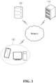
- the system includes a service server 110, a data server 120, and at least one client device 130.
- the service server 110 provides services based on artificial intelligence models. That is, the service server 110 performs learning and prediction operations using artificial intelligence models.
- the service server 110 may communicate with the data server 120 or at least one client device 130 through a network. For example, the service server 110 may receive learning data for training an artificial intelligence model from the data server 120 and perform training.
- the service server 110 may receive data required for learning and prediction operations from at least one client device 130. Additionally, the service server 110 may transmit information on the prediction result to at least one client device 130.
- the data server 120 provides learning data for training the artificial intelligence model stored in the service server 110.
- the data server 120 may provide public data that anyone can access or provide data that requires permission. If necessary, the learning data may be preprocessed by the data server 120 or the service server 110. According to another embodiment, the data server 120 may be omitted. In this case, the service server 110 may use an externally trained artificial intelligence model, or learning data may be provided to the service server 110 offline.
- the at least one client device 130 transmits and receives data related to the artificial intelligence model operated by the service server 110 to and from the service server 110.
- the at least one client device 130 is used by a user, and transmits information input by the user to the service server 110, and stores the information received from the service server 110 or provides (shows) it to the user.
- a prediction operation may be performed based on data transmitted from one client, and information related to the result of prediction may be provided to another client.
- the at least one client device 130 may be various types of computing devices, such as desktop computers, laptop computers, smartphones, tablets, and wearable devices.
- the system may further include a management device for managing the service server 110.
- the management device is a device used by an entity that manages the service, and monitors the status of the service server 110 or controls settings of the service server 110.
- the management device may be connected to the service server 110 through a network or directly through a cable connection. According to the control of the management device, the service server 110 may set parameters for operation.
- the service server 110, the data server 120, the at least one client device 130, the management device, etc. may be connected and interact through the network.
- the network may include at least one of a wired network or a wireless network, and may be composed of any one or a combination of two or more of a cellular network, a local area network, and a wide area network.
- the network may be implemented based on at least one of local area network (LAN), wireless LAN (WLAN), Bluetooth, long term evolution (LTE), LTE-advanced (LTE-A), or 5th generation (5G).
- FIG. 2 shows the structure of a device for predicting the possibility of disease occurrence according to an embodiment of the present invention.
- the structure illustrated in FIG. 2 may be understood as the structure of the service server 110, data server 120, and at least one client device 130 of FIG. 1 .
- the device includes a communication unit 210, a storage unit 220 and a controller 230.
- the communication unit 210 performs functions to connect to the network and communicate with other devices.
- the communication unit 210 may support at least one of wired communication or wireless communication.
- the communication unit 210 may include at least one of a radio frequency (RF) processing circuit or a digital data processing circuit.
- RF radio frequency
- the communication unit 210 may be understood as a component including a terminal for connecting a cable. Since the communication unit 210 is a component for transmitting and receiving data and signals, it may be referred to as a 'transceiver'.
- the storage unit 220 stores data, programs, microcode, instruction sets, applications, etc. necessary for the operation of the device.
- the storage unit 220 may be implemented as a transitory or non-transitory storage medium. Additionally, the storage unit 220 may be fixed to the device or may be implemented in a detachable form.
- the storage unit 220 may be implemented as at least one of NAND flash memories such as compact flash (CF) cards, secure digital (SD) cards, memory sticks, solid-state drives (SSDs) or micro SD cards or magnetic computer storage devices such as hard disk drives (HDDs).
- CF compact flash
- SD secure digital
- HDDs hard disk drives
- the controller 230 controls the overall operation of the device.
- the controller 230 may include at least one processor, at least one microprocessor, etc.
- the controller 230 may execute a program stored in the storage unit 220 and access the network through the communication unit 210.
- the controller 230 may perform algorithms according to various embodiments described later and control the device to operate according to embodiments described later.
- Random forest is an ensemble artificial intelligence model composed of a plurality of decision trees, and was proposed to solve problems such as overfitting and error propagation that may occur when using a single decision tree.
- FIG. 3 shows an example of a random forest model applicable to the present invention.
- the random forest model includes a plurality of decision trees 310-1 to 310-N and an aggregation network 320.
- Each of the decision trees 310-1 to 310-N performs a classification operation using one of subsets 304-1 to 304-N of the entire data set 302. Then, the classification results of each of the decision trees 310-1 to 310-N are aggregated by the aggregation network 320, thereby generating one output.
- the aggregation network 320 may provide, as output, the result with the largest number of votes among the classification results from the decision trees 310-1 to 310-N.
- a plurality of subsets include a common data element (e.g., D1).
- the bagging operation allows duplicate selection of data, even if more than M subsets are generated so that each subset includes N/M data elements in a data set containing N data elements, they may all be unique subsets.
- the decision trees 310-1 to 310-N are randomized. Through randomization, the decision trees 310-1 to 310-N forming the random forest model may have different characteristics due to randomness. Differences in characteristics cause decorrelation between predictions made by the decision trees 310-1 to 310-N, and as a result, contribute to improvement of the generalization performance of the random forest model. Additionally, the randomization characteristics provide robustness against noise of the random forest model.
- the decision trees 310-1 to 310-N included in the random forest model are composed of a plurality of nodes including questions for classification.
- each node contains a question for binary classification, and the content of the question may be generated through training. Therefore, training a decision tree may be understood as a process of generating a list of questions.
- the learning algorithm for a decision tree builds a tree by selecting an item that most closely classifies a target result from all possible tests and repeating this. This process may be repeated until a leaf node becomes a pure node. At this time, to prevent the depth of the tree from becoming infinite, pruning may be performed.
- the importance of feature items corresponding to a node may be evaluated. Importance may be expressed by the Gini index, which indicates impurity. The lower the Gini index, the higher the importance.
- the Gini index indicates how concentrated the samples classified at a given node are in one class. For example, the Gini index of a pure node is 0.
- the artificial intelligence algorithm uses medical images of the heart to diagnose abnormality in an internal organ or muscle.
- the present invention may determine feature values from medical images and then use the feature values to diagnose presence or absence of abnormality in an internal organ or muscle.
- the medical images may include images obtained from a view in which the myocardium of the left ventricle may be observed (e.g., at least one of apical 2-chamber (A2CH), apical 4-chamber (A4CH), parasternal long-axis (PLAX), parasternal short-axis (PSAX)-mid, PLAX increased depth, parasternal long axis left ventricle (PLAX LV), parasternal long axis left ventricle aorta/left atrial (PLAX LV-AO/LA), parasternal long axis zoomed left ventricle (PLAX zoomed LV), parasternal long axis zoomed aortic valve (PLAX zoomed AV), parasternal long axis zoomed mitral valve (PLAX zoomed MV), parasternal long axis right ventricle (PLAX RV) out flow, parasternal long axis right ventricle (PLAX RV) in a
- the feature values determined to diagnose presence or absence of abnormality in an internal organ or muscle are the values of items selected in advance to evaluate the texture of the tissue.
- the feature values may be selected using the random forest model described above.
- the feature values that may provide texture information enough to diagnose heart disease may be selected from various candidate feature values.
- the candidate feature values may include feature values related to a shape that may be determined from a 3D image and a 2D image. Examples of feature values related to the shape that may be determined from the 3D image are shown in [Table 1] below, and feature values related to the shape that may be determined from the 2D image are shown in [Table 2] below.
- candidate feature values may include values obtained through analysis of pixels or voxels.
- the values obtained through analysis include first order feature values obtained from the pixel histogram of the original image and second order feature values obtained from transformed information. Examples of the first order feature values are shown in [Table 3] below.
- the second order feature values may be determined from information obtained by transforming an ultrasound image.
- image transformation may be performed according to various techniques, and techniques that may express the texture of heart tissue from various perspectives may be used.
- a feature matrix representing the textural characteristics of the image may be determined.
- systems according to various embodiments may adopt feature matrices representing texture as shown in FIGS. 4A to 4E below.
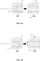
- FIGS. 4A to 4E show examples of feature matrices determined from an ultrasound image according to an embodiment of the present invention.
- the feature matrices illustrated in FIGS. 4A to 4E are for expressing the texture of muscle tissue of an ultrasound imaged heart, and are determined based on pixel values of the ultrasound image.
- FIGS. 4A to 4E illustrate transformation for an image with a size of 4 ⁇ 4 or 5 ⁇ 5 for convenience of explanation, but the feature matrix may be determined in a similar manner for a ultrasound image with a larger size.
- FIG. 4A illustrates a gray-level co-occurrence matrix (GLCM).
- GLCM gray-level co-occurrence matrix
- FIG. 4A in an image 410a, three patterns 411a where two adjacent pixel values are [1,3] are observed, and one pattern 412a where two adj acent pixel values are [4,2] is observed.
- two pixels are shown adj acent in the left and right directions, but being adj acent means being continuous in any one of the 0-degree direction, 45-degree direction, 90-degree direction, and 135-degree direction. Accordingly, adjacency may be evaluated in a different direction than the example in FIG. 4A .
- row numbers and column numbers correspond to combinations of pixel values that make up the pattern.
- an element 421a in the first row and third column of the GLCM 420a is set to 3, and an element 422a in the fourth row and second column is set to 1.
- the remaining elements are set according to the same rules. Examples of feature values that may be determined from the GLCM as shown in FIG. 4A are shown in Table 4 below.
- FIG. 4C illustrates a gray-level size zone matrix (GLSZM).
- GLSZM gray-level size zone matrix
- Table 6 Examples of feature values that may be determined from the GLSZM as shown in FIG. 4c are shown in Table 6 below.
- Table 6 Name Description 1 Small Area Emphasis Measurement of distribution of small-sized area and measurement of line texture 2 Large Area Emphasis Measurement of distribution of large-sized area and measurement of rough texture 3 Gray Level Non-Uniformity Measurement of variability of gray-level intensity value of image 4 Gray Level Non-Uniformity Normalized Normalization of variability of gray-level intensity value of image 5 Size-Zone Non-Uniformity Measurement of variability of size zone volume within image 6 Size-Zone Non-Uniformity Normalized Normalization of variability measurement of volume in size zone within image 7 Zone Percentage Measurement of stenosis of texture using ratio of the number of zones and the number of voxels in ROI 8 Gray Level Variance Measurement of variance of gray-level intensity for zone 9 Zone Variance Measurement of variance of size zone volume 10 Zone Entropy Measurement of uncertainty /randomness in zone size and gray-level distribution 11 Low Gray Level Zone Emp
- FIG. 4D illustrates a neighborhood gray-tone difference matrix (NGTDM).
- NGTDM 420d row numbers correspond to pixel values (e.g., gray level), a first column represents a pixel value, a second column represents the number of pixels with the corresponding pixel value, and a third column represents a ratio of the corresponding pixel value among all pixels, and a fourth column represents a sum of the differences with neighboring pixel values within a distance ⁇ .
- the distance ⁇ is 1.
- the pixel value is 2, and the neighboring pixel values are 1, 5, 3, 5, and 1.
- n i denotes the number of pixels with a value of i
- N denotes the total number of pixels.
- s denotes a sum of differences between neighboring pixel values with a value of i
- i denotes a pixel value
- n i denotes the number of pixels with a value of i
- X i denotes an average of neighboring pixel values of the pixel with a value of i.
- FIG. 4E illustrates a gray level dependence matrix (GLDM).
- GLDM gray level dependence matrix
- feature values obtained through a wavelet filter may be further considered.
- the above-described feature values may be generated for the filtered image.
- FIG. 5 shows an example of a procedure for determining presence or absence of abnormality in an internal organ or muscle according to an embodiment of the present invention.
- FIG. 5 illustrates a method of operating a device with computing capabilities (e.g., the service server 110 of FIG. 1 or a separate server).
- the device determines at least one feature matrix for the at least one image.
- the device may determine the at least one feature matrix based on pixel values included in a region of interest extracted from the at least one image.
- the region of interest may include at least one selected from a group consisting of the outer wall, medial wall, parietal peritoneum, fascia, intervisceral space, intracavitary, and intraperitoneal viscera, skeletal muscle, myocardium, and smooth muscle.
- the region of interest may include at least one selected from a group consisting of the outer wall, medial wall, parietal peritoneum, fascia, skeletal muscle, myocardium, and smooth muscle of the internal organ, and at least one selected from a group consisting of intervisceral space, intracavitary, and intraperitoneal viscera.
- the region of interest may include at least one selected from a group consisting of the outer wall, medial wall, parietal peritoneum, fascia, skeletal muscle, myocardium, and smooth muscle of the internal organ.
- the feature matrices may include at least one of GLCM, GLRLM, GLSZM, NGTDM, or GLDM.
- the device determines feature values from the at least one feature matrix.
- the device may determine feature values for measuring a tissue phenotype from at least one feature matrix.
- the determined feature values may include at least one of the features listed in [Table 4] to [Table 8] above.
- duplication of cases may be allowed, but duplication of feature values may not be allowed.
- the device for each decision tree, the device generates input data sets by extracting n cases under duplication conditions and extracting d feature values for the extracted cases without duplication.
- learning data for decision tree #1 610-1 may include feature values #1 to #d of case #1, case #N-3, case #N-2, and case #N.
- the device may calculate the Gini index for each node of the generated decision trees. In other words, the device may determine the impurity for each node of the generated decision trees. In addition, based on the calculated Gini index, K feature items with relatively high importance may be selected. The selected K feature items are used as items to select feature values used for later diagnosis.
- the device may sort the feature values in order of importance based on the Gini index, identify a point where an area under the curve (AUC) is saturated while the feature values with the highest importance is added one by one to the random forest model trained for diagnosis, and determine the number of feature value items to be used for diagnosis based on the identified point. For example, when checking the AUC of the model by applying 1 to 10 feature values, the AUC continues to increase, but if the increase in AUC as more feature values are added from 11 is not significant, the device may finally select ten feature value items.
- AUC area under the curve
- the tissue phenotype of an organ or muscle may be measured based on feature values extracted from the image, and the presence or absence of abnormality in the organ or muscle may be determined based on the measured phenotype.
- the above-described procedure of determining presence or absence of the abnormality may be performed on the heart using ultrasound images.
- the above-described technology of determining presence or absence of the abnormality based on the phenotype may be performed to determine the presence or absence of heart disease using ultrasound images.
- an embodiment of diagnosing heart disease based on phenotypic measurement will be described. However, it is obvious that the embodiments described below may be performed for the diagnosis of diseases other than heart disease.
- FIG. 7 shows an example of a procedure for selecting feature items for diagnosis of heart disease according to an embodiment of the present invention.
- FIG. 7 illustrates a method of operating a device with computing capabilities (e.g., the service server 110 of FIG. 1 or a separate server).
- the device obtains ultrasound images of the heart.
- the ultrasound images may be received from an external device through a wired or wireless communication connection.
- the view of ultrasound images may include at least one of A2C, A4C, PLAX, or PSAX-mid.
- the obtained ultrasound images may include images of cases with heart disease and images of cases without heart disease at a ratio of 1:1.
- the device determines feature matrices for the ultrasound images.
- the feature matrices may be determined for some of the ultrasound images.
- the device may extract a region of interest from the ultrasound images and determine feature matrices by transforming pixel values within the extracted region of interest.
- the region of interest may include at least one of the left ventricular (LV) wall or the left ventricular cavity.
- the feature matrices may include at least one of GLCM, GLRLM, GLSZM, NGTDM or GLDM.
- step S705 the device determines feature values from the feature matrices.
- the determined feature values may include at least one of the feature values listed in [Table 4] to [Table 8] above.
- step S707 the device selects some feature items using a random forest model.
- the random forest model for selecting feature items includes a plurality of decision trees, and each of the decision trees classifies disease status by using some of the feature values determined in step S705 without mutual duplication.
- the device may determine the Gini index for the classification results from each of the decision trees, and identify K feature items with the lowest impurity based on the Gini index.
- the random forest model for selecting feature items will be explained in more detail with reference to 8 below.
- the device obtains ultrasound images of a subject's heart.
- the ultrasound images may be received from an external device through a wired or wireless communication connection.
- the view of the ultrasound images may include at least one of A2C, A4C, PLAX or PSAX-mid.
- the ultrasound images may be provided in real time from ultrasound imaging equipment.
- the device determines feature matrices for the ultrasound images.
- the feature matrices may be determined for some of the ultrasound images.
- the device may extract a region of interest from the ultrasound images and determine feature matrices by transforming the extracted region of interest.
- the region of interest may include at least one of the left ventricular wall or the left ventricular cavity.
- the feature matrices may include at least one of GLCM, GLRLM, GLSZM, NGTDM, or GLDM.
- the device determines feature values for diagnosis from the feature matrices.
- the feature values determined in this step may be limited to items previously selected for diagnosis.
- the selected items include feature values that were determined to represent the phenotype enough to diagnose heart disease.
- the selected items may include feature values selected using a random forest model.
- the device performs diagnosis using the determined feature values. For example, the device may predict whether heart disease occurs using a trained artificial intelligence model, such as the random forest model trained for diagnosis.
- the random forest model for diagnosis includes decision trees trained using feature values of selected items.
- the device may input the feature values determined from the ultrasound images for each view to the random forest model, check the classification results of decision trees included in the random forest model, and combine the checked classification results to determine a final diagnosis result.
- the device outputs data including the diagnosis result.
- the device may generate data indicating the diagnosis result for heart disease and transmit the generated data to a client device.
- the client device may receive data, check the diagnosis result of heart disease based on the received data, visualize it (e.g., display, print, etc.), or send it to the subject (e.g., email, upload, etc.).
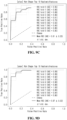
- FIGS. 9A to 9D show validation results for an artificial intelligence model according to a 10-fold cross validation method when using an image of an end systole phase according to an embodiment of the present invention.
- FIGS. 9A to 9D show receiver operating characteristic (ROC) curves for each view of A2CH, A4CH, PLAX, and PSAX. As shown in FIGS. 9A to 9D , an average area under the curve (AUC) exceeding 0.9 is confirmed in all views.
- ROC receiver operating characteristic
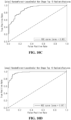
- FIGS. 10A to 10D show verification results for an artificial intelligence model according to an LOO cross validation method when using an image of the end systole phase according to an embodiment of the present invention.
- FIGS. 10A to 10D show ROC curves for each view of A2CH, A4CH, PLAX, and PSAX. As shown in FIGS. 10A to 10D , an AUC of 0.9 or more is confirmed in all views.
- FIGS. 11A to 11D show verification results for an artificial intelligence model according to a 10-fold cross validation method when using an image of an end diastole phase according to an embodiment of the present invention.
- FIGS. 11A to 11D show ROC curves for each view of A2CH, A4CH, PLAX, and PSAX. As shown in FIGS. 11A to 11D , an average AUC exceeding 0.9 is confirmed in all views.
- FIGS. 12A to 12D show verification results for an artificial intelligence model according to an LOO cross validation method when using an image of an end diastole phase according to an embodiment of the present invention.
- FIGS. 12A to 12D show ROC curves for each view of A2CH, A4CH, PLAX, and PSAX. As shown in FIGS. 12A to 12D , an AUC of approximately 0.9 is confirmed in all views.
- FIGS. 13A to 13D show verification results for an artificial intelligence model according to a 10-fold cross validation method when using a mid-phase image according to an embodiment of the present invention.
- FIGS. 13A to 13D show ROC curves for each view of A2CH, A4CH, PLAX, and PSAX. As shown in FIGS. 13A to 13D , an average AUC exceeding 0.9 is confirmed in all views.
- FIGS. 14A to 14D show verification results for an artificial intelligence model according to an LOO cross validation method when using an image of a mid-phase according to an embodiment of the present invention.
- FIGS. 14A to 14D show ROC curves for each view of A2CH, A4CH, PLAX, and PSAX. As shown in FIGS. 14A to 14D , an average AUC exceeding 0.9 is confirmed in all views.
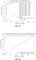
- FIG. 15 shows verification results for an artificial intelligence model according to an 10-fold cross validation method when using an image of all phases according to an embodiment of the present invention. As shown in FIG. 15 , an average AUC of 0.98 is confirmed.
- Various embodiments of the present disclosure may be implemented in hardware, firmware, software, or a combination thereof.
- the present disclosure can be implemented with application specific integrated circuits (ASICs), Digital signal processors (DSPs), digital signal processing devices (DSPDs), programmable logic devices (PLDs), field programmable gate arrays (FPGAs), general processors, controllers, microcontrollers, microprocessors, etc.
- ASICs application specific integrated circuits
- DSPs Digital signal processors
- DSPDs digital signal processing devices
- PLDs programmable logic devices
- FPGAs field programmable gate arrays
- general processors controllers, microcontrollers, microprocessors, etc.
- the scope of the present disclosure includes a non-transitory computer-readable medium in which software or machine-executable instructions (e.g., operating system, application, firmware, program, etc.) that cause operations according to the methods of various embodiments to be executed on a device or computer and such software or instructions are stored and may be executed on a device or computer.
- software or machine-executable instructions e.g., operating system, application, firmware, program, etc.
Landscapes
- Health & Medical Sciences (AREA)
- Engineering & Computer Science (AREA)
- Medical Informatics (AREA)
- Life Sciences & Earth Sciences (AREA)
- Public Health (AREA)
- General Health & Medical Sciences (AREA)
- Biomedical Technology (AREA)
- Nuclear Medicine, Radiotherapy & Molecular Imaging (AREA)
- Pathology (AREA)
- Radiology & Medical Imaging (AREA)
- Physics & Mathematics (AREA)
- Epidemiology (AREA)
- Primary Health Care (AREA)
- Animal Behavior & Ethology (AREA)
- Veterinary Medicine (AREA)
- Biophysics (AREA)
- Heart & Thoracic Surgery (AREA)
- Molecular Biology (AREA)
- Surgery (AREA)
- Data Mining & Analysis (AREA)
- Databases & Information Systems (AREA)
- High Energy & Nuclear Physics (AREA)
- Physiology (AREA)
- Computer Vision & Pattern Recognition (AREA)
- Optics & Photonics (AREA)
- Measuring And Recording Apparatus For Diagnosis (AREA)
- Cardiology (AREA)
- Vascular Medicine (AREA)
- Dentistry (AREA)
- Oral & Maxillofacial Surgery (AREA)
Applications Claiming Priority (2)
| Application Number | Priority Date | Filing Date | Title |
|---|---|---|---|
| KR1020210125252A KR102836304B1 (ko) | 2021-09-17 | 2021-09-17 | 체내 장기 또는 근육의 이상 유무를 판단하는 방법 및 장치 |
| PCT/KR2022/013534 WO2023043133A1 (ko) | 2021-09-17 | 2022-09-08 | 체내 장기 또는 근육의 이상 유무를 판단하는 방법 및 장치 |
Publications (2)
| Publication Number | Publication Date |
|---|---|
| EP4404206A1 true EP4404206A1 (de) | 2024-07-24 |
| EP4404206A4 EP4404206A4 (de) | 2025-10-08 |
Family
ID=85603183
Family Applications (1)
| Application Number | Title | Priority Date | Filing Date |
|---|---|---|---|
| EP22870218.9A Pending EP4404206A4 (de) | 2021-09-17 | 2022-09-08 | Verfahren und vorrichtung zur bestimmung von anomalien in organen oder muskeln im körper |
Country Status (4)
| Country | Link |
|---|---|
| US (1) | US20250054604A1 (de) |
| EP (1) | EP4404206A4 (de) |
| KR (1) | KR102836304B1 (de) |
| WO (1) | WO2023043133A1 (de) |
Families Citing this family (1)
| Publication number | Priority date | Publication date | Assignee | Title |
|---|---|---|---|---|
| CN120336418A (zh) * | 2025-04-01 | 2025-07-18 | 中国人民解放军61175部队 | 军事目标的图表达方法 |
Family Cites Families (6)
| Publication number | Priority date | Publication date | Assignee | Title |
|---|---|---|---|---|
| KR101716039B1 (ko) * | 2015-08-07 | 2017-03-13 | 원광대학교산학협력단 | 의료 영상 기반의 질환 진단 정보 산출 방법 및 장치 |
| KR101880678B1 (ko) * | 2016-10-12 | 2018-07-20 | (주)헬스허브 | 기계학습을 통한 의료영상 판독 및 진단 통합 시스템 |
| CN109523526B (zh) * | 2018-11-08 | 2021-10-22 | 腾讯科技(深圳)有限公司 | 组织结节检测及其模型训练方法、装置、设备和系统 |
| WO2020257046A1 (en) * | 2019-06-21 | 2020-12-24 | West Virginia University | Cardiac ultrasonic fingerprinting: an approach for highthroughput myocardial feature phenotyping |
| KR102132375B1 (ko) * | 2019-07-05 | 2020-07-09 | 한국과학기술원 | 딥 러닝 모델을 활용한 영상 진단 장치 및 그 방법 |
| KR102237198B1 (ko) * | 2020-06-05 | 2021-04-08 | 주식회사 딥노이드 | 인공지능 기반의 의료영상 판독 서비스 시스템 |
-
2021
- 2021-09-17 KR KR1020210125252A patent/KR102836304B1/ko active Active
-
2022
- 2022-09-08 EP EP22870218.9A patent/EP4404206A4/de active Pending
- 2022-09-08 US US18/693,082 patent/US20250054604A1/en active Pending
- 2022-09-08 WO PCT/KR2022/013534 patent/WO2023043133A1/ko not_active Ceased
Also Published As
| Publication number | Publication date |
|---|---|
| WO2023043133A1 (ko) | 2023-03-23 |
| KR20230041921A (ko) | 2023-03-27 |
| EP4404206A4 (de) | 2025-10-08 |
| KR102836304B1 (ko) | 2025-07-22 |
| US20250054604A1 (en) | 2025-02-13 |
Similar Documents
| Publication | Publication Date | Title |
|---|---|---|
| US12283052B2 (en) | Method and system for automatically detecting anatomical structures in a medical image | |
| Li et al. | MV-RAN: Multiview recurrent aggregation network for echocardiographic sequences segmentation and full cardiac cycle analysis | |
| EP3445250B1 (de) | Echokardiographische bildanalyse | |
| Habijan et al. | Overview of the Whole Heart and Heart Chamber Segmentation Methods: Habijan et al. | |
| US20210312242A1 (en) | Synthetically Generating Medical Images Using Deep Convolutional Generative Adversarial Networks | |
| CN109584254A (zh) | 一种基于深层全卷积神经网络的心脏左心室分割方法 | |
| JP2021513697A (ja) | 完全畳み込みニューラル・ネットワークを用いた心臓ctaにおける解剖学的構造のセグメンテーションのためのシステム | |
| CN113571183A (zh) | Covid-19患者管理的风险预测 | |
| US11521323B2 (en) | Systems and methods for generating bullseye plots | |
| Farhad et al. | A data-efficient zero-shot and few-shot Siamese approach for automated diagnosis of left ventricular hypertrophy | |
| EP4404206A1 (de) | Verfahren und vorrichtung zur bestimmung von anomalien in organen oder muskeln im körper | |
| Alam et al. | Ejection Fraction estimation using deep semantic segmentation neural network: Md. GR Alam et al. | |
| Zhou et al. | An adaptively weighted ensemble of multiple CNNs for carotid ultrasound image segmentation | |
| Chu et al. | BNU-net: a novel deep learning approach for LV MRI analysis in short-Axis MRI | |
| KR102732856B1 (ko) | 인공지능 모델에 기반하여 심장 구조를 세그멘테이션하기 위한 방법 및 장치 | |
| Shaikh et al. | Improved skin cancer detection using CNN | |
| CN116913476A (zh) | 用于医学成像研究的语义图像分析的心脏ai智能助手 | |
| Molina et al. | Automated segmentation and 4D reconstruction of the heart left ventricle from CINE MRI | |
| Zhu et al. | [Retracted] Analysis of Cardiac Ultrasound Images of Critically Ill Patients Using Deep Learning | |
| Ye et al. | Deep echocardiography: a first step toward automatic cardiac disease diagnosis using machine learning | |
| Gondova et al. | Convolutional neural networks for automated CMR image segmentation in rats with myocardial infarcts | |
| de Melo et al. | Gradient boosting decision trees for echocardiogram images | |
| CN118898580B (zh) | 基于度量学习预测肺动脉高压的构建方法、设备和介质 | |
| Li et al. | [Retracted] Diagnostic Classification of Patients with Dilated Cardiomyopathy Using Ventricular Strain Analysis Algorithm | |
| Yao et al. | MultiFlow: A unified deep learning framework for multi-vessel classification, segmentation and clustering of phase-contrast MRI validated on a multi-site single ventricle patient cohort |
Legal Events
| Date | Code | Title | Description |
|---|---|---|---|
| STAA | Information on the status of an ep patent application or granted ep patent |
Free format text: STATUS: THE INTERNATIONAL PUBLICATION HAS BEEN MADE |
|
| PUAI | Public reference made under article 153(3) epc to a published international application that has entered the european phase |
Free format text: ORIGINAL CODE: 0009012 |
|
| STAA | Information on the status of an ep patent application or granted ep patent |
Free format text: STATUS: REQUEST FOR EXAMINATION WAS MADE |
|
| 17P | Request for examination filed |
Effective date: 20240328 |
|
| AK | Designated contracting states |
Kind code of ref document: A1 Designated state(s): AL AT BE BG CH CY CZ DE DK EE ES FI FR GB GR HR HU IE IS IT LI LT LU LV MC MK MT NL NO PL PT RO RS SE SI SK SM TR |
|
| DAV | Request for validation of the european patent (deleted) | ||
| DAX | Request for extension of the european patent (deleted) | ||
| A4 | Supplementary search report drawn up and despatched |
Effective date: 20250909 |
|
| RIC1 | Information provided on ipc code assigned before grant |
Ipc: G16H 30/40 20180101AFI20250903BHEP Ipc: G16H 30/20 20180101ALI20250903BHEP Ipc: G16H 50/50 20180101ALI20250903BHEP Ipc: G16H 50/20 20180101ALI20250903BHEP Ipc: A61B 8/08 20060101ALI20250903BHEP Ipc: A61B 6/00 20240101ALI20250903BHEP Ipc: A61B 5/055 20060101ALI20250903BHEP Ipc: A61B 6/03 20060101ALI20250903BHEP Ipc: A61B 6/50 20240101ALI20250903BHEP Ipc: A61B 8/00 20060101ALI20250903BHEP |
|
| STAA | Information on the status of an ep patent application or granted ep patent |
Free format text: STATUS: EXAMINATION IS IN PROGRESS |