EP4394726A1 - Image classification method and apparatus, computer device, and storage medium - Google Patents
Image classification method and apparatus, computer device, and storage medium Download PDFInfo
- Publication number
- EP4394726A1 EP4394726A1 EP22909548.4A EP22909548A EP4394726A1 EP 4394726 A1 EP4394726 A1 EP 4394726A1 EP 22909548 A EP22909548 A EP 22909548A EP 4394726 A1 EP4394726 A1 EP 4394726A1
- Authority
- EP
- European Patent Office
- Prior art keywords
- feature
- sub
- image
- features
- spliced
- Prior art date
- Legal status (The legal status is an assumption and is not a legal conclusion. Google has not performed a legal analysis and makes no representation as to the accuracy of the status listed.)
- Pending
Links
Images





Classifications
-
- G—PHYSICS
- G06—COMPUTING OR CALCULATING; COUNTING
- G06V—IMAGE OR VIDEO RECOGNITION OR UNDERSTANDING
- G06V10/00—Arrangements for image or video recognition or understanding
- G06V10/40—Extraction of image or video features
- G06V10/44—Local feature extraction by analysis of parts of the pattern, e.g. by detecting edges, contours, loops, corners, strokes or intersections; Connectivity analysis, e.g. of connected components
- G06V10/443—Local feature extraction by analysis of parts of the pattern, e.g. by detecting edges, contours, loops, corners, strokes or intersections; Connectivity analysis, e.g. of connected components by matching or filtering
- G06V10/449—Biologically inspired filters, e.g. difference of Gaussians [DoG] or Gabor filters
- G06V10/451—Biologically inspired filters, e.g. difference of Gaussians [DoG] or Gabor filters with interaction between the filter responses, e.g. cortical complex cells
-
- G—PHYSICS
- G06—COMPUTING OR CALCULATING; COUNTING
- G06V—IMAGE OR VIDEO RECOGNITION OR UNDERSTANDING
- G06V10/00—Arrangements for image or video recognition or understanding
- G06V10/70—Arrangements for image or video recognition or understanding using pattern recognition or machine learning
- G06V10/764—Arrangements for image or video recognition or understanding using pattern recognition or machine learning using classification, e.g. of video objects
-
- G—PHYSICS
- G06—COMPUTING OR CALCULATING; COUNTING
- G06N—COMPUTING ARRANGEMENTS BASED ON SPECIFIC COMPUTATIONAL MODELS
- G06N3/00—Computing arrangements based on biological models
- G06N3/02—Neural networks
- G06N3/04—Architecture, e.g. interconnection topology
-
- G—PHYSICS
- G06—COMPUTING OR CALCULATING; COUNTING
- G06N—COMPUTING ARRANGEMENTS BASED ON SPECIFIC COMPUTATIONAL MODELS
- G06N3/00—Computing arrangements based on biological models
- G06N3/02—Neural networks
- G06N3/08—Learning methods
-
- G—PHYSICS
- G06—COMPUTING OR CALCULATING; COUNTING
- G06V—IMAGE OR VIDEO RECOGNITION OR UNDERSTANDING
- G06V10/00—Arrangements for image or video recognition or understanding
- G06V10/20—Image preprocessing
- G06V10/26—Segmentation of patterns in the image field; Cutting or merging of image elements to establish the pattern region, e.g. clustering-based techniques; Detection of occlusion
-
- G—PHYSICS
- G06—COMPUTING OR CALCULATING; COUNTING
- G06V—IMAGE OR VIDEO RECOGNITION OR UNDERSTANDING
- G06V10/00—Arrangements for image or video recognition or understanding
- G06V10/40—Extraction of image or video features
- G06V10/44—Local feature extraction by analysis of parts of the pattern, e.g. by detecting edges, contours, loops, corners, strokes or intersections; Connectivity analysis, e.g. of connected components
-
- G—PHYSICS
- G06—COMPUTING OR CALCULATING; COUNTING
- G06V—IMAGE OR VIDEO RECOGNITION OR UNDERSTANDING
- G06V10/00—Arrangements for image or video recognition or understanding
- G06V10/70—Arrangements for image or video recognition or understanding using pattern recognition or machine learning
- G06V10/82—Arrangements for image or video recognition or understanding using pattern recognition or machine learning using neural networks
-
- G—PHYSICS
- G06—COMPUTING OR CALCULATING; COUNTING
- G06V—IMAGE OR VIDEO RECOGNITION OR UNDERSTANDING
- G06V2201/00—Indexing scheme relating to image or video recognition or understanding
- G06V2201/03—Recognition of patterns in medical or anatomical images
Definitions
- Embodiments of this application relate to the field of computer technologies, and in particular, to an image classification method and apparatus, a computer device, and a storage medium.
- the image classification technology is applied more widely, and may be applied to various scenarios such as a face recognition scenario and a medical image classification scenario.
- feature extraction is performed on a to-be-classified image to obtain an image feature of the image, and the image feature of the image is directly classified to obtain a category to which the image belongs.
- the classification is directly performed according to the extracted image feature, and the amount of information in the image feature is small, resulting in poor classification accuracy.
- Embodiments of this application provide an image classification method and apparatus, a computer device, and a storage medium, which can improve the classification accuracy.
- the technical solutions will be described below.
- an image classification method which is performed by a computer device and includes:
- an image classification apparatus which includes:
- a computer device which includes a processor and a memory.
- the memory stores at least one segment of computer program that, when loaded and executed by the processor, implements operations performed in the image classification method according to the foregoing aspect.
- a computer-readable storage medium which stores at least one segment of computer program that, when loaded and executed by a processor, implements operations performed in the image classification method according to the foregoing aspect.
- an image feature of a pathological image is processed according to multiple scales respectively to obtain local features respectively corresponding to the multiple scales, and the local features respectively corresponding to the multiple scales are spliced to obtain a spliced image feature, and the spliced image feature is classified to obtain a category to which the pathological image belongs.
- the local features corresponding to different scales contain different information, so that the finally obtained spliced image feature contains feature information corresponding to different scales, and the feature information of the spliced image feature is enriched.
- the category to which the pathological image belongs is determined based on the spliced image feature, so that the accuracy of the category is ensured.
- first, second, and the like used in this application may be used herein to describe various concepts, but unless otherwise specified, these concepts are not limited by these terms. The terms are merely used for distinguishing one concept from another concept. For example, without departing from the scope of this application, a first feature vector may be referred to as a second feature vector, and similarly, the second feature vector may be referred to as the first feature vector.
- multiple scales include 3 scales, “each” refers to each of the 3 scales, and “any” refers to any one of the 3 scales, which may be the first, or the second, or the third.
- a classification model may be trained based on the artificial intelligent machine learning technology, and the trained classification model is used to realize the classification of a pathological image.
- the terminal may include, but is not limited to, a smartphone, a tablet computer, a notebook computer, a desktop computer, a smart speaker, a smartwatch, a smart voice interaction device, a smart household appliance, an on-board terminal, and the like.
- a computer program involved in the embodiments of this application may be deployed to be executed on a computer device, or executed on multiple computer devices at the same location, or executed on multiple computer devices that are distributed at multiple locations and connected to each other through a communication network, and the multiple computer devices that are distributed at multiple locations and connected to each other through a communication network can form a blockchain system.
- the pathological image is an image used for presenting the pathological morphology of a body organ, tissue or cell.
- the pathological image is a whole slide image (WSI, a digital pathological image).
- the image feature of the pathological image is used for characterizing the pathological image, which is a two-dimensional feature or a three-dimensional feature and can be represented in any form.
- the image feature is represented in the form of a feature matrix or a feature vector.
- the image feature is represented in the form of a three-dimensional feature matrix or a two-dimensional feature matrix.
- a size of the first sliding window is the same as the preset size, and the first moving step length represents a movement distance of the first sliding window during each movement.
- the first moving step length represents a quantity of pixels moved each time during movement of the first sliding window.
- the preset size includes a first size and a second size, the first size and the second size respectively represent a length and a width of the first sliding window, and the first moving step length is the same as the first size and the second size.
- the present size is 1120 ⁇ 1120
- the first moving step length is 1120
- multiple sub-images in the pathological image are obtained based on the first moving step length and the preset size
- a size of each sub-image is 1120 ⁇ 1120.
- the image feature of the pathological image is a 3 ⁇ 3 feature matrix
- the expansion parameter corresponding to any scale is 1, and the expanded image feature is a 5 ⁇ 5 feature matrix
- the expansion parameter corresponding to any scale is 2, and the expanded image feature is a 7 ⁇ 7 feature matrix.
- the computer device updates each first feature vector based on the first feature vectors corresponding to the multiple local features to obtain a second feature vector corresponding to each first feature vector.
- step 305 includes the following sub-step: for a to-be-processed feature vector in the multiple first feature vectors, the similarity between each first feature vector and the to-be-processed feature vector is obtained, and the multiple first feature vectors are weighted and fused based on the similarity corresponding to each first feature vector to obtain a second feature vector corresponding to the to-be-processed feature vector.
- the computer device uses the multiple second feature vectors to constitute a three-dimensional feature matrix based on positions of the multiple local features in the image feature of the pathological image, and determines the three-dimensional feature matrix as an aggregated feature.
- step 306 includes the following sub-steps: the multiple second feature vectors in the second feature matrix are used to constitute a three-dimensional feature matrix based on the positions of the multiple local features in the image feature of the pathological image, and the three-dimensional feature matrix is determined as the aggregated feature.
- step 307 includes the following sub-step: features at the same position in the multiple aggregated features are spliced to obtain feature vector corresponding to multiple positions; and the feature vectors corresponding to the multiple positions are used to constitute a three-dimensional feature matrix, and the three-dimensional feature matrix is determined as the spliced image feature.
- each aggregated feature includes features at multiple positions, and the feature at each position is one second feature vector.
- the features at the same position are a feature of a pixel at the same position in the pathological image. Therefore, the features at the same position in the multiple aggregated features are spliced to obtain the feature vectors corresponding to the multiple positions. Based on the multiple positions, the feature vectors corresponding to the multiple positions are used to constitute one three-dimensional feature matrix, and the three-dimensional feature matrix is determined as the spliced image feature.
- the computer device updates each first sub-feature based on multiple first sub-features in the spliced image feature to obtain a first updated feature corresponding to each first sub-feature.
- step 308 includes the following sub-steps: for a to-be-processed sub-feature in the multiple first sub-features, weights of the multiple first sub-features are acquired; and the multiple first sub-features are weighted and fused based on the weights of the multiple first sub-features to obtain the first updated feature corresponding to the to-be-processed sub-feature.
- the weight of each first sub-feature represents a degree of correlation between the first sub-feature and the to-be-processed sub-feature, and the multiple first sub-features are weighted and fused into the first updated feature according to degrees of correlations between the multiple first sub-features and the to-be-processed sub-feature, so that the accuracy of the obtained first updated feature is ensured.
- the first updated feature corresponding to each first sub-feature can be acquired.
- the process of acquiring the weight of each first sub-feature includes the following steps: for the to-be-processed sub-feature in the multiple first sub-features, a distance feature of each first sub-feature and the to-be-processed sub-feature is acquired; and the weight of each first sub-feature is acquired based on the similarity between each first sub-feature and the to-be-processed sub-feature and the distance feature.
- the distance feature of any first sub-feature and the to-be-processed sub-feature is used for indicating a distance between positions of the first sub-feature and the to-be-processed sub-feature in the spliced image feature; and the similarity between any first sub-feature and the to-be-processed sub-feature represents a level of similarity between the first sub-feature and the to-be-processed sub-feature.
- the similarity between the first sub-feature and the to-be-processed sub-feature can represent a level of similarity between the first sub-feature and the to-be-processed sub-feature
- the distance feature of the first sub-feature and the to-be-processed sub-feature can indicate the distribution of the first sub-feature and the to-be-processed sub-feature in the spliced image feature
- the weight of the first sub-feature is acquired based on the similarity between the first sub-feature and the to-be-processed sub-feature and the distance feature, which not only takes account of a level of similarity between the first sub-feature and the to-be-processed sub-feature, but also takes account of a relative position relationship between the first sub-feature and the to-be-processed feature, that is, takes full account of the correlation between the first sub-feature and the to-be-processed
- the process of acquiring the distance feature includes the following steps: a distance corresponding to each first sub-feature is determined based on positions of each first sub-feature and the to-be-processed sub-feature in the spliced image feature; and the distance corresponding to each first sub-feature is mapped to obtain the distance feature of each first sub-feature and the to-be-processed sub-feature.
- the first matching degree corresponding to any first sub-feature indicates a degree of matching of the first sub-feature with the to-be-processed sub-feature.
- the first matching degree of each first sub-feature and the to-be-processed sub-feature is acquired first.
- the ratio of the first matching degree corresponding to each first sub-feature to the sum is determined as the weight of each first sub-feature, which ensures that the first sub-feature that is matched more with the to-be-processed sub-feature has a larger weight. Therefore, the accuracy of the weight is ensured.
- the process of acquiring the first matching degree of any first sub-feature and the to-be-processed sub-feature includes the following steps: the similarity between the first sub-feature and the to-be-processed sub-feature and the distance feature are fused to obtain a second matching degree; and feature transformation is performed on the second matching degree to obtain the first matching degree corresponding to the first sub-feature.
- z i represents the first updated feature corresponding to the to-be-processed sub-feature
- E represents the spliced image feature
- W Q , W K , and W V are all constant matrices
- N represents a total quantity of multiple first sub-features
- e j " represents a j th first sub-feature in the multiple first sub-features
- the computer device uses the first updated features corresponding to the multiple first sub-features to constitute a three-dimensional feature matrix based on positions of the multiple first sub-features in the spliced image feature, and determines the three-dimensional feature matrix as an updated spliced image feature.
- a feature size of the spliced image feature before updating is 3 ⁇ 3 ⁇ 10
- each first sub-feature is a feature vector with a dimension of 10
- the spliced image feature before updating includes 9 positions
- the first updated feature corresponding to each first sub-feature is a feature vector with a dimension of 8, that is, a feature size of the updated spliced image feature composed of the multiple first updated features is 3 ⁇ 3 ⁇ 8.
- the description is made by taking a case where the first updated feature is a vector as an example.
- the first updated feature is not a vector, it is unnecessary to perform step 309, and another method can be adopted to use the first updated features corresponding to the multiple first sub-features to constitute the updated spliced image feature based on the positions of the multiple first sub-features in the spliced image feature.
- the first updated feature includes one feature element, the first updated features corresponding to the multiple first sub-features are used to constitute one two-dimensional feature matrix based on the positions of the multiple first sub-features in the spliced image feature, and the two-dimensional feature matrix is determined as the updated spliced image feature.
- Each first updated feature in the updated spliced image feature is fused with features in the local features of different scales, and is also fused with the first sub-features at the positions in the whole spliced image feature according to the degrees of correlations between the first sub-features at the positions in the whole spliced image feature, so that the updated spliced image feature is enriched, and the accuracy of the updated spliced image feature is ensured.
- the updated spliced feature is classified, so that the accuracy of the determined category is ensured.
- the category to which the pathological image belongs indicates a state of a specific region in the pathological image.
- the classified feature vector includes probabilities of multiple dimensions, and the probability of each dimension is used for indicating the probability that the pathological image belongs to a category corresponding to the dimension.
- the updated spliced image feature is classified to obtain the classified feature vector indicating probabilities that the pathological image belongs to multiple categories, and the category corresponding to the maximum probability in the classified feature vector is determined as the category to which the pathological image belongs, so that the accuracy of the determined category is ensured.
- the spliced image feature is updated according to steps 308 and 309, and the updated image feature is directly classified to obtain the category to which the pathological image belongs.
- the updated spliced image can be updated multiple times according to steps 308 and 309, and an updated spliced image feature obtained during the last updating is classified to obtain the category to which the pathological image belongs.
- the spliced image feature is updated multiple times to fully fuse the features at different positions in the spliced image feature, so that a finally obtained updated spliced image feature retains position information of different features, and the accuracy of the updated spliced image feature is ensured.
- the method according to the embodiments of this application can be applied to classification tasks for multiple types of pathological images, such as cancer classification, lymph node metastasis, and gene mutation prediction.
- pathological images such as cancer classification, lymph node metastasis, and gene mutation prediction.
- a state that corresponds to a cancerous region corresponding to the pathological image can be determined by the method according to the embodiments of this application.
- an image feature of a pathological image is processed according to multiple scales to obtain aggregated features corresponding to the multiple scales, and the aggregated features corresponding to the multiple scales are spliced into one spliced image feature, and the spliced image feature is classified to obtain a category to which the pathological image belongs.
- the aggregated feature corresponding to different scales are obtained by aggregating local features of different scales, and the local features corresponding to different scales contain different information, so that the multiple aggregated features contain feature information corresponding to different scales.
- the multiple aggregated features are spliced into the spliced image feature, so that the spliced image feature contains the feature information corresponding to different scales, and the feature information of the spliced image feature is enriched.
- the category to which the pathological image belongs is determined based on the spliced image feature, so that the accuracy of the category is ensured.
- each first sub-feature is first updated based on the first sub-features at multiple positions in the spliced image feature to obtain the updated spliced image feature, and then the updated spliced image feature is classified.
- the process of acquiring the category to which the pathological image belongs based on the spliced image feature includes the following step 1 to step 3.
- Step 1 The computer device fuses each first sub-feature in the spliced image feature with a corresponding position feature to obtain a second updated feature corresponding to each first sub-feature.
- the spliced image feature includes the first sub-features at multiple positions; and the position feature corresponding to any first sub-feature is used for indicating the position of the first sub-feature in the spliced image feature and can be represented in any form.
- the position feature is represented in the form of a vector or coordinates.
- Step 2 The computer device uses the second updated features corresponding to the multiple first sub-features to constitute an updated spliced image feature based on the positions of the multiple first sub-features in the spliced image feature.
- each second updated feature is fused with the position feature of the corresponding first sub-feature, so that the difference between different first sub-features is increased.
- the multiple second updated features are used to constitute the updated spliced image feature according to the positions of the multiple first sub-features in the spliced image feature and a position arrangement of the multiple first sub-features, so that the updated spliced image feature include the second updated features at multiple positions.
- the second updated features in the updated spliced image feature are fused with respective position features, so that the difference between different first sub-features is increased, and the accuracy of the updated spliced image feature is improved.
- the second updated feature is a vector.
- Step 2 includes the following sub-steps: the second updated features corresponding to the multiple first sub-features are used to constitute a three-dimensional feature matrix based on the positions of the multiple first sub-features in the spliced image feature, and the three-dimensional feature matrix is determined as the updated spliced image feature.
- a feature size of an updated spliced image feature obtained by splicing 9 second updated features is 3 ⁇ 3 ⁇ 10; and in a case that the dimensions of each first sub-feature and the corresponding second updated feature are different, the dimension of each second updated feature is 20, and a feature size of an updated spliced image feature obtained by splicing 9 second updated features is 3 ⁇ 3 ⁇ 20.
- Step 3 is the same as step 310, which is not described in detail here.
- each first sub-feature in the spliced image feature is fused with a corresponding position feature to obtain a second updated feature corresponding to each first sub-feature, so that a difference between different first sub-features is increased.
- the second updated features in the obtained updated spliced image feature are fused with respective position features, so that the difference between different first sub-features is increased, and the accuracy of the updated spliced image feature is improved.
- the updated spliced image feature is classified, so that the accuracy of a determined category is ensured.
- the process of acquiring the category to which the pathological image belongs based on the spliced image feature includes the following step 4 to step 8.
- Step 4 The computer device fuses each first sub-feature in the spliced image feature with a corresponding position feature to obtain a second updated feature corresponding to each first sub-feature.
- Step 4 is the same as step 1, which is not described in detail here.
- Step 5 The computer device uses the second updated features corresponding to the multiple first sub-features to constitute a first spliced image feature based on the positions of the multiple first sub-features in the spliced image feature.
- the first spliced image feature is equivalent to the updated spliced image feature obtained in step 2, and step 5 is the same as step 2, which is not described in detail here.
- Step 6 The computer device updates each second updated feature based on the multiple second updated features to obtain a third updated feature corresponding to each second updated feature.
- each second updated feature is updated based on the multiple second updated features in the first spliced image feature to obtain a third updated feature corresponding to each second updated feature; and the multiple third updated features are used to constitute a second spliced image feature based on positions of the multiple second updated features in the first spliced image feature.
- Step 3 Extract, for each scale in multiple scales, a sample local feature corresponding to the scale from the sample image feature based on the classification sub-model; splice the sample local features respectively corresponding to the scales to obtain a sample spliced image feature; and classify the sample spliced image feature to obtain a predicted label of the sample pathological image, the predicted label being used for indicating a predicted category to which the sample pathological image belongs.
- the predicted label is used for indicating the category which is predicted based on the classification model and to which the sample pathological image belongs
- the sample label is used for indicating the category to which the sample pathological image belongs
- the accuracy rate of the classification model can be reflected by comparing the predicted label with the sample label.
- the classification model is trained based on the predicted label and the sample label, so that the accuracy of the classification model is improved.
- a loss value obtained during current iterative training is less than a loss value threshold, the training of the classification model ends; or, in a case that the number of iterative training reaches the target number, the training of the classification model ends.
- the feature extraction sub-model may be any pre-trained image feature extraction model.
- the feature extraction sub-model is a residual neural network (ResNet), a densely connected convolutional network (DenseNet), or an inception network.
- ResNet residual neural network
- DenseNet densely connected convolutional network
- the feature extraction sub-model and the classification sub-model are used to constitute the classification model, and the classification model is trained according to steps 1 to 4, so that the training time is saved, and the accuracy of the classification model is also ensured.
- the performance of the classification model is evaluated according to various different position encoding policies.
- two position encoding polices are set: absolute position encoding and relative position encoding. It can be seen from Table 3 that the accuracy of image classification can be improved when the absolute position encoding and relative position encoding mechanisms are used simultaneously.
- the spliced image feature includes first sub-features at multiple positions; and as shown in FIG. 9 , the apparatus further includes:
- the operations of acquiring an image feature of a to-be-classified pathological image; extracting, for each scale in multiple scales, a local feature corresponding to the scale from the image feature; splicing the local features respectively corresponding to the scales to obtain a spliced image feature; and classifying the spliced image feature to obtain a category to which the pathological image belongs are implemented based on a classification model.
- the terminal 1000 includes: a processor 1001 and a memory 1002.
- the processor 1001 may include one or more processing cores, such as a 4-core processor or an 8-core processor.
- the processor 1001 may be implemented by at least one hardware of a digital signal processor (DSP), a field-programmable gate array (FPGA), and a programmable logic array (PLA).
- DSP digital signal processor
- FPGA field-programmable gate array
- PDA programmable logic array
- the processor 1001 may also include a main processor and a coprocessor.
- the main processor is a processor configured to process data in an awake state, and is also referred to as a central processing unit (CPU); and the coprocessor is a low power processor configured to process data in a standby state.
- the processor 1001 may be integrated with a graphics processing unit (GPU).
- the GPU is configured to render and draw content that needs to be displayed on a display screen.
- the processor 1001 may further include an artificial intelligence (AI) processor.
- the AI processor is configured to process computing operations related to machine learning.
- the memory 1002 may include one or more computer-readable storage media.
- the computer-readable storage medium may be non-transient.
- the memory 1002 may further include a high-speed random access memory and a non-volatile memory, such as one or more disk storage devices or flash storage devices.
- the non-transient computer-readable storage medium in the memory 1002 is configured to store at least one computer program that, when executed by the processor 1001, implements the image classification method according to the method embodiments of this application.
- the terminal 1000 further includes one or more sensors 1010.
- the one or more sensors 1010 include, but are not limited to: an acceleration sensor 1011, a gyroscope sensor 1012, a pressure sensor 1013, an optical sensor 1014, and a proximity sensor 1015.
- the structure shown in FIG. 10 does not constitute a limitation to the terminal 1000, the terminal 1000 may include more or less components than those shown in the figure, or some components are combined, or a different component arrangement is adopted.
- FIG. 11 is a schematic structural diagram of a server according to an embodiment of this application.
- a server 1100 may vary a lot due to different configurations or performance, and may include one or more central processing units (CPUs) 1101 and one or more memories 1102.
- the memory 1102 stores at least one segment of computer program that, when loaded and executed by the CPU 1101, implements the method according to the foregoing method embodiments.
- the server may further include components, such as a wired or wireless network interface, a keyboard, and an input/output interface, to facilitate input and output.
- the server may further include other components configured to implement functions of the device, which are not described in detail here.
- the embodiments of this application further provide a computer-readable storage medium, which stores at least one segment of computer program that, when loaded and executed by a processor, implements operations performed in the image classification method according to the foregoing embodiments.
- the embodiments of this application further provide a computer program product, which includes a computer program that, when executed by a processor, implements operations performed in the image classification method according to the foregoing aspect.
- the program may be stored in one computer-readable storage medium.
- the foregoing storage medium may be a read-only memory, a magnetic disk, an optical disc, or the like.
Landscapes
- Engineering & Computer Science (AREA)
- Theoretical Computer Science (AREA)
- Physics & Mathematics (AREA)
- General Physics & Mathematics (AREA)
- Multimedia (AREA)
- Health & Medical Sciences (AREA)
- General Health & Medical Sciences (AREA)
- Evolutionary Computation (AREA)
- Computer Vision & Pattern Recognition (AREA)
- Software Systems (AREA)
- Artificial Intelligence (AREA)
- Computing Systems (AREA)
- Biomedical Technology (AREA)
- Life Sciences & Earth Sciences (AREA)
- Molecular Biology (AREA)
- Medical Informatics (AREA)
- Databases & Information Systems (AREA)
- Data Mining & Analysis (AREA)
- Mathematical Physics (AREA)
- General Engineering & Computer Science (AREA)
- Computational Linguistics (AREA)
- Biophysics (AREA)
- Biodiversity & Conservation Biology (AREA)
- Image Analysis (AREA)
- Information Retrieval, Db Structures And Fs Structures Therefor (AREA)
Abstract
Description
- This application claims priority to
Chinese Patent Application No. 202111583275.9, entitled "IMAGE CLASSIFICATION METHOD AND APPARATUS, COMPUTER DEVICE, AND STORAGE MEDIUM" filed with China National Intellectual Property Administration on December 22, 2021 - Embodiments of this application relate to the field of computer technologies, and in particular, to an image classification method and apparatus, a computer device, and a storage medium.
- With the development of computer technologies, the image classification technology is applied more widely, and may be applied to various scenarios such as a face recognition scenario and a medical image classification scenario. In the related technology, feature extraction is performed on a to-be-classified image to obtain an image feature of the image, and the image feature of the image is directly classified to obtain a category to which the image belongs. According to the foregoing method, the classification is directly performed according to the extracted image feature, and the amount of information in the image feature is small, resulting in poor classification accuracy.
- Embodiments of this application provide an image classification method and apparatus, a computer device, and a storage medium, which can improve the classification accuracy. The technical solutions will be described below.
- In an aspect, an image classification method is provided, which is performed by a computer device and includes:
- receiving a pathological image, and acquiring an image feature of the received pathological image;
- extracting, from the image feature and for multiple scales, a local feature for each respective one of the multiple scales;
- splicing local features corresponding to the multiple scales to obtain a spliced image feature; and
- classifying the spliced image feature to obtain an image category of the pathological image.
- In another aspect, an image classification apparatus is provided, which includes:
- an acquisition module, configured to receive a pathological image, and acquire an image feature of the received pathological image;
- an aggregation module, configured to extract, from the image feature and for multiple scales, a local feature of each respective one of the multiple scales;
- a splicing module, configured to splice local features corresponding to the multiple scales to obtain a spliced image feature; and
- a classification module, configured to classify the spliced image feature to obtain an image category of the pathological image.
- In another aspect, a computer device is provided, which includes a processor and a memory. The memory stores at least one segment of computer program that, when loaded and executed by the processor, implements operations performed in the image classification method according to the foregoing aspect.
- In another aspect, a computer-readable storage medium is provided, which stores at least one segment of computer program that, when loaded and executed by a processor, implements operations performed in the image classification method according to the foregoing aspect.
- In still another aspect, a computer program product is provided, which includes a computer program that, when executed by a processor, implements operations performed in the image classification method according to the foregoing aspect.
- According to the method, the apparatus, the computer device, and the storage medium that are provided in the embodiments of this application, after being acquired, an image feature of a pathological image is processed according to multiple scales respectively to obtain local features respectively corresponding to the multiple scales, and the local features respectively corresponding to the multiple scales are spliced to obtain a spliced image feature, and the spliced image feature is classified to obtain a category to which the pathological image belongs. The local features corresponding to different scales contain different information, so that the finally obtained spliced image feature contains feature information corresponding to different scales, and the feature information of the spliced image feature is enriched. The category to which the pathological image belongs is determined based on the spliced image feature, so that the accuracy of the category is ensured.
- In order to describe the technical solutions in the embodiments of this application, the drawings need to be used in the description of the embodiments will be briefly introduced below. Obviously, the drawings in the following description are only some embodiments of this application, and those of ordinary skill in the art may obtain other drawings according to these drawings without involving any inventive effort.
-
FIG. 1 is a schematic structural diagram of an implementation environment according to an embodiment of this application. -
FIG. 2 is a flowchart of an image classification method according to an embodiment of this application. -
FIG. 3 is a flowchart of another image classification method according to an embodiment of this application. -
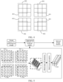
FIG. 4 is a schematic diagram of extraction of a local feature according to an embodiment of this application. -
FIG. 5 is a flowchart of acquisition of any aggregated feature according to an embodiment of this application. -
FIG. 6 is a flowchart of acquisition of a first updated feature corresponding to a to-be-processed sub-feature according to an embodiment of this application. -
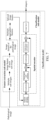
FIG. 7 is a flowchart of image classification based on a classification model according to an embodiment of this application. -
FIG. 8 is a schematic structural diagram of an image classification apparatus according to an embodiment of this application. -
FIG. 9 is a schematic structural diagram of an image classification apparatus according to an embodiment of this application. -
FIG. 10 is a schematic structural diagram of a terminal according to an embodiment of this application. -
FIG. 11 is a schematic structural diagram of a server according to an embodiment of this application. - In order to make objectives, technical solutions, and advantages of the embodiments of this application clearer, implementations of this application will be further described in detail below with reference to the drawings.
- The terms "first", "second", and the like used in this application may be used herein to describe various concepts, but unless otherwise specified, these concepts are not limited by these terms. The terms are merely used for distinguishing one concept from another concept. For example, without departing from the scope of this application, a first feature vector may be referred to as a second feature vector, and similarly, the second feature vector may be referred to as the first feature vector.
- In the terms "at least one", "multiple", "each", and "any" used in this application, "at least one" includes one, or two or more, "multiple" includes two or more, and "each" refers to each of the corresponding plurality, and "any" refers to any one of the plurality. For example, multiple scales include 3 scales, "each" refers to each of the 3 scales, and "any" refers to any one of the 3 scales, which may be the first, or the second, or the third.
- According to the solutions provided in the embodiments of this application, a classification model may be trained based on the artificial intelligent machine learning technology, and the trained classification model is used to realize the classification of a pathological image.
- The embodiments of this application provide an image classification method, which is performed by a computer device. In a possible implementation, the computer device is a terminal or a server. In a possible implementation, the server is an independent physical server, or a server cluster or distributed system composed of multiple physical servers, or a cloud server providing basic cloud computing services such as a cloud service, a cloud database, cloud computing, a cloud function, cloud storage, a network service, cloud communication, a middleware service, a domain name service, a security service, a content delivery network (CDN), and a big data and artificial intelligence platform. In a possible implementation, the terminal may include, but is not limited to, a smartphone, a tablet computer, a notebook computer, a desktop computer, a smart speaker, a smartwatch, a smart voice interaction device, a smart household appliance, an on-board terminal, and the like.
- In some embodiments, a computer program involved in the embodiments of this application may be deployed to be executed on a computer device, or executed on multiple computer devices at the same location, or executed on multiple computer devices that are distributed at multiple locations and connected to each other through a communication network, and the multiple computer devices that are distributed at multiple locations and connected to each other through a communication network can form a blockchain system.
-
FIG. 1 is a schematic diagram of an implementation environment according to an embodiment of this application. Referring toFIG. 1 , the implementation environment includes aterminal 101 and aserver 102. Theterminal 101 is directly or indirectly connected to theserver 102 in a wired or wireless communication mode, which is not defined herein. - The
terminal 101 is configured to acquire a pathological image and transmit the pathological image to theserver 102. Theserver 102 is configured to classify the pathological image transmitted by theterminal 101 to obtain a category to which the pathological image belongs. - In some embodiments, a target application for which the
server 102 provides a service is installed on theterminal 101, and theterminal 101 can realize functions, such as data transmission and message exchange, through the target application. In a possible implementation, the target application is a target application in an operating system of theterminal 101 or a target application provided by a third party. For example, the target application is an image classification application having an image classification function. Of course, the image classification application can have other functions such as a comment function and a navigation function. - The
terminal 101 is configured to acquire a pathological image, log in the target application based on a user identifier, and transmit the pathological image to theserver 102 through the target application. Theserver 102 is configured to receive the pathological image transmitted by the terminal 101, and classify the pathological image transmitted by the terminal 101 to obtain a category to which the pathological image belongs. In a possible implementation, theserver 102 transmits a classification result to the terminal 101. The classification result is used for indicating the category to which the pathological image belongs. The terminal 101 receives and displays the classification result transmitted by theserver 102. -
FIG. 2 is a flowchart of an image classification method according to an embodiment of this application. As shown inFIG. 2 , the method is performed by a computer device and includes the following steps. - 201: The computer device receives a pathological image and acquires an image feature of the received pathological image.
- The pathological image is an image used for presenting the pathological morphology of a body organ, tissue or cell. For example, the pathological image is a whole slide image (WSI, a digital pathological image). The image feature of the pathological image is used for characterizing the pathological image, which is a two-dimensional feature or a three-dimensional feature and can be represented in any form. For example, the image feature is represented in the form of a feature matrix or a feature vector. For another example, the image feature is represented in the form of a three-dimensional feature matrix or a two-dimensional feature matrix.
- 202: The computer device extracts, from the image feature and for multiple scales, a local feature for each respective one of the multiple scales.
- The scale characterizes a size of the local feature extracted from the image feature. The multiple scales correspond to different sizes. In an embodiment, the multiple scales are two-dimensional scales, which may be, for example, 3×3, 5×5, and 7×7 pixels. Each local feature is a partial feature in the image feature, any local feature corresponding to any scale represents that a feature size of the local feature is matched with the scale, that is, the local feature is extracted from the image feature based on the scale. For any scale in the multiple scales, at least one local feature corresponding to the scale is extracted from the image feature.
- 203: The computer device splices local features corresponding to the multiple scales to obtain a spliced image feature.
- The local features corresponding to difference scales contain different information, and the local features of different scales are spliced, so that it is ensured that the spliced image feature contains feature information corresponding to the multiple scales.
- In a possible implementation, in a case that multiple local features are extracted at each of the multiple scales, the method of splicing the local features corresponding to the multiple scales to obtain a spliced image feature may be implemented by aggregating the multiple local features corresponding to each of the multiple scales to obtain an aggregated feature, and splicing multiple aggregated features to obtain a spliced image feature.
- 204: The computer device classifies the spliced image feature to obtain an image category of the pathological image.
- According to the method, the apparatus, the computer device, and the storage medium that are provided in the embodiments of this application, after being acquired, an image feature of a pathological image is processed according to multiple scales respectively to obtain local features corresponding to the multiple scales, and the local features corresponding to the multiple scales are spliced to obtain a spliced image feature, and the spliced image feature is classified to obtain a category to which the pathological image belongs. The local features corresponding to different scales contain different information, so that the finally obtained spliced image feature contains feature information corresponding to different scales, and the feature information of the spliced image feature is enriched. The category to which the pathological image belongs is determined based on the spliced image feature, so that the accuracy of the category is ensured.
- Based on the embodiment shown in
FIG. 2 , the image feature of the pathological image can also be acquired by a method of first segmenting and then splicing; and multiple aggregated features are acquired by the self-attention mechanism, an acquired spliced image feature is updated, and the category to which the pathological image belongs is acquired based on the updated spliced image feature, which will be specifically described in the following embodiment. -
FIG. 3 is a flowchart of another image classification method according to an embodiment of this application. As shown inFIG. 3 , the method is performed by a computer device and includes the following steps. - 301: The computer device segments a to-be-classified pathological image to obtain multiple sub-images.
- Each sub-image is a partial image in the pathological image, and the multiple sub-images can form the pathological image. In a possible implementation, the multiple sub-images have the same image size. For example, an image size of the pathological image is 40000×40000 pixels, a size of each sub-image is 4000×4000 pixels, so the pathological image is segmented into 100 sub-images.
- In a possible implementation,
step 301 includes the following sub-step: the pathological image is segmented based on a preset size to obtain multiple sub-images. A size of each sub-image is the same as the preset size. For example, the preset size is 1120× 1120 pixels, and the size of each sub-image obtained by segmentation is 1120× 1120 pixels. - In a possible implementation, the process of segmenting the pathological image includes the following steps: a first sliding window is moved on the pathological image based on a first moving step length, and a partial pathological image within the first sliding window after each movement is determined as one sub-image.
- A size of the first sliding window is the same as the preset size, and the first moving step length represents a movement distance of the first sliding window during each movement. For example, the first moving step length represents a quantity of pixels moved each time during movement of the first sliding window. In a possible implementation, the preset size includes a first size and a second size, the first size and the second size respectively represent a length and a width of the first sliding window, and the first moving step length is the same as the first size and the second size. For example, the present size is 1120× 1120, the first moving step length is 1120, multiple sub-images in the pathological image are obtained based on the first moving step length and the preset size, and a size of each sub-image is 1120× 1120.
- In a possible implementation, the pathological image is represented in the form of a three-dimensional feature matrix, the sub-images obtained by segmentation are also represented in the form of three-dimensional feature matrices, the multiple sub-images can be spliced into the pathological image, and the pathological image and the multiple sub-images satisfy the following relationship:


where, B represents the pathological image, B ∈ R H×W×3 indicates that the image size of the pathological image is H × W × 3, H and W represent a length and a width of the pathological image, and H and W are any positive integer; p 0,0 , pr,c, and pH',W' all represent one sub-image in the multiple sub-images, p 0,0 , pr,c, pH',W' ∈ RTh ×Tw ×3 indicates that the image size of each sub-image is Th × Tw × 3, Th and Tw represent a length and a width of each sub-image, Th and Tw are both any positive integer, Th is less than H, and Tw is less than W; r, c represents the serial number of the multiple sub-image, r is an integer greater than 0 but not greater than H', and c is a positive integer greater than 0 but not greater than W'; and H' is a quantity of sub-images that are segmented from the pathological image with the image size of H according to the image size of Th, W' is a quantity of sub-images that are segmented from the pathological image with the image size of W according to the image size of Tw, and H' × W' is a total quantity of sub-images that are segmented from the pathological image. - 302: The computer device performs feature extraction on each sub-image to obtain an image feature of each sub-image.
- The image feature of each sub-image is used for characterizing the corresponding sub-image, which is a two-dimensional feature or a three-dimensional feature and can be represented in any form. For example, the image feature of the sub-image is presented in the form of a feature vector or a feature matrix. For another example, the image feature of the sub-image is presented in the form of a two-dimensional feature matrix or a three-dimensional feature matrix.
- In a possible implementation, each sub-image and the corresponding image feature satisfy the following relationship:

where, er,c any sub-image in the multiple sub-images, r, c represents the serial number of the multiple sub-images, and pr,c represents a (r, c)th sub-image in the multiple sub-images; fE (·) represents a feature extraction function, and feature extraction can be performed on each sub-image based on the feature extraction function fE (·) to obtain the image feature of each sub-image; and er,c ∈ RD represents that the image feature er,c is a feature vector with a dimension of D. - 303: The computer device splices the image features of the multiple sub-images based on positions of the multiple sub-images in the pathological image to obtain an image feature of the pathological image.
- The image feature of the pathological image is used for characterizing the pathological image, which can be represented in any form. For example, all the image features of the multiple sub-images are two-dimensional feature matrices, and the image feature of the pathological image is also a two-dimensional feature matrix; and all the image features of the multiple sub-images are three-dimensional feature matrices, and the image feature of the pathological image is also a three-dimensional feature matrix.
- The positions of different sub-images in the pathological image are different, and the image features of the multiple sub-images are spliced based on the position of each sub-image in the pathological image to obtain the image feature of the pathological image, so that the image feature of the pathological image corresponds to the pathological image. The image feature of the pathological image retains position information of the sub-images, so that the accuracy of the image feature is ensured. Moreover, first the pathological image is segmented into multiple sub-images with a small size, an image feature of each sub-image is acquired, and the image features of the multiple sub-images are spliced into an image feature of the pathological image. In this way, it is unnecessary to directly perform feature extraction on the pathological image with a large size, so that the requirements for the performance of the device are reduced. The requirements for the device configured to classify an image are reduced, so that the universality of the feature extraction method is ensured.
- In a possible implementation, in a case that all the image features of the multiple sub-images are two-dimensional feature matrices, a feature size of the image feature of the pathological image is the sum of feature sizes of the image features of the multiple sub-images. For example, the pathological image includes 4 sub-images, the feature size of the image feature of each sub-image is 2×2, and the feature size of the image feature of the pathological image is 8×8.
- In a possible implementation, in a case that all the image features of the multiple sub-images are three-dimensional feature matrices, the feature size of the image feature of the sub-image includes a first size, a second size, and a third size, the feature size of the image feature of the pathological image includes a fourth size, a fifth size, and a sixth size, the fourth size is the sum of the first sizes corresponding to the multiple sub-images, the fifth size is the sum of the second sizes corresponding to the multiple sub-images, and the sixth size is the same as the third size. For example, the pathological image includes 4 sub-images, the feature size of the image feature of each sub-image is 2×2×10, and the feature size of the image feature of the pathological image is 8×8×10.
- In a possible implementation, the image features of the multiple sub-images and the image feature of the pathological image satisfy the following relationship:

where, E represents the image feature of the pathological image, and e 0,0, er,c, and eH',W' all represent an image feature of one sub-image in the multiple sub-images; r, c represents the serial number of the multiple sub-images, r is an integer greater than 0 but not greater than H', and c is a positive integer greater than 0 but not greater than W'; H' is a quantity of sub-images that are segmented from the pathological image in a dimension of H dimension according to a feature size Th, W' is a quantity of sub-images that are segmented from the pathological image in a dimension of W according to a feature size Tw, and H' × W' represents a total quantity of sub-images that are segmented from the pathological image; and D represents a vector dimension of the image feature of each sub-image in a case that the image feature of each sub-image is a vector. - In the embodiments of this application, the method of first segmenting and then splicing is adopted, that is, a pathological image is segmented into multiple sub-images, and an image feature of the pathological image is acquired based on the multiple sub-images. However, in another embodiment, it is unnecessary to perform
steps 301 to 303, and another method can be adopted to acquire the image feature of the to-be-classified pathological image. - 304: The computer device extracts, for each scale in multiple scales, multiple local features corresponding to the scale from the image feature of the pathological image, and splices second sub-features in each extracted local feature to obtain a first feature vector corresponding to each local feature.
- In the embodiments of this application, the image feature of the pathological image includes second sub-features at multiple positions, which are one-dimensional features or two-dimensional features. For example, the second sub-feature includes one feature value, or, the second sub-feature is a feature vector composed of multiple feature values. In a case that the image feature of the pathological image is a two-dimensional feature, the second sub-feature includes one feature value; and in a case that the image feature of the pathological image is a three-dimensional feature, the second sub-feature includes multiple feature values, that is, the second sub-feature is a feature vector composed of the multiple feature values. For example, the feature size of the image feature of the pathological image is 3×3×10, the image feature includes second sub-features at 9 positions, and each second sub-feature is a feature vector composed of 10 feature values.
- For any scale in the multiple scales, at least one local feature is extracted from the image feature of the pathological image based on the scale, and each local feature includes a second sub-feature at at least one position. In a possible implementation, any scale corresponds to multiple local features, each local feature includes second sub-features at multiple positions, and different local features may include the same second sub-feature. The image feature of the pathological image includes second sub-features at 3×3 positions, and as shown in
FIG. 4 , each block corresponds to one position. A scale is 2×2, 4local features 402 are extracted from animage feature 401 of a pathological image based on the scale, and eachlocal feature 402 includes second sub-features at 4 positions. That is, in the image feature of the pathological image, second sub-features at 4 positions in the upper left corner constitute onelocal feature 402, second sub-features at four positions in the upper right corner constitute onelocal feature 402, second sub-features at four positions in the lower left corner constitute onelocal feature 402, second sub-features at four positions in the lower right corner constitute onelocal feature 402, and thelocal feature 402 in the upper left corner and thelocal feature 402 in the upper right corner include two identical second sub-features. - For any local feature, the second sub-features in the local feature are spliced into one first feature vector, and a feature size of the first feature vector is the sum of feature sizes of at least one second sub-feature in the local feature. For example, any local feature includes 4 second sub-features, a feature size of each second sub-feature is 1×3, the 4 second sub-features are spliced into one first feature vector, and a size of the first feature vector corresponding to the local feature is 1×12.
- In a possible implementation, the process of extracting, for any scale, a local feature corresponding to the scale includes the following steps: a second sliding window corresponding to the scale is moved on the image feature of the pathological image based on a second moving step length, and a second sub-feature at at least one position within the second sliding window is used to constitute one local feature.
- A size of the second sliding window corresponding to the scale is the same as the scale. In a possible implementation, the scale is a side length of the second sliding window. For example, the scale is a one-dimensional scale, and a length and a width of the second sliding window are the same as the scale. If the scale is 3, the size of the second sliding window is 3×3. For another example, the scale is a two-dimensional scale including a length and a width, and a length and a width of the second sliding window corresponding to the scale are respectively the same as the length and the width of the two-dimensional scale. The second moving step length represents a distance of the second sliding window during each movement. For example, the second moving step length represents a quantity of positions moved each time during movement of the second sliding window. For example, the second moving step length is 1, that is, the second sliding window is moved by one position during each movement. As shown in
FIG. 4 , the size of the second sliding window is 2×2, the second moving step length is 1, and the second sub-features at the 4 positions that are included in eachlocal feature 402 are extracted. - In a possible implementation, the processing of extracting a local feature corresponding to any scale includes the following steps: an expansion parameter corresponding to a scale is determined, the image feature of the pathological image is expanded based on the expansion parameter to obtain an expanded image feature, a second sliding window corresponding to the scale is moved on the image feature of the pathological image based on the second moving step length, and a second sub-feature at at least one position within the second sliding window after each movement is used to constitute one local feature.
- The expansion parameter is used for indicating a quantity of positions in the expanded image feature of the pathological image. For example, the expansion parameter is Padding. For the expansion parameter corresponding to any scale, the image feature of the pathological image is expanded to the quantity of positions indicated by the expansion parameter, so that a feature size of an expanded image feature becomes larger. For example, after the image feature of pathological image is expanded, a length and a width in the feature size of the expanded image feature are both expanded by 2 times based on the quantity of positions indicated by the expansion parameter. For example, the image feature of the pathological image is a 3×3 feature matrix, the expansion parameter corresponding to any scale is 1, and the expanded image feature is a 5×5 feature matrix; and the expansion parameter corresponding to any scale is 2, and the expanded image feature is a 7×7 feature matrix.
- In a possible implementation, any scale is a one-dimensional scale, and the process of determining an expansion parameter corresponding to the scale includes the following steps: a difference between the scale and a first numerical value is determined, and a ratio of the difference to a second numerical value is determined as an expansion parameter corresponding to the scale.
- Both the first numerical value and the second numerical value are any numerical value. For example, the first numerical value is 1, and the second numerical value is 2.
- In a possible implementation, any scale and the expansion parameter corresponding to the scale satisfy the following relationship:

where, k represents the scale, β represents the expansion parameter corresponding to the scale k, and in the embodiments of this application, the first numerical value is 1, and the second numerical value is 2. - For example, the feature size of the image feature of the pathological image is 4×4, a first scale is 3, that is, a size of a second sliding window corresponding to the first scale is 3×3, a first expansion parameter corresponding to the first scale is 1, that is, a feature size of an image feature expanded based on the first expansion parameter is 6×6, and 16 local features with a feature size of 3×3 can be extracted based on the second sliding window corresponding to the first scale. A second scale is 5, that is, a size of a second sliding window corresponding to the second scale is 5×5, a second expansion parameter corresponding to the second scale is 2, that is, a feature size of an image feature expanded based on the second expansion parameter is 8×8, and 16 local features with a feature size of 5×5 can be extracted based on the second sliding window corresponding to the second scale. That is, 16 local features are extracted based on the first scale or the second scale.
- When the local feature is extracted from the image feature of the pathological image based on each scale, the image feature of the pathological image is first expanded based on the expansion parameter corresponding to each scale to ensure that quantities of local features extracted based on the scales are consistent, which facilitates the subsequent feature fusion based on the multiple scales.
- In a possible implementation, for any scale in the multiple scales, in a case that a first feature vector that corresponds to each local feature corresponding to the scale is obtained, the multiple first feature vectors are used to constitute a first feature matrix corresponding to the scale.
- In a possible implementation, for any scale in the multiple scales, the first feature matrix corresponding to the scale and the image feature of the pathological image satisfy the following relationship:


where, T represents the first feature matrix corresponding to the scale; E represents the image feature of the pathological image, and Softsplit(·) represents a feature reconstruction function used for constituting the first feature matrix according to the image feature E of the pathological image; Concat(·) represents a splicing function used for constituting the first feature matrix corresponding to the scale according to the multiple first feature vectors; Unfold(·) represents a spread function used for splicing the second sub-features in the local feature corresponding to the scale into one first feature vector; T ∈ R H"W"×k2 D represents that a size of the first feature matrix corresponding to the scale is H"W" × k 2 D, k represents the scale, and k is a positive integer; a feature size of each local feature corresponding to the scale k is k × k; a feature size of the image feature of the pathological image is W' × H' × D, W' and H' represents a length and a width of the image feature of the pathological image, D represents a dimension of each second sub-feature in the image feature of the pathological image, and W', H', and D are all positive integers; H" represents a quantity for dividing the dimension H' of the image feature of the pathological image when the local features are extracted from the image feature of the pathological image according to the scale k; W" represents a quantity for dividing the dimension W' of the image feature of the pathological image when the local features are extracted from the image feature of the pathological image according to the scale k; D represents a vector dimension of each second sub-feature; k 2 D represents a vector dimension of each first feature vector; H"W" represents a quantity of multiple local features corresponding to the scale k, and also represents a quantity of multiple first feature vectors corresponding to the scale k; β represents an expansion parameter corresponding to the scale k; and α represents the second moving step length. - 305: The computer device updates each first feature vector based on the first feature vectors corresponding to the multiple local features to obtain a second feature vector corresponding to each first feature vector.
- In the embodiments of this application, for any scale in the multiple scales, the scale corresponds to multiple local features. In view of the correlation between different local feature, after the first feature vectors corresponding to the multiple local features are acquired, each first feature vector is updated through the first feature vectors corresponding to the multiple local features, so that a second feature vector obtained by updating is fused with the first feature vectors corresponding to other local features, and the accuracy of the second feature vector is ensured.
- In a possible implementation,
step 305 includes the following sub-step: for a to-be-processed feature vector in the multiple first feature vectors, the similarity between each first feature vector and the to-be-processed feature vector is obtained, and the multiple first feature vectors are weighted and fused based on the similarity corresponding to each first feature vector to obtain a second feature vector corresponding to the to-be-processed feature vector. - The to-be-processed feature vector is any first feature vector in the multiple first feature vectors. The similarity between any first feature vector and the to-be-processed feature vector represents a level of similarity between the first feature vector and the to-be-processed feature vector, which can be acquired by a method of acquiring a cosine similarity or other similarities. The similarity corresponding to each first feature vector can represent a level of similarity between the first feature vector and the to-be-processed feature vector, and the multiple first feature vectors are weighted and fused by the self-attention mechanism based on the similarities between the multiple first feature vectors and the to-be-processed feature vector to obtain a second feature vector corresponding to the to-be-processed feature vector, so that the second feature vector is fused with the multiple first feature vectors according to the similarities corresponding to the first feature vectors, and the accuracy of the obtained second feature vector is ensured. According to the foregoing method of acquiring the second feature vector corresponding to the to-be-processed feature vector, a second feature vector corresponding to each first feature vector may be acquired.
- In a possible implementation, the process of acquiring the second feature vector corresponding to the to-be-processed feature vector includes the following steps: for the to-be-processed feature vector of the multiple first feature vectors, the similarity between each first feature vector and the to-be-processed feature vector, the multiple first feature vectors are weighted and fused based on the similarity corresponding to each first feature vector to obtain a third feature vector, and feature transformation is performed on the third feature vector to obtain the second feature vector corresponding to the to-be-processed feature vector.
- In the embodiments of this application, feature dimensions of each first feature vector and the corresponding second feature vector are different. For each scale in the multiple scales, feature dimensions of second feature vectors corresponding to different scales are the same. In the process of updating the multiple first feature vectors corresponding to each scale, the feature vectors are transformed into feature vectors with the same feature dimension, which ensures that feature sizes of aggregated features subsequently obtained based on each scale are the same.
- In a possible implementation, the process of performing feature transformation on the third feature vector includes the following steps: a product of the third feature vector and a feature transformation matrix is determined as the second feature vector corresponding to the target feature vector.
- The feature transformation matrix is used for transforming the third feature vector and is any constant matrix.
- In a possible implementation, for any scale in the multiple scales, the first feature vectors that correspond to each local feature corresponding to the scale constitute the first feature matrix corresponding to the scale, and step 305 includes the following sub-steps: each first feature vector in the first feature matrix is updated based on the multiple first feature vectors in the first feature matrix to obtain a second feature vector corresponding to each first feature vector, and the multiple second feature vectors are used to constitute a second feature matrix.
- In a possible implementation, for any scale in the multiple scales, the first feature matrix and the second feature matrix corresponding to the scale satisfy the following relationship:


where, T' represents the second feature matrix corresponding to the scale; MLP(·) represents a multi-layer perception function used for performing fully connected transformation on a feature; MSA(·) represents a multi-head self-attention function; T represents the first feature matrix corresponding to the scale; a feature size of the image feature of the pathological image is W' × H' × D, W' and H' represents a length and a width of the image feature of the pathological image, D represents a dimension of each second sub-feature in the image feature of the pathological image, and W', H', and D are all positive integers; H" represents a quantity for dividing the dimension H' of the image feature of the pathological image when the local features are extracted from the image feature of the pathological image according to the scale; W" represents a quantity for dividing the dimension W' of the image feature of the pathological image when the local features are extracted from the image feature of the pathological image according to the scale; H"W" represents a quantity of multiple local features corresponding to a scale k, that is, a quantity of multiple second feature vectors; dz represents a vector dimension of each second feature vector; Concat(·) represents a connection function; Ω represents a quantity of heads in the multi-head self-attention function, and Ω is a positive integer greater than 1; Hw represents a feature outputted by any head in the multi-head self-attention function, w represents the serial number of each head self-attention in the multi-head self-attention function, and w is a positive integer greater than 0 but not greater than Ω; WO represents a mapping matrix that is a constant matrix;

- 306: The computer device uses the multiple second feature vectors to constitute a three-dimensional feature matrix based on positions of the multiple local features in the image feature of the pathological image, and determines the three-dimensional feature matrix as an aggregated feature.
- Positions of different local features in the image feature of the pathological image are different. The second feature vector corresponding to any local feature is a feature of a pixel of a position, corresponding to the local feature, in the pathological image. In a possible implementation, the second feature vector corresponding to any local feature is a feature of one or more pixels, corresponding to the local feature, in the pathological image. For example, the image feature of the pathological image is used for characterizing the pathological image, any local feature in the pathological image is a feature of a pixel in a partial region of the pathological image, there is one or more pixels in the partial region of the pathological image, and the second feature vector corresponding to the local feature is a feature of one or more pixels in the partial region of the pathological image.
- According to the positions of the multiple local features in the image feature of the pathological image, the second feature vectors corresponding to the multiple local features are used to constitute one three-dimensional feature matrix, that is, one aggregated feature. For example, 4 local features correspond to any scale, a dimension of a second feature vector corresponding to each local feature is 10, and a feature size of an aggregated feature constituted based on the second feature vectors corresponding to the multiple local features is 2×2×10. According to the foregoing methods in
steps 304 to 306, the image feature of the pathological image is processed based on the multiple scales to obtain multiple aggregated features. - In the process of processing the image feature of the pathological image based on each scale, the second sub-features in each local feature are spliced into one feature vector. In view of the correlation between different local features, each first feature vector is updated through the first feature vectors corresponding to the multiple local features, so that the second feature vector obtained by updating is fused with the first feature vectors corresponding to other local features, and the accuracy of the second feature vector is ensured. Moreover, the three-dimensional feature matrix that is constituted by the feature vectors corresponding to the multiple local features according to the positions of the multiple local features in the image feature of the pathological image is determined as the aggregated feature, so that the aggregated feature retains position information of the multiple local features in the image feature of the pathological image, and the accuracy of the aggregated feature is ensured.
- In a possible implementation, quantities of multiple local features corresponding to different scales are the same, feature dimensions of second feature vectors corresponding to different scales are the same, and feature sizes of aggregated features acquired based on different scales are the same.
- In a possible implementation, for any scale in the multiple scales, the multiple second feature vectors corresponding to the scale are used to constitute the second feature matrix, and step 306 includes the following sub-steps: the multiple second feature vectors in the second feature matrix are used to constitute a three-dimensional feature matrix based on the positions of the multiple local features in the image feature of the pathological image, and the three-dimensional feature matrix is determined as the aggregated feature.
- In a possible implementation, the second feature matrix and the aggregated feature satisfy the following relationship:

where, E' represents the aggregated feature obtained based on the scale; Reshape(·) represents a remodeling function used for remodeling the second feature matrix to recover spatial position information so as to obtain a new feature map; E' =∈ RH"×W"×dz represents a feature size of the aggregated feature E'; a feature size of the image feature of the pathological image is W' × H' × D, W' and H' represents a length and a width of the image feature of the pathological image, D represents a dimension of each second sub-feature in the image feature of the pathological image, and W', H', and D are all positive integers; H" represents a quantity for dividing the dimension H' of the image feature of the pathological image when the local features are extracted from the image feature of the pathological image according to the scale; W" represents a quantity for dividing the dimension W' of the image feature of the pathological image when the local features are extracted from the image feature of the pathological image according to the scale; and dz represents a vector dimension of each second feature vector. - In the process of acquiring the aggregated feature based on any scale, as shown in
FIG. 5 , a case where the image feature of the pathological image is a 4×4 matrix, any scale is 3, 4local features 501 are extracted from the image feature of the pathological image, eachlocal feature 501 is fused into onefirst feature vector 502, each first feature vector is updated by the multi-head attention mechanism and a multi-layer perceptron according tosteps feature 503. - In the embodiments of this application, the description is made by taking a case where each scale corresponds to multiple local features as an example. However, in another embodiment, any scale corresponds to one local feature, sub-features in the local feature corresponding to the scale are spliced to obtain a first feature vector corresponding to the local feature, and the first feature vector is determined as an aggregated feature.
- In the embodiments of this application, the description is made by taking a case where the local feature includes second sub-feature at multiple positions as an example, and multiple aggregated features are acquired based on each scale in the multiple scales and the image feature of the pathological image. However, in another embodiment, it is unnecessary to perform
steps 304 to 306, and another method can be adopted to extract multiple local features corresponding to each scale from the image feature for each scale in the multiple scales, and aggregate the multiple local features corresponding to the scale to obtain an aggregated feature. - 307: The computer device splices the multiple obtained aggregated features to obtain a spliced image feature.
- In the embodiments of this application, the image feature of the pathological image is processed based on the multiple scales to obtain the multiple aggregated features. Different aggregated features contain feature information corresponding to different scales, and the multiple aggregated features are spliced, so that the spliced image feature contains the feature information corresponding to the multiple scales.
- In a possible implementation,
step 307 includes the following sub-step: features at the same position in the multiple aggregated features are spliced to obtain feature vector corresponding to multiple positions; and the feature vectors corresponding to the multiple positions are used to constitute a three-dimensional feature matrix, and the three-dimensional feature matrix is determined as the spliced image feature. - In the embodiments of this application, feature sizes of the multiple aggregated features are the same, each aggregated feature includes features at multiple positions, and the feature at each position is one second feature vector. For different aggregated features, the features at the same position are a feature of a pixel at the same position in the pathological image. Therefore, the features at the same position in the multiple aggregated features are spliced to obtain the feature vectors corresponding to the multiple positions. Based on the multiple positions, the feature vectors corresponding to the multiple positions are used to constitute one three-dimensional feature matrix, and the three-dimensional feature matrix is determined as the spliced image feature.
- For example, there are 3 scales, a feature size of each aggregated feature is 4×4×10, and a feature size of a spliced image feature obtained by splicing the 3 aggregated features is 4×4×30.
- In a possible implementation, the spliced image feature obtained based on the multiple aggregated features satisfies the following relationship:


where, E" represents the spliced image feature; Concat(·) represents a splicing function;

- 308: The computer device updates each first sub-feature based on multiple first sub-features in the spliced image feature to obtain a first updated feature corresponding to each first sub-feature.
- The spliced image feature includes first sub-features at multiple positions, that is, the spliced image feature includes multiple first sub-features, and each first sub-feature contains feature information corresponding to different scales. In the embodiments of this application, there is a correlation in the first sub-features at different positions. Each first sub-feature is updated by the self-attention mechanism through the multiple first sub-features, so that the first updated feature obtained by updating is fused with other first sub-features, and the accuracy of the first updated feature is ensured.
- In a possible implementation,
step 308 includes the following sub-steps: for a to-be-processed sub-feature in the multiple first sub-features, weights of the multiple first sub-features are acquired; and the multiple first sub-features are weighted and fused based on the weights of the multiple first sub-features to obtain the first updated feature corresponding to the to-be-processed sub-feature. - The weight of any first sub-feature is used for indicating a degree of correlation between the first sub-feature and the to-be-processed sub-feature. The larger the weight is, the higher the degree of correlation between the first sub-feature and the to-be-processed sub-feature is; and the smaller the weight is, the lower the degree of correlation between the first sub-feature and the to-be-processed sub-feature is. The weight of each first sub-feature represents a degree of correlation between the first sub-feature and the to-be-processed sub-feature, and the multiple first sub-features are weighted and fused into the first updated feature according to degrees of correlations between the multiple first sub-features and the to-be-processed sub-feature, so that the accuracy of the obtained first updated feature is ensured. According to the foregoing method of acquiring the first updated feature corresponding to the to-be-processed sub-feature, the first updated feature corresponding to each first sub-feature can be acquired.
- In a possible implementation, the process of acquiring the weight of each first sub-feature includes the following steps: for the to-be-processed sub-feature in the multiple first sub-features, a distance feature of each first sub-feature and the to-be-processed sub-feature is acquired; and the weight of each first sub-feature is acquired based on the similarity between each first sub-feature and the to-be-processed sub-feature and the distance feature.
- The distance feature of any first sub-feature and the to-be-processed sub-feature is used for indicating a distance between positions of the first sub-feature and the to-be-processed sub-feature in the spliced image feature; and the similarity between any first sub-feature and the to-be-processed sub-feature represents a level of similarity between the first sub-feature and the to-be-processed sub-feature.
- In the embodiments of this application, there may be a correlation in features at different positions in the pathological image. For any first sub-feature, the similarity between the first sub-feature and the to-be-processed sub-feature can represent a level of similarity between the first sub-feature and the to-be-processed sub-feature, the distance feature of the first sub-feature and the to-be-processed sub-feature can indicate the distribution of the first sub-feature and the to-be-processed sub-feature in the spliced image feature, and the weight of the first sub-feature is acquired based on the similarity between the first sub-feature and the to-be-processed sub-feature and the distance feature, which not only takes account of a level of similarity between the first sub-feature and the to-be-processed sub-feature, but also takes account of a relative position relationship between the first sub-feature and the to-be-processed feature, that is, takes full account of the correlation between the first sub-feature and the to-be-processed sub-feature. Therefore, the accuracy of the acquired weight is ensured.
- In a possible implementation, the process of acquiring the distance feature includes the following steps: a distance corresponding to each first sub-feature is determined based on positions of each first sub-feature and the to-be-processed sub-feature in the spliced image feature; and the distance corresponding to each first sub-feature is mapped to obtain the distance feature of each first sub-feature and the to-be-processed sub-feature.
- The distance corresponding to any first sub-feature represents a distance between the positions of the first sub-feature and the to-be-processed sub-feature. The position of each first sub-feature in the spliced image feature can be represented in any form. For example, the position of each first sub-feature in the spliced image feature is represented in the form of two-dimensional coordinates. For any first sub-feature, a distance, that is, a target distance corresponding to the first sub-feature, between the positions of the first sub-feature and the to-be-processed sub-feature may be determined based on a position corresponding to the first sub-feature and a position corresponding to the to-be-processed sub-feature. The distance corresponding to the first sub-feature is mapped to the distance feature, so that the weight of the first sub-feature can be acquired based on the distance feature subsequently.
- In a possible implementation, the process of acquiring the weight of each first sub-feature based on the similarity between each first sub-feature and the to-be-processed sub-feature and the distance feature includes the following steps: for each first sub-feature in the multiple first sub-features, the similarity between the first sub-feature and the to-be-processed sub-feature and the distance feature are fused to obtain a first matching degree corresponding to the first sub-feature; the sum of the first matching degrees corresponding to the multiple first sub-features is determined; and a ratio of the first matching degree corresponding to each first sub-feature to the sum is determined as the weight of each first sub-feature.
- The first matching degree corresponding to any first sub-feature indicates a degree of matching of the first sub-feature with the to-be-processed sub-feature. When the weight of any sub-feature is acquired, the first matching degree of each first sub-feature and the to-be-processed sub-feature is acquired first. In view of the matching degrees of the multiple first sub-feature and the to-be-processed sub-feature, the ratio of the first matching degree corresponding to each first sub-feature to the sum is determined as the weight of each first sub-feature, which ensures that the first sub-feature that is matched more with the to-be-processed sub-feature has a larger weight. Therefore, the accuracy of the weight is ensured.
- In a possible implementation, the process of acquiring the first matching degree of any first sub-feature and the to-be-processed sub-feature includes the following steps: the similarity between the first sub-feature and the to-be-processed sub-feature and the distance feature are fused to obtain a second matching degree; and feature transformation is performed on the second matching degree to obtain the first matching degree corresponding to the first sub-feature.
- First the second matching degree is acquired, and feature transformation is performed on the second matching degree, so that the first matching degrees corresponding to the first sub-features belong to the same interval, which facilitates the subsequent comparison of the first matching degrees corresponding to the multiple first sub-features.
- In a possible implementation, for the to-be-processed sub-feature in the multiple first sub-features, the weight of any first sub-feature satisfies the following relationship:





where, in the multiple first sub-features, a to-be-processed sub-feature


- In a possible implementation, the first updated feature corresponding to the to-be-processed sub-feature satisfies the following relationship:


where, zi represents the first updated feature corresponding to the to-be-processed sub-feature; E" represents the spliced image feature; WQ, WK, and WV are all constant matrices; N represents a total quantity of multiple first sub-features;


z represents that the first updated feature zi corresponding to the to-be-processed sub-feature is a feature vector with a dimension of dz , and dz is any positive integer; and WQ, WK, WV ∈ RC×dz represents that sizes of the constant matrices WQ, WK, and WV are C × dz, and C and dz are both any positive integer. The process of acquiring the first updated feature corresponding to the to-be-processed sub-feature based on the multiple first sub-features in the spliced image feature is shown inFIG. 6 . - 309: The computer device uses the first updated features corresponding to the multiple first sub-features to constitute a three-dimensional feature matrix based on positions of the multiple first sub-features in the spliced image feature, and determines the three-dimensional feature matrix as an updated spliced image feature.
- The first updated feature is a vector, that is, the first updated feature corresponding to each first sub-feature is a vector. The multiple first updated features are used to constitute one three-dimensional feature matrix according to the positions of the multiple first sub-features in the spliced image feature, and the three-dimensional feature matrix is the updated spliced image feature, so that the updated spliced image feature retains position information of different features, and the accuracy of the updated spliced image feature is ensured.
- For example, a feature size of the spliced image feature before updating is 3×3×10, and each first sub-feature is a feature vector with a dimension of 10, that is, the spliced image feature before updating includes 9 positions; and the first updated feature corresponding to each first sub-feature is a feature vector with a dimension of 8, that is, a feature size of the updated spliced image feature composed of the multiple first updated features is 3×3×8.
- In a possible implementation, the updated spliced image feature satisfies the following relationship:

where, Z represents the updated spliced image feature, z 1, z 2, and zN respectively represent the first first updated feature, the second first updated feature, and the Nth first updated feature in the updated spliced image feature, N represents a total quantity of multiple first updated features, and N is a positive integer greater than 1. - In a possible implementation, according to
steps 

where, SETL(E") represents the updated spliced image feature; E" represents the spliced image feature; LN(·) represents a linear transformation function; MSASET (·) represents a multi-head self-attention function used for acquiring the first updated feature corresponding to each first sub-feature according to step 308; MLP(·) represents a multi-layer perception function used for performing fully connected transformation on a feature;

- In the embodiments of this application, the description is made by taking a case where the first updated feature is a vector as an example. However, in another embodiment, the first updated feature is not a vector, it is unnecessary to perform
step 309, and another method can be adopted to use the first updated features corresponding to the multiple first sub-features to constitute the updated spliced image feature based on the positions of the multiple first sub-features in the spliced image feature. For example, the first updated feature includes one feature element, the first updated features corresponding to the multiple first sub-features are used to constitute one two-dimensional feature matrix based on the positions of the multiple first sub-features in the spliced image feature, and the two-dimensional feature matrix is determined as the updated spliced image feature. - 310: The computer device classifies the updated spliced image feature to obtain a category to which the pathological image belongs.
- Each first updated feature in the updated spliced image feature is fused with features in the local features of different scales, and is also fused with the first sub-features at the positions in the whole spliced image feature according to the degrees of correlations between the first sub-features at the positions in the whole spliced image feature, so that the updated spliced image feature is enriched, and the accuracy of the updated spliced image feature is ensured. The updated spliced feature is classified, so that the accuracy of the determined category is ensured.
- In a possible implementation, the category to which the pathological image belongs indicates a state of a specific region in the pathological image.
- In a possible implementation,
step 310 includes the following sub-step: the updated spliced image feature is classified to obtain a classified feature vector, and a category corresponding to a maximum probability in the classified feature vector is determined as the category to which the pathological image belongs. - In the embodiments of this application, the classified feature vector includes probabilities of multiple dimensions, and the probability of each dimension is used for indicating the probability that the pathological image belongs to a category corresponding to the dimension. The updated spliced image feature is classified to obtain the classified feature vector indicating probabilities that the pathological image belongs to multiple categories, and the category corresponding to the maximum probability in the classified feature vector is determined as the category to which the pathological image belongs, so that the accuracy of the determined category is ensured.
- In a possible implementation, the updated spliced image feature and the category to which the pathological image belongs satisfy the following relationship:

where, Y represents the category to which the pathological image belongs; SETL(E") represents the updated spliced image feature; E" represents the spliced image feature; and Φ(·) represents a classification function. - In the embodiments of this application, after being acquired, the spliced image feature is updated according to
steps step 309, the updated spliced image can be updated multiple times according tosteps - In a possible implementation, after the spliced image feature is obtained, the process of updating the spliced image feature multiple times, and classifying the updated spliced image feature obtained during the last updating to obtain the category to which the pathological image belongs satisfies the following relationship:

where, Y represents the category to which the pathological image belongs; SETL(E") represents an obtained updated spliced image feature after the spliced image feature is updated once; E" represents the spliced image feature; M represents the number of updating performed on the spliced image feature, and M is any positive integer, for example, M is 6; and Φ(·) represents a classification function. - The method according to the embodiments of this application can be applied to classification tasks for multiple types of pathological images, such as cancer classification, lymph node metastasis, and gene mutation prediction. For example, for a pathological image corresponding to any cancer type, a state that corresponds to a cancerous region corresponding to the pathological image can be determined by the method according to the embodiments of this application.
- According to the method provided in the embodiments of this application, after being acquired, an image feature of a pathological image is processed according to multiple scales to obtain aggregated features corresponding to the multiple scales, and the aggregated features corresponding to the multiple scales are spliced into one spliced image feature, and the spliced image feature is classified to obtain a category to which the pathological image belongs. The aggregated feature corresponding to different scales are obtained by aggregating local features of different scales, and the local features corresponding to different scales contain different information, so that the multiple aggregated features contain feature information corresponding to different scales. The multiple aggregated features are spliced into the spliced image feature, so that the spliced image feature contains the feature information corresponding to different scales, and the feature information of the spliced image feature is enriched. The category to which the pathological image belongs is determined based on the spliced image feature, so that the accuracy of the category is ensured.
- In the embodiment shown in
FIG. 3 , after the spliced image feature is obtained, each first sub-feature is first updated based on the first sub-features at multiple positions in the spliced image feature to obtain the updated spliced image feature, and then the updated spliced image feature is classified. However, in another embodiment, it is unnecessary to performsteps 308 to 310, and after the spliced image feature is obtained, another method can be adopted to classify the spliced image feature to obtain the category to which the pathological image belongs. - In a possible implementation, after the spliced image feature is acquired, the process of acquiring the category to which the pathological image belongs based on the spliced image feature includes the following step 1 to step 3.
- Step 1: The computer device fuses each first sub-feature in the spliced image feature with a corresponding position feature to obtain a second updated feature corresponding to each first sub-feature.
- In the embodiments of this application, the spliced image feature includes the first sub-features at multiple positions; and the position feature corresponding to any first sub-feature is used for indicating the position of the first sub-feature in the spliced image feature and can be represented in any form. For example, the position feature is represented in the form of a vector or coordinates.
- Each second updated feature is obtained by fusing the corresponding first sub-feature with the corresponding position feature, so that spatial information of the first sub-features in the spliced image feature is increased, a difference between different first sub-features is increased, and the accuracy of the obtained second updated feature is improved.
- Step 2: The computer device uses the second updated features corresponding to the multiple first sub-features to constitute an updated spliced image feature based on the positions of the multiple first sub-features in the spliced image feature.
- After each first sub-feature is updated, each second updated feature is fused with the position feature of the corresponding first sub-feature, so that the difference between different first sub-features is increased. The multiple second updated features are used to constitute the updated spliced image feature according to the positions of the multiple first sub-features in the spliced image feature and a position arrangement of the multiple first sub-features, so that the updated spliced image feature include the second updated features at multiple positions. The second updated features in the updated spliced image feature are fused with respective position features, so that the difference between different first sub-features is increased, and the accuracy of the updated spliced image feature is improved.
- In a possible implementation, the second updated feature is a vector. Step 2 includes the following sub-steps: the second updated features corresponding to the multiple first sub-features are used to constitute a three-dimensional feature matrix based on the positions of the multiple first sub-features in the spliced image feature, and the three-dimensional feature matrix is determined as the updated spliced image feature.
- In the embodiments of this application, the first sub-feature is a vector, and the second updated feature obtained by fusing each first sub-feature with the corresponding position feature is also a vector. Dimensions of any first sub-feature and the corresponding second updated feature may be the same or different. For example, a feature size of the spliced image feature is 3×3×10, that is, the spliced image feature includes 9 first sub-features, and each first sub-feature is a feature vector with a dimension of 10. In a case that the dimensions of each first sub-feature and the corresponding second updated feature are the same, a feature size of an updated spliced image feature obtained by splicing 9 second updated features is 3×3×10; and in a case that the dimensions of each first sub-feature and the corresponding second updated feature are different, the dimension of each second updated feature is 20, and a feature size of an updated spliced image feature obtained by splicing 9 second updated features is 3×3×20.
- Step 3: The computer device classifies the updated spliced image feature to obtain a category to which the pathological image belongs.
- Step 3 is the same as
step 310, which is not described in detail here. - According to the method provided in the embodiments of this application, after a spliced image feature is obtained, each first sub-feature in the spliced image feature is fused with a corresponding position feature to obtain a second updated feature corresponding to each first sub-feature, so that a difference between different first sub-features is increased. The second updated features in the obtained updated spliced image feature are fused with respective position features, so that the difference between different first sub-features is increased, and the accuracy of the updated spliced image feature is improved. The updated spliced image feature is classified, so that the accuracy of a determined category is ensured.
- The foregoing multiple method of updating the spliced image feature to acquire the updated spliced image feature can be combined arbitrarily. In a possible implementation, after the spliced image feature is acquired, the process of acquiring the category to which the pathological image belongs based on the spliced image feature includes the following step 4 to step 8.
- Step 4: The computer device fuses each first sub-feature in the spliced image feature with a corresponding position feature to obtain a second updated feature corresponding to each first sub-feature.
- Step 4 is the same as step 1, which is not described in detail here.
- Step 5: The computer device uses the second updated features corresponding to the multiple first sub-features to constitute a first spliced image feature based on the positions of the multiple first sub-features in the spliced image feature.
- The first spliced image feature is equivalent to the updated spliced image feature obtained in step 2, and step 5 is the same as step 2, which is not described in detail here.
- Step 6: The computer device updates each second updated feature based on the multiple second updated features to obtain a third updated feature corresponding to each second updated feature.
- Step 6 is the same as
step 308, which is not described in detail here. - Step 7: The computer device uses the multiple third updated features to constitute a second spliced image feature based on positions of the multiple second updated features in the first spliced image feature.
- In the embodiments of this application, each third updated feature may include one feature element or may be a vector. In a case that each third updated feature includes one feature element, the multiple third updated features are used to constitute one two-dimensional feature matrix based on the positions of the multiple second updated features in the first spliced image feature, and the two-dimensional feature matrix is determined as the second spliced image feature; and in a case that each third updated feature is a vector, step 7 is the same as
step 309, which is not described in detail here. - Step 8: The computer device classifies the second spliced image feature to obtain a category to which the pathological image belongs.
- Step 8 is the same as
step 310, which is not described in detail here. - According to the method provided in the embodiments of this application, in the process of updating the spliced image feature, a feature at each position in the spliced image feature is fused with corresponding position information, and is also fused with degrees of correlations between first sub-features at positions. The feature at each position is fused with the features at the positions, which takes account of absolute position information of the features at the positions in the spliced image feature, and also takes account of a relative position relationship between different positions. Local information and global information in the pathological image can be both captured, so that the accuracy of the second spliced image feature is improved. The updated spliced feature is classified, so that the accuracy of a determined category is ensured.
- Based on the embodiments shown in
FIG. 2 andFIG. 3 , the pathological image can further be classified by using a classification model to obtain the category to which the pathological image belongs. - In a possible implementation, the classification model includes a feature extraction sub-model and a classification sub-model, and the classification sub-model includes an absolute position encoding layer, at least one spatial encoder, and a linear layer. The process of classifying the pathological image based on the classification model is shown in
FIG. 7 . Based on the feature extraction sub-model, according tosteps 301 to 303, a to-be-classified pathological image is segmented into multiple sub-images, an image feature of each sub-image is extracted, and the image features of the multiple sub-images are spliced into an image feature of the pathological image. - Based on the classification sub-model, according to
steps 304 to 306, multiple aggregated features are acquired according to multiple scales; and according to step 307, the multiple aggregated features are spliced to obtain a spliced image feature. - Based on the absolute position encoding layer, each first sub-feature in the spliced image feature is fused with a corresponding position feature to obtain a second updated feature corresponding to each first sub-feature, and the second updated features corresponding to the multiple first sub-features in the spliced image feature are used to constitute a first spliced image feature based on positions of the multiple first sub-features in the spliced image feature.
- Based on the first spatial encoder, according to
steps - Based on a qth spatial encoder, according to
steps - Based on the linear layer in the classification sub-model, the updated spliced image feature outputted by the last spatial encoder is classified to obtain a category to which the pathological image belongs.
- In a possible implementation, each spatial encoder includes a normalization layer, a multi-head attention layer, a fusion layer, and a multi-layer perceptron.
- The first spatial encoder is taken as an example, based on the normalization layer, feature transformation is performed on the first spliced image feature to obtain a transformed first spliced image feature; based on the multi-head attention layer, according to
steps - Before the pathological image is classified by using the foregoing classification model, the classification model needs to be trained. A case where the classification model includes the feature extraction sub-model and the classification sub-model is taken as an example, the process of training the classification model includes the following steps 1 to 4.
- Step 1: Acquire a sample pathological image and a sample label, the sample label being used for indicating a category to which the sample pathological image belongs.
- The sample pathological image is a pathological image used for presenting the pathological morphology of a body organ, tissue or cell, and a category indicated by the sample label is the category to which the sample pathological image belongs.
- Step 2: Based on the feature extraction sub-model, acquire a sample image feature of the sample pathological image.
- Step 2 is the same as
steps 301 to 303, which is not described in detail here. - Step 3: Extract, for each scale in multiple scales, a sample local feature corresponding to the scale from the sample image feature based on the classification sub-model; splice the sample local features respectively corresponding to the scales to obtain a sample spliced image feature; and classify the sample spliced image feature to obtain a predicted label of the sample pathological image, the predicted label being used for indicating a predicted category to which the sample pathological image belongs.
- Step 3 is the same as
steps 304 to 310, which is not described in detail here. - Step 4: Train the classification model based on the predicted label and the sample label.
- The predicted label is used for indicating the category which is predicted based on the classification model and to which the sample pathological image belongs, the sample label is used for indicating the category to which the sample pathological image belongs, and the accuracy rate of the classification model can be reflected by comparing the predicted label with the sample label. The classification model is trained based on the predicted label and the sample label, so that the accuracy of the classification model is improved.
- In a possible implementation, step 4 includes the following sub-step: a loss value is determined based on the predicted label and the sample label, and the classification model is trained based on the loss value.
- The loss value is used for representing a difference between the predicted label and the sample label. The classification model is trained based on the loss value, so that the accuracy of the classification model is improved.
- In a possible implementation model, based on multiple sample pathological images and corresponding sample labels, according to steps 1 to 4, multiple rounds of iterative training are performed on the classification model, and in a case that a loss value obtained during current iterative training is less than a loss value threshold, the training of the classification model ends; or, in a case that the number of iterative training reaches the target number, the training of the classification model ends.
- The loss value threshold or target number for training the classification model is set to make a trained classification model as accurate as possible, which ensures the accuracy of the classification model obtained by training.
- In the foregoing process of training the classification model, the feature extraction sub-model may be any pre-trained image feature extraction model. For example, the feature extraction sub-model is a residual neural network (ResNet), a densely connected convolutional network (DenseNet), or an inception network. On basis of the feature extraction sub-model, the feature extraction sub-model and the classification sub-model are used to constitute the classification model, and the classification model is trained according to steps 1 to 4, so that the training time is saved, and the accuracy of the classification model is also ensured.
- The classification model according to the embodiments of this application is compared with various classification models in the related technologies. The classification models in the related technologies include recurrent neural network-multiple instance learning (RNN-MIL), convolution neural network-multiple instance learning (CNN-MIL), and vision transformer-multiple instance learning (ViT-MIL). As shown in Table 1, in various different datasets, based on various different classification tasks, the classification model according to the embodiments of this application is compared with various classification models in the related technologies. Area under curve (AUC) of the classification model according to the embodiments of this application can be more than 90% for the first two datasets, and more than 90% for a lung adenocarcinoma-gene mutation dataset. It can be seen from Table 1 that the performance of the classification model provided in this application is superior to that of the classification models in the related technologies.
Table 1 Tumor dataset - non-small-cell lung cancer Model AUC (%) Accuracy rate (%) Precision rate (%) Recall rate Fl - score Recurrent neural network 76.69 69.38 71.28 64.42 67.68 Convolution neural network 78.64 69.86 72.53 63.46 67.69 Vision transformer 86.37 78.47 83.15 71.15 76.68 Classification model of this application 91.18 85.17 82.30 89.42 85.71 Tumor dataset - kidney cancer Model AUC (%) Accuracy rate (%) Precision rate (%) Recall rate Fl - score Recurrent neural network 68.69 57.45 70.68 57.45 53.26 Convolution neural network 69.56 61.17 65.83 61.17 55.40 Vision transformer 90.21 76.60 80.25 76.60 77.29 Classification model of this application 95.83 87.23 87.50 87.23 87.30 Lung adenocarcinoma-gene mutation Model AUC (%) Accuracy rate (%) Precision rate (%) Recall rate Fl - score Recurrent neural network 50.31 56.68 53.49 27.38 36.22 Convolution neural network 45.28 44.92 44.92 99.99 61.99 Vision transformer 76.39 70.14 81.40 50.00 61.95 Classification model of this application 83.84 73.61 84.78 55.71 67.24 Endometrial cancer-lymph node metastasis Model AUC (%) Accuracy rate (%) Precision rate (%) Recall rate Fl - score Recurrent neural network 86. 89 81.18 74.18 99.26 84.91 Convolution neural network 88.06 81.96 75.57 97.79 85.26 Vision transformer 91.15 86.27 80.24 98.53 88.45 Classification model of this application 96.34 92.94 92.75 94.12 93.43 - The classification model according to the embodiments of this application includes a position encoding layer, encoder-based pyramid multi-scale fusion, and a spatial encoder. For example, the encoder-based pyramid multi-scale fusion is transformer-based pyramid multi-scale fusion (TPMF); and the spatial encoder is a spatial encoding transformer (SET). In order to determine degrees of importance of the parts in the classification model when an image is classified by using the classification model of this application, the classification model is tested on 4 different datasets, as shown in Table 2. It can be seen from data in Table 2 that all the parts in the classification model of this application are important, so that the classification model composed of the parts has higher classification performance.
Table 2 Tumor dataset - non-small-cell lung cancer Method AUC (%) Accuracy rate (%) Precision rate (%) Recall rate Fl - score w/o encoder-based multi-scale fusion & spatial encoder 86.37 (-4.81) 78.47 83.15 71.15 76.68 w/o multi-scale fusion & spatial encoder 85.82 (-5.36) 77.03 77.45 75.96 76.70 w/o spatial encoder 88.07 (-3.11) 77.99 87.18 65.38 74.73 w/o multi-scale fusion 90.33 (-0.85) 82.30 80.18 85.58 82.79 Classification model of this application 91.18 85.17 82.30 89.42 85.71 Tumor dataset - kidney cancer Method AUC (%) Accuracy rate (%) Precision rate (%) Recall rate Fl -score w/o encoder-based multi-scale fusion & spatial encoder 90.21 (-5.62) 76.60 80.25 76.60 77.29 w/o multi-scale fusion & spatial encoder 92.77 (-3.06) 75.53 83.78 75.53 77.42 w/o spatial encoder 94.11(-1.72) 83.51 84.55 83.51 83.73 w/o multi-scale fusion 95.63 (-0.20) 83.51 83.75 83.51 83.30 Classification model of this application 95.83 87.23 87.50 87.23 87.30 Lung adenocarcinoma-gene mutation Method AUC (%) Accuracy rate (%) Precision rate (%) Recall rate Fl -score w/o encoder-based multi-scale fusion & spatial encoder 76.39 (-7.45) 70.14 81.40 50.00 61.95 w/o multi-scale fusion & spatial encoder 79.40 (-4.44) 70.83 65.56 84.29 73.75 w/o spatial encoder 81.27 (-2.57) 72.92 78.18 61.43 68.80 w/o multi-scale fusion 80.41 (-3.43) 61.11 56.36 88.57 68.89 Classification model of this application 83.84 73.61 84.78 55.71 67.24 Endometrial cancer-lymph node metastasis Method AUC (%) Accuracy rate (%) Precision rate (%) Recall rate Fl - score w/o encoder-based multi-scale fusion & spatial encoder 91. 15(-5.19) 86.27 80.24 98.53 88.45 w/o multi-scale fusion & spatial encoder 92.84 (-3.50) 87.06 85.03 91.91 88.34 w/o spatial encoder 95.42 (-0.92) 91.76 89.66 95.59 92.53 w/o multi-scale fusion 95.85(-0.49) 89.41 87.59 93.38 90.39 Classification model of this application 96.34 92.94 92.75 94.12 93.43 - In the method according to the embodiments of this application, the performance of the classification model is evaluated according to various different position encoding policies. When the classification model is tested on 4 different datasets, two position encoding polices are set: absolute position encoding and relative position encoding. It can be seen from Table 3 that the accuracy of image classification can be improved when the absolute position encoding and relative position encoding mechanisms are used simultaneously.
Table 3 Tumor dataset - non-small-cell lung cancer Method AUC (%) Accuracy rate (%) Precision rate (%) Recall rate Fl - score w/o absolute position encoding 89.91 (-1.27) 84.69 86.00 82.69 84.31 w/o relative position encoding 88.07 (-3.11) 77.99 87.18 65.38 74.73 Classification model of this application 91.18 85.17 82.30 89.42 85.71 Tumor dataset - kidney cancer Method AUC (%) Accuracy rate (%) Precision rate (%) Recall rate Fl - score w/o absolute position encoding 95.11 (-0.72) 86.70 87.28 86.70 86.77 w/o relative position encoding 94.11 (-1.72) 83.51 84.55 83.51 83.73 Classification model of this application 95.83 87.23 87.50 87.23 87.30 Lung adenocarcinoma-gene mutation Method AUC (%) Accuracy rate (%) Precision rate (%) Recall rate Fl - score w/o absolute position encoding 75.56(-8.28) 72.22 70.27 74.29 72.22 w/o relative position encoding 81.27(-2.57) 72.92 78.18 61.43 68. 80 Classification model of this application 83.84 73.61 84.78 55.71 67.24 Endometrial cancer-lymph node metastasis Method AUC (%) Accuracy rate (%) Precision rate (%) Recall rate Fl - score w/o absolute position encoding 89.32 (-7.02) 80.39 85.25 76.47 80.62 w/o relative position encoding 95.42 (-0.92) 91.76 89.66 95.59 92.53 Classification model of this application 96.34 92.94 92.75 94.12 93.43 -
FIG. 8 is a schematic structural diagram of an image classification apparatus according to an embodiment of this application. As shown inFIG. 8 , the apparatus may include: - an
acquisition module 801, configured to acquire an image feature of a to-be-classified pathological image; - an
aggregation module 802, configured to extract, for each scale in multiple scales, a local feature corresponding to the scale from the image feature; - a
splicing module 803, configured to splice the local features respectively corresponding to the scales to obtain a spliced image feature; and - a
classification module 804, configured to classify the spliced image feature to obtain a category to which the pathological image belongs. - In a possible implementation, the
aggregation module 802 is further configured to aggregate, in a case that each scale corresponds to multiple local features, the multiple local features corresponding to the scale to obtain an aggregated feature. - The
splicing module 803 is specifically configured to splice the multiple aggregated features to obtain the spliced image feature. - In a possible implementation, the spliced image feature includes first sub-features at multiple positions. As shown in
FIG. 9 , theclassification module 804 includes: - an
updating unit 8041, configured to update each first sub-feature based on the multiple first sub-features to obtain a first updated feature corresponding to each first sub-feature; - a
constitution unit 8042, configured to use the first updated features corresponding to the multiple first sub-features to constitute an updated spliced image feature based on positions of the multiple first sub-features in the spliced image features; and - a
classification unit 8043, configured to classify the updated spliced image feature to obtain the category to which the pathological image belongs. - In another possible implementation, the
updating unit 8041 is configured to acquire, for a to-be-processed sub-feature in the multiple first sub-features, weights of the multiple first sub-features, the weight being used for indicating a degree of correlation between a corresponding first sub-feature and the to-be-processed sub-feature; and weight and fuse the multiple first sub-features based on the weights of the multiple first sub-features to obtain a first updated feature corresponding to the to-be-processed sub-feature. - In another possible implementation, the
updating unit 8041 is configured to acquire, for the to-be-processed sub-feature in the multiple first sub-features, a distance feature of each first sub-feature and the to-be-processed sub-feature, the distance feature being used for indicating a distance between positions of the first sub-feature and the to-be-processed sub-feature in the spliced image feature; and acquire the weight of each first sub-feature based on the similarity between each first sub-feature and the to-be-processed sub-feature and the distance feature. - In another possible implementation, the
updating unit 8041 is configured to determine a distance corresponding to each first sub-feature based on the positions of each first sub-feature and the to-be-processed sub-feature in the spliced image feature, the distance corresponding to the first sub-feature being used for representing a distance between the positions of the first sub-feature and the to-be-processed sub-feature; and map the distance corresponding to each first sub-feature to obtain the distance feature of each first sub-feature and the to-be-processed sub-feature. - In another possible implementation, the first updated feature is a vector; and the
constitution unit 8042 is configured to use the first updated features corresponding to the multiple first sub-features to constitute a three-dimensional feature matrix based on the positions of the multiple first sub-features in the spliced image feature, and determine the three-dimensional feature matrix as the updated spliced image feature. - In another possible implementation, the spliced image feature includes first sub-features at multiple positions; and as shown in
FIG. 9 , the apparatus further includes: - a
fusion module 805, configured to fuse each first sub-feature with a corresponding position feature to obtain a second updated feature corresponding to each first sub-feature, the position feature being used for indicating a position of the corresponding first sub-feature in the spliced image feature; and - a
constitution module 806, configured to use the multiple second updated features corresponding to the multiple first sub-features to constitute the updated spliced image feature based on the positions of the multiple first sub-features in the spliced image feature. - In another possible implementation, the second updated feature is a vector; and the
constitution module 806 is configured to use the second updated features corresponding to the multiple first sub-features to constitute a three-dimensional feature matrix based on the positions of the multiple first sub-features in the spliced image feature, and determine the three-dimensional feature matrix as the updated spliced image feature. - In a possible implementation, the local feature includes second sub-features at multiple positions; and the
aggregation module 802 is configured to splice the second sub-features in each extracted local feature to obtain a first feature vector corresponding to each local feature; update each first feature vector based on the first feature vectors corresponding to the multiple local features to obtain a second feature vector corresponding to each first feature vector; and use the multiple second feature vectors to constitute a three-dimensional feature matrix based on positions of the multiple local features in the image feature, and determine the three-dimensional feature matrix as the aggregated feature. - In a possible implementation, the
acquisition module 801 is configured to segment the pathological image to obtain multiple sub-images; perform feature extraction on each sub-image to obtain an image feature of each sub-image; and splice the image features of the multiple sub-images based on positions of the multiple sub-images in the pathological image to obtain the image feature of the pathological image. - In another possible implementation, feature sizes of the multiple aggregated features are the same; and the
splicing module 803 is configured to splice features at the same position in the multiple aggregated features to obtain feature vectors corresponding to multiple positions; and use the feature vectors corresponding to multiple positions to constitute a three-dimensional feature matrix, and determine the three-dimensional feature matrix as the spliced image feature. - In another possible implementation, the operations of acquiring an image feature of a to-be-classified pathological image; extracting, for each scale in multiple scales, a local feature corresponding to the scale from the image feature; splicing the local features respectively corresponding to the scales to obtain a spliced image feature; and classifying the spliced image feature to obtain a category to which the pathological image belongs are implemented based on a classification model.
- In another possible implementation, the classification model includes a feature extraction sub-model and a classification sub-model; and as shown in
FIG. 9 , the apparatus further includes: - the
acquisition module 801, further configured to acquire a sample pathological image and a sample label, the sample label being used for indicating a category to which the sample pathological image belongs; - the
acquisition module 801, further configured to acquire a sample image feature of the sample pathological image based on the feature extraction sub-model; - a prediction module 807, configured to extract, for each scale in multiple scales, a sample local feature corresponding to the scale from the sample image feature based on the classification sub-model; splice the sample local features respectively corresponding to the scales to obtain a sample spliced image feature; and classify the sample spliced image feature to obtain a predicted label of the sample pathological image, the predicted label being used for indicating a predicted category to which the sample pathological image belongs; and
- a training module 808, configured to train the classification model based on the predicted label and the sample label.
- The image classification apparatus provided in the foregoing embodiments is illustrated with an example of division of the foregoing functional modules. In practical application, the foregoing functions may be allocated to and completed by different functional modules according to requirements, that is, an internal structure of a computer device is divided into different functional modules, so as to complete all or some of the foregoing functions. In addition, the image classification apparatus provided in the foregoing embodiments belongs to the same conception as the image classification method embodiments, and a specific implementation process may refer to the method embodiments, which is not described in detail here.
- The embodiments of this application further provide a computer device, which includes a process and a memory. The memory stores at least one segment of computer program that, when loaded and executed by the processor, implements operations performed in the image classification method according to the foregoing embodiments.
- In a possible implementation, the computer device is provided as a terminal.
FIG. 10 shows a structural block diagram of a terminal 1000 according to an exemplary embodiment of this application. The terminal may be a smartphone, a tablet computer, a notebook computer, a desktop computer, a smart speaker, a smartwatch, a smart voice interaction device, a smart household appliance, an on-board terminal, or the like. - The terminal 1000 includes: a
processor 1001 and amemory 1002. - The
processor 1001 may include one or more processing cores, such as a 4-core processor or an 8-core processor. Theprocessor 1001 may be implemented by at least one hardware of a digital signal processor (DSP), a field-programmable gate array (FPGA), and a programmable logic array (PLA). Theprocessor 1001 may also include a main processor and a coprocessor. The main processor is a processor configured to process data in an awake state, and is also referred to as a central processing unit (CPU); and the coprocessor is a low power processor configured to process data in a standby state. In some embodiments, theprocessor 1001 may be integrated with a graphics processing unit (GPU). The GPU is configured to render and draw content that needs to be displayed on a display screen. In some embodiments, theprocessor 1001 may further include an artificial intelligence (AI) processor. The AI processor is configured to process computing operations related to machine learning. - The
memory 1002 may include one or more computer-readable storage media. The computer-readable storage medium may be non-transient. Thememory 1002 may further include a high-speed random access memory and a non-volatile memory, such as one or more disk storage devices or flash storage devices. In some embodiments, the non-transient computer-readable storage medium in thememory 1002 is configured to store at least one computer program that, when executed by theprocessor 1001, implements the image classification method according to the method embodiments of this application. - In some embodiments, optionally, the terminal 1000 may further include: a
peripheral device interface 1003 and at least one peripheral device. Theprocessor 1001, thememory 1002, and theperipheral device interface 1003 may be connected via a bus or a signal cable. Each peripheral device may be connected to theperipheral device interface 1003 via a bus, a signal cable, or a circuit board. Specifically, the peripheral device includes: at least one of a radio-frequency circuit 1004, adisplay screen 1005, acamera component 1006, an audio-frequency circuit 1007, apositioning component 1008, and apower supply 1009. - In some embodiments, the terminal 1000 further includes one or
more sensors 1010. The one ormore sensors 1010 include, but are not limited to: anacceleration sensor 1011, agyroscope sensor 1012, apressure sensor 1013, anoptical sensor 1014, and aproximity sensor 1015. - Those skilled in the art will appreciate that the structure shown in
FIG. 10 does not constitute a limitation to the terminal 1000, the terminal 1000 may include more or less components than those shown in the figure, or some components are combined, or a different component arrangement is adopted. - In a possible implementation, the computer device is provided as a server.
FIG. 11 is a schematic structural diagram of a server according to an embodiment of this application. Aserver 1100 may vary a lot due to different configurations or performance, and may include one or more central processing units (CPUs) 1101 and one ormore memories 1102. Thememory 1102 stores at least one segment of computer program that, when loaded and executed by theCPU 1101, implements the method according to the foregoing method embodiments. Certainly, the server may further include components, such as a wired or wireless network interface, a keyboard, and an input/output interface, to facilitate input and output. The server may further include other components configured to implement functions of the device, which are not described in detail here. - The embodiments of this application further provide a computer-readable storage medium, which stores at least one segment of computer program that, when loaded and executed by a processor, implements operations performed in the image classification method according to the foregoing embodiments.
- The embodiments of this application further provide a computer program product, which includes a computer program that, when executed by a processor, implements operations performed in the image classification method according to the foregoing aspect.
- Those of ordinary skill in the art will appreciate that all or some of the steps of the foregoing embodiments may be implemented by hardware, or may be implemented by a program instructing relevant hardware. The program may be stored in one computer-readable storage medium. The foregoing storage medium may be a read-only memory, a magnetic disk, an optical disc, or the like.
- The foregoing descriptions are merely exemplary embodiments of the embodiments of this application, but are not intended to limit the embodiments of this application.
Claims (18)
- An image classification method, executable by a computer device and comprising:receiving a pathological image, and acquiring an image feature of the received pathological image;extracting, from the image feature and for a plurality of scales, a local feature for each respective one of the plurality of scales;splicing local features corresponding to the plurality of scales to obtain a spliced image feature; andclassifying the spliced image feature to obtain an image category of the pathological image.
- The method according to claims 1, wherein in a case that a plurality of local features are extracted at each of the plurality of scales, the splicing local features corresponding to the plurality of scales to obtain a spliced image feature comprises:for each of the plurality of scales, aggregating the plurality of local features extracted at the scale to obtain an aggregated feature; andsplicing a plurality of aggregated features to obtain the spliced image feature.
- The method according to claim 1, wherein the spliced image feature comprises a plurality of first sub-features at respective positions; and the classifying the spliced image feature to obtain an image category of the pathological image comprises:for each of the plurality of first sub-features, updating the first sub-feature to obtain a first updated feature corresponding to the first sub-feature;constituting an updated spliced image feature by using a plurality of first updated features corresponding to the plurality of first sub-features based on the positions of the plurality of first sub-features in the spliced image feature; andclassifying the updated spliced image feature to obtain the image category of the pathological image.
- The method according to claim 3, wherein the for each of the plurality of first sub-features, updating the first sub-feature to obtain a first updated feature corresponding to the first sub-feature comprises:acquiring, for a to-be-processed sub-feature among the plurality of first sub-features, weights of the plurality of first sub-features, each of the weights indicating a degree of correlation between a corresponding first sub-feature and the to-be-processed sub-feature; andweighting and fusing the plurality of first sub-features based on the weights of the plurality of first sub-features to obtain a first updated feature corresponding to the to-be-processed sub-feature.
- The method according to claim 4, wherein the acquiring, for a to-be-processed sub-feature among the plurality of first sub-features, weights of the plurality of first sub-features comprises:
for each of the plurality of first sub-features,acquiring a distance feature of the first sub-feature, the distance feature indicating a distance between a position of the first sub-feature and a position of the to-be-processed sub-feature in the spliced image feature;acquiring a similarity between the first sub-feature and the to-be-processed sub-feature; andacquiring the weight of the first sub-feature based on the similarity and the distance feature. - The method according to claim 5, wherein the acquiring a distance feature of the first sub-feature comprises:determining a distance between the position of the first sub-feature and the position of the to-be-processed sub-feature in the spliced image feature; andmapping the distance to obtain the distance feature of the first sub-feature.
- The method according to claim 3, wherein the first updated feature is a vector; and the constituting an updated spliced image feature by using a plurality of first updated features corresponding to the plurality of first sub-features based on the positions of the plurality of first sub-features in the spliced image feature comprises:
constituting a three-dimensional feature matrix by using the plurality of first updated features corresponding to the plurality of first sub-features based on the positions of the plurality of first sub-features in the spliced image feature, and determining the three-dimensional feature matrix as the updated spliced image feature. - The method according to claim 1, wherein the spliced image feature comprises a plurality of first sub-features at respective positions; and before the classifying the spliced image feature to obtain an image category of the pathological image, the method further comprises:for each of the plurality of first sub-features, fusing the first sub-feature with a corresponding position feature to obtain a second updated feature corresponding to the first sub-feature, the corresponding position feature indicating a position of the first sub-feature in the spliced image feature; andconstitute an updated spliced image feature by using second updated features corresponding to the plurality of first sub-features based on the positions of the plurality of first sub-features in the spliced image feature.
- The method according to claim 8, wherein the second updated feature is a vector; and the constituting an updated spliced image feature by using second updated features corresponding to the plurality of first sub-features based on the positions of the plurality of first sub-features in the spliced image feature comprises:
constituting a three-dimensional feature matrix by using the second updated features corresponding to the plurality of first sub-features based on the positions of the plurality of first sub-features in the spliced image feature, and determining the three-dimensional feature matrix as the updated spliced image feature. - The method according to claim 2, wherein each of the plurality of local features comprises a plurality of second sub-features at respective positions; and the aggregating the plurality of local features extracted at the scale to obtain an aggregated feature comprises:for each of the plurality of local features, splicing the plurality of second sub-features in the local feature to obtain a first feature vector, updating the first feature vector based on first feature vectors corresponding to the plurality of local features to obtain a second feature vector corresponding to the local feature; andconstitute a three-dimensional feature matrix by using multiple second feature vectors corresponding to the plurality of local features based on positions of the plurality of local features in the image feature, and determining the three-dimensional feature matrix as the aggregated feature.
- The method according to claim 2, wherein sizes of the plurality of aggregated features are the same; and the splicing a plurality of aggregated features to obtain the spliced image feature comprises:splicing features in the plurality of aggregated features at respective same positions to obtain feature vectors corresponding to a plurality of positions; andconstituting a three-dimensional feature matrix by using the feature vectors corresponding to the plurality of positions, and determining the three-dimensional feature matrix as the spliced image feature.
- The method according to any one of claims 1 to 11, wherein the operations of acquiring an image feature of the received pathological image; extracting, from the image feature and for a plurality of scales, a local feature for each respective one of the plurality of scales; splicing local features corresponding to the plurality of scales to obtain a spliced image feature; and classifying the spliced image feature to obtain an image category of the pathological image are implemented by using a classification model.
- The method according to claim 12, wherein the classification model comprises a feature extraction sub-model and a classification sub-model; and the method further comprises:acquiring a sample pathological image and a sample label, the sample label being used for indicating a category to which the sample pathological image belongs;acquiring a sample image feature of the sample pathological image by using the feature extraction sub-model;extracting, for each scale in the plurality of scales, a sample local feature corresponding to the scale from the sample image feature by using the classification sub-model; splicing the sample local features corresponding to the plurality of scales to obtain a sample spliced image feature; classifying the sample spliced image feature to obtain a predicted label of the sample pathological image, the predicted label indicating a predicted image category of the sample pathological image; andtraining the classification model by using the predicted label and the sample label.
- The method according to any one of claims 1 to 11, wherein the acquiring an image feature of the received pathological image comprises:segmenting the pathological image to obtain a plurality of sub-images;performing feature extraction on each of the plurality of sub-images to obtain an image feature of the sub-image; andsplicing image features of the plurality of sub-images based on positions of the plurality of sub-images in the pathological image to obtain the image feature of the pathological image.
- An image classification apparatus, comprising:an acquisition module, configured to receive a pathological image, and acquire an image feature of the received pathological image;an aggregation module, configured to extract, from the image feature and for a plurality of scales, a local feature of each respective one of the plurality of scales;a splicing module, configured to splice local features corresponding to the plurality of scales to obtain a spliced image feature; anda classification module, configured to classify the spliced image feature to obtain an image category of the pathological image.
- A computer device, comprising a processor and a memory, the memory storing at least one segment of computer program that, when loaded and executed by the processor, implements operations performed in the image classification method according to any one of claims 1 to 14.
- A computer-readable storage medium, storing at least one segment of computer program that, when loaded and executed by a processor, implements operations performed in the image classification method according to any one of claims 1 to 14.
- A computer program product, comprising a computer program that, when executed by a processor, implements operations performed in the image classification method according to any one of claims 1 to 14.
Applications Claiming Priority (2)
| Application Number | Priority Date | Filing Date | Title |
|---|---|---|---|
| CN202111583275.9A CN114332530B (en) | 2021-12-22 | Image classification method, device, computer equipment and storage medium | |
| PCT/CN2022/129702 WO2023116231A1 (en) | 2021-12-22 | 2022-11-04 | Image classification method and apparatus, computer device, and storage medium |
Publications (2)
| Publication Number | Publication Date |
|---|---|
| EP4394726A1 true EP4394726A1 (en) | 2024-07-03 |
| EP4394726A4 EP4394726A4 (en) | 2025-04-02 |
Family
ID=81053739
Family Applications (1)
| Application Number | Title | Priority Date | Filing Date |
|---|---|---|---|
| EP22909548.4A Pending EP4394726A4 (en) | 2021-12-22 | 2022-11-04 | IMAGE CLASSIFICATION METHOD AND APPARATUS, COMPUTER DEVICE AND STORAGE MEDIUM |
Country Status (3)
| Country | Link |
|---|---|
| US (1) | US12524992B2 (en) |
| EP (1) | EP4394726A4 (en) |
| WO (1) | WO2023116231A1 (en) |
Families Citing this family (8)
| Publication number | Priority date | Publication date | Assignee | Title |
|---|---|---|---|---|
| KR102225613B1 (en) * | 2019-08-30 | 2021-03-08 | 연세대학교 산학협력단 | Person re-identification apparatus and method |
| CN113487484B (en) * | 2021-07-09 | 2022-08-12 | 上海智砹芯半导体科技有限公司 | Image stitching method, device, electronic device, and computer-readable storage medium |
| CN117727451B (en) * | 2023-09-28 | 2025-03-25 | 郑州大学 | A chronic kidney disease risk early warning method and system |
| CN119339224B (en) * | 2024-03-11 | 2025-04-04 | 中国人民解放军海军航空大学 | Ship image classification method based on local-global multi-head attention |
| CN117975169B (en) * | 2024-03-27 | 2024-08-23 | 先临三维科技股份有限公司 | Object classification method, computer program product, device and storage medium |
| CN119379787B (en) * | 2024-09-26 | 2025-11-18 | 中国科学院自动化研究所 | Location coding method, system and device that fuses visual and textual features |
| CN119068428B (en) * | 2024-11-06 | 2025-01-17 | 福建省曾志环保科技有限公司 | Slag garbage treatment method based on image recognition |
| CN120088237B (en) * | 2025-03-07 | 2026-02-17 | 南京大学 | Wafer image quality evaluation method, device, equipment and storage medium based on position coding |
Family Cites Families (7)
| Publication number | Priority date | Publication date | Assignee | Title |
|---|---|---|---|---|
| CN110633651B (en) * | 2019-08-26 | 2022-05-13 | 武汉大学 | Abnormal cell automatic identification method based on image splicing |
| CN111444365B (en) * | 2020-03-27 | 2023-12-05 | Oppo广东移动通信有限公司 | Image classification method, device, electronic equipment and storage medium |
| CN111583220B (en) * | 2020-04-30 | 2023-04-18 | 腾讯科技(深圳)有限公司 | Image data detection method and device |
| CN112036260B (en) | 2020-08-10 | 2023-03-24 | 武汉星未来教育科技有限公司 | Expression recognition method and system for multi-scale sub-block aggregation in natural environment |
| CN112257728B (en) * | 2020-11-12 | 2021-08-17 | 腾讯科技(深圳)有限公司 | Image processing method, image processing apparatus, computer device, and storage medium |
| CN113743186B (en) * | 2021-06-15 | 2023-04-18 | 腾讯医疗健康(深圳)有限公司 | Medical image processing method, device, equipment and storage medium |
| CN113506310B (en) * | 2021-07-16 | 2022-03-01 | 首都医科大学附属北京天坛医院 | Medical image processing method, device, electronic device and storage medium |
-
2022
- 2022-11-04 WO PCT/CN2022/129702 patent/WO2023116231A1/en not_active Ceased
- 2022-11-04 EP EP22909548.4A patent/EP4394726A4/en active Pending
-
2023
- 2023-05-16 US US18/198,165 patent/US12524992B2/en active Active
Also Published As
| Publication number | Publication date |
|---|---|
| CN114332530A (en) | 2022-04-12 |
| US12524992B2 (en) | 2026-01-13 |
| WO2023116231A1 (en) | 2023-06-29 |
| EP4394726A4 (en) | 2025-04-02 |
| US20230290120A1 (en) | 2023-09-14 |
Similar Documents
| Publication | Publication Date | Title |
|---|---|---|
| EP4394726A1 (en) | Image classification method and apparatus, computer device, and storage medium | |
| US11244435B2 (en) | Method and apparatus for generating vehicle damage information | |
| CN108280477B (en) | Method and apparatus for clustering images | |
| US12394171B2 (en) | Region recognition method, apparatus and device, and readable storage medium | |
| CN111967467B (en) | Image target detection method and device, electronic equipment and computer readable medium | |
| US11756288B2 (en) | Image processing method and apparatus, electronic device and storage medium | |
| CN111582409A (en) | Training method of image label classification network, image label classification method and device | |
| EP4113376B1 (en) | Image classification model training method and apparatus, computer device, and storage medium | |
| CN114677350B (en) | Connection point extraction method, device, computer equipment and storage medium | |
| CN108564102A (en) | Image clustering evaluation of result method and apparatus | |
| CN112581477B (en) | Image processing method, image matching method, device and storage medium | |
| CN114298997B (en) | Fake picture detection method, fake picture detection device and storage medium | |
| US11557140B2 (en) | Model-independent confidence values for extracted document information using a convolutional neural network | |
| CN113269730B (en) | Image processing method, image processing device, computer equipment and storage medium | |
| US11881044B2 (en) | Method and apparatus for processing image, device and storage medium | |
| CN110689046A (en) | Image recognition method, image recognition device, computer device, and storage medium | |
| CN112069412B (en) | Information recommendation method, device, computer equipment and storage medium | |
| CN113569887A (en) | Picture recognition model training and picture recognition method, device and storage medium | |
| CN113469091B (en) | Face recognition method, training method, electronic device and storage medium | |
| CN115761397A (en) | Model training method, image classification method, device, equipment and storage medium | |
| CN110717405A (en) | Face feature point positioning method, device, medium and electronic equipment | |
| CN116310308A (en) | Image segmentation method, device, computer equipment and storage medium | |
| CN115658944B (en) | Similar location retrieval methods, devices, computer equipment, and storage media | |
| US20230289605A1 (en) | Neural bregman divergences for distance learning | |
| US20250252705A1 (en) | Pluralistic salient object detection |
Legal Events
| Date | Code | Title | Description |
|---|---|---|---|
| STAA | Information on the status of an ep patent application or granted ep patent |
Free format text: STATUS: THE INTERNATIONAL PUBLICATION HAS BEEN MADE |
|
| PUAI | Public reference made under article 153(3) epc to a published international application that has entered the european phase |
Free format text: ORIGINAL CODE: 0009012 |
|
| STAA | Information on the status of an ep patent application or granted ep patent |
Free format text: STATUS: REQUEST FOR EXAMINATION WAS MADE |
|
| 17P | Request for examination filed |
Effective date: 20240327 |
|
| AK | Designated contracting states |
Kind code of ref document: A1 Designated state(s): AL AT BE BG CH CY CZ DE DK EE ES FI FR GB GR HR HU IE IS IT LI LT LU LV MC ME MK MT NL NO PL PT RO RS SE SI SK SM TR |
|
| A4 | Supplementary search report drawn up and despatched |
Effective date: 20250228 |
|
| DAV | Request for validation of the european patent (deleted) | ||
| DAX | Request for extension of the european patent (deleted) | ||
| RIC1 | Information provided on ipc code assigned before grant |
Ipc: G06N 3/04 20230101ALI20250224BHEP Ipc: G06F 18/40 20230101ALI20250224BHEP Ipc: G06V 10/82 20220101ALI20250224BHEP Ipc: G06V 10/80 20220101ALI20250224BHEP Ipc: G06V 10/44 20220101ALI20250224BHEP Ipc: G06V 10/26 20220101ALI20250224BHEP Ipc: G06V 10/764 20220101AFI20250224BHEP |