EP4394352A1 - Vorrichtung und verfahren zur beobachtung einer fluoreszenz oder einer lumineszenz eines beweglichen teilchens - Google Patents
Vorrichtung und verfahren zur beobachtung einer fluoreszenz oder einer lumineszenz eines beweglichen teilchens Download PDFInfo
- Publication number
- EP4394352A1 EP4394352A1 EP23220208.5A EP23220208A EP4394352A1 EP 4394352 A1 EP4394352 A1 EP 4394352A1 EP 23220208 A EP23220208 A EP 23220208A EP 4394352 A1 EP4394352 A1 EP 4394352A1
- Authority
- EP
- European Patent Office
- Prior art keywords
- image
- detection
- spectral band
- emission
- sample
- Prior art date
- Legal status (The legal status is an assumption and is not a legal conclusion. Google has not performed a legal analysis and makes no representation as to the accuracy of the status listed.)
- Pending
Links
Images




Classifications
-
- G—PHYSICS
- G01—MEASURING; TESTING
- G01N—INVESTIGATING OR ANALYSING MATERIALS BY DETERMINING THEIR CHEMICAL OR PHYSICAL PROPERTIES
- G01N15/00—Investigating characteristics of particles; Investigating permeability, pore-volume or surface-area of porous materials
- G01N15/10—Investigating individual particles
- G01N15/14—Optical investigation techniques, e.g. flow cytometry
- G01N15/1429—Signal processing
-
- G—PHYSICS
- G01—MEASURING; TESTING
- G01N—INVESTIGATING OR ANALYSING MATERIALS BY DETERMINING THEIR CHEMICAL OR PHYSICAL PROPERTIES
- G01N15/00—Investigating characteristics of particles; Investigating permeability, pore-volume or surface-area of porous materials
- G01N15/10—Investigating individual particles
- G01N15/14—Optical investigation techniques, e.g. flow cytometry
- G01N15/149—Optical investigation techniques, e.g. flow cytometry specially adapted for sorting particles, e.g. by their size or optical properties
-
- C—CHEMISTRY; METALLURGY
- C12—BIOCHEMISTRY; BEER; SPIRITS; WINE; VINEGAR; MICROBIOLOGY; ENZYMOLOGY; MUTATION OR GENETIC ENGINEERING
- C12N—MICROORGANISMS OR ENZYMES; COMPOSITIONS THEREOF; PROPAGATING, PRESERVING, OR MAINTAINING MICROORGANISMS; MUTATION OR GENETIC ENGINEERING; CULTURE MEDIA
- C12N5/00—Undifferentiated human, animal or plant cells, e.g. cell lines; Tissues; Cultivation or maintenance thereof; Culture media therefor
- C12N5/06—Animal cells or tissues; Human cells or tissues
- C12N5/0602—Vertebrate cells
- C12N5/0608—Germ cells
- C12N5/0612—Germ cells sorting of gametes, e.g. according to sex or motility
-
- G—PHYSICS
- G01—MEASURING; TESTING
- G01N—INVESTIGATING OR ANALYSING MATERIALS BY DETERMINING THEIR CHEMICAL OR PHYSICAL PROPERTIES
- G01N15/00—Investigating characteristics of particles; Investigating permeability, pore-volume or surface-area of porous materials
- G01N15/01—Investigating characteristics of particles; Investigating permeability, pore-volume or surface-area of porous materials specially adapted for biological cells, e.g. blood cells
-
- G—PHYSICS
- G01—MEASURING; TESTING
- G01N—INVESTIGATING OR ANALYSING MATERIALS BY DETERMINING THEIR CHEMICAL OR PHYSICAL PROPERTIES
- G01N15/00—Investigating characteristics of particles; Investigating permeability, pore-volume or surface-area of porous materials
- G01N15/10—Investigating individual particles
- G01N15/14—Optical investigation techniques, e.g. flow cytometry
- G01N15/1429—Signal processing
- G01N15/1433—Signal processing using image recognition
-
- G—PHYSICS
- G01—MEASURING; TESTING
- G01N—INVESTIGATING OR ANALYSING MATERIALS BY DETERMINING THEIR CHEMICAL OR PHYSICAL PROPERTIES
- G01N15/00—Investigating characteristics of particles; Investigating permeability, pore-volume or surface-area of porous materials
- G01N15/10—Investigating individual particles
- G01N15/14—Optical investigation techniques, e.g. flow cytometry
- G01N15/1434—Optical arrangements
-
- G—PHYSICS
- G01—MEASURING; TESTING
- G01N—INVESTIGATING OR ANALYSING MATERIALS BY DETERMINING THEIR CHEMICAL OR PHYSICAL PROPERTIES
- G01N21/00—Investigating or analysing materials by the use of optical means, i.e. using sub-millimetre waves, infrared, visible or ultraviolet light
- G01N21/62—Systems in which the material investigated is excited whereby it emits light or causes a change in wavelength of the incident light
- G01N21/63—Systems in which the material investigated is excited whereby it emits light or causes a change in wavelength of the incident light optically excited
- G01N21/64—Fluorescence; Phosphorescence
- G01N21/645—Specially adapted constructive features of fluorimeters
- G01N21/6456—Spatial resolved fluorescence measurements; Imaging
- G01N21/6458—Fluorescence microscopy
-
- G—PHYSICS
- G01—MEASURING; TESTING
- G01N—INVESTIGATING OR ANALYSING MATERIALS BY DETERMINING THEIR CHEMICAL OR PHYSICAL PROPERTIES
- G01N15/00—Investigating characteristics of particles; Investigating permeability, pore-volume or surface-area of porous materials
- G01N15/10—Investigating individual particles
- G01N2015/1006—Investigating individual particles for cytology
-
- G—PHYSICS
- G01—MEASURING; TESTING
- G01N—INVESTIGATING OR ANALYSING MATERIALS BY DETERMINING THEIR CHEMICAL OR PHYSICAL PROPERTIES
- G01N15/00—Investigating characteristics of particles; Investigating permeability, pore-volume or surface-area of porous materials
- G01N15/10—Investigating individual particles
- G01N2015/1027—Determining speed or velocity of a particle
-
- G—PHYSICS
- G01—MEASURING; TESTING
- G01N—INVESTIGATING OR ANALYSING MATERIALS BY DETERMINING THEIR CHEMICAL OR PHYSICAL PROPERTIES
- G01N15/00—Investigating characteristics of particles; Investigating permeability, pore-volume or surface-area of porous materials
- G01N15/10—Investigating individual particles
- G01N15/14—Optical investigation techniques, e.g. flow cytometry
- G01N15/1434—Optical arrangements
- G01N2015/1454—Optical arrangements using phase shift or interference, e.g. for improving contrast
-
- G—PHYSICS
- G01—MEASURING; TESTING
- G01N—INVESTIGATING OR ANALYSING MATERIALS BY DETERMINING THEIR CHEMICAL OR PHYSICAL PROPERTIES
- G01N21/00—Investigating or analysing materials by the use of optical means, i.e. using sub-millimetre waves, infrared, visible or ultraviolet light
- G01N21/62—Systems in which the material investigated is excited whereby it emits light or causes a change in wavelength of the incident light
- G01N21/63—Systems in which the material investigated is excited whereby it emits light or causes a change in wavelength of the incident light optically excited
- G01N21/64—Fluorescence; Phosphorescence
- G01N2021/6417—Spectrofluorimetric devices
- G01N2021/6419—Excitation at two or more wavelengths
-
- G—PHYSICS
- G01—MEASURING; TESTING
- G01N—INVESTIGATING OR ANALYSING MATERIALS BY DETERMINING THEIR CHEMICAL OR PHYSICAL PROPERTIES
- G01N21/00—Investigating or analysing materials by the use of optical means, i.e. using sub-millimetre waves, infrared, visible or ultraviolet light
- G01N21/62—Systems in which the material investigated is excited whereby it emits light or causes a change in wavelength of the incident light
- G01N21/63—Systems in which the material investigated is excited whereby it emits light or causes a change in wavelength of the incident light optically excited
- G01N21/64—Fluorescence; Phosphorescence
- G01N2021/6417—Spectrofluorimetric devices
- G01N2021/6421—Measuring at two or more wavelengths
Definitions
- fluorescence imaging is a well-known technique for characterizing particles, particularly in the field of biology.
- fluorescence light is often of low intensity.
- a common practice consists of acquiring a fluorescence image over a long acquisition time, or of accumulating different fluorescence images. This makes it possible to obtain a fluorescence image with an acceptable signal-to-noise ratio.
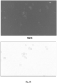
- FIG. 1 represents a sample containing spermatozoa, the latter being mobile.
- the usual practices consisting of increasing the acquisition duration, or accumulating different images, are not suitable. Indeed, the particles move along a trajectory that is difficult to predict. And each particle follows a different trajectory from another particle.
- step e) comprises the calculation of an average of the regions of interest extracted in each emission image of the sample, corresponding to the same particle.
- step e) is implemented during each iteration of steps a) and b).
- the intensity of the illumination of the sample, in the detection spectral band is adjusted so that the signal-to-noise ratio of the detection image is less than one predetermined value.
- Spermatozoa can be labeled with different fluorophores, each fluorophore having an excitation spectral band ⁇ e and a spectral band fluorescence emission ⁇ m.
- the fluorescence emission spectral band ⁇ m is distant from the excitation spectral band ⁇ e, the overlap between the fluorescence emission spectral band ⁇ m and the excitation spectral band being considered to be zero.
- the image sensor 20 is a color image sensor. Thus, it is the same image sensor which allows the acquisition of a detection image I d , in the detection spectral band ⁇ d and of a fluorescence emission image I m in the spectral band fluorescence emission ⁇ m.
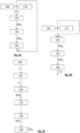
- Step 120 formation of a detection image I d of the sample, from the image I acquired by the image sensor.
- the detection image I d corresponds to a first spectral component, in the detection spectral band ⁇ d, of the image I acquired by the image sensor.
- Step 130 formation of a fluorescence emission image I m of the sample, from the image I acquired by the image sensor.
- the fluorescence emission image I m corresponds to a second spectral component, in the fluorescence spectral band ⁇ m, of the image I acquired by the image sensor.
- annotated ROI p of the detection image resulting from step 120 extraction of a so-called twin region of interest ROI' p on the fluorescence image.
- twin region of interest is meant a region of interest, extracted in the fluorescence image, which corresponds to the region of interest determined, in step 120, in the detection image.
- the region of interest ROI p formed in the detection image I d and the twin region of interest ROI' p extracted in the fluorescence image I m correspond to the same particle p.
- step 140 amounts to forming an integrated fluorescence signal, from regions of interest extracted from successive fluorescence images, and corresponding to the same particle.
- the fluorescence signal can be the integrated fluorescence image I p,N , corresponding to the accumulation of regions of interest extracted on several successive fluorescence images. It can also be a number corresponding to a cumulation of the intensities of the successive fluorescence images I m , in the regions of interest extracted from each of them, and corresponding to the same particle.
- N 1 iteration
- FIG 8D Figure 8D
- Steps 100 to 140 are repeated, the images of the sample being acquired at a high acquisition frequency, for example at 60 images per second.
- a high acquisition frequency for example at 60 images per second.
- the integrated fluorescence image I p,N for each spermatozoon p, is renewed during each iteration.
- Each iteration comprises steps 100 to 140. The iterations continue until a stopping criterion is reached: this may be a predetermined number of iterations.
- the detection images and the fluorescence images are formed from the same image, or are acquired simultaneously if several elementary sensors are used.
- the respective image acquisitions allowing the formation of detection images and fluorescence images, are alternated.
- the duration between two successive acquisitions can be such that the movement of the spermatozoa, between a fluorescence image and a detection image, is considered negligible. This facilitates the correspondence between the regions of interest ROI p determined on the detection images and the regions of interest determined on the fluorescence images.
- the position of the spermatozoa between two successive detection images can be estimated by interpolation, as described below.
Landscapes
- Chemical & Material Sciences (AREA)
- Health & Medical Sciences (AREA)
- Life Sciences & Earth Sciences (AREA)
- Biochemistry (AREA)
- General Health & Medical Sciences (AREA)
- Physics & Mathematics (AREA)
- Analytical Chemistry (AREA)
- General Physics & Mathematics (AREA)
- Immunology (AREA)
- Pathology (AREA)
- Dispersion Chemistry (AREA)
- Engineering & Computer Science (AREA)
- Signal Processing (AREA)
- Biomedical Technology (AREA)
- Wood Science & Technology (AREA)
- Biotechnology (AREA)
- Organic Chemistry (AREA)
- Bioinformatics & Cheminformatics (AREA)
- Genetics & Genomics (AREA)
- Zoology (AREA)
- Microbiology (AREA)
- Developmental Biology & Embryology (AREA)
- General Engineering & Computer Science (AREA)
- Cell Biology (AREA)
- Nuclear Medicine, Radiotherapy & Molecular Imaging (AREA)
- Investigating, Analyzing Materials By Fluorescence Or Luminescence (AREA)
Applications Claiming Priority (1)
| Application Number | Priority Date | Filing Date | Title |
|---|---|---|---|
| FR2214523A FR3144286A1 (fr) | 2022-12-27 | 2022-12-27 | Dispositif et procédé d’observation d’une fluorescence ou d’une luminescence d’une particule mobile |
Publications (1)
| Publication Number | Publication Date |
|---|---|
| EP4394352A1 true EP4394352A1 (de) | 2024-07-03 |
Family
ID=85569700
Family Applications (1)
| Application Number | Title | Priority Date | Filing Date |
|---|---|---|---|
| EP23220208.5A Pending EP4394352A1 (de) | 2022-12-27 | 2023-12-26 | Vorrichtung und verfahren zur beobachtung einer fluoreszenz oder einer lumineszenz eines beweglichen teilchens |
Country Status (3)
| Country | Link |
|---|---|
| US (1) | US20240219288A1 (de) |
| EP (1) | EP4394352A1 (de) |
| FR (1) | FR3144286A1 (de) |
Citations (4)
| Publication number | Priority date | Publication date | Assignee | Title |
|---|---|---|---|---|
| US6259807B1 (en) | 1997-05-14 | 2001-07-10 | Applied Imaging Corp. | Identification of objects of interest using multiple illumination schemes and finding overlap of features in corresponding multiple images |
| US20160169801A1 (en) | 2013-05-11 | 2016-06-16 | The Board Of Trustees Of The Leland Stanford Junior University | Target Characterization Based on Persistent Collocation of Multiple Specks of Light in Time Series Imagery |
| WO2017214023A1 (en) | 2016-06-10 | 2017-12-14 | Amnis Corporation | A method to combine brightfield and fluorescent channels for cell image segmentation and morphological analysis using images obtained from imaging flow cytometer (ifc) |
| US20180293426A1 (en) | 2017-04-05 | 2018-10-11 | Olympus Soft Imaging Solutions Gmbh | Method for the cytometric analysis of cell samples |
-
2022
- 2022-12-27 FR FR2214523A patent/FR3144286A1/fr active Pending
-
2023
- 2023-12-26 EP EP23220208.5A patent/EP4394352A1/de active Pending
- 2023-12-27 US US18/397,259 patent/US20240219288A1/en active Pending
Patent Citations (4)
| Publication number | Priority date | Publication date | Assignee | Title |
|---|---|---|---|---|
| US6259807B1 (en) | 1997-05-14 | 2001-07-10 | Applied Imaging Corp. | Identification of objects of interest using multiple illumination schemes and finding overlap of features in corresponding multiple images |
| US20160169801A1 (en) | 2013-05-11 | 2016-06-16 | The Board Of Trustees Of The Leland Stanford Junior University | Target Characterization Based on Persistent Collocation of Multiple Specks of Light in Time Series Imagery |
| WO2017214023A1 (en) | 2016-06-10 | 2017-12-14 | Amnis Corporation | A method to combine brightfield and fluorescent channels for cell image segmentation and morphological analysis using images obtained from imaging flow cytometer (ifc) |
| US20180293426A1 (en) | 2017-04-05 | 2018-10-11 | Olympus Soft Imaging Solutions Gmbh | Method for the cytometric analysis of cell samples |
Also Published As
| Publication number | Publication date |
|---|---|
| US20240219288A1 (en) | 2024-07-04 |
| FR3144286A1 (fr) | 2024-06-28 |
Similar Documents
| Publication | Publication Date | Title |
|---|---|---|
| EP1875209B1 (de) | Fluoreszenzabbildungsvorrichtung mit Reflexion bei zwei Wellenlängen | |
| EP3465153B1 (de) | Vorrichtung und verfahren zur erfassung eines partikels, das in einer probe enthalten ist | |
| AU2005256758B2 (en) | Multimarking fiber fluorescence microscopic imagery system and method | |
| EP2465416B1 (de) | Lokalisierungsverfahren eines optischen Markers in einem Diffusionsmedium | |
| FR3036187B1 (fr) | Procede de correction d’une image de fluorescence | |
| EP3257947B1 (de) | Verfahren und system zur identifizierung des gramtyps einer bakterie | |
| FR2825260A1 (fr) | Procede et dispositif de detection de caries dentaires | |
| EP3720115B1 (de) | Bildgebungsverfahren mit gepulstem licht | |
| EP3433678A1 (de) | Holographisches verfahren zur charakterisierung eines partikels in einer probe | |
| EP3241014B1 (de) | System zur analyse einer transparenten probe mit positionssteuerung und zugehöriges verfahren | |
| WO2018060619A1 (fr) | Dispositif d'observation d'un échantillon et procédé d'observation d'un échantillon | |
| EP3220131B1 (de) | Verfahren und vorrichtung zur charakterisierung einer flüssigen, partikel enthaltenden probe | |
| CA2529201A1 (fr) | Procede et dispositif d'acquisition et de traitement d'images d'un objet tel qu'une dent | |
| EP4232946B1 (de) | Verfahren zur klassifizierung einer sequenz von eingabebildern, die ein teilchen in einer probe im laufe der zeit darstellen | |
| EP4394352A1 (de) | Vorrichtung und verfahren zur beobachtung einer fluoreszenz oder einer lumineszenz eines beweglichen teilchens | |
| EP3499221B1 (de) | Vorrichtung und verfahren zur beobachtung einer probe mit einem chromatischen optischen system | |
| FR2815240A1 (fr) | Procede de detection d'une affection retinienne ou du nerf optique de l'oeil d'une personne et appareil pour la mise en oeuvre de ce procede | |
| WO2020182828A1 (fr) | Dispositif et procede pour l'observation de microparticules et de nanoparticules | |
| WO2023002114A1 (fr) | Procédé de quantification d'endotoxines d'un échantillon biologique | |
| FR3135610A1 (fr) | Dispositif et procédé d’imagerie médicale adaptés à l’observation de plusieurs bandes spectrales | |
| WO2018134522A1 (fr) | Procédé et dispositif de mesure de la fluorescence émise à la surface du tissu biologique | |
| Macháň | Introduction to Fluorescence Microscopy | |
| FR3165073A1 (fr) | Dispositif de détection de bactéries dans un échantillon | |
| FR3158560A1 (fr) | Dispositif de détection de bactéries dans un échantillon | |
| FR3164787A1 (fr) | Procédé de caractérisation de la trajectoire d’une particule mobile dans un échantillon |
Legal Events
| Date | Code | Title | Description |
|---|---|---|---|
| PUAI | Public reference made under article 153(3) epc to a published international application that has entered the european phase |
Free format text: ORIGINAL CODE: 0009012 |
|
| STAA | Information on the status of an ep patent application or granted ep patent |
Free format text: STATUS: REQUEST FOR EXAMINATION WAS MADE |
|
| 17P | Request for examination filed |
Effective date: 20231226 |
|
| AK | Designated contracting states |
Kind code of ref document: A1 Designated state(s): AL AT BE BG CH CY CZ DE DK EE ES FI FR GB GR HR HU IE IS IT LI LT LU LV MC ME MK MT NL NO PL PT RO RS SE SI SK SM TR |
|
| RAP3 | Party data changed (applicant data changed or rights of an application transferred) |
Owner name: COMMISSARIAT A L'ENERGIE ATOMIQUE ET AUX ENERGIESALTERNATIVES |
|
| GRAP | Despatch of communication of intention to grant a patent |
Free format text: ORIGINAL CODE: EPIDOSNIGR1 |
|
| STAA | Information on the status of an ep patent application or granted ep patent |
Free format text: STATUS: GRANT OF PATENT IS INTENDED |
|
| RIC1 | Information provided on ipc code assigned before grant |
Ipc: G01N 15/1429 20240101AFI20260112BHEP Ipc: G01N 21/64 20060101ALI20260112BHEP Ipc: G01N 15/1433 20240101ALI20260112BHEP Ipc: C12N 5/071 20100101ALI20260112BHEP Ipc: G01N 15/10 20240101ALN20260112BHEP Ipc: G01N 15/1434 20240101ALN20260112BHEP |
|
| INTG | Intention to grant announced |
Effective date: 20260123 |