EP4394048A2 - Verfahren zur identifizierung des typs von follikulärem lymphom - Google Patents
Verfahren zur identifizierung des typs von follikulärem lymphom Download PDFInfo
- Publication number
- EP4394048A2 EP4394048A2 EP24175406.8A EP24175406A EP4394048A2 EP 4394048 A2 EP4394048 A2 EP 4394048A2 EP 24175406 A EP24175406 A EP 24175406A EP 4394048 A2 EP4394048 A2 EP 4394048A2
- Authority
- EP
- European Patent Office
- Prior art keywords
- gene
- group
- tme
- subject
- signature
- Prior art date
- Legal status (The legal status is an assumption and is not a legal conclusion. Google has not performed a legal analysis and makes no representation as to the accuracy of the status listed.)
- Pending
Links
Images


Classifications
-
- C—CHEMISTRY; METALLURGY
- C12—BIOCHEMISTRY; BEER; SPIRITS; WINE; VINEGAR; MICROBIOLOGY; ENZYMOLOGY; MUTATION OR GENETIC ENGINEERING
- C12Q—MEASURING OR TESTING PROCESSES INVOLVING ENZYMES, NUCLEIC ACIDS OR MICROORGANISMS; COMPOSITIONS OR TEST PAPERS THEREFOR; PROCESSES OF PREPARING SUCH COMPOSITIONS; CONDITION-RESPONSIVE CONTROL IN MICROBIOLOGICAL OR ENZYMOLOGICAL PROCESSES
- C12Q1/00—Measuring or testing processes involving enzymes, nucleic acids or microorganisms; Compositions therefor; Processes of preparing such compositions
- C12Q1/68—Measuring or testing processes involving enzymes, nucleic acids or microorganisms; Compositions therefor; Processes of preparing such compositions involving nucleic acids
- C12Q1/6876—Nucleic acid products used in the analysis of nucleic acids, e.g. primers or probes
- C12Q1/6883—Nucleic acid products used in the analysis of nucleic acids, e.g. primers or probes for diseases caused by alterations of genetic material
- C12Q1/6886—Nucleic acid products used in the analysis of nucleic acids, e.g. primers or probes for diseases caused by alterations of genetic material for cancer
-
- G—PHYSICS
- G16—INFORMATION AND COMMUNICATION TECHNOLOGY [ICT] SPECIALLY ADAPTED FOR SPECIFIC APPLICATION FIELDS
- G16B—BIOINFORMATICS, i.e. INFORMATION AND COMMUNICATION TECHNOLOGY [ICT] SPECIALLY ADAPTED FOR GENETIC OR PROTEIN-RELATED DATA PROCESSING IN COMPUTATIONAL MOLECULAR BIOLOGY
- G16B20/00—ICT specially adapted for functional genomics or proteomics, e.g. genotype-phenotype associations
-
- G—PHYSICS
- G16—INFORMATION AND COMMUNICATION TECHNOLOGY [ICT] SPECIALLY ADAPTED FOR SPECIFIC APPLICATION FIELDS
- G16B—BIOINFORMATICS, i.e. INFORMATION AND COMMUNICATION TECHNOLOGY [ICT] SPECIALLY ADAPTED FOR GENETIC OR PROTEIN-RELATED DATA PROCESSING IN COMPUTATIONAL MOLECULAR BIOLOGY
- G16B25/00—ICT specially adapted for hybridisation; ICT specially adapted for gene or protein expression
-
- C—CHEMISTRY; METALLURGY
- C12—BIOCHEMISTRY; BEER; SPIRITS; WINE; VINEGAR; MICROBIOLOGY; ENZYMOLOGY; MUTATION OR GENETIC ENGINEERING
- C12Q—MEASURING OR TESTING PROCESSES INVOLVING ENZYMES, NUCLEIC ACIDS OR MICROORGANISMS; COMPOSITIONS OR TEST PAPERS THEREFOR; PROCESSES OF PREPARING SUCH COMPOSITIONS; CONDITION-RESPONSIVE CONTROL IN MICROBIOLOGICAL OR ENZYMOLOGICAL PROCESSES
- C12Q2600/00—Oligonucleotides characterized by their use
- C12Q2600/106—Pharmacogenomics, i.e. genetic variability in individual responses to drugs and drug metabolism
-
- C—CHEMISTRY; METALLURGY
- C12—BIOCHEMISTRY; BEER; SPIRITS; WINE; VINEGAR; MICROBIOLOGY; ENZYMOLOGY; MUTATION OR GENETIC ENGINEERING
- C12Q—MEASURING OR TESTING PROCESSES INVOLVING ENZYMES, NUCLEIC ACIDS OR MICROORGANISMS; COMPOSITIONS OR TEST PAPERS THEREFOR; PROCESSES OF PREPARING SUCH COMPOSITIONS; CONDITION-RESPONSIVE CONTROL IN MICROBIOLOGICAL OR ENZYMOLOGICAL PROCESSES
- C12Q2600/00—Oligonucleotides characterized by their use
- C12Q2600/112—Disease subtyping, staging or classification
-
- C—CHEMISTRY; METALLURGY
- C12—BIOCHEMISTRY; BEER; SPIRITS; WINE; VINEGAR; MICROBIOLOGY; ENZYMOLOGY; MUTATION OR GENETIC ENGINEERING
- C12Q—MEASURING OR TESTING PROCESSES INVOLVING ENZYMES, NUCLEIC ACIDS OR MICROORGANISMS; COMPOSITIONS OR TEST PAPERS THEREFOR; PROCESSES OF PREPARING SUCH COMPOSITIONS; CONDITION-RESPONSIVE CONTROL IN MICROBIOLOGICAL OR ENZYMOLOGICAL PROCESSES
- C12Q2600/00—Oligonucleotides characterized by their use
- C12Q2600/118—Prognosis of disease development
-
- C—CHEMISTRY; METALLURGY
- C12—BIOCHEMISTRY; BEER; SPIRITS; WINE; VINEGAR; MICROBIOLOGY; ENZYMOLOGY; MUTATION OR GENETIC ENGINEERING
- C12Q—MEASURING OR TESTING PROCESSES INVOLVING ENZYMES, NUCLEIC ACIDS OR MICROORGANISMS; COMPOSITIONS OR TEST PAPERS THEREFOR; PROCESSES OF PREPARING SUCH COMPOSITIONS; CONDITION-RESPONSIVE CONTROL IN MICROBIOLOGICAL OR ENZYMOLOGICAL PROCESSES
- C12Q2600/00—Oligonucleotides characterized by their use
- C12Q2600/158—Expression markers
Definitions
- aspects of the disclosure relate to methods, systems, and computer-readable storage media that can be used for determining a follicular lymphoma (FL) tumor microenvironment (TME) type for a subject.
- the disclosure provides a method for determining a follicular lymphoma (FL) tumor microenvironment (TME) type for a subject having, suspected of having, or at risk of having a follicular lymphoma (FL), comprising: using at least one computer hardware processor to perform: (a) obtaining RNA expression data for the subject, the RNA expression data indicating first RNA expression levels for genes in a first plurality of gene groups and second RNA expression levels for genes in a second plurality of gene groups different from the first plurality of gene groups, wherein genes in the second plurality of gene groups are associated with B cells; (b) generating an FL TME signature for the subject using the RNA expression data, the FL TME signature comprising a first gene expression signature comprising first gene group expression scores for respective gene groups in the first
- aspects of the present disclosure include a system, comprising: at least one computer hardware processor; and at least one computer-readable storage medium storing processor-executable instructions that, when executed by the at least one computer hardware processor, cause the at least one computer hardware processor to perform a method for determining a follicular lymphoma (FL) tumor microenvironment (TME) type for a subject having, suspected of having, or at risk of having a follicular lymphoma (FL), the method comprising: (a) obtaining RNA expression data for the subject, the RNA expression data indicating first RNA expression levels for genes in a first plurality of gene groups and second RNA expression levels for genes in a second plurality of gene groups different from the first plurality of gene groups, wherein genes in the second plurality of gene groups are associated with B cells; (b) generating an FL TME signature for the subject using the RNA expression data, the FL TME signature comprising: a first gene expression signature comprising first gene group expression scores for respective gene groups in the first plurality of gene
- the generating comprises determining the first gene expression signature by determining the first gene group expression scores using the first RNA expression levels and determining the second gene expression signature by determining the second gene group expression scores using the second RNA expression levels.
- the bulk sequencing data comprises at least 1 million reads, at least 5 million reads, at least 10 million reads, at least 20 million reads, at least 50 million reads, or at least 100 million reads.
- the sequencing data comprises bulk RNA sequencing (RNA-seq) data, single cell RNA sequencing (scRNA-seq) data, or next generation sequencing (NGS) data. In some embodiments, the sequencing data comprises microarray data.
- obtaining the RNA expression for the subject comprises sequencing a biological sample obtained from the subject.
- the first RNA expression levels for genes in the first plurality of gene groups comprise RNA expression levels for at least three genes from each of at least two of the following gene groups: (a) MHC II group: HLA-DRA, HLA-DRB1, HLA-DMA, HLA-DPA1, HLA-DPB1, HLA-DMB, HLA-DQB1, HLA-DQA1, CIITA; (b) Effector cells group: IFNG, GZMA, GZMB, PRF1, GZMK, ZAP70, GNLY, FASLG, TBX21, EOMES, CD8A, CD8B; and (c) Follicular Dendritic Cells (FDC) group: PDPN, LTBR, FDCSP, CLU, PRNP, C4A, BST1, SERPINE2, C1S, TNFRSF1A.
- MHC II group HLA-DRA, HLA-DRB1, HLA-DMA, HLA-DPA1, HLA-DPB1, HLA-
- the first RNA expression levels for genes in the first plurality of gene groups further comprise RNA expression levels for at least three genes from each of at least two of the following gene groups: (1) CD4 + T cells group: CD4, TRAT1, CD40LG, TRAC, CD28; (m) CD8 + T cells group: PRF1, GZMA, CD8B, KLRK1, CD8A, ZAP70, GZMK, TBX21, GZMB, NKG7, EOMES, CD160, KLRC2, TRAT1 ; and (n) Macrophages group: CMKLR1, IL4I1, OLR1, ADAMDEC1 , FPR3, CSF1R, MRC1, SIGLEC1, MS4A7, APOC2, APOE, CD163, SPP1, CCL7, LILRB4, C3AR1, SLAMF8, C1QC, MS4A4A, CLEC10A, C5AR1, RAB7B, CLEC5A, CD14, KMO, VSIG4, ADO
- the second RNA expression levels for genes in the second plurality of gene groups comprises RNA expression levels for at least three genes from each of at least two of the following gene groups associated with B cells: (a) Naive B cells group: CD200, CD27, DPPA4, NAAA, XBP1, MNS1, SIGLEC6, PDE8B, BCL2, IRF4, RHOBTB3, CD1A, ENTPD1, and KIF18A; (b) Centrocyte group: DHRS9, EGR3, FCER2, DPPA4, ENTPD1, FGD6, DNAJB9, ELL2, ERN1, EIF4E3, AHNAK, and FEZ1; (c) Centroblast group: KANK2, POU2AF1, PDE8B, SLAMF7, TCL1A, RBM47, MNS1, UEVLD, RASGRF1, NDE1, KIF13A, JUN, and NEK2; (d) Memory B cells group: SLC39A8, IL21R, CCR1, TCL1A
- determining the first gene group expression scores comprises: determining a respective gene expression score for each of at least two of the three following gene groups, using, for a particular gene group, first RNA expression levels for at least three genes in the particular gene group to determine the gene expression score for the particular group, the three gene groups including: (a) MHC II group: HLA-DRA, HLA-DRB1, HLA-DMA, HEA-DPA1, HLA-DPB1, HLA-DMB, HLA-DQB1, HLA-DQA1, CIITA ; (b) Effector cells group: IFNG, GZMA, GZMB, PRF1, GZMK, ZAP70, GNLY, FASLG, TBX21, EOMES, CD8A, CD8B; and (c) Follicular Dendritic Cells (FDC) group: PDPN, LTBR, FDCSP, CLU, PRNP, C4A, BST1, SERPINE2, C1S, TNFRSF1A
- the second gene expression signature comprises a plurality of BAGS scores for a respective plurality of gene groups. In some embodiments, generating the second gene expression signature comprises determining a first BAGS score for a first of the plurality of gene groups, wherein determining the first BAGS score is performed using RNA gene expression levels of at least some of the genes in the first gene group and coefficients of a BAGS classifier associated with the first group.
- Process 100 begins at act 102 where sequencing data for a subject is obtained.
- the sequencing data may be obtained by sequencing a biological sample (e.g., lymph node tissue and/or tumor tissue) obtained from the subject using any suitable sequencing technique.
- the sequencing data may include sequencing data of any suitable type, from any suitable source, and be in any suitable format. Examples of sequencing data, sources of sequencing data, and formats of sequencing data are described herein including in the section called "Obtaining RNA Expression Data".
- the sequencing data may comprise bulk sequencing data.
- the bulk sequencing data may comprise at least 1 million reads, at least 5 million reads, at least 10 million reads, at least 20 million reads, at least 50 million reads, or at least 100 million reads.
- the sequencing data comprises bulk RNA sequencing (RNA-seq) data, single cell RNA sequencing (scRNA-seq) data, or next generation sequencing (NGS) data.
- the sequencing data comprises microarray data.
- determining the second gene expression signature comprises determining, using RNA expression levels of at least some genes in the first B-cell gene group and coefficients of a first statistical model associated with the first B-cell gene group, a first score for the first B-cell gene group in the second gene expression signature, wherein the coefficients of the first statistical model were previously estimated by training the first statistical model to generate, from the RNA expression levels of the at least some genes in the first B-cell gene group, an output indicative of whether the subject is to be associated with the first B-cell gene group.
- determining the first score for the first B-cell gene group comprises: determining an initial score as a dot product between a vector of the coefficients of the first statistical model (e.g., a logistic regression model) and a vector of the RNA expression levels of the at least some of the genes in the first B-cell gene group; and determining the score by adjusting the initial score (e.g., using median scaling) to compensate for batch effects in a process used to obtain the RNA expression levels from the biological sample.
- a vector of the coefficients of the first statistical model e.g., a logistic regression model
- an FL TME type is identified for the subject using the FL TME signature generated at act 112.
- the each of the possible FL TME types is associated with a respective cluster of FL TME signatures.
- an FL TME type for the subject may be identified by associating the FL TME signature of the subject with a particular one of the plurality of FL TME signature clusters; and identifying the FL TME type for the subject as the FL TME type corresponding to the particular one of the plurality of FL TME signature clusters to which the FL TME signature of the subject is associated. Examples of FL TME types are described herein. Aspects of identifying an FL TME type for a subject are described herein including in the section below titled "Identifying FL TME Type".
- the biological sample may be from any source in the subject's body including, but not limited to, any fluid such as blood (e.g., whole blood, blood serum, or blood plasma), lymph nodes, and tonsils.
- any fluid such as blood (e.g., whole blood, blood serum, or blood plasma), lymph nodes, and tonsils.
- a sample of blood may be a sample of whole blood or a sample of fractionated blood.
- the sample of blood comprises whole blood.
- the sample of blood comprises fractionated blood.
- the sample of blood comprises buffy coat.
- the sample of blood comprises serum.
- the sample of blood comprises plasma.
- the sample of blood comprises a blood clot.
- the sample may be from a cancerous tissue or an organ or a tissue or organ suspected of having one or more cancerous cells.
- the sample may be from a healthy (e.g., non-cancerous) tissue or organ.
- the sample from a healthy (e.g., non-cancerous) tissue or organ may be from a subject who is at risk or suspected of having the risk of developing cancer.
- the sample from a healthy (e.g., non-cancerous) tissue or organ may be from tissues surrounding one or more cancerous cells.
- a sample from a subject e.g., a biopsy from a subject
- the samples may be procured at the same time (e.g., more than one sample may be taken in the same procedure), or the samples may be taken at different times ( e.g., during a different procedure including a procedure 1, 2, 3, 4, 5, 6, 7, 8, 9, 10 days; 1, 2, 3, 4, 5, 6, 7, 8, 9, 10 weeks; 1, 2, 3, 4, 5, 6, 7, 8, 9, 10 months, 1, 2, 3, 4, 5, 6, 7, 8, 9, 10 years, or 1, 2, 3, 4, 5, 6, 7, 8, 9, 10 decades after a first procedure).
- a second or subsequent sample may be taken or obtained from the same region (e.g., from the same tumor or area of tissue) or a different region (including, e.g., a different tumor).
- a second or subsequent sample may be taken or obtained from the subject after one or more treatments, and may be taken from the same region or a different region.
- a second or subsequent sample may be taken or obtained from the subject when the first sample from the subject was taken.
- two separate samples can be taken during the same procurement. These two separate samples can be pooled or compared for the analysis as disclosed herein.
- the second or subsequent sample may be useful in determining whether the cancer in each sample has different characteristics (e.g., in the case of samples taken from two physically separate tumors in a patient) or whether the cancer has responded to one or more treatments (e.g., in the case of two or more samples from the same tumor prior to and subsequent to a treatment).
- any of the biological samples described herein may be obtained from the subject using any known technique. See, for example, the following publications on collecting, processing, and storing biological samples, each of which is incorporated by reference herein in its entirety: Biospecimens and biorepositories: from afterthought to science by Vaught et al. (Cancer Epidemiol Biomarkers Prev. 2012 Feb;21(2):253-5 ), and Biological sample collection, processing, storage and information management by Vaught and Henderson (IARC Sci Publ. 2011;(163):23-42 ).
- the biological sample may be obtained from a surgical procedure (e.g., laparoscopic surgery, microscopically controlled surgery, or endoscopy), bone marrow biopsy, punch biopsy, endoscopic biopsy, or needle biopsy (e.g., a fine-needle aspiration, core needle biopsy, vacuum-assisted biopsy, or image-guided biopsy).
- each of the at least one biological sample is a bodily fluid sample such as whole blood sample, a cell sample, or a tissue biopsy.
- the biological sample is stored using cryopreservation.
- cryopreservation include, but are not limited to, step-down freezing, blast freezing, direct plunge freezing, snap freezing, slow freezing using a programmable freezer, and vitrification.
- the biological sample is stored using lyophilisation.
- a biological sample is placed into a container that already contains a preservant (e.g., RNALater to preserve RNA) and then frozen (e.g., by snap-freezing), after the collection of the biological sample from the subject.
- a preservant e.g., RNALater to preserve RNA
- such storage in frozen state is done immediately after collection of the biological sample.
- a vacutainer may be used to store blood.
- a vacutainer may comprise a preservant (e.g., a coagulant, or an anticoagulant).
- a container in which a biological sample is preserved may be contained in a secondary container, for the purpose of better preservation, or for the purpose of avoid contamination.
- a biological sample is stored at -60 °C to -8-°C (e.g., -70°C) for up to 5 years (e.g., up to 1 month, up to 2 months, up to 3 months, up to 4 months, up to 5 months, up to 6 months, up to 7 months, up to 8 months, up to 9 months, up to 10 months, up to 11 months, up to 1 year, up to 2 years, up to 3 years, up to 4 years, or up to 5 years).
- a biological sample is stored as described by any of the methods described herein for up to 20 years (e.g., up to 5 years, up to 10 years, up to 15 years, or up to 20 years).
- aspects of the disclosure relate to methods of determining a FL TME type of a subject using RNA expression data obtained from a biological sample obtained from the subject.
- RNA expression data used in methods described herein typically is derived from sequencing data obtained from the biological sample. After the sequencing data is obtained, it is processed in order to obtain the RNA expression data.
- RNA expression data may be acquired using any method known in the art including, but not limited to: whole transcriptome sequencing, total RNA sequencing, mRNA sequencing, targeted RNA sequencing, RNA exome capture sequencing, next generation sequencing, and/or deep RNA sequencing.
- RNA expression data may be obtained using a microarray assay.
- the sequencing data is processed to produce RNA expression data.
- sequencing data is processed by one or more bioinformatics methods or software tools, for example RNA sequence quantification tools (e.g., Kallisto) and genome annotation tools (e.g., Gencode v23), in order to produce the RNA expression data.
- RNA sequence quantification tools e.g., Kallisto
- genome annotation tools e.g., Gencode v23
- the Kallisto software is described in Nicolas L Bray, Harold Pimentel, Páll Melsted and Lior Pachter, Near-optimal probabilistic RNA-seq quantification, Nature Biotechnology 34, 525-527 (2016), doi:10.1038/nbt.3519 , which is incorporated by reference in its entirety herein.
- microarray expression data is processed using a bioinformatics R package, such as "affy” or “limma”, in order to produce expression data.
- a bioinformatics R package such as "affy” or “limma”
- the "affy” software is described in Bioinformatics. 2004 Feb 12;20(3):307-15. doi: 10.1093/bioinformatics/btg405 .
- sequencing data and/or expression data comprises more than 5 kilobases (kb).
- the size of the obtained RNA data is at least 10 kb.
- the size of the obtained RNA sequencing data is at least 100 kb.
- the size of the obtained RNA sequencing data is at least 500 kb.
- the size of the obtained RNA sequencing data is at least 1 megabase (Mb).
- the size of the obtained RNA sequencing data is at least 10 Mb.
- the size of the obtained RNA sequencing data is at least 100 Mb.
- the size of the obtained RNA sequencing data is at least 500 Mb.
- the size of the obtained RNA sequencing data is at least 1 gigabase (Gb). In some embodiments, the size of the obtained RNA sequencing data is at least 10 Gb. In some embodiments, the size of the obtained RNA sequencing data is at least 100 Gb. In some embodiments, the size of the obtained RNA sequencing data is at least 500 Gb.
- Gb gigabase
- the size of the obtained RNA sequencing data is at least 10 Gb. In some embodiments, the size of the obtained RNA sequencing data is at least 100 Gb. In some embodiments, the size of the obtained RNA sequencing data is at least 500 Gb.
- the expression data is acquired through bulk RNA sequencing.
- Bulk RNA sequencing may include obtaining expression levels for each gene across RNA extracted from a large population of input cells (e.g., a mixture of different cell types.)
- the expression data is acquired through single cell sequencing (e.g., scRNA-seq). Single cell sequencing may include sequencing individual cells.
- bulk sequencing data comprises at least 1 million reads, at least 5 million reads, at least 10 million reads, at least 20 million reads, at least 50 million reads, or at least 100 million reads. In some embodiments, bulk sequencing data comprises between 1 million reads and 5 million reads, 3 million reads and 10 million reads, 5 million reads and 20 million reads, 10 million reads and 50 million reads, 30 million reads and 100 million reads, or 1 million reads and 100 million reads (or any number of reads including, and between).
- the expression data comprises next-generation sequencing (NGS) data. In some embodiments, the expression data comprises microarray data.
- NGS next-generation sequencing
- Expression data (e.g., indicating expression levels) for a plurality of genes may be used for any of the methods or compositions described herein.
- the number of genes which may be examined may be up to and inclusive of all the genes of the subject.
- expression levels may be determined for all of the genes of a subject.
- the expression data may include, for each gene group listed in Tables 1 and 2, expression data for at least 5, at least 10, at least 15, at least 20, at least 25, at least 35, at least 50, at least 75, at least 100 genes selected from each gene group.
- RNA expression data is obtained by accessing the RNA expression data from at least one computer storage medium on which the RNA expression data is stored. Additionally or alternatively, in some embodiments, RNA expression data may be received from one or more sources via a communication network of any suitable type. For example, in some embodiment, the RNA expression data may be received from a server (e.g., a SFTP server, or Illumina BaseSpace).
- a server e.g., a SFTP server, or Illumina BaseSpace
- RNA expression data obtained may be in any suitable format, as aspects of the technology described herein are not limited in this respect.
- the RNA expression data may be obtained in a text-based file (e.g., in a FASTQ, FASTA, BAM, or SAM format).
- a file in which sequencing data is stored may contains quality scores of the sequencing data.
- a file in which sequencing data is stored may contain sequence identifier information.
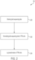
- FIG. 2 shows an exemplary process 104 for processing sequencing data to obtain RNA expression data from sequencing data.
- Process 104 may be performed by any suitable computing device or devices, as aspects of the technology described herein are not limited in this respect.
- process 104 may be performed by a computing device part of a sequencing platform.
- process 104 may be performed by one or more computing devices external to the sequencing platform.
- the bulk sequencing data obtained at act 104 comprises RNA-seq data.
- the biological sample comprises blood or tissue.
- the biological sample comprises one or more tumor cells, for example, one or more FL tumor cells.
- process 104 proceeds to act 202 where the sequencing data obtained at act 200 is normalized to transcripts per kilobase million (TPM) units.
- TPM normalization may be performed using any suitable software and in any suitable way.
- TPM normalization may be performed according to the techniques described in Wagner et al. (Theory Biosci. (2012) 131:281-285 ), which is incorporated by reference herein in its entirety.
- the TPM normalization may be performed using a software package, such as, for example, the gcrma package. Aspects of the gcrma package are described in Wu J, Gentry RIwcfJMJ (2021 ). "gcrma: Background Adjustment Using Sequence Information.
- RNA expression levels in TPM units may be log transformed.
- the log transformation is optional and may be omitted, in some embodiments, the log transformation is an important transformation to employ for calculating gene scores for gene groups associated with B cells (e.g., the gene scores that constitute the second sub-signature of a subject's FL TME signature) as it reduces the range of variability of the RNA expression levels thereby improving the resulting FL TME signature by making it more informative and effective at identifying the FL TME type for the subject.
- Process 104 is illustrative and there are variations.
- one or both of acts 202 and 204 may be omitted.
- the RNA expression levels may not be normalized to transcripts per million units and may, instead, be converted to another type of unit (e.g., reads per kilobase million (RPKM) or fragments per kilobase million (FPKM) or any other suitable unit).
- the log transformation may be omitted. Instead, no transformation may be applied in some embodiments, or one or more other transformations may be applied in lieu of the log transformation.
- expression data (e.g., RNA expression data) is processed using a computing device to determine the one or more gene expression signatures.
- the computing device may be operated by a user such as a doctor, clinician, researcher, patient, or other individual.
- the user may provide the expression data as input to the computing device (e.g., by uploading a file), and/or may provide user input specifying processing or other methods to be performed using the expression data.
- expression data may be processed by one or more software programs running on computing device.
- the disclosure is based, in part, on the recognition that a combination of certain gene expression signatures (e.g., a first gene expression signature comprising the gene groups listed in Table 1 and a second gene expression signature associated with B cells) may be combined to produce a FL TME signature that characterizes patients having FL more accurately than previously developed methods.
- a combination of certain gene expression signatures e.g., a first gene expression signature comprising the gene groups listed in Table 1 and a second gene expression signature associated with B cells
- the number of genes in a gene group used to determine a gene group expression score may vary. In some embodiments, all RNA expression levels for all genes in a particular gene group may be used to determine a gene group score for the particular gene group. In other embodiments, RNA expression data for fewer than all genes may be used (e.g., RNA expression levels for at least two genes, at least three genes, at least five genes, between 2 and 10 genes, between 5 and 15 genes, or any other suitable range within these ranges).
- the first gene group expression signature comprises a score for the Treg cells gene group. In some embodiments, this score may be calculated using RNA expression levels of at least two genes (e.g., at least two genes, at least three genes, at least four genes, at least five genes, at least six genes, or at least seven genes) in the Treg cells gene group, which is defined by its constituent genes: FOXP3, CTLA4, IL10, TNFRSF18, CCR8, IKZF4, and IKZF2.
- a first gene group expression signature comprises a score for the T helper cells gene group. In some embodiments, this score may be calculated using RNA expression levels of at least two genes (e.g., at least two genes, at least three genes, at least four genes, at least five genes, at least six genes, at least seven genes, at least eight genes, at least nine genes, at least ten genes, or more than ten genes) in the T helper cells (Follicular B Helper T cells) gene group, which is defined by its constituent gene: CXCR5, IL6, ICOS, CD40LG, CD84, IL21, BCL6, MAF, SH2D1A, and IL4.
- T helper cells Follicular B Helper T cells
- a first gene group expression signature comprises a score for the MHC II group. In some embodiments, this score may be calculated using RNA expression levels of at least two genes (e.g., at least two genes, at least three genes, at least four genes, at least five genes, at least six genes, at least seven genes, at least eight genes, or at least nine genes) in the MHC II group, which is defined by its constituent genes: HLA-DRA, HLA-DRB1, HLA-DMA, HLA-DPA1, HLA-DPB1, HLA-DMB, HLA-DQB1, HLA-DQA1, and CIITA.
- a first gene group expression signature comprises a score for the Effector cells group.
- this score may be calculated using RNA expression levels of at least two genes (e.g., at least two genes, at least three genes, at least four genes, at least five genes, at least six genes, at least seven genes, at least eight genes, at least nine genes, at least ten genes, or more than ten genes) in the Effector cells group, which is defined by its constituent genes: IFNG, GZMA, GZMB, PRF1, GZMK, ZAP70, GNLY, FASLG, TBX21, EOMES, CD8A, and CD8B.
- a first gene group expression signature comprises a score for the Follicular Dendritic Cells group. In some embodiments, this score may be calculated using RNA expression levels of at least two genes (e.g., at least two genes, at least three genes, at least four genes, at least five genes, at least six genes, at least seven genes, at least eight genes, at least nine genes, or at least ten genes) in the Follicular Dendritic Cells (FDC) group, which is defined by its constituent genes: PDPN, LTBR, FDCSP, CLU, PRNP, C4A, BST1, SERPINE2, C1S, and TNFRSF1A.
- FDC Follicular Dendritic Cells
- a first gene group expression signature comprises a score for the Lymphatic endothelial cells group. In some embodiments, this score may be calculated using RNA expression levels of at least two genes (e.g., at least two genes, at least three genes, at least four genes, at least five genes, at least six genes, at least seven genes, at least eight genes, at least nine genes, at least ten genes, or more than ten genes) in the Lymphatic endothelial cells group, which is defined by its constituent genes: CCE21, CXCE12, SOX18, PPP1R13B, FLT4, PROX1, PDPN, LYVE1, FOXC2, CXADR, EDNRB, JAM2, and JAM3.
- at least two genes e.g., at least two genes, at least three genes, at least four genes, at least five genes, at least six genes, at least seven genes, at least eight genes, at least nine genes, at least ten genes, or more than ten genes
- a first gene group expression signature comprises a score for the Proliferation rate group.
- this score may be calculated using RNA expression levels of at least two genes (e.g., at least two genes, at least three genes, at least four genes, at least five genes, at least six genes, at least seven genes, at least eight genes, at least nine genes, at least ten genes, or more than ten genes) in the Proliferation rate group, which is defined by its constituent genes: MKI67, ESCO2, CETN3, CDK2, CCND1, CCNE1, AURKA, AURKB, E2F1, MYBL2, BUB1, PLK1, CCNB1 , MCM2, and MCM6.
- a first gene group expression signature comprises a score for the M2 group.
- this score may be calculated using RNA expression levels of at least two genes (e.g., at least two genes, at least three genes, at least four genes, at least five genes, at least six genes, at least seven genes, at least eight genes, at least nine genes, at least ten genes, or more than ten genes) in the M2 group, which is defined by its constituent genes: IL10, VEGFA, TGFB1, IDO1, PTGES, MRC1, CSF1, LRP1, ARG1, PTGS1, MSR1, CD163, and CSF1R.
- determining a first gene expression signature comprises determining a respective gene expression score for each of at least two of the following gene groups, using, for a particular gene group, first RNA expression levels for at least three genes in the particular gene group to determine the gene expression score for the particular group, the gene groups including: MHC II group: HLA-DRA, HLA-DRB1, HLA-DMA, HLA-DPA1, HLA-DPB1, HLA-DMB, HLA-DQB1, HLA-DQA1, CIITA ; Effector cells group: IFNG, GZMA, GZMB, PRF1, GZMK, ZAP70, GNLY, FASLG, TBX21, EOMES, CD8A, CD8B; and Follicular Dendritic Cells (FDC) group: PDPN, LTBR, FDCSP, CLU, PRNP, C4A, BST1, SERPINE2, C1S, and TNFRSF1A.
- MHC II group HLA-DRA, HLA-
- determining a first gene expression signature comprises determining a respective gene expression score for each of at least two of the following gene groups, using, for a particular gene group, first RNA expression levels for at least three genes in the particular gene group to determine the gene expression score for the particular group, the gene groups including: Treg cells group: FOXP3, CTLA4, IL10, TNFRSF18, CCR8, IKZF4, IKZF2; T helper cells (Follicular B Helper T cells) group: CXCR5, IL6, ICOS, CD40LG, CD84, IL21, BCL6, MAF, SH2D1A, IL4; Effector cells group: IFNG, GZMA, GZMB, PRF1, GZMK, ZAP70, GNLY, FASLG, TBX21, EOMES, CD8A, CD8B; Follicular Dendritic Cells (FDC) group: PDPN, LTBR, FDCSP, CLU, PRNP, C4A,
- FDC
- determining a first gene expression signature comprises determining a respective gene group score for each of the following gene groups: Treg cells group: FOXP3, CTLA4, IL10, TNFRSF18, CCR8, IKZF4, IKZF2; T helper cells (Follicular B Helper T cells) group: CXCR5, IL6, ICOS, CD40LG, CD84, IL21, BCL6, MAF, SH2D1A, IL4 ; Effector cells group: IFNG, GZMA, GZMB, PRF1, GZMK, ZAP70, GNLY, FASLG, TBX21, EOMES, CD8A, CD8B; Follicular Dendritic Cells (FDC) group: PDPN, LTBR, FDCSP, CLU, PRNP, C4A, BST1, SERPINE2, C1S, TNFRSF1A ; Lymphatic endothelial cells group: CCL21, CXCL12, SOX18,
- determining a first gene expression signature further comprises determining a respective gene group score for each of the following gene groups: CD4 + T cells group: CD4, TRAT1, CD40LG, TRAC, CD28; CD8 + T cells group: PRF1, GZMA, CD8B, KLRK1, CD8A, ZAP70, GZMK, TBX21, GZMB, NKG7, EOMES, CD160, KLRC2, TRAT1 ; and Macrophages group: CMKLR1, IL4I1,OLR1, ADAMDEC1 , FPR3, CSF1R, MRC1, SIGLEC1, MS4A7, APOC2, APOE, CD163, SPP1, CCL7, LILRB4, C3AR1, SLAMF8, C1QC, MS4A4A, CLEC10A, C5AR1 , RAB7B, CLEC5A, CD14, KMO, VSIG4, ADORA3, IL10, CD4, TREM2, ADAP2, CD68, I
- FIG. 3 depicts an illustrative process 108 for determining a first gene expression signature, according to some embodiments of the technology as described herein.
- the first gene expression signature comprises multiple gene group scores 320 determined for respective multiple gene groups.
- Each gene group score, for a particular gene group is computed by performing GSEA 310 (e.g., using ssGSEA) on RNA expression data for one or more (e.g., at least two, at least three, at least four, at least five, at least six, etc., all) genes in the particular gene group.
- a gene group score (labelled “Gene Enrichment Score 1") for gene group 1 (e.g., the Treg cells group) is computed from RNA expression data for one or more genes in gene group 1.
- a gene group score (labelled “Gene Enrichment Score 2") for gene group 2 (e.g., the T helper cells group) is computed from RNA expression data for one or more genes in gene group 2.
- a gene group score (labelled "Gene Enrichment Score 3") for gene group 3 (e.g., the MHC II group) is computed from RNA expression data for one or more genes in gene group 3.
- determining a second gene expression signature comprises determining a respective gene expression score for each of at least two of the following gene groups associated with B cells including, using, for a particular gene group associated with B cells, second RNA expression levels for at least three genes in the particular gene group associated with B cells to determine the gene expression score for the particular group, the gene groups associated with B cells including: Naive B cells: CD200, CD27, DPPA4, NAAA, XBP1, MNS1, SIGLEC6, PDE8B, BCL2, IRF4, RHOBTB3, CD1A, ENTPD1, and KIF18A; Centrocyte: DHRS9, EGR3, FCER2, DPPA4, ENTPD1, FGD6, DNAJB9, ELL2, ERN1, EIF4E3, AHNAK, and FEZ1; Centroblast: KANK2, POU2AF1, PDE8B, SLAMF7, TCL1A, RBM47, MNS1, UEVLD, RASGRF1, NDE1, KIF13A, JUN
- a second gene expression signature is produced using a technique other than GSEA or ssGSEA.
- a second gene expression signature is determined using a B cell associated gene signature (BAGS) classification system.
- BAGS classification is known, and described for example in Dybk ⁇ r K et al., Diffuse large B-cell lymphoma classification system that associates normal B-cell subset phenotypes with prognosis. J Clin Oncol. 2015;33(12): 1379-1388 , which is incorporated by reference herein in its entirety.
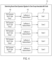
- FIG. 4 depicts an illustrative technique process 108 for determining a second gene expression signature, according to some embodiments of the technology as described herein.
- the second gene expression signature comprises multiple gene group scores 420 determined for respective multiple gene groups.
- a gene group score, for a particular gene group is computed by using: (1) coefficients 410 of a statistical model associated with the particular gene group; and (2) RNA expression data for one or more (e.g., at least two, at least three, at least four, at least five, at least six, etc., all) genes in the particular gene group.
- RNA expression levels for a particular gene group may be embodied in at least one data structure having fields storing the expression levels.
- the data structure or data structures may be provided as input to software comprising code that is configured to access coefficients of a statistical model (e.g., a logistic regression model) associated with the particular gene group, determine a dot product between the gene expression levels and the coefficients, and perform suitable scaling (e.g., median scaling) to produce a score for the particular gene group.
- a statistical model e.g., a logistic regression model
- suitable scaling e.g., median scaling
- the calculation of the CD4 + group to CD8 + group signature ratio is known by a skilled person in the art. For example, the respective gene group expression scores of the CD4 + group and the CD8 + group are first determined. The value of the gene group expression score of CD4 + group is then divided by the value of the gene group expression score of CD8 + group to obtain the CD4 + to CD8 + T-cell signal ratio. In some embodiments, the CD4 + T-cell and the CD8 + T-cell signatures can be used as standalone signatures (e.g., no ratios are calculated).
- FIG. 5 shows an illustrative FL TME signature 500.
- the FL TME signature comprises a first expression signature 510 and a second gene expression signature associated with B cells 520.
- the first expression signature 510 comprises eight gene group scores for the following gene groups: Treg cells group, T helper cells group, Effector Cells group, FDC group, Lymphatic endothelial group, Proliferation rate group, M2 group, and the MHC II group.
- the second expression signature 520 comprises five gene group scores for the following gene groups associated with B cells: Naive B cells group, Centrocyte group, Centroblast group, Memory B cells group, and the Plasmacyte group.
- an FL TME signature may be embodied in at least one data structure comprising fields storing the gene group scores part of the FL TME signature.
- a subject's FL TME signature 500 may be associated with one of four TME clusters: 602, 604, 606, and 608.
- Each of the clusters 602, 604, 608, and 610 may be associated with respect FL TME type.
- the FL TME signature 500 is compared to each cluster (e.g., using a distance-based comparison or any other suitable metric) and, based on the result of the comparison, the FL TME signature 500 is associated with the closest FL signature cluster (when a distance-based comparison is performed, or the "closest" in the sense of whatever metric or measure of distance is used).
- FL TME signature 500 is associated with FL TME Type Cluster 4 604 (as shown by the consistent shading) because the measure of distance D4 between the FL TME signature 500 and (e.g., a centroid or other point representative of) cluster 604 is smaller than the measures of the distance D1, D2, and D3 between the FL TME signature 500 and (e.g., a centroid or other point(s) representative of) clusters 602, 606, and 608, respectively.
- a subject's FL TME signature may be associated with one of four FL TME signature clusters by using a machine learning technique (e.g., such as k-nearest neighbors (KNN) or any other suitable classifier) to assign the FL TME signature to one of the four FL TME signature clusters.
- the machine learning technique may be trained to assign FL TME signatures on the metacohorts represented by the signatures in the clusters.
- the FL TME signature clusters may be generated by: (1) obtaining FL TME signatures (using the techniques described herein) for a plurality of subjects; and (2) clustering the FL TME signatures so obtained into the plurality of clusters.
- Any suitable clustering technique may be used for this purpose including, but not limited to, a dense clustering algorithm, spectral clustering algorithm, k-means clustering algorithm, hierarchical clustering algorithm, and/or an agglomerative clustering algorithm.
- generating the FL TME signature clusters involves: (A) obtaining multiple sets of RNA expression data obtained by sequencing biological samples from multiple respective subjects, each of the multiple sets of RNA expression data indicating first RNA expression levels for genes in a first plurality of gene groups (e.g., one or more of the gene groups in Table 1) and second RNA expression levels for genes in a second plurality of gene groups different from the first plurality of gene groups (e.g., one or more of the gene groups in Table 2), wherein genes in the second plurality of gene groups are associated with B cells; (B) generating multiple FL TME signatures from the multiple sets of RNA expression data, each of the multiple FL TME signatures comprising first gene group expression scores for respective gene groups in the first plurality of gene groups and second gene group expression scores for respective gene groups in the second plurality of gene groups associated with B cells, the generating comprising, for each particular one of the multiple TME signatures: (i) determining the first gene group expression scores using the first RNA expression levels in the
- the resulting FL TME signature clusters may each contain any suitable number of FL TME signatures (e.g., at least 10, at least 100, at least 500, at least 500, at least 1000, at least 5000, between 100 and 10,000, between 500 and 20,000, or any other suitable range within these ranges), as aspects of the technology described herein are not limited in this respect.
- any suitable number of FL TME signatures e.g., at least 10, at least 100, at least 500, at least 500, at least 1000, at least 5000, between 100 and 10,000, between 500 and 20,000, or any other suitable range within these ranges
- FL TME signature clusters in this example is four. And although, in some embodiments, it may be possible that the number of clusters is different, it should be appreciated that an important aspect of the present disclosure is the inventors' discovery that FL may be characterized into four types based upon the generation of FL TME signatures using methods described herein.
- FL TME types include normal-like type, PC-like (or T Helper (TH)-depleted) type, light Zone (LZ)-like type, and dark Zone (DZ)-like type.
- a "high" signal refers to a gene expression signal or score (e.g., an enrichment score, or score produced using B cell associated gene groups) that is at least 1-fold, 2-fold, 3-fold, 4-fold, 5-fold, 6-fold, 7-fold, 8-fold, 9-fold, 10-fold, 20-fold, 50-fold, 100-fold, 1000-fold, or more increased relative to the score of the same gene or gene group in a subject having a different type of FL.
- a "low" signal refers to a gene expression signal or score (e.g., an enrichment score, or score produced using B cell associated gene groups) that is at least 1-fold, 2-fold, 3-fold, 4-fold, 5-fold, 6-fold, 7-fold, 8-fold, 9-fold, 10-fold, 20-fold, 50-fold, 100-fold, 1000-fold, or more decreased relative to the score of the same gene or gene group in a subject having a different type of FL TME.
- a gene expression signal or score e.g., an enrichment score, or score produced using B cell associated gene groups
- the tumor microenvironment of FL may contain variable numbers of immune cells, stromal cells, blood vessels and extracellular matrix.
- normal-like type of FL TME is characterized by the highest stromal signal and high effector cell signal, relative to other types of FL TME, as measured by a first gene expression signal or second gene expression signal. High signal of Memory, Naive and Plasma cell signatures are determined from scores of B-cell related gene groups using the techniques described herein.
- normal-like type of FL TME is characterized by the highest signal of NF-kB signature. Normal-like type of FL TME is most similar to a normal lymph node in the selected signature space.
- normal lymph node and tonsil samples are categorized as normal-like FL TME type when this classification type is used.
- transformed samples such as cancerous tissues cannot be categorized in normal-like type.
- normal-like FL TME type is associated with the best prognosis on R-CHOP.
- PC-like type (or T Helper-depleted) of FL TME is characterized by the lowest CD4 to CD8 T-cell signal ratio and highest T-reg to T follicular helper ratio.
- PC-like type FL TME has high effector cell signal.
- the inventors of the present disclosure identified that the CD4/CD8 ratio is strongly correlated with the effector cell signature. Accordingly, these two signatures may be used interchangeably.
- PC-like type TME has high Plasma cell signal.
- PC-like type FL TME is associated with intermediate prognosis on R-CHOP (e.g., a better prognosis than DZ-type and a worse prognosis than normal-type).
- light zone (LZ)-like type FL TME is characterized by the highest centrocyte and MHC-II signal (i.e., light zone phenotype).
- LZ-like type FL TME has low effector cells signal.
- LZ-like type FL TME is associated with intermediate prognosis on R-CHOP (e.g., a better prognosis than DZ-type FL TME and a worse prognosis than normal-type FL TME).
- dark zone (DZ)-like type FL TME is characterized by the highest centroblast and proliferation rate signal (i.e., dark zone phenotype).
- DZ-like type FL TME has high PI3K signal.
- DZ-like type FL TME has low Effector cell group signal.
- DZ-like type FL TME is associated with worst prognosis on R-CHOP.
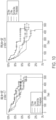
- the prediction of prognosis on R-CHOP can be based on Kaplan Meier (KM)-curves of a single dataset.
- KM Kaplan Meier
- progression-risk score can be used for determining the prediction of prognosis on R-CHOP. In some embodiments, progression-risk score can be used for evaluating the progression of FL.
- progression-risk score is for example, described by Huet et al., "A gene-expression profiling score for outcome prediction disease in patients with follicular lymphoma: a retrospective analysis on three international cohorts", the entire contents of which are incorporated by reference herein.
- high progression-risk score is strongly enriched in DZ-like subtype.
- low progression-risk score is strongly associated with normal-like subtype.
- DZ-like subtype is associated with the most aggressive FL subtype.
- the present disclosure provides methods for providing a prognosis, predicting survival or stratifying patient risk of a subject suspected of having, or at risk of having FL.
- the method comprises determining a FL TME type of the subject as described herein.
- the methods comprise identifying the subject as having an increased risk of FL progression relative to other FL TME types when the subject is assigned normal-like type.
- “increased risk of FL progression” may indicate poor prognosis of FL or increased likelihood of having advanced disease in a subject.
- “increased risk of FL progression” may indicate that the subject who has FL is expected to be less responsive or unresponsive to certain treatments.
- “increased risk of FL progression” indicates that a subject is at least 10%, 20%, 30%, 40%, 50%, 60%, 70%, 80%, 90%, or 100% less likely to experience a progression-free survival event (e.g., relapse, retreatment, or death) than another FL patient or population of FL patients (e.g., patients having FL, but not the same FL TME type as the subject).
- a progression-free survival event e.g., relapse, retreatment, or death
- the methods further comprise identifying the subject as having a decreased risk of FL progression relative to other FL TME types when the subject is assigned DZ-like type.
- decreased risk of FL progression may indicate more positive prognosis of FL or decreased likelihood of having advanced disease in a subject.
- decreased risk of FL progression may indicate that the subject who has FL is expected to be more responsive to certain treatments and show improvements of disease symptoms.
- the methods further comprise identifying the subject as having an increased risk of lacking response to R-CHOP relative to other FL TME types when the subject is assigned DZ-like type.
- "increased risk of lacking response to R-CHOP” may indicate the subject who has FL is expected to be less responsive or unresponsive to R-CHOP.
- "increased risk of lacking response to R-CHOP” indicates that a subject is at least 10%, 20%, 30%, 40%, 50%, 60%, 70%, 80%, 90%, or 100% less likely to experience the efficacy of R-CHOP treatment and/or improvements on FL symptoms than another FL patient or population of FL patients (e.g., patients having FL, but not the same FL TME type as the subject).
- the methods further comprise providing a recommendation to administer (e.g., identifying for the patient) one or more chemotherapeutic agents to the subject based upon the identifying of the patient's FL TME type.
- a recommendation to administer e.g., identifying for the patient
- the subject who is determined to have a DZ-like or a PC-like FL TME may be recommended to receive one or more chemotherapeutic agents that are different (e.g., not R-CHOP) than another FL patient or population of FL patients (e.g., patients having FL, but not the same FL TME type(s) as the subject, who may be recommended for R-CHOP therapy).
- the methods described herein further comprise administering the identified anti-cancer therapeutic to the subject based on the identifying of the subject's FL TME type.
- the methods described herein comprise the use of at least one computer hardware processor to perform the determination.
- the present disclosure provides a method for providing a prognosis, predicting survival or stratifying patient risk of a subject suspected of having, or at risk of having FL.
- the method comprises determining a FL TME type of the subject as described herein.
- data obtained from a future patient may be analyzed in a way that takes advantage of information learned from patients whose FL TME signature was computed prior to that of the future patient.
- the machine learning techniques described herein e.g., the unsupervised clustering machine learning techniques
- the unsupervised clustering machine learning techniques are adaptive and learn with the accumulation of new patient data. This facilitates improved characterization of the FL TME type that future patients may have and may improve the selection of treatment for those patients.
- methods disclosed herein comprise generating a report for assisting with the preparation of recommendation for prognosis and/or treatment.
- the generated report can provide summary of information, so that the clinician can identify the FL subtypes or suitable therapy.
- the report as described herein may be a paper report, an electronic record, or a report in any format that is deemed suitable in the art.
- the report may be shown and/or stored on a computing device known in the art (e.g., handheld device, desktop computer, smart device, website, etc.).
- the report may be shown and/or stored on any device that is suitable as understood by a skilled person in the art.
- the generated report can be provided to the subject and/or to the clinicians.
- a network connection can be established to a server computer that includes the data and report for receiving or outputting.
- the receiving and outputting of the date or report can be requested from the server computer.
- therapeutic agents include chemotherapies (e.g., cytotoxic agents, etc.), immunotherapies (e.g., immune checkpoint inhibitors, such as PD-1 inhibitors, PD-L1 inhibitors, etc.), antibodies (e.g., anti-HER2 antibodies), cellular therapies (e.g. CAR T-cell therapies), gene silencing therapies (e.g., interfering RNAs, CRISPR, etc.), antibody-drug conjugates (ADCs), and combinations thereof.
- chemotherapies e.g., cytotoxic agents, etc.
- immunotherapies e.g., immune checkpoint inhibitors, such as PD-1 inhibitors, PD-L1 inhibitors, etc.
- antibodies e.g., anti-HER2 antibodies
- cellular therapies e.g. CAR T-cell therapies
- gene silencing therapies e.g., interfering RNAs, CRISPR, etc.
- ADCs antibody-drug conjugates
- a subject is administered an effective amount of a therapeutic agent.
- “An effective amount” as used herein refers to the amount of each active agent required to confer therapeutic effect on the subject, either alone or in combination with one or more other active agents. Effective amounts vary, as recognized by those skilled in the art, depending on the particular condition being treated, the severity of the condition, the individual patient parameters including age, physical condition, size, gender and weight, the duration of the treatment, the nature of concurrent therapy (if any), the specific route of administration and like factors within the knowledge and expertise of the health practitioner. These factors are well known to those of ordinary skill in the art and can be addressed with no more than routine experimentation.
- Empirical considerations such as the half-life of a therapeutic compound, generally contribute to the determination of the dosage.
- antibodies that are compatible with the human immune system such as humanized antibodies or fully human antibodies, may be used to prolong half-life of the antibody and to prevent the antibody being attacked by the host's immune system.
- Frequency of administration may be determined and adjusted over the course of therapy, and is generally (but not necessarily) based on treatment, and/or suppression, and/or amelioration, and/or delay of a cancer.
- sustained continuous release formulations of an anti-cancer therapeutic agent may be appropriate.
- Various formulations and devices for achieving sustained release are known in the art.
- dosages for an anti-cancer therapeutic agent as described herein may be determined empirically in individuals who have been administered one or more doses of the anti-cancer therapeutic agent. Individuals may be administered incremental dosages of the anti-cancer therapeutic agent.
- a cancer e.g., tumor microenvironment, tumor formation, tumor growth, or FL TME types, etc.
- an initial candidate dosage may be about 2 mg/kg.
- a typical daily dosage might range from about any of 0.1 ⁇ g/kg to 3 ⁇ g/kg to 30 ⁇ g/kg to 300 ⁇ g/kg to 3 mg/kg, to 30 mg/kg to 100 mg/kg or more, depending on the factors mentioned above.
- the treatment is sustained until a desired suppression or amelioration of symptoms occurs or until sufficient therapeutic levels are achieved to alleviate a cancer, or one or more symptoms thereof.
- An exemplary dosing regimen comprises administering an initial dose of about 2 mg/kg, followed by a weekly maintenance dose of about 1 mg/kg of the antibody, or followed by a maintenance dose of about 1 mg/kg every other week.
- other dosage regimens may be useful, depending on the pattern of pharmacokinetic decay that the practitioner (e.g., a medical doctor) wishes to achieve. For example, dosing from one-four times a week is contemplated.
- radiation therapy examples include, but are not limited to, ionizing radiation, gamma-radiation, neutron beam radiotherapy, electron beam radiotherapy, proton therapy, brachytherapy, systemic radioactive isotopes, and radiosensitizers.
- the "shap” technique is described in Lundberg, Scott M., and Su-In Lee. "A unified approach to interpreting model predictions.” Advances in Neural Information Processing Systems. 2017, which is incorporated by reference herein in its entirety.
- the “lgbm” technique is described in Ke, G., Meng, Q., Finley, T., Wang, T., Chen, W., Ma, W., ... Liu, T.-Y. (2017).
- Lightgbm A highly efficient gradient boosting decision tree. Advances in Neural Information Processing Systems, 30, 3146-3154 , which is incorporated by reference herein in its entirety.
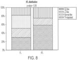
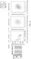
- RNA-seq samples different cell content of each type was also supported by a cell deconvolution algorithm.
- This algorithm allows for the reconstruction of cell composition from bulk RNA-seq data and estimating the percentage of different cell types (fibroblasts, B cells, T cells, macrophages, etc.).
- cell deconvolution algorithms may be used as a control to confirm that the cell types identified by FL TME type agree with cell types identified by other phenotype-based methods.
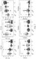
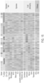
- the differences between the FL TME types are presented in FIG. 7 . Values for each signature in each subtype are provided below in Tables 3A-3C.
- Computing device 1600 may also include a network input/output (I/O) interface 1640 via which the computing device may communicate with other computing devices (e.g., over a network), and may also include one or more user I/O interfaces 1050, via which the computing device may provide output to and receive input from a user.
- the user I/O interfaces may include devices such as a keyboard, a mouse, a microphone, a display device (e.g., a monitor or touch screen), speakers, a camera, and/or various other types of I/O devices.
- program or “software” are used herein in a generic sense to refer to any type of computer code or set of computer-executable instructions that can be employed to program a computer or other processor to implement various aspects as described above. Additionally, it should be appreciated that according to one aspect, one or more computer programs that when executed perform methods of the present disclosure need not reside on a single computer or processor, but may be distributed in a modular fashion among a number of different computers or processors to implement various aspects of the present disclosure.
Landscapes
- Health & Medical Sciences (AREA)
- Life Sciences & Earth Sciences (AREA)
- Chemical & Material Sciences (AREA)
- Proteomics, Peptides & Aminoacids (AREA)
- Engineering & Computer Science (AREA)
- Organic Chemistry (AREA)
- Genetics & Genomics (AREA)
- Physics & Mathematics (AREA)
- Analytical Chemistry (AREA)
- Bioinformatics & Cheminformatics (AREA)
- Zoology (AREA)
- Pathology (AREA)
- Immunology (AREA)
- Wood Science & Technology (AREA)
- Biophysics (AREA)
- Biotechnology (AREA)
- Molecular Biology (AREA)
- General Health & Medical Sciences (AREA)
- Microbiology (AREA)
- Biochemistry (AREA)
- Oncology (AREA)
- General Engineering & Computer Science (AREA)
- Hospice & Palliative Care (AREA)
- Bioinformatics & Computational Biology (AREA)
- Evolutionary Biology (AREA)
- Medical Informatics (AREA)
- Spectroscopy & Molecular Physics (AREA)
- Theoretical Computer Science (AREA)
- Measuring Or Testing Involving Enzymes Or Micro-Organisms (AREA)
- Investigating Or Analysing Biological Materials (AREA)
Applications Claiming Priority (3)
| Application Number | Priority Date | Filing Date | Title |
|---|---|---|---|
| US202063124617P | 2020-12-11 | 2020-12-11 | |
| EP21843825.7A EP4244394B1 (de) | 2020-12-11 | 2021-12-10 | Verfahren zur identifizierung des typs von follikulärem lymphom |
| PCT/US2021/062961 WO2022125994A1 (en) | 2020-12-11 | 2021-12-10 | Techniques for identifying follicular lymphoma types |
Related Parent Applications (2)
| Application Number | Title | Priority Date | Filing Date |
|---|---|---|---|
| EP21843825.7A Division EP4244394B1 (de) | 2020-12-11 | 2021-12-10 | Verfahren zur identifizierung des typs von follikulärem lymphom |
| EP21843825.7A Division-Into EP4244394B1 (de) | 2020-12-11 | 2021-12-10 | Verfahren zur identifizierung des typs von follikulärem lymphom |
Publications (2)
| Publication Number | Publication Date |
|---|---|
| EP4394048A2 true EP4394048A2 (de) | 2024-07-03 |
| EP4394048A3 EP4394048A3 (de) | 2024-09-25 |
Family
ID=79602190
Family Applications (2)
| Application Number | Title | Priority Date | Filing Date |
|---|---|---|---|
| EP21843825.7A Active EP4244394B1 (de) | 2020-12-11 | 2021-12-10 | Verfahren zur identifizierung des typs von follikulärem lymphom |
| EP24175406.8A Pending EP4394048A3 (de) | 2020-12-11 | 2021-12-10 | Verfahren zur identifizierung des typs von follikulärem lymphom |
Family Applications Before (1)
| Application Number | Title | Priority Date | Filing Date |
|---|---|---|---|
| EP21843825.7A Active EP4244394B1 (de) | 2020-12-11 | 2021-12-10 | Verfahren zur identifizierung des typs von follikulärem lymphom |
Country Status (3)
| Country | Link |
|---|---|
| US (1) | US20220186318A1 (de) |
| EP (2) | EP4244394B1 (de) |
| WO (1) | WO2022125994A1 (de) |
Families Citing this family (3)
| Publication number | Priority date | Publication date | Assignee | Title |
|---|---|---|---|---|
| EP3879535B1 (de) | 2017-06-13 | 2024-12-11 | BostonGene Corporation | Systeme und verfahren zur identifizierung von krebsbehandlungen aus normierten biomarker-scores |
| WO2024108156A2 (en) | 2022-11-17 | 2024-05-23 | Bostongene Corporation | Comprehensive immunoprofiling of peripheral blood |
| WO2024216069A1 (en) * | 2023-04-13 | 2024-10-17 | Bostongene Corporation | Pan-cancer tumor microenvironment classification based on immune escape mechanisms and immune infiltration |
Citations (1)
| Publication number | Priority date | Publication date | Assignee | Title |
|---|---|---|---|---|
| US20200273543A1 (en) | 2017-06-13 | 2020-08-27 | Bostongene Corporation | Systems and methods for generating, visualizing and classifying molecular functional profiles |
Family Cites Families (4)
| Publication number | Priority date | Publication date | Assignee | Title |
|---|---|---|---|---|
| WO2017004153A1 (en) * | 2015-06-29 | 2017-01-05 | The Broad Institute Inc. | Tumor and microenvironment gene expression, compositions of matter and methods of use thereof |
| US10900086B1 (en) * | 2015-11-13 | 2021-01-26 | Dana-Farber Cancer Institute, Inc. | Compositions and methods for diagnosing prostate cancer using a gene expression signature |
| US20190262399A1 (en) * | 2016-09-07 | 2019-08-29 | The Broad Institute, Inc. | Compositions and methods for evaluating and modulating immune responses |
| EP3994696B1 (de) * | 2019-07-03 | 2025-05-14 | BostonGene Corporation | Systeme und verfahren zur probenvorbereitung, probensequenzierung sowie sequenzierdatenbiaskorrektur und qualitätskontrolle |
-
2021
- 2021-12-10 EP EP21843825.7A patent/EP4244394B1/de active Active
- 2021-12-10 WO PCT/US2021/062961 patent/WO2022125994A1/en not_active Ceased
- 2021-12-10 US US17/548,444 patent/US20220186318A1/en active Pending
- 2021-12-10 EP EP24175406.8A patent/EP4394048A3/de active Pending
Patent Citations (1)
| Publication number | Priority date | Publication date | Assignee | Title |
|---|---|---|---|---|
| US20200273543A1 (en) | 2017-06-13 | 2020-08-27 | Bostongene Corporation | Systems and methods for generating, visualizing and classifying molecular functional profiles |
Non-Patent Citations (15)
| Title |
|---|
| BARBIE ET AL., NATURE, vol. 462, no. 7269, 5 November 2009 (2009-11-05), pages 108 - 112 |
| CUNNINGHAM ET AL., LANCET, vol. 381, no. 9880, 25 May 2013 (2013-05-25), pages 1817 - 26 |
| DYBKAER ET AL., J CLIN ONCOL, vol. 33, no. 12, 20 April 2015 (2015-04-20), pages 1379 - 1388 |
| DYBKAER K ET AL.: "Diffuse large B-cell lymphoma classification system that associates normal B-cell subset phenotypes with prognosis", J CLIN ONCOL, vol. 33, no. 12, 2015, pages 1379 - 1388, XP055908623, DOI: 10.1200/JCO.2014.57.7080 |
| HUET ET AL., A GENE-EXPRESSION PROFILING SCORE FOR OUTCOME PREDICTION DISEASE IN PATIENTS WITH FOLLICULAR LYMPHOMA: A RETROSPECTIVE ANALYSIS ON THREE INTERNATIONAL COHORTS |
| KE, G.MENG, Q.FINLEY, T.WANG, T.CHEN, W.MA, W.LIU, T.-Y.: "Lightgbm: A highly efficient gradient boosting decision tree", ADVANCES IN NEURAL INFORMATION PROCESSING SYSTEMS, vol. 30, 2017, pages 3146 - 3154 |
| LAURENT GAUTIERLESLIE COPEBENJAMIN M BOLSTADRAFAEL A IRIZARRY: "affy--analysis of Affymetrix GeneChip data at the probe level", BIOINFORMATICS, vol. 20, no. 3, 12 February 2004 (2004-02-12), pages 307 - 15 |
| LIU ET AL., ANNALS OF LYMPHOMA, vol. 5, June 2021 (2021-06-01), pages 11 |
| LUNDBERG, SCOTT M.SU-IN LEE: "A unified approach to interpreting model predictions", ADVANCES IN NEURAL INFORMATION PROCESSING SYSTEMS, 2017 |
| NICOLAS L BRAYHAROLD PIMENTELPALL MELSTEDLIOR PACHTER: "Near-optimal probabilistic RNA-seq quantification", NATURE BIOTECHNOLOGY, vol. 34, 2016, pages 525 - 527 |
| RITCHIE MEPHIPSON BWU DHU YLAW CWSHI WSMYTH GK: "limma powers differential expression analyses for RNA-sequencing and microarray studies", NUCLEIC ACIDS RES, vol. 43, no. 7, 20 April 2015 (2015-04-20), pages e47, Retrieved from the Internet <URL:https://doi.org/10.1093/nar/gkv007> |
| VAUGHT ET AL., CANCER EPIDEMIOL BIOMARKERS PREV, vol. 21, no. 2, February 2012 (2012-02-01), pages 253 - 5 |
| VAUGHTHENDERSON, IARC SCI PUBL, no. 163, 2011, pages 23 - 42 |
| WAGNER ET AL., THEORY BIOSCI, vol. 131, 2012, pages 281 - 285 |
| WU JGENTRY RIWCFJMJ, GCRMA: BACKGROUND ADJUSTMENT USING SEQUENCE INFORMATION. R PACKAGE VERSION 2.66.0, 2021 |
Also Published As
| Publication number | Publication date |
|---|---|
| WO2022125994A1 (en) | 2022-06-16 |
| US20220186318A1 (en) | 2022-06-16 |
| EP4394048A3 (de) | 2024-09-25 |
| EP4244394A1 (de) | 2023-09-20 |
| EP4244394B1 (de) | 2024-06-19 |
Similar Documents
| Publication | Publication Date | Title |
|---|---|---|
| EP3931360B1 (de) | Systeme und verfahren zur verwendung von sequenzierungsdaten zur pathogendetektion | |
| US11091809B2 (en) | Molecular diagnostic test for cancer | |
| US20220319638A1 (en) | Predicting response to treatments in patients with clear cell renal cell carcinoma | |
| EP4244394B1 (de) | Verfahren zur identifizierung des typs von follikulärem lymphom | |
| US20230290440A1 (en) | Urothelial tumor microenvironment (tme) types | |
| EP4423301B1 (de) | Tumormikroumgebungstypen bei brustkrebs | |
| US20260038698A1 (en) | Pan-cancer tumor microenvironment classification based on immune escape mechanisms and immune infiltration | |
| US20220307088A1 (en) | B cell-enriched tumor microenvironments | |
| US20220290254A1 (en) | B cell-enriched tumor microenvironments | |
| WO2024151840A1 (en) | Tumor microenvironment types in lung adenocarcinoma | |
| WO2022245979A1 (en) | Techniques for single sample expression projection to an expression cohort sequenced with another protocol | |
| Odia | Longitudinal transcriptomic profiling of whole blood during tuberculosis treatment |
Legal Events
| Date | Code | Title | Description |
|---|---|---|---|
| PUAI | Public reference made under article 153(3) epc to a published international application that has entered the european phase |
Free format text: ORIGINAL CODE: 0009012 |
|
| STAA | Information on the status of an ep patent application or granted ep patent |
Free format text: STATUS: THE APPLICATION HAS BEEN PUBLISHED |
|
| AC | Divisional application: reference to earlier application |
Ref document number: 4244394 Country of ref document: EP Kind code of ref document: P |
|
| AK | Designated contracting states |
Kind code of ref document: A2 Designated state(s): AL AT BE BG CH CY CZ DE DK EE ES FI FR GB GR HR HU IE IS IT LI LT LU LV MC MK MT NL NO PL PT RO RS SE SI SK SM TR |
|
| PUAL | Search report despatched |
Free format text: ORIGINAL CODE: 0009013 |
|
| AK | Designated contracting states |
Kind code of ref document: A3 Designated state(s): AL AT BE BG CH CY CZ DE DK EE ES FI FR GB GR HR HU IE IS IT LI LT LU LV MC MK MT NL NO PL PT RO RS SE SI SK SM TR |
|
| RIC1 | Information provided on ipc code assigned before grant |
Ipc: C12Q 1/6886 20180101AFI20240822BHEP |
|
| STAA | Information on the status of an ep patent application or granted ep patent |
Free format text: STATUS: REQUEST FOR EXAMINATION WAS MADE |
|
| 17P | Request for examination filed |
Effective date: 20250110 |
|
| STAA | Information on the status of an ep patent application or granted ep patent |
Free format text: STATUS: EXAMINATION IS IN PROGRESS |
|
| 17Q | First examination report despatched |
Effective date: 20260217 |