Technical Field
-
The present invention relates to a creatinine risk estimation device, a creatinine risk estimation method, and a computer program.
Background Art
-
Creatinine is a metabolic product of creatinine phosphate, which is the source of energy supply to muscle. Typically, creatinine is discharged from muscle tissue to blood, filtrated through the glomerulus of the kidney, and then discharged into urine. Thus, the amount of creatinine in blood is utilized as an indicator for evaluation of the kidney function.
-
Conventionally, it has been necessary to collect blood of a subject and perform biochemical analysis to measure creatinine. However, in this method, a needle or the like needs to be invasively inserted into the skin of the subject, which causes a psychological or physical burden on the subject.
-
Patent Literature 1 discloses a technology of non-invasively measuring the level of creatinine concentration in blood by a laser sensor system on-chip.
Citation List
Patent Literature
-
Patent Literature 1
Japanese Translation of PCT International Application Publication No. 2020-520768
Non Patent Literature
-
- Non Patent Literature 1
Psychology Research and Behavior Management 2011:4 81-86, Summary of the clinical investigations E. S. Teck Complex March, 20, 2010 Overview
- Non Patent Literature 2
R. N. Chua, Y. W. Hau, C. M. Tiew and W. L. Hau, "Investigation of Attention Deficit/Hyperactivity Disorder Assessment Using Electro Interstitial Scan Based on Chronoamperometry Technique", in IEEE Access, vol. 7, pp. 144679-144690, 2019, doi: 10.1109/ACCESS.2019.2938095.
- Non Patent Literature 3
Maarek A., Electro interstitial scan system: assessment of 10 years of research and development. Med Devices (Auckl). 2012;5:23-30. doi:10.2147/MDER.S29319
Summary of Invention
Technical Problem
-
However, Patent Literature 1 discloses no specific method of measuring creatinine.
-
The present invention has been made in view of the above-described situation and is intended to provide a creatinine risk estimation device, a creatinine risk estimation method, and a computer program that are capable of extremely accurately and swiftly estimating creatinine risk on health based on non-invasive biological information without blood or urine collection.
Solution to Problem
-
A creatinine risk estimation device according to the present invention includes an information acquisition unit configured to acquire attribute information and non-invasive biological information of a predetermined user, an estimation model storage unit configured to store a creatinine risk estimation model, and an estimation processing unit configured to calculate a creatinine risk estimated value of the predetermined user based on the attribute information and/or the non-invasive biological information of the predetermined user by using the creatinine risk estimation model.
-
The creatinine risk estimation device according to the present invention further includes a training data storage unit configured to store a training data set, and a learning processing unit configured to generate the creatinine risk estimation model by machine learning based on the training data set.
-
The attribute information includes any one or a combination of age and sex, and the non-invasive biological information includes any one or a combination of BMI, blood pressure, pulse wave data, electrocardiogram data, and biological impedance.
-
The training data set includes attribute information, non-invasive biological information, and a blood-measured creatinine measured value of a subject.
-
The non-invasive biological information further includes oxygen saturation (SpO2).
-
Estimation accuracy of the creatinine risk estimated value is accuracy at which risk existence can be classified with ROC_AUC of 0.7 or larger.
-
The learning processing unit provides labels indicating existence of the creatinine risk to the training data set based on the blood-measured creatinine measured value, and when a difference between the number of pieces of data with the creatinine risk and the number of pieces of data without the creatinine risk among the labels is equal to or larger than a predetermined value, the learning processing unit increases the number of pieces of sample data in the training data set to reduce the difference.
-
The learning processing unit generates a first creatinine risk estimation model and a second creatinine risk estimation model by machine learning based on each of training data sets of different kinds, and the estimation processing unit calculates the creatinine risk estimated value of the predetermined user by using the first creatinine risk estimation model and the second creatinine risk estimation model.
-
The creatinine risk estimation device according to the present invention further includes a biological information estimation unit configured to estimate at least one piece or more of biological information among BMI, blood pressure, pulse wave data, electrocardiogram data, biological impedance, and oxygen saturation included in the biological information, and the information acquisition unit acquires, as biological information of the predetermined user, the biological information estimated by the biological information estimation unit.
-
A non-invasive creatinine risk estimation system includes the creatinine risk estimation device, and a biological information measurement device configured to measure non-invasive biological information.
-
A creatinine risk estimation method according to the present invention includes a step of storing a training data set including attribute information, non-invasive biological information, and a blood-measured creatinine measured value of a subject, a step of generating a creatinine risk estimation model by machine learning based on the training data set, and a step of calculating a creatinine risk estimated value of a predetermined user based on the attribute information and/or the non-invasive biological information of the predetermined user by using the creatinine risk estimation model.
-
A computer program according to the present invention causes a computer to execute a step of storing a training data set including attribute information, non-invasive biological information, and a blood-measured creatinine measured value of a subject, a step of generating a creatinine risk estimation model by machine learning based on the training data set, and a step of calculating a creatinine risk estimated value of a predetermined user based on the attribute information and/or the non-invasive biological information of the predetermined user by using the creatinine risk estimation model.
Advantageous Effects of Invention
-
According to the present invention, it is possible to provide a creatinine risk estimation device, a creatinine risk estimation method, and a computer program that are capable of extremely accurately estimating creatinine risk on health by machine learning by using non-invasive biological information.
Brief Description of Drawings
-
- [Figure 1] Figure 1 is a block diagram illustrating a schematic configuration of a creatinine risk estimation system.
- [Figure 2] Figure 2 is a diagram for description of an electroscangram (ESG).
- [Figure 3] Figure 3 is a hardware configuration diagram of a creatinine risk estimation device.
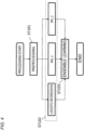
- [Figure 4] Figure 4 is a flowchart illustrating an execution procedure of generation of a creatinine risk estimation model by machine learning.
- [Figure 5] Figure 5 illustrates a hierarchical structure of a neural network (NN) used for male creatinine risk estimation.
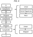
- [Figure 6] Figure 6 illustrates a hierarchical structure of a neural network (NN) used for female creatinine risk estimation.
- [Figure 7] Figure 7 is a flowchart illustrating an execution procedure of creatinine risk estimation processing.
- [Figure 8] Figure 8 illustrates an ROC AUC curve of an estimation result in Example 1.
- [Figure 9] Figure 9 illustrates an ROC AUC curve of an estimation result in Example 2.
Description of Embodiment
-
An embodiment will be described below with reference to the accompanying drawings. Note that the embodiment is exemplary and the present invention is not limited to configurations described below.
<Device functions>
-
A creatinine risk estimation system 1 and a creatinine risk estimation device 30 according to the present embodiment will be described below with reference to Figures 1 to 7. Figure 1 is a block diagram illustrating a schematic configuration of the creatinine risk estimation system 1 according to the present embodiment. The creatinine risk estimation system 1 includes a terminal device 10, a biological information measurement device 20, the creatinine risk estimation device 30, and a display device 39.
-
A "user" is a person who uses the creatinine risk estimation system to non-invasively obtain an estimated value of the creatinine risk. A "subject" is a person who provides, upon a predetermined procedure and an agreement, attribute information such as age or sex, non-invasive biological information, and a blood-measured creatinine measured value as a training data set to be used in the creatinine risk estimation system.
-
The terminal device 10 may be any information terminal to which the attribute information (such as full name, ID, age, or sex) of the user can be input and that can output the input information to the creatinine risk estimation device 30 through a wired or wireless communication network. Examples of the terminal device 10 include portable terminals including a tablet terminal, a smartphone, and a wearable terminal, and include a personal computer (PC). Note that, height, weight, or the like may be measured by the biological information measurement device 20 to be described later.
-
The biological information measurement device 20 measures the non-invasive biological information of the user. The non-invasive biological information is biological information acquired by a method that does not require insertion of an instrument into the skin or an opening part of the body. The non-invasive biological information may be measured by using, for example, a commercially available height meter, weight meter, blood pressure meter, pulse oximeter, pulse wave meter, electrocardiogram meter, impedance measurement machine, or galvanic skin measurement machine. Alternatively, ES-TECK BC-3 (Ryobi Systems Co., Ltd.) that can simultaneously measure pulse wave data, electrocardiogram data, biological impedance, and oxygen saturation (SpO2) may be used. These devices can measure non-invasive biological data without causing a psychological of physical burden on the user.
-
In the embodiment of the present invention, the non-invasive biological information includes any one or a combination of the body mass index (BMI), blood pressure, pulse wave data, electrocardiogram data, and biological impedance and may further include oxygen saturation (SpO2).
-
The BMI is calculated by the following formula based on a height h [m] and a weight w [kg],
-
The blood pressure includes any one or a combination of systolic blood pressure, diastolic blood pressure, pulse pressure, and average arterial blood pressure.
-
The pulse pressure is calculated by the following expression.
-
The average arterial blood pressure is calculated by the following expression.
-
The pulse wave data is measured by irradiating, by using a sphygmograph or a pulse oximeter, a protruding body site such as a finger with red light (up to 660 nm) from a red LED and with near-infrared light (up to 905 nm) from an IR LED and measuring transmitted light thereof by using a phototransistor.
-
The pulse wave data includes any one or a combination of pulse, the elasticity index, the peripheral vascular resistance, the acceleration plethysmogram, b/a, e/a, -d/a, the Takazawa's second derivative of photoplethysmogram aging index, the ejection fraction, the LVET, and the dicrotic elasticity index (DEI).
-
The elasticity index is a numerical value obtained by dividing the height by the time from detection of a systolic peak to detection of a diastolic peak in the photoplethysmogram. The peripheral vascular resistance is calculated by "average arterial blood pressure"/"cardiac output"×80. The dicrotic elasticity index (DEI) is an indicator of the diastolic vascular elasticity and can be measured by a PWV measurement device. The DEI of 0.3 to 0.7 is normal, the DEI of 0.3 or lower indicates the possibility of high-blood pressure or arteriosclerosis, and the DEI of 0.7 or higher indicates the possibility of acute anxiety neurosis.
-
The acceleration plethysmogram is the second derivative of photoplethysmogram (PTG) (SDPTG). The acceleration plethysmogram is constituted by the initial positive wave (a wave), the initial negative wave (b wave), the mesosystolic re-elevation wave (c wave), the late systolic re-descent wave (d wave), and the early diastolic positive wave (e wave), and b/a, e/a, and -d/a described above are calculated from ratios of the heights of the waves. It is observed that b/a increases and c/a, d/a, and e/a decrease along with aging, and thus aging of blood vessels can be evaluated by the Takazawa's second derivative of photoplethysmogram aging index (b - c - d - e)/a. The ejection fraction is the ratio of blood transferred from the ventricles at each heartbeat and is proportional to the second derivative of photoplethysmogram aging index. The LVET is the left ventricular ejection time in which blood in the left ventricle is ejected to the aorta after the aortic valve is opened.
-
The electrocardiogram data can be measured by electrocardiography (ECG) with electrodes or by photoplethysmography (PPG).
-
The electrocardiogram data includes any one or a combination of the breathing rate, the heart rate, the RR interval, the standard deviation of the RR interval, the MxDMn ratio, the power spectrum in a low frequency band, the power spectrum in a high frequency band, the heart rate variation indicator LF/HF, and the total power. The RR interval is the interval from a QRS wave to the next QRS wave in the electrocardiogram. The MxDMn ratio is the ratio of the longest RR interval and the shortest RR interval in a time period and is an index of irregular heartbeats. The total power is a calculated value of the total power of the power spectrum at the frequency of 0 to 0.4 Hz (VLF, LF, HF) in a measurement of two minutes. This value reflects the entire autonomic nerve activity dominated by sympathetic nerve activity.
-
The high-frequency power spectrum ratio (0.1875 to 0.50 Hz; HF), the low-frequency power spectrum ratio (0.05 to 0.1875 Hz; LF), the LF/HF ratio, and the very-low-frequency power spectrum ratio (0 to 0.05 Hz; VLF) can be calculated by calculating the power spectrum density based on the electrocardiogram.
-
The biological impedance (conductance) can be measured by, for example, generating small current flow between two electrodes among electrodes at the six sites of the legs, the hands, and the right and left foreheads. When current flows between two of the electrodes at the six sites, (1) anode/cathode conductance (µS), (2) cathode/anode conductance (µS), (3) the difference (delta SCRA-SCRC) between the conductance measured in (1) above and the conductance measured in (2) above, and (4) electric conductivity (µS/m) can be measured. In addition, the muscle mass, the body fat amount, the total moisture content, the phase angle, and the resistance value can be simultaneously measured. In addition, the dielectric constant (µSi) can be measured for cases of conduction between the right hand and the left hand and between the right forehead and the left forehead. The biological impedance (conductance) is preferably measured by using 22 conduction patterns with the electrodes at the six sites.
-
The biological impedance includes any one or a combination of the body fat amount (kg), the body fat amount (%), the lean body weight, the lean body rate, the muscle mass, the total moisture content (kg), the total moisture content (%), the intercellular moisture content (%), the cardiac output, 1 forehead left-side - 2 left hand/SCR A, 1 forehead left-side - 2 left hand/delta SCR C-SCR A, 5 left hand - 6 left foot/SCR C, 5 left hand - 6 left foot/delta SCR C-SCR A, 7 right hand - 8 right foot/SCR C, 7 right hand - 8 right foot/SCR A, 13 left foot - 14 right foot/SCR C, 13 left foot - 14 right foot/SCR A, 15 right hand - 16 forehead left-side /delta SCR C-SCR A, 15 right hand - 16 forehead left-side SCR C, 15 right hand - 16 forehead left-side SCR A, 19 right foot - 20 left hand/delta SCR C-SCR A, ESG 2+4+15+17 (µS/m), ESG 6+13+19 (%), ESG 6+8+19+21 (%), ESG 6+8+19+21 (µS/m), ESG 9+10 (µS/m), ESG 9+10 (%), the left-foot conductance, R (Ω), the phase angle, the forehead-path dielectric constant, the forehead-path electric conductivity (9), the one-hand-to-one-hand-path dielectric constant, the hand-to-hand electric conductivity (11, 12), the single-output amount (cardiac output/heart rate), the hand-foot impedance (sum value of 5 left hand - 6 left foot/SCR C and 7 right hand - 8 right foot/SCR).
-
SCR stands for skin conductance response, and ESG stands for electroscangram. The symbol "+" in ESG 2+4+15+17 indicates electrodes attached to the body and used for measurement. For example, ESG 2+4+15+17 means the average of conductance values measured at the left hand when conduction is made from the left hand to the left forehead, the right hand when conduction is made from the right hand to the right forehead, the right hand when conduction is made from the right hand to the left forehead, and the left hand when conduction is made from the left hand to the right forehead as illustrated in Figure 2. Detailed description of these conductance values is provided in Non Patent Literature 1. Note that details of ESG (electroscangram) measurement methods are described in Non Patent Literatures 2 and 3.
-
The value "1 forehead left-side - 2 left hand/SCR A" is conductance (or conductivity) measured through a path when current flows with "1 forehead left-side" at the cathode and "2 left hand" at the anode, and the value "5 left hand - 6 left foot/delta SCR C-SCR A" is the difference between conductance values measured when conduction is made with "5 left hand" and "6 left foot" at the anode and the cathode and at the cathode and the anode.
-
The BMI and the blood pressure can be measured by a height-weight meter and a blood pressure meter. The non-invasive biological information may include an oxygen transport amount calculated based on SpO2 and the cardiac output.
-
The measured non-invasive biological information is output to the creatinine risk estimation device 30 through a wired or wireless communication network. The biological information measurement device 20 may be a fixed measurement device or a portable measurement device such as a wearable terminal.
-
The creatinine risk estimation device 30 includes a first acquisition unit 31, a second acquisition unit 32, a user data storage unit 33, a training data storage unit 34, a learning processing unit 35, an estimation model storage unit 36, an estimation processing unit 37, and an estimation data storage unit 38. The first acquisition unit 31 acquires the attribute information of the user from the terminal device 10. The second acquisition unit 32 acquires the non-invasive biological information of the user from the biological information measurement device 20.
-
The user data storage unit 33 stores the attribute information and the non-invasive biological information of the user, which are acquired from the first acquisition unit 31 and the second acquisition unit 32.
-
The training data storage unit 34 stores, as training data sets for machine learning, a plurality of training data sets each consisting of the attribute information and the non-invasive biological information of a plurality of subjects, which are acquired in advance and sample examination information, such as the creatinine, which is obtained through a blood examination. Note that the sample examination information may further include examination information obtained from blood, urine, stool, or the like.
-
The learning processing unit 35 acquires the training data sets stored in the training data storage unit 34 and produces a creatinine risk estimation model by using the training data sets. Specifically, when the creatinine risk is estimated, the relation among the attribute information, the non-invasive biological information, and the creatinine risk is learned by machine learning with each acquired training data set by a neural network, logistic regression, or ensemble learning with results of the learning.
-
The estimation model storage unit 36 stores the creatinine risk estimation model generated by the learning processing unit 35.
-
The non-invasive biological information includes any one or a combination of the BMI, the blood pressure, the pulse wave data, the electrocardiogram data, and the biological impedance. In addition, the oxygen saturation (SpO2) is included as necessary.
-
The estimation processing unit 37 estimates the creatinine risk of the predetermined user based on the attribute information and/or the non-invasive biological information of the user by using the estimation model generated by the learning processing unit 35. Then, the creatinine risk estimated value is stored in the estimation data storage unit 38.
-
The display device 39 can display the creatinine risk estimated value together with the attribute information and the non-invasive biological information of the user. Note that these pieces of data may be displayed on the terminal device 10 possessed by the user.
<Device hardware configuration>
-
Figure 3 is a hardware configuration diagram of the creatinine risk estimation device 30. As illustrated in Figure 3, the creatinine risk estimation device 30 is configured as a computer 300 including one or a plurality of processors 301, a memory 302, a storage 303, an input-output port 304, and a communication port 305. Each processor 301 performs processing related to creatinine estimation according to the present embodiment by executing a computer program. The memory 302 temporarily stores a computer program and a calculation result of the computer program. The storage 303 stores a computer program configured to execute processing by the creatinine risk estimation device 30. The storage 303 may be any computer-readable storage and may be, for example, various kinds of recording media such as a magnetic disk, an optical disk, a random-access memory, a flash memory, and a read-only memory. The input-output port 304 performs inputting of information from the terminal device 10 and the biological information measurement device 20 and outputting of a creatinine estimated value to the display device 39. The communication port 305 transmits and receives data to and from a non-illustrated information terminal such as another computer. Communication may be performed by wireless and wired communication methods. Note that the creatinine risk estimation device 30 may be implemented by a commercially available desktop PC or notebook PC, and a time taken for calculation of a creatinine risk estimated value using an estimation model is several seconds.
-
Note that the first acquisition unit 31, the second acquisition unit 32, the learning processing unit 35, the estimation processing unit 37, and the like described above function at the processors 301 of the creatinine risk estimation device 30 when operating.
-
It is possible to perform creatinine estimation without performing blood examination, by generating a creatinine estimation model by machine learning based on non-invasive biological data including the body mass index (BMI), the blood pressure, the pulse wave data, the electrocardiogram data, the biological impedance, and the like.
-
As described below, it is also possible to determine whether the creatinine is normal even when the number of pieces of data included in the non-invasive biological data is limited.
<Generation of creatinine risk estimation model by machine learning>
-
Figure 4 is a flowchart illustrating an execution procedure of generation of a creatinine risk estimation model by machine learning.
-
At step ST101, the learning processing unit 35 performs preprocessing of input data. Specifically, for creatinine obtained through a blood examination, the learning processing unit 35 converts the creatinine equal to or lower than 1.00 mg/dL into "0" (no risk is present) and the creatinine equal to or higher than 1.01 mg/dL into "1" (risk is present) in a case of male, and converts the creatinine equal to or lower than 0.70 mg/dL into "0" (no risk is present) and the creatinine equal to or higher than 0.71 mg/dL into "1" (risk is present) in a case of female. When the number of subjects classified as "0" and the number of subjects classified as "1" are deviated and unbalanced, the learning processing unit 35 may apply SMOTE (Chawla, NV. et al. 2002) to learning data to artificially generate training samples.
-
At step ST102, the learning processing unit 35 performs machine learning by the logistic regression and the neural network (NN). In Figure 4, NN_1 represents a neural network used as a male creatinine risk estimation model, and NN_2 represents a neural network used as the female creatinine risk estimation model. Either neural network is used in accordance with sex.
-
Figure 5 illustrates the structure of the neural network (NN_1) used as the male creatinine risk estimation model. "D" represents the number of examination items. The NN structure converts data in the order of D dimensions, 64 dimensions, 64 dimensions, 64 dimensions, and one dimension through four layer groups (Compile A1, Compile A2, Compile A3, and Compile B1).
-
Compile A1, A2, and A3 each include a full connection layer "Linear" that performs full connection processing, a "Kernel Regularizer" that performs regularization, and a "ReLU" layer that performs ReLU processing, and the layer group Compile B1 includes a full connection layer "Linear" that performs full connection processing and an "Adagrad" layer that performs optimization processing. An input unit of the full connection layer of the layer group Compile A1 corresponds to an input layer, an output unit of the layer group Compile B1 corresponds to an output layer, and each unit therebetween corresponds to an intermediate layer (hidden layer). The intermediate layer includes a Dropout layer that limits some input values to zero and prevents overfitting.
-
Figure 6 illustrates the structure of the neural network (NN_2) used as the female creatinine risk estimation model. A rectangle represents a layer group that performs data conversion, and a rounded rectangle represents input-output data. "D" represents the number of examination items. The NN structure converts data in the order of D dimensions, 64 dimensions, 64 dimensions, and one dimension through three layer groups (Compile A1, Compile A2, and Compile B1).
-
Compile A1 and A2 each include a full connection layer "Linear" that performs full connection processing, a "Kernel Regularizer" that performs regularization, and a "ReLU" layer that performs ReLU processing, and the layer group Compile B1 includes a full connection layer "Linear" that performs full connection processing and an "Adagrad" layer that performs optimization processing. An input unit of the full connection layer of the layer group Compile A1 corresponds to an input layer, an output unit of the layer group Compile B1 corresponds to an output layer, and each unit therebetween corresponds to an intermediate layer (hidden layer). The intermediate layer includes a Dropout layer that limits some input values to zero and prevents overfitting.
-
For example, Logistic Regression provided in a Python open-source machine learning library Scikit-learn may be used in the machine learning by logistic regression. The number of dimensions may be reduced by primary component analysis as necessary. The creatinine risk obtained through a blood examination was compared with the creatinine risk estimated value obtained by machine learning, and parameters (C, regularization method, max_iter, and solber) of Logistic Regression were adjusted to obtain the maximum f1 score. The parameter "C" is a tradeoff parameter that determines the strength of regularization, and the strength of regularization is weaker as the value of the parameter is larger. The parameter "regularization method" means L1 regularization or L2 regularization to be selected. The parameter "max iter" is the maximum number of iterations of learning. The parameter "solber" selects a convergence method (for example, the L-BFGS method, the Newton CG method, liblinear, sag, or saga) that minimizes the cross-entropy loss. Note that the liblinear method was selected in Example 1 below.
-
The learning processing unit 35 stores the creatinine risk estimation model generated by the above-described learning processing in the estimation model storage unit 36.
-
Note that the above-described machine learning algorithm is exemplary and the present invention is not limited thereto.
<Estimation of creatinine risk using creatinine risk estimation model>
-
As illustrated in Figure 7, at step ST201, the first acquisition unit 31 of the creatinine risk estimation device 30 acquires the attribute information of the user from the terminal device 10. At step ST202, the second acquisition unit 32 of the creatinine risk estimation device 30 acquires the non-invasive biological information of the user. Then, the attribute information and the non-invasive biological information of the user are stored in the user data storage unit 33. Then at step ST203, the estimation processing unit 37 calculates probability that the user belongs to class 0 (no risk is present) or class 1 (risk is present), in other words, a creatinine risk estimated value by using the creatinine risk estimation model stored in the estimation model storage unit 36. At step ST204, the calculated creatinine risk estimated value is stored in the estimation data storage unit 38. At step ST205, the creatinine risk estimated value is output to an external terminal such as the display device 39 and displayed.
<Examples (creatinine risk estimation)>
-
Examples of creatinine risk estimation will be described below. However, aspects of the creatinine risk estimation in the present invention are not limited to the examples below.
-
The attribute information includes any one or a combination of ID, full name, age, and sex, and the non-invasive biological information includes any one or a combination of the BMI, the blood pressure, the pulse wave data, the electrocardiogram data, the biological impedance, and the oxygen saturation (SpO2). Height and weight as calculation references of the BMI were measured by a height meter and a weight meter, respectively, and the blood pressure was measured by a blood pressure meter. The pulse wave data, the electrocardiogram data, the biological impedance, and the oxygen saturation (SpO2) were measured by ES-TECK BC-3 (Ryobi Systems Co., Ltd.). Note that a pulse wave meter, an electrocardiogram meter, an impedance measurement device, and a pulse oximeter that are commercially available may be used in combination in place of ES-TECK BC-3. The above-described non-invasive biological information may be acquired by using a predetermined wearable terminal.
-
The biological impedance (conductance) was measured by generating small current flow between two electrodes among electrodes at the six sites of the feet, the hands, and the right and left foreheads. Voltage and current were 1.28 V and 200 µA, and the conductance was measured for 32 milliseconds per second. Current was caused to flow between two of the electrodes at the six sites to measure (1) anode/cathode conductance (µS), (2) cathode/anode conductance (µS), (3) the difference (delta SCRA-SCRC) between the conductance measured in (1) above and the conductance measured in (2) above, and (4) electric conductivity (µS/m).
-
In addition, the muscle mass, the body fat amount, the total moisture content, the phase angle, and the resistance value were measured, and the dielectric constant (µSi) was measured for cases of conduction between the right hand and the left hand and between the right forehead and the left forehead.
-
The pulse wave data, the electrocardiogram data, the biological impedance, and the oxygen saturation (SpO2) were measured for two minutes by using ES-TECK BC-3 for each subject. At the measurement, a device having functions of an electrocardiogram, a pulse wave meter, and a pulse oximeter was mounted on the left hand forefinger of each subject, two electrodes were mounted on the forehead, and the hands and feet were placed on electrode plates while the subject was seated on a chair.
<Learning model 1>
-
With a learning model 1, as illustrated in Figure 4, machine learning by the logistic regression and the neural network (NN_1 or NN_2) was performed, and ensemble learning by stacking was performed with results of the learning to produce a creatinine risk estimation model.
-
In this case, data described below was selected and used as the attribute information and the non-invasive biological data. (A) the attribute information: sex, (B) the non-invasive biological data: the BMI, the blood pressure, the pulse pressure, pulse wave data, d/a, electrocardiogram data, the breathing rate, the biological impedance, 1 forehead left-side - 2 left hand/delta SCR C-SCR A, 5 left hand - 6 left foot/SCR C, 7 right hand - 8 right foot/SCR A, 15 right hand - 16 forehead left-side SCRA, ESG 2+4+15+17 (µS/m), ESG 6+8+19+21 (µS/m), ESG 9+10 (%), and R (Ω), where ESG 9+10 is the average of impedance values measured at the sites illustrated in Figure 2. The unit [µS/m] is the unit of a measured average, and the unit [%] is a value obtained by scaling the measured average into the range of normally measured values.
-
In addition, the cardiac output included in the biological impedance and the oxygen transport amount estimated from the oxygen saturation (SpO2) are included in the non-invasive biological data.
<Learning model 2>
-
With a learning model 2, machine learning was performed by using the neural network (NN_1 or NN_2) in Figure 4 only. Note that ensemble learning was not performed since only the neural network was used. In this case, data described below was selected and used as the non-invasive biological data. Note that sex is not included in the attribute information for the learning model 2, and information related to sex was used for selection of the neural network NN_1 or NN_2. (A) the attribute information: age, (B) the non-invasive biological data: the blood pressure, the systolic blood pressure, the pulse pressure, pulse wave data, the elasticity index, e/a, electrocardiogram data, the breathing rate, the heart rate, the biological impedance, the body fat amount (%), the lean body weight (kg), the lean body rate (%), the total moisture (%), the cardiac output, the hand-foot impedance (summed value of 5 left hand - 6 left foot/SCR C and 7 right hand - 8 right foot/SCR C), and ESG 9+10 (%).
-
In addition, the non-invasive biological data includes the singe-output amount obtained by dividing the cardiac output by the heart rate, and the oxygen transport amount estimated based on the cardiac output and the oxygen saturation (SpO2) included in the biological impedance.
<Example 1>
-
In Example 1, the creatinine risk estimation model was generated through machine learning of the above-described learning model 1 by using (1) the attribute information, (2) the non-invasive biological information measured by a height-weight meter, a blood pressure meter, and ES-TECK BC-3, and (3) a training data set of creatinine obtained through a blood examination performed on the same day as the non-invasive biological information measurement for a total of 468 male subjects.
-
Then, an estimation result was evaluated by using an ROC_AUC curve for the estimation accuracy of the creatinine risk estimation model. As a result, ROC_AUC was 0.75, which exceeds 0.7 indicating favorable classification. The ROC AUC curve of the estimation result in Example 1 is illustrated in Figure 8.
<Example 2>
-
In Example 2, the creatinine risk estimation model was generated through machine learning of the above-described learning model 2 by using (1) the attribute information, (2) the non-invasive biological information measured by a height-weight meter, a blood pressure meter, and ES-TECK BC-3, and (3) a training data set of creatinine obtained through a blood examination performed on the same day as the non-invasive biological information measurement for a total of 244 female subjects.
-
Then, an estimation result was evaluated by using an ROC_AUC curve for the estimation accuracy of the creatinine risk estimation model. As a result, ROC AUC was 0.83, which exceeds 0.8 indicating extremely favorable classification. The ROC AUC curve of the estimation result in Example 2 is illustrated in Figure 9.
(Modification)
-
In the above-described embodiment, the creatinine risk is estimated by using the learning model 1 or the learning model 2, but the creatinine risk may be estimated by using a plurality of learning models.
-
Accordingly, the creatinine risk can be estimated at higher accuracy than the creatinine risk is estimated by using one learning model.
-
In the above-described embodiment and examples, the estimation accuracy improves in some cases when the BMI is used for a learning model data set, and thus a functional component configured to estimate the BMI may be provided in a creatinine risk estimation device.
-
The BMI is typically not acquired by a wearable terminal nor the like but is calculated based on a height and a weight input by the user. However, when the BMI is estimated, the above-described creatinine risk can be acquired by only acquiring biological information, which improves convenience for the user.
-
The BMI estimation is not limited to a particular method, but for example, it is known that the BMI is correlated with inclination of the abdominal region (predetermined position) of the user. Thus, for example, a predetermined acceleration sensor may be provided on the abdominal region of the user (or a wrist-band wearable terminal or the like including an acceleration sensor may be placed on the abdominal region), and the BMI may be estimated by calculating the inclination of the abdominal region based on data output from the acceleration sensor.
-
Similarly to the above description, the pulse wave data or the oxygen saturation may be estimated by using a wrist-band wearable terminal or the like including a pulse wave sensor or a blood oxygen level sensor.
-
Similarly to the above description, the blood pressure may be estimated by using a wrist-band wearable terminal or the like. Since it is known that the blood pressure is correlated with the speed of a pulse wave transferred through an artery by heartbeat, the blood pressure may be estimated by using a predetermined sensor configured to measure the speed of a pulse wave transferred through an artery by heartbeat.
-
Similarly to the above description, the electrocardiogram data may be estimated by using a wrist-band wearable terminal or the like. For example, the electrocardiogram data may be estimated based on data obtained from an electrode provided on a surface on a side opposite a display surface of a wrist-band wearable terminal and an electrode provided on the display surface side. Specifically, the electrocardiogram data may be estimated based on data obtained when the wrist of a hand (for example, the left hand) on which the wrist-band wearable terminal is mounted contacts the electrode provided on the opposite surface and a fingertip of a hand (for example, the right hand) opposite to the mounted hand contacts the electrode provided on the display surface side.
-
Similarly to the above description, the biological impedance may be estimated by using a wrist-band wearable terminal or the like including various electrodes. For example, the biological impedance may be estimated based on biological information obtained from the thoracic region and a wrist by using a wrist-band wearable terminal or the like.
-
Note that the above-described estimation target biological information (at least one piece or more of biological information among the BMI, the blood pressure, the pulse wave data, the electrocardiogram data, the biological impedance, and the oxygen saturation) may be estimated by using a classifier generated by using various kinds of machine learning algorithms with, as teacher data, biological information that is acquirable by a wearable terminal and the above-described estimation target biological information.
-
In this case, the above-described second acquisition unit may acquire the estimated biological information.
(Other)
-
For example, the series of above-described processing may be executed by hardware or software. In other words, the above-described functional configurations are merely exemplary and not particularly limited. Specifically, it suffices that the functionality of executing the entire series of above-described processing is provided at an information processing system, and which functional blocks are used to achieve the functionality is not particularly limited to the above-described example. Places at which functional blocks exist are not particularly limited to those in Figure 1 but may be optional. For example, functional blocks of a server may be transferred to another terminal or device. Functional blocks of another terminal or device may be transferred to a server or the like. Each functional block may be configured by hardware only, by software only, or by combination thereof.
-
When the series of processing is executed by software, computer programs constituting the software are installed on a computer or the like from a network or a recording medium. The computer may be a computer incorporated in dedicated hardware. Alternatively, the computer may be a computer capable of executing various functions with various computer programs installed thereon, such as a server or a general-purpose smartphone or personal computer.
-
A recording medium including such computer programs is not only configured as a non-illustrated removable media distributed separately from the device body to provide the computer programs to a user or the like, but also configured as, for example, a recording medium incorporated in the device body in advance and provided to the user or the like. The computer programs can be distributed through a network, and thus the recording medium may be mounted on or accessible from a computer connected or connectable to the network.
-
Note that, in the present specification, steps representing the computer programs recorded in the recording medium include not only processing performed in a temporally sequential manner in accordance with an order but also processing not necessarily processed in a temporally sequential manner but executed in parallel or individually. In the present specification, system terms mean those of an overall device including a plurality of devices, a plurality of units, or the like.
Reference Signs List
-
- 1
- creatinine risk estimation system
- 10
- terminal device
- 20
- biological information measurement device
- 30
- creatinine risk estimation device
- 31
- first acquisition unit
- 32
- second acquisition unit
- 33
- user data storage unit
- 34
- training data storage unit
- 35
- learning processing unit
- 36
- estimation model storage unit
- 37
- estimation processing unit
- 38
- estimation data storage unit
- 39
- display device
- 300
- computer
- 301
- processor
- 302
- memory
- 303
- storage
- 304
- input-output port
- 305
- communication port