EP4349268A1 - Determination device, determination method, and program - Google Patents
Determination device, determination method, and program Download PDFInfo
- Publication number
- EP4349268A1 EP4349268A1 EP21944201.9A EP21944201A EP4349268A1 EP 4349268 A1 EP4349268 A1 EP 4349268A1 EP 21944201 A EP21944201 A EP 21944201A EP 4349268 A1 EP4349268 A1 EP 4349268A1
- Authority
- EP
- European Patent Office
- Prior art keywords
- melanonychia
- image data
- color image
- value
- digital color
- Prior art date
- Legal status (The legal status is an assumption and is not a legal conclusion. Google has not performed a legal analysis and makes no representation as to the accuracy of the status listed.)
- Pending
Links
Images

Classifications
-
- A—HUMAN NECESSITIES
- A61—MEDICAL OR VETERINARY SCIENCE; HYGIENE
- A61B—DIAGNOSIS; SURGERY; IDENTIFICATION
- A61B5/00—Measuring for diagnostic purposes; Identification of persons
- A61B5/44—Detecting, measuring or recording for evaluating the integumentary system, e.g. skin, hair or nails
- A61B5/449—Nail evaluation, e.g. for nail disorder diagnosis
-
- A—HUMAN NECESSITIES
- A61—MEDICAL OR VETERINARY SCIENCE; HYGIENE
- A61B—DIAGNOSIS; SURGERY; IDENTIFICATION
- A61B5/00—Measuring for diagnostic purposes; Identification of persons
- A61B5/0059—Measuring for diagnostic purposes; Identification of persons using light, e.g. diagnosis by transillumination, diascopy, fluorescence
- A61B5/0077—Devices for viewing the surface of the body, e.g. camera, magnifying lens
-
- A—HUMAN NECESSITIES
- A61—MEDICAL OR VETERINARY SCIENCE; HYGIENE
- A61B—DIAGNOSIS; SURGERY; IDENTIFICATION
- A61B5/00—Measuring for diagnostic purposes; Identification of persons
- A61B5/103—Measuring devices for testing the shape, pattern, colour, size or movement of the body or parts thereof, for diagnostic purposes
- A61B5/1032—Determining colour of tissue for diagnostic purposes
-
- G—PHYSICS
- G01—MEASURING; TESTING
- G01J—MEASUREMENT OF INTENSITY, VELOCITY, SPECTRAL CONTENT, POLARISATION, PHASE OR PULSE CHARACTERISTICS OF INFRARED, VISIBLE OR ULTRAVIOLET LIGHT; COLORIMETRY; RADIATION PYROMETRY
- G01J3/00—Spectrometry; Spectrophotometry; Monochromators; Measuring colours
- G01J3/46—Measurement of colour; Colour measuring devices, e.g. colorimeters
-
- G—PHYSICS
- G06—COMPUTING OR CALCULATING; COUNTING
- G06T—IMAGE DATA PROCESSING OR GENERATION, IN GENERAL
- G06T5/00—Image enhancement or restoration
-
- G—PHYSICS
- G06—COMPUTING OR CALCULATING; COUNTING
- G06T—IMAGE DATA PROCESSING OR GENERATION, IN GENERAL
- G06T7/00—Image analysis
- G06T7/0002—Inspection of images, e.g. flaw detection
- G06T7/0012—Biomedical image inspection
-
- G—PHYSICS
- G06—COMPUTING OR CALCULATING; COUNTING
- G06T—IMAGE DATA PROCESSING OR GENERATION, IN GENERAL
- G06T7/00—Image analysis
- G06T7/90—Determination of colour characteristics
-
- A—HUMAN NECESSITIES
- A61—MEDICAL OR VETERINARY SCIENCE; HYGIENE
- A61B—DIAGNOSIS; SURGERY; IDENTIFICATION
- A61B2576/00—Medical imaging apparatus involving image processing or analysis
-
- A—HUMAN NECESSITIES
- A61—MEDICAL OR VETERINARY SCIENCE; HYGIENE
- A61B—DIAGNOSIS; SURGERY; IDENTIFICATION
- A61B5/00—Measuring for diagnostic purposes; Identification of persons
- A61B5/44—Detecting, measuring or recording for evaluating the integumentary system, e.g. skin, hair or nails
- A61B5/441—Skin evaluation, e.g. for skin disorder diagnosis
- A61B5/443—Evaluating skin constituents, e.g. elastin, melanin, water
-
- A—HUMAN NECESSITIES
- A61—MEDICAL OR VETERINARY SCIENCE; HYGIENE
- A61B—DIAGNOSIS; SURGERY; IDENTIFICATION
- A61B5/00—Measuring for diagnostic purposes; Identification of persons
- A61B5/44—Detecting, measuring or recording for evaluating the integumentary system, e.g. skin, hair or nails
- A61B5/441—Skin evaluation, e.g. for skin disorder diagnosis
- A61B5/444—Evaluating skin marks, e.g. mole, nevi, tumour, scar
-
- G—PHYSICS
- G06—COMPUTING OR CALCULATING; COUNTING
- G06T—IMAGE DATA PROCESSING OR GENERATION, IN GENERAL
- G06T2207/00—Indexing scheme for image analysis or image enhancement
- G06T2207/10—Image acquisition modality
- G06T2207/10024—Color image
-
- G—PHYSICS
- G06—COMPUTING OR CALCULATING; COUNTING
- G06T—IMAGE DATA PROCESSING OR GENERATION, IN GENERAL
- G06T2207/00—Indexing scheme for image analysis or image enhancement
- G06T2207/10—Image acquisition modality
- G06T2207/10056—Microscopic image
-
- G—PHYSICS
- G06—COMPUTING OR CALCULATING; COUNTING
- G06T—IMAGE DATA PROCESSING OR GENERATION, IN GENERAL
- G06T2207/00—Indexing scheme for image analysis or image enhancement
- G06T2207/30—Subject of image; Context of image processing
- G06T2207/30004—Biomedical image processing
- G06T2207/30088—Skin; Dermal
-
- G—PHYSICS
- G06—COMPUTING OR CALCULATING; COUNTING
- G06T—IMAGE DATA PROCESSING OR GENERATION, IN GENERAL
- G06T2207/00—Indexing scheme for image analysis or image enhancement
- G06T2207/30—Subject of image; Context of image processing
- G06T2207/30004—Biomedical image processing
- G06T2207/30096—Tumor; Lesion
Definitions
- the present invention relates to a determination device, a determination method, and a program.
- melanocytes present in a nail matrix are inactive and do not produce melanin. Regardless of whether the melanocytes are cancerous, the melanocytes may begin to produce melanin. As the nail grows, the produced melanin forms a pattern called melanonychia. Melanonychia when the melanocytes are not cancerous is considered to be a benign nevus. It is thought that determination as to whether the melanocytes present in the nail matrix are cancerous is possible from the melanonychia pattern.
- Patent Literature 1 and Patent Literature 2 a non-invasive and objective method for determining whether melanonychia is benign or malignant.
- RGB parameter values of each pixel in a digital color image of the melanonychia are regarded as a three-dimensional vector having those three values as components, and a threshold value is obtained from an appearance probability of each angle formed between each three-dimensional vector and a reference vector.
- a latitudinal variable and a longitudinal variable in RGB space of a three-dimensional vector are obtained, and indicator values are obtained from the distribution of points defined based on the obtained latitudinal variable and longitudinal variable.
- the indicator values are further distinguished based on a threshold value to determine whether melanonychia is malignant or benign.
- Patent Literature 3 a result of determining whether the melanonychia is benign or malignant may change when a plurality of inspections are performed over time. Therefore, the inventors proposed a determination method taking the progress of the indicator values of each inspection into consideration (Patent Literature 3).
- Patent Literature 4 discloses that the color balance of a color digital image of subject's melanonychia or the like is adjusted by performing a chromatic adaptation transformation. Patent Literature 4 makes it possible to obtain a color digital image without depending on the person carrying out the inspection or an imaging device by correcting the hue of an image in a dermoscopy inspection.
- the present invention has been devised in consideration of the above problems, and an object of the present invention is to provide a technique capable of more quickly and appropriately determining whether melanonychia is benign or malignant.
- a determination device of one aspect of the present invention includes: an acquisition unit that acquires digital color image data of a region of interest in melanonychia of a subject; a calculation unit that calculates an indicator value from variation in RGB values of each pixel of the digital color image data; and an output unit that outputs a result of determining that the melanonychia is malignant if the indicator value is higher than a threshold value and determining that the melanonychia is benign if the indicator value is lower than the threshold value.
- a determination method of one aspect of the present invention includes: a computer acquiring digital color image data of a region of interest in melanonychia of a subject; the computer calculating an indicator value from variation in RGB values of each pixel of the digital color image data; and the computer outputting a result of determining that the melanonychia is malignant if the indicator value is higher than a threshold value and determining that the melanonychia is benign if the indicator value is lower than the threshold value.
- One aspect of the present invention provides a program for causing a computer to function as the determination device.
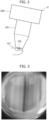
- a determination device 1 calculates an indicator value based on image data obtained by capturing an image of melanonychia 102 that has occurred on a subject's nail 101 using a dermoscope 2 as shown in Figs. 2 and 3 .
- the determination device 1 compares the indicator value with a threshold value and determines whether the melanonychia 102 is malignant or benign.
- the result of the determination performed by the determination device 1 supports a diagnosis of the melanonychia 102 performed by a user such as a physician.
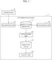
- the determination device 1 is provided with pieces of data of digital color image data 11 and an indicator value 12 and the functions of an acquisition unit 21, a conversion unit 22, a calculation unit 23, and an output unit 24.
- the pieces of data are stored in a memory 902 or a storage 903.
- the functions are implemented by a CPU 901.
- the digital color image data 11 is data of a region of interest in the subject's melanonychia 102.
- the digital color image data 11 is captured by using, for example, the dermoscope 2 with a camera function.
- the region of interest (ROI) is a portion including the subject's melanonychia 102 or a portion of the subject's melanonychia 102.
- the digital color image data 11 is data obtained by associating each pixel corresponding to the region of interest with a color value represented by the pixel and digitizing the region of interest. In the embodiment of the present invention, the digital color image data 11 is obtained by associating each pixel in the region with a color value of each of red, green, and blue (RGB).
- RGB red, green, and blue
- the indicator value 12 is calculated from variation in the RGB values of each pixel of the digital color image data 11.
- the indicator value 12 is an indicator for determining whether the region of interest is benign or malignant.
- the indicator value is also referred to as a discrimination index (DI) value.
- the acquisition unit 21 acquires the digital color image data 11 of the region of interest in the subject's melanonychia 102.
- the acquisition unit 21 acquires, from the dermoscope 2, digital-format image data obtained by capturing an image of a subject's nail 101 part.
- the acquisition unit 21 generates the digital color image data 11 by cutting out the region of interest from the acquired image data.
- the region of interest may be specified by a user or by a prescribed program.
- the conversion unit 22 adjusts the color balance of the digital color image data 11 by performing a chromatic adaptation transformation and updates the digital color image data 11.
- the conversion unit 22 adjusts the color balance, and corrects and standardizes the hue of the digital color image data 11.
- the color balance may differ due to the imaging environment using a dermoscope, the image processing engine used for the dermoscope, and the like.
- the imaging environment using the dermoscope may be influenced by the spectral distribution of illumination light, which varies depending on the device used for the dermoscopy inspection, or the like. Therefore, the conversion unit 22 eliminates the difference in the color balance caused by the imaging environment and the image processing engine in the inspection by adjusting the color balance.
- the conversion unit 22 can accurately determine the melanonychia 102 by adjusting the color balance.
- the conversion unit 22 adjusts the hue of each piece of digital color image data such that the appearance of white becomes constant in digital color image data captured by using various devices.
- the conversion unit 22 standardizes the hue of the digital color image data obtained under various conditions by converting an average chromaticity coordinate of prescribed pixels in the data to a preset reference chromaticity coordinate. The details are disclosed in Patent Literature 4. Patent Literature 4 is incorporated herein.
- the calculation unit 23 calculates the indicator value 12 from the variation in RGB values of each pixel of the digital color image data 11.
- the calculation unit 23 preferably uses digital color image data 11 of which the hue has been adjusted by the conversion unit 22.
- the calculation unit 23 calculates the indicator value 12 from variation in the R value, variation in the G value, and variation in the B value of each pixel.
- the indicator value 12 is calculated by normalizing the sum of the standard deviation of each of the R value, the G value, and the B value of each pixel of the digital color image data 11 by the number of pixels.
- the calculation unit 23 calculates the indicator value 12 from formula (1).
- the calculation unit 23 calculates a higher indicator value 12 as the diversity of the colors in the digital color image data 11 increases.
- the calculation unit 23 calculates a lower indicator value as the diversity of the colors in the digital color image data 11 decreases.
- the output unit 24 determines that the melanonychia 102 is malignant if the indicator value is higher than the threshold value, and that the melanonychia 102 is benign if the indicator value is lower than the threshold value, and outputs the determination result.
- the threshold value is, for example, 40.
- the determination result output by the output unit 24 is not the final determination made by the physician but one element for supporting the physician's determination. For example, if the indicator value 12 is higher than the threshold value, there is a high possibility that the melanonychia 102 is malignant, and therefore the output unit 24 may display a message prompting a biopsy. If the indicator value is lower than the threshold value, there is a high possibility that the melanonychia 102 is benign, and therefore the output unit 24 may display a message such as no biopsy is required or a follow-up observation is advised.
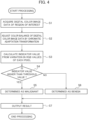
- a determination method performed by the determination device 1 according to the embodiment of the present invention will be described.
- the processing sequence shown in Fig. 4 is an example, and the sequence is not limited thereto.
- step S1 the determination device 1 acquires the digital color image data 11 of the region of interest of the subject's melanonychia 102.
- step S2 the determination device 1 adjusts the color balance of the digital color image data 11 acquired in step S1 by performing the chromatic adaptation transformation and updates the data.
- step S3 the determination device 1 calculates the indicator value from the variation in RGB values of each pixel of the digital color image data 11 of which the color balance has been adjusted in step S2.
- step S4 the determination device 1 compares the indicator value calculated in step S3 with the threshold value to determine whether the melanonychia is malignant or benign. If the indicator value is higher than the threshold value, in step S5, the determination device 1 determines that there is a high possibility that the melanonychia 102 imaged as the digital color image data 11 is malignant. If the indicator value is lower than the threshold value, in step S6, the determination device 1 determines that there is a high possibility that the melanonychia 102 imaged as the digital color image data 11 is benign.
- step S7 the determination device 1 outputs the determination result in step S5 or step S6 to an output device such as a display.
- Figs. 5 Both of Figs. 5(a) and 5(b) show receiver operating characteristic (ROC) curves in which the region of interest of the melanonychia 102 is determined.
- the vertical axis of the ROC curve is a true positive rate and the horizontal axis of the curve is a false positive rate.
- the area under the curve (AUC) is the area of the portion below the ROC curve. It is shown that the discrimination capability increases as the AUC value approaches 1.
- Fig. 5(a) shows the result of determination by means of the methods disclosed in Patent Literature 1 and Patent Literature 2.
- Fig. 5(b) shows the result of determination performed by the determination device 1 according to the embodiment of the present invention.
- the results of determination shown in Figs. 5(a) and 5(b) are obtained by performing determination for 100 cases of melanonychia. Out of the 100 cases, 80 cases are determined to be benign and 20 cases are determined to be malignant according to the physician's diagnosis. The true positive rate and false positive rate are calculated from the determination results according to each determination method and a correct label according to the physician's diagnosis result.
- the AUC is 0.711. Meanwhile, in the determination result according to the embodiment of the present invention shown in Fig. 5(b) , the AUC is 0.848, and it can be seen that the result exhibited a higher discrimination capability than that exhibited by the conventional determination result.
- the AUC of the ROC curve is 0.621 ( J.K. Winkler et al., European Journal of Cancer 127 (2020) e21-29 ). Meanwhile, in the determination result obtained by the determination device 1 according to the embodiment of the present invention, the AUC is 0.848. It can be seen that the determination device 1 has a higher discrimination capability than that exhibited by the determination result obtained by deep learning.
- the sensitivity and the specificity of the detection of malignancy (melanoma) performed by the determination device 1 are 0.85 and 0.79, respectively.
- the average sensitivity of eight dermatologists is 0.66 and the average specificity is 0.98.
- the average sensitivity of eight non-dermatologists is 0.46 and the average specificity is 0.97. This reveals that, although the specificity is higher in the determination performed by the dermatologists and non-dermatologists, the sensitivity tends to be higher in the determination performed by the determination device 1.
- the determination device 1 can appropriately determine malignancy and prompt a biopsy for the subject immediately compared with a determination made by a physician.
- the determination result by the determination device 1 exhibits a sensitivity of 0.95, a specificity of 0.82, and an AUC of 0.92.
- the determination device 1 can determine whether the melanonychia is benign or malignant at the same level as a physician who is an expert in nail melanoma.
- the determination device 1 calculates the indicator value from the variation in RGB values of each pixel of the digital color image data 11 obtained by capturing an image of the region of interest of the melanonychia 102 by using the dermoscope 2, and determines whether the melanonychia 102 is benign or malignant. According to the knowledge by the inventors, "the essence of malignant melanoma is morphological disorder including color tone", and therefore, the determination device 1 calculates an indicator value considering the diversity of color tones in the region of interest, and thereby can calculate an indicator value suitable for the determination of the melanonychia 102.
- the determination device 1 can suppress the influence on the indicator value due to differences in imaging devices, persons who capture images, imaging environments, and the like by the conversion unit 22 adjusting the color balance.
- the indicator value calculated by the determination device 1 has high robustness with respect to input images and enables objective evaluation of the melanonychia 102.
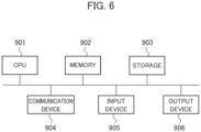
- a general-purpose computer system which includes a central processing unit (CPU, processor) 901, a memory 902, a storage 903 (HDD: hard disk drive, SSD: solid state drive), a communication device 904, an input device 905, and an output device 906.
- CPU central processing unit
- memory 902 a storage 903 (HDD: hard disk drive, SSD: solid state drive)
- storage 903 HDD: hard disk drive, SSD: solid state drive
- communication device 904 an input device 905, and an output device 906.
- each function of the determination device 1 is realized by the CPU 901 executing a program loaded into the memory 902.
- the determination device 1 may be implemented by one computer or a plurality of computers.
- the determination device 1 may be a virtual machine implemented on a computer.
- the program of the determination device 1 may be stored on a computer-readable recording medium such as an HDD, an SSD, a Universal Serial Bus (USB) memory, a compact disc (CD), or a digital versatile disc (DVD), or may be distributed via a network.
- a computer-readable recording medium such as an HDD, an SSD, a Universal Serial Bus (USB) memory, a compact disc (CD), or a digital versatile disc (DVD)
- USB Universal Serial Bus
- CD compact disc
- DVD digital versatile disc
Landscapes
- Health & Medical Sciences (AREA)
- Engineering & Computer Science (AREA)
- Life Sciences & Earth Sciences (AREA)
- Physics & Mathematics (AREA)
- Medical Informatics (AREA)
- General Health & Medical Sciences (AREA)
- General Physics & Mathematics (AREA)
- Theoretical Computer Science (AREA)
- Veterinary Medicine (AREA)
- Public Health (AREA)
- Biomedical Technology (AREA)
- Heart & Thoracic Surgery (AREA)
- Biophysics (AREA)
- Molecular Biology (AREA)
- Surgery (AREA)
- Animal Behavior & Ethology (AREA)
- Pathology (AREA)
- Computer Vision & Pattern Recognition (AREA)
- Oral & Maxillofacial Surgery (AREA)
- Dentistry (AREA)
- Nuclear Medicine, Radiotherapy & Molecular Imaging (AREA)
- Radiology & Medical Imaging (AREA)
- Quality & Reliability (AREA)
- Dermatology (AREA)
- Spectroscopy & Molecular Physics (AREA)
- Measuring And Recording Apparatus For Diagnosis (AREA)
- Image Analysis (AREA)
Abstract
Description
- The present invention relates to a determination device, a determination method, and a program.
- Under normal conditions, melanocytes present in a nail matrix are inactive and do not produce melanin. Regardless of whether the melanocytes are cancerous, the melanocytes may begin to produce melanin. As the nail grows, the produced melanin forms a pattern called melanonychia. Melanonychia when the melanocytes are not cancerous is considered to be a benign nevus. It is thought that determination as to whether the melanocytes present in the nail matrix are cancerous is possible from the melanonychia pattern.
- However, considerable experience is required to visually determine whether the pattern is benign or malignant using a dermoscope. In the case of malignant melanoma, a biopsy is generally performed. A biopsy is burdensome to a patient and does not lead to benefits for the patient. Due to these factors, it is thought that a definitive diagnosis of melanonychia is difficult and these factors result in a poor prognosis. Therefore, the realization of a non-invasive and objective method for determining melanonychia is strongly required from clinical settings.
- Therefore, the inventors have invented a non-invasive and objective method for determining whether melanonychia is benign or malignant (see, for example,
Patent Literature 1 and Patent Literature 2). InPatent Literature 1, RGB parameter values of each pixel in a digital color image of the melanonychia are regarded as a three-dimensional vector having those three values as components, and a threshold value is obtained from an appearance probability of each angle formed between each three-dimensional vector and a reference vector. InPatent Literature 2, a latitudinal variable and a longitudinal variable in RGB space of a three-dimensional vector are obtained, and indicator values are obtained from the distribution of points defined based on the obtained latitudinal variable and longitudinal variable. InPatent Literature 2, the indicator values are further distinguished based on a threshold value to determine whether melanonychia is malignant or benign. - In the methods disclosed in
Patent Literatures - In addition, the inventors invented a method of analyzing the melanonychia or the like for a diagnosis of a skin disease (see, for example, Patent Literature 4).
Patent Literature 4 discloses that the color balance of a color digital image of subject's melanonychia or the like is adjusted by performing a chromatic adaptation transformation.Patent Literature 4 makes it possible to obtain a color digital image without depending on the person carrying out the inspection or an imaging device by correcting the hue of an image in a dermoscopy inspection. -
- PTL 1:
JP 2010-252895 A - PTL 2:
JP 2011-130897 A - PTL 3:
JP 2016-174671 A - PTL 4:
WO 2018/216680 - There is a demand for development of a method of more quickly and appropriately determining whether melanonychia is benign or malignant. In the methods disclosed in
Patent Literatures - The present invention has been devised in consideration of the above problems, and an object of the present invention is to provide a technique capable of more quickly and appropriately determining whether melanonychia is benign or malignant.
- A determination device of one aspect of the present invention includes: an acquisition unit that acquires digital color image data of a region of interest in melanonychia of a subject; a calculation unit that calculates an indicator value from variation in RGB values of each pixel of the digital color image data; and an output unit that outputs a result of determining that the melanonychia is malignant if the indicator value is higher than a threshold value and determining that the melanonychia is benign if the indicator value is lower than the threshold value.
- A determination method of one aspect of the present invention includes: a computer acquiring digital color image data of a region of interest in melanonychia of a subject; the computer calculating an indicator value from variation in RGB values of each pixel of the digital color image data; and the computer outputting a result of determining that the melanonychia is malignant if the indicator value is higher than a threshold value and determining that the melanonychia is benign if the indicator value is lower than the threshold value.
- One aspect of the present invention provides a program for causing a computer to function as the determination device.
- According to the present invention, it is possible to provide a technique capable of more quickly and appropriately determining whether melanonychia is benign or malignant.
-
- [
Fig. 1] Fig. 1 is a diagram for explaining functional blocks of a determination device according to an embodiment of the present invention. - [
Fig. 2] Fig. 2 is a diagram for explaining how an image of melanonychia is captured using a dermoscope. - [
Fig. 3] Fig. 3 shows an example of melanonychia. - [
Fig. 4] Fig. 4 is a flowchart for explaining determination processing performed by a determination device. - [
Fig. 5] Fig. 5 is a diagram for explaining a result of determination performed by the determination device according to the embodiment of the present invention. - [
Fig. 6] Fig. 6 is a diagram for explaining a hardware configuration of a computer used for a determination device. - An embodiment of the present invention will be described below with reference to the drawings. In the description of the drawings, the same parts are denoted with the same reference numerals, and the description thereof is omitted.
- A
determination device 1 according to an embodiment of the present invention shown inFig. 1 calculates an indicator value based on image data obtained by capturing an image ofmelanonychia 102 that has occurred on a subject'snail 101 using adermoscope 2 as shown inFigs. 2 and 3 . Thedetermination device 1 compares the indicator value with a threshold value and determines whether themelanonychia 102 is malignant or benign. The result of the determination performed by thedetermination device 1 supports a diagnosis of themelanonychia 102 performed by a user such as a physician. - The
determination device 1 is provided with pieces of data of digitalcolor image data 11 and anindicator value 12 and the functions of anacquisition unit 21, aconversion unit 22, acalculation unit 23, and anoutput unit 24. The pieces of data are stored in amemory 902 or astorage 903. The functions are implemented by aCPU 901. - The digital
color image data 11 is data of a region of interest in the subject'smelanonychia 102. The digitalcolor image data 11 is captured by using, for example, thedermoscope 2 with a camera function. The region of interest (ROI) is a portion including the subject'smelanonychia 102 or a portion of the subject'smelanonychia 102. The digitalcolor image data 11 is data obtained by associating each pixel corresponding to the region of interest with a color value represented by the pixel and digitizing the region of interest. In the embodiment of the present invention, the digitalcolor image data 11 is obtained by associating each pixel in the region with a color value of each of red, green, and blue (RGB). - The
indicator value 12 is calculated from variation in the RGB values of each pixel of the digitalcolor image data 11. Theindicator value 12 is an indicator for determining whether the region of interest is benign or malignant. The indicator value is also referred to as a discrimination index (DI) value. - The
acquisition unit 21 acquires the digitalcolor image data 11 of the region of interest in the subject'smelanonychia 102. Theacquisition unit 21 acquires, from thedermoscope 2, digital-format image data obtained by capturing an image of a subject'snail 101 part. Theacquisition unit 21 generates the digitalcolor image data 11 by cutting out the region of interest from the acquired image data. The region of interest may be specified by a user or by a prescribed program. - The
conversion unit 22 adjusts the color balance of the digitalcolor image data 11 by performing a chromatic adaptation transformation and updates the digitalcolor image data 11. Theconversion unit 22 adjusts the color balance, and corrects and standardizes the hue of the digitalcolor image data 11. - Generally, in a dermoscopy inspection, the color balance may differ due to the imaging environment using a dermoscope, the image processing engine used for the dermoscope, and the like. For example, the imaging environment using the dermoscope may be influenced by the spectral distribution of illumination light, which varies depending on the device used for the dermoscopy inspection, or the like. Therefore, the
conversion unit 22 eliminates the difference in the color balance caused by the imaging environment and the image processing engine in the inspection by adjusting the color balance. Theconversion unit 22 can accurately determine themelanonychia 102 by adjusting the color balance. - The
conversion unit 22 adjusts the hue of each piece of digital color image data such that the appearance of white becomes constant in digital color image data captured by using various devices. Theconversion unit 22 standardizes the hue of the digital color image data obtained under various conditions by converting an average chromaticity coordinate of prescribed pixels in the data to a preset reference chromaticity coordinate. The details are disclosed inPatent Literature 4.Patent Literature 4 is incorporated herein. - The
calculation unit 23 calculates theindicator value 12 from the variation in RGB values of each pixel of the digitalcolor image data 11. Thecalculation unit 23 preferably uses digitalcolor image data 11 of which the hue has been adjusted by theconversion unit 22. - The
calculation unit 23 calculates theindicator value 12 from variation in the R value, variation in the G value, and variation in the B value of each pixel. Theindicator value 12 is calculated by normalizing the sum of the standard deviation of each of the R value, the G value, and the B value of each pixel of the digitalcolor image data 11 by the number of pixels. - The
calculation unit 23 calculates theindicator value 12 from formula (1).
[Math. 1]
- DI:
- Indicator value
- N:
- Number of pixels in digital color image data
- Ri, Gi, Bi:
- Values of each of RGB of pixel i
- R, G, B:
- Average value of values of each of RGB of digital color image data
- The
calculation unit 23 calculates ahigher indicator value 12 as the diversity of the colors in the digitalcolor image data 11 increases. Thecalculation unit 23 calculates a lower indicator value as the diversity of the colors in the digitalcolor image data 11 decreases. - The
output unit 24 determines that themelanonychia 102 is malignant if the indicator value is higher than the threshold value, and that themelanonychia 102 is benign if the indicator value is lower than the threshold value, and outputs the determination result. When each value of RGB is represented in 8 bits and has a range of 0 to 255, the threshold value is, for example, 40. - The determination result output by the
output unit 24 is not the final determination made by the physician but one element for supporting the physician's determination. For example, if theindicator value 12 is higher than the threshold value, there is a high possibility that themelanonychia 102 is malignant, and therefore theoutput unit 24 may display a message prompting a biopsy. If the indicator value is lower than the threshold value, there is a high possibility that themelanonychia 102 is benign, and therefore theoutput unit 24 may display a message such as no biopsy is required or a follow-up observation is advised. - With reference to
Fig. 4 , a determination method performed by thedetermination device 1 according to the embodiment of the present invention will be described. The processing sequence shown inFig. 4 is an example, and the sequence is not limited thereto. - In step S1, the
determination device 1 acquires the digitalcolor image data 11 of the region of interest of the subject'smelanonychia 102. In step S2, thedetermination device 1 adjusts the color balance of the digitalcolor image data 11 acquired in step S1 by performing the chromatic adaptation transformation and updates the data. - In step S3, the
determination device 1 calculates the indicator value from the variation in RGB values of each pixel of the digitalcolor image data 11 of which the color balance has been adjusted in step S2. - In step S4, the
determination device 1 compares the indicator value calculated in step S3 with the threshold value to determine whether the melanonychia is malignant or benign. If the indicator value is higher than the threshold value, in step S5, thedetermination device 1 determines that there is a high possibility that themelanonychia 102 imaged as the digitalcolor image data 11 is malignant. If the indicator value is lower than the threshold value, in step S6, thedetermination device 1 determines that there is a high possibility that themelanonychia 102 imaged as the digitalcolor image data 11 is benign. - In step S7, the
determination device 1 outputs the determination result in step S5 or step S6 to an output device such as a display. - The results of determination performed by the
determination device 1 according to the embodiment of the present invention will be described with reference toFig. 5 . Both ofFigs. 5(a) and 5(b) show receiver operating characteristic (ROC) curves in which the region of interest of themelanonychia 102 is determined. The vertical axis of the ROC curve is a true positive rate and the horizontal axis of the curve is a false positive rate. The area under the curve (AUC) is the area of the portion below the ROC curve. It is shown that the discrimination capability increases as the AUC value approaches 1. -
Fig. 5(a) shows the result of determination by means of the methods disclosed inPatent Literature 1 andPatent Literature 2.Fig. 5(b) shows the result of determination performed by thedetermination device 1 according to the embodiment of the present invention. The results of determination shown inFigs. 5(a) and 5(b) are obtained by performing determination for 100 cases of melanonychia. Out of the 100 cases, 80 cases are determined to be benign and 20 cases are determined to be malignant according to the physician's diagnosis. The true positive rate and false positive rate are calculated from the determination results according to each determination method and a correct label according to the physician's diagnosis result. - In the conventional determination result shown in
Fig. 5(a) , the AUC is 0.711. Meanwhile, in the determination result according to the embodiment of the present invention shown inFig. 5(b) , the AUC is 0.848, and it can be seen that the result exhibited a higher discrimination capability than that exhibited by the conventional determination result. - In a determination result using deep learning by means of a convolutional neural network, the AUC of the ROC curve is 0.621 (J.K. Winkler et al., European Journal of Cancer 127 (2020) e21-29). Meanwhile, in the determination result obtained by the
determination device 1 according to the embodiment of the present invention, the AUC is 0.848. It can be seen that thedetermination device 1 has a higher discrimination capability than that exhibited by the determination result obtained by deep learning. - Further, the sensitivity and the specificity of the detection of malignancy (melanoma) performed by the
determination device 1 according to the embodiment of the present invention are 0.85 and 0.79, respectively. In contrast, the average sensitivity of eight dermatologists is 0.66 and the average specificity is 0.98. The average sensitivity of eight non-dermatologists is 0.46 and the average specificity is 0.97. This reveals that, although the specificity is higher in the determination performed by the dermatologists and non-dermatologists, the sensitivity tends to be higher in the determination performed by thedetermination device 1. Thedetermination device 1 can appropriately determine malignancy and prompt a biopsy for the subject immediately compared with a determination made by a physician. - When the determination as to whether a biopsy is necessary performed by means of the majority decision of three experts with a wealth of experience in treating nail melanoma is assumed to be the correct label, the determination result by the
determination device 1 exhibits a sensitivity of 0.95, a specificity of 0.82, and an AUC of 0.92. Thedetermination device 1 can determine whether the melanonychia is benign or malignant at the same level as a physician who is an expert in nail melanoma. - The
determination device 1 according to the embodiment of the present invention calculates the indicator value from the variation in RGB values of each pixel of the digitalcolor image data 11 obtained by capturing an image of the region of interest of themelanonychia 102 by using thedermoscope 2, and determines whether themelanonychia 102 is benign or malignant. According to the knowledge by the inventors, "the essence of malignant melanoma is morphological disorder including color tone", and therefore, thedetermination device 1 calculates an indicator value considering the diversity of color tones in the region of interest, and thereby can calculate an indicator value suitable for the determination of themelanonychia 102. - In addition, the
determination device 1 can suppress the influence on the indicator value due to differences in imaging devices, persons who capture images, imaging environments, and the like by theconversion unit 22 adjusting the color balance. The indicator value calculated by thedetermination device 1 has high robustness with respect to input images and enables objective evaluation of themelanonychia 102. - For the
determination device 1 of the present embodiment described above, for example, a general-purpose computer system is used which includes a central processing unit (CPU, processor) 901, amemory 902, a storage 903 (HDD: hard disk drive, SSD: solid state drive), acommunication device 904, aninput device 905, and anoutput device 906. In this computer system, each function of thedetermination device 1 is realized by theCPU 901 executing a program loaded into thememory 902. - The
determination device 1 may be implemented by one computer or a plurality of computers. Thedetermination device 1 may be a virtual machine implemented on a computer. - The program of the
determination device 1 may be stored on a computer-readable recording medium such as an HDD, an SSD, a Universal Serial Bus (USB) memory, a compact disc (CD), or a digital versatile disc (DVD), or may be distributed via a network. - It should be noted that the present invention is not limited to the above embodiment, and various modifications can be made within the scope of the invention.
-
- 1
- Determination device
- 2
- Dermoscope
- 11
- Digital color image data
- 12
- Indicator value
- 21
- Acquisition unit
- 22
- Conversion unit
- 23
- Calculation unit
- 24
- Output unit
- 901
- CPU
- 902
- Memory
- 903
- Storage
- 904
- Communication device
- 905
- Input device
- 906
- Output device
Claims (6)
- A determination device comprising:an acquisition unit that acquires digital color image data of a region of interest in melanonychia of a subject;a calculation unit that calculates an indicator value from variation in RGB values of each pixel of the digital color image data; andan output unit that outputs a result of determining that the melanonychia is malignant if the indicator value is higher than a threshold value and determining that the melanonychia is benign if the indicator value is lower than the threshold value.
- The determination device according to claim 1, further comprising:a conversion unit that adjusts a color balance of the digital color image data by performing a chromatic adaptation transformation, whereinthe acquisition unit calculates the indicator value from the digital color image data after the chromatic adaptation transformation of the region of interest.
- The determination device according to claim 1 or 2, wherein
the calculation unit calculates the indicator value from variation of the R value, variation of the G value, and variation of the B value of each pixel. - The determination device according to claim 1 or 2, wherein
the calculation unit calculates the indicator value from formula (1).
[Math. 1] DI: Indicator valueN: Number of pixels in digital color image dataRi, Gi , Bi : Values of each of RGB of pixel i
R ,G ,B : Average value of values of each of RGB of digital color image data - A determination method comprising:a computer acquiring digital color image data of a region of interest in melanonychia of a subject;the computer calculating an indicator value from variation in RGB values of each pixel of the digital color image data; andthe computer outputting a result of determining that the melanonychia is malignant if the indicator value is higher than a threshold value and determining that the melanonychia is benign if the indicator value is lower than the threshold value.
- A program for causing a computer to function as the determination device according to any one of claims 1 to 4.
Applications Claiming Priority (1)
| Application Number | Priority Date | Filing Date | Title |
|---|---|---|---|
| PCT/JP2021/021411 WO2022254707A1 (en) | 2021-06-04 | 2021-06-04 | Determination device, determination method, and program |
Publications (2)
| Publication Number | Publication Date |
|---|---|
| EP4349268A1 true EP4349268A1 (en) | 2024-04-10 |
| EP4349268A4 EP4349268A4 (en) | 2025-04-09 |
Family
ID=84322917
Family Applications (1)
| Application Number | Title | Priority Date | Filing Date |
|---|---|---|---|
| EP21944201.9A Pending EP4349268A4 (en) | 2021-06-04 | 2021-06-04 | DETERMINATION DEVICE, DETERMINATION METHOD AND PROGRAM |
Country Status (5)
| Country | Link |
|---|---|
| US (1) | US20240245347A1 (en) |
| EP (1) | EP4349268A4 (en) |
| JP (1) | JP7667984B2 (en) |
| AU (1) | AU2021448510B2 (en) |
| WO (1) | WO2022254707A1 (en) |
Family Cites Families (7)
| Publication number | Priority date | Publication date | Assignee | Title |
|---|---|---|---|---|
| JP5565765B2 (en) | 2009-04-22 | 2014-08-06 | 学校法人早稲田大学 | Derivation method of melanoma discrimination index |
| JP5253284B2 (en) * | 2009-04-22 | 2013-07-31 | 三鷹光器株式会社 | Derivation method of threshold value for nail plate pigment line discrimination |
| JP5600426B2 (en) * | 2009-12-24 | 2014-10-01 | 三鷹光器株式会社 | Nail plate pigment line discrimination device |
| JP6467669B2 (en) * | 2015-03-19 | 2019-02-13 | 株式会社ヒューマン・エンジニアリング | Determination apparatus and determination program |
| JP6793325B2 (en) * | 2016-05-25 | 2020-12-02 | パナソニックIpマネジメント株式会社 | Skin diagnostic device and skin diagnostic method |
| JP7267545B2 (en) * | 2017-05-25 | 2023-05-02 | 学校法人早稲田大学 | ANALYSIS METHOD, DIAGNOSTIC APPARATUS AND COMPUTER PROGRAM OF NAIL PLATE STRAILS OR SKIN HOLES FOR DIAGNOSIS OF SKIN DISEASES |
| JP2019208900A (en) | 2018-06-05 | 2019-12-12 | 株式会社ニコン | Analysis device, analyzing device, analysis method, and program |
-
2021
- 2021-06-04 US US18/566,351 patent/US20240245347A1/en active Pending
- 2021-06-04 JP JP2023525325A patent/JP7667984B2/en active Active
- 2021-06-04 WO PCT/JP2021/021411 patent/WO2022254707A1/en not_active Ceased
- 2021-06-04 AU AU2021448510A patent/AU2021448510B2/en active Active
- 2021-06-04 EP EP21944201.9A patent/EP4349268A4/en active Pending
Also Published As
| Publication number | Publication date |
|---|---|
| US20240245347A1 (en) | 2024-07-25 |
| EP4349268A4 (en) | 2025-04-09 |
| WO2022254707A1 (en) | 2022-12-08 |
| AU2021448510A1 (en) | 2024-01-04 |
| AU2021448510B2 (en) | 2025-06-26 |
| JP7667984B2 (en) | 2025-04-24 |
| JPWO2022254707A1 (en) | 2022-12-08 |
Similar Documents
| Publication | Publication Date | Title |
|---|---|---|
| KR102237583B1 (en) | Estimating bilirubin levels | |
| US9629525B2 (en) | Image processing device, image processing method, and program | |
| KR101301821B1 (en) | Apparatus and method for detecting complexion, apparatus and method for determinig health using complexion, apparatus and method for generating health sort function | |
| JP5597049B2 (en) | Image processing apparatus, image processing method, and image processing program | |
| KR100931752B1 (en) | Recording medium recording pupil color correction device and program | |
| KR20150072463A (en) | Health state determining method and health state determining apparatus using image of face | |
| JP2011218090A (en) | Image processor, endoscope system, and program | |
| WO1997047235A1 (en) | Dermal diagnostic analysis system and method | |
| EP4198886B1 (en) | Feature amount acquisition device, similar image search device, display device, feature amount acquisition method, similar image search method, display method, and program | |
| JP7234086B2 (en) | Biometric information acquisition device and program | |
| JP7267545B2 (en) | ANALYSIS METHOD, DIAGNOSTIC APPARATUS AND COMPUTER PROGRAM OF NAIL PLATE STRAILS OR SKIN HOLES FOR DIAGNOSIS OF SKIN DISEASES | |
| JP7087390B2 (en) | Diagnostic support device, image processing method and program | |
| JP5253284B2 (en) | Derivation method of threshold value for nail plate pigment line discrimination | |
| US20160100789A1 (en) | Computer-aided diagnosis system and computer-aided diagnosis method | |
| US20240245347A1 (en) | Determination device, determination method, and program | |
| JP6467669B2 (en) | Determination apparatus and determination program | |
| AU2024344610A1 (en) | Method of bioimage analysis for the detection of color abnormalities through color distribution assessment | |
| US20230386660A1 (en) | System and method for detecting gastrointestinal disorders | |
| CN116491892B (en) | Myopia fundus change assessment method and device and electronic equipment | |
| Cree | Automated microaneurysm detection for screening | |
| Chan et al. | Using Computer Vision and Artificial Intelligence to Track the Healing of Severe Burns | |
| JP2024098259A (en) | Organ evaluation system and organ evaluation method | |
| JP2006146290A (en) | Image correction device and method, and image correction program |
Legal Events
| Date | Code | Title | Description |
|---|---|---|---|
| STAA | Information on the status of an ep patent application or granted ep patent |
Free format text: STATUS: THE INTERNATIONAL PUBLICATION HAS BEEN MADE |
|
| PUAI | Public reference made under article 153(3) epc to a published international application that has entered the european phase |
Free format text: ORIGINAL CODE: 0009012 |
|
| STAA | Information on the status of an ep patent application or granted ep patent |
Free format text: STATUS: REQUEST FOR EXAMINATION WAS MADE |
|
| 17P | Request for examination filed |
Effective date: 20231206 |
|
| AK | Designated contracting states |
Kind code of ref document: A1 Designated state(s): AL AT BE BG CH CY CZ DE DK EE ES FI FR GB GR HR HU IE IS IT LI LT LU LV MC MK MT NL NO PL PT RO RS SE SI SK SM TR |
|
| DAV | Request for validation of the european patent (deleted) | ||
| DAX | Request for extension of the european patent (deleted) | ||
| A4 | Supplementary search report drawn up and despatched |
Effective date: 20250306 |
|
| RIC1 | Information provided on ipc code assigned before grant |
Ipc: A61B 5/00 20060101ALI20250228BHEP Ipc: A61B 10/00 20060101AFI20250228BHEP |
|
| STAA | Information on the status of an ep patent application or granted ep patent |
Free format text: STATUS: EXAMINATION IS IN PROGRESS |
|
| 17Q | First examination report despatched |
Effective date: 20251107 |
|
| REG | Reference to a national code |
Ref country code: DE Ref legal event code: R079 Free format text: PREVIOUS MAIN CLASS: A61B0010000000 Ipc: G06T0007000000 |
|
| RAP1 | Party data changed (applicant data changed or rights of an application transferred) |
Owner name: Q'SFIX CO., LTD. Owner name: WASEDA UNIVERSITY Owner name: SHINSHU UNIVERSITY |
|
| GRAP | Despatch of communication of intention to grant a patent |
Free format text: ORIGINAL CODE: EPIDOSNIGR1 |
|
| STAA | Information on the status of an ep patent application or granted ep patent |
Free format text: STATUS: GRANT OF PATENT IS INTENDED |
|
| RIC1 | Information provided on ipc code assigned before grant |
Ipc: G06T 7/00 20170101AFI20260327BHEP |